US6132214A - Medical implant - Google Patents
Medical implantDownload PDFInfo
- Publication number
- US6132214A US6132214AUS09/091,674US9167498AUS6132214AUS 6132214 AUS6132214 AUS 6132214AUS 9167498 AUS9167498 AUS 9167498AUS 6132214 AUS6132214 AUS 6132214A
- Authority
- US
- United States
- Prior art keywords
- implant
- cast cavity
- mandrels
- biologically active
- cavity
- Prior art date
- Legal status (The legal status is an assumption and is not a legal conclusion. Google has not performed a legal analysis and makes no representation as to the accuracy of the status listed.)
- Expired - Lifetime
Links
- 239000007943implantSubstances0.000titleclaimsabstractdescription118
- 239000013543active substanceSubstances0.000claimsabstractdescription23
- 238000000034methodMethods0.000claimsabstractdescription17
- 229940088623biologically active substanceDrugs0.000claimsabstractdescription14
- 239000000463materialSubstances0.000claimsdescription104
- 210000000988bone and boneAnatomy0.000claimsdescription30
- 229920003023plasticPolymers0.000claimsdescription21
- 239000004033plasticSubstances0.000claimsdescription21
- 229910019142PO4Inorganic materials0.000claimsdescription15
- 239000011575calciumSubstances0.000claimsdescription15
- 239000011159matrix materialSubstances0.000claimsdescription14
- 102000004887Transforming Growth Factor betaHuman genes0.000claimsdescription12
- 108090001012Transforming Growth Factor betaProteins0.000claimsdescription12
- 230000015572biosynthetic processEffects0.000claimsdescription12
- ZRKFYGHZFMAOKI-QMGMOQQFSA-NtgfbetaChemical compoundC([C@H](NC(=O)[C@H](C(C)C)NC(=O)CNC(=O)[C@H](CCC(O)=O)NC(=O)[C@H](CCCNC(N)=N)NC(=O)[C@H](CC(N)=O)NC(=O)[C@H](CC(C)C)NC(=O)[C@H]([C@@H](C)O)NC(=O)[C@H](CCC(O)=O)NC(=O)[C@H]([C@@H](C)O)NC(=O)[C@H](CC(C)C)NC(=O)CNC(=O)[C@H](C)NC(=O)[C@H](CO)NC(=O)[C@H](CCC(N)=O)NC(=O)[C@@H](NC(=O)[C@H](C)NC(=O)[C@H](C)NC(=O)[C@@H](NC(=O)[C@H](CC(C)C)NC(=O)[C@@H](N)CCSC)C(C)C)[C@@H](C)CC)C(=O)N[C@@H]([C@@H](C)O)C(=O)N[C@@H](C(C)C)C(=O)N[C@@H](CC=1C=CC=CC=1)C(=O)N[C@@H](C)C(=O)N1[C@@H](CCC1)C(=O)N[C@@H]([C@@H](C)O)C(=O)N[C@@H](CC(N)=O)C(=O)N[C@@H](CCC(O)=O)C(=O)N[C@@H](C)C(=O)N[C@@H](CC=1C=CC=CC=1)C(=O)N[C@@H](CCCNC(N)=N)C(=O)N[C@@H](C)C(=O)N[C@@H](CC(C)C)C(=O)N1[C@@H](CCC1)C(=O)N1[C@@H](CCC1)C(=O)N[C@@H](CCCNC(N)=N)C(=O)N[C@@H](CCC(O)=O)C(=O)N[C@@H](CCCNC(N)=N)C(=O)N[C@@H](CO)C(=O)N[C@@H](CCCNC(N)=N)C(=O)N[C@@H](CC(C)C)C(=O)N[C@@H](CC(C)C)C(O)=O)C1=CC=C(O)C=C1ZRKFYGHZFMAOKI-QMGMOQQFSA-N0.000claimsdescription12
- 230000008569processEffects0.000claimsdescription11
- 108090000623proteins and genesProteins0.000claimsdescription11
- NBIIXXVUZAFLBC-UHFFFAOYSA-KphosphateChemical compound[O-]P([O-])([O-])=ONBIIXXVUZAFLBC-UHFFFAOYSA-K0.000claimsdescription10
- 239000010452phosphateSubstances0.000claimsdescription10
- 102000004169proteins and genesHuman genes0.000claimsdescription10
- OYPRJOBELJOOCE-UHFFFAOYSA-NCalciumChemical compound[Ca]OYPRJOBELJOOCE-UHFFFAOYSA-N0.000claimsdescription9
- 229910052791calciumInorganic materials0.000claimsdescription9
- 238000011049fillingMethods0.000claimsdescription9
- 229910052500inorganic mineralInorganic materials0.000claimsdescription8
- 239000011707mineralSubstances0.000claimsdescription8
- 230000000278osteoconductive effectEffects0.000claimsdescription7
- 108010009583Transforming Growth FactorsProteins0.000claimsdescription3
- 102000009618Transforming Growth FactorsHuman genes0.000claimsdescription3
- 238000004519manufacturing processMethods0.000claimsdescription3
- 238000010137moulding (plastic)Methods0.000claimsdescription2
- 239000012778molding materialSubstances0.000claims3
- 239000004480active ingredientSubstances0.000claims1
- 229920001940conductive polymerPolymers0.000claims1
- 230000002138osteoinductive effectEffects0.000claims1
- 238000000605extractionMethods0.000abstractdescription19
- 238000002513implantationMethods0.000abstractdescription4
- 230000001225therapeutic effectEffects0.000abstractdescription4
- 230000008901benefitEffects0.000abstractdescription3
- 239000004053dental implantSubstances0.000abstractdescription3
- 238000000465mouldingMethods0.000description28
- 239000000126substanceSubstances0.000description12
- -1e.g.Polymers0.000description11
- 229920001577copolymerPolymers0.000description9
- 238000013461designMethods0.000description9
- 239000004626polylactic acidSubstances0.000description9
- 229920000747poly(lactic acid)Polymers0.000description8
- 229920000954PolyglycolidePolymers0.000description7
- 229960005069calciumDrugs0.000description7
- 239000004633polyglycolic acidSubstances0.000description7
- JVTAAEKCZFNVCJ-UHFFFAOYSA-MLactateChemical compoundCC(O)C([O-])=OJVTAAEKCZFNVCJ-UHFFFAOYSA-M0.000description6
- OKKJLVBELUTLKV-UHFFFAOYSA-NMethanolChemical compoundOCOKKJLVBELUTLKV-UHFFFAOYSA-N0.000description6
- 239000012634fragmentSubstances0.000description6
- 239000003102growth factorSubstances0.000description6
- 238000003780insertionMethods0.000description6
- 230000037431insertionEffects0.000description6
- 235000010755mineralNutrition0.000description6
- 239000000243solutionSubstances0.000description6
- 210000002808connective tissueAnatomy0.000description5
- 230000000694effectsEffects0.000description5
- 239000000203mixtureSubstances0.000description5
- HEDRZPFGACZZDS-UHFFFAOYSA-NChloroformChemical compoundClC(Cl)ClHEDRZPFGACZZDS-UHFFFAOYSA-N0.000description4
- LFQSCWFLJHTTHZ-UHFFFAOYSA-NEthanolChemical compoundCCOLFQSCWFLJHTTHZ-UHFFFAOYSA-N0.000description4
- JVTAAEKCZFNVCJ-REOHCLBHSA-NL-lactic acidChemical compoundC[C@H](O)C(O)=OJVTAAEKCZFNVCJ-REOHCLBHSA-N0.000description4
- 206010028980NeoplasmDiseases0.000description4
- 108700020797Parathyroid Hormone-RelatedProteins0.000description4
- 102000043299Parathyroid hormone-relatedHuman genes0.000description4
- 206010052428WoundDiseases0.000description4
- 208000027418Wounds and injuryDiseases0.000description4
- 230000035876healingEffects0.000description4
- 239000011796hollow space materialSubstances0.000description4
- 239000003112inhibitorSubstances0.000description4
- 238000012856packingMethods0.000description4
- XLYOFNOQVPJJNP-UHFFFAOYSA-NwaterSubstancesOXLYOFNOQVPJJNP-UHFFFAOYSA-N0.000description4
- 102000003982Parathyroid hormoneHuman genes0.000description3
- 108090000445Parathyroid hormoneProteins0.000description3
- 229920000331PolyhydroxybutyratePolymers0.000description3
- 210000004027cellAnatomy0.000description3
- 239000012141concentrateSubstances0.000description3
- 210000004268dentinAnatomy0.000description3
- 239000012153distilled waterSubstances0.000description3
- 239000000284extractSubstances0.000description3
- 229910052588hydroxylapatiteInorganic materials0.000description3
- 229940116871l-lactateDrugs0.000description3
- 238000010883osseointegrationMethods0.000description3
- 239000000199parathyroid hormoneSubstances0.000description3
- 229960001319parathyroid hormoneDrugs0.000description3
- XYJRXVWERLGGKC-UHFFFAOYSA-Dpentacalcium;hydroxide;triphosphateChemical compound[OH-].[Ca+2].[Ca+2].[Ca+2].[Ca+2].[Ca+2].[O-]P([O-])([O-])=O.[O-]P([O-])([O-])=O.[O-]P([O-])([O-])=OXYJRXVWERLGGKC-UHFFFAOYSA-D0.000description3
- 239000005015poly(hydroxybutyrate)Substances0.000description3
- 229920001282polysaccharidePolymers0.000description3
- QORWJWZARLRLPR-UHFFFAOYSA-Htricalcium bis(phosphate)Chemical compound[Ca+2].[Ca+2].[Ca+2].[O-]P([O-])([O-])=O.[O-]P([O-])([O-])=OQORWJWZARLRLPR-UHFFFAOYSA-H0.000description3
- JVTAAEKCZFNVCJ-UWTATZPHSA-M(R)-lactateChemical compoundC[C@@H](O)C([O-])=OJVTAAEKCZFNVCJ-UWTATZPHSA-M0.000description2
- 241000894006BacteriaSpecies0.000description2
- 108010049951Bone Morphogenetic Protein 3Proteins0.000description2
- 108010049870Bone Morphogenetic Protein 7Proteins0.000description2
- 206010065687Bone lossDiseases0.000description2
- 102100024504Bone morphogenetic protein 3Human genes0.000description2
- 102100022544Bone morphogenetic protein 7Human genes0.000description2
- VTYYLEPIZMXCLO-UHFFFAOYSA-LCalcium carbonateChemical compound[Ca+2].[O-]C([O-])=OVTYYLEPIZMXCLO-UHFFFAOYSA-L0.000description2
- 102000018233Fibroblast Growth FactorHuman genes0.000description2
- 108050007372Fibroblast Growth FactorProteins0.000description2
- 108090000723Insulin-Like Growth Factor IProteins0.000description2
- 108010025020Nerve Growth FactorProteins0.000description2
- 102000015336Nerve Growth FactorHuman genes0.000description2
- 108010038512Platelet-Derived Growth FactorProteins0.000description2
- 102000010780Platelet-Derived Growth FactorHuman genes0.000description2
- 239000002202Polyethylene glycolSubstances0.000description2
- 229920001710PolyorthoesterPolymers0.000description2
- 239000004372Polyvinyl alcoholSubstances0.000description2
- 102000056172Transforming growth factor beta-3Human genes0.000description2
- 108090000097Transforming growth factor beta-3Proteins0.000description2
- 239000002253acidSubstances0.000description2
- 239000000654additiveSubstances0.000description2
- 229920003232aliphatic polyesterPolymers0.000description2
- 238000006243chemical reactionMethods0.000description2
- 150000001875compoundsChemical class0.000description2
- 238000010276constructionMethods0.000description2
- 230000007547defectEffects0.000description2
- 238000010586diagramMethods0.000description2
- JBKVHLHDHHXQEQ-UHFFFAOYSA-Nepsilon-caprolactamChemical compoundO=C1CCCCCN1JBKVHLHDHHXQEQ-UHFFFAOYSA-N0.000description2
- 229940126864fibroblast growth factorDrugs0.000description2
- 210000004195gingivaAnatomy0.000description2
- 150000004676glycansChemical class0.000description2
- 230000012010growthEffects0.000description2
- 150000002632lipidsChemical class0.000description2
- 239000007788liquidSubstances0.000description2
- KWGKDLIKAYFUFQ-UHFFFAOYSA-Mlithium chlorideChemical compound[Li+].[Cl-]KWGKDLIKAYFUFQ-UHFFFAOYSA-M0.000description2
- 238000012986modificationMethods0.000description2
- 230000004048modificationEffects0.000description2
- 239000005445natural materialSubstances0.000description2
- 229940053128nerve growth factorDrugs0.000description2
- 239000003921oilSubstances0.000description2
- 210000002997osteoclastAnatomy0.000description2
- 239000005014poly(hydroxyalkanoate)Substances0.000description2
- 229920000903polyhydroxyalkanoatePolymers0.000description2
- 229920000642polymerPolymers0.000description2
- 239000002861polymer materialSubstances0.000description2
- 239000005017polysaccharideSubstances0.000description2
- 229920002635polyurethanePolymers0.000description2
- 239000004814polyurethaneSubstances0.000description2
- 229920002451polyvinyl alcoholPolymers0.000description2
- 108090000765processed proteins & peptidesProteins0.000description2
- 102000005962receptorsHuman genes0.000description2
- 108020003175receptorsProteins0.000description2
- 238000002271resectionMethods0.000description2
- 238000001356surgical procedureMethods0.000description2
- 229920001059synthetic polymerPolymers0.000description2
- 210000001519tissueAnatomy0.000description2
- 230000001131transforming effectEffects0.000description2
- VBEQCZHXXJYVRD-GACYYNSASA-NuroantheloneChemical compoundC([C@@H](C(=O)N[C@H](C(=O)N[C@@H](CS)C(=O)N[C@@H](CC(N)=O)C(=O)N[C@@H](CS)C(=O)N[C@H](C(=O)N[C@@H]([C@@H](C)CC)C(=O)NCC(=O)N[C@@H](CC=1C=CC(O)=CC=1)C(=O)N[C@@H](CO)C(=O)NCC(=O)N[C@@H](CC(O)=O)C(=O)N[C@@H](CCCNC(N)=N)C(=O)N[C@@H](CS)C(=O)N[C@@H](CCC(N)=O)C(=O)N[C@@H]([C@@H](C)O)C(=O)N[C@@H](CCCNC(N)=N)C(=O)N[C@@H](CC(O)=O)C(=O)N[C@@H](CC(C)C)C(=O)N[C@@H](CCCNC(N)=N)C(=O)N[C@@H](CC=1C2=CC=CC=C2NC=1)C(=O)N[C@@H](CC=1C2=CC=CC=C2NC=1)C(=O)N[C@@H](CCC(O)=O)C(=O)N[C@@H](CC(C)C)C(=O)N[C@@H](CCCNC(N)=N)C(O)=O)C(C)C)[C@@H](C)O)NC(=O)[C@H](CO)NC(=O)[C@H](CC(O)=O)NC(=O)[C@H](CC(C)C)NC(=O)[C@H](CO)NC(=O)[C@H](CCC(O)=O)NC(=O)[C@@H](NC(=O)[C@H](CC=1NC=NC=1)NC(=O)[C@H](CCSC)NC(=O)[C@H](CS)NC(=O)[C@@H](NC(=O)CNC(=O)CNC(=O)[C@H](CC(N)=O)NC(=O)[C@H](CC(C)C)NC(=O)[C@H](CS)NC(=O)[C@H](CC=1C=CC(O)=CC=1)NC(=O)CNC(=O)[C@H](CC(O)=O)NC(=O)[C@H](CC=1C=CC(O)=CC=1)NC(=O)[C@H](CO)NC(=O)[C@H](CO)NC(=O)[C@H]1N(CCC1)C(=O)[C@H](CS)NC(=O)CNC(=O)[C@H]1N(CCC1)C(=O)[C@H](CC=1C=CC(O)=CC=1)NC(=O)[C@H](CO)NC(=O)[C@@H](N)CC(N)=O)C(C)C)[C@@H](C)CC)C1=CC=C(O)C=C1VBEQCZHXXJYVRD-GACYYNSASA-N0.000description2
- KIUKXJAPPMFGSW-DNGZLQJQSA-N(2S,3S,4S,5R,6R)-6-[(2S,3R,4R,5S,6R)-3-Acetamido-2-[(2S,3S,4R,5R,6R)-6-[(2R,3R,4R,5S,6R)-3-acetamido-2,5-dihydroxy-6-(hydroxymethyl)oxan-4-yl]oxy-2-carboxy-4,5-dihydroxyoxan-3-yl]oxy-5-hydroxy-6-(hydroxymethyl)oxan-4-yl]oxy-3,4,5-trihydroxyoxane-2-carboxylic acidChemical classCC(=O)N[C@H]1[C@H](O)O[C@H](CO)[C@@H](O)[C@@H]1O[C@H]1[C@H](O)[C@@H](O)[C@H](O[C@H]2[C@@H]([C@@H](O[C@H]3[C@@H]([C@@H](O)[C@H](O)[C@H](O3)C(O)=O)O)[C@H](O)[C@@H](CO)O2)NC(C)=O)[C@@H](C(O)=O)O1KIUKXJAPPMFGSW-DNGZLQJQSA-N0.000description1
- PJDINCOFOROBQW-LURJTMIESA-N(3S)-3,7-diaminoheptanoic acidChemical compoundNCCCC[C@H](N)CC(O)=OPJDINCOFOROBQW-LURJTMIESA-N0.000description1
- VOXZDWNPVJITMN-ZBRFXRBCSA-N17β-estradiolChemical compoundOC1=CC=C2[C@H]3CC[C@](C)([C@H](CC4)O)[C@@H]4[C@@H]3CCC2=C1VOXZDWNPVJITMN-ZBRFXRBCSA-N0.000description1
- ZNCFMBOWBMPEAC-UHFFFAOYSA-N3,9-di(ethylidene)-2,4,8,10-tetraoxaspiro[5.5]undecaneChemical compoundC1OC(=CC)OCC21COC(=CC)OC2ZNCFMBOWBMPEAC-UHFFFAOYSA-N0.000description1
- 239000004953Aliphatic polyamideSubstances0.000description1
- 208000002679Alveolar Bone LossDiseases0.000description1
- 206010002091AnaesthesiaDiseases0.000description1
- 206010003694AtrophyDiseases0.000description1
- 229940122361BisphosphonateDrugs0.000description1
- 108010049931Bone Morphogenetic Protein 2Proteins0.000description1
- 108010049955Bone Morphogenetic Protein 4Proteins0.000description1
- 108010049976Bone Morphogenetic Protein 5Proteins0.000description1
- 108010049974Bone Morphogenetic Protein 6Proteins0.000description1
- 208000006386Bone ResorptionDiseases0.000description1
- 102100024506Bone morphogenetic protein 2Human genes0.000description1
- 102100024505Bone morphogenetic protein 4Human genes0.000description1
- 102100022526Bone morphogenetic protein 5Human genes0.000description1
- 102100022525Bone morphogenetic protein 6Human genes0.000description1
- 229910021532CalciteInorganic materials0.000description1
- UXVMQQNJUSDDNG-UHFFFAOYSA-LCalcium chlorideChemical compound[Cl-].[Cl-].[Ca+2]UXVMQQNJUSDDNG-UHFFFAOYSA-L0.000description1
- 102400001321Cathepsin LHuman genes0.000description1
- 108090000624Cathepsin LProteins0.000description1
- 229920002101ChitinPolymers0.000description1
- 229920001661ChitosanPolymers0.000description1
- 102000008186CollagenHuman genes0.000description1
- 108010035532CollagenProteins0.000description1
- HECLRDQVFMWTQS-UHFFFAOYSA-NDicyclopentadieneChemical compoundC1C2C3CC=CC3C1C=C2HECLRDQVFMWTQS-UHFFFAOYSA-N0.000description1
- KCXVZYZYPLLWCC-UHFFFAOYSA-NEDTAChemical compoundOC(=O)CN(CC(O)=O)CCN(CC(O)=O)CC(O)=OKCXVZYZYPLLWCC-UHFFFAOYSA-N0.000description1
- 102000004190EnzymesHuman genes0.000description1
- 108090000790EnzymesProteins0.000description1
- 102400001368Epidermal growth factorHuman genes0.000description1
- 101800003838Epidermal growth factorProteins0.000description1
- 102000009123FibrinHuman genes0.000description1
- 108010073385FibrinProteins0.000description1
- BWGVNKXGVNDBDI-UHFFFAOYSA-NFibrin monomerChemical compoundCNC(=O)CNC(=O)CNBWGVNKXGVNDBDI-UHFFFAOYSA-N0.000description1
- 101500025614Homo sapiens Transforming growth factor beta-1Proteins0.000description1
- 101500025624Homo sapiens Transforming growth factor beta-2Proteins0.000description1
- 206010061218InflammationDiseases0.000description1
- 102000004218Insulin-Like Growth Factor IHuman genes0.000description1
- 108090001117Insulin-Like Growth Factor IIProteins0.000description1
- 102000048143Insulin-Like Growth Factor IIHuman genes0.000description1
- 102000014429Insulin-like growth factorHuman genes0.000description1
- SFBODOKJTYAUCM-UHFFFAOYSA-NIpriflavoneChemical compoundC=1C(OC(C)C)=CC=C(C2=O)C=1OC=C2C1=CC=CC=C1SFBODOKJTYAUCM-UHFFFAOYSA-N0.000description1
- 241000124008MammaliaSpecies0.000description1
- 102000000422Matrix Metalloproteinase 3Human genes0.000description1
- 241001465754MetazoaSpecies0.000description1
- 102100026632MimecanHuman genes0.000description1
- 108091013859MimecanProteins0.000description1
- SNIOPGDIGTZGOP-UHFFFAOYSA-NNitroglycerinChemical compound[O-][N+](=O)OCC(O[N+]([O-])=O)CO[N+]([O-])=OSNIOPGDIGTZGOP-UHFFFAOYSA-N0.000description1
- 239000000006NitroglycerinSubstances0.000description1
- 102000007399Nuclear hormone receptorHuman genes0.000description1
- 108020005497Nuclear hormone receptorProteins0.000description1
- 229920002732PolyanhydridePolymers0.000description1
- 239000004721Polyphenylene oxideSubstances0.000description1
- 229920000388PolyphosphatePolymers0.000description1
- 102100021904Potassium-transporting ATPase alpha chain 1Human genes0.000description1
- 108010083204Proton PumpsProteins0.000description1
- 102000007056Recombinant Fusion ProteinsHuman genes0.000description1
- 108010008281Recombinant Fusion ProteinsProteins0.000description1
- 240000004808Saccharomyces cerevisiaeSpecies0.000description1
- RTAQQCXQSZGOHL-UHFFFAOYSA-NTitaniumChemical compound[Ti]RTAQQCXQSZGOHL-UHFFFAOYSA-N0.000description1
- 102000046299Transforming Growth Factor beta1Human genes0.000description1
- 102000011117Transforming Growth Factor beta2Human genes0.000description1
- 101800002279Transforming growth factor beta-1Proteins0.000description1
- 101800000304Transforming growth factor beta-2Proteins0.000description1
- 241000700605VirusesSpecies0.000description1
- 239000011149active materialSubstances0.000description1
- 229920000615alginic acidPolymers0.000description1
- 235000010443alginic acidNutrition0.000description1
- 125000001931aliphatic groupChemical group0.000description1
- 229920003231aliphatic polyamidePolymers0.000description1
- 229920005576aliphatic polyanhydridePolymers0.000description1
- 150000001371alpha-amino acidsChemical class0.000description1
- 230000037005anaesthesiaEffects0.000description1
- 229940035676analgesicsDrugs0.000description1
- 238000004873anchoringMethods0.000description1
- 239000000730antalgic agentSubstances0.000description1
- 239000003242anti bacterial agentSubstances0.000description1
- 229940088710antibiotic agentDrugs0.000description1
- 239000007864aqueous solutionSubstances0.000description1
- 230000037444atrophyEffects0.000description1
- 239000011324beadSubstances0.000description1
- 230000000975bioactive effectEffects0.000description1
- 239000000560biocompatible materialSubstances0.000description1
- 238000006065biodegradation reactionMethods0.000description1
- 230000004791biological behaviorEffects0.000description1
- 150000004663bisphosphonatesChemical class0.000description1
- 229920001400block copolymerPolymers0.000description1
- 210000001124body fluidAnatomy0.000description1
- 239000010839body fluidSubstances0.000description1
- 230000008468bone growthEffects0.000description1
- 210000002805bone matrixAnatomy0.000description1
- 230000024279bone resorptionEffects0.000description1
- 229910000019calcium carbonateInorganic materials0.000description1
- JUNWLZAGQLJVLR-UHFFFAOYSA-Jcalcium diphosphateChemical compound[Ca+2].[Ca+2].[O-]P([O-])(=O)OP([O-])([O-])=OJUNWLZAGQLJVLR-UHFFFAOYSA-J0.000description1
- FUFJGUQYACFECW-UHFFFAOYSA-Lcalcium hydrogenphosphateChemical compound[Ca+2].OP([O-])([O-])=OFUFJGUQYACFECW-UHFFFAOYSA-L0.000description1
- XAAHAAMILDNBPS-UHFFFAOYSA-Lcalcium hydrogenphosphate dihydrateChemical compoundO.O.[Ca+2].OP([O-])([O-])=OXAAHAAMILDNBPS-UHFFFAOYSA-L0.000description1
- 239000001506calcium phosphateSubstances0.000description1
- 229910000389calcium phosphateInorganic materials0.000description1
- 235000011010calcium phosphatesNutrition0.000description1
- 229940043256calcium pyrophosphateDrugs0.000description1
- 159000000007calcium saltsChemical class0.000description1
- OSGAYBCDTDRGGQ-UHFFFAOYSA-Lcalcium sulfateInorganic materials[Ca+2].[O-]S([O-])(=O)=OOSGAYBCDTDRGGQ-UHFFFAOYSA-L0.000description1
- ZOMBKNNSYQHRCA-UHFFFAOYSA-Jcalcium sulfate hemihydrateChemical compoundO.[Ca+2].[Ca+2].[O-]S([O-])(=O)=O.[O-]S([O-])(=O)=OZOMBKNNSYQHRCA-UHFFFAOYSA-J0.000description1
- 239000003489carbonate dehydratase inhibitorSubstances0.000description1
- 210000000845cartilageAnatomy0.000description1
- 229920003086cellulose etherPolymers0.000description1
- 239000004568cementSubstances0.000description1
- 230000008859changeEffects0.000description1
- 238000012512characterization methodMethods0.000description1
- 210000004978chinese hamster ovary cellAnatomy0.000description1
- 229920001436collagenPolymers0.000description1
- 239000002442collagenase inhibitorSubstances0.000description1
- 239000002131composite materialSubstances0.000description1
- 238000009833condensationMethods0.000description1
- 230000005494condensationEffects0.000description1
- 239000004020conductorSubstances0.000description1
- 238000001816coolingMethods0.000description1
- 239000013078crystalSubstances0.000description1
- 238000005520cutting processMethods0.000description1
- 230000002950deficientEffects0.000description1
- 238000004925denaturationMethods0.000description1
- 230000036425denaturationEffects0.000description1
- 238000011161developmentMethods0.000description1
- 235000019821dicalcium diphosphateNutrition0.000description1
- 238000009792diffusion processMethods0.000description1
- 125000005442diisocyanate groupChemical group0.000description1
- KIQKWYUGPPFMBV-UHFFFAOYSA-NdiisocyanatomethaneChemical compoundO=C=NCN=C=OKIQKWYUGPPFMBV-UHFFFAOYSA-N0.000description1
- 210000002889endothelial cellAnatomy0.000description1
- 229940088598enzymeDrugs0.000description1
- 239000002532enzyme inhibitorSubstances0.000description1
- 229940116977epidermal growth factorDrugs0.000description1
- KAQKFAOMNZTLHT-VVUHWYTRSA-NepoprostenolChemical compoundO1C(=CCCCC(O)=O)C[C@@H]2[C@@H](/C=C/[C@@H](O)CCCCC)[C@H](O)C[C@@H]21KAQKFAOMNZTLHT-VVUHWYTRSA-N0.000description1
- 229960001123epoprostenolDrugs0.000description1
- 230000003628erosive effectEffects0.000description1
- 150000002148estersChemical class0.000description1
- 229960005309estradiolDrugs0.000description1
- 229930182833estradiolNatural products0.000description1
- 229950003499fibrinDrugs0.000description1
- 239000000706filtrateSubstances0.000description1
- 230000002068genetic effectEffects0.000description1
- 229960003711glyceryl trinitrateDrugs0.000description1
- PCHJSUWPFVWCPO-UHFFFAOYSA-NgoldChemical compound[Au]PCHJSUWPFVWCPO-UHFFFAOYSA-N0.000description1
- 239000010931goldSubstances0.000description1
- 229910052737goldInorganic materials0.000description1
- 229920000578graft copolymerPolymers0.000description1
- 229960004198guanidineDrugs0.000description1
- PJJJBBJSCAKJQF-UHFFFAOYSA-Nguanidinium chlorideChemical compound[Cl-].NC(N)=[NH2+]PJJJBBJSCAKJQF-UHFFFAOYSA-N0.000description1
- 239000011507gypsum plasterSubstances0.000description1
- 229940088597hormoneDrugs0.000description1
- 239000005556hormoneSubstances0.000description1
- 239000011396hydraulic cementSubstances0.000description1
- 229920003088hydroxypropyl methyl cellulosePolymers0.000description1
- 235000010979hydroxypropyl methyl celluloseNutrition0.000description1
- 230000008105immune reactionEffects0.000description1
- 230000006028immune-suppresssive effectEffects0.000description1
- 230000006698inductionEffects0.000description1
- 230000004054inflammatory processEffects0.000description1
- 238000002347injectionMethods0.000description1
- 239000007924injectionSubstances0.000description1
- 208000014674injuryDiseases0.000description1
- 230000003993interactionEffects0.000description1
- 238000002955isolationMethods0.000description1
- 229940073577lithium chlorideDrugs0.000description1
- 239000011976maleic acidSubstances0.000description1
- 238000010999medical injectionMethods0.000description1
- 239000000155meltSubstances0.000description1
- 239000012528membraneSubstances0.000description1
- 230000004060metabolic processEffects0.000description1
- 229910052751metalInorganic materials0.000description1
- 239000002184metalSubstances0.000description1
- 102000035118modified proteinsHuman genes0.000description1
- 108091005573modified proteinsProteins0.000description1
- 230000000921morphogenic effectEffects0.000description1
- 239000000206moulding compoundSubstances0.000description1
- 150000002823nitratesChemical class0.000description1
- 108020004017nuclear receptorsProteins0.000description1
- 210000000963osteoblastAnatomy0.000description1
- 230000001599osteoclastic effectEffects0.000description1
- 229940094443oxytocics prostaglandinsDrugs0.000description1
- WRUUGTRCQOWXEG-UHFFFAOYSA-NpamidronateChemical compoundNCCC(O)(P(O)(O)=O)P(O)(O)=OWRUUGTRCQOWXEG-UHFFFAOYSA-N0.000description1
- 239000002245particleSubstances0.000description1
- 244000052769pathogenSpecies0.000description1
- 230000037361pathwayEffects0.000description1
- 230000035515penetrationEffects0.000description1
- 210000003668pericyteAnatomy0.000description1
- 239000011505plasterSubstances0.000description1
- 229920000933poly (ε-caprolactam)Polymers0.000description1
- 229920001308poly(aminoacid)Polymers0.000description1
- 229920000117poly(dioxanone)Polymers0.000description1
- 229920000218poly(hydroxyvalerate)Polymers0.000description1
- 229920002463poly(p-dioxanone) polymerPolymers0.000description1
- 229920000515polycarbonatePolymers0.000description1
- 239000004417polycarbonateSubstances0.000description1
- 229920000570polyetherPolymers0.000description1
- 229920001223polyethylene glycolPolymers0.000description1
- 238000006116polymerization reactionMethods0.000description1
- 229920001184polypeptidePolymers0.000description1
- 239000001205polyphosphateSubstances0.000description1
- 235000011176polyphosphatesNutrition0.000description1
- 229920001296polysiloxanePolymers0.000description1
- 239000011148porous materialSubstances0.000description1
- 239000002244precipitateSubstances0.000description1
- 102000004196processed proteins & peptidesHuman genes0.000description1
- 239000000047productSubstances0.000description1
- 239000002089prostaglandin antagonistSubstances0.000description1
- 150000003180prostaglandinsChemical class0.000description1
- 229940127293prostanoidDrugs0.000description1
- 150000003814prostanoidsChemical class0.000description1
- 238000000746purificationMethods0.000description1
- 230000008439repair processEffects0.000description1
- 238000011160researchMethods0.000description1
- 238000007789sealingMethods0.000description1
- 238000004904shorteningMethods0.000description1
- 229920002379silicone rubberPolymers0.000description1
- 239000004945silicone rubberSubstances0.000description1
- 230000009645skeletal growthEffects0.000description1
- 230000004936stimulating effectEffects0.000description1
- 108091007196stromelysinProteins0.000description1
- 238000012360testing methodMethods0.000description1
- 239000012815thermoplastic materialSubstances0.000description1
- 230000008719thickeningEffects0.000description1
- 239000010936titaniumSubstances0.000description1
- 229910052719titaniumInorganic materials0.000description1
- 238000012549trainingMethods0.000description1
- 230000008733traumaEffects0.000description1
- 230000000472traumatic effectEffects0.000description1
- 238000000108ultra-filtrationMethods0.000description1
- 230000002792vascularEffects0.000description1
- 229920002554vinyl polymerPolymers0.000description1
- 238000010792warmingMethods0.000description1
Images


Classifications
- A—HUMAN NECESSITIES
- A61—MEDICAL OR VETERINARY SCIENCE; HYGIENE
- A61C—DENTISTRY; APPARATUS OR METHODS FOR ORAL OR DENTAL HYGIENE
- A61C8/00—Means to be fixed to the jaw-bone for consolidating natural teeth or for fixing dental prostheses thereon; Dental implants; Implanting tools
- A61C8/0018—Means to be fixed to the jaw-bone for consolidating natural teeth or for fixing dental prostheses thereon; Dental implants; Implanting tools characterised by the shape
- A61C8/0036—Tooth replica
- A—HUMAN NECESSITIES
- A61—MEDICAL OR VETERINARY SCIENCE; HYGIENE
- A61C—DENTISTRY; APPARATUS OR METHODS FOR ORAL OR DENTAL HYGIENE
- A61C13/00—Dental prostheses; Making same
- A61C13/20—Methods or devices for soldering, casting, moulding or melting
- A—HUMAN NECESSITIES
- A61—MEDICAL OR VETERINARY SCIENCE; HYGIENE
- A61C—DENTISTRY; APPARATUS OR METHODS FOR ORAL OR DENTAL HYGIENE
- A61C8/00—Means to be fixed to the jaw-bone for consolidating natural teeth or for fixing dental prostheses thereon; Dental implants; Implanting tools
- A—HUMAN NECESSITIES
- A61—MEDICAL OR VETERINARY SCIENCE; HYGIENE
- A61C—DENTISTRY; APPARATUS OR METHODS FOR ORAL OR DENTAL HYGIENE
- A61C8/00—Means to be fixed to the jaw-bone for consolidating natural teeth or for fixing dental prostheses thereon; Dental implants; Implanting tools
- A61C8/0003—Not used, see subgroups
- A61C8/0004—Consolidating natural teeth
- A61C8/0006—Periodontal tissue or bone regeneration
- A—HUMAN NECESSITIES
- A61—MEDICAL OR VETERINARY SCIENCE; HYGIENE
- A61F—FILTERS IMPLANTABLE INTO BLOOD VESSELS; PROSTHESES; DEVICES PROVIDING PATENCY TO, OR PREVENTING COLLAPSING OF, TUBULAR STRUCTURES OF THE BODY, e.g. STENTS; ORTHOPAEDIC, NURSING OR CONTRACEPTIVE DEVICES; FOMENTATION; TREATMENT OR PROTECTION OF EYES OR EARS; BANDAGES, DRESSINGS OR ABSORBENT PADS; FIRST-AID KITS
- A61F2/00—Filters implantable into blood vessels; Prostheses, i.e. artificial substitutes or replacements for parts of the body; Appliances for connecting them with the body; Devices providing patency to, or preventing collapsing of, tubular structures of the body, e.g. stents
- A61F2/02—Prostheses implantable into the body
- A61F2/30—Joints
- A61F2/30721—Accessories
- A61F2/30749—Fixation appliances for connecting prostheses to the body
- A—HUMAN NECESSITIES
- A61—MEDICAL OR VETERINARY SCIENCE; HYGIENE
- A61F—FILTERS IMPLANTABLE INTO BLOOD VESSELS; PROSTHESES; DEVICES PROVIDING PATENCY TO, OR PREVENTING COLLAPSING OF, TUBULAR STRUCTURES OF THE BODY, e.g. STENTS; ORTHOPAEDIC, NURSING OR CONTRACEPTIVE DEVICES; FOMENTATION; TREATMENT OR PROTECTION OF EYES OR EARS; BANDAGES, DRESSINGS OR ABSORBENT PADS; FIRST-AID KITS
- A61F2/00—Filters implantable into blood vessels; Prostheses, i.e. artificial substitutes or replacements for parts of the body; Appliances for connecting them with the body; Devices providing patency to, or preventing collapsing of, tubular structures of the body, e.g. stents
- A61F2/02—Prostheses implantable into the body
- A61F2/30—Joints
- A61F2002/30001—Additional features of subject-matter classified in A61F2/28, A61F2/30 and subgroups thereof
- A61F2002/30667—Features concerning an interaction with the environment or a particular use of the prosthesis
- A61F2002/30677—Means for introducing or releasing pharmaceutical products, e.g. antibiotics, into the body
- A—HUMAN NECESSITIES
- A61—MEDICAL OR VETERINARY SCIENCE; HYGIENE
- A61F—FILTERS IMPLANTABLE INTO BLOOD VESSELS; PROSTHESES; DEVICES PROVIDING PATENCY TO, OR PREVENTING COLLAPSING OF, TUBULAR STRUCTURES OF THE BODY, e.g. STENTS; ORTHOPAEDIC, NURSING OR CONTRACEPTIVE DEVICES; FOMENTATION; TREATMENT OR PROTECTION OF EYES OR EARS; BANDAGES, DRESSINGS OR ABSORBENT PADS; FIRST-AID KITS
- A61F2/00—Filters implantable into blood vessels; Prostheses, i.e. artificial substitutes or replacements for parts of the body; Appliances for connecting them with the body; Devices providing patency to, or preventing collapsing of, tubular structures of the body, e.g. stents
- A61F2/02—Prostheses implantable into the body
- A61F2/30—Joints
- A61F2/3094—Designing or manufacturing processes
- A61F2/30942—Designing or manufacturing processes for designing or making customized prostheses, e.g. using templates, CT or NMR scans, finite-element analysis or CAD-CAM techniques
- A61F2002/30957—Designing or manufacturing processes for designing or making customized prostheses, e.g. using templates, CT or NMR scans, finite-element analysis or CAD-CAM techniques using a positive or a negative model, e.g. moulds
- A—HUMAN NECESSITIES
- A61—MEDICAL OR VETERINARY SCIENCE; HYGIENE
- A61F—FILTERS IMPLANTABLE INTO BLOOD VESSELS; PROSTHESES; DEVICES PROVIDING PATENCY TO, OR PREVENTING COLLAPSING OF, TUBULAR STRUCTURES OF THE BODY, e.g. STENTS; ORTHOPAEDIC, NURSING OR CONTRACEPTIVE DEVICES; FOMENTATION; TREATMENT OR PROTECTION OF EYES OR EARS; BANDAGES, DRESSINGS OR ABSORBENT PADS; FIRST-AID KITS
- A61F2220/00—Fixations or connections for prostheses classified in groups A61F2/00 - A61F2/26 or A61F2/82 or A61F9/00 or A61F11/00 or subgroups thereof
- A61F2220/0008—Fixation appliances for connecting prostheses to the body
Definitions
- the present inventionconcerns a medical implant that is intended to be inserted into a space with a prespecified dimension and to be filled and a process for producing the medical implant.
- a loss of a tooth that arises through a trauma when the tooth is unfavorably fractured and the root fragments cannot be savedcan be mentioned as an example. Consequently, one must extract the fractured tooth and its root fragments.
- the consequence for the loss of a toothis the atrophy of the bone in the area of the extraction socket. All bone loss in the jaw area is extremely unfavorable for the subsequent replacement of the lost tooth.
- some substance from the jaw bone in the area of the jaw ridgeis lost, it is impossible to place the implant in the position that corresponds exactly to that of the extracted tooth and its root. Compared with the original natural position, such an implant has been strongly shifted on a horizontal and vertical level. This fact has unfavorable aesthetic and practical effects.
- Implantscan also be used as a replacement for other parts of the skeleton. If, for example, the lower jaw is stricken by a tumor, the area stricken by the tumor is separated from the jaw bone and replaced by an implant. In this case as well the difficulties mentioned above occur.
- the present inventionis based on the technical problem of shortening the time between the loss of the hard connective tissue associated with therapy--e.g., loss of bone substance, especially after the extraction of a tooth has occurred--and the insertion of an implant in such a way that bone loss of a notable dimension cannot occur.
- the present inventionis especially based on the technical problem of carrying out a single therapeutic treatment of the hard connective tissue as a result of a bone resection, especially with the required extraction of a tooth, and the insertion of an exactly matching individual implant in the sense of a so-called "custom-made system" at the intended site.
- This problemis solved by the present invention, which concerns a medical implant for insertion into a space with a prespecified dimension--e.g., the extraction socket--and a process for producing this implant.
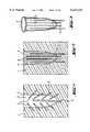
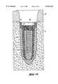
- FIG. 1Cast cavity in the form of an impression of the root of a tooth, with inserted mandrels
- FIG. 2Cast cavity filled with biodegradable material, with an alternative arrangement of the mandrels
- FIG. 3Implant with the mandrels not yet removed
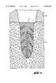
- FIG. 4Implant according to FIG. 1, which is inserted into the jaw bone and which has reservoirs with biologically active substances that arise in place of the mandrels and that are filled
- FIG. 5Part of the lower jaw to illustrate another working example of the invention
- FIG. 6Moulding material that contains the mandrels for forming reservoirs and that is filled with a biodegradable material to form one of the implants corresponding to the resection defect
- FIG. 7Implanted implant with filled reservoirs
- FIG. 8Lower jaw with implant affixed to it
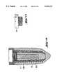
- FIG. 9Cut through an implant core serving as a fixture according to another working example of the invention
- FIG. 9ASection of FIG. 9 with a variation of a structured inner side of the implant core according to FIG. 9:
- FIG. 10View onto the implant core according to FIG. 9
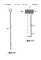
- FIG. 11Washer
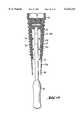
- FIG. 12Filament-shaped mandrel
- FIG. 13Mandrel with smoothbare shaft and head with screw thread
- FIG. 14Implant core according to FIG. 9 with inserted mandrel
- FIG. 15Implant core according to FIG. 14 inserted into the impression in the moulding material
- FIG. 16Implant core according to FIG. 15 with filled reservoirs
- FIG. 17Screw cover
- FIG. 18Inserted implant with reservoirs closed by a screw cover
- FIG. 19Another working example of an implant core
- FIG. 20Implant core according to FIG. 9 with an inner space partially filled with biodegradable material
- FIG. 21Implant core for inserting in moulding material
- FIG. 22Cast cavity filled with biodegradable material, with fixture
- FIG. 23Finished implant
- FIG. 24Implanted implant
- FIG. 25Another working example of an implant core with mandrels
- FIG. 26Implant core according to FIG. 25 inserted into moulding material and
- FIG. 27Implant with an implant core in accordance with FIG. 25.
- the object of the inventionis a medical implant 5 for insertion into a space with a prespecified dimension and for filling a cast cavity 2 with a hardening plastic material with mineral components on a basis of calcium and phosphate or, in the case of the new formation of bone, a biodegradable material, or a combination of both, characterized by the fact that
- At least one mandrel 3, or 23, 24,is placed in the cast cavity 2 in such a way that it can be removed from the cast cavity 2 of the formed implant 5 after the plastic material hardens and creates a space 6 or 19 (after its removal) that extends from the inner area of the implant 5 to its outer surface, thus forming a reservoir that can be filled with a biologically active substance, or
- the cast cavity 2contains a porous matrix with a biologically active substance, and the hollow spaces of this matrix are filled with the hardening plastic material.
- the inventionconcerns tooth implants or tooth root implants.
- the implanthas, for example, the advantage that with dental surgery, producing and implanting the individual implant can be carried out as a single therapeutic treatment. As a result, time can be saved and a traumatic second operation prevented. A second anesthesia is not necessary; nor is the added stress to the patient of another "surgery.”
- the implantation treatmentcan be carried out outside of dental clinics in private practices by dentists with normal training. A high acceptance among patients can be obtained because the loss of connective tissue can be replaced immediately and with little risk. By producing and inserting the individual transplant immediately, alveolar bone loss can be prevented. Other bone fragments can also be replaced with an exactly matching, biocompatible, biodegradable fitted body, which contains a fixture made of alloplastic material in specific designs.
- the process for producing the medical implant 5is also an object of the invention and is characterized by the fact
- an object of the inventionis a nontherapeutic process for producing the medical implant 5, which is carried out outside of a human or animal's body and is characterized by the fact
- ⁇ 'that one forms a cast cavity 2 in moulding material 1 in the form of an impression of the extracted tooth or the resected bone; that one fills this cast cavity 2 with a hardening material with mineral components on the basis of calcium and phosphate or, in the case of the new formation of bone, a biodegradable material; that one places at least one mandrel 3, or 23, 24, in the cast cavity 2 in such a way that it can be removed from the cast cavity 2 of the formed implant after the plastic material hardens and with the removal of the mandrel forms a space 6, or 19, serving as a reservoir, that extends from the inner area of the implant 5 to the outer surface; that one removes the formed implant 5 from the cast cavity and fills the reservoir with a biologically active substance after the removal of the mandrel 3, or 23, 24, or
- An especially preferred variation of the procedureis characterized by the fact that one first places in the cast cavity 2 several mandrels 3, or 23, 24, that extend from the cast cavity 2 into the moulding material 1 and are placed in the moulding material 1 in such a way that one can remove the mandrels 3, or 23, 24, from the implant after pouring the plastic moulding material into the cast cavity 2, letting it harden, and then removing the formed implant 5 from the cast cavity 2.
- FIGS. 1-4a first working Example of the invention and its production process is illustrated, whereby the implant fills a bone defect that has arisen from the extraction of a tooth.
- FIG. 1At first the involved tooth is extracted. If there is a fracture, its fragments--especially the root--are agglutinated. The contaminated root surface of the tooth is cleaned aseptically, mechanically, and/or chemically. The tooth is then pressed into moulding material 1 up to the tooth neck by a known method; the moulding material is a sterile, elastic moulding compound of a known type such as alginates, silicone cross-linked with additives and condensation, polyether, etc. After the moulding material 1 hardens, the tooth is taken out, and a cast cavity 2 in the form of an impression of the dental implant to be produced is formed in the moulding material 1.
- the moulding material 1is a sterile, elastic moulding compound of a known type such as alginates, silicone cross-linked with additives and condensation, polyether, etc.
- mandrels 3in the form of filaments or rods made of smooth material that resists breakage are inserted from the cast cavity into this moulding material.
- the term mandrelincludes objects and devices that are used as place holders for reservoirs or hollow spaces that can be filled.
- the filament- or rod-shaped mandrels 3are placed in such a way that they do not reach the level 4 of the limbus alveolaris.
- a cast cavity 2 in which the mandrels 3 are arranged as shown in FIG. 3is formed in the moulding material 1.
- This cast cavity 2is then filled with a hardening plastic material with mineral components on the basis of calcium and phosphate or, in the case of the new formation of bone, a biodegradable material, or a combination of the two.
- Plastic materials with mineral components on the basis of calcium and phosphatecan be obtained by combining partly neutralized phosphate materials and calcium salts in accordance with the procedure described in European patent application 416 761.
- Suitable phosphate materialsare, for example, Ca(H 2 PO 4 ) 2 .H 2 O [MCPM], CaHPO 4 .H 2 O [DCPD: brushite], Ca 9 (HPO 4 ).
- (PO 4 ) 5 (OH)[CDHA: calcium-deficient hydroxyapatite]
- CHScalcium pyrophosphate
- CaSO 4 .0.5H 2 O [CHS: "Plaster of Paris"]CaSO 4 .0.5H 2 O [CHS: "Plaster of Paris"]
- Preferredis a plastic material with mineral components on the basis of calcium and phosphate with the following shares: 60-80% ⁇ -TCP, 40-20% MCPM, an aqueous solution containing P 2 O 7 4- and SO 4 2- ions and additives of the cellulose ether type, for example, HPMC (0.5-1.0%) or polysaccharides.
- Materials suitable for the new formation of bonehave osteo-conductive properties and are preferably biodegradable and biocompatible. Osteo-conductive materials control the make-up of a structure for the new formation of bone during "guided bone repair” (GBR).
- GBRguided bone repair
- Biodegradable and biocompatible materialsare generally known--e.g., aliphatic polyesters of the types polyglycolic acid (PGA) or polylactic acid (PLA) and their compounds (PGA/PLA); enantiomeric forms and racemic compounds in various proportions, e.g., poly-L-lactate (PLLA), poly-D-lactate (PDLA), poly-DL-lactate (PDLLA), L-lactate/DL-lactate, or L-lactate/D-lactate. These materials are not only biodegradable, they are also biocompatible. PGA and PLA have metabolism channels (pathways) in the human body. Furthermore, PGA and PLA materials are not immunogens--that is, these materials do not cause immune reactions in mammals. Suitable materials are, for example, commercial products of the type Biofix®, which can be obtained commercially from the firm Bioscience (SF-33721 Tampere).
- Suitable aliphatic polyesters with osteo-conductive propertiesare, in addition, PLA copolymers, e.g., lactate/tetramethyleneglycolid copolymers, lactate/trimethylene carbonate copolymers, lactate/ ⁇ -valerolactone copolymers, lactate/ ⁇ -caprolactam copolymeres, polydepsipeptide (glycine-DL-lactate copolymers or PLA/ethylene oxide copolymers (PLA/PEO)), polylactide-polyglycolid copolymers or polylactide-ethylene oxide copolymers or polyhydroxyalkanoates, e.g., PHB [Poly( ⁇ -hydroxybutyrate)], PHB/PHA (polyhydroxybutyrate/polyhydroxyvalerate), PCL [poly( ⁇ -caprolactam), PDS [poly(p-dioxanone)], polyanhydrides, polyhydroxysuccinic acid ( ⁇ ) or polyhydroxysuccinic
- Suitable materials for the new formation of bones that have osteo-conductive propertiesare, in addition, vinyl polymers, e.g., on the basis of polyvinyl alcohol (PVA), poly- ⁇ -maleic acid, aliphatic polyamides, aliphatic polyurethanes, e.g., polyurethanes made of polyethyleneglycol-(PEG)-diols or polycaprolactam-diols and diisocyanates such as 1,4-methylene diisocyanate, polyorthoesters, e.g., of the type Alzmer® (Alza Corp.) or DETOSU, aliphatic polyanhydrides, polypeptides, e.g., synthetic polyamino acids and poly- ⁇ -amino acids, e.g., poly- ⁇ -lysine or polybenzylglutamate, polyphosphates, polysaccharides, e.g. dextran derivatives, chitin derivatives, and chitosan
- the plastic materials namedcan be put into place in the cast cavity 2 by using suitable filling instruments, e.g., injection instruments.
- FIG. 2shows a cast cavity, filled with biodegradable material, that has taken the form of the implant 5 after the plastic material has hardened.
- the mandrels 3here have an alternative arrangement and design.
- FIG. 3After the materials for implant 5 have hardened it is removed from the cast cavity 2 together with the mandrels 3. Then the filament-shaped mandrels 3 are pulled out of the implant 5. In this way, channel- and capillary-shaped spaces or gaps 6 are formed in the implant 5, which serve as reservoirs for biologically active substances (so-called active substances).
- Suitable biologically active substancesare poured into the reservoirs. They have osteo-conductive properties and can have an effect on the biological behavior of neighboring cells, for example, by stimulating the division of cells or the formation of bone, through, e.g., the formation of mesenchymal cells, endothelial tissue, pericytes, osteoclasts, osteoblasts, etc.
- Suitable biologically active substances with osteo-conductive propertiesare, for example, hormones, proteins or growth factors on a protein or lipid basis that are known by such names as epidermal growth factor (EGF); vascular epidermal growth factor (VEGF); fibroblast growth factor (FGF); platelet derived growth factor (PDGF); transforming growth factor- ⁇ (TGF- ⁇ ), e.g., of the type TGF- ⁇ -1, TGF- ⁇ -2, or TGF- ⁇ -3; insulin-like growth factor (IGF-I and IGF-II); nerve growth factor (NGF); bone morphogenic proteins (BMP), e.g., BMP-3 (osteogenin), BMP-2 (BMP 2A), BMP-4 (BMP 2B), BMP-5, BMP-6, BMP-7 (osteogenic protein-1); and proteins that are known by the names parathyroid hormone (PTH), e.g., PTH fragments such as FTH 1-34 and its derivatives; parathyroid hormone related proteins (PTHrP), e.g
- TGF- ⁇transformed growth factor of type beta
- Proteins of the type TGF- ⁇ of human originare known and described in the survey article by D. A. Cox, "Transforming Growth Factor-Beta 3.” Cell Biology International 19, no. 5 (1995): 357-71.
- Recombinant proteins of type TGF- ⁇are known and described in the following survey article: Lionel Bourdel, et al. "Recombinant Human Transforming Growth Factor- ⁇ 1: Expression by Chinese Hamster Ovary Cells, Isolation and Characterization.” Protein Expression and Purification 4 (1993): 130-40; M. P. Schlunegger and M. G. Grutter. "An Unusual Feature Revealed by the Crystal Structure at a Resolution of Human Transforming Growth Factor- ⁇ 2.” Nature 358 (1992): 430-34; S. Runser and N. Cerietti. "Transforming Growth Factors ⁇ : Conformational Stability and Features of the Denaturation of Recombinant Human Transforming Growth Factors- ⁇ 2 and ⁇ 3.” Biotechnol. Appl. Biochem. 22 (1995): 39-53.
- TGF- ⁇transforming growth factor of type beta
- Additional substances that can be poured into the reservoirs mentionedare active substances that inhibit bone resorption, e.g., bisphosphonates of the type Aredia®, nitrates, e.g., nitroglycerin, ipriflavon, active substances that bind with nuclear receptors such as estradiol, enzyme inhibitors that block enzymes that break down the bone matrix, collagenase inhibitors, stromelysine [Stromelysin] inhibitors, cathepsin L, K inhibitors, substances that inhibit osteoclast functions such as carboanhydrase inhibitors or inhibitors of the osteoclastic proton pump, etc.
- active substances that inhibit bone resorptione.g., bisphosphonates of the type Aredia®, nitrates, e.g., nitroglycerin, ipriflavon
- active substances that bind with nuclear receptorssuch as estradiol, enzyme inhibitors that block enzymes that break down the bone matrix, collagenase inhibitors,
- active substancesare those that are effective against inplantopathogens (paradontophathogens), e.g., antibiotics, antibodies (monoclonal, polyclonal), inflammation inhibitors, prostaglandin inhibitors, active substances with immune-suppressive effects such as (bio)synthetic immune suppressors, active substances with revascularization-promoting effects such as vascular-forming substances, active substances that promote circulation, or analgesics.
- inplantopathogensparadontophathogens
- antibioticse.g., antibiotics, antibodies (monoclonal, polyclonal), inflammation inhibitors, prostaglandin inhibitors, active substances with immune-suppressive effects such as (bio)synthetic immune suppressors, active substances with revascularization-promoting effects such as vascular-forming substances, active substances that promote circulation, or analgesics.
- the implant filled with the biologically active substancerepresents a "dispensing unit,” which contains a dose of the substance to be dispensed and releases it within a set time period.
- a dispensing unit as defined herecontains one dose of the substance to be dispensed, or a faction or multiple of it. It can be dispensed spontaneously, e.g., by diffusion or erosion of the system through interaction with body fluids.
- FIG. 4One places the exactly matching implant 5 in the socket in the jaw bone 7, whereby in FIG. 4 the gingiva 8 is also implied. In so doing the substances 9 are released from the implanted implant.
- FIG. 5shows a lower jaw 10 in which a tumor 11 has formed.
- the bone part 12 stricken with the tumor 11is removed from the lower jaw 10 and then an impression is made in the moulding material 1 (see the section according to FIG. 6).
- the mandrels 3are placed in the cast cavity 2 so their ends project out of the moulding material 1.
- the arrangement of the mandrels 3 shown in FIG. 6is reproduced as an example and can vary. After inserting the mandrels 3, the cast cavity 2 is filled with the biodegradable material that hardens into an implant 5.
- FIG. 6The bone part 12 removed from the lower jaw 10 according to the drawing in FIG. 6 is pressed into the moulding material 1 and rotated by 180° compared to the drawing in FIG. 5 so that the upper, rounded section of the bone part 12, in accordance with the drawing in FIG. 6, is located at the bottom as shown in FIG. 6.
- the remaining spaces or reservoirs 6 in the implant 5 after removing the mandrels 3, as described above,are filled with one or several active substances 9.
- FIG. 7The implant 5 is implanted into the intended site in the lower jaw 10, as is shown in FIG. 7.
- FIG. 8If necessary, one can attach the implant 5 by using a plate 13 in the jaw bone, whereby the plate 13 as well as the screws 14 belonging to it can also consist of a biodegradable material.
- FIGS. 9-18another working example of the invention is described.
- FIG. 9shows a sectional view of an implant core that also serves as a fixture.
- the term fixturedesignates the enossal part (inside the jaw bone) of an implant, which takes up the part of an implant construction (the visible part of the implant) projecting from the jaw bone.
- Such an implant coreconsists of an alloplastic, osseo-integratable material, e.g., titanium, Frialit, etc. It has the form of an elongated hollow body 18 with an inner space 15. Its outer side is structured, for example, in the form of a screw thread 16. Other designs also provide for a structuring of the inner side. An inside screw thread 17 is designed at the upper end of the inner space 15.
- hollow spaces 19 that run parallel to the inner space 15exist and are open at the top and closed at the bottom--i.e., at the end far away from the inside screw thread 17.
- Another arrangement of the hollow spaces 19is possible as an alternative to the parallel arrangement to the inner space 15.
- the hollow body 18is tapered to the lower end, i.e., to the root tip.
- the inside screw thread 17 at the upper wider end, i.e., in the area of the limbus alveolarisnot only serves to seal the reservoir of active substances, as will be described later, but also to attach the suprastructure later when the tooth is restored or to attach the prosthesis with help of the implant.
- the hollow spaces 19 in the wall of the hollow body 18are arranged in such a way that they cut the grooves of the screw thread 16 or the respective structure of the wall of the hollow body 18. Moreover, perforations 20 exist in the wall that run from the hollow spaces 19 to the outer surface of the hollow body.
- FIG. 9AAn alternative arrangement of the perforations is shown in FIG. 9A, which represents a cut from FIG. 9.
- the inner surface of the hollow body 18is also structured.
- additional perforations 20Awhich run from the hollow spaces 19 to the inner surface of the hollow body 18, exist and connect the hollow spaces 19 with the inner space 15 of the hollow body 18.
- a designis also provided in which only the inner surface of the hollow body 18 is structured.
- FIG. 10represents the view onto the implant core in accordance with FIG. 9.
- FIG. 11shows a packing ring 21 that is intended for insertion into the upper, recessed end section 22 of the hollow body 18.
- This packing ring 21can consist of silicone rubber or soft gold or plastic sealing metal beads so that, with the packing ring 21 inserted into the area 22 A, the openings to the hollow spaces 19 and the hollow space 15 are safely closed off to outside influences, e.g., the penetration of pathogens (bacteria, yeasts, viruses, etc.).
- FIG. 12shows a filament-shaped mandrel 23. Its diameter is calculated in such a way that it can be inserted into a respective hollow space 19.
- FIG. 13In FIG. 13 another mandrel 24 is designed that consists of a shaft 25 and a head 26 that has an exterior screw thread 26 A and a slit 27 at the top for accommodating a screwdriver like tool.
- FIG. 14To produce an implant, first the mandrel 24, which has a shaft 25 and head 26, is screwed into the inner space 15 of the hollow body 18. Then the filament-shaped mandrels 23 are inserted into the respective hollow spaces 19. In a first step, a first amount of a plastic biodegradable material is placed in the inner space 15 from below in the direction of arrow A.
- FIG. 15An impression of the root of the tooth to be replaced by the implant--that is, the cast cavity 2--is produced in the moulding material 1.
- a second stepa second amount of the biodegradable material is poured into the cast cavity; and in a third step the hollow body 18, already filled and equipped with the mandrels 23 and 24, is pressed into the still liquid biodegradable material.
- the biodegradable materialis liquified by warming it (in special application syringes or another way) depending on its composition, or it is available in liquid or plastic form at room temperature.
- Thickening and hardeningensues either by cooling the thermoplastic material or through a chemical reaction (e.g., a 2-component reaction, a photochemical process, or polymerization, etc.). It is clear from FIG. 15 how the gap between the outer wall of the hollow body 18 and the inner wall of the cast cavity 2 in the moulding material 1 is filled by this material.
- the implant 5in which the hollow body 18 that still has the mandrels 23 and 24 is imbedded, exists.
- the implant 5is removed from the moulding material 1 and then the filament-shaped mandrels 23 and the mandrel 24 are pulled and screwed out, respectively, of the implant 5.
- FIG. 16, FIG. 17The biologically active substances described earlier can be poured into the remaining spaces 6.
- the spaces 6are closed off at the top by the screw cover 28 (FIG. 17).
- an implant 5is formed that has an alloplastic fixture (hollow body 18) on the inside, which is surrounded by a biodegradable material. Reservoirs 19, 15 for active substances exist.
- the implantanatomically replicated to correspond to the extracted root, is ready to be placed in the extraction socket.
- FIG. 18shows the implant 5 inserted into the jaw bone 7, whereby the gingiva is also shown.
- the packing ring 21which closes off the inner spaces to outside influences such as bacteria, is positioned between the screw cover 28 and the hollow body 18.
- the implant 5fits exactly in the extraction socket, and the healing process can follow per primam through the close contact to the surrounding bones.
- the biodegradable materialbegins to reabsorb.
- the resultis the release of the growth factors and/or active substances. This leads to the accelerated, controlled new formation of bone and a qualitatively optimized osseo-integration of the alloplastic implant core.
- FIG. 19The hollow body 18, which consists of an osseo-integratable material, has an inner space 15 in the middle. Along the structured outer wall run elongated hollow spaces 19 corresponding to the working example described above. The hollow spaces 19 cross the grooves of the structured outer wall so that perforations 20 are formed that create a connection between the hollow spaces 19 and the outer surfaces of the hollow body 18. On the inner wall of the inner space 15 additional openings or perforations 29 to the outer surfaces are designed, whereby such openings 29 also exist in other working examples.
- the hollow body 18, which later serves as a fixture,has a screw thread 30 at the top, into which a temporary screw cover 31 can be screwed, whereby the screw thread 30 later serves as a means of affixing the tooth construction.
- FIG. 20As a first step, biodegradable material is injected from the lower opening 34 and through the openings 29 into the annulus between the mandrel 24 A and the inner wall of the hollow body 18. After the material hardens, the mandrel 24 A is pulled out so that a layer 35 of the biodegradable material exists in the inner space 15.
- FIG. 21A first amount of the active substances 9 mentioned is poured into the hollow space 6, and the hollow space 6 is closed with a peg-shaped seal 36.
- the seal 36also consists of the biodegradable material.
- FIG. 22The impression of the root of the tooth to be replaced is produced in the moulding material 1 by the known method--i.e., the cast cavity 2 is formed.
- the biodegradable materialis poured into the cast cavity 2 and then the implant core is inserted into the cast cavity 2 in accordance with FIG. 21, whereby the tips 33 of the filament-shaped mandrels 23 penetrate the moulding material 1.
- FIG. 23After the poured, biodegradable material thickens and hardens, the formed implant 5 is removed from the cast cavity 2 and the filament-shaped mandrels 23 A are pulled out of the implant core from below and the respective medical substances poured into the available spaces 19. The lower openings 37 of the filled spaces 19 are then closed.
- warmth caused by frictioncan be produced by using a rotating dental instrument, e.g., a rose-head burr, in the areas of the openings 37 so the biodegradable material melts at these sites and the openings 37 are closed.
- FIG. 24The finished implant 5 can then be implanted in the extraction socket in accordance with the diagram in FIG. 24.
- FIGS. 25-27With the use of FIGS. 25-27 another working example is described. The steps in the procedure are analogous to those already described. Thus the process will be described below in simplified form.
- FIG. 25The hollow body 18 or implant core is analogous to the working example in accordance with FIG. 19.
- the rod-shaped mandrel 24, however,does not end in a handle; it ends in a tip 38.
- FIG. 26After the cast cavity 2 in the moulding material 1 is filled with the biodegradable material, the hollow body 18 is inserted into the cast cavity 2, whereby all of the tips 33, 38 of the mandrels 23, 24 are stuck into the moulding material. After the biodegradable material thickens and hardens, the mandrels 23, 24 are pulled out of the implant 5 from below and the existing spaces--the reservoirs--are filled with the corresponding medical substances. Then the inner space 15 is closed by the peg-shaped seal 36 and the spaces 19 running along the outer side of the implant core by the lower openings 37 are welded together by frictional heat so that the implant is obtained in accordance with FIG. 27.
- FIG. 27Finished implant with an implant core in accordance with FIG. 25.
- the mandrelis placed in the moulding material 1 in such a way that--after pouring the plastic materials in the cast cavity (2), letting it harden, and removing the formed implant and mandrel from the moulding material--one uses a pin provided with a screw thread (fixing pin or screw) made of a biocompatible, but not osseo-integratable, material--e.g. high-quality steel--instead of the mandrel. Its diameter at the shaft is smaller than the diameter of the mandrel. In this way a gap is formed that one can fill with bioactive substances.
- the shaft of the pin provided with a screw threadcan be longer than the mandrel and its screw thread can cut the lowest point of the cast cavity (2).
- the fixing screw, extended in this way and cutting its own screw thread,is screwed into the jaw bone through the deepest point of the extraction socket.
- the fixing screwis surrounded by bone material on all sides. Because the screw is not osseo-integratable, it can be removed easily.
- the channel that remains in the jaw bonemakes possible the insertion of an exactly matched fixture in its natural direction with respect to the axis.
- the advantage of this variationis that an extraction and implantation can be carried out with one therapeutic treatment. Through the presence of biologically active substances on the surface of the implant an especially effective wound closure of the bordering tissue takes place, as well as the quick osseo-integration of the implant fixture.
- the cast cavity 2e.g., an extraction socket
- Suitable synthetic polymer materialsare, preferably, macrostructured and can be sponge-shaped forms with a perforated skeleton framework made of a polymer material, in which gaps connecting under each other and pores are dispersed.
- Suitable materialsare, for instance, polycarbonates, polyorthoesters, PGA, PLA, or mixtures of them, etc.
- Such a designis especially suitable for a dispensing unit that holds the biologically active substance to be dispensed in its polymer structure until it is dispensed.
- a porous matrix made of natural materialis, preferably, a dentin matrix, which can be obtained by extracting teeth or tooth fragments.
- a dentin matrix from material suitable for a patientis described by way of example.
- the tooth matter up to the size of a particle of approximately 1 mm 3is ground in a grinder.
- the ground materialis washed in warm water, the oils removed by immersing it in a (1:1) chloroform/methanol solution for 12 hours, and the material is demineralized with a 0.5 molar HCl solution for 72 hours at 4° C.
- Oilsare again removed from the demineralized material 12 hours long by immersing it in a (1:1) chloroform/methanol solution for 6 hours, and then the material is treated for 24 hours in a 2-molar calcium-chloride solution, for four hours in a 0.5 molar EDTA solution at 7.4 pH, and for 24 hours in an 8-molar lithium-chloride solution, and then washed in distilled water at 4° C.
- One centrifuges the extract10,000 g. 30 min.
- the reproduced cast cavity of an extraction socketis filled with a mixture made of PGA/PLA copolymers and the matrix material that is made of dentin and contains BMP; and a root implant with fixture, which one inserts into the jaw bone at the intended site, is formed in the way already described above.
Landscapes
- Health & Medical Sciences (AREA)
- Life Sciences & Earth Sciences (AREA)
- Veterinary Medicine (AREA)
- Epidemiology (AREA)
- Dentistry (AREA)
- Animal Behavior & Ethology (AREA)
- General Health & Medical Sciences (AREA)
- Public Health (AREA)
- Oral & Maxillofacial Surgery (AREA)
- Orthopedic Medicine & Surgery (AREA)
- Engineering & Computer Science (AREA)
- Biomedical Technology (AREA)
- Developmental Biology & Embryology (AREA)
- Materials For Medical Uses (AREA)
- Prostheses (AREA)
- Dental Preparations (AREA)
Abstract
Description
Claims (10)
Applications Claiming Priority (3)
| Application Number | Priority Date | Filing Date | Title |
|---|---|---|---|
| CH3565/95 | 1995-12-18 | ||
| CH356595 | 1995-12-18 | ||
| PCT/EP1996/005506WO1997022308A1 (en) | 1995-12-18 | 1996-12-10 | Medical implant |
Publications (1)
| Publication Number | Publication Date |
|---|---|
| US6132214Atrue US6132214A (en) | 2000-10-17 |
Family
ID=4258753
Family Applications (1)
| Application Number | Title | Priority Date | Filing Date |
|---|---|---|---|
| US09/091,674Expired - LifetimeUS6132214A (en) | 1995-12-18 | 1996-12-10 | Medical implant |
Country Status (9)
| Country | Link |
|---|---|
| US (1) | US6132214A (en) |
| EP (1) | EP0893975B1 (en) |
| JP (1) | JP2000501966A (en) |
| AT (1) | ATE237285T1 (en) |
| AU (1) | AU1369297A (en) |
| BR (1) | BR9612051A (en) |
| DE (1) | DE59610355D1 (en) |
| ES (1) | ES2192623T3 (en) |
| WO (1) | WO1997022308A1 (en) |
Cited By (54)
| Publication number | Priority date | Publication date | Assignee | Title |
|---|---|---|---|---|
| US6325627B1 (en)* | 1999-10-20 | 2001-12-04 | Arthur Ashman | Method and apparatus for performing ridge preservation and implant treatment |
| US6413089B1 (en) | 1999-02-10 | 2002-07-02 | Arthur Ashman | Immediate post-extraction implant |
| WO2002054973A1 (en)* | 2001-01-12 | 2002-07-18 | Natural Implant | Provisional dental implant for preparing an alveolus |
| US6447514B1 (en)* | 2000-03-07 | 2002-09-10 | Zimmer | Polymer filled hip fracture fixation device |
| US20030003128A1 (en)* | 1999-12-21 | 2003-01-02 | Piero Chiarelli | Dental prosthesis with means for the release of active substances |
| US20030036036A1 (en)* | 2001-08-17 | 2003-02-20 | Porter Stephan S. | Immediate load dental implant system and method of use |
| US20030135209A1 (en)* | 1999-12-03 | 2003-07-17 | Bahaa Seedhom | Fixation technology |
| WO2003039331A3 (en)* | 2001-11-05 | 2004-02-12 | Lilly Co Eli | Method for improving stability of a bone-connecting implant |
| US20040030341A1 (en)* | 2001-03-02 | 2004-02-12 | Marcel Aeschlimann | Implants, device and method for joining tissue parts |
| US20040038180A1 (en)* | 2002-08-23 | 2004-02-26 | Woodwelding Ag | Implant, in particular a dental implant |
| US20040038178A1 (en)* | 2002-08-23 | 2004-02-26 | Woodwelding Ag | Preparation for being fastened on a natural tooth part or tooth and corresponding fastening method |
| US20040209228A1 (en)* | 2001-05-03 | 2004-10-21 | Daniel Ilan | Polymeric dental implant |
| WO2004054464A3 (en)* | 2002-12-13 | 2004-12-02 | Stefan Neumeyer | Abutment for a dental implant, dental implant comprising such an abutment, and method for the production of dentures by means of said dental implant |
| US20050126680A1 (en)* | 1999-06-18 | 2005-06-16 | Woodwelding Ag | Integral joining |
| US6913666B1 (en) | 1997-03-21 | 2005-07-05 | Woodwelding Ag | Process for anchoring connecting elements in a material with pores or cavities and connecting elements therefor |
| US6918766B1 (en)* | 1999-05-31 | 2005-07-19 | Nobel Biocare Ab | Method, arrangement and use of an implant for ensuring delivery of bioactive substance to the bone and/or tissue surrounding the implant |
| US20050209704A1 (en)* | 2002-03-14 | 2005-09-22 | Maspero Fabrizio A | Porous biocompatible implant material and method for its fabrication |
| US20050220773A1 (en)* | 2002-02-19 | 2005-10-06 | Toshimasa Uemura | Implant containing cells having growhfactor gene transferred thereinto |
| US20050251266A1 (en)* | 2004-05-06 | 2005-11-10 | Maspero Fabrizio A | Biocompatible bone implant compositions and methods for repairing a bone defect |
| US6984623B2 (en) | 1993-12-07 | 2006-01-10 | Genetics, Institute Institute, LLC. | Tendon-inducing compositions |
| US20060064164A1 (en)* | 2000-03-07 | 2006-03-23 | Thelen Sarah L | Method and apparatus for reducing femoral fractures |
| US20060084034A1 (en)* | 2004-10-15 | 2006-04-20 | Hochman Mark N | Method and apparatus for performing maxillary sinus elevation |
| WO2006050106A1 (en)* | 2004-10-28 | 2006-05-11 | Microchips, Inc. | Orthopedic and dental implant devices providing controlled drug delivery |
| US20060105295A1 (en)* | 2004-02-20 | 2006-05-18 | Woodwelding Ag | Implant that can be implanted in osseous tissue and method for producing said implant corresponding implant |
| US20060122543A1 (en)* | 2003-07-31 | 2006-06-08 | Woodwelding Ag | Method for promoting tissue regeneration on wound surfaces as device and treatment instrument or implant for carrying out method |
| US20060136071A1 (en)* | 2002-12-23 | 2006-06-22 | Maspero Fabrizio A | Biodegradable biocompatible implant |
| US20060166251A1 (en)* | 2005-01-26 | 2006-07-27 | Archambault Joanne M | Use of sFRPs as markers of BMP activity |
| US7091007B2 (en) | 1993-09-17 | 2006-08-15 | Genetics Institute, Llc | DNA molecules encoding BMP receptor proteins |
| US20060239951A1 (en)* | 2005-03-30 | 2006-10-26 | Alexandre Valentin | Methods for stimulating hair growth by administering BMPs |
| US7189392B1 (en)* | 1999-10-15 | 2007-03-13 | Genetics Institute, Llc | Injectable carrier formulations of hyaluronic acid derivatives for delivery of osteogenic proteins |
| US20070088442A1 (en)* | 2005-10-14 | 2007-04-19 | Microchips, Inc. | Passive wear-indicating sensor for implantable prosthetic device |
| US7217691B2 (en) | 1986-07-01 | 2007-05-15 | Genetics Institute, Llc | Methods of treatment of periodontal disease |
| US7226587B2 (en) | 2001-06-01 | 2007-06-05 | Wyeth | Compositions and methods for systemic administration of sequences encoding bone morphogenetic proteins |
| US20070264300A1 (en)* | 2006-05-10 | 2007-11-15 | Sdgi Holdings, Inc. | Therapeutic agent carrier and method of treating bone fractures |
| US7323445B2 (en) | 1999-02-01 | 2008-01-29 | Genetics Institute, Llc | Methods and compositions for healing and repair of articular cartilage |
| US7413753B2 (en) | 2001-06-08 | 2008-08-19 | Wyeth | Calcium phosphate delivery vehicles for osteoinductive proteins |
| US20080254019A1 (en)* | 2004-03-31 | 2008-10-16 | Yukio Kato | Therapeutic Agents and Therapeutic Methods For Treating Injured Tissue |
| US20080262517A1 (en)* | 2007-04-20 | 2008-10-23 | Stryker Trauma Gmbh | Implantation pin, fixation device and method for implanting the implantation pin |
| EA010624B1 (en)* | 2006-10-23 | 2008-10-30 | Производственное Частное Унитарное Предприятие "Верлайн" | Polycomponent dental implant |
| US20090208907A1 (en)* | 2006-10-30 | 2009-08-20 | Joint Stock Company 'altimed' | Dental implant |
| US20100003639A1 (en)* | 2008-07-02 | 2010-01-07 | Salvi Joseph A | Porous implant with non-porous threads |
| US7678885B2 (en) | 1991-11-04 | 2010-03-16 | Genetics Institute, Llc | Recombinant bone morphogenetic protein heterodimers, compositions and methods of use |
| US7771755B2 (en) | 2003-09-12 | 2010-08-10 | Wyeth | Injectable calcium phosphate solid rods and pastes for delivery of osteogenic proteins |
| US20100215718A1 (en)* | 2009-02-25 | 2010-08-26 | Porex Surgical, Inc. | Bone Graft Material Containment Structures |
| US20110070557A1 (en)* | 2009-09-23 | 2011-03-24 | Elkana Elyav | Dental implant |
| US20110224739A1 (en)* | 2008-10-10 | 2011-09-15 | Stryker Trauma Gmbh | Implantation pin kit and method for implanting an implantation pin |
| US8075309B2 (en)* | 2004-11-30 | 2011-12-13 | Align Technology, Inc. | Systems and methods for intra-oral drug delivery |
| US8128706B2 (en)* | 2008-01-09 | 2012-03-06 | Innovative Health Technologies, Llc | Implant pellets and methods for performing bone augmentation and preservation |
| WO2013139349A1 (en)* | 2012-03-18 | 2013-09-26 | Elaskary Abdelsalam Thabet Abdelsalam | Tooth socket repair kit |
| US20140356797A1 (en)* | 2012-02-01 | 2014-12-04 | Organ Technologies, Inc. | Dental implant and method for producing same |
| US9095396B2 (en) | 2008-07-02 | 2015-08-04 | Zimmer Dental, Inc. | Porous implant with non-porous threads |
| US9357996B2 (en)* | 2010-09-08 | 2016-06-07 | DePuy Synthes Products, Inc. | Fixation device with magnesium core |
| IT202100009875A1 (en)* | 2021-04-19 | 2021-07-19 | Poliedrica Srl | Implant fixture for the early detection of pathological states of peri-implantitis or peri-implant mucositis |
| US20220387143A1 (en)* | 2019-11-08 | 2022-12-08 | Politechnika Warszawska | Bioactive Intraosseous Dental Implant |
Families Citing this family (7)
| Publication number | Priority date | Publication date | Assignee | Title |
|---|---|---|---|---|
| WO2000005269A1 (en)* | 1998-07-21 | 2000-02-03 | Alpenstock Holdings Limited | Polymer complexes of glucuronoglucanes |
| US8814567B2 (en) | 2005-05-26 | 2014-08-26 | Zimmer Dental, Inc. | Dental implant prosthetic device with improved osseointegration and esthetic features |
| WO2007027794A1 (en)* | 2005-08-30 | 2007-03-08 | Zimmer Dental, Inc. | Dental implant with improved osseointegration features |
| US8562346B2 (en) | 2005-08-30 | 2013-10-22 | Zimmer Dental, Inc. | Dental implant for a jaw with reduced bone volume and improved osseointegration features |
| ES2288437B1 (en)* | 2007-04-27 | 2009-04-01 | Antonio Juan Flichy Fernandez | DENTAL IMPLANT SYSTEM. |
| US20090061389A1 (en) | 2007-08-30 | 2009-03-05 | Matthew Lomicka | Dental implant prosthetic device with improved osseointegration and shape for resisting rotation |
| ITGE20120072A1 (en)* | 2012-07-20 | 2014-01-21 | Bellinvia Salvatore | DENTAL IMPLANT |
Citations (22)
| Publication number | Priority date | Publication date | Assignee | Title |
|---|---|---|---|---|
| DE2658716A1 (en)* | 1975-12-22 | 1977-07-14 | Arthur Dr Ashman | METHOD FOR PRODUCING DENTAL PROSTHESES OR IMPLANTS |
| US4051598A (en)* | 1974-04-23 | 1977-10-04 | Meer Sneer | Dental implants |
| US4186486A (en)* | 1977-11-04 | 1980-02-05 | Maurice Gordon | Dental prosthesis |
| US4244689A (en)* | 1978-06-27 | 1981-01-13 | Arthur Ashman | Endosseous plastic implant |
| US4373217A (en)* | 1979-02-16 | 1983-02-15 | Merck Patent Gesellschaft Mit Beschrankter Haftung | Implantation materials and a process for the production thereof |
| US4671768A (en)* | 1982-12-06 | 1987-06-09 | Ton Michael A | Implant as well as a dental prosthesis attached to one or more of such implants |
| US4842604A (en)* | 1985-02-19 | 1989-06-27 | The Dow Chemical Company | Composites of unsintered calcium phosphates and synthetic biodegradable polymers useful as hard tissue prosthetics |
| US4863974A (en)* | 1988-08-03 | 1989-09-05 | W. L. Gore & Associates, Inc. | Bone growth matrix and process for making it |
| US4957509A (en)* | 1986-02-28 | 1990-09-18 | Agency Of Industrial Science And Technology | Ceramic implant materials |
| US5092890A (en)* | 1989-01-12 | 1992-03-03 | Basf Aktiengesellschaft | Implant materials for hard tissue |
| US5236971A (en)* | 1992-02-10 | 1993-08-17 | Murray William M | Dental and orthopedic cement method and preforms |
| US5356629A (en)* | 1991-07-12 | 1994-10-18 | United States Surgical Corporation | Composition for effecting bone repair |
| US5433751A (en)* | 1992-04-03 | 1995-07-18 | Inoteb | Bone prosthesis material containing calcium carbonate particles dispersed in a bioresorbable polymer matrix |
| DE4432831A1 (en)* | 1994-03-22 | 1995-09-28 | Sangi Co | Medical injection device |
| US5476880A (en)* | 1992-02-20 | 1995-12-19 | Orthopaedic Research Institute, Inc., Of Wichita | Orthopaedic appliance and method of preparing |
| US5501706A (en)* | 1994-11-29 | 1996-03-26 | Wildflower Communications, Inc. | Medical implant structure and method for using the same |
| US5520923A (en)* | 1994-09-19 | 1996-05-28 | Genetics Institute, Inc. | Formulations for delivery of osteogenic proteins |
| US5525646A (en)* | 1991-03-04 | 1996-06-11 | Lundgren; Dan | Bioresorbable material and an article of manufacture made of such material for medical use |
| US5531794A (en)* | 1993-09-13 | 1996-07-02 | Asahi Kogaku Kogyo Kabushiki Kaisha | Ceramic device providing an environment for the promotion and formation of new bone |
| US5552454A (en)* | 1988-08-09 | 1996-09-03 | Henkel Kommanditgesellschaft Auf Aktien | New materials for bone replacement and for joining bones or prostheses |
| US5558517A (en)* | 1992-02-24 | 1996-09-24 | Clemson University | Polymeric prosthesis having a phosphonylated surface |
| US5697981A (en)* | 1994-08-23 | 1997-12-16 | Norian Corporation | Method for repairing bone |
- 1996
- 1996-12-10USUS09/091,674patent/US6132214A/ennot_activeExpired - Lifetime
- 1996-12-10ESES96943906Tpatent/ES2192623T3/ennot_activeExpired - Lifetime
- 1996-12-10EPEP96943906Apatent/EP0893975B1/ennot_activeExpired - Lifetime
- 1996-12-10DEDE59610355Tpatent/DE59610355D1/ennot_activeExpired - Lifetime
- 1996-12-10JPJP9522478Apatent/JP2000501966A/ennot_activeCeased
- 1996-12-10AUAU13692/97Apatent/AU1369297A/ennot_activeAbandoned
- 1996-12-10ATAT96943906Tpatent/ATE237285T1/ennot_activeIP Right Cessation
- 1996-12-10BRBR9612051Apatent/BR9612051A/ennot_activeIP Right Cessation
- 1996-12-10WOPCT/EP1996/005506patent/WO1997022308A1/enactiveIP Right Grant
Patent Citations (22)
| Publication number | Priority date | Publication date | Assignee | Title |
|---|---|---|---|---|
| US4051598A (en)* | 1974-04-23 | 1977-10-04 | Meer Sneer | Dental implants |
| DE2658716A1 (en)* | 1975-12-22 | 1977-07-14 | Arthur Dr Ashman | METHOD FOR PRODUCING DENTAL PROSTHESES OR IMPLANTS |
| US4186486A (en)* | 1977-11-04 | 1980-02-05 | Maurice Gordon | Dental prosthesis |
| US4244689A (en)* | 1978-06-27 | 1981-01-13 | Arthur Ashman | Endosseous plastic implant |
| US4373217A (en)* | 1979-02-16 | 1983-02-15 | Merck Patent Gesellschaft Mit Beschrankter Haftung | Implantation materials and a process for the production thereof |
| US4671768A (en)* | 1982-12-06 | 1987-06-09 | Ton Michael A | Implant as well as a dental prosthesis attached to one or more of such implants |
| US4842604A (en)* | 1985-02-19 | 1989-06-27 | The Dow Chemical Company | Composites of unsintered calcium phosphates and synthetic biodegradable polymers useful as hard tissue prosthetics |
| US4957509A (en)* | 1986-02-28 | 1990-09-18 | Agency Of Industrial Science And Technology | Ceramic implant materials |
| US4863974A (en)* | 1988-08-03 | 1989-09-05 | W. L. Gore & Associates, Inc. | Bone growth matrix and process for making it |
| US5552454A (en)* | 1988-08-09 | 1996-09-03 | Henkel Kommanditgesellschaft Auf Aktien | New materials for bone replacement and for joining bones or prostheses |
| US5092890A (en)* | 1989-01-12 | 1992-03-03 | Basf Aktiengesellschaft | Implant materials for hard tissue |
| US5525646A (en)* | 1991-03-04 | 1996-06-11 | Lundgren; Dan | Bioresorbable material and an article of manufacture made of such material for medical use |
| US5356629A (en)* | 1991-07-12 | 1994-10-18 | United States Surgical Corporation | Composition for effecting bone repair |
| US5236971A (en)* | 1992-02-10 | 1993-08-17 | Murray William M | Dental and orthopedic cement method and preforms |
| US5476880A (en)* | 1992-02-20 | 1995-12-19 | Orthopaedic Research Institute, Inc., Of Wichita | Orthopaedic appliance and method of preparing |
| US5558517A (en)* | 1992-02-24 | 1996-09-24 | Clemson University | Polymeric prosthesis having a phosphonylated surface |
| US5433751A (en)* | 1992-04-03 | 1995-07-18 | Inoteb | Bone prosthesis material containing calcium carbonate particles dispersed in a bioresorbable polymer matrix |
| US5531794A (en)* | 1993-09-13 | 1996-07-02 | Asahi Kogaku Kogyo Kabushiki Kaisha | Ceramic device providing an environment for the promotion and formation of new bone |
| DE4432831A1 (en)* | 1994-03-22 | 1995-09-28 | Sangi Co | Medical injection device |
| US5697981A (en)* | 1994-08-23 | 1997-12-16 | Norian Corporation | Method for repairing bone |
| US5520923A (en)* | 1994-09-19 | 1996-05-28 | Genetics Institute, Inc. | Formulations for delivery of osteogenic proteins |
| US5501706A (en)* | 1994-11-29 | 1996-03-26 | Wildflower Communications, Inc. | Medical implant structure and method for using the same |
Cited By (115)
| Publication number | Priority date | Publication date | Assignee | Title |
|---|---|---|---|---|
| US7217691B2 (en) | 1986-07-01 | 2007-05-15 | Genetics Institute, Llc | Methods of treatment of periodontal disease |
| US7300772B2 (en) | 1986-07-01 | 2007-11-27 | Genetics Institute, Llc | BMP products |
| US7678885B2 (en) | 1991-11-04 | 2010-03-16 | Genetics Institute, Llc | Recombinant bone morphogenetic protein heterodimers, compositions and methods of use |
| US7091007B2 (en) | 1993-09-17 | 2006-08-15 | Genetics Institute, Llc | DNA molecules encoding BMP receptor proteins |
| US6984623B2 (en) | 1993-12-07 | 2006-01-10 | Genetics, Institute Institute, LLC. | Tendon-inducing compositions |
| US7365052B2 (en) | 1993-12-07 | 2008-04-29 | Genetics Institute, Llc. | Tendon-inducing methods |
| US6913666B1 (en) | 1997-03-21 | 2005-07-05 | Woodwelding Ag | Process for anchoring connecting elements in a material with pores or cavities and connecting elements therefor |
| US7323445B2 (en) | 1999-02-01 | 2008-01-29 | Genetics Institute, Llc | Methods and compositions for healing and repair of articular cartilage |
| US20050084820A1 (en)* | 1999-02-10 | 2005-04-21 | Arthur Ashman | Immediate post-extraction implant |
| US6413089B1 (en) | 1999-02-10 | 2002-07-02 | Arthur Ashman | Immediate post-extraction implant |
| US6918766B1 (en)* | 1999-05-31 | 2005-07-19 | Nobel Biocare Ab | Method, arrangement and use of an implant for ensuring delivery of bioactive substance to the bone and/or tissue surrounding the implant |
| US7160405B2 (en) | 1999-06-18 | 2007-01-09 | Woodwelding Ag | Integral joining |
| US20050126680A1 (en)* | 1999-06-18 | 2005-06-16 | Woodwelding Ag | Integral joining |
| US20070062628A1 (en)* | 1999-06-18 | 2007-03-22 | Woodwelding Ag | Integral joining |
| US7815409B2 (en) | 1999-06-18 | 2010-10-19 | Woodwelding Ag | Integral joining |
| US7608580B2 (en) | 1999-10-15 | 2009-10-27 | Genetics Institute, Llc | Injectable carrier formulations of hyaluronic acid derivatives for delivery of osteogenic proteins |
| US20070134342A1 (en)* | 1999-10-15 | 2007-06-14 | Genetics Institute, Inc. | Injectable carrier formulations of hyaluronic acid derivatives for delivery of osteogenic proteins |
| US7189392B1 (en)* | 1999-10-15 | 2007-03-13 | Genetics Institute, Llc | Injectable carrier formulations of hyaluronic acid derivatives for delivery of osteogenic proteins |
| US6722884B2 (en) | 1999-10-20 | 2004-04-20 | Arthur Ashman | Method and apparatus for performing ridge preservation and implant treatment |
| WO2001028448A3 (en)* | 1999-10-20 | 2002-05-02 | Arthur Ashman | A method and apparatus for performing ridge preservation and implant treatment |
| US6325627B1 (en)* | 1999-10-20 | 2001-12-04 | Arthur Ashman | Method and apparatus for performing ridge preservation and implant treatment |
| US7867232B2 (en) | 1999-12-03 | 2011-01-11 | University Of Leeds | Fixation technology |
| US20080183300A1 (en)* | 1999-12-03 | 2008-07-31 | University Of Leeds | Fixation technology |
| US7427284B2 (en)* | 1999-12-03 | 2008-09-23 | University Of Leeds | Fixation technology |
| US20030135209A1 (en)* | 1999-12-03 | 2003-07-17 | Bahaa Seedhom | Fixation technology |
| US20080147069A1 (en)* | 1999-12-03 | 2008-06-19 | University Of Leeds | Fixation technology |
| US20030003128A1 (en)* | 1999-12-21 | 2003-01-02 | Piero Chiarelli | Dental prosthesis with means for the release of active substances |
| US6447514B1 (en)* | 2000-03-07 | 2002-09-10 | Zimmer | Polymer filled hip fracture fixation device |
| US20060064164A1 (en)* | 2000-03-07 | 2006-03-23 | Thelen Sarah L | Method and apparatus for reducing femoral fractures |
| US7485119B2 (en) | 2000-03-07 | 2009-02-03 | Zimmer Technology, Inc. | Method and apparatus for reducing femoral fractures |
| US7314375B2 (en) | 2001-01-12 | 2008-01-01 | Henkel Kgaa | Provisional dental implant for preparing an alveolus |
| FR2819395A1 (en)* | 2001-01-12 | 2002-07-19 | Natural Implant Sa | TRANSIENT DENTAL IMPLANT FOR PREPARING A CELL |
| WO2002054973A1 (en)* | 2001-01-12 | 2002-07-18 | Natural Implant | Provisional dental implant for preparing an alveolus |
| AU2002229873B2 (en)* | 2001-01-12 | 2007-01-04 | Henkel Kgaa | Provisional dental implant for preparing an alveolus |
| US20040067467A1 (en)* | 2001-01-12 | 2004-04-08 | Gault Philippe Christian | Provisional dental implant for preparing an alveolus |
| US9216083B2 (en) | 2001-03-02 | 2015-12-22 | Woodwelding Ag | Implants for creating connections to tissue parts, in particular to skeletal parts, as well as device and method for implantation thereof |
| US20040030341A1 (en)* | 2001-03-02 | 2004-02-12 | Marcel Aeschlimann | Implants, device and method for joining tissue parts |
| US9615872B2 (en) | 2001-03-02 | 2017-04-11 | Woodwelding Ag | Implants for creating connections to tissue parts, in particular to skeletal parts, as well as device and method for implantation thereof |
| US8216286B2 (en) | 2001-03-02 | 2012-07-10 | Woodwelding Ag | Implants for creating connections to tissue parts, in particular to skeletal parts, as well as device and method for implantation thereof |
| US8221477B2 (en) | 2001-03-02 | 2012-07-17 | Woodwelding Ag | Implants for creating connections to tissue parts, in particular to skeletal parts, as well as device and method for implantation thereof |
| US8221475B2 (en) | 2001-03-02 | 2012-07-17 | Woodwelding Ag | Implants for creating connections to tissue parts, in particular to skeletal parts, as well as device and method for implantation thereof |
| US7335205B2 (en) | 2001-03-02 | 2008-02-26 | Woodwelding Ag | Implants, device and method for joining tissue parts |
| US8945192B2 (en) | 2001-03-02 | 2015-02-03 | Woodwelding Ag | Implants for creating connections to tissue parts, in particular to skeletal parts, as well as device and method for implantation thereof |
| US8323323B2 (en) | 2001-03-02 | 2012-12-04 | Woodwelding Ag | Implants for creating connections to tissue parts, in particular to skeletal parts, as well as device and method for implantation thereof |
| US9924988B2 (en) | 2001-03-02 | 2018-03-27 | Woodwelding Ag | Implants for creating connections to tissue parts, in particular to skeletal parts, as well as device and method for implantation thereof |
| US8932337B2 (en) | 2001-03-02 | 2015-01-13 | Woodwelding Ag | Implants for creating connections to tissue parts, in particular to skeletal parts, as well as device and method for implantation thereof |
| US8114137B2 (en) | 2001-03-02 | 2012-02-14 | Woodwelding Ag | Implants for creating connections to tissue parts, in particular to skeletal parts, as well as device and method for implantation thereof |
| US20040209228A1 (en)* | 2001-05-03 | 2004-10-21 | Daniel Ilan | Polymeric dental implant |
| US7287983B2 (en)* | 2001-05-03 | 2007-10-30 | Polydent Medical Devices Ltd. | Polymeric dental implant |
| US7226587B2 (en) | 2001-06-01 | 2007-06-05 | Wyeth | Compositions and methods for systemic administration of sequences encoding bone morphogenetic proteins |
| US7413753B2 (en) | 2001-06-08 | 2008-08-19 | Wyeth | Calcium phosphate delivery vehicles for osteoinductive proteins |
| US6887077B2 (en) | 2001-08-17 | 2005-05-03 | Implant Innovations, Inc. | Immediate load dental implant system and method of use |
| US20030036036A1 (en)* | 2001-08-17 | 2003-02-20 | Porter Stephan S. | Immediate load dental implant system and method of use |
| US20040242489A1 (en)* | 2001-11-05 | 2004-12-02 | Mitlak Bruce Howard | Method for improving stability of a bone-connecting implant |
| WO2003039331A3 (en)* | 2001-11-05 | 2004-02-12 | Lilly Co Eli | Method for improving stability of a bone-connecting implant |
| US20050220773A1 (en)* | 2002-02-19 | 2005-10-06 | Toshimasa Uemura | Implant containing cells having growhfactor gene transferred thereinto |
| US20050209704A1 (en)* | 2002-03-14 | 2005-09-22 | Maspero Fabrizio A | Porous biocompatible implant material and method for its fabrication |
| US8153148B2 (en) | 2002-03-14 | 2012-04-10 | Degradable Solutions Ag | Porous biocompatible implant material and method for its fabrication |
| US6955540B2 (en) | 2002-08-23 | 2005-10-18 | Woodwelding Ag | Preparation for being fastened on a natural tooth part or tooth and corresponding fastening method |
| EP2108331A1 (en) | 2002-08-23 | 2009-10-14 | Woodwelding AG | Implant for implantation in bone tissue or bone tissue supplemented with bone replacement material |
| US20040038180A1 (en)* | 2002-08-23 | 2004-02-26 | Woodwelding Ag | Implant, in particular a dental implant |
| US20040038178A1 (en)* | 2002-08-23 | 2004-02-26 | Woodwelding Ag | Preparation for being fastened on a natural tooth part or tooth and corresponding fastening method |
| WO2004017857A1 (en) | 2002-08-23 | 2004-03-04 | Woodwelding Ag | Implant for implanting in bone tissue or in bone tissue supplemented with bone substitute material |
| US20040053196A1 (en)* | 2002-08-23 | 2004-03-18 | Woodwelding Ag | Implant to be implanted in bone tissue or in bone tissue supplemented with bone substitute material |
| US7008226B2 (en) | 2002-08-23 | 2006-03-07 | Woodwelding Ag | Implant, in particular a dental implant |
| US6921264B2 (en) | 2002-08-23 | 2005-07-26 | Woodwelding Ag | Implant to be implanted in bone tissue or in bone tissue supplemented with bone substitute material |
| EP2108333A1 (en) | 2002-08-23 | 2009-10-14 | Woodwelding AG | Implant for implantation in bone tissue or bone tissue supplemented with bone replacement material |
| EP2108332A1 (en) | 2002-08-23 | 2009-10-14 | Woodwelding AG | Implant for implantation in bone tissue or bone tissue supplemented with bone replacement material |
| WO2004054464A3 (en)* | 2002-12-13 | 2004-12-02 | Stefan Neumeyer | Abutment for a dental implant, dental implant comprising such an abutment, and method for the production of dentures by means of said dental implant |
| US20060136071A1 (en)* | 2002-12-23 | 2006-06-22 | Maspero Fabrizio A | Biodegradable biocompatible implant |
| US7731756B2 (en) | 2002-12-23 | 2010-06-08 | Degradable Solutions Ag | Biodegradable biocompatible implant |
| US20060122543A1 (en)* | 2003-07-31 | 2006-06-08 | Woodwelding Ag | Method for promoting tissue regeneration on wound surfaces as device and treatment instrument or implant for carrying out method |
| US7771755B2 (en) | 2003-09-12 | 2010-08-10 | Wyeth | Injectable calcium phosphate solid rods and pastes for delivery of osteogenic proteins |
| US8507008B2 (en) | 2003-09-12 | 2013-08-13 | Etex Corporation | Injectable calcium phosphate solid rods and pastes for delivery of osteogenic proteins |
| US8911234B2 (en) | 2004-02-20 | 2014-12-16 | Woodwelding Ag | Implant that can be implanted in osseous tissue, method for producing said implant and corresponding implant |
| US20060105295A1 (en)* | 2004-02-20 | 2006-05-18 | Woodwelding Ag | Implant that can be implanted in osseous tissue and method for producing said implant corresponding implant |
| AU2005228778B2 (en)* | 2004-03-31 | 2010-04-01 | Yukio Kato | Therapeutic agents and therapeutic methods for treating injured tissue |
| US20080254019A1 (en)* | 2004-03-31 | 2008-10-16 | Yukio Kato | Therapeutic Agents and Therapeutic Methods For Treating Injured Tissue |
| US8119397B2 (en) | 2004-03-31 | 2012-02-21 | Two Cells Co., Ltd. | Therapeutic agents and therapeutic methods for treating injured tissue |
| US20050251266A1 (en)* | 2004-05-06 | 2005-11-10 | Maspero Fabrizio A | Biocompatible bone implant compositions and methods for repairing a bone defect |
| US20050249773A1 (en)* | 2004-05-06 | 2005-11-10 | Maspero Fabrizio A | Biocompatible bone implant compositions and methods for repairing a bone defect |
| US8163030B2 (en) | 2004-05-06 | 2012-04-24 | Degradable Solutions Ag | Biocompatible bone implant compositions and methods for repairing a bone defect |
| US20100221681A1 (en)* | 2004-10-15 | 2010-09-02 | Mark Hochman | Method and apparatus for peristaltic pump and surgical handpiece with pressure regulated fluid sensing for maxillary sinus elevation |
| US7510397B2 (en) | 2004-10-15 | 2009-03-31 | Hochman Mark N | Method and apparatus for performing maxillary sinus elevation |
| US20060084034A1 (en)* | 2004-10-15 | 2006-04-20 | Hochman Mark N | Method and apparatus for performing maxillary sinus elevation |
| WO2006050106A1 (en)* | 2004-10-28 | 2006-05-11 | Microchips, Inc. | Orthopedic and dental implant devices providing controlled drug delivery |
| US8439674B2 (en) | 2004-11-30 | 2013-05-14 | Align Technology, Inc. | Systems and methods for intra-oral drug delivery |
| US8075309B2 (en)* | 2004-11-30 | 2011-12-13 | Align Technology, Inc. | Systems and methods for intra-oral drug delivery |
| US20060166251A1 (en)* | 2005-01-26 | 2006-07-27 | Archambault Joanne M | Use of sFRPs as markers of BMP activity |
| US20060239951A1 (en)* | 2005-03-30 | 2006-10-26 | Alexandre Valentin | Methods for stimulating hair growth by administering BMPs |
| US20070088442A1 (en)* | 2005-10-14 | 2007-04-19 | Microchips, Inc. | Passive wear-indicating sensor for implantable prosthetic device |
| US8758793B2 (en) | 2006-05-10 | 2014-06-24 | Warsaw Orthopedic, Inc. | Therapeutic agent carrier and method of treating bone fractures |
| WO2007133941A3 (en)* | 2006-05-10 | 2009-10-22 | Warsaw Orthopedic, Inc. | Therapeutic agent carrier |
| US20070264300A1 (en)* | 2006-05-10 | 2007-11-15 | Sdgi Holdings, Inc. | Therapeutic agent carrier and method of treating bone fractures |
| EA010624B1 (en)* | 2006-10-23 | 2008-10-30 | Производственное Частное Унитарное Предприятие "Верлайн" | Polycomponent dental implant |
| US20090208907A1 (en)* | 2006-10-30 | 2009-08-20 | Joint Stock Company 'altimed' | Dental implant |
| US20080262517A1 (en)* | 2007-04-20 | 2008-10-23 | Stryker Trauma Gmbh | Implantation pin, fixation device and method for implanting the implantation pin |
| US8551124B2 (en)* | 2007-04-20 | 2013-10-08 | Stryker Trauma Gmbh | Implantation pin, fixation device and method for implanting the implantation pin |
| US8728134B2 (en) | 2007-04-20 | 2014-05-20 | Stryker Trauma Gmbh | Implantation pin, fixation device and method for implanting the implantation pin |
| US8128706B2 (en)* | 2008-01-09 | 2012-03-06 | Innovative Health Technologies, Llc | Implant pellets and methods for performing bone augmentation and preservation |
| US20100003639A1 (en)* | 2008-07-02 | 2010-01-07 | Salvi Joseph A | Porous implant with non-porous threads |
| US8231387B2 (en)* | 2008-07-02 | 2012-07-31 | Zimmer, Inc. | Porous implant with non-porous threads |
| US9095396B2 (en) | 2008-07-02 | 2015-08-04 | Zimmer Dental, Inc. | Porous implant with non-porous threads |
| US20110224739A1 (en)* | 2008-10-10 | 2011-09-15 | Stryker Trauma Gmbh | Implantation pin kit and method for implanting an implantation pin |
| US9220534B2 (en) | 2008-10-10 | 2015-12-29 | Stryker Trauma Gmbh | Implantation pin kit and method for implanting an implantation pin |
| US20100215718A1 (en)* | 2009-02-25 | 2010-08-26 | Porex Surgical, Inc. | Bone Graft Material Containment Structures |
| US9433707B2 (en)* | 2009-02-25 | 2016-09-06 | Orthovita, Inc. | Bone graft material containment structures |
| US20110070557A1 (en)* | 2009-09-23 | 2011-03-24 | Elkana Elyav | Dental implant |
| US9357996B2 (en)* | 2010-09-08 | 2016-06-07 | DePuy Synthes Products, Inc. | Fixation device with magnesium core |
| US20140356797A1 (en)* | 2012-02-01 | 2014-12-04 | Organ Technologies, Inc. | Dental implant and method for producing same |
| US10022205B2 (en)* | 2012-02-01 | 2018-07-17 | Organ Technologies, Inc. | Dental implant and method for producing same |
| WO2013139349A1 (en)* | 2012-03-18 | 2013-09-26 | Elaskary Abdelsalam Thabet Abdelsalam | Tooth socket repair kit |
| US20220387143A1 (en)* | 2019-11-08 | 2022-12-08 | Politechnika Warszawska | Bioactive Intraosseous Dental Implant |
| IT202100009875A1 (en)* | 2021-04-19 | 2021-07-19 | Poliedrica Srl | Implant fixture for the early detection of pathological states of peri-implantitis or peri-implant mucositis |
| WO2022223134A1 (en)* | 2021-04-19 | 2022-10-27 | Poliedrica S.R.L. | Implant fixture for early detection of pathological states of peri-implantitis or peri-implant mucositis |
Also Published As
| Publication number | Publication date |
|---|---|
| WO1997022308A1 (en) | 1997-06-26 |
| JP2000501966A (en) | 2000-02-22 |
| ES2192623T3 (en) | 2003-10-16 |
| ATE237285T1 (en) | 2003-05-15 |
| AU1369297A (en) | 1997-07-14 |
| EP0893975A1 (en) | 1999-02-03 |
| DE59610355D1 (en) | 2003-05-22 |
| EP0893975B1 (en) | 2003-04-16 |
| BR9612051A (en) | 1999-02-09 |
Similar Documents
| Publication | Publication Date | Title |
|---|---|---|
| US6132214A (en) | Medical implant | |
| Siaili et al. | An overview of periodontal regenerative procedures for the general dental practitioner | |
| Listgarten | Soft and hard tissue response to endosseous dental implants | |
| US5759033A (en) | Dental implant | |
| DE69412120T2 (en) | IMPLANT CONSTRUCTION | |
| US9566135B2 (en) | Method of growing an artery | |
| US5915967A (en) | Implant assembly | |
| US5073114A (en) | Bone growing method and composition | |
| US20080020349A1 (en) | Bone implant | |
| Nyström et al. | Treatment of the severely resorbed maxillae with bone graft and titanium implants: histologic review of autopsy specimens. | |
| KR20080084808A (en) | Maxillary facial bone reinforcement using rhPDGF-BB and biocompatible matrix | |
| KR20050020938A (en) | Method for fixing an implant, fixing member for the implant and implant composite | |
| US6402518B1 (en) | Method and apparatus for performing ridge augmentation | |
| Shin et al. | Periodontal regeneration: Current therapies | |
| AU2010319934A1 (en) | Implants and methods for performing gums and bone augmentation and preservation | |
| US4911641A (en) | Bone growing method and composition | |
| Ishikawa et al. | Dentin materials as biological scaffolds for tissue engineering | |
| DE4040872C3 (en) | Dental implant | |
| US7314375B2 (en) | Provisional dental implant for preparing an alveolus | |
| US6406711B1 (en) | Bone regeneration material | |
| JP2000079161A (en) | Osteoanagenesis material | |
| JPH0723982A (en) | Tissue induction regeneration method and strut for use therein | |
| Santheep et al. | Biomimetics in conservative dentistry and endodontics | |
| WO1998047439A2 (en) | Method and apparatus for installation of dental implant | |
| KR20250034919A (en) | Paste-like biocompatible material for use in a method for supporting bone regeneration, composition thereof, and molded article formed from the paste-like material |
Legal Events
| Date | Code | Title | Description |
|---|---|---|---|
| AS | Assignment | Owner name:DEGUSSA AKTIENGESELLSCHAFT, GERMANY Free format text:ASSIGNMENT OF ASSIGNORS INTEREST;ASSIGNORS:SCHUG, JENS;SUHONEN, JOUKO;REEL/FRAME:009457/0217;SIGNING DATES FROM 19980707 TO 19980715 | |
| AS | Assignment | Owner name:DEGUSSA-HULS AKTIENGESELLSCHAFT, GERMANY Free format text:ASSIGNMENT OF ASSIGNORS INTEREST;ASSIGNOR:DEGUSSA AKTIENGESELLSCHAFT;REEL/FRAME:010719/0914 Effective date:19981109 | |
| AS | Assignment | Owner name:DR. JOUKO SUHONEN, NEW YORK Free format text:ASSIGNMENT OF ASSIGNORS INTEREST;ASSIGNOR:DEGUSSA-HULS AKTIENGESELLSCHAFT;REEL/FRAME:010799/0367 Effective date:20000210 Owner name:DR. JENS SCHUG, SWITZERLAND Free format text:ASSIGNMENT OF ASSIGNORS INTEREST;ASSIGNOR:DEGUSSA-HULS AKTIENGESELLSCHAFT;REEL/FRAME:010799/0367 Effective date:20000210 | |
| STCF | Information on status: patent grant | Free format text:PATENTED CASE | |
| FEPP | Fee payment procedure | Free format text:PAYOR NUMBER ASSIGNED (ORIGINAL EVENT CODE: ASPN); ENTITY STATUS OF PATENT OWNER: SMALL ENTITY | |
| FPAY | Fee payment | Year of fee payment:4 | |
| FPAY | Fee payment | Year of fee payment:8 | |
| FPAY | Fee payment | Year of fee payment:12 |