US6129667A - Luminal diagnostics employing spectral analysis - Google Patents
Luminal diagnostics employing spectral analysisDownload PDFInfo
- Publication number
- US6129667A US6129667AUS09/017,565US1756598AUS6129667AUS 6129667 AUS6129667 AUS 6129667AUS 1756598 AUS1756598 AUS 1756598AUS 6129667 AUS6129667 AUS 6129667A
- Authority
- US
- United States
- Prior art keywords
- lumen
- tissue
- tissue type
- invasive
- look
- Prior art date
- Legal status (The legal status is an assumption and is not a legal conclusion. Google has not performed a legal analysis and makes no representation as to the accuracy of the status listed.)
- Expired - Fee Related
Links
- 238000010183spectrum analysisMethods0.000titledescription2
- 230000003287optical effectEffects0.000claimsabstractdescription22
- 230000003595spectral effectEffects0.000claimsabstractdescription9
- 238000005259measurementMethods0.000claimsabstractdescription7
- 238000001228spectrumMethods0.000claimsdescription23
- 238000006073displacement reactionMethods0.000claimsdescription13
- 239000000835fiberSubstances0.000claimsdescription10
- 238000003780insertionMethods0.000claimsdescription8
- 230000037431insertionEffects0.000claimsdescription8
- 230000001678irradiating effectEffects0.000claimsdescription8
- 238000000034methodMethods0.000abstractdescription6
- 230000000877morphologic effectEffects0.000abstractdescription3
- 239000000523sampleSubstances0.000abstract6
- 239000013626chemical specieSubstances0.000abstract1
- 238000001514detection methodMethods0.000abstract1
- 230000031700light absorptionEffects0.000abstract1
- 230000004807localizationEffects0.000abstract1
- 210000001519tissueAnatomy0.000description23
- 239000008280bloodSubstances0.000description5
- 210000004369bloodAnatomy0.000description5
- 210000002808connective tissueAnatomy0.000description5
- 239000000463materialSubstances0.000description4
- 208000025865UlcerDiseases0.000description3
- 230000005540biological transmissionEffects0.000description3
- 210000003038endotheliumAnatomy0.000description3
- 239000012530fluidSubstances0.000description3
- 230000008719thickeningEffects0.000description3
- 230000000007visual effectEffects0.000description3
- 200000000007Arterial diseaseDiseases0.000description2
- 208000007536ThrombosisDiseases0.000description2
- 210000001367arteryAnatomy0.000description2
- 230000007423decreaseEffects0.000description2
- 238000003745diagnosisMethods0.000description2
- 238000010586diagramMethods0.000description2
- 230000004069differentiationEffects0.000description2
- 201000010099diseaseDiseases0.000description2
- 208000037265diseases, disorders, signs and symptomsDiseases0.000description2
- 230000003511endothelial effectEffects0.000description2
- 210000003238esophagusAnatomy0.000description2
- 230000002008hemorrhagic effectEffects0.000description2
- 238000013507mappingMethods0.000description2
- 238000012986modificationMethods0.000description2
- 230000004048modificationEffects0.000description2
- 238000004611spectroscopical analysisMethods0.000description2
- 210000002784stomachAnatomy0.000description2
- 231100000397ulcerToxicity0.000description2
- 208000019553vascular diseaseDiseases0.000description2
- 229910052688GadoliniumInorganic materials0.000description1
- 238000001069Raman spectroscopyMethods0.000description1
- FAPWRFPIFSIZLT-UHFFFAOYSA-MSodium chlorideChemical compound[Na+].[Cl-]FAPWRFPIFSIZLT-UHFFFAOYSA-M0.000description1
- 210000000683abdominal cavityAnatomy0.000description1
- 230000002159abnormal effectEffects0.000description1
- 210000000577adipose tissueAnatomy0.000description1
- 238000002399angioplastyMethods0.000description1
- 230000009286beneficial effectEffects0.000description1
- 230000015572biosynthetic processEffects0.000description1
- 230000000903blocking effectEffects0.000description1
- 230000036770blood supplyEffects0.000description1
- 210000004204blood vesselAnatomy0.000description1
- 239000013522chelantSubstances0.000description1
- 210000001072colonAnatomy0.000description1
- 239000002872contrast mediaSubstances0.000description1
- 230000008021depositionEffects0.000description1
- 238000002405diagnostic procedureMethods0.000description1
- 230000004064dysfunctionEffects0.000description1
- 239000002657fibrous materialSubstances0.000description1
- UIWYJDYFSGRHKR-UHFFFAOYSA-Ngadolinium atomChemical compound[Gd]UIWYJDYFSGRHKR-UHFFFAOYSA-N0.000description1
- 230000036541healthEffects0.000description1
- 210000000936intestineAnatomy0.000description1
- 230000005865ionizing radiationEffects0.000description1
- 238000001646magnetic resonance methodMethods0.000description1
- 238000012544monitoring processMethods0.000description1
- 208000010125myocardial infarctionDiseases0.000description1
- 230000008520organizationEffects0.000description1
- 210000000813small intestineAnatomy0.000description1
- 239000013589supplementSubstances0.000description1
- 238000011477surgical interventionMethods0.000description1
- 238000001356surgical procedureMethods0.000description1
- 230000036269ulcerationEffects0.000description1
- 230000002792vascularEffects0.000description1
- 210000005166vasculatureAnatomy0.000description1
- 238000001429visible spectrumMethods0.000description1
Images

Classifications
- A—HUMAN NECESSITIES
- A61—MEDICAL OR VETERINARY SCIENCE; HYGIENE
- A61B—DIAGNOSIS; SURGERY; IDENTIFICATION
- A61B5/00—Measuring for diagnostic purposes; Identification of persons
- A61B5/06—Devices, other than using radiation, for detecting or locating foreign bodies ; Determining position of diagnostic devices within or on the body of the patient
- A—HUMAN NECESSITIES
- A61—MEDICAL OR VETERINARY SCIENCE; HYGIENE
- A61B—DIAGNOSIS; SURGERY; IDENTIFICATION
- A61B1/00—Instruments for performing medical examinations of the interior of cavities or tubes of the body by visual or photographical inspection, e.g. endoscopes; Illuminating arrangements therefor
- A61B1/00163—Optical arrangements
- A61B1/00174—Optical arrangements characterised by the viewing angles
- A61B1/00183—Optical arrangements characterised by the viewing angles for variable viewing angles
- A—HUMAN NECESSITIES
- A61—MEDICAL OR VETERINARY SCIENCE; HYGIENE
- A61B—DIAGNOSIS; SURGERY; IDENTIFICATION
- A61B5/00—Measuring for diagnostic purposes; Identification of persons
- A61B5/0059—Measuring for diagnostic purposes; Identification of persons using light, e.g. diagnosis by transillumination, diascopy, fluorescence
- A61B5/0082—Measuring for diagnostic purposes; Identification of persons using light, e.g. diagnosis by transillumination, diascopy, fluorescence adapted for particular medical purposes
- A61B5/0084—Measuring for diagnostic purposes; Identification of persons using light, e.g. diagnosis by transillumination, diascopy, fluorescence adapted for particular medical purposes for introduction into the body, e.g. by catheters
- A—HUMAN NECESSITIES
- A61—MEDICAL OR VETERINARY SCIENCE; HYGIENE
- A61B—DIAGNOSIS; SURGERY; IDENTIFICATION
- A61B5/00—Measuring for diagnostic purposes; Identification of persons
- A61B5/02—Detecting, measuring or recording for evaluating the cardiovascular system, e.g. pulse, heart rate, blood pressure or blood flow
- A61B5/02007—Evaluating blood vessel condition, e.g. elasticity, compliance
- A—HUMAN NECESSITIES
- A61—MEDICAL OR VETERINARY SCIENCE; HYGIENE
- A61B—DIAGNOSIS; SURGERY; IDENTIFICATION
- A61B8/00—Diagnosis using ultrasonic, sonic or infrasonic waves
- A61B8/08—Clinical applications
- A61B8/0833—Clinical applications involving detecting or locating foreign bodies or organic structures
- A61B8/0841—Clinical applications involving detecting or locating foreign bodies or organic structures for locating instruments
- A—HUMAN NECESSITIES
- A61—MEDICAL OR VETERINARY SCIENCE; HYGIENE
- A61B—DIAGNOSIS; SURGERY; IDENTIFICATION
- A61B5/00—Measuring for diagnostic purposes; Identification of persons
- A61B5/0059—Measuring for diagnostic purposes; Identification of persons using light, e.g. diagnosis by transillumination, diascopy, fluorescence
- A61B5/0075—Measuring for diagnostic purposes; Identification of persons using light, e.g. diagnosis by transillumination, diascopy, fluorescence by spectroscopy, i.e. measuring spectra, e.g. Raman spectroscopy, infrared absorption spectroscopy
- A—HUMAN NECESSITIES
- A61—MEDICAL OR VETERINARY SCIENCE; HYGIENE
- A61B—DIAGNOSIS; SURGERY; IDENTIFICATION
- A61B5/00—Measuring for diagnostic purposes; Identification of persons
- A61B5/05—Detecting, measuring or recording for diagnosis by means of electric currents or magnetic fields; Measuring using microwaves or radio waves
- A61B5/055—Detecting, measuring or recording for diagnosis by means of electric currents or magnetic fields; Measuring using microwaves or radio waves involving electronic [EMR] or nuclear [NMR] magnetic resonance, e.g. magnetic resonance imaging
- A—HUMAN NECESSITIES
- A61—MEDICAL OR VETERINARY SCIENCE; HYGIENE
- A61B—DIAGNOSIS; SURGERY; IDENTIFICATION
- A61B5/00—Measuring for diagnostic purposes; Identification of persons
- A61B5/06—Devices, other than using radiation, for detecting or locating foreign bodies ; Determining position of diagnostic devices within or on the body of the patient
- A61B5/061—Determining position of a probe within the body employing means separate from the probe, e.g. sensing internal probe position employing impedance electrodes on the surface of the body
- A61B5/062—Determining position of a probe within the body employing means separate from the probe, e.g. sensing internal probe position employing impedance electrodes on the surface of the body using magnetic field
Definitions
- X-ray methodsbombard the subject with ionizing radiation, and may require addition of contrast agents which are uncomfortable to the subject. While X-ray methods can show blockage, they do not differentiate between different types of tissue which may be blocking the lumen.
- FIG. 1is a simplified block diagram of the present invention employing an MR tracking system used to follow an invasive device in real-time.
- FIG. 2is a simplified block diagram of the present invention employing an RF tracking system used to follow an invasive device in real-time.
- FIG. 3is a schematic representation of a system according to the present invention for the acquisition of a visible spectrum luminal map indicating health of luminal tissue.
- a system for creating 3D tissue maps of a selected lumen within a subjectemploys an optical spectrum acquisition device in an insertion end of the invasive device, inserted into a lumen of the subject.
- the optical spectrum acquisition deviceoperates to create a light beam and directs it to intersect the lumen at several angular displacement ⁇ around the invasive device.
- the outgoing light beam Ois reflected from the lumen wall as a reflected light beam R having a spectrum characteristic of the tissue type at that location of the lumen at the reflection point.
- a device locating meansis attached to the insertion end of the invasive devce and is tracked by a tracking means, preferably in real time, which passes the location of the device locating means to a look up device.
- a rotation sensormeasures the angular displacement ⁇ of the irradiating beam and also passes this to look up device 365. It is beneficial to rotate the outgoing beam O to acquire radial measurements before the invasive device moves significantly.
- Look up device 365converts the tracked location of the device locating means 261, and the angular displacement of the irradiating beam to estimate a 3D location of the lumen reflecting the irradiating beam.
- the look up devicealso correlates the reflected light spectrum with a known, stored tissue type.
- the 3D location and the tissue typeare stored in a storage device for later retrieval.
- the look up devicecan also be operated to display the 3D locations and corresponding tissue type as a tissue map on a display according to operator defined input.
- a user interfacemay be incorporated which is operated to receive operator defined input from the operator and provide this input to the lookup device.
- an interferometer devicemay be connected to the fiber optic cable to receive the reflected light. It will then determine a distance D from the lumen to the optical spectrum acquisition device, thereby allowing accurate measurements of radii and diameter of the lumen at various locations. When this information is provided to the look up device, the 3D location on the lumen reflecting the irradiating beam may be determined, resulting in tissue maps incorporating actual measured lumen diameters. This results in actual 3D maps with tissue types superimposed upon it.
- vascular diseaseprogresses in a somewhat predictable (although usually hidden) manner.
- Healthy arteriessuch as those found in a newborn baby have three well defined layers: the endothelium, media and adventitia.
- the endotheliumis located on the inner surface of the vessel, the media forms the internal structure of the vessel wall and the adventitia defines the outer wall.
- the endotheliumis formed by a porous layer of tissue which is sensitive to the blood moving in the vessel.
- the adventitiais formed of fibrous material and has the ability to stretch somewhat.
- the first step in the progression of arterial diseaseis the deposition of fatty material in the media layer of the vessel wall. Frequently the location of these deposits is associated with regions of low shear stress associated with vessel bifurcations. These deposits slowly increase in size and cause a thickening of the vessel wall. Because of the pressure of the arterial blood, however, the initial thickening of the wall does not result in a constriction of the internal lumen of the vessel. Rather, the adventitia is stretched and the internal lumen is maintained. At some point in the progression of the disease, however, the adventitia is stretched to its limit and further expansion is impossible. When this occurs, further increases in wall thickening result in a decrease in the caliber of the internal lumen.
- a calcified plaqueis hard and brittle. It is also possible for a plaque to develop its own blood supply, with the formation of microscopic vasculature within the wall of the vessel.
- plaques and differentiation among the types of plaquesplays an important role in the diagnosis and treatment of vascular disease. Because they are relatively soft, fatty plaques tend to respond better to mechanical treatment such as balloon angioplasty than brittle calcified plaques. Hemorrhagic plaques, however, respond better to surgical interventions.
- Optical spectroscopyhas the potential to differentiate the different types of plaques and provide useful diagnostic information. Healthy arterial walls have a smooth pink appearance. Fatty plaques, on the other hand, appear somewhat bumpy and have a yellowish hue. Calcified plaques appear white while hemorrhagic plaques appear red or brownish-red.
- FIGS. 1, and 2Systems for creating a tissue map according to the present invention are shown in FIGS. 1, and 2. These track the real-time location of an invasive device 320, such as a catheter, within a subject 1.
- an invasive device 320such as a catheter
- Invasive device 320has an element which is tracked by a tracking means.
- the tracked elementmay be an MR coil, or a plurality of MR coils. These coils may be either receive or transmit coils.
- the tracked elementmay also be a quantity of a material which is imaged well in an MR image, such as Gadolinium chelate solution.
- the tracking means for MR trackingincludes a magnet assembly 101 having RF and gradient coils, and system electronics 340.
- An MR signalis acquired in magnet assembly 101 and passed to system electronics 340 which interpret the signal into a location, or plurality of locations which are tracked in real-time, or near real-time, and displayed on a monitor 380.
- the tracked elementmay be an RF coil, or a plurality of RF coils attached to the invasive device 320.
- An external coil 201operates to transmit an RF signal which is received by the RF coils attached to the invasive device 320.
- RF tracking system electronics 350interpret the signals to determine the location and orientation of invasive device 320 in real-time, and display the location on a monitor 380.
- external coil 201may be a receive coil and the RF coils attached to invasive device 320 may be transmit coils.
- Tissue mapping system 300includes an optical spectrum acquisition device 200 which is intended for the spectral analysis of tissue.
- An invasive device 320is shown in a lumen 310 of subject 1.
- Lumen 310may be a vessel, intestine, esophagus, stomach, or other opening within the subject to be imaged. This may also include cavities such as the abdominal cavity which are only accessible through an incision.
- Invasive device 320inserted in lumen 310, is tracked by a device tracking means 360 which may be magnetic resonance (MR) tracking, or radio frequency (RF) tracking.
- MRmagnetic resonance
- RFradio frequency
- Invasive device 320may be moved further in, or retracted out of luminal cavity 310, and therefore its displacement D along the luminal cavity can be measured.
- a fiber optic cable 240connects a white light source 330 to an exit port 241.
- a white light outgoing beam Ois passed down fiber optic cable 240, exits at exit port 241, and impinges upon a fixed parabolic mirror 220.
- Rotating planar mirror 230reflects outgoing beam O to impinge on lumen wall 310.
- Lumen wall 310absorbs portions and reflects portions of the white light beam being the return beam R, with its spectrum indicating morphology of lumen wall at the impingement point.
- Return beam Ris reflected off of rotating planar mirror 230 and fixed mirror 220 and back into port 241. From port 241 it is passed back down fiber optic cable 240.
- Return beam Ris then passes to a detector 341 which converts the reflected light into an electronic signal which is passed to a spectrum analyzer 353.
- Spectrum analyzer 353determines the spectral content of the electronic signal representing the reflected light spectrum.
- a look up device 365receives the spectral information from spectrum analyzer 353 and correlates this with known, stored, morphological information. For example, if the lumen is a vessel wall and the reflected signal has an amplitude which is high in the yellow frequency band, this may indicate plaque buildup on the inside of the artery. Spectral signals with a high amplitude in the red frequencies may indicate hemorrhaging.
- a first device locating means 261 and a second locating means 263are tracked by conventional MR tracking or RF tracking to determine translational displacement D of invasive device 320.
- the translational displacement D from tracking device 360is provided to look up device 365.
- Two device locating meansare shown 261, 263, however, only one is required to determine the location of invasive device 320. By using two device locating means, the orientation of invasive device 320 may also be determined.
- a rotation sensor 395determines the angular rotation ⁇ of fiber optic cable 240, and therefore the angular displacement ⁇ of rotating mirror 230 and the optical beam. Angular rotation ⁇ from rotation sensor 395 is also provided to look up device 365
- the morphology informationis then associated with the translational displacement D and angular displacement ⁇ of the optical beam in a look up device 365 to create a morphology map in three dimensions.
- the 3D morphological mapmay then be stored in a storage device 370 for later retrieval.
- Operator 3may interact with a user interface 390 to request images of portions of lumen 310. Operator 3 may also specify how to view lumen 310, and set the viewpoint from which it is to be viewed.
- Imagesmay be color coded to distinguish between different tissue morphology.
- Look up device 365receives the user defined input and provides images on a display 380 to operator 3.
- a detector 341is a conventional interferometer which receives the reflected light beam R.
- light source 330should have a monochromatic output and detector 341 should be an interferometer. Instantaneous distances can then be determined. The measured distances would be provided to look up device 365, and stored with the other information in storage device 370. This would provide radii and diameters at different locations in subject 1. This would allow look up device 365 to create 3D maps of the lumen. These maps may be used alone, or to supplement the morphology maps.
- Another alternative embodimentwould pass a clear fluid through the inside of invasive device 320 when the optical beams are operating and the system is acquiring data.
- the fluidwould squirt through a plurality of ports 235 in invasive device 320 to facilitate transmission of the outgoing and reflected beams when the lumen is full of a fluid which attenuates or scatters light.
- invasive device 320was inserted into a vessel of subject 1, sterile saline solution could be squirted through ports 235 to temporarily displace blood in a local region allowing transmission of the optical beam. This would greatly facilitate beam transmission and produce more accurate morphology maps.
- the present inventionmay be employed for a number of different diagnostic procedures.
- invasive device 320may be used to determine the biochemical makeup of a blood vessel wall within a living patient.
- Other embodiments of the present inventioncould be used to diagnose abnormal tissue in the walls of other body structures such as the colon, small intestines, stomach or esophagus. It should be noted, however, that the present invention could also be employed in non-medical application if desired.
- the present inventioncan also employ ultra-violet, visible, or infra-red light.
- fluorescent tracerswhich accumulate in specific types of tissue may be used.
- the present inventioncan then easily accurately map the tissue by monitoring the fluorescence.
Landscapes
- Health & Medical Sciences (AREA)
- Life Sciences & Earth Sciences (AREA)
- Engineering & Computer Science (AREA)
- Surgery (AREA)
- Medical Informatics (AREA)
- Animal Behavior & Ethology (AREA)
- Veterinary Medicine (AREA)
- Public Health (AREA)
- Biomedical Technology (AREA)
- Heart & Thoracic Surgery (AREA)
- Physics & Mathematics (AREA)
- Molecular Biology (AREA)
- Biophysics (AREA)
- Pathology (AREA)
- General Health & Medical Sciences (AREA)
- Nuclear Medicine, Radiotherapy & Molecular Imaging (AREA)
- Radiology & Medical Imaging (AREA)
- Human Computer Interaction (AREA)
- Optics & Photonics (AREA)
- Vascular Medicine (AREA)
- Cardiology (AREA)
- Physiology (AREA)
- Endoscopes (AREA)
Abstract
Description
1. Field of the Invention
This invention relates to medical diagnostic systems to provide diagnostic image maps of lumens within the body.
2. Discussion of Prior Art
It is desirable to acquire an indication of the inside of lumens of a body of a subject to diagnose different medical dysfunctions.
X-ray methods bombard the subject with ionizing radiation, and may require addition of contrast agents which are uncomfortable to the subject. While X-ray methods can show blockage, they do not differentiate between different types of tissue which may be blocking the lumen.
Magnetic Resonance methods can differentiate between tissue types better than X-ray, but are relatively slow and expensive. Perhaps the most useful method for tissue differentiation is a visual examination. Unfortunately, this typically requires the surgical removal of tissue for diagnosis.
Currently there is a need for a method of examining the inside of lumens of a subject which provides visible coloration information without surgical excision.
The features of the invention believed to be novel are set forth with particularity in the appended claims. The invention itself, however, both as to organization and method of operation, together with further objects and advantages thereof, may be best understood by reference to the following description taken in conjunction with the accompanying drawing in which:
FIG. 1 is a simplified block diagram of the present invention employing an MR tracking system used to follow an invasive device in real-time.
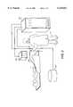
FIG. 2 is a simplified block diagram of the present invention employing an RF tracking system used to follow an invasive device in real-time.
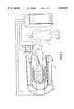
FIG. 3 is a schematic representation of a system according to the present invention for the acquisition of a visible spectrum luminal map indicating health of luminal tissue.
A system for creating 3D tissue maps of a selected lumen within a subject employs an optical spectrum acquisition device in an insertion end of the invasive device, inserted into a lumen of the subject.
The optical spectrum acquisition device operates to create a light beam and directs it to intersect the lumen at several angular displacement θ around the invasive device. The outgoing light beam O is reflected from the lumen wall as a reflected light beam R having a spectrum characteristic of the tissue type at that location of the lumen at the reflection point.
A device locating means is attached to the insertion end of the invasive devce and is tracked by a tracking means, preferably in real time, which passes the location of the device locating means to a look up device.
A rotation sensor measures the angular displacement θ of the irradiating beam and also passes this to look updevice 365. It is beneficial to rotate the outgoing beam O to acquire radial measurements before the invasive device moves significantly.
Look updevice 365 converts the tracked location of the device locatingmeans 261, and the angular displacement of the irradiating beam to estimate a 3D location of the lumen reflecting the irradiating beam. The look up device also correlates the reflected light spectrum with a known, stored tissue type.
The 3D location and the tissue type are stored in a storage device for later retrieval.
The look up device can also be operated to display the 3D locations and corresponding tissue type as a tissue map on a display according to operator defined input.
A user interface may be incorporated which is operated to receive operator defined input from the operator and provide this input to the lookup device.
In an alternative embodiment, an interferometer device may be connected to the fiber optic cable to receive the reflected light. It will then determine a distance D from the lumen to the optical spectrum acquisition device, thereby allowing accurate measurements of radii and diameter of the lumen at various locations. When this information is provided to the look up device, the 3D location on the lumen reflecting the irradiating beam may be determined, resulting in tissue maps incorporating actual measured lumen diameters. This results in actual 3D maps with tissue types superimposed upon it.
It is an object of the present invention to provide a diagnostic luminal map of the visual characteristics of a lumen wall of a subject.
It is another object of the present invention to provide a luminal map of tissue types from the visual light reflected from an internal lumen wall of a subject.
Typically, vascular disease progresses in a somewhat predictable (although usually hidden) manner. Healthy arteries, such as those found in a newborn baby have three well defined layers: the endothelium, media and adventitia. The endothelium is located on the inner surface of the vessel, the media forms the internal structure of the vessel wall and the adventitia defines the outer wall. The endothelium is formed by a porous layer of tissue which is sensitive to the blood moving in the vessel. The adventitia is formed of fibrous material and has the ability to stretch somewhat.
The first step in the progression of arterial disease is the deposition of fatty material in the media layer of the vessel wall. Frequently the location of these deposits is associated with regions of low shear stress associated with vessel bifurcations. These deposits slowly increase in size and cause a thickening of the vessel wall. Because of the pressure of the arterial blood, however, the initial thickening of the wall does not result in a constriction of the internal lumen of the vessel. Rather, the adventitia is stretched and the internal lumen is maintained. At some point in the progression of the disease, however, the adventitia is stretched to its limit and further expansion is impossible. When this occurs, further increases in wall thickening result in a decrease in the caliber of the internal lumen. As the lumen caliber decreases, the local blood velocity increases and damage to the endothelial layer begins to occur. The simultaneous occurrence of a damaged endothelial layer, increased blood velocity and altered flow patterns due to a reduced internal lumen can result in the creation of ulcerations in the vessel wall. If these ulcers become large enough, they can create regions of slow or stagnant flow. Consequently, blood clots can form within the ulcers. Blood clots are not stable, however, and it is possible for portions to break away and become lodged downstream in the vascular system, causing a stroke or heart attack.
There are several variations to the typical progression of arterial disease. For example, if the disease progresses slowly enough, some of the fatty material deposited as a plaque in the wall of the vessel can be converted to a calcified material. Unlike fatty tissue, a calcified plaque is hard and brittle. It is also possible for a plaque to develop its own blood supply, with the formation of microscopic vasculature within the wall of the vessel.
Identification of plaques and differentiation among the types of plaques plays an important role in the diagnosis and treatment of vascular disease. Because they are relatively soft, fatty plaques tend to respond better to mechanical treatment such as balloon angioplasty than brittle calcified plaques. Hemorrhagic plaques, however, respond better to surgical interventions.
Optical spectroscopy has the potential to differentiate the different types of plaques and provide useful diagnostic information. Healthy arterial walls have a smooth pink appearance. Fatty plaques, on the other hand, appear somewhat bumpy and have a yellowish hue. Calcified plaques appear white while hemorrhagic plaques appear red or brownish-red.
Systems for creating a tissue map according to the present invention are shown in FIGS. 1, and 2. These track the real-time location of aninvasive device 320, such as a catheter, within a subject 1.
Anoperator 3, typically a Physician, insertsinvasive device 320 into a lumen of subject 1.Invasive device 320 has an element which is tracked by a tracking means. For magnetic resonance (MR) tracking, the tracked element may be an MR coil, or a plurality of MR coils. These coils may be either receive or transmit coils. The tracked element may also be a quantity of a material which is imaged well in an MR image, such as Gadolinium chelate solution.
The tracking means for MR tracking includes amagnet assembly 101 having RF and gradient coils, andsystem electronics 340. An MR signal is acquired inmagnet assembly 101 and passed tosystem electronics 340 which interpret the signal into a location, or plurality of locations which are tracked in real-time, or near real-time, and displayed on amonitor 380.
In RF tracking, as shown in FIG. 2, the tracked element may be an RF coil, or a plurality of RF coils attached to theinvasive device 320. Anexternal coil 201 operates to transmit an RF signal which is received by the RF coils attached to theinvasive device 320.
RFtracking system electronics 350 interpret the signals to determine the location and orientation ofinvasive device 320 in real-time, and display the location on amonitor 380.
In an alternative embodiment,external coil 201 may be a receive coil and the RF coils attached toinvasive device 320 may be transmit coils.
In FIG. 3, a system fortissue mapping 300 is shown.Tissue mapping system 300 includes an opticalspectrum acquisition device 200 which is intended for the spectral analysis of tissue. Aninvasive device 320 is shown in alumen 310 of subject 1.Lumen 310 may be a vessel, intestine, esophagus, stomach, or other opening within the subject to be imaged. This may also include cavities such as the abdominal cavity which are only accessible through an incision.
Afiber optic cable 240 connects awhite light source 330 to an exit port 241. A white light outgoing beam O is passed downfiber optic cable 240, exits at exit port 241, and impinges upon a fixedparabolic mirror 220.
Outgoing beam O is then reflected back to a rotatingplanar mirror 230. Rotatingplanar mirror 230 reflects outgoing beam O to impinge onlumen wall 310.
Return beam R is reflected off of rotatingplanar mirror 230 and fixedmirror 220 and back into port 241. From port 241 it is passed back downfiber optic cable 240.
Return beam R is then passes to adetector 341 which converts the reflected light into an electronic signal which is passed to aspectrum analyzer 353.
A look updevice 365 receives the spectral information fromspectrum analyzer 353 and correlates this with known, stored, morphological information. For example, if the lumen is a vessel wall and the reflected signal has an amplitude which is high in the yellow frequency band, this may indicate plaque buildup on the inside of the artery. Spectral signals with a high amplitude in the red frequencies may indicate hemorrhaging.
A first device locating means 261 and a second locating means 263 are tracked by conventional MR tracking or RF tracking to determine translational displacement D ofinvasive device 320. The translational displacement D from trackingdevice 360 is provided to look updevice 365.
Two device locating means are shown 261, 263, however, only one is required to determine the location ofinvasive device 320. By using two device locating means, the orientation ofinvasive device 320 may also be determined.
Arotation sensor 395 determines the angular rotation θ offiber optic cable 240, and therefore the angular displacement θ ofrotating mirror 230 and the optical beam. Angular rotation θ fromrotation sensor 395 is also provided to look updevice 365
The morphology information is then associated with the translational displacement D and angular displacement θ of the optical beam in a look updevice 365 to create a morphology map in three dimensions. The 3D morphological map may then be stored in astorage device 370 for later retrieval.
Images may be color coded to distinguish between different tissue morphology. Look updevice 365 receives the user defined input and provides images on adisplay 380 tooperator 3.
In an alternative embodiment, adetector 341 is a conventional interferometer which receives the reflected light beam R.
If it is desired to measure distance betweenrotating mirror 230 and thelumen wall 310,light source 330 should have a monochromatic output anddetector 341 should be an interferometer. Instantaneous distances can then be determined. The measured distances would be provided to look updevice 365, and stored with the other information instorage device 370. This would provide radii and diameters at different locations in subject 1. This would allow look updevice 365 to create 3D maps of the lumen. These maps may be used alone, or to supplement the morphology maps.
Another alternative embodiment would pass a clear fluid through the inside ofinvasive device 320 when the optical beams are operating and the system is acquiring data. The fluid would squirt through a plurality ofports 235 ininvasive device 320 to facilitate transmission of the outgoing and reflected beams when the lumen is full of a fluid which attenuates or scatters light. For example, ifinvasive device 320 was inserted into a vessel of subject 1, sterile saline solution could be squirted throughports 235 to temporarily displace blood in a local region allowing transmission of the optical beam. This would greatly facilitate beam transmission and produce more accurate morphology maps.
The present invention may be employed for a number of different diagnostic procedures. For example,invasive device 320 may be used to determine the biochemical makeup of a blood vessel wall within a living patient. Other embodiments of the present invention could be used to diagnose abnormal tissue in the walls of other body structures such as the colon, small intestines, stomach or esophagus. It should be noted, however, that the present invention could also be employed in non-medical application if desired.
The present invention can also employ ultra-violet, visible, or infra-red light.
In still another embodiment, fluorescent tracers which accumulate in specific types of tissue may be used. The present invention can then easily accurately map the tissue by monitoring the fluorescence.
Conventional spectroscopy methods, such as Raman Spectroscopy, may be employed with the present invention.
While several presently preferred embodiments of the novel invention have been described in detail herein, many modifications and variations will now become apparent to those skilled in the art. It is, therefore, to be understood that the appended claims are intended to cover all such modifications and variations as fall within the true spirit of the invention.
Claims (8)
1. A system for creating a tissue map of a lumen within a subject comprising:
a) an invasive device for insertion into a subject;
b) an optical spectrum acquisition device in an insertion end of the invasive device, disposed to project a light beam to irradiate the lumen at an angular displacement θ around the insertion end of the invasive device, and to receive reflected light, the optical spectrum acquisition device being adapted to create a signal representing the light spectrum reflected from the lumen, and the optical spectrum acquisition device being adapted to measure a respective diameter of the lumen at a respective location of the invasive device within the lumen;
c) at least one device locating means attached to the insertion end of the invasive device;
d) a tracking means for tracking locations of the device locating means within the subject;
e) a rotation sensor for measuring the angular displacement θ of the irradiating beam;
f) a storage device capable of storing an indication of tissue type and corresponding 3D locations for later retrieval;
g) a display;
h) a look up device coupled to each of the following: the optical spectrum acquisition device, the storage device, the display, the tracking means and the rotation sensor, the look up device being configured to convert the tracked location from the tracking means in correspondence with the angular displacement of the irradiating beam and the respective diameter, to estimate a 3D location of the lumen reflecting the irradiating beam, said look up device to correlate the reflected light spectrum to a color frequency stored in the look up device indicating a tissue type, to store the tissue type along with the 3D location represented in the storage device, and to display the 3D locations and corresponding tissue type as a tissue map on the display.
2. The system for creating a tissue map of lumen of claim 1 further comprising:
an interferometer device operating to receive the reflected light, to accurately determine a distance D from the lumen to the optical spectrum acquisition device, and provides it to look up device to more accurately determine the 3D location on the lumen reflecting the irradiating beam, resulting in tissue maps incorporating actual measured lumen diameters.
3. The system for creating a tissue map of lumen of claim 1 wherein the an optical spectrum acquisition device comprises:
a) a fiber optic cable running through the invasive device having an optical port at one end and the other end being an equipment end;
b) a light source coupled to the equipment end of the fiber optic cable operating to create an outgoing light beam O and pass it through the fiber optic cable and out the optical port;
c) an angled rotating mirror attached to the optical port end of the fiber optic cable, having a mirror angled with respect to an axis though the light beam, and capable of rotating substantially about this axis upon rotation of the fiber optic cable, operating to reflect the outgoing beam O to the lumen and the reflected beam R from said lumen back in the opposite direction;
d) a planar mirror for reflecting the outgoing light beam O from the optical port back toward the rotating mirror causing it to reflect laterally to impinge upon said lumen, and for reflecting the return beam back R into the optical port;
e) a detector coupled to the equipment end of optical cable, for converting the reflected beam R into an electronic signal; and
f) a spectrum analyzer coupled to the detector and the look up device, operating to extract spectral information from the electronic signal from detector, and provide it to look up device for tissue determination.
4. The system for creating a tissue map of lumen of claim 1 further comprising:
a user interface for interacting with an operator to acquire operator define input and operating to provide this input to the lookup device.
5. A system for indicating tissue type of a lumen within a subject comprising:
a) an invasive device for insertion into a subject;
b) means for projecting a light beam to irradiate the lumen and to receive reflected light from the lumen, the projecting means being adapted to create a signal representing the light spectrum from the lumen and the projecting means further being adapted to measure a respective diameter of the lumen at a location within the lumen;
c) a tracking means for tracking locations of the invasive device; and,
d) means for receiving the reflected light spectrum, respective diameter measurements, and tracked locations and correlating the reflected light spectrum to a color frequency indicative of tissue type characteristic stored in the receiving and correlating means to indicate the tissue type of the lumen at said tracked locations.
6. The system for indicating tissue type of a lumen of claim 5 wherein the projecting means is configured to project a light beam to irradiate the lumen at an angular displacement θ around an insertion end of the invasive device.
7. The system for indicating tissue type of a lumen of claim 6 further comprising:
a) the invasive device being movable within the subject by an operator; and,
b) at least one device locating means attached to invasive device;
c) the receiving and correlating means being coupled to the tracking means and adapted to indicate the tissue type of the lumen according to operator movement of the invasive device.
8. The system for indicating tissue type of a lumen of claim 7 further comprising a display and means for displaying indicated tissue type on the display.
Priority Applications (1)
| Application Number | Priority Date | Filing Date | Title |
|---|---|---|---|
| US09/017,565US6129667A (en) | 1998-02-02 | 1998-02-02 | Luminal diagnostics employing spectral analysis |
Applications Claiming Priority (1)
| Application Number | Priority Date | Filing Date | Title |
|---|---|---|---|
| US09/017,565US6129667A (en) | 1998-02-02 | 1998-02-02 | Luminal diagnostics employing spectral analysis |
Publications (1)
| Publication Number | Publication Date |
|---|---|
| US6129667Atrue US6129667A (en) | 2000-10-10 |
Family
ID=21783300
Family Applications (1)
| Application Number | Title | Priority Date | Filing Date |
|---|---|---|---|
| US09/017,565Expired - Fee RelatedUS6129667A (en) | 1998-02-02 | 1998-02-02 | Luminal diagnostics employing spectral analysis |
Country Status (1)
| Country | Link |
|---|---|
| US (1) | US6129667A (en) |
Cited By (57)
| Publication number | Priority date | Publication date | Assignee | Title |
|---|---|---|---|---|
| US6263234B1 (en)* | 1996-10-01 | 2001-07-17 | Leica Microsystems Heidelberg Gmbh | Confocal surface-measuring device |
| US6405073B1 (en)* | 1997-07-22 | 2002-06-11 | Scimed Life Systems, Inc. | Miniature spectrometer system and method |
| WO2003059150A2 (en) | 2002-01-09 | 2003-07-24 | Neoguide Systems, Inc. | Apparatus and method for spectroscopic examination of the colon |
| US20040247164A1 (en)* | 2003-06-09 | 2004-12-09 | Simon Furnish | Scanning catheter with position encoder |
| US20050012597A1 (en)* | 2003-07-02 | 2005-01-20 | Anderson Peter Traneus | Wireless electromagnetic tracking system using a nonlinear passive transponder |
| US20050104776A1 (en)* | 2003-11-14 | 2005-05-19 | Anderson Peter T. | Electromagnetic tracking system and method using a three-coil wireless transmitter |
| US20050171437A1 (en)* | 2004-01-14 | 2005-08-04 | Neptec Optical Solutions, Inc. | Optical switching system for catheter-based analysis and treatment |
| US20060058604A1 (en)* | 2004-08-25 | 2006-03-16 | General Electric Company | System and method for hybrid tracking in surgical navigation |
| US20060055712A1 (en)* | 2004-08-24 | 2006-03-16 | Anderson Peter T | Method and system for field mapping using integral methodology |
| US20060103850A1 (en)* | 2004-11-12 | 2006-05-18 | Alphonse Gerard A | Single trace multi-channel low coherence interferometric sensor |
| US20060106292A1 (en)* | 2003-09-24 | 2006-05-18 | General Electric Company | System and method for employing multiple coil architectures simultaneously in one electromagnetic tracking system |
| US20060292698A1 (en)* | 2005-06-24 | 2006-12-28 | Hartle Jennifer W | Spectroscopic methods for detecting and identifying chelates |
| US7158754B2 (en) | 2003-07-01 | 2007-01-02 | Ge Medical Systems Global Technology Company, Llc | Electromagnetic tracking system and method using a single-coil transmitter |
| US20070078348A1 (en)* | 2003-12-11 | 2007-04-05 | Holman Hoi-Ying N | Catheter-based mid-infrared reflectance and reflectance generated absorption spectroscopy |
| US20070088497A1 (en)* | 2005-06-14 | 2007-04-19 | Jung Mun H | Matching camera-photographed image with map data in portable terminal and travel route guidance method |
| US20070113860A1 (en)* | 2005-11-22 | 2007-05-24 | Anderson Peter T | Tracking apparatus and a method of using |
| US20070129629A1 (en)* | 2005-11-23 | 2007-06-07 | Beauregard Gerald L | System and method for surgical navigation |
| US20070208251A1 (en)* | 2006-03-02 | 2007-09-06 | General Electric Company | Transformer-coupled guidewire system and method of use |
| US20070225559A1 (en)* | 2006-03-21 | 2007-09-27 | Boston Scientific Scimed, Inc. | Vision catheter having electromechanical navigation |
| US20070229080A1 (en)* | 2004-04-26 | 2007-10-04 | Koninklijke Philips Electronics N.V. | Electro-Optical Magnetic Resonance Transducer |
| US20080130965A1 (en)* | 2004-11-23 | 2008-06-05 | Avinash Gopal B | Method and apparatus for parameter assisted image-guided surgery (PAIGS) |
| US20080161696A1 (en)* | 2006-11-08 | 2008-07-03 | Lightlab Imaging, Inc. | Opto-acoustic imaging devices and methods |
| DE102007045988A1 (en)* | 2007-09-26 | 2008-10-23 | Siemens Ag | Spectroscopy unit for characterization of e.g. tumor, has light inlet/outlet controlling light from inspection device into examination object, where inspection device is marked in subranges with magnet resonance visible substances |
| US7471202B2 (en) | 2006-03-29 | 2008-12-30 | General Electric Co. | Conformal coil array for a medical tracking system |
| US20090062739A1 (en)* | 2007-08-31 | 2009-03-05 | General Electric Company | Catheter Guidewire Tracking System and Method |
| US7532997B2 (en) | 2006-04-17 | 2009-05-12 | General Electric Company | Electromagnetic tracking using a discretized numerical field model |
| US20100268025A1 (en)* | 2007-11-09 | 2010-10-21 | Amir Belson | Apparatus and methods for capsule endoscopy of the esophagus |
| US20110060189A1 (en)* | 2004-06-30 | 2011-03-10 | Given Imaging Ltd. | Apparatus and Methods for Capsule Endoscopy of the Esophagus |
| US20110082451A1 (en)* | 2009-10-06 | 2011-04-07 | Cardiofocus, Inc. | Cardiac ablation image analysis system and process |
| US8048063B2 (en) | 2006-06-09 | 2011-11-01 | Endosense Sa | Catheter having tri-axial force sensor |
| US8062212B2 (en) | 2000-04-03 | 2011-11-22 | Intuitive Surgical Operations, Inc. | Steerable endoscope and improved method of insertion |
| US8075498B2 (en) | 2005-03-04 | 2011-12-13 | Endosense Sa | Medical apparatus system having optical fiber load sensing capability |
| US8083879B2 (en) | 2005-11-23 | 2011-12-27 | Intuitive Surgical Operations, Inc. | Non-metallic, multi-strand control cable for steerable instruments |
| US8126531B2 (en) | 1996-11-21 | 2012-02-28 | Boston Scientific Scimed, Inc. | Miniature spectrometer |
| US8157789B2 (en) | 2007-05-24 | 2012-04-17 | Endosense Sa | Touch sensing catheter |
| US8182433B2 (en) | 2005-03-04 | 2012-05-22 | Endosense Sa | Medical apparatus system having optical fiber load sensing capability |
| US8182418B2 (en) | 2008-02-25 | 2012-05-22 | Intuitive Surgical Operations, Inc. | Systems and methods for articulating an elongate body |
| US8298227B2 (en) | 2008-05-14 | 2012-10-30 | Endosense Sa | Temperature compensated strain sensing catheter |
| US8361090B2 (en) | 2002-01-09 | 2013-01-29 | Intuitive Surgical Operations, Inc. | Apparatus and method for endoscopic colectomy |
| US8391952B2 (en) | 2007-10-11 | 2013-03-05 | General Electric Company | Coil arrangement for an electromagnetic tracking system |
| US8517923B2 (en) | 2000-04-03 | 2013-08-27 | Intuitive Surgical Operations, Inc. | Apparatus and methods for facilitating treatment of tissue via improved delivery of energy based and non-energy based modalities |
| US8568299B2 (en) | 2006-05-19 | 2013-10-29 | Intuitive Surgical Operations, Inc. | Methods and apparatus for displaying three-dimensional orientation of a steerable distal tip of an endoscope |
| US8567265B2 (en) | 2006-06-09 | 2013-10-29 | Endosense, SA | Triaxial fiber optic force sensing catheter |
| US20130331709A1 (en)* | 2012-06-07 | 2013-12-12 | Poincare Systems, Inc. | Grin lens and methods of making the same |
| US8622935B1 (en) | 2007-05-25 | 2014-01-07 | Endosense Sa | Elongated surgical manipulator with body position and distal force sensing |
| US8721530B2 (en) | 2000-04-03 | 2014-05-13 | Intuitive Surgical Operations, Inc. | Tendon-driven endoscope and methods of use |
| US8845524B2 (en) | 2000-04-03 | 2014-09-30 | Intuitive Surgical Operations, Inc. | Steerable segmented endoscope and method of insertion |
| US8882657B2 (en) | 2003-03-07 | 2014-11-11 | Intuitive Surgical Operations, Inc. | Instrument having radio frequency identification systems and methods for use |
| US8888688B2 (en) | 2000-04-03 | 2014-11-18 | Intuitive Surgical Operations, Inc. | Connector device for a controllable instrument |
| US8894589B2 (en) | 2005-08-01 | 2014-11-25 | Endosense Sa | Medical apparatus system having optical fiber load sensing capability |
| US9220398B2 (en) | 2007-10-11 | 2015-12-29 | Intuitive Surgical Operations, Inc. | System for managing Bowden cables in articulating instruments |
| US9833221B2 (en) | 2013-03-15 | 2017-12-05 | Lightlab Imaging, Inc. | Apparatus and method of image registration |
| US10512392B2 (en) | 2008-02-06 | 2019-12-24 | Intuitive Surgical Operations, Inc. | Segmented instrument having braking capabilities |
| US10561368B2 (en) | 2011-04-14 | 2020-02-18 | St. Jude Medical International Holding S.À R.L. | Compact force sensor for catheters |
| US10792012B2 (en) | 2012-11-19 | 2020-10-06 | Lightlab Imaging, Inc. | Interface devices, systems and methods for multimodal probes |
| US11096563B2 (en) | 2005-11-22 | 2021-08-24 | Intuitive Surgical Operations, Inc. | Method of determining the shape of a bendable instrument |
| US11445937B2 (en) | 2016-01-07 | 2022-09-20 | St. Jude Medical International Holding S.À R.L. | Medical device with multi-core fiber for optical sensing |
Citations (26)
| Publication number | Priority date | Publication date | Assignee | Title |
|---|---|---|---|---|
| US5010886A (en)* | 1989-08-18 | 1991-04-30 | Intertherapy, Inc. | Medical probe assembly having combined ultrasonic imaging and laser ablation capabilities |
| US5307808A (en)* | 1992-04-01 | 1994-05-03 | General Electric Company | Tracking system and pulse sequences to monitor the position of a device using magnetic resonance |
| US5377678A (en)* | 1991-09-03 | 1995-01-03 | General Electric Company | Tracking system to follow the position and orientation of a device with radiofrequency fields |
| US5408998A (en)* | 1994-03-10 | 1995-04-25 | Ethicon Endo-Surgery | Video based tissue oximetry |
| US5441053A (en)* | 1991-05-03 | 1995-08-15 | University Of Kentucky Research Foundation | Apparatus and method for multiple wavelength of tissue |
| US5582171A (en)* | 1994-07-08 | 1996-12-10 | Insight Medical Systems, Inc. | Apparatus for doppler interferometric imaging and imaging guidewire |
| US5678550A (en)* | 1995-08-11 | 1997-10-21 | The United States Of America As Represented By The Secretary Of The Department Of Health And Human Services | Apparatus and method for in situ detection of areas of cardiac electrical activity |
| US5740808A (en)* | 1996-10-28 | 1998-04-21 | Ep Technologies, Inc | Systems and methods for guilding diagnostic or therapeutic devices in interior tissue regions |
| US5749835A (en)* | 1994-09-06 | 1998-05-12 | Sims Deltec, Inc. | Method and apparatus for location of a catheter tip |
| US5752518A (en)* | 1996-10-28 | 1998-05-19 | Ep Technologies, Inc. | Systems and methods for visualizing interior regions of the body |
| US5785658A (en)* | 1992-09-14 | 1998-07-28 | Sexant Medical Corporation | In vivo tissue analysis methods and apparatus |
| US5792053A (en)* | 1997-03-17 | 1998-08-11 | Polartechnics, Limited | Hybrid probe for tissue type recognition |
| US5823942A (en)* | 1992-08-25 | 1998-10-20 | Fuji Photo Film Co., Ltd. | Endoscope with surface and deep portion imaging systems |
| US5827190A (en)* | 1994-03-28 | 1998-10-27 | Xillix Technologies Corp. | Endoscope having an integrated CCD sensor |
| US5840035A (en)* | 1995-02-07 | 1998-11-24 | Siemens Aktiengesellschaft | Method for the spectroscopic examination of a biological tissue |
| US5842995A (en)* | 1996-06-28 | 1998-12-01 | Board Of Regents, The Univerisity Of Texas System | Spectroscopic probe for in vivo measurement of raman signals |
| US5851181A (en)* | 1996-08-30 | 1998-12-22 | Esc Medical Systems Ltd. | Apparatus for simultaneously viewing and spectrally analyzing a portion of skin |
| US5868674A (en)* | 1995-11-24 | 1999-02-09 | U.S. Philips Corporation | MRI-system and catheter for interventional procedures |
| US5899860A (en)* | 1996-09-12 | 1999-05-04 | Siemens Elema Ab | Method and device for determining the position of a catheter inside the body of a patient |
| US5921926A (en)* | 1997-07-28 | 1999-07-13 | University Of Central Florida | Three dimensional optical imaging colposcopy |
| US5938602A (en)* | 1996-06-11 | 1999-08-17 | Roke Manor Research Limited | Catheter tracking system and method |
| US5951482A (en)* | 1997-10-03 | 1999-09-14 | Intraluminal Therapeutics, Inc. | Assemblies and methods for advancing a guide wire through body tissue |
| US5999844A (en)* | 1997-04-23 | 1999-12-07 | Accumed International, Inc. | Method and apparatus for imaging and sampling diseased tissue using autofluorescence |
| US6006128A (en)* | 1997-06-02 | 1999-12-21 | Izatt; Joseph A. | Doppler flow imaging using optical coherence tomography |
| US6016439A (en)* | 1996-10-15 | 2000-01-18 | Biosense, Inc. | Method and apparatus for synthetic viewpoint imaging |
| US6052610A (en)* | 1998-01-09 | 2000-04-18 | International Business Machines Corporation | Magnetic catheter tracker and method therefor |
- 1998
- 1998-02-02USUS09/017,565patent/US6129667A/ennot_activeExpired - Fee Related
Patent Citations (26)
| Publication number | Priority date | Publication date | Assignee | Title |
|---|---|---|---|---|
| US5010886A (en)* | 1989-08-18 | 1991-04-30 | Intertherapy, Inc. | Medical probe assembly having combined ultrasonic imaging and laser ablation capabilities |
| US5441053A (en)* | 1991-05-03 | 1995-08-15 | University Of Kentucky Research Foundation | Apparatus and method for multiple wavelength of tissue |
| US5377678A (en)* | 1991-09-03 | 1995-01-03 | General Electric Company | Tracking system to follow the position and orientation of a device with radiofrequency fields |
| US5307808A (en)* | 1992-04-01 | 1994-05-03 | General Electric Company | Tracking system and pulse sequences to monitor the position of a device using magnetic resonance |
| US5823942A (en)* | 1992-08-25 | 1998-10-20 | Fuji Photo Film Co., Ltd. | Endoscope with surface and deep portion imaging systems |
| US5785658A (en)* | 1992-09-14 | 1998-07-28 | Sexant Medical Corporation | In vivo tissue analysis methods and apparatus |
| US5408998A (en)* | 1994-03-10 | 1995-04-25 | Ethicon Endo-Surgery | Video based tissue oximetry |
| US5827190A (en)* | 1994-03-28 | 1998-10-27 | Xillix Technologies Corp. | Endoscope having an integrated CCD sensor |
| US5582171A (en)* | 1994-07-08 | 1996-12-10 | Insight Medical Systems, Inc. | Apparatus for doppler interferometric imaging and imaging guidewire |
| US5749835A (en)* | 1994-09-06 | 1998-05-12 | Sims Deltec, Inc. | Method and apparatus for location of a catheter tip |
| US5840035A (en)* | 1995-02-07 | 1998-11-24 | Siemens Aktiengesellschaft | Method for the spectroscopic examination of a biological tissue |
| US5678550A (en)* | 1995-08-11 | 1997-10-21 | The United States Of America As Represented By The Secretary Of The Department Of Health And Human Services | Apparatus and method for in situ detection of areas of cardiac electrical activity |
| US5868674A (en)* | 1995-11-24 | 1999-02-09 | U.S. Philips Corporation | MRI-system and catheter for interventional procedures |
| US5938602A (en)* | 1996-06-11 | 1999-08-17 | Roke Manor Research Limited | Catheter tracking system and method |
| US5842995A (en)* | 1996-06-28 | 1998-12-01 | Board Of Regents, The Univerisity Of Texas System | Spectroscopic probe for in vivo measurement of raman signals |
| US5851181A (en)* | 1996-08-30 | 1998-12-22 | Esc Medical Systems Ltd. | Apparatus for simultaneously viewing and spectrally analyzing a portion of skin |
| US5899860A (en)* | 1996-09-12 | 1999-05-04 | Siemens Elema Ab | Method and device for determining the position of a catheter inside the body of a patient |
| US6016439A (en)* | 1996-10-15 | 2000-01-18 | Biosense, Inc. | Method and apparatus for synthetic viewpoint imaging |
| US5740808A (en)* | 1996-10-28 | 1998-04-21 | Ep Technologies, Inc | Systems and methods for guilding diagnostic or therapeutic devices in interior tissue regions |
| US5752518A (en)* | 1996-10-28 | 1998-05-19 | Ep Technologies, Inc. | Systems and methods for visualizing interior regions of the body |
| US5792053A (en)* | 1997-03-17 | 1998-08-11 | Polartechnics, Limited | Hybrid probe for tissue type recognition |
| US5999844A (en)* | 1997-04-23 | 1999-12-07 | Accumed International, Inc. | Method and apparatus for imaging and sampling diseased tissue using autofluorescence |
| US6006128A (en)* | 1997-06-02 | 1999-12-21 | Izatt; Joseph A. | Doppler flow imaging using optical coherence tomography |
| US5921926A (en)* | 1997-07-28 | 1999-07-13 | University Of Central Florida | Three dimensional optical imaging colposcopy |
| US5951482A (en)* | 1997-10-03 | 1999-09-14 | Intraluminal Therapeutics, Inc. | Assemblies and methods for advancing a guide wire through body tissue |
| US6052610A (en)* | 1998-01-09 | 2000-04-18 | International Business Machines Corporation | Magnetic catheter tracker and method therefor |
Cited By (112)
| Publication number | Priority date | Publication date | Assignee | Title |
|---|---|---|---|---|
| US6263234B1 (en)* | 1996-10-01 | 2001-07-17 | Leica Microsystems Heidelberg Gmbh | Confocal surface-measuring device |
| US8660637B2 (en) | 1996-11-21 | 2014-02-25 | Boston Scientific Scimed, Inc. | Miniature spectrometer |
| US8126531B2 (en) | 1996-11-21 | 2012-02-28 | Boston Scientific Scimed, Inc. | Miniature spectrometer |
| US6405073B1 (en)* | 1997-07-22 | 2002-06-11 | Scimed Life Systems, Inc. | Miniature spectrometer system and method |
| US8062212B2 (en) | 2000-04-03 | 2011-11-22 | Intuitive Surgical Operations, Inc. | Steerable endoscope and improved method of insertion |
| US9808140B2 (en) | 2000-04-03 | 2017-11-07 | Intuitive Surgical Operations, Inc. | Steerable segmented endoscope and method of insertion |
| US10105036B2 (en) | 2000-04-03 | 2018-10-23 | Intuitive Surgical Operations, Inc. | Connector device for a controllable instrument |
| US10327625B2 (en) | 2000-04-03 | 2019-06-25 | Intuitive Surgical Operations, Inc. | Apparatus and methods for facilitating treatment of tissue via improved delivery of energy based and non-energy based modalities |
| US10736490B2 (en) | 2000-04-03 | 2020-08-11 | Intuitive Surgical Operations, Inc. | Connector device for a controllable instrument |
| US10893794B2 (en) | 2000-04-03 | 2021-01-19 | Intuitive Surgical Operations, Inc. | Steerable endoscope and improved method of insertion |
| US8517923B2 (en) | 2000-04-03 | 2013-08-27 | Intuitive Surgical Operations, Inc. | Apparatus and methods for facilitating treatment of tissue via improved delivery of energy based and non-energy based modalities |
| US11026564B2 (en) | 2000-04-03 | 2021-06-08 | Intuitive Surgical Operations, Inc. | Apparatus and methods for facilitating treatment of tissue via improved delivery of energy based and non-energy based modalities |
| US8888688B2 (en) | 2000-04-03 | 2014-11-18 | Intuitive Surgical Operations, Inc. | Connector device for a controllable instrument |
| US8641602B2 (en) | 2000-04-03 | 2014-02-04 | Intuitive Surgical Operations, Inc. | Steerable endoscope and improved method of insertion |
| US9427282B2 (en) | 2000-04-03 | 2016-08-30 | Intuitive Surgical Operations, Inc. | Apparatus and methods for facilitating treatment of tissue via improved delivery of energy based and non-energy based modalities |
| US8845524B2 (en) | 2000-04-03 | 2014-09-30 | Intuitive Surgical Operations, Inc. | Steerable segmented endoscope and method of insertion |
| US9138132B2 (en) | 2000-04-03 | 2015-09-22 | Intuitive Surgical Operations, Inc. | Steerable endoscope and improved method of insertion |
| US8834354B2 (en) | 2000-04-03 | 2014-09-16 | Intuitive Surgical Operations, Inc. | Steerable endoscope and improved method of insertion |
| US8827894B2 (en) | 2000-04-03 | 2014-09-09 | Intuitive Surgical Operations, Inc. | Steerable endoscope and improved method of insertion |
| US8721530B2 (en) | 2000-04-03 | 2014-05-13 | Intuitive Surgical Operations, Inc. | Tendon-driven endoscope and methods of use |
| US12076102B2 (en) | 2000-04-03 | 2024-09-03 | Intuitive Surgical Operations, Inc. | Connector device for a controllable instrument |
| US8361090B2 (en) | 2002-01-09 | 2013-01-29 | Intuitive Surgical Operations, Inc. | Apparatus and method for endoscopic colectomy |
| US8696694B2 (en) | 2002-01-09 | 2014-04-15 | Intuitive Surgical Operations, Inc. | Apparatus and method for endoscopic colectomy |
| US10349816B2 (en) | 2002-01-09 | 2019-07-16 | Intuitive Surgical Operations, Inc. | Apparatus and method for endoscopic colectomy |
| US9421016B2 (en) | 2002-01-09 | 2016-08-23 | Intuitive Surgical Operations, Inc. | Apparatus and method for endoscopic colectomy |
| WO2003059150A2 (en) | 2002-01-09 | 2003-07-24 | Neoguide Systems, Inc. | Apparatus and method for spectroscopic examination of the colon |
| US9980778B2 (en) | 2003-03-07 | 2018-05-29 | Intuitive Surgical Operations, Inc. | Instrument having radio frequency identification systems and methods for use |
| US8882657B2 (en) | 2003-03-07 | 2014-11-11 | Intuitive Surgical Operations, Inc. | Instrument having radio frequency identification systems and methods for use |
| US10959807B2 (en) | 2003-03-07 | 2021-03-30 | Intuitive Surgical Operations, Inc. | Systems and methods for determining the state of motion of an instrument |
| US7292715B2 (en) | 2003-06-09 | 2007-11-06 | Infraredx, Inc. | Display of diagnostic data |
| US20040247164A1 (en)* | 2003-06-09 | 2004-12-09 | Simon Furnish | Scanning catheter with position encoder |
| US7158754B2 (en) | 2003-07-01 | 2007-01-02 | Ge Medical Systems Global Technology Company, Llc | Electromagnetic tracking system and method using a single-coil transmitter |
| US20050012597A1 (en)* | 2003-07-02 | 2005-01-20 | Anderson Peter Traneus | Wireless electromagnetic tracking system using a nonlinear passive transponder |
| US8354837B2 (en) | 2003-09-24 | 2013-01-15 | Ge Medical Systems Global Technology Company Llc | System and method for electromagnetic tracking operable with multiple coil architectures |
| US20060106292A1 (en)* | 2003-09-24 | 2006-05-18 | General Electric Company | System and method for employing multiple coil architectures simultaneously in one electromagnetic tracking system |
| US7715898B2 (en) | 2003-09-24 | 2010-05-11 | General Electric Company | System and method for employing multiple coil architectures simultaneously in one electromagnetic tracking system |
| US7015859B2 (en) | 2003-11-14 | 2006-03-21 | General Electric Company | Electromagnetic tracking system and method using a three-coil wireless transmitter |
| US20050104776A1 (en)* | 2003-11-14 | 2005-05-19 | Anderson Peter T. | Electromagnetic tracking system and method using a three-coil wireless transmitter |
| US8571640B2 (en)* | 2003-12-11 | 2013-10-29 | The Regents Of The University Of California | Catheter based mid-infrared reflectance and reflectance generated absorption spectroscopy |
| US20070078348A1 (en)* | 2003-12-11 | 2007-04-05 | Holman Hoi-Ying N | Catheter-based mid-infrared reflectance and reflectance generated absorption spectroscopy |
| US20050171437A1 (en)* | 2004-01-14 | 2005-08-04 | Neptec Optical Solutions, Inc. | Optical switching system for catheter-based analysis and treatment |
| US20070229080A1 (en)* | 2004-04-26 | 2007-10-04 | Koninklijke Philips Electronics N.V. | Electro-Optical Magnetic Resonance Transducer |
| US20110060189A1 (en)* | 2004-06-30 | 2011-03-10 | Given Imaging Ltd. | Apparatus and Methods for Capsule Endoscopy of the Esophagus |
| US9968290B2 (en) | 2004-06-30 | 2018-05-15 | Given Imaging Ltd. | Apparatus and methods for capsule endoscopy of the esophagus |
| US8131342B2 (en) | 2004-08-24 | 2012-03-06 | General Electric Company | Method and system for field mapping using integral methodology |
| US20060055712A1 (en)* | 2004-08-24 | 2006-03-16 | Anderson Peter T | Method and system for field mapping using integral methodology |
| US7702379B2 (en) | 2004-08-25 | 2010-04-20 | General Electric Company | System and method for hybrid tracking in surgical navigation |
| US20060058604A1 (en)* | 2004-08-25 | 2006-03-16 | General Electric Company | System and method for hybrid tracking in surgical navigation |
| US7417740B2 (en) | 2004-11-12 | 2008-08-26 | Medeikon Corporation | Single trace multi-channel low coherence interferometric sensor |
| US20060103850A1 (en)* | 2004-11-12 | 2006-05-18 | Alphonse Gerard A | Single trace multi-channel low coherence interferometric sensor |
| US20080130965A1 (en)* | 2004-11-23 | 2008-06-05 | Avinash Gopal B | Method and apparatus for parameter assisted image-guided surgery (PAIGS) |
| US10973606B2 (en) | 2005-03-04 | 2021-04-13 | St. Jude Medical International Holding S.À R.L. | Medical apparatus system having optical fiber load sensing capability |
| US11998404B2 (en) | 2005-03-04 | 2024-06-04 | St. Jude Medical International Holding S.À R.L. | Medical apparatus system having optical fiber load sensing capability |
| US9907618B2 (en) | 2005-03-04 | 2018-03-06 | St Jude Medical International Holding S.À R.L. | Medical apparatus system having optical fiber sensing capability |
| US8961436B2 (en) | 2005-03-04 | 2015-02-24 | St. Jude Medical Luxembourg Holding S.á.r.l. | Medical apparatus system having optical fiber load sensing capability |
| US8075498B2 (en) | 2005-03-04 | 2011-12-13 | Endosense Sa | Medical apparatus system having optical fiber load sensing capability |
| US8182433B2 (en) | 2005-03-04 | 2012-05-22 | Endosense Sa | Medical apparatus system having optical fiber load sensing capability |
| US8932288B2 (en) | 2005-03-04 | 2015-01-13 | Endosense Sa | Medical apparatus system having optical fiber load sensing capability |
| US20070088497A1 (en)* | 2005-06-14 | 2007-04-19 | Jung Mun H | Matching camera-photographed image with map data in portable terminal and travel route guidance method |
| US20060292698A1 (en)* | 2005-06-24 | 2006-12-28 | Hartle Jennifer W | Spectroscopic methods for detecting and identifying chelates |
| US8894589B2 (en) | 2005-08-01 | 2014-11-25 | Endosense Sa | Medical apparatus system having optical fiber load sensing capability |
| US11096563B2 (en) | 2005-11-22 | 2021-08-24 | Intuitive Surgical Operations, Inc. | Method of determining the shape of a bendable instrument |
| US11617499B2 (en) | 2005-11-22 | 2023-04-04 | Intuitive Surgical Operations, Inc. | System for determining the shape of a bendable instrument |
| US20070113860A1 (en)* | 2005-11-22 | 2007-05-24 | Anderson Peter T | Tracking apparatus and a method of using |
| US8083879B2 (en) | 2005-11-23 | 2011-12-27 | Intuitive Surgical Operations, Inc. | Non-metallic, multi-strand control cable for steerable instruments |
| US20070129629A1 (en)* | 2005-11-23 | 2007-06-07 | Beauregard Gerald L | System and method for surgical navigation |
| US20070208251A1 (en)* | 2006-03-02 | 2007-09-06 | General Electric Company | Transformer-coupled guidewire system and method of use |
| US8016749B2 (en)* | 2006-03-21 | 2011-09-13 | Boston Scientific Scimed, Inc. | Vision catheter having electromechanical navigation |
| US20070225559A1 (en)* | 2006-03-21 | 2007-09-27 | Boston Scientific Scimed, Inc. | Vision catheter having electromechanical navigation |
| US7471202B2 (en) | 2006-03-29 | 2008-12-30 | General Electric Co. | Conformal coil array for a medical tracking system |
| US7532997B2 (en) | 2006-04-17 | 2009-05-12 | General Electric Company | Electromagnetic tracking using a discretized numerical field model |
| US8568299B2 (en) | 2006-05-19 | 2013-10-29 | Intuitive Surgical Operations, Inc. | Methods and apparatus for displaying three-dimensional orientation of a steerable distal tip of an endoscope |
| US12256891B2 (en) | 2006-05-19 | 2025-03-25 | Intuitive Surgical Operations, Inc. | Methods and apparatus for displaying three-dimensional orientation of a steerable distal tip of an endoscope |
| US10426412B2 (en) | 2006-05-19 | 2019-10-01 | Intuitive Surgical Operations, Inc. | Methods and apparatus for displaying three-dimensional orientation of a steerable distal tip of an endoscope |
| US9357901B2 (en) | 2006-05-19 | 2016-06-07 | Intuitive Surgical Operations, Inc. | Methods and apparatus for displaying three-dimensional orientation of a steerable distal tip of an endoscope |
| US10596346B2 (en) | 2006-06-09 | 2020-03-24 | St. Jude Medical International Holding S.À R.L. | Triaxial fiber optic force sensing catheter |
| US8435232B2 (en) | 2006-06-09 | 2013-05-07 | Nicolas Aeby | Catheter having tri-axial force sensor |
| US9597036B2 (en) | 2006-06-09 | 2017-03-21 | St. Jude Medical International Holding S.À R.L. | Triaxial fiber optic force sensing catheter and method of use |
| US8567265B2 (en) | 2006-06-09 | 2013-10-29 | Endosense, SA | Triaxial fiber optic force sensing catheter |
| US11883131B2 (en) | 2006-06-09 | 2024-01-30 | St. Jude Medical International Holding S.À R.L. | Triaxial fiber optic force sensing catheter |
| US8048063B2 (en) | 2006-06-09 | 2011-11-01 | Endosense Sa | Catheter having tri-axial force sensor |
| US8449468B2 (en) | 2006-11-08 | 2013-05-28 | Lightlab Imaging, Inc. | Opto-acoustic imaging devices and methods |
| US8753281B2 (en) | 2006-11-08 | 2014-06-17 | Lightlab Imaging Inc. | Opto-acoustic imaging devices and methods |
| US7935060B2 (en) | 2006-11-08 | 2011-05-03 | Lightlab Imaging, Inc. | Opto-acoustic imaging devices and methods |
| US20080161696A1 (en)* | 2006-11-08 | 2008-07-03 | Lightlab Imaging, Inc. | Opto-acoustic imaging devices and methods |
| US8157789B2 (en) | 2007-05-24 | 2012-04-17 | Endosense Sa | Touch sensing catheter |
| US9993617B1 (en) | 2007-05-25 | 2018-06-12 | St. Jude Medical International Holdings S.À R.L. | Elongated surgical manipulator with body position and distal force sensing |
| US8622935B1 (en) | 2007-05-25 | 2014-01-07 | Endosense Sa | Elongated surgical manipulator with body position and distal force sensing |
| US10905855B2 (en) | 2007-05-25 | 2021-02-02 | St. Jude Medical International Holding S.ár.l. | Elongated surgical manipulator with body position and distal force sensing |
| US20090062739A1 (en)* | 2007-08-31 | 2009-03-05 | General Electric Company | Catheter Guidewire Tracking System and Method |
| DE102007045988A1 (en)* | 2007-09-26 | 2008-10-23 | Siemens Ag | Spectroscopy unit for characterization of e.g. tumor, has light inlet/outlet controlling light from inspection device into examination object, where inspection device is marked in subranges with magnet resonance visible substances |
| US9220398B2 (en) | 2007-10-11 | 2015-12-29 | Intuitive Surgical Operations, Inc. | System for managing Bowden cables in articulating instruments |
| US8391952B2 (en) | 2007-10-11 | 2013-03-05 | General Electric Company | Coil arrangement for an electromagnetic tracking system |
| US20100268025A1 (en)* | 2007-11-09 | 2010-10-21 | Amir Belson | Apparatus and methods for capsule endoscopy of the esophagus |
| US10512392B2 (en) | 2008-02-06 | 2019-12-24 | Intuitive Surgical Operations, Inc. | Segmented instrument having braking capabilities |
| US10952594B2 (en) | 2008-02-06 | 2021-03-23 | Intuitive Surgical Operations, Inc. | Segmented instrument having braking capabilities |
| US8608647B2 (en) | 2008-02-25 | 2013-12-17 | Intuitive Surgical Operations, Inc. | Systems and methods for articulating an elongate body |
| US8182418B2 (en) | 2008-02-25 | 2012-05-22 | Intuitive Surgical Operations, Inc. | Systems and methods for articulating an elongate body |
| US8298227B2 (en) | 2008-05-14 | 2012-10-30 | Endosense Sa | Temperature compensated strain sensing catheter |
| US20110082451A1 (en)* | 2009-10-06 | 2011-04-07 | Cardiofocus, Inc. | Cardiac ablation image analysis system and process |
| US8702688B2 (en)* | 2009-10-06 | 2014-04-22 | Cardiofocus, Inc. | Cardiac ablation image analysis system and process |
| US11564628B2 (en) | 2011-04-14 | 2023-01-31 | St. Jude Medical International Holding S.À R.L. | Compact force sensor for catheters |
| US10561368B2 (en) | 2011-04-14 | 2020-02-18 | St. Jude Medical International Holding S.À R.L. | Compact force sensor for catheters |
| US10539731B2 (en)* | 2012-06-07 | 2020-01-21 | Poinare Systems, Inc. | Grin lens and methods of making the same |
| US20130331709A1 (en)* | 2012-06-07 | 2013-12-12 | Poincare Systems, Inc. | Grin lens and methods of making the same |
| US10792012B2 (en) | 2012-11-19 | 2020-10-06 | Lightlab Imaging, Inc. | Interface devices, systems and methods for multimodal probes |
| US11701089B2 (en) | 2012-11-19 | 2023-07-18 | Lightlab Imaging, Inc. | Multimodal imaging systems, probes and methods |
| US12127882B2 (en) | 2012-11-19 | 2024-10-29 | Lightlab Imaging, Inc. | Multimodal imaging systems probes and methods |
| US12127881B2 (en) | 2012-11-19 | 2024-10-29 | Lightlab Imaging, Inc. | Interface devices, systems and methods for multimodal probes |
| US9833221B2 (en) | 2013-03-15 | 2017-12-05 | Lightlab Imaging, Inc. | Apparatus and method of image registration |
| US11445937B2 (en) | 2016-01-07 | 2022-09-20 | St. Jude Medical International Holding S.À R.L. | Medical device with multi-core fiber for optical sensing |
| US11998310B2 (en) | 2016-01-07 | 2024-06-04 | St. Jude Medical International Holding S.À R.L. | Medical device with multi-core fiber for optical sensing |
Similar Documents
| Publication | Publication Date | Title |
|---|---|---|
| US6129667A (en) | Luminal diagnostics employing spectral analysis | |
| US11412985B2 (en) | Biopsy guidance by image-based X-ray system and photonic needle | |
| US20030032880A1 (en) | Apparatus and method for ultrasonically identifying vulnerable plaque | |
| US8029446B2 (en) | Ultrasonic imaging system and imaging method | |
| CN105899142B (en) | Detecting endoleaks associated with aneurysm repair | |
| CA2108910C (en) | Coronary artery imaging system | |
| JP4226788B2 (en) | System and method for controlling tissue ablation | |
| US20090203991A1 (en) | Multiple imaging and/or spectroscopic modality probe | |
| EP2443992A2 (en) | Diagnosis support apparatus, diagnosis support method, lesioned part detection apparatus, and lesioned part detection method | |
| US20100198081A1 (en) | Scanning light imager | |
| WO1999030608A1 (en) | Apparatus and method for determining tissue characteristics | |
| WO2003088816A2 (en) | Methods and apparatus for the identification and stabilization of vulnerable plaque | |
| JP2005066311A (en) | Ultrasonic vein detector and detection method | |
| CN111462117B (en) | Data processing system and data processing method based on blood vessel image | |
| Berland et al. | Porta hepatis: sonographic discrimination of bile ducts from arteries with pulsed Doppler with new anatomic criteria | |
| KR20120028154A (en) | Diagnose method and apparatus for atherosclerotic lesions | |
| CN212365043U (en) | Data processing system based on blood vessel image | |
| Madjar et al. | Continuous-wave and pulsed Doppler studies of the breast: clinical results and effect of transducer frequency | |
| KR20110028916A (en) | Apparatus and method for measuring atherosclerotic plaque component using endovascular ultrasound image | |
| Pandian et al. | Intravascular ultrasound estimation of arterial stenosis | |
| Li et al. | Temporal averaging for quantification of lumen dimensions in intravascular ultrasound images | |
| Winter-Warnars et al. | Interobserver variation in duplex sonographic scanning in the femoropopliteal tract. | |
| Bandyk et al. | Classification of carotid bifurcation disease using quantitative Doppler spectrum analysis | |
| Sumner | Use of color-flow imaging technique in carotid artery disease | |
| Bagi et al. | Doppler waveform analysis in evaluation of occlusive arterial disease in the lower limb: comparison with distal blood pressure measurement and arteriography |
Legal Events
| Date | Code | Title | Description |
|---|---|---|---|
| AS | Assignment | Owner name:GENERAL ELECTRIC COMPANY, NEW YORK Free format text:ASSIGNMENT OF ASSIGNORS INTEREST;ASSIGNORS:DUMOULIN, CHARLES LUCIAN;DARROW, ROBERT DAVID;WATKINS, RONALD DEAN;REEL/FRAME:008956/0590 Effective date:19980129 | |
| FEPP | Fee payment procedure | Free format text:PAYOR NUMBER ASSIGNED (ORIGINAL EVENT CODE: ASPN); ENTITY STATUS OF PATENT OWNER: LARGE ENTITY | |
| FPAY | Fee payment | Year of fee payment:4 | |
| REMI | Maintenance fee reminder mailed | ||
| FPAY | Fee payment | Year of fee payment:8 | |
| SULP | Surcharge for late payment | Year of fee payment:7 | |
| REMI | Maintenance fee reminder mailed | ||
| LAPS | Lapse for failure to pay maintenance fees | ||
| STCH | Information on status: patent discontinuation | Free format text:PATENT EXPIRED DUE TO NONPAYMENT OF MAINTENANCE FEES UNDER 37 CFR 1.362 | |
| FP | Lapsed due to failure to pay maintenance fee | Effective date:20121010 |