US6111096A - Nucleic acid isolation and purification - Google Patents
Nucleic acid isolation and purificationDownload PDFInfo
- Publication number
- US6111096A US6111096AUS08/962,280US96228097AUS6111096AUS 6111096 AUS6111096 AUS 6111096AUS 96228097 AUS96228097 AUS 96228097AUS 6111096 AUS6111096 AUS 6111096A
- Authority
- US
- United States
- Prior art keywords
- pressure
- solid phase
- dna
- cartridge
- nucleic acid
- Prior art date
- Legal status (The legal status is an assumption and is not a legal conclusion. Google has not performed a legal analysis and makes no representation as to the accuracy of the status listed.)
- Expired - Lifetime
Links
Images


Classifications
- G—PHYSICS
- G01—MEASURING; TESTING
- G01N—INVESTIGATING OR ANALYSING MATERIALS BY DETERMINING THEIR CHEMICAL OR PHYSICAL PROPERTIES
- G01N30/00—Investigating or analysing materials by separation into components using adsorption, absorption or similar phenomena or using ion-exchange, e.g. chromatography or field flow fractionation
- B—PERFORMING OPERATIONS; TRANSPORTING
- B01—PHYSICAL OR CHEMICAL PROCESSES OR APPARATUS IN GENERAL
- B01L—CHEMICAL OR PHYSICAL LABORATORY APPARATUS FOR GENERAL USE
- B01L3/00—Containers or dishes for laboratory use, e.g. laboratory glassware; Droppers
- B01L3/50—Containers for the purpose of retaining a material to be analysed, e.g. test tubes
- B01L3/502—Containers for the purpose of retaining a material to be analysed, e.g. test tubes with fluid transport, e.g. in multi-compartment structures
- B01L3/5027—Containers for the purpose of retaining a material to be analysed, e.g. test tubes with fluid transport, e.g. in multi-compartment structures by integrated microfluidic structures, i.e. dimensions of channels and chambers are such that surface tension forces are important, e.g. lab-on-a-chip
- B01L3/50273—Containers for the purpose of retaining a material to be analysed, e.g. test tubes with fluid transport, e.g. in multi-compartment structures by integrated microfluidic structures, i.e. dimensions of channels and chambers are such that surface tension forces are important, e.g. lab-on-a-chip characterised by the means or forces applied to move the fluids
- C—CHEMISTRY; METALLURGY
- C12—BIOCHEMISTRY; BEER; SPIRITS; WINE; VINEGAR; MICROBIOLOGY; ENZYMOLOGY; MUTATION OR GENETIC ENGINEERING
- C12N—MICROORGANISMS OR ENZYMES; COMPOSITIONS THEREOF; PROPAGATING, PRESERVING, OR MAINTAINING MICROORGANISMS; MUTATION OR GENETIC ENGINEERING; CULTURE MEDIA
- C12N1/00—Microorganisms, e.g. protozoa; Compositions thereof; Processes of propagating, maintaining or preserving microorganisms or compositions thereof; Processes of preparing or isolating a composition containing a microorganism; Culture media therefor
- C12N1/06—Lysis of microorganisms
- C—CHEMISTRY; METALLURGY
- C12—BIOCHEMISTRY; BEER; SPIRITS; WINE; VINEGAR; MICROBIOLOGY; ENZYMOLOGY; MUTATION OR GENETIC ENGINEERING
- C12N—MICROORGANISMS OR ENZYMES; COMPOSITIONS THEREOF; PROPAGATING, PRESERVING, OR MAINTAINING MICROORGANISMS; MUTATION OR GENETIC ENGINEERING; CULTURE MEDIA
- C12N15/00—Mutation or genetic engineering; DNA or RNA concerning genetic engineering, vectors, e.g. plasmids, or their isolation, preparation or purification; Use of hosts therefor
- C12N15/09—Recombinant DNA-technology
- C12N15/10—Processes for the isolation, preparation or purification of DNA or RNA
- C12N15/1003—Extracting or separating nucleic acids from biological samples, e.g. pure separation or isolation methods; Conditions, buffers or apparatuses therefor
- C12N15/1006—Extracting or separating nucleic acids from biological samples, e.g. pure separation or isolation methods; Conditions, buffers or apparatuses therefor by means of a solid support carrier, e.g. particles, polymers
- C12N15/101—Extracting or separating nucleic acids from biological samples, e.g. pure separation or isolation methods; Conditions, buffers or apparatuses therefor by means of a solid support carrier, e.g. particles, polymers by chromatography, e.g. electrophoresis, ion-exchange, reverse phase
- G—PHYSICS
- G01—MEASURING; TESTING
- G01N—INVESTIGATING OR ANALYSING MATERIALS BY DETERMINING THEIR CHEMICAL OR PHYSICAL PROPERTIES
- G01N27/00—Investigating or analysing materials by the use of electric, electrochemical, or magnetic means
- G01N27/26—Investigating or analysing materials by the use of electric, electrochemical, or magnetic means by investigating electrochemical variables; by using electrolysis or electrophoresis
- G01N27/416—Systems
- G01N27/447—Systems using electrophoresis
- G01N27/44704—Details; Accessories
- G—PHYSICS
- G01—MEASURING; TESTING
- G01N—INVESTIGATING OR ANALYSING MATERIALS BY DETERMINING THEIR CHEMICAL OR PHYSICAL PROPERTIES
- G01N30/00—Investigating or analysing materials by separation into components using adsorption, absorption or similar phenomena or using ion-exchange, e.g. chromatography or field flow fractionation
- G01N30/02—Column chromatography
- G01N30/50—Conditioning of the sorbent material or stationary liquid
- G01N30/52—Physical parameters
- B—PERFORMING OPERATIONS; TRANSPORTING
- B01—PHYSICAL OR CHEMICAL PROCESSES OR APPARATUS IN GENERAL
- B01L—CHEMICAL OR PHYSICAL LABORATORY APPARATUS FOR GENERAL USE
- B01L2200/00—Solutions for specific problems relating to chemical or physical laboratory apparatus
- B01L2200/10—Integrating sample preparation and analysis in single entity, e.g. lab-on-a-chip concept
- B—PERFORMING OPERATIONS; TRANSPORTING
- B01—PHYSICAL OR CHEMICAL PROCESSES OR APPARATUS IN GENERAL
- B01L—CHEMICAL OR PHYSICAL LABORATORY APPARATUS FOR GENERAL USE
- B01L2300/00—Additional constructional details
- B01L2300/06—Auxiliary integrated devices, integrated components
- B01L2300/0681—Filter
- B—PERFORMING OPERATIONS; TRANSPORTING
- B01—PHYSICAL OR CHEMICAL PROCESSES OR APPARATUS IN GENERAL
- B01L—CHEMICAL OR PHYSICAL LABORATORY APPARATUS FOR GENERAL USE
- B01L2300/00—Additional constructional details
- B01L2300/08—Geometry, shape and general structure
- B01L2300/0809—Geometry, shape and general structure rectangular shaped
- B01L2300/0816—Cards, e.g. flat sample carriers usually with flow in two horizontal directions
- B—PERFORMING OPERATIONS; TRANSPORTING
- B01—PHYSICAL OR CHEMICAL PROCESSES OR APPARATUS IN GENERAL
- B01L—CHEMICAL OR PHYSICAL LABORATORY APPARATUS FOR GENERAL USE
- B01L2300/00—Additional constructional details
- B01L2300/14—Means for pressure control
- B—PERFORMING OPERATIONS; TRANSPORTING
- B01—PHYSICAL OR CHEMICAL PROCESSES OR APPARATUS IN GENERAL
- B01L—CHEMICAL OR PHYSICAL LABORATORY APPARATUS FOR GENERAL USE
- B01L2400/00—Moving or stopping fluids
- B01L2400/04—Moving fluids with specific forces or mechanical means
- B01L2400/0403—Moving fluids with specific forces or mechanical means specific forces
- B01L2400/0406—Moving fluids with specific forces or mechanical means specific forces capillary forces
- B—PERFORMING OPERATIONS; TRANSPORTING
- B01—PHYSICAL OR CHEMICAL PROCESSES OR APPARATUS IN GENERAL
- B01L—CHEMICAL OR PHYSICAL LABORATORY APPARATUS FOR GENERAL USE
- B01L2400/00—Moving or stopping fluids
- B01L2400/04—Moving fluids with specific forces or mechanical means
- B01L2400/0403—Moving fluids with specific forces or mechanical means specific forces
- B01L2400/0415—Moving fluids with specific forces or mechanical means specific forces electrical forces, e.g. electrokinetic
- B01L2400/0418—Moving fluids with specific forces or mechanical means specific forces electrical forces, e.g. electrokinetic electro-osmotic flow [EOF]
- B—PERFORMING OPERATIONS; TRANSPORTING
- B01—PHYSICAL OR CHEMICAL PROCESSES OR APPARATUS IN GENERAL
- B01L—CHEMICAL OR PHYSICAL LABORATORY APPARATUS FOR GENERAL USE
- B01L2400/00—Moving or stopping fluids
- B01L2400/04—Moving fluids with specific forces or mechanical means
- B01L2400/0403—Moving fluids with specific forces or mechanical means specific forces
- B01L2400/0415—Moving fluids with specific forces or mechanical means specific forces electrical forces, e.g. electrokinetic
- B01L2400/0421—Moving fluids with specific forces or mechanical means specific forces electrical forces, e.g. electrokinetic electrophoretic flow
- B—PERFORMING OPERATIONS; TRANSPORTING
- B01—PHYSICAL OR CHEMICAL PROCESSES OR APPARATUS IN GENERAL
- B01L—CHEMICAL OR PHYSICAL LABORATORY APPARATUS FOR GENERAL USE
- B01L2400/00—Moving or stopping fluids
- B01L2400/04—Moving fluids with specific forces or mechanical means
- B01L2400/0475—Moving fluids with specific forces or mechanical means specific mechanical means and fluid pressure
- B01L2400/0487—Moving fluids with specific forces or mechanical means specific mechanical means and fluid pressure fluid pressure, pneumatics
- B—PERFORMING OPERATIONS; TRANSPORTING
- B01—PHYSICAL OR CHEMICAL PROCESSES OR APPARATUS IN GENERAL
- B01L—CHEMICAL OR PHYSICAL LABORATORY APPARATUS FOR GENERAL USE
- B01L2400/00—Moving or stopping fluids
- B01L2400/06—Valves, specific forms thereof
- B01L2400/0688—Valves, specific forms thereof surface tension valves, capillary stop, capillary break
- G—PHYSICS
- G01—MEASURING; TESTING
- G01N—INVESTIGATING OR ANALYSING MATERIALS BY DETERMINING THEIR CHEMICAL OR PHYSICAL PROPERTIES
- G01N30/00—Investigating or analysing materials by separation into components using adsorption, absorption or similar phenomena or using ion-exchange, e.g. chromatography or field flow fractionation
- G01N2030/009—Extraction
- G—PHYSICS
- G01—MEASURING; TESTING
- G01N—INVESTIGATING OR ANALYSING MATERIALS BY DETERMINING THEIR CHEMICAL OR PHYSICAL PROPERTIES
- G01N30/00—Investigating or analysing materials by separation into components using adsorption, absorption or similar phenomena or using ion-exchange, e.g. chromatography or field flow fractionation
- G01N30/02—Column chromatography
- G01N30/26—Conditioning of the fluid carrier; Flow patterns
- G01N30/28—Control of physical parameters of the fluid carrier
- G01N2030/285—Control of physical parameters of the fluid carrier electrically driven carrier
- G—PHYSICS
- G01—MEASURING; TESTING
- G01N—INVESTIGATING OR ANALYSING MATERIALS BY DETERMINING THEIR CHEMICAL OR PHYSICAL PROPERTIES
- G01N30/00—Investigating or analysing materials by separation into components using adsorption, absorption or similar phenomena or using ion-exchange, e.g. chromatography or field flow fractionation
- G01N30/02—Column chromatography
- G01N30/50—Conditioning of the sorbent material or stationary liquid
- G01N30/52—Physical parameters
- G01N2030/522—Physical parameters pressure
- G—PHYSICS
- G01—MEASURING; TESTING
- G01N—INVESTIGATING OR ANALYSING MATERIALS BY DETERMINING THEIR CHEMICAL OR PHYSICAL PROPERTIES
- G01N30/00—Investigating or analysing materials by separation into components using adsorption, absorption or similar phenomena or using ion-exchange, e.g. chromatography or field flow fractionation
- G01N30/02—Column chromatography
- G01N30/88—Integrated analysis systems specially adapted therefor, not covered by a single one of the groups G01N30/04 - G01N30/86
- G01N2030/8809—Integrated analysis systems specially adapted therefor, not covered by a single one of the groups G01N30/04 - G01N30/86 analysis specially adapted for the sample
- G01N2030/8813—Integrated analysis systems specially adapted therefor, not covered by a single one of the groups G01N30/04 - G01N30/86 analysis specially adapted for the sample biological materials
- G—PHYSICS
- G01—MEASURING; TESTING
- G01N—INVESTIGATING OR ANALYSING MATERIALS BY DETERMINING THEIR CHEMICAL OR PHYSICAL PROPERTIES
- G01N30/00—Investigating or analysing materials by separation into components using adsorption, absorption or similar phenomena or using ion-exchange, e.g. chromatography or field flow fractionation
- G01N30/02—Column chromatography
- G01N30/04—Preparation or injection of sample to be analysed
- G01N30/06—Preparation
- G01N30/14—Preparation by elimination of some components
- G—PHYSICS
- G01—MEASURING; TESTING
- G01N—INVESTIGATING OR ANALYSING MATERIALS BY DETERMINING THEIR CHEMICAL OR PHYSICAL PROPERTIES
- G01N30/00—Investigating or analysing materials by separation into components using adsorption, absorption or similar phenomena or using ion-exchange, e.g. chromatography or field flow fractionation
- G01N30/02—Column chromatography
- G01N30/60—Construction of the column
- G01N30/6047—Construction of the column with supporting means; Holders
Definitions
- the inventionis in the general field of methods and devices for isolating and purifying compounds from mixtures. More specifically, the invention relates to pressure-assisted isolation and purification of nucleic acid molecules.
- Nucleic acid moleculescan be separated from mixtures such as cell lysates or synthetic preparations. Although many methods have been used for this purpose, most are based on a procedure in which the sample is loaded onto a column packed with a solid phase. The negatively charged, anionic phosphate backbone binds to and is thereby effectively immobilized by the solid phase. The solid phase is washed with a low salt solution (e.g., 0.2 M sodium chloride) which flushes away the neutral, cationic, and less highly charged anionic components of the original mixture without disrupting the binding of the nucleic acid molecules to the solid phase.
- a low salt solutione.g., 0.2 M sodium chloride
- a high salt buffer solution(e.g., a buffer containing 1 M sodium chloride) is then used to elute the nucleic acid molecules away from the solid phase.
- the high salt concentrationcan interfere with mass spectroscopy, electrophoresis, and many downstream enzymatic processes commonly employed in the laboratory or clinic, for example, for diagnostics, forensics, or genomic analysis. It is therefore necessary, in many cases, to remove at least some of the salt from the nucleic acid in an additional, frequently time-consuming step. Desalting can be accomplished by any of several procedures, including ethanol precipitation, dialysis, and purification from glass or silica beads or resin. In some cases it may also be necessary to add nuclease inhibitors to the wash and buffer solutions to prevent degradation of the nucleic acid.
- the inventionis based on the discovery that hyperbaric, hydrostatic pressure reversibly alters the partitioning of nucleic acids between certain adsorbed and solvated phases relative to partitioning at ambient pressure.
- the new methods and devices disclosed hereinmake use of this discovery for highly selective and efficient, low salt isolation and purification of nucleic acids from a broad range of sample types, including forensic samples, blood and other body fluids, and cultured cells.
- the inventionfeatures a device for insertion into a pressure-modulation apparatus.
- the deviceincludes an electrode array system arranged along two axes; tubes interconnecting the electrodes along the axes, and electrical terminals that contact a current source in the pressure modulation apparatus.
- the tubescontain a solid phase.
- the tubescan be, for example, electrophoretic capillaries or electroosmotic capillaries.
- the electrode array systemcan be configured on a microchip.
- the chipcan be made, for example, from plastic, glass, silicon, or other metal.
- the solid phasecan be made from, for example, hydroxyapatite, oligo-dT, silica gel, an anion-exchange resin, or microfabricated silicon.
- the inventionfeatures a cartridge for insertion into a high pressure apparatus.
- the cartridgeincludes a column adapted to fit into the high pressure apparatus and a solid phase packed in the column.
- the columncan have a capacity of, for example, 10 ⁇ l to 5 ml (e.g., 10 ⁇ l to 1 ml).
- the inventionfeatures a method for isolating and purifying desired nucleic acid molecules from a sample.
- the methodincludes the steps of applying the sample to a solid phase at an initial pressure; washing the sample through the solid phase with a wash solution to flush away non-nucleic acid components that do not bind to the solid phase; modifying the pressure to a level sufficient to disrupt the binding of the desired nucleic acid molecules to the solid phase; and eluting the desired nucleic acid molecules with the wash solution at the modified pressure.
- the eluted nucleic acid moleculescan then be isolated.
- the initial pressurecan be, for example, ambient pressure and the modified pressure can be an elevated pressure (e.g., 500 to 100,000 psi).
- the samplecan include cells, in which case the method can also includes the steps of subjecting the sample to a hyperbaric pressure sufficient to lyse the cells prior to applying the sample to the solid phase.
- the samplecan be, for example, a biological fluid, whole blood, serum, cultured cells, living tissue taken from a living or freshly killed organism (e.g., tumor biopsy tissue), primary cells (e.g., stem cells, umbilical cord cells), plant or fungal cells,
- the desired nucleic acid moleculescan include RNA (e.g., total RNA, messenger RNA (mRNA), or viral RNA) or DNA (e.g., chromosomal or genomic DNA; a vector such as a cosmid, a phage, a plasmid, a yeast artificial chromosome, or an artificial human chromosome; or viral DNA).
- RNAe.g., total RNA, messenger RNA (mRNA), or viral RNA
- DNAe.g., chromosomal or genomic DNA
- a vectorsuch as a cosmid, a phage, a plasmid, a yeast artificial chromosome, or an artificial human chromosome
- viral DNAe.g., viral DNA.
- the increased pressurecan be sufficient to elute vector DNA but not high enough to elute chromosomal DNA.
- the increased pressurecan be sufficient to elute RNA but not high enough to elute chromosomal DNA.
- the solid phasecan be made from hydroxyapatite, oligo-dT, silica gel, an anion-exchange resin, microfabricated silicon, or a pressure-stable medium such as a non-porous resin comprising 1 to 50 ⁇ m beads (e.g., glass, quartz, or thermoplastic polymer beads) having a positively charged surface.
- a pressure-stable mediumsuch as a non-porous resin comprising 1 to 50 ⁇ m beads (e.g., glass, quartz, or thermoplastic polymer beads) having a positively charged surface.
- the methodcan optionally include the step of concentrating the nucleic acids between two membranes by electrophoresis, wherein one of the membranes is substantially impermeable to nucleic acids and the second membrane has increased permeability to nucleic acids under applied electrical potential.
- reagentssuch as enzymes and PCR primers can be preloaded between the membranes, and the whole chip or apparatus thermally cycled to perform integrated PCR or thermal sequencing.
- the methodcan also include the step of trapping the nucleic acid in a filter or gel by electrophoresis, then, optionally, transferring the nucleic acid from the filter to an analytical device (e.g., a matrix-assisted laser desorption and ionization (MALDI) mass spectrometer).
- an analytical devicee.g., a matrix-assisted laser desorption and ionization (MALDI) mass spectrometer.
- MALDImatrix-assisted laser desorption and ionization
- a single solventcan be used for 1) loading the nucleic acid-containing sample onto the immobilized solid phase, 2) washing non-nucleic acid impurities away from the immobilized nucleic acid, and 3) dissociating the nucleic acid from the solid phase.
- Single solvent methodscan be particularly suitable for purifying high molecular weight nucleic acids.
- the nucleic acid-containing sampleincludes cells, the cells can be lysed by hyperbaric pressure in the same solvent as is used for loading, washing, and dissociating.
- a single solvent methodcan be more cost-efficient, can generate less waste, and is generally simpler to implement.
- the solventcan be the same buffer that is used for a downstream reaction.
- prepackaged bufferssuch as those containing magnesium salts and other cofactors for use in the polymerase chain reaction (PCR) can be used as the loading, washing, and elution buffer in the new methods.
- Another advantage of the new methodsis the use of solvents that minimize damage to biomolecular constituents. Because pressure can be used to assist the lysis of the cells (if any) in the sample, there is no need for harsh lysis solutions (e.g., phenol/chloroform, guanidinium salts, chaotropic salts) that are often used in vast excess and must subsequently be removed. Since pressure is also used to reduce the affinity of the nucleic acids for the solid phase, high-salt elution solvents are not necessary.
- harsh lysis solutionse.g., phenol/chloroform, guanidinium salts, chaotropic salts
- Electrophoretic devicesare generally inexpensive, can be incorporated into other devices, and can allow isolation of less fragmented nucleic acids (e.g., compared to flow techniques). Electrophoresis can also be used to concentrate nucleic acid samples (i.e., electroconcentration).
- Still another advantage of the present methodsis that the methods avoid the need for addition of nuclease inhibitors.
- the majority of proteinsare believed to be denatured at pressures lower than 100,000 psi at ambient temperature and neutral pH, whereas nucleic acids can withstand pressures up to about 150,000 psi. Altering pH or temperature can further enhance protein denaturation.
- a pressure pulse of, for example, 120,000 psi at pH 4 and 25° C.can effectively inactivate nuclease activity without adversely affecting the desired nucleic acids.
- Centrifugationgenerally is avoided in the processing of the samples for the new methods. This is an advantage in that centrifugation can generate shearing forces and pressure drops that may irreparably damage the integrity of the nucleic acids, thereby decreasing the yield and quality of the isolation.
- the new methodseliminate much of the handling and pipetting of the nucleic acid solutions. As a result, much longer mRNA strands, which may be shorn by routine handling and pipetting, can be isolated intact, thereby facilitating formation of more reliable cDNA libraries, even from mRNA molecules present in low concentration or low copy number.
- the new methodscan give yields of greater than 95% with high purity and speed.
- the new methodscan be scaled up or down over a large range of sample sizes, from the isolation of the genomic DNA from a single hair follicle to the purification of a plasmid from a megaprep of bacteria.
- Sample volumes as small as 1 ⁇ l or as large as 5 lcan be accommodated by the new methods.
- Small-scale nucleic acid isolationscan be completed within seconds; large-scale isolations may take a few minutes.
- small nucleic acidse.g., less than 50 bp
- large nucleic acidse.g., larger than 1,000,000 bp
- the small moleculeselute at lower pressures and lower salt concentrations, and can therefore be independently isolated from samples containing both large and small nucleic acids.
- the new methodsare also suitable for isolating nucleic acid from a broad range of samples, including, but not limited to, blood, urine, semen, mucal scrapings, sweat, hair, bone, pus, saliva, fecal matter, biopsy tissue, amniotic fluid, synovial fluid, plasma, prokaryotic (e.g., bacteria) or eukaryotic cultures (e.g., plant tissue, yeast, tumor cells), viruses, viroids, and blood-stained materials.
- samplesincluding, but not limited to, blood, urine, semen, mucal scrapings, sweat, hair, bone, pus, saliva, fecal matter, biopsy tissue, amniotic fluid, synovial fluid, plasma, prokaryotic (e.g., bacteria) or eukaryotic cultures (e.g., plant tissue, yeast, tumor cells), viruses, viroids, and blood-stained materials.
- the new methodsare also amenable to automation.
- the new methodsrequire little human intervention; no pipetting, decanting, centrifugation, precipitation, or resuspension of the nucleic acid is generally required.
- the methodsare also highly efficient, and are thus both cost-effective and suitable for high-throughput screening processes (e.g., genetic screening, drug screening). Since the new methods rely on physical processes, little customization is required for different applications.
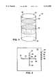
- FIG. 1is a drawing of a resin-filled cartridge for use in a pressure-modulation apparatus.
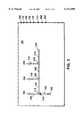
- FIG. 2is a drawing of a five electrode chip for use in a pressure-modulation apparatus.
- FIG. 3is a drawing of an eight electrode chip for use in a pressure-modulation apparatus.
- FIG. 4Ais a cut-away perspective view of a chip of the invention.
- FIG. 4Bis an exploded side view of a chip of the invention that includes a diaphragm for relaying pressure.
- FIG. 4Cis a top view of a chip of the invention that includes a diaphragm for relaying pressure.
- FIG. 5Ais a cut-away perspective view of a chip of the invention.
- FIG. 5Bis an exploded side view of a chip of the invention that includes a hydrophobic valve for relaying pressure.
- FIG. 5Cis a top view of a chip of the invention that includes a hydrophobic valve for relaying pressure.
- FIG. 6Ais a cut-away perspective view of a chip of the invention.
- FIG. 6Bis an exploded side view of a chip of the invention that includes a compressible piston for relaying pressure.
- FIG. 6Cis a top view of a chip of the invention that includes a compressible piston for relaying pressure.
- FIG. 7is a graph of percent recovery of nucleic acids as a function of sodium chloride concentration at constant pressure for three sizes of DNA: 50 bp (•••), 4.6 kb (---), and 48.4 kb (--).
- FIG. 8is a graph of percent recovery of nucleic acids as a function of pressure at constant sodium chloride concentration for three sizes of DNA: 50 bp (•••), 4.6 kb (---), and 48.4 kb (--).
- Eukaryotic cellscan express cloned genes (i.e., transient and stable heterologous expression), using eukaryotic expression vectors purified by the new methods.
- cloned genesi.e., transient and stable heterologous expression
- eukaryotic expression vectorspurified by the new methods.
- eukaryotic expression plasmids carrying the gene of interestcan be obtained in a form suitable for introduction into mammalian cells. It is often necessary to generate a large panel of mutants for structure-function studies of a particular eukaryotic gene. Therefore, the new methods provide a method for rapid and facile analysis.
- Isolation of DNA by the new methodscan be used for numerous applications including, but not limited to, protein expression and protein structure function studies in eukaryotic cells, Southern blot analysis, in vitro transcription, ligation, and transformation, heterologous protein expression in bacteria or yeast, microinjection studies, PCR, DNA sequencing, viral DNA detection, paternity testing by RFLP analysis, and genetic screening by single-strand conformation polymorphism (SSCP) or non-isotopic RNAse cleavage assay (NIRCATM; Ambion, Austin, Tex.).
- SSCPsingle-strand conformation polymorphism
- NIRCATMnon-isotopic RNAse cleavage assay
- RNA isolationprovides a variety of applications including, but not limited to, genetic analysis, cDNA library construction, microinjection into oocytes, differential display, Northern blot analysis, RNase protection assays, in vitro translation, reverse transcriptase PCR (RT-PCR), and detection of viral RNA (e.g. HIV, hepatitic C, hepatitis A and HTLV-1) in human blood.
- viral RNAe.g. HIV, hepatitic C, hepatitis A and HTLV-1
- a solution containing nucleic acidsis introduced onto a solid phase at low pressure (e.g., ambient pressure).
- the solid phaseto which any nucleic acids present in the solution should now be bound, is then washed with a low salt buffer under ambient conditions.
- This washing stepremoves the non-nucleic acid contaminants (e.g., proteins and lipids) from the solid phase, while the nucleic acids remain bound.
- the pressureis increased to a level sufficient to cause the nucleic acids to be freed from the solid phase.
- fresh low salt buffercan be used to wash the liberated nucleic acids away from the solid phase and into a collected vessel.
- Nucleic acids that can be purified by this procedureinclude chromosomal DNA, viral DNA, plasmid DNA, mitochondrial DNA, a DNA vector, an oligonucleotide, mRNA, mitochondrial RNA, viral RNA, and also mixtures of nucleic acids.
- the solid phasecan be made from any substance that selectively binds to nucleic acids at ambient pressure and has reduced affinity at elevated pressure such as an anion-exchange column, an oligo-dT column, an affinity column (e.g., with immobilized nucleic acid-binding proteins or other molecules), or an electrode coated with absorptive polymers.
- the solid phasecan have other functions.
- the solid phasecan absorb the biological samples (e.g., a sponge-type polymer); it can assist in the lysis of the cells, for example by mixing the solid phase material with proteases (e.g., pepsin or trypsin), lipases, or glycosidases (e.g., lysozyme) to digest proteins, lipids, and polysaccharides, respectively; or it can include DNAse for RNA purifications or RNAse for DNA purification.
- proteasese.g., pepsin or trypsin
- lipasese.g., lysozyme
- Some solid phasescan bind nucleic acids, but only weakly interact with other negatively charged molecules such as some proteins or lipids.
- the same solution or different solutionscan be used to load the nucleic acid sample onto the solid phase, wash the impurities away, and elute the nucleic acid away from the solid phase. Nonetheless, it is generally most desirable to use a single buffer, both for ease of operation and to reduce waste. Whether the solution acts as a wash buffer or as an elution buffer depends on the pressure. At pressures greater than about 25,000 psi, large (e.g., >5,000 bp) nucleic acids can be eluted in low salt buffers. In addition, at 25,000 psi, small nucleic acids such as those used in the Sanger sequencing method can be eluted at still lower salt concentrations.
- Examples of applications of the present methodinclude purification of DNA or RNA from blood, cell-culture (genomics or infectious disease) or tissue (e.g., tumor biopsy) for clinical or research purposes, purification of microbial DNA for genetic or biotechnology research, desalting of DNA, forensic analysis (e.g., purification of DNA from hair, blood, semen, or tissue found at the scene of a crime), and purification of PCR products.
- the cellsFor the purification of nucleic acids from cells (e.g., from cell cultures or tissue), the cells must be lysed prior to the introduction of the sample onto the solid phase.
- cell lysisThere are many known methods for cell lysis, including chemical methods (e.g., phenol/chloroform extraction, treatment with guanidinium salts, chaotropic salts, detergents such as sodium dodecyl sulfate, or enzymes such as proteinase K) and physical methods (e.g., boiling, French pressing, douncing). Often these methods can be sensitive to variations in time and temperature.
- Hyperbaric pressurecan be carried out in the same solvent as will be used as the loading buffer for later introducing the sample onto the solid phase, or can be carried out in a different solvent.
- a chemical agente.g., a detergent
- a small amount of a chaotropic saltcan be used to prime the cells for lysis; after the cells have been treated with the chaotropic agent, they can be lysed at lower pressure.
- More gentle lysis procedurescan be especially preferable for isolating single-strand nucleic acid molecules (e.g., RNA), or if the nucleic acid to be isolated is of high molecular weight, since single strands are easier to shear and longer nucleic acid strands are statistically more likely to be shorn during lysis.
- nucleic acid moleculese.g., RNA
- nucleasesMost proteins (e.g., nucleases) are inhibited by pressure.
- a 120,000 psi pulsefor example, can irreversibly denature the nucleases in a sample. It is important, especially in attempted isolations of RNA, to denature nucleases such as RNAse to prevent degradation of the desired nucleic acids during the isolation process. Pressure can be used here in place of chemical inhibitors. In some cases, nuclease denaturation and cell lysis can be accomplished simultaneously.
- nucleic acid purificationcan be automated. Additionally, the pressure can be ramped to allow elution of progressively larger nucleic acids, thereby facilitating the isolation of specific sizes of molecules.
- a pressure gradient(i.e., either stepped or continuous) can also be set up within the devices. A pressure gradient can be used, for example, to fractionate samples. Fractionation can be used to purify specific fragments from a partially degraded sample or a highly diverse sample (e.g., a cDNA library).
- FIG. 1One design for an isolation device is shown in FIG. 1.
- This deviceis a cartridge 10 made of metal (e.g., titanium, stainless steel, or aluminum), plastic (e.g., a thermoplastic such as polypropylene or polytetrafluoroethylene), glass, quartz, stone (e.g., sapphire), or a ceramic, adapted to fit into a pressure-modulation apparatus such as those described at pages 4-9 and 29-44 and in FIGS. 1 and 4 of PCT Appln. No. US/96/03232, which was given Ser. No. 08/793,213 in the U.S. National Phase and is now allowed.
- the cartridgeis generally formed in the shape of a tubular column, although other designs can be used. Regardless of the shape, the cartridge usually has two openings 12 and 14, one 12 to allow fluid to enter and another 14 to allow the fluid to exit. Between the two openings, but within a channel 16 common to the openings, a solid phase material 18 is packed.
- the solid phasecan be any of a multitude of nucleic acid-binding materials, including silica gel, anion-exchange resin (e.g., DEAE), tethered nucleotides or nucleic acids (e.g., oligo-dT and complementary oligonucleotide probes), tethered proteins or peptides, polymers, DNA-binding molecules (e.g., ethidium, acridinium), or other small molecules (e.g., sugars, benzodiazepines, drugs).
- silica gelsilica gel
- anion-exchange resine.g., DEAE
- tethered nucleotides or nucleic acidse.g., oligo-dT and complementary oligonucleotide probes
- tethered proteins or peptidespolymers
- DNA-binding moleculese.g., ethidium, acridinium
- other small moleculese.g
- the cartridgecan be designed such that the openings are in direct fluid contact with the reaction chamber of the pressure-modulation apparatus, or can be designed as a closed system with valves and pistons that can open and close to regulate the pressure and the fluid flow within the cartridge.
- the valves and pistons in this embodimentcan be controlled either electronically or mechanically.
- Cartridges designed for use with samples derived from lysed whole cellscan optionally include a micro-porous filter or membrane 19 positioned to filter off any remaining cell debris prior to introduction of the sample onto the solid phase.
- This filtermay be larger in cross-sectional area than the resin chamber to prevent pressure gradients.
- the volume of the cartridgescan vary widely.
- the cartridgecan have an internal volume of 10 ⁇ l or 10 ml.
- the internal volumeis 100 ⁇ l to 1 ml.
- the solid phaseoccupies about half of the internal volume of the cartridge, although some cartridges can be filled to nearly to their full capacity while others may be filled just one tenth of the way. In some cases, the cartridges can be reused.
- the cartridge sizeis ideally matched to the size of the sample, the cartridges can frequently be loaded with samples whose volumes exceed the cartridge capacity 2-, 10-, or even 100-fold, especially if the nucleic acid concentration in the sample is relatively low.
- One limiting criterionis the ratio of free nucleic acid-binding sites on the solid phase to the number of nucleic acid molecules; if the amount of nucleic acid exceeds the number of binding sites, then some of the nucleic acid will pass through the cartridge without being bound.
- the solid phase materialis very dense, the capacity can be further limited since the bound nucleic acid molecules can further impede the flow of additional fluid through the cartridge.
- the sampleis dissolved or suspended in a low-salt buffer solution and introduced at opening 12.
- the cartridge 10is placed in the pressure-modulation apparatus.
- a low pressure flow of buffer solutionis used to force the sample through the membrane 19 and through the solid phase 18.
- Nucleic acids in the samplebind to the solid phase; the flow-through continues through the solid phase and emerges from opening 14.
- the flow-throughis taken up by a sample output tube leading to an input on a detection device (e.g., a UV-vis spectrophotometer).
- the low pressure flow of the buffer solutionis continued until the detection device shows that no additional residues are washed away. The flow-through is discarded.
- the pressureis then increased to 500 to 100,000 psi, causing the nucleic acid to be released from the solid phase. More of the buffer solution is introduced through opening 12, and the nucleic acid-containing flow-through that emerges from opening 14 is collected. This flow-through can also be fed into a detection device and analyzed, and the flow continued until the nucleic acid detected in the flow-through falls below a set threshold level.
- the cartridgescan also include multiple compartments.
- the individual compartmentscan contain different solid phase materials (e.g., ion-exchange resin, silica gel, tethered oligonucleotides). Reactions can be carried out within the cartridges.
- a cartridge of the present inventioncan be used as a PCR reaction vessel, if placed within a thermal cycling apparatus after the solid phase has been washed to remove non-nucleic acid impurities and the nucleic acid has been eluted from the solid phase into, for example, a second compartment in the cartridge.
- a multi-compartment cartridgecan also be used to concentrate nucleic acids.
- fluidscan be moved hydrodynamically or electrically, or both.
- DNA from a large samplecan be concentrated hydrodynamically onto a resin, small molecule impurities can be washed away, then the DNA can be electrically electrophoresed into a downstream cartridge. This process is termed electroconcentration.
- nucleic acidsare eluted from a first compartment (e.g., containing an anion-exchange resin), using pressure, and concentrated in a second compartment (e.g., containing silica gel) that requires different conditions for elution.
- a first compartmente.g., containing an anion-exchange resin
- a second compartmente.g., containing silica gel
- concentrated nucleic acidscan be isolated from eve dilute samples containing many impurities.
- the eluted samplecan be automatically transferred to another device (e.g., a disk, a pad, a bead, or a detection device).
- FIGS. 2 and 3An alternative design for an isolation device is shown in FIGS. 2 and 3. These devices are in the form of a chip, with an electrode array aligned along at least two axes.
- the individual electrodesare coated with a solid phase material. In some cases, all of the electrodes are coated with the same material; in other cases, the coatings differ from electrode to electrode or form a coating gradient along a capillary connecting two or more electrodes.
- the chipscan optionally be interfaced with an analytical device such as a mass spectrometer or a capillary electrophoresis device.
- Electrode 20is coated with a material that absorbs the sample.
- the chip 25is placed within the sample chamber of a pressure-modulation apparatus (e.g., the apparatus described in U.S. Ser. No. 08/903,615) adapted to supply a switchable electrical voltage at the contact points 22, 32, 52, 72, and 82.
- a voltage potentialis supplied between electrodes 20 and 30 (i.e., electrode 20 is the anode and electrode 30 is the cathode) while the system is at ambient pressure.
- the potentialcauses the sample to flow through capillary 40, which is filled with a size-exclusion filtration material (e.g., 0.5% agarose) that retains large cellular debris but allows nucleic acids, proteins, lipids, and other small cellular components to pass through.
- a size-exclusion filtration materiale.g. 0.5% agarose
- Electrode 50includes a reservoir that traps the impurities that reach it. The voltage potential between electrodes 20 and 30 is then discontinued.
- the pressure in the systemis increased to a moderately elevated level (e.g., 500 to 10,000 psi).
- a voltage potentialis set up between electrodes 50 (anode) and 80 (cathode).
- the moderate pressurecauses the smallest nucleic acids (e.g., less than 5,000 bp) to dissociate from the anion-exchange resin at electrode 50, and the potential causes the nucleic acid to migrate through the liquid phase in capillary 90 and finally into electrode 80, which includes a reservoir.
- the potentialis discontinued.
- the pressure in the systemis increased to a more elevated level (e.g., 12,000 to 100,000 psi).
- a voltage potentialis set up between electrodes 50 (anode) and 70 (cathode).
- the high pressurecauses the remaining nucleic acids to dissociate from the anion-exchange resin at electrode 50, and the potential causes the nucleic acid to migrate through the liquid phase in capillary 75 and finally into electrode 70, which includes a reservoir.
- the potentialis discontinued, the pressure is lowered to ambient pressure, the chip is removed from the pressure-modulation apparatus, and the large nucleic acid fraction, including the chromosomal DNA, can be removed from electrode 70.
- electrodes 100, 110, 120, 130, 140, 170, 200, and 220are electrically connected to contact points 102, 112, 122, 132, 142, 172, 202, and 222, respectively.
- a whole blood sampleis introduced at electrode 100 at ambient pressure.
- Electrode 100is coated with a wicking material that absorbs the sample.
- the chip 190is placed within the sample chamber of a pressure-modulation apparatus adapted to supply a switchable electrical voltage at the contact points 102, 112, 122, 132, 142, 172, 202, and 222.
- a voltage potentialis supplied between electrodes 100 (anode) and 120 (cathode) while the system is at ambient pressure. The potential causes the sample to flow through liquid-filled capillaries 105 and 125 and electrode 110.
- a filter 114prevents white blood cells to pass. Thus, the white blood cells become trapped at electrode 110, while red blood cells continue to migrate to electrode 120.
- Capillary 115contains a size-exclusion material or an ion-exchange material, for example, that can retain large cellular debris but allow nucleic acids, proteins, lipids, and other small cellular components to pass through.
- Electrode 220includes a reservoir that traps the impurities that reach it. The potential is then discontinued.
- a potentialis supplied between electrodes 140 (anode) and 130 (cathode).
- the potentialcauses the RNA to dissociate from the solid phase at electrode 140, migrate through the liquid phase in capillary 144, and finally to electrode 130, where a reservoir traps the RNA.
- the potentialis discontinued.
- the pressure in the systemis then increased to an elevated level (e.g., 20,000 to 100,000 psi), and a potential is supplied between electrodes 170 (anode) and 200 (cathode).
- the pressurecauses the DNA to dissociate from the solid phase at electrode 170, and the potential causes the nucleic acid to migrate through the liquid phase in capillary 174, and finally to electrode 200, where a reservoir traps the DNA.
- the potentialis discontinued, the chip is removed from the pressure-modulation apparatus, the purified white blood cell RNA can be removed from electrode 130, and the purified white blood cell DNA can be removed from electrode 200.
- a miniature versioncan be highly parallel and/or interface into a downstream biochip.
- sample sizesinclude 10 ⁇ l, 100 ⁇ l, 1 ml and 10 ml.
- the chipscan be made from metal (e.g., silicon), plastic (e.g., polypropylene or polytetrafluoroethylene), glass, or ceramic.
- the contact points and electrodescan be made, for example, from gold, silver, copper, or iron.
- the chipscan be designed such that the fluids in the capillaries are in direct fluid contact with the reaction chamber of the pressure-modulation apparatus. More preferably, the chips can be designed as a closed system with a diaphragm (FIG. 4), a piston (FIG. 5), or a hydrophobic valve (FIG. 6), which relays the pressure from the reaction chamber to the capillaries and electrodes.
- a diaphragmFIG. 4
- a pistonFIG. 5
- a hydrophobic valveFIG. 6
- the chip 240can include a recessed area 242, in which the electrode array is situated.
- a flexible, elastic membrane 244spans the recessed area 242, to form a diaphragm.
- One or both surfaces of the membranecan be flexible. The membrane transmits external pressure to the electrode array, while simultaneously providing a hermetic seal that prevents fluids from being transferred.
- the chip 250 shown in FIGS. 5A to 5Calso includes a recessed area 252, in which the electrode array is situated.
- a solid lid 254is placed over the recessed area.
- a channel 256is drilled through one side of the chip, leading into the recessed area 252.
- the wall of the channel 256is precoated with a hydrophobic material, such that water and other fluids are unable to traverse the length of the channel 256 under ambient conditions. As the pressure is increased, however, the fluids overcome the hydrophobic interactions and pass through the channel 256, thereby modulating the pressure within the recessed area 252.
- a chip 260includes a recessed area 262, in which the electrode array is situated.
- a compressible, elastomeric piston 264is mounted in the recessed area 262. When the pressure in the reaction chamber is increased, the piston 264 becomes compressed, thereby increasing the pressure at the electrode array without allowing fluid transfer between the reaction chamber and the electrode array.
- the sampleis placed into a first (loading) syringe having a DEAE resin cartridge attached at the narrow end.
- This systemmust have a very small resin chamber so that high pressures can be generated and the materials must be able to withstand the high pressures.
- the plungeris slowly depressed, so as not to create a significant pressure gradient.
- Low salt (e.g., 10 to 300 mM) bufferis placed in the syringe.
- the buffercan contain magnesium and other cofactors necessary for downstream enzymatic techniques.
- a measured quantitye.g., 100 ⁇ l to 10 ml
- a measured quantityis used to wash the resin to remove non-DNA contaminants.
- a second (collection) syringeis added to the first.
- the resistance of the plunger of the second syringeis adjusted such that the pressure needed to move the loading syringe causes the dissociation of the nucleic acid from the resin.
- the resisting force exerted by the collection syringecan be adjusted by means of low-angle threads in the syringe and piston. The angle of the threads can be adjusted to change the pressure.
- the pressurizing stepcan be carried out by, or with the aid of, a machine that maintains a consistent pressure and flow rate. For non-critical applications, this step can be done manually.
- Another version of this systemwould use a device which applied an equal force to two opposing pistons and (with much less force) moved the two syringes simultaneously to achieve a flow.
- two pistonssupply force, with a small pressure differential between them.
- This systemcan be immersed in a pressurizing medium (such as water) so as to avoid the use of pressure resistant materials and small resin capacity in the disposable component.
- DNA sampleswere separated using a Qiagen DEAE anion-exchange resin (Qiagen, Inc., Santa Clarita, Calif.) at ambient and elevated pressures.
- the DEAE resinwas packed into a 9 mm ⁇ 4 mm I.D. (5 mm O.D.) stainless steel ⁇ half-column ⁇ capped with Teflon® frits with a 2 ⁇ m pore size (Valco Instrument Company, Inc., Houston, Tex.).
- Two half-columns, one containing resin and the other acting as a spacer and devoid of resin,were placed into a column holder.
- the column holderwas a metal tube with an inner diameter of 5 mm and syringe fittings at the ends to allow fluid to flow through the columns.
- Pressure elution of DNAwas performed using a pressure apparatus as described at pages 29 et seq. of PCT Appln. No. US/96/03232, which was given Ser. No. 08/793,213 in the U.S. National Phase and is now allowed, controlled by a microcomputer with LABVIEWTM software (National Instruments, Austin, Tex.).
- the columnswere inserted into a pressure chamber adapted to receive the columns. Liquid was injected and removed from the chamber using a series of pneumatic valves and pistons as described in the '232 application, allowing for elution of DNA from the column while maintaining elevated pressure within the column.
- the DEAE columnwas initially washed with 1 ml high salt elution buffer (1.25 M sodium chloride; 50 mM Tris-HCl, pH 8.5; 15% ethanol) and equilibrated with 1 ml equilibration buffer (750 mM sodium chloride; 50 mM MOPS, pH 7.0; 15% ethanol; 0.15% Triton X-100).
- 1 ml equilibration buffer750 mM sodium chloride; 50 mM MOPS, pH 7.0; 15% ethanol; 0.15% Triton X-100.
- Approximately 300 ⁇ l of 21 ⁇ g/mL DNA in loading buffer(1 M potassium acetate; 33 mM NaCl; 33 mM Tris-HCl, pH 5; 8 mM EDTA), was injected into the packed column over five minutes, in four 1 minute intervals.
- MO washing buffercontaining 1 M NaCl; 50 mM MOPS, pH 7.0; 15% ethanol
- Elution buffers used during the DNA elution stepcontained 50 mM Tris-HCl, pH 8.5, and various concentrations of sodium chloride.
- DNA in the collected sampleswas quantified using OliGreen DNA binding dye (Molecular Probes, Eugene, Oreg.).
- OliGreen DNA binding dyeMolecular Probes, Eugene, Oreg.
- ⁇ DNA(Worthington Biochemical Company, Freehold, N.J.) was released from the DEAE resin with a buffer of lower salt concentration, as shown in Table 1.
- the percentages given in the tableare percent recoveries, and numbers in parentheses after the percentages are the number of times the corresponding experiments were carried out.
- the error rangewas calculated to be approximately 5% based on the duplicated data.
- Table 1shows a correlation: the higher the pressure, the lower the salt concentration needed for dissociation.
- 1 M sodium chloridewas required for more than just a trace amount (i.e., less than 10%) of DNA to be eluted.
- the DNAshowed a slightly increased tendency to dissociate; at 170 MPa (about 24,000 psi), 70% of the ⁇ DNA was dissociated with 0.50 M NaCl. 100% of the ⁇ DNA dissociated with 0.25 M NaCl at 220 MPa (about 32,000 psi).
- nucleic acidscan be separated on the basis of size by varying the salt concentration.
- the effect of pressurewas also studied, using the same three nucleic acid fragments.
- the sodium chloride concentrationwas held constant at 250 mM, as the pressure was increased from 14 to 40,000 psi.
- the nucleic acidswere detected as they eluted from the cartridge. The results are shown in the graph in FIG. 8. Most of the smallest nucleic acid, 50 bp, was eluted at around 7,000 psi, as indicated by the dotted line. The 4.6 kb fragment was eluted at about 20,000 psi, as shown by the dashed line. The solid line indicates that approximately 32,000 psi was necessary to elute the largest nucleic acid, 48.4 kb.
- the nucleic acidscan be separated on the basis of size by varying the elution pressure.
- bovine serum albumin(BSA) was applied to the DEAE column.
- Serum albuminsare multivalent and highly absorptive, and are the most abundant proteins in mammalian blood. It is therefore highly desirable that any DNA purification procedure for isolating DNA from blood be capable of separating BSA from DNA. Indeed, all of the protein was recovered in the flow through and MO washing solutions.
- pCMV-SV40T plasmidwas isolated from a JM109 E. coli strain by alkaline lysis (Sambrook et al., Molecular Cloning: A Laboratory Manual, 2nd ed., Cold Spring Harbor Laboratory Press: Plainview, N.Y., 1989, pp. 1.25-1.26). 1.5 ml of an overnight bacterial culture was placed in a microcentrifuge tube and spun for 30 seconds at 12,000 g. The medium was removed and the cells were resuspended with vigorous vortexing in 100 ⁇ l of a solution containing 50 mM glucose, 25 mM Tris-HCl (pH 8.0), and 10 mM EDTA (pH 8.0).
- Purified pCMV-TAg(Campbell et al., Genes & Dev. 11:1098-1110, 1997) and purified pCMV control vector are each eluted from columns, collected, and assayed for transient expression of simian virus SV40 large TAg in a monkey kidney cell line, BSC 40.
- BSC 40monkey kidney cell line
- 100 mM plates of ⁇ 50% confluent cellsare transfected by the calcium phosphate procedure (Cherington et al., PNAS, 83:4307-4311, 1986).
- HBS buffercontaining 137 mM sodium chloride, 0.37 mg/ml potassium chloride, 0.188 mg/ml dibasic sodium phosphate heptahydrate, 1.0 mg/ml dextrose, and 5 mg/ml HEPES.
- HBS buffercontaining 137 mM sodium chloride, 0.37 mg/ml potassium chloride, 0.188 mg/ml dibasic sodium phosphate heptahydrate, 1.0 mg/ml dextrose, and 5 mg/ml HEPES.
- the pH of the HBS bufferis maintained at 7.05.
- 127 ⁇ l of 1 M calcium chlorideis added dropwise to the 1.0 ml of HBS buffer containing the pCMV-TAg vector.
- the resulting DNA-calcium phosphate precipitateis added to the culture medium and left in contact with the cells for 4 hours.
- a 7 minute glycerol shock treatmentis carried out, the cells are washed once with serum free DMEM, 10 ml of fresh complete medium was added, and the samples were assayed for protein expression 40-48 hours later.
- the transfected cellsare washed twice with ice cold PBS and lysed for 15 minutes in 1.0 ml Nonidet P-40 (NP40) lysis buffer (1% NP40, 10% glycerol, 137 mM sodium chloride, 5 mM EDTA, 20 mM Tris, pH 8.0) containing 5 mM sodium fluoride, 1 mM sodium orthovanadate, 25 mM ⁇ -glycerophosphate, and 10 ⁇ g of each of the following protease inhibitors: aprotinin, pepstatin, and leupeptin.
- the cell lysatesare scraped from the dishes, centrifuged at 13,000 g, and the protein concentration is determined by Bradford assay (Biorad).
- Immunoblotswere developed using enhanced chemiluminescence (ECL; Amersham, Arlington Heights, Ill.) and visualized on Kodak XAR film. Visualization of the film indicates SV40 TAg protein expression in cells transfected with pCMV-TAg as compared with expression in cells expressing the pCMV control vector.
- ECLenhanced chemiluminescence
- BSC 40 monkey kidney cells that stably express SV40 large T antigenwere grown to a density of ⁇ 5 ⁇ 10 6 by the method of Chomczynski et al. (Anal. Biochem., 162:156-159, 1987).
- 1 ml RNA STAT-60TM(Tel-test, Inc., Friendswood, Tex.) was added directly to the cells. After incubating at room temperature for 5 minutes, the cells were scraped from the plate, homogenized by pipetting, and transferred to a sterile microcentrifuge tube. After addition of 0.2 ml chloroform, the solution was mixed vigorously for 15 seconds and the upper aqueous phase was separated by centrifugation.
- RNAwas pelleted by centrifugation in a microcentrifuge, and resuspended in 50 ⁇ l of sterile RNAse-free water. 10 ⁇ l of the RNA sample was then mixed with 500 ⁇ l of equilibration buffer (containing 750 mM sodium chloride; 50 mM MOPS, pH 7.0; 15% ethanol; and 0.15% Triton X-100).
- equilibration buffercontaining 750 mM sodium chloride; 50 mM MOPS, pH 7.0; 15% ethanol; and 0.15% Triton X-100.
- Qiagen DEAE anion-exchange resinwas packed in the "half-column" as described in Example 1, and washed with 1 ml equilibration buffer. 300 ⁇ l RNA sample was injected onto the DEAE column over 3 minutes. Then, 1 ml MO buffer (containing 1 M NaCl; 50 mM MOPS, pH 7.0; 15% ethanol) and 200 ⁇ l elution buffer (containing 250 mM sodium chloride and 50 mM Tris, pH 8.5) was applied to wash the column. Four consecutive 100 ⁇ l and three 300 ⁇ l elution fractions were collected at 23,600 psi. After taking the DEAE column out of the pressure flow apparatus, the column was washed with 1 ml of high salt buffer (containing 1.25 M sodium chloride; 50 mM Tris-HCl, pH 8.5; and 15% ethanol).
- high salt buffercontaining 1.25 M sodium chloride; 50 mM Tris-HCl, pH 8.5; and 15% ethanol.
- RNA in the collected sampleswas quantified using OliGreen DNA binding dye.
- RNAwas released from the DEAE resin in the first 4 fractions.
- the other three fractionscontained about 40%; thus 100% recovery was achieved.
- the high salt wash solutionwas analyzed to verify this result; indeed, no RNA was detected in the subsequent high salt wash solutions.
- a 0.8% agarose gelwas used to determine the integrity and purity of the RNA product in the collected samples. No degradation of the 28S and 18S rRNA was observed when compared to the control.
- RNAMessenger RNA
- Two cartridges containing a solid phaseare arranged in serial order, such that eluent from the first cartridge enters the second cartridge.
- the first cartridgeis packed with an acidic DEAE activated anion exchange resin and the second is packed with a resin containing covalently linked polythymidine (poly-dT) resin.
- mRNA standard(positive control) is purified by standard methods using the POLY(A)PURETM Kit (Ambion, Austin, Tex.). A salt concentration is found at which poly-dA mRNA is poorly bound at atmospheric pressure, but is more tightly bound at high pressure.
- Samplesare loaded onto the cartridge containing the poly-dT resin, in a buffer containing 100 mM NaCl and 10 mM Tris-HCl, pH 7.2, at atmospheric pressure. The samples are then eluted with 300 ⁇ l of buffer containing 10 mM Tris-HCl (pH 8.0) and NaCl concentrations of 0 to 100 mM in increments of 2 mM.
- the saltis removed from the sample by washing twice in 10 mM Tris-HCl (pH 8.0) using a Centricon-100TM spin-filter (Millipore). The experiment is repeated at 25,000 psi for each sample. A buffer which gives poor binding at atmospheric pressure, but improved binding at high pressure is selected and referred to as "solution A.”
- a mammalian tissue sampleis lysed by standard procedures (Chomczynski et al., supra) and total RNA is isolated on the DEAE column as described in Example 4.
- the cell debrisis removed by centrifugation.
- the sampleis applied to the double anion-exchange/poly-dT column.
- the columnis washed with 300 ⁇ l of solution A.
- the columnis then washed at 25,000 psi with 300 ⁇ l of solution A, thereby transferring the mRNA from the anion-exchange resin to the poly-da resin.
- the pressureis then lowered to atmospheric pressure and the resin is washed with 300 ⁇ l of either solution A or distilled water to recover the mRNA.
- the sampleis analyzed for purity by agarose gel electrophoresis, UV spectroscopy, and a protein binding dye assay.
- RNA obtained from tumor samplesis prepared by the method of Chomczynski et al. (supra) and collected from the DEAE column as in Example 4. Poly-A + RNA is subsequently isolated using oligo-dT cellulose (Ambion, Austin, Tex.). The gene of interest (i.e., p53) is amplified by reverse transcriptase-polymerase chain reaction (RT-PCR) (Promega, Madison, Wis.). The resulting cDNA is then analyzed for mutations by sequencing with the dideoxy chain termination method using a Sequenase 2.0 kit (United States Biochemicals), then compared with consensus mutations.
- RT-PCRreverse transcriptase-polymerase chain reaction
- RNAis extracted by the method of Chomczynski (Biotechniques, 15:532-536, 1993). 300 ⁇ l of whole blood is mixed with 1.0 ml of red blood cell lysis solution (RBCS; containing 40 mM ammonium chloride, 10 mM potassium hydroxide, 7.5 mM potassium acetate, 2.5 mM sodium bicarbonate, 0.125 mM EDTA, and 0.1% glacial acetic acid). After 10 minutes at 4° C., the residual red blood cells and hemoglobin are pelleted by centrifugation (30 seconds at 12,000 g). An additional 1.0 ml RBCS is added to the pellet and mixed thoroughly. The centrifugation step described above is repeated.
- RBCSred blood cell lysis solution
- LLSleukocyte lysis solution
- RNAis eluted as described in Example 4, and collected in consecutive fractions, precipitated with isopropanol, and resuspended in 30 ⁇ l of RNAse-free water. The quality and integrity of total RNA eluted from the column is analyzed on a native 1.0% agarose gel stained with ethidium bromide. 5 ⁇ l of total RNA isolated from the column is sufficient for analyzing p53 mutations following RT-PCR with RETROSCRIPTTM and the p53 cDNA screening module with SUPERTAQTM (Ambion, Austin, Tex.).
- RNAis amplified by RT-PCR using RETROSCRIPTTM (Ambion, Austin, Tex.). Primers specific to the viral genes of HIV are used for subsequent PCR amplification. The PCR products are then assayed by traditional DNA sequencing methods or Southern blot analysis. This procedure is repeated using primers specific for other viruses, including hepatitis A and hepatitis C.
- cartridges containing multiple, layered resinsare also within the scope of the claims.
- a layer of cation-exchange resinfor instance, will capture any positively charged proteins which might bind to the DNA.
- Hydrophobic (e.g., reverse-phase) resinscan bind to the lipids in the sample.
Landscapes
- Chemical & Material Sciences (AREA)
- Health & Medical Sciences (AREA)
- Life Sciences & Earth Sciences (AREA)
- Analytical Chemistry (AREA)
- General Health & Medical Sciences (AREA)
- Engineering & Computer Science (AREA)
- Genetics & Genomics (AREA)
- Biochemistry (AREA)
- Bioinformatics & Cheminformatics (AREA)
- Biotechnology (AREA)
- Physics & Mathematics (AREA)
- Organic Chemistry (AREA)
- Biomedical Technology (AREA)
- Wood Science & Technology (AREA)
- Zoology (AREA)
- General Physics & Mathematics (AREA)
- Chemical Kinetics & Catalysis (AREA)
- General Engineering & Computer Science (AREA)
- Immunology (AREA)
- Pathology (AREA)
- Molecular Biology (AREA)
- Microbiology (AREA)
- Dispersion Chemistry (AREA)
- Hematology (AREA)
- Clinical Laboratory Science (AREA)
- Mycology (AREA)
- Medicinal Chemistry (AREA)
- Tropical Medicine & Parasitology (AREA)
- Crystallography & Structural Chemistry (AREA)
- Biophysics (AREA)
- Virology (AREA)
- Plant Pathology (AREA)
- Electrochemistry (AREA)
- Apparatus Associated With Microorganisms And Enzymes (AREA)
- Enzymes And Modification Thereof (AREA)
- Measuring Or Testing Involving Enzymes Or Micro-Organisms (AREA)
- Micro-Organisms Or Cultivation Processes Thereof (AREA)
- Pens And Brushes (AREA)
- Investigating Or Analysing Biological Materials (AREA)
Abstract
Description
TABLE 1 ______________________________________ [NaC1] (M) in Tris Pressure (MPa) Buffer 0.1 90 170 220 ______________________________________ 0.10 trace (1) trace (1) 40% (1) 0.25 trace (2) 20% (3) 100% (1) 0.40 25% (1) 0.50 trace (2) 15% (1) 70% (2) 60% (1) 0.75 trace (2) 15% (1) 100% (2) trace (1) 1.00 100% (1) 100% (1) ______________________________________
Claims (17)
Priority Applications (13)
| Application Number | Priority Date | Filing Date | Title |
|---|---|---|---|
| US08/962,280US6111096A (en) | 1997-10-31 | 1997-10-31 | Nucleic acid isolation and purification |
| US09/016,062US6274726B1 (en) | 1997-10-31 | 1998-01-30 | Pressure-enhanced extraction and purification |
| US09/083,651US6120985A (en) | 1997-10-31 | 1998-05-22 | Pressure-enhanced extraction and purification |
| AT98956405TATE492343T1 (en) | 1997-10-31 | 1998-10-30 | PRESSURE-ENHANCED EXTRACTION AND PURIFICATION |
| AU12936/99AAU745925B2 (en) | 1997-10-31 | 1998-10-30 | Pressure-enhanced extraction and purification |
| JP2000518788AJP2001521818A (en) | 1997-10-31 | 1998-10-30 | Pressure enhanced extraction and purification method |
| CA002307876ACA2307876A1 (en) | 1997-10-31 | 1998-10-30 | Pressure-enhanced extraction and purification |
| DE69842067TDE69842067D1 (en) | 1997-10-31 | 1998-10-30 | PRESSURE REINFORCED EXTRACTION AND CLEANING |
| EP98956405AEP1027160B1 (en) | 1997-10-31 | 1998-10-30 | Pressure-enhanced extraction and purification |
| PCT/US1998/023141WO1999022868A1 (en) | 1997-10-31 | 1998-10-30 | Pressure-enhanced extraction and purification |
| US09/898,404US20020016450A1 (en) | 1997-10-31 | 2001-07-03 | Pressure-enhanced extraction and purification |
| US10/918,878US7626017B2 (en) | 1997-10-31 | 2004-08-16 | Pressure-enhanced extraction and purification |
| JP2009181058AJP2009254384A (en) | 1997-10-31 | 2009-08-03 | Method for pressure-enhanced extraction and purification |
Applications Claiming Priority (1)
| Application Number | Priority Date | Filing Date | Title |
|---|---|---|---|
| US08/962,280US6111096A (en) | 1997-10-31 | 1997-10-31 | Nucleic acid isolation and purification |
Related Child Applications (2)
| Application Number | Title | Priority Date | Filing Date |
|---|---|---|---|
| US09/016,062Continuation-In-PartUS6274726B1 (en) | 1997-10-31 | 1998-01-30 | Pressure-enhanced extraction and purification |
| US09/016,062ContinuationUS6274726B1 (en) | 1997-10-31 | 1998-01-30 | Pressure-enhanced extraction and purification |
Publications (1)
| Publication Number | Publication Date |
|---|---|
| US6111096Atrue US6111096A (en) | 2000-08-29 |
Family
ID=25505646
Family Applications (3)
| Application Number | Title | Priority Date | Filing Date |
|---|---|---|---|
| US08/962,280Expired - LifetimeUS6111096A (en) | 1997-10-31 | 1997-10-31 | Nucleic acid isolation and purification |
| US09/016,062Expired - LifetimeUS6274726B1 (en) | 1997-10-31 | 1998-01-30 | Pressure-enhanced extraction and purification |
| US09/898,404AbandonedUS20020016450A1 (en) | 1997-10-31 | 2001-07-03 | Pressure-enhanced extraction and purification |
Family Applications After (2)
| Application Number | Title | Priority Date | Filing Date |
|---|---|---|---|
| US09/016,062Expired - LifetimeUS6274726B1 (en) | 1997-10-31 | 1998-01-30 | Pressure-enhanced extraction and purification |
| US09/898,404AbandonedUS20020016450A1 (en) | 1997-10-31 | 2001-07-03 | Pressure-enhanced extraction and purification |
Country Status (4)
| Country | Link |
|---|---|
| US (3) | US6111096A (en) |
| JP (1) | JP2009254384A (en) |
| AT (1) | ATE492343T1 (en) |
| DE (1) | DE69842067D1 (en) |
Cited By (39)
| Publication number | Priority date | Publication date | Assignee | Title |
|---|---|---|---|---|
| US20020125134A1 (en)* | 2001-01-24 | 2002-09-12 | Santiago Juan G. | Electrokinetic instability micromixer |
| WO2002088296A1 (en)* | 2001-04-26 | 2002-11-07 | Boston Biomedica, Inc. | Multichamber device and uses thereof for processing of biological samples |
| WO2002097422A1 (en)* | 2001-05-31 | 2002-12-05 | Electron-Bio, Inc. | A micro valve apparatus using micro bead and method for controlling the same |
| US20030170617A1 (en)* | 2002-01-28 | 2003-09-11 | Pasloske Brittan L. | Crude biological derivatives competent for nucleic acid detection |
| US6632349B1 (en)* | 1996-11-15 | 2003-10-14 | Lifescan, Inc. | Hemoglobin sensor |
| US20030215845A1 (en)* | 2002-02-19 | 2003-11-20 | Bille Todd William | Selective extraction of DNA from groups of cells |
| US20040026244A1 (en)* | 1995-11-16 | 2004-02-12 | Lifescan, Inc. | Antioxidant sensor |
| US20050059024A1 (en)* | 2003-07-25 | 2005-03-17 | Ambion, Inc. | Methods and compositions for isolating small RNA molecules |
| US20050072686A1 (en)* | 2001-04-12 | 2005-04-07 | Astrazeneca Ab | Micro-engineered reactors |
| US20050095626A1 (en)* | 2003-09-03 | 2005-05-05 | Hiroyuki Komazawa | Method for separation and purification method of nucleic acid |
| US20050214765A1 (en)* | 2002-04-05 | 2005-09-29 | Evy Reitan | Process for isolating nucleic acid with chaotrope agents and ammonium compounds |
| US20050214926A1 (en)* | 2004-02-20 | 2005-09-29 | Ralf Zielenski | Adsorption of nucleic acids to a solid phase |
| WO2005047521A3 (en)* | 2003-11-10 | 2005-10-06 | Investigen Inc | Methods of preparing nucleic acid for detection |
| US20060051799A1 (en)* | 2004-09-03 | 2006-03-09 | Fiji Photo Film Co., Ltd. | Method for separating and purifying nucleic acid |
| US20070238109A1 (en)* | 2006-04-06 | 2007-10-11 | Samsung Electronics Co., Ltd. | Method and apparatus for purifying nucleic acid on hydrophilic surface of solid support using hydrogen bonding |
| WO2008101047A1 (en)* | 2007-02-15 | 2008-08-21 | Honeywell International Inc. | Active biochip for nucleic acid analysis |
| US20090111170A1 (en)* | 2003-05-19 | 2009-04-30 | Brandeis University | Nucleic acid processing methods, kits and devices |
| US7569342B2 (en) | 1997-12-10 | 2009-08-04 | Sierra Molecular Corp. | Removal of molecular assay interferences |
| US20100035770A1 (en)* | 2003-07-25 | 2010-02-11 | Life Technologies Corporation | Methods and compositions for preparing rna from a fixed sample |
| US20100159482A1 (en)* | 2008-12-19 | 2010-06-24 | Life Technologies Corporation | Proteinase k inhibitors, methods and compositions therefor |
| US20100216225A1 (en)* | 2009-02-25 | 2010-08-26 | Ag-Defense Systems, Inc. | Portable microorganism detection unit |
| US20100281955A1 (en)* | 2009-05-05 | 2010-11-11 | Pressure Biosciences Inc. | Microtube and related methods therefor |
| US20110077388A1 (en)* | 2009-09-30 | 2011-03-31 | Samsung Electronics Co., Ltd. | Method and apparatus for isolating nucleic acids |
| US7964350B1 (en) | 2007-05-18 | 2011-06-21 | Applied Biosystems, Llc | Sample preparation for in situ nucleic acid analysis |
| KR20110072276A (en)* | 2009-12-22 | 2011-06-29 | 삼성전자주식회사 | Nucleic Acid Isolation Method and Apparatus |
| USRE43389E1 (en)* | 1998-08-12 | 2012-05-15 | Preanalytix Gmbh | Vessel for blood sampling |
| US20140242594A1 (en)* | 2013-02-27 | 2014-08-28 | Syngenta Participations Ag | Methods and compositions for preparation of nucleic acids |
| WO2015007800A1 (en)* | 2013-07-18 | 2015-01-22 | Commissariat à l'énergie atomique et aux énergies alternatives | Method for extracting and purifying nucleic acids and buffers used |
| WO2015184360A1 (en) | 2014-05-30 | 2015-12-03 | Pressure Biosciences, Inc. | Sample preparation devices and methods |
| EP3179243A1 (en)* | 2015-12-09 | 2017-06-14 | ARKRAY, Inc. | Analytical tool for capillary electrophoresis with pressure fluctuation reducer |
| WO2017114844A1 (en)* | 2015-12-28 | 2017-07-06 | Koninklijke Philips N.V. | Nucleic acid purification system using a single wash and elution buffer solution |
| CN107058075A (en)* | 2017-06-20 | 2017-08-18 | 商丘师范学院 | A kind of plant protoplast purifying instrument and purification process |
| JP2017537652A (en)* | 2014-10-20 | 2017-12-21 | ジェン−プローブ・インコーポレーテッド | Erythrocyte lysis solution |
| CN110546259A (en)* | 2017-04-11 | 2019-12-06 | 罗伯特·博世有限公司 | Desorption of nucleic acids |
| US10712320B2 (en)* | 2015-10-14 | 2020-07-14 | Alps Alpine Co., Ltd. | Flow channel structure and measuring device for measurement target liquid |
| WO2021011512A1 (en)* | 2019-07-15 | 2021-01-21 | Lexagene, Inc. | Sample preparation cartridges and apparatuses |
| US11015185B1 (en) | 2016-04-27 | 2021-05-25 | Gen-Probe Incorporated | Blood cell lysis reagent |
| CN116790578A (en)* | 2023-08-22 | 2023-09-22 | 赛奥斯博生物科技(北京)有限公司 | Production process for rapidly purifying plasmid based on alkaline cracking method |
| EP4282962A4 (en)* | 2021-01-25 | 2025-05-14 | Yokogawa Electric Corporation | METHOD FOR MEASURING NUCLEIC ACID SEQUENCE |
Families Citing this family (89)
| Publication number | Priority date | Publication date | Assignee | Title |
|---|---|---|---|---|
| US7626017B2 (en)* | 1997-10-31 | 2009-12-01 | Pressure Biosciences, Inc. | Pressure-enhanced extraction and purification |
| US7799521B2 (en)* | 1998-06-24 | 2010-09-21 | Chen & Chen, Llc | Thermal cycling |
| US6780617B2 (en) | 2000-12-29 | 2004-08-24 | Chen & Chen, Llc | Sample processing device and method |
| US6537502B1 (en)* | 2000-07-25 | 2003-03-25 | Harvard Apparatus, Inc. | Surface coated housing for sample preparation |
| US7666588B2 (en) | 2001-03-02 | 2010-02-23 | Ibis Biosciences, Inc. | Methods for rapid forensic analysis of mitochondrial DNA and characterization of mitochondrial DNA heteroplasmy |
| US20030027135A1 (en) | 2001-03-02 | 2003-02-06 | Ecker David J. | Method for rapid detection and identification of bioagents |
| US20040121309A1 (en) | 2002-12-06 | 2004-06-24 | Ecker David J. | Methods for rapid detection and identification of bioagents in blood, bodily fluids, and bodily tissues |
| US7718354B2 (en) | 2001-03-02 | 2010-05-18 | Ibis Biosciences, Inc. | Methods for rapid identification of pathogens in humans and animals |
| US7226739B2 (en) | 2001-03-02 | 2007-06-05 | Isis Pharmaceuticals, Inc | Methods for rapid detection and identification of bioagents in epidemiological and forensic investigations |
| US6418968B1 (en) | 2001-04-20 | 2002-07-16 | Nanostream, Inc. | Porous microfluidic valves |
| US20020186263A1 (en)* | 2001-06-07 | 2002-12-12 | Nanostream, Inc. | Microfluidic fraction collectors |
| US20020187557A1 (en)* | 2001-06-07 | 2002-12-12 | Hobbs Steven E. | Systems and methods for introducing samples into microfluidic devices |
| US7217510B2 (en) | 2001-06-26 | 2007-05-15 | Isis Pharmaceuticals, Inc. | Methods for providing bacterial bioagent characterizing information |
| US8073627B2 (en) | 2001-06-26 | 2011-12-06 | Ibis Biosciences, Inc. | System for indentification of pathogens |
| CA2460192C (en)* | 2001-09-11 | 2011-04-19 | Iquum, Inc. | Sample vessels |
| WO2003031618A1 (en)* | 2001-09-28 | 2003-04-17 | Hitachi, Ltd. | Method of collecting nucleic acid |
| DE10149251B4 (en)* | 2001-10-05 | 2007-04-19 | Sartorius Ag | Device for genetic immunization by introducing active substances into a tissue and method for producing a solution for injection |
| US7261812B1 (en) | 2002-02-13 | 2007-08-28 | Nanostream, Inc. | Multi-column separation devices and methods |
| US7010964B2 (en)* | 2002-10-31 | 2006-03-14 | Nanostream, Inc. | Pressurized microfluidic devices with optical detection regions |
| JP2006516193A (en) | 2002-12-06 | 2006-06-29 | アイシス・ファーマシューティカルス・インコーポレーテッド | Rapid identification of pathogens in humans and animals |
| JP4632262B2 (en)* | 2003-02-05 | 2011-02-16 | アイキューム,インク. | Sample processing |
| EP1604184A4 (en)* | 2003-02-27 | 2010-10-27 | Stephen A Lesko | Standardized evaluation of therapeutic efficacy based on cellular biomarkers |
| US8501402B2 (en)* | 2003-03-24 | 2013-08-06 | Boehringer Ingelheim Rcv Gmbh & Co Kg | Methods and devices for producing biomolecules |
| US6991253B2 (en)* | 2003-04-11 | 2006-01-31 | Delphi Technologies, Inc. | Air bag assembly having controlled cushion deployment |
| US8046171B2 (en) | 2003-04-18 | 2011-10-25 | Ibis Biosciences, Inc. | Methods and apparatus for genetic evaluation |
| US8057993B2 (en) | 2003-04-26 | 2011-11-15 | Ibis Biosciences, Inc. | Methods for identification of coronaviruses |
| US8158354B2 (en) | 2003-05-13 | 2012-04-17 | Ibis Biosciences, Inc. | Methods for rapid purification of nucleic acids for subsequent analysis by mass spectrometry by solution capture |
| US7964343B2 (en)* | 2003-05-13 | 2011-06-21 | Ibis Biosciences, Inc. | Method for rapid purification of nucleic acids for subsequent analysis by mass spectrometry by solution capture |
| US20050032238A1 (en)* | 2003-08-07 | 2005-02-10 | Nanostream, Inc. | Vented microfluidic separation devices and methods |
| US8546082B2 (en) | 2003-09-11 | 2013-10-01 | Ibis Biosciences, Inc. | Methods for identification of sepsis-causing bacteria |
| US20120122103A1 (en) | 2003-09-11 | 2012-05-17 | Rangarajan Sampath | Compositions for use in identification of bacteria |
| US8097416B2 (en) | 2003-09-11 | 2012-01-17 | Ibis Biosciences, Inc. | Methods for identification of sepsis-causing bacteria |
| US8163895B2 (en) | 2003-12-05 | 2012-04-24 | Ibis Biosciences, Inc. | Compositions for use in identification of orthopoxviruses |
| US7666592B2 (en) | 2004-02-18 | 2010-02-23 | Ibis Biosciences, Inc. | Methods for concurrent identification and quantification of an unknown bioagent |
| US8119336B2 (en) | 2004-03-03 | 2012-02-21 | Ibis Biosciences, Inc. | Compositions for use in identification of alphaviruses |
| CA2567839C (en) | 2004-05-24 | 2011-06-28 | Isis Pharmaceuticals, Inc. | Mass spectrometry with selective ion filtration by digital thresholding |
| US20050266411A1 (en) | 2004-05-25 | 2005-12-01 | Hofstadler Steven A | Methods for rapid forensic analysis of mitochondrial DNA |
| US7811753B2 (en) | 2004-07-14 | 2010-10-12 | Ibis Biosciences, Inc. | Methods for repairing degraded DNA |
| US7592139B2 (en) | 2004-09-24 | 2009-09-22 | Sandia National Laboratories | High temperature flow-through device for rapid solubilization and analysis |
| US8084207B2 (en) | 2005-03-03 | 2011-12-27 | Ibis Bioscience, Inc. | Compositions for use in identification of papillomavirus |
| WO2006094238A2 (en) | 2005-03-03 | 2006-09-08 | Isis Pharmaceuticals, Inc. | Compositions for use in identification of adventitious viruses |
| CA2616281C (en) | 2005-07-21 | 2014-04-22 | Isis Pharmaceuticals, Inc. | Methods for rapid identification and quantitation of mitochondrial dna variants |
| US8921102B2 (en)* | 2005-07-29 | 2014-12-30 | Gpb Scientific, Llc | Devices and methods for enrichment and alteration of circulating tumor cells and other particles |
| EP4524246A3 (en) | 2006-05-31 | 2025-06-25 | Sequenom, Inc. | Methods and compositions for the extraction and amplification of nucleic acid from a sample |
| WO2007147018A1 (en)* | 2006-06-14 | 2007-12-21 | Cellpoint Diagnostics, Inc. | Analysis of rare cell-enriched samples |
| EP2589668A1 (en) | 2006-06-14 | 2013-05-08 | Verinata Health, Inc | Rare cell analysis using sample splitting and DNA tags |
| US8137912B2 (en) | 2006-06-14 | 2012-03-20 | The General Hospital Corporation | Methods for the diagnosis of fetal abnormalities |
| WO2007147074A2 (en)* | 2006-06-14 | 2007-12-21 | Living Microsystems, Inc. | Use of highly parallel snp genotyping for fetal diagnosis |
| US20080050739A1 (en)* | 2006-06-14 | 2008-02-28 | Roland Stoughton | Diagnosis of fetal abnormalities using polymorphisms including short tandem repeats |
| JP5420412B2 (en) | 2006-09-14 | 2014-02-19 | アイビス バイオサイエンシズ インコーポレイティッド | Targeted whole genome amplification method for pathogen identification |
| US20080124777A1 (en)* | 2006-11-29 | 2008-05-29 | Canon U.S. Life Sciences, Inc. | Method for releasing genetic material from solid phase |
| US8871471B2 (en) | 2007-02-23 | 2014-10-28 | Ibis Biosciences, Inc. | Methods for rapid forensic DNA analysis |
| US9598724B2 (en) | 2007-06-01 | 2017-03-21 | Ibis Biosciences, Inc. | Methods and compositions for multiple displacement amplification of nucleic acids |
| JP5587770B2 (en)* | 2007-06-04 | 2014-09-10 | プレッシャー バイオサイエンシズ インコーポレイテッド | Pressure-enhanced molecular extraction and distribution |
| US20090286301A1 (en)* | 2008-05-07 | 2009-11-19 | Pressure Biosciences Inc. | Extraction of biomolecular complexes assisted by alternating hydrostatic pressure |
| US8148163B2 (en) | 2008-09-16 | 2012-04-03 | Ibis Biosciences, Inc. | Sample processing units, systems, and related methods |
| EP2344893B1 (en) | 2008-09-16 | 2014-10-15 | Ibis Biosciences, Inc. | Microplate handling systems and methods |
| EP2349549B1 (en) | 2008-09-16 | 2012-07-18 | Ibis Biosciences, Inc. | Mixing cartridges, mixing stations, and related kits, and system |
| WO2010033578A2 (en) | 2008-09-20 | 2010-03-25 | The Board Of Trustees Of The Leland Stanford Junior University | Noninvasive diagnosis of fetal aneuploidy by sequencing |
| WO2010093943A1 (en) | 2009-02-12 | 2010-08-19 | Ibis Biosciences, Inc. | Ionization probe assemblies |
| US9447461B2 (en) | 2009-03-24 | 2016-09-20 | California Institute Of Technology | Analysis devices, kits, and related methods for digital quantification of nucleic acids and other analytes |
| CN104722342B (en) | 2009-03-24 | 2017-01-11 | 芝加哥大学 | Slip chip device and method |
| US9464319B2 (en) | 2009-03-24 | 2016-10-11 | California Institute Of Technology | Multivolume devices, kits and related methods for quantification of nucleic acids and other analytes |
| US10196700B2 (en) | 2009-03-24 | 2019-02-05 | University Of Chicago | Multivolume devices, kits and related methods for quantification and detection of nucleic acids and other analytes |
| EP3514244B1 (en) | 2009-04-03 | 2021-07-07 | Sequenom, Inc. | Nucleic acid preparation methods |
| US9194877B2 (en) | 2009-07-17 | 2015-11-24 | Ibis Biosciences, Inc. | Systems for bioagent indentification |
| US8950604B2 (en) | 2009-07-17 | 2015-02-10 | Ibis Biosciences, Inc. | Lift and mount apparatus |
| EP3225695A1 (en) | 2009-10-15 | 2017-10-04 | Ibis Biosciences, Inc. | Multiple displacement amplification |
| US8187979B2 (en)* | 2009-12-23 | 2012-05-29 | Varian Semiconductor Equipment Associates, Inc. | Workpiece patterning with plasma sheath modulation |
| US20110312503A1 (en) | 2010-01-23 | 2011-12-22 | Artemis Health, Inc. | Methods of fetal abnormality detection |
| JP5624487B2 (en) | 2011-01-31 | 2014-11-12 | 横河電機株式会社 | Nucleic acid extraction method |
| DE12722942T1 (en) | 2011-03-31 | 2021-09-30 | Modernatx, Inc. | RELEASE AND FORMULATION OF MANIPULATED NUCLEIC ACIDS |
| US9487820B2 (en) | 2011-04-27 | 2016-11-08 | Peking University | Methods of nucleic acid liquid-phase extraction and detection |
| JP6212488B2 (en) | 2011-07-25 | 2017-10-11 | クヴェッラ コーポレーション | Methods and devices for electrical sample preparation |
| JP6321622B2 (en)* | 2012-03-30 | 2018-05-09 | ビーディー キエストラ ベスローテン フェンノートシャップ | Automated selection of microorganisms and identification using MALDI |
| KR101483085B1 (en) | 2012-11-28 | 2015-01-16 | 한국전기연구원 | Apparatus for microalgae treatment using wire explosion in liquid |
| US10077439B2 (en) | 2013-03-15 | 2018-09-18 | Modernatx, Inc. | Removal of DNA fragments in mRNA production process |
| EP4279610A3 (en) | 2013-03-15 | 2024-01-03 | ModernaTX, Inc. | Ribonucleic acid purification |
| WO2014144767A1 (en) | 2013-03-15 | 2014-09-18 | Moderna Therapeutics, Inc. | Ion exchange purification of mrna |
| WO2014152027A1 (en) | 2013-03-15 | 2014-09-25 | Moderna Therapeutics, Inc. | Manufacturing methods for production of rna transcripts |
| WO2015051169A2 (en) | 2013-10-02 | 2015-04-09 | Moderna Therapeutics, Inc. | Polynucleotide molecules and uses thereof |
| US10286086B2 (en) | 2014-06-19 | 2019-05-14 | Modernatx, Inc. | Alternative nucleic acid molecules and uses thereof |
| AU2015289656A1 (en) | 2014-07-16 | 2017-02-16 | Modernatx, Inc. | Circular polynucleotides |
| WO2017049286A1 (en) | 2015-09-17 | 2017-03-23 | Moderna Therapeutics, Inc. | Polynucleotides containing a morpholino linker |
| ES2908449T3 (en) | 2015-09-17 | 2022-04-29 | Modernatx Inc | Polynucleotides that contain a stabilizing tail region |
| WO2017223176A1 (en) | 2016-06-24 | 2017-12-28 | Modernatx, Inc. | Methods and apparatus for filtration |
| US11623188B2 (en) | 2018-11-28 | 2023-04-11 | V&P Scientific, Inc. | Spinning vessel systems and methods for mixing, suspending particulates, aliquoting, washing magnetic beads, and concentrating analytes |
| TW202221248A (en) | 2020-11-20 | 2022-06-01 | 美商伊路米納有限公司 | Actuation systems and methods |
| WO2025166356A1 (en)* | 2024-02-01 | 2025-08-07 | Fmrk Diagnostic Technologies Incorporated | Specimen collection containers and related transfer and/or enrichment systems and methods |
Citations (3)
| Publication number | Priority date | Publication date | Assignee | Title |
|---|---|---|---|---|
| US3609252A (en)* | 1967-01-23 | 1971-09-28 | Texas Instruments Inc | Transducer apparatus and system utilizing insulated gate semiconductor field effect devices |
| US5003047A (en)* | 1989-01-10 | 1991-03-26 | Massachusetts Institute Of Technology | Method for purifying biologically active ligate |
| US5934766A (en)* | 1994-11-18 | 1999-08-10 | Itt Manufacturing Enterprises Inc. | Magnetically operated, multi-position, pressure control valve having integral metering orifice |
Family Cites Families (24)
| Publication number | Priority date | Publication date | Assignee | Title |
|---|---|---|---|---|
| US4323439A (en) | 1979-12-31 | 1982-04-06 | The Regents Of The University Of California | Method and apparatus for dynamic equilibrium electrophoresis |
| GB8311905D0 (en) | 1983-04-29 | 1983-06-02 | Cox R A | Isolation and detection of nucleotide sequence |
| US5346999A (en) | 1985-01-18 | 1994-09-13 | Applied Biosystems, Inc. | Method of nucleic acid extraction |
| JPH02255059A (en)* | 1989-03-25 | 1990-10-15 | Chuetsu Koubo Kogyo Kk | Production of yeast essence |
| IT1240870B (en) | 1990-02-14 | 1993-12-17 | Talent | PROCEDURE FOR THE EXTRACTION AND PURIFICATION OF HUMAN GENOMIC DNA |
| JP2873971B2 (en)* | 1990-03-02 | 1999-03-24 | オリエンタル酵母工業株式会社 | How to leak microbial contents |
| JPH0779658B2 (en)* | 1990-03-16 | 1995-08-30 | 株式会社ジャパンエナジー | How to treat onion plants |
| US5605839A (en) | 1990-04-11 | 1997-02-25 | Ludwig Institute For Cancer Research | Methods and apparatus for use in sequential chemical reactions |
| JPH04349892A (en)* | 1991-05-21 | 1992-12-04 | Toshiba Corp | Nucleic acid isolation method |
| DE4319506A1 (en) | 1993-06-12 | 1994-12-15 | Boehringer Mannheim Gmbh | Method for the detection of metabolically labeled DNA |
| DE4321904B4 (en)* | 1993-07-01 | 2013-05-16 | Qiagen Gmbh | Method for chromatographic purification and separation of nucleic acid mixtures |
| GB9314249D0 (en)* | 1993-07-09 | 1993-08-18 | Proofname Ltd | Purification method and apparatus |
| US5637687A (en) | 1993-08-31 | 1997-06-10 | Wiggins; James C. | Methods and compositions for isolating nucleic acids |
| US5438127A (en) | 1993-09-27 | 1995-08-01 | Becton Dickinson And Company | DNA purification by solid phase extraction using a PCl3 modified glass fiber membrane |
| WO1995034569A1 (en)* | 1994-06-14 | 1995-12-21 | Invitek Gmbh | Universal process for isolating and purifying nucleic acids from extremely small amounts of highly contaminated various starting materials |
| JP2992737B2 (en)* | 1994-06-27 | 1999-12-20 | 備前化成株式会社 | Extraction method of animal and plant extracts by disruption of frozen cell membrane |
| DE4431280A1 (en)* | 1994-09-02 | 1996-03-07 | Hoechst Ag | Process for the isolation and purification of nucleotide-activated sugars from biological sources |
| CA2214263A1 (en)* | 1995-03-07 | 1996-09-12 | Biomolecular Assays, Inc. | Pressure cycling reactor |
| IL113935A (en)* | 1995-05-31 | 1999-09-22 | Imi Tami Institute Research | Process for the recovery of carotenoid pigment from algae |
| US5856174A (en)* | 1995-06-29 | 1999-01-05 | Affymetrix, Inc. | Integrated nucleic acid diagnostic device |
| JPH0919284A (en)* | 1995-07-04 | 1997-01-21 | Oriental Yeast Co Ltd | Method for obtaining fission yeast multiple strains |
| US6100079A (en) | 1996-02-25 | 2000-08-08 | Precision System Science Co., Ltd. | Method for treating biopolymers, microorganisms or materials by using more than one type of magnetic particles |
| DE19610354C1 (en)* | 1996-03-15 | 1997-11-20 | Innova Gmbh | Device, method and device for isolating nucleic acids |
| US5707812A (en) | 1996-08-06 | 1998-01-13 | Vical Incorporated | Purification of plasmid DNA during column chromatography |
- 1997
- 1997-10-31USUS08/962,280patent/US6111096A/ennot_activeExpired - Lifetime
- 1998
- 1998-01-30USUS09/016,062patent/US6274726B1/ennot_activeExpired - Lifetime
- 1998-10-30ATAT98956405Tpatent/ATE492343T1/ennot_activeIP Right Cessation
- 1998-10-30DEDE69842067Tpatent/DE69842067D1/ennot_activeExpired - Lifetime
- 2001
- 2001-07-03USUS09/898,404patent/US20020016450A1/ennot_activeAbandoned
- 2009
- 2009-08-03JPJP2009181058Apatent/JP2009254384A/enactivePending
Patent Citations (3)
| Publication number | Priority date | Publication date | Assignee | Title |
|---|---|---|---|---|
| US3609252A (en)* | 1967-01-23 | 1971-09-28 | Texas Instruments Inc | Transducer apparatus and system utilizing insulated gate semiconductor field effect devices |
| US5003047A (en)* | 1989-01-10 | 1991-03-26 | Massachusetts Institute Of Technology | Method for purifying biologically active ligate |
| US5934766A (en)* | 1994-11-18 | 1999-08-10 | Itt Manufacturing Enterprises Inc. | Magnetically operated, multi-position, pressure control valve having integral metering orifice |
Cited By (87)
| Publication number | Priority date | Publication date | Assignee | Title |
|---|---|---|---|---|
| US20040026244A1 (en)* | 1995-11-16 | 2004-02-12 | Lifescan, Inc. | Antioxidant sensor |
| US6632349B1 (en)* | 1996-11-15 | 2003-10-14 | Lifescan, Inc. | Hemoglobin sensor |
| US20030201177A1 (en)* | 1996-11-15 | 2003-10-30 | Alastair Hodges | Hemoglobin sensor |
| US7569342B2 (en) | 1997-12-10 | 2009-08-04 | Sierra Molecular Corp. | Removal of molecular assay interferences |
| USRE43389E1 (en)* | 1998-08-12 | 2012-05-15 | Preanalytix Gmbh | Vessel for blood sampling |
| US20020125134A1 (en)* | 2001-01-24 | 2002-09-12 | Santiago Juan G. | Electrokinetic instability micromixer |
| US7070681B2 (en)* | 2001-01-24 | 2006-07-04 | The Board Of Trustees Of The Leland Stanford Junior University | Electrokinetic instability micromixer |
| US20050072686A1 (en)* | 2001-04-12 | 2005-04-07 | Astrazeneca Ab | Micro-engineered reactors |
| WO2002088296A1 (en)* | 2001-04-26 | 2002-11-07 | Boston Biomedica, Inc. | Multichamber device and uses thereof for processing of biological samples |
| US20020197631A1 (en)* | 2001-04-26 | 2002-12-26 | Lawrence Nathan P. | Multichamber device and uses thereof for processing of biological samples |
| US20070014690A1 (en)* | 2001-04-26 | 2007-01-18 | Bbi Bioseq, Inc., A Massachusetts Corporation | Multichamber device for processing of biological samples using high pressure |
| JP2004536292A (en)* | 2001-04-26 | 2004-12-02 | ボストン・バイオメディカ・インコーポレーテッド | Multi-chamber device and its use for processing biological samples |
| US20100055771A1 (en)* | 2001-05-31 | 2010-03-04 | Jae Chern Yoo | Micro valve apparatus using micro bead and method for controlling the same |
| US8241891B2 (en) | 2001-05-31 | 2012-08-14 | Samsung Electronics Co., Ltd. | Micro valve apparatus using micro bead and method for controlling the same |
| WO2002097422A1 (en)* | 2001-05-31 | 2002-12-05 | Electron-Bio, Inc. | A micro valve apparatus using micro bead and method for controlling the same |
| US20040155213A1 (en)* | 2001-05-31 | 2004-08-12 | Yoo Jae Chern | Micro valve apparatus using micro bead and method for controlling the same |
| US7635585B2 (en) | 2001-05-31 | 2009-12-22 | Jae Chern Yoo | Micro valve apparatus using micro bead and method for controlling the same |
| US10590411B2 (en) | 2002-01-28 | 2020-03-17 | Applied Biosystems, Llc | Crude biological derivatives competent for nucleic acid detection |
| US9611497B2 (en) | 2002-01-28 | 2017-04-04 | Applied Biosystems, Llc | Crude biological derivatives competent for nucleic acid detection |
| US10011831B2 (en) | 2002-01-28 | 2018-07-03 | Applied Biosystems, Llc | Crude biological derivatives competent for nucleic acid detection |
| US10233444B2 (en) | 2002-01-28 | 2019-03-19 | Applied Biosystems, Llc | Crude biological derivatives competent for nucleic acid detection |
| US20030170617A1 (en)* | 2002-01-28 | 2003-09-11 | Pasloske Brittan L. | Crude biological derivatives competent for nucleic acid detection |
| US20030215845A1 (en)* | 2002-02-19 | 2003-11-20 | Bille Todd William | Selective extraction of DNA from groups of cells |
| US20050214765A1 (en)* | 2002-04-05 | 2005-09-29 | Evy Reitan | Process for isolating nucleic acid with chaotrope agents and ammonium compounds |
| US7517969B2 (en)* | 2002-04-05 | 2009-04-14 | Qiagen Gmbh | Process for isolating nucleic acid with chaotrope agents and ammonium compounds |
| US9279121B2 (en) | 2003-05-19 | 2016-03-08 | Lawrence J Wangh | Nucleic acid processing kits and devices |
| US20090111170A1 (en)* | 2003-05-19 | 2009-04-30 | Brandeis University | Nucleic acid processing methods, kits and devices |
| US8404439B2 (en) | 2003-07-25 | 2013-03-26 | Applied Biosystems, Llc | Methods and compositions for isolating small RNA molecules |
| US10619150B2 (en) | 2003-07-25 | 2020-04-14 | Applied Biosystems, Llc | Methods and compositions for isolating small RNA |
| US20100035770A1 (en)* | 2003-07-25 | 2010-02-11 | Life Technologies Corporation | Methods and compositions for preparing rna from a fixed sample |
| US20130245245A1 (en)* | 2003-07-25 | 2013-09-19 | Life Technologies Corporation | Methods and compositions for isolating small rna molecules |
| US20050059024A1 (en)* | 2003-07-25 | 2005-03-17 | Ambion, Inc. | Methods and compositions for isolating small RNA molecules |
| US10138475B2 (en) | 2003-07-25 | 2018-11-27 | Applied Biosystems, Llc | Methods and compositions for isolating small RNA molecules |
| US20100222564A1 (en)* | 2003-07-25 | 2010-09-02 | Life Technologies Corporation | Methods and compositions for isolating small rna molecules |
| US9193748B2 (en)* | 2003-07-25 | 2015-11-24 | Applied Biosystems, Llc | Methods and compositions for isolating small RNA molecules |
| US20050095626A1 (en)* | 2003-09-03 | 2005-05-05 | Hiroyuki Komazawa | Method for separation and purification method of nucleic acid |
| WO2005047521A3 (en)* | 2003-11-10 | 2005-10-06 | Investigen Inc | Methods of preparing nucleic acid for detection |
| US20080286786A1 (en)* | 2003-11-10 | 2008-11-20 | Investigen, Inc. | Methods of preparing nucleic acid for detection |
| US20050214926A1 (en)* | 2004-02-20 | 2005-09-29 | Ralf Zielenski | Adsorption of nucleic acids to a solid phase |
| US7491495B2 (en)* | 2004-02-20 | 2009-02-17 | Roche Diagnostics Operations, Inc. | Adsorption of nucleic acids to a solid phase |
| US20060051799A1 (en)* | 2004-09-03 | 2006-03-09 | Fiji Photo Film Co., Ltd. | Method for separating and purifying nucleic acid |
| US20070238109A1 (en)* | 2006-04-06 | 2007-10-11 | Samsung Electronics Co., Ltd. | Method and apparatus for purifying nucleic acid on hydrophilic surface of solid support using hydrogen bonding |
| US8367397B2 (en) | 2007-02-15 | 2013-02-05 | Honeywell International Inc. | Active biochip for nucleic acid analysis |
| WO2008101047A1 (en)* | 2007-02-15 | 2008-08-21 | Honeywell International Inc. | Active biochip for nucleic acid analysis |
| US20080199862A1 (en)* | 2007-02-15 | 2008-08-21 | Honeywell International, Inc. | Active biochip for nucleic acid analysis |
| US8828664B2 (en) | 2007-05-18 | 2014-09-09 | Applied Biosystems, Llc. | Sample preparation for in situ nucleic acid analysis, methods and compositions therefor |
| US8288106B2 (en) | 2007-05-18 | 2012-10-16 | Applied Biosystems, Llc | Sample preparation for in situ nucleic acid analysis, methods and compositions therefor |
| US7964350B1 (en) | 2007-05-18 | 2011-06-21 | Applied Biosystems, Llc | Sample preparation for in situ nucleic acid analysis |
| US10501780B2 (en) | 2007-05-18 | 2019-12-10 | Applied Biosystems, Llc | Compositions for in situ nucleic acid analysis |
| US9279152B2 (en) | 2007-05-18 | 2016-03-08 | Applied Biosystems, Llc | Sample preparation for in situ nucleic acid analysis, methods and compositions therefor |
| US20100159482A1 (en)* | 2008-12-19 | 2010-06-24 | Life Technologies Corporation | Proteinase k inhibitors, methods and compositions therefor |
| US9114104B2 (en) | 2008-12-19 | 2015-08-25 | Life Technologies Corporation | Proteinase K inhibitors, methods and compositions therefor |
| US8211637B2 (en) | 2008-12-19 | 2012-07-03 | Life Technologies Corporation | Proteinase K inhibitors, methods and compositions therefor |
| US20100216225A1 (en)* | 2009-02-25 | 2010-08-26 | Ag-Defense Systems, Inc. | Portable microorganism detection unit |
| US20100281955A1 (en)* | 2009-05-05 | 2010-11-11 | Pressure Biosciences Inc. | Microtube and related methods therefor |
| WO2010129704A2 (en) | 2009-05-05 | 2010-11-11 | Pressure Biosciences Inc. | Microtube and related methods therefor |
| US8664377B2 (en)* | 2009-09-30 | 2014-03-04 | Samsung Electronics Co., Ltd. | Method and apparatus for isolating nucleic acids |
| US20110077388A1 (en)* | 2009-09-30 | 2011-03-31 | Samsung Electronics Co., Ltd. | Method and apparatus for isolating nucleic acids |
| KR20110072276A (en)* | 2009-12-22 | 2011-06-29 | 삼성전자주식회사 | Nucleic Acid Isolation Method and Apparatus |
| US9556427B2 (en)* | 2013-02-27 | 2017-01-31 | Syngenta Participations Ag | Methods and compositions for preparation of nucleic acids |
| US20140242594A1 (en)* | 2013-02-27 | 2014-08-28 | Syngenta Participations Ag | Methods and compositions for preparation of nucleic acids |
| FR3008713A1 (en)* | 2013-07-18 | 2015-01-23 | Commissariat Energie Atomique | PROCESS FOR EXTRACTING AND PURIFYING NUCLEIC ACIDS AND BUFFERS IMPLEMENTED |
| WO2015007800A1 (en)* | 2013-07-18 | 2015-01-22 | Commissariat à l'énergie atomique et aux énergies alternatives | Method for extracting and purifying nucleic acids and buffers used |
| WO2015184360A1 (en) | 2014-05-30 | 2015-12-03 | Pressure Biosciences, Inc. | Sample preparation devices and methods |
| US10093989B2 (en) | 2014-10-20 | 2018-10-09 | Gen-Probe Incorporated | Red blood cell lysis solution |
| EP3209799A4 (en)* | 2014-10-20 | 2018-05-23 | Gen-Probe Incorporated | Red blood cell lysis solution |
| JP2017537652A (en)* | 2014-10-20 | 2017-12-21 | ジェン−プローブ・インコーポレーテッド | Erythrocyte lysis solution |
| EP3739062A1 (en)* | 2014-10-20 | 2020-11-18 | Gen-Probe Incorporated | Red blood cell lysis solution |
| US10712320B2 (en)* | 2015-10-14 | 2020-07-14 | Alps Alpine Co., Ltd. | Flow channel structure and measuring device for measurement target liquid |
| EP3179243A1 (en)* | 2015-12-09 | 2017-06-14 | ARKRAY, Inc. | Analytical tool for capillary electrophoresis with pressure fluctuation reducer |
| US11187674B2 (en) | 2015-12-09 | 2021-11-30 | Arkray, Inc. | Analytical tool and analytical system |
| CN108473983B (en)* | 2015-12-28 | 2022-09-13 | 皇家飞利浦有限公司 | Nucleic acid purification system using single wash and elution buffer |
| RU2766005C2 (en)* | 2015-12-28 | 2022-02-07 | Конинклейке Филипс Н.В. | System for purifying nucleic acid using one buffer solution for washing and elution |
| CN108473983A (en)* | 2015-12-28 | 2018-08-31 | 皇家飞利浦有限公司 | Use the nucleic acid purification system of single washing and elution buffer solution |
| US11427815B2 (en) | 2015-12-28 | 2022-08-30 | Koninklijke Philips N.V. | Nucleic acid purification system using a single wash and elution buffer solution |
| WO2017114844A1 (en)* | 2015-12-28 | 2017-07-06 | Koninklijke Philips N.V. | Nucleic acid purification system using a single wash and elution buffer solution |
| US11015185B1 (en) | 2016-04-27 | 2021-05-25 | Gen-Probe Incorporated | Blood cell lysis reagent |
| US11162091B2 (en) | 2016-04-27 | 2021-11-02 | Gen-Probe Incorporated | Blood cell lysis reagent |
| US12071615B2 (en) | 2016-04-27 | 2024-08-27 | Gen-Probe Incorporated | Blood cell lysis reagent |
| CN110546259A (en)* | 2017-04-11 | 2019-12-06 | 罗伯特·博世有限公司 | Desorption of nucleic acids |
| CN107058075A (en)* | 2017-06-20 | 2017-08-18 | 商丘师范学院 | A kind of plant protoplast purifying instrument and purification process |
| CN107058075B (en)* | 2017-06-20 | 2023-10-20 | 商丘师范学院 | Plant cell protoplast purification instrument and purification method |
| WO2021011512A1 (en)* | 2019-07-15 | 2021-01-21 | Lexagene, Inc. | Sample preparation cartridges and apparatuses |
| US11964279B2 (en) | 2019-07-15 | 2024-04-23 | Lexagene, Inc. | Sample preparation cartridges and apparatuses |
| EP4282962A4 (en)* | 2021-01-25 | 2025-05-14 | Yokogawa Electric Corporation | METHOD FOR MEASURING NUCLEIC ACID SEQUENCE |
| CN116790578A (en)* | 2023-08-22 | 2023-09-22 | 赛奥斯博生物科技(北京)有限公司 | Production process for rapidly purifying plasmid based on alkaline cracking method |
| CN116790578B (en)* | 2023-08-22 | 2023-12-12 | 赛奥斯博生物科技(北京)有限公司 | Production process for rapidly purifying plasmid based on alkaline cracking method |
Also Published As
| Publication number | Publication date |
|---|---|
| US6274726B1 (en) | 2001-08-14 |
| DE69842067D1 (en) | 2011-02-03 |
| JP2009254384A (en) | 2009-11-05 |
| ATE492343T1 (en) | 2011-01-15 |
| US20020016450A1 (en) | 2002-02-07 |
Similar Documents
| Publication | Publication Date | Title |
|---|---|---|
| US6111096A (en) | Nucleic acid isolation and purification | |
| EP1027160B1 (en) | Pressure-enhanced extraction and purification | |
| US7626017B2 (en) | Pressure-enhanced extraction and purification | |
| US6383393B1 (en) | Chromatographic purification and separation process for mixtures of nucleic acids | |
| CA2243829C (en) | Method and device for the simultaneous isolation of genomic dna and high-purity total rna | |
| EP1056758B1 (en) | Electrophoretic nucleic acid purification method | |
| EP0747388B1 (en) | Method and kit for purifying nucleic acids | |
| US9624252B2 (en) | Selective nucleic acid fragment recovery | |
| US6020186A (en) | Device and process for isolating nucleic acids from cell suspensions | |
| Tan et al. | DNA, RNA, and protein extraction: the past and the present | |
| US5652141A (en) | Device and process for isolating nucleic acids from cell suspension | |
| US6586585B1 (en) | Method for the size reduction of high-molecular structures | |
| CA1339772C (en) | Method for separating long-chain nucleic acid | |
| AU772213C (en) | Biochemical purification devices with immobilized capture probes and their uses | |
| AU778440B2 (en) | Sample processing device | |
| US8202693B2 (en) | Method of isolation of nucleic acids | |
| KR20100001826A (en) | Nucleic acid extraction apparatus | |
| EP2055385B1 (en) | Method and device for purifying nucleic acids | |
| US20140251809A1 (en) | Apparatus for preparing nucleic acids and method for preparing nucleic acids | |
| US7364857B2 (en) | Method of purifying nucleic acid using silver nanoparticles | |
| CN106867994B (en) | Rapid precipitation buffer solution applied to plasmid DNA extraction and application thereof | |
| JP2001299341A (en) | Improved method of isolating ribonucleic acid | |
| KR20050061767A (en) | A method for isolating a nucleic acid using alumina |
Legal Events
| Date | Code | Title | Description |
|---|---|---|---|
| AS | Assignment | Owner name:BIOSEQ, INC., MASSACHUSETTS Free format text:ASSIGNMENT OF ASSIGNORS INTEREST;ASSIGNORS:LAUGHARN, JAMES A., JR.;HESS, ROBERT A.;TAO, FENG;REEL/FRAME:009573/0798;SIGNING DATES FROM 19981026 TO 19981029 | |
| AS | Assignment | Owner name:BBI BIOSEQ, INC., MASSACHUSETTS Free format text:CHANGE OF NAME;ASSIGNOR:BIOSEQ, INC.;REEL/FRAME:010569/0986 Effective date:19981030 | |
| AS | Assignment | Owner name:BANKBOSTON, N.A., MASSACHUSETTS Free format text:SECURITY INTEREST;ASSIGNOR:BBI BIOSEQ, INC.;REEL/FRAME:010645/0437 Effective date:19990630 | |
| STCF | Information on status: patent grant | Free format text:PATENTED CASE | |
| CC | Certificate of correction | ||
| FEPP | Fee payment procedure | Free format text:PAYOR NUMBER ASSIGNED (ORIGINAL EVENT CODE: ASPN); ENTITY STATUS OF PATENT OWNER: LARGE ENTITY | |
| FPAY | Fee payment | Year of fee payment:4 | |
| FEPP | Fee payment procedure | Free format text:PAYER NUMBER DE-ASSIGNED (ORIGINAL EVENT CODE: RMPN); ENTITY STATUS OF PATENT OWNER: LARGE ENTITY | |
| FEPP | Fee payment procedure | Free format text:PAYOR NUMBER ASSIGNED (ORIGINAL EVENT CODE: ASPN); ENTITY STATUS OF PATENT OWNER: LARGE ENTITY | |
| FPAY | Fee payment | Year of fee payment:8 | |
| REMI | Maintenance fee reminder mailed | ||
| AS | Assignment | Owner name:PRESSURE BIOSCIENCES, INC., MASSACHUSETTS Free format text:ASSIGNMENT OF ASSIGNORS INTEREST;ASSIGNOR:PBI BIOSEQ, INC.;REEL/FRAME:021971/0015 Effective date:20081023 Owner name:PBI BIOSEQ, INC., MASSACHUSETTS Free format text:CHANGE OF NAME;ASSIGNOR:BBI BIOSEQ, INC.;REEL/FRAME:021971/0021 Effective date:20050201 | |
| AS | Assignment | Owner name:DAMASIO JR, JOSPEH, MASSACHUSETTS Free format text:RELEASE BY SECURED PARTY;ASSIGNOR:BANK OF AMERICA, N.A. SUCCESSOR BY MERGER OR ACQUISITION TO BANKBOSTON, N.A.;REEL/FRAME:026567/0172 Effective date:20110616 | |
| FPAY | Fee payment | Year of fee payment:12 |