US6110100A - System for stress relieving the heart muscle and for controlling heart function - Google Patents
System for stress relieving the heart muscle and for controlling heart functionDownload PDFInfo
- Publication number
- US6110100A US6110100AUS09/064,370US6437098AUS6110100AUS 6110100 AUS6110100 AUS 6110100AUS 6437098 AUS6437098 AUS 6437098AUS 6110100 AUS6110100 AUS 6110100A
- Authority
- US
- United States
- Prior art keywords
- heart
- strips
- protrusions
- wall
- elongation
- Prior art date
- Legal status (The legal status is an assumption and is not a legal conclusion. Google has not performed a legal analysis and makes no representation as to the accuracy of the status listed.)
- Expired - Lifetime
Links
Images


Classifications
- A—HUMAN NECESSITIES
- A61—MEDICAL OR VETERINARY SCIENCE; HYGIENE
- A61F—FILTERS IMPLANTABLE INTO BLOOD VESSELS; PROSTHESES; DEVICES PROVIDING PATENCY TO, OR PREVENTING COLLAPSING OF, TUBULAR STRUCTURES OF THE BODY, e.g. STENTS; ORTHOPAEDIC, NURSING OR CONTRACEPTIVE DEVICES; FOMENTATION; TREATMENT OR PROTECTION OF EYES OR EARS; BANDAGES, DRESSINGS OR ABSORBENT PADS; FIRST-AID KITS
- A61F2/00—Filters implantable into blood vessels; Prostheses, i.e. artificial substitutes or replacements for parts of the body; Appliances for connecting them with the body; Devices providing patency to, or preventing collapsing of, tubular structures of the body, e.g. stents
- A61F2/02—Prostheses implantable into the body
- A61F2/24—Heart valves ; Vascular valves, e.g. venous valves; Heart implants, e.g. passive devices for improving the function of the native valve or the heart muscle; Transmyocardial revascularisation [TMR] devices; Valves implantable in the body
- A61F2/2478—Passive devices for improving the function of the heart muscle, i.e. devices for reshaping the external surface of the heart, e.g. bags, strips or bands
- A61F2/2481—Devices outside the heart wall, e.g. bags, strips or bands
- A—HUMAN NECESSITIES
- A61—MEDICAL OR VETERINARY SCIENCE; HYGIENE
- A61M—DEVICES FOR INTRODUCING MEDIA INTO, OR ONTO, THE BODY; DEVICES FOR TRANSDUCING BODY MEDIA OR FOR TAKING MEDIA FROM THE BODY; DEVICES FOR PRODUCING OR ENDING SLEEP OR STUPOR
- A61M60/00—Blood pumps; Devices for mechanical circulatory actuation; Balloon pumps for circulatory assistance
- A61M60/10—Location thereof with respect to the patient's body
- A61M60/122—Implantable pumps or pumping devices, i.e. the blood being pumped inside the patient's body
- A61M60/165—Implantable pumps or pumping devices, i.e. the blood being pumped inside the patient's body implantable in, on, or around the heart
- A61M60/191—Implantable pumps or pumping devices, i.e. the blood being pumped inside the patient's body implantable in, on, or around the heart mechanically acting upon the outside of the patient's native heart, e.g. compressive structures placed around the heart
- A—HUMAN NECESSITIES
- A61—MEDICAL OR VETERINARY SCIENCE; HYGIENE
- A61M—DEVICES FOR INTRODUCING MEDIA INTO, OR ONTO, THE BODY; DEVICES FOR TRANSDUCING BODY MEDIA OR FOR TAKING MEDIA FROM THE BODY; DEVICES FOR PRODUCING OR ENDING SLEEP OR STUPOR
- A61M60/00—Blood pumps; Devices for mechanical circulatory actuation; Balloon pumps for circulatory assistance
- A61M60/20—Type thereof
- A61M60/289—Devices for mechanical circulatory actuation assisting the residual heart function by means mechanically acting upon the patient's native heart or blood vessel structure, e.g. direct cardiac compression [DCC] devices
- A—HUMAN NECESSITIES
- A61—MEDICAL OR VETERINARY SCIENCE; HYGIENE
- A61M—DEVICES FOR INTRODUCING MEDIA INTO, OR ONTO, THE BODY; DEVICES FOR TRANSDUCING BODY MEDIA OR FOR TAKING MEDIA FROM THE BODY; DEVICES FOR PRODUCING OR ENDING SLEEP OR STUPOR
- A61M60/00—Blood pumps; Devices for mechanical circulatory actuation; Balloon pumps for circulatory assistance
- A61M60/50—Details relating to control
- A61M60/508—Electronic control means, e.g. for feedback regulation
- A61M60/515—Regulation using real-time patient data
- A—HUMAN NECESSITIES
- A61—MEDICAL OR VETERINARY SCIENCE; HYGIENE
- A61M—DEVICES FOR INTRODUCING MEDIA INTO, OR ONTO, THE BODY; DEVICES FOR TRANSDUCING BODY MEDIA OR FOR TAKING MEDIA FROM THE BODY; DEVICES FOR PRODUCING OR ENDING SLEEP OR STUPOR
- A61M60/00—Blood pumps; Devices for mechanical circulatory actuation; Balloon pumps for circulatory assistance
- A61M60/50—Details relating to control
- A61M60/508—Electronic control means, e.g. for feedback regulation
- A61M60/538—Regulation using real-time blood pump operational parameter data, e.g. motor current
- A—HUMAN NECESSITIES
- A61—MEDICAL OR VETERINARY SCIENCE; HYGIENE
- A61M—DEVICES FOR INTRODUCING MEDIA INTO, OR ONTO, THE BODY; DEVICES FOR TRANSDUCING BODY MEDIA OR FOR TAKING MEDIA FROM THE BODY; DEVICES FOR PRODUCING OR ENDING SLEEP OR STUPOR
- A61M2205/00—General characteristics of the apparatus
- A61M2205/33—Controlling, regulating or measuring
- A61M2205/3303—Using a biosensor
Definitions
- the present inventiondeals with treatment of heart disease. More particularly, the present invention deals with a system and method for treating heart disease by regulating blood flow in the vasculature.
- Congestive heart failureis a common heart disease.
- the prevalence of incidents of congestive heart failurehas recently increased, and there is considerable morbidity and mortality associated with its diagnosis.
- congestive heart failureis an extremely lethal disease with an estimated five year mortality for a vast majority of both men and women who encounter the disease.
- Congestive heart failureresults from loss of, or impairment of, normal heart function. This loss or impairment reduces cardiac output. This, in turn, results in a reduction in both blood flow and blood pressure in the kidneys. This reduction in flow and pressure causes a renin-angiotensin response that exacerbates congestive heart failure.
- renincleaves a ten-amino acid polypeptide called angiotensin I from a plasma protein in the blood called angiotensinogen.
- a converting enzyme in the bloodremoves two amino acids from the angiotensin I polypeptide leaving an eight amino acid polypeptide called angiotensin II.
- Angiotensin IIhas numerous effects on the smooth muscle layers of arterioles, including causing vasoconstriction. Further, an indirect effect of an increase in angiotensin II increases blood volume.
- Blood volumeis increased because angiotensin II stimulates secretion of aldosterone from the adrenal cortex which, in turn, causes an increase in salt and water retention in the kidneys. Angiotensin II also stimulates thirst centers in the hypothalamus causing more water to be ingested.
- the increase in blood volume and the corresponding vasoconstrictioncause an increase in blood pressure and hence a volume overload on the heart which causes further deterioration of the heart condition.
- Baroreceptorsreferred to as stretch receptors, reside in the aortic arch and carotid sinuses.
- the baroreceptorsare essentially pressure sensors sensing blood pressure in that area.
- the baroreceptorsprovide physiological feedback in two ways. First, in response to a reduction in blood pressure, the baroreceptors provide a neurohormonal feedback response which acts to increase the heart rate in an attempt to increase cardiac output. The increased heart rate causes the heart to work harder which, in turn, causes the heart muscle to stretch further. Also, a reduction in pressure caused by a reduction in cardiac output causes the baroreceptors to provide a feedback response which acts to constrict the distal vasculature thus increasing pressure in that area.
- impairment of heart functioncan lead to a cyclical feedback response which increases, rather than reduces, the impairment.
- a cyclical feedback responseis sometimes referred to as a cascade.
- Musclesare thought of as being composed of many fibers which contract and lengthen to accomplish muscular action.
- Each fiberincludes many densely packed subunits referred to as myofibrils which are on the order of 1 ⁇ m in diameter and extend in parallel from one end of the muscle fiber to the other.
- Each myofibrilhas spaced regions of thick filaments (about 110 ⁇ thick) and thin filaments (about 50-60 ⁇ thick).
- the thick filamentsare formed of a protein, myosin, and the thin filaments are formed of a protein, actin.
- the actin and myosin filamentsoverlap in regions periodically spaced along the myofibrils.
- the units in the repeated overlapping patternare referred to as sarcomeres.
- Contraction of a muscle fiberresults from shortening of the myofibrils which form the muscle fiber.
- the myofibrilsare shortened, but the individual filaments in the myofibrils do not decrease in length. Instead, the actin and myosin filaments slide longitudinally relative to one another to shorten the overall length of the myofibrils. Sliding occurs as a result of cross-bridges extending from the myosin toward the actin attaching to the actin at bonding sites.
- the cross bridgesare oriented to draw overlapping actin filaments on either longitudinal side of the myosin filament toward the longitudinal center of the myosin filament.
- This inefficient or impaired heart functioncauses blood pressure in the areas of both the kidneys and the baroreceptors to decrease.
- the feedback response generated by the kidneyscauses further overload and stress on the heart.
- the feedback response generated by the baroreceptorscauses increased heart rate. Both of these feedback responses cause the heart to work harder, causing further stretching of the heart muscle and thus leading to greater inefficiencies. In response, the feedback responses become even more acute--and the cascade continues.
- This cascade effectwhich is a natural progression of congestive heart failure, leads to increased muscle mass and stretching of the heart muscle fibers which, in turn, leads to muscular hypertrophy of the left ventricle.
- the hypertrophyis a compensatory mechanism which, if maintained at a given level such that muscle fibers maintain inherent contractile properties (i.e., actin-myosin overlap), can be beneficial for maintaining proper heart function.
- prolonged and continuous stretchingcauses muscular fatigue and reduced muscle performance as explained by the known Frank-Starling mechanism.
- An apparatus and methodrestrict elongation of heart muscle tissue in a wall of a heart having a chamber.
- a plurality of biocompatible and implantable elongate stripsare configured to be connected to the heart wall and disposed about the chamber such that the elongate strips are arranged in spaced relation to one another.
- the elongate stripsare bendable and are sufficiently resistant to elongation such that natural stretching of the heart wall does not cause elongation of the plurality of strips.
- FIG. 1Ais a side sectional view of a heart.
- FIG. 1Bis a greatly enlarged sectional view of a portion of the heart shown in FIG. 1A.
- FIG. 2illustrates a retention strip in accordance with one aspect of the present invention.
- FIG. 3illustrates the retention strip shown in FIG. 2 embedded in the heart wall.
- FIG. 4illustrates a plurality of the strips as shown in FIG. 2 embedded in the wall of the left ventricle.
- FIG. 5illustrates a second embodiment of a retention mechanism in accordance with one aspect of the present invention.
- FIG. 6Ashows the retention mechanism illustrated in FIG. 5 deployed on the outer surface of the left ventricle.
- FIG. 6Bis a sectional view of a portion of the heart wall shown in FIG. 6A.
- FIG. 7illustrates another embodiment of a monitoring system in accordance with one aspect of the present invention.
- FIG. 8illustrates a portion of the monitoring system shown in FIG. 7 in schematic and partial block diagram form.
- FIG. 9illustrates another embodiment of a monitoring and control system in schematic and partial block diagram form.
- FIG. 1Aillustrates a portion of a heart 10.
- FIG. 1Aillustrates a plurality of chambers in heart 10 including right ventricle 12 and left ventricle 14.
- FIG. 1Aalso illustrates heart wall 16 which extends around chambers 12 and 14, and separates chambers 12 and 14.
- congestive heart failurecan lead to hypertrophy of the muscle fibers in heart 10, particularly those surrounding left ventricle 14.
- FIG. 1Billustrates a greatly enlarged sectional view of a portion of wall 16 of heart 10 shown in FIG. 1A.
- FIG. 1Bis taken from the portion labeled 1B in FIG. 1A.
- FIG. 1Billustrates that wall 16 is formed of an endocardium layer 18 which comprises the inner tissue of heart wall 16.
- Heart wall 16also includes an epicardium layer 20 which comprises the outer tissue in heart wall 16.
- Mid-wall portion 22comprises a portion of wall 16 between the endocardium 18 and epicardium 20.
- the pericardiumis a double-walled sac which encloses the heart.
- the inner layer of the pericardial saccomprises the epicardium 20.
- Photomicrographs available from the American Heart Association librariesshow that the alignment of cardiac muscle fibers are generally perpendicular to the ventricular cavity wall in the endocardium 18 and epicardium 20. Also, the muscle fibers are typically nearly parallel to the ventricular cavity wall in the mid-wall portion 22. During hypertrophy of the muscular wall 16 around the left ventricle 14, the muscle fibers stretch and elongate in a direction generally parallel to their longitudinal orientation. Thus, the muscle fibers in the endocardium 18 and epicardium 20 elongate in a direction generally perpendicular to the ventricular cavity wall. Also, the muscle fibers in the mid-wall region 22 elongate in a direction generally parallel to the ventricular cavity wall. Additional muscle fibers also develop. All of these effects contribute to an increase in muscle mass and stretching of the heart muscle fibers.
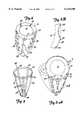
- FIG. 2illustrates a retention strip 24 in accordance with one aspect of the present invention.
- Retention strip 24in one preferred embodiment, includes a generally longitudinal strut 26 with a first set of protrusions 28, 30 and 32 extending from strut 26 in a first direction and a second plurality of protrusions 34 and 36 extending from longitudinal strut 26 in a second direction, generally opposite the first direction. While protrusions 28, 30 and 32 are shown to have a generally T-shaped conformation, and while protrusions 34 and 36 have a generally linear conformation, it should be noted that all of struts 28-36 could either have a T conformation or a linear conformation.
- Retention strip 24is also shown having a plurality of apertures 38 which are used for suturing retention strip 24 in place.
- the number and placement of the suture apertures 38 shown in FIG. 2is illustrative only. More or less apertures 38, and apertures having different placement on retention strip 24 are contemplated as well.
- Strut 26 and protrusions 28, 30, 32, 34, and 36are preferably formed of a material which allows repeated bending cycles, without permanent deformation or breakage.
- strut 26 and protrusions 28, 30, 32, 34, and 36are also preferably formed of a material which exhibits high resistance to elongation in the longitudinal direction. Suitable materials include collagen, or biocompatable and implantable polymer strips, as well as biocompatable and implantable metals, cartilage, or composite materials, Nitinol and bovine and porcine byproducts, as examples.
- FIG. 3illustrates a portion of wall 16 with retention strip 24 implanted therein.
- longitudinal strut 26is implanted in the mid-wall region 22 of wall 16.
- Protrusions 28, 30 and 32extend into the endocardium layer 18, while protrusions 34 and 36 (and any other suitable number of protrusions) extend into the epicardium layer 20.
- Retention strip 24is then sutured in place by an appropriate suturing technique using apertures 38 in retention strip 24.
- Retention strip 24preferably extends throughout substantially the entire longitudinal length of the ventricular cavity wall, such as from the base to the apex of the ventricular cavity wall.
- FIG. 4illustrates a plurality of retention strips 24 embedded in wall 16 about left ventricle 14.
- Retention strips 24are preferably arranged at regular intervals about left ventricle 14 to encircle left ventricle 14 in wall 16. Such placement forms a restrictive cage around left ventricle 14 of heart 10. Strips 24 thus provide very little, or no impedance to the natural contractile motion of the heart. However, fixturing of retention strips 24 to the tissue of the heart with sutures prevents enlargement or substantially eliminates enlargement and extensive stretch of the muscle fibers.
- longitudinal strut 26is substantially resistant to elongation in its longitudinal direction, it helps to prevent elongation of the muscle fibers in mid-wall region 22. Also, since protrusions 28, 30 and 32 are highly resistant to elongation in their longitudinal directions, they greatly inhibit elongation or stretch of the muscle fibers in endocardium layer 18. Similarly, since protrusions 34 and 36 are highly resistant to elongation in their longitudinal direction, they greatly inhibit elongation or stretch of the muscle fibers in epicardium layer 20.
- Placement of retention strips 24 in the positions illustrated in FIG. 4thus restrict the progression of congestive heart failure of the muscle due to the pressure overload on left ventricle 14.
- the muscleis supported in such a way that it is not allowed to progressively increase its mass, and also such that uncontrolled stretching is limited by physically restricting its growth and stretch. Thus, heart failure can be controlled.
- the recruitable muscle masscan still perform a satisfactory job due to the limited constraint on its contraction, no additional or compensatory muscle mass is generated, which also restricts growth of new muscle tissue in wall 16.
- This aspect of the present inventionthus reduces fatigue of the heart muscle and allows for natural regeneration of healthy cardiac tissue.
- supporting the cardiac muscle tissuerelives tension on the chordae tendinae which, in turn, helps prevent mitral valve regurgitation which prevents higher mean atrial pressures and thus pulmonary edema.
- FIG. 5illustrates another embodiment of a retention device 40 in accordance with another aspect of the present invention.
- heart 10may already have started to hypertrophy and thus may be vulnerable to trauma. Thus, extensive incision in heart 10 may be undesirable.
- retention device 40can be used by attaching it to the epicardial surface of heart 10 with only minimal intrusion into the heart muscle (such as with sutures, adhesives, staples, or other connection techniques).
- Retention device 40includes a first generally circular strap 42 and a second generally circular strap 44. Straps 42 and 44 are connected to one another by a plurality of generally longitudinal straps 46, 48, 50, 52 and 54. Straps 46, 48, 50, 52, and 54 are preferably attached to circular strips 42 and 44 by a suitable adhesive, by welding, or by another suitable mechanism, or are formed integrally with circular straps 42. Straps 42, 44, 46, 48, 50, 52, and 54 are preferably formed of collagen, polymer or metal fibers which exhibit the capability of undergoing many bending cycles, without permanent deformation of damage. Straps 42, 44, 46, 48, 50, 52, and 54 are also formed to exhibit high resistance to elongation in the generally longitudinal directions.
- straps 42, 44, 46, 48, 50, 52, and 54have a plurality of apertures 56 therein which are used to attach retention device 40 to the heart wall 16 with an appropriate suturing technique.
- any suitable number of straps 42, 44, 46, 48, 50, 52, and 54can be used.
- the arrangement of straps 42, 44, 46, 48, 50, 52, and 54can also be changed as desired.
- the number and placement of suture apertures 56can be changed to any suitable number and location on straps 42, 44, 46, 48, 50, 52, and 54.
- FIG. 6Aillustrates retention device 40 deployed on wall 16 of left ventricle 14.
- FIG. 6Aillustrates that, in one preferred embodiment, straps 42, 44, 46, 48, 50, 52, and 54 are periodically, and alternately, sutured to the outer surface of epicardium 20, and embedded within wall 16.
- FIG. 6Aalso illustrates that more longitudinal straps can be used than are illustrated in FIG. 5. This simply illustrates that any desired number of longitudinal straps can be used.
- the preferred embodimentin the areas where straps 42, 44, 46, 48, 50, 52, and 54 are embedded in wall 16, they are embedded only in the epicardium layer 20 such that extensive incisions into wall 16 need not be made.
- FIG. 6Bbetter illustrates embedding of straps 42, 44, 46, 48, 50, 52, and 54 in the wall 16 of heart 10.
- FIG. 6Bis a greatly enlarged cross-section of wall 16 taken in the region labeled 6B in FIG. 6A.
- FIG. 6Billustrates that strap 46, at alternate portions 58 is simply sutured to the exterior of epicardium layer 20, while at other portions 60 is embedded within the epicardium layer 20.
- straps 42, 44, 46, 48, 50, 52, and 54could be embedded more deeply in the wall 16. However, embedding in epicardial layer 20 is preferred.
- retention device 40restricts the progression of failure of heart muscle 16 due to pressure overload on the left ventricle 14 of heart 10.
- Heart 10is not allowed to progressively increase its mass and since the uncontrolled stretching of the heart muscle is physically restricted, heart failure can be controlled. Further, since the recruitable muscle mass is still capable of operating satisfactorily, no additional, compensatory muscle mass needs to be generated. Retention device 40 thus restricts growth of new muscle. Further, retention device 40 allows for minimal internal damage to heart 10.
- FIG. 7illustrates another embodiment of retention device 40 in accordance with one aspect of the present invention.
- Retention device 40shown in FIG. 7, is similar to that shown in FIG. 5, and similar items are similarly numbered.
- the longitudinal straps 42, 44, 46, 48, 50, 52, and 54(only four of which are shown in FIG. 7) are each provided with a plurality of sensors 62 which are configured to sense stretching, and/or other physiologic parameters, such as electrical activity, acceleration or physicochemical activity, of the cardiac muscle wall 16.
- sensors 62are only provided on longitudinal strap 48, it will be appreciated that, in a preferred embodiment, sensors 62 are similarly disposed on each of the straps 42, 44, 46, 48, 50, 52, and 54.
- Sensors 62are preferably wire bond strain gauges, piezopolymer strips, or other strain measuring sensors. As is generally known, some such strain gauges are provided with a resistive bridge having a signal, such as a voltage, applied thereacross. As strain on the bridge changes, the values of signals received from the bridge change in a differential manner. In piezopolymer elements, application of a mechanical stress to the device generates electric polarization which can also be sensed. Thus, each sensor 62 provides one or more conductors 64 which carry signals indicative of the stretching of muscle wall 16. Such conductors are preferably provided through a suitable cable 66 to monitor circuit 68 which, in turn, is coupled to a user input/output (I/O) device 70.
- I/Ouser input/output
- the strain information captured by the signals conducted by conductors 64 to monitor circuit 68is processed to obtain a total stretch response in the myocardium of heart 10. Such processing preferably occurs in monitor circuit 68 and is described below.
- the total stretch responseis preferably monitored for variations and thus provides information about the stretching and condition of heart 10. This information is preferably used for the treatment and management of the heart failure condition, either by itself through observation, or used to generate a feedback signal which can be used to pace heart 10 for maximal contraction (which is described in greater detail with respect to FIG. 9).
- User I/O device 70is preferably any suitable I/O device, such as a cathode ray tube, an LCD display, a strip or other printer, or any other suitable I/O device. I/O device 70 may also allow user input functions by including a keypad, a keyboard, or other user actuable elements.
- FIG. 8illustrates a more detailed block diagram of one embodiment of monitor circuit 68.
- Monitor circuit 68preferably includes a plurality of differential amplifiers 72, 74 and 76, a circuit (such as a summing amplifier, multiplexer, etc.) 78 and a microprocessor or microcontroller based circuit 80.
- monitor circuit 68may also include other signal filtering and amplification, and other general signal conditioning circuitry, which is generally known for conditioning signals from strain sensors and is not described here in detail.
- differential amplifiers 72, 74, and 76are provided for amplifying the signals received from strain sensors 62.
- each strain sensor 62has a corresponding differential amplifier.
- multiplexing circuitycan be used to switch the signals from sensors 62 into a single, or into one or more of the differential amplifiers.
- the output signals from differential amplifiers 72, 74, and 76are provided to circuit 78.
- circuit 78is a summing amplifier, the signals are summed in a desired manner to obtain the total stretch response of the myocardium of heart 10.
- the signal from amplifier 78is provided to microprocessor 80 where it is preferably corrected for any non-linearities and temperature affects, in a known manner.
- Microprocessor 80then generates a suitable output signal to user I/O device 70.
- circuit 78In another embodiment in which circuit 78 is a multiplexer, each of the signals from amplifiers 72, 74, and 76 are switched into microprocessor 80 under the control of microprocessor 80. Alternatively, circuit 78 can also be eliminated. In that embodiment, the outputs from amplifiers 72, 74, and 76 are provided as discrete inputs to microprocessor 80. It should also be noted that other inputs can be provided to microprocessor 80 as well, such as EKG information, blood pressure information, or other sources of information. In any case, microprocessor 80 generates a signal to user I/O device 70 based on the signals from amplifiers 72, 74, and 76.
- FIG. 9illustrates another embodiment of a monitoring and control system 82 in accordance with one aspect of the present invention.
- system 82each sensor 62 is provided with a strain sensing element 84, as discussed above, and an excitation electrode 86.
- Excitation electrodes 86are preferably conventional pacing electrodes capable of delivering pacing voltages to the myocardium of heart 10. While the sensing elements 84 and pacing electrodes 86 are shown attached to one another in FIG. 9, it should be noted that they can be separated from one another, but are preferably closely proximate one another when deployed on one of straps 42, 44, 46, 48, 50, 52, and 54.
- microprocessor 80receives the stretch response information from sensor elements 84 which indicates not only long term stretching of the heart muscle fibers, but which also indicates contractile motion of the heart 10 in a pulsatile fashion. Based upon this information, microprocessor 80 generates a plurality of feedback signals which are provided to each of pacing electrodes 86. The feedback signals are used to energize pacing electrodes 86 to deliver the necessary pacing voltages to the myocardium of heart 10 in order to pace heart 10.
- microprocessor 80simply energizes all of electrodes 86 at one time to cause contraction of the heart muscle. In another preferred embodiment, however, the microprocessor 80 selectively and sequentially energizes each of the excitation electrodes 86 in order to sequentially pace different sets of electrodes 86 to achieve optimal contraction of the ventricles. In that embodiment, the output of each of differential amplifiers 72, 74 and 76 can be individually provided to microprocessor 80, as well as through, for example, a summing amplifier 78. Microprocessor 80, in a preferred embodiment, also calculates and delivers appropriate pacing voltages to the various sets of excitation electrodes 86 being controlled.
- system 82when used in conjunction with retention device 40 restricts the progression of failure of the muscle of heart 10 due to the pressure overload in the left ventricle 14.
- the muscle in heart 10is preferably supported such that it is not allowed to progressively increase its mass, and so as to restrict uncontrolled stretch of the heart muscle in order to control heart failure. Since the muscle is not allowed to reach a point of destructive stretching, the muscle fibers maintain their inherent contractile properties (actin-myosin overlap) and the progression to failure (or cascade) can be stopped. Also, since the recruitable muscle mass is still performing satisfactorily, no additional or compensatory muscle mass needs to be generated, thus further restricting growth of new muscle.
- the present inventionis used to deliver a drug or other therapeutic agent to the tissue with which it is used.
- the struts, protrusions, retention strips, etc.are coated or impregnated with or otherwise provided with the therapeutic agent which is preferably engineered to be released into the adjacent tissue over time.
- drugs or therapeutic agentsillustratively include genetic therapeutic agents like growth factors, angiogenics, angiotensin converting enzymes, or contractibility promoters (such as that sold under the name Digitalis) or other suitable drugs.
- the heart musclecan benefit from suturing or other manipulations of the heart muscle in accordance with the present invention. This benefit results from myocardial revascularization which is a known angiogenic effect which regenerates cardiac tissue.
- the present inventionreduces fatigue of the heart and allows for natural regeneration of healthy cardiac tissue, or increases the efficiency of pharmacologically administered treatments. It is also believed that supporting the cardiac muscle in this way relieves tension on the chordae tendinae which in turn prevents mitral valve regurgitation thus preventing higher mean atrial pressures and pulmonary edema. All of these factors contribute to the cascade of failures in organs and systems associated with congestive heart failure. Once the relaxed heart muscle has regained many of its own contractile properties, it can be weaned from the pacing routine.
- the present inventioncontemplates implementing the techniques and devices described herein not only on the left ventricle, but also on the right ventricle or the other chambers of the heart. Further, the present invention can be implemented on any desired combination of chambers.
Landscapes
- Health & Medical Sciences (AREA)
- Cardiology (AREA)
- Engineering & Computer Science (AREA)
- Heart & Thoracic Surgery (AREA)
- Public Health (AREA)
- Animal Behavior & Ethology (AREA)
- Veterinary Medicine (AREA)
- Biomedical Technology (AREA)
- General Health & Medical Sciences (AREA)
- Life Sciences & Earth Sciences (AREA)
- Hematology (AREA)
- Mechanical Engineering (AREA)
- Anesthesiology (AREA)
- Vascular Medicine (AREA)
- Medical Informatics (AREA)
- Oral & Maxillofacial Surgery (AREA)
- Transplantation (AREA)
- Prostheses (AREA)
- External Artificial Organs (AREA)
Abstract
Description
The present invention deals with treatment of heart disease. More particularly, the present invention deals with a system and method for treating heart disease by regulating blood flow in the vasculature.
Congestive heart failure is a common heart disease. The prevalence of incidents of congestive heart failure has recently increased, and there is considerable morbidity and mortality associated with its diagnosis. In fact, congestive heart failure is an extremely lethal disease with an estimated five year mortality for a vast majority of both men and women who encounter the disease.
Congestive heart failure results from loss of, or impairment of, normal heart function. This loss or impairment reduces cardiac output. This, in turn, results in a reduction in both blood flow and blood pressure in the kidneys. This reduction in flow and pressure causes a renin-angiotensin response that exacerbates congestive heart failure.
Briefly, as blood flow and pressure is reduced in the kidneys, cells in the kidneys referred to as juxtaglomerular apparatus secret an enzyme referred to as renin into the blood. The enzyme renin cleaves a ten-amino acid polypeptide called angiotensin I from a plasma protein in the blood called angiotensinogen. A converting enzyme in the blood removes two amino acids from the angiotensin I polypeptide leaving an eight amino acid polypeptide called angiotensin II. Angiotensin II has numerous effects on the smooth muscle layers of arterioles, including causing vasoconstriction. Further, an indirect effect of an increase in angiotensin II increases blood volume. Blood volume is increased because angiotensin II stimulates secretion of aldosterone from the adrenal cortex which, in turn, causes an increase in salt and water retention in the kidneys. Angiotensin II also stimulates thirst centers in the hypothalamus causing more water to be ingested. The increase in blood volume and the corresponding vasoconstriction cause an increase in blood pressure and hence a volume overload on the heart which causes further deterioration of the heart condition.
Another response is also related to congestive heart failure. Baroreceptors, referred to as stretch receptors, reside in the aortic arch and carotid sinuses. The baroreceptors are essentially pressure sensors sensing blood pressure in that area. The baroreceptors provide physiological feedback in two ways. First, in response to a reduction in blood pressure, the baroreceptors provide a neurohormonal feedback response which acts to increase the heart rate in an attempt to increase cardiac output. The increased heart rate causes the heart to work harder which, in turn, causes the heart muscle to stretch further. Also, a reduction in pressure caused by a reduction in cardiac output causes the baroreceptors to provide a feedback response which acts to constrict the distal vasculature thus increasing pressure in that area.
It can thus be seen that impairment of heart function can lead to a cyclical feedback response which increases, rather than reduces, the impairment. Such a cyclical feedback response is sometimes referred to as a cascade.
For instance, if the heart muscle is stressed, the heart works harder and begins to stretch. This reduces the efficiency of the heart in the following way. Muscles are thought of as being composed of many fibers which contract and lengthen to accomplish muscular action. Each fiber includes many densely packed subunits referred to as myofibrils which are on the order of 1 μm in diameter and extend in parallel from one end of the muscle fiber to the other. Each myofibril has spaced regions of thick filaments (about 110 Å thick) and thin filaments (about 50-60 Å thick). The thick filaments are formed of a protein, myosin, and the thin filaments are formed of a protein, actin. The actin and myosin filaments overlap in regions periodically spaced along the myofibrils. The units in the repeated overlapping pattern are referred to as sarcomeres.
Contraction of a muscle fiber results from shortening of the myofibrils which form the muscle fiber. The myofibrils are shortened, but the individual filaments in the myofibrils do not decrease in length. Instead, the actin and myosin filaments slide longitudinally relative to one another to shorten the overall length of the myofibrils. Sliding occurs as a result of cross-bridges extending from the myosin toward the actin attaching to the actin at bonding sites. The cross bridges are oriented to draw overlapping actin filaments on either longitudinal side of the myosin filament toward the longitudinal center of the myosin filament. When the muscle fiber is stretched such that the actin and myosin only overlap a short distance, only a small number of cross-bridges are available for bonding to the adjacent actin, and contraction is highly inefficient. When the muscle is stretched to a point where the actin and myosin filaments no longer overlap, contraction is rendered impossible.
This inefficient or impaired heart function causes blood pressure in the areas of both the kidneys and the baroreceptors to decrease. The feedback response generated by the kidneys causes further overload and stress on the heart. The feedback response generated by the baroreceptors causes increased heart rate. Both of these feedback responses cause the heart to work harder, causing further stretching of the heart muscle and thus leading to greater inefficiencies. In response, the feedback responses become even more acute--and the cascade continues.
This cascade effect, which is a natural progression of congestive heart failure, leads to increased muscle mass and stretching of the heart muscle fibers which, in turn, leads to muscular hypertrophy of the left ventricle. The hypertrophy is a compensatory mechanism which, if maintained at a given level such that muscle fibers maintain inherent contractile properties (i.e., actin-myosin overlap), can be beneficial for maintaining proper heart function. However, prolonged and continuous stretching causes muscular fatigue and reduced muscle performance as explained by the known Frank-Starling mechanism.
An apparatus and method restrict elongation of heart muscle tissue in a wall of a heart having a chamber. A plurality of biocompatible and implantable elongate strips are configured to be connected to the heart wall and disposed about the chamber such that the elongate strips are arranged in spaced relation to one another. The elongate strips are bendable and are sufficiently resistant to elongation such that natural stretching of the heart wall does not cause elongation of the plurality of strips.
FIG. 1A is a side sectional view of a heart.
FIG. 1B is a greatly enlarged sectional view of a portion of the heart shown in FIG. 1A.
FIG. 2 illustrates a retention strip in accordance with one aspect of the present invention.
FIG. 3 illustrates the retention strip shown in FIG. 2 embedded in the heart wall.
FIG. 4 illustrates a plurality of the strips as shown in FIG. 2 embedded in the wall of the left ventricle.
FIG. 5 illustrates a second embodiment of a retention mechanism in accordance with one aspect of the present invention.
FIG. 6A shows the retention mechanism illustrated in FIG. 5 deployed on the outer surface of the left ventricle.
FIG. 6B is a sectional view of a portion of the heart wall shown in FIG. 6A.
FIG. 7 illustrates another embodiment of a monitoring system in accordance with one aspect of the present invention.
FIG. 8 illustrates a portion of the monitoring system shown in FIG. 7 in schematic and partial block diagram form.
FIG. 9 illustrates another embodiment of a monitoring and control system in schematic and partial block diagram form.
FIG. 1A illustrates a portion of a heart 10. FIG. 1A illustrates a plurality of chambers in heart 10 includingright ventricle 12 andleft ventricle 14. FIG. 1A also illustratesheart wall 16 which extends aroundchambers chambers left ventricle 14.
FIG. 1B illustrates a greatly enlarged sectional view of a portion ofwall 16 of heart 10 shown in FIG. 1A. FIG. 1B is taken from the portion labeled 1B in FIG. 1A. FIG. 1B illustrates thatwall 16 is formed of anendocardium layer 18 which comprises the inner tissue ofheart wall 16.Heart wall 16 also includes anepicardium layer 20 which comprises the outer tissue inheart wall 16.Mid-wall portion 22 comprises a portion ofwall 16 between theendocardium 18 andepicardium 20. The pericardium is a double-walled sac which encloses the heart. The inner layer of the pericardial sac comprises theepicardium 20.
Photomicrographs available from the American Heart Association libraries show that the alignment of cardiac muscle fibers are generally perpendicular to the ventricular cavity wall in theendocardium 18 andepicardium 20. Also, the muscle fibers are typically nearly parallel to the ventricular cavity wall in themid-wall portion 22. During hypertrophy of themuscular wall 16 around theleft ventricle 14, the muscle fibers stretch and elongate in a direction generally parallel to their longitudinal orientation. Thus, the muscle fibers in theendocardium 18 andepicardium 20 elongate in a direction generally perpendicular to the ventricular cavity wall. Also, the muscle fibers in themid-wall region 22 elongate in a direction generally parallel to the ventricular cavity wall. Additional muscle fibers also develop. All of these effects contribute to an increase in muscle mass and stretching of the heart muscle fibers.
FIG. 2 illustrates aretention strip 24 in accordance with one aspect of the present invention.Retention strip 24, in one preferred embodiment, includes a generallylongitudinal strut 26 with a first set ofprotrusions strut 26 in a first direction and a second plurality ofprotrusions longitudinal strut 26 in a second direction, generally opposite the first direction. Whileprotrusions protrusions
Also, while only threeprotrusions protrusions retention strip 24.Strut 26 andprotrusions protrusions
FIG. 3 illustrates a portion ofwall 16 withretention strip 24 implanted therein. In a preferred embodiment,longitudinal strut 26 is implanted in themid-wall region 22 ofwall 16.Protrusions endocardium layer 18, whileprotrusions 34 and 36 (and any other suitable number of protrusions) extend into theepicardium layer 20.Retention strip 24 is then sutured in place by an appropriate suturingtechnique using apertures 38 inretention strip 24.Retention strip 24 preferably extends throughout substantially the entire longitudinal length of the ventricular cavity wall, such as from the base to the apex of the ventricular cavity wall.
FIG. 4 illustrates a plurality of retention strips 24 embedded inwall 16 aboutleft ventricle 14. Retention strips 24 are preferably arranged at regular intervals aboutleft ventricle 14 to encircleleft ventricle 14 inwall 16. Such placement forms a restrictive cage aroundleft ventricle 14 of heart 10.Strips 24 thus provide very little, or no impedance to the natural contractile motion of the heart. However, fixturing of retention strips 24 to the tissue of the heart with sutures prevents enlargement or substantially eliminates enlargement and extensive stretch of the muscle fibers.
Sincelongitudinal strut 26 is substantially resistant to elongation in its longitudinal direction, it helps to prevent elongation of the muscle fibers inmid-wall region 22. Also, sinceprotrusions endocardium layer 18. Similarly, sinceprotrusions epicardium layer 20.
Placement of retention strips 24 in the positions illustrated in FIG. 4 thus restrict the progression of congestive heart failure of the muscle due to the pressure overload onleft ventricle 14. The muscle is supported in such a way that it is not allowed to progressively increase its mass, and also such that uncontrolled stretching is limited by physically restricting its growth and stretch. Thus, heart failure can be controlled. Further, since the recruitable muscle mass can still perform a satisfactory job due to the limited constraint on its contraction, no additional or compensatory muscle mass is generated, which also restricts growth of new muscle tissue inwall 16. This aspect of the present invention thus reduces fatigue of the heart muscle and allows for natural regeneration of healthy cardiac tissue. Also, it is believed that supporting the cardiac muscle tissue relives tension on the chordae tendinae which, in turn, helps prevent mitral valve regurgitation which prevents higher mean atrial pressures and thus pulmonary edema.
FIG. 5 illustrates another embodiment of aretention device 40 in accordance with another aspect of the present invention. With progressive congestive heart failure, heart 10 may already have started to hypertrophy and thus may be vulnerable to trauma. Thus, extensive incision in heart 10 may be undesirable. In that case,retention device 40 can be used by attaching it to the epicardial surface of heart 10 with only minimal intrusion into the heart muscle (such as with sutures, adhesives, staples, or other connection techniques).
FIG. 6A illustratesretention device 40 deployed onwall 16 ofleft ventricle 14. FIG. 6A illustrates that, in one preferred embodiment, straps 42, 44, 46, 48, 50, 52, and 54 are periodically, and alternately, sutured to the outer surface ofepicardium 20, and embedded withinwall 16. FIG. 6A also illustrates that more longitudinal straps can be used than are illustrated in FIG. 5. This simply illustrates that any desired number of longitudinal straps can be used. In the preferred embodiment, in the areas where straps 42, 44, 46, 48, 50, 52, and 54 are embedded inwall 16, they are embedded only in theepicardium layer 20 such that extensive incisions intowall 16 need not be made.
FIG. 6B better illustrates embedding ofstraps wall 16 of heart 10. FIG. 6B is a greatly enlarged cross-section ofwall 16 taken in the region labeled 6B in FIG. 6A. FIG. 6B illustrates thatstrap 46, atalternate portions 58 is simply sutured to the exterior ofepicardium layer 20, while atother portions 60 is embedded within theepicardium layer 20. Of course, straps 42, 44, 46, 48, 50, 52, and 54 could be embedded more deeply in thewall 16. However, embedding inepicardial layer 20 is preferred.
As with the embodiment illustrated in FIGS. 2-4,retention device 40 restricts the progression of failure ofheart muscle 16 due to pressure overload on theleft ventricle 14 of heart 10. Heart 10 is not allowed to progressively increase its mass and since the uncontrolled stretching of the heart muscle is physically restricted, heart failure can be controlled. Further, since the recruitable muscle mass is still capable of operating satisfactorily, no additional, compensatory muscle mass needs to be generated.Retention device 40 thus restricts growth of new muscle. Further,retention device 40 allows for minimal internal damage to heart 10.
FIG. 7 illustrates another embodiment ofretention device 40 in accordance with one aspect of the present invention.Retention device 40, shown in FIG. 7, is similar to that shown in FIG. 5, and similar items are similarly numbered. However, thelongitudinal straps sensors 62 which are configured to sense stretching, and/or other physiologic parameters, such as electrical activity, acceleration or physicochemical activity, of thecardiac muscle wall 16. In the illustration of FIG. 7,sensors 62 are only provided onlongitudinal strap 48, it will be appreciated that, in a preferred embodiment,sensors 62 are similarly disposed on each of thestraps
In one preferred embodiment, the strain information captured by the signals conducted byconductors 64 to monitorcircuit 68 is processed to obtain a total stretch response in the myocardium of heart 10. Such processing preferably occurs inmonitor circuit 68 and is described below. The total stretch response is preferably monitored for variations and thus provides information about the stretching and condition of heart 10. This information is preferably used for the treatment and management of the heart failure condition, either by itself through observation, or used to generate a feedback signal which can be used to pace heart 10 for maximal contraction (which is described in greater detail with respect to FIG. 9).
User I/O device 70 is preferably any suitable I/O device, such as a cathode ray tube, an LCD display, a strip or other printer, or any other suitable I/O device. I/O device 70 may also allow user input functions by including a keypad, a keyboard, or other user actuable elements.
FIG. 8 illustrates a more detailed block diagram of one embodiment ofmonitor circuit 68.Monitor circuit 68 preferably includes a plurality ofdifferential amplifiers circuit 80. Of course, monitorcircuit 68 may also include other signal filtering and amplification, and other general signal conditioning circuitry, which is generally known for conditioning signals from strain sensors and is not described here in detail.
In the embodiment illustrated in FIG. 8,differential amplifiers strain sensors 62. In one preferred embodiment, eachstrain sensor 62 has a corresponding differential amplifier. Alternatively, of course, multiplexing circuity can be used to switch the signals fromsensors 62 into a single, or into one or more of the differential amplifiers. In any case, the output signals fromdifferential amplifiers circuit 78. In the embodiment in whichcircuit 78 is a summing amplifier, the signals are summed in a desired manner to obtain the total stretch response of the myocardium of heart 10. The signal fromamplifier 78 is provided tomicroprocessor 80 where it is preferably corrected for any non-linearities and temperature affects, in a known manner.Microprocessor 80 then generates a suitable output signal to user I/O device 70.
In another embodiment in whichcircuit 78 is a multiplexer, each of the signals fromamplifiers microprocessor 80 under the control ofmicroprocessor 80. Alternatively,circuit 78 can also be eliminated. In that embodiment, the outputs fromamplifiers microprocessor 80. It should also be noted that other inputs can be provided tomicroprocessor 80 as well, such as EKG information, blood pressure information, or other sources of information. In any case,microprocessor 80 generates a signal to user I/O device 70 based on the signals fromamplifiers
FIG. 9 illustrates another embodiment of a monitoring andcontrol system 82 in accordance with one aspect of the present invention. Some items insystem 82 are similar to those shown in FIGS. 7 and 8 and are similarly numbered. Insystem 82, eachsensor 62 is provided with astrain sensing element 84, as discussed above, and anexcitation electrode 86.Excitation electrodes 86 are preferably conventional pacing electrodes capable of delivering pacing voltages to the myocardium of heart 10. While thesensing elements 84 andpacing electrodes 86 are shown attached to one another in FIG. 9, it should be noted that they can be separated from one another, but are preferably closely proximate one another when deployed on one ofstraps
In one preferred embodiment,microprocessor 80 receives the stretch response information fromsensor elements 84 which indicates not only long term stretching of the heart muscle fibers, but which also indicates contractile motion of the heart 10 in a pulsatile fashion. Based upon this information,microprocessor 80 generates a plurality of feedback signals which are provided to each of pacingelectrodes 86. The feedback signals are used to energize pacingelectrodes 86 to deliver the necessary pacing voltages to the myocardium of heart 10 in order to pace heart 10.
In one preferred embodiment,microprocessor 80 simply energizes all ofelectrodes 86 at one time to cause contraction of the heart muscle. In another preferred embodiment, however, themicroprocessor 80 selectively and sequentially energizes each of theexcitation electrodes 86 in order to sequentially pace different sets ofelectrodes 86 to achieve optimal contraction of the ventricles. In that embodiment, the output of each ofdifferential amplifiers microprocessor 80, as well as through, for example, a summingamplifier 78.Microprocessor 80, in a preferred embodiment, also calculates and delivers appropriate pacing voltages to the various sets ofexcitation electrodes 86 being controlled.
As with the other embodiments discussed herein,system 82, when used in conjunction withretention device 40 restricts the progression of failure of the muscle of heart 10 due to the pressure overload in theleft ventricle 14. The muscle in heart 10 is preferably supported such that it is not allowed to progressively increase its mass, and so as to restrict uncontrolled stretch of the heart muscle in order to control heart failure. Since the muscle is not allowed to reach a point of destructive stretching, the muscle fibers maintain their inherent contractile properties (actin-myosin overlap) and the progression to failure (or cascade) can be stopped. Also, since the recruitable muscle mass is still performing satisfactorily, no additional or compensatory muscle mass needs to be generated, thus further restricting growth of new muscle.
In another preferred embodiment, the present invention is used to deliver a drug or other therapeutic agent to the tissue with which it is used. For example, in one embodiment, the struts, protrusions, retention strips, etc. are coated or impregnated with or otherwise provided with the therapeutic agent which is preferably engineered to be released into the adjacent tissue over time. Such drugs or therapeutic agents illustratively include genetic therapeutic agents like growth factors, angiogenics, angiotensin converting enzymes, or contractibility promoters (such as that sold under the name Digitalis) or other suitable drugs.
In addition, it is believed that the heart muscle can benefit from suturing or other manipulations of the heart muscle in accordance with the present invention. This benefit results from myocardial revascularization which is a known angiogenic effect which regenerates cardiac tissue.
The present invention reduces fatigue of the heart and allows for natural regeneration of healthy cardiac tissue, or increases the efficiency of pharmacologically administered treatments. It is also believed that supporting the cardiac muscle in this way relieves tension on the chordae tendinae which in turn prevents mitral valve regurgitation thus preventing higher mean atrial pressures and pulmonary edema. All of these factors contribute to the cascade of failures in organs and systems associated with congestive heart failure. Once the relaxed heart muscle has regained many of its own contractile properties, it can be weaned from the pacing routine.
It should also be noted that the present invention contemplates implementing the techniques and devices described herein not only on the left ventricle, but also on the right ventricle or the other chambers of the heart. Further, the present invention can be implemented on any desired combination of chambers.
Although the present invention has been described with reference to preferred embodiments, workers skilled in the art will recognize that changes may be made in form and detail without departing from the spirit and scope of the invention.
Claims (36)
1. An apparatus for relieving stress of heart muscle tissue in a wall of a heart having a chamber, the muscle tissue having an epicardium layer, an endocardium layer and a midwall portion between the epicardium and endocardium layers, the epicardium layer, the endocardium layer and the midwall portion extending generally longitudinally to form the wall of the heart, the apparatus comprising:
a plurality of biocompatible and implantable elongate strips, elongate in a longitudinal direction, configured to be connected to the heart wall and disposed about the chamber such that the elongate strips are arranged in spaced relation to one another, the elongate strips being bendable and being sufficiently resistant to elongation that natural stretching of the heart wall does not cause elongation of the plurality of strips.
2. The apparatus of claim 1 wherein the elongate strips are resistant to elongation in the longitudinal direction.
3. The apparatus of claim 1 wherein each of the elongate strips comprises:
a longitudinal strut; and
a first plurality of protrusions extending from the longitudinal strut in a first direction, the first protrusions being sufficiently resistant to elongation in the first direction such that natural stretching of the heart wall does not cause elongation of the first protrusions in the first direction.
4. The apparatus of claim 3 wherein each of the elongate strips comprises:
a second plurality of protrusions extending from the longitudinal strut in a second direction, generally opposite the first direction, the second protrusions being sufficiently resistant to elongation in the second direction such that natural stretching of the heart wall does not cause elongation of the second protrusions in the second direction.
5. The apparatus of claim 4 wherein the longitudinal strut and the first and second protrusions are arranged such that when the longitudinal strut is positioned longitudinally in the midwall portion, the first protrusions extend into the epicardium layer and the second protrusions extend into the endocardium layer.
6. The apparatus of claim 5 wherein the first protrusions have a generally linear conformation.
7. The apparatus of claim 5 wherein the second protrusions have a generally T-shaped conformation.
8. The apparatus of claim 5 wherein the longitudinal strut and the first and second protrusions define a plurality of apertures suitable for receiving sutures.
9. The apparatus of claim 1 wherein the plurality of elongate strips are connected to the epicardium layer.
10. The apparatus of claim 9 wherein the plurality of strips are connected to one another, in spaced relation to one another to from a cage structure sized to be disposed about the chamber.
11. The apparatus of claim 10 wherein the plurality of strips carry a plurality of sensors configured to sense physiological parameters in the wall of the heart and provide sensor signals indicative of the sensed parameters.
12. The apparatus of claim 11 wherein the plurality of sensors are configured to sense stretching of the wall of the heart and provide the sensor signals indicative of the sensed stretching.
13. The apparatus of claim 12 and further comprising:
a monitor coupled to the plurality of sensors to receive the sensor signals and provide a monitor signal based on the sensor signals; and
an input/output device, coupled to the monitor, to receive the monitor signal and provide user observable indicia, indicative of the sensed stretching based on the monitor signal.
14. The apparatus of claim 13 and further comprising:
a plurality of electrodes carried by the plurality of strips; and
wherein the monitor includes a controller coupled to the electrodes to control application of excitation signals to the electrodes, the electrodes stimulating the heart muscle in response to the excitation signals.
15. The apparatus of claim 14 wherein the controller is coupled to the sensors and is configured to apply the excitation signals based on the sensor signals.
16. The apparatus of claim 15 wherein the controller is configured to apply the excitation signals to all electrodes substantially simultaneously.
17. The apparatus of claim 15 wherein the controller is configured to apply the excitation signals to the electrodes sequentially.
18. The apparatus of claim 17 wherein the controller is configured to apply the excitation signals to sets of the electrodes sequentially.
19. The apparatus of claim 11 wherein the strips define a plurality of apertures therein sized to receive sutures.
20. The apparatus of claim 1 wherein the elongate strips include a drug releasably coupled thereto.
21. A method of restricting elongation of heart muscle tissue in a wall of a heart having a chamber, the muscle tissue having an epicardium layer, an endocardium layer and a midwall portion between the epicardium and endocardium layers, the epicardium layer, the endocardium layer and the midwall portion extending generally longitudinally to form the wall of the heart, the method comprising:
providing a plurality of biocompatible and implantable elongate strips, elongate in a longitudinal direction, the elongate strips being bendable and being sufficiently resistant to elongation in the longitudinal direction such that natural stretching of the heart wall does not cause elongation of the plurality of strips; and
connecting the elongate strips to the heart wall and about the chamber such that the elongate strips are arranged in spaced relation to one another.
22. The method of claim 21 wherein providing a plurality of elongate strips comprises providing the elongate strips such that each of the elongate strips comprises:
a longitudinal strut;
a first plurality of protrusions extending from the longitudinal strut in a first direction, the first protrusions being sufficiently resistant to elongation in the first direction such that natural stretching of the heart wall does not cause elongation of the first protrusions in the first direction; and
a second plurality of protrusions extending from the longitudinal strut in a second direction, generally opposite the first direction, the second protrusions being sufficiently resistant to elongation in the second direction such that natural stretching of the heart wall does not cause elongation of the second protrusions in the second direction.
23. The method of claim 22 wherein connecting the elongate strips to the heart comprises:
implanting the longitudinal strut longitudinally in the midwall portion;
arranging the strip such that the first protrusions extend into the epicardium layer and the second protrusions extend into the endocardium layer; and
attaching the longitudinal strut and the first and second protrusions to surrounding tissue in the heart wall.
24. The method of claim 23 wherein attaching comprises:
attaching utilizing a method which promotes revascularization of the tissue in the heart wall.
25. The method of claim 23 wherein the longitudinal strut and the first and second protrusions define a plurality of apertures, and wherein attaching comprises:
suturing the longitudinal strut and the first and second protrusions to the surrounding tissue through the apertures.
26. The method of claim 21 wherein the plurality of strips are connected to one another, in spaced relation to one another to from a cage structure, and wherein connecting comprises:
attaching the cage structure to the heart wall around the chamber.
27. The method of claim 26 wherein attaching comprises:
implanting first portions of the strips in the heart wall; and
connecting second portions of the strips between the between the first portions, to an exterior surface of the epicardium layer.
28. The method of claim 27 wherein implanting comprises:
implanting the first portions in the epicardium layer.
29. The method of claim 26 wherein providing a plurality of strips comprises:
providing a plurality of sensors carried by the elongate strips;
sensing, with the sensors, stretching of the wall of the heart; and
providing sensor signals indicative of the sensed stretching.
30. The method of claim 29 and further comprising:
providing user observable indicia, indicative of the sensed stretching, based on the sensor signals.
31. The method of claim 30 wherein providing a plurality of elongate strips further comprises:
providing a plurality of electrodes carried by the plurality of strips; and
controlling application of excitation signals to the electrodes, the electrodes stimulating the heart muscle in response to the excitation signals.
32. The method of claim 31 wherein controlling application of excitation signals comprises:
applying the excitation signals based on the sensor signals.
33. The method of claim 32 wherein applying the excitation signals comprises:
applying the excitation signals to all electrodes substantially simultaneously.
34. The method of claim 32 wherein applying the excitation signals comprises:
applying the excitation signals to the electrodes sequentially.
35. The method of claim 34 wherein applying the excitation signals sequentially comprises:
applying the excitation signals to sets of the electrodes sequentially.
36. The method of claim 26 wherein the strips define a plurality of apertures, and wherein attaching the cage structure comprises:
suturing the strips to the heart wall through the apertures.
Priority Applications (6)
| Application Number | Priority Date | Filing Date | Title |
|---|---|---|---|
| US09/064,370US6110100A (en) | 1998-04-22 | 1998-04-22 | System for stress relieving the heart muscle and for controlling heart function |
| EP99917645AEP1073488A1 (en) | 1998-04-22 | 1999-04-20 | System for stress relieving the heart muscle and for controlling heart function |
| CA002329735ACA2329735A1 (en) | 1998-04-22 | 1999-04-20 | System for stress relieving the heart muscle and for controlling heart function |
| JP2000544377AJP4190730B6 (en) | 1998-04-22 | 1999-04-20 | A system to control cardiac function by relieving myocardial stress |
| PCT/US1999/008713WO1999053977A1 (en) | 1998-04-22 | 1999-04-20 | System for stress relieving the heart muscle and for controlling heart function |
| US09/415,638US6494825B1 (en) | 1998-04-22 | 1999-10-12 | System for stress relieving the heart muscle and for controlling heart function |
Applications Claiming Priority (1)
| Application Number | Priority Date | Filing Date | Title |
|---|---|---|---|
| US09/064,370US6110100A (en) | 1998-04-22 | 1998-04-22 | System for stress relieving the heart muscle and for controlling heart function |
Related Child Applications (1)
| Application Number | Title | Priority Date | Filing Date |
|---|---|---|---|
| US09/415,638ContinuationUS6494825B1 (en) | 1998-04-22 | 1999-10-12 | System for stress relieving the heart muscle and for controlling heart function |
Publications (1)
| Publication Number | Publication Date |
|---|---|
| US6110100Atrue US6110100A (en) | 2000-08-29 |
Family
ID=22055469
Family Applications (2)
| Application Number | Title | Priority Date | Filing Date |
|---|---|---|---|
| US09/064,370Expired - LifetimeUS6110100A (en) | 1998-04-22 | 1998-04-22 | System for stress relieving the heart muscle and for controlling heart function |
| US09/415,638Expired - Fee RelatedUS6494825B1 (en) | 1998-04-22 | 1999-10-12 | System for stress relieving the heart muscle and for controlling heart function |
Family Applications After (1)
| Application Number | Title | Priority Date | Filing Date |
|---|---|---|---|
| US09/415,638Expired - Fee RelatedUS6494825B1 (en) | 1998-04-22 | 1999-10-12 | System for stress relieving the heart muscle and for controlling heart function |
Country Status (4)
| Country | Link |
|---|---|
| US (2) | US6110100A (en) |
| EP (1) | EP1073488A1 (en) |
| CA (1) | CA2329735A1 (en) |
| WO (1) | WO1999053977A1 (en) |
Cited By (101)
| Publication number | Priority date | Publication date | Assignee | Title |
|---|---|---|---|---|
| US6260552B1 (en) | 1998-07-29 | 2001-07-17 | Myocor, Inc. | Transventricular implant tools and devices |
| US6264602B1 (en) | 1998-07-29 | 2001-07-24 | Myocor, Inc. | Stress reduction apparatus and method |
| WO2001054618A1 (en)* | 2000-01-31 | 2001-08-02 | Mitralife | Percutaneous mitral annuloplasty and cardiac reinforcement |
| US6332863B1 (en) | 1997-01-02 | 2001-12-25 | Myocor, Inc. | Heart wall tension reduction kit |
| US6343605B1 (en)* | 2000-08-08 | 2002-02-05 | Scimed Life Systems, Inc. | Percutaneous transluminal myocardial implantation device and method |
| US20020022861A1 (en)* | 2000-05-19 | 2002-02-21 | Daniel Jacobs | Multi-point tissue tension distribution device, a combined orbital rim repair and suspension variation, and a method of tissue approximation using the device |
| US6402679B1 (en)* | 1998-09-21 | 2002-06-11 | Myocor, Inc. | External stress reduction device and method |
| US6406420B1 (en) | 1997-01-02 | 2002-06-18 | Myocor, Inc. | Methods and devices for improving cardiac function in hearts |
| US6494825B1 (en)* | 1998-04-22 | 2002-12-17 | Scimed Life Systems, Inc. | System for stress relieving the heart muscle and for controlling heart function |
| US20030045771A1 (en)* | 1997-01-02 | 2003-03-06 | Schweich Cyril J. | Heart wall tension reduction devices and methods |
| US6537198B1 (en) | 2000-03-21 | 2003-03-25 | Myocor, Inc. | Splint assembly for improving cardiac function in hearts, and method for implanting the splint assembly |
| US20030074021A1 (en)* | 2000-05-19 | 2003-04-17 | Morriss John H. | Remotely anchored tissue fixation device |
| WO2003037217A1 (en)* | 2001-10-31 | 2003-05-08 | Paracor Medical, Inc. | Heart failure treatment device |
| US6592619B2 (en) | 1996-01-02 | 2003-07-15 | University Of Cincinnati | Heart wall actuation device for the natural heart |
| US20030135267A1 (en)* | 2002-01-11 | 2003-07-17 | Solem Jan Otto | Delayed memory device |
| US6595912B2 (en) | 2000-03-10 | 2003-07-22 | Paracor Surgical, Inc. | Expandable cardiac harness for treating congestive heart failure |
| US20030142676A1 (en)* | 2002-01-25 | 2003-07-31 | Raymond Zeisz | Method and apparauts for admission control in packet switch |
| US6612978B2 (en) | 1999-12-22 | 2003-09-02 | Paracor Surgical, Inc. | Expandable cardiac harness for treating congestive heart failure |
| US6622730B2 (en) | 2001-03-30 | 2003-09-23 | Myocor, Inc. | Device for marking and aligning positions on the heart |
| US6629921B1 (en) | 1997-01-02 | 2003-10-07 | Myocor, Inc. | Heart wall tension reduction apparatus and method |
| WO2003007778A3 (en)* | 2001-07-16 | 2003-11-27 | Relaxis Ltd | In-vivo method and device for improving diastolic function of the left ventricle |
| US6673009B1 (en)* | 2000-11-08 | 2004-01-06 | Acorn Cardiovascular, Inc. | Adjustment clamp |
| US20040010276A1 (en)* | 2000-05-19 | 2004-01-15 | Daniel Jacobs | Multi-point tissue tension distribution device and method, a chin lift variation |
| US20040015039A1 (en)* | 2002-07-16 | 2004-01-22 | The University Of Cincinnati | Modular power system and method for a heart wall actuation system for the natural heart |
| US20040034271A1 (en)* | 2002-08-19 | 2004-02-19 | The University Of Cincinnati | Heart wall actuation system for the natural heart with shape limiting elements |
| US6695769B2 (en) | 2001-09-25 | 2004-02-24 | The Foundry, Inc. | Passive ventricular support devices and methods of using them |
| US20040059180A1 (en)* | 2002-09-23 | 2004-03-25 | The University Of Cincinnati | Basal mounting cushion frame component to facilitate extrinsic heart wall actuation |
| US6723038B1 (en) | 2000-10-06 | 2004-04-20 | Myocor, Inc. | Methods and devices for improving mitral valve function |
| US6723041B2 (en) | 2001-09-10 | 2004-04-20 | Lilip Lau | Device for treating heart failure |
| US20040102840A1 (en)* | 1999-06-30 | 2004-05-27 | Solem Jan Otto | Method and device for treatment of mitral insufficiency |
| US20040133273A1 (en)* | 2002-11-15 | 2004-07-08 | Cox Daniel L. | Apparatuses and methods for heart valve repair |
| US20040138744A1 (en)* | 2000-01-31 | 2004-07-15 | Randall Lashinski | Transluminal mitral annuloplasty with active anchoring |
| US6764510B2 (en) | 2002-01-09 | 2004-07-20 | Myocor, Inc. | Devices and methods for heart valve treatment |
| US6810882B2 (en) | 2001-01-30 | 2004-11-02 | Ev3 Santa Rosa, Inc. | Transluminal mitral annuloplasty |
| US20050043792A1 (en)* | 1999-06-29 | 2005-02-24 | Edwards Lifesciences Ag | Device and method for treatment of mitral insufficiency |
| US20050060030A1 (en)* | 2000-01-31 | 2005-03-17 | Lashinski Randall T. | Remotely activated mitral annuloplasty system and methods |
| US20050080483A1 (en)* | 2001-12-28 | 2005-04-14 | Solem Jan Otto | Delayed memory device |
| US20050119694A1 (en)* | 2000-05-19 | 2005-06-02 | Jacobs Daniel I. | Remotely anchored tissue fixation device and method |
| US20050171590A1 (en)* | 1998-11-04 | 2005-08-04 | Acorn Cardiovascular, Inc. | Cardiomyopathy treatment device with electrode therapy |
| US20050177228A1 (en)* | 2003-12-16 | 2005-08-11 | Solem Jan O. | Device for changing the shape of the mitral annulus |
| US20050197699A1 (en)* | 2004-02-10 | 2005-09-08 | Jacobs Daniel I. | Tissue repair apparatus and method |
| US6945978B1 (en) | 2002-11-15 | 2005-09-20 | Advanced Cardiovascular Systems, Inc. | Heart valve catheter |
| US20050209542A1 (en)* | 2004-03-16 | 2005-09-22 | Jacobs Daniel I | Tissue approximation sling and method |
| US20050222678A1 (en)* | 2004-04-05 | 2005-10-06 | Lashinski Randall T | Remotely adjustable coronary sinus implant |
| US20050288715A1 (en)* | 2003-11-07 | 2005-12-29 | Lilip Lau | Cardiac harness for treating congestive heart failure and for defibrillating and/or pacing/sensing |
| US6989028B2 (en) | 2000-01-31 | 2006-01-24 | Edwards Lifesciences Ag | Medical system and method for remodeling an extravascular tissue structure |
| US20060063964A1 (en)* | 2004-08-20 | 2006-03-23 | Massen Richard J | Micro electromechanical machine-based ventricular assist apparatus |
| US20060116756A1 (en)* | 1999-06-30 | 2006-06-01 | Solem Jan O | Method and device for treatment of mitral insufficiency |
| US7060023B2 (en) | 2001-09-25 | 2006-06-13 | The Foundry Inc. | Pericardium reinforcing devices and methods of using them |
| US20060129051A1 (en)* | 2004-12-09 | 2006-06-15 | Rowe Stanton J | Diagnostic kit to assist with heart valve annulus adjustment |
| US7087064B1 (en) | 2002-10-15 | 2006-08-08 | Advanced Cardiovascular Systems, Inc. | Apparatuses and methods for heart valve repair |
| US20060178551A1 (en)* | 2003-06-09 | 2006-08-10 | Melvin David B | Securement system for a heart actuation device |
| US7112219B2 (en) | 2002-11-12 | 2006-09-26 | Myocor, Inc. | Devices and methods for heart valve treatment |
| US20060241334A1 (en)* | 2003-01-27 | 2006-10-26 | Corassist Cardiovascular Ltd. | In vivo device for improving diastolic ventricular function |
| US7146226B2 (en) | 2003-11-07 | 2006-12-05 | Paracor Medical, Inc. | Cardiac harness for treating congestive heart failure and for defibrillating and/or pacing/sensing |
| US20060276890A1 (en)* | 2005-06-03 | 2006-12-07 | Solem Jan O | Devices and methods for percutaneous repair of the mitral valve via the coronary sinus |
| US7158839B2 (en) | 2003-11-07 | 2007-01-02 | Paracor Medical, Inc. | Cardiac harness for treating heart disease |
| US7174896B1 (en) | 2002-01-07 | 2007-02-13 | Paracor Medical, Inc. | Method and apparatus for supporting a heart |
| US20070038297A1 (en)* | 2005-08-12 | 2007-02-15 | Bobo Donald E Jr | Medical implant with reinforcement mechanism |
| US7189203B2 (en) | 2002-11-15 | 2007-03-13 | Paracor Medical, Inc. | Cardiac harness delivery device and method |
| US20070073391A1 (en)* | 2005-09-28 | 2007-03-29 | Henry Bourang | System and method for delivering a mitral valve repair device |
| US7229405B2 (en) | 2002-11-15 | 2007-06-12 | Paracor Medical, Inc. | Cardiac harness delivery device and method of use |
| US7247134B2 (en) | 2002-11-12 | 2007-07-24 | Myocor, Inc. | Devices and methods for heart valve treatment |
| US20070173926A1 (en)* | 2005-12-09 | 2007-07-26 | Bobo Donald E Jr | Anchoring system for medical implant |
| US7282024B2 (en) | 2004-01-12 | 2007-10-16 | Paracor Medical, Inc. | Cardiac harness having interconnected strands |
| US7291105B2 (en) | 2003-07-10 | 2007-11-06 | Paracor Medical, Inc. | Self-anchoring cardiac harness |
| US20080039681A1 (en)* | 2004-07-15 | 2008-02-14 | Micardia Corporation | Shape memory devices and methods for reshaping heart anatomy |
| US7331972B1 (en) | 2002-11-15 | 2008-02-19 | Abbott Cardiovascular Systems Inc. | Heart valve chord cutter |
| US7335213B1 (en) | 2002-11-15 | 2008-02-26 | Abbott Cardiovascular Systems Inc. | Apparatus and methods for heart valve repair |
| US7338436B1 (en)* | 2005-06-27 | 2008-03-04 | Pacesetter, Inc. | Implantable cardiac physical motion mapping device |
| US20080065205A1 (en)* | 2006-09-11 | 2008-03-13 | Duy Nguyen | Retrievable implant and method for treatment of mitral regurgitation |
| US20080161796A1 (en)* | 2006-12-29 | 2008-07-03 | Hong Cao | Design of ablation electrode with tactile sensor |
| US7404824B1 (en) | 2002-11-15 | 2008-07-29 | Advanced Cardiovascular Systems, Inc. | Valve aptation assist device |
| US20080221673A1 (en)* | 2005-08-12 | 2008-09-11 | Donald Bobo | Medical implant with reinforcement mechanism |
| US20080255447A1 (en)* | 2007-04-16 | 2008-10-16 | Henry Bourang | Diagnostic catheter |
| US7485089B2 (en) | 2002-09-05 | 2009-02-03 | Paracor Medical, Inc. | Cardiac harness |
| US7510576B2 (en) | 2001-01-30 | 2009-03-31 | Edwards Lifesciences Ag | Transluminal mitral annuloplasty |
| US7587247B2 (en) | 2005-08-01 | 2009-09-08 | Paracor Medical, Inc. | Cardiac harness having an optimal impedance range |
| US20090234318A1 (en)* | 2007-10-19 | 2009-09-17 | Guided Delivery Systems, Inc. | Systems and methods for cardiac remodeling |
| US7637946B2 (en) | 2006-02-09 | 2009-12-29 | Edwards Lifesciences Corporation | Coiled implant for mitral valve repair |
| US7658705B2 (en) | 2003-06-09 | 2010-02-09 | Cardioenergetics, Inc. | Actuation mechanisms for a heart actuation device |
| US20100063375A1 (en)* | 2007-01-22 | 2010-03-11 | Kassab Ghassan S | Devices, systems and methods for epicardial cardiac monitoring system |
| US7715918B2 (en) | 2005-10-18 | 2010-05-11 | University Of Cincinnati | Muscle energy converter with smooth continuous tissue interface |
| US7736299B2 (en) | 2002-11-15 | 2010-06-15 | Paracor Medical, Inc. | Introducer for a cardiac harness delivery |
| US7753837B2 (en) | 2003-06-09 | 2010-07-13 | The University Of Cincinnati | Power system for a heart actuation device |
| US7850729B2 (en) | 2002-07-18 | 2010-12-14 | The University Of Cincinnati | Deforming jacket for a heart actuation device |
| US20110008853A1 (en)* | 2003-12-02 | 2011-01-13 | Yvonne Armitage | Manufacture of amides |
| US7883539B2 (en) | 1997-01-02 | 2011-02-08 | Edwards Lifesciences Llc | Heart wall tension reduction apparatus and method |
| US7976454B2 (en) | 2002-01-07 | 2011-07-12 | Paracor Medical, Inc. | Cardiac harness |
| US20110172786A1 (en)* | 2006-10-31 | 2011-07-14 | Rousseau Robert A | Implantable Repair Device |
| US7981152B1 (en) | 2004-12-10 | 2011-07-19 | Advanced Cardiovascular Systems, Inc. | Vascular delivery system for accessing and delivering devices into coronary sinus and other vascular sites |
| US7998112B2 (en) | 2003-09-30 | 2011-08-16 | Abbott Cardiovascular Systems Inc. | Deflectable catheter assembly and method of making same |
| US8034098B1 (en) | 2002-11-15 | 2011-10-11 | Advanced Cardiovascular Systems, Inc. | Valve annulus constriction apparatus and method |
| US8092367B2 (en) | 2001-09-07 | 2012-01-10 | Mardil, Inc. | Method for external stabilization of the base of the heart |
| US8100820B2 (en) | 2007-08-22 | 2012-01-24 | Edwards Lifesciences Corporation | Implantable device for treatment of ventricular dilation |
| US8187324B2 (en) | 2002-11-15 | 2012-05-29 | Advanced Cardiovascular Systems, Inc. | Telescoping apparatus for delivering and adjusting a medical device in a vessel |
| US8192351B2 (en) | 2007-08-13 | 2012-06-05 | Paracor Medical, Inc. | Medical device delivery system having integrated introducer |
| US8226711B2 (en) | 1997-12-17 | 2012-07-24 | Edwards Lifesciences, Llc | Valve to myocardium tension members device and method |
| US9149602B2 (en) | 2005-04-22 | 2015-10-06 | Advanced Cardiovascular Systems, Inc. | Dual needle delivery system |
| US9949792B2 (en) | 2006-12-29 | 2018-04-24 | St. Jude Medical, Atrial Fibrillation Division, Inc. | Pressure-sensitive flexible polymer bipolar electrode |
| US11529058B2 (en)* | 2017-11-29 | 2022-12-20 | Edwards Lifesciences Corporation | Atrial stretch measurement for atrial fibrillation prevention |
Families Citing this family (31)
| Publication number | Priority date | Publication date | Assignee | Title |
|---|---|---|---|---|
| US5957977A (en) | 1996-01-02 | 1999-09-28 | University Of Cincinnati | Activation device for the natural heart including internal and external support structures |
| US6520904B1 (en) | 1996-01-02 | 2003-02-18 | The University Of Cincinnati | Device and method for restructuring heart chamber geometry |
| US6190408B1 (en) | 1998-03-05 | 2001-02-20 | The University Of Cincinnati | Device and method for restructuring the heart chamber geometry |
| US20050095268A1 (en)* | 2000-06-12 | 2005-05-05 | Acorn Cardiovascular, Inc. | Cardiac wall tension relief with cell loss management |
| US6923646B2 (en)* | 2001-04-18 | 2005-08-02 | Air Techniques, Inc. | Process and apparatus for treating an exhaust stream from a dental operatory |
| US7291165B2 (en)* | 2002-01-31 | 2007-11-06 | Boston Scientific Scimed, Inc. | Medical device for delivering biologically active material |
| US20040133243A1 (en)* | 2002-07-26 | 2004-07-08 | Santamore William P. | Cardiac rhythm management system with intramural myocardial pacing leads and electrodes |
| US7883500B2 (en)* | 2003-03-26 | 2011-02-08 | G&L Consulting, Llc | Method and system to treat and prevent myocardial infarct expansion |
| US7004176B2 (en) | 2003-10-17 | 2006-02-28 | Edwards Lifesciences Ag | Heart valve leaflet locator |
| WO2005099374A2 (en)* | 2004-04-05 | 2005-10-27 | Genesee Biomedical, Inc. | Method and apparatus for the surgical treatment of congestive heart failure |
| US20060079736A1 (en) | 2004-10-13 | 2006-04-13 | Sing-Fatt Chin | Method and device for percutaneous left ventricular reconstruction |
| WO2006055820A2 (en)* | 2004-11-19 | 2006-05-26 | G & L Consulting Llc | Biodegradable pericardial constraint system and method |
| US20060189840A1 (en)* | 2005-02-18 | 2006-08-24 | Acorn Cardiovascular, Inc. | Transmyocardial delivery of cardiac wall tension relief |
| US7766816B2 (en) | 2005-06-09 | 2010-08-03 | Chf Technologies, Inc. | Method and apparatus for closing off a portion of a heart ventricle |
| US8506474B2 (en)* | 2005-08-19 | 2013-08-13 | Bioventrix, Inc. | Method and device for treating dysfunctional cardiac tissue |
| EP1933756B1 (en) | 2005-08-19 | 2016-07-20 | CHF Technologies Inc. | Steerable lesion excluding heart implants for congestive heart failure |
| US20070208217A1 (en) | 2006-03-03 | 2007-09-06 | Acorn Cardiovascular, Inc. | Self-adjusting attachment structure for a cardiac support device |
| US9375313B2 (en)* | 2006-09-08 | 2016-06-28 | The Regents Of The University Of California | Intramyocardial patterning for global cardiac resizing and reshaping |
| US20090012413A1 (en)* | 2006-09-08 | 2009-01-08 | Sabbah Hani N | Cardiac patterning for improving diastolic function |
| US8123668B2 (en) | 2006-09-28 | 2012-02-28 | Bioventrix (A Chf Technologies' Company) | Signal transmitting and lesion excluding heart implants for pacing defibrillating and/or sensing of heart beat |
| US9211115B2 (en) | 2006-09-28 | 2015-12-15 | Bioventrix, Inc. | Location, time, and/or pressure determining devices, systems, and methods for deployment of lesion-excluding heart implants for treatment of cardiac heart failure and other disease states |
| US8092363B2 (en) | 2007-09-05 | 2012-01-10 | Mardil, Inc. | Heart band with fillable chambers to modify heart valve function |
| US8491455B2 (en) | 2007-10-03 | 2013-07-23 | Bioventrix, Inc. | Treating dysfunctional cardiac tissue |
| EP2760328B1 (en) | 2011-09-30 | 2023-02-22 | Bioventrix, Inc. | Remote pericardial hemostasis for ventricular access and reconstruction or other organ therapies |
| AU2013328871B2 (en) | 2012-10-12 | 2018-08-16 | Diaxamed, Llc | Cardiac treatment system and method |
| AU2014268717A1 (en) | 2013-05-24 | 2015-12-03 | Bioventrix, Inc. | Cardiac tissue penetrating devices, methods, and systems for treatment of congestive heart failure and other conditions |
| US10575953B2 (en) | 2013-08-30 | 2020-03-03 | Bioventrix, Inc. | Heart anchor positioning devices, methods, and systems for treatment of congestive heart failure and other conditions |
| CA2922126A1 (en) | 2013-08-30 | 2015-03-05 | Bioventrix, Inc. | Cardiac tissue anchoring devices, methods, and systems for treatment of congestive heart failure and other conditions |
| USD717954S1 (en) | 2013-10-14 | 2014-11-18 | Mardil, Inc. | Heart treatment device |
| US10206779B2 (en) | 2015-09-10 | 2019-02-19 | Bioventrix, Inc. | Systems and methods for deploying a cardiac anchor |
| HK1257217A1 (en) | 2016-01-29 | 2019-10-18 | Bioventrix, Inc. | Percutaneous arterial access to position transmyocardial implant devices and methods |
Citations (44)
| Publication number | Priority date | Publication date | Assignee | Title |
|---|---|---|---|---|
| US3455298A (en)* | 1967-04-10 | 1969-07-15 | George L Anstadt | Instrument for direct mechanical cardiac massage |
| US4030509A (en)* | 1975-09-30 | 1977-06-21 | Mieczyslaw Mirowski | Implantable electrodes for accomplishing ventricular defibrillation and pacing and method of electrode implantation and utilization |
| US4144889A (en)* | 1977-05-31 | 1979-03-20 | Research Corporation | Cardiac electrodes for temporary pacing |
| US4536893A (en)* | 1982-03-03 | 1985-08-27 | Roberto Parravicini | Implant device for substaining the activity of the myocardium |
| US4685446A (en)* | 1984-02-21 | 1987-08-11 | Choy Daniel S J | Method for using a ventricular assist device |
| US4781716A (en)* | 1987-02-13 | 1988-11-01 | Marc Richelsoph | Artificial heart |
| US4817586A (en)* | 1987-11-24 | 1989-04-04 | Nimbus Medical, Inc. | Percutaneous bloom pump with mixed-flow output |
| US4846831A (en)* | 1988-04-27 | 1989-07-11 | Skillin David E | Manual back-up drive for artificial heart |
| US4861330A (en)* | 1987-03-12 | 1989-08-29 | Gene Voss | Cardiac assist device and method |
| US4863461A (en)* | 1988-03-21 | 1989-09-05 | Symbion, Inc. | Artificial ventricle |
| US4888011A (en)* | 1988-07-07 | 1989-12-19 | Abiomed, Inc. | Artificial heart |
| US4902291A (en)* | 1989-01-31 | 1990-02-20 | University Of Utah Research Foundation | Collapsible artificial ventricle and pumping shell |
| US4906229A (en)* | 1988-05-03 | 1990-03-06 | Nimbus Medical, Inc. | High-frequency transvalvular axisymmetric blood pump |
| US4919647A (en)* | 1988-10-13 | 1990-04-24 | Kensey Nash Corporation | Aortically located blood pumping catheter and method of use |
| US4925443A (en)* | 1987-02-27 | 1990-05-15 | Heilman Marlin S | Biocompatible ventricular assist and arrhythmia control device |
| US4925377A (en)* | 1985-12-05 | 1990-05-15 | Data Promeditech I.N.C. Ab | Pump |
| US4936857A (en)* | 1987-02-23 | 1990-06-26 | Kulik Yaroslav P | Prosthetic pericardium |
| US4938766A (en)* | 1987-08-28 | 1990-07-03 | Jarvik Robert K | Prosthetic compliance devices |
| US4957477A (en)* | 1986-05-22 | 1990-09-18 | Astra Tech Ab | Heart assist jacket and method of using it |
| US5067960A (en)* | 1989-12-06 | 1991-11-26 | Medtronic, Inc. | Muscle fitness detection by colorimetry |
| US5069680A (en)* | 1989-12-06 | 1991-12-03 | Medtronic, Inc. | Muscle stimulator with variable duty cycle |
| US5089019A (en)* | 1989-12-06 | 1992-02-18 | Medtronic, Inc. | Muscle work output monitor by intramuscular temperature variation measurement |
| US5098442A (en)* | 1989-12-06 | 1992-03-24 | Medtronic, Inc. | Muscle contraction control by intramuscular pressure monitoring |
| US5119804A (en)* | 1990-11-19 | 1992-06-09 | Anstadt George L | Heart massage apparatus |
| US5131905A (en)* | 1990-07-16 | 1992-07-21 | Grooters Ronald K | External cardiac assist device |
| US5167628A (en)* | 1991-05-02 | 1992-12-01 | Boyles Paul W | Aortic balloon catheter assembly for indirect infusion of the coronary arteries |
| US5205810A (en)* | 1990-10-15 | 1993-04-27 | Medtronic, Inc. | Muscle powered cardiac assist system |
| US5290227A (en)* | 1992-08-06 | 1994-03-01 | Pasque Michael K | Method of implanting blood pump in ascending aorta or main pulmonary artery |
| US5326374A (en)* | 1992-12-01 | 1994-07-05 | Michael N. Ilbawi | Body-implantable device for controlling the size of a fluid passageway |
| US5332403A (en)* | 1992-08-17 | 1994-07-26 | Jack Kolff | LVAD with t-shape and unidirectional valve |
| US5358519A (en)* | 1989-12-06 | 1994-10-25 | Medtronic, Inc. | Muscle control and monitoring system |
| US5383840A (en)* | 1992-07-28 | 1995-01-24 | Vascor, Inc. | Biocompatible ventricular assist and arrhythmia control device including cardiac compression band-stay-pad assembly |
| US5406946A (en)* | 1991-02-15 | 1995-04-18 | Cardiac Pathways Corporation | Endocardial mapping and ablation system and catheter probe and method |
| US5411025A (en)* | 1992-06-30 | 1995-05-02 | Cordis Webster, Inc. | Cardiovascular catheter with laterally stable basket-shaped electrode array |
| US5425364A (en)* | 1991-02-15 | 1995-06-20 | Cardiac Pathways Corporation | Flexible strip assembly without feedthrough holes and device utilizing the same |
| US5429584A (en)* | 1990-11-09 | 1995-07-04 | Mcgill University | Cardiac assist method and apparatus |
| US5447533A (en)* | 1992-09-03 | 1995-09-05 | Pacesetter, Inc. | Implantable stimulation lead having an advanceable therapeutic drug delivery system |
| US5484385A (en)* | 1994-04-21 | 1996-01-16 | C. R. Bard, Inc. | Intra-aortic balloon catheter |
| US5505701A (en)* | 1993-11-22 | 1996-04-09 | Anaya Fernandez De Lomana; Eugenio F. | Intra-aortic balloon catheter |
| US5598848A (en)* | 1994-03-31 | 1997-02-04 | Ep Technologies, Inc. | Systems and methods for positioning multiple electrode structures in electrical contact with the myocardium |
| US5702343A (en)* | 1996-10-02 | 1997-12-30 | Acorn Medical, Inc. | Cardiac reinforcement device |
| WO1998017347A1 (en)* | 1996-10-18 | 1998-04-30 | Cardio Technologies, Inc. | Method and apparatus for assisting a heart |
| WO1999044534A1 (en)* | 1998-03-05 | 1999-09-10 | The University Of Cincinnati | Device and method for restructuring heart chamber geometry |
| US5961440A (en)* | 1997-01-02 | 1999-10-05 | Myocor, Inc. | Heart wall tension reduction apparatus and method |
Family Cites Families (11)
| Publication number | Priority date | Publication date | Assignee | Title |
|---|---|---|---|---|
| FR94491E (en)* | 1965-10-13 | 1969-08-22 | Philips Massiot Mat Medic | Pacemaker. |
| US3827426A (en)* | 1971-07-16 | 1974-08-06 | P Sawyer | Prosthetic pump |
| US3983863A (en)* | 1975-06-02 | 1976-10-05 | American Hospital Supply Corporation | Heart support for coronary artery surgery |
| US5095916A (en)* | 1985-06-20 | 1992-03-17 | Medtronic, Inc. | Cardioversion and defibrillation lead system |
| US5800528A (en)* | 1995-06-13 | 1998-09-01 | Abiomed R & D, Inc. | Passive girdle for heart ventricle for therapeutic aid to patients having ventricular dilatation |
| US5957977A (en)* | 1996-01-02 | 1999-09-28 | University Of Cincinnati | Activation device for the natural heart including internal and external support structures |
| US5824028A (en)* | 1996-09-20 | 1998-10-20 | The Uab Research Foundation | Line electrode oriented relative to fiber direction |
| US6050936A (en)* | 1997-01-02 | 2000-04-18 | Myocor, Inc. | Heart wall tension reduction apparatus |
| AUPO723197A0 (en)* | 1997-06-06 | 1997-07-03 | Seare, William J Jr | Cardiac assist device |
| EP0991373B1 (en)* | 1997-06-21 | 2004-09-15 | Acorn Cardiovascular, Inc. | Bag for at least partially enveloping a heart |
| US6110100A (en)* | 1998-04-22 | 2000-08-29 | Scimed Life Systems, Inc. | System for stress relieving the heart muscle and for controlling heart function |
- 1998
- 1998-04-22USUS09/064,370patent/US6110100A/ennot_activeExpired - Lifetime
- 1999
- 1999-04-20EPEP99917645Apatent/EP1073488A1/ennot_activeWithdrawn
- 1999-04-20CACA002329735Apatent/CA2329735A1/ennot_activeAbandoned
- 1999-04-20WOPCT/US1999/008713patent/WO1999053977A1/ennot_activeApplication Discontinuation
- 1999-10-12USUS09/415,638patent/US6494825B1/ennot_activeExpired - Fee Related
Patent Citations (46)
| Publication number | Priority date | Publication date | Assignee | Title |
|---|---|---|---|---|
| US3455298A (en)* | 1967-04-10 | 1969-07-15 | George L Anstadt | Instrument for direct mechanical cardiac massage |
| US4030509A (en)* | 1975-09-30 | 1977-06-21 | Mieczyslaw Mirowski | Implantable electrodes for accomplishing ventricular defibrillation and pacing and method of electrode implantation and utilization |
| US4144889A (en)* | 1977-05-31 | 1979-03-20 | Research Corporation | Cardiac electrodes for temporary pacing |
| US4536893A (en)* | 1982-03-03 | 1985-08-27 | Roberto Parravicini | Implant device for substaining the activity of the myocardium |
| US4685446A (en)* | 1984-02-21 | 1987-08-11 | Choy Daniel S J | Method for using a ventricular assist device |
| US4925377A (en)* | 1985-12-05 | 1990-05-15 | Data Promeditech I.N.C. Ab | Pump |
| US4957477A (en)* | 1986-05-22 | 1990-09-18 | Astra Tech Ab | Heart assist jacket and method of using it |
| US4781716A (en)* | 1987-02-13 | 1988-11-01 | Marc Richelsoph | Artificial heart |
| US4936857A (en)* | 1987-02-23 | 1990-06-26 | Kulik Yaroslav P | Prosthetic pericardium |
| US4925443A (en)* | 1987-02-27 | 1990-05-15 | Heilman Marlin S | Biocompatible ventricular assist and arrhythmia control device |
| US4861330A (en)* | 1987-03-12 | 1989-08-29 | Gene Voss | Cardiac assist device and method |
| US4938766A (en)* | 1987-08-28 | 1990-07-03 | Jarvik Robert K | Prosthetic compliance devices |
| US4817586A (en)* | 1987-11-24 | 1989-04-04 | Nimbus Medical, Inc. | Percutaneous bloom pump with mixed-flow output |
| US4863461A (en)* | 1988-03-21 | 1989-09-05 | Symbion, Inc. | Artificial ventricle |
| US4846831A (en)* | 1988-04-27 | 1989-07-11 | Skillin David E | Manual back-up drive for artificial heart |
| US4906229A (en)* | 1988-05-03 | 1990-03-06 | Nimbus Medical, Inc. | High-frequency transvalvular axisymmetric blood pump |
| US4888011A (en)* | 1988-07-07 | 1989-12-19 | Abiomed, Inc. | Artificial heart |
| US4919647A (en)* | 1988-10-13 | 1990-04-24 | Kensey Nash Corporation | Aortically located blood pumping catheter and method of use |
| US4902291A (en)* | 1989-01-31 | 1990-02-20 | University Of Utah Research Foundation | Collapsible artificial ventricle and pumping shell |
| US5098442A (en)* | 1989-12-06 | 1992-03-24 | Medtronic, Inc. | Muscle contraction control by intramuscular pressure monitoring |
| US5358519A (en)* | 1989-12-06 | 1994-10-25 | Medtronic, Inc. | Muscle control and monitoring system |
| US5089019A (en)* | 1989-12-06 | 1992-02-18 | Medtronic, Inc. | Muscle work output monitor by intramuscular temperature variation measurement |
| US5067960A (en)* | 1989-12-06 | 1991-11-26 | Medtronic, Inc. | Muscle fitness detection by colorimetry |
| US5069680A (en)* | 1989-12-06 | 1991-12-03 | Medtronic, Inc. | Muscle stimulator with variable duty cycle |
| US5131905A (en)* | 1990-07-16 | 1992-07-21 | Grooters Ronald K | External cardiac assist device |
| US5364337A (en)* | 1990-10-15 | 1994-11-15 | Medtronic, Inc. | Muscle powered cardiac assist system |
| US5205810A (en)* | 1990-10-15 | 1993-04-27 | Medtronic, Inc. | Muscle powered cardiac assist system |
| US5429584A (en)* | 1990-11-09 | 1995-07-04 | Mcgill University | Cardiac assist method and apparatus |
| US5119804A (en)* | 1990-11-19 | 1992-06-09 | Anstadt George L | Heart massage apparatus |
| US5425364A (en)* | 1991-02-15 | 1995-06-20 | Cardiac Pathways Corporation | Flexible strip assembly without feedthrough holes and device utilizing the same |
| US5406946A (en)* | 1991-02-15 | 1995-04-18 | Cardiac Pathways Corporation | Endocardial mapping and ablation system and catheter probe and method |
| US5167628A (en)* | 1991-05-02 | 1992-12-01 | Boyles Paul W | Aortic balloon catheter assembly for indirect infusion of the coronary arteries |
| US5411025A (en)* | 1992-06-30 | 1995-05-02 | Cordis Webster, Inc. | Cardiovascular catheter with laterally stable basket-shaped electrode array |
| US5383840A (en)* | 1992-07-28 | 1995-01-24 | Vascor, Inc. | Biocompatible ventricular assist and arrhythmia control device including cardiac compression band-stay-pad assembly |
| US5558617A (en)* | 1992-07-28 | 1996-09-24 | Vascor, Inc. | Cardiac compression band-stay-pad assembly and method of replacing the same |
| US5290227A (en)* | 1992-08-06 | 1994-03-01 | Pasque Michael K | Method of implanting blood pump in ascending aorta or main pulmonary artery |
| US5332403A (en)* | 1992-08-17 | 1994-07-26 | Jack Kolff | LVAD with t-shape and unidirectional valve |
| US5447533A (en)* | 1992-09-03 | 1995-09-05 | Pacesetter, Inc. | Implantable stimulation lead having an advanceable therapeutic drug delivery system |
| US5326374A (en)* | 1992-12-01 | 1994-07-05 | Michael N. Ilbawi | Body-implantable device for controlling the size of a fluid passageway |
| US5505701A (en)* | 1993-11-22 | 1996-04-09 | Anaya Fernandez De Lomana; Eugenio F. | Intra-aortic balloon catheter |
| US5598848A (en)* | 1994-03-31 | 1997-02-04 | Ep Technologies, Inc. | Systems and methods for positioning multiple electrode structures in electrical contact with the myocardium |
| US5484385A (en)* | 1994-04-21 | 1996-01-16 | C. R. Bard, Inc. | Intra-aortic balloon catheter |
| US5702343A (en)* | 1996-10-02 | 1997-12-30 | Acorn Medical, Inc. | Cardiac reinforcement device |
| WO1998017347A1 (en)* | 1996-10-18 | 1998-04-30 | Cardio Technologies, Inc. | Method and apparatus for assisting a heart |
| US5961440A (en)* | 1997-01-02 | 1999-10-05 | Myocor, Inc. | Heart wall tension reduction apparatus and method |
| WO1999044534A1 (en)* | 1998-03-05 | 1999-09-10 | The University Of Cincinnati | Device and method for restructuring heart chamber geometry |
Non-Patent Citations (12)
| Title |
|---|
| "Baroreflex Regulation of Regional Blood Flow in Congestive Heart Failure", by Mark A. Creager et al., The American Physiological Society, 1990, pp. H1409-H1414. |
| "Control of Sympathetic Nerve Activity by Vagal Mechanoreflexes is Blunted in Heart Failure", by Mark E. Dibner-Dunlap, M.D. and Marc D. Thames, MD, vol. 86, No. 6, Dec. 1992, pp. 1929-1934. |
| "Mechanisms of Contraction", Human Physiology, Fourth Edition, by Stuart Ira Fox, pp. 300-323. |
| "Reversal of Chronic Ventricular Dilation in Patients With End-Stage Cardiomyopathy by Prolonged Mechanical Unloading", by Howard R. Levin, M.D. et al., pp. 2717-2718, vol. 91, No. 11, Jun. 1, 1995. |
| "The Management of Chronic Heart Failure", by Jay N. Cohn, M.D., The New England Jouranl of Medicine, pp. 490-498, Aug. 15, 1996. |
| "The Nature of the Atrial Receptors Responsible for a Reflex Decrease in Activity in Renal Nerves in the Dog", by R.J. Linden, D.A.S.G. Mary and D. Weatherill, The Physiological Society, 1980, pp. 31-40. |
| Baroreflex Regulation of Regional Blood Flow in Congestive Heart Failure , by Mark A. Creager et al., The American Physiological Society, 1990, pp. H1409 H1414.* |
| Control of Sympathetic Nerve Activity by Vagal Mechanoreflexes is Blunted in Heart Failure , by Mark E. Dibner Dunlap, M.D. and Marc D. Thames, MD, vol. 86, No. 6, Dec. 1992, pp. 1929 1934.* |
| Mechanisms of Contraction , Human Physiology, Fourth Edition, by Stuart Ira Fox, pp. 300 323.* |
| Reversal of Chronic Ventricular Dilation in Patients With End Stage Cardiomyopathy by Prolonged Mechanical Unloading , by Howard R. Levin, M.D. et al., pp. 2717 2718, vol. 91, No. 11, Jun. 1, 1995.* |
| The Management of Chronic Heart Failure , by Jay N. Cohn, M.D., The New England Jouranl of Medicine, pp. 490 498, Aug. 15, 1996.* |
| The Nature of the Atrial Receptors Responsible for a Reflex Decrease in Activity in Renal Nerves in the Dog , by R.J. Linden, D.A.S.G. Mary and D. Weatherill, The Physiological Society, 1980, pp. 31 40.* |
Cited By (218)
| Publication number | Priority date | Publication date | Assignee | Title |
|---|---|---|---|---|
| US7361191B2 (en) | 1996-01-02 | 2008-04-22 | The University Of Cincinnati | Heart wall actuation device for the natural heart |
| US20040024286A1 (en)* | 1996-01-02 | 2004-02-05 | The University Of Cincinnati | Heart wall actuation device for the natural heart |
| US6592619B2 (en) | 1996-01-02 | 2003-07-15 | University Of Cincinnati | Heart wall actuation device for the natural heart |
| US6406420B1 (en) | 1997-01-02 | 2002-06-18 | Myocor, Inc. | Methods and devices for improving cardiac function in hearts |
| US6755777B2 (en) | 1997-01-02 | 2004-06-29 | Myocor, Inc. | Heart wall tension reduction apparatus and method |
| US20070112244A1 (en)* | 1997-01-02 | 2007-05-17 | Myocor, Inc. | Methods and devices for improving cardiac function in hearts |
| US6332863B1 (en) | 1997-01-02 | 2001-12-25 | Myocor, Inc. | Heart wall tension reduction kit |
| US7883539B2 (en) | 1997-01-02 | 2011-02-08 | Edwards Lifesciences Llc | Heart wall tension reduction apparatus and method |
| US7189199B2 (en) | 1997-01-02 | 2007-03-13 | Myocor, Inc. | Methods and devices for improving cardiac function in hearts |
| US6629921B1 (en) | 1997-01-02 | 2003-10-07 | Myocor, Inc. | Heart wall tension reduction apparatus and method |
| US8460173B2 (en) | 1997-01-02 | 2013-06-11 | Edwards Lifesciences, Llc | Heart wall tension reduction apparatus and method |
| US6589160B2 (en) | 1997-01-02 | 2003-07-08 | Myocor Inc | Heart wall tension reduction apparatus |
| US6514194B2 (en) | 1997-01-02 | 2003-02-04 | Myocor, Inc. | Heart wall tension reduction apparatus and method |
| US20030045771A1 (en)* | 1997-01-02 | 2003-03-06 | Schweich Cyril J. | Heart wall tension reduction devices and methods |
| US8267852B2 (en) | 1997-01-02 | 2012-09-18 | Edwards Lifesciences, Llc | Heart wall tension reduction apparatus and method |
| US7695425B2 (en) | 1997-01-02 | 2010-04-13 | Edwards Lifesciences Llc | Heart wall tension reduction apparatus and method |
| US6332864B1 (en) | 1997-01-02 | 2001-12-25 | Myocor, Inc. | Heart wall tension reduction apparatus |
| US6793618B2 (en) | 1997-01-02 | 2004-09-21 | Myocor, Inc. | Heart wall tension reduction apparatus |
| US8226711B2 (en) | 1997-12-17 | 2012-07-24 | Edwards Lifesciences, Llc | Valve to myocardium tension members device and method |
| US6494825B1 (en)* | 1998-04-22 | 2002-12-17 | Scimed Life Systems, Inc. | System for stress relieving the heart muscle and for controlling heart function |
| US6402680B2 (en)* | 1998-07-29 | 2002-06-11 | Myocor, Inc. | Stress reduction apparatus and method |
| US8439817B2 (en) | 1998-07-29 | 2013-05-14 | Edwards Lifesciences, Llc | Chordae capturing methods for stress reduction |
| US7722523B2 (en) | 1998-07-29 | 2010-05-25 | Edwards Lifesciences Llc | Transventricular implant tools and devices |
| US6746471B2 (en) | 1998-07-29 | 2004-06-08 | Myocor, Inc. | Transventricular implant tools and devices |
| US6908424B2 (en) | 1998-07-29 | 2005-06-21 | Myocor, Inc. | Stress reduction apparatus and method |
| US7981020B2 (en) | 1998-07-29 | 2011-07-19 | Edwards Lifesciences Llc | Transventricular implant tools and devices |
| US6260552B1 (en) | 1998-07-29 | 2001-07-17 | Myocor, Inc. | Transventricular implant tools and devices |
| US20050143620A1 (en)* | 1998-07-29 | 2005-06-30 | Myocor, Inc. | Stress reduction apparatus and method |
| US6264602B1 (en) | 1998-07-29 | 2001-07-24 | Myocor, Inc. | Stress reduction apparatus and method |
| US8579798B2 (en) | 1998-09-21 | 2013-11-12 | Edwards Lifesciences, Llc | External cardiac stress reduction method |
| US6808488B2 (en) | 1998-09-21 | 2004-10-26 | Myocor, Inc. | External stress reduction device and method |
| US6402679B1 (en)* | 1998-09-21 | 2002-06-11 | Myocor, Inc. | External stress reduction device and method |
| US20050171590A1 (en)* | 1998-11-04 | 2005-08-04 | Acorn Cardiovascular, Inc. | Cardiomyopathy treatment device with electrode therapy |
| US20100222636A1 (en)* | 1998-11-04 | 2010-09-02 | Acorn Cardiovascular, Inc. | Cardiomyopathy treatment device with electrode therapy |
| US7637945B2 (en) | 1999-06-29 | 2009-12-29 | Edwards Lifesciences Corporation | Device and method for treatment of mitral insufficiency |
| US7311728B2 (en) | 1999-06-29 | 2007-12-25 | Edwards Lifesciences Ag | Device and method for treatment of mitral insufficiency |
| US20050043792A1 (en)* | 1999-06-29 | 2005-02-24 | Edwards Lifesciences Ag | Device and method for treatment of mitral insufficiency |
| US20070288090A1 (en)* | 1999-06-29 | 2007-12-13 | Solem Jan O | Device and method for treatment of mitral insufficiency |
| US7717954B2 (en) | 1999-06-29 | 2010-05-18 | Edwards Lifesciences Ag | Device and method for treatment of mitral insufficiency |
| US20100185273A1 (en)* | 1999-06-29 | 2010-07-22 | Edwards Lifesciences Ag | Device and method for treatment of mitral insufficiency |
| US20060116756A1 (en)* | 1999-06-30 | 2006-06-01 | Solem Jan O | Method and device for treatment of mitral insufficiency |
| US20040102840A1 (en)* | 1999-06-30 | 2004-05-27 | Solem Jan Otto | Method and device for treatment of mitral insufficiency |
| US8109984B2 (en) | 1999-06-30 | 2012-02-07 | Edwards Lifesciences Ag | Method and device for treatment of mitral insufficiency |
| US20090182418A1 (en)* | 1999-06-30 | 2009-07-16 | Jan Otto Solem | Treatment of mitral insufficiency |
| US6612978B2 (en) | 1999-12-22 | 2003-09-02 | Paracor Surgical, Inc. | Expandable cardiac harness for treating congestive heart failure |
| US6702732B1 (en) | 1999-12-22 | 2004-03-09 | Paracor Surgical, Inc. | Expandable cardiac harness for treating congestive heart failure |
| US6663558B2 (en) | 1999-12-23 | 2003-12-16 | Paracor Surgical, Inc. | Expandable cardiac harness for treating congestive heart failure |
| US6537314B2 (en) | 2000-01-31 | 2003-03-25 | Ev3 Santa Rosa, Inc. | Percutaneous mitral annuloplasty and cardiac reinforcement |
| US7935146B2 (en) | 2000-01-31 | 2011-05-03 | Edwards Lifesciences Ag | Percutaneous mitral annuloplasty with hemodynamic monitoring |
| US6706065B2 (en) | 2000-01-31 | 2004-03-16 | Ev3 Santa Rosa, Inc. | Endoluminal ventricular retention |
| US20040153146A1 (en)* | 2000-01-31 | 2004-08-05 | Randall Lashinski | Methods and apparatus for remodeling an extravascular tissue structure |
| US6709456B2 (en) | 2000-01-31 | 2004-03-23 | Ev3 Santa Rosa, Inc. | Percutaneous mitral annuloplasty with hemodynamic monitoring |
| US7011682B2 (en) | 2000-01-31 | 2006-03-14 | Edwards Lifesciences Ag | Methods and apparatus for remodeling an extravascular tissue structure |
| US7296577B2 (en) | 2000-01-31 | 2007-11-20 | Edwards Lifescience Ag | Transluminal mitral annuloplasty with active anchoring |
| US20040138744A1 (en)* | 2000-01-31 | 2004-07-15 | Randall Lashinski | Transluminal mitral annuloplasty with active anchoring |
| US20050060030A1 (en)* | 2000-01-31 | 2005-03-17 | Lashinski Randall T. | Remotely activated mitral annuloplasty system and methods |
| US7695512B2 (en) | 2000-01-31 | 2010-04-13 | Edwards Lifesciences Ag | Remotely activated mitral annuloplasty system and methods |
| US7988726B2 (en) | 2000-01-31 | 2011-08-02 | Edward Lifesciences Llc | Percutaneous mitral annuloplasty with cardiac rhythm management |
| US6989028B2 (en) | 2000-01-31 | 2006-01-24 | Edwards Lifesciences Ag | Medical system and method for remodeling an extravascular tissue structure |
| US6402781B1 (en) | 2000-01-31 | 2002-06-11 | Mitralife | Percutaneous mitral annuloplasty and cardiac reinforcement |
| US7507252B2 (en) | 2000-01-31 | 2009-03-24 | Edwards Lifesciences Ag | Adjustable transluminal annuloplasty system |
| WO2001054618A1 (en)* | 2000-01-31 | 2001-08-02 | Mitralife | Percutaneous mitral annuloplasty and cardiac reinforcement |
| US6612979B2 (en) | 2000-03-10 | 2003-09-02 | Paracor Surgical, Inc. | Expandable cardiac harness for treating congestive heart failure |
| US6602184B2 (en) | 2000-03-10 | 2003-08-05 | Paracor Surgical, Inc. | Expandable cardiac harness for treating congestive heart failure |
| US7097611B2 (en) | 2000-03-10 | 2006-08-29 | Paracor Medical, Inc. | Expandable cardiac harness for treating congestive heart failure |
| US7410461B2 (en) | 2000-03-10 | 2008-08-12 | Paracor Medical, Inc. | Cardiac treatment apparatus |
| US7124493B2 (en) | 2000-03-10 | 2006-10-24 | Paracor Medical, Inc. | Method of manufacturing a cardiac harness |
| US7081086B2 (en) | 2000-03-10 | 2006-07-25 | Paracor Medical, Inc. | Expandable cardiac harness for treating congestive heart failure |
| US6682474B2 (en) | 2000-03-10 | 2004-01-27 | Paracor Surgical, Inc. | Expandable cardiac harness for treating congestive heart failure |
| US7077802B2 (en) | 2000-03-10 | 2006-07-18 | Paracor Medical, Inc. | Expandable cardiac harness for treating congestive heart failure |
| US6595912B2 (en) | 2000-03-10 | 2003-07-22 | Paracor Surgical, Inc. | Expandable cardiac harness for treating congestive heart failure |
| US7189202B2 (en) | 2000-03-10 | 2007-03-13 | Paracor Medical, Inc. | Self-sizing cardiac harness for treating congestive heart failure |
| US6537198B1 (en) | 2000-03-21 | 2003-03-25 | Myocor, Inc. | Splint assembly for improving cardiac function in hearts, and method for implanting the splint assembly |
| US7044905B2 (en) | 2000-03-21 | 2006-05-16 | Myocor, Inc. | Splint assembly for improving cardiac function in hearts, and method for implanting the splint assembly |
| US20040010276A1 (en)* | 2000-05-19 | 2004-01-15 | Daniel Jacobs | Multi-point tissue tension distribution device and method, a chin lift variation |
| US20030074021A1 (en)* | 2000-05-19 | 2003-04-17 | Morriss John H. | Remotely anchored tissue fixation device |
| US20050119694A1 (en)* | 2000-05-19 | 2005-06-02 | Jacobs Daniel I. | Remotely anchored tissue fixation device and method |
| US7510566B2 (en) | 2000-05-19 | 2009-03-31 | Coapt Systems, Inc. | Multi-point tissue tension distribution device and method, a chin lift variation |
| US7172615B2 (en) | 2000-05-19 | 2007-02-06 | Coapt Systems, Inc. | Remotely anchored tissue fixation device |
| US20020022861A1 (en)* | 2000-05-19 | 2002-02-21 | Daniel Jacobs | Multi-point tissue tension distribution device, a combined orbital rim repair and suspension variation, and a method of tissue approximation using the device |
| US6343605B1 (en)* | 2000-08-08 | 2002-02-05 | Scimed Life Systems, Inc. | Percutaneous transluminal myocardial implantation device and method |
| US7766812B2 (en) | 2000-10-06 | 2010-08-03 | Edwards Lifesciences Llc | Methods and devices for improving mitral valve function |
| US9198757B2 (en) | 2000-10-06 | 2015-12-01 | Edwards Lifesciences, Llc | Methods and devices for improving mitral valve function |
| US6723038B1 (en) | 2000-10-06 | 2004-04-20 | Myocor, Inc. | Methods and devices for improving mitral valve function |
| US6673009B1 (en)* | 2000-11-08 | 2004-01-06 | Acorn Cardiovascular, Inc. | Adjustment clamp |
| US7510576B2 (en) | 2001-01-30 | 2009-03-31 | Edwards Lifesciences Ag | Transluminal mitral annuloplasty |
| US6810882B2 (en) | 2001-01-30 | 2004-11-02 | Ev3 Santa Rosa, Inc. | Transluminal mitral annuloplasty |
| US6622730B2 (en) | 2001-03-30 | 2003-09-23 | Myocor, Inc. | Device for marking and aligning positions on the heart |
| US7186210B2 (en) | 2001-07-16 | 2007-03-06 | Relaxis Ltd. | In-vivo method and device for improving diastolic function of the left ventricle |
| US7993258B2 (en) | 2001-07-16 | 2011-08-09 | Corassist Cardiovascular Ltd. | In-vivo method and device for improving diastolic function of the left ventricle |
| WO2003007778A3 (en)* | 2001-07-16 | 2003-11-27 | Relaxis Ltd | In-vivo method and device for improving diastolic function of the left ventricle |
| US20060276683A1 (en)* | 2001-07-16 | 2006-12-07 | Relaxis Ltd. | In-vivo method and device for improving diastolic function of the left ventricle |
| US20040002626A1 (en)* | 2001-07-16 | 2004-01-01 | Yair Feld | In-vivo method and device for improving diastolic function of the left ventricle |
| US8092367B2 (en) | 2001-09-07 | 2012-01-10 | Mardil, Inc. | Method for external stabilization of the base of the heart |
| US8715160B2 (en) | 2001-09-07 | 2014-05-06 | Mardil, Inc. | Method and apparatus for external stabilization of the heart |
| US8128553B2 (en) | 2001-09-07 | 2012-03-06 | Mardil, Inc. | Method and apparatus for external stabilization of the heart |
| US9289298B2 (en) | 2001-09-07 | 2016-03-22 | Mardil, Inc. | Method and apparatus for external stabilization of the heart |
| US7435213B2 (en) | 2001-09-10 | 2008-10-14 | Paracor Medical, Inc. | Device for treating heart failure |
| US6723041B2 (en) | 2001-09-10 | 2004-04-20 | Lilip Lau | Device for treating heart failure |
| US7422558B2 (en) | 2001-09-10 | 2008-09-09 | Paracor Medical, Inc. | Device for treating heart failure |
| US7381181B2 (en) | 2001-09-10 | 2008-06-03 | Paracor Medical, Inc. | Device for treating heart failure |
| US20070073100A1 (en)* | 2001-09-25 | 2007-03-29 | The Foundry Inc. | Pericardium reinforcing devices and methods for using them |
| US6695769B2 (en) | 2001-09-25 | 2004-02-24 | The Foundry, Inc. | Passive ventricular support devices and methods of using them |
| US7060023B2 (en) | 2001-09-25 | 2006-06-13 | The Foundry Inc. | Pericardium reinforcing devices and methods of using them |
| US7354396B2 (en) | 2001-09-25 | 2008-04-08 | The Foundry, Inc. | Passive ventricular support devices and methods of using them |
| US7276021B2 (en) | 2001-10-31 | 2007-10-02 | Paracor Medical, Inc. | Heart failure treatment device and method |
| US7404793B2 (en) | 2001-10-31 | 2008-07-29 | Paracor Medical, Inc. | Heart failure treatment device and method |
| WO2003037217A1 (en)* | 2001-10-31 | 2003-05-08 | Paracor Medical, Inc. | Heart failure treatment device |
| US8075616B2 (en) | 2001-12-28 | 2011-12-13 | Edwards Lifesciences Ag | Apparatus for applying a compressive load on body tissue |
| US20050080483A1 (en)* | 2001-12-28 | 2005-04-14 | Solem Jan Otto | Delayed memory device |
| US7976454B2 (en) | 2002-01-07 | 2011-07-12 | Paracor Medical, Inc. | Cardiac harness |
| US7174896B1 (en) | 2002-01-07 | 2007-02-13 | Paracor Medical, Inc. | Method and apparatus for supporting a heart |
| US7077862B2 (en) | 2002-01-09 | 2006-07-18 | Myocor, Inc. | Devices and methods for heart valve treatment |
| US7678145B2 (en) | 2002-01-09 | 2010-03-16 | Edwards Lifesciences Llc | Devices and methods for heart valve treatment |
| US8506624B2 (en) | 2002-01-09 | 2013-08-13 | Edwards Lifesciences, Llc | Devices and methods for heart valve treatment |
| US6764510B2 (en) | 2002-01-09 | 2004-07-20 | Myocor, Inc. | Devices and methods for heart valve treatment |
| US8070805B2 (en) | 2002-01-09 | 2011-12-06 | Edwards Lifesciences Llc | Devices and methods for heart valve treatment |
| US20030135267A1 (en)* | 2002-01-11 | 2003-07-17 | Solem Jan Otto | Delayed memory device |
| US20060184230A1 (en)* | 2002-01-11 | 2006-08-17 | Solem Jan O | Delayed memory device |
| US7192443B2 (en) | 2002-01-11 | 2007-03-20 | Edwards Lifesciences Ag | Delayed memory device |
| US20030142676A1 (en)* | 2002-01-25 | 2003-07-31 | Raymond Zeisz | Method and apparauts for admission control in packet switch |
| US7081084B2 (en) | 2002-07-16 | 2006-07-25 | University Of Cincinnati | Modular power system and method for a heart wall actuation system for the natural heart |
| US20040015039A1 (en)* | 2002-07-16 | 2004-01-22 | The University Of Cincinnati | Modular power system and method for a heart wall actuation system for the natural heart |
| US7850729B2 (en) | 2002-07-18 | 2010-12-14 | The University Of Cincinnati | Deforming jacket for a heart actuation device |
| US20040034271A1 (en)* | 2002-08-19 | 2004-02-19 | The University Of Cincinnati | Heart wall actuation system for the natural heart with shape limiting elements |
| US6988982B2 (en) | 2002-08-19 | 2006-01-24 | Cardioenergetics | Heart wall actuation system for the natural heart with shape limiting elements |
| US7485089B2 (en) | 2002-09-05 | 2009-02-03 | Paracor Medical, Inc. | Cardiac harness |
| US20040059180A1 (en)* | 2002-09-23 | 2004-03-25 | The University Of Cincinnati | Basal mounting cushion frame component to facilitate extrinsic heart wall actuation |
| US8133272B2 (en) | 2002-10-15 | 2012-03-13 | Advanced Cardiovascular Systems, Inc. | Apparatuses and methods for heart valve repair |
| US7740638B2 (en) | 2002-10-15 | 2010-06-22 | Abbott Cardiovascular Systems Inc. | Apparatuses and methods for heart valve repair |
| US7087064B1 (en) | 2002-10-15 | 2006-08-08 | Advanced Cardiovascular Systems, Inc. | Apparatuses and methods for heart valve repair |
| US7247134B2 (en) | 2002-11-12 | 2007-07-24 | Myocor, Inc. | Devices and methods for heart valve treatment |
| US7666224B2 (en) | 2002-11-12 | 2010-02-23 | Edwards Lifesciences Llc | Devices and methods for heart valve treatment |
| US7112219B2 (en) | 2002-11-12 | 2006-09-26 | Myocor, Inc. | Devices and methods for heart valve treatment |
| US7331972B1 (en) | 2002-11-15 | 2008-02-19 | Abbott Cardiovascular Systems Inc. | Heart valve chord cutter |
| US7828819B2 (en) | 2002-11-15 | 2010-11-09 | Advanced Cardiovascular Systems, Inc. | Cord locking mechanism for use in small systems |
| US7485143B2 (en) | 2002-11-15 | 2009-02-03 | Abbott Cardiovascular Systems Inc. | Apparatuses and methods for heart valve repair |
| US7927370B2 (en) | 2002-11-15 | 2011-04-19 | Advanced Cardiovascular Systems, Inc. | Valve aptation assist device |
| US7500946B2 (en) | 2002-11-15 | 2009-03-10 | Paracor Medical, Inc. | Cardiac harness delivery device and method |
| US7914577B2 (en) | 2002-11-15 | 2011-03-29 | Advanced Cardiovascular Systems, Inc. | Apparatuses and methods for heart valve repair |
| US6945978B1 (en) | 2002-11-15 | 2005-09-20 | Advanced Cardiovascular Systems, Inc. | Heart valve catheter |
| US7404824B1 (en) | 2002-11-15 | 2008-07-29 | Advanced Cardiovascular Systems, Inc. | Valve aptation assist device |
| US7361139B2 (en) | 2002-11-15 | 2008-04-22 | Paracor Medical, Inc. | Cardiac harness delivery device and method |
| US7572219B2 (en) | 2002-11-15 | 2009-08-11 | Paracor Medical, Inc. | Cardiac harness delivery device and method |
| US7338435B2 (en) | 2002-11-15 | 2008-03-04 | Paracor Medical, Inc. | Cardiac harness delivery device and method |
| US7942928B2 (en) | 2002-11-15 | 2011-05-17 | Advanced Cardiovascular Systems, Inc. | Valve aptation assist device |
| US8034098B1 (en) | 2002-11-15 | 2011-10-11 | Advanced Cardiovascular Systems, Inc. | Valve annulus constriction apparatus and method |
| US7335213B1 (en) | 2002-11-15 | 2008-02-26 | Abbott Cardiovascular Systems Inc. | Apparatus and methods for heart valve repair |
| US8070804B2 (en) | 2002-11-15 | 2011-12-06 | Abbott Cardiovascular Systems Inc. | Apparatus and methods for heart valve repair |
| US7229405B2 (en) | 2002-11-15 | 2007-06-12 | Paracor Medical, Inc. | Cardiac harness delivery device and method of use |
| US7226477B2 (en) | 2002-11-15 | 2007-06-05 | Advanced Cardiovascular Systems, Inc. | Apparatuses and methods for heart valve repair |
| US8187324B2 (en) | 2002-11-15 | 2012-05-29 | Advanced Cardiovascular Systems, Inc. | Telescoping apparatus for delivering and adjusting a medical device in a vessel |
| US7189203B2 (en) | 2002-11-15 | 2007-03-13 | Paracor Medical, Inc. | Cardiac harness delivery device and method |
| US7736299B2 (en) | 2002-11-15 | 2010-06-15 | Paracor Medical, Inc. | Introducer for a cardiac harness delivery |
| US8579967B2 (en) | 2002-11-15 | 2013-11-12 | Advanced Cardiovascular Systems, Inc. | Valve aptation assist device |
| US20040133273A1 (en)* | 2002-11-15 | 2004-07-08 | Cox Daniel L. | Apparatuses and methods for heart valve repair |
| US20060241334A1 (en)* | 2003-01-27 | 2006-10-26 | Corassist Cardiovascular Ltd. | In vivo device for improving diastolic ventricular function |
| US7658705B2 (en) | 2003-06-09 | 2010-02-09 | Cardioenergetics, Inc. | Actuation mechanisms for a heart actuation device |
| US20060178551A1 (en)* | 2003-06-09 | 2006-08-10 | Melvin David B | Securement system for a heart actuation device |
| US7753837B2 (en) | 2003-06-09 | 2010-07-13 | The University Of Cincinnati | Power system for a heart actuation device |
| US7291105B2 (en) | 2003-07-10 | 2007-11-06 | Paracor Medical, Inc. | Self-anchoring cardiac harness |
| US8016784B1 (en) | 2003-09-30 | 2011-09-13 | Abbott Cardiovascular Systems Inc. | Deflectable catheter assembly having compression compensation mechanism |
| US7998112B2 (en) | 2003-09-30 | 2011-08-16 | Abbott Cardiovascular Systems Inc. | Deflectable catheter assembly and method of making same |
| US7146226B2 (en) | 2003-11-07 | 2006-12-05 | Paracor Medical, Inc. | Cardiac harness for treating congestive heart failure and for defibrillating and/or pacing/sensing |
| US7155295B2 (en) | 2003-11-07 | 2006-12-26 | Paracor Medical, Inc. | Cardiac harness for treating congestive heart failure and for defibrillating and/or pacing/sensing |
| US7225036B2 (en) | 2003-11-07 | 2007-05-29 | Paracor Medical, Inc | Cardiac harness for treating congestive heart failure and for defibrillating and/or pacing/sensing |
| US7187984B2 (en) | 2003-11-07 | 2007-03-06 | Paracor Medical, Inc. | Cardiac harness for treating congestive heart failure and for defibrillating and/or pacing/sensing |
| US7164952B2 (en) | 2003-11-07 | 2007-01-16 | Paracor Medical, Inc. | Cardiac harness for treating congestive heart failure and for defibrillating and/or pacing/sensing |
| US20050288715A1 (en)* | 2003-11-07 | 2005-12-29 | Lilip Lau | Cardiac harness for treating congestive heart failure and for defibrillating and/or pacing/sensing |
| US7158839B2 (en) | 2003-11-07 | 2007-01-02 | Paracor Medical, Inc. | Cardiac harness for treating heart disease |
| US7149588B2 (en) | 2003-11-07 | 2006-12-12 | Paracor Medical, Inc. | Cardiac harness for treating congestive heart failure and for defibrillating and/or pacing/sensing |
| US20110008853A1 (en)* | 2003-12-02 | 2011-01-13 | Yvonne Armitage | Manufacture of amides |
| US20050177228A1 (en)* | 2003-12-16 | 2005-08-11 | Solem Jan O. | Device for changing the shape of the mitral annulus |
| US7282024B2 (en) | 2004-01-12 | 2007-10-16 | Paracor Medical, Inc. | Cardiac harness having interconnected strands |
| US20050197699A1 (en)* | 2004-02-10 | 2005-09-08 | Jacobs Daniel I. | Tissue repair apparatus and method |
| US20050209542A1 (en)* | 2004-03-16 | 2005-09-22 | Jacobs Daniel I | Tissue approximation sling and method |
| US7993397B2 (en) | 2004-04-05 | 2011-08-09 | Edwards Lifesciences Ag | Remotely adjustable coronary sinus implant |
| US20050222678A1 (en)* | 2004-04-05 | 2005-10-06 | Lashinski Randall T | Remotely adjustable coronary sinus implant |
| US7594887B2 (en)* | 2004-07-15 | 2009-09-29 | Micardia Corporation | Shape memory devices and methods for reshaping heart anatomy |
| US20100081868A1 (en)* | 2004-07-15 | 2010-04-01 | Micardia Corporation | Shape memory devices and methods for reshaping heart anatomy |
| US20080039681A1 (en)* | 2004-07-15 | 2008-02-14 | Micardia Corporation | Shape memory devices and methods for reshaping heart anatomy |
| US20060063964A1 (en)* | 2004-08-20 | 2006-03-23 | Massen Richard J | Micro electromechanical machine-based ventricular assist apparatus |
| US20060129051A1 (en)* | 2004-12-09 | 2006-06-15 | Rowe Stanton J | Diagnostic kit to assist with heart valve annulus adjustment |
| US7806928B2 (en) | 2004-12-09 | 2010-10-05 | Edwards Lifesciences Corporation | Diagnostic kit to assist with heart valve annulus adjustment |
| US7211110B2 (en) | 2004-12-09 | 2007-05-01 | Edwards Lifesciences Corporation | Diagnostic kit to assist with heart valve annulus adjustment |
| US7981152B1 (en) | 2004-12-10 | 2011-07-19 | Advanced Cardiovascular Systems, Inc. | Vascular delivery system for accessing and delivering devices into coronary sinus and other vascular sites |
| US9149602B2 (en) | 2005-04-22 | 2015-10-06 | Advanced Cardiovascular Systems, Inc. | Dual needle delivery system |
| US9950144B2 (en) | 2005-04-22 | 2018-04-24 | Advanced Cardiovascular Systems, Inc. | Dual needle delivery system |
| US7500989B2 (en) | 2005-06-03 | 2009-03-10 | Edwards Lifesciences Corp. | Devices and methods for percutaneous repair of the mitral valve via the coronary sinus |
| US20060276890A1 (en)* | 2005-06-03 | 2006-12-07 | Solem Jan O | Devices and methods for percutaneous repair of the mitral valve via the coronary sinus |
| US7338436B1 (en)* | 2005-06-27 | 2008-03-04 | Pacesetter, Inc. | Implantable cardiac physical motion mapping device |
| US7587247B2 (en) | 2005-08-01 | 2009-09-08 | Paracor Medical, Inc. | Cardiac harness having an optimal impedance range |
| US20080221673A1 (en)* | 2005-08-12 | 2008-09-11 | Donald Bobo | Medical implant with reinforcement mechanism |
| US20070038297A1 (en)* | 2005-08-12 | 2007-02-15 | Bobo Donald E Jr | Medical implant with reinforcement mechanism |
| US20070073391A1 (en)* | 2005-09-28 | 2007-03-29 | Henry Bourang | System and method for delivering a mitral valve repair device |
| US7715918B2 (en) | 2005-10-18 | 2010-05-11 | University Of Cincinnati | Muscle energy converter with smooth continuous tissue interface |
| US20070173926A1 (en)* | 2005-12-09 | 2007-07-26 | Bobo Donald E Jr | Anchoring system for medical implant |
| US7637946B2 (en) | 2006-02-09 | 2009-12-29 | Edwards Lifesciences Corporation | Coiled implant for mitral valve repair |
| US10925491B2 (en) | 2006-06-30 | 2021-02-23 | Cvdevices, Llc | Devices, systems, and methdos for epicardial cardiac monitoring |
| US10165947B2 (en) | 2006-06-30 | 2019-01-01 | Cvdevices, Llc | Devices, systems, and methods for epicardial cardiac monitoring |
| US20080065205A1 (en)* | 2006-09-11 | 2008-03-13 | Duy Nguyen | Retrievable implant and method for treatment of mitral regurgitation |
| US20110172786A1 (en)* | 2006-10-31 | 2011-07-14 | Rousseau Robert A | Implantable Repair Device |
| US9636207B2 (en)* | 2006-10-31 | 2017-05-02 | Ethicon, Inc. | Implantable repair device |
| US20080161796A1 (en)* | 2006-12-29 | 2008-07-03 | Hong Cao | Design of ablation electrode with tactile sensor |
| US9949792B2 (en) | 2006-12-29 | 2018-04-24 | St. Jude Medical, Atrial Fibrillation Division, Inc. | Pressure-sensitive flexible polymer bipolar electrode |
| US10085798B2 (en)* | 2006-12-29 | 2018-10-02 | St. Jude Medical, Atrial Fibrillation Division, Inc. | Ablation electrode with tactile sensor |
| US20110022045A1 (en)* | 2006-12-29 | 2011-01-27 | Hong Cao | Ablation electrodes with capacitive sensors for resolving magnitude and direction of forces imparted to a distal portion of a cardiac catheter |
| US20100063375A1 (en)* | 2007-01-22 | 2010-03-11 | Kassab Ghassan S | Devices, systems and methods for epicardial cardiac monitoring system |
| US8682411B2 (en) | 2007-01-22 | 2014-03-25 | Cvdevices, Llc | Devices, systems and methods for epicardial cardiac monitoring system |
| US20080255447A1 (en)* | 2007-04-16 | 2008-10-16 | Henry Bourang | Diagnostic catheter |
| US8192351B2 (en) | 2007-08-13 | 2012-06-05 | Paracor Medical, Inc. | Medical device delivery system having integrated introducer |
| US8764626B2 (en) | 2007-08-22 | 2014-07-01 | Edwards Lifesciences Corporation | Method of treating a dilated ventricle |
| US8100820B2 (en) | 2007-08-22 | 2012-01-24 | Edwards Lifesciences Corporation | Implantable device for treatment of ventricular dilation |
| US20090234318A1 (en)* | 2007-10-19 | 2009-09-17 | Guided Delivery Systems, Inc. | Systems and methods for cardiac remodeling |
| US9125632B2 (en) | 2007-10-19 | 2015-09-08 | Guided Delivery Systems, Inc. | Systems and methods for cardiac remodeling |
| US11529058B2 (en)* | 2017-11-29 | 2022-12-20 | Edwards Lifesciences Corporation | Atrial stretch measurement for atrial fibrillation prevention |
| US20230119442A1 (en)* | 2017-11-29 | 2023-04-20 | Edwards Lifesciences Corporation | Atrial stretch measurement for atrial fibrillation prevention |
| US12364398B2 (en)* | 2017-11-29 | 2025-07-22 | Edwards Lifesciences Corporation | Atrial stretch measurement for atrial fibrillation prevention |
Also Published As
| Publication number | Publication date |
|---|---|
| US6494825B1 (en) | 2002-12-17 |
| JP4190730B2 (en) | 2008-12-03 |
| JP2002512091A (en) | 2002-04-23 |
| WO1999053977A1 (en) | 1999-10-28 |
| EP1073488A1 (en) | 2001-02-07 |
| CA2329735A1 (en) | 1999-10-28 |
Similar Documents
| Publication | Publication Date | Title |
|---|---|---|
| US6110100A (en) | System for stress relieving the heart muscle and for controlling heart function | |
| US7499749B2 (en) | Method and apparatus for minimizing post-infarct ventricular remodeling | |
| US8121684B2 (en) | Method and apparatus for minimizing post-infarct ventricular remodeling | |
| US7440800B2 (en) | System and method for managing detrimental cardiac remodeling | |
| US6169922B1 (en) | Defibrillating cardiac jacket with interwoven electrode grids | |
| US20060217774A1 (en) | Cardiac contractile augmentation device and method therefor | |
| US20040010180A1 (en) | Cardiac assist system | |
| US9295845B2 (en) | Post-MI pacing with autocapture function | |
| US20020147406A1 (en) | Cardiac stabilizers and methods of use thereof | |
| CA2652372A1 (en) | Cardiac harness having diagnostic sensors and method of use | |
| JPH10263093A (en) | Implantable heart stimulator | |
| US8050761B2 (en) | Method for treating myocardial infarction | |
| US8447399B2 (en) | System and method for managing detrimental cardiac remodeling | |
| US10220128B1 (en) | Implanted cardiac device to treat heart failure | |
| JP4190730B6 (en) | A system to control cardiac function by relieving myocardial stress | |
| KR20070062455A (en) | Systems and Methods for Managing Harmful Heart Restructuring |
Legal Events
| Date | Code | Title | Description |
|---|---|---|---|
| AS | Assignment | Owner name:SCIMED LIFE SYSTEMS, INC., MINNESOTA Free format text:ASSIGNMENT OF ASSIGNORS INTEREST;ASSIGNOR:TALPADE, DNYANESH;REEL/FRAME:009132/0241 Effective date:19980420 | |
| STCF | Information on status: patent grant | Free format text:PATENTED CASE | |
| CC | Certificate of correction | ||
| FPAY | Fee payment | Year of fee payment:4 | |
| AS | Assignment | Owner name:BOSTON SCIENTIFIC SCIMED, INC., MINNESOTA Free format text:CHANGE OF NAME;ASSIGNOR:SCIMED LIFE SYSTEMS, INC.;REEL/FRAME:018505/0868 Effective date:20050101 Owner name:BOSTON SCIENTIFIC SCIMED, INC.,MINNESOTA Free format text:CHANGE OF NAME;ASSIGNOR:SCIMED LIFE SYSTEMS, INC.;REEL/FRAME:018505/0868 Effective date:20050101 | |
| FPAY | Fee payment | Year of fee payment:8 | |
| FPAY | Fee payment | Year of fee payment:12 |