US6099475A - Volumetric image ultrasound transducer underfluid catheter system - Google Patents
Volumetric image ultrasound transducer underfluid catheter systemDownload PDFInfo
- Publication number
- US6099475A US6099475AUS09/087,520US8752098AUS6099475AUS 6099475 AUS6099475 AUS 6099475AUS 8752098 AUS8752098 AUS 8752098AUS 6099475 AUS6099475 AUS 6099475A
- Authority
- US
- United States
- Prior art keywords
- catheter
- array
- ultrasonic transducer
- distal end
- imaging
- Prior art date
- Legal status (The legal status is an assumption and is not a legal conclusion. Google has not performed a legal analysis and makes no representation as to the accuracy of the status listed.)
- Expired - Lifetime
Links
- 238000002604ultrasonographyMethods0.000titleclaimsabstractdescription48
- 238000003384imaging methodMethods0.000claimsabstractdescription49
- 230000001225therapeutic effectEffects0.000abstractdescription28
- 239000013078crystalSubstances0.000abstractdescription17
- 210000004204blood vesselAnatomy0.000abstractdescription9
- 210000002216heartAnatomy0.000abstractdescription9
- 239000012530fluidSubstances0.000abstractdescription5
- 238000012285ultrasound imagingMethods0.000abstractdescription4
- 210000003932urinary bladderAnatomy0.000abstractdescription3
- 230000000004hemodynamic effectEffects0.000description17
- 238000002679ablationMethods0.000description11
- 238000012800visualizationMethods0.000description9
- 238000001574biopsyMethods0.000description8
- 230000007170pathologyEffects0.000description8
- 230000008901benefitEffects0.000description7
- 230000000747cardiac effectEffects0.000description7
- 238000005516engineering processMethods0.000description7
- 230000002792vascularEffects0.000description7
- 238000003491arrayMethods0.000description6
- 210000003484anatomyAnatomy0.000description5
- 230000017531blood circulationEffects0.000description5
- 230000004807localizationEffects0.000description5
- 238000005259measurementMethods0.000description5
- 229920002379silicone rubberPolymers0.000description5
- 239000004945silicone rubberSubstances0.000description5
- 238000002560therapeutic procedureMethods0.000description5
- 239000004020conductorSubstances0.000description4
- 230000000694effectsEffects0.000description4
- 239000000463materialSubstances0.000description4
- 238000000034methodMethods0.000description4
- 239000004033plasticSubstances0.000description4
- 238000001356surgical procedureMethods0.000description4
- 206010028980NeoplasmDiseases0.000description3
- 210000004369bloodAnatomy0.000description3
- 239000008280bloodSubstances0.000description3
- 238000012512characterization methodMethods0.000description3
- 230000007547defectEffects0.000description3
- 239000003814drugSubstances0.000description3
- 238000012544monitoring processMethods0.000description3
- 210000000056organAnatomy0.000description3
- 230000000007visual effectEffects0.000description3
- 230000001154acute effectEffects0.000description2
- 238000011161developmentMethods0.000description2
- 238000002059diagnostic imagingMethods0.000description2
- 238000002405diagnostic procedureMethods0.000description2
- 238000010586diagramMethods0.000description2
- 229940079593drugDrugs0.000description2
- 238000002592echocardiographyMethods0.000description2
- 230000003902lesionEffects0.000description2
- 231100000241scarToxicity0.000description2
- 210000000591tricuspid valveAnatomy0.000description2
- 210000002620vena cava superiorAnatomy0.000description2
- 208000032544CicatrixDiseases0.000description1
- 206010049119Emotional distressDiseases0.000description1
- 239000004593EpoxySubstances0.000description1
- 206010066087Intracardiac massDiseases0.000description1
- 206010049082Pancreatic massDiseases0.000description1
- FAPWRFPIFSIZLT-UHFFFAOYSA-MSodium chlorideChemical compound[Na+].[Cl-]FAPWRFPIFSIZLT-UHFFFAOYSA-M0.000description1
- 208000007536ThrombosisDiseases0.000description1
- 230000002411adverseEffects0.000description1
- 230000004075alterationEffects0.000description1
- 230000003466anti-cipated effectEffects0.000description1
- 238000013459approachMethods0.000description1
- 210000003157atrial septumAnatomy0.000description1
- 210000000941bileAnatomy0.000description1
- 210000000013bile ductAnatomy0.000description1
- 230000036772blood pressureEffects0.000description1
- 210000005242cardiac chamberAnatomy0.000description1
- 210000000748cardiovascular systemAnatomy0.000description1
- 238000010276constructionMethods0.000description1
- 238000003745diagnosisMethods0.000description1
- 210000003743erythrocyteAnatomy0.000description1
- 238000000605extractionMethods0.000description1
- 210000003754fetusAnatomy0.000description1
- 238000001914filtrationMethods0.000description1
- 238000002594fluoroscopyMethods0.000description1
- 208000001130gallstonesDiseases0.000description1
- 201000010235heart cancerDiseases0.000description1
- 208000024348heart neoplasmDiseases0.000description1
- 208000025339heart septal defectDiseases0.000description1
- 230000002452interceptive effectEffects0.000description1
- 238000007917intracranial administrationMethods0.000description1
- 238000002430laser surgeryMethods0.000description1
- 210000004185liverAnatomy0.000description1
- 210000001370mediastinumAnatomy0.000description1
- 238000012986modificationMethods0.000description1
- 230000004048modificationEffects0.000description1
- 230000037361pathwayEffects0.000description1
- 230000035515penetrationEffects0.000description1
- 229920000642polymerPolymers0.000description1
- 238000007674radiofrequency ablationMethods0.000description1
- 210000000574retroperitoneal spaceAnatomy0.000description1
- 238000005070samplingMethods0.000description1
- 230000037387scarsEffects0.000description1
- 239000011780sodium chlorideSubstances0.000description1
- 210000000626ureterAnatomy0.000description1
- 238000009834vaporizationMethods0.000description1
- 230000008016vaporizationEffects0.000description1
- 210000001835visceraAnatomy0.000description1
Images


Classifications
- A—HUMAN NECESSITIES
- A61—MEDICAL OR VETERINARY SCIENCE; HYGIENE
- A61B—DIAGNOSIS; SURGERY; IDENTIFICATION
- A61B8/00—Diagnosis using ultrasonic, sonic or infrasonic waves
- A61B8/12—Diagnosis using ultrasonic, sonic or infrasonic waves in body cavities or body tracts, e.g. by using catheters
- A—HUMAN NECESSITIES
- A61—MEDICAL OR VETERINARY SCIENCE; HYGIENE
- A61B—DIAGNOSIS; SURGERY; IDENTIFICATION
- A61B17/00—Surgical instruments, devices or methods
- A61B17/22—Implements for squeezing-off ulcers or the like on inner organs of the body; Implements for scraping-out cavities of body organs, e.g. bones; for invasive removal or destruction of calculus using mechanical vibrations; for removing obstructions in blood vessels, not otherwise provided for
- A—HUMAN NECESSITIES
- A61—MEDICAL OR VETERINARY SCIENCE; HYGIENE
- A61B—DIAGNOSIS; SURGERY; IDENTIFICATION
- A61B8/00—Diagnosis using ultrasonic, sonic or infrasonic waves
- A61B8/06—Measuring blood flow
- A—HUMAN NECESSITIES
- A61—MEDICAL OR VETERINARY SCIENCE; HYGIENE
- A61B—DIAGNOSIS; SURGERY; IDENTIFICATION
- A61B8/00—Diagnosis using ultrasonic, sonic or infrasonic waves
- A61B8/44—Constructional features of the ultrasonic, sonic or infrasonic diagnostic device
- A61B8/4483—Constructional features of the ultrasonic, sonic or infrasonic diagnostic device characterised by features of the ultrasound transducer
- A61B8/4488—Constructional features of the ultrasonic, sonic or infrasonic diagnostic device characterised by features of the ultrasound transducer the transducer being a phased array
- A—HUMAN NECESSITIES
- A61—MEDICAL OR VETERINARY SCIENCE; HYGIENE
- A61B—DIAGNOSIS; SURGERY; IDENTIFICATION
- A61B8/00—Diagnosis using ultrasonic, sonic or infrasonic waves
- A61B8/48—Diagnostic techniques
- A61B8/483—Diagnostic techniques involving the acquisition of a 3D volume of data
- A—HUMAN NECESSITIES
- A61—MEDICAL OR VETERINARY SCIENCE; HYGIENE
- A61B—DIAGNOSIS; SURGERY; IDENTIFICATION
- A61B10/00—Instruments for taking body samples for diagnostic purposes; Other methods or instruments for diagnosis, e.g. for vaccination diagnosis, sex determination or ovulation-period determination; Throat striking implements
- A61B10/02—Instruments for taking cell samples or for biopsy
- A61B10/04—Endoscopic instruments, e.g. catheter-type instruments
- A—HUMAN NECESSITIES
- A61—MEDICAL OR VETERINARY SCIENCE; HYGIENE
- A61B—DIAGNOSIS; SURGERY; IDENTIFICATION
- A61B17/00—Surgical instruments, devices or methods
- A61B17/22—Implements for squeezing-off ulcers or the like on inner organs of the body; Implements for scraping-out cavities of body organs, e.g. bones; for invasive removal or destruction of calculus using mechanical vibrations; for removing obstructions in blood vessels, not otherwise provided for
- A61B17/22004—Implements for squeezing-off ulcers or the like on inner organs of the body; Implements for scraping-out cavities of body organs, e.g. bones; for invasive removal or destruction of calculus using mechanical vibrations; for removing obstructions in blood vessels, not otherwise provided for using mechanical vibrations, e.g. ultrasonic shock waves
- A61B17/22012—Implements for squeezing-off ulcers or the like on inner organs of the body; Implements for scraping-out cavities of body organs, e.g. bones; for invasive removal or destruction of calculus using mechanical vibrations; for removing obstructions in blood vessels, not otherwise provided for using mechanical vibrations, e.g. ultrasonic shock waves in direct contact with, or very close to, the obstruction or concrement
- A—HUMAN NECESSITIES
- A61—MEDICAL OR VETERINARY SCIENCE; HYGIENE
- A61B—DIAGNOSIS; SURGERY; IDENTIFICATION
- A61B17/00—Surgical instruments, devices or methods
- A61B17/32—Surgical cutting instruments
- A61B17/3205—Excision instruments
- A61B17/3207—Atherectomy devices working by cutting or abrading; Similar devices specially adapted for non-vascular obstructions
- A61B17/320758—Atherectomy devices working by cutting or abrading; Similar devices specially adapted for non-vascular obstructions with a rotating cutting instrument, e.g. motor driven
- A—HUMAN NECESSITIES
- A61—MEDICAL OR VETERINARY SCIENCE; HYGIENE
- A61B—DIAGNOSIS; SURGERY; IDENTIFICATION
- A61B18/00—Surgical instruments, devices or methods for transferring non-mechanical forms of energy to or from the body
- A61B18/04—Surgical instruments, devices or methods for transferring non-mechanical forms of energy to or from the body by heating
- A61B18/12—Surgical instruments, devices or methods for transferring non-mechanical forms of energy to or from the body by heating by passing a current through the tissue to be heated, e.g. high-frequency current
- A61B18/14—Probes or electrodes therefor
- A61B18/1477—Needle-like probes
- A—HUMAN NECESSITIES
- A61—MEDICAL OR VETERINARY SCIENCE; HYGIENE
- A61B—DIAGNOSIS; SURGERY; IDENTIFICATION
- A61B17/00—Surgical instruments, devices or methods
- A61B2017/00743—Type of operation; Specification of treatment sites
- A61B2017/00778—Operations on blood vessels
- A61B2017/00783—Valvuloplasty
- A—HUMAN NECESSITIES
- A61—MEDICAL OR VETERINARY SCIENCE; HYGIENE
- A61B—DIAGNOSIS; SURGERY; IDENTIFICATION
- A61B18/00—Surgical instruments, devices or methods for transferring non-mechanical forms of energy to or from the body
- A61B2018/00315—Surgical instruments, devices or methods for transferring non-mechanical forms of energy to or from the body for treatment of particular body parts
- A61B2018/00345—Vascular system
- A61B2018/00351—Heart
- A61B2018/00392—Transmyocardial revascularisation
- A—HUMAN NECESSITIES
- A61—MEDICAL OR VETERINARY SCIENCE; HYGIENE
- A61B—DIAGNOSIS; SURGERY; IDENTIFICATION
- A61B18/00—Surgical instruments, devices or methods for transferring non-mechanical forms of energy to or from the body
- A61B18/04—Surgical instruments, devices or methods for transferring non-mechanical forms of energy to or from the body by heating
- A61B18/12—Surgical instruments, devices or methods for transferring non-mechanical forms of energy to or from the body by heating by passing a current through the tissue to be heated, e.g. high-frequency current
- A61B18/14—Probes or electrodes therefor
- A61B2018/1405—Electrodes having a specific shape
- A61B2018/1425—Needle
- A—HUMAN NECESSITIES
- A61—MEDICAL OR VETERINARY SCIENCE; HYGIENE
- A61B—DIAGNOSIS; SURGERY; IDENTIFICATION
- A61B90/00—Instruments, implements or accessories specially adapted for surgery or diagnosis and not covered by any of the groups A61B1/00 - A61B50/00, e.g. for luxation treatment or for protecting wound edges
- A61B90/36—Image-producing devices or illumination devices not otherwise provided for
- A61B90/37—Surgical systems with images on a monitor during operation
- A61B2090/378—Surgical systems with images on a monitor during operation using ultrasound
- A61B2090/3782—Surgical systems with images on a monitor during operation using ultrasound transmitter or receiver in catheter or minimal invasive instrument
- A61B2090/3784—Surgical systems with images on a monitor during operation using ultrasound transmitter or receiver in catheter or minimal invasive instrument both receiver and transmitter being in the instrument or receiver being also transmitter
- A—HUMAN NECESSITIES
- A61—MEDICAL OR VETERINARY SCIENCE; HYGIENE
- A61B—DIAGNOSIS; SURGERY; IDENTIFICATION
- A61B2217/00—General characteristics of surgical instruments
- A61B2217/002—Auxiliary appliance
- A61B2217/005—Auxiliary appliance with suction drainage system
- A—HUMAN NECESSITIES
- A61—MEDICAL OR VETERINARY SCIENCE; HYGIENE
- A61B—DIAGNOSIS; SURGERY; IDENTIFICATION
- A61B8/00—Diagnosis using ultrasonic, sonic or infrasonic waves
- A61B8/44—Constructional features of the ultrasonic, sonic or infrasonic diagnostic device
- A61B8/4444—Constructional features of the ultrasonic, sonic or infrasonic diagnostic device related to the probe
- A61B8/445—Details of catheter construction
- A—HUMAN NECESSITIES
- A61—MEDICAL OR VETERINARY SCIENCE; HYGIENE
- A61M—DEVICES FOR INTRODUCING MEDIA INTO, OR ONTO, THE BODY; DEVICES FOR TRANSDUCING BODY MEDIA OR FOR TAKING MEDIA FROM THE BODY; DEVICES FOR PRODUCING OR ENDING SLEEP OR STUPOR
- A61M25/00—Catheters; Hollow probes
- A61M25/0067—Catheters; Hollow probes characterised by the distal end, e.g. tips
- A61M25/0082—Catheter tip comprising a tool
- A61M2025/0096—Catheter tip comprising a tool being laterally outward extensions or tools, e.g. hooks or fibres
- Y—GENERAL TAGGING OF NEW TECHNOLOGICAL DEVELOPMENTS; GENERAL TAGGING OF CROSS-SECTIONAL TECHNOLOGIES SPANNING OVER SEVERAL SECTIONS OF THE IPC; TECHNICAL SUBJECTS COVERED BY FORMER USPC CROSS-REFERENCE ART COLLECTIONS [XRACs] AND DIGESTS
- Y10—TECHNICAL SUBJECTS COVERED BY FORMER USPC
- Y10S—TECHNICAL SUBJECTS COVERED BY FORMER USPC CROSS-REFERENCE ART COLLECTIONS [XRACs] AND DIGESTS
- Y10S128/00—Surgery
- Y10S128/916—Ultrasound 3-D imaging
Definitions
- Noninvasive ultrasonic imaging systemsare widely used for performing ultrasonic imaging and taking measurements. Such systems typically use scan heads which are placed against a patients skin. Exemplary uses for such systems include heart and internal organ examinations as well as examinations of developing fetuses. These systems operate by transmitting ultrasonic waves into the body, receiving echoes returned from tissue interfaces upon which the waves impinge, and translating the received echo information into a structural representation of the planar slice of the body through which the ultrasonic waves are directed.
- Catheter based invasive ultrasound imaging systemsare a relatively new addition to ultrasound armamentarian.
- Conventional underfluid transducers for use on cathetersare comprised of crystal arrays (e.g. linear phased array) or a single crystal translated over a surface, producing a tomographic field of view in an azimuthal plane of the array.
- Typical arraysinclude: 1) linear array (linear sequential array), usually producing a rectangular or rhomboidal picture; 2) cylindrical array or rotating crystal, producing a round pie-shaped tomographic cut of structures; and 3) sector array (linear phased array), producing a triangular shaped image emanating from a small transducer source. All images are tomographic in nature and are focused in the azimuthal and elevation plane. The intent of having these conventional transducer configurations is to produce a thin ultrasound cut of the insonaned structures. Such tomographic planes by nature are thin and of high resolution.
- the present inventionrelates generally to a volumetric, 3-dimensional image ultrasound transducer underfluid catheter system.
- the present inventionalso relates to an ultrasonic and interventional catheter device operated in an intracardiac or transvascular system, with the aid of a volumetric 3-dimensional imaging capability.
- the present inventionrelates to a catheter apparatus comprising an underfluid catheter body having proximal and distal ends.
- An ultrasonic transducer arrayis mounted longitudinally along the catheter body proximate the distal end.
- the transducer arrayhas a volumetric field of view that projects radially/laterally outward from the catheter. Additional features in such wide volumetric field of view can be imaged, measured, or intervened by an underfluid therapeutic device with an aid of the real-time image.
- a first dimensionreferred to as an azimuthal direction
- a second dimensionreferred to as a depth direction
- a third dimensionis perpendicular to both the azimuthal and the depth directions.
- the transducer arraycomprises a linear phased array having a single row of piezoelectric crystals
- a 3-dimensional field of viewcan be generated by focusing the ultrasound signals in the azimuthal direction (parallel to the longitudinal axis of the catheter) and diverging the ultrasound signals in the elevational direction (transverse to the longitudinal axis of the catheter).
- the ultrasonic signalscan be diverged in the elevational direction through the use of lenses.
- the signalscan be diverged by mounting a silicone rubber concave lens or a plastic convex lens in front of the transducer array.
- the transducer arraycomprises multiple rows of piezoelectric crystals
- a 3-dimensional field of viewcan be generated by electronically phasing the ultrasound signals in both the azimuthal and elevational directions.
- lensescan be used in association with multiple row arrays to further widen the field of view.
- catheters constructed in accordance with the principles of the present inventioncan optionally include one or more ports that extend longitudinally through the catheter bodies.
- the portsare preferably adapted for guiding therapeutic instruments through the catheter and preferably have exit ends adjacent to the field of view of the catheter imaging system.
- the portsguide the therapeutic instruments such that the operative ends of a therapeutic instruments are directed toward the 3-dimensional field of view of the imaging system.
- catheters constructed in accordance with the principles of the present inventioncan include one or more guidewire ports which extend longitudinally through the catheters and are adapted for receiving guidewires.
- One advantage of the present inventionis to provide real-time 3-dimensional images of underfluid features so as to visualize contiguous anatomy, such as a large volume of tissues without frequently rotating, flexing, or extending the catheter.
- the present inventionprovides a much better underfluid "eye"--a 3-dimensional "motion picture”--for an operator when he/she intervenes the underfluid features by using an underfluid therapeutic device. These images provide the operator a direct aid without opening a large area of a body.
- a further advantageis that the present invention have numerous clinical applications.
- Oneis,related to underfluid imaging: There is a considerable need to increase the field of view when imaging from within chambers or blood vessels. The physical space of the chambers or blood vessels is small, and the anatomy in question is closed approximated and usually totally surrounds the transducer. A conventional tomographic presentation provides only a limited slice, thus requiring frequent manipulation of transducer in order to visualize contiguous anatomy.
- the present inventionis a solution to visualizing larger volumes of tissue.
- the underfluid defocusing transducer arraydoes not appreciably affect the electronics and does not require alteration in the display format.
- the catheter apparatusis immersed in fluid, a homogeneous, low scattering medium, which is an ideal environment for this particular transducer modification.
- Another main clinical applicationis related to underfluid intervention: In diagnostic and therapeutic procedures, there is an increasing need for volumetric 3-dimensional visualization which would improve anatomic localization and recognition of continuous structures and events.
- a further advantage of the present inventionis that by using such a catheter system, major surgical procedures can be avoided. It dramatically reduces the patient's physical pain in operation and mental distress after operation due to any large visible scars, etc.
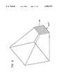
- FIG. 1is a partial perspective view of an embodiment of a catheter in accordance with the principles of the present invention
- FIG. 2is an enlarged cross-sectional view taken proximate the distal end of the catheter shown in FIG. 1;
- FIG. 3is a block diagram in part and sectional diagram in part illustrating an embodiment of a system utilizing the catheter shown in FIG. 1;
- FIG. 4Ais an illustration illustrating an application of a catheter in accordance with the principles of the present invention.
- FIG. 4Bis a partially enlarged illustration of the catheter shown in FIG. 4A.
- FIG. 5Ashows a partial perspective and cross-sectional view of a first alternate embodiment of a catheter in accordance with the principles of the present invention
- FIG. 5Bshows a view of the distal end of the embodiment of the catheter shown in FIG. 5A;
- FIG. 6Ashows a partial perspective and cross-sectional view of a second alternate embodiment of a catheter in accordance with the principles of the present invention
- FIG. 6Bshows a view of the distal end of the catheter shown in FIG. 6A;
- FIG. 7Ashows a partial perspective and cross-sectional view of a variation of the second alternate embodiment of the catheter shown in FIG. 6A;
- FIG. 7Bshows a view of the distal end of the embodiment of the catheter shown in FIG. 7A;
- FIG. 8Ashows a partial perspective and cross-sectional view of a third alternate embodiment of a catheter in accordance with the principles of the present invention
- FIG. 8Bshows a view of the distal end of the catheter shown in FIG. 8A;
- FIG. 8Cshows a view of the distal end of the catheter shown in FIG. 8A having an alternatively shaped secondary port
- FIG. 9Ashows partial perspective and cross-sectional view of a fourth alternate embodiment of a catheter in accordance with the principles of the present invention.
- FIG. 9Bshows a view of the distal end of the catheter shown in FIG. 9A.
- FIG. 10is a partial schematic view of an embodiment of an underfluid catheter system in accordance with the principles of the present invention.
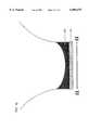
- FIG. 11is an enlarged view illustrating the catheter system operated underfluid with an aid of a volumetric field of view.
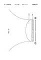
- FIG. 12is an enlarged perspective view illustrating the catheter system providing a volumetric field of view.
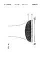
- FIG. 13is an enlarged perspective view of an ultrasonic transducer array having a single row of crystals covered by a lens which provides a volumetric field of view.
- FIG. 14is an enlarged perspective view of an alternative ultrasonic transducer array having multiple rows of crystals which provides a volumetric field of view.
- FIG. 15is an enlarged perspective view of a second alternative ultrasonic transducer array having equal number of rows and columns of crystals which provides a volumetric field of view.
- FIG. 16is an enlarged schematic cross-sectional view of a concave silicone rubber lens being placed on the ultrasonic transducer array, providing an outwardly defocused ultrasound beam.
- FIG. 17is an enlarged schematic cross-sectional view of a convex plastic lens being placed on the ultrasonic transducer array, providing an outwardly defocused ultrasound beam.
- FIG. 18is an enlarged schematic cross-sectional view of a convex silicone rubber lens being placed on the ultrasonic transducer array, providing an inwardly focused ultrasound beam.
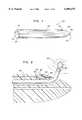
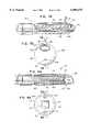
- catheter 20includes an elongated flexible or rigid tubular catheter body 22 having a proximal end 24 and a distal end 26.
- catheter 20includes proximate its longitudinal distal end 26 a phased array ultrasonic transducer 30 which is used to transmit ultrasound and receive resultant echoes so as to provide a field of view within which Doppler flow rates can be measured and features imaged.
- ultrasonic transducerscan be used in the present invention, such as any mechanical types, or any dynamic array types, or any offset stereoscopic imaging types, or any multidimensional imaging types incorporated into a virtual reality environment for underblood operation, etc.
- An electrical conductoris disposed in the catheter body 22 for electrically connecting transducer 30 to control circuitry 34 external of catheter body 22.
- An access port 40is disposed in catheter body 22 and extends from proximate the proximal end 24 of catheter body 22 to proximate the distal end 26 of catheter body 22.
- Access port 40is configured to receive a therapeutic device, such as a catheter, medication, sensors, etc., so as to enable such items to be delivered via access port 40 to distal end 26 of catheter body 22 for operation within the ultrasonic transducer field of view. Such items might be used for intervention; e.g., ablation catheter, surgical device, etc., monitoring blood pressure, sampling blood, etc.
- a guide wire access port 42is also disposed within catheter body 22 and extends from proximate proximal end 24 of the catheter body 22 to proximate distal end 26 of catheter body 22 for receiving a guide wire 44.
- the ultrasonic transducerpreferably has a frequency of 5 to 30 megahertz (MHz) and more preferably a frequency of 7 to 10 MHz. Intracardiac imaging in an adult will require image penetration of up to 2 to 10 centimeters (cm).
- catheter body 22preferably has a diameter of 4 to 24 French (one French divided by Pi equals one millimeter (mm)) and, more preferably, a diameter of 6 to 12 French.
- access port 40has a diameter of 7 to 8 French and guide wire port 42 has a diameter of 0.025 to 0.038 inches.
- catheter 20 of the present inventioncan be utilized in a medical system including the appropriate control circuitry 34 for controlling operation of the ultrasonic transducer.
- control circuitry 34is electrically interconnected to transceiver circuitry 35 (T/R) for receiving and transmitting signals via a cable 36 to ultrasonic transducer 30.
- transceiver circuitry 35is electrically interconnected to Doppler circuitry 37 and an appropriate display device 38 for displaying hemodynamics or blood flow.
- transceiver circuitry 35is electrically interconnected to suitable imaging circuitry 39 which is interconnected to a display 41 for displaying images.
- control circuitry 34might be designed to cause ultrasonic transducer 30 to vibrate so as to cause an appropriate ultrasound wave to project from proximate the distal end 26 of catheter body 22.
- the ultrasound waverepresented by lines 50 in FIG. 2, will propagate through the blood surrounding distal end 26 and a portion of the body structure. A portion of the ultrasound wave so transmitted will be reflected back from both the moving red blood cells and the like and the body structures to impinge upon transducer 30.
- An electrical signalis thereby generated and transmitted by the cable 36 to the input of transceiver 35.
- a signalmight then be transmitted to Doppler circuitry 37 which will include conventional amplifying and filtering circuitry commonly used in Doppler flow metering equipment.
- Doppler circuitry 37will analyze the Doppler shift between the transmitted frequency and the receive frequency to thereby derive an output proportional to flow rate. This output may then be conveniently displayed at display 38 which might be a conventional display terminal. Accordingly, the user will be able to obtain a readout of blood flow rates or hemodynamic information.
- control circuitry 34will likewise trigger ultrasonic transducer 30 via transceiver 35 to vibrate and produce an ultrasound wave. Once again, a portion of the wave or energy will be reflected back to ultrasonic transducer 30 by the body features. A corresponding signal will then be sent by cable 36 to transceiver circuitry 35. A corresponding signal is then sent to the imaging circuitry 39 which will analyze the incoming signal to provide, at display 41, which also might be a conventional display apparatus, an image of the body features.
- This imagingcan occur while a therapeutic or surgical device is being used at distal end 26 of catheter 20 within the field of view provided by ultrasonic transducer 30. Accordingly, the user will be able to monitor his/her actions and the result thereof.
- catheter body 22might include proximate its proximal end 24 a suitable mounting structure 52 to the access port 40.
- a therapeutic or surgical device structure 53might be suitably attached to structure 52 by suitable means, e.g., threaded, etc.
- an elongated cable-like member 54will extend along access port 40 and slightly beyond distal end 26 of catheter body 22 wherein an operative portion 56 of the surgical tool might be interconnected.
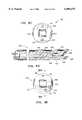
- ultrasonic transducer 30might include a piezoelectric polymer, such as Polyvinylidenedifloride (PVDF) 60, which is bonded by an epoxy layer 62 to a depression 64 approximate distal end 26.
- PVDFPolyvinylidenedifloride
- FIGS. 2, 4A, and 4Bultrasonic transducer 30 might include a piezoelectric polymer, such as Polyvinylidenedifloride (PVDF) 60, which is bonded by an epoxy layer 62 to a depression 64 approximate distal end 26.
- PVDFPolyvinylidenedifloride
- the operational portion 56 of the therapeutic deviceis illustrated as generally being capable of operation in the field of view of ultrasonic transducer 30. Accordingly, it is possible for the user to monitor operation of the therapeutic device by use of the ultrasonic transducer. Moreover, it is possible for the user to monitor the features of the body within the field of view before, during and after interventional activity.
- ultrasonic transducerscan be used in the present invention, such as any mechanical types, or any dynamic array types, or any offset stereoscopic imaging types, or any multidimensional imaging types incorporated into a virtual reality environment for underblood operation, etc., so that all forms of field of views, such as 1) tomographic (slices), 2) stereoscopic, 3) three-dimensional, 4) virtual reality (multidimensional) can be provided in the present invention.
- the orientations of the scan array on the cathetercan be include side-view, end-view, multiview (two or more views that are moveable or imminently directional transducer referred to in the literature as "omnidirectional"), etc.
- FIG. 5Ashows a partial cross-sectional view of a first alternative embodiment 70 of the catheter apparatus.
- the catheter apparatushas an elongated flexible or rigid body 72 having a longitudinal axis and a proximal end 74 and a distal end 76.
- a port 78Disposed proximate a second side of body 72 is a port 78 extending through body 72 from proximate proximal end 74 to proximate distal end 76 of body 72.
- Port 78is for receiving and delivering to distal end 76 of body 72 a working tool 84.
- Working tool 84 shown in the Figuresis illustrative only, others types of tools now known or later developed may also be delivered to distal end 76 through port 78.
- Proximate a first side of body 72is a guide wire port 80 extending through body 72 from proximate proximal end 74 to proximate distal end 76. Shown in guide port 80 is a guide wire
- Distal end 76is disposed at an oblique angle to the longitudinal axis of body 72, the first side of body 72 extending further in the direction of the distal end than the second side of body 72.
- An ultrasonic transducer 82having a first side and a second side, is disposed at an oblique angle to the longitudinal axis of body 72 approximately corresponding to the oblique angle of distal end 76 of body 72.
- the first side of ultrasonic transducer 82is disposed proximate the first side of body 72 and the second side of transducer 82 is disposed proximate the second side of body 72.
- transducer 82Extending from transducer 82 to proximate proximal end 74 of body 72 is an electrical conductor 83 connecting transducer 82 to control circuitry external of catheter 70, as described with respect to catheter 20 above. Having transducer 82 disposed on an oblique angle toward port 78 allows for easy visualization of tools, such as tool 84, extending beyond distal end 76 of body 72.
- FIG. 5Bshows a view of distal end 76 of body 72, showing guide wire port means 80, transducer 82, and port means 78.
- FIG. 6Ashows a partial cross-sectional view of a second alternative embodiment of the catheter in accordance with the present invention, generally referred to as 88.
- catheter 88has an elongated flexible or rigid body 90 having a proximal end 92 and a distal end 94.
- catheter 88also has a port 96 extending through body 90 from proximate proximal end 92 to proximate distal end 94.
- Port 96has a distal end 97 proximal distal end 94 of body 90.
- Distal end 97 of port 96exits body 90 at an acute angle to a first side of body 90 toward distal end 94.
- Port 96is for receiving and delivering to distal end 94 a working tool, such as working tool 84.
- Catheter 88also has a guide wire port 98 extending through body 90 from proximate proximal end 92 to proximate distal end 94.
- Guide wire port 98is for receiving a guide wire 86.
- transducer 100disposed to a first side of body 90 between distal end 94 and distal end 97 of port 96. Extending from transducer 100 to proximate proximal end 92 of body 90 is an electrical conductor 102 disposed in the catheter body 90 for electrically connecting transducer 100 to control circuitry external of the catheter. With transducer 100 disposed to the first side of body 90 and distal end 97 of port 96 exiting body 90 at an acute angle relative to the first side of body 90 toward distal end 94, working tools extending from distal end 97 of port 96 will be within the field of view of transducer 100.
- FIG. 6Bshows a view of distal end 94 of catheter 88, as shown in FIG. 6A.
- FIG. 7Ashows second alternative embodiment 104, as shown in FIG. 6A, except instead of having a guide wire port 98, this variation of the second alternative embodiment 104 has a deflection wire guidance system 106 for manipulating distal end 94.
- FIG. 7Bshows a view of distal end 94 of the catheter shown in FIG. 7A.
- FIG. 8Ashows a third alternative embodiment 110 of the catheter in accordance with the present invention.
- Third alternative embodiment 110has a body 112 having a distal end 114 and proximal end. Disposed proximate a first side of body 112 is a primary port 118 extending through body 112 from proximate proximal end 116 to proximate distal end 114.
- Primary port 118has a distal end 119 proximate distal end 114 of body 112.
- proximate a second side of body 112is a secondary port 120 extending through body 112 from proximate proximal end 116 to proximate distal end 114.
- Secondary port 120has a distal end 121 proximate distal end 114 of body 112.
- transducer 122is mounted proximate distal end 114 of body 112 . Extending from transducer 122 through body 112 to proximate proximal end is an electrical conductor for electrically connecting the transducer 122 to control circuitry external of the catheter. Transducer 122 is disposed between distal ends of primary and secondary ports 119 and 121, respectively. With working ports 118 and 120 oppositely disposed an either side of transducer 122, it is possible to conduct two simultaneous applications, such as holding an object with a first tool disposed through one port and operating on the object held by the first tool with a second tool disposed through the other port. A typical working tool 123 and working tool 84 are shown disposed within ports 118 and 120.
- FIG. 8Bshows a view of distal end 114 of catheter 110.
- FIG. 8Cshows a view of a distal end 124 of a catheter 126 substantially like catheter 110 shown in FIG. 8A and FIG. 8B, except that catheter 126 has a primary port 128 having an arc-like shaped cross-section, rather than a circular shaped cross-section.
- a circular cross-sectionhas been shown in the Figures for the various ports described herein, the size and shape of the ports can be varied without departing from the principals of the present invention.
- FIG. 9Ashows a fourth alternative embodiment of a catheter 130 of the present invention.
- Catheter 130is similar to catheter 70 shown in FIG. 5A and FIG. 5B except that a plurality of ports 132 are disposed proximate a second side of flexible body 131, rather than one port 78, as shown in FIG. 5A.
- a plurality of portsit is possible, for example, to use a therapeutic tool through one port while simultaneously suctioning and removing debris through another port; or a therapeutic tool can be used through one port while simultaneously electrophysiologically monitoring, suctioning and/or biopsying through a second port, third or fourth port.
- the use of the catheter of the present inventionis described with respect to the preferred embodiment 20. It is understood that the use of alternative embodiments 70, 88, 110, 126 and 130 is analogous.
- the userwould insert flexible catheter body 22 into the body via the appropriate vascular access to the desired location in the body, such as selected venous locations, heart chamber, etc.
- a guide wiremight be first inserted into place and then the catheter body fed along the guide wire.
- the usermight then insert a surgical device into the body through access port 40 and feed the surgical device to proximate distal end 26 of catheter body 22.
- the userPrior to, during and after operation of the surgical device, the user might obtain both hemodynamic measurements and images from the ultrasonic transducer field of view. By operation of the surgical device within the field of view of transducer, the user can monitor operation of the surgical device at all times.
- Frequency agilityrefers to the ability of a transducer to send and receive at various frequencies, most commonly 3, 5, and 7 MHz. It is also appreciated that a single frequency from a single transducer device can be sent and received. In general, higher frequencies are used to image fine detail of more proximal or closely related objects while lower frequency transducers scan more remote objects with less detail.
- the proposed deviceoptimally uses a 5 to 20 mHz transducer with the most optimally applied frequency of 7 to 10 mHz.
- the lower frequency used in the UIHCreflects the need to image larger objects such as the cardiac septa, valves, and extravascular anatomy.
- InterventionOne primary function of this catheter system is to guide the logical and safe use of various a) ablation, b) laser, c) cutting, occluding, e) etc., catheter-based interventional tools.
- the inventionhas the access port through which other technologies (devices) can be passed. Once the interventional tool exits the catheter tip, it can be directed repeatedly and selectively to specific site for controlled intervention.
- the inventionis also an imaging system capable of visualizing intracardiac, intravascular, and extravascular structures. Because the transducer frequencies utilized are usually lower than intravascular systems, the catheter 20 can see multiple cardiac cavities and visualize structures outside the vascular system.
- the imaging capabilityis basically two-fold: 1) diagnostic and 2) application.
- Diagnostic imagingThe catheter 20 can effectively perform diagnostic intracardiac and transvascular imaging. This application will more than likely be performed just prior to an interventional application. The intervention then will follow using the same catheter system and, its unique delivery capability.
- diagnostic imaginginclude 1) accurate visualization and measurement of an intracardiac defect, 2) characterization of valve orifice, 3) localization of a tumor and its connections, 4) etc.
- Extravascular diagnoseswould include 1) visualize pancreatic mass/pathology, 2) retroperitoneal pathology, 3) intracranial imaging, 4) recognition of perivascular pathology, and 5) imaging of other fluid containing space such as urinary bladder, bile system, fluid filled orifice or cavity (e.g. filled saline), etc.
- Application imagingrefers to the use of the catheter and its imaging capability to deliver and then apply another technology such as 1) occlusion device for closure of a septal defect, 2) ablation catheters for treatment of bypass tracts, 3) creation of a defect such as that with the blade septostomy catheter or laser-based catheter system, and 4) directing of valvuloplasty (such as prostrate surgery, placement of stents, gallstone removal etc.), etc.
- occlusion devicefor closure of a septal defect
- ablation cathetersfor treatment of bypass tracts
- creation of a defectsuch as that with the blade septostomy catheter or laser-based catheter system
- valvuloplastysuch as prostrate surgery, placement of stents, gallstone removal etc.
- the catheter 20is a truly combined ultrasound Doppler and conventional hemodynamic catheter. There are Doppler catheters, and there are catheters capable of imaging and measuring hemodynamic pressure. However, the catheter 20 is capable of Doppler hemodynamics (continuous and pulsed wave Doppler) as well as high-fidelity hemodynamic pressure recording while simultaneously imaging the heart and blood vessel. The catheter 20 provides a combination of imaging, hemodynamic, and interventional delivery catheter.
- intracardiac ultrasoundis capable of 1) imaging, 2) delivering a therapeutic device, and 3) obtaining simultaneous hemodynamics which can be used to develop less invasive cardiac surgical techniques. This simultaneous use of one or more devices within the heart or vascular tree opens up the potential to develop less invasive surgical therapies.
- Exampleswould include 1) removal of a cardiac tumor by visually grasping the tumor with one device and visually cutting its attachment with a second device, thus allowing less invasive extraction of intracardiac mass lesions, 2) visually placing an electrophysiologic catheter on a bypass tract and then with direct ultrasound visualization ablate the underlying tract with the second device, 3) visually performing laser surgery such as creating an intra-atrial defect, vaporization of obstructing thrombus such as is seen in pseudointimal occlusion of conduits, 4) visually removing a foreign body from the heart or vascular tree, and 5) directing intravascular surgery from within a blood vessel or monitoring concomitant hemodynamic changes.
- Radio-frequency ablationPresently a bypass tract is localized by an electrophysiologic study which systematically maps the atrioventricular valve annulus. Positioning of the ablation catheter is determined by x-ray fluoroscopy and certain electrical measurements which relate the distance of the ablation catheter from a reference catheter. The catheter 20 will allow an operator to map the atrioventricular valve under direct ultrasound visualization. Thus, increased accuracy of catheter placement, precision of the applied therapy, and immediate assessment of outcome would result.
- the above ablation techniquewould be particularly applicable for right-sided bypass tracts (in and around the tricuspid valve annulus). This would be accomplished by placement of the catheter 20 through the superior vena cava above the tricuspid annulus.
- the catheter 20could be placed across the atrial septum under direct ultrasound visualization.
- the mitral annuluscould thus be mapped directly and the localized bypass tract precisely ablated under visual ultrasonic and hemodynamic direction. Complications such as valve perforation, multiple imprecise applications of ablation energy, and inadvertent ablation of normal conduction tissue would be substantially reduced.
- the catheter 20will allow visualization of perivascular and extravascular pathology. Transvascular or transorgan imaging and localization of pathology out of the immediate vascular tree will result in a substantial step forward in the diagnosis and possible treatment of difficult to reach pathology.
- the catheter 20cannot only diagnose but guide a biopsy needle and therapeutic device to an extravascular lesion in question.
- the retroperitoneum, mediastinum, and basal cerebrovascular pathologyare logical areas of interest. Accurate characterization of various pathologies will be more feasible. Every organ has its own vascular system, and the proposed ultrasound transvascular system is an ideal tool to assess difficult to reach areas of the body.
- the vascular systemis a conduit to each organ, and the catheter 20 can be delivered to each organ. Characterization of the underlying parenchyma and possible transvascular biopsy or treatment will ultimately be developed.
- the catheter 20opens the potential not only to visualize but to directly intervene with the same catheter system.
- There are numerous intraoperative catheter-based systemswhich to date use conventional x-ray to accomplish their goal of placement and application of a specified therapy.
- the catheter 20has all the prerequisites of an ideal imaging and interventional instrument and has the ability to 1) image, 2) obtain hemodynamics by multiple means (pressure dynamics and Doppler), 3) function as a diagnostic as well as therapeutic device, and 4) accommodate other unique technologies which would enhance the application of both systems.
- intravascular, transvascular, and intracardiac devicescould be delivered through the port means described above within or about the heart and blood vessels of the body.
- the catheters described abovecould also be used in any ectogenic tissue, such as liver, parenchyma, bile ducts, ureters, urinary bladder, and intracranial--i.e., any place in the body which is echogenic which would allow passage of a catheter for either diagnostic or therapeutic applications using ultrasound visualization.
- the catheter 20is a new and exciting innovation to invasive medicine. There are multiple other and yet-to-be-determined applications. However, the new concept described opens the potential development of less expensive, more precise, and safe intravascular and transvascular diagnostic and surgical devices.
- the catheter 20is very much different from any conventional ultrasound catheter-based system.
- the catheter 20incorporates image and hemodynamic capability as well as the ability to deliver other diverse technologies to specified sites within the cardiovascular system (heart and blood vessels).
- the catheter 20is seen as an ideal diagnostic and therapeutic tool for future development.
- the proposed applicationsfoster greater preciseness, adaptability, and safety.
- Ultrasoundpermits visualization from within blood-filled spaces as well as through bloodfilled spaces into other water- or fluid-filled tissue.
- the catheter 20will evolve into the ultimate interventional system.
- FIG. 4Ais an illustration showing one potential use of the ultrasound imaging and hemodynamic catheter (UIHC).
- UIHCultrasound imaging and hemodynamic catheter
- the UIHCis advanced from the superior vena cava to the tricuspid valve annulus. Simultaneously visualized in the annulus, electrophysiologic and ultimately and ablation procedure are performed. The ability to directly visualize and direct therapeutic catheter devices highlights only one of the many applications of the UIHC.
- FIG. 10Another embodiment of the catheter system, generally in accordance with the principles of the present invention is shown in FIG. 10, which is designated as reference numeral 200.
- the catheter system 200has a catheter body 202 and an ultrasonic transducer array 204 mounted on proximate the distal end of the catheter body 202. It is appreciated that other parts of the catheter system can be similar to those in the catheter systems 20, 70, 88, 104, 110, and 130 as shown in FIGS. 1, 5A, 6A, 7A, 8A, and 9A, respectively.
- FIG. 10shows a partial schematic view of the catheter system 200.
- the catheter body 202 of the catheter system 200is inserted into an underfluid cavity of a body 206.
- a therapeutic device 208projects from the catheter system 200 proximate the distal end of the catheter system 200 and manipulates features in the cavity of the body 208. This manipulation is under observation of a 3-dimensional image shown on a display, which can be similarly connected to the ultrasound transducer array as shown in FIG. 3, outside the body 208 proximate the proximal end of the catheter system 200.
- the underfluid catheter body 202has tool port 210 disposed in the catheter body 202 extending from proximate the proximal end to proximate the distal end of the catheter body 202 for receiving the therapeutic device 208, such as a catheter, medication, sensor, surgical device, etc., so as to enable such items to be delivered via the tool port to proximate the distal end of the catheter body 202.
- the tool portis optional.
- additional tool portscan be disposed in the catheter body 202.
- the therapeutic device 208is projected into an underfluid environment, as shown in FIGS. 11-12, and operated therein with the aid of a volumetric 3-dimensional image of the underfluid environment and the therapeutic device 208.
- the catheter system 200can also optionally include a guidewire port 212 disposed in the catheter body 202 extending from proximate the proximal end to proximate the distal end of the catheter body 202 for receiving a guide wire 214.
- the guide wire 214guides the catheter body 202 when inserting into a body, such as the body 206.
- the catheter system 200includes a control circuit which can be similar to the control circuitry 34 shown in FIG. 3.
- the control circuitry 34is used to control the operation of the ultrasonic transducer array 204.
- the control circuitry 34is electrically interconnected to a transceiver circuitry 35 (T/R) for receiving and transmitting signals via a cable 36 to ultrasonic transducer array 204.
- the transceiver circuitry 35is electrically interconnected to a measuring circuitry, such as the Doppler circuitry 37, which is interconnected to a first display 38 for displaying hemodynamics, blood flow, etc.
- the transceiver circuitry 35is electrically interconnected to an imaging circuitry 39 which is interconnected to a second display 41 for displaying a 3-dimensional image of the underfluid environment.
- the catheter body 202can also house some encased electronics 216.
- the ultrasonic transducer array 204is mounted on a side of the catheter body 202.
- the array 204can also be mounted on the tip of the catheter body 202.
- the catheter body 202is a flexible catheter capable of manual or electronic interactive flexible tip.
- the guidewire port 212has a diameter of 0.035 inches. It is appreciated that the range of the diameter of the guidewire port 212 can be varied from 0.025 to 0.038 inches.
- the tool port 210 for transporting the therapeutic device 208is a 7 French port. It is appreciated that the range of the tool port 210 can be varied from 3 French to 20 French.
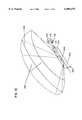
- the ultrasonic transducer array 204is comprised of a single row of individual crystals 218. Each crystal 218 is arranged side by side.
- a field of view generated by the ultrasonic transducer array 204has a primary tomographic plane 220 in azimuthal dimension along an AZ axis.
- the row of the array 204is parallel to the AZ axis.
- An elevation axis (EL)is perpendicular to the AZ axis.
- a primary beam from the ultrasonic transducer array 204lies in the primary tomographic plane 220.
- the primary beamhas usually a sector configuration (generally a fan or triangle shape) or a linear configuration (generally rectangular shape).
- the volumetric field of viewcan be produced by defocusing the primary tomographic plane 220 such that a plurality of elevation planes 222 spread laterally outward from the primary tomographic plane 220.
- the primary tomographic plane 220 and the elevation planes 222together form a volumetric field of view.
- a lens 224is placed on the top of the ultrasonic transducer array 204.
- the ultrasound beams which are usually collimatedare defocused along the elevation direction (EL) after the beams go through the lens 224 (or other lenses 226,228 as shown in FIGS. 16 and 17).
- the lenses 224, 226, or 228are preferred to be made from materials such as a plastic material or silicone rubber. It is appreciated that other types materials can be used to make the lens.
- the lens 226is a concave lens, preferably made of silicone rubber, which transmits ultrasound waves slower than the surrounding environment, such as body tissues.
- the ultrasound wavespass through the lens 226 and then impact on the body tissues.
- the speed of the ultrasound wavesis slower in the lens but faster in tissue (e.g. 1,540 m/sec). Accordingly, the transmitted ultrasound waves, after passing through more slowly transmitted lens 226 and striking faster transmitted body tissues, will be directed outward. As a result, the collimated ultrasound beams are defocused in the elevation dimension.
- the defocusingcan also be achieved by placing a convex lens 228 on the ultrasonic transducer array as shown in FIG. 17.
- the ultrasound wavespass through the convex lens 228 and then impact on the body tissues.
- the ultrasound beamsare pulled outward due to the faster velocity in the convex lens 228.
- the ultrasound beamsare defocused in the elevation dimension.
- FIG. 18,demonstrates a way of using a lens 230 to, in fact, focus the beams from the transducer array.
- the collimated ultrasound beamsare generated from the ultrasonic transducer array.
- the convex lens 230transmits ultrasound waves slower than the surrounding environment, such as body tissues, do. Accordingly, the ultrasound beams are pulled inward due to the faster velocity in body tissues. As a result, the ultrasound beams are focused toward the primary tomographic plane 220.
- FIG. 14shows an alternative embodiment of ultrasonic transducer array 204' which is comprised of multiple rows of individual piezoelectric crystals 218.
- the rows of the array 204'are parallel to the AZ axis.
- the columns of the array 204'are parallel to the elevation dimension along the EL axis, which is perpendicular to AZ axis.
- This type of arrayis also called volumetric one and one-half (1 and 1/2) dimensional array.
- the "elevation" image and the ultimate 3-dimensional imageare the result of phasing the crystals in the elevation direction as well as in the azimuthal direction.
- FIG. 15shows a second alternative embodiment of ultrasonic transducer array 204" which is comprised of equal number of crystals 218 in all dimensions. Similar to the one and one-half (1 and 1/2) dimensional array 204', the rows of the array 204" are parallel to the AZ axis, and the columns of the array 204" are parallel to the EL axis. This type of array is also called a two (2) dimensional array. The "elevation" image and the ultimate 3-dimensional image are the result of phasing the crystals in the EL direction as well as the AZ direction.
- the volumetric field of view as shown in FIGS. 14-15provides 3-dimensional images of structures under observation. Further, the volumetric field of view not only shows, for example, a primary tomographic cut, but also volumes of features, such as tissue.
- FIGS. 14 and 15it will be appreciated that no lens is required to generate a volumetric image. Consequently, the ultrasonic beams are focused in both the azimuthal and elevational directions.
- the volumetric imageis generated because the arrays of FIGS. 14 and 15 are 2 dimensional.
- a volumetric imagecan be generated by electronically phasing and steering the ultrasonic impulses in both the azimuthal and the elevational directions.
- the ultrasound transduceris a 7-10 MHz sector array transducer. It is appreciated that the range of the sector array transducer can be varied from 3.7 MHz to 30 MHz.
- the lenses 224, 226, 228, 230can be made of different materials which will have variable effects on the transmitted ultrasound beams.
- a 3-dimensional imagecan be seen on a 2-dimensional display outside the body 206 in a real-time operation. Elevation defocusing in using a lens does not interfere with the inherent frame rate or adversely affect conventional echo data.
- the lenscan also be fabricated to reduce the strength of the dominant tomographic plane (AZ plane).
- AZ planedominant tomographic plane
- the lensis optional, for example, as shown in FIGS. 14 and 15 whereby the beams are phased in both the azimuthal and elevational planes.
- the present inventionhas numerous clinical applications.
- One of whichis the underfluid imaging when imaging from within chambers, cavities or blood vessels. Since the physical space is small, and the anatomy in question is closely approximated and usually totally surrounds the transducer, a 3-dimensional imaging is a solution to visualizing larger volumes of underfluid tissue.
- the defocusing lens or electronically controlled phasing in both the azimuthal and elevational directionsi.e., using multi-dimensional arrays such as 11/2 dimensional or 2 dimensional arrays
- Working port(s) and guidewire(s)are optional. Further, catheter lengths and transducer frequencies are variable.
- Another applicationwhen the working port is optionally used in the catheter, is to intervene or manipulate an underfluid structure, such as cutting an underfluid tissue, etc., by a therapeutic device, such as the therapeutic devices 50, 84, 123, 208 shown in FIGS. 4B, 5A, 8A, and 12, respectively. Under such direct volumetric visual guidance, diagnostic and therapeutic procedures can be performed with better spatial orientation.
- the measurementcan also be performed under direct volumetric visual guidance in the present invention.
Landscapes
- Health & Medical Sciences (AREA)
- Life Sciences & Earth Sciences (AREA)
- Surgery (AREA)
- Public Health (AREA)
- Veterinary Medicine (AREA)
- Nuclear Medicine, Radiotherapy & Molecular Imaging (AREA)
- General Health & Medical Sciences (AREA)
- Engineering & Computer Science (AREA)
- Biomedical Technology (AREA)
- Heart & Thoracic Surgery (AREA)
- Medical Informatics (AREA)
- Molecular Biology (AREA)
- Animal Behavior & Ethology (AREA)
- Biophysics (AREA)
- Radiology & Medical Imaging (AREA)
- Physics & Mathematics (AREA)
- Pathology (AREA)
- Gynecology & Obstetrics (AREA)
- Hematology (AREA)
- Orthopedic Medicine & Surgery (AREA)
- Vascular Medicine (AREA)
- Ultra Sonic Daignosis Equipment (AREA)
Abstract
Description
This application is a Continuation of application Ser. No. 09/003,248, filed Jan. 6, 1998 (now pending), which is a Continuation of application Ser. No. 08/678,380, filed Jun. 28, 1996 (now issued as U.S. Pat. No. 5,704,361), which is a Continuation-in-Part of application Ser. No. 08/305,138, filed Sep. 13, 1994 (now abandoned), which is a Continuation of application Ser. No. 07/972,626, filed Nov. 6, 1992 (now issued as U.S. Pat. No. 5,345,940), which is a Continuation-in-Part application Ser. No. 07/790,580, filed Nov. 8,1991 (now issued as U.S. Pat. No. 5,325,860), which applications are incorporated herein by reference.
Noninvasive ultrasonic imaging systems are widely used for performing ultrasonic imaging and taking measurements. Such systems typically use scan heads which are placed against a patients skin. Exemplary uses for such systems include heart and internal organ examinations as well as examinations of developing fetuses. These systems operate by transmitting ultrasonic waves into the body, receiving echoes returned from tissue interfaces upon which the waves impinge, and translating the received echo information into a structural representation of the planar slice of the body through which the ultrasonic waves are directed.
Catheter based invasive ultrasound imaging systems, typically used for intracardiac or transvascular imaging, are a relatively new addition to ultrasound armamentarian. Conventional underfluid transducers for use on catheters are comprised of crystal arrays (e.g. linear phased array) or a single crystal translated over a surface, producing a tomographic field of view in an azimuthal plane of the array. Typical arrays include: 1) linear array (linear sequential array), usually producing a rectangular or rhomboidal picture; 2) cylindrical array or rotating crystal, producing a round pie-shaped tomographic cut of structures; and 3) sector array (linear phased array), producing a triangular shaped image emanating from a small transducer source. All images are tomographic in nature and are focused in the azimuthal and elevation plane. The intent of having these conventional transducer configurations is to produce a thin ultrasound cut of the insonaned structures. Such tomographic planes by nature are thin and of high resolution.
The narrow field of view provided by conventional catheter transducer configurations is problematic because structures lying outside of the plane of view can only be visualized by reorienting or manipulating the catheter. Due to the tortuous and confined nature of a typical catheter pathway, catheter manipulation is impractical and often impossible. Consequently, the localization of specific targets is difficult and at times can be disorienting because of an inability to appreciate contiguous anatomic landmarks.
Advances in 3-dimensional imaging capabilities have been made with respect to non-catheter related ultrasonic imaging systems. For example, U.S. Pat. No. 5,305,756, issued to Entrekin et al., which is hereby incorporated by reference, discloses general 3-dimensional imaging techniques in a non-catheter based context. What is needed is a catheter based imaging system that utilizes 3-dimensional imaging techniques to provide a wide field of view so as to improve anatomic localization for precision underfluid diagnostics and interventions.
The present invention relates generally to a volumetric, 3-dimensional image ultrasound transducer underfluid catheter system.
The present invention also relates to an ultrasonic and interventional catheter device operated in an intracardiac or transvascular system, with the aid of a volumetric 3-dimensional imaging capability.
In one particular embodiment, the present invention relates to a catheter apparatus comprising an underfluid catheter body having proximal and distal ends. An ultrasonic transducer array is mounted longitudinally along the catheter body proximate the distal end. The transducer array has a volumetric field of view that projects radially/laterally outward from the catheter. Features in such wide volumetric field of view can be imaged, measured, or intervened by an underfluid therapeutic device with an aid of the real-time image.
It is significant that the transducer array described in the previous paragraph has a 3-dimensional field of view. A first dimension, referred to as an azimuthal direction, is aligned with the length of the transducer array. A second dimension, referred to as a depth direction, is the depth into the body which an ultrasonic signal is transmitted and from which an echo return. A third dimension, referred to as an elevation direction, is perpendicular to both the azimuthal and the depth directions.
If the transducer array comprises a linear phased array having a single row of piezoelectric crystals, a 3-dimensional field of view can be generated by focusing the ultrasound signals in the azimuthal direction (parallel to the longitudinal axis of the catheter) and diverging the ultrasound signals in the elevational direction (transverse to the longitudinal axis of the catheter). The ultrasonic signals can be diverged in the elevational direction through the use of lenses. For example, the signals can be diverged by mounting a silicone rubber concave lens or a plastic convex lens in front of the transducer array.
If the transducer array comprises multiple rows of piezoelectric crystals, a 3-dimensional field of view can be generated by electronically phasing the ultrasound signals in both the azimuthal and elevational directions. Of course, lenses can be used in association with multiple row arrays to further widen the field of view.
It will be appreciated that catheters constructed in accordance with the principles of the present invention can optionally include one or more ports that extend longitudinally through the catheter bodies. The ports are preferably adapted for guiding therapeutic instruments through the catheter and preferably have exit ends adjacent to the field of view of the catheter imaging system. In operation, the ports guide the therapeutic instruments such that the operative ends of a therapeutic instruments are directed toward the 3-dimensional field of view of the imaging system.
It will also be appreciated that catheters constructed in accordance with the principles of the present invention can include one or more guidewire ports which extend longitudinally through the catheters and are adapted for receiving guidewires.
One advantage of the present invention is to provide real-time 3-dimensional images of underfluid features so as to visualize contiguous anatomy, such as a large volume of tissues without frequently rotating, flexing, or extending the catheter.
Another advantage is that the present invention provides a much better underfluid "eye"--a 3-dimensional "motion picture"--for an operator when he/she intervenes the underfluid features by using an underfluid therapeutic device. These images provide the operator a direct aid without opening a large area of a body.
A further advantage is that the present invention have numerous clinical applications. One is,related to underfluid imaging: There is a considerable need to increase the field of view when imaging from within chambers or blood vessels. The physical space of the chambers or blood vessels is small, and the anatomy in question is closed approximated and usually totally surrounds the transducer. A conventional tomographic presentation provides only a limited slice, thus requiring frequent manipulation of transducer in order to visualize contiguous anatomy. The present invention is a solution to visualizing larger volumes of tissue. The underfluid defocusing transducer array does not appreciably affect the electronics and does not require alteration in the display format. The catheter apparatus is immersed in fluid, a homogeneous, low scattering medium, which is an ideal environment for this particular transducer modification.
Another main clinical application is related to underfluid intervention: In diagnostic and therapeutic procedures, there is an increasing need for volumetric 3-dimensional visualization which would improve anatomic localization and recognition of continuous structures and events.
A further advantage of the present invention is that by using such a catheter system, major surgical procedures can be avoided. It dramatically reduces the patient's physical pain in operation and mental distress after operation due to any large visible scars, etc.
These and various other advantages and features of novelty which characterize the invention are pointed out with particularity in the claims annexed hereto and forming a part hereof. However, for a better understanding of the invention, its advantages and objects obtained by its use, reference should be made to the drawings which form a further part hereof, and to the accompanying descriptive matter, in which there is illustrated and described a preferred embodiment of the invention.
A better understanding of the construction and operational characteristics of a preferred embodiment(s) can be realized from a reading of the following detailed description, especially in light of the accompanying drawings in which like reference numerals in the several views generally refer to corresponding parts.
FIG. 1 is a partial perspective view of an embodiment of a catheter in accordance with the principles of the present invention;
FIG. 2 is an enlarged cross-sectional view taken proximate the distal end of the catheter shown in FIG. 1;
FIG. 3 is a block diagram in part and sectional diagram in part illustrating an embodiment of a system utilizing the catheter shown in FIG. 1;
FIG. 4A is an illustration illustrating an application of a catheter in accordance with the principles of the present invention;
FIG. 4B is a partially enlarged illustration of the catheter shown in FIG. 4A.
FIG. 5A shows a partial perspective and cross-sectional view of a first alternate embodiment of a catheter in accordance with the principles of the present invention;
FIG. 5B shows a view of the distal end of the embodiment of the catheter shown in FIG. 5A;
FIG. 6A shows a partial perspective and cross-sectional view of a second alternate embodiment of a catheter in accordance with the principles of the present invention;
FIG. 6B shows a view of the distal end of the catheter shown in FIG. 6A;
FIG. 7A shows a partial perspective and cross-sectional view of a variation of the second alternate embodiment of the catheter shown in FIG. 6A;
FIG. 7B shows a view of the distal end of the embodiment of the catheter shown in FIG. 7A;
FIG. 8A shows a partial perspective and cross-sectional view of a third alternate embodiment of a catheter in accordance with the principles of the present invention;
FIG. 8B shows a view of the distal end of the catheter shown in FIG. 8A;
FIG. 8C shows a view of the distal end of the catheter shown in FIG. 8A having an alternatively shaped secondary port;
FIG. 9A shows partial perspective and cross-sectional view of a fourth alternate embodiment of a catheter in accordance with the principles of the present invention; and
FIG. 9B shows a view of the distal end of the catheter shown in FIG. 9A.
FIG. 10 is a partial schematic view of an embodiment of an underfluid catheter system in accordance with the principles of the present invention.
FIG. 11 is an enlarged view illustrating the catheter system operated underfluid with an aid of a volumetric field of view.
FIG. 12 is an enlarged perspective view illustrating the catheter system providing a volumetric field of view.
FIG. 13 is an enlarged perspective view of an ultrasonic transducer array having a single row of crystals covered by a lens which provides a volumetric field of view.
FIG. 14 is an enlarged perspective view of an alternative ultrasonic transducer array having multiple rows of crystals which provides a volumetric field of view.
FIG. 15 is an enlarged perspective view of a second alternative ultrasonic transducer array having equal number of rows and columns of crystals which provides a volumetric field of view.
FIG. 16 is an enlarged schematic cross-sectional view of a concave silicone rubber lens being placed on the ultrasonic transducer array, providing an outwardly defocused ultrasound beam.
FIG. 17 is an enlarged schematic cross-sectional view of a convex plastic lens being placed on the ultrasonic transducer array, providing an outwardly defocused ultrasound beam.
FIG. 18 is an enlarged schematic cross-sectional view of a convex silicone rubber lens being placed on the ultrasonic transducer array, providing an inwardly focused ultrasound beam.
Referring now to FIGS. 1-3, there is, generally illustrated byreference numeral 20, a catheter in accordance with the principles of the present invention. As shown,catheter 20 includes an elongated flexible or rigidtubular catheter body 22 having aproximal end 24 and adistal end 26.Catheter 20 includes proximate its longitudinal distal end 26 a phased arrayultrasonic transducer 30 which is used to transmit ultrasound and receive resultant echoes so as to provide a field of view within which Doppler flow rates can be measured and features imaged. It is appreciated that the other types of ultrasonic transducers can be used in the present invention, such as any mechanical types, or any dynamic array types, or any offset stereoscopic imaging types, or any multidimensional imaging types incorporated into a virtual reality environment for underblood operation, etc. An electrical conductor is disposed in thecatheter body 22 for electrically connectingtransducer 30 to controlcircuitry 34 external ofcatheter body 22. Anaccess port 40 is disposed incatheter body 22 and extends from proximate theproximal end 24 ofcatheter body 22 to proximate thedistal end 26 ofcatheter body 22.Access port 40 is configured to receive a therapeutic device, such as a catheter, medication, sensors, etc., so as to enable such items to be delivered viaaccess port 40 todistal end 26 ofcatheter body 22 for operation within the ultrasonic transducer field of view. Such items might be used for intervention; e.g., ablation catheter, surgical device, etc., monitoring blood pressure, sampling blood, etc. A guidewire access port 42 is also disposed withincatheter body 22 and extends from proximateproximal end 24 of thecatheter body 22 to proximatedistal end 26 ofcatheter body 22 for receiving aguide wire 44.
In the preferred embodiment of the present invention, the ultrasonic transducer preferably has a frequency of 5 to 30 megahertz (MHz) and more preferably a frequency of 7 to 10 MHz. Intracardiac imaging in an adult will require image penetration of up to 2 to 10 centimeters (cm). In the preferred embodiment,catheter body 22 preferably has a diameter of 4 to 24 French (one French divided by Pi equals one millimeter (mm)) and, more preferably, a diameter of 6 to 12 French. In the preferred embodiment,access port 40 has a diameter of 7 to 8 French and guidewire port 42 has a diameter of 0.025 to 0.038 inches.
As generally illustrated in FIG. 3,catheter 20 of the present invention can be utilized in a medical system including theappropriate control circuitry 34 for controlling operation of the ultrasonic transducer. As illustrated in FIG. 3,control circuitry 34 is electrically interconnected to transceiver circuitry 35 (T/R) for receiving and transmitting signals via acable 36 toultrasonic transducer 30. In turn,transceiver circuitry 35 is electrically interconnected toDoppler circuitry 37 and anappropriate display device 38 for displaying hemodynamics or blood flow. In addition,transceiver circuitry 35 is electrically interconnected tosuitable imaging circuitry 39 which is interconnected to adisplay 41 for displaying images.
During operation,control circuitry 34 might be designed to causeultrasonic transducer 30 to vibrate so as to cause an appropriate ultrasound wave to project from proximate thedistal end 26 ofcatheter body 22. The ultrasound wave, represented bylines 50 in FIG. 2, will propagate through the blood surroundingdistal end 26 and a portion of the body structure. A portion of the ultrasound wave so transmitted will be reflected back from both the moving red blood cells and the like and the body structures to impinge upontransducer 30. An electrical signal is thereby generated and transmitted by thecable 36 to the input oftransceiver 35. A signal might then be transmitted toDoppler circuitry 37 which will include conventional amplifying and filtering circuitry commonly used in Doppler flow metering equipment.Doppler circuitry 37 will analyze the Doppler shift between the transmitted frequency and the receive frequency to thereby derive an output proportional to flow rate. This output may then be conveniently displayed atdisplay 38 which might be a conventional display terminal. Accordingly, the user will be able to obtain a readout of blood flow rates or hemodynamic information.
In order to obtain imaging information,control circuitry 34 will likewise triggerultrasonic transducer 30 viatransceiver 35 to vibrate and produce an ultrasound wave. Once again, a portion of the wave or energy will be reflected back toultrasonic transducer 30 by the body features. A corresponding signal will then be sent bycable 36 totransceiver circuitry 35. A corresponding signal is then sent to theimaging circuitry 39 which will analyze the incoming signal to provide, atdisplay 41, which also might be a conventional display apparatus, an image of the body features.
This imaging can occur while a therapeutic or surgical device is being used atdistal end 26 ofcatheter 20 within the field of view provided byultrasonic transducer 30. Accordingly, the user will be able to monitor his/her actions and the result thereof.
As illustrated in FIG. 3,catheter body 22 might include proximate its proximal end 24 asuitable mounting structure 52 to theaccess port 40. A therapeutic orsurgical device structure 53 might be suitably attached to structure 52 by suitable means, e.g., threaded, etc. As illustrated, an elongated cable-like member 54 will extend alongaccess port 40 and slightly beyonddistal end 26 ofcatheter body 22 wherein anoperative portion 56 of the surgical tool might be interconnected.
Additional detail ofdistal end 26 ofcatheter body 22 is illustrated in FIGS. 2, 4A, and 4B. As illustrated in FIGS. 2, 4A, and 4B,ultrasonic transducer 30 might include a piezoelectric polymer, such as Polyvinylidenedifloride (PVDF) 60, which is bonded by anepoxy layer 62 to adepression 64 approximatedistal end 26. Although some detail is provided with respect to an embodiment of an ultrasonic transducer which might be used, it will be appreciated that various types of transducers having various configurations and orientations might be utilized in keeping with the present invention.
As illustrated in FIGS. 4A and 4B, theoperational portion 56 of the therapeutic device is illustrated as generally being capable of operation in the field of view ofultrasonic transducer 30. Accordingly, it is possible for the user to monitor operation of the therapeutic device by use of the ultrasonic transducer. Moreover, it is possible for the user to monitor the features of the body within the field of view before, during and after interventional activity. It is appreciated that the other types of ultrasonic transducers can be used in the present invention, such as any mechanical types, or any dynamic array types, or any offset stereoscopic imaging types, or any multidimensional imaging types incorporated into a virtual reality environment for underblood operation, etc., so that all forms of field of views, such as 1) tomographic (slices), 2) stereoscopic, 3) three-dimensional, 4) virtual reality (multidimensional) can be provided in the present invention. In addition, it is appreciated that the orientations of the scan array on the catheter can be include side-view, end-view, multiview (two or more views that are moveable or imminently directional transducer referred to in the literature as "omnidirectional"), etc.
FIG. 5A shows a partial cross-sectional view of a firstalternative embodiment 70 of the catheter apparatus. The catheter apparatus has an elongated flexible orrigid body 72 having a longitudinal axis and aproximal end 74 and adistal end 76. Disposed proximate a second side ofbody 72 is aport 78 extending throughbody 72 from proximateproximal end 74 to proximatedistal end 76 ofbody 72.Port 78 is for receiving and delivering todistal end 76 of body 72 a workingtool 84. Workingtool 84 shown in the Figures is illustrative only, others types of tools now known or later developed may also be delivered todistal end 76 throughport 78. Proximate a first side ofbody 72 is aguide wire port 80 extending throughbody 72 from proximateproximal end 74 to proximatedistal end 76. Shown inguide port 80 is aguide wire 86.
FIG. 5B shows a view ofdistal end 76 ofbody 72, showing guide wire port means 80,transducer 82, and port means 78.
FIG. 6A shows a partial cross-sectional view of a second alternative embodiment of the catheter in accordance with the present invention, generally referred to as 88. Like firstalternative embodiment 70,catheter 88 has an elongated flexible orrigid body 90 having aproximal end 92 and adistal end 94.Catheter 88 also has aport 96 extending throughbody 90 from proximateproximal end 92 to proximatedistal end 94.Port 96 has adistal end 97 proximaldistal end 94 ofbody 90.Distal end 97 ofport 96exits body 90 at an acute angle to a first side ofbody 90 towarddistal end 94.Port 96 is for receiving and delivering to distal end 94 a working tool, such as workingtool 84.Catheter 88 also has aguide wire port 98 extending throughbody 90 from proximateproximal end 92 to proximatedistal end 94.Guide wire port 98 is for receiving aguide wire 86.
Also shown in FIG. 6A is atransducer 100 disposed to a first side ofbody 90 betweendistal end 94 anddistal end 97 ofport 96. Extending fromtransducer 100 to proximateproximal end 92 ofbody 90 is anelectrical conductor 102 disposed in thecatheter body 90 for electrically connectingtransducer 100 to control circuitry external of the catheter. Withtransducer 100 disposed to the first side ofbody 90 anddistal end 97 ofport 96 exitingbody 90 at an acute angle relative to the first side ofbody 90 towarddistal end 94, working tools extending fromdistal end 97 ofport 96 will be within the field of view oftransducer 100.
FIG. 6B shows a view ofdistal end 94 ofcatheter 88, as shown in FIG. 6A.
FIG. 7A shows secondalternative embodiment 104, as shown in FIG. 6A, except instead of having aguide wire port 98, this variation of the secondalternative embodiment 104 has a deflectionwire guidance system 106 for manipulatingdistal end 94. FIG. 7B shows a view ofdistal end 94 of the catheter shown in FIG. 7A.
FIG. 8A shows a thirdalternative embodiment 110 of the catheter in accordance with the present invention. Thirdalternative embodiment 110 has abody 112 having adistal end 114 and proximal end. Disposed proximate a first side ofbody 112 is aprimary port 118 extending throughbody 112 from proximate proximal end 116 to proximatedistal end 114.Primary port 118 has adistal end 119 proximatedistal end 114 ofbody 112. oppositely disposed fromprimary port 118, proximate a second side ofbody 112 is asecondary port 120 extending throughbody 112 from proximate proximal end 116 to proximatedistal end 114.Secondary port 120 has adistal end 121 proximatedistal end 114 ofbody 112.
Mounted proximatedistal end 114 ofbody 112 is atransducer 122. Extending fromtransducer 122 throughbody 112 to proximate proximal end is an electrical conductor for electrically connecting thetransducer 122 to control circuitry external of the catheter.Transducer 122 is disposed between distal ends of primary andsecondary ports ports transducer 122, it is possible to conduct two simultaneous applications, such as holding an object with a first tool disposed through one port and operating on the object held by the first tool with a second tool disposed through the other port. Atypical working tool 123 and workingtool 84 are shown disposed withinports
Although the third alternative embodiment of thecatheter 110 of the present invention does not include a guide wire port means, a guide wire could be used inprimary port 118 orsecondary port 120 to initially positioncatheter 110. Then the guide wire could be retracted fromport distal end 114 ofcatheter 110.
FIG. 8C shows a view of adistal end 124 of acatheter 126 substantially likecatheter 110 shown in FIG. 8A and FIG. 8B, except thatcatheter 126 has aprimary port 128 having an arc-like shaped cross-section, rather than a circular shaped cross-section. Although a circular cross-section has been shown in the Figures for the various ports described herein, the size and shape of the ports can be varied without departing from the principals of the present invention.
FIG. 9A shows a fourth alternative embodiment of acatheter 130 of the present invention.Catheter 130 is similar tocatheter 70 shown in FIG. 5A and FIG. 5B except that a plurality ofports 132 are disposed proximate a second side offlexible body 131, rather than oneport 78, as shown in FIG. 5A. With a plurality of ports, it is possible, for example, to use a therapeutic tool through one port while simultaneously suctioning and removing debris through another port; or a therapeutic tool can be used through one port while simultaneously electrophysiologically monitoring, suctioning and/or biopsying through a second port, third or fourth port.
The use of the catheter of the present invention is described with respect to thepreferred embodiment 20. It is understood that the use ofalternative embodiments flexible catheter body 22 into the body via the appropriate vascular access to the desired location in the body, such as selected venous locations, heart chamber, etc. In one approach, a guide wire might be first inserted into place and then the catheter body fed along the guide wire. The user might then insert a surgical device into the body throughaccess port 40 and feed the surgical device to proximatedistal end 26 ofcatheter body 22. Prior to, during and after operation of the surgical device, the user might obtain both hemodynamic measurements and images from the ultrasonic transducer field of view. By operation of the surgical device within the field of view of transducer, the user can monitor operation of the surgical device at all times.
I. Detailed Features of the Disclosed Catheters
A. Frequency agility Ultrasound frequency: Frequency agility refers to the ability of a transducer to send and receive at various frequencies, most commonly 3, 5, and 7 MHz. It is also appreciated that a single frequency from a single transducer device can be sent and received. In general, higher frequencies are used to image fine detail of more proximal or closely related objects while lower frequency transducers scan more remote objects with less detail. The proposed device optimally uses a 5 to 20 mHz transducer with the most optimally applied frequency of 7 to 10 mHz. The lower frequency used in the UIHC reflects the need to image larger objects such as the cardiac septa, valves, and extravascular anatomy.
B. Catheter size: Catheter diameters will generally be larger than intravascular catheters and will range 4 to 24 French with theoptimal catheter diameter 6 to 12 French (French size=French divided by Pi plus millimeter diameter).
C. Intervention: One primary function of this catheter system is to guide the logical and safe use of various a) ablation, b) laser, c) cutting, occluding, e) etc., catheter-based interventional tools. The invention has the access port through which other technologies (devices) can be passed. Once the interventional tool exits the catheter tip, it can be directed repeatedly and selectively to specific site for controlled intervention.
D. Imaging: The invention is also an imaging system capable of visualizing intracardiac, intravascular, and extravascular structures. Because the transducer frequencies utilized are usually lower than intravascular systems, thecatheter 20 can see multiple cardiac cavities and visualize structures outside the vascular system. The imaging capability is basically two-fold: 1) diagnostic and 2) application.
1. Diagnostic imaging: Thecatheter 20 can effectively perform diagnostic intracardiac and transvascular imaging. This application will more than likely be performed just prior to an interventional application. The intervention then will follow using the same catheter system and, its unique delivery capability. Some examples of diagnostic imaging include 1) accurate visualization and measurement of an intracardiac defect, 2) characterization of valve orifice, 3) localization of a tumor and its connections, 4) etc. Extravascular diagnoses would include 1) visualize pancreatic mass/pathology, 2) retroperitoneal pathology, 3) intracranial imaging, 4) recognition of perivascular pathology, and 5) imaging of other fluid containing space such as urinary bladder, bile system, fluid filled orifice or cavity (e.g. filled saline), etc.
2. Application imaging refers to the use of the catheter and its imaging capability to deliver and then apply another technology such as 1) occlusion device for closure of a septal defect, 2) ablation catheters for treatment of bypass tracts, 3) creation of a defect such as that with the blade septostomy catheter or laser-based catheter system, and 4) directing of valvuloplasty (such as prostrate surgery, placement of stents, gallstone removal etc.), etc. By direct imaging of an application, such as ablation, the procedure will be able to be performed more safely and repeatedly, and the result can be better assessed.
E. Hemodynamics: Thecatheter 20 is a truly combined ultrasound Doppler and conventional hemodynamic catheter. There are Doppler catheters, and there are catheters capable of imaging and measuring hemodynamic pressure. However, thecatheter 20 is capable of Doppler hemodynamics (continuous and pulsed wave Doppler) as well as high-fidelity hemodynamic pressure recording while simultaneously imaging the heart and blood vessel. Thecatheter 20 provides a combination of imaging, hemodynamic, and interventional delivery catheter.
II. Analogy with Other Existing Therapeutic Technologies: Like interventional peritoneoscopy, intracardiac ultrasound is capable of 1) imaging, 2) delivering a therapeutic device, and 3) obtaining simultaneous hemodynamics which can be used to develop less invasive cardiac surgical techniques. This simultaneous use of one or more devices within the heart or vascular tree opens up the potential to develop less invasive surgical therapies. Examples would include 1) removal of a cardiac tumor by visually grasping the tumor with one device and visually cutting its attachment with a second device, thus allowing less invasive extraction of intracardiac mass lesions, 2) visually placing an electrophysiologic catheter on a bypass tract and then with direct ultrasound visualization ablate the underlying tract with the second device, 3) visually performing laser surgery such as creating an intra-atrial defect, vaporization of obstructing thrombus such as is seen in pseudointimal occlusion of conduits, 4) visually removing a foreign body from the heart or vascular tree, and 5) directing intravascular surgery from within a blood vessel or monitoring concomitant hemodynamic changes.
III. Selected Applications Include the Following:
A. Radio-frequency ablation: Presently a bypass tract is localized by an electrophysiologic study which systematically maps the atrioventricular valve annulus. Positioning of the ablation catheter is determined by x-ray fluoroscopy and certain electrical measurements which relate the distance of the ablation catheter from a reference catheter. Thecatheter 20 will allow an operator to map the atrioventricular valve under direct ultrasound visualization. Thus, increased accuracy of catheter placement, precision of the applied therapy, and immediate assessment of outcome would result.
The above ablation technique would be particularly applicable for right-sided bypass tracts (in and around the tricuspid valve annulus). This would be accomplished by placement of thecatheter 20 through the superior vena cava above the tricuspid annulus.
For left-sided bypass tracts, thecatheter 20 could be placed across the atrial septum under direct ultrasound visualization. The mitral annulus could thus be mapped directly and the localized bypass tract precisely ablated under visual ultrasonic and hemodynamic direction. Complications such as valve perforation, multiple imprecise applications of ablation energy, and inadvertent ablation of normal conduction tissue would be substantially reduced.
Ablation of bypass tracts would be an ideal utilization of the proposed ultrasonic interventional catheter system.
B. Cardiac biopsy: In the era of safe cardiac biopsy, there is a need for precision biopsy. Ultrasound direction of the biopsy device to an intracardiac tumor, avoidance of scar, and selective biopsy of suspect tissue are feasible with thecatheter 20 device. One of the more frequently life-threatening complications in the cardiac catheterization laboratory is catheter perforation of the heart. Such complications most commonly accompany cardiac biopsy, electrophysiologic catheter manipulation, and valvuloplasty. Use of an intracardiac ultrasound imaging, hemodynamics, and delivery catheter should substantially increase or improve safety of these procedures.
C. Transvascular diagnoses: Thecatheter 20 will allow visualization of perivascular and extravascular pathology. Transvascular or transorgan imaging and localization of pathology out of the immediate vascular tree will result in a substantial step forward in the diagnosis and possible treatment of difficult to reach pathology. Thecatheter 20 cannot only diagnose but guide a biopsy needle and therapeutic device to an extravascular lesion in question. The retroperitoneum, mediastinum, and basal cerebrovascular pathology are logical areas of interest. Accurate characterization of various pathologies will be more feasible. Every organ has its own vascular system, and the proposed ultrasound transvascular system is an ideal tool to assess difficult to reach areas of the body. The vascular system is a conduit to each organ, and thecatheter 20 can be delivered to each organ. Characterization of the underlying parenchyma and possible transvascular biopsy or treatment will ultimately be developed.
D. Ultrasound manipulation of therapeutic devices within the heart and blood vessels: Thecatheter 20 opens the potential not only to visualize but to directly intervene with the same catheter system. There are numerous intraoperative catheter-based systems which to date use conventional x-ray to accomplish their goal of placement and application of a specified therapy. There is a need for a device which can more precisely guide such catheter-based systems. It is too expensive and technically impractical to incorporate ultrasound into every catheter based technology. Thecatheter 20 has all the prerequisites of an ideal imaging and interventional instrument and has the ability to 1) image, 2) obtain hemodynamics by multiple means (pressure dynamics and Doppler), 3) function as a diagnostic as well as therapeutic device, and 4) accommodate other unique technologies which would enhance the application of both systems.
E. General applications: It is anticipated that intravascular, transvascular, and intracardiac devices could be delivered through the port means described above within or about the heart and blood vessels of the body. The catheters described above, however, could also be used in any ectogenic tissue, such as liver, parenchyma, bile ducts, ureters, urinary bladder, and intracranial--i.e., any place in the body which is echogenic which would allow passage of a catheter for either diagnostic or therapeutic applications using ultrasound visualization.
F. Expanding applications of technologies: Thecatheter 20 is a new and exciting innovation to invasive medicine. There are multiple other and yet-to-be-determined applications. However, the new concept described opens the potential development of less expensive, more precise, and safe intravascular and transvascular diagnostic and surgical devices.
IV. SUMMARY
Thecatheter 20 is very much different from any conventional ultrasound catheter-based system. Thecatheter 20 incorporates image and hemodynamic capability as well as the ability to deliver other diverse technologies to specified sites within the cardiovascular system (heart and blood vessels). Thecatheter 20 is seen as an ideal diagnostic and therapeutic tool for future development. The proposed applications foster greater preciseness, adaptability, and safety. Ultrasound permits visualization from within blood-filled spaces as well as through bloodfilled spaces into other water- or fluid-filled tissue. Thecatheter 20 will evolve into the ultimate interventional system.
FIG. 4A is an illustration showing one potential use of the ultrasound imaging and hemodynamic catheter (UIHC). In this particular example, the UIHC is advanced from the superior vena cava to the tricuspid valve annulus. Simultaneously visualized in the annulus, electrophysiologic and ultimately and ablation procedure are performed. The ability to directly visualize and direct therapeutic catheter devices highlights only one of the many applications of the UIHC.
Another embodiment of the catheter system, generally in accordance with the principles of the present invention is shown in FIG. 10, which is designated asreference numeral 200. Thecatheter system 200 has acatheter body 202 and anultrasonic transducer array 204 mounted on proximate the distal end of thecatheter body 202. It is appreciated that other parts of the catheter system can be similar to those in thecatheter systems catheter system 200.
In FIG. 11, thecatheter body 202 of thecatheter system 200 is inserted into an underfluid cavity of abody 206. In FIG. 12, atherapeutic device 208 projects from thecatheter system 200 proximate the distal end of thecatheter system 200 and manipulates features in the cavity of thebody 208. This manipulation is under observation of a 3-dimensional image shown on a display, which can be similarly connected to the ultrasound transducer array as shown in FIG. 3, outside thebody 208 proximate the proximal end of thecatheter system 200.
Likewise, theunderfluid catheter body 202 hastool port 210 disposed in thecatheter body 202 extending from proximate the proximal end to proximate the distal end of thecatheter body 202 for receiving thetherapeutic device 208, such as a catheter, medication, sensor, surgical device, etc., so as to enable such items to be delivered via the tool port to proximate the distal end of thecatheter body 202. It will be appreciated that the tool port is optional. It will also be appreciated that additional tool ports can be disposed in thecatheter body 202. Thetherapeutic device 208 is projected into an underfluid environment, as shown in FIGS. 11-12, and operated therein with the aid of a volumetric 3-dimensional image of the underfluid environment and thetherapeutic device 208.
Further, thecatheter system 200 can also optionally include aguidewire port 212 disposed in thecatheter body 202 extending from proximate the proximal end to proximate the distal end of thecatheter body 202 for receiving aguide wire 214. Theguide wire 214 guides thecatheter body 202 when inserting into a body, such as thebody 206.
Further, thecatheter system 200 includes a control circuit which can be similar to thecontrol circuitry 34 shown in FIG. 3. Thecontrol circuitry 34 is used to control the operation of theultrasonic transducer array 204. Thecontrol circuitry 34 is electrically interconnected to a transceiver circuitry 35 (T/R) for receiving and transmitting signals via acable 36 toultrasonic transducer array 204. In turn, thetransceiver circuitry 35 is electrically interconnected to a measuring circuitry, such as theDoppler circuitry 37, which is interconnected to afirst display 38 for displaying hemodynamics, blood flow, etc. In addition, thetransceiver circuitry 35 is electrically interconnected to animaging circuitry 39 which is interconnected to asecond display 41 for displaying a 3-dimensional image of the underfluid environment.
As shown in FIG. 10, thecatheter body 202 can also house some encasedelectronics 216.
In the preferred embodiment of the present invention, theultrasonic transducer array 204 is mounted on a side of thecatheter body 202. Thearray 204 can also be mounted on the tip of thecatheter body 202. Thecatheter body 202 is a flexible catheter capable of manual or electronic interactive flexible tip. Theguidewire port 212 has a diameter of 0.035 inches. It is appreciated that the range of the diameter of theguidewire port 212 can be varied from 0.025 to 0.038 inches. Thetool port 210 for transporting thetherapeutic device 208 is a 7 French port. It is appreciated that the range of thetool port 210 can be varied from 3 French to 20 French.
As shown in FIG. 13, theultrasonic transducer array 204 is comprised of a single row ofindividual crystals 218. Eachcrystal 218 is arranged side by side. A field of view generated by theultrasonic transducer array 204 has aprimary tomographic plane 220 in azimuthal dimension along an AZ axis. The row of thearray 204 is parallel to the AZ axis. An elevation axis (EL) is perpendicular to the AZ axis. A primary beam from theultrasonic transducer array 204 lies in theprimary tomographic plane 220. The primary beam has usually a sector configuration (generally a fan or triangle shape) or a linear configuration (generally rectangular shape).
The volumetric field of view can be produced by defocusing theprimary tomographic plane 220 such that a plurality ofelevation planes 222 spread laterally outward from theprimary tomographic plane 220. Theprimary tomographic plane 220 and the elevation planes 222 together form a volumetric field of view. To defocus theprimary tomographic plane 220 as shown in FIG. 13, alens 224 is placed on the top of theultrasonic transducer array 204. The ultrasound beams which are usually collimated are defocused along the elevation direction (EL) after the beams go through the lens 224 (or other lenses 226,228 as shown in FIGS. 16 and 17).
Thelenses
In FIG. 16, thelens 226 is a concave lens, preferably made of silicone rubber, which transmits ultrasound waves slower than the surrounding environment, such as body tissues. The ultrasound waves pass through thelens 226 and then impact on the body tissues. The speed of the ultrasound waves is slower in the lens but faster in tissue (e.g. 1,540 m/sec). Accordingly, the transmitted ultrasound waves, after passing through more slowly transmittedlens 226 and striking faster transmitted body tissues, will be directed outward. As a result, the collimated ultrasound beams are defocused in the elevation dimension.
The defocusing can also be achieved by placing aconvex lens 228 on the ultrasonic transducer array as shown in FIG. 17. Theconvex lens 228, preferably made of plastic, transmits ultrasound waves faster than the surrounding environment, such as body tissues. The ultrasound waves pass through theconvex lens 228 and then impact on the body tissues. The ultrasound beams are pulled outward due to the faster velocity in theconvex lens 228. As a result, the ultrasound beams are defocused in the elevation dimension.
FIG. 18, on the other hand, demonstrates a way of using alens 230 to, in fact, focus the beams from the transducer array. The collimated ultrasound beams are generated from the ultrasonic transducer array. Theconvex lens 230 transmits ultrasound waves slower than the surrounding environment, such as body tissues, do. Accordingly, the ultrasound beams are pulled inward due to the faster velocity in body tissues. As a result, the ultrasound beams are focused toward theprimary tomographic plane 220.
FIG. 14 shows an alternative embodiment of ultrasonic transducer array 204' which is comprised of multiple rows of individualpiezoelectric crystals 218. The rows of the array 204' are parallel to the AZ axis. The columns of the array 204' are parallel to the elevation dimension along the EL axis, which is perpendicular to AZ axis. This type of array is also called volumetric one and one-half (1 and 1/2) dimensional array. The "elevation" image and the ultimate 3-dimensional image are the result of phasing the crystals in the elevation direction as well as in the azimuthal direction.
FIG. 15 shows a second alternative embodiment ofultrasonic transducer array 204" which is comprised of equal number ofcrystals 218 in all dimensions. Similar to the one and one-half (1 and 1/2) dimensional array 204', the rows of thearray 204" are parallel to the AZ axis, and the columns of thearray 204" are parallel to the EL axis. This type of array is also called a two (2) dimensional array. The "elevation" image and the ultimate 3-dimensional image are the result of phasing the crystals in the EL direction as well as the AZ direction.
Accordingly, the volumetric field of view as shown in FIGS. 14-15 provides 3-dimensional images of structures under observation. Further, the volumetric field of view not only shows, for example, a primary tomographic cut, but also volumes of features, such as tissue.
In the embodiments of FIGS. 14 and 15, it will be appreciated that no lens is required to generate a volumetric image. Consequently, the ultrasonic beams are focused in both the azimuthal and elevational directions. The volumetric image is generated because the arrays of FIGS. 14 and 15 are 2 dimensional. As a result, a volumetric image can be generated by electronically phasing and steering the ultrasonic impulses in both the azimuthal and the elevational directions.
In the preferred embodiment, the ultrasound transducer is a 7-10 MHz sector array transducer. It is appreciated that the range of the sector array transducer can be varied from 3.7 MHz to 30 MHz.
It is also appreciated that thelenses body 206 in a real-time operation. Elevation defocusing in using a lens does not interfere with the inherent frame rate or adversely affect conventional echo data.
The lens can also be fabricated to reduce the strength of the dominant tomographic plane (AZ plane). One means of accomplishing this is by changing the attenuation characteristics of the lens so as to reduce the tomographic effect and enhance the volumetric effect of the insonated and displayed object.
The lens is optional, for example, as shown in FIGS. 14 and 15 whereby the beams are phased in both the azimuthal and elevational planes.
The present invention has numerous clinical applications. One of which is the underfluid imaging when imaging from within chambers, cavities or blood vessels. Since the physical space is small, and the anatomy in question is closely approximated and usually totally surrounds the transducer, a 3-dimensional imaging is a solution to visualizing larger volumes of underfluid tissue. In this imaging application, the defocusing lens or electronically controlled phasing in both the azimuthal and elevational directions (i.e., using multi-dimensional arrays such as 11/2 dimensional or 2 dimensional arrays) produces volumetric images. Working port(s) and guidewire(s) are optional. Further, catheter lengths and transducer frequencies are variable.
Another application, when the working port is optionally used in the catheter, is to intervene or manipulate an underfluid structure, such as cutting an underfluid tissue, etc., by a therapeutic device, such as thetherapeutic devices
Another application, when the guidewire is optionally used in the catheter, is to measure some underfluid features, such as blood flow, etc. The measurement can also be performed under direct volumetric visual guidance in the present invention.
Other generic applications include Doppler blood flow determination, color flow imaging, etc.
Thus, the preferred embodiment of the present invention has been described in detail. It is to be understood, however, that even though numerous characteristics and advantages of the present invention have been set forth in the foregoing description, together with details of the structure and function of the invention, the disclosure is illustrative only, and changes may be made in detail, especially in matters of shape, size and arrangement of parts within the principles of the invention to the full extent indicated by the broad general meaning of the terms in which the appended claims are expressed.
Claims (4)
1. A catheter apparatus, comprising:
an elongated body having a longitudinal axis;
an ultrasonic transducer array mounted on the body, the array being operated for transmitting ultrasonic signals for imaging;
a control circuitry operatively connected to the ultrasonic transducer array, the control circuitry being external of the elongated body; and
wherein the ultrasonic transducer array is an offset stereoscopic imaging ultrasound transducer array.
2. A catheter apparatus, comprising:
an elongated body having a longitudinal axis;
an ultrasonic transducer array mounted on the body, the array being operated for transmitting ultrasonic signals for imaging;
a control circuitry operatively connected to the ultrasonic transducer array, the control circuitry being external of the elongated body; and
a lens mounted on the ultrasonic transducer array for defocusing ultrasound beams, at least some of the beams being directed laterally outward which are perpendicular to the longitudinal axis of the body, providing a real time three-dimensional image.
3. A catheter apparatus, comprising:
an elongated body having a longitudinal axis;
an ultrasonic transducer array mounted on the body, the array being operated for transmitting ultrasonic signals for imaging;
a control circuitry operatively connected to the ultrasonic transducer array, the control circuitry being external of the elongated body; and
wherein the ultrasonic transducer array includes a plurality of rows of ultrasonic transducer elements, the ultrasonic transducer elements being phased in both an elevation direction of the array as well as an azimuthal direction of the array, providing a real time three-dimensional image.
4. A catheter apparatus, comprising:
an elongated body having a longitudinal axis;
an ultrasonic transducer array mounted on the body, the array being operated for transmitting ultrasonic signals for imaging;
a control circuitry operatively connected to the ultrasonic transducer array, the control circuitry being external of the elongated body; and
wherein the ultrasonic transducer array includes equal numbers of rows and columns of ultrasonic transducer elements, the ultrasonic transducer elements being phased in both an elevation direction of the array as well as an azimuthal direction of the array, providing a real time three-dimensional image.
Priority Applications (7)
| Application Number | Priority Date | Filing Date | Title |
|---|---|---|---|
| US07/790,580US5325860A (en) | 1991-11-08 | 1991-11-08 | Ultrasonic and interventional catheter and method |
| PCT/US1992/009835WO1993008738A1 (en) | 1991-11-08 | 1992-11-06 | Transvascular ultrasound hemodynamic catheter and method |
| US07/972,626US5345940A (en) | 1991-11-08 | 1992-11-06 | Transvascular ultrasound hemodynamic and interventional catheter and method |
| US09/087,520US6099475A (en) | 1991-11-08 | 1998-05-29 | Volumetric image ultrasound transducer underfluid catheter system |
| US09/586,193US6306096B1 (en) | 1991-11-08 | 2000-06-02 | Volumetric image ultrasound transducer underfluid catheter system |
| US10/003,666US20020058873A1 (en) | 1991-11-08 | 2001-10-23 | Volumetric image ultrasound transducer underfluid catheter system |
| US10/401,287US7156812B2 (en) | 1991-11-08 | 2003-03-27 | Volumetric image ultrasound transducer underfluid catheter system |
Applications Claiming Priority (6)
| Application Number | Priority Date | Filing Date | Title |
|---|---|---|---|
| US07/790,580US5325860A (en) | 1991-11-08 | 1991-11-08 | Ultrasonic and interventional catheter and method |
| US07/972,626US5345940A (en) | 1991-11-08 | 1992-11-06 | Transvascular ultrasound hemodynamic and interventional catheter and method |
| US30513894A | 1994-09-13 | 1994-09-13 | |
| US08/678,380US5704361A (en) | 1991-11-08 | 1996-06-28 | Volumetric image ultrasound transducer underfluid catheter system |
| US09/003,248US6129672A (en) | 1991-11-08 | 1998-01-06 | Volumetric image ultrasound transducer underfluid catheter system |
| US09/087,520US6099475A (en) | 1991-11-08 | 1998-05-29 | Volumetric image ultrasound transducer underfluid catheter system |
Related Parent Applications (1)
| Application Number | Title | Priority Date | Filing Date |
|---|---|---|---|
| US09/003,248ContinuationUS6129672A (en) | 1991-11-08 | 1998-01-06 | Volumetric image ultrasound transducer underfluid catheter system |
Related Child Applications (1)
| Application Number | Title | Priority Date | Filing Date |
|---|---|---|---|
| US09/586,193ContinuationUS6306096B1 (en) | 1991-11-08 | 2000-06-02 | Volumetric image ultrasound transducer underfluid catheter system |
Publications (1)
| Publication Number | Publication Date |
|---|---|
| US6099475Atrue US6099475A (en) | 2000-08-08 |
Family
ID=24722544
Family Applications (4)
| Application Number | Title | Priority Date | Filing Date |
|---|---|---|---|
| US08/678,380Expired - LifetimeUS5704361A (en) | 1991-11-08 | 1996-06-28 | Volumetric image ultrasound transducer underfluid catheter system |
| US09/003,248Expired - Fee RelatedUS6129672A (en) | 1991-11-08 | 1998-01-06 | Volumetric image ultrasound transducer underfluid catheter system |
| US09/087,419Expired - LifetimeUS6039693A (en) | 1991-11-08 | 1998-05-29 | Volumetric image ultrasound transducer underfluid catheter system |
| US09/087,520Expired - LifetimeUS6099475A (en) | 1991-11-08 | 1998-05-29 | Volumetric image ultrasound transducer underfluid catheter system |
Family Applications Before (3)
| Application Number | Title | Priority Date | Filing Date |
|---|---|---|---|
| US08/678,380Expired - LifetimeUS5704361A (en) | 1991-11-08 | 1996-06-28 | Volumetric image ultrasound transducer underfluid catheter system |
| US09/003,248Expired - Fee RelatedUS6129672A (en) | 1991-11-08 | 1998-01-06 | Volumetric image ultrasound transducer underfluid catheter system |
| US09/087,419Expired - LifetimeUS6039693A (en) | 1991-11-08 | 1998-05-29 | Volumetric image ultrasound transducer underfluid catheter system |
Country Status (3)
| Country | Link |
|---|---|
| US (4) | US5704361A (en) |
| AU (1) | AU3649497A (en) |
| WO (1) | WO1998000064A2 (en) |
Cited By (101)
| Publication number | Priority date | Publication date | Assignee | Title |
|---|---|---|---|---|
| US6485425B2 (en) | 2000-03-02 | 2002-11-26 | Mayo Foundation For Medical Education And Research | Apparatus and method of holding and manipulating small ultrasound transducers |
| WO2003055133A1 (en)* | 2001-12-10 | 2003-07-03 | Vitalux Gmbh | Intervention tool |
| WO2003053226A2 (en) | 2001-12-11 | 2003-07-03 | C2Cure Inc. | Apparatus, method and system for intravascular photographic imaging |
| US20040034306A1 (en)* | 2000-03-02 | 2004-02-19 | Seward James B. | Small ultrasound transducers |
| US20040068191A1 (en)* | 1991-11-08 | 2004-04-08 | Mayo Foundation For Medical Education Research | Volumetric image ultrasound transducer underfluid catheter system |
| US20040082833A1 (en)* | 2000-04-10 | 2004-04-29 | Doron Adler | Image sensor and an endoscope using the same |
| US6877376B1 (en) | 2002-01-08 | 2005-04-12 | Battelle Memorial Institute | Apparatus, systems, and methods for ultrasound synthetic aperature focusing |
| US20050143657A1 (en)* | 2003-11-26 | 2005-06-30 | Roth Scott L. | Transesophageal ultrasound using a narrow probe |
| US20050215895A1 (en)* | 2003-11-12 | 2005-09-29 | Popp Richard L | Devices and methods for obtaining three-dimensional images of an internal body site |
| US20060025651A1 (en)* | 2004-07-29 | 2006-02-02 | Doron Adler | Endoscope electronics assembly |
| US20060221218A1 (en)* | 2005-04-05 | 2006-10-05 | Doron Adler | Image sensor with improved color filter |
| US20070100241A1 (en)* | 2001-08-10 | 2007-05-03 | C2Cure, Inc. | Method for viewing through blood |
| US20070118035A1 (en)* | 2005-11-22 | 2007-05-24 | General Electric Company | Catheter tip |
| US20090046171A1 (en)* | 2007-08-16 | 2009-02-19 | C2Cure, Inc. | Non-linear color correction |
| US20100023117A1 (en)* | 2005-12-15 | 2010-01-28 | Georgia Tech Research Corporation | Papillary muscle position control devices, systems, & methods |
| US7819915B2 (en) | 2000-07-27 | 2010-10-26 | Edwards Lifesciences Corporation | Heart valve holders and handling clips therefor |
| US7830069B2 (en) | 2004-04-20 | 2010-11-09 | Sunnybrook Health Sciences Centre | Arrayed ultrasonic transducer |
| US7901358B2 (en) | 2005-11-02 | 2011-03-08 | Visualsonics Inc. | High frequency array ultrasound system |
| US20110077525A1 (en)* | 2009-05-07 | 2011-03-31 | Aloka Co., Ltd. | Ultrasound Systems and Methods For Orthopedic Applications |
| US7951197B2 (en) | 2005-04-08 | 2011-05-31 | Medtronic, Inc. | Two-piece prosthetic valves with snap-in connection and methods for use |
| US7959674B2 (en) | 2002-07-16 | 2011-06-14 | Medtronic, Inc. | Suture locking assembly and method of use |
| US7959673B2 (en) | 2007-02-09 | 2011-06-14 | Edwards Lifesciences Corporation | Degenerative valvular disease specific annuloplasty rings |
| US7967857B2 (en) | 2006-01-27 | 2011-06-28 | Medtronic, Inc. | Gasket with spring collar for prosthetic heart valves and methods for making and using them |
| US7972377B2 (en) | 2001-12-27 | 2011-07-05 | Medtronic, Inc. | Bioprosthetic heart valve |
| US7981153B2 (en) | 2002-12-20 | 2011-07-19 | Medtronic, Inc. | Biologically implantable prosthesis methods of using |
| US20110208062A1 (en)* | 2009-05-07 | 2011-08-25 | Aloka Company, Ltd. | Ultrasound Systems and Methods For Orthopedic Applications |
| US8021421B2 (en) | 2003-08-22 | 2011-09-20 | Medtronic, Inc. | Prosthesis heart valve fixturing device |
| US8021161B2 (en) | 2006-05-01 | 2011-09-20 | Edwards Lifesciences Corporation | Simulated heart valve root for training and testing |
| US8142495B2 (en) | 2006-05-15 | 2012-03-27 | Edwards Lifesciences Ag | System and a method for altering the geometry of the heart |
| US8194121B2 (en) | 2002-05-16 | 2012-06-05 | C2Cure, Inc. | Miniature camera head |
| US8211169B2 (en) | 2005-05-27 | 2012-07-03 | Medtronic, Inc. | Gasket with collar for prosthetic heart valves and methods for using them |
| US8308798B2 (en) | 2008-12-19 | 2012-11-13 | Edwards Lifesciences Corporation | Quick-connect prosthetic heart valve and methods |
| US8316518B2 (en) | 2008-09-18 | 2012-11-27 | Visualsonics Inc. | Methods for manufacturing ultrasound transducers and other components |
| US8348998B2 (en) | 2009-06-26 | 2013-01-08 | Edwards Lifesciences Corporation | Unitary quick connect prosthetic heart valve and deployment system and methods |
| US8506625B2 (en) | 2005-07-13 | 2013-08-13 | Edwards Lifesciences Corporation | Contoured sewing ring for a prosthetic mitral heart valve |
| US8529621B2 (en) | 2001-05-17 | 2013-09-10 | Edwards Lifesciences Corporation | Methods of repairing an abnormal mitral valve |
| US8568473B2 (en) | 2005-12-15 | 2013-10-29 | Georgia Tech Research Corporation | Systems and methods for enabling heart valve replacement |
| US8574257B2 (en) | 2005-02-10 | 2013-11-05 | Edwards Lifesciences Corporation | System, device, and method for providing access in a cardiovascular environment |
| US8603161B2 (en) | 2003-10-08 | 2013-12-10 | Medtronic, Inc. | Attachment device and methods of using the same |
| US8641757B2 (en) | 2010-09-10 | 2014-02-04 | Edwards Lifesciences Corporation | Systems for rapidly deploying surgical heart valves |
| US8685083B2 (en) | 2005-06-27 | 2014-04-01 | Edwards Lifesciences Corporation | Apparatus, system, and method for treatment of posterior leaflet prolapse |
| US8821569B2 (en) | 2006-04-29 | 2014-09-02 | Medtronic, Inc. | Multiple component prosthetic heart valve assemblies and methods for delivering them |
| US8845720B2 (en) | 2010-09-27 | 2014-09-30 | Edwards Lifesciences Corporation | Prosthetic heart valve frame with flexible commissures |
| US8986374B2 (en) | 2010-05-10 | 2015-03-24 | Edwards Lifesciences Corporation | Prosthetic heart valve |
| US9078747B2 (en) | 2011-12-21 | 2015-07-14 | Edwards Lifesciences Corporation | Anchoring device for replacing or repairing a heart valve |
| US9101472B2 (en) | 2007-09-07 | 2015-08-11 | Edwards Lifesciences Corporation | Active holder for annuloplasty ring delivery |
| US9125741B2 (en) | 2010-09-10 | 2015-09-08 | Edwards Lifesciences Corporation | Systems and methods for ensuring safe and rapid deployment of prosthetic heart valves |
| US9149359B2 (en) | 2001-08-28 | 2015-10-06 | Edwards Lifesciences Corporation | Three-dimensional annuloplasty ring |
| US9155617B2 (en) | 2004-01-23 | 2015-10-13 | Edwards Lifesciences Corporation | Prosthetic mitral valve |
| US9173047B2 (en) | 2008-09-18 | 2015-10-27 | Fujifilm Sonosite, Inc. | Methods for manufacturing ultrasound transducers and other components |
| US9184369B2 (en) | 2008-09-18 | 2015-11-10 | Fujifilm Sonosite, Inc. | Methods for manufacturing ultrasound transducers and other components |
| US9248016B2 (en) | 2009-03-31 | 2016-02-02 | Edwards Lifesciences Corporation | Prosthetic heart valve system |
| US9257763B2 (en) | 2013-07-02 | 2016-02-09 | Gyrus Acmi, Inc. | Hybrid interconnect |
| US9277996B2 (en) | 2011-12-09 | 2016-03-08 | Edwards Lifesciences Corporation | Force-based heart valve sizer |
| US9314334B2 (en) | 2008-11-25 | 2016-04-19 | Edwards Lifesciences Corporation | Conformal expansion of prosthetic devices to anatomical shapes |
| US9326858B2 (en) | 2010-08-24 | 2016-05-03 | Edwards Lifesciences Corporation | Flexible annuloplasty ring |
| US9345574B2 (en) | 2011-12-09 | 2016-05-24 | Edwards Lifesciences Corporation | Force-based heart valve sizer |
| US9370418B2 (en) | 2010-09-10 | 2016-06-21 | Edwards Lifesciences Corporation | Rapidly deployable surgical heart valves |
| US9439762B2 (en) | 2000-06-01 | 2016-09-13 | Edwards Lifesciences Corporation | Methods of implant of a heart valve with a convertible sewing ring |
| US9468527B2 (en) | 2013-06-12 | 2016-10-18 | Edwards Lifesciences Corporation | Cardiac implant with integrated suture fasteners |
| US9474607B2 (en) | 2010-11-30 | 2016-10-25 | Edwards Lifesciences Corporation | Methods of implanting an annuloplasty ring for reduced dehiscence |
| US9504566B2 (en) | 2014-06-20 | 2016-11-29 | Edwards Lifesciences Corporation | Surgical heart valves identifiable post-implant |
| US9510739B2 (en) | 2013-07-12 | 2016-12-06 | Gyrus Acmi, Inc. | Endoscope small imaging system |
| US9549816B2 (en) | 2014-04-03 | 2017-01-24 | Edwards Lifesciences Corporation | Method for manufacturing high durability heart valve |
| US9554901B2 (en) | 2010-05-12 | 2017-01-31 | Edwards Lifesciences Corporation | Low gradient prosthetic heart valve |
| US9554903B2 (en) | 2005-05-24 | 2017-01-31 | Edwards Lifesciences Corporation | Rapid deployment prosthetic heart valve |
| US9585752B2 (en) | 2014-04-30 | 2017-03-07 | Edwards Lifesciences Corporation | Holder and deployment system for surgical heart valves |
| US9687346B2 (en) | 2013-03-14 | 2017-06-27 | Edwards Lifesciences Corporation | Multi-stranded heat set annuloplasty rings |
| US9919137B2 (en) | 2013-08-28 | 2018-03-20 | Edwards Lifesciences Corporation | Integrated balloon catheter inflation system |
| US10039531B2 (en) | 2005-12-15 | 2018-08-07 | Georgia Tech Research Corporation | Systems and methods to control the dimension of a heart valve |
| US10058425B2 (en) | 2013-03-15 | 2018-08-28 | Edwards Lifesciences Corporation | Methods of assembling a valved aortic conduit |
| US10080653B2 (en) | 2015-09-10 | 2018-09-25 | Edwards Lifesciences Corporation | Limited expansion heart valve |
| US10117564B2 (en) | 2010-04-16 | 2018-11-06 | Hitachi Healthcare Americas Corporation | Ultrasound and detachable instrument for procedures |
| US10166101B2 (en) | 2001-05-17 | 2019-01-01 | Edwards Lifesciences Corporation | Methods for repairing mitral valves |
| USD846122S1 (en) | 2016-12-16 | 2019-04-16 | Edwards Lifesciences Corporation | Heart valve sizer |
| US10314707B2 (en) | 2015-06-09 | 2019-06-11 | Edwards Lifesciences, Llc | Asymmetric mitral annuloplasty band |
| US10441415B2 (en) | 2013-09-20 | 2019-10-15 | Edwards Lifesciences Corporation | Heart valves with increased effective orifice area |
| US10456245B2 (en) | 2016-05-16 | 2019-10-29 | Edwards Lifesciences Corporation | System and method for applying material to a stent |
| US10456246B2 (en) | 2015-07-02 | 2019-10-29 | Edwards Lifesciences Corporation | Integrated hybrid heart valves |
| US10463485B2 (en) | 2017-04-06 | 2019-11-05 | Edwards Lifesciences Corporation | Prosthetic valve holders with automatic deploying mechanisms |
| USD867594S1 (en) | 2015-06-19 | 2019-11-19 | Edwards Lifesciences Corporation | Prosthetic heart valve |
| US10543080B2 (en) | 2011-05-20 | 2020-01-28 | Edwards Lifesciences Corporation | Methods of making encapsulated heart valves |
| US10667904B2 (en) | 2016-03-08 | 2020-06-02 | Edwards Lifesciences Corporation | Valve implant with integrated sensor and transmitter |
| US10695170B2 (en) | 2015-07-02 | 2020-06-30 | Edwards Lifesciences Corporation | Hybrid heart valves adapted for post-implant expansion |
| US10722316B2 (en) | 2013-11-06 | 2020-07-28 | Edwards Lifesciences Corporation | Bioprosthetic heart valves having adaptive seals to minimize paravalvular leakage |
| US10799353B2 (en) | 2017-04-28 | 2020-10-13 | Edwards Lifesciences Corporation | Prosthetic heart valve with collapsible holder |
| USD908874S1 (en) | 2018-07-11 | 2021-01-26 | Edwards Lifesciences Corporation | Collapsible heart valve sizer |
| US11007058B2 (en) | 2013-03-15 | 2021-05-18 | Edwards Lifesciences Corporation | Valved aortic conduits |
| US11135057B2 (en) | 2017-06-21 | 2021-10-05 | Edwards Lifesciences Corporation | Dual-wireform limited expansion heart valves |
| US11213393B2 (en) | 2011-04-01 | 2022-01-04 | Edwards Lifesciences Corporation | Compressible heart valve annulus sizing templates |
| US11337805B2 (en) | 2018-01-23 | 2022-05-24 | Edwards Lifesciences Corporation | Prosthetic valve holders, systems, and methods |
| US11554012B2 (en) | 2019-12-16 | 2023-01-17 | Edwards Lifesciences Corporation | Valve holder assembly with suture looping protection |
| US11554015B2 (en) | 2018-07-30 | 2023-01-17 | Edwards Lifesciences Corporation | Minimally-invasive low strain annuloplasty ring |
| US11660073B2 (en)* | 2015-10-30 | 2023-05-30 | Georgia Tech Research Corporation | Foldable 2-D CMUT-on-CMOS arrays |
| US11690709B2 (en) | 2015-09-02 | 2023-07-04 | Edwards Lifesciences Corporation | Methods for securing a transcatheter valve to a bioprosthetic cardiac structure |
| US12201734B2 (en) | 2017-10-13 | 2025-01-21 | Edwards Lifesciences Corporation | Method for sterilizing heart valves |
| US12311095B2 (en) | 2014-08-06 | 2025-05-27 | Edwards Lifesciences Corporation | Multi-lumen cannulae |
| US12336908B2 (en) | 2012-07-31 | 2025-06-24 | Edwards Lifesciences Corporation | Pre-constricted prosthetic heart valves |
| US12385163B2 (en) | 2019-08-02 | 2025-08-12 | Edwards Lifesciences Corporation | Rotary fibrous material application to medical devices |
| US12383397B2 (en) | 2019-02-05 | 2025-08-12 | Edwards Lifesciences Corporation | Prosthetic heart valve with suture loop preventing member |
| US12440328B2 (en) | 2022-03-25 | 2025-10-14 | Edwards Lifesciences Corporation | Modified prosthetic heart valve stent |
Families Citing this family (272)
| Publication number | Priority date | Publication date | Assignee | Title |
|---|---|---|---|---|
| US5704361A (en) | 1991-11-08 | 1998-01-06 | Mayo Foundation For Medical Education And Research | Volumetric image ultrasound transducer underfluid catheter system |
| US7497828B1 (en)* | 1992-01-10 | 2009-03-03 | Wilk Ultrasound Of Canada, Inc. | Ultrasonic medical device and associated method |
| US5608690A (en)* | 1995-03-02 | 1997-03-04 | Acuson Corporation | Transmit beamformer with frequency dependent focus |
| DE69633411T2 (en) | 1995-10-13 | 2005-10-20 | Transvascular, Inc., Menlo Park | METHOD AND DEVICE FOR PREVENTING ARTERIAL ATTRACTIONS AND / OR FOR CARRYING OUT OTHER TRANSVASCULAR INTERVENTIONS |
| US6302875B1 (en)* | 1996-10-11 | 2001-10-16 | Transvascular, Inc. | Catheters and related devices for forming passageways between blood vessels or other anatomical structures |
| US6375615B1 (en) | 1995-10-13 | 2002-04-23 | Transvascular, Inc. | Tissue penetrating catheters having integral imaging transducers and their methods of use |
| ATE440559T1 (en) | 1995-10-13 | 2009-09-15 | Medtronic Vascular Inc | DEVICE FOR INTERSTITIAL TRANSVASCULAR PROCEDURES |
| US6283951B1 (en)* | 1996-10-11 | 2001-09-04 | Transvascular, Inc. | Systems and methods for delivering drugs to selected locations within the body |
| US6709444B1 (en)* | 1996-02-02 | 2004-03-23 | Transvascular, Inc. | Methods for bypassing total or near-total obstructions in arteries or other anatomical conduits |
| WO1997032277A1 (en)* | 1996-02-29 | 1997-09-04 | Acuson Corporation | Multiple ultrasound image registration system, method and transducer |
| US6296608B1 (en)* | 1996-07-08 | 2001-10-02 | Boston Scientific Corporation | Diagnosing and performing interventional procedures on tissue in vivo |
| KR100274653B1 (en)* | 1996-10-01 | 2000-12-15 | 이민화 | Method and apparatus for ultrasonic three-dimensional imaging using crossover array |
| US5820564A (en)* | 1996-12-16 | 1998-10-13 | Albatross Technologies, Inc. | Method and apparatus for surface ultrasound imaging |
| US5846205A (en)* | 1997-01-31 | 1998-12-08 | Acuson Corporation | Catheter-mounted, phased-array ultrasound transducer with improved imaging |
| US5938616A (en) | 1997-01-31 | 1999-08-17 | Acuson Corporation | Steering mechanism and steering line for a catheter-mounted ultrasonic transducer |
| US6045508A (en)* | 1997-02-27 | 2000-04-04 | Acuson Corporation | Ultrasonic probe, system and method for two-dimensional imaging or three-dimensional reconstruction |
| US5876345A (en)* | 1997-02-27 | 1999-03-02 | Acuson Corporation | Ultrasonic catheter, system and method for two dimensional imaging or three-dimensional reconstruction |
| US6171247B1 (en) | 1997-06-13 | 2001-01-09 | Mayo Foundation For Medical Education And Research | Underfluid catheter system and method having a rotatable multiplane transducer |
| US5984871A (en)* | 1997-08-12 | 1999-11-16 | Boston Scientific Technologies, Inc. | Ultrasound transducer with extended focus |
| US5928151A (en)* | 1997-08-22 | 1999-07-27 | Acuson Corporation | Ultrasonic system and method for harmonic imaging in three dimensions |
| US6050943A (en) | 1997-10-14 | 2000-04-18 | Guided Therapy Systems, Inc. | Imaging, therapy, and temperature monitoring ultrasonic system |
| CA2318771A1 (en) | 1998-01-26 | 1999-07-29 | Scimed Life Systems, Inc. | Catheter assembly with distal end inductive coupler and embedded transmission line |
| US6175757B1 (en)* | 1998-02-02 | 2001-01-16 | General Electric Company | Luminal mapping |
| WO1999058059A1 (en)* | 1998-03-05 | 1999-11-18 | Vardi Gil M | Optical-acoustic imaging device |
| US6261234B1 (en) | 1998-05-07 | 2001-07-17 | Diasonics Ultrasound, Inc. | Method and apparatus for ultrasound imaging with biplane instrument guidance |
| US6066096A (en) | 1998-05-08 | 2000-05-23 | Duke University | Imaging probes and catheters for volumetric intraluminal ultrasound imaging and related systems |
| EP0962785B1 (en)* | 1998-06-04 | 2003-04-16 | GE Medical Systems Kretztechnik GmbH & Co OHG | Method for examining objects with ultrasound |
| US6059731A (en) | 1998-08-19 | 2000-05-09 | Mayo Foundation For Medical Education And Research | Simultaneous side-and-end viewing underfluid catheter |
| US6254601B1 (en)* | 1998-12-08 | 2001-07-03 | Hysterx, Inc. | Methods for occlusion of the uterine arteries |
| US6398736B1 (en) | 1999-03-31 | 2002-06-04 | Mayo Foundation For Medical Education And Research | Parametric imaging ultrasound catheter |
| US9572519B2 (en) | 1999-05-18 | 2017-02-21 | Mediguide Ltd. | Method and apparatus for invasive device tracking using organ timing signal generated from MPS sensors |
| US6233476B1 (en)* | 1999-05-18 | 2001-05-15 | Mediguide Ltd. | Medical positioning system |
| US7399279B2 (en)* | 1999-05-28 | 2008-07-15 | Physiosonics, Inc | Transmitter patterns for multi beam reception |
| USRE43900E1 (en) | 1999-06-02 | 2013-01-01 | Ge Medical Systems Kretztechnik Gmbh & Co. Ohg | Procedure for an examination of objects by the means of ultrasound waves |
| US6423002B1 (en)* | 1999-06-24 | 2002-07-23 | Acuson Corporation | Intra-operative diagnostic ultrasound multiple-array transducer probe and optional surgical tool |
| US6712812B2 (en) | 1999-08-05 | 2004-03-30 | Broncus Technologies, Inc. | Devices for creating collateral channels |
| US20050137715A1 (en)* | 1999-08-05 | 2005-06-23 | Broncus Technologies, Inc. | Methods and devices for maintaining patency of surgically created channels in a body organ |
| US7422563B2 (en) | 1999-08-05 | 2008-09-09 | Broncus Technologies, Inc. | Multifunctional tip catheter for applying energy to tissue and detecting the presence of blood flow |
| US6749606B2 (en) | 1999-08-05 | 2004-06-15 | Thomas Keast | Devices for creating collateral channels |
| US7815590B2 (en)* | 1999-08-05 | 2010-10-19 | Broncus Technologies, Inc. | Devices for maintaining patency of surgically created channels in tissue |
| EP1400204A1 (en)* | 1999-08-05 | 2004-03-24 | Broncus Technologies, Inc. | Methods and devices for creating collateral channels in the lungs |
| US20040073155A1 (en)* | 2000-01-14 | 2004-04-15 | Broncus Technologies, Inc. | Methods and devices for maintaining patency of surgically created channels in tissue |
| US7175644B2 (en)* | 2001-02-14 | 2007-02-13 | Broncus Technologies, Inc. | Devices and methods for maintaining collateral channels in tissue |
| US7462162B2 (en) | 2001-09-04 | 2008-12-09 | Broncus Technologies, Inc. | Antiproliferative devices for maintaining patency of surgically created channels in a body organ |
| US20030130657A1 (en)* | 1999-08-05 | 2003-07-10 | Tom Curtis P. | Devices for applying energy to tissue |
| US7022088B2 (en)* | 1999-08-05 | 2006-04-04 | Broncus Technologies, Inc. | Devices for applying energy to tissue |
| US20050060044A1 (en)* | 1999-08-05 | 2005-03-17 | Ed Roschak | Methods and devices for maintaining patency of surgically created channels in a body organ |
| US6996430B1 (en) | 1999-08-16 | 2006-02-07 | Super Dimension Ltd | Method and system for displaying cross-sectional images of a body |
| US8328829B2 (en)* | 1999-08-19 | 2012-12-11 | Covidien Lp | High capacity debulking catheter with razor edge cutting window |
| US7713279B2 (en) | 2000-12-20 | 2010-05-11 | Fox Hollow Technologies, Inc. | Method and devices for cutting tissue |
| US7708749B2 (en) | 2000-12-20 | 2010-05-04 | Fox Hollow Technologies, Inc. | Debulking catheters and methods |
| US20040097996A1 (en) | 1999-10-05 | 2004-05-20 | Omnisonics Medical Technologies, Inc. | Apparatus and method of removing occlusions using an ultrasonic medical device operating in a transverse mode |
| US6551337B1 (en) | 1999-10-05 | 2003-04-22 | Omnisonics Medical Technologies, Inc. | Ultrasonic medical device operating in a transverse mode |
| US6524251B2 (en) | 1999-10-05 | 2003-02-25 | Omnisonics Medical Technologies, Inc. | Ultrasonic device for tissue ablation and sheath for use therewith |
| US20050240170A1 (en)* | 1999-10-25 | 2005-10-27 | Therus Corporation | Insertable ultrasound probes, systems, and methods for thermal therapy |
| JP2003513691A (en) | 1999-10-25 | 2003-04-15 | シーラス、コーポレイション | Use of focused ultrasound to seal blood vessels |
| US6689062B1 (en) | 1999-11-23 | 2004-02-10 | Microaccess Medical Systems, Inc. | Method and apparatus for transesophageal cardiovascular procedures |
| US6626855B1 (en)* | 1999-11-26 | 2003-09-30 | Therus Corpoation | Controlled high efficiency lesion formation using high intensity ultrasound |
| US6361500B1 (en) | 2000-02-07 | 2002-03-26 | Scimed Life Systems, Inc. | Three transducer catheter |
| DE60135836D1 (en)* | 2000-03-24 | 2008-10-30 | Prorhythm Inc | Gerät zur intrakorporalen thermotherapie |
| US6550482B1 (en)* | 2000-04-21 | 2003-04-22 | Vascular Control Systems, Inc. | Methods for non-permanent occlusion of a uterine artery |
| EP2430997A3 (en)* | 2000-07-13 | 2014-05-07 | ReCor Medical, Inc. | Ultrasonic emitter with reflective interface |
| CN1241658C (en)* | 2000-07-13 | 2006-02-15 | 普罗里森姆股份有限公司 | A device for applying energy within the body of a living subject |
| US6679845B2 (en)* | 2000-08-30 | 2004-01-20 | The Penn State Research Foundation | High frequency synthetic ultrasound array incorporating an actuator |
| US6635065B2 (en)* | 2000-11-16 | 2003-10-21 | Vascular Control Systems, Inc. | Doppler directed suture ligation device and method |
| US6638286B1 (en)* | 2000-11-16 | 2003-10-28 | Vascular Control Systems, Inc. | Doppler directed suture ligation device and method |
| JP4080874B2 (en)* | 2000-12-20 | 2008-04-23 | フォックス ハロウ テクノロジーズ,インコーポレイティド | Bulking catheter |
| US7914453B2 (en) | 2000-12-28 | 2011-03-29 | Ardent Sound, Inc. | Visual imaging system for ultrasonic probe |
| US6645195B1 (en)* | 2001-01-05 | 2003-11-11 | Advanced Cardiovascular Systems, Inc. | Intraventricularly guided agent delivery system and method of use |
| DE10118797A1 (en)* | 2001-04-05 | 2002-10-17 | Biotronik Mess & Therapieg | electrode line |
| US6524247B2 (en)* | 2001-05-15 | 2003-02-25 | U-Systems, Inc. | Method and system for ultrasound imaging of a biopsy needle |
| US6537300B2 (en)* | 2001-05-30 | 2003-03-25 | Scimed Life Systems, Inc. | Implantable obstruction device for septal defects |
| US6763722B2 (en) | 2001-07-13 | 2004-07-20 | Transurgical, Inc. | Ultrasonic transducers |
| EP1414347A1 (en)* | 2001-07-31 | 2004-05-06 | Koninklijke Philips Electronics N.V. | Transesophageal and transnasal, transesophageal ultrasound imaging systems |
| US6572547B2 (en)* | 2001-07-31 | 2003-06-03 | Koninklijke Philips Electronics N.V. | Transesophageal and transnasal, transesophageal ultrasound imaging systems |
| US20030032936A1 (en)* | 2001-08-10 | 2003-02-13 | Lederman Robert J. | Side-exit catheter and method for its use |
| US7708712B2 (en) | 2001-09-04 | 2010-05-04 | Broncus Technologies, Inc. | Methods and devices for maintaining patency of surgically created channels in a body organ |
| US20050137611A1 (en)* | 2001-09-04 | 2005-06-23 | Broncus Technologies, Inc. | Methods and devices for maintaining surgically created channels in a body organ |
| US20050060042A1 (en)* | 2001-09-04 | 2005-03-17 | Broncus Technologies, Inc. | Methods and devices for maintaining surgically created channels in a body organ |
| US20080146943A1 (en)* | 2006-12-14 | 2008-06-19 | Ep Medsystems, Inc. | Integrated Beam Former And Isolation For An Ultrasound Probe |
| US7648462B2 (en) | 2002-01-16 | 2010-01-19 | St. Jude Medical, Atrial Fibrillation Division, Inc. | Safety systems and methods for ensuring safe use of intra-cardiac ultrasound catheters |
| US7285094B2 (en) | 2002-01-30 | 2007-10-23 | Nohara Timothy J | 3D ultrasonic imaging apparatus and method |
| US8277411B2 (en)* | 2002-01-31 | 2012-10-02 | Boston Scientific Scimed, Inc. | Needle device |
| US7207996B2 (en)* | 2002-04-04 | 2007-04-24 | Vascular Control Systems, Inc. | Doppler directed suturing and compression device and method |
| WO2003088820A2 (en)* | 2002-04-19 | 2003-10-30 | Broncus Technologies, Inc. | Devices for maintaining surgically created openings |
| US6746401B2 (en)* | 2002-05-06 | 2004-06-08 | Scimed Life Systems, Inc. | Tissue ablation visualization |
| US7477763B2 (en) | 2002-06-18 | 2009-01-13 | Boston Scientific Scimed, Inc. | Computer generated representation of the imaging pattern of an imaging device |
| US20040082859A1 (en) | 2002-07-01 | 2004-04-29 | Alan Schaer | Method and apparatus employing ultrasound energy to treat body sphincters |
| US20070083118A1 (en)* | 2002-07-22 | 2007-04-12 | Ep Medsystems, Inc. | Method and System For Estimating Cardiac Ejection Volume Using Ultrasound Spectral Doppler Image Data |
| US7314446B2 (en)* | 2002-07-22 | 2008-01-01 | Ep Medsystems, Inc. | Method and apparatus for time gating of medical images |
| US20050245822A1 (en)* | 2002-07-22 | 2005-11-03 | Ep Medsystems, Inc. | Method and apparatus for imaging distant anatomical structures in intra-cardiac ultrasound imaging |
| US20070167809A1 (en)* | 2002-07-22 | 2007-07-19 | Ep Medsystems, Inc. | Method and System For Estimating Cardiac Ejection Volume And Placing Pacemaker Electrodes Using Speckle Tracking |
| US7245789B2 (en) | 2002-10-07 | 2007-07-17 | Vascular Imaging Corporation | Systems and methods for minimally-invasive optical-acoustic imaging |
| US20040097961A1 (en)* | 2002-11-19 | 2004-05-20 | Vascular Control System | Tenaculum for use with occlusion devices |
| US8388540B2 (en)* | 2002-12-13 | 2013-03-05 | Boston Scientific Scimed, Inc. | Method and apparatus for orienting a medical image |
| MXPA05006781A (en)* | 2002-12-20 | 2005-09-30 | Mclean Hospital Corp | Compounds for the normalization of the sleep/wake cycle. |
| US7404821B2 (en)* | 2003-01-30 | 2008-07-29 | Vascular Control Systems, Inc. | Treatment for post partum hemorrhage |
| US7651511B2 (en)* | 2003-02-05 | 2010-01-26 | Vascular Control Systems, Inc. | Vascular clamp for caesarian section |
| US7837676B2 (en)* | 2003-02-20 | 2010-11-23 | Recor Medical, Inc. | Cardiac ablation devices |
| US7333844B2 (en) | 2003-03-28 | 2008-02-19 | Vascular Control Systems, Inc. | Uterine tissue monitoring device and method |
| US20040202694A1 (en)* | 2003-04-11 | 2004-10-14 | Vascular Control Systems, Inc. | Embolic occlusion of uterine arteries |
| US8246640B2 (en) | 2003-04-22 | 2012-08-21 | Tyco Healthcare Group Lp | Methods and devices for cutting tissue at a vascular location |
| US20040254471A1 (en)* | 2003-06-13 | 2004-12-16 | Andreas Hadjicostis | Miniature ultrasonic phased array for intracardiac and intracavity applications |
| US8308682B2 (en) | 2003-07-18 | 2012-11-13 | Broncus Medical Inc. | Devices for maintaining patency of surgically created channels in tissue |
| US8002740B2 (en)* | 2003-07-18 | 2011-08-23 | Broncus Technologies, Inc. | Devices for maintaining patency of surgically created channels in tissue |
| US20050113693A1 (en)* | 2003-10-03 | 2005-05-26 | Smith Stephen W. | Kits including 3-D ultrasound imaging catheters, connectable deployable tools, and deployment devices for use in deployment of such tools |
| US20050251127A1 (en)* | 2003-10-15 | 2005-11-10 | Jared Brosch | Miniature ultrasonic transducer with focusing lens for intracardiac and intracavity applications |
| US7325546B2 (en)* | 2003-11-20 | 2008-02-05 | Vascular Control Systems, Inc. | Uterine artery occlusion device with cervical receptacle |
| US7686817B2 (en)* | 2003-11-25 | 2010-03-30 | Vascular Control Systems, Inc. | Occlusion device for asymmetrical uterine artery anatomy |
| US7794414B2 (en) | 2004-02-09 | 2010-09-14 | Emigrant Bank, N.A. | Apparatus and method for an ultrasonic medical device operating in torsional and transverse modes |
| US20050203410A1 (en)* | 2004-02-27 | 2005-09-15 | Ep Medsystems, Inc. | Methods and systems for ultrasound imaging of the heart from the pericardium |
| US7261695B2 (en)* | 2004-03-09 | 2007-08-28 | General Electric Company | Trigger extraction from ultrasound doppler signals |
| US7507205B2 (en)* | 2004-04-07 | 2009-03-24 | St. Jude Medical, Atrial Fibrillation Division, Inc. | Steerable ultrasound catheter |
| US20050234305A1 (en)* | 2004-04-15 | 2005-10-20 | Frederick Licciardi | Medical device and system for providing an image |
| US7654958B2 (en)* | 2004-04-20 | 2010-02-02 | St. Jude Medical, Atrial Fibrillation Division, Inc. | Method and apparatus for ultrasound imaging with autofrequency selection |
| US7914454B2 (en)* | 2004-06-25 | 2011-03-29 | Wilk Ultrasound Of Canada, Inc. | Real-time 3D ultrasonic imaging apparatus and method |
| US8409167B2 (en)* | 2004-07-19 | 2013-04-02 | Broncus Medical Inc | Devices for delivering substances through an extra-anatomic opening created in an airway |
| US20060015144A1 (en)* | 2004-07-19 | 2006-01-19 | Vascular Control Systems, Inc. | Uterine artery occlusion staple |
| CA2591543A1 (en)* | 2004-07-19 | 2006-02-09 | Broncus Technologies, Inc. | Methods and devices for maintaining patency of surgically created channels in a body organ |
| US7824348B2 (en) | 2004-09-16 | 2010-11-02 | Guided Therapy Systems, L.L.C. | System and method for variable depth ultrasound treatment |
| US9011336B2 (en) | 2004-09-16 | 2015-04-21 | Guided Therapy Systems, Llc | Method and system for combined energy therapy profile |
| US7393325B2 (en) | 2004-09-16 | 2008-07-01 | Guided Therapy Systems, L.L.C. | Method and system for ultrasound treatment with a multi-directional transducer |
| US10864385B2 (en) | 2004-09-24 | 2020-12-15 | Guided Therapy Systems, Llc | Rejuvenating skin by heating tissue for cosmetic treatment of the face and body |
| US20120165848A1 (en) | 2010-08-02 | 2012-06-28 | Guided Therapy Systems, Llc | System and method for treating cartilage |
| US8535228B2 (en) | 2004-10-06 | 2013-09-17 | Guided Therapy Systems, Llc | Method and system for noninvasive face lifts and deep tissue tightening |
| US8444562B2 (en) | 2004-10-06 | 2013-05-21 | Guided Therapy Systems, Llc | System and method for treating muscle, tendon, ligament and cartilage tissue |
| JP2008522642A (en)* | 2004-10-06 | 2008-07-03 | ガイデッド セラピー システムズ, エル.エル.シー. | Method and system for beauty enhancement |
| US11883688B2 (en) | 2004-10-06 | 2024-01-30 | Guided Therapy Systems, Llc | Energy based fat reduction |
| US11235179B2 (en) | 2004-10-06 | 2022-02-01 | Guided Therapy Systems, Llc | Energy based skin gland treatment |
| US9827449B2 (en) | 2004-10-06 | 2017-11-28 | Guided Therapy Systems, L.L.C. | Systems for treating skin laxity |
| US8133180B2 (en) | 2004-10-06 | 2012-03-13 | Guided Therapy Systems, L.L.C. | Method and system for treating cellulite |
| US9694212B2 (en) | 2004-10-06 | 2017-07-04 | Guided Therapy Systems, Llc | Method and system for ultrasound treatment of skin |
| US7758524B2 (en) | 2004-10-06 | 2010-07-20 | Guided Therapy Systems, L.L.C. | Method and system for ultra-high frequency ultrasound treatment |
| US8690779B2 (en) | 2004-10-06 | 2014-04-08 | Guided Therapy Systems, Llc | Noninvasive aesthetic treatment for tightening tissue |
| US20060111744A1 (en) | 2004-10-13 | 2006-05-25 | Guided Therapy Systems, L.L.C. | Method and system for treatment of sweat glands |
| JP5094402B2 (en) | 2004-10-06 | 2012-12-12 | ガイデッド セラピー システムズ, エル.エル.シー. | Method and system for ultrasonic tissue processing |
| US11724133B2 (en) | 2004-10-07 | 2023-08-15 | Guided Therapy Systems, Llc | Ultrasound probe for treatment of skin |
| US11207548B2 (en) | 2004-10-07 | 2021-12-28 | Guided Therapy Systems, L.L.C. | Ultrasound probe for treating skin laxity |
| US7875036B2 (en)* | 2004-10-27 | 2011-01-25 | Vascular Control Systems, Inc. | Short term treatment for uterine disorder |
| US20060106315A1 (en)* | 2004-11-17 | 2006-05-18 | Roger Edens | Guided hypodermic cannula |
| US20060111704A1 (en)* | 2004-11-22 | 2006-05-25 | Rox Medical, Inc. | Devices, systems, and methods for energy assisted arterio-venous fistula creation |
| US7713210B2 (en)* | 2004-11-23 | 2010-05-11 | St. Jude Medical, Atrial Fibrillation Division, Inc. | Method and apparatus for localizing an ultrasound catheter |
| WO2006094044A1 (en)* | 2005-02-28 | 2006-09-08 | Wilson-Cook Medical Inc. | Echogenic markers on gi medical devices |
| US7674256B2 (en)* | 2005-03-17 | 2010-03-09 | Boston Scientific Scimed, Inc. | Treating internal body tissue |
| WO2006116480A2 (en) | 2005-04-25 | 2006-11-02 | Guided Therapy Systems, L.L.C. | Method and system for enhancing computer peripheral saftey |
| WO2006130873A2 (en)* | 2005-06-01 | 2006-12-07 | Broncus Technologies, Inc. | Methods and devices for maintaining surgically created channels in a body organ |
| US20070016058A1 (en)* | 2005-07-15 | 2007-01-18 | Scott Kerwin | System and method for controlling ultrasound probe having multiple transducer arrays |
| US20070049973A1 (en)* | 2005-08-29 | 2007-03-01 | Vascular Control Systems, Inc. | Method and device for treating adenomyosis and endometriosis |
| US7500954B2 (en)* | 2005-09-22 | 2009-03-10 | Siemens Medical Solutions Usa, Inc. | Expandable ultrasound transducer array |
| US20070213616A1 (en)* | 2005-10-20 | 2007-09-13 | Thomas Anderson | Systems and methods for arteriotomy localization |
| US7599588B2 (en) | 2005-11-22 | 2009-10-06 | Vascular Imaging Corporation | Optical imaging probe connector |
| US20070167825A1 (en)* | 2005-11-30 | 2007-07-19 | Warren Lee | Apparatus for catheter tips, including mechanically scanning ultrasound probe catheter tip |
| US20070167824A1 (en)* | 2005-11-30 | 2007-07-19 | Warren Lee | Method of manufacture of catheter tips, including mechanically scanning ultrasound probe catheter tip, and apparatus made by the method |
| US8070684B2 (en)* | 2005-12-14 | 2011-12-06 | St. Jude Medical, Atrial Fibrillation Division, Inc. | Method and system for evaluating valvular function |
| US20070167793A1 (en)* | 2005-12-14 | 2007-07-19 | Ep Medsystems, Inc. | Method and system for enhancing spectral doppler presentation |
| JP2007175431A (en)* | 2005-12-28 | 2007-07-12 | Olympus Medical Systems Corp | Ultrasonic diagnostic equipment |
| WO2007092054A2 (en) | 2006-02-06 | 2007-08-16 | Specht Donald F | Method and apparatus to visualize the coronary arteries using ultrasound |
| US20070232949A1 (en)* | 2006-03-31 | 2007-10-04 | Ep Medsystems, Inc. | Method For Simultaneous Bi-Atrial Mapping Of Atrial Fibrillation |
| US8840560B2 (en)* | 2006-04-04 | 2014-09-23 | Volcano Corporation | Ultrasound catheter and hand-held device for manipulating a transducer on the catheter's distal end |
| US20070239010A1 (en)* | 2006-04-11 | 2007-10-11 | Medtronic Vascular, Inc. | Catheters with Laterally Deployable Elements and Linear Ultrasound Arrays |
| US10499937B2 (en)* | 2006-05-19 | 2019-12-10 | Recor Medical, Inc. | Ablation device with optimized input power profile and method of using the same |
| US20070276419A1 (en) | 2006-05-26 | 2007-11-29 | Fox Hollow Technologies, Inc. | Methods and devices for rotating an active element and an energy emitter on a catheter |
| US20080009733A1 (en)* | 2006-06-27 | 2008-01-10 | Ep Medsystems, Inc. | Method for Evaluating Regional Ventricular Function and Incoordinate Ventricular Contraction |
| US20070299479A1 (en)* | 2006-06-27 | 2007-12-27 | Ep Medsystems, Inc. | Method for Reversing Ventricular Dyssynchrony |
| EP2056713A4 (en)* | 2006-08-14 | 2009-12-02 | Novelis Inc | IMAGING DEVICE, IMAGING SYSTEM, AND IMAGING METHODS |
| US9566454B2 (en) | 2006-09-18 | 2017-02-14 | Guided Therapy Systems, Llc | Method and sysem for non-ablative acne treatment and prevention |
| US8403858B2 (en)* | 2006-10-12 | 2013-03-26 | Perceptive Navigation Llc | Image guided catheters and methods of use |
| US8147413B2 (en)* | 2006-10-12 | 2012-04-03 | Innoscion, Llc | Image guided catheter having deployable balloons and pericardial access procedure |
| US9855021B2 (en) | 2006-10-12 | 2018-01-02 | Perceptive Navigation, LLC | Image guided catheters and methods of use |
| US10772600B2 (en) | 2015-09-25 | 2020-09-15 | Perceptive Navigation Llc | Image guided catheters and methods of use |
| US8147414B2 (en)* | 2006-10-12 | 2012-04-03 | Innoscion, Llc | Image guided catheter having remotely controlled surfaces-mounted and internal ultrasound transducers |
| WO2008051639A2 (en) | 2006-10-25 | 2008-05-02 | Maui Imaging, Inc. | Method and apparatus to produce ultrasonic images using multiple apertures |
| EP2091439B1 (en)* | 2006-11-22 | 2012-10-24 | Broncus Technologies, Inc. | Devices for creating passages and sensing for blood vessels |
| US20080146942A1 (en)* | 2006-12-13 | 2008-06-19 | Ep Medsystems, Inc. | Catheter Position Tracking Methods Using Fluoroscopy and Rotational Sensors |
| US20080146940A1 (en)* | 2006-12-14 | 2008-06-19 | Ep Medsystems, Inc. | External and Internal Ultrasound Imaging System |
| US8187190B2 (en)* | 2006-12-14 | 2012-05-29 | St. Jude Medical, Atrial Fibrillation Division, Inc. | Method and system for configuration of a pacemaker and for placement of pacemaker electrodes |
| JP5623749B2 (en)* | 2007-01-11 | 2014-11-12 | コーニンクレッカ フィリップス エヌ ヴェ | Catheter for 3D intracardiac echocardiography and system having same |
| WO2008105869A1 (en)* | 2007-02-26 | 2008-09-04 | Welch Allyn, Inc. | Multi-site infrared thermometer |
| US20100172830A1 (en)* | 2007-03-29 | 2010-07-08 | Cellx Inc. | Extraembryonic Tissue cells and method of use thereof |
| JP2010526589A (en) | 2007-05-07 | 2010-08-05 | ガイデッド セラピー システムズ, エル.エル.シー. | Method and system for modulating a mediant using acoustic energy |
| US20150174388A1 (en) | 2007-05-07 | 2015-06-25 | Guided Therapy Systems, Llc | Methods and Systems for Ultrasound Assisted Delivery of a Medicant to Tissue |
| US20080287942A1 (en)* | 2007-05-17 | 2008-11-20 | Amundson David C | RF Ablation Catheter with Side-Eye Infrared Imager |
| US8317711B2 (en) | 2007-06-16 | 2012-11-27 | St. Jude Medical, Atrial Fibrillation Division, Inc. | Oscillating phased-array ultrasound imaging catheter system |
| US8057394B2 (en) | 2007-06-30 | 2011-11-15 | St. Jude Medical, Atrial Fibrillation Division, Inc. | Ultrasound image processing to render three-dimensional images from two-dimensional images |
| US20090030317A1 (en)* | 2007-07-25 | 2009-01-29 | Mayo Foundation For Medical Education And Research | Ultrasonic imaging devices, systems, and methods |
| US10226234B2 (en) | 2011-12-01 | 2019-03-12 | Maui Imaging, Inc. | Motion detection using ping-based and multiple aperture doppler ultrasound |
| US9282945B2 (en) | 2009-04-14 | 2016-03-15 | Maui Imaging, Inc. | Calibration of ultrasound probes |
| US9788813B2 (en) | 2010-10-13 | 2017-10-17 | Maui Imaging, Inc. | Multiple aperture probe internal apparatus and cable assemblies |
| US8235903B2 (en) | 2007-10-12 | 2012-08-07 | Innoscion, Llc | Remotely controlled implantable transducer and associated displays and controls |
| FI121554B (en) | 2008-01-31 | 2010-12-31 | Patria Weapon Systems Oy | Arrangement for supporting a grenade in the barrel of a rear loading weapon and a method for attaching a support to the grenade |
| US8784440B2 (en) | 2008-02-25 | 2014-07-22 | Covidien Lp | Methods and devices for cutting tissue |
| US8052607B2 (en)* | 2008-04-22 | 2011-11-08 | St. Jude Medical, Atrial Fibrillation Division, Inc. | Ultrasound imaging catheter with pivoting head |
| DE102008026237A1 (en)* | 2008-05-28 | 2009-12-03 | Charité Universitätsmedizin Berlin | Intracerebral injection instrument |
| EP2297673B1 (en) | 2008-06-03 | 2020-04-22 | Covidien LP | Feature-based registration method |
| US8218847B2 (en) | 2008-06-06 | 2012-07-10 | Superdimension, Ltd. | Hybrid registration method |
| KR20110091832A (en) | 2008-06-06 | 2011-08-12 | 얼테라, 인크 | Tissue Imaging and Treatment Systems |
| US12102473B2 (en) | 2008-06-06 | 2024-10-01 | Ulthera, Inc. | Systems for ultrasound treatment |
| US8771170B2 (en)* | 2008-08-01 | 2014-07-08 | Microaccess, Inc. | Methods and apparatus for transesophageal microaccess surgery |
| WO2010017445A2 (en) | 2008-08-08 | 2010-02-11 | Maui Imaging, Inc. | Imaging with multiple aperture medical ultrasound and synchronization of add-on systems |
| US20100130835A1 (en)* | 2008-09-30 | 2010-05-27 | Rox Medical, Inc. | Methods for screening and treating patients with compromised cardiopulmonary function |
| WO2010039950A1 (en) | 2008-10-02 | 2010-04-08 | Eberle Michael J | Optical ultrasound receiver |
| CA2748362A1 (en) | 2008-12-24 | 2010-07-01 | Michael H. Slayton | Methods and systems for fat reduction and/or cellulite treatment |
| US8900150B2 (en)* | 2008-12-30 | 2014-12-02 | St. Jude Medical, Atrial Fibrillation Division, Inc. | Intracardiac imaging system utilizing a multipurpose catheter |
| WO2010080886A1 (en)* | 2009-01-09 | 2010-07-15 | Recor Medical, Inc. | Methods and apparatus for treatment of mitral valve in insufficiency |
| ES2568605T3 (en) | 2009-02-13 | 2016-05-03 | The Regents Of The University Of California | Composition and method for tissue-based diagnosis |
| EP2419023A4 (en)* | 2009-04-14 | 2013-01-16 | Maui Imaging Inc | Universal multiple aperture medical ultrasound probe |
| KR101659723B1 (en) | 2009-04-14 | 2016-09-26 | 마우이 이미징, 인코포레이티드 | Multiple aperture ultrasound array alignment fixture |
| US8715186B2 (en) | 2009-11-24 | 2014-05-06 | Guided Therapy Systems, Llc | Methods and systems for generating thermal bubbles for improved ultrasound imaging and therapy |
| JP5650248B2 (en) | 2010-02-01 | 2015-01-07 | コビディエン エルピー | Region expansion algorithm |
| JP6274724B2 (en) | 2010-02-18 | 2018-02-07 | マウイ イマギング,インコーポレーテッド | Point source transmission and sound velocity correction using multi-aperture ultrasound imaging |
| US9504446B2 (en) | 2010-08-02 | 2016-11-29 | Guided Therapy Systems, Llc | Systems and methods for coupling an ultrasound source to tissue |
| KR101906838B1 (en) | 2010-10-13 | 2018-10-11 | 마우이 이미징, 인코포레이티드 | Concave ultrasound transducers and 3d arrays |
| US8857438B2 (en) | 2010-11-08 | 2014-10-14 | Ulthera, Inc. | Devices and methods for acoustic shielding |
| AR093183A1 (en) | 2010-12-31 | 2015-05-27 | Anthrogenesis Corp | INCREASE IN THE POWER OF PLACENTA MOTHER CELLS USING MODULATING RNA MOLECULES |
| US9345532B2 (en) | 2011-05-13 | 2016-05-24 | Broncus Medical Inc. | Methods and devices for ablation of tissue |
| US8709034B2 (en) | 2011-05-13 | 2014-04-29 | Broncus Medical Inc. | Methods and devices for diagnosing, monitoring, or treating medical conditions through an opening through an airway wall |
| CN113559126A (en) | 2011-06-01 | 2021-10-29 | 人类起源公司 | Treating pain with placental stem cells |
| US20130012816A1 (en) | 2011-07-10 | 2013-01-10 | Guided Therapy Systems, Llc | Methods and systems for controlling acoustic energy deposition into a medium |
| WO2013012641A1 (en) | 2011-07-11 | 2013-01-24 | Guided Therapy Systems, Llc | Systems and methods for coupling an ultrasound source to tissue |
| WO2013078235A1 (en) | 2011-11-23 | 2013-05-30 | Broncus Medical Inc | Methods and devices for diagnosing, monitoring, or treating medical conditions through an opening through an airway wall |
| JP2015503404A (en) | 2011-12-29 | 2015-02-02 | マウイ イマギング,インコーポレーテッド | Arbitrary path M-mode ultrasound imaging |
| JP6438769B2 (en) | 2012-02-21 | 2018-12-19 | マウイ イマギング,インコーポレーテッド | Determination of material hardness using multiple aperture ultrasound. |
| WO2013148673A1 (en) | 2012-03-26 | 2013-10-03 | Maui Imaging, Inc. | Systems and methods for improving ultrasound image quality by applying weighting factors |
| US9263663B2 (en) | 2012-04-13 | 2016-02-16 | Ardent Sound, Inc. | Method of making thick film transducer arrays |
| US9492140B2 (en)* | 2012-06-12 | 2016-11-15 | Volcano Corporation | Devices, systems, and methods for forward looking imaging |
| JP6270843B2 (en) | 2012-08-10 | 2018-01-31 | マウイ イマギング,インコーポレーテッド | Calibration of multiple aperture ultrasonic probes |
| WO2014031642A1 (en) | 2012-08-21 | 2014-02-27 | Maui Imaging, Inc. | Ultrasound imaging system memory architecture |
| US20140064513A1 (en) | 2012-09-06 | 2014-03-06 | MUSIC Group IP Ltd. | System and method for remotely controlling audio equipment |
| US9532844B2 (en) | 2012-09-13 | 2017-01-03 | Covidien Lp | Cleaning device for medical instrument and method of use |
| US9510802B2 (en) | 2012-09-21 | 2016-12-06 | Guided Therapy Systems, Llc | Reflective ultrasound technology for dermatological treatments |
| US9943329B2 (en) | 2012-11-08 | 2018-04-17 | Covidien Lp | Tissue-removing catheter with rotatable cutter |
| CN104027893B (en) | 2013-03-08 | 2021-08-31 | 奥赛拉公司 | Apparatus and method for multifocal ultrasound therapy |
| WO2014160291A1 (en) | 2013-03-13 | 2014-10-02 | Maui Imaging, Inc. | Alignment of ultrasound transducer arrays and multiple aperture probe assembly |
| WO2014146022A2 (en) | 2013-03-15 | 2014-09-18 | Guided Therapy Systems Llc | Ultrasound treatment device and methods of use |
| US9629985B2 (en)* | 2013-04-05 | 2017-04-25 | Sanford Health | Drug delivery balloon apparatus and methods for use |
| CN105228685A (en)* | 2013-04-05 | 2016-01-06 | 美国桑福德医疗集团 | Drug delivery airbag apparatus |
| US20200367868A1 (en)* | 2013-08-29 | 2020-11-26 | Mrinal K. Sanyal | Self-recovery of Preimplantation Stage Human Embryos and Characterization of their Morphological, Physiological and Genomic Features |
| US9808225B2 (en)* | 2013-08-29 | 2017-11-07 | Mrinal K. Sanyal | Retrieval of biological materials from the human uterus, ovary and cervix by suction |
| US10751031B2 (en)* | 2013-08-29 | 2020-08-25 | Mrinal K. Sanyal | Retrieval of biological materials from the human uterus, ovary and cervix by suction |
| WO2015034609A1 (en)* | 2013-09-03 | 2015-03-12 | The Johns Hopkins University | Device for utilizing transmission ultrasonography to enable ultrasound-guided placement of central venous catheters |
| US9883848B2 (en) | 2013-09-13 | 2018-02-06 | Maui Imaging, Inc. | Ultrasound imaging using apparent point-source transmit transducer |
| WO2015074036A2 (en) | 2013-11-18 | 2015-05-21 | Jeremy Stigall | Methods and devices for thrombus dispersal |
| US10213182B2 (en)* | 2014-03-26 | 2019-02-26 | Volcano Corporation | Devices, systems, and methods for assessing a vessel utilizing angled flow-sensing elements |
| WO2015160708A1 (en) | 2014-04-18 | 2015-10-22 | Ulthera, Inc. | Band transducer ultrasound therapy |
| WO2015200702A1 (en) | 2014-06-27 | 2015-12-30 | Covidien Lp | Cleaning device for catheter and catheter including the same |
| CN106794007B (en) | 2014-08-18 | 2021-03-09 | 毛伊图像公司 | Network-based ultrasound imaging system |
| US10314667B2 (en) | 2015-03-25 | 2019-06-11 | Covidien Lp | Cleaning device for cleaning medical instrument |
| KR102681141B1 (en) | 2015-03-30 | 2024-07-02 | 마우이 이미징, 인코포레이티드 | Ultrasonic imaging systems and methods for detecting object motion |
| US11027141B2 (en) | 2015-09-25 | 2021-06-08 | Innoscion Llc | Pericardial implantable cardioverter defibrillator |
| US10314664B2 (en) | 2015-10-07 | 2019-06-11 | Covidien Lp | Tissue-removing catheter and tissue-removing element with depth stop |
| CN105232147A (en)* | 2015-11-19 | 2016-01-13 | 郑州大学 | Ultrasonic probe for catheter positioning |
| ES2939604T3 (en) | 2016-01-18 | 2023-04-25 | Ulthera Inc | Compact ultrasonic device having an annular ultrasonic array peripherally electrically connected to a flexible printed circuit board |
| CN108778530B (en) | 2016-01-27 | 2021-07-27 | 毛伊图像公司 | Ultrasound imaging with sparse array detectors |
| PL3981466T3 (en) | 2016-08-16 | 2023-11-20 | Ulthera, Inc. | Systems and methods for cosmetic ultrasound treatment of skin |
| US11660121B2 (en) | 2016-10-18 | 2023-05-30 | East End Medical Llc | Transseptal insertion device |
| US20180235571A1 (en) | 2016-10-28 | 2018-08-23 | Thomas C. Moore | Device and Method for Intravascular Imaging and Sensing |
| EP3606457A4 (en) | 2017-04-03 | 2021-04-21 | Broncus Medical Inc. | ELECTROSURGICAL ACCESS SHEATH |
| US20190000558A1 (en) | 2017-06-28 | 2019-01-03 | Theodore P. Abraham | Devices and methods for image-guided percutaneous cardiac valve implantation and repair |
| TWI797235B (en) | 2018-01-26 | 2023-04-01 | 美商奧賽拉公司 | Systems and methods for simultaneous multi-focus ultrasound therapy in multiple dimensions |
| US11944849B2 (en) | 2018-02-20 | 2024-04-02 | Ulthera, Inc. | Systems and methods for combined cosmetic treatment of cellulite with ultrasound |
| CA3141251A1 (en) | 2019-06-11 | 2020-12-17 | East End Medical, Llc | Directional balloon transseptal insertion device for medical procedures with improved transseptal puncture system with puncture member balloon seal |
| US11564655B2 (en) | 2019-07-03 | 2023-01-31 | Olympus Medical Systems Corporation | Real-time sampling device |
| US12377293B2 (en) | 2019-07-15 | 2025-08-05 | Ulthera, Inc. | Systems and methods for measuring elasticity with imaging of ultrasound multi-focus shearwaves in multiple dimensions |
| US12089902B2 (en) | 2019-07-30 | 2024-09-17 | Coviden Lp | Cone beam and 3D fluoroscope lung navigation |
| EP4017384A1 (en) | 2019-08-22 | 2022-06-29 | Edwards Lifesciences Corporation | Puncture needles |
| AU2020349508A1 (en) | 2019-09-20 | 2022-04-14 | East End Medical, Llc | Directional balloon transseptal insertion device for medical procedures with improved transseptal puncture system with puncture member balloon seal |
| AU2020357991A1 (en) | 2019-10-04 | 2022-04-21 | East End Medical, Llc | Directional balloon transseptal insertion device for medical procedures with improved handle |
| CR20220218A (en) | 2019-11-14 | 2022-08-22 | Edwards Lifesciences Corp | Transcatheter medical implant delivery |
| AU2021221979A1 (en) | 2020-02-18 | 2022-09-15 | East End Medical, Llc | Deflectable anchor balloon catheter for vascular procedures |
| WO2022066644A1 (en)* | 2020-09-28 | 2022-03-31 | Boston Scientific Scimed, Inc. | Dual-function medical devices with diagnostic and therapeutic tools |
| US20240219544A1 (en)* | 2022-12-30 | 2024-07-04 | GE Precision Healthcare LLC | Method and system for suppressing grating lobes using variable frequency as a function of beam steering |
| WO2024249426A1 (en)* | 2023-06-01 | 2024-12-05 | Edwards Lifesciences Corporation | Pericardial space access |
Citations (109)
| Publication number | Priority date | Publication date | Assignee | Title |
|---|---|---|---|---|
| US3789833A (en)* | 1971-03-31 | 1974-02-05 | Medische Faculteit Rotterdam | Heart examination by means of ultrasound waves |
| US3938502A (en)* | 1972-02-22 | 1976-02-17 | Nicolaas Bom | Apparatus with a catheter for examining hollow organs or bodies with the ultrasonic waves |
| US4028934A (en)* | 1975-11-04 | 1977-06-14 | Yeda Research & Development Co. Ltd. | Ultrasonic stereoscopic imaging device |
| US4110723A (en)* | 1974-03-27 | 1978-08-29 | Walter Hetz | Ultrasonic apparatus for medical diagnosis |
| US4354502A (en)* | 1979-08-28 | 1982-10-19 | The Board Of Regents Of The University Of Washington | Intravascular catheter including untrasonic transducer for use in detection and aspiration of air emboli |
| US4374525A (en)* | 1980-04-28 | 1983-02-22 | Olympus Optical Co., Ltd. | Ultrasonic diagnostic apparatus for endoscope |
| US4391282A (en)* | 1979-10-24 | 1983-07-05 | Olympus Optical Company Limited | Coeliac cavity ultrasonic diagnosis apparatus |
| US4462408A (en)* | 1982-05-17 | 1984-07-31 | Advanced Technology Laboratories, Inc. | Ultrasonic endoscope having elongated array mounted in manner allowing it to remain flexible |
| US4466444A (en)* | 1981-05-20 | 1984-08-21 | Olympus Optical Co., Ltd. | Ultrasonic diagnostic apparatus |
| US4543960A (en)* | 1983-04-11 | 1985-10-01 | Advanced Technology Laboratories, Inc. | Transesophageal echo cardiography scanhead |
| US4550607A (en)* | 1984-05-07 | 1985-11-05 | Acuson | Phased array acoustic imaging system |
| US4582067A (en)* | 1983-02-14 | 1986-04-15 | Washington Research Foundation | Method for endoscopic blood flow detection by the use of ultrasonic energy |
| US4699009A (en)* | 1985-11-05 | 1987-10-13 | Acuson | Dynamically focused linear phased array acoustic imaging system |
| US4748985A (en)* | 1985-05-10 | 1988-06-07 | Olympus Optical Co., Ltd. | Ultrasonic imaging apparatus having circulating cooling liquid for cooling ultrasonic transducers thereof |
| US4757821A (en)* | 1986-11-12 | 1988-07-19 | Corazonix Corporation | Omnidirectional ultrasonic probe |
| US4771788A (en)* | 1986-07-18 | 1988-09-20 | Pfizer Hospital Products Group, Inc. | Doppler tip wire guide |
| US4794931A (en)* | 1986-02-28 | 1989-01-03 | Cardiovascular Imaging Systems, Inc. | Catheter apparatus, system and method for intravascular two-dimensional ultrasonography |
| US4802487A (en)* | 1987-03-26 | 1989-02-07 | Washington Research Foundation | Endoscopically deliverable ultrasound imaging system |
| US4841979A (en)* | 1988-01-25 | 1989-06-27 | Capistrano Labs, Inc. | Ultrasonic prostate probe assembly |
| US4841977A (en)* | 1987-05-26 | 1989-06-27 | Inter Therapy, Inc. | Ultra-thin acoustic transducer and balloon catheter using same in imaging array subassembly |
| US4869258A (en)* | 1986-12-05 | 1989-09-26 | Siemens Aktiengesellschaft | Intracavitary ultrasound scanner means |
| US4869256A (en)* | 1987-04-22 | 1989-09-26 | Olympus Optical Co., Ltd. | Endoscope apparatus |
| US4887605A (en)* | 1988-02-18 | 1989-12-19 | Angelsen Bjorn A J | Laser catheter delivery system for controlled atheroma ablation combining laser angioplasty and intra-arterial ultrasonic imagining |
| US4911170A (en)* | 1988-08-22 | 1990-03-27 | General Electric Company | High frequency focused ultrasonic transducer for invasive tissue characterization |
| US4917097A (en)* | 1987-10-27 | 1990-04-17 | Endosonics Corporation | Apparatus and method for imaging small cavities |
| US4930515A (en)* | 1988-10-04 | 1990-06-05 | Diasonics, Inc. | Ultrasound probe with multi-orientation tip-mounted transducer |
| US4936281A (en)* | 1989-04-13 | 1990-06-26 | Everest Medical Corporation | Ultrasonically enhanced RF ablation catheter |
| US4947852A (en)* | 1988-10-05 | 1990-08-14 | Cardiometrics, Inc. | Apparatus and method for continuously measuring volumetric blood flow using multiple transducer and catheter for use therewith |
| US4951677A (en)* | 1988-03-21 | 1990-08-28 | Prutech Research And Development Partnership Ii | Acoustic imaging catheter and the like |
| US4957111A (en)* | 1985-09-13 | 1990-09-18 | Pfizer Hospital Products Group, Inc. | Method of using a doppler catheter |
| US5000185A (en)* | 1986-02-28 | 1991-03-19 | Cardiovascular Imaging Systems, Inc. | Method for intravascular two-dimensional ultrasonography and recanalization |
| US5002059A (en)* | 1989-07-26 | 1991-03-26 | Boston Scientific Corporation | Tip filled ultrasound catheter |
| US5010886A (en)* | 1989-08-18 | 1991-04-30 | Intertherapy, Inc. | Medical probe assembly having combined ultrasonic imaging and laser ablation capabilities |
| US5014710A (en)* | 1988-09-13 | 1991-05-14 | Acuson Corporation | Steered linear color doppler imaging |
| US5022399A (en)* | 1989-05-10 | 1991-06-11 | Biegeleisen Ken P | Venoscope |
| US5029588A (en)* | 1989-06-15 | 1991-07-09 | Cardiovascular Imaging Systems, Inc. | Laser catheter with imaging capability |
| US5038789A (en)* | 1989-09-28 | 1991-08-13 | Frazin Leon J | Method and device for doppler-guided retrograde catheterization |
| US5070879A (en)* | 1989-11-30 | 1991-12-10 | Acoustic Imaging Technologies Corp. | Ultrasound imaging method and apparatus |
| US5076279A (en)* | 1990-07-17 | 1991-12-31 | Acuson Corporation | Needle guide for assembly upon an ultrasound imaging transducer |
| US5076278A (en)* | 1990-10-15 | 1991-12-31 | Catheter Technology Co. | Annular ultrasonic transducers employing curved surfaces useful in catheter localization |
| US5081993A (en)* | 1987-11-11 | 1992-01-21 | Circulation Research Limited | Methods and apparatus for the examination and treatment of internal organs |
| US5105819A (en)* | 1988-09-01 | 1992-04-21 | Kon-Tron Elektronik AG | Ultrasound endoscope device |
| US5107844A (en)* | 1989-04-06 | 1992-04-28 | Olympus Optical Co., Ltd. | Ultrasonic observing apparatus |
| US5115814A (en)* | 1989-08-18 | 1992-05-26 | Intertherapy, Inc. | Intravascular ultrasonic imaging probe and methods of using same |
| US5125410A (en)* | 1989-10-13 | 1992-06-30 | Olympus Optical Co., Ltd. | Integrated ultrasonic diagnosis device utilizing intra-blood-vessel probe |
| US5135001A (en)* | 1990-12-05 | 1992-08-04 | C. R. Bard, Inc. | Ultrasound sheath for medical diagnostic instruments |
| US5140558A (en)* | 1988-08-29 | 1992-08-18 | Acoustic Imaging Technologies Corporation | Focused ultrasound imaging system and method |
| US5148810A (en)* | 1990-02-12 | 1992-09-22 | Acuson Corporation | Variable origin-variable angle acoustic scanning method and apparatus |
| US5152294A (en)* | 1989-12-14 | 1992-10-06 | Aloka Co., Ltd. | Three-dimensional ultrasonic scanner |
| US5159931A (en)* | 1988-11-25 | 1992-11-03 | Riccardo Pini | Apparatus for obtaining a three-dimensional reconstruction of anatomic structures through the acquisition of echographic images |
| US5161537A (en)* | 1990-03-26 | 1992-11-10 | Matsushita Electric Industrial Co., Ltd. | Ultrasonic diagnostic system |
| US5165413A (en)* | 1988-09-13 | 1992-11-24 | Acuson Corporation | Steered linear color doppler imaging |
| US5174296A (en)* | 1990-03-29 | 1992-12-29 | Fujitsu Limited | Ultrasonic probe having a piezoelectrical element |
| US5181514A (en)* | 1991-05-21 | 1993-01-26 | Hewlett-Packard Company | Transducer positioning system |
| US5183048A (en)* | 1991-06-24 | 1993-02-02 | Endosonics Corporation | Method and apparatus for removing artifacts from an ultrasonically generated image of a small cavity |
| US5186175A (en)* | 1990-07-11 | 1993-02-16 | Kabushiki Kaisha Toshiba | Ultrasonic diagnostic apparatus |
| US5186177A (en)* | 1991-12-05 | 1993-02-16 | General Electric Company | Method and apparatus for applying synthetic aperture focusing techniques to a catheter based system for high frequency ultrasound imaging of small vessels |
| US5193546A (en)* | 1991-05-15 | 1993-03-16 | Alexander Shaknovich | Coronary intravascular ultrasound imaging method and apparatus |
| US5199433A (en)* | 1989-02-06 | 1993-04-06 | Arzco Medical Systems, Inc. | Esophageal recording/pacing catheter with thermistor and cardiac imaging transceiver |
| US5199437A (en)* | 1991-09-09 | 1993-04-06 | Sensor Electronics, Inc. | Ultrasonic imager |
| US5211168A (en)* | 1991-12-20 | 1993-05-18 | Hewlett-Packard Company | Moving electrode transducer for real time ultrasound imaging for use in medical applications |
| US5215092A (en)* | 1992-02-25 | 1993-06-01 | Interspec, Inc. | Ultrasonic probe assembly |
| US5222501A (en)* | 1992-01-31 | 1993-06-29 | Duke University | Methods for the diagnosis and ablation treatment of ventricular tachycardia |
| US5226422A (en)* | 1991-05-08 | 1993-07-13 | Advanced Technology Laboratories, Inc. | Transesophageal echocardiography scanner with rotating image plane |
| US5235986A (en)* | 1990-02-12 | 1993-08-17 | Acuson Corporation | Variable origin-variable angle acoustic scanning method and apparatus for a curved linear array |
| US5257629A (en)* | 1989-05-26 | 1993-11-02 | Intravascular Research Limited | Methods and apparatus for the examination and treatment of internal organs |
| US5261408A (en)* | 1990-02-12 | 1993-11-16 | Acuson Corporation | Variable origin-variable acoustic scanning method and apparatus |
| US5285788A (en)* | 1992-10-16 | 1994-02-15 | Acuson Corporation | Ultrasonic tissue imaging method and apparatus with doppler velocity and acceleration processing |
| US5291893A (en)* | 1992-10-09 | 1994-03-08 | Acoustic Imaging Technologies Corporation | Endo-luminal ultrasonic instrument and method for its use |
| US5297553A (en)* | 1992-09-23 | 1994-03-29 | Acuson Corporation | Ultrasound transducer with improved rigid backing |
| US5299578A (en)* | 1990-08-02 | 1994-04-05 | B.V. Optische Industrie "De Oude Delft" | Endoscopic probe |
| US5305756A (en)* | 1993-04-05 | 1994-04-26 | Advanced Technology Laboratories, Inc. | Volumetric ultrasonic imaging with diverging elevational ultrasound beams |
| US5305755A (en)* | 1991-03-12 | 1994-04-26 | Fujitsu Limited | Ultrasonic probe, having transducer array capable of turning around its aperture axis and having a convex lens comprising a viscous resin |
| US5311871A (en)* | 1993-01-12 | 1994-05-17 | Yock Paul G | Syringe with ultrasound emitting transducer for flow-directed cannulation of arteries and veins |
| US5320104A (en)* | 1991-04-17 | 1994-06-14 | Hewlett-Packard Company | Transesophageal ultrasound probe |
| US5325860A (en)* | 1991-11-08 | 1994-07-05 | Mayo Foundation For Medical Education And Research | Ultrasonic and interventional catheter and method |
| US5329496A (en)* | 1992-10-16 | 1994-07-12 | Duke University | Two-dimensional array ultrasonic transducers |
| US5329927A (en)* | 1993-02-25 | 1994-07-19 | Echo Cath, Inc. | Apparatus and method for locating an interventional medical device with a ultrasound color imaging system |
| US5360007A (en)* | 1991-03-24 | 1994-11-01 | Hitachi, Ltd. | Ultrasonic apparatus |
| US5373849A (en)* | 1993-01-19 | 1994-12-20 | Cardiovascular Imaging Systems, Inc. | Forward viewing imaging catheter |
| US5373845A (en)* | 1992-05-22 | 1994-12-20 | Echo Cath, Ltd. | Apparatus and method for forward looking volume imaging |
| US5377685A (en)* | 1993-12-17 | 1995-01-03 | Baylis Medical Company, Inc. | Ultrasound catheter with mechanically steerable beam |
| US5385148A (en)* | 1993-07-30 | 1995-01-31 | The Regents Of The University Of California | Cardiac imaging and ablation catheter |
| US5398689A (en)* | 1993-06-16 | 1995-03-21 | Hewlett-Packard Company | Ultrasonic probe assembly and cable therefor |
| US5402793A (en)* | 1993-11-19 | 1995-04-04 | Advanced Technology Laboratories, Inc. | Ultrasonic transesophageal probe for the imaging and diagnosis of multiple scan planes |
| US5415175A (en)* | 1993-09-07 | 1995-05-16 | Acuson Corporation | Broadband phased array transducer design with frequency controlled two dimension capability and methods for manufacture thereof |
| US5421336A (en)* | 1994-04-04 | 1995-06-06 | Echo Cath, Inc. | Method for attaching an interventional medical device to a vibratory member associated with visualization by an ultrasound imaging system |
| US5425370A (en)* | 1994-03-23 | 1995-06-20 | Echocath, Inc. | Method and apparatus for locating vibrating devices |
| US5438997A (en)* | 1991-03-13 | 1995-08-08 | Sieben; Wayne | Intravascular imaging apparatus and methods for use and manufacture |
| US5438998A (en)* | 1993-09-07 | 1995-08-08 | Acuson Corporation | Broadband phased array transducer design with frequency controlled two dimension capability and methods for manufacture thereof |
| US5460181A (en)* | 1994-10-06 | 1995-10-24 | Hewlett Packard Co. | Ultrasonic transducer for three dimensional imaging |
| US5464016A (en)* | 1993-05-24 | 1995-11-07 | Boston Scientific Corporation | Medical acoustic imaging catheter and guidewire |
| US5465724A (en)* | 1993-05-28 | 1995-11-14 | Acuson Corporation | Compact rotationally steerable ultrasound transducer |
| US5467779A (en)* | 1994-07-18 | 1995-11-21 | General Electric Company | Multiplanar probe for ultrasonic imaging |
| US5469852A (en)* | 1993-03-12 | 1995-11-28 | Kabushiki Kaisha Toshiba | Ultrasound diagnosis apparatus and probe therefor |
| US5474075A (en)* | 1993-11-24 | 1995-12-12 | Thomas Jefferson University | Brush-tipped catheter for ultrasound imaging |
| US5479929A (en) | 1994-06-27 | 1996-01-02 | Acuson Corporation | Drive system with a multiturn rotary stop |
| US5485846A (en) | 1992-06-30 | 1996-01-23 | Cardiovascular Imaging Systems, Inc. | Automated longitudinal position translator for ultrasonic imaging probes, and methods of using same |
| US5487388A (en) | 1994-11-01 | 1996-01-30 | Interspec. Inc. | Three dimensional ultrasonic scanning devices and techniques |
| US5499630A (en) | 1993-11-22 | 1996-03-19 | Kabushiki Kaisha Toshiba | Catheter type ultrasound probe |
| US5503152A (en) | 1994-09-28 | 1996-04-02 | Tetrad Corporation | Ultrasonic transducer assembly and method for three-dimensional imaging |
| US5549111A (en) | 1994-08-05 | 1996-08-27 | Acuson Corporation | Method and apparatus for adjustable frequency scanning in ultrasound imaging |
| US5569276A (en) | 1990-05-21 | 1996-10-29 | Cardiovascular Imaging Systems, Inc. | Intravascular catheter having combined imaging abrasion head |
| US5630416A (en) | 1994-09-19 | 1997-05-20 | Fujitsu, Ltd. | Ultrasonic diagnostic probe |
| US5634464A (en) | 1992-10-05 | 1997-06-03 | Cardiovascular Imaging Systems Inc. | Method and apparatus for ultrasound imaging and atherectomy |
| US5697377A (en) | 1995-11-22 | 1997-12-16 | Medtronic, Inc. | Catheter mapping system and method |
| US5699805A (en) | 1996-06-20 | 1997-12-23 | Mayo Foundation For Medical Education And Research | Longitudinal multiplane ultrasound transducer underfluid catheter system |
| US5704361A (en) | 1991-11-08 | 1998-01-06 | Mayo Foundation For Medical Education And Research | Volumetric image ultrasound transducer underfluid catheter system |
| US5749833A (en) | 1995-08-15 | 1998-05-12 | Hakki; A-Hamid | Combined echo-electrocardiographic probe |
Family Cites Families (12)
| Publication number | Priority date | Publication date | Assignee | Title |
|---|---|---|---|---|
| US3817089A (en)* | 1971-06-30 | 1974-06-18 | Interscience Res Inst | Rotating probe high data acquistion rate apparatus |
| US4756313A (en)* | 1986-11-05 | 1988-07-12 | Advanced Diagnostic Medical Systems, Inc. | Ultrasonic probe |
| DE3914619A1 (en)* | 1989-05-03 | 1990-11-08 | Kontron Elektronik | DEVICE FOR TRANSOESOPHAGEAL ECHOCARDIOGRAPHY |
| US5085221A (en)* | 1990-06-14 | 1992-02-04 | Interspec, Inc. | Ultrasonic imaging probe |
| US5243988A (en)* | 1991-03-13 | 1993-09-14 | Scimed Life Systems, Inc. | Intravascular imaging apparatus and methods for use and manufacture |
| US5335663A (en)* | 1992-12-11 | 1994-08-09 | Tetrad Corporation | Laparoscopic probes and probe sheaths useful in ultrasonic imaging applications |
| CA2110148C (en) | 1992-12-24 | 1999-10-05 | Aaron Fenster | Three-dimensional ultrasound imaging system |
| WO1995019143A1 (en)* | 1994-01-14 | 1995-07-20 | Paul G. Yock And Robert J. Siegel, A Joint Venture | Ultrasonic ablation of stenoses and occlusions with imaging guidance |
| US5562096A (en)* | 1994-06-28 | 1996-10-08 | Acuson Corporation | Ultrasonic transducer probe with axisymmetric lens |
| US5667373A (en)* | 1994-08-05 | 1997-09-16 | Acuson Corporation | Method and apparatus for coherent image formation |
| US5555534A (en)* | 1994-08-05 | 1996-09-10 | Acuson Corporation | Method and apparatus for doppler receive beamformer system |
| US5570691A (en)* | 1994-08-05 | 1996-11-05 | Acuson Corporation | Method and apparatus for real-time, concurrent adaptive focusing in an ultrasound beamformer imaging system |
- 1996
- 1996-06-28USUS08/678,380patent/US5704361A/ennot_activeExpired - Lifetime
- 1997
- 1997-06-27AUAU36494/97Apatent/AU3649497A/ennot_activeAbandoned
- 1997-06-27WOPCT/US1997/011603patent/WO1998000064A2/enactiveApplication Filing
- 1998
- 1998-01-06USUS09/003,248patent/US6129672A/ennot_activeExpired - Fee Related
- 1998-05-29USUS09/087,419patent/US6039693A/ennot_activeExpired - Lifetime
- 1998-05-29USUS09/087,520patent/US6099475A/ennot_activeExpired - Lifetime
Patent Citations (115)
| Publication number | Priority date | Publication date | Assignee | Title |
|---|---|---|---|---|
| US3789833A (en)* | 1971-03-31 | 1974-02-05 | Medische Faculteit Rotterdam | Heart examination by means of ultrasound waves |
| US3938502A (en)* | 1972-02-22 | 1976-02-17 | Nicolaas Bom | Apparatus with a catheter for examining hollow organs or bodies with the ultrasonic waves |
| US4110723A (en)* | 1974-03-27 | 1978-08-29 | Walter Hetz | Ultrasonic apparatus for medical diagnosis |
| US4028934A (en)* | 1975-11-04 | 1977-06-14 | Yeda Research & Development Co. Ltd. | Ultrasonic stereoscopic imaging device |
| US4354502A (en)* | 1979-08-28 | 1982-10-19 | The Board Of Regents Of The University Of Washington | Intravascular catheter including untrasonic transducer for use in detection and aspiration of air emboli |
| US4391282A (en)* | 1979-10-24 | 1983-07-05 | Olympus Optical Company Limited | Coeliac cavity ultrasonic diagnosis apparatus |
| US4374525A (en)* | 1980-04-28 | 1983-02-22 | Olympus Optical Co., Ltd. | Ultrasonic diagnostic apparatus for endoscope |
| US4466444A (en)* | 1981-05-20 | 1984-08-21 | Olympus Optical Co., Ltd. | Ultrasonic diagnostic apparatus |
| US4462408A (en)* | 1982-05-17 | 1984-07-31 | Advanced Technology Laboratories, Inc. | Ultrasonic endoscope having elongated array mounted in manner allowing it to remain flexible |
| US4582067A (en)* | 1983-02-14 | 1986-04-15 | Washington Research Foundation | Method for endoscopic blood flow detection by the use of ultrasonic energy |
| US4543960A (en)* | 1983-04-11 | 1985-10-01 | Advanced Technology Laboratories, Inc. | Transesophageal echo cardiography scanhead |
| US4550607A (en)* | 1984-05-07 | 1985-11-05 | Acuson | Phased array acoustic imaging system |
| US4748985A (en)* | 1985-05-10 | 1988-06-07 | Olympus Optical Co., Ltd. | Ultrasonic imaging apparatus having circulating cooling liquid for cooling ultrasonic transducers thereof |
| US4957111A (en)* | 1985-09-13 | 1990-09-18 | Pfizer Hospital Products Group, Inc. | Method of using a doppler catheter |
| US4699009A (en)* | 1985-11-05 | 1987-10-13 | Acuson | Dynamically focused linear phased array acoustic imaging system |
| US5000185A (en)* | 1986-02-28 | 1991-03-19 | Cardiovascular Imaging Systems, Inc. | Method for intravascular two-dimensional ultrasonography and recanalization |
| US4794931A (en)* | 1986-02-28 | 1989-01-03 | Cardiovascular Imaging Systems, Inc. | Catheter apparatus, system and method for intravascular two-dimensional ultrasonography |
| US5313949A (en)* | 1986-02-28 | 1994-05-24 | Cardiovascular Imaging Systems Incorporated | Method and apparatus for intravascular two-dimensional ultrasonography |
| EP0234951B1 (en) | 1986-02-28 | 1994-07-27 | Cardiovascular Imaging Systems, Inc. | Catheter apparatus |
| US4771788A (en)* | 1986-07-18 | 1988-09-20 | Pfizer Hospital Products Group, Inc. | Doppler tip wire guide |
| US4757821A (en)* | 1986-11-12 | 1988-07-19 | Corazonix Corporation | Omnidirectional ultrasonic probe |
| US4869258A (en)* | 1986-12-05 | 1989-09-26 | Siemens Aktiengesellschaft | Intracavitary ultrasound scanner means |
| US4802487A (en)* | 1987-03-26 | 1989-02-07 | Washington Research Foundation | Endoscopically deliverable ultrasound imaging system |
| US4869256A (en)* | 1987-04-22 | 1989-09-26 | Olympus Optical Co., Ltd. | Endoscope apparatus |
| US4841977A (en)* | 1987-05-26 | 1989-06-27 | Inter Therapy, Inc. | Ultra-thin acoustic transducer and balloon catheter using same in imaging array subassembly |
| US4917097A (en)* | 1987-10-27 | 1990-04-17 | Endosonics Corporation | Apparatus and method for imaging small cavities |
| US5081993A (en)* | 1987-11-11 | 1992-01-21 | Circulation Research Limited | Methods and apparatus for the examination and treatment of internal organs |
| US4841979A (en)* | 1988-01-25 | 1989-06-27 | Capistrano Labs, Inc. | Ultrasonic prostate probe assembly |
| US4887605A (en)* | 1988-02-18 | 1989-12-19 | Angelsen Bjorn A J | Laser catheter delivery system for controlled atheroma ablation combining laser angioplasty and intra-arterial ultrasonic imagining |
| US4951677A (en)* | 1988-03-21 | 1990-08-28 | Prutech Research And Development Partnership Ii | Acoustic imaging catheter and the like |
| US4911170A (en)* | 1988-08-22 | 1990-03-27 | General Electric Company | High frequency focused ultrasonic transducer for invasive tissue characterization |
| US5140558A (en)* | 1988-08-29 | 1992-08-18 | Acoustic Imaging Technologies Corporation | Focused ultrasound imaging system and method |
| US5105819A (en)* | 1988-09-01 | 1992-04-21 | Kon-Tron Elektronik AG | Ultrasound endoscope device |
| US5014710A (en)* | 1988-09-13 | 1991-05-14 | Acuson Corporation | Steered linear color doppler imaging |
| US5165413A (en)* | 1988-09-13 | 1992-11-24 | Acuson Corporation | Steered linear color doppler imaging |
| US4930515A (en)* | 1988-10-04 | 1990-06-05 | Diasonics, Inc. | Ultrasound probe with multi-orientation tip-mounted transducer |
| US4947852A (en)* | 1988-10-05 | 1990-08-14 | Cardiometrics, Inc. | Apparatus and method for continuously measuring volumetric blood flow using multiple transducer and catheter for use therewith |
| US5159931A (en)* | 1988-11-25 | 1992-11-03 | Riccardo Pini | Apparatus for obtaining a three-dimensional reconstruction of anatomic structures through the acquisition of echographic images |
| US5199433A (en)* | 1989-02-06 | 1993-04-06 | Arzco Medical Systems, Inc. | Esophageal recording/pacing catheter with thermistor and cardiac imaging transceiver |
| US5107844A (en)* | 1989-04-06 | 1992-04-28 | Olympus Optical Co., Ltd. | Ultrasonic observing apparatus |
| US4936281A (en)* | 1989-04-13 | 1990-06-26 | Everest Medical Corporation | Ultrasonically enhanced RF ablation catheter |
| US5022399A (en)* | 1989-05-10 | 1991-06-11 | Biegeleisen Ken P | Venoscope |
| US5257629A (en)* | 1989-05-26 | 1993-11-02 | Intravascular Research Limited | Methods and apparatus for the examination and treatment of internal organs |
| US5029588A (en)* | 1989-06-15 | 1991-07-09 | Cardiovascular Imaging Systems, Inc. | Laser catheter with imaging capability |
| US5002059A (en)* | 1989-07-26 | 1991-03-26 | Boston Scientific Corporation | Tip filled ultrasound catheter |
| US5115814A (en)* | 1989-08-18 | 1992-05-26 | Intertherapy, Inc. | Intravascular ultrasonic imaging probe and methods of using same |
| US5010886A (en)* | 1989-08-18 | 1991-04-30 | Intertherapy, Inc. | Medical probe assembly having combined ultrasonic imaging and laser ablation capabilities |
| US5038789A (en)* | 1989-09-28 | 1991-08-13 | Frazin Leon J | Method and device for doppler-guided retrograde catheterization |
| US5125410A (en)* | 1989-10-13 | 1992-06-30 | Olympus Optical Co., Ltd. | Integrated ultrasonic diagnosis device utilizing intra-blood-vessel probe |
| US5070879A (en)* | 1989-11-30 | 1991-12-10 | Acoustic Imaging Technologies Corp. | Ultrasound imaging method and apparatus |
| US5152294A (en)* | 1989-12-14 | 1992-10-06 | Aloka Co., Ltd. | Three-dimensional ultrasonic scanner |
| US5261408A (en)* | 1990-02-12 | 1993-11-16 | Acuson Corporation | Variable origin-variable acoustic scanning method and apparatus |
| US5235986A (en)* | 1990-02-12 | 1993-08-17 | Acuson Corporation | Variable origin-variable angle acoustic scanning method and apparatus for a curved linear array |
| US5148810A (en)* | 1990-02-12 | 1992-09-22 | Acuson Corporation | Variable origin-variable angle acoustic scanning method and apparatus |
| US5161537A (en)* | 1990-03-26 | 1992-11-10 | Matsushita Electric Industrial Co., Ltd. | Ultrasonic diagnostic system |
| US5174296A (en)* | 1990-03-29 | 1992-12-29 | Fujitsu Limited | Ultrasonic probe having a piezoelectrical element |
| US5569276A (en) | 1990-05-21 | 1996-10-29 | Cardiovascular Imaging Systems, Inc. | Intravascular catheter having combined imaging abrasion head |
| US5186175A (en)* | 1990-07-11 | 1993-02-16 | Kabushiki Kaisha Toshiba | Ultrasonic diagnostic apparatus |
| US5076279A (en)* | 1990-07-17 | 1991-12-31 | Acuson Corporation | Needle guide for assembly upon an ultrasound imaging transducer |
| US5351691A (en)* | 1990-08-02 | 1994-10-04 | B.V. Optische Inductrie "De Oude Delft" | Endoscopic probe |
| US5299578A (en)* | 1990-08-02 | 1994-04-05 | B.V. Optische Industrie "De Oude Delft" | Endoscopic probe |
| US5076278A (en)* | 1990-10-15 | 1991-12-31 | Catheter Technology Co. | Annular ultrasonic transducers employing curved surfaces useful in catheter localization |
| US5135001A (en)* | 1990-12-05 | 1992-08-04 | C. R. Bard, Inc. | Ultrasound sheath for medical diagnostic instruments |
| US5305755A (en)* | 1991-03-12 | 1994-04-26 | Fujitsu Limited | Ultrasonic probe, having transducer array capable of turning around its aperture axis and having a convex lens comprising a viscous resin |
| US5438997A (en)* | 1991-03-13 | 1995-08-08 | Sieben; Wayne | Intravascular imaging apparatus and methods for use and manufacture |
| US5360007A (en)* | 1991-03-24 | 1994-11-01 | Hitachi, Ltd. | Ultrasonic apparatus |
| US5320104A (en)* | 1991-04-17 | 1994-06-14 | Hewlett-Packard Company | Transesophageal ultrasound probe |
| US5226422A (en)* | 1991-05-08 | 1993-07-13 | Advanced Technology Laboratories, Inc. | Transesophageal echocardiography scanner with rotating image plane |
| US5193546A (en)* | 1991-05-15 | 1993-03-16 | Alexander Shaknovich | Coronary intravascular ultrasound imaging method and apparatus |
| US5181514A (en)* | 1991-05-21 | 1993-01-26 | Hewlett-Packard Company | Transducer positioning system |
| US5183048A (en)* | 1991-06-24 | 1993-02-02 | Endosonics Corporation | Method and apparatus for removing artifacts from an ultrasonically generated image of a small cavity |
| US5199437A (en)* | 1991-09-09 | 1993-04-06 | Sensor Electronics, Inc. | Ultrasonic imager |
| US5325860A (en)* | 1991-11-08 | 1994-07-05 | Mayo Foundation For Medical Education And Research | Ultrasonic and interventional catheter and method |
| US5704361A (en) | 1991-11-08 | 1998-01-06 | Mayo Foundation For Medical Education And Research | Volumetric image ultrasound transducer underfluid catheter system |
| US5345940A (en)* | 1991-11-08 | 1994-09-13 | Mayo Foundation For Medical Education And Research | Transvascular ultrasound hemodynamic and interventional catheter and method |
| US5186177A (en)* | 1991-12-05 | 1993-02-16 | General Electric Company | Method and apparatus for applying synthetic aperture focusing techniques to a catheter based system for high frequency ultrasound imaging of small vessels |
| US5211168A (en)* | 1991-12-20 | 1993-05-18 | Hewlett-Packard Company | Moving electrode transducer for real time ultrasound imaging for use in medical applications |
| US5222501A (en)* | 1992-01-31 | 1993-06-29 | Duke University | Methods for the diagnosis and ablation treatment of ventricular tachycardia |
| US5215092A (en)* | 1992-02-25 | 1993-06-01 | Interspec, Inc. | Ultrasonic probe assembly |
| US5373845A (en)* | 1992-05-22 | 1994-12-20 | Echo Cath, Ltd. | Apparatus and method for forward looking volume imaging |
| US5485846A (en) | 1992-06-30 | 1996-01-23 | Cardiovascular Imaging Systems, Inc. | Automated longitudinal position translator for ultrasonic imaging probes, and methods of using same |
| US5297553A (en)* | 1992-09-23 | 1994-03-29 | Acuson Corporation | Ultrasound transducer with improved rigid backing |
| US5634464A (en) | 1992-10-05 | 1997-06-03 | Cardiovascular Imaging Systems Inc. | Method and apparatus for ultrasound imaging and atherectomy |
| US5291893A (en)* | 1992-10-09 | 1994-03-08 | Acoustic Imaging Technologies Corporation | Endo-luminal ultrasonic instrument and method for its use |
| US5285788A (en)* | 1992-10-16 | 1994-02-15 | Acuson Corporation | Ultrasonic tissue imaging method and apparatus with doppler velocity and acceleration processing |
| US5329496A (en)* | 1992-10-16 | 1994-07-12 | Duke University | Two-dimensional array ultrasonic transducers |
| US5311871A (en)* | 1993-01-12 | 1994-05-17 | Yock Paul G | Syringe with ultrasound emitting transducer for flow-directed cannulation of arteries and veins |
| US5373849A (en)* | 1993-01-19 | 1994-12-20 | Cardiovascular Imaging Systems, Inc. | Forward viewing imaging catheter |
| US5343865A (en)* | 1993-02-25 | 1994-09-06 | Echocath | Apparatus and method for locating an interventional medical device with a ultrasound color imaging system |
| US5329927A (en)* | 1993-02-25 | 1994-07-19 | Echo Cath, Inc. | Apparatus and method for locating an interventional medical device with a ultrasound color imaging system |
| US5469852A (en)* | 1993-03-12 | 1995-11-28 | Kabushiki Kaisha Toshiba | Ultrasound diagnosis apparatus and probe therefor |
| US5305756A (en)* | 1993-04-05 | 1994-04-26 | Advanced Technology Laboratories, Inc. | Volumetric ultrasonic imaging with diverging elevational ultrasound beams |
| US5464016A (en)* | 1993-05-24 | 1995-11-07 | Boston Scientific Corporation | Medical acoustic imaging catheter and guidewire |
| US5465724A (en)* | 1993-05-28 | 1995-11-14 | Acuson Corporation | Compact rotationally steerable ultrasound transducer |
| US5398689A (en)* | 1993-06-16 | 1995-03-21 | Hewlett-Packard Company | Ultrasonic probe assembly and cable therefor |
| US5385148A (en)* | 1993-07-30 | 1995-01-31 | The Regents Of The University Of California | Cardiac imaging and ablation catheter |
| US5438998A (en)* | 1993-09-07 | 1995-08-08 | Acuson Corporation | Broadband phased array transducer design with frequency controlled two dimension capability and methods for manufacture thereof |
| US5415175A (en)* | 1993-09-07 | 1995-05-16 | Acuson Corporation | Broadband phased array transducer design with frequency controlled two dimension capability and methods for manufacture thereof |
| US5479930A (en) | 1993-11-19 | 1996-01-02 | Advanced Technology Laboratories, Inc. | Ultrasonic transesophageal probe with articulation control for the imaging and diagnosis of multiple scan planes |
| US5402793A (en)* | 1993-11-19 | 1995-04-04 | Advanced Technology Laboratories, Inc. | Ultrasonic transesophageal probe for the imaging and diagnosis of multiple scan planes |
| US5499630A (en) | 1993-11-22 | 1996-03-19 | Kabushiki Kaisha Toshiba | Catheter type ultrasound probe |
| US5474075A (en)* | 1993-11-24 | 1995-12-12 | Thomas Jefferson University | Brush-tipped catheter for ultrasound imaging |
| US5377685A (en)* | 1993-12-17 | 1995-01-03 | Baylis Medical Company, Inc. | Ultrasound catheter with mechanically steerable beam |
| US5425370A (en)* | 1994-03-23 | 1995-06-20 | Echocath, Inc. | Method and apparatus for locating vibrating devices |
| US5421336A (en)* | 1994-04-04 | 1995-06-06 | Echo Cath, Inc. | Method for attaching an interventional medical device to a vibratory member associated with visualization by an ultrasound imaging system |
| US5479929A (en) | 1994-06-27 | 1996-01-02 | Acuson Corporation | Drive system with a multiturn rotary stop |
| US5467779A (en)* | 1994-07-18 | 1995-11-21 | General Electric Company | Multiplanar probe for ultrasonic imaging |
| US5549111A (en) | 1994-08-05 | 1996-08-27 | Acuson Corporation | Method and apparatus for adjustable frequency scanning in ultrasound imaging |
| US5630416A (en) | 1994-09-19 | 1997-05-20 | Fujitsu, Ltd. | Ultrasonic diagnostic probe |
| US5503152A (en) | 1994-09-28 | 1996-04-02 | Tetrad Corporation | Ultrasonic transducer assembly and method for three-dimensional imaging |
| US5460181A (en)* | 1994-10-06 | 1995-10-24 | Hewlett Packard Co. | Ultrasonic transducer for three dimensional imaging |
| US5487388A (en) | 1994-11-01 | 1996-01-30 | Interspec. Inc. | Three dimensional ultrasonic scanning devices and techniques |
| US5749833A (en) | 1995-08-15 | 1998-05-12 | Hakki; A-Hamid | Combined echo-electrocardiographic probe |
| US5697377A (en) | 1995-11-22 | 1997-12-16 | Medtronic, Inc. | Catheter mapping system and method |
| US5699805A (en) | 1996-06-20 | 1997-12-23 | Mayo Foundation For Medical Education And Research | Longitudinal multiplane ultrasound transducer underfluid catheter system |
Non-Patent Citations (66)
| Title |
|---|
| Belohlavek, M. et al., "Three- and Four-Dimensional Cardiovascular Ultrasound Imaging: A New Era for Echocardiography", Mayo Clin Proc., 68:221-240 (Mar. 1993). |
| Belohlavek, M. et al., "Toroidal Geometry: Novel Three-Dimensional Intracardiac Imaging with a Phased-Array Transducer", Journal Am. Soc. of Echocardiography, 10(5):493-498 (Jun. 1997). |
| Belohlavek, M. et al., Three and Four Dimensional Cardiovascular Ultrasound Imaging: A New Era for Echocardiography , Mayo Clin Proc. , 68:221 240 (Mar. 1993).* |
| Belohlavek, M. et al., Toroidal Geometry: Novel Three Dimensional Intracardiac Imaging with a Phased Array Transducer , Journal Am. Soc. of Echocardiography , 10(5):493 498 (Jun. 1997).* |
| Bom, N. et al., "Early and Recent Intraluminal Ultrasound Devices", International Journal of Cardiac Imaging, 4:79-88 (1989). |
| Bom, N. et al., "Intravascular Ultrasound: Newest Branch of the Echo--Tree", Cardiovascular Imaging, 4:55-59 (1992). |
| Bom, N. et al., Early and Recent Intraluminal Ultrasound Devices , International Journal of Cardiac Imaging , 4:79 88 (1989).* |
| Bom, N. et al., Intravascular Ultrasound: Newest Branch of the Echo Tree , Cardiovascular Imaging , 4:55 59 (1992).* |
| Devonald, K.J. et al., "Volume Imaging: Three-Dimensional Appreciation of the Fetal Head and Face", J. Ultrasound Med., 14:919-925 (1995). |
| Devonald, K.J. et al., Volume Imaging: Three Dimensional Appreciation of the Fetal Head and Face , J. Ultrasound Med. , 14:919 925 (1995).* |
| Entrekin, R. et al., "Real-Time 3-D Ultrasound Imaging with a 1-D `Fan Beam` Transducer Array", SPIE, 1733:264-272 (1992). |
| Entrekin, R. et al., Real Time 3 D Ultrasound Imaging with a 1 D Fan Beam Transducer Array , SPIE , 1733:264 272 (1992).* |
| F D C Reports, Inc., Cardiovascular Imaging Systems Intracardiac Imaging Catheter , M D D 1 Reports , pp. I&W 6 and I&W 7 (Mar. 30, 1992).* |
| F-D-C Reports, Inc., "Cardiovascular Imaging Systems' Intracardiac Imaging Catheter", M-D-D-1 Reports, pp. I&W-6 and I&W-7 (Mar. 30, 1992). |
| Hung, J. et al., "Usefulness of Intracardiac Echocardiography in Transseptal Puncture During Percutaneous Transvenous Mitral Commissurotomy", Section of Cardiology, Chang Gung Medical College and Chang Gung Memorial Hospital, p. 853 (May 10, 1993). |
| Hung, J. et al., Usefulness of Intracardiac Echocardiography in Transseptal Puncture During Percutaneous Transvenous Mitral Commissurotomy , Section of Cardiology, Chang Gung Medical College and Chang Gung Memorial Hospital , p. 853 (May 10, 1993).* |
| Kossoff, G. et al., "Real-Time Quasi-Three-Dimensional Viewing in Sonography, with Conventional, Gray-Scale Volume Imaging", Ultrasound Obstet. Gynecol., 4:211-216 (1994). |
| Kossoff, G. et al., Real Time Quasi Three Dimensional Viewing in Sonography, with Conventional, Gray Scale Volume Imaging , Ultrasound Obstet. Gynecol. , 4:211 216 (1994).* |
| Kremkau, F.W., "AAPM Tutorial. Multiple-Element Transducers", RadioGraphics, pp. 1163-1176 (Sep. 1993). |
| Kremkau, F.W., AAPM Tutorial. Multiple Element Transducers , RadioGraphics , pp. 1163 1176 (Sep. 1993).* |
| Lam, J. et al., "Transesophageal Echocardiography with the Use of a Four-Millimeter Probe", Journal Am. Soc. of Echocardiography, 10(5):499-504 (Jun. 1997). |
| Lam, J. et al., Transesophageal Echocardiography with the Use of a Four Millimeter Probe , Journal Am. Soc. of Echocardiography , 10(5):499 504 (Jun. 1997).* |
| McCann, H.A. et al., "Multidimensional Ultrasonic Imaging for Cardiology", Proc IEEE, 76(9):1063-1071 (Sep. 1988). |
| McCann, H.A. et al., Multidimensional Ultrasonic Imaging for Cardiology , Proc IEEE , 76(9):1063 1071 (Sep. 1988).* |
| Moriuchi, M. et al., "Transvenous Echocardiography: Experimental Feasibility Study", Jpn J Med Ultrasonics, 19(3):228-235 (1992). |
| Moriuchi, M. et al., Transvenous Echocardiography: Experimental Feasibility Study , Jpn J Med Ultrasonics , 19(3):228 235 (1992).* |
| Nishimura, R.A. et al., "Intravascular Ultrasound Imaging: In Vitro Validation and Pathologic Correlation", JACC, 16(1):145-154 (Jul. 1990). |
| Nishimura, R.A. et al., Intravascular Ultrasound Imaging: In Vitro Validation and Pathologic Correlation , JACC , 16(1):145 154 (Jul. 1990).* |
| Pandian, N.G. et al., "Intracardiac Echocardiography. Experimental Observations on Intracavitary Imaging of Cardiac Structures with 20-MHz Ultrasound Catheters", Echocardiography, 8:127-134 (Jan. 1991). |
| Pandian, N.G. et al., "Intracardiac, Intravascular, Two-Dimensional, High-Frequency Ultrasound Imaging of Pulmonary Artery and Its Branches in Humans and Animals", Circulation, 81(6):2007-2012 (Jun. 1990). |
| Pandian, N.G. et al., "Real-Time, Intracardiac, Two-Dimensional Echocardiography. Enhanced Depth of Field with a Low-Frequency (12.5 MHz) Ultrasound Catheter", Echocardiography, 8(4):407-422 (1991). |
| Pandian, N.G. et al., Intracardiac Echocardiography. Experimental Observations on Intracavitary Imaging of Cardiac Structures with 20 MHz Ultrasound Catheters , Echocardiography , 8:127 134 (Jan. 1991).* |
| Pandian, N.G. et al., Intracardiac, Intravascular, Two Dimensional, High Frequency Ultrasound Imaging of Pulmonary Artery and Its Branches in Humans and Animals , Circulation , 81(6):2007 2012 (Jun. 1990).* |
| Pandian, N.G. et al., Real Time, Intracardiac, Two Dimensional Echocardiography. Enhanced Depth of Field with a Low Frequency (12.5 MHz) Ultrasound Catheter , Echocardiography , 8(4):407 422 (1991).* |
| Rothman, A. et al., "Intraluminal Ultrasound Imaging Through a Balloon Dilation Catheter in an Animal Model of Coarctation of the Aorta", Circulation, 85(6):2291-2295 (Jun. 1992). |
| Rothman, A. et al., Intraluminal Ultrasound Imaging Through a Balloon Dilation Catheter in an Animal Model of Coarctation of the Aorta , Circulation , 85(6):2291 2295 (Jun. 1992).* |
| Schwartz, S. et al., "Intracardiac Echocardiographic Guidance and Monitoring During Aortic and Mitral Balloon Valvuloplasty: In Vivo Experimental Studies", Abstract, JACC, 15(2):104A (Feb. 1990). |
| Schwartz, S. et al., Intracardiac Echocardiographic Guidance and Monitoring During Aortic and Mitral Balloon Valvuloplasty: In Vivo Experimental Studies , Abstract, JACC , 15(2):104A (Feb. 1990).* |
| Schwartz, S.L. et al., "Intracardiac Echocardiographic Imaging of Cardiac Abnormalities, Ischemic Myocardial Dysfunction, and Myocardial Perfusion: Studies With a 10 MHz Ultrasound Catheter", Journal Am Soc. of Echocardiography, 6(4):345-355 (Jul.-Aug. 1993). |
| Schwartz, S.L. et al., "Intracardiac Echocardiography During Simulated Aortic and Mitral Balloon Valvuloplasty: In Vivo Experimental Studies", Am. Heart Journal, 123(3):665-674 (Mar. 1992). |
| Schwartz, S.L. et al., "Intracardiac Echocardiography in Humans Using a Small-Sized (6F), Low Frequency (12.5 MHz) Ultrasound Catheter", JACC, 21(1):189-198 (Jan. 1993). |
| Schwartz, S.L. et al., "Intracardiac Echocardiography Without Flouroscopy: Potential of a Balloon-Tipped, Flow-Directed Ultrasound Catheter", Am. Heart Journal, 129(3):598-603 (Mar. 1995). |
| Schwartz, S.L. et al., "Real-Time Intracardiac Two-Dimensional Echocardiography: An Experimental Study of In Vivo Feasibility, Imaging Planes, and Echocardiographic Anatomy", Echocardiography, 7(4):443-455 (Jul. 1990). |
| Schwartz, S.L. et al., Intracardiac Echocardiographic Imaging of Cardiac Abnormalities, Ischemic Myocardial Dysfunction, and Myocardial Perfusion: Studies With a 10 MHz Ultrasound Catheter , Journal Am Soc. of Echocardiography , 6(4):345 355 (Jul. Aug. 1993).* |
| Schwartz, S.L. et al., Intracardiac Echocardiography During Simulated Aortic and Mitral Balloon Valvuloplasty: In Vivo Experimental Studies , Am. Heart Journal , 123(3):665 674 (Mar. 1992).* |
| Schwartz, S.L. et al., Intracardiac Echocardiography in Humans Using a Small Sized (6F), Low Frequency (12.5 MHz) Ultrasound Catheter , JACC , 21(1):189 198 (Jan. 1993).* |
| Schwartz, S.L. et al., Intracardiac Echocardiography Without Flouroscopy: Potential of a Balloon Tipped, Flow Directed Ultrasound Catheter , Am. Heart Journal , 129(3):598 603 (Mar. 1995).* |
| Schwartz, S.L. et al., Real Time Intracardiac Two Dimensional Echocardiography: An Experimental Study of In Vivo Feasibility, Imaging Planes, and Echocardiographic Anatomy , Echocardiography , 7(4):443 455 (Jul. 1990).* |
| Seward, J.B. et al., "Multiplane Transesophageal Echocardiography: Image Orientation, Examination Technique, Anatomic Correlations, and Clinical Applications", Mayo Clin Proc., 68:523-551 (Jun. 1993). |
| Seward, J.B. et al., "Transvascular and Intracardiac Two-Dimensional Echocardiography", Echocardiography, 7(4):457-464 (Jul. 1990). |
| Seward, J.B. et al., "Ultrasound Cardioscopy: Embarking on a New Journey", Mayo Clin Proc., 71(7):629-635 (Jul. 1996). |
| Seward, J.B. et al., Multiplane Transesophageal Echocardiography: Image Orientation, Examination Technique, Anatomic Correlations, and Clinical Applications , Mayo Clin Proc. , 68:523 551 (Jun. 1993).* |
| Seward, J.B. et al., Transvascular and Intracardiac Two Dimensional Echocardiography , Echocardiography , 7(4):457 464 (Jul. 1990).* |
| Seward, J.B. et al., Ultrasound Cardioscopy: Embarking on a New Journey , Mayo Clin Proc. , 71(7):629 635 (Jul. 1996).* |
| Smith, S.W. et al., "High-Speed Ultrasound Volumetric Imaging System--Part I: Tranducer Design and Beam Steering", IEEE Transactions on Ultrasonics, Ferroelectrics, and Frequency Control, 38(2):100-108 (Mar. 1991). |
| Smith, S.W. et al., High Speed Ultrasound Volumetric Imaging System Part I: Tranducer Design and Beam Steering , IEEE Transactions on Ultrasonics, Ferroelectrics, and Frequency Control , 38(2):100 108 (Mar. 1991).* |
| Talbert, D.G., "An `Add-On` Modification for Linear Array Real Time Ultrasound Scanners to Produce 3 Dimensional Displays", Conference: Ultrasonics International 1977. Brighton, England, pp. 52-67 (Jun. 28-30 1997). |
| Talbert, D.G., An Add On Modification for Linear Array Real Time Ultrasound Scanners to Produce 3 Dimensional Displays , Conference: Ultrasonics International 1977. Brighton, England, pp. 52 67 (Jun. 28 30 1997).* |
| Tardif, J., et al., "Intracardiac Echocardiography With a Steerable Low-Frequency Linear-Array Probe for Left-Sided Heart Imaging From the Right Side; Experimental Studies" Journal Am. Soc. of Echocardiography, 8(2):132-138 (Mar.-Apr. 1995). |
| Tardif, J., et al., Intracardiac Echocardiography With a Steerable Low Frequency Linear Array Probe for Left Sided Heart Imaging From the Right Side; Experimental Studies Journal Am. Soc. of Echocardiography , 8(2):132 138 (Mar. Apr. 1995).* |
| von Ramm, O.T. et al., "High-Speed Ultrasound Volumetric Imaging System--Part II: Parallel Processing and Image Display", IEEE Transactions on Ultrasonics, Ferroelectrics, and Frequency Control, 38(2):109-115 (Mar. 1991). |
| von Ramm, O.T. et al., High Speed Ultrasound Volumetric Imaging System Part II: Parallel Processing and Image Display , IEEE Transactions on Ultrasonics, Ferroelectrics, and Frequency Control , 38(2):109 115 (Mar. 1991).* |
| Weintraub, A. et al., "Realtime Intracardiac Two-Dimensional Echocardiography in the Catheterization Laboratory in Humans". Abstract JACC, 15(2):16A (Feb. 1990). |
| Weintraub, A. et al., Realtime Intracardiac Two Dimensional Echocardiography in the Catheterization Laboratory in Humans . Abstract JACC , 15(2):16A (Feb. 1990).* |
| Weintraub, A.R. et al., "Intracardiac Two-dimensional Echocardiography in Patients with Pericardial Effusion and Cardiac Tamponade", Journal Am Soc. of Echocardiography, 4(6):571-576 (Nov.-Dec. 1991). |
| Weintraub, A.R. et al., Intracardiac Two dimensional Echocardiography in Patients with Pericardial Effusion and Cardiac Tamponade , Journal Am Soc. of Echocardiography , 4(6):571 576 (Nov. Dec. 1991).* |
Cited By (226)
| Publication number | Priority date | Publication date | Assignee | Title |
|---|---|---|---|---|
| US7156812B2 (en)* | 1991-11-08 | 2007-01-02 | Mayo Foundation For Medical Education & Research | Volumetric image ultrasound transducer underfluid catheter system |
| US20040068191A1 (en)* | 1991-11-08 | 2004-04-08 | Mayo Foundation For Medical Education Research | Volumetric image ultrasound transducer underfluid catheter system |
| US6485425B2 (en) | 2000-03-02 | 2002-11-26 | Mayo Foundation For Medical Education And Research | Apparatus and method of holding and manipulating small ultrasound transducers |
| US7037270B2 (en) | 2000-03-02 | 2006-05-02 | Mayo Foundation For Medical Education And Research | Small ultrasound transducers |
| US20040034306A1 (en)* | 2000-03-02 | 2004-02-19 | Seward James B. | Small ultrasound transducers |
| US20040082833A1 (en)* | 2000-04-10 | 2004-04-29 | Doron Adler | Image sensor and an endoscope using the same |
| US7559892B2 (en) | 2000-04-10 | 2009-07-14 | C2Cure, Inc. | Medical wireless imaging device |
| US20060183976A1 (en)* | 2000-04-10 | 2006-08-17 | C2C Cure, Inc. | Medical wireless imaging device |
| US7241262B2 (en) | 2000-04-10 | 2007-07-10 | C2Cure, Inc. | Image-distorting endoscopes and methods of making and using such endoscope |
| US10238486B2 (en) | 2000-06-01 | 2019-03-26 | Edwards Lifesciences Corporation | Heart valve with integrated stent and sewing ring |
| US9439762B2 (en) | 2000-06-01 | 2016-09-13 | Edwards Lifesciences Corporation | Methods of implant of a heart valve with a convertible sewing ring |
| US7819915B2 (en) | 2000-07-27 | 2010-10-26 | Edwards Lifesciences Corporation | Heart valve holders and handling clips therefor |
| US10166101B2 (en) | 2001-05-17 | 2019-01-01 | Edwards Lifesciences Corporation | Methods for repairing mitral valves |
| US8529621B2 (en) | 2001-05-17 | 2013-09-10 | Edwards Lifesciences Corporation | Methods of repairing an abnormal mitral valve |
| US20070100241A1 (en)* | 2001-08-10 | 2007-05-03 | C2Cure, Inc. | Method for viewing through blood |
| US7758499B2 (en) | 2001-08-10 | 2010-07-20 | C2Cure, Inc. | Method and apparatus for viewing through blood |
| US9149359B2 (en) | 2001-08-28 | 2015-10-06 | Edwards Lifesciences Corporation | Three-dimensional annuloplasty ring |
| US9414922B2 (en) | 2001-08-28 | 2016-08-16 | Edwards Lifesciences Corporation | Three-dimensional annuloplasty ring |
| US10188518B2 (en) | 2001-08-28 | 2019-01-29 | Edwards Lifesciences Corporation | Annuloplasty ring with variable cross-section |
| WO2003055133A1 (en)* | 2001-12-10 | 2003-07-03 | Vitalux Gmbh | Intervention tool |
| WO2003053226A2 (en) | 2001-12-11 | 2003-07-03 | C2Cure Inc. | Apparatus, method and system for intravascular photographic imaging |
| US20050165279A1 (en)* | 2001-12-11 | 2005-07-28 | Doron Adler | Apparatus, method and system for intravascular photographic imaging |
| US7588535B2 (en) | 2001-12-11 | 2009-09-15 | C2Cure Inc. | Apparatus, method and system for intravascular photographic imaging |
| US7972377B2 (en) | 2001-12-27 | 2011-07-05 | Medtronic, Inc. | Bioprosthetic heart valve |
| US6877376B1 (en) | 2002-01-08 | 2005-04-12 | Battelle Memorial Institute | Apparatus, systems, and methods for ultrasound synthetic aperature focusing |
| US8194121B2 (en) | 2002-05-16 | 2012-06-05 | C2Cure, Inc. | Miniature camera head |
| US8349003B2 (en) | 2002-07-16 | 2013-01-08 | Medtronic, Inc. | Suture locking assembly and method of use |
| US7959674B2 (en) | 2002-07-16 | 2011-06-14 | Medtronic, Inc. | Suture locking assembly and method of use |
| US8460373B2 (en) | 2002-12-20 | 2013-06-11 | Medtronic, Inc. | Method for implanting a heart valve within an annulus of a patient |
| US8623080B2 (en) | 2002-12-20 | 2014-01-07 | Medtronic, Inc. | Biologically implantable prosthesis and methods of using the same |
| US8025695B2 (en) | 2002-12-20 | 2011-09-27 | Medtronic, Inc. | Biologically implantable heart valve system |
| US8551162B2 (en) | 2002-12-20 | 2013-10-08 | Medtronic, Inc. | Biologically implantable prosthesis |
| US9333078B2 (en) | 2002-12-20 | 2016-05-10 | Medtronic, Inc. | Heart valve assemblies |
| US7981153B2 (en) | 2002-12-20 | 2011-07-19 | Medtronic, Inc. | Biologically implantable prosthesis methods of using |
| US10595991B2 (en) | 2002-12-20 | 2020-03-24 | Medtronic, Inc. | Heart valve assemblies |
| US8747463B2 (en) | 2003-08-22 | 2014-06-10 | Medtronic, Inc. | Methods of using a prosthesis fixturing device |
| US8021421B2 (en) | 2003-08-22 | 2011-09-20 | Medtronic, Inc. | Prosthesis heart valve fixturing device |
| US8603161B2 (en) | 2003-10-08 | 2013-12-10 | Medtronic, Inc. | Attachment device and methods of using the same |
| US20050215895A1 (en)* | 2003-11-12 | 2005-09-29 | Popp Richard L | Devices and methods for obtaining three-dimensional images of an internal body site |
| US20050143657A1 (en)* | 2003-11-26 | 2005-06-30 | Roth Scott L. | Transesophageal ultrasound using a narrow probe |
| US20100125210A1 (en)* | 2003-11-26 | 2010-05-20 | Hastings Harold M | Ultrasound transducer for transesophageal imaging |
| US8641627B2 (en)* | 2003-11-26 | 2014-02-04 | Imacor Inc. | Transesophageal ultrasound using a narrow probe |
| US20100125200A1 (en)* | 2003-11-26 | 2010-05-20 | Hastings Harold M | Transesophageal ultrasound using a narrow probe |
| US9730794B2 (en) | 2004-01-23 | 2017-08-15 | Edwards Lifesciences Corporation | Prosthetic mitral valve |
| US10342661B2 (en) | 2004-01-23 | 2019-07-09 | Edwards Lifesciences Corporation | Prosthetic mitral valve |
| US10085836B2 (en) | 2004-01-23 | 2018-10-02 | Edwards Lifesciences Corporation | Prosthetic mitral valve |
| US9155617B2 (en) | 2004-01-23 | 2015-10-13 | Edwards Lifesciences Corporation | Prosthetic mitral valve |
| US7830069B2 (en) | 2004-04-20 | 2010-11-09 | Sunnybrook Health Sciences Centre | Arrayed ultrasonic transducer |
| US20070276182A1 (en)* | 2004-07-29 | 2007-11-29 | C2Cure, Inc. | Endoscope electronics assembly |
| US20060025651A1 (en)* | 2004-07-29 | 2006-02-02 | Doron Adler | Endoscope electronics assembly |
| US7300397B2 (en) | 2004-07-29 | 2007-11-27 | C2C Cure, Inc. | Endoscope electronics assembly |
| US8574257B2 (en) | 2005-02-10 | 2013-11-05 | Edwards Lifesciences Corporation | System, device, and method for providing access in a cardiovascular environment |
| US20060221218A1 (en)* | 2005-04-05 | 2006-10-05 | Doron Adler | Image sensor with improved color filter |
| US7951197B2 (en) | 2005-04-08 | 2011-05-31 | Medtronic, Inc. | Two-piece prosthetic valves with snap-in connection and methods for use |
| US8500802B2 (en) | 2005-04-08 | 2013-08-06 | Medtronic, Inc. | Two-piece prosthetic valves with snap-in connection and methods for use |
| US11284998B2 (en) | 2005-05-24 | 2022-03-29 | Edwards Lifesciences Corporation | Surgical methods of replacing prosthetic heart valves |
| US9554903B2 (en) | 2005-05-24 | 2017-01-31 | Edwards Lifesciences Corporation | Rapid deployment prosthetic heart valve |
| US10130468B2 (en) | 2005-05-24 | 2018-11-20 | Edwards Lifesciences Corporation | Replacement prosthetic heart valves |
| US10456251B2 (en) | 2005-05-24 | 2019-10-29 | Edwards Lifesciences Corporation | Surgical methods of replacing prosthetic heart valves |
| US8211169B2 (en) | 2005-05-27 | 2012-07-03 | Medtronic, Inc. | Gasket with collar for prosthetic heart valves and methods for using them |
| US8685083B2 (en) | 2005-06-27 | 2014-04-01 | Edwards Lifesciences Corporation | Apparatus, system, and method for treatment of posterior leaflet prolapse |
| US8506625B2 (en) | 2005-07-13 | 2013-08-13 | Edwards Lifesciences Corporation | Contoured sewing ring for a prosthetic mitral heart valve |
| US7901358B2 (en) | 2005-11-02 | 2011-03-08 | Visualsonics Inc. | High frequency array ultrasound system |
| USRE46185E1 (en) | 2005-11-02 | 2016-10-25 | Fujifilm Sonosite, Inc. | High frequency array ultrasound system |
| US20070118035A1 (en)* | 2005-11-22 | 2007-05-24 | General Electric Company | Catheter tip |
| US7819802B2 (en) | 2005-11-22 | 2010-10-26 | General Electric Company | Catheter tip |
| US9125742B2 (en) | 2005-12-15 | 2015-09-08 | Georgia Tech Research Foundation | Papillary muscle position control devices, systems, and methods |
| US10010419B2 (en) | 2005-12-15 | 2018-07-03 | Georgia Tech Research Corporation | Papillary muscle position control devices, systems, and methods |
| US10039531B2 (en) | 2005-12-15 | 2018-08-07 | Georgia Tech Research Corporation | Systems and methods to control the dimension of a heart valve |
| US8568473B2 (en) | 2005-12-15 | 2013-10-29 | Georgia Tech Research Corporation | Systems and methods for enabling heart valve replacement |
| US20100023117A1 (en)* | 2005-12-15 | 2010-01-28 | Georgia Tech Research Corporation | Papillary muscle position control devices, systems, & methods |
| US7967857B2 (en) | 2006-01-27 | 2011-06-28 | Medtronic, Inc. | Gasket with spring collar for prosthetic heart valves and methods for making and using them |
| US8821569B2 (en) | 2006-04-29 | 2014-09-02 | Medtronic, Inc. | Multiple component prosthetic heart valve assemblies and methods for delivering them |
| US8021161B2 (en) | 2006-05-01 | 2011-09-20 | Edwards Lifesciences Corporation | Simulated heart valve root for training and testing |
| US8142495B2 (en) | 2006-05-15 | 2012-03-27 | Edwards Lifesciences Ag | System and a method for altering the geometry of the heart |
| US8591576B2 (en) | 2006-05-15 | 2013-11-26 | Edwards Lifesciences Ag | Method for altering the geometry of the heart |
| US20110224786A1 (en)* | 2007-02-09 | 2011-09-15 | Edwards Lifesciences Corporation | Degenerative Valvular Disease Specific Annuloplasty Rings |
| US9011529B2 (en) | 2007-02-09 | 2015-04-21 | Edwards Lifesciences Corporation | Mitral annuloplasty rings with sewing cuff |
| US8764821B2 (en) | 2007-02-09 | 2014-07-01 | Edwards Lifesciences Corporation | Degenerative vavlular disease specific annuloplasty ring sets |
| US7959673B2 (en) | 2007-02-09 | 2011-06-14 | Edwards Lifesciences Corporation | Degenerative valvular disease specific annuloplasty rings |
| US20110238171A1 (en)* | 2007-02-09 | 2011-09-29 | Carpentier Alain F | Mitral annuloplasty rings with sewing cuff |
| US20090046171A1 (en)* | 2007-08-16 | 2009-02-19 | C2Cure, Inc. | Non-linear color correction |
| US11576784B2 (en) | 2007-09-07 | 2023-02-14 | Edwards Lifesciences Corporation | Active holder for annuloplasty ring delivery |
| US9101472B2 (en) | 2007-09-07 | 2015-08-11 | Edwards Lifesciences Corporation | Active holder for annuloplasty ring delivery |
| US10842629B2 (en) | 2007-09-07 | 2020-11-24 | Edwards Lifesciences Corporation | Active holder for annuloplasty ring delivery |
| US11094875B2 (en) | 2008-09-18 | 2021-08-17 | Fujifilm Sonosite, Inc. | Methods for manufacturing ultrasound transducers and other components |
| US9184369B2 (en) | 2008-09-18 | 2015-11-10 | Fujifilm Sonosite, Inc. | Methods for manufacturing ultrasound transducers and other components |
| US9173047B2 (en) | 2008-09-18 | 2015-10-27 | Fujifilm Sonosite, Inc. | Methods for manufacturing ultrasound transducers and other components |
| US9555443B2 (en) | 2008-09-18 | 2017-01-31 | Fujifilm Sonosite, Inc. | Methods for manufacturing ultrasound transducers and other components |
| US10596597B2 (en) | 2008-09-18 | 2020-03-24 | Fujifilm Sonosite, Inc. | Methods for manufacturing ultrasound transducers and other components |
| US8316518B2 (en) | 2008-09-18 | 2012-11-27 | Visualsonics Inc. | Methods for manufacturing ultrasound transducers and other components |
| US11845108B2 (en) | 2008-09-18 | 2023-12-19 | Fujifilm Sonosite, Inc. | Methods for manufacturing ultrasound transducers and other components |
| US12029131B2 (en) | 2008-09-18 | 2024-07-02 | Fujifilm Sonosite, Inc. | Methods for patterning electrodes of ultrasound transducers and other components |
| US9935254B2 (en) | 2008-09-18 | 2018-04-03 | Fujifilm Sonosite, Inc. | Methods for manufacturing ultrasound transducers and other components |
| US10667906B2 (en) | 2008-11-25 | 2020-06-02 | Edwards Lifesciences Corporation | Methods of conformal expansion of prosthetic heart valves |
| US9314334B2 (en) | 2008-11-25 | 2016-04-19 | Edwards Lifesciences Corporation | Conformal expansion of prosthetic devices to anatomical shapes |
| US9005278B2 (en) | 2008-12-19 | 2015-04-14 | Edwards Lifesciences Corporation | Quick-connect prosthetic heart valve |
| US11504232B2 (en) | 2008-12-19 | 2022-11-22 | Edwards Lifesciences Corporation | Rapid implant prosthetic heart valve system |
| US10799346B2 (en) | 2008-12-19 | 2020-10-13 | Edwards Lifesciences Corporation | Methods for quickly implanting a prosthetic heart valve |
| US9561100B2 (en) | 2008-12-19 | 2017-02-07 | Edwards Lifesciences Corporation | Systems for quickly delivering a prosthetic heart valve |
| US12011350B2 (en) | 2008-12-19 | 2024-06-18 | Edwards Lifesciences Corporation | Rapid implant prosthetic heart valve system |
| US8308798B2 (en) | 2008-12-19 | 2012-11-13 | Edwards Lifesciences Corporation | Quick-connect prosthetic heart valve and methods |
| US10182909B2 (en) | 2008-12-19 | 2019-01-22 | Edwards Lifesciences Corporation | Methods for quickly implanting a prosthetic heart valve |
| US9248016B2 (en) | 2009-03-31 | 2016-02-02 | Edwards Lifesciences Corporation | Prosthetic heart valve system |
| US10842623B2 (en) | 2009-03-31 | 2020-11-24 | Edwards Lifesciences Corporation | Methods of implanting prosthetic heart valve using position markers |
| US9931207B2 (en) | 2009-03-31 | 2018-04-03 | Edwards Lifesciences Corporation | Methods of implanting a heart valve at an aortic annulus |
| US9980818B2 (en) | 2009-03-31 | 2018-05-29 | Edwards Lifesciences Corporation | Prosthetic heart valve system with positioning markers |
| US20110077525A1 (en)* | 2009-05-07 | 2011-03-31 | Aloka Co., Ltd. | Ultrasound Systems and Methods For Orthopedic Applications |
| US20110208062A1 (en)* | 2009-05-07 | 2011-08-25 | Aloka Company, Ltd. | Ultrasound Systems and Methods For Orthopedic Applications |
| US8206306B2 (en) | 2009-05-07 | 2012-06-26 | Hitachi Aloka Medical, Ltd. | Ultrasound systems and methods for orthopedic applications |
| US8343056B2 (en) | 2009-05-07 | 2013-01-01 | Hitachi Aloka Medical, Ltd. | Ultrasound systems and methods for orthopedic applications |
| US8696742B2 (en) | 2009-06-26 | 2014-04-15 | Edwards Lifesciences Corporation | Unitary quick-connect prosthetic heart valve deployment methods |
| US10555810B2 (en) | 2009-06-26 | 2020-02-11 | Edwards Lifesciences Corporation | Prosthetic heart valve deployment systems |
| US8348998B2 (en) | 2009-06-26 | 2013-01-08 | Edwards Lifesciences Corporation | Unitary quick connect prosthetic heart valve and deployment system and methods |
| US9005277B2 (en) | 2009-06-26 | 2015-04-14 | Edwards Lifesciences Corporation | Unitary quick-connect prosthetic heart valve deployment system |
| US10117564B2 (en) | 2010-04-16 | 2018-11-06 | Hitachi Healthcare Americas Corporation | Ultrasound and detachable instrument for procedures |
| US11571299B2 (en) | 2010-05-10 | 2023-02-07 | Edwards Lifesciences Corporation | Methods for manufacturing resilient prosthetic surgical heart valves |
| US8986374B2 (en) | 2010-05-10 | 2015-03-24 | Edwards Lifesciences Corporation | Prosthetic heart valve |
| US10702383B2 (en) | 2010-05-10 | 2020-07-07 | Edwards Lifesciences Corporation | Methods of delivering and implanting resilient prosthetic surgical heart valves |
| US11266497B2 (en) | 2010-05-12 | 2022-03-08 | Edwards Lifesciences Corporation | Low gradient prosthetic heart valves |
| US9554901B2 (en) | 2010-05-12 | 2017-01-31 | Edwards Lifesciences Corporation | Low gradient prosthetic heart valve |
| US10463480B2 (en) | 2010-05-12 | 2019-11-05 | Edwards Lifesciences Corporation | Leaflet for low gradient prosthetic heart valve |
| US10182912B2 (en) | 2010-08-24 | 2019-01-22 | Edwards Lifesciences Corporation | Methods of delivering a flexible annuloplasty ring |
| US10940003B2 (en) | 2010-08-24 | 2021-03-09 | Edwards Lifesciences Corporation | Methods of delivering a flexible annuloplasty ring |
| US9326858B2 (en) | 2010-08-24 | 2016-05-03 | Edwards Lifesciences Corporation | Flexible annuloplasty ring |
| US9968450B2 (en) | 2010-09-10 | 2018-05-15 | Edwards Lifesciences Corporation | Methods for ensuring safe and rapid deployment of prosthetic heart valves |
| US9504563B2 (en) | 2010-09-10 | 2016-11-29 | Edwards Lifesciences Corporation | Rapidly deployable surgical heart valves |
| US10722358B2 (en) | 2010-09-10 | 2020-07-28 | Edwards Lifesciences Corporation | Systems for rapidly deployable surgical heart valves |
| US9370418B2 (en) | 2010-09-10 | 2016-06-21 | Edwards Lifesciences Corporation | Rapidly deployable surgical heart valves |
| US10548728B2 (en) | 2010-09-10 | 2020-02-04 | Edwards Lifesciences Corporation | Safety systems for expansion of prosthetic heart valves |
| US8641757B2 (en) | 2010-09-10 | 2014-02-04 | Edwards Lifesciences Corporation | Systems for rapidly deploying surgical heart valves |
| US12164598B2 (en) | 2010-09-10 | 2024-12-10 | Edwards Lifesciences Corporation | Expandable prosthetic heart valve safety systems |
| US11197757B2 (en) | 2010-09-10 | 2021-12-14 | Edwards Lifesciences Corporation | Methods of safely expanding prosthetic heart valves |
| US11471279B2 (en) | 2010-09-10 | 2022-10-18 | Edwards Lifesciences Corporation | Systems for rapidly deployable surgical heart valves |
| US9125741B2 (en) | 2010-09-10 | 2015-09-08 | Edwards Lifesciences Corporation | Systems and methods for ensuring safe and rapid deployment of prosthetic heart valves |
| US12053377B2 (en) | 2010-09-10 | 2024-08-06 | Edwards Lifesciences Corporation | Methods for rapidly deployable surgical heart valves |
| US10039641B2 (en) | 2010-09-10 | 2018-08-07 | Edwards Lifesciences Corporation | Methods of rapidly deployable surgical heart valves |
| US11775613B2 (en) | 2010-09-10 | 2023-10-03 | Edwards Lifesciences Corporation | Methods of safely expanding prosthetic heart valves |
| US10736741B2 (en) | 2010-09-27 | 2020-08-11 | Edwards Lifesciences Corporation | Methods of delivery of heart valves |
| US11207178B2 (en) | 2010-09-27 | 2021-12-28 | Edwards Lifesciences Corporation | Collapsible-expandable heart valves |
| US9861479B2 (en) | 2010-09-27 | 2018-01-09 | Edwards Lifesciences Corporation | Methods of delivery of flexible heart valves |
| US8845720B2 (en) | 2010-09-27 | 2014-09-30 | Edwards Lifesciences Corporation | Prosthetic heart valve frame with flexible commissures |
| US9474607B2 (en) | 2010-11-30 | 2016-10-25 | Edwards Lifesciences Corporation | Methods of implanting an annuloplasty ring for reduced dehiscence |
| US10543089B2 (en) | 2010-11-30 | 2020-01-28 | Edwards Lifesciences Corporation | Annuloplasty ring with reduced dehiscence |
| US11872132B2 (en) | 2010-11-30 | 2024-01-16 | Edwards Lifesciences Corporation | Methods of implanting an annuloplasty ring for reduced dehiscence |
| US11213393B2 (en) | 2011-04-01 | 2022-01-04 | Edwards Lifesciences Corporation | Compressible heart valve annulus sizing templates |
| US11622861B2 (en) | 2011-04-01 | 2023-04-11 | Edwards Lifesciences Corporation | Compressible heart valve annulus sizing templates |
| US10543080B2 (en) | 2011-05-20 | 2020-01-28 | Edwards Lifesciences Corporation | Methods of making encapsulated heart valves |
| US11517426B2 (en) | 2011-05-20 | 2022-12-06 | Edwards Lifesciences Corporation | Encapsulated heart valves |
| US9277996B2 (en) | 2011-12-09 | 2016-03-08 | Edwards Lifesciences Corporation | Force-based heart valve sizer |
| US9345574B2 (en) | 2011-12-09 | 2016-05-24 | Edwards Lifesciences Corporation | Force-based heart valve sizer |
| US10849752B2 (en) | 2011-12-21 | 2020-12-01 | Edwards Lifesciences Corporation | Methods for anchoring a device at a native heart valve annulus |
| US9078747B2 (en) | 2011-12-21 | 2015-07-14 | Edwards Lifesciences Corporation | Anchoring device for replacing or repairing a heart valve |
| US10238489B2 (en) | 2011-12-21 | 2019-03-26 | Edwards Lifesciences Corporation | Anchoring device and method for replacing or repairing a heart valve |
| US11452602B2 (en) | 2011-12-21 | 2022-09-27 | Edwards Lifesciences Corporation | Anchoring device for replacing or repairing a native heart valve annulus |
| US12336908B2 (en) | 2012-07-31 | 2025-06-24 | Edwards Lifesciences Corporation | Pre-constricted prosthetic heart valves |
| US12186190B2 (en) | 2013-03-14 | 2025-01-07 | Edwards Lifesciences Corporation | Methods of forming heat set annuloplasty rings |
| US11045319B2 (en) | 2013-03-14 | 2021-06-29 | Edwards Lifesciences Corporation | Methods of forming heat set annuloplasty rings |
| US9687346B2 (en) | 2013-03-14 | 2017-06-27 | Edwards Lifesciences Corporation | Multi-stranded heat set annuloplasty rings |
| US10265171B2 (en) | 2013-03-14 | 2019-04-23 | Edwards Lifesciences Corporation | Multi-stranded heat set annuloplasty rings |
| US11648116B2 (en) | 2013-03-15 | 2023-05-16 | Edwards Lifesciences Corporation | Methods of assembling valved aortic conduits |
| US12150855B2 (en) | 2013-03-15 | 2024-11-26 | Edwards Lifesciences Corporation | Valved conduit assemblies |
| US11007058B2 (en) | 2013-03-15 | 2021-05-18 | Edwards Lifesciences Corporation | Valved aortic conduits |
| US10058425B2 (en) | 2013-03-15 | 2018-08-28 | Edwards Lifesciences Corporation | Methods of assembling a valved aortic conduit |
| US9968451B2 (en) | 2013-06-12 | 2018-05-15 | Edwards Lifesciences Corporation | Cardiac implant with integrated suture fasteners |
| US10314706B2 (en) | 2013-06-12 | 2019-06-11 | Edwards Lifesciences Corporation | Methods of implanting a cardiac implant with integrated suture fasteners |
| US11464633B2 (en) | 2013-06-12 | 2022-10-11 | Edwards Lifesciences Corporation | Heart valve implants with side slits |
| US9468527B2 (en) | 2013-06-12 | 2016-10-18 | Edwards Lifesciences Corporation | Cardiac implant with integrated suture fasteners |
| US9257763B2 (en) | 2013-07-02 | 2016-02-09 | Gyrus Acmi, Inc. | Hybrid interconnect |
| US9510739B2 (en) | 2013-07-12 | 2016-12-06 | Gyrus Acmi, Inc. | Endoscope small imaging system |
| US10702680B2 (en) | 2013-08-28 | 2020-07-07 | Edwards Lifesciences Corporation | Method of operating an integrated balloon catheter inflation system |
| US9919137B2 (en) | 2013-08-28 | 2018-03-20 | Edwards Lifesciences Corporation | Integrated balloon catheter inflation system |
| US10441415B2 (en) | 2013-09-20 | 2019-10-15 | Edwards Lifesciences Corporation | Heart valves with increased effective orifice area |
| US12144730B2 (en) | 2013-09-20 | 2024-11-19 | Edwards Lifesciences Corporation | Heart valves with increased effective orifice area |
| US11266499B2 (en) | 2013-09-20 | 2022-03-08 | Edwards Lifesciences Corporation | Heart valves with increased effective orifice area |
| US12089971B2 (en) | 2013-11-06 | 2024-09-17 | Edwards Lifesciences Corporation | Bioprosthetic heart valves having adaptive seals to minimize perivalvular leakage |
| US10722316B2 (en) | 2013-11-06 | 2020-07-28 | Edwards Lifesciences Corporation | Bioprosthetic heart valves having adaptive seals to minimize paravalvular leakage |
| US9549816B2 (en) | 2014-04-03 | 2017-01-24 | Edwards Lifesciences Corporation | Method for manufacturing high durability heart valve |
| US11980544B2 (en) | 2014-04-30 | 2024-05-14 | Edwards Lifesciences Corporation | Holder and deployment system for prosthetic heart valves |
| US11376122B2 (en) | 2014-04-30 | 2022-07-05 | Edwards Lifesciences Corporation | Holder and deployment system for surgical heart valves |
| US9585752B2 (en) | 2014-04-30 | 2017-03-07 | Edwards Lifesciences Corporation | Holder and deployment system for surgical heart valves |
| US10307249B2 (en) | 2014-04-30 | 2019-06-04 | Edwards Lifesciences Corporation | Holder and deployment system for surgical heart valves |
| US10130469B2 (en) | 2014-06-20 | 2018-11-20 | Edwards Lifesciences Corporation | Expandable surgical heart valve indicators |
| US9504566B2 (en) | 2014-06-20 | 2016-11-29 | Edwards Lifesciences Corporation | Surgical heart valves identifiable post-implant |
| US11154394B2 (en) | 2014-06-20 | 2021-10-26 | Edwards Lifesciences Corporation | Methods of identifying and replacing implanted heart valves |
| US12311095B2 (en) | 2014-08-06 | 2025-05-27 | Edwards Lifesciences Corporation | Multi-lumen cannulae |
| US11471280B2 (en) | 2015-06-09 | 2022-10-18 | Edwards Lifesciences, Llc | Asymmetric mitral annuloplasty band |
| US11938027B2 (en) | 2015-06-09 | 2024-03-26 | Edwards Lifesciences, Llc | Asymmetric mitral annuloplasty band |
| US10314707B2 (en) | 2015-06-09 | 2019-06-11 | Edwards Lifesciences, Llc | Asymmetric mitral annuloplasty band |
| US12350154B2 (en) | 2015-06-09 | 2025-07-08 | Edwards Lifesciences, Llc | Method for implanting an asymmetric mitral annuloplasty band |
| US11324593B2 (en) | 2015-06-09 | 2022-05-10 | Edwards Lifesciences, Llc | Asymmetric mitral annuloplasty band |
| USD893031S1 (en) | 2015-06-19 | 2020-08-11 | Edwards Lifesciences Corporation | Prosthetic heart valve |
| USD867594S1 (en) | 2015-06-19 | 2019-11-19 | Edwards Lifesciences Corporation | Prosthetic heart valve |
| US10456246B2 (en) | 2015-07-02 | 2019-10-29 | Edwards Lifesciences Corporation | Integrated hybrid heart valves |
| US11654020B2 (en) | 2015-07-02 | 2023-05-23 | Edwards Lifesciences Corporation | Hybrid heart valves |
| US11690714B2 (en) | 2015-07-02 | 2023-07-04 | Edwards Lifesciences Corporation | Hybrid heart valves adapted for post-implant expansion |
| US10695170B2 (en) | 2015-07-02 | 2020-06-30 | Edwards Lifesciences Corporation | Hybrid heart valves adapted for post-implant expansion |
| US12357455B2 (en) | 2015-07-02 | 2025-07-15 | Edwards Lifesciences Corporation | Hybrid heart valves |
| US11690709B2 (en) | 2015-09-02 | 2023-07-04 | Edwards Lifesciences Corporation | Methods for securing a transcatheter valve to a bioprosthetic cardiac structure |
| US10751174B2 (en) | 2015-09-10 | 2020-08-25 | Edwards Lifesciences Corporation | Limited expansion heart valve |
| US10080653B2 (en) | 2015-09-10 | 2018-09-25 | Edwards Lifesciences Corporation | Limited expansion heart valve |
| US11806232B2 (en) | 2015-09-10 | 2023-11-07 | Edwards Lifesciences Corporation | Limited expansion valve-in-valve procedures |
| US12419741B2 (en) | 2015-09-10 | 2025-09-23 | Edwards Lifesciences Corporation | Limited expansion heart valves |
| US11660073B2 (en)* | 2015-10-30 | 2023-05-30 | Georgia Tech Research Corporation | Foldable 2-D CMUT-on-CMOS arrays |
| US10667904B2 (en) | 2016-03-08 | 2020-06-02 | Edwards Lifesciences Corporation | Valve implant with integrated sensor and transmitter |
| US11471275B2 (en) | 2016-03-08 | 2022-10-18 | Edwards Lifesciences Corporation | Valve implant with integrated sensor and transmitter |
| US10456245B2 (en) | 2016-05-16 | 2019-10-29 | Edwards Lifesciences Corporation | System and method for applying material to a stent |
| USD846122S1 (en) | 2016-12-16 | 2019-04-16 | Edwards Lifesciences Corporation | Heart valve sizer |
| US11376125B2 (en) | 2017-04-06 | 2022-07-05 | Edwards Lifesciences Corporation | Prosthetic valve holders with automatic deploying mechanisms |
| US10463485B2 (en) | 2017-04-06 | 2019-11-05 | Edwards Lifesciences Corporation | Prosthetic valve holders with automatic deploying mechanisms |
| US10799353B2 (en) | 2017-04-28 | 2020-10-13 | Edwards Lifesciences Corporation | Prosthetic heart valve with collapsible holder |
| US11911273B2 (en) | 2017-04-28 | 2024-02-27 | Edwards Lifesciences Corporation | Prosthetic heart valve with collapsible holder |
| US11135057B2 (en) | 2017-06-21 | 2021-10-05 | Edwards Lifesciences Corporation | Dual-wireform limited expansion heart valves |
| US12201734B2 (en) | 2017-10-13 | 2025-01-21 | Edwards Lifesciences Corporation | Method for sterilizing heart valves |
| US12419742B2 (en) | 2018-01-23 | 2025-09-23 | Edwards Lifesciences Corporation | Method for delivering a surgical heart valve |
| US11337805B2 (en) | 2018-01-23 | 2022-05-24 | Edwards Lifesciences Corporation | Prosthetic valve holders, systems, and methods |
| USD952143S1 (en) | 2018-07-11 | 2022-05-17 | Edwards Lifesciences Corporation | Collapsible heart valve sizer |
| USD908874S1 (en) | 2018-07-11 | 2021-01-26 | Edwards Lifesciences Corporation | Collapsible heart valve sizer |
| USD995774S1 (en) | 2018-07-11 | 2023-08-15 | Edwards Lifesciences Corporation | Collapsible heart valve sizer |
| US12167964B2 (en) | 2018-07-30 | 2024-12-17 | Edwards Lifesciences Corporation | Minimally-invasive low strain annuloplasty ring |
| US11554015B2 (en) | 2018-07-30 | 2023-01-17 | Edwards Lifesciences Corporation | Minimally-invasive low strain annuloplasty ring |
| US12383397B2 (en) | 2019-02-05 | 2025-08-12 | Edwards Lifesciences Corporation | Prosthetic heart valve with suture loop preventing member |
| US12385163B2 (en) | 2019-08-02 | 2025-08-12 | Edwards Lifesciences Corporation | Rotary fibrous material application to medical devices |
| US11951006B2 (en) | 2019-12-16 | 2024-04-09 | Edwards Lifesciences Corporation | Valve holder assembly with suture looping protection |
| US11554012B2 (en) | 2019-12-16 | 2023-01-17 | Edwards Lifesciences Corporation | Valve holder assembly with suture looping protection |
| US12440328B2 (en) | 2022-03-25 | 2025-10-14 | Edwards Lifesciences Corporation | Modified prosthetic heart valve stent |
Also Published As
| Publication number | Publication date |
|---|---|
| US6129672A (en) | 2000-10-10 |
| US6039693A (en) | 2000-03-21 |
| US5704361A (en) | 1998-01-06 |
| AU3649497A (en) | 1998-01-21 |
| WO1998000064A3 (en) | 1998-03-05 |
| WO1998000064A2 (en) | 1998-01-08 |
Similar Documents
| Publication | Publication Date | Title |
|---|---|---|
| US6099475A (en) | Volumetric image ultrasound transducer underfluid catheter system | |
| US6306096B1 (en) | Volumetric image ultrasound transducer underfluid catheter system | |
| US5713363A (en) | Ultrasound catheter and method for imaging and hemodynamic monitoring | |
| US11000185B2 (en) | Devices, systems, and methods for visualizing an occluded vessel | |
| CA2121353C (en) | Ultrasonic and interventional catheter and method | |
| US5699805A (en) | Longitudinal multiplane ultrasound transducer underfluid catheter system | |
| US6066096A (en) | Imaging probes and catheters for volumetric intraluminal ultrasound imaging and related systems | |
| EP2077760B1 (en) | Image guided catheters | |
| US6171247B1 (en) | Underfluid catheter system and method having a rotatable multiplane transducer | |
| US20160081656A1 (en) | Image Guided Catheters and Methods of Use | |
| US20160235485A1 (en) | Systems and methods for navigating a catheter and delivering a needle | |
| US20090292204A1 (en) | Method and device for recognizing tissue structure using doppler effect | |
| JP2021536282A (en) | Control and display of imaging surfaces for intraluminal ultrasonography, as well as related devices, systems and methods. | |
| Liang et al. | Volumetric ultrasound imaging with a sparse matrix array and integrated fiber-optic sensing for robust needle tracking in interventional procedures | |
| BOM et al. | 24. Intra-arterial ultrasonic imaging |
Legal Events
| Date | Code | Title | Description |
|---|---|---|---|
| STCF | Information on status: patent grant | Free format text:PATENTED CASE | |
| FEPP | Fee payment procedure | Free format text:PAYER NUMBER DE-ASSIGNED (ORIGINAL EVENT CODE: RMPN); ENTITY STATUS OF PATENT OWNER: LARGE ENTITY Free format text:PAYOR NUMBER ASSIGNED (ORIGINAL EVENT CODE: ASPN); ENTITY STATUS OF PATENT OWNER: LARGE ENTITY | |
| FPAY | Fee payment | Year of fee payment:4 | |
| FPAY | Fee payment | Year of fee payment:8 | |
| REFU | Refund | Free format text:REFUND - PAYMENT OF MAINTENANCE FEE, 12TH YEAR, LARGE ENTITY (ORIGINAL EVENT CODE: R1553); ENTITY STATUS OF PATENT OWNER: LARGE ENTITY | |
| FPAY | Fee payment | Year of fee payment:12 |