US6081738A - Method and apparatus for the guided bypass of coronary occlusions - Google Patents
Method and apparatus for the guided bypass of coronary occlusionsDownload PDFInfo
- Publication number
- US6081738A US6081738AUS09/007,434US743498AUS6081738AUS 6081738 AUS6081738 AUS 6081738AUS 743498 AUS743498 AUS 743498AUS 6081738 AUS6081738 AUS 6081738A
- Authority
- US
- United States
- Prior art keywords
- lumen
- blood vessel
- working element
- catheter shaft
- vein
- Prior art date
- Legal status (The legal status is an assumption and is not a legal conclusion. Google has not performed a legal analysis and makes no representation as to the accuracy of the status listed.)
- Expired - Fee Related
Links
Images


Classifications
- A—HUMAN NECESSITIES
- A61—MEDICAL OR VETERINARY SCIENCE; HYGIENE
- A61B—DIAGNOSIS; SURGERY; IDENTIFICATION
- A61B17/00—Surgical instruments, devices or methods
- A61B17/32—Surgical cutting instruments
- A61B17/3205—Excision instruments
- A61B17/3207—Atherectomy devices working by cutting or abrading; Similar devices specially adapted for non-vascular obstructions
- A—HUMAN NECESSITIES
- A61—MEDICAL OR VETERINARY SCIENCE; HYGIENE
- A61B—DIAGNOSIS; SURGERY; IDENTIFICATION
- A61B8/00—Diagnosis using ultrasonic, sonic or infrasonic waves
- A61B8/12—Diagnosis using ultrasonic, sonic or infrasonic waves in body cavities or body tracts, e.g. by using catheters
- A—HUMAN NECESSITIES
- A61—MEDICAL OR VETERINARY SCIENCE; HYGIENE
- A61B—DIAGNOSIS; SURGERY; IDENTIFICATION
- A61B17/00—Surgical instruments, devices or methods
- A61B17/32—Surgical cutting instruments
- A61B17/3205—Excision instruments
- A61B17/3207—Atherectomy devices working by cutting or abrading; Similar devices specially adapted for non-vascular obstructions
- A61B17/320725—Atherectomy devices working by cutting or abrading; Similar devices specially adapted for non-vascular obstructions with radially expandable cutting or abrading elements
- A—HUMAN NECESSITIES
- A61—MEDICAL OR VETERINARY SCIENCE; HYGIENE
- A61B—DIAGNOSIS; SURGERY; IDENTIFICATION
- A61B17/00—Surgical instruments, devices or methods
- A61B17/32—Surgical cutting instruments
- A61B17/3205—Excision instruments
- A61B17/3207—Atherectomy devices working by cutting or abrading; Similar devices specially adapted for non-vascular obstructions
- A61B17/320758—Atherectomy devices working by cutting or abrading; Similar devices specially adapted for non-vascular obstructions with a rotating cutting instrument, e.g. motor driven
- A—HUMAN NECESSITIES
- A61—MEDICAL OR VETERINARY SCIENCE; HYGIENE
- A61B—DIAGNOSIS; SURGERY; IDENTIFICATION
- A61B17/00—Surgical instruments, devices or methods
- A61B17/00234—Surgical instruments, devices or methods for minimally invasive surgery
- A61B2017/00238—Type of minimally invasive operation
- A61B2017/00243—Type of minimally invasive operation cardiac
- A61B2017/00247—Making holes in the wall of the heart, e.g. laser Myocardial revascularization
- A61B2017/00252—Making holes in the wall of the heart, e.g. laser Myocardial revascularization for by-pass connections, i.e. connections from heart chamber to blood vessel or from blood vessel to blood vessel
- A—HUMAN NECESSITIES
- A61—MEDICAL OR VETERINARY SCIENCE; HYGIENE
- A61B—DIAGNOSIS; SURGERY; IDENTIFICATION
- A61B17/00—Surgical instruments, devices or methods
- A61B17/22—Implements for squeezing-off ulcers or the like on inner organs of the body; Implements for scraping-out cavities of body organs, e.g. bones; for invasive removal or destruction of calculus using mechanical vibrations; for removing obstructions in blood vessels, not otherwise provided for
- A61B2017/22072—Implements for squeezing-off ulcers or the like on inner organs of the body; Implements for scraping-out cavities of body organs, e.g. bones; for invasive removal or destruction of calculus using mechanical vibrations; for removing obstructions in blood vessels, not otherwise provided for with an instrument channel, e.g. for replacing one instrument by the other
- A61B2017/22074—Implements for squeezing-off ulcers or the like on inner organs of the body; Implements for scraping-out cavities of body organs, e.g. bones; for invasive removal or destruction of calculus using mechanical vibrations; for removing obstructions in blood vessels, not otherwise provided for with an instrument channel, e.g. for replacing one instrument by the other the instrument being only slidable in a channel, e.g. advancing optical fibre through a channel
- A61B2017/22077—Implements for squeezing-off ulcers or the like on inner organs of the body; Implements for scraping-out cavities of body organs, e.g. bones; for invasive removal or destruction of calculus using mechanical vibrations; for removing obstructions in blood vessels, not otherwise provided for with an instrument channel, e.g. for replacing one instrument by the other the instrument being only slidable in a channel, e.g. advancing optical fibre through a channel with a part piercing the tissue
- A—HUMAN NECESSITIES
- A61—MEDICAL OR VETERINARY SCIENCE; HYGIENE
- A61B—DIAGNOSIS; SURGERY; IDENTIFICATION
- A61B17/00—Surgical instruments, devices or methods
- A61B17/30—Surgical pincettes, i.e. surgical tweezers without pivotal connections
- A61B2017/306—Surgical pincettes, i.e. surgical tweezers without pivotal connections holding by means of suction
- A61B2017/308—Surgical pincettes, i.e. surgical tweezers without pivotal connections holding by means of suction with suction cups
- A—HUMAN NECESSITIES
- A61—MEDICAL OR VETERINARY SCIENCE; HYGIENE
- A61B—DIAGNOSIS; SURGERY; IDENTIFICATION
- A61B17/00—Surgical instruments, devices or methods
- A61B17/32—Surgical cutting instruments
- A61B2017/320004—Surgical cutting instruments abrasive
- A—HUMAN NECESSITIES
- A61—MEDICAL OR VETERINARY SCIENCE; HYGIENE
- A61B—DIAGNOSIS; SURGERY; IDENTIFICATION
- A61B17/00—Surgical instruments, devices or methods
- A61B17/32—Surgical cutting instruments
- A61B2017/320044—Blunt dissectors
- A—HUMAN NECESSITIES
- A61—MEDICAL OR VETERINARY SCIENCE; HYGIENE
- A61B—DIAGNOSIS; SURGERY; IDENTIFICATION
- A61B18/00—Surgical instruments, devices or methods for transferring non-mechanical forms of energy to or from the body
- A61B2018/00315—Surgical instruments, devices or methods for transferring non-mechanical forms of energy to or from the body for treatment of particular body parts
- A61B2018/00345—Vascular system
- A61B2018/00351—Heart
- A61B2018/00392—Transmyocardial revascularisation
- A—HUMAN NECESSITIES
- A61—MEDICAL OR VETERINARY SCIENCE; HYGIENE
- A61B—DIAGNOSIS; SURGERY; IDENTIFICATION
- A61B90/00—Instruments, implements or accessories specially adapted for surgery or diagnosis and not covered by any of the groups A61B1/00 - A61B50/00, e.g. for luxation treatment or for protecting wound edges
- A61B90/36—Image-producing devices or illumination devices not otherwise provided for
- A61B90/37—Surgical systems with images on a monitor during operation
- A61B2090/378—Surgical systems with images on a monitor during operation using ultrasound
- A61B2090/3782—Surgical systems with images on a monitor during operation using ultrasound transmitter or receiver in catheter or minimal invasive instrument
- A61B2090/3784—Surgical systems with images on a monitor during operation using ultrasound transmitter or receiver in catheter or minimal invasive instrument both receiver and transmitter being in the instrument or receiver being also transmitter
- A—HUMAN NECESSITIES
- A61—MEDICAL OR VETERINARY SCIENCE; HYGIENE
- A61M—DEVICES FOR INTRODUCING MEDIA INTO, OR ONTO, THE BODY; DEVICES FOR TRANSDUCING BODY MEDIA OR FOR TAKING MEDIA FROM THE BODY; DEVICES FOR PRODUCING OR ENDING SLEEP OR STUPOR
- A61M25/00—Catheters; Hollow probes
- A61M25/10—Balloon catheters
- A61M2025/1043—Balloon catheters with special features or adapted for special applications
- A61M2025/1097—Balloon catheters with special features or adapted for special applications with perfusion means for enabling blood circulation only while the balloon is in an inflated state, e.g. temporary by-pass within balloon
- A—HUMAN NECESSITIES
- A61—MEDICAL OR VETERINARY SCIENCE; HYGIENE
- A61M—DEVICES FOR INTRODUCING MEDIA INTO, OR ONTO, THE BODY; DEVICES FOR TRANSDUCING BODY MEDIA OR FOR TAKING MEDIA FROM THE BODY; DEVICES FOR PRODUCING OR ENDING SLEEP OR STUPOR
- A61M25/00—Catheters; Hollow probes
- A61M25/01—Introducing, guiding, advancing, emplacing or holding catheters
- A61M25/0105—Steering means as part of the catheter or advancing means; Markers for positioning
- A61M25/0133—Tip steering devices
- A61M25/0147—Tip steering devices with movable mechanical means, e.g. pull wires
Definitions
- This inventionrelates generally to catheters and more particularly to catheter apparatus for treating severe or total arterial occlusions.
- the inventionrelates especially to the combined use of an intraluminal transvascular catheter shaft and a minimally invasive extraluminal imaging locator to treat an occluded coronary artery.
- Atherosclerosisis a disease in which the lumen (interior passage) of an artery becomes stenosed (narrowed) or even totally occluded (blocked) by an accumulation of fibrous, fatty, or calcified tissue. Over time this tissue, known in medicine as an atheroma, hardens and occludes the artery. In the coronary arteries, which supply the heart muscle, this process leads to ischemia (deficient blood flow) of the heart muscle, angina (chest pain), and, eventually, infarction (heart attack) and death.
- a typical mechanical device for such operationsis a thin, flexible, tubular device called a catheter.
- the catheteris introduced into a major artery. Under fluoroscopic observation, the catheter is advanced and steered through the arterial system until it enters the stenosed region.
- a balloon, cutter, or other devicedilates the stenosed lumen or removes atheromatous tissue.
- Cardiac catheterization procedures for treating stenosesinclude percutaneous transluminal coronary angioplasty (PTCA), directional coronary atherectomy (DCA), and stenting.
- PTCAemploys a balloon to mechanically dilate the stenosis.
- a steerable guide wireis inserted into and through the stenosis.
- a balloon-tipped angioplasty catheteris advanced over the guide wire to the stenosis.
- the balloonis inflated, separating or fracturing the atheroma.
- the lumenwill remain dilated for a long time. Sometimes, however, it will restenose.
- Stentingis a procedure in which a wire or tubular framework, known as a stent, is compressed onto a balloon catheter and advanced over the guide wire to the stenosis.
- the balloonis inflated, expanding the stent.
- the stentwill hold the arterial lumen open for a prolonged period during which the lumen will remodel itself to a healthy, smooth configuration.
- Stentsare often placed immediately following PTCA or DCA.
- a severe stenosismay be untreatable by stenting, DCA, or PTCA.
- the catheters used in these operationsare advanced to their target over a guide wire which has already crossed the stenosis.
- Most guide wiresare too slender and soft-tipped to penetrate the calcified tissue of a total occlusion.
- most guide wireshave a bent steering tip which is easily trapped or diverted by the complex, hard tissues of a severe stenosis.
- neither PTCA nor DCA nor stentingis feasible and the interventionist may have to refer the patient to bypass surgery. Additionally, degeneration makes a saphenous vein graph a risky and therefore undesirable site of intervention.
- What is neededis a way of reliably selecting the points at which the wall of an artery can be safely penetrated by a catheter working element. What is also needed is a way of reliably guiding the working element from that point of penetration through interstitial tissues. What is also needed is a way of guiding the working element through interstitial tissue to an appropriately selected point of re-entry into the arterial system or, alternatively, an appropriately selected point of entry into the venous system.
- fluoroscopya real-time X-ray technique which is widely used to position devices within the vascular system of a patient.
- fluoroscopya real-time X-ray technique which is widely used to position devices within the vascular system of a patient.
- biplane fluoroscopycan be used wherein the interventionist observes two real-time x-ray images acquired from different angles. Biplane fluoroscopy, however, is unreliable, costly and slow.
- Another way of imaging the coronary arteries and surrounding tissuesis intravascular ultrasound, which employs an ultrasonic transducer in the distal end of a catheter.
- the cathetermay be equipped with an ultraminiature, very high frequency scanning ultrasonic transducer designed to be introduced into the lumen of the diseased artery.
- the stenosisis often so severe that the transducer will not fit into the part that the interventionist most urgently needs to explore. Indeed, if the occlusion is too severe to be crossed by a guide wire, it may be too difficult to steer the transducer into the segment of greatest interest. Additionally, an attempt to force an imaging catheter into a severely stenosed artery may have undesirable consequences.
- the intravascular ultrasonic cathetercan be placed in a vein adjacent the occluded artery.
- a larger transducermay be employed.
- a larger transducermay acquire images over greater distances, with finer resolution, or both.
- What is neededis an effective combination of a working element and a locating system for bypassing total coronary occlusions without causing cardiac tamponade.
- a combinationis desired which continuously displays a stable image of the atheroma, the structure of the arteries, veins, and interstitial tissues, and the working element as the interventionist maneuvers the working element.
- What is especially neededis such a combination which is deliverable and operable with minimal trauma to blood vessels and surrounding tissues.
- What is also especially neededis a method for using such a combination to create a bypass so that blood can perfuse portions of the occluded artery downstream of the occlusion.
- a method for bypassing an occlusion in a vascular system having a first blood vessel and a second blood vesselcomprises the steps of:
- a catheter apparatuscomprising:
- an elongated flexible catheter shafthaving a distal end zone and a working element disposed in the distal end zone;
- an extraluminally operable locatorincluding an imaging device operatively disposed therein;
- a catheter shaftincludes several steering wires anchored in the distal end thereof and, optionally, a plurality of rings in the distal end zone defining paths for the steering wires. Also optionally, a plurality of slots partially circumscribe the distal end zone of the catheter shaft to provide increased flexibility. The slots provide the ability to deflect the distal end of the catheter shaft with only a gentle force supplied by the steering member.
- the catheter shaftalso includes a working element including a tissue-penetrating wire or a nose cone. The catheter shaft is introduced via a puncture incision into the arterial system of a human patient and advanced into a coronary artery to a point proximal to an occlusion therein.
- An imaging tube for this preferred embodimentincludes a scanning ultrasonic imaging device and a suction cup and suction system for anchoring the imaging tube to the surface of the beating heart.
- the imaging tubeis introduced through a small incision in the patient's chest, placed extraluminally adjacent the heart proximate the occluded artery, and anchored to the heart surface by the suction cup.
- the interventionistplans a bypass path out of the artery proximal to the occlusion, across the interstitial tissues, into the vein, distally within the vein, then from the vein back to the artery distal to the occlusion.
- the interventionistthen steers the catheter shaft to an appropriate point by appying differential tension to the steering wires, activates a motor to rotate the nose cone, and advances the catheter shaft to urge the nose cone into and through the arterial wall.
- the interventioniststeers the nose cone along the planned path until the nose cone enters the artery distal to the occlusion.
- Another exemplary method according to the present inventionincludes the steps of placing the catheter shaft in the lumen of an artery proximal to an occlusion, operating the working element to create a fluid path from the arterial lumen, through the arterial wall and interstitial tissues through the wall of an adjacent vein, and into the venous lumen, allowing arterial blood to flow into the vein.
- the vein distal to the fluid pathis then arterialized by installing a stent graft which directs blood from the fluid path into the distal venous lumen, or by installing a stent and occluding the vein proximal to the stented fluid path.
- the imaging tubeis stabilized and utilized as described above to provide anatomical information and guidance in planning the bypass path. Using the locator image, the interventionist guides the working element along the planned path.
- the catheter shaftis introduced via a puncture incision into the venous system of a human patient, is advanced into a vein adjacent a coronary artery having an occlusion, and operated to create a fluid path between the vein and the artery proximal to the occlusion.
- the imaging tubeis stabilized and utilized as described above.
- the fluid pathcan be opened, a stent placed in the fluid path, and then the vein occluded proximal to the fluid path, without delivering the occluding device or plug through the stent.
- a catheter shaft employed in this methodmay include a blood perfusion lumen having proximal and distal ends in the catheter shaft. The catheter shaft is placed in the path created by the working element, allowing blood to flow through the lumen between the two blood vessels.
- the working elementincluding a tissue-penetrating wire having a sharp point.
- the sharp pointprovides the apparatus of the present invention with the ability to precisely select the point of entry of the working element into the tissue that is to be penetrated.
- the working elementincludes a signal emitter and the locator detects the signal emitted by the emitter to discern a spatial relationship between the working element and the locator. This provides the ability to readily locate the working element.
- An advantage of the present inventionis that it permits the use of cardiac catheterization techniques for restoring blood flow to severely or totally occluded coronary arteries previously inaccessible to those techniques.
- a related advantageis that patients can enjoy relief from cardiac ischemia while avoiding the trauma of coronary bypass surgery.
- Another related advantageis that the native artery can be preserved and, with it, the artery's superior blood-carrying characteristics and ability to withstand repeated surgical intervention.
- An additional advantage of the present inventionis the ability to guide a penetrating element from an artery into an adjacent vein, or from a vein into an adjacent artery, to create a fluid path bypassing an occlusion without causing cardiac tamponade.
- An additional advantage of the present inventionis the ability to place a guide wire or other catheter device through such a fluid path, permitting the installation of a stent graft in the fluid path, or the placement of a stent therein and the occlusion of the vein at a point proximal to the stent, to arterialize the vein without subjecting a patient to the prolonged invasion and gross trauma of a traditional thoracotomy.
- An additional advantage of the present inventionis the provision of a stable, real-time image both of the arterial anatomy and of the working element that is being guided therein, allowing accurate determination of the spatial relationships of the working element, the boundaries of the occlusion, and the structures of the artery, vein, and interstitial tissues so that the working element can safely and accurately penetrate venous and arterial walls to re-route blood flow.
- An additional advantage of the present inventionis the effectively micro-invasive placement of the locating device in the vicinity of the occlusion, requiring only a small, minimally traumatic incision to gain access to the chest cavity.
- An advantage of the present inventionis that the working element and the distal end of the catheter shaft can be steered, the catheter shaft pushed, pulled or twisted, and the working element operated according to its particular design, all while the effect of these actions is immediately and continuously observable via the locator.
- catheter shaftmay be provided with a highly flexible distal end zone for precise maneuvering to exploit high resolution imaging available from the extravascularly operable locator.
- An additional advantage of the present inventionis the provision of a scanning ultrasound image of the catheter shaft and its anatomical environment from an imaging device which is stabilized on the surface of a beating heart. Thus, it is easier to visualize important spatial relationships while manipulating the catheter shaft and working element.
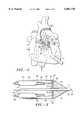
- FIG. 1illustrates an exemplary embodiment of a catheter apparatus for the method in accordance with this invention placed in a human chest cavity in a coronary artery having an occlusion.
- FIG. 2is an enlarged view of the exemplary embodiment of a catheter apparatus shown in FIG. 1 placed proximate the occlusion.
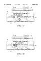
- FIG. 3is an enlarged side view of an exemplary embodiment of a catheter apparatus for the method according to the present invention showing the structure of a catheter shaft.
- FIG. 4is an enlarged side view of an exemplary embodiment of a catheter apparatus for the method according to the present invention showing the structure of a locator.
- FIG. 5is a cut-away view of an exemplary embodiment of a catheter apparatus for the method according to the present invention operating transvascularly to bypass an arterial occlusion by traversing a segment of a vein proximate an occluded artery.
- FIG. 6is a cut-away view of an exemplary embodiment of a catheter apparatus for the method according to the present invention operating transvascularly to create a fluid path between an artery and a vein.
- FIG. 7is a cut-away view of an arterio-venous stent graft placed to maintain a fluid path between an artery and a vein in a step of an exemplary method according to the present invention.
- FIG. 8is a cut-away view of an arterio-venous stent placed to maintain a fluid path between an artery and a vein in a step of an exemplary method according to the present invention.
- FIG. 9is a cut-away view of venous plug placed to occlude a vein at a point proximal to a stented arterio-venous fluid path in a step of an exemplary method according to the present invention.
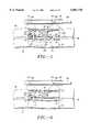
- FIG. 10is a side view of an exemplary embodiment of a catheter apparatus for the method according to the present invention showing the structure of a catheter shaft including rings and slots in the distal end zone of the catheter shaft.
- FIG. 11is a side view of an exemplary embodiment of a catheter apparatus for the method according to the present invention showing the structure of a locator including a plurality of suction cups.
- FIG. 12is a sectional view of an exemplary embodiment of a catheter apparatus for the method according to the present invention showing the structure of a locator including a plurality of suction cups.
- FIG. 13is a side view of an exemplary embodiment of a catheter apparatus for the method according to the present invention showing the structure of a catheter shaft including a signal-emitting working element.
- FIG. 1an exemplary embodiment of a catheter apparatus for the method in accordance with the present invention is shown placed proximate the occlusion 62 in a coronary artery 60.
- the apparatusembodies a combination of two devices which cooperate to safely bypass the occlusion 62.
- the first deviceis an intraluminally operable catheter shaft 100 including a distal end zone 104 having a working element 102 for bypassing the occlusion 62.
- the second device of the combinationis an extraluminally operable locator 160 for locating the working element 102 with respect to the arterial lumen 71, the arterial walls 72, and the tissues and boundaries of the occlusion 62.
- the locator 160includes an imaging tube 162 which is introduced through a small incision (not shown) in the patient's chest and is positioned in the chest cavity adjacent the heart and proximate the occlusion 62.
- the imaging tube 162is introduced through an incision (not shown) which need only be large enough to slip the imaging tube 162 into the patient's chest.
- the imaging tubeis introduced, for example, by thoracotomy, thoracoscopy or sub-xyphoid access, is passed through a puncture in the pericardium, and is advanced until it is adjacent the surface of the heart.
- the external imaging instruments(not shown) are then activated to display an ultrasound image.
- the catheter shaft 100is introduced through a puncture incision (not shown) into a major artery or vein (not shown) and is advanced and guided intraluminally into an arterial or venous branch which serves a portion of the heart. If placed in an artery, the distal end zone 104 of the catheter shaft 100 is positioned at a point proximal to (upstream of) the occlusion 62. If placed in a vein, the distal end zone 104 of the catheter shaft 100 is positioned at a point in the vein 61 adjacent a point where entry into the artery 60 is planned.
- the locator 160is stabilized adjacent the heart and activated to provide an image from a vantage point close to the occluded artery 60 but outside the arterial lumen 71. It will be appreciated that because only small punctures or incisions are needed in order for the catheter shaft and locator to reach the operation site, the patient can expect a comfortable, uncomplicated recovery. With the present method there is no need to saw through the patient's sternum or rib cage.
- the catheter shaft 100 and locator 160are positionable with minimal trauma in the proximity of the occlusion 62 and are simultaneously operable to create a path for perfusion of the myocardium distal to the occlusion.
- the elongated flexible catheter shaft 100(greatly shortened in FIG. 3) includes a steerable distal end zone 104 and a working element 102 which is carried into the proximity of the occlusion by the distal end zone 104.
- the proximal end zone 106 of the catheter shaft 100is connectable to external apparatus (not shown) for manipulating the catheter shaft 100 and working element 102.
- the locator 160includes an imaging tube 162 for micro-invasive placement of the imaging device 168 proximate the occluded artery 60.
- the imaging tube 162includes a proximal end zone 164 connectable to external imaging instruments (not shown).
- the locator 160also includes an imaging device 168 which is locatable extraluminally near the occlusion 62 and is operatively coupled to the external imaging instruments.
- the catheter shaft 100includes a proximal end 110 connectable to external apparatus (not shown), a distal end zone 104 including a distal end 112, and at least one lumen 114 therebetween.
- a working element 102 for penetrating tissuesis disposed in the distal end zone 104.
- a steering member 122is disposed in the distal end zone 104 for directing the working element 102 at and through tissues.
- the steering member 122includes a plurality of steering wires 124 slidably disposed in the catheter shaft 100.
- the steering wires 124have proximal ends 126 manipulable from the proximal end 110 of the catheter shaft 100 and distal ends 128 fixed in the distal end zone 104 of the catheter shaft 100.
- braid-reinforced tubes 130slidably confine the wires 124 to prevent the wires 124 from interfering with other parts of the catheter shaft 100.
- the steering wires 124may be affixed to a retaining ring 132 disposed in the distal end zone 104 of the catheter shaft 100.
- rigid tubes 136may be disposed about braid-reinforced tubes 130, the rigid tubes 136 having distal ends 138 some distance proximal to the distal ends 128 of the steering wires 124. Between the distal ends 138 of the rigid tubes 136 and the distal end 112 of the catheter shaft 100, the absence of the rigid tubes 136 increases the flexibility of the distal end zone 104 to facilitate steering.
- unequal tension on the steering wires 124will deflect the distal end zone 104 of the catheter shaft 100 toward a wire 124 having greater tension.
- the distal ends 128 of four steering wires 124may be fixed in the distal end zone 104 of the catheter shaft 100 at ninety degree intervals about the longitudinal axis of the catheter shaft 100, with the result that the distal end 112 of the catheter shaft 100 can be deflected in two dimensions somewhat independently by manipulating the steering wires 124 in combination.
- the working element 102is steered by deflecting the distal end zone 104 of the catheter shaft 100. Because the working element 102 is carried in the distal end zone 104, the distal end zone 104 will impart to the working element 102 the deflection imparted to the distal end zone 104 by the steering member 122. In conjunction with the guidance provided by the locator 160 (discussed in detail below), this deflection enables an operator of the present invention to guide the working element 102 along a chosen path into and through the arterial wall 72, venous wall 62, or interstitial tissues 69.
- the apparatusmay include a guide wire and the guide wire may include a deflected distal end which functions to steer the guide wire.
- the introduction of a working element into a vascular system and the operation thereof to treat an occlusion without the specific step of steering the working element during treatmentis also within the scope and spirit of the method according to the present invention.
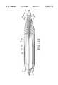
- the working element 102includes a tissue-penetrating wire 116 disposed in a lumen 114 of the catheter shaft 100.
- the tissue-penetrating wire 116includes a proximal end 118, manipulable through the proximal end 110 of the catheter shaft 100, and a sharp distal end 120 projectable from the distal end 112 of the catheter shaft 100.
- pressureis applied to the proximal end of the tissue-penetrating wire 116, urging the wire 116 into and through tissues as the catheter shaft 100 and steering member 122 are manipulated to direct the wire 116.
- the locator 160includes an imaging device 168 (in this embodiment, an acoustic transducer 170), an imaging tube 162 for placing the imaging device 168 extraluminally proximate the occlusion 62, and one or more external imaging instruments (not shown) operatively coupled to the imaging device 168 for discerning the spatial interrelationships of the working element 102, occlusion 62, arterial lumen 71, arterial wall 72, venous wall 73, venous lumen 75, and interstitial tissues 69.
- an imaging device 168in this embodiment, an acoustic transducer 170
- an imaging tube 162for placing the imaging device 168 extraluminally proximate the occlusion 62
- one or more external imaging instruments(not shown) operatively coupled to the imaging device 168 for discerning the spatial interrelationships of the working element 102, occlusion 62, arterial lumen 71, arterial wall 72, venous wall 73, venous lumen 75, and intersti
- the imaging tube 162has an exterior surface 172 which forms one or more suction cups 174 for stabilizing the imaging tube 162 on tissues near the artery 60 having the occlusion 62.
- the imaging tube 162has a suction cup activator 176 for selectively activating the suction cup 174.
- the activator 176includes a lumen 178 having a distal end 180 communicating with a suction cup 174 and a proximal end 182 communicating with a pressure-modulating device (not shown).
- the activator 176may, however, encompass an aspirator, a mechanical means of activating the suction cup 174, or any other convenient way of establishing and interrupting a vacuum to temporarily stabilize a surface of the imaging tube 162 upon a surface proximate the artery 60 and occlusion 62.
- the suction cupmay take the form of any other suction-coupling area or feature, defined by a surface 172 of the imaging tube 162, which affords adhesion to a surface.
- the imaging tube 162can be made flexible enough to enable the distal end zone 186 of the imaging tube 162 to be secured adjacent a beating heart while the proximal end zone 164 of the imaging tube 162 remains connected to external instruments (not shown) for support and control.
- the flexibility of the imaging tube 162contributes to its micro-invasive quality by reducing the trauma inflicted upon tissues and by permitting the tube 162 to conform to the natural contours of bodily surfaces.
- the imaging tube 162may be given a shape well suited to the route of entry into the chest, or may be stabilized or flexibly supported by external apparatus at its proximal end 188.
- the imaging tube 162includes a proximal end 188, a lumen 190 originating in the proximal end 188, and a motor assembly (not shown) proximate the proximal end 188.
- the transducer control shaft 194is flexible enough to bend with the imaging tube 162.
- the external imaging instrumentsinclude an acoustic signal generator-processor (not shown) and video display device controlled by a suitably programmed general purpose computer.
- the locator 160 in this exemplary embodimentprovides a scanning ultrasound image of the environment of the occlusion 62.
- the imaging tube 162is stabilized on the heart adjacent the artery 60 containing the occlusion 62.
- the motor assembly(not shown) drives the transducer control shaft 194 within the lumen 190 of the imaging tube 162 in a scanning pattern appropriate for producing an image.
- the motor assembly(not shown) may drive the transducer control shaft 194 in a repeating reciprocating pattern while at the same time rotating the shaft.
- the transducer 170which is coupled to the transducer control shaft 194, describes a two-dimensional scanning pattern which may be registered by appropriate measuring devices as combinations of a rotational angle ⁇ and a longitudinal position Z within the imaging tube 162.
- the acoustic signal generator-processor(not shown) causes the transducer 170 to emit acoustic energy.
- a signal conducting path 200carries an electric signal from the external instruments (not shown) (which include, in this illustration, a signal generator-processor, also not shown) to the transducer 170, which may include a piezoelectric crystal or other device for producing acoustic energy.
- This acoustic energyis of the type referred to as ultrasonic or ultrasound, although these terms may encompass a variety of acoustical signals embodying a variety of frequencies.
- the energypasses through the surface 172 of the imaging tube 162 and into the occluded artery 60 and surrounding tissues.
- the transducer 170 and acoustic signalare configured such that the energy is emitted in a narrowly focused beam 202 in a known direction (at a known value of the angle ⁇ from a known position (at a known value of Z) with respect to the imaging tube 162.
- the transducer 107also functions as a similarly directional acoustic signal detector, converting acoustic energy reflected by features in the environment of the imaging tube 162 to a signal which is conducted back to the signal generator-processor and measured accordingly.
- the detected signalsare associated with values of ⁇ and Z.
- a third dimensionwhich shall be referred to as depth or as radius from the transducer 170 and given the letter r, is computable as a function of the time elapsed between the emission of a given signal by the transducer 170 and the detection of the echo of that signal.
- the value detected at any given timeis a function of the intensity of the echo.
- this intensitycan be reported via suitable video equipment as a two or three dimensional image of the environment of the imaging tube 162.
- General purpose computersare programmable to accomplish this function.
- U.S. Pat. No. 4,794,931the disclosure of which is incorporated herein by reference in its entirety, describes a computer and instrument system implementing such a function.
- a rotating or translating scanning transducermay be supplanted by an array of directional transducers (not shown), a phased array of transducers (not shown) or other appropriately energized and interrogated set of transducers operatively connected to the external signal generator-processor for displaying the desired image.
- the locator 160provides an image of nearby anatomical features so that the position of the locator 160 with respect to the artery 60, occlusion 62, vein 61 and interstitial tissues 69 is ascertained.
- the locator 160is manipulated until its position is ideal for imaging the vessels and tissues to be penetrated.
- the locator 160is then stabilized.
- the imaging tube 162has an exterior surface 172 which including one or more suction cups 174 for stabilizing the imaging tube 162 on tissues near the occluded artery 60.
- the positions of the distal end 112 of the catheter shaft 100 and the distal end 120 of the working element 102are ascertained.
- the contours of the occlusion 62, the artery 60, the adjacent vein 61, and interstitial tissues 69, as revealed by the locator 160are also evaluated.
- the catheter shaft 100 and locator 160are placed proximate the occlusion 62.
- the catheter shaft 100 and steering member 122are manipulated to direct the working element 102 and the catheter shaft 100 at a point of penetration 70 through the arterial wall 72 for exit from the artery 60.
- the point of penetration 70will have been identified in the image provided by the locator 160.
- the image provided by the locator 160is also studied to determine an appropriate path around the occlusion 62.
- the working element 102is then steered and advanced along that planned path under continuous observation via the locator 160.
- the working element 102 and catheter shaft 100While control of the working element 102 and catheter shaft 100 is maintained via the steering member, the working element 102 and catheter shaft 100 are urged and steered though the arterial wall 72, through the interstitial tissues 69, through the wall 73 of the adjacent vein 61 and then distally within the venous lumen 75.
- the steering member 122is manipulated to direct the working element 102 away from any contact perceived as likely to cause cardiac tamponade.
- the steering member 122is used to direct the working element 102 and catheter shaft 100 through the venous wall 73, across the interstitial tissues 69, and back through the arterial wall 72 into the arterial lumen 71 distal to the occlusion 62.
- a pathhas now been created bypassing the occlusion 62. Blood can now flow out of the artery 60 proximal to the occlusion 62, through the vein 61, and back into the artery 60 distal to the occlusion 62.
- FIG. 6an exemplary method is according to the present invention is described in which the catheter shaft 100 is introduced into the arterial system and is advanced to a position in the lumen 71 of a coronary artery 60 proximal to an occlusion 62.
- the working element 102is directed through the arterial wall 72 and interstitial tissues 69, through the wall 73 of an adjacent vein 61, and into the venous lumen 75 to create a fluid path 77 from the occluded artery 60 to the vein 61.
- the interventionistis also able to place the catheter shaft 100 or a guide wire (not shown) or other catheter-borne device from the artery 60 through the path 77 into the vein 61, rendering further procedures, such as, for example, stenting or stent grafting, feasible.
- FIG. 7illustrates a stent graft 79 placed in the artery 60 and vein 61.
- the stent graft 79maintains the fluid path 77 and directs the flow of blood in the distal direction within the venous lumen 75, effectively arterializing the vein 61 distal to the stent graft 77.
- Apparatus and methods for stent graftingare described in U.S. Pat. No. 5,549,663 (Cottone, Jr., 1996) and U.S. Pat. No. 5,122,154 (Rhodes, 1992), the entire disclosures of which are incorporated herein by reference.
- FIG. 8illustrates a stent 80 placed in the artery 60 and vein 61.
- the stent 80maintains the fluid path 77, allowing arterial blood into the vein 61.
- Apparatus and methods for stentingare described in U.S. Pat. No. 4,739,762 (Palmaz, 1988) and U.S. Pat. No. 4,580,568 (Gianturco, 1986), the entire disclosures of which are incorporated herein by reference.
- FIG. 9illustrates a collagen plug 81 placed in the vein 61 proximal to the fluid path 77 and stent 80.
- the plug 81occludes the vein 61, directing the flow of arterial blood from the fluid path 77 distally within the vein 61 and effectively arterializing the vein 61 distal to the fluid path 77.
- Apparatus and methods for occluding a vessel with such a plugare described in U.S. Pat. No. 5,540,657 (Kurjan et al., 1996), U.S. Pat. No. 5,643,464 (Rhee et al., 1997) and U.S. Pat. No. 5,658,593 (Orly et al., 1997), the entire disclosures of which are incorporated herein by reference.
- a catheterization procedure for occluding the vein 61can be performed via the fluid path 77 or, alternatively, via a catheter introduced from a point of access to the venous system proximal to the fluid path 77. The latter approach may be preferable if for any reason it is inconvenient or difficult to reach proximally into the venous lumen 75 from the fluid path 77.
- FIG. 9shows the catheter shaft 100 and working element 102 being introduced via the arterial lumen 71
- the catheter shaft 100 and working element 102can alternatively be introduced via the venous lumen 75.
- the fluid path 77can first be created, then a stent 80 installed, and finally a plug 81 placed in the venous lumen 75. In this manner, the occlusion of the vein 61 by the plug 81 is effected after the stent 80 has been installed and is performed without the need to deliver the plug 80 through the stented fluid path 77.
- the catheter shaft 100 and the locator 160 of the present inventioncooperate to enable the operator to guide the working element 102 into and through the arterial wall 72, venous wall 73, and interstitial tissues 69 while knowing and maintaining control of the anatomical location and orientation of the catheter shaft.
- the image provided by the locator 160is observed for guidance while manipulating the catheter shaft 100, steering member 122 and working element 102. It is also within the scope of the present invention to employ, for example, a guide wire in place of a steerable working element or steerable catheter shaft.
- a bypass(fluid path) can be created while avoiding uncontrolled hemorrhage or unintended trauma.
- the suction cups 174may be released, the apparatus withdrawn from the patient, and the incisions closed.
- the micro-invasive locator 160provides the necessary spatial information for guidance of the working element 102 while completely avoiding the gross trauma that would be inflicted by traditional bypass operations.
- a lumen of the catheter shaft 100such as the lumen 114, may penetrate the catheter shaft 100 at one or more points, some more proximal than others, to establish a blood perfusion path through the catheter shaft 100 while the catheter shaft 100 is placed in the bypass.
- the lumen so employedmay be the same lumen 114 occupied by the working element 102.
- the working element 102 and lumen 114may, for example, be configured such that advancement, withdrawal, rotation, or other manipulation of the working element 102 opens or closes the lumen 114 to the flow of blood or other fluid between two or more places where the lumen 114 communicates with the environment of the catheter shaft 100.
- an exemplary embodiment of the present inventionis shown including the above-described locator 160, catheter shaft 100, lumen 114, steering member 122, and tissue-penetrating wire 116.
- a metal nose cone 134is included in the distal end 112 of the catheter shaft 100 and defines a distal orifice through which the tissue-penetrating wire 116 can project from the lumen 114.
- the catheter shaft 100can be steered via the steering member 122 and urged into the tissues along the path made by the wire 116.
- the nose cone 134reduces the resistance encountered by the catheter shaft 100. Progress is observed via the locator 160 (discussed below).
- the distal end zone 104 of the catheter shaft 100also includes a plurality of rings 140.
- the rings 140define paths 141 for the steering wires 124 (described above) of the steering member 122.
- One or more of the rings 140may serve to anchor the distal ends 128 of the steering wires 124.
- the distal end zone 104 of the catheter shaft 100also includes a plurality of slots 142 inscribed therein for increasing the steerability of the distal end zone 104.
- unequal tension on the steering wires 124will deflect the distal end zone 104 and the working element 102 toward a wire having greater tension.
- the slots 142 in the distal end zone 104 of the catheter shaft 100reduce the force required to compress one side of the distal end zone 104 and extend the opposite side. A steering wire can thus more easily deflect the distal end zone 104. Because the slots 142 only partially circumscribe the distal end zone 104 of the catheter shaft 100, they do not appreciably reduce its axial stiffness. As a result, the distal end 112 of the catheter shaft may still be pushed firmly against a tissue surface at a point where the working element 102 is intended to enter.
- the imaging tube 162 of the locator 160includes an exterior surface 172 defining a plurality of suction cups 174.
- the suction cups 174are arrayed in two roughly parallel rows 204. Between the rows 204 is a region of the surface defining an imaging window 206.
- the imaging window 206includes an acoustically transparent portion of the imaging tube 162 adjacent the lumen 190.
- the transducer 170has a view through the window 206 unobstructed by the suction cups 174.
- the imaging tube 162optionally includes a suction cup activating lumen 178 having a distal end zone 180 communicating with the suction cups 174 and a proximal end 182 coupled with a pressure modulating device (not shown).
- the lumen 178 and pressure modulating devicepermit rapid, minimally traumatic temporary stabilization of the imaging tube 160 on the heart surface.
- the working elementincludes a tissue-penetrating working element 102 having a distal end 260.
- a signal emitter 262is disposed in the distal end 260.
- a signal generator(not shown) is operatively coupled to the signal emitter 262.
- the signal generatoris external to the body and is coupled to the emitter through an electrically conductive path 266 originating in the proximal end zone 106 of the catheter shaft 100 and terminating at the emitter 262.
- the electrically conductive pathincludes an outer conductor 268 disposed in the catheter shaft 100, a tubular dielectric layer 270 therein, and an inner conductor 272 disposed within the dielectric layer.
- any other energy-delivering or converting meanscan be employed to energize the emitter 262.
- the locator 160 and emitter 262are activated within the body, the locator 160 selectively detects the signal emitted by the emitter 262 in order to discern a spatial relationship between the working element 102 and the locator 160.
- the signal emitter 262may be disposed in a working element which is essentially a guide wire, optionally steerable.
- the catheter shaftmay be of a simpler design than the one shown in FIG. 13; in particular, a catheter shaft without a steering member, and a signal-emitting guide wire distal end not surrounded by a separate catheter shaft, are both within the scope of the present invention.
- the imaging deviceneed not be an acoustic transducer and need not accomplish its imaging by scanning or mechanical movement in any particular manner.
- the imaging devicemay be operatively coupled to external instruments by any appropriate mechanical, electromagnetic, optical, wave guide or other path.
- the image that is displayedmay be computed by any of a variety of algorithms for extracting one-, two-, or three-dimensional information from energy reflected, scattered or absorbed within tissues.
- the imaging tubemay be stabilized proximate the occlusion 62 by any appropriate mechanical, pneumatic, hydraulic or other means.
- the locator 160need not approach the heart in the particular manner described; alternative routes may be taken.
- the catheter shaftmay include a lumen to facilitate blood flow in the bypass.
- the catheter shaft or work elementmay include a balloon for stabilization, for interruption of flow, or for other purposes.
- the steering membermay include more or fewer than the two wires illustrated in the drawing figures or may be omitted for any step not necessitating steering.
- the working element 102may include any mechanical, optical, thermal, chemical, or other device for penetrating tissues, treating an occlusion, or delivering a medicament.
- the catheter shaft 100 and working element 102may be configured such that only the working element 102 traverses certain tissues or, alternatively, the catheter shaft 100 itself may follow along with the working element 102. It will be appreciated that the embodiments discussed above and the virtually infinite embodiments that are not mentioned could easily be within the scope and spirit of the present invention. Thus, the invention is to be limited only by the claims as set forth below.
Landscapes
- Health & Medical Sciences (AREA)
- Life Sciences & Earth Sciences (AREA)
- Surgery (AREA)
- Animal Behavior & Ethology (AREA)
- Public Health (AREA)
- Engineering & Computer Science (AREA)
- Biomedical Technology (AREA)
- Heart & Thoracic Surgery (AREA)
- Medical Informatics (AREA)
- Molecular Biology (AREA)
- Veterinary Medicine (AREA)
- General Health & Medical Sciences (AREA)
- Nuclear Medicine, Radiotherapy & Molecular Imaging (AREA)
- Vascular Medicine (AREA)
- Physics & Mathematics (AREA)
- Biophysics (AREA)
- Pathology (AREA)
- Radiology & Medical Imaging (AREA)
- Surgical Instruments (AREA)
- Media Introduction/Drainage Providing Device (AREA)
- Ultra Sonic Daignosis Equipment (AREA)
Abstract
Description
Claims (20)
Priority Applications (5)
| Application Number | Priority Date | Filing Date | Title |
|---|---|---|---|
| US09/008,198US6241667B1 (en) | 1998-01-15 | 1998-01-15 | Catheter apparatus for guided transvascular treatment of arterial occlusions |
| US09/007,434US6081738A (en) | 1998-01-15 | 1998-01-15 | Method and apparatus for the guided bypass of coronary occlusions |
| US09/008,033US6157852A (en) | 1998-01-15 | 1998-01-16 | Catheter apparatus for treating arterial occlusions |
| AU19274/99AAU1927499A (en) | 1998-01-15 | 1998-12-18 | Method and apparatus for the guided bypass of coronary occlusions |
| PCT/US1998/026933WO1999035978A1 (en) | 1998-01-15 | 1998-12-18 | Method and apparatus for the guided bypass of coronary occlusions |
Applications Claiming Priority (1)
| Application Number | Priority Date | Filing Date | Title |
|---|---|---|---|
| US09/007,434US6081738A (en) | 1998-01-15 | 1998-01-15 | Method and apparatus for the guided bypass of coronary occlusions |
Related Child Applications (2)
| Application Number | Title | Priority Date | Filing Date |
|---|---|---|---|
| US09/008,198Continuation-In-PartUS6241667B1 (en) | 1998-01-15 | 1998-01-15 | Catheter apparatus for guided transvascular treatment of arterial occlusions |
| US09/008,033Continuation-In-PartUS6157852A (en) | 1998-01-15 | 1998-01-16 | Catheter apparatus for treating arterial occlusions |
Publications (1)
| Publication Number | Publication Date |
|---|---|
| US6081738Atrue US6081738A (en) | 2000-06-27 |
Family
ID=21726132
Family Applications (3)
| Application Number | Title | Priority Date | Filing Date |
|---|---|---|---|
| US09/007,434Expired - Fee RelatedUS6081738A (en) | 1998-01-15 | 1998-01-15 | Method and apparatus for the guided bypass of coronary occlusions |
| US09/008,198Expired - LifetimeUS6241667B1 (en) | 1998-01-15 | 1998-01-15 | Catheter apparatus for guided transvascular treatment of arterial occlusions |
| US09/008,033Expired - Fee RelatedUS6157852A (en) | 1998-01-15 | 1998-01-16 | Catheter apparatus for treating arterial occlusions |
Family Applications After (2)
| Application Number | Title | Priority Date | Filing Date |
|---|---|---|---|
| US09/008,198Expired - LifetimeUS6241667B1 (en) | 1998-01-15 | 1998-01-15 | Catheter apparatus for guided transvascular treatment of arterial occlusions |
| US09/008,033Expired - Fee RelatedUS6157852A (en) | 1998-01-15 | 1998-01-16 | Catheter apparatus for treating arterial occlusions |
Country Status (3)
| Country | Link |
|---|---|
| US (3) | US6081738A (en) |
| AU (1) | AU1927499A (en) |
| WO (1) | WO1999035978A1 (en) |
Cited By (124)
| Publication number | Priority date | Publication date | Assignee | Title |
|---|---|---|---|---|
| US20010000041A1 (en)* | 1997-12-19 | 2001-03-15 | Selmon Matthew R. | Methods and apparatus for crossing vascular occlusions |
| US6266550B1 (en)* | 1998-01-16 | 2001-07-24 | Lumend, Inc. | Catheter apparatus for treating arterial occlusions |
| US6302892B1 (en)* | 1999-08-04 | 2001-10-16 | Percardia, Inc. | Blood flow conduit delivery system and method of use |
| US20020022788A1 (en)* | 1999-08-19 | 2002-02-21 | Tim Corvi | Apparatus and methods for material capture and removal |
| US20020049457A1 (en)* | 1999-05-20 | 2002-04-25 | Kaplan Aaron V. | Methods and apparatus for transpericardial left atrial appendage closure |
| US20020077642A1 (en)* | 2000-12-20 | 2002-06-20 | Fox Hollow Technologies, Inc. | Debulking catheter |
| US6511458B2 (en)* | 1998-01-13 | 2003-01-28 | Lumend, Inc. | Vascular re-entry catheter |
| US6562031B2 (en) | 1999-02-24 | 2003-05-13 | Scime Life Systems, Inc. | Guide wire system for RF recanalization of vascular blockages |
| US20030120295A1 (en)* | 2000-12-20 | 2003-06-26 | Fox Hollow Technologies, Inc. | Debulking catheters and methods |
| US6585650B1 (en) | 2000-03-20 | 2003-07-01 | Jomed N.V. | Method and system for bypassing an artery block |
| US20030125758A1 (en)* | 2000-12-20 | 2003-07-03 | Fox Hollow Technologies, Inc. | Debulking catheters and methods |
| US20040111138A1 (en)* | 2002-10-18 | 2004-06-10 | Radiant Medical, Inc. | Valved connector assembly and sterility barriers for heat exchange catheters and other closed loop catheters |
| US20040162485A1 (en)* | 2000-02-25 | 2004-08-19 | Wendlandt Jeffrey M. | Diagnostic catheter using a vacuum for tissue positioning |
| US20040167553A1 (en)* | 2000-12-20 | 2004-08-26 | Fox Hollow Technologies, Inc. | Methods and devices for cutting tissue |
| US20040167554A1 (en)* | 2000-12-20 | 2004-08-26 | Fox Hollow Technologies, Inc. | Methods and devices for reentering a true lumen from a subintimal space |
| US20050154407A1 (en)* | 2000-12-20 | 2005-07-14 | Fox Hollow Technologies, Inc. | Method of evaluating drug efficacy for treating atherosclerosis |
| US20050171478A1 (en)* | 1998-01-13 | 2005-08-04 | Selmon Matthew R. | Catheter system for crossing total occlusions in vasculature |
| US20050177068A1 (en)* | 2000-12-20 | 2005-08-11 | Fox Hollow Technologies, Inc. | Debulking catheters and methods |
| US20050261591A1 (en)* | 2003-07-21 | 2005-11-24 | The Johns Hopkins University | Image guided interventions with interstitial or transmission ultrasound |
| US20060030871A1 (en)* | 2004-08-05 | 2006-02-09 | Matthew Hain | Vascular tunneler |
| US7004173B2 (en) | 2000-12-05 | 2006-02-28 | Lumend, Inc. | Catheter system for vascular re-entry from a sub-intimal space |
| US20060235366A1 (en)* | 2000-12-20 | 2006-10-19 | Fox Hollow Technologies, Inc. | Method of evaluating a treatment for vascular disease |
| US20060236019A1 (en)* | 2005-04-19 | 2006-10-19 | Fox Hollow Technologies, Inc. | Libraries and data structures of materials removed by debulking catheters |
| US20060239982A1 (en)* | 2000-12-20 | 2006-10-26 | Fox Hollow Technologies, Inc. | Debulking catheters and methods |
| US20060253129A1 (en)* | 2005-04-07 | 2006-11-09 | Liddicoat John R | Apparatus and method for the ligation of tissue |
| US20060264738A1 (en)* | 2003-07-24 | 2006-11-23 | Dune Medical Devices Ltd. | Method and apparatus for examining a substance, particularly tissue, to characterize its type |
| US20070032739A1 (en)* | 2005-08-04 | 2007-02-08 | Dune Medical Devices Ltd. | Device for forming an effective sensor-to-tissue contact |
| US20070032808A1 (en)* | 2005-08-03 | 2007-02-08 | Azam Anwar | System and method for addressing total occlusion in a vascular environment |
| US20070032747A1 (en)* | 2005-08-04 | 2007-02-08 | Dune Medical Devices Ltd. | Tissue-characterization probe with effective sensor-to-tissue contact |
| US20070038173A1 (en)* | 2005-07-27 | 2007-02-15 | Fox Hollow Technologies, Inc. | Methods affecting markers in patients having vascular disease |
| US20070078469A1 (en)* | 2000-12-20 | 2007-04-05 | Fox Hollow Technologies, Inc | Testing a patient population having a cardiovascular condition for drug efficacy |
| US20070093779A1 (en)* | 2005-09-12 | 2007-04-26 | Kugler Chad J | Endovascular devices and methods |
| US20070093783A1 (en)* | 2005-09-12 | 2007-04-26 | Kugler Chad J | Endovascular devices and methods |
| US20070093780A1 (en)* | 2005-09-12 | 2007-04-26 | Kugler Chad J | Endovascular devices and methods for exploiting intramural space |
| US20070093781A1 (en)* | 2005-09-12 | 2007-04-26 | Kugler Chad J | Endovascular devices and methods for exploiting intramural space |
| US20070156220A1 (en)* | 2002-05-10 | 2007-07-05 | Cardiac Pacemakers, Inc. | Methods for lead placement on a surface of the heart |
| WO2005117710A3 (en)* | 2004-05-07 | 2007-07-05 | Univ Johns Hopkins | Image guided interventions with interstitial or transmission ultrasound |
| US20070179397A1 (en)* | 2002-01-04 | 2007-08-02 | Dune Medical Devices Ltd. | Probes, systems, and methods for examining tissue according to the dielectric properties thereof |
| US20070196926A1 (en)* | 2006-02-17 | 2007-08-23 | Fox Hollow Technologies, Inc. | Testing lumenectomy samples for Markers of non-vascular diseases |
| US20080091105A1 (en)* | 2006-09-28 | 2008-04-17 | Sheldon Weinbaum | System and method for in vivo imaging of blood vessel walls to detect microcalcifications |
| US20080119879A1 (en)* | 2005-06-30 | 2008-05-22 | Rox Medical, Inc. | Devices, systems and methods for creation of a peripherally located fistula |
| US20080147097A1 (en)* | 2003-10-09 | 2008-06-19 | Sentreheart, Inc. | Apparatus and method for the ligation of tissue |
| US20080154090A1 (en)* | 2005-01-04 | 2008-06-26 | Dune Medical Devices Ltd. | Endoscopic System for In-Vivo Procedures |
| US20080228171A1 (en)* | 2006-11-21 | 2008-09-18 | Kugler Chad J | Endovascular devices and methods for exploiting intramural space |
| US20080243183A1 (en)* | 2007-03-30 | 2008-10-02 | Miller Gary H | Devices, systems, and methods for closing the left atrial appendage |
| US20080283574A1 (en)* | 2007-05-16 | 2008-11-20 | Searete Llc, A Limited Liability Corporation Of The State Of Delaware | Maneuverable surgical stapler |
| US20080283576A1 (en)* | 2007-05-16 | 2008-11-20 | Searete Llc. A Limited Liability Corporation Of The State Of Delaware | Surgical fastening device with cutter |
| US20080283577A1 (en)* | 2007-05-16 | 2008-11-20 | Searete Llc, A Limited Liability Corporation Of The State Of Delaware | Steerable surgical stapler |
| US20080287750A1 (en)* | 2002-01-04 | 2008-11-20 | Dune Medical Devices Ltd. | Ergonomic probes |
| US20080283570A1 (en)* | 2007-05-16 | 2008-11-20 | Searete Llc, A Limited Liability Corporation Of The State Of Delaware | Gentle touch surgical stapler |
| US20080287987A1 (en)* | 2007-05-16 | 2008-11-20 | Searete Llc, A Limited Liability Corporation Of The State Of Delaware | Dispensing system for tissue sealants |
| US20080294175A1 (en)* | 2007-05-21 | 2008-11-27 | Epitek, Inc. | Left atrial appendage closure |
| US20080312664A1 (en)* | 2007-05-21 | 2008-12-18 | Epitek, Inc. | Left atrial appendage closure |
| US20090112256A1 (en)* | 2007-10-30 | 2009-04-30 | Searete Llc, A Limited Liability Corporation Of The State Of Delaware | Suturing device with tissue sealant dispenser |
| US20090112243A1 (en)* | 2007-10-25 | 2009-04-30 | Searete Llc, A Limited Liability Corporation Of The State Of Delaware | Surgical cutter with dispensing system for tissue sealants |
| US20090125044A1 (en)* | 2007-11-14 | 2009-05-14 | Lary Todd P | Treatment of Coronary Stenosis |
| US20090125100A1 (en)* | 2007-11-13 | 2009-05-14 | Cook Incorporated | Intraluminal Bypass Prosthesis and Prosthesis Delivery and Deployment Kit |
| US20090143816A1 (en)* | 2007-11-30 | 2009-06-04 | Searete Llc, A Limited Liability Corporation Of The State Of Delaware | Grasper with surgical sealant dispenser |
| US20090187109A1 (en)* | 2001-11-19 | 2009-07-23 | Dune Medical Devices Ltd. | Method and apparatus for examining tissue for predefined target cells, particularly cancerous cells, and a probe useful in such method and apparatus |
| US20090187073A1 (en)* | 2008-01-22 | 2009-07-23 | Olympus Medical Systems Corp. | Medical device and process of installing medical device in patient |
| US7708749B2 (en) | 2000-12-20 | 2010-05-04 | Fox Hollow Technologies, Inc. | Debulking catheters and methods |
| US7798385B2 (en) | 2007-05-16 | 2010-09-21 | The Invention Science Fund I, Llc | Surgical stapling instrument with chemical sealant |
| US20100294826A1 (en)* | 2007-05-16 | 2010-11-25 | Searete Llc, A Limited Liability Corporation Of The State Of Delaware | Gentle touch surgical stapler |
| US7899515B2 (en) | 2005-03-29 | 2011-03-01 | Dune Medical Devices Ltd. | Electromagnetic sensors for tissue characterization |
| WO2011025855A2 (en) | 2009-08-28 | 2011-03-03 | Si Therapies Ltd. | Inverted balloon neck on catheter |
| WO2011028632A1 (en) | 2009-09-03 | 2011-03-10 | Si Therapies Ltd. | Lancet micro-catheter |
| US8032211B2 (en) | 2002-01-04 | 2011-10-04 | Dune Medical Devices Ltd. | Probes, systems, and methods for examining tissue according to the dielectric properties thereof |
| US8172863B2 (en) | 2008-04-28 | 2012-05-08 | Bridgepoint Medical, Inc. | Methods and apparatus for crossing occlusions in blood vessels |
| US8192452B2 (en) | 2009-05-14 | 2012-06-05 | Tyco Healthcare Group Lp | Easily cleaned atherectomy catheters and methods of use |
| US8202246B2 (en) | 2008-02-05 | 2012-06-19 | Bridgepoint Medical, Inc. | Crossing occlusions in blood vessels |
| US8246640B2 (en) | 2003-04-22 | 2012-08-21 | Tyco Healthcare Group Lp | Methods and devices for cutting tissue at a vascular location |
| US8328829B2 (en) | 1999-08-19 | 2012-12-11 | Covidien Lp | High capacity debulking catheter with razor edge cutting window |
| US8337425B2 (en) | 2008-02-05 | 2012-12-25 | Bridgepoint Medical, Inc. | Endovascular device with a tissue piercing distal probe and associated methods |
| US8414604B2 (en) | 2008-10-13 | 2013-04-09 | Covidien Lp | Devices and methods for manipulating a catheter shaft |
| US8469983B2 (en) | 2007-09-20 | 2013-06-25 | Sentreheart, Inc. | Devices and methods for remote suture management |
| US8496677B2 (en) | 2009-12-02 | 2013-07-30 | Covidien Lp | Methods and devices for cutting tissue |
| US8597315B2 (en) | 1999-08-19 | 2013-12-03 | Covidien Lp | Atherectomy catheter with first and second imaging devices |
| US8632556B2 (en) | 2007-10-22 | 2014-01-21 | Bridgepoint Medical, Inc. | Methods and devices for crossing chronic total occlusions |
| US8784440B2 (en) | 2008-02-25 | 2014-07-22 | Covidien Lp | Methods and devices for cutting tissue |
| US8808186B2 (en) | 2010-11-11 | 2014-08-19 | Covidien Lp | Flexible debulking catheters with imaging and methods of use and manufacture |
| US8920450B2 (en) | 2010-10-28 | 2014-12-30 | Covidien Lp | Material removal device and method of use |
| US8956376B2 (en) | 2011-06-30 | 2015-02-17 | The Spectranetics Corporation | Reentry catheter and method thereof |
| US20150051450A1 (en)* | 2012-03-30 | 2015-02-19 | The Regents Of The University Of California | System, device and method for measurement of esophageal wall blood perfusion |
| US8992717B2 (en) | 2011-09-01 | 2015-03-31 | Covidien Lp | Catheter with helical drive shaft and methods of manufacture |
| US8998936B2 (en) | 2011-06-30 | 2015-04-07 | The Spectranetics Corporation | Reentry catheter and method thereof |
| US9028512B2 (en) | 2009-12-11 | 2015-05-12 | Covidien Lp | Material removal device having improved material capture efficiency and methods of use |
| US9060802B2 (en) | 2006-11-21 | 2015-06-23 | Bridgepoint Medical, Inc. | Endovascular devices and methods for exploiting intramural space |
| US9119662B2 (en) | 2010-06-14 | 2015-09-01 | Covidien Lp | Material removal device and method of use |
| US9198664B2 (en) | 2009-04-01 | 2015-12-01 | Sentreheart, Inc. | Tissue ligation devices and controls therefor |
| US9301777B2 (en) | 2013-07-29 | 2016-04-05 | Invatec S.P.A. | Occlusion bypassing apparatuses and methods for bypassing an occlusion in a blood vessel |
| US9308356B2 (en) | 2013-07-29 | 2016-04-12 | Invatec S.P.A. | Occlusion bypassing apparatuses and methods for bypassing an occlusion in a blood vessel |
| US9320874B2 (en) | 2013-08-15 | 2016-04-26 | Invatec S.P.A. | Catheter systems with a blocking mechanism and methods for bypassing an occlusion in a blood vessel |
| US9364642B2 (en) | 2013-08-14 | 2016-06-14 | Invatec S.P.A. | Balloon catheter systems and methods for bypassing an occlusion in a blood vessel |
| US9408608B2 (en) | 2013-03-12 | 2016-08-09 | Sentreheart, Inc. | Tissue ligation devices and methods therefor |
| US9446222B2 (en) | 2014-03-05 | 2016-09-20 | Invatec S.P.A. | Catheter assemblies and methods for stabilizing a catheter assembly within a subintimal space |
| US9486281B2 (en) | 2010-04-13 | 2016-11-08 | Sentreheart, Inc. | Methods and devices for accessing and delivering devices to a heart |
| US9498206B2 (en) | 2011-06-08 | 2016-11-22 | Sentreheart, Inc. | Tissue ligation devices and tensioning devices therefor |
| US9532844B2 (en) | 2012-09-13 | 2017-01-03 | Covidien Lp | Cleaning device for medical instrument and method of use |
| US9687266B2 (en) | 2009-04-29 | 2017-06-27 | Covidien Lp | Methods and devices for cutting and abrading tissue |
| US9801647B2 (en) | 2006-05-26 | 2017-10-31 | Covidien Lp | Catheter including cutting element and energy emitting element |
| US9814862B2 (en) | 2011-06-30 | 2017-11-14 | The Spectranetics Corporation | Reentry catheter and method thereof |
| US9936956B2 (en) | 2015-03-24 | 2018-04-10 | Sentreheart, Inc. | Devices and methods for left atrial appendage closure |
| US9943329B2 (en) | 2012-11-08 | 2018-04-17 | Covidien Lp | Tissue-removing catheter with rotatable cutter |
| US10130369B2 (en) | 2015-03-24 | 2018-11-20 | Sentreheart, Inc. | Tissue ligation devices and methods therefor |
| US10172632B2 (en) | 2015-09-22 | 2019-01-08 | Medtronic Vascular, Inc. | Occlusion bypassing apparatus with a re-entry needle and a stabilization tube |
| US10213224B2 (en) | 2014-06-27 | 2019-02-26 | Covidien Lp | Cleaning device for catheter and catheter including the same |
| US10258408B2 (en) | 2013-10-31 | 2019-04-16 | Sentreheart, Inc. | Devices and methods for left atrial appendage closure |
| US10292721B2 (en) | 2015-07-20 | 2019-05-21 | Covidien Lp | Tissue-removing catheter including movable distal tip |
| US10292710B2 (en) | 2016-02-26 | 2019-05-21 | Sentreheart, Inc. | Devices and methods for left atrial appendage closure |
| US10314664B2 (en) | 2015-10-07 | 2019-06-11 | Covidien Lp | Tissue-removing catheter and tissue-removing element with depth stop |
| US10314667B2 (en) | 2015-03-25 | 2019-06-11 | Covidien Lp | Cleaning device for cleaning medical instrument |
| US10327791B2 (en) | 2015-10-07 | 2019-06-25 | Medtronic Vascular, Inc. | Occlusion bypassing apparatus with a re-entry needle and a distal stabilization balloon |
| US10456557B2 (en) | 2014-08-14 | 2019-10-29 | Invatec S.P.A. | Occlusion bypassing apparatus with varying flexibility and methods for bypassing an occlusion in a blood vessel |
| US11020141B2 (en) | 2005-09-12 | 2021-06-01 | Bridgepoint Medical, Inc. | Endovascular devices and methods |
| US11116943B2 (en) | 2018-10-09 | 2021-09-14 | Limflow Gmbh | Methods for accessing pedal veins |
| US11241304B2 (en) | 2006-04-20 | 2022-02-08 | Limflow Gmbh | Method for fluid flow through body passages |
| US11298511B2 (en) | 2006-11-21 | 2022-04-12 | Bridgepoint Medical, Inc. | Endovascular devices and methods for exploiting intramural space |
| US11446170B2 (en) | 2004-09-08 | 2022-09-20 | Limflow Gmbh | Minimally invasive surgical apparatus and methods |
| US11471262B2 (en) | 2013-03-08 | 2022-10-18 | Limflow Gmbh | Methods for targeting a body passage to effect fluid flow |
| US11612397B2 (en) | 2019-11-01 | 2023-03-28 | Limflow Gmbh | Devices and methods for increasing blood perfusion to a distal extremity |
| US11826504B2 (en) | 2017-04-10 | 2023-11-28 | Limflow Gmbh | Methods for routing a guidewire from a first vessel and through a second vessel in lower extremity vasculature |
| US11992238B2 (en) | 2008-02-05 | 2024-05-28 | Boston Scientific Scimed, Inc. | Endovascular device with a tissue piercing distal probe and associated methods |
| US12408907B1 (en) | 2019-11-14 | 2025-09-09 | Edwards Lifesciences Corporation | Method of reducing left atrial pressure |
| US12414797B2 (en) | 2019-08-22 | 2025-09-16 | Edwards Lifesciences Corporation | Puncture needles |
Families Citing this family (98)
| Publication number | Priority date | Publication date | Assignee | Title |
|---|---|---|---|---|
| US6026814A (en) | 1997-03-06 | 2000-02-22 | Scimed Life Systems, Inc. | System and method for percutaneous coronary artery bypass |
| US6406488B1 (en)* | 1998-08-27 | 2002-06-18 | Heartstent Corporation | Healing transmyocardial implant |
| AU6384699A (en)* | 1998-09-10 | 2000-04-03 | Percardia, Inc. | Tmr shunt |
| US6261304B1 (en) | 1998-09-10 | 2001-07-17 | Percardia, Inc. | Delivery methods for left ventricular conduit |
| JP2002524196A (en)* | 1998-09-10 | 2002-08-06 | パーカーディア,インコーポレイティド | Transmyocardial shunt for left ventricular revascularization and its mounting mechanism |
| US6290728B1 (en)* | 1998-09-10 | 2001-09-18 | Percardia, Inc. | Designs for left ventricular conduit |
| US6254564B1 (en) | 1998-09-10 | 2001-07-03 | Percardia, Inc. | Left ventricular conduit with blood vessel graft |
| US6641610B2 (en) | 1998-09-10 | 2003-11-04 | Percardia, Inc. | Valve designs for left ventricular conduits |
| US6475226B1 (en) | 1999-02-03 | 2002-11-05 | Scimed Life Systems, Inc. | Percutaneous bypass apparatus and method |
| US6409697B2 (en) | 1999-05-04 | 2002-06-25 | Heartstent Corporation | Transmyocardial implant with forward flow bias |
| US6638237B1 (en) | 1999-08-04 | 2003-10-28 | Percardia, Inc. | Left ventricular conduits and methods for delivery |
| US7033372B1 (en) | 1999-08-04 | 2006-04-25 | Percardia, Inc. | Corkscrew reinforced left ventricle to coronary artery channel |
| US20030125757A1 (en)* | 2000-12-20 | 2003-07-03 | Fox Hollow Technologies, Inc. | Debulking catheters and methods |
| US6626930B1 (en)* | 1999-10-21 | 2003-09-30 | Edwards Lifesciences Corporation | Minimally invasive mitral valve repair method and apparatus |
| US6488671B1 (en)* | 1999-10-22 | 2002-12-03 | Corazon Technologies, Inc. | Methods for enhancing fluid flow through an obstructed vascular site, and systems and kits for use in practicing the same |
| JP3782297B2 (en)* | 2000-03-28 | 2006-06-07 | 株式会社東芝 | Solid-state imaging device and manufacturing method thereof |
| US7083628B2 (en) | 2002-09-03 | 2006-08-01 | Edwards Lifesciences Corporation | Single catheter mitral valve repair device and method for use |
| US6854467B2 (en)* | 2000-05-04 | 2005-02-15 | Percardia, Inc. | Methods and devices for delivering a ventricular stent |
| US20020032478A1 (en)* | 2000-08-07 | 2002-03-14 | Percardia, Inc. | Myocardial stents and related methods of providing direct blood flow from a heart chamber to a coronary vessel |
| SE0002878D0 (en)* | 2000-08-11 | 2000-08-11 | Kimblad Ola | Device and method of treatment of atrioventricular regurgitation |
| US6506178B1 (en)* | 2000-11-10 | 2003-01-14 | Vascular Architects, Inc. | Apparatus and method for crossing a position along a tubular body structure |
| US6976990B2 (en) | 2001-01-25 | 2005-12-20 | Percardia, Inc. | Intravascular ventriculocoronary bypass via a septal passageway |
| US20020107531A1 (en)* | 2001-02-06 | 2002-08-08 | Schreck Stefan G. | Method and system for tissue repair using dual catheters |
| US6746408B2 (en)* | 2001-05-29 | 2004-06-08 | Transonic Systems Inc. | Method of blood flow measurement in arterio-venous hemodialysis shunts by indicator dilution |
| US7066953B2 (en)* | 2001-10-12 | 2006-06-27 | Rosengart Todd K | Method and apparatus for performing an anastamosis |
| US6814751B2 (en) | 2001-10-12 | 2004-11-09 | Rosengart Todd K | Method and apparatus for performing an anastamosis |
| US6663577B2 (en)* | 2001-12-07 | 2003-12-16 | Abbott Laboratories | Catheter deployment device |
| US6949118B2 (en) | 2002-01-16 | 2005-09-27 | Percardia, Inc. | Encased implant and methods |
| US7008397B2 (en) | 2002-02-13 | 2006-03-07 | Percardia, Inc. | Cardiac implant and methods |
| US20030195415A1 (en)* | 2002-02-14 | 2003-10-16 | Iddan Gavriel J. | Device, system and method for accoustic in-vivo measuring |
| US7112176B2 (en)* | 2002-02-20 | 2006-09-26 | Transonic Systems, Inc. | Compensation method for thermodilution catheter having an injectate induced thermal effect in a blood flow measurement |
| US6623436B2 (en) | 2002-02-20 | 2003-09-23 | Transonic Systems, Inc. | Retrograde catheter with reduced injectate induced temperature offset |
| US7094244B2 (en)* | 2002-03-26 | 2006-08-22 | Edwards Lifesciences Corporation | Sequential heart valve leaflet repair device and method of use |
| US7326219B2 (en) | 2002-09-09 | 2008-02-05 | Wilk Patent Development | Device for placing transmyocardial implant |
| CA2499389A1 (en)* | 2002-09-18 | 2004-04-01 | The Board Of Trustees Of The Leland Stanford Junior University | Tubular compliant mechanisms for ultrasonic imaging systems and intravascular interventional devices |
| US20070167804A1 (en)* | 2002-09-18 | 2007-07-19 | Byong-Ho Park | Tubular compliant mechanisms for ultrasonic imaging systems and intravascular interventional devices |
| US7493154B2 (en)* | 2002-10-23 | 2009-02-17 | Medtronic, Inc. | Methods and apparatus for locating body vessels and occlusions in body vessels |
| FR2846520B1 (en)* | 2002-11-06 | 2006-09-29 | Roquette Freres | USE OF MALTODEXTRINS BRANCHED AS BLEACHES OF GRANULATION |
| EP1585574A4 (en)* | 2002-12-20 | 2006-04-26 | Cardiac Inv S Unltd Inc | Apparatus and method for implanting left ventricular pacing leads within the coronary sinus |
| US6928669B2 (en)* | 2003-01-10 | 2005-08-16 | Tyler Pipe Company | Closet carrier system and method of assembly |
| US7381210B2 (en)* | 2003-03-14 | 2008-06-03 | Edwards Lifesciences Corporation | Mitral valve repair system and method for use |
| US7715896B2 (en) | 2003-03-21 | 2010-05-11 | Boston Scientific Scimed, Inc. | Systems and methods for internal tissue penetration |
| US20050059993A1 (en)* | 2003-09-17 | 2005-03-17 | Kamal Ramzipoor | Embolectomy device |
| US7109861B2 (en)* | 2003-11-26 | 2006-09-19 | International Business Machines Corporation | System and method for alarm generation based on the detection of the presence of a person |
| CN1713192A (en)* | 2004-06-25 | 2005-12-28 | 国际商业机器公司 | Method and device for processing logic mode establishment and carrying out |
| US7618410B2 (en)* | 2004-10-05 | 2009-11-17 | Cardia Access, Inc. | Devices and methods for access through a tissue wall |
| DE102005003632A1 (en) | 2005-01-20 | 2006-08-17 | Fraunhofer-Gesellschaft zur Förderung der angewandten Forschung e.V. | Catheter for the transvascular implantation of heart valve prostheses |
| EP2461180A1 (en)* | 2005-05-04 | 2012-06-06 | Volcano Corporation | Miniature actuator mechanism for intravascular imaging |
| WO2006121851A2 (en)* | 2005-05-05 | 2006-11-16 | Volcano Corporation | Capacitive microfabricated ultrasound transducer-based intravascular ultrasound probes |
| US20080065156A1 (en)* | 2006-09-08 | 2008-03-13 | Hauser David L | Expandable clip for tissue repair |
| WO2008120209A1 (en) | 2007-03-29 | 2008-10-09 | Dan Rottenberg | Lumen reentry devices and methods |
| US7896915B2 (en) | 2007-04-13 | 2011-03-01 | Jenavalve Technology, Inc. | Medical device for treating a heart valve insufficiency |
| US8753373B2 (en) | 2007-05-08 | 2014-06-17 | Edwards Lifesciences Corporation | Suture-fastening clip |
| US8989842B2 (en)* | 2007-05-16 | 2015-03-24 | General Electric Company | System and method to register a tracking system with intracardiac echocardiography (ICE) imaging system |
| DE102008031146B4 (en)* | 2007-10-05 | 2012-05-31 | Siemens Aktiengesellschaft | Device for navigating a catheter through a closure region of a vessel |
| US8128677B2 (en) | 2007-12-12 | 2012-03-06 | Intact Vascular LLC | Device and method for tacking plaque to a blood vessel wall |
| US9603730B2 (en) | 2007-12-12 | 2017-03-28 | Intact Vascular, Inc. | Endoluminal device and method |
| US10166127B2 (en) | 2007-12-12 | 2019-01-01 | Intact Vascular, Inc. | Endoluminal device and method |
| US10022250B2 (en) | 2007-12-12 | 2018-07-17 | Intact Vascular, Inc. | Deployment device for placement of multiple intraluminal surgical staples |
| US9375327B2 (en) | 2007-12-12 | 2016-06-28 | Intact Vascular, Inc. | Endovascular implant |
| US7896911B2 (en) | 2007-12-12 | 2011-03-01 | Innovasc Llc | Device and method for tacking plaque to blood vessel wall |
| US9044318B2 (en) | 2008-02-26 | 2015-06-02 | Jenavalve Technology Gmbh | Stent for the positioning and anchoring of a valvular prosthesis |
| BR112012021347A2 (en) | 2008-02-26 | 2019-09-24 | Jenavalve Tecnology Inc | stent for positioning and anchoring a valve prosthesis at an implantation site in a patient's heart |
| US8241273B2 (en) | 2009-01-09 | 2012-08-14 | Ncontact Surgical, Inc. | Method and devices for coagulation of tissue |
| JP5702365B2 (en)* | 2009-05-07 | 2015-04-15 | 日立アロカメディカル株式会社 | Ultrasound system for orthopedic applications |
| US8206306B2 (en)* | 2009-05-07 | 2012-06-26 | Hitachi Aloka Medical, Ltd. | Ultrasound systems and methods for orthopedic applications |
| US8961420B2 (en) | 2010-04-01 | 2015-02-24 | Siemens Medical Solutions Usa, Inc. | System for cardiac condition detection and characterization |
| US10117564B2 (en) | 2010-04-16 | 2018-11-06 | Hitachi Healthcare Americas Corporation | Ultrasound and detachable instrument for procedures |
| US10856978B2 (en) | 2010-05-20 | 2020-12-08 | Jenavalve Technology, Inc. | Catheter system |
| WO2011147849A1 (en) | 2010-05-25 | 2011-12-01 | Jenavalve Technology Inc. | Prosthetic heart valve and transcatheter delivered endoprosthesis comprising a prosthetic heart valve and a stent |
| US8864777B2 (en) | 2011-01-28 | 2014-10-21 | Anchor Orthopedics Xt Inc. | Methods for facilitating tissue puncture |
| US10285831B2 (en) | 2011-06-03 | 2019-05-14 | Intact Vascular, Inc. | Endovascular implant |
| US9402981B2 (en) | 2011-09-19 | 2016-08-02 | Boston Scientific Scimed, Inc. | Subintimal re-entry catheter and retrograde recanalization |
| US8632467B2 (en) | 2011-10-12 | 2014-01-21 | Volcano Corporation | Rotational shape-memory actuators and associated devices, systems, and methods |
| JP6118335B2 (en) | 2011-12-09 | 2017-04-19 | ボストン サイエンティフィック サイムド,インコーポレイテッドBoston Scientific Scimed,Inc. | Recanalization catheter assembly |
| CN110464520A (en) | 2012-01-25 | 2019-11-19 | 因特脉管有限公司 | Intracavitary unit and method |
| US20130204125A1 (en)* | 2012-02-03 | 2013-08-08 | Ninepoint Medical, Inc. | Inflation apparatus with pressure relief, related systems, methods and kits |
| JP6085674B2 (en) | 2012-05-24 | 2017-02-22 | ボストン サイエンティフィック サイムド,インコーポレイテッドBoston Scientific Scimed,Inc. | Subintimal re-entry device |
| WO2014012011A1 (en) | 2012-07-13 | 2014-01-16 | Boston Scientific Scimed, Inc. | Wire-guided recanalization system |
| US9174032B2 (en) | 2012-07-13 | 2015-11-03 | Boston Scientific Scimed, Inc. | Subintimal reentry system |
| EP2967601B1 (en) | 2013-03-14 | 2017-11-29 | Boston Scientific Scimed, Inc. | Systems and apparatus for treating blood vessels |
| JP6182660B2 (en) | 2013-03-14 | 2017-08-16 | ボストン サイエンティフィック サイムド,インコーポレイテッドBoston Scientific Scimed,Inc. | Subintimal reentry catheter with shape-controlled balloon |
| CN105491978A (en) | 2013-08-30 | 2016-04-13 | 耶拿阀门科技股份有限公司 | Radially collapsible frame for a prosthetic valve and method for manufacturing such a frame |
| US10098650B2 (en) | 2014-06-09 | 2018-10-16 | Boston Scientific Scimed, Inc. | Systems and methods for treating atherosclerotic plaque |
| US10905396B2 (en) | 2014-11-18 | 2021-02-02 | C. R. Bard, Inc. | Ultrasound imaging system having automatic image presentation |
| WO2016081321A2 (en) | 2014-11-18 | 2016-05-26 | C.R. Bard, Inc. | Ultrasound imaging system having automatic image presentation |
| US9433520B2 (en) | 2015-01-29 | 2016-09-06 | Intact Vascular, Inc. | Delivery device and method of delivery |
| US9375336B1 (en) | 2015-01-29 | 2016-06-28 | Intact Vascular, Inc. | Delivery device and method of delivery |
| WO2016149272A1 (en) | 2015-03-19 | 2016-09-22 | Boston Scientific Scimed, Inc. | Subintimal re-entry balloon catheter |
| EP3270825B1 (en) | 2015-03-20 | 2020-04-22 | JenaValve Technology, Inc. | Heart valve prosthesis delivery system |
| US10709555B2 (en) | 2015-05-01 | 2020-07-14 | Jenavalve Technology, Inc. | Device and method with reduced pacemaker rate in heart valve replacement |
| US10993824B2 (en) | 2016-01-01 | 2021-05-04 | Intact Vascular, Inc. | Delivery device and method of delivery |
| WO2017143151A1 (en)* | 2016-02-18 | 2017-08-24 | Boston Scientific Scimed, Inc. | Systems with sonic visualization capability |
| WO2017195125A1 (en) | 2016-05-13 | 2017-11-16 | Jenavalve Technology, Inc. | Heart valve prosthesis delivery system and method for delivery of heart valve prosthesis with introducer sheath and loading system |
| US11633176B2 (en) | 2016-08-03 | 2023-04-25 | Boston Scientific Scimed Inc. | Positioning devices, methods, and systems |
| WO2018138658A1 (en) | 2017-01-27 | 2018-08-02 | Jenavalve Technology, Inc. | Heart valve mimicry |
| US11660218B2 (en) | 2017-07-26 | 2023-05-30 | Intact Vascular, Inc. | Delivery device and method of delivery |
| WO2024102411A1 (en) | 2022-11-09 | 2024-05-16 | Jenavalve Technology, Inc. | Catheter system for sequential deployment of an expandable implant |
Citations (83)
| Publication number | Priority date | Publication date | Assignee | Title |
|---|---|---|---|---|
| FR1585065A (en)* | 1968-06-24 | 1970-01-09 | ||
| US3640270A (en)* | 1969-08-02 | 1972-02-08 | Niess Elektromed Ingeborg | Electric contactor with venturi-suction means for organic tissue |
| DE2945237A1 (en)* | 1979-11-09 | 1981-05-14 | Stavros Prof. Dr.med. 5100 Aachen Lymberopoulos | Urinary passage stone-removal grip - has dish-shaped head contracted to catheter tube dia. by pulling control member |
| US4355643A (en)* | 1980-03-05 | 1982-10-26 | University Of Iowa Research Foundation | Vacuum cup doppler flow transducer and method for using same |
| WO1983003188A1 (en)* | 1982-03-11 | 1983-09-29 | Laserscope Inc | Endoscopic device |
| US4447227A (en)* | 1982-06-09 | 1984-05-08 | Endoscopy Surgical Systems, Inc. | Multi-purpose medical devices |
| EP0117519A1 (en)* | 1983-02-23 | 1984-09-05 | Johannes Dipl.-Ing. Theermann | Catheter |
| US4484412A (en)* | 1981-07-27 | 1984-11-27 | Ishikawa Kenichi | Method and apparatus for cutting hard and brittle material using system for applying vibration in two directions |
| US4541433A (en)* | 1984-06-01 | 1985-09-17 | Medtronic, Inc. | Cardiac output monitor |
| US4631052A (en)* | 1984-01-03 | 1986-12-23 | Intravascular Surgical Instruments, Inc. | Method and apparatus for surgically removing remote deposits |
| US4648402A (en)* | 1985-10-03 | 1987-03-10 | Santos Manuel V | Blood vessel dilating surgical instrument |
| US4698057A (en)* | 1986-06-09 | 1987-10-06 | Joishy Suresh K | Built in assembly for stabilizing and securing intravascular needle or catheter like device |
| EP0245211A1 (en)* | 1986-03-04 | 1987-11-11 | HOECHST ITALIA SUD S.p.A. | Kidney-ureter catheter unit particularly adapated to evacuate crumbled calculi |
| EP0377269A1 (en)* | 1989-01-03 | 1990-07-11 | C.R. Bard, Inc. | Steerable guidewire with soft adjustable tip |
| US5001556A (en)* | 1987-09-30 | 1991-03-19 | Olympus Optical Co., Ltd. | Endoscope apparatus for processing a picture image of an object based on a selected wavelength range |
| US5000185A (en)* | 1986-02-28 | 1991-03-19 | Cardiovascular Imaging Systems, Inc. | Method for intravascular two-dimensional ultrasonography and recanalization |
| US5019040A (en)* | 1989-08-31 | 1991-05-28 | Koshin Sangyo Kabushiki Kaisha | Catheter |
| WO1991019528A1 (en)* | 1990-06-11 | 1991-12-26 | Schneider (Usa) Inc. | Tracking guidewire |
| US5085221A (en)* | 1990-06-14 | 1992-02-04 | Interspec, Inc. | Ultrasonic imaging probe |
| US5099850A (en)* | 1989-01-17 | 1992-03-31 | Olympus Optical Co., Ltd. | Ultrasonic diagnostic apparatus |
| US5099845A (en)* | 1989-05-24 | 1992-03-31 | Micronix Pty Ltd. | Medical instrument location means |
| US5102390A (en)* | 1985-05-02 | 1992-04-07 | C. R. Bard, Inc. | Microdilatation probe and system for performing angioplasty in highly stenosed blood vessels |
| US5109867A (en)* | 1991-04-19 | 1992-05-05 | Target Therapeutics | Extendable guidewire assembly |
| US5114414A (en)* | 1984-09-18 | 1992-05-19 | Medtronic, Inc. | Low profile steerable catheter |
| WO1992008510A1 (en)* | 1990-11-09 | 1992-05-29 | Boston Scientific Corporation | Guidewire for crossing occlusions in blood vessels |
| US5125410A (en)* | 1989-10-13 | 1992-06-30 | Olympus Optical Co., Ltd. | Integrated ultrasonic diagnosis device utilizing intra-blood-vessel probe |
| US5158564A (en)* | 1990-02-14 | 1992-10-27 | Angiomed Ag | Atherectomy apparatus |
| US5181514A (en)* | 1991-05-21 | 1993-01-26 | Hewlett-Packard Company | Transducer positioning system |
| US5193546A (en)* | 1991-05-15 | 1993-03-16 | Alexander Shaknovich | Coronary intravascular ultrasound imaging method and apparatus |
| US5203338A (en)* | 1990-12-17 | 1993-04-20 | Cardiovascular Imaging Systems, Inc. | Vascular catheter having low-profile distal end |
| US5247938A (en)* | 1990-01-11 | 1993-09-28 | University Of Washington | Method and apparatus for determining the motility of a region in the human body |
| WO1993018818A1 (en)* | 1992-03-18 | 1993-09-30 | The Spectranetics Corporation | Fiber optic catheter with twistable tip |
| US5256141A (en)* | 1992-12-22 | 1993-10-26 | Nelson Gencheff | Biological material deployment method and apparatus |
| WO1993020882A1 (en)* | 1992-04-20 | 1993-10-28 | Advanced Cardiovascular Systems, Inc. | Low profile dilatation catheter |
| US5279565A (en)* | 1993-02-03 | 1994-01-18 | Localmed, Inc. | Intravascular treatment apparatus and method |
| US5306244A (en)* | 1984-05-14 | 1994-04-26 | Surgical Systems & Instruments, Inc. | Method of guidewire insertion |
| US5321501A (en)* | 1991-04-29 | 1994-06-14 | Massachusetts Institute Of Technology | Method and apparatus for optical imaging with means for controlling the longitudinal range of the sample |
| US5325860A (en)* | 1991-11-08 | 1994-07-05 | Mayo Foundation For Medical Education And Research | Ultrasonic and interventional catheter and method |
| US5325845A (en)* | 1992-06-08 | 1994-07-05 | Adair Edwin Lloyd | Steerable sheath for use with selected removable optical catheter |
| US5336252A (en)* | 1992-06-22 | 1994-08-09 | Cohen Donald M | System and method for implanting cardiac electrical leads |
| US5350377A (en)* | 1992-10-26 | 1994-09-27 | Ultrasonic Sensing & Monitoring Systems, Inc. | Medical catheter using optical fibers that transmit both laser energy and ultrasonic imaging signals |
| US5383467A (en)* | 1992-11-18 | 1995-01-24 | Spectrascience, Inc. | Guidewire catheter and apparatus for diagnostic imaging |
| US5386828A (en)* | 1991-12-23 | 1995-02-07 | Sims Deltec, Inc. | Guide wire apparatus with location sensing member |
| US5409453A (en)* | 1992-08-12 | 1995-04-25 | Vidamed, Inc. | Steerable medical probe with stylets |
| US5419767A (en)* | 1992-01-07 | 1995-05-30 | Thapliyal And Eggers Partners | Methods and apparatus for advancing catheters through severely occluded body lumens |
| US5423846A (en)* | 1991-10-21 | 1995-06-13 | Cathco, Inc. | Dottering auger catheter system |
| US5425705A (en)* | 1993-02-22 | 1995-06-20 | Stanford Surgical Technologies, Inc. | Thoracoscopic devices and methods for arresting the heart |
| WO1995019143A1 (en)* | 1994-01-14 | 1995-07-20 | Paul G. Yock And Robert J. Siegel, A Joint Venture | Ultrasonic ablation of stenoses and occlusions with imaging guidance |
| US5439000A (en)* | 1992-11-18 | 1995-08-08 | Spectrascience, Inc. | Method of diagnosing tissue with guidewire |
| US5452733A (en)* | 1993-02-22 | 1995-09-26 | Stanford Surgical Technologies, Inc. | Methods for performing thoracoscopic coronary artery bypass |
| US5474065A (en)* | 1994-04-04 | 1995-12-12 | Graphic Controls Corporation | Non-invasive fetal probe |
| US5486170A (en)* | 1992-10-26 | 1996-01-23 | Ultrasonic Sensing And Monitoring Systems | Medical catheter using optical fibers that transmit both laser energy and ultrasonic imaging signals |
| WO1996001590A1 (en)* | 1994-07-08 | 1996-01-25 | Rizik David G | Guide wire passage creation device |
| DE4429117A1 (en)* | 1994-08-17 | 1996-02-22 | Bess Medizintechnik Gmbh | Dilatation catheter for vessel or tissue restrictions |
| US5501698A (en)* | 1994-02-14 | 1996-03-26 | Heartport, Inc. | Endoscopic microsurgical instruments and methods |
| US5507295A (en)* | 1992-07-01 | 1996-04-16 | British Technology Group Limited | Medical devices |
| US5507795A (en)* | 1994-04-29 | 1996-04-16 | Devices For Vascular Intervention, Inc. | Catheter with perfusion system |
| WO1996016268A1 (en)* | 1994-11-24 | 1996-05-30 | Nippondenso Co., Ltd. | Starter with planetary reduction gear mechanism |
| WO1996016483A2 (en)* | 1994-11-17 | 1996-05-30 | Ericsson Inc. | A system for decoding digital data using a variable decision depth |
| US5549542A (en)* | 1992-11-17 | 1996-08-27 | Life Medical Technologies, Inc. | Deflectable endoscope |
| US5569276A (en)* | 1990-05-21 | 1996-10-29 | Cardiovascular Imaging Systems, Inc. | Intravascular catheter having combined imaging abrasion head |
| US5571215A (en)* | 1993-02-22 | 1996-11-05 | Heartport, Inc. | Devices and methods for intracardiac procedures |
| US5573531A (en)* | 1994-06-20 | 1996-11-12 | Gregory; Kenton W. | Fluid core laser angioscope |
| WO1997001459A1 (en)* | 1995-06-27 | 1997-01-16 | Pars Passive Rückhaltesysteme Gmbh | Airbag module |
| WO1997001463A1 (en)* | 1995-06-26 | 1997-01-16 | Rozim Peter | Anti-theft system for vehicles |
| WO1997001468A1 (en)* | 1995-06-29 | 1997-01-16 | Itt Automotive Europe Gmbh | Closure piece for a small metal pipe |
| US5607421A (en)* | 1991-05-01 | 1997-03-04 | The Trustees Of Columbia University In The City Of New York | Myocardial revascularization through the endocardial surface using a laser |
| WO1997013463A1 (en)* | 1995-10-13 | 1997-04-17 | Transvascular, Inc. | Methods and apparatus for bypassing arterial obstructions and/or performing other transvascular procedures |
| US5651378A (en)* | 1996-02-20 | 1997-07-29 | Cardiothoracic Systems, Inc. | Method of using vagal nerve stimulation in surgery |
| WO1997027897A1 (en)* | 1996-02-02 | 1997-08-07 | Transvascular, Inc. | A device, system and method for interstitial transvascular intervention |
| US5702368A (en)* | 1991-07-16 | 1997-12-30 | Heartport, Inc. | System for cardiac procedures |
| US5704898A (en)* | 1995-11-17 | 1998-01-06 | Circon Corporation | Articulation mechanism for an endoscope |
| US5709693A (en)* | 1996-02-20 | 1998-01-20 | Cardiothoracic System, Inc. | Stitcher |
| WO1998008456A1 (en)* | 1996-08-26 | 1998-03-05 | Transvascular, Inc. | Methods and apparatus for transmyocardial direct coronary revascularization |
| WO1998016161A1 (en)* | 1996-10-11 | 1998-04-23 | Transvascular, Inc. | Methods and apparatus for bypassing arterial obstructions and/or performing other transvascular procedures |
| US5743876A (en)* | 1994-03-10 | 1998-04-28 | Schneider (Usa) Inc | Catheter having shaft of varying stiffness |
| US5799655A (en)* | 1992-10-05 | 1998-09-01 | Cardiovascular Imaging Systems, Inc. | Method and apparatus for ultrasound imaging and atherectomy |
| US5807388A (en)* | 1994-05-25 | 1998-09-15 | The Trustees Of Columbia University In The City Of New York | Myocardial revascularization through the endocardial surface using a laser |
| US5830222A (en)* | 1995-10-13 | 1998-11-03 | Transvascular, Inc. | Device, system and method for intersititial transvascular intervention |
| US5836882A (en)* | 1997-03-17 | 1998-11-17 | Frazin; Leon J. | Method and apparatus of localizing an insertion end of a probe within a biotic structure |
| US5868760A (en)* | 1994-12-07 | 1999-02-09 | Mcguckin, Jr.; James F. | Method and apparatus for endolumenally resectioning tissue |
| US5876373A (en)* | 1997-04-04 | 1999-03-02 | Eclipse Surgical Technologies, Inc. | Steerable catheter |
| US5968064A (en)* | 1997-02-28 | 1999-10-19 | Lumend, Inc. | Catheter system for treating a vascular occlusion |
Family Cites Families (13)
| Publication number | Priority date | Publication date | Assignee | Title |
|---|---|---|---|---|
| US4580568A (en) | 1984-10-01 | 1986-04-08 | Cook, Incorporated | Percutaneous endovascular stent and method for insertion thereof |
| US4733665C2 (en) | 1985-11-07 | 2002-01-29 | Expandable Grafts Partnership | Expandable intraluminal graft and method and apparatus for implanting an expandable intraluminal graft |
| US4794931A (en) | 1986-02-28 | 1989-01-03 | Cardiovascular Imaging Systems, Inc. | Catheter apparatus, system and method for intravascular two-dimensional ultrasonography |
| US4905698A (en)* | 1988-09-13 | 1990-03-06 | Pharmacia Deltec Inc. | Method and apparatus for catheter location determination |
| US5643464A (en) | 1988-11-21 | 1997-07-01 | Collagen Corporation | Process for preparing a sterile, dry crosslinking agent |
| US5122154A (en) | 1990-08-15 | 1992-06-16 | Rhodes Valentine J | Endovascular bypass graft |
| US5304115A (en) | 1991-01-11 | 1994-04-19 | Baxter International Inc. | Ultrasonic angioplasty device incorporating improved transmission member and ablation probe |
| US5658593A (en) | 1992-01-16 | 1997-08-19 | Coletica | Injectable compositions containing collagen microcapsules |
| US5549663A (en) | 1994-03-09 | 1996-08-27 | Cordis Corporation | Endoprosthesis having graft member and exposed welded end junctions, method and procedure |
| US5484412A (en) | 1994-04-19 | 1996-01-16 | Pierpont; Brien E. | Angioplasty method and means for performing angioplasty |
| US5540657A (en) | 1994-07-15 | 1996-07-30 | Collagen Corporation | Delivery device for injectable materials |
| CA2244080A1 (en) | 1996-02-02 | 1997-08-07 | Transvascular, Inc. | Methods and apparatus for blocking flow through blood vessels |
| CN1216929A (en) | 1996-02-02 | 1999-05-19 | 血管转换公司 | Method and apparatus for blocking flow through blood vessels |
- 1998
- 1998-01-15USUS09/007,434patent/US6081738A/ennot_activeExpired - Fee Related
- 1998-01-15USUS09/008,198patent/US6241667B1/ennot_activeExpired - Lifetime
- 1998-01-16USUS09/008,033patent/US6157852A/ennot_activeExpired - Fee Related
- 1998-12-18WOPCT/US1998/026933patent/WO1999035978A1/enactiveApplication Filing
- 1998-12-18AUAU19274/99Apatent/AU1927499A/ennot_activeAbandoned
Patent Citations (87)
| Publication number | Priority date | Publication date | Assignee | Title |
|---|---|---|---|---|
| FR1585065A (en)* | 1968-06-24 | 1970-01-09 | ||
| US3640270A (en)* | 1969-08-02 | 1972-02-08 | Niess Elektromed Ingeborg | Electric contactor with venturi-suction means for organic tissue |
| DE2945237A1 (en)* | 1979-11-09 | 1981-05-14 | Stavros Prof. Dr.med. 5100 Aachen Lymberopoulos | Urinary passage stone-removal grip - has dish-shaped head contracted to catheter tube dia. by pulling control member |
| US4355643A (en)* | 1980-03-05 | 1982-10-26 | University Of Iowa Research Foundation | Vacuum cup doppler flow transducer and method for using same |
| US4484412A (en)* | 1981-07-27 | 1984-11-27 | Ishikawa Kenichi | Method and apparatus for cutting hard and brittle material using system for applying vibration in two directions |
| WO1983003188A1 (en)* | 1982-03-11 | 1983-09-29 | Laserscope Inc | Endoscopic device |
| US4447227A (en)* | 1982-06-09 | 1984-05-08 | Endoscopy Surgical Systems, Inc. | Multi-purpose medical devices |
| EP0117519A1 (en)* | 1983-02-23 | 1984-09-05 | Johannes Dipl.-Ing. Theermann | Catheter |
| US4631052A (en)* | 1984-01-03 | 1986-12-23 | Intravascular Surgical Instruments, Inc. | Method and apparatus for surgically removing remote deposits |
| US5306244A (en)* | 1984-05-14 | 1994-04-26 | Surgical Systems & Instruments, Inc. | Method of guidewire insertion |
| US4541433A (en)* | 1984-06-01 | 1985-09-17 | Medtronic, Inc. | Cardiac output monitor |
| US5114414A (en)* | 1984-09-18 | 1992-05-19 | Medtronic, Inc. | Low profile steerable catheter |
| US5102390A (en)* | 1985-05-02 | 1992-04-07 | C. R. Bard, Inc. | Microdilatation probe and system for performing angioplasty in highly stenosed blood vessels |
| US4648402A (en)* | 1985-10-03 | 1987-03-10 | Santos Manuel V | Blood vessel dilating surgical instrument |
| US5000185A (en)* | 1986-02-28 | 1991-03-19 | Cardiovascular Imaging Systems, Inc. | Method for intravascular two-dimensional ultrasonography and recanalization |
| US5676151A (en)* | 1986-02-28 | 1997-10-14 | Cardiovascular Imaging Systems, Inc. | Method and apparatus for intravascular ultrasonography |
| EP0245211A1 (en)* | 1986-03-04 | 1987-11-11 | HOECHST ITALIA SUD S.p.A. | Kidney-ureter catheter unit particularly adapated to evacuate crumbled calculi |
| US4698057A (en)* | 1986-06-09 | 1987-10-06 | Joishy Suresh K | Built in assembly for stabilizing and securing intravascular needle or catheter like device |
| US5001556A (en)* | 1987-09-30 | 1991-03-19 | Olympus Optical Co., Ltd. | Endoscope apparatus for processing a picture image of an object based on a selected wavelength range |
| EP0377269A1 (en)* | 1989-01-03 | 1990-07-11 | C.R. Bard, Inc. | Steerable guidewire with soft adjustable tip |
| US5099850A (en)* | 1989-01-17 | 1992-03-31 | Olympus Optical Co., Ltd. | Ultrasonic diagnostic apparatus |
| US5099845A (en)* | 1989-05-24 | 1992-03-31 | Micronix Pty Ltd. | Medical instrument location means |
| US5019040A (en)* | 1989-08-31 | 1991-05-28 | Koshin Sangyo Kabushiki Kaisha | Catheter |
| US5125410A (en)* | 1989-10-13 | 1992-06-30 | Olympus Optical Co., Ltd. | Integrated ultrasonic diagnosis device utilizing intra-blood-vessel probe |
| US5247938A (en)* | 1990-01-11 | 1993-09-28 | University Of Washington | Method and apparatus for determining the motility of a region in the human body |
| US5158564A (en)* | 1990-02-14 | 1992-10-27 | Angiomed Ag | Atherectomy apparatus |
| US5569276A (en)* | 1990-05-21 | 1996-10-29 | Cardiovascular Imaging Systems, Inc. | Intravascular catheter having combined imaging abrasion head |
| WO1991019528A1 (en)* | 1990-06-11 | 1991-12-26 | Schneider (Usa) Inc. | Tracking guidewire |
| US5085221A (en)* | 1990-06-14 | 1992-02-04 | Interspec, Inc. | Ultrasonic imaging probe |
| WO1992008510A1 (en)* | 1990-11-09 | 1992-05-29 | Boston Scientific Corporation | Guidewire for crossing occlusions in blood vessels |
| US5203338A (en)* | 1990-12-17 | 1993-04-20 | Cardiovascular Imaging Systems, Inc. | Vascular catheter having low-profile distal end |
| US5109867A (en)* | 1991-04-19 | 1992-05-05 | Target Therapeutics | Extendable guidewire assembly |
| US5321501A (en)* | 1991-04-29 | 1994-06-14 | Massachusetts Institute Of Technology | Method and apparatus for optical imaging with means for controlling the longitudinal range of the sample |
| US5459570A (en)* | 1991-04-29 | 1995-10-17 | Massachusetts Institute Of Technology | Method and apparatus for performing optical measurements |
| US5607421A (en)* | 1991-05-01 | 1997-03-04 | The Trustees Of Columbia University In The City Of New York | Myocardial revascularization through the endocardial surface using a laser |
| US5193546A (en)* | 1991-05-15 | 1993-03-16 | Alexander Shaknovich | Coronary intravascular ultrasound imaging method and apparatus |
| US5181514A (en)* | 1991-05-21 | 1993-01-26 | Hewlett-Packard Company | Transducer positioning system |
| US5885238A (en)* | 1991-07-16 | 1999-03-23 | Heartport, Inc. | System for cardiac procedures |
| US5702368A (en)* | 1991-07-16 | 1997-12-30 | Heartport, Inc. | System for cardiac procedures |
| US5423846A (en)* | 1991-10-21 | 1995-06-13 | Cathco, Inc. | Dottering auger catheter system |
| US5325860A (en)* | 1991-11-08 | 1994-07-05 | Mayo Foundation For Medical Education And Research | Ultrasonic and interventional catheter and method |
| US5386828A (en)* | 1991-12-23 | 1995-02-07 | Sims Deltec, Inc. | Guide wire apparatus with location sensing member |
| US5419767A (en)* | 1992-01-07 | 1995-05-30 | Thapliyal And Eggers Partners | Methods and apparatus for advancing catheters through severely occluded body lumens |
| WO1993018818A1 (en)* | 1992-03-18 | 1993-09-30 | The Spectranetics Corporation | Fiber optic catheter with twistable tip |
| WO1993020882A1 (en)* | 1992-04-20 | 1993-10-28 | Advanced Cardiovascular Systems, Inc. | Low profile dilatation catheter |
| US5325845A (en)* | 1992-06-08 | 1994-07-05 | Adair Edwin Lloyd | Steerable sheath for use with selected removable optical catheter |
| US5336252A (en)* | 1992-06-22 | 1994-08-09 | Cohen Donald M | System and method for implanting cardiac electrical leads |
| US5507295A (en)* | 1992-07-01 | 1996-04-16 | British Technology Group Limited | Medical devices |
| US5409453A (en)* | 1992-08-12 | 1995-04-25 | Vidamed, Inc. | Steerable medical probe with stylets |
| US5531677A (en)* | 1992-08-12 | 1996-07-02 | Vidamed, Inc. | Steerable medical probe with stylets |
| US5799655A (en)* | 1992-10-05 | 1998-09-01 | Cardiovascular Imaging Systems, Inc. | Method and apparatus for ultrasound imaging and atherectomy |
| US5486170A (en)* | 1992-10-26 | 1996-01-23 | Ultrasonic Sensing And Monitoring Systems | Medical catheter using optical fibers that transmit both laser energy and ultrasonic imaging signals |
| US5350377A (en)* | 1992-10-26 | 1994-09-27 | Ultrasonic Sensing & Monitoring Systems, Inc. | Medical catheter using optical fibers that transmit both laser energy and ultrasonic imaging signals |
| US5549542A (en)* | 1992-11-17 | 1996-08-27 | Life Medical Technologies, Inc. | Deflectable endoscope |
| US5439000A (en)* | 1992-11-18 | 1995-08-08 | Spectrascience, Inc. | Method of diagnosing tissue with guidewire |
| US5383467A (en)* | 1992-11-18 | 1995-01-24 | Spectrascience, Inc. | Guidewire catheter and apparatus for diagnostic imaging |
| US5256141A (en)* | 1992-12-22 | 1993-10-26 | Nelson Gencheff | Biological material deployment method and apparatus |
| US5279565A (en)* | 1993-02-03 | 1994-01-18 | Localmed, Inc. | Intravascular treatment apparatus and method |
| US5452733A (en)* | 1993-02-22 | 1995-09-26 | Stanford Surgical Technologies, Inc. | Methods for performing thoracoscopic coronary artery bypass |
| US5571215A (en)* | 1993-02-22 | 1996-11-05 | Heartport, Inc. | Devices and methods for intracardiac procedures |
| US5425705A (en)* | 1993-02-22 | 1995-06-20 | Stanford Surgical Technologies, Inc. | Thoracoscopic devices and methods for arresting the heart |
| WO1995019143A1 (en)* | 1994-01-14 | 1995-07-20 | Paul G. Yock And Robert J. Siegel, A Joint Venture | Ultrasonic ablation of stenoses and occlusions with imaging guidance |
| US5501698A (en)* | 1994-02-14 | 1996-03-26 | Heartport, Inc. | Endoscopic microsurgical instruments and methods |
| US5743876A (en)* | 1994-03-10 | 1998-04-28 | Schneider (Usa) Inc | Catheter having shaft of varying stiffness |
| US5474065A (en)* | 1994-04-04 | 1995-12-12 | Graphic Controls Corporation | Non-invasive fetal probe |
| US5507795A (en)* | 1994-04-29 | 1996-04-16 | Devices For Vascular Intervention, Inc. | Catheter with perfusion system |
| US5807388A (en)* | 1994-05-25 | 1998-09-15 | The Trustees Of Columbia University In The City Of New York | Myocardial revascularization through the endocardial surface using a laser |
| US5573531A (en)* | 1994-06-20 | 1996-11-12 | Gregory; Kenton W. | Fluid core laser angioscope |
| WO1996001590A1 (en)* | 1994-07-08 | 1996-01-25 | Rizik David G | Guide wire passage creation device |
| DE4429117A1 (en)* | 1994-08-17 | 1996-02-22 | Bess Medizintechnik Gmbh | Dilatation catheter for vessel or tissue restrictions |
| WO1996016483A2 (en)* | 1994-11-17 | 1996-05-30 | Ericsson Inc. | A system for decoding digital data using a variable decision depth |
| WO1996016268A1 (en)* | 1994-11-24 | 1996-05-30 | Nippondenso Co., Ltd. | Starter with planetary reduction gear mechanism |
| US5868760A (en)* | 1994-12-07 | 1999-02-09 | Mcguckin, Jr.; James F. | Method and apparatus for endolumenally resectioning tissue |
| WO1997001463A1 (en)* | 1995-06-26 | 1997-01-16 | Rozim Peter | Anti-theft system for vehicles |
| WO1997001459A1 (en)* | 1995-06-27 | 1997-01-16 | Pars Passive Rückhaltesysteme Gmbh | Airbag module |
| WO1997001468A1 (en)* | 1995-06-29 | 1997-01-16 | Itt Automotive Europe Gmbh | Closure piece for a small metal pipe |
| US5830222A (en)* | 1995-10-13 | 1998-11-03 | Transvascular, Inc. | Device, system and method for intersititial transvascular intervention |
| WO1997013463A1 (en)* | 1995-10-13 | 1997-04-17 | Transvascular, Inc. | Methods and apparatus for bypassing arterial obstructions and/or performing other transvascular procedures |
| US5704898A (en)* | 1995-11-17 | 1998-01-06 | Circon Corporation | Articulation mechanism for an endoscope |
| WO1997027897A1 (en)* | 1996-02-02 | 1997-08-07 | Transvascular, Inc. | A device, system and method for interstitial transvascular intervention |
| US5709693A (en)* | 1996-02-20 | 1998-01-20 | Cardiothoracic System, Inc. | Stitcher |
| US5651378A (en)* | 1996-02-20 | 1997-07-29 | Cardiothoracic Systems, Inc. | Method of using vagal nerve stimulation in surgery |
| WO1998008456A1 (en)* | 1996-08-26 | 1998-03-05 | Transvascular, Inc. | Methods and apparatus for transmyocardial direct coronary revascularization |
| WO1998016161A1 (en)* | 1996-10-11 | 1998-04-23 | Transvascular, Inc. | Methods and apparatus for bypassing arterial obstructions and/or performing other transvascular procedures |
| US5968064A (en)* | 1997-02-28 | 1999-10-19 | Lumend, Inc. | Catheter system for treating a vascular occlusion |
| US5836882A (en)* | 1997-03-17 | 1998-11-17 | Frazin; Leon J. | Method and apparatus of localizing an insertion end of a probe within a biotic structure |
| US5876373A (en)* | 1997-04-04 | 1999-03-02 | Eclipse Surgical Technologies, Inc. | Steerable catheter |
Cited By (296)
| Publication number | Priority date | Publication date | Assignee | Title |
|---|---|---|---|---|
| US20010000041A1 (en)* | 1997-12-19 | 2001-03-15 | Selmon Matthew R. | Methods and apparatus for crossing vascular occlusions |
| US6511458B2 (en)* | 1998-01-13 | 2003-01-28 | Lumend, Inc. | Vascular re-entry catheter |
| US20050171478A1 (en)* | 1998-01-13 | 2005-08-04 | Selmon Matthew R. | Catheter system for crossing total occlusions in vasculature |
| US6266550B1 (en)* | 1998-01-16 | 2001-07-24 | Lumend, Inc. | Catheter apparatus for treating arterial occlusions |
| US6562031B2 (en) | 1999-02-24 | 2003-05-13 | Scime Life Systems, Inc. | Guide wire system for RF recanalization of vascular blockages |
| US20020049457A1 (en)* | 1999-05-20 | 2002-04-25 | Kaplan Aaron V. | Methods and apparatus for transpericardial left atrial appendage closure |
| US20020099390A1 (en)* | 1999-05-20 | 2002-07-25 | Kaplan Aaron V. | Methods and apparatus for transpericardial left atrial appendage closure |
| US20020103492A1 (en)* | 1999-05-20 | 2002-08-01 | Kaplan Aaron V. | Methods and apparatus for transpericardial left atrial appendage closure |
| US6488689B1 (en) | 1999-05-20 | 2002-12-03 | Aaron V. Kaplan | Methods and apparatus for transpericardial left atrial appendage closure |
| US8974473B2 (en) | 1999-05-20 | 2015-03-10 | Sentreheart, Inc. | Methods and apparatus for transpericardial left atrial appendage closure |
| US8721663B2 (en) | 1999-05-20 | 2014-05-13 | Sentreheart, Inc. | Methods and apparatus for transpericardial left atrial appendage closure |
| US7318829B2 (en) | 1999-05-20 | 2008-01-15 | Aaron V. Kaplan | Methods and apparatus for transpericardial left atrial appendage closure |
| US9724105B2 (en) | 1999-05-20 | 2017-08-08 | Sentreheart, Inc. | Methods and apparatus for transpericardial left atrial appendage closure |
| US7226458B2 (en) | 1999-05-20 | 2007-06-05 | Aaron V. Kaplan | Methods and apparatus for transpericardial left atrial appendage closure |
| US6582444B2 (en) | 1999-08-04 | 2003-06-24 | Percardia, Inc. | Blood flow conduit delivery system and method of use |
| US6302892B1 (en)* | 1999-08-04 | 2001-10-16 | Percardia, Inc. | Blood flow conduit delivery system and method of use |
| US8328829B2 (en) | 1999-08-19 | 2012-12-11 | Covidien Lp | High capacity debulking catheter with razor edge cutting window |
| US9532799B2 (en) | 1999-08-19 | 2017-01-03 | Covidien Lp | Method and devices for cutting tissue |
| US8998937B2 (en) | 1999-08-19 | 2015-04-07 | Covidien Lp | Methods and devices for cutting tissue |
| US8784333B2 (en) | 1999-08-19 | 2014-07-22 | Covidien Lp | Apparatus and methods for material capture and removal |
| US20020022788A1 (en)* | 1999-08-19 | 2002-02-21 | Tim Corvi | Apparatus and methods for material capture and removal |
| US8911459B2 (en) | 1999-08-19 | 2014-12-16 | Covidien Lp | Debulking catheters and methods |
| US8597315B2 (en) | 1999-08-19 | 2013-12-03 | Covidien Lp | Atherectomy catheter with first and second imaging devices |
| US9486237B2 (en) | 1999-08-19 | 2016-11-08 | Covidien Lp | Methods and devices for cutting tissue |
| US10022145B2 (en) | 1999-08-19 | 2018-07-17 | Covidien Lp | Methods and devices for cutting tissue |
| US9788854B2 (en) | 1999-08-19 | 2017-10-17 | Covidien Lp | Debulking catheters and methods |
| US9615850B2 (en) | 1999-08-19 | 2017-04-11 | Covidien Lp | Atherectomy catheter with aligned imager |
| US8882656B2 (en)* | 2000-02-25 | 2014-11-11 | Boston Scientific Scimed, Inc. | Diagnostic catheter using a vacuum for tissue positioning |
| US20150025379A1 (en)* | 2000-02-25 | 2015-01-22 | Boston Scientific Scimed, Inc. | Diagnostic catheter using a vacuum for tissue positioning |
| US20040162485A1 (en)* | 2000-02-25 | 2004-08-19 | Wendlandt Jeffrey M. | Diagnostic catheter using a vacuum for tissue positioning |
| US6585650B1 (en) | 2000-03-20 | 2003-07-01 | Jomed N.V. | Method and system for bypassing an artery block |
| US7004173B2 (en) | 2000-12-05 | 2006-02-28 | Lumend, Inc. | Catheter system for vascular re-entry from a sub-intimal space |
| US8226674B2 (en) | 2000-12-20 | 2012-07-24 | Tyco Healthcare Group Lp | Debulking catheters and methods |
| US20060239982A1 (en)* | 2000-12-20 | 2006-10-26 | Fox Hollow Technologies, Inc. | Debulking catheters and methods |
| US20020077642A1 (en)* | 2000-12-20 | 2002-06-20 | Fox Hollow Technologies, Inc. | Debulking catheter |
| US20070078469A1 (en)* | 2000-12-20 | 2007-04-05 | Fox Hollow Technologies, Inc | Testing a patient population having a cardiovascular condition for drug efficacy |
| US7887556B2 (en) | 2000-12-20 | 2011-02-15 | Fox Hollow Technologies, Inc. | Debulking catheters and methods |
| US20030120295A1 (en)* | 2000-12-20 | 2003-06-26 | Fox Hollow Technologies, Inc. | Debulking catheters and methods |
| US20030125758A1 (en)* | 2000-12-20 | 2003-07-03 | Fox Hollow Technologies, Inc. | Debulking catheters and methods |
| US7771444B2 (en) | 2000-12-20 | 2010-08-10 | Fox Hollow Technologies, Inc. | Methods and devices for removing material from a body lumen |
| US20040167553A1 (en)* | 2000-12-20 | 2004-08-26 | Fox Hollow Technologies, Inc. | Methods and devices for cutting tissue |
| US20040167554A1 (en)* | 2000-12-20 | 2004-08-26 | Fox Hollow Technologies, Inc. | Methods and devices for reentering a true lumen from a subintimal space |
| US20100121360A9 (en)* | 2000-12-20 | 2010-05-13 | Fox Hollow Technologies, Inc | Testing a patient population having a cardiovascular condition for drug efficacy |
| US7713279B2 (en) | 2000-12-20 | 2010-05-11 | Fox Hollow Technologies, Inc. | Method and devices for cutting tissue |
| US7708749B2 (en) | 2000-12-20 | 2010-05-04 | Fox Hollow Technologies, Inc. | Debulking catheters and methods |
| US20060235366A1 (en)* | 2000-12-20 | 2006-10-19 | Fox Hollow Technologies, Inc. | Method of evaluating a treatment for vascular disease |
| US7699790B2 (en) | 2000-12-20 | 2010-04-20 | Ev3, Inc. | Debulking catheters and methods |
| US7927784B2 (en) | 2000-12-20 | 2011-04-19 | Ev3 | Vascular lumen debulking catheters and methods |
| US8052704B2 (en) | 2000-12-20 | 2011-11-08 | Foxhollow Technologies, Inc. | High capacity debulking catheter with distal driven cutting wheel |
| US20050154407A1 (en)* | 2000-12-20 | 2005-07-14 | Fox Hollow Technologies, Inc. | Method of evaluating drug efficacy for treating atherosclerosis |
| US20050177068A1 (en)* | 2000-12-20 | 2005-08-11 | Fox Hollow Technologies, Inc. | Debulking catheters and methods |
| US8469979B2 (en) | 2000-12-20 | 2013-06-25 | Covidien Lp | High capacity debulking catheter with distal driven cutting wheel |
| US9241733B2 (en) | 2000-12-20 | 2016-01-26 | Covidien Lp | Debulking catheter |
| US20090187109A1 (en)* | 2001-11-19 | 2009-07-23 | Dune Medical Devices Ltd. | Method and apparatus for examining tissue for predefined target cells, particularly cancerous cells, and a probe useful in such method and apparatus |
| US8019411B2 (en) | 2002-01-04 | 2011-09-13 | Dune Medical Devices Ltd. | Probes, systems, and methods for examining tissue according to the dielectric properties thereof |
| US8032211B2 (en) | 2002-01-04 | 2011-10-04 | Dune Medical Devices Ltd. | Probes, systems, and methods for examining tissue according to the dielectric properties thereof |
| US20080287750A1 (en)* | 2002-01-04 | 2008-11-20 | Dune Medical Devices Ltd. | Ergonomic probes |
| US20070179397A1 (en)* | 2002-01-04 | 2007-08-02 | Dune Medical Devices Ltd. | Probes, systems, and methods for examining tissue according to the dielectric properties thereof |
| US20070203554A1 (en)* | 2002-05-10 | 2007-08-30 | Cardiac Pacemakers, Inc. | Methods and apparatus for lead placement on a surface of the heart |
| US20070156220A1 (en)* | 2002-05-10 | 2007-07-05 | Cardiac Pacemakers, Inc. | Methods for lead placement on a surface of the heart |
| US8996133B2 (en) | 2002-05-10 | 2015-03-31 | Sentreheart, Inc. | Methods and apparatus for lead placement on a surface of the heart |
| US20070156217A1 (en)* | 2002-05-10 | 2007-07-05 | Cardiac Pacemakers, Inc. | Apparatus for lead placement on a surface of the heart |
| US7610104B2 (en) | 2002-05-10 | 2009-10-27 | Cerebral Vascular Applications, Inc. | Methods and apparatus for lead placement on a surface of the heart |
| US20050143798A1 (en)* | 2002-10-18 | 2005-06-30 | Radiant Medical, Inc. | Valved connector assembly and sterility barriers for heat exchange catheters and other closed loop catheters |
| US8262716B2 (en) | 2002-10-18 | 2012-09-11 | Zoll Circulation, Inc. | Valved connector assembly and sterility barriers for heat exchange catheters and other closed loop catheters |
| US20040111138A1 (en)* | 2002-10-18 | 2004-06-10 | Radiant Medical, Inc. | Valved connector assembly and sterility barriers for heat exchange catheters and other closed loop catheters |
| US7510568B2 (en) | 2002-10-18 | 2009-03-31 | Zoll Circulation, Inc. | Valved connector assembly and sterility barriers for heat exchange catheters and other closed looped catheters |
| US20090247963A1 (en)* | 2002-10-18 | 2009-10-01 | Zoll Circulation, Inc. | Valved connector assembly and sterility barriers for heat exchange catheters and other closed loop catheters |
| US8961546B2 (en) | 2003-04-22 | 2015-02-24 | Covidien Lp | Methods and devices for cutting tissue at a vascular location |
| US9999438B2 (en) | 2003-04-22 | 2018-06-19 | Covidien Lp | Methods and devices for cutting tissue at a vascular location |
| US8246640B2 (en) | 2003-04-22 | 2012-08-21 | Tyco Healthcare Group Lp | Methods and devices for cutting tissue at a vascular location |
| US20050261591A1 (en)* | 2003-07-21 | 2005-11-24 | The Johns Hopkins University | Image guided interventions with interstitial or transmission ultrasound |
| US20060264738A1 (en)* | 2003-07-24 | 2006-11-23 | Dune Medical Devices Ltd. | Method and apparatus for examining a substance, particularly tissue, to characterize its type |
| US7809425B2 (en) | 2003-07-24 | 2010-10-05 | Dune Medical Devices Ltd. | Method and apparatus for examining a substance, particularly tissue, to characterize its type |
| US7846168B2 (en) | 2003-10-09 | 2010-12-07 | Sentreheart, Inc. | Apparatus and method for the ligation of tissue |
| US8795297B2 (en) | 2003-10-09 | 2014-08-05 | Sentreheart, Inc. | Apparatus and method for the ligation of tissue |
| US10327780B2 (en) | 2003-10-09 | 2019-06-25 | Sentreheart, Inc. | Apparatus and method for the ligation of tissue |
| US10806460B2 (en) | 2003-10-09 | 2020-10-20 | Sentreheart Llc | Apparatus and method for the ligation of tissue |
| US9271819B2 (en) | 2003-10-09 | 2016-03-01 | Sentreheart, Inc. | Apparatus and method for the ligation of tissue |
| US11350944B2 (en) | 2003-10-09 | 2022-06-07 | Sentreheart Llc | Apparatus and method for the ligation of tissue |
| US7828810B2 (en) | 2003-10-09 | 2010-11-09 | Sentreheart, Inc. | Apparatus and method for the ligation of tissue |
| US20080147097A1 (en)* | 2003-10-09 | 2008-06-19 | Sentreheart, Inc. | Apparatus and method for the ligation of tissue |
| WO2005117710A3 (en)* | 2004-05-07 | 2007-07-05 | Univ Johns Hopkins | Image guided interventions with interstitial or transmission ultrasound |
| EP1768568A4 (en)* | 2004-05-07 | 2009-03-18 | Univ Johns Hopkins | GUIDED INTERVENTIONS BY IMAGES WITH ULTRASOUND INTERSTITIAL OR TRANSMISSION |
| US20060030871A1 (en)* | 2004-08-05 | 2006-02-09 | Matthew Hain | Vascular tunneler |
| US11446170B2 (en) | 2004-09-08 | 2022-09-20 | Limflow Gmbh | Minimally invasive surgical apparatus and methods |
| US20080154090A1 (en)* | 2005-01-04 | 2008-06-26 | Dune Medical Devices Ltd. | Endoscopic System for In-Vivo Procedures |
| US7899515B2 (en) | 2005-03-29 | 2011-03-01 | Dune Medical Devices Ltd. | Electromagnetic sensors for tissue characterization |
| US20060253129A1 (en)* | 2005-04-07 | 2006-11-09 | Liddicoat John R | Apparatus and method for the ligation of tissue |
| US9522006B2 (en) | 2005-04-07 | 2016-12-20 | Sentreheart, Inc. | Apparatus and method for the ligation of tissue |
| US7918865B2 (en) | 2005-04-07 | 2011-04-05 | Sentreheart, Inc. | Apparatus and method for the ligation of tissue |
| US7794413B2 (en) | 2005-04-19 | 2010-09-14 | Ev3, Inc. | Libraries and data structures of materials removed by debulking catheters |
| US20060236019A1 (en)* | 2005-04-19 | 2006-10-19 | Fox Hollow Technologies, Inc. | Libraries and data structures of materials removed by debulking catheters |
| US9179916B2 (en) | 2005-06-30 | 2015-11-10 | Rox Medical Inc. | Devices, systems and methods for creation of a peripherally located fistula |
| US8641724B2 (en)* | 2005-06-30 | 2014-02-04 | Rox Medical, Inc. | Devices, systems and methods for creation of a peripherally located fistula |
| US20080119879A1 (en)* | 2005-06-30 | 2008-05-22 | Rox Medical, Inc. | Devices, systems and methods for creation of a peripherally located fistula |
| US9955970B2 (en) | 2005-06-30 | 2018-05-01 | Rox Medical, Inc. | Devices, systems and methods for creation of peripherally located fistula |
| US20070038173A1 (en)* | 2005-07-27 | 2007-02-15 | Fox Hollow Technologies, Inc. | Methods affecting markers in patients having vascular disease |
| US20070032808A1 (en)* | 2005-08-03 | 2007-02-08 | Azam Anwar | System and method for addressing total occlusion in a vascular environment |
| US8116845B2 (en) | 2005-08-04 | 2012-02-14 | Dune Medical Devices Ltd. | Tissue-characterization probe with effective sensor-to-tissue contact |
| US20070032747A1 (en)* | 2005-08-04 | 2007-02-08 | Dune Medical Devices Ltd. | Tissue-characterization probe with effective sensor-to-tissue contact |
| US20070032739A1 (en)* | 2005-08-04 | 2007-02-08 | Dune Medical Devices Ltd. | Device for forming an effective sensor-to-tissue contact |
| US8721565B2 (en) | 2005-08-04 | 2014-05-13 | Dune Medical Devices Ltd. | Device for forming an effective sensor-to-tissue contact |
| US9526460B2 (en) | 2005-08-04 | 2016-12-27 | Dune Medical Devices Ltd. | Tissue-characterization probe with effective sensor-to-tissue contact |
| US10342569B2 (en) | 2005-09-12 | 2019-07-09 | Bridgeport Medical, Inc. | Endovascular devices and methods |
| US10806487B2 (en) | 2005-09-12 | 2020-10-20 | Bridgepoint Medical, Inc. | Endovascular devices and methods for exploiting intramural space |
| US11229776B2 (en) | 2005-09-12 | 2022-01-25 | Bridgepoint Medical, Inc. | Endovascular devices and methods |
| US11076882B2 (en) | 2005-09-12 | 2021-08-03 | Bridgepoint Medical, Inc. | Endovascular devices and methods |
| US8083727B2 (en) | 2005-09-12 | 2011-12-27 | Bridgepoint Medical, Inc. | Endovascular devices and methods for exploiting intramural space |
| US10143487B2 (en) | 2005-09-12 | 2018-12-04 | Bridgepoint Medical, Inc. | Endovascular devices and methods |
| US11607245B2 (en) | 2005-09-12 | 2023-03-21 | Boston Scientific Scimed, Inc. | Endovascular devices and methods for exploiting intramural space |
| US9788855B2 (en) | 2005-09-12 | 2017-10-17 | Bridgepoint Medical, Inc. | Endovascular devices and methods for exploiting intramural space |
| US11793978B2 (en) | 2005-09-12 | 2023-10-24 | Boston Scientific Scimed, Inc. | Endovascular devices and methods |
| US11020141B2 (en) | 2005-09-12 | 2021-06-01 | Bridgepoint Medical, Inc. | Endovascular devices and methods |
| US10166035B2 (en) | 2005-09-12 | 2019-01-01 | Bridgepoint Medical, Inc. | Endovascular devices and methods for exploiting intramural space |
| US10315010B2 (en) | 2005-09-12 | 2019-06-11 | Bridgepoint Medical, Inc. | Endovascular devices and methods |
| US10939928B2 (en) | 2005-09-12 | 2021-03-09 | Bridgepoint Medical, Inc. | Endovascular devices and methods for exploiting intramural space |
| US8961494B2 (en) | 2005-09-12 | 2015-02-24 | Bridgepoint Medical, Inc. | Endovascular devices and methods for exploiting intramural space |
| US7938819B2 (en) | 2005-09-12 | 2011-05-10 | Bridgepoint Medical, Inc. | Endovascular devices and methods |
| US8323261B2 (en) | 2005-09-12 | 2012-12-04 | Bridgepoint Medical, Inc. | Methods of accessing an intramural space |
| US11779361B2 (en) | 2005-09-12 | 2023-10-10 | Boston Scientific Scimed, Inc. | Endovascular devices and methods for exploiting intramural space |
| US12214144B2 (en) | 2005-09-12 | 2025-02-04 | Boston Scientific Scimed, Inc. | Endovascular devices and methods |
| US11826070B2 (en) | 2005-09-12 | 2023-11-28 | Boston Scientific Scimed, Inc. | Endovascular devices and methods |
| US20070093779A1 (en)* | 2005-09-12 | 2007-04-26 | Kugler Chad J | Endovascular devices and methods |
| US12295597B2 (en) | 2005-09-12 | 2025-05-13 | Boston Scientific Scimed, Inc. | Endovascular devices and methods for exploiting intramural space |
| US12161358B2 (en) | 2005-09-12 | 2024-12-10 | Boston Scientific Scimed, Inc. | Endovascular devices and methods |
| US9308019B2 (en) | 2005-09-12 | 2016-04-12 | Bridgepoint Medical, Inc. | Endovascular devices and methods for exploiting intramural space |
| US20070093783A1 (en)* | 2005-09-12 | 2007-04-26 | Kugler Chad J | Endovascular devices and methods |
| US8512310B2 (en) | 2005-09-12 | 2013-08-20 | Bridgepoint Medical, Inc. | Endovascular devices and methods for exploiting intramural space |
| US8025655B2 (en) | 2005-09-12 | 2011-09-27 | Bridgepoint Medical, Inc. | Endovascular devices and methods |
| US20070093780A1 (en)* | 2005-09-12 | 2007-04-26 | Kugler Chad J | Endovascular devices and methods for exploiting intramural space |
| US12251128B2 (en) | 2005-09-12 | 2025-03-18 | Boston Scientific Scimed, Inc. | Endovascular devices and methods for exploiting intramural space |
| US9237897B2 (en) | 2005-09-12 | 2016-01-19 | Bridgepoint Medical, Inc. | Endovascular devices and methods |
| US8636712B2 (en) | 2005-09-12 | 2014-01-28 | Bridgepoint Medical, Inc. | Endovascular devices and methods |
| US20070093781A1 (en)* | 2005-09-12 | 2007-04-26 | Kugler Chad J | Endovascular devices and methods for exploiting intramural space |
| US7918870B2 (en) | 2005-09-12 | 2011-04-05 | Bridgepoint Medical, Inc. | Endovascular devices and methods |
| US20070093782A1 (en)* | 2005-09-12 | 2007-04-26 | Kugler Chad J | Endovascular devices and methods |
| US7989207B2 (en) | 2006-02-17 | 2011-08-02 | Tyco Healthcare Group Lp | Testing lumenectomy samples for markers of non-vascular diseases |
| US20070196926A1 (en)* | 2006-02-17 | 2007-08-23 | Fox Hollow Technologies, Inc. | Testing lumenectomy samples for Markers of non-vascular diseases |
| US11241304B2 (en) | 2006-04-20 | 2022-02-08 | Limflow Gmbh | Method for fluid flow through body passages |
| US10588653B2 (en) | 2006-05-26 | 2020-03-17 | Covidien Lp | Catheter including cutting element and energy emitting element |
| US9801647B2 (en) | 2006-05-26 | 2017-10-31 | Covidien Lp | Catheter including cutting element and energy emitting element |
| US11666355B2 (en) | 2006-05-26 | 2023-06-06 | Covidien Lp | Catheter including cutting element and energy emitting element |
| US20080091105A1 (en)* | 2006-09-28 | 2008-04-17 | Sheldon Weinbaum | System and method for in vivo imaging of blood vessel walls to detect microcalcifications |
| US8882674B2 (en)* | 2006-09-28 | 2014-11-11 | Research Foundation Of The City University Of New York | System and method for in vivo imaging of blood vessel walls to detect microcalcifications |
| US9060802B2 (en) | 2006-11-21 | 2015-06-23 | Bridgepoint Medical, Inc. | Endovascular devices and methods for exploiting intramural space |
| US11298511B2 (en) | 2006-11-21 | 2022-04-12 | Bridgepoint Medical, Inc. | Endovascular devices and methods for exploiting intramural space |
| US20080228171A1 (en)* | 2006-11-21 | 2008-09-18 | Kugler Chad J | Endovascular devices and methods for exploiting intramural space |
| US12194259B2 (en) | 2006-11-21 | 2025-01-14 | Boston Scientific Scimed, Inc. | Endovascular devices and methods for exploiting intramural space |
| US12115325B2 (en) | 2006-11-21 | 2024-10-15 | Boston Scientific Scimed, Inc. | Endovascular devices and methods for exploiting intramural space |
| US10537716B2 (en) | 2006-11-21 | 2020-01-21 | Bridgepoint Medical, Inc. | Endovascular devices and methods for exploiting intramural space |
| US10888354B2 (en) | 2006-11-21 | 2021-01-12 | Bridgepoint Medical, Inc. | Endovascular devices and methods for exploiting intramural space |
| US9717889B2 (en) | 2006-11-21 | 2017-08-01 | Bridgepoint Medical, Inc. | Endovascular devices and methods for exploiting intramural space |
| US9498223B2 (en) | 2007-03-30 | 2016-11-22 | Sentreheart, Inc. | Devices for closing the left atrial appendage |
| US8771297B2 (en) | 2007-03-30 | 2014-07-08 | Sentreheart, Inc. | Devices, systems, and methods for closing the left atrial appendage |
| US20080243183A1 (en)* | 2007-03-30 | 2008-10-02 | Miller Gary H | Devices, systems, and methods for closing the left atrial appendage |
| US8986325B2 (en) | 2007-03-30 | 2015-03-24 | Sentreheart, Inc. | Devices, systems, and methods for closing the left atrial appendage |
| US11020122B2 (en) | 2007-03-30 | 2021-06-01 | Sentreheart Llc | Methods for closing the left atrial appendage |
| US10966725B2 (en) | 2007-03-30 | 2021-04-06 | Sentreheart Llc | Devices and systems for closing the left atrial appendage |
| US11826050B2 (en) | 2007-03-30 | 2023-11-28 | Atricure, Inc. | Devices, systems, and methods for closing the left atrial appendage |
| US20100294827A1 (en)* | 2007-05-16 | 2010-11-25 | Searete Llc, A Limited Liability Corporation Of The State Of Delaware | Maneuverable surgical stapler |
| US8485411B2 (en) | 2007-05-16 | 2013-07-16 | The Invention Science Fund I, Llc | Gentle touch surgical stapler |
| US20080283570A1 (en)* | 2007-05-16 | 2008-11-20 | Searete Llc, A Limited Liability Corporation Of The State Of Delaware | Gentle touch surgical stapler |
| US20080283571A1 (en)* | 2007-05-16 | 2008-11-20 | Searete Llc, A Limited Liability Corporation Of The State Of Delaware | Sensing surgical fastener |
| US20080287987A1 (en)* | 2007-05-16 | 2008-11-20 | Searete Llc, A Limited Liability Corporation Of The State Of Delaware | Dispensing system for tissue sealants |
| US7798385B2 (en) | 2007-05-16 | 2010-09-21 | The Invention Science Fund I, Llc | Surgical stapling instrument with chemical sealant |
| US20100294825A1 (en)* | 2007-05-16 | 2010-11-25 | Searete Llc, A Limited Liability Corporation Of The State Of Delaware | Steerable surgical stapler |
| US8172120B2 (en) | 2007-05-16 | 2012-05-08 | The Invention Science Fund I, Llc | Maneuverable surgical stapler |
| US20080283576A1 (en)* | 2007-05-16 | 2008-11-20 | Searete Llc. A Limited Liability Corporation Of The State Of Delaware | Surgical fastening device with cutter |
| US20100301094A1 (en)* | 2007-05-16 | 2010-12-02 | Boyden Edward S | Surgical stapling instrument with chemical sealant |
| US7922064B2 (en) | 2007-05-16 | 2011-04-12 | The Invention Science Fund, I, LLC | Surgical fastening device with cutter |
| US20080283577A1 (en)* | 2007-05-16 | 2008-11-20 | Searete Llc, A Limited Liability Corporation Of The State Of Delaware | Steerable surgical stapler |
| US7810691B2 (en) | 2007-05-16 | 2010-10-12 | The Invention Science Fund I, Llc | Gentle touch surgical stapler |
| US20080283574A1 (en)* | 2007-05-16 | 2008-11-20 | Searete Llc, A Limited Liability Corporation Of The State Of Delaware | Maneuverable surgical stapler |
| US7931182B2 (en) | 2007-05-16 | 2011-04-26 | The Invention Science Fund I, Llc | Steerable surgical stapler |
| US7975894B2 (en) | 2007-05-16 | 2011-07-12 | The Invention Science Fund I, Llc | Sensing surgical fastener |
| US8157147B2 (en) | 2007-05-16 | 2012-04-17 | The Invention Science Fund I, Llc | Surgical stapling instrument with chemical sealant |
| US20100294826A1 (en)* | 2007-05-16 | 2010-11-25 | Searete Llc, A Limited Liability Corporation Of The State Of Delaware | Gentle touch surgical stapler |
| US7832611B2 (en) | 2007-05-16 | 2010-11-16 | The Invention Science Fund I, Llc | Steerable surgical stapler |
| US7823761B2 (en) | 2007-05-16 | 2010-11-02 | The Invention Science Fund I, Llc | Maneuverable surgical stapler |
| US20080312664A1 (en)* | 2007-05-21 | 2008-12-18 | Epitek, Inc. | Left atrial appendage closure |
| US20080294175A1 (en)* | 2007-05-21 | 2008-11-27 | Epitek, Inc. | Left atrial appendage closure |
| US8469983B2 (en) | 2007-09-20 | 2013-06-25 | Sentreheart, Inc. | Devices and methods for remote suture management |
| US8632556B2 (en) | 2007-10-22 | 2014-01-21 | Bridgepoint Medical, Inc. | Methods and devices for crossing chronic total occlusions |
| US11510663B2 (en) | 2007-10-22 | 2022-11-29 | Boston Scientific Scimed, Inc. | Methods and devices for crossing chronic total occlusions |
| US10448940B2 (en) | 2007-10-22 | 2019-10-22 | Bridgepoint Medical, Inc. | Methods and devices for crossing chronic total occlusions |
| US12059145B2 (en) | 2007-10-22 | 2024-08-13 | Boston Scientific Scimed, Inc. | Methods and devices for crossing chronic total occlusions |
| US20090112243A1 (en)* | 2007-10-25 | 2009-04-30 | Searete Llc, A Limited Liability Corporation Of The State Of Delaware | Surgical cutter with dispensing system for tissue sealants |
| US20090112256A1 (en)* | 2007-10-30 | 2009-04-30 | Searete Llc, A Limited Liability Corporation Of The State Of Delaware | Suturing device with tissue sealant dispenser |
| US20090125100A1 (en)* | 2007-11-13 | 2009-05-14 | Cook Incorporated | Intraluminal Bypass Prosthesis and Prosthesis Delivery and Deployment Kit |
| US8551129B2 (en)* | 2007-11-14 | 2013-10-08 | Todd P. Lary | Treatment of coronary stenosis |
| US20090125044A1 (en)* | 2007-11-14 | 2009-05-14 | Lary Todd P | Treatment of Coronary Stenosis |
| US20090143816A1 (en)* | 2007-11-30 | 2009-06-04 | Searete Llc, A Limited Liability Corporation Of The State Of Delaware | Grasper with surgical sealant dispenser |
| US20090187073A1 (en)* | 2008-01-22 | 2009-07-23 | Olympus Medical Systems Corp. | Medical device and process of installing medical device in patient |
| US8317814B2 (en)* | 2008-01-22 | 2012-11-27 | Olympus Medical Systems Corp. | Medical device and process of installing medical device in patient |
| US11065002B2 (en) | 2008-02-05 | 2021-07-20 | Bridgepoint Medical, Inc. | Crossing occlusions in blood vessels |
| US11992238B2 (en) | 2008-02-05 | 2024-05-28 | Boston Scientific Scimed, Inc. | Endovascular device with a tissue piercing distal probe and associated methods |
| US8337425B2 (en) | 2008-02-05 | 2012-12-25 | Bridgepoint Medical, Inc. | Endovascular device with a tissue piercing distal probe and associated methods |
| US8202246B2 (en) | 2008-02-05 | 2012-06-19 | Bridgepoint Medical, Inc. | Crossing occlusions in blood vessels |
| US11540857B2 (en) | 2008-02-05 | 2023-01-03 | Boston Scientific Scimed, Inc. | Endovascular device with a tissue piercing distal probe and associated methods |
| US10575869B2 (en) | 2008-02-05 | 2020-03-03 | Bridgepoint Medical, Inc. | Endovascular device with a tissue piercing distal probe and associated methods |
| US9872685B2 (en) | 2008-02-05 | 2018-01-23 | Bridgepoint Medical, Inc. | Crossing occlusions in blood vessels |
| US10219824B2 (en) | 2008-02-25 | 2019-03-05 | Covidien Lp | Methods and devices for cutting tissue |
| US9445834B2 (en) | 2008-02-25 | 2016-09-20 | Covidien Lp | Methods and devices for cutting tissue |
| US8784440B2 (en) | 2008-02-25 | 2014-07-22 | Covidien Lp | Methods and devices for cutting tissue |
| US8709028B2 (en) | 2008-04-28 | 2014-04-29 | Bridgepoint Medical, Inc. | Methods and appartus for crossing occlusions in blood vessels |
| US8496679B2 (en) | 2008-04-28 | 2013-07-30 | Bridgepoint Medical, Inc. | Methods and apparatus for crossing occlusions in blood vessels |
| US9005225B2 (en) | 2008-04-28 | 2015-04-14 | Bridgepoint Medical, Inc. | Methods and appartus for crossing occlusions in blood vessels |
| US8172863B2 (en) | 2008-04-28 | 2012-05-08 | Bridgepoint Medical, Inc. | Methods and apparatus for crossing occlusions in blood vessels |
| US9192406B2 (en) | 2008-10-13 | 2015-11-24 | Covidien Lp | Method for manipulating catheter shaft |
| US10507037B2 (en) | 2008-10-13 | 2019-12-17 | Covidien Lp | Method for manipulating catheter shaft |
| US8414604B2 (en) | 2008-10-13 | 2013-04-09 | Covidien Lp | Devices and methods for manipulating a catheter shaft |
| US9198664B2 (en) | 2009-04-01 | 2015-12-01 | Sentreheart, Inc. | Tissue ligation devices and controls therefor |
| US10799241B2 (en) | 2009-04-01 | 2020-10-13 | Sentreheart Llc | Tissue ligation devices and controls therefor |
| US11950784B2 (en) | 2009-04-01 | 2024-04-09 | Atricure, Inc. | Tissue ligation devices and controls therefor |
| US9687266B2 (en) | 2009-04-29 | 2017-06-27 | Covidien Lp | Methods and devices for cutting and abrading tissue |
| US10555753B2 (en) | 2009-04-29 | 2020-02-11 | Covidien Lp | Methods and devices for cutting and abrading tissue |
| US8192452B2 (en) | 2009-05-14 | 2012-06-05 | Tyco Healthcare Group Lp | Easily cleaned atherectomy catheters and methods of use |
| US8574249B2 (en) | 2009-05-14 | 2013-11-05 | Covidien Lp | Easily cleaned atherectomy catheters and methods of use |
| US9220530B2 (en) | 2009-05-14 | 2015-12-29 | Covidien Lp | Easily cleaned atherectomy catheters and methods of use |
| US10744304B2 (en) | 2009-08-28 | 2020-08-18 | Boston Scientific Limited | Inverted balloon neck on catheter |
| WO2011025855A2 (en) | 2009-08-28 | 2011-03-03 | Si Therapies Ltd. | Inverted balloon neck on catheter |
| WO2011028632A1 (en) | 2009-09-03 | 2011-03-10 | Si Therapies Ltd. | Lancet micro-catheter |
| US10499947B2 (en) | 2009-12-02 | 2019-12-10 | Covidien Lp | Device for cutting tissue |
| US9687267B2 (en) | 2009-12-02 | 2017-06-27 | Covidien Lp | Device for cutting tissue |
| US8496677B2 (en) | 2009-12-02 | 2013-07-30 | Covidien Lp | Methods and devices for cutting tissue |
| US9028512B2 (en) | 2009-12-11 | 2015-05-12 | Covidien Lp | Material removal device having improved material capture efficiency and methods of use |
| US9913659B2 (en) | 2009-12-11 | 2018-03-13 | Covidien Lp | Material removal device having improved material capture efficiency and methods of use |
| US10751082B2 (en) | 2009-12-11 | 2020-08-25 | Covidien Lp | Material removal device having improved material capture efficiency and methods of use |
| US9486281B2 (en) | 2010-04-13 | 2016-11-08 | Sentreheart, Inc. | Methods and devices for accessing and delivering devices to a heart |
| US10405919B2 (en) | 2010-04-13 | 2019-09-10 | Sentreheart, Inc. | Methods and devices for treating atrial fibrillation |
| US9855072B2 (en) | 2010-06-14 | 2018-01-02 | Covidien Lp | Material removal device and method of use |
| US9119662B2 (en) | 2010-06-14 | 2015-09-01 | Covidien Lp | Material removal device and method of use |
| US10952762B2 (en) | 2010-10-28 | 2021-03-23 | Covidien Lp | Material removal device and method of use |
| US9717520B2 (en) | 2010-10-28 | 2017-08-01 | Covidien Lp | Material removal device and method of use |
| US8920450B2 (en) | 2010-10-28 | 2014-12-30 | Covidien Lp | Material removal device and method of use |
| US9326789B2 (en) | 2010-11-11 | 2016-05-03 | Covidien Lp | Flexible debulking catheters with imaging and methods of use and manufacture |
| US8808186B2 (en) | 2010-11-11 | 2014-08-19 | Covidien Lp | Flexible debulking catheters with imaging and methods of use and manufacture |
| US11026690B2 (en) | 2011-06-08 | 2021-06-08 | Sentreheart Llc | Tissue ligation devices and tensioning devices therefor |
| US9498206B2 (en) | 2011-06-08 | 2016-11-22 | Sentreheart, Inc. | Tissue ligation devices and tensioning devices therefor |
| US8956376B2 (en) | 2011-06-30 | 2015-02-17 | The Spectranetics Corporation | Reentry catheter and method thereof |
| US10709872B2 (en) | 2011-06-30 | 2020-07-14 | The Spectranetics Corporation | Reentry catheter and method thereof |
| US10603467B2 (en) | 2011-06-30 | 2020-03-31 | The Spectranetics Corporation | Reentry catheter and method thereof |
| US9814862B2 (en) | 2011-06-30 | 2017-11-14 | The Spectranetics Corporation | Reentry catheter and method thereof |
| US9408998B2 (en) | 2011-06-30 | 2016-08-09 | The Spectranetics Corporation | Reentry catheter and method thereof |
| US8998936B2 (en) | 2011-06-30 | 2015-04-07 | The Spectranetics Corporation | Reentry catheter and method thereof |
| US9775969B2 (en) | 2011-06-30 | 2017-10-03 | The Spectranetics Corporation | Reentry catheter and method thereof |
| US10183151B2 (en) | 2011-06-30 | 2019-01-22 | Spectranetics Corporation | Reentry catheter and method thereof |
| US8992717B2 (en) | 2011-09-01 | 2015-03-31 | Covidien Lp | Catheter with helical drive shaft and methods of manufacture |
| US9770259B2 (en) | 2011-09-01 | 2017-09-26 | Covidien Lp | Catheter with helical drive shaft and methods of manufacture |
| US10335188B2 (en) | 2011-09-01 | 2019-07-02 | Covidien Lp | Methods of manufacture of catheter with helical drive shaft |
| US20150051450A1 (en)* | 2012-03-30 | 2015-02-19 | The Regents Of The University Of California | System, device and method for measurement of esophageal wall blood perfusion |
| US10406316B2 (en) | 2012-09-13 | 2019-09-10 | Covidien Lp | Cleaning device for medical instrument and method of use |
| US9579157B2 (en) | 2012-09-13 | 2017-02-28 | Covidien Lp | Cleaning device for medical instrument and method of use |
| US9532844B2 (en) | 2012-09-13 | 2017-01-03 | Covidien Lp | Cleaning device for medical instrument and method of use |
| US10434281B2 (en) | 2012-09-13 | 2019-10-08 | Covidien Lp | Cleaning device for medical instrument and method of use |
| US10932811B2 (en) | 2012-11-08 | 2021-03-02 | Covidien Lp | Tissue-removing catheter with rotatable cutter |
| US9943329B2 (en) | 2012-11-08 | 2018-04-17 | Covidien Lp | Tissue-removing catheter with rotatable cutter |
| US11471262B2 (en) | 2013-03-08 | 2022-10-18 | Limflow Gmbh | Methods for targeting a body passage to effect fluid flow |
| US11207073B2 (en) | 2013-03-12 | 2021-12-28 | Sentreheart Llc | Tissue ligation devices and methods therefor |
| US10251650B2 (en) | 2013-03-12 | 2019-04-09 | Sentreheart, Inc. | Tissue litigation devices and methods therefor |
| US9408608B2 (en) | 2013-03-12 | 2016-08-09 | Sentreheart, Inc. | Tissue ligation devices and methods therefor |
| US9308356B2 (en) | 2013-07-29 | 2016-04-12 | Invatec S.P.A. | Occlusion bypassing apparatuses and methods for bypassing an occlusion in a blood vessel |
| US9301777B2 (en) | 2013-07-29 | 2016-04-05 | Invatec S.P.A. | Occlusion bypassing apparatuses and methods for bypassing an occlusion in a blood vessel |
| US9364642B2 (en) | 2013-08-14 | 2016-06-14 | Invatec S.P.A. | Balloon catheter systems and methods for bypassing an occlusion in a blood vessel |
| US9320874B2 (en) | 2013-08-15 | 2016-04-26 | Invatec S.P.A. | Catheter systems with a blocking mechanism and methods for bypassing an occlusion in a blood vessel |
| US10080868B2 (en) | 2013-08-15 | 2018-09-25 | Invatec S.P.A. | Catheter systems with a blocking mechanism and methods for bypassing an occlusion in a blood vessel |
| US11844566B2 (en) | 2013-10-31 | 2023-12-19 | Atricure, Inc. | Devices and methods for left atrial appendage closure |
| US10258408B2 (en) | 2013-10-31 | 2019-04-16 | Sentreheart, Inc. | Devices and methods for left atrial appendage closure |
| US10799288B2 (en) | 2013-10-31 | 2020-10-13 | Sentreheart Llc | Devices and methods for left atrial appendage closure |
| US9446222B2 (en) | 2014-03-05 | 2016-09-20 | Invatec S.P.A. | Catheter assemblies and methods for stabilizing a catheter assembly within a subintimal space |
| US10143824B2 (en) | 2014-03-05 | 2018-12-04 | Invatec S.P.A. | Catheter assemblies and methods for stabilizing a catheter assembly within a subintimal space |
| US12048453B2 (en) | 2014-06-27 | 2024-07-30 | Covidien Lp | Cleaning device for catheter and catheter including the same |
| US10213224B2 (en) | 2014-06-27 | 2019-02-26 | Covidien Lp | Cleaning device for catheter and catheter including the same |
| US10456557B2 (en) | 2014-08-14 | 2019-10-29 | Invatec S.P.A. | Occlusion bypassing apparatus with varying flexibility and methods for bypassing an occlusion in a blood vessel |
| US10130369B2 (en) | 2015-03-24 | 2018-11-20 | Sentreheart, Inc. | Tissue ligation devices and methods therefor |
| US10959734B2 (en) | 2015-03-24 | 2021-03-30 | Sentreheart Llc | Tissue ligation devices and methods therefor |
| US9936956B2 (en) | 2015-03-24 | 2018-04-10 | Sentreheart, Inc. | Devices and methods for left atrial appendage closure |
| US10716571B2 (en) | 2015-03-24 | 2020-07-21 | Sentreheart Llc | Devices and methods for left atrial appendage closure |
| US10314667B2 (en) | 2015-03-25 | 2019-06-11 | Covidien Lp | Cleaning device for cleaning medical instrument |
| US10292721B2 (en) | 2015-07-20 | 2019-05-21 | Covidien Lp | Tissue-removing catheter including movable distal tip |
| US10172632B2 (en) | 2015-09-22 | 2019-01-08 | Medtronic Vascular, Inc. | Occlusion bypassing apparatus with a re-entry needle and a stabilization tube |
| US10327791B2 (en) | 2015-10-07 | 2019-06-25 | Medtronic Vascular, Inc. | Occlusion bypassing apparatus with a re-entry needle and a distal stabilization balloon |
| US10314664B2 (en) | 2015-10-07 | 2019-06-11 | Covidien Lp | Tissue-removing catheter and tissue-removing element with depth stop |
| US11389167B2 (en) | 2016-02-26 | 2022-07-19 | Atricure, Inc. | Devices and methods for left atrial appendage closure |
| US10292710B2 (en) | 2016-02-26 | 2019-05-21 | Sentreheart, Inc. | Devices and methods for left atrial appendage closure |
| US11826504B2 (en) | 2017-04-10 | 2023-11-28 | Limflow Gmbh | Methods for routing a guidewire from a first vessel and through a second vessel in lower extremity vasculature |
| US11850379B2 (en) | 2018-10-09 | 2023-12-26 | Limflow Gmbh | Devices and methods for catheter alignment |
| US11116943B2 (en) | 2018-10-09 | 2021-09-14 | Limflow Gmbh | Methods for accessing pedal veins |
| US11129965B2 (en) | 2018-10-09 | 2021-09-28 | Limflow Gmbh | Devices and methods for catheter alignment |
| US11311700B2 (en) | 2018-10-09 | 2022-04-26 | Limflow Gmbh | Methods for accessing pedal veins |
| US11478614B2 (en) | 2018-10-09 | 2022-10-25 | Limflow Gmbh | Method for accessing pedal veins for deep vein arterialization |
| US12414797B2 (en) | 2019-08-22 | 2025-09-16 | Edwards Lifesciences Corporation | Puncture needles |
| US12096938B2 (en) | 2019-11-01 | 2024-09-24 | Limflow Gmbh | Devices and methods for increasing blood perfusion to a distal extremity |
| US11612397B2 (en) | 2019-11-01 | 2023-03-28 | Limflow Gmbh | Devices and methods for increasing blood perfusion to a distal extremity |
| US12408907B1 (en) | 2019-11-14 | 2025-09-09 | Edwards Lifesciences Corporation | Method of reducing left atrial pressure |
Also Published As
| Publication number | Publication date |
|---|---|
| US6241667B1 (en) | 2001-06-05 |
| WO1999035978A1 (en) | 1999-07-22 |
| US6157852A (en) | 2000-12-05 |
| AU1927499A (en) | 1999-08-02 |
Similar Documents
| Publication | Publication Date | Title |
|---|---|---|
| US6081738A (en) | Method and apparatus for the guided bypass of coronary occlusions | |
| US6266550B1 (en) | Catheter apparatus for treating arterial occlusions | |
| WO1999035980A1 (en) | Catheter apparatus for guided transvascular treatment of arterial occlusions | |
| US6010449A (en) | Intravascular catheter system for treating a vascular occlusion | |
| US6120516A (en) | Method for treating vascular occlusion | |
| US6645195B1 (en) | Intraventricularly guided agent delivery system and method of use | |
| AU721034B2 (en) | Catheter based surgery | |
| EP1600110B1 (en) | Tissue penetrating catheters having integral imaging transducers | |
| EP1054704B1 (en) | Catheter system for treating a vascular occlusion | |
| US5389096A (en) | System and method for percutaneous myocardial revascularization | |
| US6283983B1 (en) | Percutaneous in-situ coronary bypass method and apparatus | |
| US6660024B1 (en) | Tissue penetrating catheters having integral imaging transducers and their methods of use | |
| US6511458B2 (en) | Vascular re-entry catheter | |
| JP3045306B2 (en) | Vascular catheter | |
| US7179249B2 (en) | Directional needle injection drug delivery device and method of use | |
| US5885272A (en) | System and method for percutaneous myocardial revascularization | |
| US20080039830A1 (en) | Method and Apparatus for Ablative Recanalization of Blocked Vasculature | |
| US20040230219A1 (en) | Anchoring, supporting and centering catheter system for treating chronic total occlusions | |
| JP2006181370A (en) | Method and apparatus for bypassing arterial obstructions and/or performing other transvascular procedures | |
| WO1996039081A1 (en) | Rotational correlation of intravascular ultrasound image with guide catheter position | |
| CA2234389A1 (en) | A device, system and method for interstitial transvascular intervention | |
| US20240023813A1 (en) | Systems and methods for ultrasound and photoacoustic guidance of coronary procedures | |
| WO2003055133A9 (en) | Intervention tool |
Legal Events
| Date | Code | Title | Description |
|---|---|---|---|
| AS | Assignment | Owner name:LUMEND, INC., CALIFORNIA Free format text:ASSIGNMENT OF ASSIGNORS INTEREST;ASSIGNORS:HINOHARA, TOMOAKI;SELMON, MATTHEW R.;VETTER, JAMES W.;AND OTHERS;REEL/FRAME:009056/0161;SIGNING DATES FROM 19971215 TO 19980108 | |
| AS | Assignment | Owner name:LUMEND, INC., CALIFORNIA Free format text:CORRECTIVE ASSIGNMENT TO CORRECT THE ASSIGNEE'S ADDRESS PREVIOUSLY RECORDED ON REEL 9056, FRAME 0161;ASSIGNORS:HINOHARA, TOMOAKI;SELMON, MATTHEW R.;VETTER, JAMES W.;AND OTHERS;REEL/FRAME:011122/0442;SIGNING DATES FROM 19970108 TO 19971219 | |
| FEPP | Fee payment procedure | Free format text:PAT HOLDER NO LONGER CLAIMS SMALL ENTITY STATUS, ENTITY STATUS SET TO UNDISCOUNTED (ORIGINAL EVENT CODE: STOL); ENTITY STATUS OF PATENT OWNER: LARGE ENTITY | |
| REMI | Maintenance fee reminder mailed | ||
| AS | Assignment | Owner name:COMERICA BANK, CALIFORNIA Free format text:SECURITY AGREEMENT;ASSIGNOR:LUMEND, INC.;REEL/FRAME:014782/0652 Effective date:20040614 | |
| LAPS | Lapse for failure to pay maintenance fees | ||
| AS | Assignment | Owner name:CHL MEDICAL PARTNERS II, L.P., AS COLLATERAL AGENT Free format text:SECURITY INTEREST;ASSIGNOR:LUMEND, INC.;REEL/FRAME:014845/0191 Effective date:20040624 | |
| FP | Lapsed due to failure to pay maintenance fee | Effective date:20040627 | |
| AS | Assignment | Owner name:LUMEND, INC., CALIFORNIA Free format text:RELEASE OF SECURITY INTEREST;ASSIGNOR:COMERICA BANK;REEL/FRAME:016570/0392 Effective date:20050509 | |
| AS | Assignment | Owner name:LUMEND, INC., CALIFORNIA Free format text:REASSIGNMENT AND RELEASE OF SECURITY INTEREST;ASSIGNOR:COMERICA BANK;REEL/FRAME:016580/0055 Effective date:20050509 | |
| AS | Assignment | Owner name:LUMEND, INC., CALIFORNIA Free format text:TERMINATION OF PATENT SECURITY INTEREST;ASSIGNOR:CHL MEDICAL PARTNERS II, L.P., AS COLLATERAL AGENT FOR THE SECURED PARTIES;REEL/FRAME:017006/0472 Effective date:20050919 | |
| AS | Assignment | Owner name:LUMEND, INC., CALIFORNIA Free format text:RELEASE BY SECURED PARTY;ASSIGNOR:COMERICA BANK;REEL/FRAME:020339/0297 Effective date:20080107 | |
| AS | Assignment | Owner name:CORDIS CORPORATION, CALIFORNIA Free format text:MERGER;ASSIGNOR:LUMEND, INC.;REEL/FRAME:033053/0980 Effective date:20131210 | |
| STCH | Information on status: patent discontinuation | Free format text:PATENT EXPIRED DUE TO NONPAYMENT OF MAINTENANCE FEES UNDER 37 CFR 1.362 |