US6077261A - Device for permanent vessel occlusion - Google Patents
Device for permanent vessel occlusionDownload PDFInfo
- Publication number
- US6077261A US6077261AUS09/001,968US196897AUS6077261AUS 6077261 AUS6077261 AUS 6077261AUS 196897 AUS196897 AUS 196897AUS 6077261 AUS6077261 AUS 6077261A
- Authority
- US
- United States
- Prior art keywords
- lumen
- opposed
- shaft
- distal end
- occlusion device
- Prior art date
- Legal status (The legal status is an assumption and is not a legal conclusion. Google has not performed a legal analysis and makes no representation as to the accuracy of the status listed.)
- Expired - Lifetime
Links
Images


Classifications
- A—HUMAN NECESSITIES
- A61—MEDICAL OR VETERINARY SCIENCE; HYGIENE
- A61B—DIAGNOSIS; SURGERY; IDENTIFICATION
- A61B18/00—Surgical instruments, devices or methods for transferring non-mechanical forms of energy to or from the body
- A61B18/04—Surgical instruments, devices or methods for transferring non-mechanical forms of energy to or from the body by heating
- A61B18/12—Surgical instruments, devices or methods for transferring non-mechanical forms of energy to or from the body by heating by passing a current through the tissue to be heated, e.g. high-frequency current
- A61B18/14—Probes or electrodes therefor
- A61B18/1492—Probes or electrodes therefor having a flexible, catheter-like structure, e.g. for heart ablation
- A—HUMAN NECESSITIES
- A61—MEDICAL OR VETERINARY SCIENCE; HYGIENE
- A61B—DIAGNOSIS; SURGERY; IDENTIFICATION
- A61B18/00—Surgical instruments, devices or methods for transferring non-mechanical forms of energy to or from the body
- A61B2018/00053—Mechanical features of the instrument of device
- A61B2018/00214—Expandable means emitting energy, e.g. by elements carried thereon
Definitions
- the present inventionrelates generally to methods and devices for the selective occlusion of body lumens. More particularly, the present invention relates to the methods and devices for drawing opposed portions of the body lumen wall together and subsequently fusing regions of said opposed portions which are in contact with each other together.
- the selective occlusion of blood vessels in a patientis a part of many modern therapeutic treatments, including the control of internal bleeding, the occlusion of blood supply to tumors, the isolation of diseased body organs prior to removal, the relief of blood pressure in a region of aneurism, and the like. While such procedures rely generally on the blockage of arteries, the selective occlusion of veins is also useful in procedures such as veiniotomy.
- the selective occlusion of blood vesselscan be achieved by a variety of specific techniques. Such techniques fall generally into two categories.
- chemical occlusion of blood vesselsis typically accomplished by introduction of a non-physiologic solution into the vessel lumen.
- the solutionis selected to destroy the vessel lining and injure the underlying tissue, causing edema, fibrin deposition, and eventually fibrosis of the lumen.
- heatcan also be applied to induce fibrosis of the lumen.
- the second general approach for vessel occlusionis mechanical.
- the body vesselcan be externally clamped and radiofrequency energy applied. While the external procedures can be very effective, it requires external access to the lumen and is unsuitable for endoluminal techniques.
- Electrode endoluminal techniques for selective vessel occlusionare also in use. Such techniques include the use of detachable balloons, embolic coils, and the like to physically block the vessel lumen.
- Detachable balloonsare difficult to deliver and usually not suitable for permanent implantation.
- Embolic coilsare difficult to position, difficult to size for a particular site within a vessel lumen, frequently migrate from the point of initial implantation, and sometimes fail to initiate thrombosis or fibrosis in order to permanently occlude the lumen.
- Such methods and devicesshould permit the endoluminal occlusion and sealing of body lumens, being effective with large body lumens as well as being suitable for accessing and closure of small body lumens.
- the methods and devicesshould provide for relatively immediate occlusion of the vessel, thus permitting occlusion to be radiologically verified at the end of the procedure.
- the occlusions thus achievedshould be permanent and not be dependent on the implantation of coils, balloons, embolization particles, or other devices. Preferably, however, it will be possible to test the occlusion, e.g.
- vasoocclusive elementssuch as coils
- Methods and devices for implanting vasoocclusive elements, such as coils, in blood vessels and other lumenare described in U.S. Pat. Nos. 5,312,415; 5,261,916; 5,250,071; 5,234,437; 5,226,911; 5,217,484; 5,122,136; 5,108,407; 4,994,069; and 3,868,956; and published PCT applications WO 94/11051; WO 94/10936; WO 94/09705; WO 94/06503; and WO 93/06884.
- Some of the devices described in the above listed patents and published applicationssuggest passing electrical current through the element to enhance blood clotting.
- Electrosurgical probes for electrosurgical, electrocautery, and other proceduresare described in U.S. Pat. Nos. 5,405,322; 5,385,544; 5,366,490; 5,364,393; 5,281,216; 4,685,459; 4,655,216; 4,582,057; 4,492,231; 4,209,018; 4,041,952; 4,011,872; 4,005,714; 3,100,489; 2,022,065; 1,995,526; 1,943,543; 1,908,583; and 1,814,791; and published Japanese application 2-121675; published German applications DE 4139029; DT 2646228; and DT 2540968; and published PCT applications WO 95/02366 and WO 93/01758.
- U.S. Pat. No. 5,405,322discloses a dual balloon catheter having a radiofrequency current source with means for evacuating blood from between the balloons
- U.S. Pat. No. 4,011,872discloses bipolar graspers which can grasp and excise tissue.
- Methods and apparatusare provided for selectively occluding body lumens, such as blood vessels, by first drawing opposed portions of the lumen wall together over an occlusion region (to slow blood flow in the case of blood vessels), and thereafter at least partially sealing the opposed portions of the wall together while they are being held in contact by a force applied from within the lumen.
- the sealing stepnormally comprises injuring the lumenal wall to initiate clotting and subsequent fibrosis to permanently occlude the lumen, usually by initiating a radiofrequency current flow through the wall region. In some cases however, injury could be induced by the application of chemical agents, heat, or other energy sources.
- the methodpreferably comprises advancing a probe through the body lumen to a target site, applying a force through the probe to at least partially draw the opposed lumen portions together over an occlusion region, and positioning at least one electrode on the probe proximate to (i.e., in or near) the occlusion region.
- the lumenal wallwill be injured, resulting in closure of the lumen.
- fusionwill occur at least partially through thrombosis and fibrosis, where the initial blockage of blood flow resulting from mechanical closure of the lumen greatly accelerates such processes.
- the probewhich is typically in the form of an intravascular catheter which can be percutaneously introduced via well-known procedures, is advanced to the target site in a body lumen in a known manner, typically over a guide wire.
- the force which draws opposed portions of the lumen wall togethercan be applied by any of a number of techniques. In a preferred example, the force is applied by spreading a pair of opposed elements in a radially outward direction from the distal end of the probe proximate the at least one electrode. The outward movement of the opposed elements will flatten the blood vessel in a desired manner.
- the probecan utilize a pair of reciprocatable jaws having distal penetrating elements.
- the lumen wallBy opening the jaws, engaging the vessel wall with the penetrating elements, and closing the jaws, the lumen wall can be flattened or pinched closed (and optionally the penetrating elements can act as bipolar electrodes in applying the radiofrequency current flow).
- the lumen-closing forcecan also be applied by drawing a negative pressure through the probe to collapse the blood vessel lumen.
- the negative pressurecan be drawn while the vessel is occluded by a proximal balloon, or by either the opposed elements or the reciprocatable jaw elements described above. In the latter cases, provision of a negative pressure will enhance the closure effected by the mechanical closing means.
- the at least one electrodecan be engaged against the lumen wall in a variety of ways.
- the electrode(and optionally a pair of electrodes for bipolar operation) can simply be disposed at a location on the probe which will be located near the occlusion region in the vessel lumen when the lumen wall is drawn closed.
- the electrodemay be provided by a separate member, such as a conventional or specialized guide wire which lies insulated within the catheter body which extends into the closed region of the blood vessel wall while the radiofrequency current flow is being initiated.
- the guide wirecan act as the active electrode, while a dispersive electrode is located on the probe or externally to the patient.
- a pair of bipolar electrodescan be provided as the penetrating elements on the reciprocatable jaw device described above.
- a variety of other specific designscould also be employed so long as the electrode can be brought near or within the closed region of the body lumen while the radiofrequency current flow is being initiated.
- Devices according to the present inventionwill generally comprise a shaft having a proximal end and a distal end.
- the shaftwill typically be a tubular catheter body capable of being introduced to the vascular system over a guide wire in a conventional manner.
- a mechanismwill be provided on the shaft for engaging the interior wall of a treated body lumen in order to draw opposed portions of the wall together over an occlusion region.
- the shaftwill further include a mechanism for heating the opposed wall portions over the occlusion region, typically comprising electrodes for applying radiofrequency current through the tissue, but alternatively comprising heat-applying means, chemical agent-applying means, or the like.
- the lumen-closing mechanismcomprises a pair of opposed elements which can also comprise spread apart members to flatten the lumen along a transverse line defined by said elements.
- the lumen-closing mechanismcan also comprise opposed, penetrating elements which can be selectively opened and closed to engage and draw together the wall portions.
- the opposed elementswill be openable jaws, each having a reciprocatably mounted pin selectively penetrating the vessel wall. The lumen can then be closed by opening the jaws to engage (and in some cases spread) the lumenal wall, penetrating the pins into the lumen wall, and closing the jaws.
- the radiofrequency heating meanstypically comprises at least one active electrode, and usually at least two electrodes, where the second electrode can be either a second bipolar electrode or a dispersive electrode (where the first electrode will function in a monopolar manner).
- the electrodesare thus utilized to apply monopolar or bipolar radiofrequency energy to the occlusion region within the vessel lumen.
- the electrode(s)will be associated with the vessel-closing elements.
- the opposed spreading elementscan also define the treatment electrodes on the probe.
- separate radiofrequency current-supplying electrodescan be provided on the probe.
- a separate guide wirecan be provided as a monopolar or bipolar electrode.
- separate bipolar active and/or dispersive electrodescan be mounted on the probe body in the region of the vessel-closing elements.
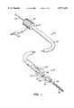
- FIG. 1is a perspective view of a first embodiment of a lumen occlusion device constructed in accordance with the principles of the present invention.
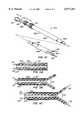
- FIG. 2is a detailed view of a distal end of the device of FIG. 1, shown in section with a pair of opposed vessel-closing elements shown in their collapsed (non-deployed) configuration.
- FIG. 3is a view similar to FIG. 2, except that the opposed vessel-closing elements are shown in their spread-apart (deployed) configuration.
- FIGS. 4A-4Dillustrate the use of the device of FIG. 1 and a method for occluding a blood vessel according to the principles of the present invention.
- FIG. 5is a perspective view of a second embodiment of a lumen occlusion device constructed in accordance with the principles of the present invention.
- FIG. 6is a detailed, cross-sectional view taken along line 6--6 of FIG. 5, shown with a pair of vessel-engaging electrodes in a retracted configuration (solid line) and a deployed configuration (broken line).
- FIG. 7is a detailed, cross-sectional view taken along line 7--7 of FIG. 5, shown with a pair of vessel-spreading elements in a non-deployed configuration (solid line) and a deployed configuration (broken line).
- FIG. 8is a detailed view of the proximal end of the device of FIG. 5, shown in section.
- FIGS. 9A-9Cillustrate use of the device of FIG. 5 in a method for occluding a blood vessel according to the principles of the present invention.
- FIG. 10is a perspective view of a third embodiment of a lumen occlusion device constructed in accordance with the principles of the present invention.
- FIGS. 11A-11Care detailed views of the distal end of the device of FIG. 10, shown with a reciprocatable jaw assembly in a retracted configuration (FIG. 11A), an open configuration (FIG. 11B), and an open configuration with penetrating elements extended (FIG. 11C).
- FIGS. 12A-12Eillustrate use of the device of FIG. 10 in a method for occluding a blood vessel according to the principles of the present invention.
- FIGS. 13A-13Dillustrate a fourth embodiment of a lumen occlusion device constructed in accordance with the principles of the present invention, and use of that device in a method for occluding a blood vessel according to the principles of the present invention.
- the methods and devices of the present inventionwill be useful for selectively occluding virtually any body lumen having a luminal wall that can be mechanically closed followed by the application of energy or other conditions which to injure the vessel to cause permanent closure and luminal occlusion. While the present invention will find its greatest use in the selective occlusion of blood vessels, including both arteries and veins, it will also find use with other body lumens, such as the fallopian tubes, ureter, bile duct, and the like.
- the lumenwill be mechanically closed to bring opposed portions of the endothelial wall of the blood vessel partially or totally together, and energy applied to the occlusion region between said opposed wall portions.
- the energywill injure or destroy the endothelial cells and underlying tissue in the occlusion region, thus initiating a process of thrombosis and fibrosis which will result in relatively rapid vessel occlusion.
- the vessel closurewill be substantially complete within a very short time, typically 10 minutes or less, usually 5 minutes or less, and often 1 minute or less.
- a particular advantage of the present inventionis that by mechanically closing the vessel to occlude the vessel lumen, blood flow is substantially slowed or stopped, greatly enhancing the rate of thermal transfer, which in turn enhances the rate of fibrosis and thrombosis. Even in the case where a small residual lumen remains after the device is removed, complete closure of the remaining area of the lumen will occur relatively rapidly.
- Initial mechanical (non-permanent) occlusion of the blood vesselis also an advantage since it permits confirmation that the site of occlusion is proper. That is, the physician can observe the effects of vessel occlusion (e.g. by radiography) prior to permanent occlusion.
- Closure of the opposed wall portions in the occlusion regionwill usually be effected by the application of energy, such as heat energy, laser energy, electrical energy, or the like.
- the energy sourcewill be radiofrequency electrical energy, such as that supplied by conventional electrosurgical power supplies, such as those available from commercial vendors, including Valleylab, Aspen., Bovie, and Birtcher.
- the power supplywill usually provide energy at frequencies from 200 kHz to 1.25 MHz, and may employ a conventional sinusoidal or non-sinusoidal wave form.
- the current providedwill usually be in the range from 50 mA to 1 A, with the actual current depending primarily on vessel size, i.e. larger vessels will usually require higher currents.
- the RF currentmay be applied in a monopolar or a bipolar fashion in or near the occlusion region.
- monopolarit is meant that current flow will pass between (1) one or more "active" electrodes on the probe which have areas and configurations which concentrate the energy flux in order to have an injurious effect on the surrounding tissue and (2) a “dispersive” electrode which is located remotely from the active electrode(s) and which has a sufficiently large area so that the current density is low and non-injurious to surrounding tissue.
- the dispersive electrodemay be on the same probe as the active electrode, and in other cases, the dispersive electrode may be attached externally to the patient, e.g., using a contact pad placed on the patient's flank.
- Bipolar deviceswill generally employ a pair of electrodes in close proximity each having an area and geometry selected to increase current density sufficient to injure or have other desired physiologic effect on adjacent tissue.
- one or more electrodeswill be connected to each pole of the radiofrequency power supply.
- the current flow in the occlusion regionwill be concentrated through tissue located between electrode pair(s), rather than from one or more electrodes to a remote, dispersive electrode (which is the case in monopolar operation).
- Devices according to the present inventionwill comprise a probe, typically including a shaft having a proximal end and a distal end.
- the shaftmay be in the form of a conventional catheter body, typically having a length in the range from 40 cm to 200 cm, usually from 75 cm to 120 cm.
- the catheter bodywill usually include means for introducing the body over a movable guide wire, typically having a guide wire lumen running through at least a distal portion of the catheter body.
- the catheter bodycan have either conventional "over-the-wire” design where a movable guide wire is received through the entire length of the catheter body or may have a “rapid exchange” or “monorail” design where the guide wire is received through a lumen which extends only over a distal length of the body, typically from 5 cm to 25 cm.
- the catheter bodywill have an outside diameter consistent with its intended use, typically being from 1 mm to 5 mm, usually from 2 mm to 4 mm.
- the catheter bodymay be formed from a variety of conventional catheter materials, including natural and synthetic polymers, such as polyvinyl chloride, polyurethanes, polyesters, polyethylenes, polytetrafluoroethylenes (PTFE's), nylons, and the like.
- the catheter bodiesmay optionally be reinforced to enhance their strength, torqueability, and the like.
- Exemplary reinforcement layersinclude metal fiber braids, polymeric fiber braids, metal or fiber helical windings, and the like.
- a portion of the catheter bodycould be formed from a metal rod or hypo tube, particularly when the catheter body is a rapid exchange or monorail design.
- the lumen occlusion devicewill also include at least one electrode for initiating radiofrequency current flow, as described above.
- the electrodemay be disposed on the shaft, may be part of the vessel closing means (described below), and/or may be associated with the guide wire used to introduce the shaft to the body lumen, usually a blood vessel.
- Configuration of the electrode elementwill vary depending on whether it is intended to be an "active" electrode or a "dispersive" electrode. Active electrodes will typically have relatively small areas, typically being below about 20 mm 2 , usually being below about 10 mm 2 .
- Dispersive electrodeswill typically have a somewhat larger area, typically being greater than 50 mm 2 for probe-mounted dispersive electrodes and greater than 100 cm 2 for external dispersive pads.
- the vessel-closing mechanism on the lumen occlusion devicemay take any form which mechanically flattens or otherwise pinches or occludes the vessel while the occlusion energy is being applied.
- the vessel-closing mechanismwill comprise a pair of thin, opposed elements which may be spread apart to flatten the vessel by radially moving opposed wall portions apart.
- the vessel-closing mechanismcan comprise spreadable jaws having penetrating elements which engage and close the vessel wall as the jaws are closed.
- the vessel closing mechanismcan employ negative pressure for collapsing the vessel wall. Specific examples of each of these approaches are described in more detail in connection with the figures below.
- a first lumen occlusion device 10constructed in accordance with the principles of the present invention comprises a shaft in the form of a flexible catheter body 12 having a proximal end 14 and a distal end 16.
- a reciprocatable tip structure 18has a reduced neck portion 20 which is reciprocatably received in a sleeve 22 formed on the body 12.
- a pair of opposed elements 24are fixedly secured at the distal end of the catheter body 12 extending between the main portion of the body and the tip structure 18.
- the opposed elements 24By proximally translating the tip section 18 relative to the main portion of body 12, as illustrated in FIG. 3, the opposed elements 24 (which will be formed from a resilient material such as spring steel, superelastic alloy or plastic, or a resilient organic polymer) will bow in a radially outward direction.
- the lumen occlusion device 10further comprises a guide wire 30 having a tip 32 with an enlarged proximal end 34, as best seen in FIG. 2.
- the guide wire 34may be proximally translated relative to the catheter body 12 (i.e., by pulling on the proximal end of the guide wire) in order to proximally translate tip structure 18 as illustrated in FIG. 3.
- guide wire 30also serves as an active electrode for providing radiofrequency current flow in a monopolar procedure.
- the occlusion device 10further includes a dispersive electrode 36 which may be conveniently mounted over the sleeve 22 (as illustrated) or alternatively at a proximal location on the catheter body 12.
- the dispersive electrode 36could be located elsewhere on the catheter body, or it could be mounted separately from the catheter on the patient, e.g., by using an external dispersive plate which is mounted on the patient's skin.
- the dispersive electrode 36 and guide wire 30may be connected to a suitable RF power supply (not shown) using a connector plug 38. Connection to the guide wire 30 can be completed using a bushing within the proximal housing 39, thus allowing relative rotation and translation.
- FIGS. 4A-4DUse of the lumen occlusion device 10 is illustrated in FIGS. 4A-4D.
- the device 10is introduced transluminally to a desired target site TS within a blood vessel BV or other body lumen.
- the guide wire 30will first be introduced to the target site TS in a conventional manner.
- the catheter body 12will be introduced over the guide wire 30 in a conventional "over-the-wire" manner until the distal end of the device 10 reaches the target site, as shown in FIG. 4A.
- the guide wire 30is pulled proximally (i.e., to the left, as illustrated in FIG. 4B) in order to proximally translate the tip structure 18 relative to the remainder of the catheter body 12.
- proximally translation of the tip structure 18causes opposed members 24 to move radially outward and collapse the blood vessel, as illustrated in FIGS. 4B and 4D.
- a radiofrequency current flowcan be initiated between the guide wire tip 32 and the dispersive electrode 36, typically using a conventional radiofrequency power supply which is optionally modified to provide and optimum impedance match.
- the device 10will be withdrawn.
- the blood vesselwill be highly thrombosed and totally or mostly occluded.
- a small lumenmay remain in the region where guide wire tip 32 had been deployed. Any such remaining lumen, however, will quickly occlude by normal inflammatory and clotting processes, thus assuring the closure of the blood vessel BV at the target site TS, as shown in FIG. 4C.
- the thrombosed regionwill fibrose to form permanent sealing of the blood vessel or other body lumen.
- FIGS. 5-8A second embodiment 50 of the lumen occlusion device of the present invention is illustrated in FIGS. 5-8.
- the device 50is similar to device 10 in that it includes the pair of spreadable, opposed elements for mechanically collapsing the blood vessel lumen.
- the device 50differs from device 10, however, in that it includes a pair of deployable bipolar pins or electrodes, as will now be described in more detail.
- the lumen occlusion device 50includes a flexible catheter body 52 having a proximal end 54 and a distal end 56.
- the flexible catheterincludes both an outer sheath 58 and an inner member 60, where the outer sheath and inner member are axially reciprocatable relative to one another.
- the outer sheath 58includes a first pair of opposed slots 62 (best seen in FIG. 7) and a second pair of opposed slots 64 (best seen in FIG. 6).
- the first slots 62are aligned with opposed members 66 which are attached between distal end 68 of the inner member 60 and the distal end of the outer sheath 58, as best observed in FIG. 7.
- the opposed elements 60are similar to the opposed elements 24 of device 10, and distal translation of the inner member 60 relative to the outer sheath 58 will cause the members 66 to bow in a radially outward direction, as shown in broken line in FIG. 7.
- a pair of penetrating electrode members 70are also attached to the distal end of the inner member 60, and are further aligned in the opposed slots 64, as best observed in FIG. 6. As the inner member 60 is distally translated, the electrodes 70 will pass outwardly through the slots 64 and forwardly of the distal end of the outer sheath 58, as shown in broken line in FIG. 6.
- Structure for axially translating an inner member 60 relative to outer sheath 58is located at the proximal end 54 of the device 50 and is illustrated in FIG. 8.
- a flared proximal hub 72is attached and hermetically sealed to the proximal end of outer sheath 58 and includes a circumferential detent 74.
- a threaded region 76is provided at the proximal end of the inner member 60, and sealing between the inner member 60 and outer sheath 58 is provided by an o-ring 78.
- the inner member 60can be axially reciprocated relative to the outer sheath 58.
- Control of the reciprocationis provided by a collar 80 which has a flange 82 received in detent 74 and a threaded follower 84 secured over the threaded region 76.
- a collar 80which has a flange 82 received in detent 74 and a threaded follower 84 secured over the threaded region 76.
- An electrical connector 86is provided on a cap 88 at the proximal end of the inner member 60. The connector 86 permits connection of the electrodes 70 to a conventional electrosurgical power supply.
- FIGS. 9A-9CUse of the lumen occlusion device 50 is illustrated in FIGS. 9A-9C.
- the device 50is introduced to a target site TS in a blood vessel BV over a guide wire GW in a conventional manner, as illustrated in FIG. 9A.
- the guide wireis optionally withdrawn, and the inner member 60 distally translated relative to the outer sheath 58, as illustrated in FIG. 9B.
- the vessel lumenis mechanically collapsed (flattened to pinch the lumen) by expansion of the opposed members 66, and the penetrating electrodes 70 pass subsequently through the epithelial wall of the blood vessel, as illustrated in FIG. 9B.
- the occlusion region between the collapsed vessel wallscan be occluded, as illustrated in FIG. 9C.
- FIGS. 10 and 11A-11CA third embodiment 100 of the occlusion device of the present invention is illustrated in FIGS. 10 and 11A-11C.
- the device 100comprises a flexible catheter body 102 including an inner member 104 and outer sheath 106.
- the inner member 104 and outer sheath 106are axially reciprocatable relative to one another, and include finger grips 108 and 110, respectively.
- a pair of penetrating electrodes 112are reciprocatably mounted in the inner member 104 and attached to connector assembly 114 at their proximal ends.
- the distal end of the lumen occlusion deviceis best shown in FIGS. 11A-11C.
- the inner member 104is axially split along a line 116 and is treated or spring-loaded to have mechanical "memory" so that it will spread apart when advanced distally relative to the outer sheath 106, as shown in FIG. 11B.
- the distal end of the inner member 104forms a selectively reciprocating "jaw" mechanism, where the jaws open and close by axially sliding the inner member within the outer sheath 106.
- a central lumen 120is provided in the inner member 104 to permit the device 100 to be introduced over a guide wire in a conventional manner.
- the penetrating electrode members 112may be axially advanced by pushing connector 14 in the distal direction, as illustrated in FIG. 11C.
- the device 100is introduced over a guide wire GW so that its distal end lies at the target site, as illustrated in FIG. 12A.
- the inner member 104is then reciprocated distally relative to the outer sheath, opening opposed jaws 113, as illustrated in FIG. 12B.
- the penetrating distal ends of electrodes 112are then advanced into opposed sections of the blood vessel wall, as illustrated in FIG. 12C.
- the jaw members 113are then closed by at least partially advancing the outer sheath 106 over the inner member 103, as shown in FIG. 12D, to pinch the vessel closed between the electrodes 112.
- the vesselcan then be occluded by passing bipolar radiofrequency current through the collapsed region of the blood vessel, resulting in the occlusion shown at the target site TS in FIG. 12E.
- FIGS. 13A-13DA fourth embodiment 150 of the lumen occlusion device of the present invention is illustrated in FIGS. 13A-13D.
- the lumen occlusion device 150is particularly useful for the occlusion of veins V upstream of a valve VL, as shown in FIG. 13A.
- the device 150includes an inflatable balloon 152 which is shown in its non-inflated condition in FIG. 13A.
- the devicefurther includes an electrode 154 which is located distally of the occlusion balloon 152.
- a negative pressurecan then be drawn through the catheter body, causing a collapse of the vein in region TS, as illustrated in FIG. 13C.
- Monopolar RF energycan then be delivered through the electrode 154 to occlude the vein, as illustrated in FIG. 13D.
- the device 150may be used to deliver bipolar RF current by using a guide wire (or other thin wire electrode) extending from the distal tip of the device. Use of the guide wire will cause clotting distally of the device tip, where such clot is less likely to be disrupted by withdrawal of the catheter.
Landscapes
- Health & Medical Sciences (AREA)
- Surgery (AREA)
- Life Sciences & Earth Sciences (AREA)
- Engineering & Computer Science (AREA)
- Heart & Thoracic Surgery (AREA)
- Medical Informatics (AREA)
- Otolaryngology (AREA)
- Plasma & Fusion (AREA)
- Physics & Mathematics (AREA)
- Biomedical Technology (AREA)
- Cardiology (AREA)
- Nuclear Medicine, Radiotherapy & Molecular Imaging (AREA)
- Molecular Biology (AREA)
- Animal Behavior & Ethology (AREA)
- General Health & Medical Sciences (AREA)
- Public Health (AREA)
- Veterinary Medicine (AREA)
- Surgical Instruments (AREA)
- Media Introduction/Drainage Providing Device (AREA)
- Filling Or Discharging Of Gas Storage Vessels (AREA)
Abstract
Description
Claims (12)
Priority Applications (1)
| Application Number | Priority Date | Filing Date | Title |
|---|---|---|---|
| US09/001,968US6077261A (en) | 1995-06-07 | 1997-12-31 | Device for permanent vessel occlusion |
Applications Claiming Priority (2)
| Application Number | Priority Date | Filing Date | Title |
|---|---|---|---|
| US08/488,444US5709224A (en) | 1995-06-07 | 1995-06-07 | Method and device for permanent vessel occlusion |
| US09/001,968US6077261A (en) | 1995-06-07 | 1997-12-31 | Device for permanent vessel occlusion |
Related Parent Applications (1)
| Application Number | Title | Priority Date | Filing Date |
|---|---|---|---|
| US08/488,444ContinuationUS5709224A (en) | 1995-06-07 | 1995-06-07 | Method and device for permanent vessel occlusion |
Publications (1)
| Publication Number | Publication Date |
|---|---|
| US6077261Atrue US6077261A (en) | 2000-06-20 |
Family
ID=23939726
Family Applications (2)
| Application Number | Title | Priority Date | Filing Date |
|---|---|---|---|
| US08/488,444Expired - LifetimeUS5709224A (en) | 1995-06-07 | 1995-06-07 | Method and device for permanent vessel occlusion |
| US09/001,968Expired - LifetimeUS6077261A (en) | 1995-06-07 | 1997-12-31 | Device for permanent vessel occlusion |
Family Applications Before (1)
| Application Number | Title | Priority Date | Filing Date |
|---|---|---|---|
| US08/488,444Expired - LifetimeUS5709224A (en) | 1995-06-07 | 1995-06-07 | Method and device for permanent vessel occlusion |
Country Status (7)
| Country | Link |
|---|---|
| US (2) | US5709224A (en) |
| EP (1) | EP0836432B1 (en) |
| JP (1) | JP3743804B2 (en) |
| AU (1) | AU709895B2 (en) |
| CA (1) | CA2222611C (en) |
| DE (1) | DE69636035T2 (en) |
| WO (1) | WO1996039961A1 (en) |
Cited By (34)
| Publication number | Priority date | Publication date | Assignee | Title |
|---|---|---|---|---|
| US20030125759A1 (en)* | 2001-12-21 | 2003-07-03 | Vnus Medical Technologies, Inc. | Method and apparatus for avulsion of varicose veins |
| US6599299B2 (en) | 2001-06-26 | 2003-07-29 | Leonard S. Schultz | Device and method for body lumen occlusion |
| US6638277B2 (en) | 2000-07-06 | 2003-10-28 | Scimed Life Systems, Inc. | Tumor ablation needle with independently activated and independently traversing tines |
| US20040030335A1 (en)* | 2002-05-14 | 2004-02-12 | University Of Pittsburgh | Device and method of use for functional isolation of animal or human tissues |
| US20040176758A1 (en)* | 2003-03-04 | 2004-09-09 | Cardiva Medical, Inc. | Apparatus and methods for closing vascular penetrations |
| US20040199156A1 (en)* | 2003-04-02 | 2004-10-07 | Rioux Robert F. | Endovenous ablation mechanism with feedback control |
| US20040215132A1 (en)* | 2003-04-22 | 2004-10-28 | Inbae Yoon | Spot coagulating & occluding instrument and method of use |
| US20050208645A1 (en)* | 2001-03-09 | 2005-09-22 | Palermo Gianpiero D | Electrofusion microelectrode |
| US6964274B1 (en) | 2004-06-07 | 2005-11-15 | Ethicon, Inc. | Tubal sterilization device having expanding electrodes and method for performing sterilization using the same |
| US20050273096A1 (en)* | 2004-05-27 | 2005-12-08 | Roop John A | Anchoring introducer sheath with distal slots for catheter delivery and translation |
| US20050273094A1 (en)* | 2004-06-07 | 2005-12-08 | Ryan Thomas P | Tubal sterilization device having sesquipolar electrodes and method for performing sterilization using the same |
| US20060030849A1 (en)* | 2004-08-05 | 2006-02-09 | Vnus Medical Technologies, Inc. | Methods and apparatus for coagulating and/or constricting hollow anatomical structures |
| US20060189979A1 (en)* | 2005-02-23 | 2006-08-24 | Esch Brady D | Methods and apparatus for coagulating and/or constricting hollow anatomical structures |
| US20070106213A1 (en)* | 2005-10-28 | 2007-05-10 | Gianluca Spera | Gastrointestinal applicator and method of using same |
| US20070148757A1 (en)* | 2005-12-22 | 2007-06-28 | Cornell Research Foundation | Electrofusion microelectrode and methods of using it to manipulate cells and/or cellular components |
| US20080125797A1 (en)* | 2006-11-27 | 2008-05-29 | Brian Kelleher | Methods and Devices for Organ Partitioning |
| US20090005776A1 (en)* | 2007-06-25 | 2009-01-01 | Terumo Kabushiki Kaisha | Medical device |
| US20090069810A1 (en)* | 2007-08-28 | 2009-03-12 | Terumo Kabushiki Kaisha | Biological tissue closing device |
| US20090069809A1 (en)* | 2007-08-28 | 2009-03-12 | Terumo Kabushiki Kaisha | Pfo closing device |
| US20090076525A1 (en)* | 2007-08-28 | 2009-03-19 | Terumo Kabushiki Kaisha | Pfo closing device |
| US20090143713A1 (en)* | 2007-11-30 | 2009-06-04 | Jacques Van Dam | Biliary Shunts, Delivery Systems, Methods of Using the Same and Kits Therefor |
| US20090240250A1 (en)* | 2000-07-25 | 2009-09-24 | Endovascular Technologies, Inc. | Apparatus and method for electrically induced thrombosis |
| US20100268217A1 (en)* | 2006-05-24 | 2010-10-21 | Emcision Limited | Vessel sealing device and methods |
| US20110054381A1 (en)* | 2009-05-29 | 2011-03-03 | Jacques Van Dam | Biliary shunts, delivery systems, and methods of using the same |
| US20110166518A1 (en)* | 2010-01-04 | 2011-07-07 | Tyco Healthcare Group, L.P. | Apparatus and methods for treating hollow anatomical structures |
| US8157818B2 (en) | 2005-08-01 | 2012-04-17 | Ension, Inc. | Integrated medical apparatus for non-traumatic grasping, manipulating and closure of tissue |
| US8172839B2 (en) | 2006-02-24 | 2012-05-08 | Terumo Kabushiki Kaisha | PFO closing device |
| US9393023B2 (en) | 2009-01-13 | 2016-07-19 | Atricure, Inc. | Apparatus and methods for deploying a clip to occlude an anatomical structure |
| US9585714B2 (en) | 2006-07-13 | 2017-03-07 | Bovie Medical Corporation | Surgical sealing and cutting apparatus |
| US10182824B2 (en) | 2010-11-11 | 2019-01-22 | Atricure, Inc. | Clip applicator |
| US10433854B2 (en) | 2010-10-27 | 2019-10-08 | Atricure, Inc. | Appendage clamp deployment assist device |
| US11998211B2 (en) | 2013-11-21 | 2024-06-04 | Atricure, Inc. | Occlusion clip |
| US12004752B2 (en) | 2012-11-21 | 2024-06-11 | Atricure, Inc. | Occlusion clip |
| US12070257B2 (en) | 2020-08-14 | 2024-08-27 | Ictero Medical, Inc. | Systems, devices, and methods for ablation and defunctionalization of a gallbladder |
Families Citing this family (366)
| Publication number | Priority date | Publication date | Assignee | Title |
|---|---|---|---|---|
| US6071280A (en) | 1993-11-08 | 2000-06-06 | Rita Medical Systems, Inc. | Multiple electrode ablation apparatus |
| US6405732B1 (en)* | 1994-06-24 | 2002-06-18 | Curon Medical, Inc. | Method to treat gastric reflux via the detection and ablation of gastro-esophageal nerves and receptors |
| US6056744A (en) | 1994-06-24 | 2000-05-02 | Conway Stuart Medical, Inc. | Sphincter treatment apparatus |
| US6009877A (en) | 1994-06-24 | 2000-01-04 | Edwards; Stuart D. | Method for treating a sphincter |
| US6071274A (en)* | 1996-12-19 | 2000-06-06 | Ep Technologies, Inc. | Loop structures for supporting multiple electrode elements |
| US7175619B2 (en)* | 1994-10-07 | 2007-02-13 | Boston Scientific Scimed, Inc. | Loop structures for positioning a diagnostic or therapeutic element on the epicardium or other organ surface |
| US6464700B1 (en) | 1994-10-07 | 2002-10-15 | Scimed Life Systems, Inc. | Loop structures for positioning a diagnostic or therapeutic element on the epicardium or other organ surface |
| AU733341B2 (en) | 1996-02-02 | 2001-05-10 | Transvascular, Inc. | A device, system and method for interstitial transvascular intervention |
| US6139527A (en)* | 1996-03-05 | 2000-10-31 | Vnus Medical Technologies, Inc. | Method and apparatus for treating hemorrhoids |
| US6033397A (en)* | 1996-03-05 | 2000-03-07 | Vnus Medical Technologies, Inc. | Method and apparatus for treating esophageal varices |
| US6152899A (en) | 1996-03-05 | 2000-11-28 | Vnus Medical Technologies, Inc. | Expandable catheter having improved electrode design, and method for applying energy |
| US6036687A (en)* | 1996-03-05 | 2000-03-14 | Vnus Medical Technologies, Inc. | Method and apparatus for treating venous insufficiency |
| WO1997032532A1 (en)* | 1996-03-05 | 1997-09-12 | Vnus Medical Technologies, Inc. | Vascular catheter-based system for heating tissue |
| US6419673B1 (en)* | 1996-05-06 | 2002-07-16 | Stuart Edwards | Ablation of rectal and other internal body structures |
| WO1998007375A1 (en)* | 1996-08-22 | 1998-02-26 | The Trustees Of Columbia University | Endovascular flexible stapling device |
| US8353908B2 (en) | 1996-09-20 | 2013-01-15 | Novasys Medical, Inc. | Treatment of tissue in sphincters, sinuses, and orifices |
| US6464697B1 (en)* | 1998-02-19 | 2002-10-15 | Curon Medical, Inc. | Stomach and adjoining tissue regions in the esophagus |
| US6091995A (en)* | 1996-11-08 | 2000-07-18 | Surx, Inc. | Devices, methods, and systems for shrinking tissues |
| US6203525B1 (en) | 1996-12-19 | 2001-03-20 | Ep Technologies, Inc. | Catheterdistal assembly with pull wires |
| US6048329A (en)* | 1996-12-19 | 2000-04-11 | Ep Technologies, Inc. | Catheter distal assembly with pull wires |
| US6071279A (en) | 1996-12-19 | 2000-06-06 | Ep Technologies, Inc. | Branched structures for supporting multiple electrode elements |
| US6332880B1 (en) | 1996-12-19 | 2001-12-25 | Ep Technologies, Inc. | Loop structures for supporting multiple electrode elements |
| US6076012A (en) | 1996-12-19 | 2000-06-13 | Ep Technologies, Inc. | Structures for supporting porous electrode elements |
| AU6146798A (en)* | 1997-03-04 | 1998-09-22 | Vnus Medical Technologies, Inc. | Method and apparatus for treating venous insufficiency using directionally applied energy |
| US6231507B1 (en) | 1997-06-02 | 2001-05-15 | Vnus Medical Technologies, Inc. | Pressure tourniquet with ultrasound window and method of use |
| US9023031B2 (en)* | 1997-08-13 | 2015-05-05 | Verathon Inc. | Noninvasive devices, methods, and systems for modifying tissues |
| US6401719B1 (en)* | 1997-09-11 | 2002-06-11 | Vnus Medical Technologies, Inc. | Method of ligating hollow anatomical structures |
| US6179832B1 (en)* | 1997-09-11 | 2001-01-30 | Vnus Medical Technologies, Inc. | Expandable catheter having two sets of electrodes |
| US6200312B1 (en)* | 1997-09-11 | 2001-03-13 | Vnus Medical Technologies, Inc. | Expandable vein ligator catheter having multiple electrode leads |
| US6258084B1 (en) | 1997-09-11 | 2001-07-10 | Vnus Medical Technologies, Inc. | Method for applying energy to biological tissue including the use of tumescent tissue compression |
| US6610055B1 (en) | 1997-10-10 | 2003-08-26 | Scimed Life Systems, Inc. | Surgical method for positioning a diagnostic or therapeutic element on the epicardium or other organ surface |
| US6014589A (en)* | 1997-11-12 | 2000-01-11 | Vnus Medical Technologies, Inc. | Catheter having expandable electrodes and adjustable stent |
| US6917834B2 (en) | 1997-12-03 | 2005-07-12 | Boston Scientific Scimed, Inc. | Devices and methods for creating lesions in endocardial and surrounding tissue to isolate focal arrhythmia substrates |
| WO1999035986A1 (en) | 1998-01-14 | 1999-07-22 | Conway-Stuart Medical, Inc. | Electrosurgical apparatus for treating gastroesophageal reflux disease (gerd) and method |
| AU2114299A (en)* | 1998-01-14 | 1999-08-02 | Conway-Stuart Medical, Inc. | Electrosurgical device for sphincter treatment |
| AU2317899A (en)* | 1998-01-14 | 1999-08-02 | Conway-Stuart Medical, Inc. | Gerd treatment apparatus and method |
| US6440128B1 (en) | 1998-01-14 | 2002-08-27 | Curon Medical, Inc. | Actively cooled electrode assemblies for forming lesions to treat dysfunction in sphincters and adjoining tissue regions |
| EP1685808B1 (en) | 1998-01-30 | 2016-09-14 | St.Jude Medical ATG, Inc. | Device for use in closing septal defects and an installation assembly for such device |
| US8906010B2 (en)* | 1998-02-19 | 2014-12-09 | Mederi Therapeutics, Inc. | Graphical user interface for association with an electrode structure deployed in contact with a tissue region |
| US6258087B1 (en) | 1998-02-19 | 2001-07-10 | Curon Medical, Inc. | Expandable electrode assemblies for forming lesions to treat dysfunction in sphincters and adjoining tissue regions |
| US6423058B1 (en) | 1998-02-19 | 2002-07-23 | Curon Medical, Inc. | Assemblies to visualize and treat sphincters and adjoining tissue regions |
| WO1999042044A1 (en) | 1998-02-19 | 1999-08-26 | Conway-Stuart Medical, Inc. | Electrosurgical sphincter treatment apparatus |
| US6358245B1 (en) | 1998-02-19 | 2002-03-19 | Curon Medical, Inc. | Graphical user interface for association with an electrode structure deployed in contact with a tissue region |
| US6355031B1 (en) | 1998-02-19 | 2002-03-12 | Curon Medical, Inc. | Control systems for multiple electrode arrays to create lesions in tissue regions at or near a sphincter |
| US7165551B2 (en) | 1998-02-19 | 2007-01-23 | Curon Medical, Inc. | Apparatus to detect and treat aberrant myoelectric activity |
| US6402744B2 (en) | 1998-02-19 | 2002-06-11 | Curon Medical, Inc. | Systems and methods for forming composite lesions to treat dysfunction in sphincters and adjoining tissue regions |
| US6325798B1 (en)* | 1998-02-19 | 2001-12-04 | Curon Medical, Inc. | Vacuum-assisted systems and methods for treating sphincters and adjoining tissue regions |
| US6273886B1 (en) | 1998-02-19 | 2001-08-14 | Curon Medical, Inc. | Integrated tissue heating and cooling apparatus |
| US6790207B2 (en) | 1998-06-04 | 2004-09-14 | Curon Medical, Inc. | Systems and methods for applying a selected treatment agent into contact with tissue to treat disorders of the gastrointestinal tract |
| US7329254B2 (en)* | 1998-02-19 | 2008-02-12 | Curon Medical, Inc. | Systems and methods for treating dysfunctions in the intestines and rectum that adapt to the anatomic form and structure of different individuals |
| US6645201B1 (en)* | 1998-02-19 | 2003-11-11 | Curon Medical, Inc. | Systems and methods for treating dysfunctions in the intestines and rectum |
| US20100114087A1 (en)* | 1998-02-19 | 2010-05-06 | Edwards Stuart D | Methods and devices for treating urinary incontinence |
| EP1056405A1 (en) | 1998-02-27 | 2000-12-06 | Curon Medical, Inc. | Apparatus to electrosurgically treat esophageal sphincters |
| US20030135206A1 (en) | 1998-02-27 | 2003-07-17 | Curon Medical, Inc. | Method for treating a sphincter |
| JP2002505138A (en)* | 1998-03-06 | 2002-02-19 | キューロン メディカル,インコーポレイテッド | Instrument for electrosurgically treating the esophageal sphincter |
| AU3672299A (en) | 1998-04-30 | 1999-11-16 | Stuart D Edwards | Electrosurgical sphincter treatment apparatus |
| US6514252B2 (en) | 1998-05-01 | 2003-02-04 | Perfect Surgical Techniques, Inc. | Bipolar surgical instruments having focused electrical fields |
| US6030384A (en)* | 1998-05-01 | 2000-02-29 | Nezhat; Camran | Bipolar surgical instruments having focused electrical fields |
| US20110071468A1 (en)* | 1998-06-04 | 2011-03-24 | Mederi Therapeutics, Inc. | Systems and methods for applying a selected treatment agent into contact with tissue to treat sphincter dysfunction |
| US6802841B2 (en)* | 1998-06-04 | 2004-10-12 | Curon Medical, Inc. | Systems and methods for applying a selected treatment agent into contact with tissue to treat sphincter dysfunction |
| US6322559B1 (en) | 1998-07-06 | 2001-11-27 | Vnus Medical Technologies, Inc. | Electrode catheter having coil structure |
| US6212433B1 (en) | 1998-07-28 | 2001-04-03 | Radiotherapeutics Corporation | Method for treating tumors near the surface of an organ |
| US6889089B2 (en) | 1998-07-28 | 2005-05-03 | Scimed Life Systems, Inc. | Apparatus and method for treating tumors near the surface of an organ |
| US6024742A (en)* | 1998-08-22 | 2000-02-15 | Tu; Lily Chen | Ablation apparatus for treating hemorrhoids |
| US6152144A (en)* | 1998-11-06 | 2000-11-28 | Appriva Medical, Inc. | Method and device for left atrial appendage occlusion |
| US7044134B2 (en)* | 1999-11-08 | 2006-05-16 | Ev3 Sunnyvale, Inc | Method of implanting a device in the left atrial appendage |
| US7713282B2 (en) | 1998-11-06 | 2010-05-11 | Atritech, Inc. | Detachable atrial appendage occlusion balloon |
| US7128073B1 (en)* | 1998-11-06 | 2006-10-31 | Ev3 Endovascular, Inc. | Method and device for left atrial appendage occlusion |
| US6254601B1 (en)* | 1998-12-08 | 2001-07-03 | Hysterx, Inc. | Methods for occlusion of the uterine arteries |
| US6217528B1 (en) | 1999-02-11 | 2001-04-17 | Scimed Life Systems, Inc. | Loop structure having improved tissue contact capability |
| US6210409B1 (en)* | 1999-05-03 | 2001-04-03 | Alan G. Ellman | Electrosurgical handpiece for treating tissue |
| WO2000066017A1 (en) | 1999-05-04 | 2000-11-09 | Curon Medical, Inc. | Electrodes for creating lesions in tissue regions at or near a sphincter |
| US8597290B2 (en) | 1999-07-14 | 2013-12-03 | Mederi Therapeutics | Method for treating fecal incontinence |
| WO2001017452A1 (en)* | 1999-09-08 | 2001-03-15 | Curon Medical, Inc. | System for controlling a family of treatment devices |
| AU7352500A (en) | 1999-09-08 | 2001-04-10 | Curon Medical, Inc. | Systems and methods for monitoring and controlling use of medical devices |
| CA2388376A1 (en) | 1999-09-08 | 2001-03-15 | Curon Medical, Inc. | Systems and methods for monitoring and controlling use of medical devices |
| US6231561B1 (en)* | 1999-09-20 | 2001-05-15 | Appriva Medical, Inc. | Method and apparatus for closing a body lumen |
| US6287304B1 (en) | 1999-10-15 | 2001-09-11 | Neothermia Corporation | Interstitial cauterization of tissue volumes with electrosurgically deployed electrodes |
| US6689150B1 (en) | 1999-10-27 | 2004-02-10 | Atritech, Inc. | Filter apparatus for ostium of left atrial appendage |
| US6652555B1 (en)* | 1999-10-27 | 2003-11-25 | Atritech, Inc. | Barrier device for covering the ostium of left atrial appendage |
| US6551303B1 (en)* | 1999-10-27 | 2003-04-22 | Atritech, Inc. | Barrier device for ostium of left atrial appendage |
| US6994092B2 (en)* | 1999-11-08 | 2006-02-07 | Ev3 Sunnyvale, Inc. | Device for containing embolic material in the LAA having a plurality of tissue retention structures |
| US20060095032A1 (en)* | 1999-11-16 | 2006-05-04 | Jerome Jackson | Methods and systems for determining physiologic characteristics for treatment of the esophagus |
| US20040215235A1 (en) | 1999-11-16 | 2004-10-28 | Barrx, Inc. | Methods and systems for determining physiologic characteristics for treatment of the esophagus |
| WO2001035846A1 (en)* | 1999-11-16 | 2001-05-25 | Ganz Robert A | System and method of treating abnormal tissue in the human esophagus |
| US6613046B1 (en) | 1999-11-22 | 2003-09-02 | Scimed Life Systems, Inc. | Loop structures for supporting diagnostic and therapeutic elements in contact with body tissue |
| US6529756B1 (en) | 1999-11-22 | 2003-03-04 | Scimed Life Systems, Inc. | Apparatus for mapping and coagulating soft tissue in or around body orifices |
| US6542781B1 (en) | 1999-11-22 | 2003-04-01 | Scimed Life Systems, Inc. | Loop structures for supporting diagnostic and therapeutic elements in contact with body tissue |
| US6645199B1 (en)* | 1999-11-22 | 2003-11-11 | Scimed Life Systems, Inc. | Loop structures for supporting diagnostic and therapeutic elements contact with body tissue and expandable push devices for use with same |
| US6547776B1 (en) | 2000-01-03 | 2003-04-15 | Curon Medical, Inc. | Systems and methods for treating tissue in the crura |
| US6461364B1 (en)* | 2000-01-05 | 2002-10-08 | Integrated Vascular Systems, Inc. | Vascular sheath with bioabsorbable puncture site closure apparatus and methods of use |
| US9579091B2 (en)* | 2000-01-05 | 2017-02-28 | Integrated Vascular Systems, Inc. | Closure system and methods of use |
| US6391048B1 (en)* | 2000-01-05 | 2002-05-21 | Integrated Vascular Systems, Inc. | Integrated vascular device with puncture site closure component and sealant and methods of use |
| US8758400B2 (en) | 2000-01-05 | 2014-06-24 | Integrated Vascular Systems, Inc. | Closure system and methods of use |
| US6402745B1 (en)* | 2000-02-23 | 2002-06-11 | Peter J. Wilk | Intravenous whip electrode for vein ablation |
| US8845632B2 (en) | 2000-05-18 | 2014-09-30 | Mederi Therapeutics, Inc. | Graphical user interface for monitoring and controlling use of medical devices |
| EP1309289A2 (en)* | 2000-08-18 | 2003-05-14 | Atritech, Inc. | Expandable implant devices for filtering blood flow from atrial appendages |
| DE10042493A1 (en) | 2000-08-30 | 2002-03-14 | Ethicon Endo Surgery Europe | System for treating varicose veins |
| DE60144328D1 (en)* | 2000-09-08 | 2011-05-12 | Abbott Vascular Inc | Surgical clamp |
| EP1318766A2 (en) | 2000-09-21 | 2003-06-18 | Atritech, Inc. | Apparatus for implanting devices in atrial appendages |
| US7306591B2 (en) | 2000-10-02 | 2007-12-11 | Novasys Medical, Inc. | Apparatus and methods for treating female urinary incontinence |
| US6626918B1 (en)* | 2000-10-06 | 2003-09-30 | Medical Technology Group | Apparatus and methods for positioning a vascular sheath |
| US6916306B1 (en)* | 2000-11-10 | 2005-07-12 | Boston Scientific Scimed, Inc. | Steerable loop structures for supporting diagnostic and therapeutic elements in contact with body tissue |
| US6896682B1 (en)* | 2000-11-14 | 2005-05-24 | Biomedical Engineering Solutions, Inc. | Method and system for internal ligation of tubular structures |
| US20040087936A1 (en)* | 2000-11-16 | 2004-05-06 | Barrx, Inc. | System and method for treating abnormal tissue in an organ having a layered tissue structure |
| US7785323B2 (en) | 2000-12-04 | 2010-08-31 | Boston Scientific Scimed, Inc. | Loop structure including inflatable therapeutic device |
| US8690910B2 (en) | 2000-12-07 | 2014-04-08 | Integrated Vascular Systems, Inc. | Closure device and methods for making and using them |
| US7905900B2 (en) | 2003-01-30 | 2011-03-15 | Integrated Vascular Systems, Inc. | Clip applier and methods of use |
| US6695867B2 (en) | 2002-02-21 | 2004-02-24 | Integrated Vascular Systems, Inc. | Plunger apparatus and methods for delivering a closure device |
| US7211101B2 (en) | 2000-12-07 | 2007-05-01 | Abbott Vascular Devices | Methods for manufacturing a clip and clip |
| US6623510B2 (en) | 2000-12-07 | 2003-09-23 | Integrated Vascular Systems, Inc. | Closure device and methods for making and using them |
| US6464702B2 (en) | 2001-01-24 | 2002-10-15 | Ethicon, Inc. | Electrosurgical instrument with closing tube for conducting RF energy and moving jaws |
| US6458128B1 (en) | 2001-01-24 | 2002-10-01 | Ethicon, Inc. | Electrosurgical instrument with a longitudinal element for conducting RF energy and moving a cutting element |
| US6620161B2 (en) | 2001-01-24 | 2003-09-16 | Ethicon, Inc. | Electrosurgical instrument with an operational sequencing element |
| US6652521B2 (en) | 2001-01-24 | 2003-11-25 | Ethicon, Inc. | Surgical instrument with a bi-directional cutting element |
| US6554829B2 (en) | 2001-01-24 | 2003-04-29 | Ethicon, Inc. | Electrosurgical instrument with minimally invasive jaws |
| US6443970B1 (en) | 2001-01-24 | 2002-09-03 | Ethicon, Inc. | Surgical instrument with a dissecting tip |
| IL157732A0 (en)* | 2001-03-08 | 2004-03-28 | Atritech Inc | Atrial filter implants |
| US20030069502A1 (en)* | 2001-05-29 | 2003-04-10 | Makin Inder Raj. S. | Ultrasound feedback in medically-treated patients |
| US7338514B2 (en)* | 2001-06-01 | 2008-03-04 | St. Jude Medical, Cardiology Division, Inc. | Closure devices, related delivery methods and tools, and related methods of use |
| CA2449468A1 (en)* | 2001-06-04 | 2002-12-12 | Albert Einstein Healthcare Network | Cardiac stimulating apparatus having a blood clot filter and atrial pacer |
| IES20010547A2 (en)* | 2001-06-07 | 2002-12-11 | Christy Cummins | Surgical Staple |
| US7011671B2 (en)* | 2001-07-18 | 2006-03-14 | Atritech, Inc. | Cardiac implant device tether system and method |
| EP1420702B1 (en)* | 2001-08-31 | 2005-04-20 | Boston Scientific Limited | Percutaneous pringle occlusion device |
| US20070112358A1 (en)* | 2001-09-06 | 2007-05-17 | Ryan Abbott | Systems and Methods for Treating Septal Defects |
| US20070129755A1 (en)* | 2005-12-05 | 2007-06-07 | Ovalis, Inc. | Clip-based systems and methods for treating septal defects |
| US6776784B2 (en)* | 2001-09-06 | 2004-08-17 | Core Medical, Inc. | Clip apparatus for closing septal defects and methods of use |
| US20090054912A1 (en)* | 2001-09-06 | 2009-02-26 | Heanue Taylor A | Systems and Methods for Treating Septal Defects |
| US20050267495A1 (en)* | 2004-05-17 | 2005-12-01 | Gateway Medical, Inc. | Systems and methods for closing internal tissue defects |
| US20060052821A1 (en)* | 2001-09-06 | 2006-03-09 | Ovalis, Inc. | Systems and methods for treating septal defects |
| US6702835B2 (en) | 2001-09-07 | 2004-03-09 | Core Medical, Inc. | Needle apparatus for closing septal defects and methods for using such apparatus |
| US20030181942A1 (en) | 2002-01-25 | 2003-09-25 | Sutton Gregg S. | Atrial appendage blood filtration systems |
| US6736822B2 (en) | 2002-02-20 | 2004-05-18 | Mcclellan Scott B. | Device and method for internal ligation of tubular structures |
| US6949099B2 (en)* | 2002-04-12 | 2005-09-27 | Olympus Corporation | Incising device for use with an endoscope |
| US7976564B2 (en)* | 2002-05-06 | 2011-07-12 | St. Jude Medical, Cardiology Division, Inc. | PFO closure devices and related methods of use |
| IES20030424A2 (en) | 2002-06-04 | 2003-12-10 | Robert Stevenson | Blood vessel closure clip and delivery device |
| US20040122362A1 (en)* | 2002-09-10 | 2004-06-24 | Houser Russell A. | Pseudo aneurysm repair system |
| US7087064B1 (en)* | 2002-10-15 | 2006-08-08 | Advanced Cardiovascular Systems, Inc. | Apparatuses and methods for heart valve repair |
| US8187324B2 (en)* | 2002-11-15 | 2012-05-29 | Advanced Cardiovascular Systems, Inc. | Telescoping apparatus for delivering and adjusting a medical device in a vessel |
| US7404824B1 (en)* | 2002-11-15 | 2008-07-29 | Advanced Cardiovascular Systems, Inc. | Valve aptation assist device |
| US9149602B2 (en) | 2005-04-22 | 2015-10-06 | Advanced Cardiovascular Systems, Inc. | Dual needle delivery system |
| US7981152B1 (en) | 2004-12-10 | 2011-07-19 | Advanced Cardiovascular Systems, Inc. | Vascular delivery system for accessing and delivering devices into coronary sinus and other vascular sites |
| US8398656B2 (en) | 2003-01-30 | 2013-03-19 | Integrated Vascular Systems, Inc. | Clip applier and methods of use |
| US8821534B2 (en) | 2010-12-06 | 2014-09-02 | Integrated Vascular Systems, Inc. | Clip applier having improved hemostasis and methods of use |
| US8905937B2 (en)* | 2009-02-26 | 2014-12-09 | Integrated Vascular Systems, Inc. | Methods and apparatus for locating a surface of a body lumen |
| US8202293B2 (en) | 2003-01-30 | 2012-06-19 | Integrated Vascular Systems, Inc. | Clip applier and methods of use |
| US8758398B2 (en)* | 2006-09-08 | 2014-06-24 | Integrated Vascular Systems, Inc. | Apparatus and method for delivering a closure element |
| US8021359B2 (en)* | 2003-02-13 | 2011-09-20 | Coaptus Medical Corporation | Transseptal closure of a patent foramen ovale and other cardiac defects |
| US7257450B2 (en)* | 2003-02-13 | 2007-08-14 | Coaptus Medical Corporation | Systems and methods for securing cardiovascular tissue |
| WO2004082532A1 (en)* | 2003-03-17 | 2004-09-30 | Ev3 Sunnyvale, Inc. | Thin film composite lamination |
| US6939348B2 (en)* | 2003-03-27 | 2005-09-06 | Cierra, Inc. | Energy based devices and methods for treatment of patent foramen ovale |
| US7186251B2 (en)* | 2003-03-27 | 2007-03-06 | Cierra, Inc. | Energy based devices and methods for treatment of patent foramen ovale |
| US7972330B2 (en)* | 2003-03-27 | 2011-07-05 | Terumo Kabushiki Kaisha | Methods and apparatus for closing a layered tissue defect |
| US8021362B2 (en)* | 2003-03-27 | 2011-09-20 | Terumo Kabushiki Kaisha | Methods and apparatus for closing a layered tissue defect |
| US7293562B2 (en)* | 2003-03-27 | 2007-11-13 | Cierra, Inc. | Energy based devices and methods for treatment of anatomic tissue defects |
| AU2004226374B2 (en)* | 2003-03-27 | 2009-11-12 | Terumo Kabushiki Kaisha | Methods and apparatus for treatment of patent foramen ovale |
| US7165552B2 (en)* | 2003-03-27 | 2007-01-23 | Cierra, Inc. | Methods and apparatus for treatment of patent foramen ovale |
| US7153298B1 (en)* | 2003-03-28 | 2006-12-26 | Vandolay, Inc. | Vascular occlusion systems and methods |
| US20040267306A1 (en)* | 2003-04-11 | 2004-12-30 | Velocimed, L.L.C. | Closure devices, related delivery methods, and related methods of use |
| US8372112B2 (en)* | 2003-04-11 | 2013-02-12 | St. Jude Medical, Cardiology Division, Inc. | Closure devices, related delivery methods, and related methods of use |
| US7597704B2 (en)* | 2003-04-28 | 2009-10-06 | Atritech, Inc. | Left atrial appendage occlusion device with active expansion |
| US7025768B2 (en)* | 2003-05-06 | 2006-04-11 | Boston Scientific Scimed, Inc. | Systems and methods for ablation of tissue |
| US7311701B2 (en)* | 2003-06-10 | 2007-12-25 | Cierra, Inc. | Methods and apparatus for non-invasively treating atrial fibrillation using high intensity focused ultrasound |
| US7735493B2 (en) | 2003-08-15 | 2010-06-15 | Atritech, Inc. | System and method for delivering a left atrial appendage containment device |
| US7160294B2 (en)* | 2003-09-02 | 2007-01-09 | Curon Medical, Inc. | Systems and methods for treating hemorrhoids |
| US7314479B2 (en)* | 2003-10-31 | 2008-01-01 | Parris Wellman | Space-creating retractor with vessel manipulator |
| US20050096645A1 (en)* | 2003-10-31 | 2005-05-05 | Parris Wellman | Multitool surgical device |
| US20060173474A1 (en)* | 2003-10-31 | 2006-08-03 | Parris Wellman | Surgical device having a track to guide an actuator |
| US20050096646A1 (en)* | 2003-10-31 | 2005-05-05 | Parris Wellman | Surgical system for retracting and severing tissue |
| US20050096670A1 (en)* | 2003-10-31 | 2005-05-05 | Parris Wellman | Surgical end effector |
| US20050096671A1 (en)* | 2003-10-31 | 2005-05-05 | Parris Wellman | Control mechanism for a surgical instrument |
| US20050107779A1 (en)* | 2003-11-19 | 2005-05-19 | Ellman Alan G. | Electrosurgical electrode for treating tissue |
| US7150745B2 (en) | 2004-01-09 | 2006-12-19 | Barrx Medical, Inc. | Devices and methods for treatment of luminal tissue |
| US8801746B1 (en) | 2004-05-04 | 2014-08-12 | Covidien Lp | System and method for delivering a left atrial appendage containment device |
| US20050267520A1 (en)* | 2004-05-12 | 2005-12-01 | Modesitt D B | Access and closure device and method |
| IES20040368A2 (en)* | 2004-05-25 | 2005-11-30 | James E Coleman | Surgical stapler |
| US7367975B2 (en) | 2004-06-21 | 2008-05-06 | Cierra, Inc. | Energy based devices and methods for treatment of anatomic tissue defects |
| US7678133B2 (en)* | 2004-07-10 | 2010-03-16 | Arstasis, Inc. | Biological tissue closure device and method |
| US8388671B2 (en)* | 2004-07-15 | 2013-03-05 | Medtronic Vascular, Inc. | Methods for treatment of aneurysmal tissue |
| US20060068827A1 (en)* | 2004-09-28 | 2006-03-30 | Lucent Technologies, Inc. | Measuring power of a pilot channel of a transmit signal |
| US7041070B2 (en)* | 2004-10-05 | 2006-05-09 | Wen-Hsu Hsieh | Massaging and oscillating device |
| US7473252B2 (en)* | 2004-10-07 | 2009-01-06 | Coaptus Medical Corporation | Systems and methods for shrinking and/or securing cardiovascular tissue |
| US7731712B2 (en)* | 2004-12-20 | 2010-06-08 | Cytyc Corporation | Method and system for transcervical tubal occlusion |
| US9510732B2 (en)* | 2005-10-25 | 2016-12-06 | Intuitive Surgical Operations, Inc. | Methods and apparatus for efficient purging |
| US7860555B2 (en) | 2005-02-02 | 2010-12-28 | Voyage Medical, Inc. | Tissue visualization and manipulation system |
| US8137333B2 (en) | 2005-10-25 | 2012-03-20 | Voyage Medical, Inc. | Delivery of biological compounds to ischemic and/or infarcted tissue |
| US11478152B2 (en) | 2005-02-02 | 2022-10-25 | Intuitive Surgical Operations, Inc. | Electrophysiology mapping and visualization system |
| US7930016B1 (en) | 2005-02-02 | 2011-04-19 | Voyage Medical, Inc. | Tissue closure system |
| US8050746B2 (en)* | 2005-02-02 | 2011-11-01 | Voyage Medical, Inc. | Tissue visualization device and method variations |
| US7918787B2 (en)* | 2005-02-02 | 2011-04-05 | Voyage Medical, Inc. | Tissue visualization and manipulation systems |
| US20080015569A1 (en) | 2005-02-02 | 2008-01-17 | Voyage Medical, Inc. | Methods and apparatus for treatment of atrial fibrillation |
| US7860556B2 (en)* | 2005-02-02 | 2010-12-28 | Voyage Medical, Inc. | Tissue imaging and extraction systems |
| US10064540B2 (en) | 2005-02-02 | 2018-09-04 | Intuitive Surgical Operations, Inc. | Visualization apparatus for transseptal access |
| US8078266B2 (en) | 2005-10-25 | 2011-12-13 | Voyage Medical, Inc. | Flow reduction hood systems |
| US8109274B2 (en)* | 2005-04-11 | 2012-02-07 | Terumo Kabushiki Kaisha | Methods and electrode apparatus to achieve a closure of a layered tissue defect |
| US20060247615A1 (en)* | 2005-04-28 | 2006-11-02 | Boston Scientific Scimed, Inc. | Multi-element bi-polar ablation electrode |
| CN103190942A (en)* | 2005-05-12 | 2013-07-10 | 阿尔斯塔西斯公司 | Access and closure device and method |
| US8926633B2 (en)* | 2005-06-24 | 2015-01-06 | Abbott Laboratories | Apparatus and method for delivering a closure element |
| US8313497B2 (en) | 2005-07-01 | 2012-11-20 | Abbott Laboratories | Clip applier and methods of use |
| US8579936B2 (en)* | 2005-07-05 | 2013-11-12 | ProMed, Inc. | Centering of delivery devices with respect to a septal defect |
| US20070055326A1 (en)* | 2005-07-21 | 2007-03-08 | Farley Brian E | Method of treating a hollow anatomical structure with a thermal catheter |
| US7846179B2 (en)* | 2005-09-01 | 2010-12-07 | Ovalis, Inc. | Suture-based systems and methods for treating septal defects |
| US7972359B2 (en) | 2005-09-16 | 2011-07-05 | Atritech, Inc. | Intracardiac cage and method of delivering same |
| US20070093804A1 (en)* | 2005-10-17 | 2007-04-26 | Coaptus Medical Corporation | Control systems for patient devices, including devices for securing cardiovascular tissue, and associated methods |
| US8221310B2 (en)* | 2005-10-25 | 2012-07-17 | Voyage Medical, Inc. | Tissue visualization device and method variations |
| US7997278B2 (en) | 2005-11-23 | 2011-08-16 | Barrx Medical, Inc. | Precision ablating method |
| US8702694B2 (en) | 2005-11-23 | 2014-04-22 | Covidien Lp | Auto-aligning ablating device and method of use |
| US7959627B2 (en) | 2005-11-23 | 2011-06-14 | Barrx Medical, Inc. | Precision ablating device |
| US20070135826A1 (en) | 2005-12-01 | 2007-06-14 | Steve Zaver | Method and apparatus for delivering an implant without bias to a left atrial appendage |
| US20080243068A1 (en)* | 2005-12-29 | 2008-10-02 | Kamal Ramzipoor | Methods and apparatus for treatment of venous insufficiency |
| US8808310B2 (en)* | 2006-04-20 | 2014-08-19 | Integrated Vascular Systems, Inc. | Resettable clip applier and reset tools |
| US8712529B2 (en) | 2010-03-05 | 2014-04-29 | Endostim, Inc. | Device and implantation system for electrical stimulation of biological systems |
| US9020597B2 (en) | 2008-11-12 | 2015-04-28 | Endostim, Inc. | Device and implantation system for electrical stimulation of biological systems |
| US12343530B2 (en) | 2006-05-18 | 2025-07-01 | Paras Holdings, Llc | Device and implantation system for electrical stimulation of biological systems |
| GB0700560D0 (en)* | 2007-01-11 | 2007-02-21 | Emcision Ltd | Device and method for the treatment of diseased tissue such as tumours |
| GB0700553D0 (en)* | 2007-01-11 | 2007-02-21 | Emcision Ltd | Vessel sealing device |
| US20080017043A1 (en)* | 2006-06-01 | 2008-01-24 | The Coca-Cola Company | Tea Stick Brewing Package and Method |
| US9055906B2 (en)* | 2006-06-14 | 2015-06-16 | Intuitive Surgical Operations, Inc. | In-vivo visualization systems |
| US8556930B2 (en) | 2006-06-28 | 2013-10-15 | Abbott Laboratories | Vessel closure device |
| US20080033241A1 (en)* | 2006-08-01 | 2008-02-07 | Ruey-Feng Peh | Left atrial appendage closure |
| US9220487B2 (en)* | 2006-08-09 | 2015-12-29 | Coherex Medical, Inc. | Devices for reducing the size of an internal tissue opening |
| US8529597B2 (en) | 2006-08-09 | 2013-09-10 | Coherex Medical, Inc. | Devices for reducing the size of an internal tissue opening |
| US8167894B2 (en) | 2006-08-09 | 2012-05-01 | Coherex Medical, Inc. | Methods, systems and devices for reducing the size of an internal tissue opening |
| WO2008028149A2 (en) | 2006-09-01 | 2008-03-06 | Voyage Medical, Inc. | Electrophysiology mapping and visualization system |
| US10004388B2 (en) | 2006-09-01 | 2018-06-26 | Intuitive Surgical Operations, Inc. | Coronary sinus cannulation |
| US20080097476A1 (en)* | 2006-09-01 | 2008-04-24 | Voyage Medical, Inc. | Precision control systems for tissue visualization and manipulation assemblies |
| JP4201037B2 (en)* | 2006-09-14 | 2008-12-24 | ソニー株式会社 | Lens barrel rotation imaging device |
| US9345879B2 (en) | 2006-10-09 | 2016-05-24 | Endostim, Inc. | Device and implantation system for electrical stimulation of biological systems |
| US20150224310A1 (en) | 2006-10-09 | 2015-08-13 | Endostim, Inc. | Device and Implantation System for Electrical Stimulation of Biological Systems |
| US9724510B2 (en) | 2006-10-09 | 2017-08-08 | Endostim, Inc. | System and methods for electrical stimulation of biological systems |
| US11577077B2 (en) | 2006-10-09 | 2023-02-14 | Endostim, Inc. | Systems and methods for electrical stimulation of biological systems |
| US10335131B2 (en) | 2006-10-23 | 2019-07-02 | Intuitive Surgical Operations, Inc. | Methods for preventing tissue migration |
| US8784439B1 (en)* | 2006-11-28 | 2014-07-22 | Stephen V. Ward | Percutaneous medical procedures and devices for closing vessels using mechanical closures |
| US20080140002A1 (en)* | 2006-12-06 | 2008-06-12 | Kamal Ramzipoor | System for delivery of biologically active substances with actuating three dimensional surface |
| US20080140069A1 (en)* | 2006-12-07 | 2008-06-12 | Cierra, Inc. | Multi-electrode apparatus for tissue welding and ablation |
| US20080183036A1 (en) | 2006-12-18 | 2008-07-31 | Voyage Medical, Inc. | Systems and methods for unobstructed visualization and ablation |
| US9226648B2 (en)* | 2006-12-21 | 2016-01-05 | Intuitive Surgical Operations, Inc. | Off-axis visualization systems |
| US20080154286A1 (en)* | 2006-12-21 | 2008-06-26 | Ryan Abbott | Systems and Methods for Treating Septal Defects with Capture Devices and Other Devices |
| US8131350B2 (en) | 2006-12-21 | 2012-03-06 | Voyage Medical, Inc. | Stabilization of visualization catheters |
| US8979872B2 (en)* | 2007-03-13 | 2015-03-17 | Longevity Surgical, Inc. | Devices for engaging, approximating and fastening tissue |
| WO2008115922A1 (en) | 2007-03-19 | 2008-09-25 | Michael Brenzel | Methods and apparatus for occlusion of body lumens |
| EP2148608A4 (en) | 2007-04-27 | 2010-04-28 | Voyage Medical Inc | Complex shape steerable tissue visualization and manipulation catheter |
| US8435235B2 (en)* | 2007-04-27 | 2013-05-07 | Covidien Lp | Systems and methods for treating hollow anatomical structures |
| US8641711B2 (en) | 2007-05-04 | 2014-02-04 | Covidien Lp | Method and apparatus for gastrointestinal tract ablation for treatment of obesity |
| US8657805B2 (en)* | 2007-05-08 | 2014-02-25 | Intuitive Surgical Operations, Inc. | Complex shape steerable tissue visualization and manipulation catheter |
| WO2008141238A1 (en)* | 2007-05-11 | 2008-11-20 | Voyage Medical, Inc. | Visual electrode ablation systems |
| US8784338B2 (en) | 2007-06-22 | 2014-07-22 | Covidien Lp | Electrical means to normalize ablational energy transmission to a luminal tissue surface of varying size |
| US20090012518A1 (en)* | 2007-07-06 | 2009-01-08 | Utley David S | Method and Apparatus for Ablation of Benign, Pre-Cancerous and Early Cancerous Lesions That Originate Within the Epithelium and are Limited to the Mucosal Layer of the Gastrointestinal Tract |
| CN102688092B (en)* | 2007-07-06 | 2015-04-22 | 柯惠有限合伙公司 | Ablation in the gastrointestinal tract to achieve hemostasis and eradicate lesions with a propensity for bleeding |
| WO2009009443A1 (en) | 2007-07-06 | 2009-01-15 | Barrx Medical, Inc. | Method and apparatus for gastrointestinal tract ablation to achieve loss of persistent and/or recurrent excess body weight following a weight-loss operation |
| US8317771B2 (en)* | 2007-07-11 | 2012-11-27 | Apollo Endosurgery, Inc. | Methods and systems for performing submucosal medical procedures |
| US20100217151A1 (en)* | 2007-07-11 | 2010-08-26 | Zach Gostout | Methods and Systems for Performing Submucosal Medical Procedures |
| US8066689B2 (en) | 2007-07-11 | 2011-11-29 | Apollo Endosurgery, Inc. | Methods and systems for submucosal implantation of a device for diagnosis and treatment with a therapeutic agent |
| US8128592B2 (en) | 2007-07-11 | 2012-03-06 | Apollo Endosurgery, Inc. | Methods and systems for performing submucosal medical procedures |
| US8929988B2 (en) | 2007-07-11 | 2015-01-06 | Apollo Endosurgery, Inc. | Methods and systems for submucosal implantation of a device for diagnosis and treatment of a body |
| US20090030276A1 (en)* | 2007-07-27 | 2009-01-29 | Voyage Medical, Inc. | Tissue visualization catheter with imaging systems integration |
| US8273012B2 (en) | 2007-07-30 | 2012-09-25 | Tyco Healthcare Group, Lp | Cleaning device and methods |
| US8646460B2 (en) | 2007-07-30 | 2014-02-11 | Covidien Lp | Cleaning device and methods |
| US8235985B2 (en)* | 2007-08-31 | 2012-08-07 | Voyage Medical, Inc. | Visualization and ablation system variations |
| US20090062790A1 (en)* | 2007-08-31 | 2009-03-05 | Voyage Medical, Inc. | Direct visualization bipolar ablation systems |
| US20090084386A1 (en)* | 2007-10-01 | 2009-04-02 | Mcclellan Annette M L | Tubal ligation |
| US20090118726A1 (en)* | 2007-10-05 | 2009-05-07 | Coaptus Medical Corporation | Systems and Methods for Transeptal Cardiac Procedures, Including Tissue Sealing Members Associated Methods |
| US20090105744A1 (en)* | 2007-10-17 | 2009-04-23 | Modesitt D Bruce | Methods for forming tracts in tissue |
| EP2207485A1 (en)* | 2007-11-07 | 2010-07-21 | Ovalis, Inc. | Systems devices and methods for achieving transverse orientation in the treatment of septal defects |
| US20090125022A1 (en)* | 2007-11-12 | 2009-05-14 | Voyage Medical, Inc. | Tissue visualization and ablation systems |
| US20090143640A1 (en)* | 2007-11-26 | 2009-06-04 | Voyage Medical, Inc. | Combination imaging and treatment assemblies |
| US20090157101A1 (en)* | 2007-12-17 | 2009-06-18 | Abbott Laboratories | Tissue closure system and methods of use |
| US8893947B2 (en)* | 2007-12-17 | 2014-11-25 | Abbott Laboratories | Clip applier and methods of use |
| US7841502B2 (en)* | 2007-12-18 | 2010-11-30 | Abbott Laboratories | Modular clip applier |
| US20090187215A1 (en)* | 2007-12-19 | 2009-07-23 | Abbott Laboratories | Methods and apparatus to reduce a dimension of an implantable device in a smaller state |
| US8858609B2 (en) | 2008-02-07 | 2014-10-14 | Intuitive Surgical Operations, Inc. | Stent delivery under direct visualization |
| WO2009121001A1 (en)* | 2008-03-28 | 2009-10-01 | Coherex Medical, Inc. | Delivery systems for a medical device and related methods |
| US20090287045A1 (en) | 2008-05-15 | 2009-11-19 | Vladimir Mitelberg | Access Systems and Methods of Intra-Abdominal Surgery |
| US9282965B2 (en)* | 2008-05-16 | 2016-03-15 | Abbott Laboratories | Apparatus and methods for engaging tissue |
| AU2009249063A1 (en)* | 2008-05-20 | 2009-11-26 | Ovalis, Inc. | Wire-like and other devices for treating septal defects and systems and methods for delivering the same |
| US8849395B2 (en)* | 2008-05-30 | 2014-09-30 | Boston Scientific Scimed, Inc. | Guide catheter having vasomodulating electrodes |
| US9770297B2 (en)* | 2008-06-04 | 2017-09-26 | Covidien Lp | Energy devices and methods for treating hollow anatomical structures |
| CN102112063A (en)* | 2008-06-06 | 2011-06-29 | 瓦里克斯医疗公司 | Vein therapy device and method |
| US20090318914A1 (en)* | 2008-06-18 | 2009-12-24 | Utley David S | System and method for ablational treatment of uterine cervical neoplasia |
| US20090326572A1 (en)* | 2008-06-27 | 2009-12-31 | Ruey-Feng Peh | Apparatus and methods for rapid tissue crossing |
| US9101735B2 (en)* | 2008-07-07 | 2015-08-11 | Intuitive Surgical Operations, Inc. | Catheter control systems |
| JP2011528606A (en)* | 2008-07-21 | 2011-11-24 | アルスタシス,インコーポレイテッド | Apparatus and method for forming a tract in tissue |
| JP2011528605A (en)* | 2008-07-21 | 2011-11-24 | アルスタシス,インコーポレイテッド | Device, method, and kit for forming a tube in tissue |
| US8894643B2 (en) | 2008-10-10 | 2014-11-25 | Intuitive Surgical Operations, Inc. | Integral electrode placement and connection systems |
| US8333012B2 (en) | 2008-10-10 | 2012-12-18 | Voyage Medical, Inc. | Method of forming electrode placement and connection systems |
| US8398676B2 (en) | 2008-10-30 | 2013-03-19 | Abbott Vascular Inc. | Closure device |
| US9468364B2 (en) | 2008-11-14 | 2016-10-18 | Intuitive Surgical Operations, Inc. | Intravascular catheter with hood and image processing systems |
| US8858594B2 (en)* | 2008-12-22 | 2014-10-14 | Abbott Laboratories | Curved closure device |
| US20100179589A1 (en) | 2009-01-09 | 2010-07-15 | Abbott Vascular Inc. | Rapidly eroding anchor |
| US9089311B2 (en)* | 2009-01-09 | 2015-07-28 | Abbott Vascular Inc. | Vessel closure devices and methods |
| US9173644B2 (en)* | 2009-01-09 | 2015-11-03 | Abbott Vascular Inc. | Closure devices, systems, and methods |
| US9414820B2 (en)* | 2009-01-09 | 2016-08-16 | Abbott Vascular Inc. | Closure devices, systems, and methods |
| US20100179567A1 (en)* | 2009-01-09 | 2010-07-15 | Abbott Vascular Inc. | Closure devices, systems, and methods |
| US9486191B2 (en) | 2009-01-09 | 2016-11-08 | Abbott Vascular, Inc. | Closure devices |
| US20100185234A1 (en)* | 2009-01-16 | 2010-07-22 | Abbott Vascular Inc. | Closure devices, systems, and methods |
| US20100198209A1 (en)* | 2009-01-30 | 2010-08-05 | Tartaglia Joseph M | Hemorrhoid Therapy and Method |
| US20100256629A1 (en)* | 2009-04-06 | 2010-10-07 | Voyage Medical, Inc. | Methods and devices for treatment of the ostium |
| US20110125178A1 (en)* | 2009-05-15 | 2011-05-26 | Michael Drews | Devices, methods and kits for forming tracts in tissue |
| US20110054492A1 (en)* | 2009-08-26 | 2011-03-03 | Abbott Laboratories | Medical device for repairing a fistula |
| US10386990B2 (en) | 2009-09-22 | 2019-08-20 | Mederi Rf, Llc | Systems and methods for treating tissue with radiofrequency energy |
| US9750563B2 (en) | 2009-09-22 | 2017-09-05 | Mederi Therapeutics, Inc. | Systems and methods for treating tissue with radiofrequency energy |
| US9474565B2 (en) | 2009-09-22 | 2016-10-25 | Mederi Therapeutics, Inc. | Systems and methods for treating tissue with radiofrequency energy |
| US9775664B2 (en) | 2009-09-22 | 2017-10-03 | Mederi Therapeutics, Inc. | Systems and methods for treating tissue with radiofrequency energy |
| AU2010298315A1 (en)* | 2009-09-22 | 2012-04-19 | Arstasis, Inc. | Devices, methods, and kits for forming tracts in tissue |
| JP5764564B2 (en) | 2009-09-22 | 2015-08-19 | メデリ セラピューティクス インコーポレイテッド | Systems and methods for controlling the use and operation of various therapeutic device groups |
| US8308723B2 (en)* | 2009-10-09 | 2012-11-13 | Coaptus Medical Corporation | Tissue-penetrating guidewires with shaped tips, and associated systems and methods |
| US8694071B2 (en) | 2010-02-12 | 2014-04-08 | Intuitive Surgical Operations, Inc. | Image stabilization techniques and methods |
| US9504572B2 (en)* | 2010-02-17 | 2016-11-29 | Medtronic Vascular, Inc. | Apparatus and methods for creating a venous valve from autologous tissue |
| US11717681B2 (en) | 2010-03-05 | 2023-08-08 | Endostim, Inc. | Systems and methods for treating gastroesophageal reflux disease |
| US9814522B2 (en) | 2010-04-06 | 2017-11-14 | Intuitive Surgical Operations, Inc. | Apparatus and methods for ablation efficacy |
| US8758399B2 (en) | 2010-08-02 | 2014-06-24 | Abbott Cardiovascular Systems, Inc. | Expandable bioabsorbable plug apparatus and method |
| EP3113692B1 (en) | 2011-01-11 | 2025-09-10 | Amsel Medical Corporation | Apparatus for occluding a blood vessel and/or other tubular structures |
| US10820895B2 (en) | 2011-01-11 | 2020-11-03 | Amsel Medical Corporation | Methods and apparatus for fastening and clamping tissue |
| US10398445B2 (en) | 2011-01-11 | 2019-09-03 | Amsel Medical Corporation | Method and apparatus for clamping tissue layers and occluding tubular body structures |
| US10278774B2 (en) | 2011-03-18 | 2019-05-07 | Covidien Lp | Selectively expandable operative element support structure and methods of use |
| US9149276B2 (en) | 2011-03-21 | 2015-10-06 | Abbott Cardiovascular Systems, Inc. | Clip and deployment apparatus for tissue closure |
| EP2696792B8 (en) | 2011-04-14 | 2018-05-23 | Endostim, Inc. | Systems for treating gastroesophageal reflux disease |
| US10758262B2 (en)* | 2011-06-20 | 2020-09-01 | Medtronic, Inc. | Medical assemblies and methods for implantation of multiple medical leads through a single entry |
| US9925367B2 (en) | 2011-09-02 | 2018-03-27 | Endostim, Inc. | Laparoscopic lead implantation method |
| JP2015500677A (en) | 2011-11-08 | 2015-01-08 | ボストン サイエンティフィック サイムド,インコーポレイテッドBoston Scientific Scimed,Inc. | Handle assembly for left atrial appendage closure device |
| US9332976B2 (en) | 2011-11-30 | 2016-05-10 | Abbott Cardiovascular Systems, Inc. | Tissue closure device |
| US8403927B1 (en) | 2012-04-05 | 2013-03-26 | William Bruce Shingleton | Vasectomy devices and methods |
| US20130317438A1 (en) | 2012-05-25 | 2013-11-28 | Arstasis, Inc. | Vascular access configuration |
| US20130317481A1 (en) | 2012-05-25 | 2013-11-28 | Arstasis, Inc. | Vascular access configuration |
| US20140039586A1 (en)* | 2012-08-03 | 2014-02-06 | Boston Scientific Neuromodulation Corporation | Systems and methods for making and using a multi-lead introducer for use with electrical stimulation systems |
| US9623238B2 (en) | 2012-08-23 | 2017-04-18 | Endostim, Inc. | Device and implantation system for electrical stimulation of biological systems |
| US9737648B2 (en)* | 2012-12-06 | 2017-08-22 | Nico Corporation | Aspirating cautery forceps |
| US9795440B2 (en)* | 2012-12-06 | 2017-10-24 | Nico Corporation | Single lumen cautery forceps |
| US9364209B2 (en) | 2012-12-21 | 2016-06-14 | Abbott Cardiovascular Systems, Inc. | Articulating suturing device |
| US9498619B2 (en)* | 2013-02-26 | 2016-11-22 | Endostim, Inc. | Implantable electrical stimulation leads |
| CN110101422A (en) | 2013-05-07 | 2019-08-09 | 阿姆泽尔医药公司 | For occluding vascular and/or the method and apparatus that two objects are fixed together |
| GB2516423B (en) | 2013-07-10 | 2015-07-15 | Cook Medical Technologies Llc | Vascular closure device |
| US9827140B2 (en)* | 2013-07-17 | 2017-11-28 | William Thomas McClellan | Percutaneous blepharoplasty device and method |
| WO2015034867A2 (en) | 2013-09-03 | 2015-03-12 | Endostim, Inc. | Methods and systems of electrode polarity switching in electrical stimulation therapy |
| US9730701B2 (en) | 2014-01-16 | 2017-08-15 | Boston Scientific Scimed, Inc. | Retrieval wire centering device |
| JP6362895B2 (en)* | 2014-03-24 | 2018-07-25 | テルモ株式会社 | Therapy device |
| JP2015181705A (en)* | 2014-03-24 | 2015-10-22 | テルモ株式会社 | treatment device |
| GB2525029B (en) | 2014-04-10 | 2016-02-24 | Cook Medical Technologies Llc | Apparatus and method for occluding a vessel by RF embolization |
| US9682234B2 (en) | 2014-11-17 | 2017-06-20 | Endostim, Inc. | Implantable electro-medical device programmable for improved operational life |
| GB2536714B (en)* | 2015-03-27 | 2017-05-10 | Cook Medical Technologies Llc | Vessel ablation system with adjustable ablation terminal |
| CN108882941B (en) | 2015-11-13 | 2021-08-24 | 心脏起搏器公司 | Bioabsorbable left atrial appendage closure with endothelialization-promoting surface |
| WO2018094207A1 (en) | 2016-11-17 | 2018-05-24 | Endostim, Inc. | Modular stimulation system for the treatment of gastrointestinal disorders |
| EP3606603A1 (en) | 2017-04-06 | 2020-02-12 | Endostim, Inc. | Implantable surface electrodes and method of implantation |
| EP3614933A1 (en) | 2017-04-27 | 2020-03-04 | Boston Scientific Scimed, Inc. | Occlusive medical device with fabric retention barb |
| US11173281B2 (en)* | 2017-06-19 | 2021-11-16 | W. L. Gore & Associates, Inc. | Fenestration devices, systems, and methods |
| EP3727164B1 (en) | 2017-12-18 | 2024-03-13 | Boston Scientific Scimed, Inc. | Occlusive device with expandable member |
| US11413048B2 (en) | 2018-01-19 | 2022-08-16 | Boston Scientific Scimed, Inc. | Occlusive medical device with delivery system |
| US11246644B2 (en) | 2018-04-05 | 2022-02-15 | Covidien Lp | Surface ablation using bipolar RF electrode |
| EP4512318A3 (en) | 2018-05-02 | 2025-04-09 | Boston Scientific Scimed, Inc. | Occlusive sealing sensor system |
| US11241239B2 (en) | 2018-05-15 | 2022-02-08 | Boston Scientific Scimed, Inc. | Occlusive medical device with charged polymer coating |
| US11123079B2 (en) | 2018-06-08 | 2021-09-21 | Boston Scientific Scimed, Inc. | Occlusive device with actuatable fixation members |
| EP3801300A1 (en) | 2018-06-08 | 2021-04-14 | Boston Scientific Scimed, Inc. | Medical device with occlusive member |
| US11382635B2 (en) | 2018-07-06 | 2022-07-12 | Boston Scientific Scimed, Inc. | Occlusive medical device |
| CN112714632B (en) | 2018-08-21 | 2024-08-30 | 波士顿科学医学有限公司 | Barbed protruding member for cardiovascular device |
| WO2020146483A1 (en) | 2019-01-09 | 2020-07-16 | Covidien Lp | Electrosurgical fallopian tube sealing devices with suction and methods of use thereof |
| CN111513837B (en)* | 2019-02-03 | 2024-09-20 | 上海魅丽纬叶医疗科技有限公司 | Radio frequency ablation catheter with movable guide wire function |
| US11622789B2 (en) | 2019-05-13 | 2023-04-11 | Covidien Lp | Surgical systems and methods for controlling an angular position trajectory for tissue shavers and other rotating surgical instruments |
| CN114126540A (en) | 2019-07-17 | 2022-03-01 | 波士顿科学医学有限公司 | Continuously covered left atrial appendage implant |
| EP4563099A3 (en) | 2019-08-30 | 2025-08-06 | Boston Scientific Scimed, Inc. | Left atrial appendage implant with sealing disk |
| WO2021195085A1 (en) | 2020-03-24 | 2021-09-30 | Boston Scientific Scimed, Inc. | Medical system for treating a left atrial appendage |
| JP7627761B2 (en) | 2020-11-30 | 2025-02-06 | ボストン サイエンティフィック サイムド,インコーポレイテッド | Implantable passive mean pressure sensor |
| US12023036B2 (en) | 2020-12-18 | 2024-07-02 | Boston Scientific Scimed, Inc. | Occlusive medical device having sensing capabilities |
| US12383201B2 (en) | 2021-02-03 | 2025-08-12 | Boston Scientific Scimed, Inc. | Medical system for treating a left atrial appendage |
| EP4358872A1 (en) | 2021-06-22 | 2024-05-01 | Boston Scientific Scimed, Inc. | Left atrial appendage implant |
| CN117615720A (en) | 2021-07-08 | 2024-02-27 | 波士顿科学医学有限公司 | Left auricle closing device |
| WO2023038929A1 (en) | 2021-09-08 | 2023-03-16 | Boston Scientific Scimed, Inc. | Occlusive implant with multi-sharpness split tip soft tissue anchors |
Citations (45)
| Publication number | Priority date | Publication date | Assignee | Title |
|---|---|---|---|---|
| US1814791A (en)* | 1928-05-04 | 1931-07-14 | Frank M Ende | Diathermy |
| US1908583A (en)* | 1929-09-13 | 1933-05-09 | Reinhold H Wappler | Diathermic electrode |
| US1943543A (en)* | 1932-06-21 | 1934-01-16 | William J Mcfadden | Surgical instrument |
| US1995526A (en)* | 1932-09-27 | 1935-03-26 | Wappler Frederick Charles | Electrosurgical apparatus |
| US2022065A (en)* | 1932-07-07 | 1935-11-26 | Frederick C Wappler | Therapeutic applicator device |
| US3100489A (en)* | 1957-09-30 | 1963-08-13 | Medtronic Inc | Cautery device |
| US3868956A (en)* | 1972-06-05 | 1975-03-04 | Ralph J Alfidi | Vessel implantable appliance and method of implanting it |
| US4005714A (en)* | 1975-05-03 | 1977-02-01 | Richard Wolf Gmbh | Bipolar coagulation forceps |
| US4011872A (en)* | 1974-04-01 | 1977-03-15 | Olympus Optical Co., Ltd. | Electrical apparatus for treating affected part in a coeloma |
| DE2540968A1 (en)* | 1975-09-13 | 1977-03-17 | Erbe Elektromedizin | Circuit for bipolar coagulation tweezers - permits preparation of tissues prior to coagulation |
| US4041952A (en)* | 1976-03-04 | 1977-08-16 | Valleylab, Inc. | Electrosurgical forceps |
| DE2646228A1 (en)* | 1976-10-13 | 1978-04-27 | Erbe Elektromedizin | HF surgical appts. with protector circuit - has active monopolar and bipolar electrodes and neutral electrode and alarm signal indicator |
| US4209018A (en)* | 1975-02-01 | 1980-06-24 | Karl Fastenmeier | Tissue coagulation apparatus and method |
| US4492231A (en)* | 1982-09-17 | 1985-01-08 | Auth David C | Non-sticking electrocautery system and forceps |
| US4582057A (en)* | 1981-07-20 | 1986-04-15 | Regents Of The University Of Washington | Fast pulse thermal cautery probe |
| US4655216A (en)* | 1985-07-23 | 1987-04-07 | Alfred Tischer | Combination instrument for laparoscopical tube sterilization |
| US4685459A (en)* | 1985-03-27 | 1987-08-11 | Fischer Met Gmbh | Device for bipolar high-frequency coagulation of biological tissue |
| JPH02121675A (en)* | 1988-10-31 | 1990-05-09 | Olympus Optical Co Ltd | Probe for hyperthermia |
| US4994069A (en)* | 1988-11-02 | 1991-02-19 | Target Therapeutics | Vaso-occlusion coil and method |
| US5108407A (en)* | 1990-06-08 | 1992-04-28 | Rush-Presbyterian St. Luke's Medical Center | Method and apparatus for placement of an embolic coil |
| US5122136A (en)* | 1990-03-13 | 1992-06-16 | The Regents Of The University Of California | Endovascular electrolytically detachable guidewire tip for the electroformation of thrombus in arteries, veins, aneurysms, vascular malformations and arteriovenous fistulas |
| WO1993001758A1 (en)* | 1991-07-15 | 1993-02-04 | Jerome Canady | Surgical coagulation device |
| WO1993006884A1 (en)* | 1991-10-02 | 1993-04-15 | Target Therapeutics, Inc. | Vaso-occlusion coil with attached fibrous element(s) |
| US5217484A (en)* | 1991-06-07 | 1993-06-08 | Marks Michael P | Retractable-wire catheter device and method |
| DE4139029A1 (en)* | 1991-11-27 | 1993-06-24 | Erbe Elektromedizin | Unit for coagulating biological tissue for use with endoscope having working duct - has lead for connection to HF voltage source for supplying coagulation current to tissue from distal end of endoscope and has ionisable gas supplied through duct |
| US5234437A (en)* | 1991-12-12 | 1993-08-10 | Target Therapeutics, Inc. | Detachable pusher-vasoocclusion coil assembly with threaded coupling |
| US5250071A (en)* | 1992-09-22 | 1993-10-05 | Target Therapeutics, Inc. | Detachable embolic coil assembly using interlocking clasps and method of use |
| US5261916A (en)* | 1991-12-12 | 1993-11-16 | Target Therapeutics | Detachable pusher-vasoocclusive coil assembly with interlocking ball and keyway coupling |
| US5281216A (en)* | 1992-03-31 | 1994-01-25 | Valleylab, Inc. | Electrosurgical bipolar treating apparatus |
| WO1994006503A1 (en)* | 1992-09-22 | 1994-03-31 | Target Therapeutics, Inc. | Detachable embolic coil assembly |
| US5303719A (en)* | 1992-08-14 | 1994-04-19 | Wilk Peter J | Surgical method and associated instrument assembly |
| WO1994009705A1 (en)* | 1992-10-26 | 1994-05-11 | Target Therapeutics, Inc. | Vasoocclusion coil with woven fibrous tubular covering |
| US5312415A (en)* | 1992-09-22 | 1994-05-17 | Target Therapeutics, Inc. | Assembly for placement of embolic coils using frictional placement |
| WO1994010936A1 (en)* | 1992-11-18 | 1994-05-26 | Target Therapeutics, Inc. | Ultrasoft embolism devices and process for using |
| WO1994011051A1 (en)* | 1992-11-19 | 1994-05-26 | Target Therapeutics, Inc. | Large diameter vasoocclusion coil |
| US5364393A (en)* | 1990-07-02 | 1994-11-15 | Heart Technology, Inc. | Tissue dissipative recanalization catheter |
| US5364389A (en)* | 1992-11-25 | 1994-11-15 | Premier Laser Systems, Inc. | Method and apparatus for sealing and/or grasping luminal tissue |
| US5366490A (en)* | 1992-08-12 | 1994-11-22 | Vidamed, Inc. | Medical probe device and method |
| WO1995002366A1 (en)* | 1993-07-13 | 1995-01-26 | Symbiosis Corporation | Endoscopic bipolar electrocautery instruments |
| US5385544A (en)* | 1992-08-12 | 1995-01-31 | Vidamed, Inc. | BPH ablation method and apparatus |
| US5403311A (en)* | 1993-03-29 | 1995-04-04 | Boston Scientific Corporation | Electro-coagulation and ablation and other electrotherapeutic treatments of body tissue |
| US5405322A (en)* | 1993-08-12 | 1995-04-11 | Boston Scientific Corporation | Method for treating aneurysms with a thermal source |
| US5415657A (en)* | 1992-10-13 | 1995-05-16 | Taymor-Luria; Howard | Percutaneous vascular sealing method |
| US5437664A (en)* | 1994-01-18 | 1995-08-01 | Endovascular, Inc. | Apparatus and method for venous ligation |
| US5507744A (en)* | 1992-04-23 | 1996-04-16 | Scimed Life Systems, Inc. | Apparatus and method for sealing vascular punctures |
Family Cites Families (1)
| Publication number | Priority date | Publication date | Assignee | Title |
|---|---|---|---|---|
| US5437644A (en)* | 1993-05-26 | 1995-08-01 | Visioneering, Inc. | Method and apparatus for replacing a cannula |
- 1995
- 1995-06-07USUS08/488,444patent/US5709224A/ennot_activeExpired - Lifetime
- 1996
- 1996-05-31CACA002222611Apatent/CA2222611C/ennot_activeExpired - Fee Related
- 1996-05-31DEDE69636035Tpatent/DE69636035T2/ennot_activeExpired - Lifetime
- 1996-05-31WOPCT/US1996/008466patent/WO1996039961A1/enactiveIP Right Grant
- 1996-05-31AUAU59721/96Apatent/AU709895B2/ennot_activeCeased
- 1996-05-31JPJP50106397Apatent/JP3743804B2/ennot_activeExpired - Fee Related
- 1996-05-31EPEP96917022Apatent/EP0836432B1/ennot_activeExpired - Lifetime
- 1997
- 1997-12-31USUS09/001,968patent/US6077261A/ennot_activeExpired - Lifetime
Patent Citations (46)
| Publication number | Priority date | Publication date | Assignee | Title |
|---|---|---|---|---|
| US1814791A (en)* | 1928-05-04 | 1931-07-14 | Frank M Ende | Diathermy |
| US1908583A (en)* | 1929-09-13 | 1933-05-09 | Reinhold H Wappler | Diathermic electrode |
| US1943543A (en)* | 1932-06-21 | 1934-01-16 | William J Mcfadden | Surgical instrument |
| US2022065A (en)* | 1932-07-07 | 1935-11-26 | Frederick C Wappler | Therapeutic applicator device |
| US1995526A (en)* | 1932-09-27 | 1935-03-26 | Wappler Frederick Charles | Electrosurgical apparatus |
| US3100489A (en)* | 1957-09-30 | 1963-08-13 | Medtronic Inc | Cautery device |
| US3868956A (en)* | 1972-06-05 | 1975-03-04 | Ralph J Alfidi | Vessel implantable appliance and method of implanting it |
| US4011872A (en)* | 1974-04-01 | 1977-03-15 | Olympus Optical Co., Ltd. | Electrical apparatus for treating affected part in a coeloma |
| US4209018A (en)* | 1975-02-01 | 1980-06-24 | Karl Fastenmeier | Tissue coagulation apparatus and method |
| US4005714A (en)* | 1975-05-03 | 1977-02-01 | Richard Wolf Gmbh | Bipolar coagulation forceps |
| DE2540968A1 (en)* | 1975-09-13 | 1977-03-17 | Erbe Elektromedizin | Circuit for bipolar coagulation tweezers - permits preparation of tissues prior to coagulation |
| US4041952A (en)* | 1976-03-04 | 1977-08-16 | Valleylab, Inc. | Electrosurgical forceps |
| DE2646228A1 (en)* | 1976-10-13 | 1978-04-27 | Erbe Elektromedizin | HF surgical appts. with protector circuit - has active monopolar and bipolar electrodes and neutral electrode and alarm signal indicator |
| US4582057A (en)* | 1981-07-20 | 1986-04-15 | Regents Of The University Of Washington | Fast pulse thermal cautery probe |
| US4492231A (en)* | 1982-09-17 | 1985-01-08 | Auth David C | Non-sticking electrocautery system and forceps |
| US4685459A (en)* | 1985-03-27 | 1987-08-11 | Fischer Met Gmbh | Device for bipolar high-frequency coagulation of biological tissue |
| US4655216A (en)* | 1985-07-23 | 1987-04-07 | Alfred Tischer | Combination instrument for laparoscopical tube sterilization |
| JPH02121675A (en)* | 1988-10-31 | 1990-05-09 | Olympus Optical Co Ltd | Probe for hyperthermia |
| US4994069A (en)* | 1988-11-02 | 1991-02-19 | Target Therapeutics | Vaso-occlusion coil and method |
| US5122136A (en)* | 1990-03-13 | 1992-06-16 | The Regents Of The University Of California | Endovascular electrolytically detachable guidewire tip for the electroformation of thrombus in arteries, veins, aneurysms, vascular malformations and arteriovenous fistulas |
| US5108407A (en)* | 1990-06-08 | 1992-04-28 | Rush-Presbyterian St. Luke's Medical Center | Method and apparatus for placement of an embolic coil |
| US5364393A (en)* | 1990-07-02 | 1994-11-15 | Heart Technology, Inc. | Tissue dissipative recanalization catheter |
| US5217484A (en)* | 1991-06-07 | 1993-06-08 | Marks Michael P | Retractable-wire catheter device and method |
| WO1993001758A1 (en)* | 1991-07-15 | 1993-02-04 | Jerome Canady | Surgical coagulation device |
| WO1993006884A1 (en)* | 1991-10-02 | 1993-04-15 | Target Therapeutics, Inc. | Vaso-occlusion coil with attached fibrous element(s) |
| US5226911A (en)* | 1991-10-02 | 1993-07-13 | Target Therapeutics | Vasoocclusion coil with attached fibrous element(s) |
| DE4139029A1 (en)* | 1991-11-27 | 1993-06-24 | Erbe Elektromedizin | Unit for coagulating biological tissue for use with endoscope having working duct - has lead for connection to HF voltage source for supplying coagulation current to tissue from distal end of endoscope and has ionisable gas supplied through duct |
| US5234437A (en)* | 1991-12-12 | 1993-08-10 | Target Therapeutics, Inc. | Detachable pusher-vasoocclusion coil assembly with threaded coupling |
| US5261916A (en)* | 1991-12-12 | 1993-11-16 | Target Therapeutics | Detachable pusher-vasoocclusive coil assembly with interlocking ball and keyway coupling |
| US5281216A (en)* | 1992-03-31 | 1994-01-25 | Valleylab, Inc. | Electrosurgical bipolar treating apparatus |
| US5507744A (en)* | 1992-04-23 | 1996-04-16 | Scimed Life Systems, Inc. | Apparatus and method for sealing vascular punctures |
| US5385544A (en)* | 1992-08-12 | 1995-01-31 | Vidamed, Inc. | BPH ablation method and apparatus |
| US5366490A (en)* | 1992-08-12 | 1994-11-22 | Vidamed, Inc. | Medical probe device and method |
| US5303719A (en)* | 1992-08-14 | 1994-04-19 | Wilk Peter J | Surgical method and associated instrument assembly |
| WO1994006503A1 (en)* | 1992-09-22 | 1994-03-31 | Target Therapeutics, Inc. | Detachable embolic coil assembly |
| US5312415A (en)* | 1992-09-22 | 1994-05-17 | Target Therapeutics, Inc. | Assembly for placement of embolic coils using frictional placement |
| US5250071A (en)* | 1992-09-22 | 1993-10-05 | Target Therapeutics, Inc. | Detachable embolic coil assembly using interlocking clasps and method of use |
| US5415657A (en)* | 1992-10-13 | 1995-05-16 | Taymor-Luria; Howard | Percutaneous vascular sealing method |
| WO1994009705A1 (en)* | 1992-10-26 | 1994-05-11 | Target Therapeutics, Inc. | Vasoocclusion coil with woven fibrous tubular covering |
| WO1994010936A1 (en)* | 1992-11-18 | 1994-05-26 | Target Therapeutics, Inc. | Ultrasoft embolism devices and process for using |
| WO1994011051A1 (en)* | 1992-11-19 | 1994-05-26 | Target Therapeutics, Inc. | Large diameter vasoocclusion coil |
| US5364389A (en)* | 1992-11-25 | 1994-11-15 | Premier Laser Systems, Inc. | Method and apparatus for sealing and/or grasping luminal tissue |
| US5403311A (en)* | 1993-03-29 | 1995-04-04 | Boston Scientific Corporation | Electro-coagulation and ablation and other electrotherapeutic treatments of body tissue |
| WO1995002366A1 (en)* | 1993-07-13 | 1995-01-26 | Symbiosis Corporation | Endoscopic bipolar electrocautery instruments |
| US5405322A (en)* | 1993-08-12 | 1995-04-11 | Boston Scientific Corporation | Method for treating aneurysms with a thermal source |
| US5437664A (en)* | 1994-01-18 | 1995-08-01 | Endovascular, Inc. | Apparatus and method for venous ligation |
Non-Patent Citations (12)
| Title |
|---|
| Becker, C. D. et al. "Catheter for Endoluminal Bipolar Electrocoagulation" (1989) Radiology 170:561-562. |
| Becker, C. D. et al. "Long-Term Occlusion of the Porcine Cystic Duct by Means of Endoluminal Radio-Frequency Electrocoagulation" (1988) Radiology 167:63-68. |
| Becker, C. D. et al. Catheter for Endoluminal Bipolar Electrocoagulation (1989) Radiology 170:561 562.* |
| Becker, C. D. et al. Long Term Occlusion of the Porcine Cystic Duct by Means of Endoluminal Radio Frequency Electrocoagulation (1988) Radiology 167:63 68.* |
| Brunelle, F. et al. "A Bipolar Electrode for Vascular Electrocoagulation with Alternating Current" (1980) Technical Notes 137:239-240. |
| Brunelle, F. et al. "Endovascular Electrocoagulation with a Bipolar Electrode and Alternative current: A Follow-up Study in Dogs" (1983) Radiology 148:413-415. |
| Brunelle, F. et al. A Bipolar Electrode for Vascular Electrocoagulation with Alternating Current (1980) Technical Notes 137:239 240.* |
| Brunelle, F. et al. Endovascular Electrocoagulation with a Bipolar Electrode and Alternative current: A Follow up Study in Dogs (1983) Radiology 148:413 415.* |
| Cragg, A.H. et al. "Endovascular Diathermic Vessel Occlusion," (1982) Radiology 144:303-308. |
| Cragg, A.H. et al. Endovascular Diathermic Vessel Occlusion, (1982) Radiology 144:303 308.* |
| Tanigawa, N. et al. "Intraarterial Occlusion by Radiofrequency" (1994) Acta Radiologica ISSN 0248-1851 pp. 626-628. |
| Tanigawa, N. et al. Intraarterial Occlusion by Radiofrequency (1994) Acta Radiologica ISSN 0248 1851 pp. 626 628.* |
Cited By (79)
| Publication number | Priority date | Publication date | Assignee | Title |
|---|---|---|---|---|
| US6638277B2 (en) | 2000-07-06 | 2003-10-28 | Scimed Life Systems, Inc. | Tumor ablation needle with independently activated and independently traversing tines |
| US7025767B2 (en) | 2000-07-06 | 2006-04-11 | Boston Scientific Scimed, Inc. | Tumor ablation needle with independently activated and independently traversing tines |
| US20090240250A1 (en)* | 2000-07-25 | 2009-09-24 | Endovascular Technologies, Inc. | Apparatus and method for electrically induced thrombosis |
| US8197478B2 (en)* | 2000-07-25 | 2012-06-12 | Abbott Laboratories | Apparatus and method for electrically induced thrombosis |
| US20050208645A1 (en)* | 2001-03-09 | 2005-09-22 | Palermo Gianpiero D | Electrofusion microelectrode |
| US7186547B2 (en) | 2001-03-09 | 2007-03-06 | Cornell Research Foundation, Inc. | Electrofusion microelectrode |
| US7101703B2 (en) | 2001-03-09 | 2006-09-05 | Cornell Research Foundation, Inc. | Electrofusion microelectrode |
| US20040073317A1 (en)* | 2001-06-26 | 2004-04-15 | Schultz Leonard S. | Device and method for body lumen occlusion |
| US6599299B2 (en) | 2001-06-26 | 2003-07-29 | Leonard S. Schultz | Device and method for body lumen occlusion |
| US20030125759A1 (en)* | 2001-12-21 | 2003-07-03 | Vnus Medical Technologies, Inc. | Method and apparatus for avulsion of varicose veins |
| US7163546B2 (en)* | 2001-12-21 | 2007-01-16 | Mirizzi Michael S | Method and apparatus for avulsion of varicose veins |
| US7527634B2 (en) | 2002-05-14 | 2009-05-05 | University Of Pittsburgh | Device and method of use for functional isolation of animal or human tissues |
| US20090182326A1 (en)* | 2002-05-14 | 2009-07-16 | Marco Zenati | Device and method of use for functional isolation of animal or human tissues |
| US8007504B2 (en) | 2002-05-14 | 2011-08-30 | University Of Pittsburgh Of The Commonwealth System Of Higher Education | Device and method of use for functional isolation of animal or human tissues |
| US20040030335A1 (en)* | 2002-05-14 | 2004-02-12 | University Of Pittsburgh | Device and method of use for functional isolation of animal or human tissues |
| US20080065064A1 (en)* | 2003-03-04 | 2008-03-13 | Cardiva Medical, Inc. | Apparatus and methods for closing vascular penetrations |
| US7322976B2 (en) | 2003-03-04 | 2008-01-29 | Cardiva Medical, Inc. | Apparatus and methods for closing vascular penetrations |
| US20040176758A1 (en)* | 2003-03-04 | 2004-09-09 | Cardiva Medical, Inc. | Apparatus and methods for closing vascular penetrations |
| US7815640B2 (en) | 2003-03-04 | 2010-10-19 | Cardiva Medical, Inc. | Apparatus and methods for closing vascular penetrations |
| US20040199156A1 (en)* | 2003-04-02 | 2004-10-07 | Rioux Robert F. | Endovenous ablation mechanism with feedback control |
| US6964661B2 (en) | 2003-04-02 | 2005-11-15 | Boston Scientific Scimed, Inc. | Endovenous ablation mechanism with feedback control |
| US20040215132A1 (en)* | 2003-04-22 | 2004-10-28 | Inbae Yoon | Spot coagulating & occluding instrument and method of use |
| US20050273096A1 (en)* | 2004-05-27 | 2005-12-08 | Roop John A | Anchoring introducer sheath with distal slots for catheter delivery and translation |
| US20050273094A1 (en)* | 2004-06-07 | 2005-12-08 | Ryan Thomas P | Tubal sterilization device having sesquipolar electrodes and method for performing sterilization using the same |
| US7250050B2 (en) | 2004-06-07 | 2007-07-31 | Ethicon, Inc. | Tubal sterilization device having sesquipolar electrodes and method for performing sterilization using the same |
| US6964274B1 (en) | 2004-06-07 | 2005-11-15 | Ethicon, Inc. | Tubal sterilization device having expanding electrodes and method for performing sterilization using the same |
| US8357157B2 (en) | 2004-08-05 | 2013-01-22 | Covidien Lp | Methods and apparatus for coagulating and/or constricting hollow anatomical structures |
| US8721639B2 (en) | 2004-08-05 | 2014-05-13 | Covidien Lp | Methods and apparatus for coagulating and/or constricting hollow anatomical structures |
| US20060030849A1 (en)* | 2004-08-05 | 2006-02-09 | Vnus Medical Technologies, Inc. | Methods and apparatus for coagulating and/or constricting hollow anatomical structures |
| US8083738B2 (en) | 2004-08-05 | 2011-12-27 | Tyco Healthcare Group Lp | Method and apparatus for coagulating and/or constricting hollow anatomical structures |
| US20110144642A1 (en)* | 2004-08-05 | 2011-06-16 | Tyco Healthcare Group, Lp | Method and apparatus for coagulating and/or constricting hollow anatomical structures |
| US7824408B2 (en) | 2004-08-05 | 2010-11-02 | Tyco Healthcare Group, Lp | Methods and apparatus for coagulating and/or constricting hollow anatomical structures |
| US20060189979A1 (en)* | 2005-02-23 | 2006-08-24 | Esch Brady D | Methods and apparatus for coagulating and/or constricting hollow anatomical structures |
| US7625372B2 (en) | 2005-02-23 | 2009-12-01 | Vnus Medical Technologies, Inc. | Methods and apparatus for coagulating and/or constricting hollow anatomical structures |
| US20100152723A1 (en)* | 2005-02-23 | 2010-06-17 | Tyco Healthcare Group, Lp | Methods and apparatus for coagulating and/or constricting hollow anatomical structures |
| US8795266B2 (en) | 2005-02-23 | 2014-08-05 | Covidien Lp | Methods and apparatus for coagulating and/or constricting hollow anatomical structures |
| US8361061B2 (en) | 2005-02-23 | 2013-01-29 | Covidien Lp | Methods and apparatus for coagulating and/or constricting hollow anatomical structures |
| US8157818B2 (en) | 2005-08-01 | 2012-04-17 | Ension, Inc. | Integrated medical apparatus for non-traumatic grasping, manipulating and closure of tissue |
| US20070106213A1 (en)* | 2005-10-28 | 2007-05-10 | Gianluca Spera | Gastrointestinal applicator and method of using same |
| US9101742B2 (en) | 2005-10-28 | 2015-08-11 | Baxter International Inc. | Gastrointestinal applicator and method of using same |
| US20070148757A1 (en)* | 2005-12-22 | 2007-06-28 | Cornell Research Foundation | Electrofusion microelectrode and methods of using it to manipulate cells and/or cellular components |
| US7915044B2 (en) | 2005-12-22 | 2011-03-29 | Cornell Research Foundation, Inc. | Electrofusion microelectrode and methods of using it to manipulate cells and/or cellular components |
| US8603139B2 (en) | 2006-02-24 | 2013-12-10 | Terumo Kabushiki Kaisha | PFO closing device |
| US8574265B2 (en) | 2006-02-24 | 2013-11-05 | Terumo Kabushiki Kaisha | PFO closing device |
| US9044247B2 (en) | 2006-02-24 | 2015-06-02 | Terumo Kabushiki Kaisha | PFO closing device |
| US8172839B2 (en) | 2006-02-24 | 2012-05-08 | Terumo Kabushiki Kaisha | PFO closing device |
| US20100268217A1 (en)* | 2006-05-24 | 2010-10-21 | Emcision Limited | Vessel sealing device and methods |
| US9585714B2 (en) | 2006-07-13 | 2017-03-07 | Bovie Medical Corporation | Surgical sealing and cutting apparatus |
| US20080125797A1 (en)* | 2006-11-27 | 2008-05-29 | Brian Kelleher | Methods and Devices for Organ Partitioning |
| US8382752B2 (en) | 2007-06-25 | 2013-02-26 | Terumo Kabushiki Kaisha | Medical device |
| US20090005776A1 (en)* | 2007-06-25 | 2009-01-01 | Terumo Kabushiki Kaisha | Medical device |
| US20090069810A1 (en)* | 2007-08-28 | 2009-03-12 | Terumo Kabushiki Kaisha | Biological tissue closing device |
| US20090069809A1 (en)* | 2007-08-28 | 2009-03-12 | Terumo Kabushiki Kaisha | Pfo closing device |
| US8920418B2 (en) | 2007-08-28 | 2014-12-30 | Terumo Kabushiki Kaisha | PFO closing device |
| US8460287B2 (en) | 2007-08-28 | 2013-06-11 | Terumo Kabushiki Kaisha | PFO closing device |
| US8182481B2 (en) | 2007-08-28 | 2012-05-22 | Terumo Kabushiki Kaisha | PFO closing device |
| US8597294B2 (en) | 2007-08-28 | 2013-12-03 | Terumo Kabushiki Kaisha | Biological tissue closing device |
| US20090076525A1 (en)* | 2007-08-28 | 2009-03-19 | Terumo Kabushiki Kaisha | Pfo closing device |
| US20090143759A1 (en)* | 2007-11-30 | 2009-06-04 | Jacques Van Dam | Methods, Devices, Kits and Systems for Defunctionalizing the Cystic Duct |
| US9486219B2 (en) | 2007-11-30 | 2016-11-08 | Treus Medical, Inc. | Biliary shunts, delivery systems, methods of using the same and kits therefor |
| US20090143713A1 (en)* | 2007-11-30 | 2009-06-04 | Jacques Van Dam | Biliary Shunts, Delivery Systems, Methods of Using the Same and Kits Therefor |
| US20110071350A1 (en)* | 2007-11-30 | 2011-03-24 | Jacques Van Dam | Applicator for endoscopic treatment of biliary disease |
| US9282968B2 (en) | 2007-11-30 | 2016-03-15 | Treus Medical, Inc. | Applicator for endoscopic treatment of biliary disease |
| US9393023B2 (en) | 2009-01-13 | 2016-07-19 | Atricure, Inc. | Apparatus and methods for deploying a clip to occlude an anatomical structure |
| US20110054381A1 (en)* | 2009-05-29 | 2011-03-03 | Jacques Van Dam | Biliary shunts, delivery systems, and methods of using the same |
| US9901347B2 (en) | 2009-05-29 | 2018-02-27 | Terus Medical, Inc. | Biliary shunts, delivery systems, and methods of using the same |
| US8936631B2 (en) | 2010-01-04 | 2015-01-20 | Covidien Lp | Apparatus and methods for treating hollow anatomical structures |
| US20110166518A1 (en)* | 2010-01-04 | 2011-07-07 | Tyco Healthcare Group, L.P. | Apparatus and methods for treating hollow anatomical structures |
| US9616246B2 (en) | 2010-01-04 | 2017-04-11 | Covidien Lp | Apparatus and methods for treating hollow anatomical structures |
| US20110166519A1 (en)* | 2010-01-04 | 2011-07-07 | Tyco Healthcare Group, L.P. | Apparatus and methods for treating hollow anatomical structures |
| US10433854B2 (en) | 2010-10-27 | 2019-10-08 | Atricure, Inc. | Appendage clamp deployment assist device |
| US11883035B2 (en) | 2010-10-27 | 2024-01-30 | Atricure, Inc. | Appendage clamp deployment assist device |
| US10182824B2 (en) | 2010-11-11 | 2019-01-22 | Atricure, Inc. | Clip applicator |
| US12004752B2 (en) | 2012-11-21 | 2024-06-11 | Atricure, Inc. | Occlusion clip |
| US12193680B2 (en) | 2012-11-21 | 2025-01-14 | Atricure, Inc. | Occlusion clip |
| US11998211B2 (en) | 2013-11-21 | 2024-06-04 | Atricure, Inc. | Occlusion clip |
| US11998212B2 (en) | 2013-11-21 | 2024-06-04 | Atricure, Inc. | Occlusion clip |
| US12076019B2 (en) | 2013-11-21 | 2024-09-03 | Atricure, Inc. | Occlusion clip |
| US12070257B2 (en) | 2020-08-14 | 2024-08-27 | Ictero Medical, Inc. | Systems, devices, and methods for ablation and defunctionalization of a gallbladder |
Also Published As
| Publication number | Publication date |
|---|---|
| WO1996039961A1 (en) | 1996-12-19 |
| AU709895B2 (en) | 1999-09-09 |
| JPH11506651A (en) | 1999-06-15 |
| DE69636035T2 (en) | 2007-04-12 |
| CA2222611C (en) | 2007-09-11 |
| JP3743804B2 (en) | 2006-02-08 |
| US5709224A (en) | 1998-01-20 |
| CA2222611A1 (en) | 1996-12-19 |
| DE69636035D1 (en) | 2006-05-24 |
| EP0836432A4 (en) | 1999-11-03 |
| EP0836432A1 (en) | 1998-04-22 |
| EP0836432B1 (en) | 2006-04-12 |
| AU5972196A (en) | 1996-12-30 |
Similar Documents
| Publication | Publication Date | Title |
|---|---|---|
| US6077261A (en) | Device for permanent vessel occlusion | |
| US6270495B1 (en) | Method and device for enhancing vessel occlusion | |
| US5827268A (en) | Device for the treatment of patent ductus arteriosus and method of using the device | |
| US6102908A (en) | Rotatable apparatus having ablation capabilities | |
| US5972026A (en) | Bronchial stenter having diametrically adjustable electrodes | |
| US5100423A (en) | Ablation catheter | |
| JP5410426B2 (en) | Electrophysiological cautery device | |
| EP0921765B1 (en) | Vascular catheter-based system for heating tissue | |
| US10368942B2 (en) | Devices and methods for treating cardiac tissue | |
| US9888962B2 (en) | Apparatus and method of use for treating blood vessels | |
| US20090054918A1 (en) | Thrombectomy System and Method | |
| US20070106296A1 (en) | Expandable electode devices and methods of treating bronchial tubes | |
| WO2004093693A1 (en) | Endovenous ablation mechanism with feedback control | |
| WO2008084239A2 (en) | Apparatus for administering therapy at a remote location in the body | |
| US20210212756A1 (en) | Tissue cutting systems and methods | |
| US20200360080A1 (en) | Apparatus and method of occluding a vessel by ablation | |
| KR100973307B1 (en) | Bipolar electrode guide wire and catheter system using same | |
| CN104771822B (en) | Energy-triggered embolization system | |
| CN117796895B (en) | Steep pulse ablation catheter and equipment | |
| US20150223880A1 (en) | Method for the treatment of benign prostatic hyperplasia |
Legal Events
| Date | Code | Title | Description |
|---|---|---|---|
| STCF | Information on status: patent grant | Free format text:PATENTED CASE | |
| CC | Certificate of correction | ||
| CC | Certificate of correction | ||
| FEPP | Fee payment procedure | Free format text:PAT HOLDER NO LONGER CLAIMS SMALL ENTITY STATUS, ENTITY STATUS SET TO UNDISCOUNTED (ORIGINAL EVENT CODE: STOL); ENTITY STATUS OF PATENT OWNER: LARGE ENTITY | |
| REFU | Refund | Free format text:REFUND - SURCHARGE, PETITION TO ACCEPT PYMT AFTER EXP, UNINTENTIONAL (ORIGINAL EVENT CODE: R2551); ENTITY STATUS OF PATENT OWNER: LARGE ENTITY | |
| FPAY | Fee payment | Year of fee payment:4 | |
| FEPP | Fee payment procedure | Free format text:PAYOR NUMBER ASSIGNED (ORIGINAL EVENT CODE: ASPN); ENTITY STATUS OF PATENT OWNER: LARGE ENTITY | |
| AS | Assignment | Owner name:BOSTON SCIENTIFIC SCIMED, INC., MINNESOTA Free format text:CHANGE OF NAME;ASSIGNOR:SCIMED LIFE SYSTEMS, INC.;REEL/FRAME:018463/0101 Effective date:20050101 Owner name:SCIMED LIFE SYSTEMS, INC., MINNESOTA Free format text:ASSIGNMENT OF ASSIGNORS INTEREST;ASSIGNOR:RADIO THERAPEUTICS CORPORATION;REEL/FRAME:018463/0076 Effective date:20031219 | |
| FPAY | Fee payment | Year of fee payment:8 | |
| FPAY | Fee payment | Year of fee payment:12 |