US6077230A - Biopsy instrument with removable extractor - Google Patents
Biopsy instrument with removable extractorDownload PDFInfo
- Publication number
- US6077230A US6077230AUS09/078,476US7847698AUS6077230AUS 6077230 AUS6077230 AUS 6077230AUS 7847698 AUS7847698 AUS 7847698AUS 6077230 AUS6077230 AUS 6077230A
- Authority
- US
- United States
- Prior art keywords
- tissue
- extractor
- needle
- cutter
- receiving port
- Prior art date
- Legal status (The legal status is an assumption and is not a legal conclusion. Google has not performed a legal analysis and makes no representation as to the accuracy of the status listed.)
- Expired - Lifetime
Links
- 238000001574biopsyMethods0.000titleclaimsabstractdescription69
- 238000000605extractionMethods0.000claimsabstractdescription6
- 239000000523sampleSubstances0.000claimsdescription167
- 238000000034methodMethods0.000claimsdescription15
- 230000007246mechanismEffects0.000claimsdescription8
- 230000000149penetrating effectEffects0.000claims1
- 230000035515penetrationEffects0.000abstractdescription5
- 238000003780insertionMethods0.000description10
- 230000037431insertionEffects0.000description10
- 230000033001locomotionEffects0.000description6
- 206010028980NeoplasmDiseases0.000description4
- 208000014674injuryDiseases0.000description4
- 230000008733traumaEffects0.000description4
- 208000012287ProlapseDiseases0.000description3
- 230000008901benefitEffects0.000description3
- 210000000481breastAnatomy0.000description3
- 230000009471actionEffects0.000description2
- 230000000903blocking effectEffects0.000description2
- 230000004044responseEffects0.000description2
- 238000005070samplingMethods0.000description2
- 238000007920subcutaneous administrationMethods0.000description2
- 238000012800visualizationMethods0.000description2
- 206010006187Breast cancerDiseases0.000description1
- 208000026310Breast neoplasmDiseases0.000description1
- 206010015995Eyelid ptosisDiseases0.000description1
- 206010033557PalpitationsDiseases0.000description1
- 241001422033ThestylusSpecies0.000description1
- 201000011510cancerDiseases0.000description1
- 230000009849deactivationEffects0.000description1
- 230000007812deficiencyEffects0.000description1
- 238000001514detection methodMethods0.000description1
- 201000010099diseaseDiseases0.000description1
- 208000037265diseases, disorders, signs and symptomsDiseases0.000description1
- 238000011156evaluationMethods0.000description1
- 230000006870functionEffects0.000description1
- 239000003589local anesthetic agentSubstances0.000description1
- 230000003211malignant effectEffects0.000description1
- 230000013011matingEffects0.000description1
- 230000005012migrationEffects0.000description1
- 238000013508migrationMethods0.000description1
- 230000008520organizationEffects0.000description1
- 238000003825pressingMethods0.000description1
- 201000003004ptosisDiseases0.000description1
- 230000009467reductionEffects0.000description1
- 238000006467substitution reactionMethods0.000description1
- 238000001356surgical procedureMethods0.000description1
- 230000004083survival effectEffects0.000description1
- 238000002604ultrasonographyMethods0.000description1
- 238000012285ultrasound imagingMethods0.000description1
- 230000000007visual effectEffects0.000description1
- 239000011800void materialSubstances0.000description1
Images


Classifications
- A—HUMAN NECESSITIES
- A61—MEDICAL OR VETERINARY SCIENCE; HYGIENE
- A61B—DIAGNOSIS; SURGERY; IDENTIFICATION
- A61B10/00—Instruments for taking body samples for diagnostic purposes; Other methods or instruments for diagnosis, e.g. for vaccination diagnosis, sex determination or ovulation-period determination; Throat striking implements
- A61B10/02—Instruments for taking cell samples or for biopsy
- A61B10/0233—Pointed or sharp biopsy instruments
- A61B10/0266—Pointed or sharp biopsy instruments means for severing sample
- A61B10/0275—Pointed or sharp biopsy instruments means for severing sample with sample notch, e.g. on the side of inner stylet
- A—HUMAN NECESSITIES
- A61—MEDICAL OR VETERINARY SCIENCE; HYGIENE
- A61B—DIAGNOSIS; SURGERY; IDENTIFICATION
- A61B10/00—Instruments for taking body samples for diagnostic purposes; Other methods or instruments for diagnosis, e.g. for vaccination diagnosis, sex determination or ovulation-period determination; Throat striking implements
- A61B10/02—Instruments for taking cell samples or for biopsy
- A61B10/0233—Pointed or sharp biopsy instruments
- A61B10/0283—Pointed or sharp biopsy instruments with vacuum aspiration, e.g. caused by retractable plunger or by connected syringe
- A—HUMAN NECESSITIES
- A61—MEDICAL OR VETERINARY SCIENCE; HYGIENE
- A61B—DIAGNOSIS; SURGERY; IDENTIFICATION
- A61B10/00—Instruments for taking body samples for diagnostic purposes; Other methods or instruments for diagnosis, e.g. for vaccination diagnosis, sex determination or ovulation-period determination; Throat striking implements
- A61B10/02—Instruments for taking cell samples or for biopsy
- A61B2010/0225—Instruments for taking cell samples or for biopsy for taking multiple samples
Definitions
- the present inventionrelates, in general, to biopsy instruments and methods of taking biopsies, and more particularly, to instruments and methods for acquiring repeated subcutaneous biopsies in a minimally invasive manner.
- Biopsiesare performed on human patients as a means of investigating a suspicious tumor, mass, or growth, on or within the patient.
- the tumoris identified by visual examination, palpitation, x-ray, MRI, ultrasound imaging, or other detection means. Once identified, there is a pressing need to rapidly evaluate the tumor as to whether it is malignant, or life threatening, or benign. This evaluation is generally performed by taking a biopsy. In a biopsy, a sample of tissue is removed from the patient and examined, usually under a microscope.
- Biopsiescan be performed as an open or percutaneous procedure. With today's focus on women's breast cancer and the high mortality rate associated with this disease, there has been a tremendous effort to develop improved percutaneous methods of acquiring breast biopsies for analysis. If a cancer is detected and treated in the early stages of growth, there is a significant increase in the survival rate of the patient.
- Percutaneous biopsiesare usually performed with a needle-like instrument as a fine needle aspiration (FNA) or a core biopsy.
- the fine needle aspiration instrumentretrieves a small amount of cells or cluster of cells that can be examined as a smear.
- a core biopsya core of tissue is removed from the patient. It is important to note that the core biopsy method is attaining favor among physicians as it provides an intact tissue sample of the suspected area which makes it easier to properly diagnose the type, condition, and location of the suspected tissue mass.
- the original core biopsy devicesconsisted of a coring needle, e.g. a hollow tube with a sharpened edge to obtain a plug of tissue. Such a device was inserted into the tumorous mass and withdrawn, sometimes without a core sample. Once the coring device was removed from the body, the plug of tissue was pushed out of the coring needle.
- a coring needlee.g. a hollow tube with a sharpened edge to obtain a plug of tissue.
- the coring needlesdid provide a tissue core, they were slow and had the additional disadvantage of removing a section of healthy breast tissue from the skin to the suspected cancerous site. If repeated tissue samples were required, the coring needle caused repeated trauma to the breast tissue from the repeated insertion and withdrawal of the needle through the tissue.
- the TRUE CUT® needle(sold by Travenol Laboratories, Deerfield, Ill.) provides the following advantages over the original core biopsy device or the use of hollow needles.
- the TRUE CUT® devicehas a pointed stylus that enables the device to penetrate the body to the surgical site without removing a core of healthy tissue.
- the deviceuses an exterior sliding cutter tube that covers or shields the biopsy or tissue sample within the device as it is being withdrawn.
- the TRUE CUT® needledepends upon the passive prolapse of tissue into a tissue receiving notch within the stylus. Once the tissue is prolapsed into the notch, the cutting tube is advanced to sever the tissue sample.
- the above devicewas revolutionary in the field at the time because it only removed tissue samples from the desired surgical site and removed the sample from the body in an intact manner. However, the device still required multiple insertions and removals from the surgical site, which did not address the repeated tissue trauma issues. Additionally, size and shape of the tissue samples tended to be inconsistent. This may have been caused by the need for the tissue to passively prolapse into the device and by the forced migration of the tissue away from the cutter as it is advanced.
- the Muller devicedid address the need for vacuum to effectively hold the tissue in place to provide consistent samples, it did require an insertion and a removal of the device from the body for each tissue sample.
- Hayafuji et al.uses suction to the tissue for drawing the tissue into an aperture or multiple apertures within the biopsy device.
- the multiple aperture biopsy device described by Hayafuji et al.has multiple ports in a longitudinal orientation, e.g. the ports are linearly spaced along the longitudinal axis. Once the tissue is drawn or prolapsed into the aperture (or apertures), an internal cutting blade (or blades) is used to sever the tissue within the biopsy device. Once the tissue sample is sectioned, it is withdrawn from the instrument by the vacuum.
- the Hayafuji et al. devicewas a breakthrough device in that: it did not need to be withdrawn and reinserted; provided multiple tissue samples in a short period of time; used vacuum to draw the tissue into the instrument for improved cutting; and withdrew the tissue samples out of the body.
- the Hayafuji at al. devicewas better suited for the removal of a tissue mass as it did not provide intact tissue samples for analysis. Additionally, the device produces multiple cut tissue samples which are not withdrawn from the body in an orderly manner, so the actual site location of the tissue sample is suspect at best.
- an improved surgical biopsy devicewas developed and is disclosed in U.S. Pat. No. 5,526,822 to Burbank et al.
- the deviceis embodied in the MAMMOTOME® biopsy device (manufactured and sold by Ethicon Endo-Surgery, Inc., Cincinnati, Ohio). It is an automated surgical biopsy device that is inserted into a surgical site only once for removing multiple tissue samples through the use of vacuum to draw the tissue into the device. The vacuum is also used to hold the tissue while it is cut in order to provide elongated, intact tissue samples.
- This deviceincludes an outer piercing needle with a single aperture port and an inner rotating tubular cutter that cuts tissue drawn into the single aperture port.
- the tissueis withdrawn from the surgical site within the tubular cutter and is ejected in a biopsy cage or cartridge for ease of identification.
- the MAMMOTOME® devicecan be attached to a stereotactic table which provides visualization of the tissue site, via ultrasound or other visualization means, and provides precise movement of the device into the suspected surgical site.
- the MAMMOTOME® deviceutilizes only one tissue port. Hence, multiple samples can be taken from the site in 360 degree fashion, by rotating the outer circumference of the piercing needle in order to position the aperture port at various locations around the circumference of the piercing needle.
- the present inventionis a surgical biopsy instrument for extracting at least one tissue sample from a body.
- the surgical biopsy instrument according to the present inventionhas a vacuum source and an elongated hollow piercing needle for the penetration of the body.
- the needlehas a proximal end, and a distal piercing tip.
- the needlealso includes a needle passageway extending between the proximal end and the tip.
- the needlehas at least one tissue receiving port adjacent to the piercing tip. The tissue receiving port communicates with the needle passageway for the reception of the tissue sample.
- a cutteris moveably disposed relative to the needle.
- the cutterhas a distal cutting blade at the distal end of the cutter.
- the cutting bladeis moveable across the tissue receiving port of the piercing needle in order to cut a tissue sample.
- a tissue extractoris connected to the vacuum source and is moveably disposed relative to the needle.
- the tissue extractorhas a tissue receptacle and an extractor channel extending therethrough.
- the tissue extractorcommunicates with the extractor channel and is alignable with the tissue receiving port of the piercing needle for reception of a tissue sample therein upon an application of vacuum from the vacuum source.
- the tissue extractoris removably positioned within the needle for extracting a tissue sample from the body upon cutting the tissue sample.
- a detent elementis located between the tissue extractor and the piercing needle.
- the detentensures alignment between the tissue extractor of the tissue extractor and the tissue receiving port of the piercing needle.
- the present inventionmay utilize multiple tissue ports within the piercing needle.
- the use of multiple receiving portsnegates the need to rotate the piercing needle within body tissue to reach body tissue around the outer circumference of the piercing needle. This reduces the possibility of the piercing needle twisting or binding the surrounding body tissue.
- the tissue extractorrotates within the piercing needle of the present invention to align with any one of the multiple receiving ports while obstructing the remaining port. Once within alignment, the tissue sample is cut by advancing the cutter and removed by withdrawing the extractor. If a tissue sample is desired to be taken through another tissue receiving port, the tissue extractor is merely reinserted within the instrument and rotated into alignment with the detent of the desired tissue receiving port.
- the extractorhas a configuration that permits one tissue port to be exposed or open while obstructing the non-selected tissue ports.
- At least one vacuum opening within the tissue receptacleis utilized to connect and communicate with the tissue extractor with the extractor channel.
- the addition of vacuum to the tissue extractordraws the body tissue within the tissue receiving port, firmly holds the tissue sample in place against the tissue extractor during the cutting procedure, and holds the tissue sample in place during the removal of the tissue extractor from the cutter.
- the piercing needlealso has an alignment block that restricts the rotation of the probe when it is mounted within a probe driver.
- the probe driverattaches to a stereotactic table for the distal and proximal movement of the probe as it is inserted and withdrawn from body tissue, respectively.
- the probe driverhas a cutter drive mechanism for the rotation of the cutter when the cutter is mounted within the probe driver with the probe.
- the rotation of the cutting bladeas it is moved through tissue, produces a cleaner cut and a better biopsy sample for analysis.
- a control unit which controls the application of vacuum to the tissue extractoris used to apply vacuum as it is required. Additionally, the control unit controls the rotation of the cutting blade during the cutting sequence. Additionally, the control unit can be used to: drive the motor to advance the cutting blade to sever the tissue sample; or drive motors within the stereotactic table to insert or withdraw the probe from the body.
- the tissue extractor of the tissue extractorhas a proximal wall, a distal wall, and a floor.
- the at least one opening in the extractoris provided in the floor of the tissue extractor for the passage of vacuum to the tissue extractor.
- the proximal wall of extractorhas a vacuum orifice.
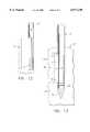
- FIG. 1is an exploded isometric view of a surgical biopsy instrument of the present invention
- FIG. 2is an exploded isometric view of the distal ends of the components of a probe of the surgical biopsy instrument of FIG. 1;
- FIG. 3is an enlarged isometric view of the proximal and distal ends of the probe of the surgical biopsy instrument of FIG. 1;
- FIG. 4is a side elevational view, in cross section, of the probe of the surgical biopsy instrument of FIG. 1;
- FIG. 5is a cross section of the probe of FIG. 4 along line 5--5;
- FIG. 6is an enlarged isometric view of the proximal end of a tissue extractor of the surgical biopsy instrument of FIG. 1;
- FIG. 7is an elevational view of the proximal side of a back plate of the surgical biopsy instrument of FIG. 1;
- FIG. 8is a side elevational view, in cross section, of the distal end of the probe of the surgical biopsy instrument of FIG. 1 wherein the probe is poised above a body prior to penetration;
- FIG. 9is a side elevational view, in cross section, of the distal end of the probe of the surgical biopsy instrument of FIG. 1, wherein the probe has penetrated into body tissue and a cutter is in a distal-most position;
- FIG. 10is a side elevational view, in cross section, of the distal end of the probe of the surgical biopsy instrument of FIG. 1, wherein the probe has penetrated into the body tissue, the cutter has been moved to a proximal-most position, and a vacuum has been applied, drawing a tissue sample into the probe;
- FIG. 11is a side elevational view, in cross section, of the distal end of the probe of the surgical biopsy instrument of FIG. 1, wherein the probe has penetrated into the body tissue, the vacuum is still applied, and the cutter has been returned or moved back to the distal-most position severing a tissue sample within;
- FIG. 12is a side elevational view, in cross section, of the distal end of a tissue extractor of the surgical biopsy instrument of FIG. 1, wherein the tissue extractor has been removed from the probe, the vacuum has been turned off and the tissue sample is shown extracted from the body;
- FIG. 13is a side elevational view, in cross section, of the distal end of the probe of the surgical biopsy instrument of FIG. 1, wherein the probe remains in the body tissue, and the tissue extractor has been removed for the extraction of the tissue sample from the surgical site;
- FIG. 14is an isometric view of an alternate embodiment of the surgical biopsy instrument having a blade detent mechanism
- FIG. 15is an isometric view of yet another alternate embodiment of a surgical biopsy instrument of the present invention having a single port with a large width.
- the present inventionis a surgical biopsy instrument 30 that is a minimally invasive type instrument for acquiring repeated subcutaneous biopsies.
- the surgical biopsy instrument 30has a probe 45 having at least one port 55 for insertion into a human body 25 (FIG. 8) for extraction of a biopsy or tissue sample 27 (FIG. 11) therefrom.
- the probe 45 of the surgical biopsy instrument 30is removably mounted to a powered probe driver 31.
- the probe driver 31is attached to a moveable table 20 such as a stereotactic guidance system (not shown) for moving the probe 45 distally in order to pierce the body 25 and as well as proximally in order to remove the probe 45 from the body.
- a vacuum source 86is attached to the proximal end of the probe 45 by a vacuum line 85 for the delivery of a vacuum to the probe 45.
- a control unit 87is used to control the sequence of actions performed by the surgical biopsy instrument 30 in order to obtain a biopsy sample or tissue sample 27 from the body 25.
- the control unit 87controls the movement of; the motorized stereotactic table, the application of vacuum to the probe 45, and activates the cutter motor (not shown) within the probe driver 31.
- the probe 45has three components; a piercing probe 50 having a elongated hollow piercing needle 51 for piercing the body 25, a cutter 60 for cutting the tissue sample 27, and a tissue extractor 65 for the extraction of the tissue sample 27 from the body 25.
- the components of the probeare assembled by the insertion of the distal end of the tissue extractor 65 into the proximal end of the cutter 60 and insertion of the distal end of the cutter into the proximal end of the piercing probe 50.
- Each of the componentsare shown in a partially inserted condition prior to the mounting of the probe 45 into the probe driver 31 (FIG. 1).
- Each one of these componentsinterfaces or mates with a portion of the probe driver 31 as described below.
- the probe driver 31includes a housing 31a having a moveable cover 38 hingedly attached thereto. Within the housing 31a there is a housing mount fork 35 for receiving the piercing probe 50, a cutter advance fork 33 for receiving the cutter 60, and an elongated driver gear 34 to mate with and rotate the cutter 60. The housing 31a also includes a back plate 36 to mate with the tissue extractor 65.
- a cutter advance knob 32is movably positioned on the driver 31 for providing rapid advancement and retraction of the cutter 60. Longitudinal movement of the cutter advance knob 32 rapidly advances and retracts the cuter 60 within the probe 45. The cutter advance knob 32 is rotated during the tissue sample 27 cutting sequence in order to provide a slower rate of advancement of the cutter 60. This slower manual advancement produces a cleaner cut and a more intact tissue sample 27. Actuation of the knob 32 moves both the cutter advance fork 33 and the cutter 60 proximally and distally respectively.
- the piercing probe 50consists of the hollow piercing needle 51 attached to a probe housing 52 and an alignment mount 53.
- the hollow piercing needle 51extends the full length of the piercing probe 50 and has a distal piercing tip 54 for the penetration of tissue, an open proximal end, and a needle passageway 59 extending therebetween for reception of the cutter 60 and the tissue extractor 65 therein.
- a first embodiment of the piercing probe 50includes three equally spaced tissue receiving ports 55 interconnected with the needle passageway 59 for the reception of tissue samples 27 therein as best shown in FIG. 2.
- the tissue receiving ports 55are elongated apertures for the retrieval of a sizable localized tissue sample 27.
- the ports 55have proximal port edges 57 and distal port edges 58.
- the use of multiple receiving ports 55eliminates the need to rotate the piercing needle 51 within the body 25 and eliminates any potential twisting and binding of the surrounding body tissue 26 that could occur with rotation of the piercing needle 51.
- the use of the multiple ports 55allows for the sampling of multiple tissue samples 27 from a tissue site 29 (FIG. 9) in a 360 degree fashion without having to rotate the piercing needle 51.
- the alignment mount 53is attached at the distal end of the piercing needle 51.
- the alignment mount 53holds the receiving ports 55 in a fixed angular orientation about the longitudinal axis "L" of the piercing probe 50.
- a pair of alignment slots 53aare located on the lateral portions or sides on the alignment mount 53 for mating and aligning the piercing needle 51 to the housing mount fork 35 in the probe driver 31. It is important to note that once the probe 50 is mated to the probe driver 31, the probe 50 cannot be rotated about the longitudinal axis and the receiving ports 55 are aligned in a preferred orientation.
- the elongated tubular cutter 60is slidable and rotatable within the needle passageway 59 of the piercing needle 51.
- the cutter 60has an elongated hollow cutter tube 60a with a cutter bore 64 extending the entire length of the cutter 60.
- a cutting blade 63is located on the distal end of the hollow cutter tube 60a.
- the cutter 60also has a cutter body 61 and a cutter gear 62 adjacent to the proximal end of the cutter 60 as shown in FIG. 1.
- the cutter gear 62mates with the cutter advance fork 33 and the elongated driver gear 34 in the probe driver 31.
- the cutter advance fork 33moves the cutter 60 proximally and distally within the probe driver 31, the cutter gear 62 remains engaged with the elongated driver gear 34.
- the driver gear 34is driven by the motor (not shown) in the probe driver 31 to rotate the cutter 60 as directed by the control unit 87.
- the innermost component of the probe 45is the tissue extractor 65, which is slidably and rotatably mounted within the cutter bore 64 of the cutter 60 (FIG. 2) and removably positioned within the probe 45.
- the tissue extractor 65is a hollow tubular structure having a closed distal end plate 67 and a generally concave tissue receptacle 70 adjacent to the distal end as illustrated in FIG. 2.
- An extractor channel 76extends from the proximal face of the distal end plate 67 to the open proximal end of the extractor 65.
- a vacuum connector 66is located at the proximal end for the attachment of the vacuum line 85 to the extractor channel 76 for the delivery of vacuum thereto as best shown in FIG. 1.
- the vacuum from the vacuum source 86can be turned on and off as required by the control unit 87.
- a detent 80is located at the proximal end of the extractor 65 adjacent to the vacuum connector 66.
- the detent 80has a detent knob 81, and a distally extending detent pin 82 or detent member.
- the detent pin 82has a specific angular orientation with the tissue receptacle 70 which will be discussed later.
- the tissue extractor 65is rotatable within the piercing needle 51 to position the tissue receptacle 70 behind any tissue receiving port 55 in the piercing needle 51 for the reception of tissue samples 27 therein.
- the tissue receptacle 70is concave and is defined by the outer diameter of the distal end plate 67, a distal wall 71 of the distal end plate 67, a concave floor 72, and a proximal wall 73 for the reception of tissue samples 27 therein.
- the tissue receptacle 70has a series of evenly spaced vacuum openings 75 within the floor 72 that interconnect and communicate with the tissue extractor channel 76.
- a proximal orifice 74is located within the proximal wall 73 and also interconnects and communicates with the tissue extractor channel 76. As mentioned above, vacuum is applied uninterrupted to the tissue extractor channel 76 and the tissue receptacle 70 through the openings 75 and the orifice 74 for drawing tissue samples 27 through the receiving port 55 and into the tissue receptacle 70.
- FIG. 3shows the coaxial placement of the tissue extractor 65 within the cutter 60 and the cutter 60 within the piercing needle 51 to make the probe 45.
- the cutter 60is shown fully inserted into the piercing needle 51 so that the receiving ports 55 are closed by the cutter 60.
- the distal end of the probe 55has been sectioned along the longitudinal axis to show the components.
- the cutting blade 63 of the cutter 60is shown in the most distal position, referred to henceforth as the first position, such that the cutting blade 63 is located distal to distal port edge 58 of the piercing tip 54.
- the tissue receptacle 70is shown aligned with one of the tissue receiving ports 55.
- the extractor channel 76reduces from a circular cross section as shown on the right to a crescent-shape passageway underneath the floor 72 of the tissue receptacle 70.
- FIG. 5shows a section view of the distal end of the probe 45 of FIG. 4, along the line 5--5. The section is taken across the multiple receiving ports 55 located in the piercing needle 51. As shown, the cutter 60 is in the first or distal-most position and has closed off the tissue receiving ports 55. The following components have been removed from this view for clarity: the probe housing 52, the alignment mount 53, the cutter body 61, and the cutter gear 62. This view better illustrates the alignment of the detent pin 82 with the tissue receptacle 70. The tissue receptacle 70, located adjacent to the distal end of the tissue extractor 65, is shown aligned with the lowermost tissue receiving port 55.
- the detent pin 82located near to the proximal end of the tissue extractor 65, is also aligned with the lowermost tissue receiving port 55. It is important to note that when the detent pin 82 is aligned with one of the receiving ports 55, the tissue receptacle 70 is also aligned with the same receiving port 55 while at the same time obstructing the remaining two receiving ports 55.
- the crescent cross section of the tissue extractor channel 76is clearly shown. Whereas the cutter is shown as a tubular structure that closes off all the ports, it is possible to use a multi port cutter such that the cutting blade 60 is moveably disposed with respect to, and covers only the tissue receiving port 55 that is in alignment with the tissue receptacle 70.
- the detent pin 82extends distally from the indexing knob 80 adjacent to the distal end of the tissue extractor 65.
- FIG. 7shows the proximal face of the back plate 36 of the probe driver 31.
- a series of detent holes 40are shown in a specific angular orientation around the probe slot 39.
- FIGS. 8-13best illustrate the method of use according to the present invention.
- the surgical biopsy device 30(FIG. 1) is used to obtain multiple biopsy samples from a surgical site within a patient's body 25.
- the patientis generally positioned upon a surgical table (not shown) and is given a local anesthetic to anesthetize the desired penetration site.
- the surgical biopsy device 30is attached to a moveable table 20, such as a stereotactic table, and is moved into an approximate position above the body 25.
- the position of the probe 45undergoes fine adjustment above the body using the moveable table 20 (FIG. 1), prior to the insertion of the piercing needle 51 into the body 25.
- the tissue extractor 65is positioned within the probe 45 to place the tissue receptacle 70 in alignment with one of the receiving ports, for instance, the left hand tissue receiving port 55. Accordingly, the distal end of the extractor 65 obstructs the remaining two receiving ports 55 which will cause vacuum to be drawn and directed to the non-obstructed tissue port 55, in this example, the left hand receiving port 55 (FIG. 5).
- tissue samples 27can only be withdrawn into the tissue receptacle 70 through this non-obstructed port 55 when the cutter 60 is moved to it's proximal most position.
- the cutting blade 63Prior to insertion, the cutting blade 63 is moved to the first or most distal position to in order to close the tissue receiving ports 55 with the cutter 60.
- the piercing needle 51is advanced into the body 25 by distal movement of the moveable table 20. As best illustrated in FIG. 9, the piercing tip 54 is advanced until the tissue ports 55 reach the required depth at the tissue site 29.
- the body tissue 26presses inwardly upon the probe 45 and is shown partially prolapsed into the tissue receiving ports 55.
- the prolapsed tissue 26, within the tissue receiving ports 55is in contact with the exterior of the cutter 60 where the exterior of the cutter 60 is exposed at the open tissue receiving ports 55. However, the prolapsed tissue 26 is blocked from entering the tissue extractor 70.
- the tissue extractor 65 and the cutter 60are still in the same position, e.g. in the distal-most position relative to the piercing needle 51 as also shown in FIG. 6.
- the cutting blade 63 of the cutter 60is moved proximally from the first, or distal-most position to a second position, e.g. a position near the proximal port edge 57 of the receiving port 55, henceforth described as the second position.
- a second positione.g. a position near the proximal port edge 57 of the receiving port 55, henceforth described as the second position.
- the cutter 60When the cutter 60 is in the second position, it exposes the distal end of the tissue receptacle 70 to the open tissue receiving port 55.
- the tissue receptacle 70is aligned with the left tissue receiving port 55 for the reception of body tissue 26 into the tissue receptacle 70.
- the control unit 87actuates the vacuum source 86 to deliver vacuum to the extractor channel 76 of the tissue extractor 65.
- the tissue receptacle 70is connected to the extractor channel 76 by the vacuum openings 75 in the floor 72 of the tissue extractor and by the proximal orifice 74 in the proximal wall 73.
- the application of vacuum to the extractor channel 76 of the tissue extractor 65causes the tissue sample 27 of the body tissue 26 to ptosis or prolapse, drawing the prolapsed tissue sample 27 into the receiving port 55 and into the tissue receptacle 70 as shown.
- the prolapsed tissue sample 27is securely held within the tissue receptacle 70 by the uninterrupted vacuum delivered through the vacuum openings 75 and the proximal orifice 74. Since the other receiving ports 55 are obstructed by the distal end of the extractor 65, tissue samples 27 are only taken through the selected port 55, i.e. the port 55 directly aligned with the tissue receptacle 70.
- the prolapsed tissue sample 27is then severed from the tissue site 29 by the advancement of the cutting blade 63 from its second position near to the proximal port edge 57 to the first position distal to the distal port edge 58.
- the advancement of the cutting blade 63closes the tissue receiving ports 55 with the cutter tube 60a.
- the control unit 87activates the motor (not shown) within the probe driver 31 to rotate the driver gear 34, the cutter gear 62 and the cutting blade 63 (FIG. 1).
- the rotating cutting blade 63is advanced distally to the first position in order to sever the tissue sample 27. This is accomplished by the rotation of the cutter advance knob 32 by the user.
- the tissue sample 27is tightly held within the tissue receptacle 70 by the application of vacuum to the extractor channel 76. This enables the user to cut the securely held tissue sample 27 without twisting and distortion.
- the tissue sample 27is now ready for removal from the body 25 by the tissue extractor 65.
- the vacuumactively holds the tissue sample 27 in a fixed position within the tissue receptacle 70 as the tissue extractor 65 is removed or extracted from the probe 45.
- the continuous application of vacuum to the tissue sample 27ensures the collection of a fully intact and undistorted tissue sample 27 for analysis.
- the tissue sample 27is securely held within the extracted tissue extractor 65 until the user elects to release the sample 27 from the tissue extractor by the deactivation of vacuum to the extractor channel 76. As shown in FIG. 12, the vacuum is deactivated and tissue sample 27 is separated from the tissue extractor and ready for analysis. While not critical to the present invention, the vacuum supplied by the vacuum source 86 can be released manually or under the control of the control unit 87 to release the intact and undistorted tissue sample 27 from the tissue extractor 65.
- FIG. 13a view of the distal end of the piercing needle 51 and cutter 60 is shown with the remaining components in position at the tissue site 29 for the removal of the next tissue sample 27.
- a void 28is left at the position from where the tissue sample 27 was removed.
- Additional tissue samplescan be removed from the same port 55, e.g. the left port 55 if desired.
- the tissue extractor 65is then rotated upon reinsertion to one of the other ports 55 for removal of additional tissue samples 27 around the outer circumference of the piercing needle 51, e.g. in 360 degree fashion, without the trauma associated with frequent removal or rotation of the piercing probe 50 to obtain multiple tissue samples 27 from the tissue site 29.
- Probe 45ais a hand held instrument for the removal of tissue samples 27 from the body 25.
- the piercing probe 50 of the probe 45ahas a piercing needle 51 extending the full length of the piercing probe 50 and a blade detent mechanism 90 near the proximal end.
- the blade detent mechanism 90has a detent member or detent blade 92 or detent member.
- a blade extractor disk 94is fixedly attached to the proximal end of the piercing needle 51.
- Detent receptacles 91are equally spaced around the periphery of the disk 94 and are in direct alignment with the receiving ports 55 of the piercing needle 51.
- the piercing probe 50is held in one of the operator's hands during operation.
- the cutter gear 62 attached to the distal end of the cutter 60rotates and moves the cutting blade 63 (FIG. 2) from the first position to the second position. Likewise, cutter gear 62 moves the cutting blade 63 from the second position to the first-position in reverse motion of the cutter gear 62 by the user.
- the cutter 60is operated by the hand not holding the piercing probe 50.
- the blade detent mechanism 90includes a blade body 93 having an elongated deflectable detent blade 92 that extends outwardly and distally from the blade body 93.
- the distal end of the detent blade 92is received within one of the detent receptacles 91 for the alignment of the tissue receptacle 70 (FIG. 4) with one of the receiving ports 55 on the distal end of the multi port probe 45a.
- the blade body 93 and the detent blade 92are rotated to another detent receptacle 91 to position the distal tissue receptacle 70 behind the desired, respective receiving port 55.
- the detent blade 92is a spring member and holds the detent blade within the detent receptacle 91.
- the vacuum connector 66is located on the distal end of the tissue extractor 65 for connection to the vacuum line 85 and a vacuum source 86 as depicted in FIG. 1. Whereas the blade detent mechanism 90 described above is used with a hand held probe 45a, the blade detent mechanism 90 can also be used with any other probe such as the previously described probe 45 for use on a probe driver 31.
- the method of use of the multi-probe 45a device as depicted in FIG. 14is manual and operated without the use of the driver 31 (FIG. 1).
- the sequence whereby a tissue sample 27 is removedis the same method as generally depicted in FIGS. 8-13.
- the vacuummust be manually activated to obtain a tissue sample as shown in FIGS. 10, 11, and 12.
- FIG. 15shows another embodiment of the present invention wherein a probe 45b has a single receiving port 55a.
- the cutter 60 and tissue extractor 65operate in the same manner described above for the retrieval of multiple tissue samples 27.
- the width of the single receiving port 55ais larger than the width of receiving ports 55 on the probe 45 of FIG. 1.
- This receiving port 55adefines an opening in the needle 51 ranging from approximately 15° to approximately 330° and preferably ranging from approximately 180° to approximately 270°. This enables the tissue receptacle 70 of the tissue extractor to be rotated throughout a wide angular band within the width of the single receiving port 55a for the reception of tissue samples 27 therein.
- Another embodiment of the method according to the present inventionallows for the cutter to be moved to the second position prior to insertion.
- the probe 45is inserted with the cutter 60 in the second position.
- the tissue extractor 65covers all but one of the multiple tissue receiving ports 55.
- this alternate embodimentenables the operator to rotate the tissue extractor to any tissue receiving port 55 while blocking the non-selected tissue receiving ports 55, if applicable.
- the proximal orifice 74communicates with the extractor channel 76 (FIG. 4) and supplies vacuum to the tissue receptacle. By enlarging this orifice 74, or removing the proximal wall 73 (FIG.
- tissue samplescan be removed from the piercing probe 50 by the application of vacuum.
- the application of vacuum to the tissue extractor 65draws tissue into the tissue receiving port 55 and into the tissue receptacle 70.
- the cutter 60is moved to the distal most position to cut the tissue sample 27 as shown.
- the continuing application of vacuum to the extractor channel 76draws the tissue sample 27 into the proximal orifice 74 or through the opening created by the removed proximal wall 73, and into the extractor channel 76. Once the sample is in the channel 76, it is drawn out of the probe 45 by the continuing application of vacuum. Once the sample is out of the probe 45, it can be captured for examination.
Landscapes
- Health & Medical Sciences (AREA)
- Life Sciences & Earth Sciences (AREA)
- Medical Informatics (AREA)
- Engineering & Computer Science (AREA)
- Biomedical Technology (AREA)
- Heart & Thoracic Surgery (AREA)
- Pathology (AREA)
- Molecular Biology (AREA)
- Surgery (AREA)
- Animal Behavior & Ethology (AREA)
- General Health & Medical Sciences (AREA)
- Public Health (AREA)
- Veterinary Medicine (AREA)
- Sampling And Sample Adjustment (AREA)
Abstract
Description
Claims (11)
Priority Applications (1)
| Application Number | Priority Date | Filing Date | Title |
|---|---|---|---|
| US09/078,476US6077230A (en) | 1998-05-14 | 1998-05-14 | Biopsy instrument with removable extractor |
Applications Claiming Priority (1)
| Application Number | Priority Date | Filing Date | Title |
|---|---|---|---|
| US09/078,476US6077230A (en) | 1998-05-14 | 1998-05-14 | Biopsy instrument with removable extractor |
Publications (1)
| Publication Number | Publication Date |
|---|---|
| US6077230Atrue US6077230A (en) | 2000-06-20 |
Family
ID=22144262
Family Applications (1)
| Application Number | Title | Priority Date | Filing Date |
|---|---|---|---|
| US09/078,476Expired - LifetimeUS6077230A (en) | 1998-05-14 | 1998-05-14 | Biopsy instrument with removable extractor |
Country Status (1)
| Country | Link |
|---|---|
| US (1) | US6077230A (en) |
Cited By (145)
| Publication number | Priority date | Publication date | Assignee | Title |
|---|---|---|---|---|
| US6193673B1 (en)* | 1998-02-20 | 2001-02-27 | United States Surgical Corporation | Biopsy instrument driver apparatus |
| US20010047183A1 (en)* | 2000-04-05 | 2001-11-29 | Salvatore Privitera | Surgical device for the collection of soft tissue |
| US6325801B1 (en)* | 1999-12-04 | 2001-12-04 | Karl Storz Gmbh & Co. Kg | Instrument for severing tissue with HF current |
| US6432064B1 (en)* | 2001-04-09 | 2002-08-13 | Ethicon Endo-Surgery, Inc. | Biopsy instrument with tissue marking element |
| WO2002062232A1 (en)* | 2001-02-05 | 2002-08-15 | Tyco Healthcare Group Lp | Biopsy apparatus and method |
| US20030120175A1 (en)* | 2001-12-20 | 2003-06-26 | Ehr Timothy G.J. | Pressure-sensing guidewire and sheath |
| US20030199753A1 (en)* | 2002-04-23 | 2003-10-23 | Ethicon Endo-Surgery | MRI compatible biopsy device with detachable probe |
| US20030199785A1 (en)* | 2002-04-23 | 2003-10-23 | Ethicon Endo-Surgery | Localization mechanism for an MRI compatible biopsy device |
| US20030199754A1 (en)* | 2002-04-23 | 2003-10-23 | Ethicon Endo-Surgery | Method for using an MRI compatible biopsy device with detachable probe |
| US6638235B2 (en) | 2000-11-06 | 2003-10-28 | Suros Surgical Systems, Inc. | Biopsy apparatus |
| US20040049128A1 (en)* | 2000-11-06 | 2004-03-11 | Miller Michael E. | Biopsy apparatus |
| US20040059254A1 (en)* | 2001-03-23 | 2004-03-25 | Stryker Puerto Rico Limited | Micro-invasive breast biopsy device |
| US20040077972A1 (en)* | 2002-10-18 | 2004-04-22 | Mark Tsonton | Localization mechanism for an MRI compatible biopsy device |
| GB2397242A (en)* | 2000-11-06 | 2004-07-21 | Suros Surgical Systems Inc | A tissue cutting device consisting of an outer cannula and an inner cutting member coupled to a drive mechanism |
| US20040153003A1 (en)* | 2002-12-11 | 2004-08-05 | Chris Cicenas | Biopsy device with sample tube |
| WO2004075728A2 (en) | 2003-02-25 | 2004-09-10 | Ethicon Endo-Surgery, Inc. | Biopsy device with variable speed cutter advance |
| WO2004075719A2 (en) | 2003-02-24 | 2004-09-10 | Senorx, Inc. | Biopsy device with inner cutting member |
| WO2005004700A2 (en) | 2003-06-27 | 2005-01-20 | Depuy Spine, Inc. | Controlled orifice sampling needle |
| US20050027210A1 (en)* | 2000-11-06 | 2005-02-03 | Miller Michael E. | Biopsy apparatus |
| US20050054945A1 (en)* | 2003-09-10 | 2005-03-10 | Scimed Life Systems, Inc | Forceps and collection assembly with accompanying mechanisms and related methods of use |
| US20050085838A1 (en)* | 2003-02-25 | 2005-04-21 | Bennie Thompson | Method of operating a biopsy device |
| US20050101880A1 (en)* | 2002-12-11 | 2005-05-12 | Chris Cicenas | Biopsy device with piston advance |
| US20050209530A1 (en)* | 2001-03-23 | 2005-09-22 | Stryker Puerto Rico Limited | Micro-invasive tissue removal device |
| US20050228311A1 (en)* | 2004-03-31 | 2005-10-13 | Beckman Andrew T | Marker device and method of deploying a cavity marker using a surgical biopsy device |
| US20050261581A1 (en)* | 2004-05-21 | 2005-11-24 | Hughes Robert J | MRI biopsy device |
| EP1642534A2 (en) | 2004-09-29 | 2006-04-05 | Ethicon Endo-Surgery, Inc. | Biopsy device with sample storage |
| US20060074345A1 (en)* | 2004-09-29 | 2006-04-06 | Hibner John A | Biopsy apparatus and method |
| EP1665989A2 (en) | 2004-09-29 | 2006-06-07 | Ethicon Endo-Surgery, Inc. | Fluid control for biopsy device |
| US20060129185A1 (en)* | 1999-10-25 | 2006-06-15 | Boston Scientific Scimed, Inc. | Forceps for medical use |
| US20060173377A1 (en)* | 2005-01-31 | 2006-08-03 | Mccullough Adam B | Quick cycle biopsy system |
| EP1698282A1 (en) | 2005-03-04 | 2006-09-06 | Ethicon Endo-Surgery, Inc. | Biopsy device with variable side aperture |
| EP1698283A1 (en) | 2005-03-04 | 2006-09-06 | Ethicon Endo-Surgery, Inc. | Biopsy device incorporating an adjustable probe sleeve |
| US20060217635A1 (en)* | 2005-03-24 | 2006-09-28 | Mccombs Elizabeth S | Biopsy device marker deployment |
| US20060229528A1 (en)* | 2003-03-29 | 2006-10-12 | C. R. Brad, Inc. | Coaxial cannula provided with a sealing element |
| US20060241385A1 (en)* | 2005-04-12 | 2006-10-26 | Ethicon Endo-Surgery, Inc. | Guided disposable fiducial for breast biopsy localization fixture |
| US20060260994A1 (en)* | 2005-05-18 | 2006-11-23 | Mark Joseph L | Selectively openable tissue filter |
| WO2006136588A1 (en)* | 2005-06-22 | 2006-12-28 | H.S. - Hospital Service - S.P.A. | Biopsy device |
| US20070010738A1 (en)* | 2004-10-14 | 2007-01-11 | Mark Joseph L | Surgical site marker delivery system |
| US20070032743A1 (en)* | 2005-08-05 | 2007-02-08 | Hibner John A | Vacuum Syringe Assisted Biopsy Device |
| US20070032742A1 (en)* | 2005-08-05 | 2007-02-08 | Monson Gavin M | Biopsy Device with Vacuum Assisted Bleeding Control |
| US20070149893A1 (en)* | 2002-03-19 | 2007-06-28 | C.R. Bard, Inc. | Biopsy device and biopsy needle module that can be inserted into the biopsy device |
| US20070149894A1 (en)* | 2002-03-19 | 2007-06-28 | C.R. Bard, Inc. | Biopsy device for removing tissue specimens using a vacuum |
| US20070167736A1 (en)* | 2004-05-21 | 2007-07-19 | Dietz Timothy G | MRI biopsy apparatus incorporating an imageable penetrating portion |
| US20070179401A1 (en)* | 2006-02-01 | 2007-08-02 | Ethicon Endo-Surgery, Inc. | Biopsy device with replaceable probe incorporating static vacuum source dual valve sample stacking retrieval and saline flush |
| EP1815799A1 (en)* | 2006-02-03 | 2007-08-08 | Ethicon Endo-Surgery, Inc. | Biopsy needle and method for assembling the needle |
| US20070191732A1 (en)* | 2006-02-10 | 2007-08-16 | Voegele James W | Cryogenic probe |
| US20070213630A1 (en)* | 2006-03-07 | 2007-09-13 | Beckman Andrew T | Device for minimally invasive internal tissue removal |
| US20070213755A1 (en)* | 2006-03-07 | 2007-09-13 | Beckman Andrew T | Device for minimally invasive internal tissue removal |
| US20070239064A1 (en)* | 2006-03-29 | 2007-10-11 | Cicenas Chris W | Device for minimally invasive internal tissue removal |
| US20070239067A1 (en)* | 2005-08-05 | 2007-10-11 | Hibner John A | Tissue Sample Revolver Drum Biopsy Device |
| US20070255170A1 (en)* | 2006-05-01 | 2007-11-01 | Hibner John A | Biopsy cannula adjustable depth stop |
| US20070255168A1 (en)* | 2006-05-01 | 2007-11-01 | Hibner John A | Grid and rotatable cube guide localization fixture for biopsy device |
| US20070255172A1 (en)* | 2001-03-23 | 2007-11-01 | Stryker Puerto Rico Limited | Micro-invasive nucleotomy device and method |
| US20080004545A1 (en)* | 2005-08-05 | 2008-01-03 | Garrison William A | Trigger Fired Radial Plate Specimen Retrieval Biopsy Instrument |
| US20080114265A1 (en)* | 2006-10-23 | 2008-05-15 | Tom Tarter | Double Core Biopsy Instrumentation Kit |
| EP1932482A1 (en) | 2006-12-13 | 2008-06-18 | Ethicon Endo-Surgery, Inc. | Biopsy sample storage |
| EP1932481A1 (en) | 2006-12-13 | 2008-06-18 | Ethicon Endo-Surgery, Inc. | Biopsy system with vacuum control module |
| US20080146965A1 (en)* | 2003-08-11 | 2008-06-19 | Salvatore Privitera | Surgical Device for The Collection of Soft Tissue |
| US20080195066A1 (en)* | 2006-12-13 | 2008-08-14 | Speeg Trevor W V | Revolving Tissue Sample Holder For Biopsy Device |
| US20080200836A1 (en)* | 2006-12-13 | 2008-08-21 | Speeg Trevor W V | Biopsy Device With Motorized Needle Cocking |
| US20080214955A1 (en)* | 2006-12-13 | 2008-09-04 | Speeg Trevor W V | Presentation of Biopsy Sample By Biopsy Device |
| US20080228103A1 (en)* | 2006-12-13 | 2008-09-18 | Ritchie Paul G | Vacuum Timing Algorithm For Biopsy Device |
| US20080281224A1 (en)* | 2007-05-11 | 2008-11-13 | Johnson Michael E | Biopsy device needle tip |
| US20080300506A1 (en)* | 2007-05-29 | 2008-12-04 | Boston Scientific Scimed, Inc. | Biopsy device with multiple cutters |
| US20080319342A1 (en)* | 2003-02-24 | 2008-12-25 | Shabaz Martin V | Biopsy device with selectable tissue receiving aperture orientation and site illumination |
| EP2022407A2 (en) | 2007-07-25 | 2009-02-11 | Ethicon Endo-Surgery, Inc. | Biopsy device with manually rotated sample barrel |
| US20090131817A1 (en)* | 2007-11-20 | 2009-05-21 | Speeg Trevor W V | Deployment device interface for biopsy device |
| US20090131818A1 (en)* | 2007-11-20 | 2009-05-21 | Speeg Trevor W V | Biopsy Device Tissue Sample Holder Rotation Control |
| US20090131823A1 (en)* | 2007-11-20 | 2009-05-21 | Andreyko Michael J | Biopsy Device With Illuminated Tissue Holder |
| US20090131816A1 (en)* | 2006-12-13 | 2009-05-21 | Ritchie Paul G | Engagement Interface For Biopsy System Vacuum Module |
| US20090131824A1 (en)* | 2007-11-20 | 2009-05-21 | Andrisek John R | Biopsy Device With Fine Pitch Drive Train |
| US20090131821A1 (en)* | 2007-11-20 | 2009-05-21 | Speeg Trevor W V | Graphical User Interface For Biopsy System Control Module |
| US20090131822A1 (en)* | 2007-11-20 | 2009-05-21 | Hibner John A | Biopsy Device With Sharps Reduction Feature |
| US20090131819A1 (en)* | 2007-11-20 | 2009-05-21 | Ritchie Paul G | User Interface On Biopsy Device |
| US20090131820A1 (en)* | 2007-11-20 | 2009-05-21 | Speeg Trevor W V | Icon-Based User Interface On Biopsy System Control Module |
| US20100152610A1 (en)* | 2008-12-16 | 2010-06-17 | Parihar Shailendra K | Hand Actuated Tetherless Biopsy Device with Pistol Grip |
| US20100160777A1 (en)* | 2008-12-22 | 2010-06-24 | Hardin Terry D | Reverse deployment device |
| US20100160824A1 (en)* | 2008-12-18 | 2010-06-24 | Parihar Shailendra K | Biopsy Device with Discrete Tissue Chambers |
| US20100160818A1 (en)* | 2005-04-12 | 2010-06-24 | Ethicon Endo-Surgery, Inc. | MRI Biopsy Device |
| US7762960B2 (en) | 2005-05-13 | 2010-07-27 | Boston Scientific Scimed, Inc. | Biopsy forceps assemblies |
| US7762961B2 (en) | 2003-03-29 | 2010-07-27 | C. R. Bard, Inc. | Pressure generating unit |
| US7766843B2 (en) | 2006-03-03 | 2010-08-03 | Ethicon Endo-Surgery, Inc. | Biopsy method |
| US20100210967A1 (en)* | 2007-08-02 | 2010-08-19 | Sjunnesson Haakan | Surgical kits and methods |
| US20100312140A1 (en)* | 2008-12-16 | 2010-12-09 | Smith Eric B | Needle for Biopsy Device |
| US20100317994A1 (en)* | 2009-06-16 | 2010-12-16 | Leimbach Jessica P | Biopsy Targeting Cube with Living Hinges |
| US20100317990A1 (en)* | 2009-06-16 | 2010-12-16 | Leimbach Jessica P | Biopsy Targeting Cube with Elastomeric Edges |
| US20100317991A1 (en)* | 2009-06-16 | 2010-12-16 | Leimbach Jessica P | Biopsy Targeting Cube with Elastomeric Body |
| US20100317993A1 (en)* | 2009-06-16 | 2010-12-16 | Leimbach Jessica P | Biopsy Targeting Cube with Angled Interface |
| US20100317992A1 (en)* | 2009-06-16 | 2010-12-16 | Leimbach Jessica P | Biopsy Targeting Cube with Malleable Members |
| US7854706B2 (en) | 2007-12-27 | 2010-12-21 | Devicor Medical Products, Inc. | Clutch and valving system for tetherless biopsy device |
| EP2263577A2 (en) | 2009-06-17 | 2010-12-22 | Ethicon Endo-Surgery, Inc. | MRI biopsy cylindraceous targeting guide |
| EP2263578A2 (en) | 2009-06-17 | 2010-12-22 | Ethicon Endo-Surgery, Inc. | MRI biopsy targeting grid with round openings |
| EP2263551A1 (en) | 2009-06-17 | 2010-12-22 | Ethicon Endo-Surgery, Inc. | MRI biopsy targeting grid wall guide |
| US20110004119A1 (en)* | 2009-07-01 | 2011-01-06 | Michael Hoffa | Surgical system |
| WO2011028319A2 (en) | 2009-08-26 | 2011-03-10 | Devicor Medical Products, Inc. | Method and device for obtaining tissue samples |
| US20110082364A1 (en)* | 2009-10-05 | 2011-04-07 | Hibner John A | MRI Biopsy Targeting Cube with Retention Wiper |
| US20110092848A1 (en)* | 2009-10-16 | 2011-04-21 | Hibner John A | MRI Biopsy Targeting Cube with Eccentric Lock |
| US20110092983A1 (en)* | 2009-10-16 | 2011-04-21 | Pawar Ajay D | MRI Biopsy Targeting Cube with Locking Flap |
| US20110092847A1 (en)* | 2009-10-16 | 2011-04-21 | Wale Nitin P | MRI Biopsy Targeting Cube with Snap Corners |
| US20110092850A1 (en)* | 2009-10-16 | 2011-04-21 | Kulkarni Abhijit G | MRI Biopsy Targeting Guide with Rotational Lock |
| US7942896B2 (en) | 2003-11-25 | 2011-05-17 | Scimed Life Systems, Inc. | Forceps and collection assembly and related methods of use and manufacture |
| US7988642B2 (en) | 2003-10-14 | 2011-08-02 | Suros Surgical Systems, Inc. | Vacuum assisted biopsy device |
| US20110201965A1 (en)* | 2010-02-18 | 2011-08-18 | John Anthony Hibner | MRI Compatible Biopsy Device |
| US8048003B2 (en) | 2003-10-14 | 2011-11-01 | Suros Surgical Systems, Inc. | Vacuum assisted biopsy device |
| US8052615B2 (en) | 2004-07-09 | 2011-11-08 | Bard Peripheral Vascular, Inc. | Length detection system for biopsy device |
| US20110313316A1 (en)* | 2009-03-16 | 2011-12-22 | Ranpura Himanshu M | Biopsy device having rotational cutting |
| US8162849B2 (en) | 2009-10-16 | 2012-04-24 | Devicor Medical Products, Inc. | MRI biopsy targeting cube with gripping arms |
| US8187204B2 (en) | 2007-10-01 | 2012-05-29 | Suros Surgical Systems, Inc. | Surgical device and method for using same |
| US8231544B2 (en) | 2003-10-14 | 2012-07-31 | Suros Surgical Systems, Inc. | Vacuum assisted biopsy needle set |
| US8251917B2 (en) | 2006-08-21 | 2012-08-28 | C. R. Bard, Inc. | Self-contained handheld biopsy needle |
| US8262585B2 (en) | 2005-08-10 | 2012-09-11 | C. R. Bard, Inc. | Single-insertion, multiple sampling biopsy device with linear drive |
| US8262586B2 (en) | 2006-10-24 | 2012-09-11 | C. R. Bard, Inc. | Large sample low aspect ratio biopsy needle |
| US8267868B2 (en) | 2005-08-10 | 2012-09-18 | C. R. Bard, Inc. | Single-insertion, multiple sample biopsy device with integrated markers |
| US20120253226A1 (en)* | 2008-12-18 | 2012-10-04 | Devicor Medical Products, Inc. | Biopsy device with central thumbwheel |
| US8282573B2 (en) | 2003-02-24 | 2012-10-09 | Senorx, Inc. | Biopsy device with selectable tissue receiving aperture orientation and site illumination |
| US8282574B2 (en) | 2005-08-10 | 2012-10-09 | C. R. Bard, Inc. | Single-insertion, multiple sampling biopsy device usable with various transport systems and integrated markers |
| US8342851B1 (en) | 2008-09-19 | 2013-01-01 | Devicor Medical Products, Inc. | Tissue model for testing biopsy needles |
| US8430824B2 (en) | 2009-10-29 | 2013-04-30 | Bard Peripheral Vascular, Inc. | Biopsy driver assembly having a control circuit for conserving battery power |
| US8465471B2 (en) | 2009-08-05 | 2013-06-18 | Rocin Laboratories, Inc. | Endoscopically-guided electro-cauterizing power-assisted fat aspiration system for aspirating visceral fat tissue within the abdomen of a patient |
| US8485987B2 (en) | 2006-10-06 | 2013-07-16 | Bard Peripheral Vascular, Inc. | Tissue handling system with reduced operator exposure |
| US8485989B2 (en) | 2009-09-01 | 2013-07-16 | Bard Peripheral Vascular, Inc. | Biopsy apparatus having a tissue sample retrieval mechanism |
| US8597205B2 (en) | 2007-12-20 | 2013-12-03 | C. R. Bard, Inc. | Biopsy device |
| US8597206B2 (en) | 2009-10-12 | 2013-12-03 | Bard Peripheral Vascular, Inc. | Biopsy probe assembly having a mechanism to prevent misalignment of components prior to installation |
| US8708930B2 (en) | 2009-04-15 | 2014-04-29 | Bard Peripheral Vascular, Inc. | Biopsy apparatus having integrated fluid management |
| WO2014084961A1 (en) | 2012-10-08 | 2014-06-05 | Devicor Medical Products, Inc. | Tissue biopsy device with thumbwheel and sample holder |
| US8808200B2 (en) | 2007-10-01 | 2014-08-19 | Suros Surgical Systems, Inc. | Surgical device and method of using same |
| US8845548B2 (en) | 2009-06-12 | 2014-09-30 | Devicor Medical Products, Inc. | Cutter drive assembly for biopsy device |
| US9173641B2 (en) | 2009-08-12 | 2015-11-03 | C. R. Bard, Inc. | Biopsy apparatus having integrated thumbwheel mechanism for manual rotation of biopsy cannula |
| US9220485B2 (en) | 2010-08-28 | 2015-12-29 | Endochoice, Inc. | Tissue collection and separation device |
| USRE46135E1 (en) | 2005-08-05 | 2016-09-06 | Devicor Medical Products, Inc. | Vacuum syringe assisted biopsy device |
| WO2017176990A1 (en)* | 2016-04-08 | 2017-10-12 | Parakrama Chandrasoma | Pathologic assessment of lower esophageal sphincter damage |
| US9925314B2 (en) | 2009-08-05 | 2018-03-27 | Rocin Laboratories, Inc. | Method of performing intra-abdominal tissue aspiration to ameliorate the metabolic syndrome, or abdominal obesity |
| US9943293B2 (en) | 2011-11-09 | 2018-04-17 | Teesuvac Aps | Handheld tissue sample extraction device |
| CN109620305A (en)* | 2019-01-02 | 2019-04-16 | 河南牧业经济学院 | A kind of pet tumor examination sampler |
| US10285673B2 (en) | 2013-03-20 | 2019-05-14 | Bard Peripheral Vascular, Inc. | Biopsy device |
| US10456120B2 (en) | 2013-11-05 | 2019-10-29 | C. R. Bard, Inc. | Biopsy device having integrated vacuum |
| US10463350B2 (en) | 2015-05-01 | 2019-11-05 | C. R. Bard, Inc. | Biopsy device |
| US10595831B2 (en) | 2012-05-30 | 2020-03-24 | Devicor Medical Products, Inc. | Control for biopsy device |
| CN113331878A (en)* | 2021-06-02 | 2021-09-03 | 王宝菊 | Animal doctor's laboratory detects special animal tissue sampler |
| US11116483B2 (en) | 2017-05-19 | 2021-09-14 | Merit Medical Systems, Inc. | Rotating biopsy needle |
| US11179141B2 (en) | 2006-12-13 | 2021-11-23 | Devicor Medical Products, Inc. | Biopsy system |
| US11793498B2 (en) | 2017-05-19 | 2023-10-24 | Merit Medical Systems, Inc. | Biopsy needle devices and methods of use |
| US11844500B2 (en) | 2017-05-19 | 2023-12-19 | Merit Medical Systems, Inc. | Semi-automatic biopsy needle device and methods of use |
| US12150627B2 (en) | 2019-12-11 | 2024-11-26 | Merit Medical Systems, Inc. | Bone biopsy device and related methods |
| US12295556B2 (en) | 2019-09-27 | 2025-05-13 | Merit Medical Systems, Inc. | Rotation biopsy system and handle |
Citations (18)
| Publication number | Priority date | Publication date | Assignee | Title |
|---|---|---|---|---|
| US3590808A (en)* | 1968-09-04 | 1971-07-06 | Us Catheter & Instr Corp | Biopsy tool |
| US3732858A (en)* | 1968-09-16 | 1973-05-15 | Surgical Design Corp | Apparatus for removing blood clots, cataracts and other objects from the eye |
| US3844272A (en)* | 1969-02-14 | 1974-10-29 | A Banko | Surgical instruments |
| US3945375A (en)* | 1972-04-04 | 1976-03-23 | Surgical Design Corporation | Rotatable surgical instrument |
| US4708147A (en)* | 1985-02-25 | 1987-11-24 | Haaga John R | Universal biopsy needle |
| US4799495A (en)* | 1987-03-20 | 1989-01-24 | National Standard Company | Localization needle assembly |
| US4907599A (en)* | 1988-02-01 | 1990-03-13 | Hart Enterprises, Inc. | Soft tissue core biopsy instrument |
| US4976269A (en)* | 1989-10-26 | 1990-12-11 | Creative Research & Manufacturing | Tissue needle |
| US5106364A (en)* | 1989-07-07 | 1992-04-21 | Kabushiki Kaisha Topcon | Surgical cutter |
| US5125413A (en)* | 1989-03-29 | 1992-06-30 | Baran Gregory W | Automated biopsy instrument |
| US5152744A (en)* | 1990-02-07 | 1992-10-06 | Smith & Nephew Dyonics | Surgical instrument |
| US5183054A (en)* | 1991-09-09 | 1993-02-02 | Sherwood Medical Company | Actuated biopsy cutting needle with removable stylet |
| US5240011A (en)* | 1991-11-27 | 1993-08-31 | Fischer Imaging Corporation | Motorized biopsy needle positioner |
| US5284156A (en)* | 1991-08-30 | 1994-02-08 | M3 Systems, Inc. | Automatic tissue sampling apparatus |
| US5415182A (en)* | 1992-05-11 | 1995-05-16 | Boston Scientific Corporation | Multiple needle biopsy instrument |
| US5458112A (en)* | 1994-08-15 | 1995-10-17 | Arrow Precision Products, Inc. | Biliary biopsy device |
| US5526822A (en)* | 1994-03-24 | 1996-06-18 | Biopsys Medical, Inc. | Method and apparatus for automated biopsy and collection of soft tissue |
| US5564436A (en)* | 1995-09-21 | 1996-10-15 | Hakky; Said I. | Automatic rotating cassette multiple biopsy device |
- 1998
- 1998-05-14USUS09/078,476patent/US6077230A/ennot_activeExpired - Lifetime
Patent Citations (18)
| Publication number | Priority date | Publication date | Assignee | Title |
|---|---|---|---|---|
| US3590808A (en)* | 1968-09-04 | 1971-07-06 | Us Catheter & Instr Corp | Biopsy tool |
| US3732858A (en)* | 1968-09-16 | 1973-05-15 | Surgical Design Corp | Apparatus for removing blood clots, cataracts and other objects from the eye |
| US3844272A (en)* | 1969-02-14 | 1974-10-29 | A Banko | Surgical instruments |
| US3945375A (en)* | 1972-04-04 | 1976-03-23 | Surgical Design Corporation | Rotatable surgical instrument |
| US4708147A (en)* | 1985-02-25 | 1987-11-24 | Haaga John R | Universal biopsy needle |
| US4799495A (en)* | 1987-03-20 | 1989-01-24 | National Standard Company | Localization needle assembly |
| US4907599A (en)* | 1988-02-01 | 1990-03-13 | Hart Enterprises, Inc. | Soft tissue core biopsy instrument |
| US5125413A (en)* | 1989-03-29 | 1992-06-30 | Baran Gregory W | Automated biopsy instrument |
| US5106364A (en)* | 1989-07-07 | 1992-04-21 | Kabushiki Kaisha Topcon | Surgical cutter |
| US4976269A (en)* | 1989-10-26 | 1990-12-11 | Creative Research & Manufacturing | Tissue needle |
| US5152744A (en)* | 1990-02-07 | 1992-10-06 | Smith & Nephew Dyonics | Surgical instrument |
| US5284156A (en)* | 1991-08-30 | 1994-02-08 | M3 Systems, Inc. | Automatic tissue sampling apparatus |
| US5183054A (en)* | 1991-09-09 | 1993-02-02 | Sherwood Medical Company | Actuated biopsy cutting needle with removable stylet |
| US5240011A (en)* | 1991-11-27 | 1993-08-31 | Fischer Imaging Corporation | Motorized biopsy needle positioner |
| US5415182A (en)* | 1992-05-11 | 1995-05-16 | Boston Scientific Corporation | Multiple needle biopsy instrument |
| US5526822A (en)* | 1994-03-24 | 1996-06-18 | Biopsys Medical, Inc. | Method and apparatus for automated biopsy and collection of soft tissue |
| US5458112A (en)* | 1994-08-15 | 1995-10-17 | Arrow Precision Products, Inc. | Biliary biopsy device |
| US5564436A (en)* | 1995-09-21 | 1996-10-15 | Hakky; Said I. | Automatic rotating cassette multiple biopsy device |
Cited By (435)
| Publication number | Priority date | Publication date | Assignee | Title |
|---|---|---|---|---|
| US6554779B2 (en) | 1998-02-20 | 2003-04-29 | United States Surgical Corporation | Biopsy instrument driver apparatus |
| US6193673B1 (en)* | 1998-02-20 | 2001-02-27 | United States Surgical Corporation | Biopsy instrument driver apparatus |
| US10166010B2 (en) | 1998-10-23 | 2019-01-01 | Devicor Medical Products, Inc. | Surgical device for the collection of soft tissue |
| US8206409B2 (en) | 1998-10-23 | 2012-06-26 | Devicor Medical Products, Inc. | Surgical device for the collection of soft tissue |
| US9433402B2 (en) | 1998-10-23 | 2016-09-06 | Devicor Medical Products, Inc. | Surgical device for the collection of soft tissue |
| US20110125055A1 (en)* | 1998-10-23 | 2011-05-26 | Devicor Medical Products, Inc. | Surgical device for the collection of soft tissue |
| US8979768B2 (en) | 1998-10-23 | 2015-03-17 | Devicor Medical Products, Inc. | Surgical device for the collection of soft tissue |
| US8016844B2 (en) | 1998-10-23 | 2011-09-13 | Devicor Medical Products, Inc. | Surgical device for the collection of soft tissue |
| US20040034280A1 (en)* | 1998-10-23 | 2004-02-19 | Salvatore Privitera | Surgical device for the collection of soft tissue |
| US7909850B2 (en) | 1999-10-25 | 2011-03-22 | Boston Scientific Scimed, Inc. | Forceps for medical use |
| US20060129185A1 (en)* | 1999-10-25 | 2006-06-15 | Boston Scientific Scimed, Inc. | Forceps for medical use |
| US6325801B1 (en)* | 1999-12-04 | 2001-12-04 | Karl Storz Gmbh & Co. Kg | Instrument for severing tissue with HF current |
| US20010047183A1 (en)* | 2000-04-05 | 2001-11-29 | Salvatore Privitera | Surgical device for the collection of soft tissue |
| US8568332B2 (en) | 2000-11-06 | 2013-10-29 | Suros Surgical Systems, Inc. | Biopsy apparatus |
| US20070027407A1 (en)* | 2000-11-06 | 2007-02-01 | Suros Surgical Systems, Inc. | Biopsy apparatus with vacuum relief |
| US6758824B1 (en) | 2000-11-06 | 2004-07-06 | Suros Surgical Systems, Inc. | Biopsy apparatus |
| GB2397242A (en)* | 2000-11-06 | 2004-07-21 | Suros Surgical Systems Inc | A tissue cutting device consisting of an outer cannula and an inner cutting member coupled to a drive mechanism |
| US7837630B2 (en) | 2000-11-06 | 2010-11-23 | Suros Surgical Systems, Inc. | Fluid control element for biopsy apparatus |
| US8167818B2 (en) | 2000-11-06 | 2012-05-01 | Suros Surgical Systems, Inc. | Biopsy apparatus with vacuum relief |
| US7458940B2 (en)* | 2000-11-06 | 2008-12-02 | Suros Surgical Systems, Inc. | Biopsy apparatus |
| US20090137927A1 (en)* | 2000-11-06 | 2009-05-28 | Miller Michael E | Biopsy apparatus with vacuum relief |
| US20040267157A1 (en)* | 2000-11-06 | 2004-12-30 | Miller Michael E | Biopsy apparatus |
| GB2397242B (en)* | 2000-11-06 | 2005-01-12 | Suros Surgical Systems Inc | Biopsy apparatus |
| US8192370B2 (en) | 2000-11-06 | 2012-06-05 | Suros Surgical Systems, Inc. | Biopsy apparatus |
| US20050027210A1 (en)* | 2000-11-06 | 2005-02-03 | Miller Michael E. | Biopsy apparatus |
| US20050049521A1 (en)* | 2000-11-06 | 2005-03-03 | Suros Surgical Systems, Inc. | Collection filter for biopsy apparatus |
| US20060155209A1 (en)* | 2000-11-06 | 2006-07-13 | Miller Michael E | Selectively detachable outer cannula hub |
| US8109886B2 (en) | 2000-11-06 | 2012-02-07 | Suros Surgical Systems, Inc. | Biopsy apparatus |
| US6638235B2 (en) | 2000-11-06 | 2003-10-28 | Suros Surgical Systems, Inc. | Biopsy apparatus |
| US20050113715A1 (en)* | 2000-11-06 | 2005-05-26 | Jeffrey Schwindt | Biopsy apparatus |
| US20060129062A1 (en)* | 2000-11-06 | 2006-06-15 | Nicoson Zachary R | Fluid control element for biopsy apparatus |
| US8764679B2 (en) | 2000-11-06 | 2014-07-01 | Suros Surgical Systems, Inc. | Biopsy apparatus |
| US8277393B2 (en) | 2000-11-06 | 2012-10-02 | Suros Surgical Systems, Inc. | Biopsy apparatus |
| US20090048533A1 (en)* | 2000-11-06 | 2009-02-19 | Miller Michael E | Biopsy apparatus |
| US7497833B2 (en) | 2000-11-06 | 2009-03-03 | Suros Surgical Systems, Inc. | Biopsy apparatus with vacuum relief |
| US8986222B2 (en) | 2000-11-06 | 2015-03-24 | Hologic, Inc. | Biopsy apparatus |
| US7883476B2 (en) | 2000-11-06 | 2011-02-08 | Suros Surgical Systems, Inc. | Selectively detachable outer cannula hub |
| US20040049128A1 (en)* | 2000-11-06 | 2004-03-11 | Miller Michael E. | Biopsy apparatus |
| WO2002062232A1 (en)* | 2001-02-05 | 2002-08-15 | Tyco Healthcare Group Lp | Biopsy apparatus and method |
| US20040059254A1 (en)* | 2001-03-23 | 2004-03-25 | Stryker Puerto Rico Limited | Micro-invasive breast biopsy device |
| US20050209530A1 (en)* | 2001-03-23 | 2005-09-22 | Stryker Puerto Rico Limited | Micro-invasive tissue removal device |
| US20070255172A1 (en)* | 2001-03-23 | 2007-11-01 | Stryker Puerto Rico Limited | Micro-invasive nucleotomy device and method |
| US7591790B2 (en) | 2001-03-23 | 2009-09-22 | Stryker Puerto Rico Limited | Micro-invasive device |
| US6432064B1 (en)* | 2001-04-09 | 2002-08-13 | Ethicon Endo-Surgery, Inc. | Biopsy instrument with tissue marking element |
| US8668650B2 (en)* | 2001-12-20 | 2014-03-11 | Boston Scientific Scimed, Inc. | Pressure-sensing guidewire and sheath |
| US9113790B2 (en) | 2001-12-20 | 2015-08-25 | Boston Scientific Scimed, Inc. | Pressure-sensing guidewire and sheath |
| US20030120175A1 (en)* | 2001-12-20 | 2003-06-26 | Ehr Timothy G.J. | Pressure-sensing guidewire and sheath |
| US10335128B2 (en) | 2002-03-19 | 2019-07-02 | C. R. Bard, Inc. | Biopsy device and insertable biopsy needle module |
| US20070149893A1 (en)* | 2002-03-19 | 2007-06-28 | C.R. Bard, Inc. | Biopsy device and biopsy needle module that can be inserted into the biopsy device |
| US8109885B2 (en) | 2002-03-19 | 2012-02-07 | C. R. Bard, Inc. | Biopsy device for removing tissue specimens using a vacuum |
| US8016772B2 (en) | 2002-03-19 | 2011-09-13 | C. R. Bard, Inc. | Biopsy device for removing tissue specimens using a vacuum |
| US9439631B2 (en) | 2002-03-19 | 2016-09-13 | C. R. Bard, Inc. | Biopsy device and insertable biopsy needle module |
| US10271827B2 (en) | 2002-03-19 | 2019-04-30 | C. R. Bard, Inc. | Disposable biopsy unit |
| US9072502B2 (en) | 2002-03-19 | 2015-07-07 | C. R. Bard, Inc. | Disposable biopsy unit |
| US20070149894A1 (en)* | 2002-03-19 | 2007-06-28 | C.R. Bard, Inc. | Biopsy device for removing tissue specimens using a vacuum |
| US8052614B2 (en) | 2002-03-19 | 2011-11-08 | C. R. Bard, Inc. | Biopsy device having a vacuum pump |
| US8002713B2 (en) | 2002-03-19 | 2011-08-23 | C. R. Bard, Inc. | Biopsy device and insertable biopsy needle module |
| US9421002B2 (en) | 2002-03-19 | 2016-08-23 | C. R. Bard, Inc. | Disposable biopsy unit |
| US8172773B2 (en) | 2002-03-19 | 2012-05-08 | C. R. Bard, Inc. | Biopsy device and biopsy needle module that can be inserted into the biopsy device |
| US8951209B2 (en) | 2002-03-19 | 2015-02-10 | C. R. Bard, Inc. | Biopsy device and insertable biopsy needle module |
| US11382608B2 (en) | 2002-03-19 | 2022-07-12 | C. R. Bard, Inc. | Disposable biopsy unit |
| US7826883B2 (en) | 2002-04-23 | 2010-11-02 | Devicor Medical Products, Inc. | Localization mechanism for an MRI compatible biopsy device |
| US20030199753A1 (en)* | 2002-04-23 | 2003-10-23 | Ethicon Endo-Surgery | MRI compatible biopsy device with detachable probe |
| US20030199754A1 (en)* | 2002-04-23 | 2003-10-23 | Ethicon Endo-Surgery | Method for using an MRI compatible biopsy device with detachable probe |
| US7769426B2 (en) | 2002-04-23 | 2010-08-03 | Ethicon Endo-Surgery, Inc. | Method for using an MRI compatible biopsy device with detachable probe |
| US20030199785A1 (en)* | 2002-04-23 | 2003-10-23 | Ethicon Endo-Surgery | Localization mechanism for an MRI compatible biopsy device |
| US8808198B2 (en) | 2002-04-23 | 2014-08-19 | Devicor Medical Products, Inc. | MRI compatible biopsy device with detachable probe |
| US20110015542A1 (en)* | 2002-04-23 | 2011-01-20 | Devicor Medical Products, Inc. | Mri compatible biopsy device with detachable probe |
| US20040077972A1 (en)* | 2002-10-18 | 2004-04-22 | Mark Tsonton | Localization mechanism for an MRI compatible biopsy device |
| US7438692B2 (en) | 2002-10-18 | 2008-10-21 | Mark Tsonton | Localization mechanism for an MRI compatible biopsy device |
| JP2006509545A (en)* | 2002-12-11 | 2006-03-23 | エシコン・エンド−サージェリィ・インコーポレイテッド | Biopsy device with sample tube |
| US7351210B2 (en) | 2002-12-11 | 2008-04-01 | Ethicon-Endo-Surgery, Inc. | Biopsy device with piston advance |
| US7727164B2 (en) | 2002-12-11 | 2010-06-01 | Ethicon Endo-Surgery, Inc. | Method for operating a biopsy device |
| EP1575431A4 (en)* | 2002-12-11 | 2008-04-09 | Ethicon Endo Surgery Inc | Biopsy device with sample tube |
| US20080103413A1 (en)* | 2002-12-11 | 2008-05-01 | Chris Cicenas | Method for operating a biopsy device |
| US20040153003A1 (en)* | 2002-12-11 | 2004-08-05 | Chris Cicenas | Biopsy device with sample tube |
| US20050101880A1 (en)* | 2002-12-11 | 2005-05-12 | Chris Cicenas | Biopsy device with piston advance |
| US20100063416A1 (en)* | 2002-12-11 | 2010-03-11 | Chris Cicenas | Biopsy Device and Method |
| US11534147B2 (en) | 2003-02-24 | 2022-12-27 | Senorx, Inc. | Biopsy device with a removable sample recieving cartridge |
| US10172595B2 (en)* | 2003-02-24 | 2019-01-08 | Senorx, Inc. | Biopsy device with selectable tissue receiving aperture orientation and site illumination |
| US20100262037A1 (en)* | 2003-02-24 | 2010-10-14 | Senorx, Inc. | Biopsy device with inner cutting member |
| US20080319342A1 (en)* | 2003-02-24 | 2008-12-25 | Shabaz Martin V | Biopsy device with selectable tissue receiving aperture orientation and site illumination |
| EP2649946A3 (en)* | 2003-02-24 | 2013-12-18 | Senorx, Inc. | Biopsy device with inner cutting member |
| US10231715B2 (en) | 2003-02-24 | 2019-03-19 | Senorx, Inc. | Biopsy device with inner cutting member |
| WO2004075719A2 (en) | 2003-02-24 | 2004-09-10 | Senorx, Inc. | Biopsy device with inner cutting member |
| US10335127B2 (en) | 2003-02-24 | 2019-07-02 | Senorx, Inc. | Biopsy device with selectable tissue receiving aperature orientation and site illumination |
| US8460204B2 (en) | 2003-02-24 | 2013-06-11 | Senorx, Inc. | Biopsy device with inner cutting member |
| US9204866B2 (en) | 2003-02-24 | 2015-12-08 | Senorx, Inc. | Biopsy device with selectable tissue receiving aperture orientation and site illumination |
| US8282573B2 (en) | 2003-02-24 | 2012-10-09 | Senorx, Inc. | Biopsy device with selectable tissue receiving aperture orientation and site illumination |
| US9044215B2 (en) | 2003-02-24 | 2015-06-02 | Senorx, Inc. | Biopsy device with selectable tissue receiving aperature orientation and site illumination |
| US11589849B2 (en) | 2003-02-24 | 2023-02-28 | Senorx, Inc. | Biopsy device with selectable tissue receiving aperature orientation and site illumination |
| EP1599125A4 (en)* | 2003-02-24 | 2011-02-16 | Senorx Inc | Biopsy device with inner cutting member |
| US7025732B2 (en) | 2003-02-25 | 2006-04-11 | Ethicon Endo-Surgery, Inc. | Biopsy device with variable speed cutter advance |
| US20050085838A1 (en)* | 2003-02-25 | 2005-04-21 | Bennie Thompson | Method of operating a biopsy device |
| WO2004075728A2 (en) | 2003-02-25 | 2004-09-10 | Ethicon Endo-Surgery, Inc. | Biopsy device with variable speed cutter advance |
| US20110077552A1 (en)* | 2003-02-25 | 2011-03-31 | Devicor Medical Products, Inc. | Biopsy device with variable speed cutter advance |
| US9101351B2 (en) | 2003-02-25 | 2015-08-11 | Devicor Medical Products, Inc. | Biopsy device |
| US20040249307A1 (en)* | 2003-02-25 | 2004-12-09 | Bennie Thompson | Biopsy device with variable speed cutter advance |
| US7645240B2 (en) | 2003-02-25 | 2010-01-12 | Ethicon Endo-Surgery, Inc | Biopsy device with variable speed cutter advance |
| US20090105609A1 (en)* | 2003-02-25 | 2009-04-23 | Bennie Thompson | Biopsy device with variable speed cutter advance |
| US8313444B2 (en) | 2003-02-25 | 2012-11-20 | Devicor Medical Products, Inc. | Biopsy device with variable speed cutter advance |
| US7871384B2 (en) | 2003-02-25 | 2011-01-18 | Devicor Medical Products, Inc. | Biopsy device with variable speed cutter advance |
| US7252641B2 (en) | 2003-02-25 | 2007-08-07 | Ethicon Endo-Surgery, Inc. | Method of operating a biopsy device |
| US7828747B2 (en) | 2003-03-29 | 2010-11-09 | C. R. Bard, Inc. | Pressure generating unit |
| US8845547B2 (en) | 2003-03-29 | 2014-09-30 | C. R. Bard, Inc. | Cannula provided with a sealing element for use in a medical procedure |
| US20100076341A1 (en)* | 2003-03-29 | 2010-03-25 | C. R. Bard, Inc. | Cannula provided with a sealing element for use in a medical procedure |
| US7740598B2 (en) | 2003-03-29 | 2010-06-22 | C. R. Bard, Inc. | Coaxial cannula provided with a sealing element |
| US7645239B2 (en) | 2003-03-29 | 2010-01-12 | C. R. Bard, Inc. | Coaxial cannula provided with a sealing element |
| US8728004B2 (en) | 2003-03-29 | 2014-05-20 | C.R. Bard, Inc. | Biopsy needle system having a pressure generating unit |
| US11071529B2 (en) | 2003-03-29 | 2021-07-27 | C.R. Bard, Inc. | Cannula provided with a sealing element for use in a medical procedure |
| US20060229528A1 (en)* | 2003-03-29 | 2006-10-12 | C. R. Brad, Inc. | Coaxial cannula provided with a sealing element |
| US7762961B2 (en) | 2003-03-29 | 2010-07-27 | C. R. Bard, Inc. | Pressure generating unit |
| US9706980B2 (en) | 2003-03-29 | 2017-07-18 | C. R. Bard, Inc. | Cannula provided with a sealing element for use in a medical procedure |
| US9980706B2 (en) | 2003-03-29 | 2018-05-29 | C. R. Bard, Inc. | Cannula provided with a sealing element for use in a medical procedure |
| US20070179403A1 (en)* | 2003-03-29 | 2007-08-02 | C.R. Bard, Inc. | Coaxial cannula provided with a sealing element |
| US8162851B2 (en) | 2003-03-29 | 2012-04-24 | C. R. Bard, Inc. | Biopsy needle system having a pressure generating unit |
| WO2005004700A2 (en) | 2003-06-27 | 2005-01-20 | Depuy Spine, Inc. | Controlled orifice sampling needle |
| EP1641379A4 (en)* | 2003-06-27 | 2009-03-04 | Depuy Spine Inc | Controlled orifice sampling needle |
| US20080146965A1 (en)* | 2003-08-11 | 2008-06-19 | Salvatore Privitera | Surgical Device for The Collection of Soft Tissue |
| US7588545B2 (en) | 2003-09-10 | 2009-09-15 | Boston Scientific Scimed, Inc. | Forceps and collection assembly with accompanying mechanisms and related methods of use |
| US8460205B2 (en) | 2003-09-10 | 2013-06-11 | Boston Scientific Scimed, Inc. | Forceps and collection assembly with accompanying mechanisms and related methods of use |
| US8083686B2 (en) | 2003-09-10 | 2011-12-27 | Boston Scientific Scimed, Inc. | Forceps and collection assembly with accompanying mechanisms and related methods of use |
| US20050054945A1 (en)* | 2003-09-10 | 2005-03-10 | Scimed Life Systems, Inc | Forceps and collection assembly with accompanying mechanisms and related methods of use |
| US8430827B2 (en) | 2003-10-14 | 2013-04-30 | Suros Surgical Sysytems, Inc. | Vacuum assisted biopsy device |
| US7988642B2 (en) | 2003-10-14 | 2011-08-02 | Suros Surgical Systems, Inc. | Vacuum assisted biopsy device |
| US8231544B2 (en) | 2003-10-14 | 2012-07-31 | Suros Surgical Systems, Inc. | Vacuum assisted biopsy needle set |
| US8048003B2 (en) | 2003-10-14 | 2011-11-01 | Suros Surgical Systems, Inc. | Vacuum assisted biopsy device |
| US7942896B2 (en) | 2003-11-25 | 2011-05-17 | Scimed Life Systems, Inc. | Forceps and collection assembly and related methods of use and manufacture |
| US20050228311A1 (en)* | 2004-03-31 | 2005-10-13 | Beckman Andrew T | Marker device and method of deploying a cavity marker using a surgical biopsy device |
| US20090088665A1 (en)* | 2004-03-31 | 2009-04-02 | Beckman Andrew T | Marker device and method of deploying a cavity marker using a surgical biopsy device |
| US7465279B2 (en) | 2004-03-31 | 2008-12-16 | Ethicon Endo-Surgery, Inc. | Marker device and method of deploying a cavity marker using a surgical biopsy device |
| US7862517B2 (en) | 2004-05-21 | 2011-01-04 | Devicor Medical Products, Inc. | MRI biopsy device |
| US20050283069A1 (en)* | 2004-05-21 | 2005-12-22 | Hughes Robert J | MRI biopsy device localization fixture |
| US20090281453A1 (en)* | 2004-05-21 | 2009-11-12 | Ethicon Endo-Surgery, Inc. | MRI Biopsy Apparatus Incorporating a Sleeve and Multi-Function Obturator |
| US9638770B2 (en) | 2004-05-21 | 2017-05-02 | Devicor Medical Products, Inc. | MRI biopsy apparatus incorporating an imageable penetrating portion |
| US9504453B2 (en) | 2004-05-21 | 2016-11-29 | Devicor Medical Products, Inc. | MRI biopsy device |
| US9795365B2 (en) | 2004-05-21 | 2017-10-24 | Devicor Medical Products, Inc. | MRI biopsy apparatus incorporating a sleeve and multi-function obturator |
| US7831290B2 (en) | 2004-05-21 | 2010-11-09 | Devicor Medical Products, Inc. | MRI biopsy device localization fixture |
| US20070038144A1 (en)* | 2004-05-21 | 2007-02-15 | Hughes Robert J | MRI Biopsy Device Localization Fixture |
| AU2005202204B2 (en)* | 2004-05-21 | 2011-12-08 | Devicor Medical Products, Inc. | MRI biopsy apparatus incorporating a sleeve and multi-function obturator |
| US7693567B2 (en)* | 2004-05-21 | 2010-04-06 | Ethicon Endo-Surgery, Inc. | MRI biopsy apparatus incorporating a sleeve and multi-function obturator |
| US7708751B2 (en) | 2004-05-21 | 2010-05-04 | Ethicon Endo-Surgery, Inc. | MRI biopsy device |
| US7711407B2 (en) | 2004-05-21 | 2010-05-04 | Ethicon Endo-Surgery, Inc. | MRI biopsy device localization fixture |
| US20070167736A1 (en)* | 2004-05-21 | 2007-07-19 | Dietz Timothy G | MRI biopsy apparatus incorporating an imageable penetrating portion |
| US8932233B2 (en) | 2004-05-21 | 2015-01-13 | Devicor Medical Products, Inc. | MRI biopsy device |
| US20050277829A1 (en)* | 2004-05-21 | 2005-12-15 | Mark Tsonton | Mri biopsy apparatus incorporating a sleeve and multi-function obturator |
| US20050261581A1 (en)* | 2004-05-21 | 2005-11-24 | Hughes Robert J | MRI biopsy device |
| US9392999B2 (en) | 2004-05-21 | 2016-07-19 | Devicor Medical Products, Inc. | MRI biopsy device |
| US10499888B2 (en) | 2004-07-09 | 2019-12-10 | Bard Peripheral Vascular, Inc. | Tissue sample flushing system for biopsy device |
| US9345458B2 (en) | 2004-07-09 | 2016-05-24 | Bard Peripheral Vascular, Inc. | Transport system for biopsy device |
| US8366636B2 (en) | 2004-07-09 | 2013-02-05 | Bard Peripheral Vascular, Inc. | Firing system for biopsy device |
| US8864680B2 (en) | 2004-07-09 | 2014-10-21 | Bard Peripheral Vascular, Inc. | Transport system for biopsy device |
| US10166011B2 (en) | 2004-07-09 | 2019-01-01 | Bard Peripheral Vascular, Inc. | Transport system for biopsy device |
| US8157744B2 (en) | 2004-07-09 | 2012-04-17 | Bard Peripheral Vascular, Inc. | Tissue sample flushing system for biopsy device |
| US9872672B2 (en) | 2004-07-09 | 2018-01-23 | Bard Peripheral Vascular, Inc. | Length detection system for biopsy device |
| US9456809B2 (en) | 2004-07-09 | 2016-10-04 | Bard Peripheral Vascular, Inc. | Tissue sample flushing system for biopsy device |
| US8926527B2 (en) | 2004-07-09 | 2015-01-06 | Bard Peripheral Vascular, Inc. | Tissue sample flushing system for biopsy device |
| US8992440B2 (en) | 2004-07-09 | 2015-03-31 | Bard Peripheral Vascular, Inc. | Length detection system for biopsy device |
| US8052615B2 (en) | 2004-07-09 | 2011-11-08 | Bard Peripheral Vascular, Inc. | Length detection system for biopsy device |
| US7758515B2 (en) | 2004-09-29 | 2010-07-20 | Ethicon Endo-Surgery, Inc. | Biopsy device with integral vacuum assist and tissue sample and fluid capturing canister |
| US9901325B2 (en) | 2004-09-29 | 2018-02-27 | Devicor Medical Products, Inc. | Biopsy device with sample storage |
| US20100222700A1 (en)* | 2004-09-29 | 2010-09-02 | Hibner John A | BIOPSY Device With Sample Storage |
| US20100228146A1 (en)* | 2004-09-29 | 2010-09-09 | Hibner John A | Biopsy Device With Integral vacuum Assist And Tissue Sample And Fluid Capturing Canister |
| EP1642534A2 (en) | 2004-09-29 | 2006-04-05 | Ethicon Endo-Surgery, Inc. | Biopsy device with sample storage |
| US7753857B2 (en) | 2004-09-29 | 2010-07-13 | Ethicon Endo-Surgery, Inc. | Tissue sample serial capturing biopsy device |
| US20070255174A1 (en)* | 2004-09-29 | 2007-11-01 | Ethicon Endo-Surgery, Inc. | Tissue Sample Serial Capturing Biopsy Device |
| US8491496B2 (en) | 2004-09-29 | 2013-07-23 | Devicor Medical Products, Inc. | Biopsy device with sample storage |
| US20070255173A1 (en)* | 2004-09-29 | 2007-11-01 | Ethicon Endo-Surgery, Inc. | Biopsy Device with Integral Vacuum Assist and Tissue Sample and Fluid Capturing Canister |
| US9265485B2 (en) | 2004-09-29 | 2016-02-23 | Devicor Medical Products, Inc. | Biopsy device with integral vacuum assist and tissue sample and fluid capturing canister |
| US7740596B2 (en) | 2004-09-29 | 2010-06-22 | Ethicon Endo-Surgery, Inc. | Biopsy device with sample storage |
| US20060074345A1 (en)* | 2004-09-29 | 2006-04-06 | Hibner John A | Biopsy apparatus and method |
| US20060074343A1 (en)* | 2004-09-29 | 2006-04-06 | Hibner John A | Biopsy device with sample storage |
| US8956306B2 (en) | 2004-09-29 | 2015-02-17 | Devicor Medical Products, Inc. | Biopsy device with integral vacuum assist and tissue sample and fluid capturing canister |
| EP1665989A2 (en) | 2004-09-29 | 2006-06-07 | Ethicon Endo-Surgery, Inc. | Fluid control for biopsy device |
| US9757100B2 (en) | 2004-09-29 | 2017-09-12 | Devicor Medical Products, Inc. | Biopsy device with integral vacuum assist and tissue sample and fluid capturing canister |
| US9468425B2 (en) | 2004-09-29 | 2016-10-18 | Devicor Medical Products, Inc. | Biopsy device with integral vacuum assist and tissue sample and fluid capturing canister |
| US20070016017A1 (en)* | 2004-10-14 | 2007-01-18 | Mark Joseph L | Surgical site marker delivery system |
| US20070010738A1 (en)* | 2004-10-14 | 2007-01-11 | Mark Joseph L | Surgical site marker delivery system |
| US8702621B2 (en) | 2005-01-31 | 2014-04-22 | C.R. Bard, Inc. | Quick cycle biopsy system |
| US8012102B2 (en) | 2005-01-31 | 2011-09-06 | C. R. Bard, Inc. | Quick cycle biopsy system |
| US7517321B2 (en) | 2005-01-31 | 2009-04-14 | C. R. Bard, Inc. | Quick cycle biopsy system |
| US20060173377A1 (en)* | 2005-01-31 | 2006-08-03 | Mccullough Adam B | Quick cycle biopsy system |
| US20070149895A1 (en)* | 2005-01-31 | 2007-06-28 | C.R. Bard, Inc. | Quick cycle biopsy system |
| US9161743B2 (en) | 2005-01-31 | 2015-10-20 | C. R. Bard, Inc. | Quick cycle biopsy system |
| US11166702B2 (en) | 2005-01-31 | 2021-11-09 | C.R. Bard, Inc. | Quick cycle biopsy system |
| US8702622B2 (en) | 2005-01-31 | 2014-04-22 | C.R. Bard, Inc. | Quick cycle biopsy system |
| US10058308B2 (en) | 2005-01-31 | 2018-08-28 | C. R. Bard, Inc. | Method for operating a biopsy apparatus |
| US7959580B2 (en) | 2005-01-31 | 2011-06-14 | C.R. Bard, Inc. | Quick cycle biopsy system |
| US20080114264A1 (en)* | 2005-03-04 | 2008-05-15 | Ethicon Endo-Surgery, Inc. | Biopsy Device With Variable Side Aperture |
| US9095327B2 (en) | 2005-03-04 | 2015-08-04 | Devicor Medical Products, Inc. | Biopsy device with variable side aperture |
| US9668717B2 (en) | 2005-03-04 | 2017-06-06 | Devicor Medical Products, Inc. | Biopsy device with variable side aperture |
| US7481775B2 (en) | 2005-03-04 | 2009-01-27 | Ethicon Endo-Surgery, Inc. | Biopsy device incorporating an adjustable probe sleeve |
| US7517322B2 (en) | 2005-03-04 | 2009-04-14 | Ethicon Endo-Surgery, Inc. | Biopsy device with variable side aperture |
| US7717861B2 (en) | 2005-03-04 | 2010-05-18 | Ethicon Endo-Surgery, Inc. | Biopsy device with variable side aperture |
| US8287466B2 (en) | 2005-03-04 | 2012-10-16 | Devicor Medical Products, Inc. | Biopsy device with variable side aperture |
| EP1698282A1 (en) | 2005-03-04 | 2006-09-06 | Ethicon Endo-Surgery, Inc. | Biopsy device with variable side aperture |
| EP1698283A1 (en) | 2005-03-04 | 2006-09-06 | Ethicon Endo-Surgery, Inc. | Biopsy device incorporating an adjustable probe sleeve |
| US20060200041A1 (en)* | 2005-03-04 | 2006-09-07 | Ethicon Endo-Surgery, Inc. | Biopsy device incorporating an adjustable probe sleeve |
| US20060200042A1 (en)* | 2005-03-04 | 2006-09-07 | Ethicon Endo-Surgery, Inc. | Biopsy device incorporating an adjustable probe sleeve |
| US20060200040A1 (en)* | 2005-03-04 | 2006-09-07 | Ethicon Endo-Surgery, Inc. | Biopsy device with variable side aperture |
| US20060217635A1 (en)* | 2005-03-24 | 2006-09-28 | Mccombs Elizabeth S | Biopsy device marker deployment |
| US20060241385A1 (en)* | 2005-04-12 | 2006-10-26 | Ethicon Endo-Surgery, Inc. | Guided disposable fiducial for breast biopsy localization fixture |
| US20100160818A1 (en)* | 2005-04-12 | 2010-06-24 | Ethicon Endo-Surgery, Inc. | MRI Biopsy Device |
| US8317726B2 (en) | 2005-05-13 | 2012-11-27 | Boston Scientific Scimed, Inc. | Biopsy forceps assemblies |
| US8672859B2 (en) | 2005-05-13 | 2014-03-18 | Boston Scientific Scimed, Inc. | Biopsy forceps assemblies |
| US7762960B2 (en) | 2005-05-13 | 2010-07-27 | Boston Scientific Scimed, Inc. | Biopsy forceps assemblies |
| US20060260994A1 (en)* | 2005-05-18 | 2006-11-23 | Mark Joseph L | Selectively openable tissue filter |
| US7556622B2 (en) | 2005-05-18 | 2009-07-07 | Suros Surgical Systems, Inc. | Selectively openable tissue filter |
| WO2006136588A1 (en)* | 2005-06-22 | 2006-12-28 | H.S. - Hospital Service - S.P.A. | Biopsy device |
| US20110144532A1 (en)* | 2005-08-05 | 2011-06-16 | Devicor Medical Products, Inc. | Biopsy device with vacuum assisted bleeding control |
| US9005136B2 (en) | 2005-08-05 | 2015-04-14 | Devicor Medical Products, Inc. | Biopsy device with vacuum assisted bleeding control |
| US20100113971A1 (en)* | 2005-08-05 | 2010-05-06 | Ethicon Endo-Surgery, Inc. | Biopsy Device with Translating Valve Mechanism |
| US20100113973A1 (en)* | 2005-08-05 | 2010-05-06 | Ethicon Endo-Surgery, Inc. | Biopsy Device with Rotatable Tissue Sample Holder |
| US20070032743A1 (en)* | 2005-08-05 | 2007-02-08 | Hibner John A | Vacuum Syringe Assisted Biopsy Device |
| US8905943B2 (en) | 2005-08-05 | 2014-12-09 | Devicor Medical Products, Inc. | Biopsy device with rotatable tissue sample holder |
| US8911381B2 (en) | 2005-08-05 | 2014-12-16 | Devicor Medical Products, Inc. | Biopsy device with translating valve member |
| US20070032742A1 (en)* | 2005-08-05 | 2007-02-08 | Monson Gavin M | Biopsy Device with Vacuum Assisted Bleeding Control |
| US20070032741A1 (en)* | 2005-08-05 | 2007-02-08 | Hibner John A | Biopsy device with replaceable probe and incorporating vibration insertion assist and static vacuum source sample stacking retrieval |
| US11224412B2 (en) | 2005-08-05 | 2022-01-18 | Devicor Medical Products, Inc. | Biopsy device with translating valve member |
| US7918804B2 (en) | 2005-08-05 | 2011-04-05 | Devicor Medical Products, Inc. | Biopsy device with vacuum assisted bleeding control |
| US8568335B2 (en) | 2005-08-05 | 2013-10-29 | Devicor Medical Products, Inc. | Biopsy device with vacuum assisted bleeding control |
| US20110071433A1 (en)* | 2005-08-05 | 2011-03-24 | Devicor Medical Products, Inc. | Biopsy device with translating valve member |
| US7828748B2 (en) | 2005-08-05 | 2010-11-09 | Devicor Medical Products, Inc. | Vacuum syringe assisted biopsy device |
| US8979769B2 (en) | 2005-08-05 | 2015-03-17 | Devicor Medical Products, Inc. | Biopsy device with vacuum assisted bleeding control |
| US8038627B2 (en) | 2005-08-05 | 2011-10-18 | Devicor Medical Products, Inc. | Biopsy device with translating valve mechanism |
| US7896817B2 (en) | 2005-08-05 | 2011-03-01 | Devicor Medical Products, Inc. | Biopsy device with manually rotated sample barrel |
| US7867173B2 (en) | 2005-08-05 | 2011-01-11 | Devicor Medical Products, Inc. | Biopsy device with replaceable probe and incorporating vibration insertion assist and static vacuum source sample stacking retrieval |
| US20150141867A1 (en)* | 2005-08-05 | 2015-05-21 | Devicor Medical Products, Inc. | Biopsy device with translating valve member |
| US9414814B2 (en) | 2005-08-05 | 2016-08-16 | Devicor Medical Products, Inc. | Biopsy device with rotatable tissue sample holder |
| USRE46135E1 (en) | 2005-08-05 | 2016-09-06 | Devicor Medical Products, Inc. | Vacuum syringe assisted biopsy device |
| US7854707B2 (en) | 2005-08-05 | 2010-12-21 | Devicor Medical Products, Inc. | Tissue sample revolver drum biopsy device |
| US9901327B2 (en)* | 2005-08-05 | 2018-02-27 | Devicor Medical Products, Inc. | Biopsy device with translating valve member |
| US20080004545A1 (en)* | 2005-08-05 | 2008-01-03 | Garrison William A | Trigger Fired Radial Plate Specimen Retrieval Biopsy Instrument |
| US8241226B2 (en) | 2005-08-05 | 2012-08-14 | Devicor Medical Products, Inc. | Biopsy device with rotatable tissue sample holder |
| US8235913B2 (en) | 2005-08-05 | 2012-08-07 | Devicor Medical Products, Inc. | Biopsy device with translating valve member |
| US9907542B2 (en) | 2005-08-05 | 2018-03-06 | Devicor Medical Products, Inc. | Biopsy device with translating valve member |
| US9968339B2 (en) | 2005-08-05 | 2018-05-15 | Devicor Medical Products, Inc. | Biopsy device with rotatable tissue sample holder |
| US20070239067A1 (en)* | 2005-08-05 | 2007-10-11 | Hibner John A | Tissue Sample Revolver Drum Biopsy Device |
| EP3326539A1 (en) | 2005-08-05 | 2018-05-30 | Devicor Medical Products, Inc. | Biopsy device |
| EP2455000A1 (en) | 2005-08-05 | 2012-05-23 | Devicor Medical Products, Inc. | Biopsy device with translating valve member |
| US8282574B2 (en) | 2005-08-10 | 2012-10-09 | C. R. Bard, Inc. | Single-insertion, multiple sampling biopsy device usable with various transport systems and integrated markers |
| US8728003B2 (en) | 2005-08-10 | 2014-05-20 | C.R. Bard Inc. | Single insertion, multiple sample biopsy device with integrated markers |
| US8267868B2 (en) | 2005-08-10 | 2012-09-18 | C. R. Bard, Inc. | Single-insertion, multiple sample biopsy device with integrated markers |
| US8771200B2 (en) | 2005-08-10 | 2014-07-08 | C.R. Bard, Inc. | Single insertion, multiple sampling biopsy device with linear drive |
| US11219431B2 (en) | 2005-08-10 | 2022-01-11 | C.R. Bard, Inc. | Single-insertion, multiple sampling biopsy device with linear drive |
| US10010307B2 (en) | 2005-08-10 | 2018-07-03 | C. R. Bard, Inc. | Single-insertion, multiple sampling biopsy device with linear drive |
| US8262585B2 (en) | 2005-08-10 | 2012-09-11 | C. R. Bard, Inc. | Single-insertion, multiple sampling biopsy device with linear drive |
| US11849928B2 (en) | 2005-08-10 | 2023-12-26 | C. R. Bard, Inc. | Single-insertion, multiple sampling biopsy device usable with various transport systems and integrated markers |
| US8961430B2 (en) | 2005-08-10 | 2015-02-24 | C.R. Bard, Inc. | Single-insertion, multiple sampling biopsy device usable with various transport systems and integrated markers |
| US8721563B2 (en) | 2005-08-10 | 2014-05-13 | C. R. Bard, Inc. | Single-insertion, multiple sample biopsy device with integrated markers |
| US10368849B2 (en) | 2005-08-10 | 2019-08-06 | C. R. Bard, Inc. | Single-insertion, multiple sampling biopsy device usable with various transport systems and integrated markers |
| US20070179401A1 (en)* | 2006-02-01 | 2007-08-02 | Ethicon Endo-Surgery, Inc. | Biopsy device with replaceable probe incorporating static vacuum source dual valve sample stacking retrieval and saline flush |
| US7662109B2 (en) | 2006-02-01 | 2010-02-16 | Ethicon Endo-Surgery, Inc. | Biopsy device with replaceable probe incorporating static vacuum source dual valve sample stacking retrieval and saline flush |
| EP2319418A2 (en) | 2006-02-01 | 2011-05-11 | Ethicon Endo-Surgery, Inc. | Biopsy device with replaceable probe incorporating static vacuum source dual valve sample stacking retrieval and saline flush |
| EP1815799A1 (en)* | 2006-02-03 | 2007-08-08 | Ethicon Endo-Surgery, Inc. | Biopsy needle and method for assembling the needle |
| AU2007200201B2 (en)* | 2006-02-03 | 2011-09-29 | Devicor Medical Products, Inc. | Biopsy needle and method |
| US7491177B2 (en) | 2006-02-03 | 2009-02-17 | Ethicon Endo-Surgery, Inc. | Biopsy needle and method |
| CN101011268B (en)* | 2006-02-03 | 2010-06-16 | 伊西康内外科公司 | Biopsy device |
| US20070185411A1 (en)* | 2006-02-03 | 2007-08-09 | Hibner John A | Biopsy needle and method |
| US20070191732A1 (en)* | 2006-02-10 | 2007-08-16 | Voegele James W | Cryogenic probe |
| US7766843B2 (en) | 2006-03-03 | 2010-08-03 | Ethicon Endo-Surgery, Inc. | Biopsy method |
| CN101032419B (en)* | 2006-03-07 | 2010-10-27 | 伊西康内外科公司 | Device for minimally invasive internal tissue removal |
| US7806834B2 (en) | 2006-03-07 | 2010-10-05 | Devicor Medical Products, Inc. | Device for minimally invasive internal tissue removal |
| US20070213630A1 (en)* | 2006-03-07 | 2007-09-13 | Beckman Andrew T | Device for minimally invasive internal tissue removal |
| US20070213755A1 (en)* | 2006-03-07 | 2007-09-13 | Beckman Andrew T | Device for minimally invasive internal tissue removal |
| US7670299B2 (en)* | 2006-03-07 | 2010-03-02 | Ethincon Endo-Surgery, Inc. | Device for minimally invasive internal tissue removal |
| US7465278B2 (en) | 2006-03-29 | 2008-12-16 | Ethicon Endo-Surgery, Inc. | Device for minimally invasive internal tissue removal |
| US20070239064A1 (en)* | 2006-03-29 | 2007-10-11 | Cicenas Chris W | Device for minimally invasive internal tissue removal |
| EP2111799A1 (en) | 2006-05-01 | 2009-10-28 | Ethicon Endo-Surgery, Inc. | Biopsy cannula adjustable depth stop |
| EP1852076A1 (en) | 2006-05-01 | 2007-11-07 | Ethicon Endo-Surgery, Inc. | Grid and rotatable cube guide localization fixture for biopsy device |
| US9433401B2 (en) | 2006-05-01 | 2016-09-06 | Devicor Medical Products, Inc. | Biopsy cannula adjustable depth stop |
| US20070255170A1 (en)* | 2006-05-01 | 2007-11-01 | Hibner John A | Biopsy cannula adjustable depth stop |
| US10123820B2 (en) | 2006-05-01 | 2018-11-13 | Devicor Medical Products, Inc. | Grid and rotatable cube guide localization fixture for biopsy device |
| EP3263041A1 (en) | 2006-05-01 | 2018-01-03 | Devicor Medical Products, Inc. | Biopsy cannula adjustable depth stop |
| EP1852070A1 (en) | 2006-05-01 | 2007-11-07 | Ethicon Endo-Surgery, Inc. | Biopsy cannula adjustable depth stop |
| US20090163830A1 (en)* | 2006-05-01 | 2009-06-25 | Hibner John A | Biopsy cannula adjustable depth stop |
| US10327805B2 (en) | 2006-05-01 | 2019-06-25 | Devicor Medical Products, Inc. | Biopsy cannula adjustable depth stop |
| US8568333B2 (en) | 2006-05-01 | 2013-10-29 | Devicor Medical Products, Inc. | Grid and rotatable cube guide localization fixture for biopsy device |
| US7507210B2 (en) | 2006-05-01 | 2009-03-24 | Ethicon Endo-Surgery, Inc. | Biopsy cannula adjustable depth stop |
| US20070255168A1 (en)* | 2006-05-01 | 2007-11-01 | Hibner John A | Grid and rotatable cube guide localization fixture for biopsy device |
| EP1889573A1 (en) | 2006-08-17 | 2008-02-20 | Ethicon Endo-Surgery, Inc. | Biopsy device with vacuum assisted bleeding control |
| US8951208B2 (en) | 2006-08-21 | 2015-02-10 | C. R. Bard, Inc. | Self-contained handheld biopsy needle |
| US10617399B2 (en) | 2006-08-21 | 2020-04-14 | C.R. Bard, Inc. | Self-contained handheld biopsy needle |
| US8251917B2 (en) | 2006-08-21 | 2012-08-28 | C. R. Bard, Inc. | Self-contained handheld biopsy needle |
| US8485987B2 (en) | 2006-10-06 | 2013-07-16 | Bard Peripheral Vascular, Inc. | Tissue handling system with reduced operator exposure |
| US11559289B2 (en) | 2006-10-06 | 2023-01-24 | Bard Peripheral Vascular, Inc. | Tissue handling system with reduced operator exposure |
| US9566045B2 (en) | 2006-10-06 | 2017-02-14 | Bard Peripheral Vascular, Inc. | Tissue handling system with reduced operator exposure |
| US10172594B2 (en) | 2006-10-06 | 2019-01-08 | Bard Peripheral Vascular, Inc. | Tissue handling system with reduced operator exposure |
| US20080114265A1 (en)* | 2006-10-23 | 2008-05-15 | Tom Tarter | Double Core Biopsy Instrumentation Kit |
| US7914463B2 (en) | 2006-10-23 | 2011-03-29 | Clipius Technologies, Inc. | Double core biopsy instrumentation kit |
| US8262586B2 (en) | 2006-10-24 | 2012-09-11 | C. R. Bard, Inc. | Large sample low aspect ratio biopsy needle |
| US11583261B2 (en) | 2006-10-24 | 2023-02-21 | C. R. Bard, Inc. | Large sample low aspect ratio biopsy needle |
| US10149664B2 (en) | 2006-10-24 | 2018-12-11 | C. R. Bard, Inc. | Large sample low aspect ratio biopsy needle |
| US9974523B2 (en) | 2006-12-13 | 2018-05-22 | Devicor Medical Products, Inc. | Biopsy sample storage |
| US8251916B2 (en) | 2006-12-13 | 2012-08-28 | Devicor Medical Products, Inc. | Revolving tissue sample holder for biopsy device |
| US7981049B2 (en) | 2006-12-13 | 2011-07-19 | Devicor Medical Products, Inc. | Engagement interface for biopsy system vacuum module |
| US11179141B2 (en) | 2006-12-13 | 2021-11-23 | Devicor Medical Products, Inc. | Biopsy system |
| US10966691B2 (en) | 2006-12-13 | 2021-04-06 | Devicor Medical Products, Inc. | Biopsy sample storage |
| US10905403B2 (en) | 2006-12-13 | 2021-02-02 | Devicor Medical Products, Inc. | Presentation of biopsy sample by biopsy device |
| US10517577B2 (en) | 2006-12-13 | 2019-12-31 | Devicor Medical Products, Inc. | Presentation of biopsy sample by biopsy device |
| US20090131816A1 (en)* | 2006-12-13 | 2009-05-21 | Ritchie Paul G | Engagement Interface For Biopsy System Vacuum Module |
| US8968212B2 (en) | 2006-12-13 | 2015-03-03 | Devicor Medical Products, Inc. | Biopsy device with motorized needle cocking |
| US7938786B2 (en) | 2006-12-13 | 2011-05-10 | Devicor Medical Products, Inc. | Vacuum timing algorithm for biopsy device |
| US9345457B2 (en) | 2006-12-13 | 2016-05-24 | Devicor Medical Products, Inc. | Presentation of biopsy sample by biopsy device |
| US9095326B2 (en) | 2006-12-13 | 2015-08-04 | Devicor Medical Products, Inc. | Biopsy system with vacuum control module |
| EP1932482A1 (en) | 2006-12-13 | 2008-06-18 | Ethicon Endo-Surgery, Inc. | Biopsy sample storage |
| US8480595B2 (en) | 2006-12-13 | 2013-07-09 | Devicor Medical Products, Inc. | Biopsy device with motorized needle cocking |
| EP1932481A1 (en) | 2006-12-13 | 2008-06-18 | Ethicon Endo-Surgery, Inc. | Biopsy system with vacuum control module |
| US20080195066A1 (en)* | 2006-12-13 | 2008-08-14 | Speeg Trevor W V | Revolving Tissue Sample Holder For Biopsy Device |
| US20080200836A1 (en)* | 2006-12-13 | 2008-08-21 | Speeg Trevor W V | Biopsy Device With Motorized Needle Cocking |
| US20080214955A1 (en)* | 2006-12-13 | 2008-09-04 | Speeg Trevor W V | Presentation of Biopsy Sample By Biopsy Device |
| US9486184B2 (en) | 2006-12-13 | 2016-11-08 | Devicor Medical Products, Inc. | Biopsy sample storage |
| US20080228103A1 (en)* | 2006-12-13 | 2008-09-18 | Ritchie Paul G | Vacuum Timing Algorithm For Biopsy Device |
| US9462999B2 (en) | 2006-12-13 | 2016-10-11 | Devicor Medical Products, Inc. | Biopsy sample storage |
| US20080281224A1 (en)* | 2007-05-11 | 2008-11-13 | Johnson Michael E | Biopsy device needle tip |
| US20080300506A1 (en)* | 2007-05-29 | 2008-12-04 | Boston Scientific Scimed, Inc. | Biopsy device with multiple cutters |
| WO2008150742A3 (en)* | 2007-05-29 | 2009-02-05 | Boston Scient Scimed Inc | Biopsy device with multiple cutters |
| EP2022406A2 (en) | 2007-07-25 | 2009-02-11 | Ethicon Endo-Surgery, Inc. | Trigger fired radial plate specimen retrieval biopsy instrument |
| EP2022407A2 (en) | 2007-07-25 | 2009-02-11 | Ethicon Endo-Surgery, Inc. | Biopsy device with manually rotated sample barrel |
| US8939915B2 (en) | 2007-08-02 | 2015-01-27 | Novoaim Ab | Surgical kits and methods |
| US20100210967A1 (en)* | 2007-08-02 | 2010-08-19 | Sjunnesson Haakan | Surgical kits and methods |
| US8808200B2 (en) | 2007-10-01 | 2014-08-19 | Suros Surgical Systems, Inc. | Surgical device and method of using same |
| US8202229B2 (en) | 2007-10-01 | 2012-06-19 | Suros Surgical Systems, Inc. | Surgical device |
| US8187204B2 (en) | 2007-10-01 | 2012-05-29 | Suros Surgical Systems, Inc. | Surgical device and method for using same |
| US20090131821A1 (en)* | 2007-11-20 | 2009-05-21 | Speeg Trevor W V | Graphical User Interface For Biopsy System Control Module |
| US7575556B2 (en) | 2007-11-20 | 2009-08-18 | Ethicon Endo-Surgery, Inc. | Deployment device interface for biopsy device |
| US8454531B2 (en) | 2007-11-20 | 2013-06-04 | Devicor Medical Products, Inc. | Icon-based user interface on biopsy system control module |
| US7806835B2 (en) | 2007-11-20 | 2010-10-05 | Devicor Medical Products, Inc. | Biopsy device with sharps reduction feature |
| US9039634B2 (en) | 2007-11-20 | 2015-05-26 | Devicor Medical Products, Inc. | Biopsy device tissue sample holder rotation control |
| US7858038B2 (en) | 2007-11-20 | 2010-12-28 | Devicor Medical Products, Inc. | Biopsy device with illuminated tissue holder |
| US20090131824A1 (en)* | 2007-11-20 | 2009-05-21 | Andrisek John R | Biopsy Device With Fine Pitch Drive Train |
| US20090131819A1 (en)* | 2007-11-20 | 2009-05-21 | Ritchie Paul G | User Interface On Biopsy Device |
| US20090131820A1 (en)* | 2007-11-20 | 2009-05-21 | Speeg Trevor W V | Icon-Based User Interface On Biopsy System Control Module |
| US20090131823A1 (en)* | 2007-11-20 | 2009-05-21 | Andreyko Michael J | Biopsy Device With Illuminated Tissue Holder |
| US20090131818A1 (en)* | 2007-11-20 | 2009-05-21 | Speeg Trevor W V | Biopsy Device Tissue Sample Holder Rotation Control |
| US20090131817A1 (en)* | 2007-11-20 | 2009-05-21 | Speeg Trevor W V | Deployment device interface for biopsy device |
| US9433403B2 (en) | 2007-11-20 | 2016-09-06 | Devicor Medical Products, Inc. | Icon-based user interface on biopsy system control module |
| US20090131822A1 (en)* | 2007-11-20 | 2009-05-21 | Hibner John A | Biopsy Device With Sharps Reduction Feature |
| US8052616B2 (en) | 2007-11-20 | 2011-11-08 | Devicor Medical Products, Inc. | Biopsy device with fine pitch drive train |
| US10687791B2 (en) | 2007-12-20 | 2020-06-23 | C. R. Bard, Inc. | Biopsy device |
| US8597205B2 (en) | 2007-12-20 | 2013-12-03 | C. R. Bard, Inc. | Biopsy device |
| US9775588B2 (en) | 2007-12-20 | 2017-10-03 | C. R. Bard, Inc. | Biopsy device |
| US8858463B2 (en) | 2007-12-20 | 2014-10-14 | C. R. Bard, Inc. | Biopsy device |
| US7854706B2 (en) | 2007-12-27 | 2010-12-21 | Devicor Medical Products, Inc. | Clutch and valving system for tetherless biopsy device |
| US8864682B2 (en) | 2007-12-27 | 2014-10-21 | Devicor Medical Products, Inc. | Clutch and valving system for tetherless biopsy device |
| US8454532B2 (en) | 2007-12-27 | 2013-06-04 | Devicor Medical Products, Inc. | Clutch and valving system for tetherless biopsy device |
| US8342851B1 (en) | 2008-09-19 | 2013-01-01 | Devicor Medical Products, Inc. | Tissue model for testing biopsy needles |
| US20100152610A1 (en)* | 2008-12-16 | 2010-06-17 | Parihar Shailendra K | Hand Actuated Tetherless Biopsy Device with Pistol Grip |
| US9498193B2 (en) | 2008-12-16 | 2016-11-22 | Devicor Medical Products, Inc. | Biopsy device |
| US20100312140A1 (en)* | 2008-12-16 | 2010-12-09 | Smith Eric B | Needle for Biopsy Device |
| US8574167B2 (en)* | 2008-12-16 | 2013-11-05 | Devicor Medical Products, Inc. | Needle for biopsy device |
| US20120253226A1 (en)* | 2008-12-18 | 2012-10-04 | Devicor Medical Products, Inc. | Biopsy device with central thumbwheel |
| US20100160824A1 (en)* | 2008-12-18 | 2010-06-24 | Parihar Shailendra K | Biopsy Device with Discrete Tissue Chambers |
| US8702623B2 (en) | 2008-12-18 | 2014-04-22 | Devicor Medical Products, Inc. | Biopsy device with discrete tissue chambers |
| US20100160777A1 (en)* | 2008-12-22 | 2010-06-24 | Hardin Terry D | Reverse deployment device |
| US8690793B2 (en)* | 2009-03-16 | 2014-04-08 | C. R. Bard, Inc. | Biopsy device having rotational cutting |
| US20110313316A1 (en)* | 2009-03-16 | 2011-12-22 | Ranpura Himanshu M | Biopsy device having rotational cutting |
| US8708928B2 (en) | 2009-04-15 | 2014-04-29 | Bard Peripheral Vascular, Inc. | Biopsy apparatus having integrated fluid management |
| US8708929B2 (en) | 2009-04-15 | 2014-04-29 | Bard Peripheral Vascular, Inc. | Biopsy apparatus having integrated fluid management |
| US8708930B2 (en) | 2009-04-15 | 2014-04-29 | Bard Peripheral Vascular, Inc. | Biopsy apparatus having integrated fluid management |
| US9468424B2 (en) | 2009-06-12 | 2016-10-18 | Devicor Medical Products, Inc. | Cutter drive assembly for biopsy device |
| US8845548B2 (en) | 2009-06-12 | 2014-09-30 | Devicor Medical Products, Inc. | Cutter drive assembly for biopsy device |
| EP2266479A2 (en) | 2009-06-16 | 2010-12-29 | Ethicon Endo-Surgery, Inc. | Biopsy targeting cube with living hinges |
| US8241302B2 (en) | 2009-06-16 | 2012-08-14 | Devicor Medical Products, Inc. | Biopsy targeting cube with angled interface |
| US8858537B2 (en) | 2009-06-16 | 2014-10-14 | Devicor Medical Products, Inc. | Biopsy targeting cube with living hinges |
| US20100317991A1 (en)* | 2009-06-16 | 2010-12-16 | Leimbach Jessica P | Biopsy Targeting Cube with Elastomeric Body |
| EP2266477A2 (en) | 2009-06-16 | 2010-12-29 | Ethicon Endo-Surgery, Inc. | Biopsy targeting cube with malleable members |
| US8197495B2 (en) | 2009-06-16 | 2012-06-12 | Devicor Medical Products, Inc. | Biopsy targeting cube with elastomeric edges |
| US20100317992A1 (en)* | 2009-06-16 | 2010-12-16 | Leimbach Jessica P | Biopsy Targeting Cube with Malleable Members |
| EP2266480A2 (en) | 2009-06-16 | 2010-12-29 | Ethicon Endo-Surgery, Inc. | Biopsy targeting cube with angled interface |
| US20100317993A1 (en)* | 2009-06-16 | 2010-12-16 | Leimbach Jessica P | Biopsy Targeting Cube with Angled Interface |
| US20100317990A1 (en)* | 2009-06-16 | 2010-12-16 | Leimbach Jessica P | Biopsy Targeting Cube with Elastomeric Edges |
| US8366634B2 (en) | 2009-06-16 | 2013-02-05 | Devicor Medical Products, Inc. | Biopsy targeting cube with elastomeric body |
| US9463043B2 (en) | 2009-06-16 | 2016-10-11 | Devicor Medical Products, Inc. | Biopsy targeting cube with living hinges |
| EP2263579A2 (en) | 2009-06-16 | 2010-12-22 | Ethicon Endo-Surgery, Inc. | Biopsy targeting cube with elastomeric edges |
| EP2266478A2 (en) | 2009-06-16 | 2010-12-29 | Ethicon Endo-Surgery, Inc. | Biopsy targeting cube with elastomeric body |
| US8167814B2 (en) | 2009-06-16 | 2012-05-01 | Devicor Medical Products, Inc. | Biopsy targeting cube with malleable members |
| US20100317994A1 (en)* | 2009-06-16 | 2010-12-16 | Leimbach Jessica P | Biopsy Targeting Cube with Living Hinges |
| US8206314B2 (en) | 2009-06-17 | 2012-06-26 | Devicor Medical Products, Inc. | MRI biopsy targeting grid with round openings |
| US20100324445A1 (en)* | 2009-06-17 | 2010-12-23 | Mollere Rebecca J | MRI Biopsy Cylindraceous Targeting Guide |
| EP2263551A1 (en) | 2009-06-17 | 2010-12-22 | Ethicon Endo-Surgery, Inc. | MRI biopsy targeting grid wall guide |
| EP2263577A2 (en) | 2009-06-17 | 2010-12-22 | Ethicon Endo-Surgery, Inc. | MRI biopsy cylindraceous targeting guide |
| EP2263578A2 (en) | 2009-06-17 | 2010-12-22 | Ethicon Endo-Surgery, Inc. | MRI biopsy targeting grid with round openings |
| US20100324444A1 (en)* | 2009-06-17 | 2010-12-23 | Mollere Rebecca J | MRI Biopsy Targeting Grid Wall Guide |
| US20110004119A1 (en)* | 2009-07-01 | 2011-01-06 | Michael Hoffa | Surgical system |
| US8529468B2 (en) | 2009-07-01 | 2013-09-10 | Suros Surgical Systems, Inc. | Surgical system |
| US8858464B2 (en) | 2009-07-01 | 2014-10-14 | Suros Surgical Systems, Inc. | Surgical system |
| US8465471B2 (en) | 2009-08-05 | 2013-06-18 | Rocin Laboratories, Inc. | Endoscopically-guided electro-cauterizing power-assisted fat aspiration system for aspirating visceral fat tissue within the abdomen of a patient |
| US9925314B2 (en) | 2009-08-05 | 2018-03-27 | Rocin Laboratories, Inc. | Method of performing intra-abdominal tissue aspiration to ameliorate the metabolic syndrome, or abdominal obesity |
| US11259862B2 (en) | 2009-08-05 | 2022-03-01 | Rocin Laboratories, Inc. | Coaxial-driven tissue aspiration instrument system |
| US9833279B2 (en) | 2009-08-05 | 2017-12-05 | Rocin Laboratories, Inc. | Twin-cannula tissue aspiration instrument system |
| US12171482B2 (en) | 2009-08-05 | 2024-12-24 | Rocin Laboratories, Inc. | Bariatric surgery operating room with a laparoscopic-based visceral fat tissue aspiration system configured and operational for treating metabolic syndrome in human patients on an ambulatory basis |
| US12178494B2 (en) | 2009-08-05 | 2024-12-31 | Rocin Laboratories, Inc | Laparoscopic-based method of safely removing visceral fat tissue deposits from within the mesenteric region of a human patient suffering from metabolic syndrome |
| US10575833B2 (en) | 2009-08-12 | 2020-03-03 | C. R. Bard, Inc. | Biopsy apparatus having integrated thumbwheel mechanism for manual rotation of biopsy cannula |
| US9173641B2 (en) | 2009-08-12 | 2015-11-03 | C. R. Bard, Inc. | Biopsy apparatus having integrated thumbwheel mechanism for manual rotation of biopsy cannula |
| US9655599B2 (en) | 2009-08-12 | 2017-05-23 | C. R. Bard, Inc. | Biopsy apparatus having integrated thumbwheel mechanism for manual rotation of biopsy cannula |
| WO2011028319A2 (en) | 2009-08-26 | 2011-03-10 | Devicor Medical Products, Inc. | Method and device for obtaining tissue samples |
| US8485989B2 (en) | 2009-09-01 | 2013-07-16 | Bard Peripheral Vascular, Inc. | Biopsy apparatus having a tissue sample retrieval mechanism |
| US9949726B2 (en) | 2009-09-01 | 2018-04-24 | Bard Peripheral Vscular, Inc. | Biopsy driver assembly having a control circuit for conserving battery power |
| US20110082364A1 (en)* | 2009-10-05 | 2011-04-07 | Hibner John A | MRI Biopsy Targeting Cube with Retention Wiper |
| US8597206B2 (en) | 2009-10-12 | 2013-12-03 | Bard Peripheral Vascular, Inc. | Biopsy probe assembly having a mechanism to prevent misalignment of components prior to installation |
| US20110092848A1 (en)* | 2009-10-16 | 2011-04-21 | Hibner John A | MRI Biopsy Targeting Cube with Eccentric Lock |
| US8162848B2 (en) | 2009-10-16 | 2012-04-24 | Devicor Medical Products, Inc. | MRI biopsy targeting cube with eccentric lock |
| US20110092850A1 (en)* | 2009-10-16 | 2011-04-21 | Kulkarni Abhijit G | MRI Biopsy Targeting Guide with Rotational Lock |
| US8162847B2 (en) | 2009-10-16 | 2012-04-24 | Devicor Medical Products, Inc. | MRI biopsy targeting cube with snap corners |
| US8162849B2 (en) | 2009-10-16 | 2012-04-24 | Devicor Medical Products, Inc. | MRI biopsy targeting cube with gripping arms |
| US20110092847A1 (en)* | 2009-10-16 | 2011-04-21 | Wale Nitin P | MRI Biopsy Targeting Cube with Snap Corners |
| US20110092983A1 (en)* | 2009-10-16 | 2011-04-21 | Pawar Ajay D | MRI Biopsy Targeting Cube with Locking Flap |
| US8808197B2 (en) | 2009-10-29 | 2014-08-19 | Bard Peripheral Vascular, Inc. | Biopsy driver assembly having a control circuit for conserving battery power |
| US8430824B2 (en) | 2009-10-29 | 2013-04-30 | Bard Peripheral Vascular, Inc. | Biopsy driver assembly having a control circuit for conserving battery power |
| US20110201965A1 (en)* | 2010-02-18 | 2011-08-18 | John Anthony Hibner | MRI Compatible Biopsy Device |
| US9788818B2 (en) | 2010-08-28 | 2017-10-17 | Endochoice, Inc. | Tissue collection and separation device |
| US9220485B2 (en) | 2010-08-28 | 2015-12-29 | Endochoice, Inc. | Tissue collection and separation device |
| US9943293B2 (en) | 2011-11-09 | 2018-04-17 | Teesuvac Aps | Handheld tissue sample extraction device |
| US11026665B2 (en) | 2011-11-09 | 2021-06-08 | Teesuvac Aps | Handheld tissue sample extraction device |
| US10595831B2 (en) | 2012-05-30 | 2020-03-24 | Devicor Medical Products, Inc. | Control for biopsy device |
| US9474511B2 (en) | 2012-10-08 | 2016-10-25 | Devicor Medical Products, Inc. | Tissue biopsy device with selectively rotatable linked thumbwheel and tissue sample holder |
| WO2014084961A1 (en) | 2012-10-08 | 2014-06-05 | Devicor Medical Products, Inc. | Tissue biopsy device with thumbwheel and sample holder |
| US9980705B2 (en) | 2012-10-08 | 2018-05-29 | Devicor Medical Products, Inc. | Tissue biopsy device with selectively rotatably linked thumbwheel and tissue sample holder |
| US11779316B2 (en) | 2013-03-20 | 2023-10-10 | Bard Peripheral Vascular, Inc. | Biopsy device |
| US10285673B2 (en) | 2013-03-20 | 2019-05-14 | Bard Peripheral Vascular, Inc. | Biopsy device |
| US10456120B2 (en) | 2013-11-05 | 2019-10-29 | C. R. Bard, Inc. | Biopsy device having integrated vacuum |
| US11534148B2 (en) | 2013-11-05 | 2022-12-27 | C. R. Bard, Inc. | Biopsy device having integrated vacuum |
| US10463350B2 (en) | 2015-05-01 | 2019-11-05 | C. R. Bard, Inc. | Biopsy device |
| US11179142B2 (en) | 2015-05-01 | 2021-11-23 | C.R. Bard, Inc. | Biopsy device |
| WO2017176990A1 (en)* | 2016-04-08 | 2017-10-12 | Parakrama Chandrasoma | Pathologic assessment of lower esophageal sphincter damage |
| US10898127B2 (en) | 2016-04-08 | 2021-01-26 | Parakrama Chandrasoma | Pathologic assessment of lower esophageal sphincter damage |
| US11844500B2 (en) | 2017-05-19 | 2023-12-19 | Merit Medical Systems, Inc. | Semi-automatic biopsy needle device and methods of use |
| US11793498B2 (en) | 2017-05-19 | 2023-10-24 | Merit Medical Systems, Inc. | Biopsy needle devices and methods of use |
| US11116483B2 (en) | 2017-05-19 | 2021-09-14 | Merit Medical Systems, Inc. | Rotating biopsy needle |
| CN109620305A (en)* | 2019-01-02 | 2019-04-16 | 河南牧业经济学院 | A kind of pet tumor examination sampler |
| US12295556B2 (en) | 2019-09-27 | 2025-05-13 | Merit Medical Systems, Inc. | Rotation biopsy system and handle |
| US12150627B2 (en) | 2019-12-11 | 2024-11-26 | Merit Medical Systems, Inc. | Bone biopsy device and related methods |
| CN113331878B (en)* | 2021-06-02 | 2022-08-12 | 王宝菊 | Animal doctor's laboratory detects special animal tissue sampler |
| CN113331878A (en)* | 2021-06-02 | 2021-09-03 | 王宝菊 | Animal doctor's laboratory detects special animal tissue sampler |
Similar Documents
| Publication | Publication Date | Title |
|---|---|---|
| US6077230A (en) | Biopsy instrument with removable extractor | |
| US5964716A (en) | Method of use for a multi-port biopsy instrument | |
| US5944673A (en) | Biopsy instrument with multi-port needle | |
| US5823970A (en) | Biopsy needle set | |
| EP1093757B1 (en) | Device for collection of soft tissue | |
| US6019733A (en) | Biopsy apparatus and method | |
| US5928164A (en) | Apparatus for automated biopsy and collection of soft tissue | |
| EP0808129B1 (en) | Device for automated biopsy and collection of soft tissue | |
| US6050955A (en) | Biopsy apparatus and method | |
| US6193673B1 (en) | Biopsy instrument driver apparatus | |
| EP1317211B1 (en) | Biopsy system | |
| US7419472B2 (en) | Biopsy instrument with internal specimen collection mechanism | |
| US6231522B1 (en) | Biopsy instrument with breakable sample segments | |
| US6083237A (en) | Biopsy instrument with tissue penetrating spiral | |
| US6241687B1 (en) | Method of use for a biopsy instrument with breakable sample segments | |
| WO2002062231A2 (en) | Biopsy apparatus and method | |
| WO2002062228A1 (en) | Biopsy apparatus and method | |
| WO2002062229A2 (en) | Biopsy apparatus and method | |
| WO2002062232A1 (en) | Biopsy apparatus and method | |
| WO2002062230A1 (en) | Biopsy apparatus and method |
Legal Events
| Date | Code | Title | Description |
|---|---|---|---|
| AS | Assignment | Owner name:ETHICON ENDO-SURGERY, INC., OHIO Free format text:ASSIGNMENT OF ASSIGNORS INTEREST;ASSIGNORS:GREGOIRE, DAVID K.;PRIVITERA, SALVATORE;RHAD, EDWARD;REEL/FRAME:009184/0760;SIGNING DATES FROM 19980508 TO 19980512 | |
| STCF | Information on status: patent grant | Free format text:PATENTED CASE | |
| FPAY | Fee payment | Year of fee payment:4 | |
| FPAY | Fee payment | Year of fee payment:8 | |
| AS | Assignment | Owner name:DEVICOR MEDICAL PRODUCTS, INC., WISCONSIN Free format text:ASSIGNMENT OF ASSIGNORS INTEREST;ASSIGNOR:ETHICON ENDO-SURGERY, INC.;REEL/FRAME:024656/0606 Effective date:20100709 | |
| AS | Assignment | Owner name:GENERAL ELECTRIC CAPITAL CORPORATION, AS AGENT, MA Free format text:SECURITY AGREEMENT;ASSIGNOR:DEVICOR MEDICAL PRODUCTS, INC.;REEL/FRAME:024672/0088 Effective date:20100709 | |
| FEPP | Fee payment procedure | Free format text:PAT HOLDER CLAIMS SMALL ENTITY STATUS, ENTITY STATUS SET TO SMALL (ORIGINAL EVENT CODE: LTOS); ENTITY STATUS OF PATENT OWNER: SMALL ENTITY | |
| FPAY | Fee payment | Year of fee payment:12 |