US6074659A - Therapeutic inhibitor of vascular smooth muscle cells - Google Patents
Therapeutic inhibitor of vascular smooth muscle cellsDownload PDFInfo
- Publication number
- US6074659A US6074659AUS09/113,733US11373398AUS6074659AUS 6074659 AUS6074659 AUS 6074659AUS 11373398 AUS11373398 AUS 11373398AUS 6074659 AUS6074659 AUS 6074659A
- Authority
- US
- United States
- Prior art keywords
- smooth muscle
- cells
- therapeutic
- vascular smooth
- vascular
- Prior art date
- Legal status (The legal status is an assumption and is not a legal conclusion. Google has not performed a legal analysis and makes no representation as to the accuracy of the status listed.)
- Expired - Fee Related
Links
Images


Classifications
- A—HUMAN NECESSITIES
- A61—MEDICAL OR VETERINARY SCIENCE; HYGIENE
- A61K—PREPARATIONS FOR MEDICAL, DENTAL OR TOILETRY PURPOSES
- A61K9/00—Medicinal preparations characterised by special physical form
- A61K9/0012—Galenical forms characterised by the site of application
- A61K9/0019—Injectable compositions; Intramuscular, intravenous, arterial, subcutaneous administration; Compositions to be administered through the skin in an invasive manner
- A61K9/0024—Solid, semi-solid or solidifying implants, which are implanted or injected in body tissue
- A—HUMAN NECESSITIES
- A61—MEDICAL OR VETERINARY SCIENCE; HYGIENE
- A61K—PREPARATIONS FOR MEDICAL, DENTAL OR TOILETRY PURPOSES
- A61K31/00—Medicinal preparations containing organic active ingredients
- A—HUMAN NECESSITIES
- A61—MEDICAL OR VETERINARY SCIENCE; HYGIENE
- A61K—PREPARATIONS FOR MEDICAL, DENTAL OR TOILETRY PURPOSES
- A61K31/00—Medicinal preparations containing organic active ingredients
- A61K31/13—Amines
- A61K31/135—Amines having aromatic rings, e.g. ketamine, nortriptyline
- A—HUMAN NECESSITIES
- A61—MEDICAL OR VETERINARY SCIENCE; HYGIENE
- A61K—PREPARATIONS FOR MEDICAL, DENTAL OR TOILETRY PURPOSES
- A61K31/00—Medicinal preparations containing organic active ingredients
- A61K31/13—Amines
- A61K31/135—Amines having aromatic rings, e.g. ketamine, nortriptyline
- A61K31/138—Aryloxyalkylamines, e.g. propranolol, tamoxifen, phenoxybenzamine
- A—HUMAN NECESSITIES
- A61—MEDICAL OR VETERINARY SCIENCE; HYGIENE
- A61K—PREPARATIONS FOR MEDICAL, DENTAL OR TOILETRY PURPOSES
- A61K31/00—Medicinal preparations containing organic active ingredients
- A61K31/33—Heterocyclic compounds
- A61K31/335—Heterocyclic compounds having oxygen as the only ring hetero atom, e.g. fungichromin
- A61K31/337—Heterocyclic compounds having oxygen as the only ring hetero atom, e.g. fungichromin having four-membered rings, e.g. taxol
- A—HUMAN NECESSITIES
- A61—MEDICAL OR VETERINARY SCIENCE; HYGIENE
- A61K—PREPARATIONS FOR MEDICAL, DENTAL OR TOILETRY PURPOSES
- A61K31/00—Medicinal preparations containing organic active ingredients
- A61K31/33—Heterocyclic compounds
- A61K31/395—Heterocyclic compounds having nitrogen as a ring hetero atom, e.g. guanethidine or rifamycins
- A61K31/40—Heterocyclic compounds having nitrogen as a ring hetero atom, e.g. guanethidine or rifamycins having five-membered rings with one nitrogen as the only ring hetero atom, e.g. sulpiride, succinimide, tolmetin, buflomedil
- A61K31/4025—Heterocyclic compounds having nitrogen as a ring hetero atom, e.g. guanethidine or rifamycins having five-membered rings with one nitrogen as the only ring hetero atom, e.g. sulpiride, succinimide, tolmetin, buflomedil not condensed and containing further heterocyclic rings, e.g. cromakalim
- A—HUMAN NECESSITIES
- A61—MEDICAL OR VETERINARY SCIENCE; HYGIENE
- A61K—PREPARATIONS FOR MEDICAL, DENTAL OR TOILETRY PURPOSES
- A61K31/00—Medicinal preparations containing organic active ingredients
- A61K31/33—Heterocyclic compounds
- A61K31/395—Heterocyclic compounds having nitrogen as a ring hetero atom, e.g. guanethidine or rifamycins
- A61K31/40—Heterocyclic compounds having nitrogen as a ring hetero atom, e.g. guanethidine or rifamycins having five-membered rings with one nitrogen as the only ring hetero atom, e.g. sulpiride, succinimide, tolmetin, buflomedil
- A61K31/403—Heterocyclic compounds having nitrogen as a ring hetero atom, e.g. guanethidine or rifamycins having five-membered rings with one nitrogen as the only ring hetero atom, e.g. sulpiride, succinimide, tolmetin, buflomedil condensed with carbocyclic rings, e.g. carbazole
- A61K31/4035—Isoindoles, e.g. phthalimide
- A—HUMAN NECESSITIES
- A61—MEDICAL OR VETERINARY SCIENCE; HYGIENE
- A61K—PREPARATIONS FOR MEDICAL, DENTAL OR TOILETRY PURPOSES
- A61K31/00—Medicinal preparations containing organic active ingredients
- A61K31/33—Heterocyclic compounds
- A61K31/395—Heterocyclic compounds having nitrogen as a ring hetero atom, e.g. guanethidine or rifamycins
- A61K31/40—Heterocyclic compounds having nitrogen as a ring hetero atom, e.g. guanethidine or rifamycins having five-membered rings with one nitrogen as the only ring hetero atom, e.g. sulpiride, succinimide, tolmetin, buflomedil
- A61K31/407—Heterocyclic compounds having nitrogen as a ring hetero atom, e.g. guanethidine or rifamycins having five-membered rings with one nitrogen as the only ring hetero atom, e.g. sulpiride, succinimide, tolmetin, buflomedil condensed with other heterocyclic ring systems, e.g. ketorolac, physostigmine
- A—HUMAN NECESSITIES
- A61—MEDICAL OR VETERINARY SCIENCE; HYGIENE
- A61K—PREPARATIONS FOR MEDICAL, DENTAL OR TOILETRY PURPOSES
- A61K47/00—Medicinal preparations characterised by the non-active ingredients used, e.g. carriers or inert additives; Targeting or modifying agents chemically bound to the active ingredient
- A61K47/50—Medicinal preparations characterised by the non-active ingredients used, e.g. carriers or inert additives; Targeting or modifying agents chemically bound to the active ingredient the non-active ingredient being chemically bound to the active ingredient, e.g. polymer-drug conjugates
- A61K47/51—Medicinal preparations characterised by the non-active ingredients used, e.g. carriers or inert additives; Targeting or modifying agents chemically bound to the active ingredient the non-active ingredient being chemically bound to the active ingredient, e.g. polymer-drug conjugates the non-active ingredient being a modifying agent
- A61K47/68—Medicinal preparations characterised by the non-active ingredients used, e.g. carriers or inert additives; Targeting or modifying agents chemically bound to the active ingredient the non-active ingredient being chemically bound to the active ingredient, e.g. polymer-drug conjugates the non-active ingredient being a modifying agent the modifying agent being an antibody, an immunoglobulin or a fragment thereof, e.g. an Fc-fragment
- A61K47/6801—Drug-antibody or immunoglobulin conjugates defined by the pharmacologically or therapeutically active agent
- A61K47/6803—Drugs conjugated to an antibody or immunoglobulin, e.g. cisplatin-antibody conjugates
- A61K47/6807—Drugs conjugated to an antibody or immunoglobulin, e.g. cisplatin-antibody conjugates the drug or compound being a sugar, nucleoside, nucleotide, nucleic acid, e.g. RNA antisense
- A61K47/6809—Antibiotics, e.g. antitumor antibiotics anthracyclins, adriamycin, doxorubicin or daunomycin
- A—HUMAN NECESSITIES
- A61—MEDICAL OR VETERINARY SCIENCE; HYGIENE
- A61K—PREPARATIONS FOR MEDICAL, DENTAL OR TOILETRY PURPOSES
- A61K47/00—Medicinal preparations characterised by the non-active ingredients used, e.g. carriers or inert additives; Targeting or modifying agents chemically bound to the active ingredient
- A61K47/50—Medicinal preparations characterised by the non-active ingredients used, e.g. carriers or inert additives; Targeting or modifying agents chemically bound to the active ingredient the non-active ingredient being chemically bound to the active ingredient, e.g. polymer-drug conjugates
- A61K47/51—Medicinal preparations characterised by the non-active ingredients used, e.g. carriers or inert additives; Targeting or modifying agents chemically bound to the active ingredient the non-active ingredient being chemically bound to the active ingredient, e.g. polymer-drug conjugates the non-active ingredient being a modifying agent
- A61K47/68—Medicinal preparations characterised by the non-active ingredients used, e.g. carriers or inert additives; Targeting or modifying agents chemically bound to the active ingredient the non-active ingredient being chemically bound to the active ingredient, e.g. polymer-drug conjugates the non-active ingredient being a modifying agent the modifying agent being an antibody, an immunoglobulin or a fragment thereof, e.g. an Fc-fragment
- A61K47/6801—Drug-antibody or immunoglobulin conjugates defined by the pharmacologically or therapeutically active agent
- A61K47/6803—Drugs conjugated to an antibody or immunoglobulin, e.g. cisplatin-antibody conjugates
- A61K47/6811—Drugs conjugated to an antibody or immunoglobulin, e.g. cisplatin-antibody conjugates the drug being a protein or peptide, e.g. transferrin or bleomycin
- A61K47/6817—Toxins
- A61K47/6831—Fungal toxins, e.g. alpha sarcine, mitogillin, zinniol or restrictocin
- A—HUMAN NECESSITIES
- A61—MEDICAL OR VETERINARY SCIENCE; HYGIENE
- A61K—PREPARATIONS FOR MEDICAL, DENTAL OR TOILETRY PURPOSES
- A61K47/00—Medicinal preparations characterised by the non-active ingredients used, e.g. carriers or inert additives; Targeting or modifying agents chemically bound to the active ingredient
- A61K47/50—Medicinal preparations characterised by the non-active ingredients used, e.g. carriers or inert additives; Targeting or modifying agents chemically bound to the active ingredient the non-active ingredient being chemically bound to the active ingredient, e.g. polymer-drug conjugates
- A61K47/51—Medicinal preparations characterised by the non-active ingredients used, e.g. carriers or inert additives; Targeting or modifying agents chemically bound to the active ingredient the non-active ingredient being chemically bound to the active ingredient, e.g. polymer-drug conjugates the non-active ingredient being a modifying agent
- A61K47/68—Medicinal preparations characterised by the non-active ingredients used, e.g. carriers or inert additives; Targeting or modifying agents chemically bound to the active ingredient the non-active ingredient being chemically bound to the active ingredient, e.g. polymer-drug conjugates the non-active ingredient being a modifying agent the modifying agent being an antibody, an immunoglobulin or a fragment thereof, e.g. an Fc-fragment
- A61K47/6835—Medicinal preparations characterised by the non-active ingredients used, e.g. carriers or inert additives; Targeting or modifying agents chemically bound to the active ingredient the non-active ingredient being chemically bound to the active ingredient, e.g. polymer-drug conjugates the non-active ingredient being a modifying agent the modifying agent being an antibody, an immunoglobulin or a fragment thereof, e.g. an Fc-fragment the modifying agent being an antibody or an immunoglobulin bearing at least one antigen-binding site
- A61K47/6843—Medicinal preparations characterised by the non-active ingredients used, e.g. carriers or inert additives; Targeting or modifying agents chemically bound to the active ingredient the non-active ingredient being chemically bound to the active ingredient, e.g. polymer-drug conjugates the non-active ingredient being a modifying agent the modifying agent being an antibody, an immunoglobulin or a fragment thereof, e.g. an Fc-fragment the modifying agent being an antibody or an immunoglobulin bearing at least one antigen-binding site the antibody targeting a material from animals or humans
- A—HUMAN NECESSITIES
- A61—MEDICAL OR VETERINARY SCIENCE; HYGIENE
- A61K—PREPARATIONS FOR MEDICAL, DENTAL OR TOILETRY PURPOSES
- A61K47/00—Medicinal preparations characterised by the non-active ingredients used, e.g. carriers or inert additives; Targeting or modifying agents chemically bound to the active ingredient
- A61K47/50—Medicinal preparations characterised by the non-active ingredients used, e.g. carriers or inert additives; Targeting or modifying agents chemically bound to the active ingredient the non-active ingredient being chemically bound to the active ingredient, e.g. polymer-drug conjugates
- A61K47/69—Medicinal preparations characterised by the non-active ingredients used, e.g. carriers or inert additives; Targeting or modifying agents chemically bound to the active ingredient the non-active ingredient being chemically bound to the active ingredient, e.g. polymer-drug conjugates the conjugate being characterised by physical or galenical forms, e.g. emulsion, particle, inclusion complex, stent or kit
- A61K47/6921—Medicinal preparations characterised by the non-active ingredients used, e.g. carriers or inert additives; Targeting or modifying agents chemically bound to the active ingredient the non-active ingredient being chemically bound to the active ingredient, e.g. polymer-drug conjugates the conjugate being characterised by physical or galenical forms, e.g. emulsion, particle, inclusion complex, stent or kit the form being a particulate, a powder, an adsorbate, a bead or a sphere
- A61K47/6927—Medicinal preparations characterised by the non-active ingredients used, e.g. carriers or inert additives; Targeting or modifying agents chemically bound to the active ingredient the non-active ingredient being chemically bound to the active ingredient, e.g. polymer-drug conjugates the conjugate being characterised by physical or galenical forms, e.g. emulsion, particle, inclusion complex, stent or kit the form being a particulate, a powder, an adsorbate, a bead or a sphere the form being a solid microparticle having no hollow or gas-filled cores
- A61K47/6929—Medicinal preparations characterised by the non-active ingredients used, e.g. carriers or inert additives; Targeting or modifying agents chemically bound to the active ingredient the non-active ingredient being chemically bound to the active ingredient, e.g. polymer-drug conjugates the conjugate being characterised by physical or galenical forms, e.g. emulsion, particle, inclusion complex, stent or kit the form being a particulate, a powder, an adsorbate, a bead or a sphere the form being a solid microparticle having no hollow or gas-filled cores the form being a nanoparticle, e.g. an immuno-nanoparticle
- A61K47/6931—Medicinal preparations characterised by the non-active ingredients used, e.g. carriers or inert additives; Targeting or modifying agents chemically bound to the active ingredient the non-active ingredient being chemically bound to the active ingredient, e.g. polymer-drug conjugates the conjugate being characterised by physical or galenical forms, e.g. emulsion, particle, inclusion complex, stent or kit the form being a particulate, a powder, an adsorbate, a bead or a sphere the form being a solid microparticle having no hollow or gas-filled cores the form being a nanoparticle, e.g. an immuno-nanoparticle the material constituting the nanoparticle being a polymer
- G—PHYSICS
- G01—MEASURING; TESTING
- G01N—INVESTIGATING OR ANALYSING MATERIALS BY DETERMINING THEIR CHEMICAL OR PHYSICAL PROPERTIES
- G01N2800/00—Detection or diagnosis of diseases
- G01N2800/32—Cardiovascular disorders
- G01N2800/323—Arteriosclerosis, Stenosis
Definitions
- This inventionrelates generally to therapeutic methods involving surgical or intravenous introduction of binding partners directed to certain target cell populations, such as smooth muscle cells, cancer cells, somatic cells requiring modulation to ameliorate a disease state and effector cells of the immune system, particularly for treating conditions such as stenosis following vascular trauma or disease, cancer, diseases resulting from hyperactivity or hyperplasia of somatic cells and diseases that are mediated by immune system effector cells.
- Surgical or intravenous introduction of active agents capable of altering the proliferation or migration of smooth muscle cells or contraction of smooth muscle proteinsis also described.
- the inventionalso relates to the direct or targeted delivery of therapeutic agents to vascular smooth muscle cells that results in dilation and fixation of the vascular lumen (biological stenting effect).
- Combined administration of a cytocidal conjugate and a sustained release dosage form of a vascular smooth muscle cell inhibitoris also disclosed.
- Mechanisms for in vivo vascular smooth muscle cell proliferation modulation, agents that impact those mechanisms and protocols for the use of those agentsare discussed.
- PTCAPercutaneous transluminal coronary angioplasty
- PTCAPercutaneous transluminal coronary angioplasty
- the use of this surgical procedurehas grown rapidly, with 39,000 procedures performed in 1983, nearly 150,000 in 1987, 200,000 in 1988, 250,000 in 1989, and over 500,000 PTCAs per year are estimated by 1994 (1, 2, 3).
- Stenosis following PTCAremains a significant problem, with from 25% to 35% of the patients developing restenosis within 1 to 3 months. Restenosis results in significant morbidity and mortality and frequently necessitates further interventions such as repeat angioplasty or coronary bypass surgery. No surgical intervention or post-surgical treatment (to date) has proven effective in preventing restenosis.
- Heparinis an example of one such compound, which reportedly inhibits smooth muscle cell proliferation in vitro but when used in vivo has the potential adverse side effect of inhibiting coagulation.
- Heparin peptideswhile having reduced anti-coagulant activity, have the undesirable pharmacological property of having a short pharmacological half-life.
- Attemptshave been made to solve such problems by using a double balloon catheter, i.e., for regional delivery of the therapeutic agent at the angioplasty site (e.g., 8; U.S. Pat. No. 4,824,436), and by using biodegradable materials impregnated with a drug, i.e., to compensate for problems of short half-life (e.g., 9; U.S. Pat. No. 4,929,602).
- Verrucarins and Roridinsare trichothecene drugs produced as secondary metabolites by the soil fungi Myrothecium verrucaria and Myrotheclum roridium.
- Verrucarinis a macrocyclic triester.
- Roridinis a macrocyclic diester of verrucarol (10).
- the trichothecenesare structurally related to sesquiterpenoid mycotoxins produced by several species of fungi and characterized by the 12,13-epoxytrichothec-9-ene basic structure. Their cytotoxic activity to eukaryotic cells is closely correlated with their ability to bind to the cell, to be internalized, and to inhibit protein and macromolecular synthesis in the cell.
- inhibitory agentsmay have systemic toxicity that could create an unacceptable level of risk for patients with cardiovascular disease.
- inhibitory agentsmight interfere with vascular wound healing following surgery and that could either delay healing or weaken the structure or elasticity of the newly healed vessel wall.
- inhibitory agents killing smooth muscle cellscould damage surrounding endothelium and/or other medial smooth muscle cells. Dead and dying cells also release mitogenic agents that might stimulate additional smooth muscle cell proliferation and exacerbate stenosis.
- delivery of therapeutically effective levels of an inhibitory agentmay be problematic from several standpoints: namely, a) delivery of a large number of molecules into the intercellular spaces between smooth muscle cells may be necessary, i.e., to establish favorable conditions for allowing a therapeutically effective dose of molecules to cross the cell membrane; b) directing an inhibitory drug into the proper intracellular compartment, i.e., where its action is exerted, may be difficult to control; and, c) optimizing the association of the inhibitory drug with its intracellular target, e.g, a ribosome, while minimizing intercellular redistribution of the drug, e.g. to neighboring cells, may be difficult.
- inhibitory drugsincluding cytotoxic agents
- cytotoxic agentsto effectively treat smooth muscle cell proliferation. It would be highly advantageous to develop new methods for inhibiting stenosis due to proliferation of vascular smooth muscle cells following traumatic injury to vessels such as occurs during vascular surgery.
- delivery of compounds that produce inhibitory effects of extended duration to the vascular smooth muscle cellswould be advantageous. Local administration of such sustained release compounds would also be useful in the treatment of other conditions where the target cell population is accessible by such administration.
- new therapeutic methods and therapeutic conjugatesare provided for inhibiting vascular smooth muscle cells in a mammalian host.
- the therapeutic conjugatescontain a vascular smooth muscle binding protein or peptide that binds in a specific manner to the cell membranes of a vascular smooth muscle cell or an interstitial matrix binding protein/peptide that binds in a specific manner to interstitial matrix (e.g., collagen) of the artery wall, coupled to a therapeutic agent that inhibits the activity of the cell.
- inhibition of cellular activityresults in reducing, delaying, or eliminating stenosis after angioplasty or other vascular surgical procedures.
- the therapeutic conjugates of the inventionachieve these advantageous effects by associating with vascular smooth muscle cells and pericytes, which may transform into smooth muscle cells.
- the therapeutic conjugatemay contain: (1) therapeutic agents that alter cellular metabolism or are inhibitors of protein synthesis, cellular proliferation, or cell migration; (2) microtubule and microfilament inhibitors that affect morphology or increases in cell volume; and/or (3) inhibitors of extracellular matrix synthesis or secretion.
- the conjugatesinclude a cytotoxic therapeutic agent that is a sesquiterpenoid mycotoxin such as a verrucarin or a roridin.
- cytostatic therapeutic agentsthat inhibit DNA synthesis and proliferation at doses that have a minimal effect on protein synthesis
- protein kinase inhibitorse.g., staurosporin
- suramine.g., suramin
- TGF-betatransforming growth factor-beta
- production stimulatorssuch as trans-2-[4-(1,2-diphenyl-1-butenyl)phenoxy]-N,N- dimethylethylamine (tamoxifen), TGF-beta itself, and nitric oxide releasing compounds (e.g., nitroglycerin) or analogs or functional equivalents thereof.
- moieties that inhibit cell division and are, therefore, useful in the practice of the present inventioninclude, for example, taxol and analogs thereof such as taxotere.
- therapeutic agents that inhibit the contraction or migration of smooth muscle cells and maintain an enlarged luminal area following, for example, angioplasty traumae.g., the cytochalasins, such as cytochalasin B, cytochalasin C, cytochalasin D, taxol or analogs thereof such as taxotere or the like
- angioplasty traumae.g., the cytochalasins, such as cytochalasin B, cytochalasin C, cytochalasin D, taxol or analogs thereof such as taxotere or the like
- vascular smooth muscle binding proteinsthat specifically associate with a chondroitin sulfate proteoglycan (CSPG) expressed on the membranes of a vascular smooth muscle cell, and in a preferred embodiment this CSPG has a molecular weight of about 250 kDaltons.
- CSPGchondroitin sulfate proteoglycan
- the vascular smooth muscle binding proteinbinds to a CSPG target on the cell surface with an association constant of at least 10 -4 M.
- the vascular smooth muscle binding proteincontains a sequence of amino acids found in the Fab, Fv or CDR (complementarity determining regions) of monoclonal antibody NR-AN-01 or functional equivalents thereof.
- aspects of the inventioninclude methods for inhibiting stenosis, e.g., following angioplasty in a mammalian host, by administering to a human or animal subject in need of such treatment a therapeutically effective dosage of a therapeutic conjugate of the invention.
- the dosage of therapeutic conjugatemay be administered with an infusion catheter, to achieve a 10 -3 M to 10 -12 M concentration of said therapeutic conjugate at the site of administration in a blood vessel.
- the present inventionalso contemplates therapeutic methods and therapeutic dosage forms involving sustained release of therapeutic agent to target cells.
- the target cellsare vascular smooth muscle cells, cancer cells, somatic cells requiring modulation to ameliorate a disease state and cells involved in immune system-mediated diseases that are accessible by local administration of the dosage form. Consequently, the methods and dosage forms of this aspect of the present invention are useful for inhibiting vascular smooth muscle cells in a mammalian host, employing a therapeutic agent that inhibits the activity of the cell (e.g., proliferation, contraction, migration or the like) but does not kill the cell and, optionally, a vascular smooth muscle cell binding protein.
- a therapeutic agentthat inhibits the activity of the cell (e.g., proliferation, contraction, migration or the like) but does not kill the cell and, optionally, a vascular smooth muscle cell binding protein.
- the methods and dosage forms of this aspect of the present inventionare useful for inhibiting target cell proliferation or killing such target cells, employing a therapeutic agent that inhibits proliferation or is cytotoxic to the target cells and, optionally, a target cell binding protein.
- the methods and dosage forms of this aspect of the present inventionare useful for delivering cytostatic, cytocidal or metabolism modulating therapeutic agents to target cells, such as effector cells of the immune system, that are accessible by local administration of the dosage form, optionally employing a target cell binding protein.
- dosage forms of the present inventionare useful to reduce or eliminate pathological proliferation or hyperactivity of normal tissue (ie., somatic cells).
- the dosage forms of the present inventionare preferably either non-degradable microparticulates or nanoparticulates or biodegradable microparticulates or nanoparticulates. More preferably, the microparticles or nanoparticles are formed of a polymer containing matrix that biodegrades by random, nonenzymatic, hydrolytic scissioning.
- a particularly preferred structureis formed of a mixture of thermoplastic polyesters (e.g., polylactide or polyglycolide) or a copolymer of lactide and glycolide components.
- the lactide/glycolide structurehas the added advantage that biodegradation thereof forms lactic acid and glycolic acid, both normal metabolic products of mammals.
- Preferable therapeutic agents dispersed within the microparticulates or nanoparticulatesare those exhibiting inhibition of a therapeutically significant target cell activity without killing the target cell, or target cell killing activity.
- useful therapeutic agentsinhibit target cell activity (e.g., proliferation or migration) without killing the target cells.
- Preferred therapeutic moieties for this purposeare protein kinase inhibitors (e.g., staurosporin or the like), TGF-beta production or activation stimulators, such as tamoxifen or TGF-beta itself, taxol or analogs thereof (e.g., taxotere), smooth muscle migration and/or contraction inhibitors (e.g., the cytochalasins, such as cytochalasin B, cytochalasin C, cytochalasin D or the like), suramin, and nitric oxide-releasing compounds, such as nitroglycerin, or analogs or functional equivalents thereof.
- useful therapeutic agentsinhibit proliferation or are cytotoxic to the target cells.
- TGF-beta production or activation stimulatorssuch as tamoxifen or TGF-beta itself, taxol or analogs thereof (e.g., taxotere), Roridin A and Pseudomonas exotoxin, or analogs or functional equivalents thereof.
- useful therapeutic agentsdeliver cytostatic, cytocidal or metabolism-modulating therapeutic agents to target cells that are accessible by local administration of the dosage form.
- Preferred therapeutic moieties for this purposeare Roridin A, Pseudomonas exotoxin, suramin, TGF-beta production or activation stimulators, such as tamoxifen or TGF-beta itself, taxol or analogs thereof (e.g., taxotere) and protein kinase inhibitors (e.g., staurosporin), sphingosine, or analogs or functional equivalents thereof.
- Roridin APseudomonas exotoxin, suramin, TGF-beta production or activation stimulators, such as tamoxifen or TGF-beta itself, taxol or analogs thereof (e.g., taxotere) and protein kinase inhibitors (e.g., staurosporin), sphingosine, or analogs or functional equivalents thereof.
- anti-proliferative agents or antimigration agentsare preferred (e.g., cytochalasins, taxol or analogs thereof, somatostatin, somatostatin analogs, N-ethylmaleimide, antisense oligonucleotides, TGF-beta production or activation stimulators, such as tamoxifen or TGF-beta itself and the like).
- the dosage forms of the present inventionare optionally targeted to a relevant target cell population by a binding protein or peptide.
- Preferred binding proteins/peptides of the present inventionare vascular smooth muscle cell binding protein, tumor cell binding protein and immune system effector cell binding protein.
- Preferred vascular smooth muscle cell binding proteinsspecifically associate with a chondroitin sulfate proteoglycan (CSPG) expressed on the membranes of a vascular smooth muscle cell, and in a preferred embodiment this CSPG has a molecular weight of about 250 kDaltons.
- CSPGchondroitin sulfate proteoglycan
- the vascular smooth muscle binding proteinbinds to a CSPG target on the cell surface with an association constant of at least 10 -4 M.
- the vascular smooth muscle binding proteincontains a sequence of amino acids found in the Fab, Fv or CDR (complementarity determining regions) of monoclonal antibody NR-AN-01 or functional equivalents thereof.
- Other preferred binding peptides useful in this embodiment of the present inventioninclude those that localize to intercellular stroma and matrix located between and among vascular smooth muscle cells. Preferred binding peptides of this type are specifically associated with collagen, reticulum fibers or other intercellular matrix compounds.
- Preferred tumor cell binding proteinsare associated with surface cell markers expressed by the target tumor cell population or cytoplasmic epitopes thereof.
- Preferred immune system-modulated target cell binding proteinsare associated with cell surface markers of the target immune system effector cells or cytoplasmic epitopes thereof. Binding peptides/proteins of the present invention also target pathologically proliferating normal tissues.
- the present inventionalso provides therapeutic methods and therapeutic dosage forms involving administration of free (i.e., non-targeted or non-binding partner associated) therapeutic agent to target cells.
- the target cellsare vascular smooth muscle cells and the therapeutic agent is an inhibitor of vascular smooth muscle cell contraction, allowing the normal hydrostatic pressure to dilate the vascular lumen.
- Such contraction inhibitionmay be achieved by actin inhibition, which is preferably achievable and sustainable at a lower dose level than that necessary to inhibit protein synthesis. Consequently, the vascular smooth muscle cells synthesize protein required to repair minor cell trauma and secrete interstitial matrix, thereby facilitating the fixation of the vascular lumen in a dilated state near its maximal systolic diameter.
- cytochalasinswhich inhibit the polymerization of G- to F-actin which, in turn, inhibits the migration and contraction of vascular smooth muscle cells
- Free therapeutic agent protocols of this typeeffect a reduction, a delay, or an elimination of stenosis after angioplasty or other vascular surgical procedures.
- free therapeutic agentis administered directly or substantially directly to vascular smooth muscle tissue. Such administration is preferably effected by an infusion catheter, to achieve a 10 -3 to 10 -12 M concentration of said therapeutic agent at the site of administration in a blood vessel.
- Another embodiment of the present inventionincorporates administration of a cytocidal targeted conjugate to destroy proliferating vascular smooth muscle cells involved in vascular stenosis.
- the mitogenic agents released after this biological arteromyectomyare prevented from stimulating the remaining viable vascular smooth muscle cells to proliferate and restenose the vessel by administration of the anti-contraction (anti-migration) or anti-proliferative sustained release agents of the present invention.
- TGF-beta, TGF-beta activator and TGF-beta production stimulator sustained release dosage forms of the present inventionmay be employed in the prevention or treatment of conditions characterized by inappropriate proliferation of smooth muscle cells, such as the prevention or reduction of restenosis following angioplasty or other vascular trauma.
- TGF-beta or such TGF-beta activators and production stimulatorsinhibit abnormal proliferation of smooth muscle cells.
- a preferred TGF-beta activator/production stimulatoris trans 2-[4-(1,2-diphenyl-1-butenyl) phenoxy-N,N-dimethylethylamine.
- TGF-beta, TGF-beta activator or TGF-beta production stimulator therapeutic or prophylactic agent administered in sustained release dosage formsis selected to treat vascular trauma of differing severity, with smaller doses being sufficient to treat lesser vascular trauma such as in the prevention of vascular rejection following graft or transplant.
- Such dosage formsare also amenable to chronic use for prophylactic purposes with respect to disease states involving proliferation of vascular smooth muscle cells over time (e.g., atherosclerosis, coronary heart disease, thrombosis, myocardial infarction, stroke, smooth muscle neoplasms such as leiomyoma and leiomyosarcoma of the bowel and uterus, uterine fibroid or fibroma and the like).
- vascular smooth muscle cellse.g., atherosclerosis, coronary heart disease, thrombosis, myocardial infarction, stroke, smooth muscle neoplasms such as leiomyoma and leiomyosarcoma of the bowel and uterus, uterine fibroid or fibroma and the like).
- a large doseis administered before or during an angioplasty procedure, followed by a sustained release dosage form designed to release smaller, follow up doses over time to maintain an anti-proliferative effect for a time sufficient to substantially reduce the risk of or prevent restenosis.
- a preferred therapeutic protocol duration for this purposeis from about 3 to about 26 weeks.
- TGF-betaa method for upregulating cellular mRNA coding for TGF-beta.
- Cellse.g., smooth muscle cells
- a TGF-beta mRNA regulatori.e., a subset of TGF-beta production stimulators.
- TGF-beta productionis stimulated, thereby inhibiting the abnormal proliferation of smooth muscle cells.
- Free TGF-beta, TGF-beta production stimulator or TGF-beta activatormay be employed in combination protocols to prevent or combat conditions characterized by abnormal proliferation of smooth muscle cells.
- systemic TGF-beta or TGF-beta activator or TGF-beta production stimulatoris administered prior to a local (e.g., via catheter) administration of a cytotoxic agent (e.g., free cytotoxic agent, a cytotoxic agent-containing conjugate, or a cytotoxic agent-containing sustained release dosage form).
- a cytotoxic agente.g., free cytotoxic agent, a cytotoxic agent-containing conjugate, or a cytotoxic agent-containing sustained release dosage form.
- the TGF-beta, TGF-beta activator or TGF-beta production stimulatordecreases the effect of the proliferative stimulus provided upon cell death caused by the action of the cytotoxic agent.
- TGF-beta or TGF-beta activator or TGF-beta production stimulator administrationsoccur following cytotoxic agent administration to maintain an anti-proliferative environment.
- localized TGF-beta, TGF-beta activator or TGF-beta production stimulator administrationcan optionally be carried out in conjunction with the localized delivery of cytotoxic agent.
- TGF-beta, TGF-beta activator or TGF-beta production stimulatormay be administered in combination with one or more cytostatic agents.
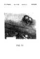
- FIG. 1Ais a photomicrograph of vascular smooth muscle cells of a 24-year-old male patient.
- FIG. 1Bis a photomicrograph of vascular smooth muscle cells in an artery of a 24-year-old male patient with vascular smooth muscle binding protein bound to the cell surface and membrane.
- the patientreceived the vascular smooth muscle binding protein by i.v. administration 4 days before the arterial tissue was prepared for histology.
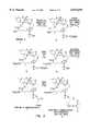
- FIG. 2depicts a first scheme for chemical coupling of a therapeutic agent to a vascular smooth muscle binding protein.
- FIG. 3depicts a second scheme for chemical coupling of a therapeutic agent to a vascular smooth muscle binding protein.
- FIG. 4Agraphically depicts experimental data showing rapid binding of vascular smooth muscle binding protein to marker-positive test cells in vitro.
- FIG. 4Bgraphically depicts experimental data showing rapid binding of vascular smooth muscle binding protein to vascular smooth muscle cells in vitro.
- FIG. 5Apresents graphically experimental data showing undesirable cytotoxicity of even low levels of therapeutic conjugate (i.e., RA-NR-AN-01), and the free RA therapeutic agent, when vascular smooth muscle cells were treated for 24 hours in vitro.
- therapeutic conjugatei.e., RA-NR-AN-01
- FIG. 5Bgraphically presents experimental data showing the effects of RA-NR-AN-01 therapeutic conjugate on metabolic activity of marker-positive and -negative cells.
- the datashow undesirable nonspecific cytotoxicity of the conjugate for all these cells in a 24 hour treatment in vitro.
- the non-specificityresults from extracellular hydrolysis of the coupling ligand which exposes the tested cells to free drug.
- FIG. 6Agraphically depicts experimental data showing undesirable nonspecific cytotoxicity of PE-NR-AN-01 therapeutic conjugate for marker-positive and marker-negative test cells after 24 hours of treatment in vitro, even though the 24 hour treatment was followed by an overnight recovery period prior to testing the metabolic activity.
- FIG. 6Bdepicts experimental data showing nonspecific cytotoxicity of the free PE therapeutic agent on marker-positive and -negative test cells after 24 hours of treatment in vitro.
- FIG. 7Agraphically presents experimental data showing that a short 5 minute "pulse” treatment, i.e., instead of 24 hours, followed by exposure to [3H]leucine, with free RA therapeutic agent being nonspecifically cytotoxic, i.e., for control HT29 marker-negative cells, but, in contrast, the RA-NR-AN-01 therapeutic conjugate is not cytotoxic in this "pulse" treatment.
- FIG. 7Bpresents graphically experimental data showing that free RA therapeutic agent is nonspecifically cytotoxic for control HT29 marker-negative cells, even in a 5' "pulse” treatment followed by a 24 hour recovery period prior to [3H]leucine exposure, but, in contrast, the RA-NR-AN-01 therapeutic conjugate is not cytotoxic to cells.
- FIG. 7Cpresents graphically results of experiments showing that "pulse" treatment of cells in vitro with the RA-NR-AN-01 therapeutic conjugate inhibits cellular activity in marker-positive A375 cells, as measured by protein synthesis.
- FIG. 7Dpresents graphically experimental data showing that "pulse" treatment of cells in vitro with the RA-NR-AN-01 therapeutic conjugate did not exert long-lasting inhibitory effects on cellular activity in marker-positive cells, since protein synthesis in A375 cells was not inhibited when the cells were allowed an overnight recovery period prior to testing in vitro.
- FIG. 8Apresents graphically experimental data showing that while a "pulse" treatment of cells in vitro with free RA therapeutic agent was non-specifically cytotoxic, the RA-NR-AN-01 therapeutic conjugate did not exert long-lasting inhibitory effects on cellular activity in vascular smooth muscle cells, as evidenced by metabolic activity in BO54 cells that were allowed a 48 hour recovery period prior to testing.
- FIG. 8Bgraphically depicts experimental data similar to those presented in FIG. 8A, above, but using a second marker-positive cell type, namely A375, the data show that "pulse" treatment with the RA-NR-AN-01 therapeutic conjugate did not exert long-lasting inhibitory effects on cellular activity, as measured by metabolic activity in A375 cells that were allowed a 48 hour recovery period prior to testing.
- a second marker-positive cell typenamely A375
- FIG. 8Cgraphically depicts results similar to those presented in FIG. 8A and FIG. 8B, above, but using a marker-negative control cell type, namely HT29.
- the resultsshow that the "pulse" treatment with the RA-NR-AN-01 therapeutic conjugate did not exert long-lasting inhibitory effects on the cellular activity of marker-negative control cells, as measured by metabolic activity in HT29 cells that were allowed a 48 hour recovery period prior to testing.
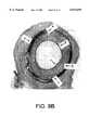
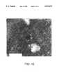
- FIG. 9Ashows stenosis due to intimal smooth muscle cell proliferation in a histological section of an untreated artery 5 weeks after angioplasty in an animal model.
- FIG. 9Bshows inhibition of stenosis in a histological section of an artery treated with therapeutic conjugate at 5 weeks after angioplasty in an animal model.
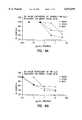
- FIG. 10Agraphically depicts experimental data comparing protein synthesis and DNA synthesis inhibition capability of suramin with respect to vascular smooth muscle cells.
- FIG. 10Bgraphically depicts experimental data comparing protein synthesis and DNA synthesis inhibition capability of staurosporin with respect to vascular smooth muscle cells.
- FIG. 10Cgraphically depicts experimental data comparing protein synthesis and DNA synthesis inhibition capability of nitroglycerin with respect to vascular smooth muscle cells.
- FIG. 10Dgraphically depicts experimental data comparing protein synthesis and DNA synthesis inhibition capability of cytochalasin B with respect to vascular smooth muscle cells.
- FIG. 11shows a tangential section parallel to the inner surface of a smooth muscle cell which is magnified 62,500 times and is characterized by numerous endocytic vesicles, several of which contain antibody coated gold beads in the process of being internalized by the cell in vitro.
- FIG. 12shows a smooth muscle cell which is magnified 62,500 times and is characterized by a marked accumulation of gold beads in lysosomes at 24 hours following exposure of the cell to the beads in vitro.
- FIG. 13shows a smooth muscle cell which is magnified 62,500 times and is characterized by accumulation of gold beads in lysosomes in vivo.
- FIG. 14depicts an in vivo dose response study of the effect of cytochalasin B on the luminal area of pig femoral arteries.
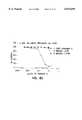
- FIGS. 15 and 16depict pathways for the modulation of vascular smooth muscle cell proliferation in vivo.
- “Therapeutic conjugate”means a vascular smooth muscle or an interstitial matrix binding protein coupled (e.g., optionally through a linker) to a therapeutic agent.
- Therapeutic agentincludes any moiety capable of exerting a therapeutic or prophylactic effect in the practice of the present invention.
- Target and markerare used interchangeably in describing the conjugate aspects of the present invention to mean a molecule recognized in a specific manner by the matrix or vascular smooth muscle binding protein, e.g., an antigen, polypeptide antigen or cell surface carbohydrate (e.g., a glycolipid, glycoprotein, or proteoglycan) that is expressed on the cell surface membranes of a vascular smooth muscle cell or a matrix structure.
- an antigen, polypeptide antigen or cell surface carbohydratee.g., a glycolipid, glycoprotein, or proteoglycan
- Epitopeis used to refer to a specific site within the “target” molecule that is bound by the matrix or smooth muscle binding protein, e.g., a sequence of three or more amino acids or saccharides.
- Coupledis used to mean covalent or non-covalent chemical association (i.e., hydrophobic as through van der Waals forces or charge-charge interactions) of the matrix or vascular smooth muscle binding protein with the therapeutic agent. Due to the nature of the therapeutic agents employed, the binding proteins will normally be associated with the therapeutic agents by means of covalent bonding.
- Linkermeans an agent that couples the matrix or smooth muscle binding protein to a therapeutic agent, e.g., an organic chemical coupler.
- “Migration” of smooth muscle cellsmeans movement of these cells in vivo from the medial layers of a vessel into the intima, such as may also be studied in vitro by following the motion of a cell from one location to another (e.g., using time-lapse cinematography or a video recorder and manual counting of smooth muscle cell migration out of a defined area in the tissue culture over time).
- Proliferationi.e., of smooth muscle cells or cancer cells, means increase in cell number, i.e., by mitosis of the cells.
- Abnormal or Pathological or Inappropriate Proliferationmeans division, growth or migration of cells occurring more rapidly or to a significantly greater extent than typically occurs in a normally functioning cell of the same type.
- “Expressed”means mRNA transcription and translation with resultant synthesis, glycosylation, and/or secretion of a polypeptide by a cell, e.g., CSPG synthesized by a vascular smooth muscle cell or pericyte.
- Microcyclic trichotheceneis intended to mean any one of the group of structurally related sesquiterpenoid macrocyclic mycotoxins produced by several species of fungi and characterized by the 12,13-epoxytrichothec-9-ene basic structure, e.g., verrucarins and roridins that are the products of secondary metabolism in the soil fungi Myrothecium verrucaria and Myrothecium roridium.
- sustained releasemeans a dosage form designed to release a therapeutic agent therefrom for a time period ranging from about 3 to about 21 days. Release over a longer time period is also contemplated as a “sustained release” dosage form of the present invention.
- Dosage formmeans a free (non-targeted or non-binding partner associated) therapeutic agent formulation, as well as sustained release therapeutic formulations, such as those incorporating microparticulate or nanoparticulate, biodegradable or non-biodegradable polymeric material capable of binding to one or more binding proteins or peptides to deliver a therapeutic moiety dispersed therein to a target cell population.
- Staurosporinincludes staurosporin, a protein kinase C inhibitor of the following formula, ##STR1## as well as diindoloalkaloids having one of the following general structures: ##STR2## More specifically, the term “staurosporin” includes K-252 (see, for example, Japanese Patent Application No. 62,164,626), BMY-41950 (U.S. Pat. No. 5,015,578), UCN-01 (U.S. Pat. No. 4,935,415), TAN-999 (Japanese Patent Application No. 01,149,791), TAN-1030A (Japanese Patent Application No. 01,246,288), RK-286C (Japanese Patent Application No.
- Derivatives of staurosporininclude those discussed in Japanese Patent Application Nos. 03,72,485; 01,143,877; 02,09,819 and 03,220,194, as well as in PCT International Application Nos. WO 89 07,105 and WO 91 09,034 and European Patent Application Nos. EP 410,389 and EP 296,110.
- Derivatives of K-252, a natural productare known. See, for example, Japanese Patent Application Nos.
- Cytochalasinincludes fungal metabolites exhibiting an inhibitory effect on target cellular metabolism, including prevention of contraction or migration of vascular smooth muscle cells.
- cytochalasinsinhibit the polymerization of monomeric actin (G-actin) to polymeric form (F-actin), thereby inhibiting cell functions requiring cytoplasmic microfilaments
- Cytochalasinstypically are derived from phenylalanine (cytochalasins), tryptophan (chaetoglobosins), or leucine (aspochalasins), resulting in a benzyl, indol-3-yl methyl or isobutyl group, respectively, at position C-3 of a substituted perhydroisoindole-1-one moiety (Formula V or VI).
- the perhydroisoindole moietyin turn contains an 11-, 13- or 14-atom carbocyclic- or oxygen-containing ring linked to positions C-8 and C-9.
- All naturally occurring cytochalasinscontain a methyl group at C-5; a methyl or methylene group at C-12; and a methyl group at C-14 or C-16.
- Exemplary moleculesinclude cytochalasin A, cytochalasin B, cytochalasin C, cytochalasin D, cytochalasin E, cytochalasin F, cytochalasin G, cytochalasin H, cytochalasin J.
- cytochalasin Kcytochalasin L
- cytochalasin Mcytochalasin N
- cytochalasin Ocytochalasin P
- cytochalasin Qcytochalasin R
- cytochalasin Schaetoglobosin A
- chaetoglobosin Bchaetoglobosin C
- chaetoglobosin Dchaetoglobosin E
- chaetoglobosin Fchaetoglobosin G
- chaetoglobosin Jchaetoglobosin K
- deoxaphominproxiphomin, protophomin
- zygosporin Dzygosporin E
- zygosporin Fzygosporin G
- aspochalasin Baspochalasin C
- aspochalasin D and the likeas well as functional equivalents and derivatives thereof.
- cytochalasin derivativesare set forth in Japanese Patent Nos. 72 01,925; 72 14,219; 72 08,533; 72 23,394; 72 01924; and 72 04,164.
- Cytochalasin Bis used in this description as a prototypical cytochalasin.
- tamoxifenincludes trans-2-[4-(1,2-diphenyl-1-butenyl)phenoxy]-N,N-dimethyl-ethylamine which is capable of enhancing the production or activation of TGF-beta.
- the activated form of TGF-betain turn, inhibits vascular smooth muscle cell proliferation.
- Evidence exists that tamoxifenalso acts to stabilize or organize areas of smooth muscle cell trauma. This organization/stabilization may stem from a blockage of smooth muscle cell maturation.
- Functional equivalents and derivatives of the aforementioned chemical compoundare also included within the scope of the term "tamoxifen” for the purposes of this disclosure.
- Exemplary tamoxifen functional equivalentsare plasmin, heparin, compounds capable of reducing the level or inactivating the lipoprotein Lp(a) or the glycoprotein apolipoprotein(a) and derivatives or analogs thereof.
- TGF-betaincludes transforming growth factor-beta as well as functional equivalents, derivatives and analogs thereof.
- TGF-betais a polypeptide produced in a latent propeptide form having, at this time, no identified biological activity. To be rendered active and, therefore, capable of inhibiting vascular smooth muscle cell proliferation, the propeptide form of TGF-beta must be cleaved.
- Functional equivalents of TGF-betaare, for example, moieties capable of disrupting cyclin-dependent protein kinase (CDK) transformation from a slow migrating form to a rapid migrating form, disrupting CDK-cyclin complex formation or activation or the like.
- CDKcyclin-dependent protein kinase
- TGF-beta activatorincludes moieties capable of directly or indirectly activating the latent form of TGF-beta to the active form thereof. Plasmin, plasmin activators, tamoxifen as well as analogs, derivatives or functional equivalents thereof are exemplary TGF-beta activators useful in the practice of the present invention.
- TGF-beta production stimulatorincludes moieties capable of directly or indirectly stimulating the production of TGF-beta (generally the latent form thereof).
- TGF-beta production stimulatorsmay be TGF-beta mRNA regulators (i.e., moieties that increase the production of TGF-beta mRNA), enhancers of TGF-beta mRNA expression or the like.
- Direct actionimplies that a first moiety acts on a second moiety, e.g., a TGF-beta activator acts on the latent form of TGF-beta.
- a TGF-beta activatoracts on the latent form of TGF-beta.
- Such direct action of TGF-beta production stimulatorsindicates that cells upon which the production stimulate acts to increase TGF-beta mRNA production or expression of TGF-beta.
- TGF-beta activatoracts on a moiety that itself or through one or more other moieties acts on latent TGF-beta.
- TGF-beta production stimulatorsindicates that the stimulators act on a moiety that itself or through one or more other moieties acts on a population of cells to stimulate the production of TGF-beta mRNA or the expression of TGF-beta.
- taxolincludes taxol, analogs thereof such as taxotere as well as functional equivalents or derivatives thereof. Taxol is readily taken up into cells and stabilizes such cells against cell division.
- cytostatic agentincludes moieties capable of inhibiting one or more pathological activities of target cells for a time sufficient to achieve a therapeutic benefit.
- smooth muscle cells and pericytesinclude those cells derived from the medial layers of vessels and adventitia vessels which proliferate in intimal hyperplastic vascular sites following injury, such as that caused during PTCA.
- Characteristics of smooth muscle cellsinclude a histological morphology (under light microscopic examination) of a spindle shape with an oblong nucleus located centrally in the cell with nucleoli present and myofibrils in the sarcoplasm. Under electron microscopic examination, smooth muscle cells have long slender mitochondria in the juxtanuclear sarcoplasm, a few tubular elements of granular endoplasmic reticulum, and numerous clusters of free ribosomes. A small Golgi complex may also be located near one pole of the nucleus.
- the majority of the sarcoplasmis occupied by thin, parallel myofilaments that may be, for the most part, oriented to the long axis of the muscle cell. These actin containing myofibrils may be arranged in bundles with mitochondria interspersed among them. Scattered through the contractile substance of the cell may also be oval dense areas, with similar dense areas distributed at intervals along the inner aspects of the plasmalemma.
- Pericytesinclude a histological morphology (under light microscopic examination) characterized by an irregular cell shape. Pericytes are found within the basement membrane that surrounds vascular endothelial cells and their identity may be confirmed by positive immuno-staining with antibodies specific for alpha smooth muscle actin (e.g., anti-alpha-sm1, Biomakor, Rehovot, Israel), HMW-MAA, and pericyte ganglioside antigens such as MAb 3G5 (11); and, negative immuno-staining with antibodies to cytokeratins (i.e., epithelial and fibroblast markers) and von Willdebrand factor (i.e., an endothelial marker). Both vascular smooth muscle cells and pericytes are positive by immunostaining with the NR-AN-01 monoclonal antibody.
- alpha smooth muscle actine.g., anti-alpha-sm1, Biomakor, Rehovot, Israel
- HMW-MAAe.g.
- the therapeutic conjugates and dosage forms of the inventionare useful for inhibiting the activity of vascular smooth muscle cells, e.g., for reducing, delaying, or eliminating stenosis following angioplasty.
- reducingmeans decreasing the intimal thickening that results from stimulation of smooth muscle cell proliferation following angioplasty, either in an animal model or in man.
- Delayingmeans delaying the time until onset of visible intimal hyperplasia (e.g., observed histologically or by angiographic examination) following angioplasty and may also be accompanied by "reduced” restenosis.
- "Eliminating" restenosis following angioplastymeans completely “treducing” and/or completely “delaying” intimal hyperplasia in a patient to an extent which makes it no longer necessary to surgically intervene, i.e., to re-establish a suitable blood flow through the vessel by repeat angioplasty, atheroectomy, or coronary artery bypass surgery.
- the effects of reducing, delaying, or eliminating stenosismay be determined by methods routine to those skilled in the art including, but not limited to, angiography, ultrasonic evaluation, fluoroscopic imaging, fiber optic endoscopic examination or biopsy and histology.
- the therapeutic conjugates of the inventionachieve these advantageous effects by specifically binding to the cellular membranes of smooth muscle cells and pericytes.
- Therapeutic conjugates of the inventionare obtained by coupling a vascular smooth muscle binding protein to a therapeutic agent.
- the vascular smooth muscle binding proteinperforms the function of targeting the therapeutic conjugate to vascular smooth muscle cells or pericytes, and the therapeutic agent performs the function of inhibiting the cellular activity of the smooth muscle cell or pericyte.
- Therapeutic dosage forms (sustained release-type) of the present inventionexhibit the capability to deliver therapeutic agent to target cells over a sustained period of time.
- Therapeutic dosage forms of this aspect of the present inventionmay be of any configuration suitable for this purpose.
- Preferred sustained release therapeutic dosage formsexhibit one or more of the following characteristics:
- microparticulatee.g., from about 0.5 micrometers to about 100 micrometers in diameter, with from about 0.5 to about 2 micrometers more preferred
- nanoparticulatee.g., from about 1.0 nanometer to about 1000 nanometers in diameter, with from about 50 to about 250 nanometers more preferred
- free flowing powder structuree.g., from about 0.5 micrometers to about 100 micrometers in diameter, with from about 0.5 to about 2 micrometers more preferred
- nanoparticulatee.g., from about 1.0 nanometer to about 1000 nanometers in diameter, with from about 50 to about 250 nanometers more preferred
- biodegradable structuredesigned to biodegrade over a period of time between from about 3 to about 180 days, with from about 10 to about 21 days more preferred, or non-biodegradable structure to allow therapeutic agent diffusion to occur over a time period of between from about 3 to about 180 days, with from about 10 to about 21 days preferred;
- biocompatible with target tissue and the local physiological environment into which the dosage form is being administeredincluding biocompatible biodegradation products;
- binding proteins or peptidesare capable of coupling to the particulate therapeutic dosage form through covalent ligand sandwich or non-covalent modalities as set forth herein.
- Nanoparticulate sustained release therapeutic dosage forms of preferred embodiments of the present inventionare biodegradable and bind to the vascular smooth muscle cells and enter such cells primarily by endocytosis.
- the biodegradation of such nanoparticulatesoccurs over time (e.g., 10 to 21 days) in prelysosomic vesicles and lysosomes.
- the preferred larger microparticulate therapeutic dosage forms of the present inventionbind to the target cell surface or interstitial matrix, depending on the binding protein or peptide selected, and release the therapeutic agents for subsequent target cell uptake with only a few of the smaller microparticles entering the cell by phagocytosis.
- a practitioner in the artwill appreciate that the precise mechanism by which a target cell assimilates and metabolizes a dosage form of the present invention depends on the morphology, physiology and metabolic processes of those cells.
- the size of the targeted sustained release therapeutic particulate dosage formsis also important with respect to the mode of cellular assimilation.
- the smaller nanoparticlescan flow with the interstitial fluid between cells and penetrate the infused tissue until it binds to the normal or neoplastic tissue that the binding protein/peptide is selected to target. This feature is important, for example, because the nanoparticles follow lymphatic drainage channels from infused primary neoplastic foci, targeting metastatic foci along the lymphatic tract. The larger microparticles tend to be more easily trapped interstitially in the infused primary tissue.
- biodegradable microparticulates or nanoparticulatesare biodegradable microparticulates or nanoparticulates. More preferably, biodegradable microparticles or nanoparticles are formed of a polymer containing matrix that biodegrades by random, nonenzymatic, hydrolytic scissioning to release therapeutic agent, thereby forming pores within the particulate structure.
- Polymers derived from the condensation of alpha hydroxycarboxylic acids and related lactonesare preferred for use in the present invention.
- a particularly preferred moietyis formed of a mixture of thermoplastic polyesters (e.g., polylactide or polyglycolide) or a copolymer of lactide and glycolide components, such as poly(lactide-co-glycolide).
- thermoplastic polyesterse.g., polylactide or polyglycolide
- a copolymer of lactide and glycolide componentssuch as poly(lactide-co-glycolide).
- An exemplary structure, a random poly(DL-lactide-co-glycolide),is shown below, with the values of x and y being manipulable by a practitioner in the art to achieve desirable microparticulate or nanoparticulate properties.
- agents suitable for forming particulate dosage forms of the present inventioninclude polyorthoesters and polyacetals (Polymer Letters, 18:293, 1980) and polyorthocarbonates (U.S. Pat. No. 4,093,709) and the like.
- Preferred lactic acid/glycolic acid polymer containing matrix particulates of the present inventionare prepared by emulsion-based processes, that constitute modified solvent extraction processes such as those described by Cowsar et al., "Poly(Lactide-Co-Glycolide) Microcapsules for Controlled Release of Steroids," Methods Enzymology, 11:101-116, 1985 (steroid entrapment in microparticulates); Eldridge et al., "Biodegradable and Biocompatible Poly(DL-Lactide-Co-Glycolide) Microspheres as an Adjuvant for Staphylococcal Enterotoxin B Toxoid Which Enhances the Level of Toxin-Neutralizing Antibodies," Infection and Immunity, 59:2978-2986, 1991 (toxoid entrapment); Cohen et al., “Controlled Delivery Systems for Proteins Based on Poly(Lactic/Glycolic Acid) Microspheres," Pharmaceutical Research, 8(6):713-
- the procedure for forming particulate dosage forms of the present inventioninvolves dissolving the polymer in a halogenated hydrocarbon solvent, dispersing a therapeutic agent solution (preferably aqueous) therein, and adding an additional agent that acts as a solvent for the halogenated hydrocarbon solvent but not for the polymer.
- the polymerprecipitates out from the polymer-halogenated hydrocarbon solution onto droplets of the therapeutic agent containing solution and entraps the therapeutic agent.
- the therapeutic agentis substantially uniformly dispersed within the sustained release dosage form of the present invention.
- theyare washed and hardened with an organic solvent. Water washing and aqueous non-ionic surfactant washing steps follow, prior to drying at room temperature under vacuum.
- particulate dosage formscharacterized by a therapeutic agent dispersed therein in matrix form
- Sterilizationmay be conducted in any convenient manner therefor.
- the particulatescan be irradiated with gamma radiation, provided that exposure to such radiation does not adversely impact the structure or function of the therapeutic agent dispersed in the therapeutic agent-polymer matrix or the binding protein/peptide attached thereto. If the therapeutic agent or binding protein/peptide is so adversely impacted, the particulate dosage forms can be produced under sterile conditions.
- Biodegradation ratedirectly impacts therapeutic agent release kinetics.
- the biodegradation rateis regulable by alteration of the composition or structure of the sustained release dosage form.
- alteration of the lactide/glycolide ratio in preferred dosage forms of the present inventioncan be conducted, as described by Tice et al., "Biodegradable Controlled-Release Parenteral Systems," Pharmaceutical Technology, pp. 26-35, 1984; by inclusion of polymer hydrolysis modifying agents, such as citric acid and sodium carbonate, as described by Kent et al., "Microencapsulation of Water Soluble Active Polypeptides," U.S. Pat.
- the preferred lactide/glycolide structureis biocompatible with the mammalian physiological environment. Also, these preferred sustained release-dosage forms have the advantage that biodegradation thereof forms lactic acid and glycolic acid, both normal metabolic products of mammals.
- Functional groups required for binding protein/peptide-particulate dosage form bonding to the particlesare optionally included in the particulate structure, along with the non-degradable or biodegradable polymeric units.
- Functional groups that are exploitable for this purposeinclude those that are reactive with peptides, such as carboxyl groups, amine groups, sulfhydryl groups and the like.
- Preferred binding enhancement moietiesinclude the terminal carboxyl groups of the preferred (lactide-glycolide) polymer containing matrix or the like.
- Useful vascular smooth muscle binding proteinis a polypeptide, peptidic, or mimetic compound (as described below) that is capable of binding to a target or marker on a surface component of an intact or disrupted vascular smooth muscle cell in such a manner that allows for either release of therapeutic agent extracellularly in the immediate interstitial matrix with subsequent diffusion of therapeutic agent into the remaining intact smooth muscle cells and/or internalization by the cell into an intracellular compartment of the entire targeted biodegradable moiety, permitting delivery of the therapeutic agent.
- useful vascular smooth muscle binding proteinsinclude antibodies (e.g., monoclonal and polyclonal affinity-purified antibodies, F(ab') 2 , Fab', Fab, and Fv fragments and/or complementarity determining regions (CDR) of antibodies or functional equivalents thereof, (e.g., binding peptides and the like)); growth factors, cytokines, and polypeptide hormones and the like; and macromolecules recognizing extracellular matrix receptors (e.g., integrin and fibronectin receptors and the like).
- antibodiese.g., monoclonal and polyclonal affinity-purified antibodies, F(ab') 2 , Fab', Fab, and Fv fragments and/or complementarity determining regions (CDR) of antibodies or functional equivalents thereof, (e.g., binding peptides and the like)); growth factors, cytokines, and polypeptide hormones and the like; and macromolecules recognizing extracellular matrix receptors (e.g.,
- binding peptides useful in targeting the dosage form embodiment of the present inventioninclude those that localize to intercellular stroma and matrix located between and among vascular smooth muscle cells. Such binding peptides deliver the therapeutic agent to the interstitial space between the target cells. The therapeutic agent is released into such interstitial spaces for subsequent uptake by the vascular smooth muscle cells.
- Preferred binding peptides of this typeare associated with epitopes on collagen, extracellular glycoproteins such as tenascin, reticulum and elastic fibers and other intercellular matrix material.
- Preferred tumor cell binding peptidesare associated with epitopes of myc, ras, bcr/Abl, erbB and like gene products, as well as mucins, cytokine receptors such as IL-6, EGF, TGF and the like, which binding peptides localize to certain lymphomas (myc), carcinomas such as colon cancer (ras), carcinoma (erbB), adenocarcinomas (mucins), breast cancer and hepatoma (IL-6 receptor), and breast cancer (EGF and TGF), respectively.
- Preferred immune system effector cell-binding peptidesare anti-TAC, IL-2 and the like, which localize to activated T cells and macrophages, respectively.
- Other preferred binding proteins/peptides useful in the practice of the present inventioninclude moieties capable of localizing to pathologically proliferating normal tissues, such as pericytes of the intraocular vasculature implicated in degenerative eye disease.
- Therapeutic agents of the inventionare selected to inhibit a cellular activity of a vascular smooth muscle cell, e.g., proliferation, migration, increase in cell volume, increase in extracellular matrix synthesis (e.g., collagens, proteoglycans, and the like), or secretion of extracellular matrix materials by the cell.
- a vascular smooth muscle celle.g., proliferation, migration, increase in cell volume, increase in extracellular matrix synthesis (e.g., collagens, proteoglycans, and the like), or secretion of extracellular matrix materials by the cell.
- the therapeutic agentacts either: a) as a "cytostatic agent” to prevent or delay cell division in proliferating cells by inhibiting replication of DNA (e.g., a drug such as adriamycin, staurosporin, tamoxifen or the like), or by inhibiting spindle fiber formation (e.g., a drug such as colchicine) and the like; or b) as an inhibitor of migration of vascular smooth muscle cells from the medial wall into the intima, e.g., an "anti-migratory agent” such as a cytochalasin; or c) as an inhibitor of the intracellular increase in cell volume (i.e., the tissue volume occupied by a cell; a "cytoskeletal inhibitor” or “metabolic inhibitor”); or d) as an inhibitor that blocks cellular protein synthesis and/or secretion or organization of extracellular matrix (i.e., an "anti-matrix agent” such as tamoxifen).
- DNAe.g.,
- cytostatic agentsinclude, e.g., modified toxins, methotrexate, adriamycin, radionuclides (e.g., such as disclosed in Fritzberg et al., U.S. Pat. No.
- protein kinase inhibitorse.g., staurosporin
- stimulators of the production or activation of TGF-betaincluding tamoxifen and functional equivalents or derivatives thereof, TGF-beta or functional equivalents, derivatives or analogs thereof, taxol or analogs thereof (e.g., taxotere), inhibitors of specific enzymes (such as the nuclear enzyme DNA topoisomerase II and DNA polymerase, RNA polymerase, adenyl guanyl cyclase), superoxide dismutase inhibitors, terminal deoxynucleotidyl- transferase, reverse transcriptase, antisense oligonucleotides that suppress smooth muscle cell proliferation and the like, which when delivered into a cellular compartment at an appropriate dosage will act to impair proliferation of a smooth muscle cell or pericyte without killing the cell.
- specific enzymessuch as the nuclear enzyme DNA topoisomerase II and DNA polymerase, RNA polymerase,
- cytostatic agentsinclude peptidic or mimetic inhibitors (i.e., antagonists, agonists, or competitive or non-competitive inhibitors) of cellular factors that may (e.g., in the presence of extracellular matrix) trigger proliferation of smooth muscle cells or pericytes: e.g., cytokines (e.g., interleukins such as IL-1), growth factors, (e.g., PDGF, TGF-alpha or -beta, tumor necrosis factor, smooth muscle- and endothelial-derived growth factors, i.e., endothelin, FGF), homing receptors (e.g., for platelets or leukocytes), and extracellular matrix receptors (e.g., integrins).
- cytokinese.g., interleukins such as IL-1
- growth factorse.g., PDGF, TGF-alpha or -beta, tumor necrosis factor, smooth muscle- and endothelial
- cytostatic agents for smooth muscle proliferationinclude: subfragments of heparin, triazolopyrimidine (Trapidil; a PDGF antagonist), lovastatin, and prostaglandins E1 or I2.
- anti-migratory agentsinclude inhibitors (i.e., agonists and antagonists, and competitive or non-competitive inhibitors) of chemotactic factors and their receptors (e.g., complement chemotaxins such as C5a, C5a desarg or C4a; extracellular matrix factors, e.g., collagen degradation fragments), or of intracellular cytoskeletal proteins involved in locomotion (e.g., actin, cytoskeletal elements, and phosphatases and kinases involved in locomotion).
- useful therapeutic agents in this category of anti-migratory agentsinclude: caffeic acid derivatives and nilvadipine (a calcium antagonist), and steroid hormones.
- Preferred anti-migratory therapeutic agentsare the cytochalasins.
- cytoskeletal inhibitorsinclude colchicine, vinblastin, cytochalasins, taxol and the like that act on microtubule and microfilament networks within a cell.
- the therapeutic conjugateis constructed with a therapeutic agent that is a simple trichothecene or a macrocyclic trichothecene, e.g., a verrucarin or roridin.
- Trichothecenesare drugs produced by soil fungi of the class Fungi imperfecti or isolated from Baccharus megapotamica (Bamburg, J. R. Proc. Molec. Subcell. Biol. 8:41-110, 1983; Jarvis & Mazzola, Acc. Chem. Res.
- trichothecenesThere are two broad classes of trichothecenes: those that have only a central sesquiterpenoid structure and those that have an additional macrocyclic ring (simple and macrocyclic trichothecenes, respectively).
- the simple trichothecenesmay be subdivided into three groups (i.e., Group A, B, and C) as described in U.S. Pat. Nos. 4,744,981 and 4,906,452 (incorporated herein by reference).
- Group A simple trichothecenesinclude: Scirpene, Roridin C, dihydrotrichothecene, Scirpen-4, 8-diol, Verrucarol, Scirpentriol, T-2 tetraol, pentahydroxyscirpene, 4-deacetylneosolaniol, trichodermin, deacetylcalonectrin, calonectrin, diacetylverrucarol, 4-monoacetoxyscirpenol, 4,15-diacetoxyscirpenol, 7-hydroxydiacetoxyscirpenol, 8-hydroxydiacetoxy-scirpenol (Neosolaniol), 7,8 - dihydroxydiacetoxyscirpenol, 7-hydroxy-8-acetyldiacetoxyscirpenol, 8-acetylneosolaniol, NT-1, NT-2, HT-2, T-2, and acet
- Group B simple trichothecenesinclude: Trichothecolone, Trichothecin, deoxynivalenol, 3-acetyldeoxynivalenol, 5-acetyldeoxynivalenol, 3,15-diacetyldeoxynivalenol, Nivalenol, 4-acetylnivalenol (Fusarenon-X), 4,15-idacetylnivalenol, 4,7,15-triacetylnivalenol, and tetra-acetylnivalenol.
- Group C simple trichothecenesinclude: Crotocol and Crotocin.
- Representative macrocyclic trichothecenesinclude Verrucarin A, Verrucarin B, Verrucarin J (Satratoxin C), Roridin A, Roridin D, Roridin E (Satratoxin D), Roridin H, Satratoxin F, Satratoxin G, Satratoxin H, Vertisporin, Mytoxin A, Mytoxin C, Mytoxin B, Myrotoxin A, Myrotoxin B, Myrotoxin C, Myrotoxin D, Roritoxin A, Roritoxin B, and Roritoxin D.
- trichothecenesesquiterpenoid ring structure
- baccharinsisolated from the higher plant Baccharis megapotamica, and these are described in the literature, for instance as disclosed by Jarvis et al. (Chemistry of Alleopathy, ACS Symposium Series No. 268: ed. A. C. Thompson, 1984, pp. 149-159).
- anti-matrix agentsinclude inhibitors (i.e., agonists and antagonists and competitive and non-competitive inhibitors) of matrix synthesis, secretion and assembly, organizational cross-linking (e.g., transglutaminases cross-linking collagen), and matrix remodeling (e.g., following wound healing).
- a representative example of a useful therapeutic agent in this category of anti-matrix agentsis colchicine, an inhibitor of secretion of extracellular matrix.
- Another exampleis tamoxifen for which evidence exists regarding its capability to organize and/or stabilize as well as diminish smooth muscle cell proliferation following angioplasty. The organization or stabilization may stem from the blockage of vascular smooth muscle cell maturation in to a pathologically proliferating form.
- therapeutic agentspreferably are those that inhibit vascular smooth muscle cell activity without killing the cells (i.e., cytostatic therapeutic agents).
- cytostatic therapeutic agentsAnother way to define a cytostatic agent is a moiety capable of inhibiting one or more pathological activities of the target cells for a time sufficient to achieve a therapeutic benefit.
- Preferred therapeutic agents for this purposeexhibit one or more of the following capabilities: to inhibit DNA synthesis prior to protein synthesis inhibition or to inhibit migration of vascular smooth muscle cells into the intima. These therapeutic agents do not significantly inhibit protein synthesis (i.e., do not kill the target cells) and, therefore, facilitate cellular repair and matrix production to stabilize the vascular wall lesion caused by angioplasty, by reducing smooth muscle cell proliferation.
- Exemplary of such preferred therapeutic agentsare protein kinase inhibitors, such as staurosporin (staurosporine is available from Sigma Chemical Co., St. Louis, Mo.) cytochalasins, such as cytochalasin B (Sigma Chemical Co.), and suramin (FBA Pharmaceuticals, West Haven, Connecticut), as well as nitroglycerin (DuPont Pharmaceuticals, Inc., Manuti, Puerto Rico) or analogs or functional equivalents thereof. These compounds are cytostatic and have been shown to exert minimal protein synthesis inhibition and cytotoxicity at concentrations where significant DNA synthesis inhibition occurs (see Example 8 and FIGS. 10A-10D).
- staurosporinstaurosporine is available from Sigma Chemical Co., St. Louis, Mo.
- cytochalasinssuch as cytochalasin B (Sigma Chemical Co.)
- suraminFBA Pharmaceuticals, West Haven, Connecticut
- nitroglycerinDuPont Pharmaceuticals, Inc., Manuti, Puerto Rico
- TGF-beta activators or production stimulatorssuch as tamoxifen and functional equivalents or derivatives thereof.
- TGF-beta and its analogs, derivatives or functional equivalentsmay also be employed.
- Taxol and its analogs, derivatives or functional equivalentsare also useful in the practice of the present invention.
- a useful protocol for identifying therapeutic agents useful in sustained release dosage form embodiments of the present inventionis set forth in Example 8, for example.
- a practitioner in the artis capable of designing substantially equivalent experimental protocols for making such an identification for different target cell populations, such as adherent monolayer target cell types.
- agents that are cytotoxic to cancer cellsinclude agents that are cytotoxic to cancer cells.
- Preferred agents for these embodimentsare Roridin A, Pseudomonas exotoxin and the like or analogs or functional equivalents thereof.
- a plethora of such therapeutic agents, including radioisotopes and the like,have been identified and are known in the art.
- protocols for the identification of cytotoxic moietiesare known and employed routinely in the art.
- Modulation of immune system-mediated disease effector cellscan also be accomplished using the sustained release dosage forms of the present invention. Such modulation is preferably conducted with respect to diseases having an effector cell population that is accessible through local sustained release dosage form administration.
- Therapeutic moieties having the requisite modulating activitye.g., cytocidal, cytostatic, metabolism modulation or like activity upon lymphorecticular cells in the treatment of arthritis (intra-articular administration), sprue (oral administration), uveitis and endophthalmitis (intra-ocular administration) and keratitis (sub-conjunctival administration), are identifiable using techniques that are known in the art. These agents can also be used to reduce hyperactivity of epithelial glands and endocrine organs that results in multiple disorders.
- Preferred agents for these embodimentsinclude Roridin A, Pseudomonas exotoxin, suramin, protein kinase inhibitors (e.g., staurosporin), TGF-beta and TGF-beta activators or production stimulators such as tamoxifen, taxol and the like, or analogs or functional equivalents thereof.
- compositions useful in the practice of the present inventioninclude moieties capable of reducing or eliminating pathological proliferation, migration or hyperactivity of normal tissues.
- exemplary of such therapeutic agentsare those capable of reducing or eliminating hyperactivity of corneal epithelium and stroma, pathological proliferation or prolonged contraction of smooth muscle cells or pericytes of the intraocular vasculature implicated in degenerative eye disease resulting from hyperplasia or decreased vascular lumen area.
- Preferred agents for this purposeare TGF-beta and TGF-beta activators or production stimulators such as tamoxifen, taxol and analogs thereof, staurosporin and cytochalasin B as well as functional equivalents or derivatives thereof.
- Vascular smooth muscle binding proteins of the inventionbind to targets on the surface of vascular smooth muscle cells.
- targetse.g., polypeptides or carbohydrates, proteoglycans and the like, that are associated with the cell membranes of vascular smooth muscle cells are useful for selecting (e.g., by cloning) or constructing (e.g., by genetic engineering or chemical synthesis) appropriately specific vascular smooth muscle binding proteins.
- Particularly useful "targets”are internalized by smooth muscle cells, e.g., as membrane constituent antigen turnover occurs in renewal. Internalization by cells may also be by mechanisms involving phagolysosomes, clathrin-coated pits, receptor-mediated redistribution or endocytosis and the like.
- such a “target”is exemplified by chondroitin sulfate proteoglycans (CSPGs) synthesized by vascular smooth muscle cells and pericytes, and a discrete portion (termed an epitope herein) of the CSPG molecule having an apparent molecular weight of about 250 kD is especially preferred.
- the 250 kD targetis an N-linked glycoprotein that is a component of a larger 400 kD proteoglycan complex (14).
- a vascular smooth muscle binding proteinis provided by NR-AN-01 monoclonal antibody (a subculture of NR-ML-05) that binds to an epitope in a vascular smooth muscle CSPG target molecule.
- the monoclonal antibody designated NR-ML-05reportedly binds a 250 kD CSPG synthesized by melanoma cells (Morgan et al., U.S. Pat. No. 4,897,255). Smooth muscle cells and pericytes also reportedly synthesize a 250 kD CSPG as well as other CSPGs (11).
- NR-ML-05 binding to smooth muscle cellshas been disclosed (Fritzberg et al., U.S. Pat. No.
- NR-ML-05Monoclonal antibody NR-ML-05 and subculture NR-ML-05 No. 85-41-4I-A2, freeze # A2106, have both been deposited with the American Type Culture Collection, Rockville, Md. and granted Accession Nos. HB-5350 and HB-9350, respectively.
- NR-ML-05is the parent of, and structurally and functionally equivalent to, subclone NR-AN-01, disclosed herein. It will be recognized that NR-AN-01 is just one example of a vascular smooth muscle binding protein that specifically associates with the 400 kD CSPG target, and that other binding proteins associating with this target and other epitopes in this target (14) are also useful in the therapeutic conjugates and methods of the invention.
- amino acid residues involved in the multi-point kinetic association of the NR-AN-01 monoclonal antibody with a CSPG marker antigenic epitopeare determined by computer-assisted molecular modeling and by the use of mutants having altered antibody binding affinity.
- the binding-site amino acids and three dimensional model of the NR-AN-01 antigen binding siteserve as a molecular model for constructing functional equivalents, e.g., short polypeptides ("minimal polypeptides"), that have binding affinity for a CSPG synthesized by vascular smooth muscle cells and pericytes.
- binding proteinse.g., antibodies or fragments
- selected binding proteinsfor use in the practice of the invention have a binding affinity of >10 4 liter/mole for the vascular smooth muscle 250 kD CSPG, and also the ability to be bound to and internalized by smooth muscle cells or pericytes.
- Three-dimensional modelingis also useful to construct other functional equivalents that mimic the binding of NR-AN-01 to its antigenic epitope, e.g., "mimetic” chemical compounds that mimic the three-dimensional aspects of NR-AN-01 binding to its epitope in a CSPG target antigen.
- mimal polypeptiderefers to an amino acid sequence of at least six amino acids in length.
- mimalrefers to an organic chemical polymer constructed to achieve the proper spacing for binding to the amino acids of, for example, an NR-AN-01 CSPG target synthesized by vascular smooth muscle cells or pericytes.
- murine monoclonal antibodymay be "chimerized” by genetically recombining the nucleotide sequence encoding the murine Fv region (i.e., containing the antigen binding sites) with the nucleotide sequence encoding a human constant domain region and an Fc region, e.g., in a manner similar to that disclosed in European Patent Application No. 0,411,893 A2. Some murine residues may also be retained within the human variable region framework domains to ensure proper target site binding characteristics.
- Humanized vascular smooth muscle binding partnerswill be recognized to have the advantage of decreasing the immunoreactivity of the antibody or polypeptide in the host recipient, which may thereby be useful for increasing the in vivo half-life and reducing the possibility of adverse immune reactions.
- binding peptides for restenosis treatment sustained release dosage forms of the present inventionare those that localize to intercellular stroma and matrix located between and among vascular smooth muscle cells. Such binding peptides deliver the therapeutic agent to the interstitial space between the target cells. The therapeutic agent is released into such interstitial spaces for subsequent uptake by the vascular smooth muscle cells.
- Preferred binding peptides of this typeare associated with epitopes on collagen, extracellular glycoproteins such as tenascin, reticulum and elastic fibers, cytokeratin and other intercellular matrix components.
- binding peptidesare also useful as binding peptides in this embodiment of the present invention.
- binding peptidesmay be identified and constructed or isolated in accordance with known techniques.
- the interstitial matrix binding proteinbinds to a target epitope with an association constant of at least about 10 -4 M.
- Useful binding peptides for cancer treatment embodiments of the present inventioninclude those associated with cell membrane and cytoplasmic epitopes of cancer cells and the like. These binding peptides localize to the surface membrane of intact cells and internal epitopes of disrupted cells, respectively, and deliver the therapeutic agent for assimilation into the target cells.
- Minimal peptides, mimetic organic compounds and human or humanized antibodies that localize to the requisite tumor cell typesare also useful as binding peptides of the present invention.
- Such binding peptidesmay be identified and constructed or isolated in accordance with known techniques.
- Preferred binding peptides of these embodiments of the present inventionbind to a target epitope with an association constant of at least about 10 -6 M.
- Binding peptides to membrane and cytoplasmic epitopes and the like that localize to immune system-mediated disease effector cells, e.g., cells of the lymphoreticular system,are also useful to deliver sustained release dosage forms of the present invention.
- the therapeutic agentis delivered to target cells for internalization therein by such sustained release dosage forms.
- Minimal peptides, mimetic organic compounds and human or humanized antibodies that localize to the requisite effector cell typesare also useful as binding peptides of the present invention.
- binding peptidesmay be identified and constructed or isolated in accordance with known techniques.
- Preferred binding peptides of these embodiments of the present inventionbind to a target epitope with an association constant of at least about 10 -6 M.
- binding proteins or peptides useful in the practice of the present inventioninclude moieties capable of localizing to pathologically proliferating normal tissues, such as pericytes of the intraocular vasculature implicated in degenerative eye disease.
- the therapeutic agentis delivered to target cells for internalization therein by such sustained release dosage forms.
- Minimal peptides, mimetic organic compounds and human or humanized antibodies that localize to the requisite pathologically proliferating normal cell typesare also useful as binding peptides of the present invention.
- binding peptidesmay be identified and constructed or isolated in accordance with known techniques.
- Preferred binding peptides of these embodiments of the present inventionbind to a target epitope with an association constant of at least about 1 -6 M.
- Coupled methods for linking the therapeutic agent through covalent or non-covalent bonds to the vascular smooth muscle binding proteininclude chemical cross-linkers and heterobifunctional cross-linking compounds (i.e., "linkers") that react to form a bond between reactive groups (such as hydroxyl, amino, amido, or sulfhydryl groups) in a therapeutic agent and other reactive groups (of a similar nature) in the vascular smooth muscle binding protein.
- This bondmay be, for example, a peptide bond, disulfide bond, thioester bond, amide bond, thioether bond, and the like.
- conjugates of monoclonal antibodies with drugshave been summarized by Morgan and Foon (Monoclonal Antibody Therapy to Cancer: Preclinical Models and Investigations, Basic and Clinical Tumor Immunology, Vol. 2, Kluwer Academic Publishers, Hingham, Mass.) and by Uhr J. of Immunol. 133:i-vii, 1984).
- the conjugatecontains a radionuclide cytostatic agent
- U.S. Pat. No. 4,897,255, Fritzberg et al.incorporated herein by reference, is instructive of coupling methods that may be useful.
- the therapeutic conjugatecontains a vascular smooth muscle binding protein coupled covalently to a trichothecene drug.
- the covalent bond of the linkagemay be formed between one or more amino, sulfhydryl, or carboxyl groups of the vascular smooth muscle binding protein and a) the trichothecene itself; b) a trichothecene hemisuccinate carboxylic acid; c) a trichothecene hemisuccinate (HS) N-hydroxy succinimidate ester; or d) trichothecene complexes with poly-L-lysine or any polymeric carrier.
- Representative examples of coupling methods for preparing therapeutic conjugates containing a trichothecene therapeutic agentare described in U.S. Pat. Nos. 4,906,452 and 4,744,981, incorporated herein by reference.
- Other examples using a hydrazide for forming a Schiff base linkage between binding proteins and trichothecenesare disclosed inpending U.S. patent application Ser. No. 07/415,154, incorporated herein by reference.
- the choice of coupling methodwill be influenced by the choice of vascular smooth muscle binding protein or peptide, interstitial matrix binding protein or peptide and therapeutic agent, and also by such physical properties as, e.g., shelf life stability, and/or by such biological properties as, e.g., half-life in cells and blood, intracellular compartmentalization route, and the like.
- vascular smooth muscle binding protein or peptideinterstitial matrix binding protein or peptide and therapeutic agent
- physical propertiese.g., shelf life stability
- biological propertiese.g., half-life in cells and blood, intracellular compartmentalization route, and the like.
- hemisuccinate conjugates of the Roridin A therapeutic agenthave a longer serum half-life than those of Verrucarin A, and this increased stability results in a significantly increased biological activity.
- the sustained release embodiment of the present inventionincludes a therapeutic agent dispersed within a non-biodegradable or biodegradable polymeric structure.
- a therapeutic agentdispersed within a non-biodegradable or biodegradable polymeric structure.
- Such dispersionis conducted in accordance with the procedure described by Cowsar et al., "Poly(Lactide-Co-Glycolide) Microcapsules for Controlled Release of Steroids," Methods Enzymology, 112:101-116, 1985; Eldridge et al., "Biodegradable and Biocompatible Poly(DL-Lactide-Co-Glycolide) Microspheres as an Adjuvant for Staphylococcal Enterotoxin B Toxoid Which Enhances the Level of Toxin-Neutralizing Antibodies," Infection and Immunity, 59:2978-2986, 1991; Cohen et al., “Controlled Delivery Systems for Proteins Based on Poly(Lactic/Glycolic Acid) Microsphere
- sustained release dosage form of the present inventionis amenable to several alternative modes of attachment to binding proteins or peptides.
- Dosage forms (sustained release-type) of the present inventionare capable of binding to binding proteins/peptides through, for example, covalent linkages, intermediate ligand sandwich attachment, or non-covalent adsorption or partial entrapment.
- the preferred poly-lactic/glycolic acid particulatesare formed with the therapeutic agent dispersed therein, the uncharged polymer backbone is oriented both inward (with the quasi lipophilic therapeutic agent contained therein) and outward along with a majority of the terminal carboxy groups.
- These surface carboxy groupsmay serve as covalent attachment sites when activated by, for example, a carbodiimide) for nucleophilic groups of the binding protein/peptide.
- nucleophilic groupsinclude lysine epsilon amino groups (amide linkage), serine hydroxyl groups (ester linkage) or cysteine mercaptan groups (thioester linkage). Reactions with particular groups depend upon pH and the reduction state of the reaction conditions.
- poly-lactic/glycolic acid particulates having terminal carboxylic acid groupsare reacted with N-hydroxybenztriazole in the presence of a water soluble carbodiimide of the formula R--N ⁇ C ⁇ N--R' (wherein R is a 3-dimethylaminopropyl group or the like and R' is an ethyl group or the like).
- the benztriazole-derivatized particulatesi.e., activated imidate-bearing moieties
- a protein/peptide nucleophilic moietysuch as an available epsilon amino moiety.
- binding protein/peptide nucleophilic moietiesinclude hydroxyl groups of serine, endogenous free thiols of cysteine, thiol groups resulting from reduction of binding protein/peptide disulfide bridges using reducing agents commonly employed for that purpose (e.g., cysteine, dithiothreitol, mercaptoethanol and the like) and the like.
- terminal carboxy groups of the poly lactic/glycolic acid particulatesare activatable by reaction with thionyl chloride to form an acyl chloride derivatized moiety.
- the derivatized particulatesare then reacted with binding peptide/protein nucleophilic groups to form targeted dosage forms of the present invention.
- Direct sustained release dosage form-binding protein or peptide conjugationmay disrupt binding protein/peptide target cell recognition.
- Ligand sandwich attachment techniquesare useful alternatives to achieve sustained release dosage form-binding protein/peptide attachment. Such techniques involve the formation of a primary peptide or protein shell using a protein that does not bind to the target cell population. Binding protein/peptide is then bound to the primary peptide or protein shell to provide the resultant particulate with functional binding protein/peptide.
- An exemplary ligand sandwich approachinvolves covalent attachment of avidin or streptavidin to the particulates through functional groups as described above with respect to the "direct" binding approach.
- the binding protein or peptideis derivatized, preferably minimally, with functionalized biotin (e.g., through active ester, hydrazide, iodoacetal, maleimidyl or like functional groups).
- Ligandi.e., binding peptide or protein/functionalized biotin attachment to the available biotin binding sites of the avidin/streptavidin primary protein shell occurs through the use of a saturating amount of biotinylated protein/peptide.
- poly-lactic/glycolic acid particulates having terminal carboxylic acid groupsare activated with carbodiimide and subsequently reacted with avidin or streptavidin.
- the binding protein or peptideis reacted with biotinamidocaproate N-hydroxysuccinimide ester at a 1-3 molar offering of biotin-containing compound to the binding protein/peptide to form a biotinylated binding protein/peptide.
- a molar excess of the biotinylated binding protein/peptideis incubated with the avidin-derivatized particulates to form a targeted dosage form of the present invention.
- the particulate carboxy groupsare biotinylated (e.g., through carbodiimide activation of the carboxy group and subsequent reaction with amino alkyl biotinamide).
- the biotinylated particulatesare then incubated with a saturating concentration (i.e., a molar excess) of avidin or streptavidin to form protein coated particulates having free biotin binding sites.
- a saturating concentrationi.e., a molar excess
- Such coated particulatesare then capable of reaction with a molar excess of biotinylated binding protein formed as described above.
- Another optioninvolves avidin or streptavidin bound binding peptide or protein attachment to biotinylated particulates.
- binding protein/peptide-particulate attachmentcan be achieved by adsorption of the binding peptide to the particulate, resulting from the nonionic character of the partially exposed polymer backbone of the particulate.
- high ionic strength conditionse.g., 1.0 molar NaCl
- hydrogen and hydrophobic particulate-binding protein/peptide bindingare favored.
- binding protein/peptidemay be partially entrapped in the particulate polymeric matrix upon formation thereof. Under these circumstances, such entrapped binding protein/peptide provides residual selective binding character to the particulate. Mild particulate formation conditions, such as those employed by Cohen et al., Pharmaceutical Research, 8: 713-720 (1991), are preferred for this embodiment of the present invention. Such entrapped binding protein is also useful in target cell reattachment of a partially degraded particulate that has undergone exocytosis.
- Other polymeric particulate dosage formse.g., non-biodegradable dosage forms having different exposed functional groups can be bound to binding proteins or peptides in accordance with the principles discussed above.
- non-biodegradable polymers useful in the practice of the present inventionare polystyrenes, polypropylenes, styrene acrylic copolymers and the like. Such non-biodegradable polymers incorporate or can be derivatized to incorporate functional groups for attachment of binding protein/peptide, including carboxylic acid groups, aliphatic primary amino groups aromatic amino groups and hydroxyl groups.
- Carboxylic acid functional groupsare coupled to binding protein or peptide using, for example, the reaction mechanisms set forth above for poly-lactic/glycolic acid biodegradable polymeric particulate dosage forms.
- Primary amino functional groupsare coupled by, for example, reaction thereof with succinic anhydride to form a terminal carboxy moiety that can be bound to binding peptide/protein as described above. Additionally, primary amino groups can be activated with cyanogen bromide and form guanidine linkages with binding protein/peptide primary amino groups.
- Aromatic amino functional groupsare, for example, diazotized with nitrous acid to form diazonium moieties which react with binding protein/peptide tyrosines, thereby producing a diazo bond between the non-biodegradable particulate and the binding protein/peptide.
- Hydroxyl functional groupsare coupled to binding protein/peptide primary amino groups by, for example, converting the hydroxyl moiety to a terminal carboxylic acid functional group. Such a conversion can be accomplished through reaction with chloroacetic acid followed by reaction with carbodiimide.
- Sandwich, adsorption and entrapment techniques, discussed above with respect to biodegradable particulates,are analogously applicable to non-biodegradable particulate-binding protein/peptide affixation.
- targetingis specific for potentially proliferating cells that result in increased smooth muscle in the intimal region of a traumatized vascular site, e.g., following angioplasty, e.g., pericytes and vascular smooth muscle cells.
- angioplastye.g., pericytes and vascular smooth muscle cells.
- aspects of the inventionrelate to therapeutic modalities in which the therapeutic conjugate of the invention is used to delay, reduce, or eliminate smooth muscle proliferation after angioplasty, e.g., PTCA, atheroectomy and percutaneous transluminal coronary rotational atheroblation.
- targetingis specific for primary or metastatic tumor foci accessible to local administration, e.g., tumors exposed for infiltration by laparotomy or visible for fluoroscopic or computerized tomography guiding and infusion needle administration to internal tumor foci or tumors confined to a small area or cavity within the mammal, e.g., ovarian cancer located in the abdomen, focal or multifocal liver tumors or the like.
- aspects of this embodiment of the inventioninvolve therapeutical modalities wherein the therapeutic agent is cytotoxic to the target cells or metabolically modulates the cells, increasing their sensitivity to chemotherapy and/or radiation therapy.
- targetingis specific for a local administration accessible effector cell population implicated in immune system-mediated diseases, e.g., arthritis, intra-ocular immune system-mediated disease or sprue.
- immune system-mediated diseasese.g., arthritis, intra-ocular immune system-mediated disease or sprue.
- aspects of this embodiment of the present inventioninvolve therapeutic modalities wherein the therapeutic agent is cytotoxic or modifies the biological response of the target cells to effect a therapeutic objective.
- targetingis specific for a local administration accessible pathologically proliferating or hyperactive normal cell population implicated in, e.g., degenerative eye disease, corneal pannus, hyperactive endocrine glands or the like.
- aspects of this embodiment of the present inventioninvolve therapeutic modalities wherein the therapeutic agent reduces or eliminates proliferation or hyperactivity of the target cell population.
- the therapeutic conjugates or dosage forms of the inventionmay be administered to the host using an infusion catheter, such as produced by C. R. Bard Inc., Billerica, Mass., or that disclosed by Wolinsky (7; U.S. Pat. No. 4,824,436) or Spears (U.S. Pat. No. 4,512,762).
- a therapeutically effective dosage of the therapeutic conjugatewill be typically reached when the concentration of conjugate in the fluid space between the balloons of the catheter is in the range of about 10 -3 to 10 -12 M.
- therapeutic conjugates of the inventionmay only need to be delivered in an anti-proliferative therapeutic dosage sufficient to expose the proximal (6 to 9) cell layers of the intimal or tunica media cells lining the lumen to the therapeutic anti-proliferative conjugate, whereas the anti-contractile therapeutic dosage needs to expose the entire tunica media, and further that this dosage can be determined empirically, e.g., by a) infusing vessels from suitable animal model systems and using immunohistochemical methods to detect the conjugate and its effects (e.g., such as exemplified in the EXAMPLES below); and b) conducting suitable in vitro studies such as exemplified in EXAMPLES 3, 4, and 5, below).
- this therapeutically effective dosageis achieved by preparing 10 ml of a 200 ⁇ g/ml therapeutic conjugate solution, wherein the vascular smooth muscle protein binding protein is NR-AN-01 and the therapeutic agent is Roridin A, a trichothecene drug.
- 10 mlwill commonly be sufficient volume to fill the catheter and infuse 1 to 1.5 ml into one to three traumatic lesion sites in the vessel wall.
- desired therapeutically effective dosages of a therapeutic conjugate according to the inventionwill be dependent on several factors, including, e.g.: a) the binding affinity of the vascular smooth muscle binding protein in the therapeutic conjugate; b) the atmospheric pressure applied during infusion; c) the time over which the therapeutic conjugate administered resides at the vascular site; d) the nature of the therapeutic agent employed; and/or e) the nature of the vascular trauma and therapy desired.
- Those skilled practitioners trained to deliver drugs at therapeutically effective dosagese.g., by monitoring drug levels and observing clinical effects in patients) will determine the optimal dosage for an individual patient based on experience and professional judgment.
- 0.3 atm (i.e., 300 mm of Hg) to about 5 atm of pressure applied for 15 seconds to 3 minutes directly to the vascular wallis adequate to achieve infiltration of a therapeutic conjugate containing the NR-AN-01 binding protein into the smooth muscle layers of a mammalian artery wall.
- a therapeutic conjugate containing the NR-AN-01 binding proteininto the smooth muscle layers of a mammalian artery wall.
- Sustained release dosage forms of an embodiment of the inventionmay only need to be delivered in an anti-proliferative therapeutic dosage sufficient to expose the proximal (6 to 9) cell layers of the tunica media smooth muscle cells lining the lumen to the dosage form.
- This dosageis determinable empirically, e.g., by a) infusing vessels from suitable animal model systems and using immunohistochemical, fluorescent or electron microscopy methods to detect the dosage form and its effects; and b) conducting suitable in vitro studies.
- this therapeutically effective dosageis achieved by determining in smooth muscle cell tissue culture the pericellular agent dosage, which at a continuous exposure results in a therapeutic effect between the toxic and minimal effective doses.
- This therapeutic levelis obtained in vivo by determining the size, number and therapeutic agent concentration and release rate required for particulates infused between the smooth muscle cells of the artery wall to maintain this pericellular therapeutic dosage.
- the dosage formshould release the therapeutic agent at a rate that approximates the pericellular dose of the following exemplary therapeutic agents: from about 0.01 to about 100 micrograms/ml nitroglycerin, from about 1.0 to about 1000 micrograms/ml of suramin, from about 0.001 to about 100 micrograms/ml for cytochalasin, and from about 0.01 to about 10 5 nanograms/ml of staurosporin as well as from about 0.001 to about 100 micrograms/ml taxol.
- TGF-beta activators or production stimulatorssuch as tamoxifen
- several exemplary dosing regimensare contemplated, depending upon the condition being treated and the stage to which the condition has progressed.
- a low chronic dosesufficient to elevate in vivo TGF-beta production or activation is contemplated.
- An exemplary dose of this typeis about 0.1 mg/kg/day (ranging between about 0.1 and about 10 mg/kg/day).
- Another exemplary dose rangeis from about 0.01 to about 1000 micrograms/ml.
- Low dosessuch as 0.1 ng/Kg/day, are also contemplated for use with respect to ameliorating stenosis following relatively low trauma injury or intervention, such as vein grafts or transplants or organ allografts, for example.
- relatively low trauma injury or interventionsuch as vein grafts or transplants or organ allografts, for example.
- No adverse side effectse.g., nausea as experienced by recipients of higher dose administrations when tamoxifen has been employed in the treatment of breast cancer
- a dosing regimenwhich involves a single "pre-loading" dose (or multiple, smaller pre-loading doses) given before or at the time of the intervention, with a chronic smaller (follow up) dose delivered for two to three weeks or longer following intervention.
- a single pre-loading dosemay be administered about 24 hours prior to intervention, while multiple preloading doses may be administered daily for several days prior to intervention.
- An exemplary single pre-loading doseis about 50 mg/kg (ranging between about 5 and about 1000 mg/kg), while an exemplary multiple pre-loading individual dose is about 10 mg/kg/day (ranging between about 0.01 and 10 mg/kg/day).
- Such a dosing regimenmay involve a systemic pre-loading dose followed by a sustained release chronic dose, or the sustained release dosage form may be designed to deliver a large dose over a short time interval as well as a smaller chronic dose for the desired time period thereafter.
- Some nauseamay be encountered at the higher dose; however, the use of a sustained release or other targeted dosage form is expected to obviate this side effect, because the recipient will not be subjected to a high systemic dose of the therapeutic agent.
- desired therapeutically effective dosages of the catheter administered sustained release dosage forms of the inventionwill be dependent on several factors, including, e.g.: a) the binding affinity of the binding protein associated with the dosage form, if any; b) the atmospheric pressure and duration of the infusion; c) the time over which the dosage form administered resides at the target site; d) the rate of therapeutic agent release from the particulate dosage form; e) the nature of the therapeutic agent employed; f) the nature of the trauma and/or therapy desired; and/or g) the intercellular and/or intracellular localization of the particulate dosage form.
- Those skilled practitioners trained to deliver drugs at therapeutically effective dosagesare capable of determining the optimal dosage for an individual patient based on experience and professional judgment.
- about 0.3 atm (i.e., 300 mm of Hg) to about 3 atm of pressure applied for 15 seconds to 3 minutes to the arterial wallis adequate to achieve infiltration of a sustained release dosage form bound to the NR-AN-01 binding protein into the smooth muscle layers of a mammalian artery wall.
- Wolinsky et al."Direct Intraarterial Wall Injection of Microparticles Via a Catheter: A Potential Drug Delivery Strategy Following Angioplasty," Am. Heart Jour., 122 (4:1136-1140, 1991.
- Those skilled in the artwill recognize that infiltration of a sustained release dosage form into a target cell population will probably be variable and will need to be determined on an individual basis.
- therapeutic conjugates radiolabeled with alpha-, beta- or gamma-emitters of known specific activitiesare useful for determining the therapeutically effective dosage by using them in animal studies and human trials with quantitative imaging or autoradiography of histological tissue sections to determine the concentration of therapeutic conjugate that is required by the therapeutic protocol.
- a therapeutically effective dosage of the therapeutic conjugate or dosage formwill be reached when at least three conditions are met: namely, (1) the therapeutic conjugate or dosage form is distributed in the intimal layers of the traumatically injured vessel; (2) the therapeutic conjugate or dosage form is distributed within the desired intracellular compartment of the smooth muscle cells, i.e., that compartment necessary for the action of the therapeutic agent, or the therapeutic agent released from the dosage form extracellularly is distributed within the relevant intracellular compartment; and (3) the therapeutic agent inhibits the desired cellular activity of the vascular smooth muscle cell, e.g., proliferation, migration, increased cellular volume, matrix synthesis, cell contraction and the like described above.
- the therapeutic dosage required to achieve the desired inhibitory activity for a therapeutic conjugate or dosage formcan also be anticipated through the use of in vitro studies.
- the infusion cathetermay be conveniently a double balloon or quadruple balloon catheter with a permeable membrane.
- a therapeutically effective dosage of a therapeutic conjugate or dosage formis useful in treating vascular trauma resulting from disease (e.g., atherosclerosis, aneurysm, or the like) or vascular surgical procedures such as angioplasty, atheroectomy, placement of a stent (e.g., in a vessel), thrombectomy, and grafting.
- Atheroectomymay be performed, for example, by surgical excision, ultrasound or laser treatment, or by high pressure fluid flow.
- Graftingmay be, for example, vascular grafting using natural or synthetic materials or surgical anastomosis of vessels such as, e.g., during organ grafting.
- a therapeutic conjugate containing Roridin A and NR-AN-01achieved a therapeutically effective dosage in vivo at a concentration which inhibited cellular protein synthesis in test cells in vitro by at least 5 to 50%, as judged by incorporation of radiolabeled amino acids.
- cell migration and cell adherence in in vitro assaysmay be used for determining the concentration at which a therapeutically effective dosage will be reached in the fluid space created by the infusion catheter in the vessel wall.
- While one representative embodiment of the inventionrelates to therapeutic methods employing an infusion catheter, it will be recognized that other methods for drug delivery or routes of administration may also be useful, e.g., injection by the intravenous, intralymphatic, intrathecal, intraarterial, local delivery by implanted osmotic pumps or other intracavity routes.
- Dosage form administration by these routes in accordance with the present inventionmay be continuous or intermittent, depending, for example, upon the patient's physiological condition, whether the purpose of the administration is therapeutic or prophylactic and other factors known to and evaluable by a skilled practitioner.
- nanoparticulate dosage forms of the present inventionare preferred.
- Intravenous administration of nanoparticulatesis useful, for example, where vascular permeability is increased in tumors for leakage, especially in necrotic areas of tumors having damaged vessels which allow the leakage of particles into the interstitial fluid, and where artery walls have been denuded and traumatized allowing the particles to enter the interstitial area of the tunica media.
- catheter and other administration routesare preferably conducted using dosage forms or therapeutic agents dispersed in a pharmaceutically acceptable carrier that is in liquid phase.
- pharmaceutically acceptable carriersfor these purposes include generally employed carriers, such as phosphate buffered saline solution, water, emulsions (e.g., oil/water and water/oil emulsions) and wetting agents of various types.
- non-coupled vascular smooth muscle cell binding proteine.g., free NR-AN-01 antibody
- non-coupled vascular smooth muscle cell binding proteinis administered prior to administration of the therapeutic agent conjugate or dosage form to provide a blocker of non-specific binding to cross-reactive sites. Blocking of such sites is important because vascular smooth muscle cell binding proteins will generally have some low level of cross-reactivity with cells in tissues other than the desired smooth muscle cells. Such blocking can improve localization of the therapeutic conjugate or dosage form at the specific vascular site, e.g., by making more of the therapeutic conjugate available to the cells.
- non-coupled vascular smooth muscle binding proteinis administered from about 5 minutes to about 48 hours, most preferably from about 5 minutes to about 30 minutes, prior to administration of the therapeutic conjugate or dosage form.
- the unlabeled specific "blocker”is a monovalent or bivalent form of an antibody (e.g., a whole antibody or an F(ab)' 2 , Fab, Fab', or Fv fragment of an antibody).
- the monovalent form of the antibodyhas the advantage of minimizing displacement of the therapeutic conjugate or dosage form while maximizing blocking of the non-specific cross-reactive sites.
- the non-coupled vascular smooth muscle cell binding proteinis administered in an amount effective to blocking binding of a least a portion of the non-specific cross-reactive sites in a patient. The amount may vary according to such factors as the weight of the patient and the nature of the binding protein. In general, about 0.06 mg to 0.20 mg per kg body weight or more of the unlabeled specific blocker is administered to a human.
- a second irrelevant vascular smooth muscle cell binding proteinmay optionally be administered to a patient prior to administration of the therapeutic conjugate or dosage form to reduce non-specific binding of the therapeutic conjugate or dosage form to tissues.
- the irrelevant binding proteinmay be an antibody which does not bind to sites in the patient through antigen-specific binding, but instead binds in a non-specific manner, e.g., through Fc receptor binding reticuloendothelial cells, asialo-receptor binding, and by binding to ubiquitin-expressing cells.
- the irrelevant "blocker”decreases non-specific binding of the therapeutic conjugate or dosage form and thus reduces side-effects, e.g., tissue toxicity, associated with the use of the therapeutic conjugate or dosage form.
- the irrelevant “blocker”is advantageously administered from 5 minutes to 48 hours, most preferably from 15 minutes to one hour, prior to administration of the therapeutic conjugate or dosage form, although the length of time may vary depending upon the therapeutic conjugate and route or method of injection.
- Representative examples of irrelevant "blockers”include antibodies that are nonreactive with human tissues and receptors or cellular and serum proteins prepared from animal sources that when tested are found not to bind in a specific manner (e.g., with a Ka ⁇ 10 3 M -1 ) to human cell membrane targets.
- conjugates and dosage forms of the inventionare not restricted in use for therapy following angioplasty; rather, the usefulness of the therapeutic conjugates and dosage forms will be proscribed by their ability to inhibit cellular activities of smooth muscle cells and pericytes in the vascular wall.
- other aspects of the inventioninclude therapeutic conjugates and dosage forms and protocols useful in early therapeutic intervention for reducing, delaying, or eliminating (and even reversing) atherosclerotic plaques and areas of vascular wall hypertrophy and/or hyperplasia.
- Therapeutic conjugates and dosage forms of the inventionalso find utility for early intervention in pre-atherosclerotic conditions, e.g., they are useful in patients at a high risk of developing atherosclerosis or with signs of hypertension resulting from atherosclerotic changes in vessels or vessel stenosis due to hypertrophy of the vessel wall.
- the therapeutic conjugates and dosage forms of the inventionmay also be used in therapeutic modalities for enhancing the regrowth of endothelial cells in injured vascular tissues and in many kinds of wound sites including epithelial wounds.
- the therapeutic conjugates and dosage forms of the inventionfind utility in inhibiting the migration and/or proliferation of smooth muscle cells or pericytes. Smooth muscle cells and pericytes have been implicated in the production of factors in vitro that inhibit endothelial cell proliferation, and their proliferation can also result in a physical barrier to establishing a continuous endothelium.
- the therapeutic conjugates and dosage forms of the inventionfind utility in promoting neo-angiogenesis and increased re-endothelialization, e.g., during wound healing, vessel grafts and following vascular surgery.
- the dosage formsmay also release therapeutic modalities that stimulate or speed up re-endothelialization of the damaged vessel wall.
- An exemplary therapeutic agent for this purposeis vascular permeability factor.
- Still other aspects of the inventionrelate to therapeutic modalities for enhancing wound healing in a vascular site and improving the structural and elastic properties of healed vascular tissues.
- these therapeutic modalities using the therapeutic conjugate or dosage form of the inventioni.e., to inhibit the migration and proliferation of smooth muscle cells or pericytes in a vessel wall
- the strength and quality of healing of the vessel wallare improved.
- Smooth muscle cells in the vascular wound sitecontribute to the normal process of contraction of the wound site which promotes wound healing. It is presently believed that migration and proliferation of smooth muscle cells and matrix secretion by transformed smooth muscle cells may detract from this normal process and impair the long-term structural and elastic qualities of the healed vessel.
- other aspects of the inventionprovide for therapeutic conjugates and dosage forms that inhibit smooth muscle and pericyte proliferation and migration as well as morphological transformation, and improve the quality of the healed vasculature.
- the present inventionalso provides a combination therapeutic method involving a cytocidal therapeutic conjugate and a cytostatic therapeutic agent.
- the cytocidal conjugateincludes a binding partner (such as a protein or peptide) capable of specifically localizing to vascular smooth muscle cells and an active agent capable of killing such cells.
- the cytocidal conjugateis administered, preferably intravenously or through any other convenient route therefor, localizes to the target smooth muscle cells, and destroys proliferating cells involved in stenotic or restenotic events. This cellular destruction causes the release of mitogens and other metabolic events, which events generally lead, in turn, to vascular smooth muscle cell proliferation.
- sustained release anti-proliferative or anti-contractile dosage forms of the present inventionare next administered, preferably through an infusion catheter or any convenient dosage form therefor.
- the sustained release dosage formretards the vascular smooth muscle cell proliferation and/or migration and contraction, thereby maintaining luminal diameter.
- This treatment methodologyconstitutes a biological arteromyectomy useful in stenotic vessels resulting from vascular smooth muscle cell hyperplasia and the like.
- a combination protocolcan be employed involving a, for example, systemically administered TGF-beta, TGF-beta activator or TGF-beta production stimulator capable of stabilizing or organizing the proliferation occurring at a diseased or traumatized smooth muscle site.
- the therapeutic or prophylactic agent combined by, for example, local administration in protocols employing the aforementioned stabilizer/organizermay be either a cytotoxic agent (e.g., free cytotoxic agent, a cytotoxic conjugate, or a sustained dosage form incorporating a cytotoxic agent) or a cytostatic agent (e.g., free, targeted or sustained release formulations of an agent capable of generating a biological stenting effect, an anti-migratory agent, a cytoskeletal inhibitor, a metabolic inhibitor, an anti-proliferative agent or the like).
- a cytotoxic agente.g., free cytotoxic agent, a cytotoxic conjugate, or a sustained dosage form incorporating a cytotoxic agent
- a cytostatic agente.g., free, targeted or sustained release formulations of an agent capable of generating a biological stenting effect, an anti-migratory agent, a cytoskeletal inhibitor, a metabolic inhibitor, an anti-proliferative agent or the like.
- the stabilizer or organizeris preferably administered prior to cytotoxic agent administration.
- a preferred embodiment of this aspect of the present invention for the prevention or treatment of restenosisfeatures the following steps:
- an effective amount of a, for example, Pseudomonas exotoxin-monoclonal antibody conjugate capable of localizing to vascular smooth muscle cellsis locally administered (e.g., via a catheter during an angioplasty procedure); and
- a follow up dose of tamoxifencould also be locally administered in step 2.
- Using this protocoloffers reduced and more highly organized or more stable proliferation by smooth muscles cells that are susceptible to a cytotoxic agent targeted thereto.
- the cytotoxic agentacts on the proliferating cells.
- the follow up doses of tamoxifenfacilitate the prevention of proliferation resulting from smooth muscle cell death caused by the action of the cytotoxic agent.
- the stabilizer or organizeris preferably administered prior to cytostatic agent administration.
- a preferred embodiment of this aspect of the present invention for the prevention or treatment of restenosisfeatures the following steps:
- an effective amount of cytochalasin Bis locally administered (e.g., via a catheter during an angioplasty procedure);
- a follow up dose of tamoxifencould also be locally administered in step 2.
- the present inventionalso provides methods for the treatment of cancer and immune system-mediated diseases through local administration of a targeted particulate dosage form.
- the particulate dosage formis, for example, administered locally into primary and/or metastatic foci of cancerous target cells.
- Local administrationis preferably conducted using an infusion needle or intraluminal administration route, infusing the particulate dosage form in the intercellular region of the tumor tissue or in luminal fluid surrounding the tumor cells.
- Primary foci introductionis preferably conducted with respect to target cells that are generally situated in confined areas within a mammal, e.g., ovarian carcinomas located in the abdominal cavity.
- the dosage form of the present inventionbinds to the target cell population and, optionally, is internalized therein for release of the therapeutic agent over time.
- Local administration of dosage forms of the present invention to such primary fociresults in a localized effect on such target cells, with limited exposure of other sensitive organs, e.g., the bone marrow and kidneys, to the therapeutic agent.
- the administered microparticles and larger nanoparticlesare primarily bound to the target cells situated near the infusion site and are, optionally, internalized for release of the therapeutic agent, thereby generating a marked and localized effect on the target cells immediately surrounding the infusion site.
- smaller nanoparticlesfollow interstitial fluid flow or lymphatic drainage channels and bind to target cells that are distal to the infusion site and undergoing lymphatic metastasis.
- the targeted dosage forms of this embodiment of the present inventioncan be used in combination with more commonly employed immunoconjugate therapy.
- the immunoconjugateachieves a systemic effect within the limits of systemic toxicity, while the dosage form of the present invention delivers a concentrated and sustained dose of therapeutic agent to the primary and metastatic foci, which often receive an inadequate therapeutic dose from such "systemic" immunoconjugate administration alone, and avoids or minimizes systemic toxic effects.
- the dosage forms of the present inventionare utilized to control immune system-mediated diseases.
- Exemplary of such diseasesare arthritis, sprue, uveitis, endophthalmitis, keratitis and the like.
- the target cell populations implicated in these embodiments of the present inventionare confined to a body cavity or space, such as joint capsules, pleural and abdominal cavity, eye and subconjunctival space, respectively.
- Local administrationis preferably conducted using an infusion needle for a intrapleural, intraperitoneal, intraocular or sub-conjunctival administration route.
- This embodiment of the present inventionprovides a more intense, localized modulation of immune system cells with minimal effect on the systemic immune system cells.
- the systemic cells of the immune systemare simultaneously treatable with a chemotherapeutic agent conjugated to a binding protein or peptide.
- a conjugatepreferably penetrates from the vascular lumen into target immune system cells.
- the local particulate dosage form administrationmay also localize to normal tissues that have been stimulated to proliferate, thereby reducing or eliminating such pathological (i.e., hyperactive) conditions.
- An example of this embodiment of the present inventioninvolves intraocular administration of a particulate dosage form coated with a binding protein or peptide that localizes to pericytes and smooth muscle cells of neovascularizing tissue. Proliferation of these pericytes causes degenerative eye disease.
- Preferred dosage forms of the present inventionrelease compounds capable of suppressing the pathological proliferation of the target cell population.
- the preferred dosage formscan also release compounds that increase vessel lumen area and blood flow, reducing the pathological alterations produced by this reduced blood supply.
- Still another aspect of the present inventionrelates to therapeutic modalities for maintaining an expanded luminal volume following angioplasty or other vessel trauma.
- a therapeutic agentcapable of inhibiting the ability of vascular smooth muscle cells to contract.
- agents useful in the practice of this aspect of the present inventionare those capable of causing a traumatized artery to lose vascular tone, such that normal vascular hydrostatic pressure (i.e., blood pressure) expands the flaccid vessel to or near to its maximal physiological diameter.
- Loss of vascular tonemay be caused by agents that interfere with the formation or function of contractile proteins (e.g., actin, myosin, tropomyosin, caldesmon, calponin or the like). This interference can occur directly or indirectly through, for example, inhibition of calcium modulation, phosphorylation or other metabolic pathways implicated in contraction of vascular smooth muscle cells.
- Inhibition of cellular contractionmay operate through two mechanisms to reduce the degree of vascular stenosis.
- Second, inhibition of cellular contractioncauses the smooth muscle wall to relax and dilate under normal vascular hydrostatic pressure (i.e., blood pressure).
- Therapeutic agentssuch as the cytochalasins, inhibit smooth muscle cell contraction without abolishing the protein synthesis necessary for traumatized, post-angioplasty or other surgically- or disease-damaged, smooth muscle cells to repair themselves. Protein synthesis is also necessary for the smooth muscle cells to secrete matrix, which fixes or retains the lumen in a state near its maximum systolic diameter as the vascular lesion stabilizes (i.e., a biologically-induced stenting effect).
- the cliniciancan eliminate over-inflation and its resultant trauma shock as a means to diminish or delay the vessel spasm or elastic recoil. Reduction or elimination of over-inflation decreases trauma to the muscular wall of the vessel, thereby reducing the determinants of smooth muscle cell proliferation in the intima and, therefore, reducing the incidence or severity of restenosis.
- Biological stentingalso decreases the incidence of thrombus formation.
- the incidence of mural microthrombiwas decreased as compared to the balloon traumatized arteries that were not treated with the therapeutic agent. This phenomenon appears to be a secondary benefit that may result from the increased blood flow through the traumatized vessel, said benefit being obtained through the practice of the present invention.
- Cytochalasinsare exemplary therapeutic agents capable of generating a biological stenting effect on vascular smooth muscle cells. Cytochalasins are thought to inhibit both migration and contraction of vascular smooth muscle cells by interacting with actin. The cytochalasins interact with the ends of filamentous actin to inhibit the elongation of the actin filaments. Low doses of cytochalasins (e.g., cytochalasin B) also disrupt microfilament networks of actin. In vitro data indicate that after vascular smooth muscle cells clear cytochalasin B, the cells regenerate enough polymerized actin to resume migration within about 24 hours. In vivo assessments reveal that vascular smooth muscle cells regain vascular tone within 2 to 4 days. It is during this recuperative period that the lumen diameter fixation and biological stenting effect occurs.
- cytochalasinsare thought to inhibit both migration and contraction of vascular smooth muscle cells by interacting with actin. The cytochalasins interact with the ends of filamentous
- the therapeutic agentmay be targeted, but is preferably administered directly to the traumatized vessel following the angioplasty or other traumatic event.
- the biological stenting effect of cytochalasin Bis achievable using a single infusion of the therapeutic agent into the traumatized region of the vessel wall at a dose concentration ranging from about 0.1 microgram/ml to about 1.0 micrograms/ml.
- vascular smooth muscle cell migrationhas been demonstrated in the same dose range (Example 11); however, a sustained exposure of the vessel to the therapeutic agent is preferable in order to maximize these anti-migratory effects. If the vascular smooth muscle cells cannot migrate into the intima, they cannot proliferate there. Should vascular smooth muscle cells migrate to the intima, a subsequently administered anti-proliferative sustained release dosage form inhibits the intimal proliferation.
- the sustained release dosage form of the present inventionincorporating a cytochalasin or other anti-proliferative therapeutic agent, can be administered in combination with a free cytochalasin therapeutic agent. In this manner, the biological stenting effect, as well as an anti-proliferative or anti-migratory effect, can be achieved in a single administration protocol.
- Agents useful in the protocols of the present inventionare identifiable, for example, in accordance with the following procedures.
- a potential agent for free agent (i.e., non-targeted) administrationexhibits one or more of the following characteristics:
- angioplastye.g., PTCA, percutaneous transluminal angioplasty (PTA) or the like
- PTApercutaneous transluminal angioplasty
- atheroectomye.g., rotoblater, laser and the like
- coronary artery bypass procedurese.g., coronary artery bypass procedures or the like
- vascular diseasee.g., atherosclerosis, eye diseases secondary to vascular stenosis or atrophy, cerebral vascular stenotic diseases or the like
- a therapeutic agent employed hereinwill have all four properties; however, the first and third are more important than the second and fourth for practice of the present invention.
- Cytochalasin Bwas evaluated to determine suitability for use in free therapeutic agent protocols.
- the biological stenting effect of cytochalasin Bis achievable using a single infusion of the therapeutic agent into the traumatized region of the vessel wall at a dose concentration ranging from about 0.1 microgram/ml to about 1.0 micrograms/ml.
- angioplastye.g., PTCA, percutaneous transluminal angioplasty (PTA) or the like
- PTApercutaneous transluminal angioplasty
- atheroectomye.g., rotoblater, laser and the like
- coronary artery bypass procedurese.g., coronary artery bypass procedures or the like
- vascular diseasee.g., atherosclerosis, eye diseases secondary to vascular stenosis or atrophy, cerebral vascular stenotic diseases or the like
- target cell proliferatione.g, following 5 minute and 24 hour exposure to the agent, in vitro vascular smooth muscle tissue cultures demonstrate a level of inhibition of 3 H-thymidine uptake and, preferably, display relatively less inhibition of 3 H-leucine uptake;
- (v)is cytostatic.
- the agentUpon identification of a therapeutic agent exhibiting one or more of the preceding attributes, the agent is subjected to a second testing protocol that involves longer exposure of vascular smooth muscle cells to the therapeutic agent.
- the agentupon long term (e .g, 5 days) exposure, the agent produces the same or similar in vitro effect on vascular smooth muscle tissue culture DNA synthesis and protein synthesis, as described above for the 5 minute and 24 hour exposures;
- the agentat an effective dose in the long term in vitro assay for DNA synthesis inhibition, the agent exhibits mild to moderate morphological cytotoxic effects over a longer term (e .g, 10 days).
- agents within the present inventiondemonstrate a 50% or greater inhibition of cell proliferation in the tunica media vascular smooth muscle cells, as indicated by a 1 hour "BRDU flash labeling" prior to tissue collection and histological evaluation. If an agent is effective for a period of time sufficient to inhibit intimal smooth muscle proliferation 50% or greater with a single exposure, it is an agent within the present invention that does not require administration in a sustained release dosage form.
- Agents having shorter duration activityare evaluated for sustained release if the systemic toxicity and potential therapeutic index appear to permit intravenous administration to achieve the 50% inhibition, or if the agent is amenable to local delivery to the vascular smooth muscle cells with sustained release at an effective anti-proliferative dose.
- Sustained release agentsare evaluated in a sustained release dosage form for dose optimization and efficacy studies.
- anti-proliferative agents useful in the practice of the present inventiondecrease vascular stenosis by 50% in balloon traumatized pig femoral arteries and, more preferably, to decrease vascular stenosis to a similar extent in pig coronary arteries. Such agents are then evaluable in human clinical trials.
- Staurosporinfor example, exhibits a differential between 3 H-leucine and 3 H-thymidine uptake such that it is cytostatic at administered doses. Longer duration cytotoxicity studies did not indicate that prolonged exposure to the therapeutic agent would adversely impact the target cells.
- BRDU pulsingindicated that staurosporin inhibits target cell proliferation. Any convenient method for evaluating the capability of inhibiting cell proliferation may alternatively be employed, however. Consequently, staurosporin is effective in retaining an expanded luminal volume.
- Lp(a)lipoprotein Lp(a)
- FIG. 15An in vivo pathway for the modulation of vascular smooth muscle cell proliferation is shown in FIG. 15. This mechanism is believed to constitute a portion of the mechanism that maintains vascular smooth muscle cells in a non-proliferative state in healthy vessels. The pathway has been elucidated by the inventors of a patent application filed on even date herewith, entitled Prevention and Treatment of Pathologies Associated with Abnormally Proliferative Smooth Muscle Cells.
- Vascular smooth muscle cell proliferationis inhibited by an active form of TGF-beta.
- Tamoxifenhas been shown by the experimentation detailed in Example 16 hereof to stimulate both the production and the activation of TGF-beta.
- Heparinstimulates the activation of TGF-beta by affecting the release of the active form of TGF-beta from inactive complexes present in serum.
- TGF-beta neutralizing antibodiesinhibit the activity of TGF-beta, thereby facilitating the proliferation of vascular smooth muscle cells.
- the apparent in vivo physiological regulator of the activation of TGF-betais plasmin. Plasmin is derived from plasminogen through activation by, for example, tPA (tissue plasminogen activator).
- Plasminogen and, therefore, plasmin activityis inhibited by the lipoprotein Lp(a) or apolipoprotein(a) (apo(a)), thereby decreasing the activation of the latent form of TGF-beta and facilitating proliferation of vascular smooth muscle cells.
- Resting smooth muscle cellsconstitute cells in their normal, quiescent non-proliferative state. Such resting smooth muscle cells may be converted to proliferating smooth muscle cells through activation by platelet derived growth factor (PDGF), fibroblast growth factor (FGF) or other stimulatory moieties.
- the proliferating smooth muscle cellsmay be converted to continual proliferating smooth muscle cells (ie., smooth muscle cells capable of generating a pathological state resulting from over-proliferation thereof) by an autocrine growth factor. This growth factor is believed to be produced by proliferating smooth muscle cells.
- An increased level of autocrine growth factorwhich can be inhibited by the active form of TGF-beta or an appropriately structured (e.g., designed) small molecule inhibitor, is believed to mediate the production of continual proliferating smooth muscle cells.
- Lp(a)consists of low density lipoprotein (LDL) and apo(a). Apo(a) shares approximately 80% amino acid identity with plasminogen (see MacLean et al., Nature, 330: 132, 1987). Lp(a) has been found to inhibit cell-associated plasminogen activity (see, for example, Harpel et al., Proc. Natl. Acad. Sci. USA, 86: 3847, 1989).
- DMENDulbecco's modified Eagles medium
- FCSfetal calf serum
- TGF-betais a potent growth inhibitor of a number of anchorage-dependent cells, including smooth muscle cells.
- TGF-betais produced as a latent propeptide having a covalently linked homodimer structure in which the active moiety is non-covalently linked to the amino-terminal portion of the propeptide.
- Latent TGF-betamust be cleaved (e.g., in vitro by acid treatment or in vivo by the serine protease plasmin) in order to become capable of inhibiting the proliferation of vascular smooth muscle cells. Plasmin is therefore a leading candidate to be a physiological regulator of TGF-beta.
- Lp(a) and apo(a)were acting on cultured human vascular smooth muscle cells by interfering with activation of latent TGF-beta.
- plasmin activity associated with vascular smooth muscle cellswas reduced 7-fold by Lp(a) and 5-fold by apo(a).
- the plasmin activity in the conditioned mediumwas also reduced by Lp(a) and apo(a) by about 2-fold, but was much lower than cell-associated plasmin activity in vascular smooth muscle cell cultures.
- plasminogen activator levels in human vascular smooth muscle cell cultureswere measured in the presence and absence of the lipoproteins and in the presence of a large excess of plasminogen, so that the lipoproteins present would not significantly act as competitive inhibitors.
- Total plasminogen activator activitywas not affected by the presence of any of the lipoproteins in the vascular smooth muscle cell cultures. For example, plasminogen activator activity in the conditioned medium remained at 0.7+/-0.06 mU/ml with Lp(a) additions up to 500 nM.
- Lp(a) and apo(a)both reduced the level of active TGF-beta by more than 100-fold compared to control or LDL-treated cultures.
- the level of total latent plus active TGF-beta measured by ELISA as described in Example 16was unaffected by the presence of Lp(a) or apo(a), however.
- plasminin the pathway was confirmed by studies in which inhibitors of plasmin activity were added to human vascular smooth muscle cells. Like Lp(a), these protease inhibitors increased cell number. Aprotinin, for example, decreased the population doubling time from 82+/-4 hours in control cultures to 48+/-5 hours, and alpha2-antiplasmin decreased the population doubling time to 45+/-2 hours. 500 nM Lp(a) and aprotinin addition resulted in only a slight additional stimulation of proliferation, with the population doubling time for cultures of this experiment being 45+/-6 hours. Neutralizing antibodies to TGF-beta similarly decreased population doubling time in vascular smooth muscle cells (see, for example, Example 16).
- Lp(a)plasmin inhibitors and neutralizing antibody to TGF-beta stimulate proliferation of vascular smooth muscle cells, while plasmin nullifies the growth stimulation of Lp(a).
- Tamoxifendid not significantly reduce the proportion of cells completing the cell cycle and dividing. Inhibition of vascular smooth muscle cells caused by tamoxifen therefore appears to be the result of an increase in the cell cycle time of nearly all (>90%) of the proliferating cells.
- Tamoxifendecreases the rate of proliferation of serum-stimulated vascular smooth muscle cells by increasing the time taken to traverse the G 2 to M phase of the cell cycle.
- Tamoxifendecreased the rate of proliferation of vascular smooth muscle cells by inducing TGF-beta activity.
- Vascular smooth muscle cellsproduced TGF-beta in response to tamoxifen. Tamoxifen appears to increase TGF-beta activity in cultures of rat vascular smooth muscle cells by stimulating the production of latent TGF-beta and increasing the proportion of the total TGF-beta which has been activated.
- Tamoxifenunlike heparin, does not act by releasing TGF-beta from inactive complexes present in serum.
- TGF-beta1 mRNAwas increased by approximately 10-fold by 24 hours after addition of tamoxifen (10 micromolar). This result suggests that the expression of TGF-beta mRNA by the smooth muscle cells will be increased, thereby facilitating decreased proliferation thereof by activated TGF-beta. This mechanism can be exploited using cells incorporating nucleic acids encoding TGF-beta mRNA, which cells are identifiable by persons skilled in the art employing known techniques.
- Tamoxifenis a selective inhibitor of vascular smooth muscle proliferation with an ED 50 at least 10-fold lower for vascular smooth muscle cells than for adventitial fibroblasts.
- Lp(a) or apo(a)substantially reduced the vascular smooth muscle cell proliferation inhibitory activity of tamoxifen, with the population doubling time in the presence of tamoxifen and Lp(a) being 42+/-2 hours. Also, the presence of Lp(a) reduced the levels of active TGF-beta produced in response to the addition of tamoxifen by about 50-fold. Addition of plasmin to rat vascular smooth muscle cells treated with tamoxifen and Lp(a) resulted in most of the TGF-beta being activated, and proliferation was again slowed (with the population doubling time being 57+/-3 hours). These observations are consistent with the theory that Lp(a) acts by inhibiting TGF-beta activation.
- TGF-beta activityincludes, but is not limited to, disruption of cyclin-dependent protein kinase (CDK) transformation from a slow migrating form to a rapid migrating form, disruption of CDK-cyclin complex formation or activation or the like.
- CDKcyclin-dependent protein kinase
- Identification of therapeutic agentsthat act to inhibit vascular smooth muscle cell proliferation by the pathway shown in FIG. 16 can be identified by a practitioner in the art by conducting experimentation using known techniques that is designed to identify growth factors made by proliferating smooth muscle cells, pericytes, lymphorecticular cells or the like, which growth factors also act on those cells (ie., autocrine growth factors).
- known techniques for rational drug designare then used to screen small molecules for the ability to inhibit the production or activity of such autocrine growth factors.
- Such experimental protocolsfacilitate the identification of therapeutic agents useful in the practice of the present invention and capable of one of the following activities:
- Smooth muscle cell proliferationis a pathological factor in myocardial infarctions, atherosclerosis, thrombosis, restenosis and the like.
- Therapeutic agents of the present inventionincluding tamoxifen, TGF-beta and the like, having at least one of the activities recited above and therefore being capable of inhibiting proliferation of vascular smooth muscle cells, are useful in the prevention or treatment of these conditions.
- Manipulation of the proliferation modulation pathway for vascular smooth muscle cells to prevent or reduce such proliferationremoves or reduces a major component of the arterial lesions of atherosclerosis and the restenosed arteries following angioplasty, for example.
- TGF-betachronically maintaining an elevated level of activated TGF-beta reduces the probability of atherosclerotic lesions forming as a result of vascular smooth muscle cell proliferation. Consequently, administration of TGF-beta, TGF-beta activators or TGF-beta production stimulators protects against atherosclerosis and subsequent myocardial infarctions that are consequent to coronary artery blockage. Also, substantially increasing the activated TGF-beta level for a short time period allows a recipient to at least partially offset the strong stimulus for vascular smooth muscle cell proliferation caused by highly traumatic injuries or procedures such as angioplasty. Continued lower dose delivery to the traumatized site further protects against restenosis resulting from vascular smooth muscle cell proliferation in the traumatized area.
- Taxolis believed to stabilize vascular smooth muscle cells against division by binding to microtubules and inhibiting the organization and ordering of the microtubule network. Cell migration may also be inhibited by this mechanism. Taxotere, an exemplary taxol analog, has a different method of action, but also inhibits cell division.
- FIG. 1Billustrates the binding of NR-AN-01 (a murine IgG2b MAb) to the smooth muscle cells in the vascular wall of an artery in a 24-year old male patient, 4 days after the i.v. administration of NR-AN-01.
- FIG. 1Bis a photomicrograph of a histological section taken through the medial region of an arterial wall of the patient after NR-AN-01 administration, where the section was reacted ex vivo with HRP-conjugated goat anti-mouse IgG.
- the reaction of the HRP-conjugate with NR-AN-01 MAbwas visualized by adding 4-chloro-1-naphthol or 3,3'-diaminobenzidine tetrahydrochloride as a peroxidase substrate (chromogen).
- the reaction product of the substrateforms an insoluble purple or dark brown precipitate at the reaction site (shown at #2, FIG. 1B).
- a counter stainwas used to visualize collagenous extracellular matrix material (shown at #2, FIG. 1B) or cell nuclei (#1, FIG. 1). Smooth muscle cells are visualized under microscopic examination as purple stained cells (FIG. 1A and FIG. 1B).
- This photomicrograph (FIG. 1B)demonstrates the ability of the MAb to specifically bind to human vascular smooth muscle in vivo, and to be internalized by the cells and remain in the cells for extended periods.
- Conjugates of NR-AN-01 and Roridin Awere constructed by chemically coupling a hemisuccinate derivative of the trichothecene cytotoxin (as described below) to a monoclonal antibody designated NR-AN-01. Two conjugates were prepared, one coupled at the Roridin A 2' position and one at the 13' position. Two schemes were used in this synthesis, as depicted in FIG. 2 and FIG. 3.
- the conjugatewas then purified from unreacted Roridin A by PD-10 SEPHAROSE® column chromatography (Pharmacia; Piscataway, N.J.), analyzed by size exclusion high pressure liquid chromatography, and the column fractions were characterized by SDS-PAGE and isoelectric focusing (IEF), as described below.
- FIG. 2shows diagrammatically the first reaction scheme for synthesis of Roridin A hemisuccinyl succinimidate (RA-HS-NHS) through a two step process with reagents: succinic anhydride, triethylamine (NEt 3 ) and dimethyl amino pyridine (DMAP) present in dichloromethane (CH 2 Cl 2 ) at room temperature (RT); and, N-hydroxysuccinimide (NHS) and dicyclohexyl carbodiimide (DCC) reagents also in CH 2 Cl 2 at RT.
- reagentssuccinic anhydride, triethylamine (NEt 3 ) and dimethyl amino pyridine (DMAP) present in dichloromethane (CH 2 Cl 2 ) at room temperature (RT); and, N-hydroxysuccinimide (NHS) and dicyclohexyl carbodiimide (DCC) reagents also in CH 2 Cl 2 at RT.
- NHSN-hydroxysucc
- FIG. 3shows diagrammatically the second reaction scheme for synthesis of Roridin A hemisuccinyl succinimidate (RA-HS-NHS) through a five step process with reagents: t-butyl dimethyl silyl chloride (TBMS-Cl) and imidazole in dimethylformamide (DMF) at room temperature (RT); acetic anhydride, triethylamine (TEA), and diethylaminopyridine in dichloromethane (CH 2 Cl 2 ) at RT; succinic anhydride, triethylamine (TEA) and dimethylaminopyridine (DMAP) in (CH 2 Cl 2 ) at RT; and, N-hydroxysuccinimide (NHS) and dicyclohexyl carbodiimide (DCC) reagents.
- TBMS-Clt-butyl dimethyl silyl chloride
- DMFdimethylformamide
- RTroom temperature
- acetic anhydridetri
- Conjugation reactionswere performed at pH 8.0 in borate buffer in the presence of 25% dimethylsulfoxide (DMSO) solvent at room temperature with gentle mixing for 45 minutes prior to purification by gel permeation chromatography.
- DMSOdimethylsulfoxide
- the molar trichothecene drug precursor to antibody offeringswere 25:1 and 40:1 for the 2' and 13' Roridin A analogues (3 and 8), respectively.
- Antibody concentrationwas 0.9 to 1.0 mg/ml during the conjugation reaction.
- the 2' conjugatewas quick frozen in liquid nitrogen and then stored at -70° C. until use.
- the 13' Roridin A NR-AN-01 conjugatewas stored frozen or refrigerated (i.e., 5-10° C.).
- Protein concentrationwas determined by BCA assay using the copper reagent method (Pierce Chemical Corp.).
- Immunoassay of the Roridin A-antibody conjugateswas performed by either competitive ELISA using biotinylated-Ab with Streptavidin/Peroxidase detection or by a competitive cell binding assay using 125 I-labeled antibody. Alternatively, immunoreactivity was measured under conditions of antigen saturation in a cell binding assay wherein antibody was first trace labeled with I-125 by the chloramine T method and then subsequently derivatized with 2' and 13' Roridin A precursors.
- the therapeutic conjugates of the inventionbe administered in less than 3 to 5 minutes, so that blood flow can be reestablished in the patient. Therefore, studies were conducted to determine the binding kinetics of a smooth muscle binding protein with a Ka of >10 9 liter/mole. Because human vascular smooth muscle cells grow slowly in culture, and baboon smooth muscle cells were found to express the human CSPG cell surface marker, B054 baboon artery smooth muscle cells and human A375 M/M (melanoma; ATCC #CRL1619) cells bearing CSPG surface marker were used in many of the studies described in the Examples, below.
- A375 M/M and B054 cellswere seeded in sterile 96 well microtiter plates at 2500 cells/well. Plates were wrapped in aluminum foil, and incubated at 37° C. overnight in a humidified atmosphere of 5% CO 2 /95% air. After approximately 18 hr, incubation plates were removed and cells were fixed with 0.05% glutaraldehyde for 5 minutes to prevent membrane turnover. Following fixation, the plates were exhaustively washed with PBS containing 0.5% Tween-20®.
- NR-AN-01 bound to the cellswas quantified by measuring the absorbance at 415 nm and 490 nm using an ELISA plate reader equipped for data acquisition by a Compaq computer.
- FIG. 4Agraphically depicts the results of in vitro studies in which A375 m/m marker-positive cells were held at 4° C. (i.e., to prevent membrane turnover) for 5 minutes (open squares, FIG. 4A), 15 minutes (closed diamonds, FIG. 4A), 30 minutes (closed squares, FIG. 4A) or 60 minutes (open diamonds, FIG. 4A) with different concentrations of NR-AN-01 (NRAN01 ⁇ g/ml).
- the binding of the NR-AN-01 MAb to the A375 cellswas quantified by washing to remove unbound antibody, adding HRP-conjugated goat anti-mouse IgG to react with the cell-bound MAb, washing to remove unbound goat second antibody, and adding 2,2'-azino-bis(3-ethylbenzthiazoline-6-sulfonic acid) (ABTS) substrate for peroxidase. Color development was monitored after 30 minutes at both 415 nm and 490 nm (ABS415,490).
- FIG. 4Bgraphically depicts the results of in vitro studies conducted in a manner similar to those described above in regard to FIG. 4A, but using B054 marker-positive smooth muscle cells, i.e., instead of the A375 m/m cells.
- FIG. 4A and FIG. 4Bshow significant binding of NR-AN-01 to A375 and B054 cells within 5 minutes at 4° C., even at the lowest dose of 20 ng/ml.
- the effects of Roridin A (RA) and RA-NR-AN-0l conjugates on cellular protein synthesis (i.e., by 3 H-leucine incorporation) and metabolic activity (i.e., by mitochondrial MTT assay)were tested in the experiments detailed in EXAMPLE 5 and EXAMPLE 6, below.
- the studies in EXAMPLE 4include experiments to determine the effects of long-term (i.e., 24 hour) treatment with the agents.
- the studies in EXAMPLE 5include experiments to determine the effects of "pulse” (i.e., 5 minute) treatment on cells. In both studies, the cellular specificity of the effects were evaluated by including "target" cells (i.e., cells bearing the CSPG "marker") and non-target cells.
- free-RAi.e., uncoupled
- the effects on cellular protein synthesis or metabolic activitywere evaluated either immediately following the treatment, or a "recovery period" was allowed (i.e., involving incubation of the cells overnight at 37° C.) to determine the long-term effects of the agents on the cell populations.
- a modified methyl-tetrazolium blue (MTT) assaywas utilized with 3- (4,5-dimethylthiazol-2-yl) -2,5-diphenyl tetrazoliumbromide (Sigma) to determine cellular metabolic activity. This assay is thought to measure cellular mitochondrial dehydrogenase activity.
- M14melanoma
- B054smooth muscle cell lines
- HT29 cellscolon carcinoma; ATCC #HTB38
- A375was used as a marker-positive cell.
- the HT29 and M14 cellswere seeded in 96-well microtiter plates at a concentration of 5.0 ⁇ 10 3 cells/well, and the B054 cells were seeded at 2.5 ⁇ 10 3 cells/well.
- Serial two-fold dilutions of free Roridin A and 2'RA-HS-NR-AN-01i.e., Roridin A coupled through a hemisuccinate (HS) coupling agent at the 2' position to NR-AN-01
- HShemisuccinate
- cellular metabolic activitywas determined by adding 20 ml to each well of a 5mg/ml MTT solution. The plates were covered and incubated at 37° C. for 4 hours and then the reaction was developed by adding 100 ml/well of 10% SDS/0.1N HCl. The dark blue solubilized formazan reaction product was developed at room temperature after 16-18 hours and quantified using an ELISA microtiter plate reader at an absorbance of 570 nm.
- FIG. 5Agraphically depicts the results of in vitro studies in which B054 marker-positive smooth muscle cells were incubated with different concentrations of RA-NR-AN-01 (NRAN01-RA; open squares, FIG. 5A) or free Roridin A (Free RA; closed diamonds, FIG. 5A) for a period of 24 hours, washed, and then returned to culture for an additional 16-18 hour overnight (o/n) recovery period prior to testing metabolic activity in an MTT assay.
- RA-NR-AN-01NRAN01-RA; open squares, FIG. 5A
- Free Roridin AFree RA; closed diamonds, FIG. 5A
- the concentrations of Free RA and RA-NR-AN-01are expressed as the calculated concentration of Roridin A (in mg/ml plotted on a log scale) in the assay (i.e., rather than the total mg/ml of NR-AN-01 protein in the assay), so that direct comparisons could be made.
- the metabolic activity of the cells in the MTT assayis presented as the percentage of the metabolic activity measured in a control untreated culture of cells (i.e., % control).
- FIG. 5Bgraphically depicts the results of in vitro studies conducted in a manner similar to those described above in regard to FIG. 5A, but comparing the effects of only RA-NR-AN-01 (NRAN01-RA) on three different cell types: namely, B054 marker-positive smooth muscle cells (BO54-NRAN01-RA; open squares, FIG. 5B); ET29 marker-negative control cells (HT29-NRAN01-RA; closed diamonds, FIG. 5B); and, M14 marker-positive cells (M14-NRAN01-RA; closed squares, FIG. 5B).
- the concentrations in the present experimentare expressed in terms of ug/ml of Roridin A.
- Metabolic activity of the cellsis expressed in a manner similar to that in FIG. 5A, i.e., as the percentage of activity measured in an untreated control culture of cells (% control).
- FIG. 5A and FIG. 5Bshow that metabolic activity measured in the MTT assay was significantly decreased in all populations of test cells, even 16-18 hours after a 24-hour incubation in either free Roridin A or the 2' or 13' RA-NR-AN-01 conjugates.
- the effects of the RA-NR-AN-01-conjugatesappeared to be non-specifically inhibitory for both target (BO54 and M14) and non-target (HT29) cells (FIGS. 5A and 5B). The inhibitory effects were observed at a free Roridin A or RA-conjugate concentration of >10 ng/ml.
- PEPseudomonas exotoxin
- FIG. 6Agraphically depicts the results of in vitro studies conducted in a manner similar to those described above in regard to FIG. 5A, but designed to study the metabolic effects of PE-NR-AN-01 (NRAN01-PE) on cells, i.e., rather than RA-NR-AN-01.
- Three different cell typeswere utilized: namely, B054 marker-positive smooth muscle cells (B054; open squares, FIG. 6A); HT29 marker-negative control cells (HT29; closed diamonds, FIG. 6A); and, M14 maker-positive cells (MT14; closed squares, FIG. 6A).
- B054 marker-positive smooth muscle cellsB054; open squares, FIG. 6A
- HT29 marker-negative control cellsHT29; closed diamonds, FIG. 6A
- M14 maker-positive cellsMT14; closed squares, FIG. 6A.
- the concentration of conjugateis expressed in ⁇ g/ml NR-AN-01 protein (plotted on a log scale), and the metabolic activity is expressed
- FIG. 6Bgraphically depicts the results of in vitro studies conducted in manner similar to those discussed above in regard to FIG. 6A, but designed to compare the effects obtained with free PE (PE) to those obtained above, i.e., in FIG. 6A, with PE-NR-AN-01.
- PEfree PE
- the cells, culture conditions, calculations, and presentation of the resultsare the same as in FIG. 6A, above.
- FIG. 6A and FIG. 6Bshow that 24 hours exposure to PE-NR-AN-01 or free PE was non-specifically inhibitory to cells at concentrations of >100 ng/ml.
- Roridin A-NR-AN-01coupled through a hemisuccinyl (HS) at either the 2' position (2'RA-HS-NR-AN-01) or the 13' position (13'RA-HS-NR-AN-01) were employed. (In the case of 13'RA-HS-NR-AN-01, the 2' position of Roridin A was also acetylated.)
- the RA, 2' or 13'RA-NR-AN-01 conjugateswere diluted two fold in sterile DMEM over a range of concentrations from 400 ng/ml to 780 pg/ml of Roridin A.
- test sampleswere all normalized to Roridin A, so that direct comparisons could be made of the effects at comparable doses.
- Sampleswere aliquoted (in duplicate) into duplicate microtiter plates at 100 ml/well and incubated at room temperature for five minutes.
- the incubation mediumwas removed (i.e., by aspiration) and 3 H-leucine was added (as above).
- protein synthesis of the cellswas evaluated by incubating the cells with the 3 H-leucine for 4 hours at 37° C. in a humidified chamber (as above), and all results are calculated by comparison with non-treated cells (i.e., 100% control).
- the cellswere removed from the substrata by trypsin-treatment, aspirated (using a PHDTM cell harvester (Cambridge Technology, Inc., Cambridge, Mass.)) and collected by filtration on glass fiber filters.
- the glass fiber filterswere dried and radioactivity quantified by liquid scintillation spectroscopy in a Beckman liquid scintillation counter.
- FIG. 7Agraphically depicts the results of in vitro studies conducted to investigate the effects on control HT29 marker-negative cells of a 5 minute exposure to different concentrations of Roridin A (Free RA; open squares, FIG. 7A), or 2'RA-NR-AN-01 (2'RA-NRAN01; closed squares, FIG. 7A), or 13'RA-NR-AN-01 (13'RA-NRAN01; closed triangles, FIG. 7A) conjugates.
- Roridin AFree RA; open squares, FIG. 7A
- 2'RA-NR-AN-01(2'RA-NRAN01; closed squares, FIG. 7A
- 13'RA-NR-AN-0113'RA-NRAN01; closed triangles, FIG. 7A
- the concentrations of Free RA, 2'RA-NR-AN-01 or 13'NR-AN-01are expressed as the calculated concentration of Roridin A in the assay (in ⁇ g/ml plotted on a log scale), i.e., rather than the total ⁇ g/ml of NR-AN-01 protein, so that direct comparisons of the results can be made.
- the cellswere treated for 5 minutes, washed, and then returned to culture for 4 hours, during which time cellular protein synthesis was evaluated by adding 0.5 mCi/ml of 3 H-leucine to the culture medium. At the end of the 4 hour period, cellular proteins were collected and radioactivity was determined. The results are expressed as the percentage of the radioactivity recorded in a control (non-treated) HT29 cell culture (i.e., %control).
- FIG. 7Bgraphically depicts the results of in vitro studies investigating the effects on control HT29 marker-negative cells of a 5 minute expose to different concentrations of Free RA (open squares, FIG. 7B), 2'RA-NRAN01 ( closed squares, FIG. 7B), or 13'RA-NRAN01 (closed triangles, FIG. 7B), as described above in regard to FIG. 7A, but in the present experiments the cells were incubated for a 16-18 hour recovery period (i.e., overnight; o/n) prior to testing protein synthesis in a four hour 3 H-leucine protein synthesis assay. The results are presented in a manner similar to those above in FIG. 7A.
- FIG. 7A and FIG. 7Bshow the short-term and long-term effects, respectively, of RA, 2'RA-HS-NR-AN-01, and 13'RA-HS-NR-AN-01 on protein synthesis by HT29 control cells.
- the resultsshow a dose-response inhibition of cellular protein synthesis by the free Roridin A, but not by RA-NR-AN-01, in HT29 cells.
- the inhibition triggered by RA during the 5 minutes of incubationwas still manifest after the 16-18 hours recovery period (FIG. 7B).
- treatment of non-target HT29 cells with 2'RA-HS-NR-AN-01 or 13'RA-HS-NR-AN-01did not result in detectable inhibition of protein synthesis.
- FIG. 7Cgraphically depicts the results of in vitro studies investigating the effects on A375 m/m marker-positive cells of a 5 minute exposure to different concentrations of Free RA (open squares, FIG. 7C), 2'RA-NR-AN-01 (closed squares, FIG. 7C) or 13'RA-NR-AN-01 (closed triangles, FIG. 7C), as described above in regard to FIG. 7A.
- the A375 cellswere incubated for 5 minutes in the test agent, washed, and tested for protein synthesis over the next 4 hours by adding 0.5 mCi/ml 3 H-leucine to the culture medium.
- the results of the experimentsare plotted in a manner similar to those described, above, in regard to FIG. 7A.
- FIG. 7Dgraphically depicts the results of in vitro studies investigating the effects on A375 m/ml marker-positive cells of a 5 minute exposure to different concentrations of Free RA (open squares, FIG. 7D), 2'RA-NRAN01 (closed squares, FIG. 7D), 13'RA-NRAN01 (closed triangles, FIG. 7D), as described above in regard to FIG. 7B.
- the A375 cellswere incubated for 5 minutes in the test agent, washed, and then returned to culture for a 16-18 hour recovery period (i.e., overnight; o/n Recovery), after which time protein synthesis was evaluated during a 4 hour 3 H-leucine protein synthesis assay.
- the results of the experimentsare plotted in a manner similar to those described above in regard to FIG. 7A.
- FIGS. 7C and FIG. 7Dshow the short-term and long-term effects, respectively, of RA, 2'RA-HS-NR-AN-01 and 13'-RA-HS-NR-AN-01 on protein synthesis by A375 target cells.
- Treatment of target cells with either the 2' or 13'RA-NR-AN-01 therapeutic conjugateresulted in a short-term inhibition of protein synthesis, ire., observed immediately after the 5-minute pulse treatment (FIG. 7C).
- FIG. 7Dshows that Roridin A is non-specifically inhibitory to test cells (i.e., in a manner similar to FIG. 7A and FIG. 7B, above) and that its effect on the cells is manifest even after a 16-18 hour recovery period.
- the specific effects of the RA-NR-AN-01 conjugates on target cells during a "pulse” treatmentappear to be a property of the NR-AN-01 binding protein.
- MTT assayswere conducted at 48 hours following a 5-minute exposure of target and non-target cells to RA or RA-NR-AN-01 conjugates.
- Target cells in these studiesincluded B054 and A375, and non-target cells included HT29 cells.
- Sterile 96 well microtiter plateswere seeded with 2500 cells/well, wrapped in aluminum foil and incubated in a humidified chamber containing 5% CO 2 /95% air for 16-18 hours.
- FIG. 8Agraphically depicts the results of in vitro studies investigating the effects on B054 marker-positive smooth muscle cells of a 5 minute exposure to different concentrations of Roridin A (open squares, FIG. 8A), 2'RA-NR-AN-01 (NRAN01-2'RA; closed diamonds, FIG. 8A), or 13' RA-NR-AN-01 (NRAN01-13'RA; closed squares, FIG. 8A).
- the experimentswere conducted in a manner similar to those described above in regard to FIG. 7B, but metabolic activity was assayed by MTT assay, i.e., rather than protein synthesis as in FIG. 7B, and cells were also given 48 hours to recover (rather than 24 hours, as in FIG. 7B).
- the results of the experimentsare plotted in a manner similar to those described (above) in regard to FIG. 7A .
- FIG. 8Bgraphically depicts the results of in vitro studies investigating the effects on A375 m/m marker-positive cells of a 5 minute exposure to different concentrations of Roridin A (open squares, FIG. 8B), 2'RA-NR-AN-01 (NRAN01-2'RA; closed diamonds, FIG. 8B), 13'RA-NR-AN-01 (NRAN01-13'RA; closed squares, FIG. 8B).
- the experimentswere conducted (and the results plotted) in a manner similar to those described above in regard to FIG. 8A.
- FIG. 8Cgraphically depicts the results of in vitro studies investigating the effects on HT29 marker-negative cells of a 5 minute exposure to different concentrations of Roridin A (open squares, FIG. 8C), 2'RA-NR-AN-01 (NRAN01-2'RA; closed diamonds, FIG. 8C), 13'RA-NR-AN-01 (NRAN01-13'RA; closed squares, FIG. 8C).
- the experimentswere conducted (and the results plotted) in a manner similar to those described above in regard to FIG. 8A.
- FIGS. 8A-8Cshow slight differences between the different RA-NR-AN-01 conjugates at the highest doses, but at the lower doses the 2' and 13'RA,-NR-AN-01 did not significantly inhibit target cell (i.e., B054 and A375) or non-target cell (i.e., HT29) metabolic activity over the long-term (i.e., 48 hours).
- target celli.e., B054 and A375
- non-target celli.e., HT29
- the therapeutic conjugates of the inventionare useful for inhibiting stenosis following vascular trauma or disease.
- vascular trauma that is induced during angioplastyis treated during the surgical procedure by removing the catheter used to perform the angioplasty, and inserting a balloon infusion catheter into the vessel.
- the infusion catheteris positioned with the instillation port (or, alternatively, a permeable membrane region) in the traumatized area of the vessel, and then pressure is applied to introduce the therapeutic conjugate.
- an infusion catheter with two balloonsmay be used, and when one balloon is inflated on either side of the trauma site a fluid space is created that can be filled with a suitable infusion fluid containing the therapeutic conjugate.
- HRPhorseradish peroxidase
- the coronary and femoral arteries of domestic pigs or non-human primateswere infused with NR-AN-01 for 45 seconds to 3 minutes at multiple pressures in the range of about 0.4 atmospheres (300 mm Hg) to 3 atmospheres.
- the vesselswere flushed with sterile saline and prepared for immunohistochemistry using HRP-conjugated goat anti-mouse IgG to detect the NR-AN-01 mouse IgG in the vessel wall. It was determined that full penetration was achieved of NR-AN-01 into these vessel walls at a pressure of 3 atmospheres after 3 minutes.
- Immunohistologywas also used to determine which animal model systems expressed the target antigen for NR-AN-01.
- Vascular tissue sections from readily available experimental animal specieswere exposed to NR-AN-01, washed, and reacted with HRP-conjugated goat anti-mouse IgG. Only non-human primates and swine were found to share the 250 kD NR-AN-01 target antigen with man.
- NR-AN-01could bind in a specific manner to its target antigen in vivo.
- the coronary and femoral arteries of domestic pigswere infused with therapeutic conjugates using an infusion catheter, the infusion sites were flushed with sterile saline, the surgical sites were then closed, and the animals were maintained for an additional 3-5 days.
- the vascular infusion siteswere excised and prepared for immunohistology, once again using goat anti-mouse IgG to identify NR-AN-01.
- NR-AN-01was identified in the vessel wall of swine coronary and femoral arteries 3-5 days after surgery, and the NR-AN-01 appeared to be associated only with vascular smooth muscle cells.
- Intimal smooth muscle proliferationthat follows balloon catheter-induced trauma is a good model to evaluate the therapeutic efficacy of conjugates for inhibiting smooth muscle cell activity in vivo in response to vascular trauma, including restenosis following angioplasty.
- Domestic pigswere used to study the effects of NR-AN-01 (i.e., termed vascular smooth muscle binding protein or simply VSMBP in these studies; and therapeutic conjugates with Roridin A are termed VSMBP - RA).
- VSMBP - RAvascular smooth muscle binding protein
- the events which normally follow balloon angioplasty in the porcine arteryhave been described previously (12).
- dilation of the carotid artery using an oversized balloonballoon: artery ratio approximately 1.5:1 resulted in complete endothelial denudation over an area of 1.5-2 cm in length.
- a single dose test protocolwas conducted in domestic pigs with NR-AN-01-Roridin A conjugates.
- Localized administration of the test conjugatesi.e., through a catheter into a region of traumatized vessel confined by temporary slip ligatures, was designed to reduce systemic toxicity while providing a high level of exposure for the target smooth muscle cells.
- This intra-artery route of administration in animal model studiessimulates the proposed route in human coronary arteries.
- the test protocolwas designed as an initial in vivo screening of intra-arteriolar, site specific, catheter administered, vascular smooth muscle binding protein (VSMBP) conjugates. Toxicity of free drug was also evaluated, i.e., for pathobiological effects on arteriolar smooth muscle cells.
- the therapeutically effective dosage of the Roridin A-NR-AN-01 conjugatewas determined by in vitro studies, and the proper intra-arteriolar administration pressure was determined by administering free MAb and MAb conjugates to animals, as described above in Example 7.
- Test conjugates and control compoundswere administered as a single intra-artery infusion at the site of endothelial denuding and trauma induced by a balloon catheter. Both the carotid and femoral arteries were abraded over 1 cm to 2 cm of endothelium by intraluminal passage of a 23 cm, size 3 (femoral) and size 4 (carotid) Uresil Vascu-Flo® silicone occlusion balloon catheter (Uresil Technology Center, Skokie, Ill.), sufficiently distended with saline to generate slight resistance. This technique produced slight distension of the artery.
- proximal and distal slip ligatureswere placed near the ends of the abraded region, and a size 8 French, Infant Feeding Catheter (Cutter-Resiflex, Berkeley, Calif.) attached to an Inflation Pro® (USCI, C. R. Bard, Inc., Billerica, Mass.) pressure syringe was used to administer the test conjugates and control compounds directly to the denuded segment at a pressure of three atmospheres for three minutes.
- the slip ligatureswere removed after the three minute exposure period and arterial blood flow was re-established.
- branches of the femoral or carotid arterieswere ligated with 00 silk suture as required to attain pressurized infusion in the treated region.
- the largest distal branch of the femoral artery(the saphenous artery) was incised and used as an entry site for the catheters which were then passed into the main femoral artery. Following this catheterization procedure in the main femoral artery, the secondary branch was ligated. In these cases, ligation or incision was used to allow entry of the catheters and the opening was then closed with 3 to 4 sutures of 5-0 monosilamen polybutester (Novafil, D & G Monofil Inc., Monati, PR).
- the pigswere kept in 3 ⁇ 5 foot indoor runs with cement floors during the quarantine and surgical recovery periods. They were then transferred to indoor/outdoor pens for the remainder of the five week healing period prior to collection of tissues for histology.
- the animalsrecovered normally from surgery with no evidence of hemorrhage or inflammation at the surgical sites. All six animals were examined 5 to 6 days after treatment with a doppler stethoscope, and all arteries in each of the animals were patent. Post treatment all animals had normal appetite, activity and weight gain.
- the arterieswere measured and the location of ligatures and gross abnormalities noted.
- the arterieswere transected at 2 mm intervals and arranged in order in cryomolds with O.C.T. (optimum cutting temperature) compound (Tissue Tek®, Miles Laboratories Inc., Elkhart, Ind.) and frozen in liquid nitrogen.
- O.C.T.optimum cutting temperature
- the blockswere sectioned at 5 microns and stained with H & E, Massons Trichrome and Movats Pentachrome for morphological studies. Sections were also used for immunohistological staining of vascular smooth muscle.
- FIG. 9Ashow (at 160 ⁇ magnification) a cross-sectional of an untreated artery 5 weeks after angioplasty.
- Dominant histological features of the arteryinclude displacement of the endothelium (see #1 in FIG. 9A) away from the internal elastic lamina (see #2, FIG. 9A), apparently due to intimal smooth muscle proliferation (see #3, FIG. 9A).
- FIG. 9Bshow (at 160 ⁇ magnification) a cross-section of a treated artery 5 weeks after angioplasty and infusion of the RA-NR-AN-01 therapeutic conjugate.
- the vessel in this sectionwas subjected to greater mechanical stresses than the vessel shown in FIG. 9A, with multiple sites where the external elastic membrane was ruptured and associated proliferation of smooth muscle cells in the outer layers of the media was observed (i.e., see #4 in FIG. 9B).
- Treatment with therapeutic conjugateinhibited intimal hypertrophy, as evidenced by the lack of displacement of the endothelium (see #1, FIG. 9B) from the internal elastic lamina (see #2, FIG. 9B).
- this inhibitory effect on intimal smooth muscle cellswas accomplished without inhibiting hypertrophy of medial smooth muscle cells in the areas where the external elastic membrane was ruptured (see #4, FIG. 9B).
- therapeutic conjugate administered at the site following angioplastyresulted in approximately 95% inhibition of the smooth muscle hypertrophy that restricted the lumen of the untreated vessel (FIG. 9A).
- the therapeutic conjugateexerted its effects on the smooth muscle cells migrating from the medial smooth muscle layers into the intima, without affecting either endothelium, or producing any signs of necrosis (i.e., cell death) in the smooth muscle cells in the medial layers of the arterial wall.
- necrosisi.e., cell death
- Studiesalso failed to show any histological signs of mononuclear infiltration or fibrosis such as might result from toxic effects on the vessel wall.
- Vascular smooth muscle cells at 40,000 cells/mlwere seeded in sterile 24 well plates at 1 ml/well. The plates were incubated overnight at 37° C., 5% CO 2 , 95% air in a humidified atmosphere (saturation). Log dilutions of the therapeutic agent of interest were incubated with the vascular smooth muscle cells for 5 minutes or 24 hours. Samples of the therapeutic agents were diluted in DMEM:F-12 medium (Whittaker Bioproducts, Walkersville, Md.) with 5% fetal bovine serum (FBS, Gibco BRL, Gaithersburg, Md.) and 5% Serum Plus® (JRH Biosciences, Lenexa, Kans.).
- Vascular smooth muscle cellswere incubated in complete medium with 5% FBS (Gibco) overnight at 37° C. in a humidified, 5% CO 2 environment in sterile 24 well plates.
- the mediumwas aspirated from the wells and serum free medium supplemented with growth factors (DMEM: F-12 basal medium supplemented with growth factor cocktail, catalog number I1884, which contains insulin (5 micrograms/ml), transferrin (5 micrograms/ml) and sodium selenite (5 nanograms/ml), available from Sigma Chemical, St. Louis, Mo.) was added. Cells were incubated in this medium for 24 hours.
- DMEMF-12 basal medium supplemented with growth factor cocktail, catalog number I1884, which contains insulin (5 micrograms/ml), transferrin (5 micrograms/ml) and sodium selenite (5 nanograms/ml), available from Sigma Chemical, St. Louis, Mo.
- Vascular smooth muscle cellswere seeded at 4.0 ⁇ 10 4 cells/ml medium/well on a commercially prepared four well slide (Nunc, Inc., Naperville, Ill.). Enough slides were seeded to accommodate two pulsed exposure lengths (5 minutes and 24 hours) and prescribed increment evaluation points (24 hours to 128 hours). All slides were run in duplicate to reveal any assay anomalies.
- the therapeutic agentwas diluted in the same medium used in the 3 H-leucine and 3 H-thymidine assays. Each four well slide was concentration bracketed to one log greater concentration (well "B"), one log lower concentration (well "D") of the minimal effective concentration (well "C”), as determined by the 3H-leucine and 3H-thymidine assays described above.
- one well(well "A") was left untreated (medium only). Incubation took place in a 37° C., 5% CO 2 humidified incubator. After each of the two (5 minutes and 24 hours) exposure points, the therapeutic agent medium was aspirated from each well, including the untreated well. One milliliter of fresh medium was then added to replace the aspirated medium. Re-incubation followed until each of the incremented evaluation points were achieved. At those points, the medium was aspirated and subsequently replaced with 1 ml of 10% neutral buffered formalin for one hour to allow for proper fixation. These fixed slides were stained by hematoxylin (nuclear) and eosin (cytoplasmic) for morphologic evaluation and grading.
- results of the 24 hour 3 H-leucine protein inhibition assay and the 24 hour 3 H-thymidine DNA synthesis inhibition assayare shown in FIGS. 10A-10D for suramin, staurosporin, nitroglycerin and cytochalasin B, respectively. All of the tested compounds showed an available therapeutic range (area under the curve of 3 H-leucine assay is greater than that resulting from the 3 H-thymidine assay), indicating usefulness in the practice of sustained release dosage form embodiments of the present invention. More specifically, the compounds inhibited the ability of vascular smooth muscle cells to undergo DNA synthesis in the presence of 5% FBS to a greater extent than they inhibited protein synthesis of vascular smooth muscle cells.
- the protein and DNA synthesis inhibitory effects of suramin, staurosporin, nitroglycerin and cytochalasin B during a 5 minute and 24 hour pulsed exposureare shown in FIG. 10 A-D, respectively.
- vascular smooth muscle cellsThe ability of vascular smooth muscle cells to bind and internalize particles coated with binding protein or peptide was demonstrated with monoclonal antibody (NR-AN-01) coated gold beads both in vitro and in vivo.
- NR-AN-01monoclonal antibody coated gold beads both in vitro and in vivo.
- the vascular smooth muscle cell tissue cultures (B054), an antigen positive control cell line (A375) and an antigen negative control cell line (HT29)were incubated with 10 nm gold beads, with one group coated with NR-AN-01 and a second, uncoated control group.
- the cellswere exposed to the beads as monolayer and cell suspension cultures, and were examined at six time points (i.e., 1 minute, 5 minutes, 15 minutes, 30 minutes, 60 minutes and 24 hours) for binding and internalization by electron microscopy.
- Table 3shows the results of the experimentation, indicating that the binding to the cell surface is specific.
- FIG. 11shows a tangential section parallel to the inner surface of a smooth muscle cell characterized by numerous endocytic vesicles, several of which contain antibody coated gold beads in the process of being internalized by the cell. These endocytic vesicles with particles attached to cell surface antigens were stimulated to fuse with lysosomes at a higher than expected rate for normal cell surface membrane recycling. The resultant marked accumulation of internalized particles was observed at the 24 hour time point and is shown in FIG. 12.
- NR-AN-01 coated gold beadswere infused via intravascular catheter, open ended with treated area occluded proximally and distally with slip ligatures, at 3 atm pressure applied for 3 minutes into the wall of a pig femoral artery immediately following balloon trauma.
- the bead internalization ratevaried with the degree of damage sustained by the vascular smooth muscle cell during the balloon trauma. Cells with minimal or no damage rapidly internalized the particles by endocytosis and phagocytosis, concentrating the internalized particles in lysosomes. Cells that were killed by the trauma exhibited surface bead binding. Cells that were damaged by the trauma but survived were characterized by bead surface binding with delayed internalization and lysosome concentration.
- FIG. 13shows particulate concentration in the lysosomes in vivo at one week following bead administration.
- B054 cells(baboon smooth muscle cells) were derived from explants of aortic baboon smooth muscle cells. Cells were expanded in DMEM (Dulbecco's Modified Eagle's Medium) :F-12 medium (Whittaker Bioproducts, Walkersville, Md.) with 5% fetal bovine serum (FBS, Gibco) and 5% Serum Plus® (JRH Biologicals) ("complete medium”), and a seed lot of cells was frozen in liquid nitrogen for future use at passage seven.
- DMEMDynabecco's Modified Eagle's Medium
- F-12 mediumWoodtaker Bioproducts, Walkersville, Md.
- Serum Plus®JRH Biologicals
- Vascular smooth muscle cells at 40,000-50,000 cells/mlwere seeded and processed as described in Example 8, "15 minute exposure; 3 H-leucine uptake.”
- Log dilutions of staurosporin200 ng/ml, 20 ng/ml, 2 ng/ml, 0.2 ng/ml and 0.02 ng/ml
- cytochalasin Blog dilutions at 20 ⁇ g/ml, 2.0 ⁇ g/ml, 0.2 ⁇ g/ml, 0.02 ⁇ g/ml and 0.002 ⁇ g/ml were dispersed in complete medium. Complete medium was then added to the control wells.
- DNA Synthesis AssayVascular smooth muscle (BO54) cells were seeded and processed in 24 well plates, as described above under "5 Minute Exposure: Protein Synthesis Assay.” After 5 min incubation with the test therapeutic agent, the medium was aspirated and 1 ml/well of 1.0 ⁇ Ci/ml 3 H-thymidine (rather than 3 H-leucine) dispersed in complete medium was added. The cells were then incubated overnight at 37° C. in a humidified, 5% CO 2 environment. The toxic effect of the therapeutic agent was then determined, as described in the Protein Synthesis Assay, above.
- Vascular smooth muscle (B054) cells at 20,000 cells/mlwere seeded in sterile 24 well plates and incubated in complete medium (1 ml/well) overnight at 37° C., 5% CO 2 , 95% air in a humidified atmosphere (saturation).
- Log dilutions of staurosporin100 ng/ml, 10 ng/ml, 1 ng/ml, 0.1 ng/ml and 0.01 ng/ml were dispersed sequentially in the two media, as described below.
- cytochalasin Blog dilutions at 10 ⁇ g/ml, 1.0 ⁇ g/ml, 0.1 ⁇ g/ml, 0.01 ⁇ g/ml and 0.001 ⁇ g/ml were dispersed sequentially in the two media, as described below:
- Medium (2)DMEM (leucine-free) with 0..5 ⁇ Ci/ml 3 H-leucine. Medium (2) is used for the final 24 hour incubation period of the experiment.
- each therapeutic agentwas diluted in Medium (2), as noted above.
- Medium (1)was aspirated from the wells, and aliquots of therapeutic agent dilutions in Medium (2) were added in quadruplicate to the appropriate wells.
- Medium (2)was then added to the control wells.
- each therapeutic agentwas diluted in Medium (1), as noted above.
- Medium (1)was aspirated from the wells, and aliquots of therapeutic agent dilutions in Medium (1) were added in quadruplicate to the appropriate wells.
- Medium (1)was then added to the control wells.
- the mediumwas changed every 24 hours, and fresh therapeutic agent was added to the test wells.
- each therapeutic agentwas diluted in Medium (2), as noted above.
- Medium (1)was aspirated from the wells, and aliquots of therapeutic agent dilutions in Medium (2) were added in quadruplicate to the appropriate wells.
- Medium (2)was then added to the control wells.
- test agents in 3 H-leucinewere incubated overnight at 37° C., 5% CO 2 in a humidified atmosphere.
- the toxic effect of the therapeutic agentswas then determined, as described in the 5 Minute Exposure: Protein Synthesis Assay, described above.
- the changes in cells at each dilutionwere photographed using a Zeiss microscope (Zeiss, West Germany) at 320X. The medium was then aspirated, and the cells were processed with TCA, as described above.
- DNA Synthesis AssayThis assay was performed according to the procedure described for "24 and 120 Hour Exposure; Protein Synthesis Assay", except Medium (2) in this 24 & 120 hr DNA Synthesis Assay is:
- the minimum effective dose (MED) of each agentwas determined as a percentage of the control that was treated with medium only; 50% of control values was chosen as the cytotoxicity benchmark.
- MEDminimum effective dose
- staurosporindemonstrated an MED of 100 ng/ml in the protein synthesis assay and 1 ng/ml in the DNA assay.
- the 24 hour MED for staurosporinwas 10 ng/ml in the protein synthesis assay and 1 ng/ml in the DNA synthesis assay. Both assays gave an MED of 1 ng/ml for a 120 hour exposure of staurosporin.
- cytochalasin Bdemonstrated an MED of 10 ⁇ g/ml in the protein synthesis assay as well as in the DNA assay.
- the 24 hour MED for cytochalasin Bwas 1.0 ⁇ g/ml in the protein synthesis assay and 0.1 ⁇ g/ml in the DNA synthesis assay. Both assays gave an MED of approximately 0.1 ⁇ g/ml for a 120 hour exposure of staurosporin.
- Cytochalasin C and cytochalasin D therapeutic agentswere tested at 24 and 48 hour exposures using the same dilutions as described for cytochalasin B, above.
- cytochalasin Cdemonstrated an MED of 1.0 ⁇ g/ml in the protein synthesis assay and an MED of 0.01 ⁇ g/ml in the DNA synthesis assay.
- cytochalasin Cdemonstrated an MED of 0.1 ⁇ g/ml in the protein synthesis assay and 0.01 ⁇ g/ml in the DNA synthesis assay.
- Cytochalasin Ddemonstrated an MED of 1.0 ⁇ g/ml in the 24 hour protein synthesis assay and an MED of 0.1 ⁇ g/ml in the 24 hr DNA synthesis assay.
- a 48 hour exposure to cytochalasin Dgave an MED ranging between 0.1 and 0.01 ⁇ g/ml in both the protein synthesis and DNA synthesis assays.
- Vascular smooth muscle cells(B054) were derived from explants of baboon aortic smooth muscle, as described in Example 10. The cells were grown in flat bottom, six well tissue culture plates, which hold about 5 ml of medium. The vascular smooth muscle cells were plated at 200,000 cells/well and placed at 37° C. in a humidified 5% CO 2 incubator for 18 hours. The wells were then scratched with a sterile portion of a single edge razor blade that was held by clamp or pliers and was brought aseptically into contact with the bottom of the well at a 90° angle. The cells from a small area along the scratch were removed by a sterile cotton tipped applicator while the blade was in contact with the bottom of the well. After incubation, the presence of cells in the "scratched" area is indicative of cell migration across the scratch line. A control incubation showed significant cellular migration, and serves as the standard against which the migration of cells exposed to the therapeutic agent is compared.
- cytochalasin BSigma Chemical Co.
- DMSOdimethyl sulfoxide
- a setTest agent exposure for 1, 3, 6, 8 and 10 days only;
- Both sets of plateswere fixed (10% formalin in PBS) and stained (0.02% crystal violet) at the end of the timed exposures.
- Test concentrations for cytochalasin Bwere 1, 0.1 and 0.01 ⁇ g/ml, and a negative medium control was included. Fresh medium and drug were supplied 3 times per week.
- Table 4shows the results of these experiments.
- the migration resultsare expressed as "Grade in the Cleared Area of the Well/Grade in an Undisturbed Region of the Well.”
- the toxicity valuesrepresent a grade for all cells in each well.
- cytochalasin Binhibits the migration (+1 to +2) of vascular smooth muscle cells into the cleared area adjacent to the scratch at a dose of 0.1 ⁇ g/ml with only minimal (- to +1) morphological toxicity.
- the dataalso show that the treated cells (0.1 ⁇ g/ml) regain the ability to migrate (+3 to +4) following removal of the therapeutic agent, even after 10 days of continuous exposure to the therapeutic agent.
- Vascular smooth muscle cellswere exposed to a therapeutic agent in one of two exposure formats:
- Pulse exposureThe pulse exposure protocol is described in Example 8 above (see “Morphological Cytotoxicity Evaluation--Pulsed Exposure”).
- Continuous exposureThe same methodology is used for continuous exposure morphological cytotoxicity evaluation as for the pulse exposure, except that the test wells were continuously exposed to therapeutic agent in medium during the exposure period.
- the medium and therapeutic agentwere aspirated from each well daily, including from the untreated control well, and were replaced with 1 ml of fresh medium and therapeutic agent (or medium alone for control wells). Re-incubation followed, until each of the incremental evaluation points of the long term continuous exposure protocol was achieved. These incremental evaluation time points were at 6, 24, 48, 72, 96, 120, 168, 216 and 264 hours. At the designated time period, the appropriate cells were fixed, stained and evaluated as in the pulse exposure protocol.
- cytochalasin B(1 ⁇ g/ml; an anti-migration/contraction effective dose) and staurosporin (1 ng/ml; an anti-proliferative effective dose) exhibited a cytotoxicity grade of 1 (minimal) and 2 (mild), respectively.
- cytotoxicity grade1 (minimal) and 2 (mild), respectively.
- Independent studieshave indicated that a grade of 3 (moderate) or less is preferred for a cytostatic, anti-proliferative agent of the present invention.
- BRDU assayIn vivo vascular smooth muscle proliferation was quantitated by measuring incorporation of the base analog 5-bromo-2'-deoxyuridine (BRDU, available from Sigma Chemical Co.) into DNA during cellular DNA synthesis and proliferation. BRDU incorporation was demonstrated histochemically using commercially available anti-BRDU monoclonal antibodies. The 1 hour pulse labeling permits assessment of the number of cells undergoing division during the pulse period.
- BRDUbase analog 5-bromo-2'-deoxyuridine
- the BRDU pulse labeling protocol described aboveis used as a standard evaluation technique with in vivo pig vascular studies. Following surgical and treatment procedures (discussed, for example, in Examples 7 and 11 herein) and a post-surgical recovery period, pigs were sedated and pulsed with BRDU 1 hour prior to tissue collection.
- BRDUwas then administered intravenously via the lateral ear vein.
- Two ml of BRDU at a concentration of 50 mg/mlwas administered to each 30-40 lb pig.
- Test artery segmentswere then removed (a segment included normal vessel located proximally and, if possible, distally with respect to the treated artery segment).
- the artery segmentswere transected at 2 mm intervals; arranged in order in cryomolds with O.C.T.
- optimum cutting temperature compoundTissue Tek®, Miles Laboratories, Inc., Elkhart, Ind.
- frozen in liquid nitrogenThe blocks were sectioned at 5 microns and immunohistologically stained to detect BRDU using the following procedure.
- BRDU-labeled cell detectionAfter BRDU (1 g BRDU diluted in 17 ml sterile water and 3 ml 1N NaOH) pulse labeling and test artery segment removal and sectioning (as above), immunohistochemical staining with anti-BRDU monoclonal antibody provides a visual means of determining a mitotic index over a specified time period.
- the immunohistochemical staining methodwas performed as follows:
- Sectionswere mounted on glass microscope slides, and the slides were dried in a 37° C. oven for 10 minutes;
- Sectionswere incubated with mouse anti-BRDU antibody (DAKO Corporation, Carpinteria, Calif.) at 10 ⁇ g/ml for 30 min;
- Sectionswere incubated with horseradish peroxidase-labeled (HRPO) goat anti-mouse IgG 1 (Jackson Immunoresearch Laboratories, Inc., West Grove, Pa.; diluted 1:20 in PBS) and 4% human AB serum for 30 min;
- HRPOhorseradish peroxidase-labeled
- Sectionswere incubated with chromogen (3,3'-diaminobenzidine (DAB; Sigma) at 5 mg/ml in 200 ml PBS) and 200 ⁇ l of 30% H 2 O 2 for 10 min;
- chromogen3,3'-diaminobenzidine (DAB; Sigma) at 5 mg/ml in 200 ml PBS) and 200 ⁇ l of 30% H 2 O 2 for 10 min;
- a positive immunohistological stainexhibits a brown color at the site(s) of reactivity.
- Cytocidal agentsinhibited BRDU uptake relative to a PBS control; however, cytochalasin B and staurosporin inhibited BRDU uptake (i.e., cell proliferation) without killing the vascular smooth muscle cells.
- the number of vascular smooth muscle cells labeled with BRDUwas assigned a grade at 400X magnification as follows:
- cytochalasin B and staurosporininhibited proliferation for 24 hours following balloon trauma (grade 1), yielding a BRDU labeling grade equivalent to that of a pre-trauma baseline (grade 1).
- PBS and monoclonal antibody controlsexhibited grade 2.5 to 4 BRDU labeling during the same time period.
- arteries treated with cytochalasin B or staurosporin, as well as PBS and monoclonal antibody controlsexhibited a BRDU labeling grade of 4.
- the anti-proliferative, non-cytocidal properties of cytochalasin B and staurosporinsuggest that these agents are amenable to sustained release dosage formulations for reduction of vascular stenosis.
- Balloon traumatized pig arteries that had been treated with cytochalasin Bdisplayed a larger luminal area at the 4 day and 3 week post-treatment time points, as compared to arteries treated with other test agents or controls.
- Ten femoral arteriestwo arteries obtained from each of the 5 pigs that were treated according to the single dose protocol described in Example 7) were evaluated histologically. The maximal luminal area of each artery was measured and calculated from digitized microscopic images by a BQ System IV computerized morphometric analysis system (R & M Biometrics, Inc., Nashville, Tenn.).
- the luminal area of the traumatized and cytochalasin B-treated segments of the arterieswere also compared to the luminal area of the normal, untreated region of the femoral artery proximal to the test area.
- the resultsshowed that the lumen area in the test region was approximately two times as large as the area of the normal control segment of the same artery.
- the negative control agents, PBS and monoclonal antibody NR-AN-01showed no increase or a slight decrease in lumen area as compared to the normal control segment of the same artery.
- cytochalasin B dose response studywas then conducted on 10 pigs, following the experimental protocol described in Example 7. Briefly, both arteries in each of 2 pigs were treated with one of the following doses of cytochalasin B: 0.0 ⁇ g/ml (i.e., PBS negative control); 0.01 ⁇ g/ml; 0.10 ⁇ g/ml; 1.0 ⁇ g/ml; and 10.0 ⁇ g/ml.
- the agentwas delivered by intraluminal catheter at 1 atm pressure for 3 min, and the arteries were evaluated 3 weeks later by the morphometric analysis system described above. The ratio of treated artery luminal area to proximal normal artery luminal area was determined as a percent change in treated vs. normal area.
- cytochalasin Bacts as a "biological stent" when delivered to traumatized arteries.
- Antibody-coated latex particles(a model of an antibody-coated, sustained release dosage form) may be obtained using the following aseptic technique:
- the reaction mixtureis then diluted with 50 ml of 50 mM phosphate buffer, pH 6.6, containing 0.2% gelatin stabilizer (phosphate/gelatin buffer).
- the mixtureis centrifuged at 40,000 ⁇ g for 2 hr at 4-10° C. The supernatant is decanted, and the pellet is resuspended in 50 ml phosphate/gelatin buffer using low level sonication for 10 sec. Centrifugation is repeated, and the pellet is resuspended two times, followed by resuspension in the phosphate/gelatin buffer.
- the conjugated particlesare then lyophilized using standard protocols and sorbitol excipients.
- Specific binding to smooth muscle cellsis determined by histological examination of tissue or cell pellet microtome slices after incubation of protein/peptide conjugates with conjugated particles, with or without blocker protein/peptide included in the incubation mixture.
- Preferred detection techniquesinclude second antibody assays (i.e., anti-mouse Ig) or competition assays (i.e., radioscintigraphic detection in conjunction with radioisotopically labeled protein/peptide conjugates).
- proteins or peptidesmay be directly conjugated with sustained release dosage forms of the present invention. More specifically, poly-lactic/glycolic acid particulates having terminal carboxylic acid groups will be conjugated according to the procedure described herein or the alternative procedures described in the specification hereof.
- Rat vascular smooth muscle cellswere cultured after enzymatic dispersion of the aortic media from 12-17 week old Wistar rats as described in Grainger et al., Biochem. J., 277: 145-151, 1991. When the cells reached confluence (after about 6 days) the cells were released with trypsin/EDTA (available from Gibco) and diluted 1:2 in Dulbecco's modification of Eagle's medium (DMEM; available from ICN/Flow) supplemented with 100 U/ml penicillin and 10% fetal calf serum (FCS). The cells were then replated on tissue culture plastic (available from ICN/Flow) at approximately 1 ⁇ 10 4 cells/cm 2 . The cells were subcultured repeatedly in this way when confluence was attained (about every 4 days), and the cells were used between passages 6 and 12.
- trypsin/EDTAavailable from Gibco
- DMEMDulbecco's modification of Eagle's medium
- FCSfetal calf serum
- Rat adventitial fibroblastswere cultured as described in Grainger et al., Biochem. J., 283: 403-408, 1992. Briefly, the aortae were treated with collagenase (3 mg/ml) for 30 minutes at 37° C. The tunica adventitia was stripped away from the media. The adventitia was dispersed for 2 hours in elastase (1 mg/ml) and collagenase (3 mg/ml) dissolved in medium M199 (available from ICN/Flow). The cells were then spun out (900 ⁇ g, 3 minutes), resuspended in DMEM+10% FCS and plated out at 8 ⁇ 10 4 cells/cm 2 on tissue culture plastic. When the cells reached confluence (after about 10 days), they were subcultured as described for vascular smooth muscle cells.
- adventitial fibroblastswere subcultured every 3 days at 1:3 dilution and used between passages 3 and 9.
- DNA synthesiswas assayed by [ 3 H]-thymidine incorporation as described in Grainger et al., Biochem. J., 277:145-151, 1991.
- Vascular smooth muscle cellswere subcultured, grown in DMEM+10% FCS for 24 hours, made quiescent in serum-free DMEM for 48 hours and restimulated with 10% FCS at "0" hours.
- [ 3 H]-thymidine(5 microcuries/ml; available from Amersham International) was added 12 hours after restimulation and the cells were harvested after 24 hours. DNA synthesis by adventitial fibroblasts was determined similarly, except that the cells were made quiescent in serum-free DMEM for 24 hours.
- Cellswere prepared for counting by hemocytometer from triplicate culture dishes as described in Grainger et al., Biochem. J., 277:145-151, 1991. Cells were also counted by direct microscopic observation of gridded culture dishes. The grids were scored into the plastic on the inner surface, so that the cells could not migrate into or out of the area being counted during the experiment. Cells in each of four squares in two separate wells were counted at each time point. All cell counting experiments were repeated on at least three separate cultures.
- tamoxifen5 mM; available from ICI Pharmaceuticals
- EtOH10% ethanol
- FCS10% FCS
- the effects of each tamoxifen concentrationwere compared with the effects observed in control wells containing the same final concentration of the ethanol vehicle.
- Recombinant TGF-beta(available from Amersham International) was dissolved in 25 mM Tris/Cl to give a 5 microgram/ml stock solution and sterile filtered through a Spinnex Tube (such as a Centrex Disposable Microfilter Unit available from Rainin Instrument Company, Inc., Woburn, Mass.).
- TGF-betaNeutralizing antiserum to TGF-beta (BDA19; available from R & D Systems) was reconstituted in sterile MilliQ water (available from Millipore Corporation, Bedford, Mass.). At 10 micrograms/ml, this antibody completely abolished the activity of 10 ng/ml recombinant TGF-beta on subcultured (8th passage) vascular smooth muscle cells.
- TGF-BetaAssays for TGF-Beta.
- the TGF-beta activity present in medium conditioned on various cellswas determined by DNA synthesis assay on mink lung endothelial (MvLu) cells; a modification of the assay described in Danielpour et al., J. Cell. Physiol., 138: 79-83, 1989. MvLu cells were subcultured at 1:5 dilution in DMEM+10% FCS. After 24 hours, the medium was replaced with the conditioned medium to be tested in the absence or presence of the neutralizing antiserum to TGF-beta at 10 micrograms/ml.
- TGF-beta activitywas calculated as the proportion of the inhibition of DNA synthesis which was reversed in the presence of neutralizing antibody, using a standard curve to convert the inhibition values into quantities of TGF-beta.
- the TGF-beta1 standards and conditioned mediaboth contained 10% FCS in DMEM.
- the total latent and active TGF-beta presentwas determined by a sandwich ELISA. Maxisorb 96-well ELISA plates (available from Gibco) were coated with neutralizing antiserum against TGF-beta (BDA19; available from R & D Systems) at 2 micrograms/cm 2 in phosphate buffered saline (PBS) overnight at room temperature. The plates were washed between each step with tris-buffered saline containing 0.1% Triton X-100 (available from Sigma Chemical Company).
- TGF-betaBDA5; available from R & D Systems
- anti-rabbit IgG peroxidase conjugated to antibodyavailable from Sigma Chemical Co.
- Absorbances at 492 nmwere converted into quantities of TGF-beta protein using a standard curve. Both conditioned media and standards were assayed in the presence of 10% FCS in DMEM. This assay was linear for TGF-beta concentrations in the range from 0.1 ng/ml to 20 ng/ml in the presence of 10% FCS in DMEM.
- RNA Preparation and Northern AnalysisTotal cytoplasmic RNA was isolated from cultured vascular smooth muscle cells as described in Kemp et al., Biochem. J., 277: 285-288, 1991. Northern analysis was performed by electrophoresis of total cytoplasmic RNA in 1.5% agarose gels in a buffer containing 2.2M formaldehyde, 20 mM 3-(N-morpholino)propanesulfonic acid, 1 mM EDTA, 5 mM sodium acetate and 0.5 micrograms/ml ethidium bromide. The integrity of the RNA was checked by visualizing the gel under UV illumination prior to transfer onto Hybond N (available from Pharmacia LKB) as specified by the manufacturer.
- Hybond Navailable from Pharmacia LKB
- vascular smooth muscle cells from the aorta of adult ratsproliferate with a cell cycle time of approximately 35 hours in DMEM+10% FCS (see, for example, Grainger et al., Biochem. J., 277: 145-151, 1991).
- Addition of tamoxifendecreased the rate of proliferation with maximal inhibition at concentrations above 33 micromolar.
- the slower rate of proliferationwas hypothesized to stem from a complete blockage of proliferation for a proportion of the vascular smooth muscle cells or from an increase in the cell cycle time of all of the cells. To distinguish between these possibilities, the proportion of the cells passing through M phase and the time course of entry into cell division were determined.
- tamoxifenincreased the duration of the cell cycle of vascular smooth muscle cells by increasing the duration of the G 0 to S phase.
- Tamoxifen at concentrations up to 50 micromolardid not significantly affect the time course or the proportion of cells entering DNA synthesis following serum stimulation of quiescent vascular smooth muscle cells (DNA synthesis between 12 hours and 24 hours after stimulation was measured by [ 3 H]-thymidine incorporation: control at 17614+/-1714 cpm; 10 micromolar tamoxifen at 16898+/-3417 cpm; and 50 micromolar tamoxifen at 18002+/-4167 cpm).
- tamoxifendecreases the rate of proliferation of serum-stimulated vascular smooth muscle cells by increasing the time taken to traverse the G 2 to M phase of the cell cycle.
- tamoxifenexhibited effects similar to those previously described for TGF-beta (see, for example, Assoian et al., J. Cell. Biol., 109: 441-448, 1986) with respect to proliferation of subcultured vascular smooth muscle cells in the presence of serum.
- Tamoxifenis known to induce TGF-beta activity in cultures of breast carcinoma cell lines as described, for example, in Knabbe, et al., Cell, 48: 417-425, 1987. Consequently, experimentation was conducted to determine whether tamoxifen decreased the rate of proliferation of vascular smooth muscle cells by inducing TGF-beta activity.
- TGF-beta activitywas determined by the modified mink lung epithelial (MvLu) cell assay described above. Tamoxifen increased the TGF-beta activity in the medium by >50-fold. Addition of tamoxifen (50 micromolar) in fresh DMEM+10% FCS to the MvLu cells had no effect on DNA synthesis, demonstrating that tamoxifen did not induce production of active TGF-beta by the MvLu cells.
- MvLumodified mink lung epithelial
- TGF-betais produced as a latent propeptide which can be activated outside the cell by proteases such as plasmin.
- proteasessuch as plasmin.
- the total latent plus active TGF-beta present in the conditioned mediumwas determined by sandwich ELISA as described above. After 96 hours in the presence of tamoxifen (50 micromolar), the total TGF-beta protein present was increased by approximately 4-fold.
- the proportion of the TGF-beta present in active formwas increased from ⁇ 5% in the medium conditioned on vascular smooth muscle cells in the presence of ethanol vehicle alone to approximately 35% in the medium conditioned on cells treated with tamoxifen.
- tamoxifenappears to increase TGF-beta activity in cultures of rat vascular smooth muscle cells by stimulating the production of latent TGF-beta and increasing the proportion of the total TGF-beta which has been activated.
- Heparinincreases TGF-beta activity in medium conditioned on vascular smooth muscle cells (unpublished data).
- the mechanism of action of heparin in this regardappears to involve the release of TGF-beta from inactive complexes present in serum, because pretreatment of serum with heparin immobilized on agarose beads is as effective as direct addition of free heparin to the cells.
- 10% FCS+DMEMwas treated with 50 micromolar tamoxifen for 96 hours at 37° C. in the absence of cells.
- Medium treated in this waycontained similar levels of TGF-beta protein and activity to untreated medium. It appears, therefore, that tamoxifen, unlike heparin, does not act by releasing TGF-beta from inactive complexes present in serum.
- TGF-beta1 mRNAThe content of TGF-beta1 mRNA was also analyzed by Northern analysis at various time points after addition of tamoxifen. Subcultured rat vascular smooth muscle cells (6th passage in exponential growth) in the absence or presence of ethanol vehicle alone contain very little MRNA for TGF-beta1. By 24 hours after addition of tamoxifen (10 micromolar), TGF-beta1 mRNA was increased approximately 10-fold.
- TGF-betadecreases the rate of proliferation of vascular smooth muscle cells, it does not affect the rate of proliferation of fibroblasts.
- Tamoxifen at concentrations of up to 50 micromolardid not reduce the rate of proliferation of subcultured adventitial fibroblasts. Tamoxifen is therefore a selective inhibitor of vascular smooth muscle proliferation with an ED 50 at least 10-fold lower for vascular smooth muscle cells than for adventitial fibroblasts.
Landscapes
- Health & Medical Sciences (AREA)
- Life Sciences & Earth Sciences (AREA)
- Engineering & Computer Science (AREA)
- Chemical & Material Sciences (AREA)
- Bioinformatics & Cheminformatics (AREA)
- Veterinary Medicine (AREA)
- Pharmacology & Pharmacy (AREA)
- Epidemiology (AREA)
- Medicinal Chemistry (AREA)
- Animal Behavior & Ethology (AREA)
- General Health & Medical Sciences (AREA)
- Public Health (AREA)
- Immunology (AREA)
- Toxicology (AREA)
- Molecular Biology (AREA)
- Nanotechnology (AREA)
- Biochemistry (AREA)
- Dermatology (AREA)
- Neurosurgery (AREA)
- Microbiology (AREA)
- Mycology (AREA)
- Zoology (AREA)
- Biomedical Technology (AREA)
- Medicines That Contain Protein Lipid Enzymes And Other Medicines (AREA)
- Pharmaceuticals Containing Other Organic And Inorganic Compounds (AREA)
- Medicinal Preparation (AREA)
Abstract
Description
TABLE 1 ______________________________________ GROUP TREATMENT NO. GROUP MATERIAL DESCRIPTION ______________________________________ 1 CONTROL, VSMBP VSMBP, 200 μg/ml in PBS, pH 6.5 2 CONTROL, PBS PBS, pH 6.5, in injectionsterile water 3 CONTROL, DRUG Roridin A, 2.5 μg/ml in PBS, pH 6.5 4 TEST, CONJUGATE VSMBP-RA2' (200 μg/ml VSMBP & 2.5 μg/ml RA) 5 TEST, CONJUGATE VSMBP-RA13' (200 μg/ml VSMBP & 3.1 μg/ml RA) 6 TEST, CONJ + RA VSMBP-RA2' (200 μg/ml VSMBP & 2.5 μg/ml RA) PLUS free Roridin A (2.5 μg/ml) 7 TEST, CONJ + RA VSMBP-RA13' (200 μg/ml VSMBP & 3.1 μg/ml RA) PLUS free Roridin A (2.5 μg/ml) ______________________________________
TABLE 2 ______________________________________ INTIMAL SMOOTH MUSCLE PROLIFERATION IN TRAUMATIZED AND TREATED PORCINE ARTERIES INTIMAL SMC NO. ARTERIES HYPERTROPHY* TREATMENT EVALUATED ave. (range) ______________________________________ Control,MAB 4 3.75 (3-4) Control,PBS 4 4 (4) Control,RA 2 4 (4) Test, 2' RA (High pressure) 1 1 (1) (Low pressure) 1 3 (3) Test, 13' RA (High pressure) 1 1 (1) (Low pressure) 1 1 (1) ______________________________________ *Intimal SMC Hypertrophy: intimal smooth muscle cell hypertrophy scored o a scale from 1+ (minimal) to 4+ (maximal).
TABLE 3 __________________________________________________________________________ Primary vessicle micro/macro Cell Cell phagastasis coated secondary endoplasmic Time Grid Product Line Surface pinocytosis pit vessicle lysosome golgi reticulum __________________________________________________________________________Cell Monolevel 1 min Aa 05(G) A375 2 0 0 0 0 0 0 Ba 05(G) HT29 0 0 0 0 0 0 0 C 05(G) B054 2 1 0 0 0 0 0 Da (G) A375 0 0 0 0 0 0 0 Eb (G) HT29 0 0 0 0 0 0 0 F (G) B054 0 0 0 0 0 0 0 5 min Ac 05(G) A375 4 1 0 0 0 0 0 Bb 05(G) HT29 0 0 0 0 0 0 0 Ca 05(G) B054 3 0 0 0 0 0 0 Dc (G) A375 0 0 0 0 0 0 0 Ea (G) HT29 0 0 0 0 0 0 0 Fa (G) B054 0 0 0 0 0 0 0 15 min Aa 05(G) A375 3 1 0 0 0 0 0 Bb 05(G) HT29 0 0 0 0 0 0 0 Ca 05(G) B054 2 1 0 0 0 0 0 Da (G) A375 0 0 0 0 0 0 0 Ea (G) HT29 0 0 0 0 0 0 0 Fa (G) B054 0 0 0 0 0 0 0 30 min A 05(G) A375 4 3 0 2 0 0 0 B 05(G) HT29 0 0 0 0 0 0 0 C 05(G) B054 3 2 0 1 0 0 0 D (G) A375 0 0 0 0 0 0 0 E (G) HT29 0 1 0 0 0 0 0 F (G) B054 1 1 0 0 0 0 0 60 Aa 05(G) A375 4 3 2 3 2 0 1 Ba 05(G) HT29 0 0 0 0 0 0 0 Cc 05(G) B054 3 2 0 2 0 0 1 Da (G) A375 0 1 0 0 0 0 1 Ec (G) HT29 1 1 0 1 0 0 0 Fa (G) B054 1 2 0 1 0 0 0 24 hrs Ab 05(G) A375 2 1 1 2 4 0 2 Ba 05(G) HT29 0 1 1 2 3 0 0 Cc 05(G) B054 3 3 1 3 4 1 1 Da (G) A375 0 3 0 2 3 0 0 Eb (G) HT29 0 3 0 3 1 0 0 Fb (G) B054 0 2 0 2 3 0 0 Cell Pellets 1 min 1A 05(G) A375 2 0 0 0 0 0 0 7A 05(G) HT29 0 0 0 0 0 0 0 13A 05(G) B054 3 0 1 0 0 0 0 1B (G) A375 0 0 0 0 0 0 0 7B (G) HT29 0 0 0 0 0 0 0 13B (G) B054 0 0 0 0 0 0 0 5 min 2A 05(G) A375 3 1 0 0 0 0 0 8A 05(G) HT29 0 0 0 0 0 0 0 14A 05(G) B054 2 1 0 0 0 0 0 2B (G) A375 0 0 0 0 0 0 0 8B (G) HT29 0 0 0 0 0 0 0 15B (G) B054 0 0 0 0 0 0 0 15 min 3A 05(G) A375 4 1 0 1 0 0 0 9A 05(G) HT29 0 0 0 0 0 0 0 15A 05(G) B054 1 1 0 0 0 0 0 3B (G) A375 0 0 0 0 0 0 0 9B (G) HT29 0 0 0 0 0 0 0 15B (G) B054 0 0 0 0 0 0 0 30 min 4A 05(G) A375 4 2 0 0 0 0 0 10A 05(G) HT29 0 0 0 0 0 0 0 16A 05(G) B054 2 1 0 0 0 0 0 4B (G) A375 0 0 0 0 0 0 0 10B (G) HT29 0 0 0 0 0 0 0 16G (G) B054 0 0 0 0 0 0 0 60 min 5A 05(G) A375 3 3 0 2 1 0 0 11A 05(G) HT29 0 0 0 0 0 0 0 17A 05(G) B054 2 2 0 2 0 0 0 5B (G) A375 0 0 0 0 0 0 0 11B (G) HT29 0 0 0 0 0 0 0 17B (G) B054 0 0 0 0 0 0 0 24 hrs 6A 05(G) A375 3 1 0 3 3 0 0 12A 05(G) HT29 0 0 0 0 0 0 0 18A 05(G) B054 2 1 0 1 3 0 0 6B (G) A375 0 0 0 0 0 0 0 12B (G) HT29 1 2 0 2 2 0 0 18B (G) B054 0 0 0 0 0 0 0 __________________________________________________________________________
TABLE 4 ______________________________________ SCRATCH-MIGRATION ASSAY: INHIBITION OF VASCULAR SMOOTH MUSCLE CELL MIGRATION BY CYTOCHALASIN B 7-day Continuous Exposure Recovery Post Exposure Dosage μg/mL Dosage μg/mL Control Control Day 0.0 0.01 0.1 1.0 0.0 0.01 0.1 1.0 ______________________________________ 1 M +1/+3 +1/+3 +1/+3 -/+2 +3/+4 +3/+4 +3/+4 +2/+3 T - - - +3 - - - +2 3 M +3/+4 +3/+4 +1/+4 -/+2 +3/+4 +3/+4 +3/+4 +2/+3 T - - +1 +3 - - - +1 6 M +3/+4 +3/+4 +2/+4 -/+2 +4/+4 +4/+4 +3/+4 +2/+3 T - - +1 +4 - - - +3 8 M +3/+4 +3/+4 +2/+4 -/+2 +4/+4 +4/+4 +3/+4 +2/+3 T - - +1 +4 - - - +3 10 M +3/+4 +3/+4 +2/+4 -/+2 +4/+4 +4/+4 +4/+4 +2/+3 T - - +1 +4 - - - +3 ______________________________________
TABLE 5 __________________________________________________________________________MORPHOLOGICAL CYTOTOXICITY ASSAY Drug & Dose Cytochalasin B Suramin Staurosporine Exposure Protocol 10 ug 1 ug 0.1 ug 0.01 ug 10 mg 1 mg 0.1 mg 0.01 mg 100 ng 10 ng 1 ng 0.1 ng __________________________________________________________________________5 min + 2 hrs 0.5 0 0 -- 0 0 0 -- 0 0 0 -- 5 min + 6 hrs 4 1 0 -- 1 0 0 -- 0 0 0 -- 5 min + 24 hrs 4 0.5 0 -- 1 0 0 -- 0 0 0 -- 5 min + 48 hrs 4 1 0 -- 2 0 0 -- 2 1 0 -- 5 min + 72 hrs 4.5 1 0 -- 3 1 0 -- 3 1.5 0 -- 5 min + 96 hrs 5 1 0 -- 3 1 0 -- 3.5 1.5 0 -- 5 min + 120 hrs 5 1 0 -- 3 1 0 -- 4 1.5 0 -- Continuous 6 hrs -- 3 0 0 3 1 0 -- 0 0 0 0 Continuous 24 hrs -- 3 1 0 3 2 0 -- -- 0 0 0 24 hrs + 24 hrs -- 3 0.5 0 4 3 0 -- -- 0.5 0 0 24 hrs + 48 hrs -- 4 1 0 4 3 0 -- -- 2 0 0 24 hrs + 72 hrs -- 4 0.5 0 4 3 0.5 -- -- 1 0 0 24 hrs + 96 hrs -- 4 0 0 4 3.5 1 -- -- 1.5 0 0 24 hrs + 120 hrs -- 4 0 0 -- -- -- -- -- 1.5 0 0 Continuous 24 hrs -- 3 0 0 -- 1 1 0 -- 3 1 0 Continuous 48 hrs -- 3 1 0 -- 3 2 0 -- 3 2 0 Continuous 72 hrs -- 3 1 0 -- 4 3 0 -- 3 2 0 Continuous 96 hrs -- 3 2 0 -- 4 3 0 -- 3 2 1 Continuous 120 hrs -- 3 1 0 -- 5 4 0 -- 3 2 1 Continuous 168 hrs -- 4 1 0 -- 5 4 0 -- 3 2 1 Continuous 216 hrs -- 4 1 0 -- 5 4 0 -- 3 2 1 Continuous 264 hrs -- 4 1 0 -- 5 4 0 -- 4 2 1 __________________________________________________________________________
TABLE 6 ______________________________________ Characterization of NR-AN-01-Coated Latex Particles Particle Offering of μg Ab Bound/ Ab Molecules Diameter Ab/Particle 5 mg Latex Per Particle ______________________________________ 1.2 μm 40,000 42 3520 1.2 μm 84,000 66 5470 0.4 μm 32,000 99 3160 0.4 μm 64,000 140 4550 0.1 μm 932 140 65 ______________________________________
TABLE 7 ______________________________________ Effect of pH During Antibody Conjugation - Particle Aggregation Particle pH* During Particle Aggregation** Diameter Conjugation +Tween 20 ® -Tween 20 ® ______________________________________ 1.2 μm 8.5 <5% <2.5% 1.2 μm 7.0 ≈20% ≈10% 1.2 μm 5.5 10% 100% 0.4 μm 8.5 <10% <5% 0.4 μm 7.0 ≈30% ≈20% 0.4 μm 5.5 100% 100% 0.1 μm 8.5 <20% <10% 0.1 μm 7.0 ≈50% ≈40% 0.1 μm 5.5 100% 100% ______________________________________ *Using 50 mM MES (pH 5.5); phosphate (pH 7.0); or borate (pH 8.5) buffer, as described. **As assessed by microscopic examination, on a scale of 0-100%.
Claims (26)
Priority Applications (4)
| Application Number | Priority Date | Filing Date | Title |
|---|---|---|---|
| US09/113,733US6074659A (en) | 1991-09-27 | 1998-07-10 | Therapeutic inhibitor of vascular smooth muscle cells |
| US09/470,662US6268390B1 (en) | 1991-09-27 | 1999-12-22 | Therapeutic inhibitor of vascular smooth muscle cells |
| US09/910,388US8067022B2 (en) | 1992-09-25 | 2001-07-20 | Therapeutic inhibitor of vascular smooth muscle cells |
| US12/418,209US20090196931A1 (en) | 1993-01-28 | 2009-04-03 | Therapeutic inhibitor of vascular smooth muscle cells |
Applications Claiming Priority (6)
| Application Number | Priority Date | Filing Date | Title |
|---|---|---|---|
| US76725491A | 1991-09-27 | 1991-09-27 | |
| PCT/US1992/008220WO1994007529A1 (en) | 1992-09-25 | 1992-09-25 | Therapeutic inhibitor of vascular smooth muscle cells |
| US1166993A | 1993-01-28 | 1993-01-28 | |
| US6245193A | 1993-05-13 | 1993-05-13 | |
| US08/450,793US5811447A (en) | 1993-01-28 | 1995-05-25 | Therapeutic inhibitor of vascular smooth muscle cells |
| US09/113,733US6074659A (en) | 1991-09-27 | 1998-07-10 | Therapeutic inhibitor of vascular smooth muscle cells |
Related Parent Applications (1)
| Application Number | Title | Priority Date | Filing Date |
|---|---|---|---|
| US08/450,793ContinuationUS5811447A (en) | 1991-09-27 | 1995-05-25 | Therapeutic inhibitor of vascular smooth muscle cells |
Related Child Applications (1)
| Application Number | Title | Priority Date | Filing Date |
|---|---|---|---|
| US09/470,662ContinuationUS6268390B1 (en) | 1991-09-27 | 1999-12-22 | Therapeutic inhibitor of vascular smooth muscle cells |
Publications (1)
| Publication Number | Publication Date |
|---|---|
| US6074659Atrue US6074659A (en) | 2000-06-13 |
Family
ID=26682654
Family Applications (6)
| Application Number | Title | Priority Date | Filing Date |
|---|---|---|---|
| US08/450,793Expired - LifetimeUS5811447A (en) | 1991-09-27 | 1995-05-25 | Therapeutic inhibitor of vascular smooth muscle cells |
| US08/738,733Expired - LifetimeUS5733925A (en) | 1993-01-28 | 1996-10-28 | Therapeutic inhibitor of vascular smooth muscle cells |
| US09/113,733Expired - Fee RelatedUS6074659A (en) | 1991-09-27 | 1998-07-10 | Therapeutic inhibitor of vascular smooth muscle cells |
| US09/470,662Expired - Fee RelatedUS6268390B1 (en) | 1991-09-27 | 1999-12-22 | Therapeutic inhibitor of vascular smooth muscle cells |
| US09/910,388Expired - Fee RelatedUS8067022B2 (en) | 1992-09-25 | 2001-07-20 | Therapeutic inhibitor of vascular smooth muscle cells |
| US12/418,209AbandonedUS20090196931A1 (en) | 1993-01-28 | 2009-04-03 | Therapeutic inhibitor of vascular smooth muscle cells |
Family Applications Before (2)
| Application Number | Title | Priority Date | Filing Date |
|---|---|---|---|
| US08/450,793Expired - LifetimeUS5811447A (en) | 1991-09-27 | 1995-05-25 | Therapeutic inhibitor of vascular smooth muscle cells |
| US08/738,733Expired - LifetimeUS5733925A (en) | 1993-01-28 | 1996-10-28 | Therapeutic inhibitor of vascular smooth muscle cells |
Family Applications After (3)
| Application Number | Title | Priority Date | Filing Date |
|---|---|---|---|
| US09/470,662Expired - Fee RelatedUS6268390B1 (en) | 1991-09-27 | 1999-12-22 | Therapeutic inhibitor of vascular smooth muscle cells |
| US09/910,388Expired - Fee RelatedUS8067022B2 (en) | 1992-09-25 | 2001-07-20 | Therapeutic inhibitor of vascular smooth muscle cells |
| US12/418,209AbandonedUS20090196931A1 (en) | 1993-01-28 | 2009-04-03 | Therapeutic inhibitor of vascular smooth muscle cells |
Country Status (1)
| Country | Link |
|---|---|
| US (6) | US5811447A (en) |
Cited By (209)
| Publication number | Priority date | Publication date | Assignee | Title |
|---|---|---|---|---|
| US6268390B1 (en) | 1991-09-27 | 2001-07-31 | Neorx Corporation | Therapeutic inhibitor of vascular smooth muscle cells |
| US6306421B1 (en)* | 1992-09-25 | 2001-10-23 | Neorx Corporation | Therapeutic inhibitor of vascular smooth muscle cells |
| US6333347B1 (en) | 1999-01-29 | 2001-12-25 | Angiotech Pharmaceuticals & Advanced Research Tech | Intrapericardial delivery of anti-microtubule agents |
| US20020018795A1 (en)* | 2000-04-13 | 2002-02-14 | Whitbourne Richard J. | Targeted therapeutic agent release devices and methods of making and using the same |
| US6358989B1 (en) | 1993-05-13 | 2002-03-19 | Neorx Corporation | Therapeutic inhibitor of vascular smooth muscle cells |
| US20020082680A1 (en)* | 2000-10-16 | 2002-06-27 | Shanley John F. | Expandable medical device for delivery of beneficial agent |
| WO2002051432A1 (en)* | 2001-11-16 | 2002-07-04 | New River Pharmaceuticals Inc. | A novel pharmaceutical compound and methods of making and using same |
| US20020099013A1 (en)* | 2000-11-14 | 2002-07-25 | Thomas Piccariello | Active agent delivery systems and methods for protecting and administering active agents |
| US20020142561A1 (en)* | 1997-07-03 | 2002-10-03 | Stanton William A. | Method for improving a stepper signal in a planarized surface over alignment topography |
| US6491938B2 (en) | 1993-05-13 | 2002-12-10 | Neorx Corporation | Therapeutic inhibitor of vascular smooth muscle cells |
| US6506411B2 (en) | 1993-07-19 | 2003-01-14 | Angiotech Pharmaceuticals, Inc. | Anti-angiogenic compositions and methods of use |
| US6515016B2 (en) | 1996-12-02 | 2003-02-04 | Angiotech Pharmaceuticals, Inc. | Composition and methods of paclitaxel for treating psoriasis |
| US20030040790A1 (en)* | 1998-04-15 | 2003-02-27 | Furst Joseph G. | Stent coating |
| US20030065377A1 (en)* | 2001-09-28 | 2003-04-03 | Davila Luis A. | Coated medical devices |
| US20030097173A1 (en)* | 2000-04-13 | 2003-05-22 | Debashis Dutta | Biodegradable drug delivery material for stent |
| US20030124197A1 (en)* | 1999-02-23 | 2003-07-03 | University Of British Columbia | Compositions and methods for improving integrity of compromised body passageways and cavities |
| US6599928B2 (en) | 1992-09-25 | 2003-07-29 | Neorx Corporation | Therapeutic inhibitor of vascular smooth muscle cells |
| US20030199425A1 (en)* | 1997-06-27 | 2003-10-23 | Desai Neil P. | Compositions and methods for treatment of hyperplasia |
| US6663881B2 (en) | 1993-01-28 | 2003-12-16 | Neorx Corporation | Therapeutic inhibitor of vascular smooth muscle cells |
| US6689803B2 (en) | 1996-12-02 | 2004-02-10 | Angiotech Pharmaceuticals, Inc. | Compositions and methods for treating surgical adhesions |
| US20040038303A1 (en)* | 2002-04-08 | 2004-02-26 | Unger Gretchen M. | Biologic modulations with nanoparticles |
| US20040063628A1 (en)* | 1999-03-10 | 2004-04-01 | New River Pharmaceuticals Inc. | Active agent delivery systems and methods for protecting and administering active agents |
| US6716452B1 (en) | 2000-08-22 | 2004-04-06 | New River Pharmaceuticals Inc. | Active agent delivery systems and methods for protecting and administering active agents |
| US6726923B2 (en) | 2001-01-16 | 2004-04-27 | Vascular Therapies, Llc | Apparatus and methods for preventing or treating failure of hemodialysis vascular access and other vascular grafts |
| US20040117007A1 (en)* | 2001-03-16 | 2004-06-17 | Sts Biopolymers, Inc. | Medicated stent having multi-layer polymer coating |
| US20040143322A1 (en)* | 2002-11-08 | 2004-07-22 | Conor Medsystems, Inc. | Method and apparatus for treating vulnerable artherosclerotic plaque |
| US20040142014A1 (en)* | 2002-11-08 | 2004-07-22 | Conor Medsystems, Inc. | Method and apparatus for reducing tissue damage after ischemic injury |
| US20040146546A1 (en)* | 2002-09-26 | 2004-07-29 | Angiotech Pharmaceuticals, Inc. | Perivascular wraps |
| US6776796B2 (en) | 2000-05-12 | 2004-08-17 | Cordis Corportation | Antiinflammatory drug and delivery device |
| US20040199241A1 (en)* | 2002-12-30 | 2004-10-07 | Angiotech International Ag | Silk stent grafts |
| US20040204756A1 (en)* | 2004-02-11 | 2004-10-14 | Diaz Stephen Hunter | Absorbent article with improved liquid acquisition capacity |
| US20040249443A1 (en)* | 2001-08-20 | 2004-12-09 | Shanley John F. | Expandable medical device for treating cardiac arrhythmias |
| US20040253203A1 (en)* | 2003-06-11 | 2004-12-16 | Hossainy Syed F.A. | Bioabsorbable, biobeneficial polyester polymers for use in drug eluting stent coatings |
| US20040265294A1 (en)* | 1999-09-24 | 2004-12-30 | Nicholas Franano | Systems and methods for opening obstructed biological conduits |
| US20050010170A1 (en)* | 2004-02-11 | 2005-01-13 | Shanley John F | Implantable medical device with beneficial agent concentration gradient |
| US6846841B2 (en) | 1993-07-19 | 2005-01-25 | Angiotech Pharmaceuticals, Inc. | Anti-angiogenic compositions and methods of use |
| US20050021126A1 (en)* | 1998-12-31 | 2005-01-27 | Angiotech Pharmaceuticals, Inc. | Stent grafts with bioactive coatings |
| US20050038472A1 (en)* | 2002-07-31 | 2005-02-17 | Icon Interventional Systems, Inc. | Sutures and surgical staples for anastamoses, wound closures, and surgical closures |
| US20050054561A1 (en)* | 2002-02-22 | 2005-03-10 | New River Pharmaceuticals Inc. | Abuse-resistant amphetamine compounds |
| US20050065086A1 (en)* | 2002-02-22 | 2005-03-24 | New River Pharmaceuticals Inc. | Use of peptide-drug conjugation to reduce inter-subject variability of drug serum levels |
| US20050080012A1 (en)* | 2002-02-22 | 2005-04-14 | New River Pharmaceuticals Inc. | Sustained release pharmaceutical compounds to prevent abuse of controlled substances |
| US20050149080A1 (en)* | 2003-11-10 | 2005-07-07 | Angiotech International Ag | Medical implants and anti-scarring agents |
| US20050149175A1 (en)* | 2003-11-10 | 2005-07-07 | Angiotech International Ag | Intravascular devices and fibrosis-inducing agents |
| US20050165476A1 (en)* | 2004-01-23 | 2005-07-28 | Furst Joseph G. | Vascular grafts with amphiphilic block copolymer coatings |
| US20050171594A1 (en)* | 1998-12-31 | 2005-08-04 | Angiotech International Ag | Stent grafts with bioactive coatings |
| US20050176644A1 (en)* | 2003-09-30 | 2005-08-11 | New River Pharmaceuticals Inc. | Compounds and compositions for prevention of overdose of oxycodone |
| US20050176646A1 (en)* | 2003-09-30 | 2005-08-11 | New River Pharmaceuticals Inc. | Pharmaceutical compositions for prevention of overdose or abuse |
| US20050176645A1 (en)* | 2002-02-22 | 2005-08-11 | New River Pharmaceuticals Inc. | Abuse-resistant hydrocodone compounds |
| US20050175665A1 (en)* | 2003-11-20 | 2005-08-11 | Angiotech International Ag | Polymer compositions and methods for their use |
| US20050214339A1 (en)* | 2004-03-29 | 2005-09-29 | Yiwen Tang | Biologically degradable compositions for medical applications |
| US20050232971A1 (en)* | 2000-04-13 | 2005-10-20 | Hossainy Syed F | Biodegradable polymers for use with implantable medical devices |
| US20050266070A1 (en)* | 2003-09-30 | 2005-12-01 | New River Pharmaceuticals Inc. | Abuse-resistant hydrocodone compounds |
| US20050277696A1 (en)* | 2004-06-14 | 2005-12-15 | Igor Sokolov | Novel use of cytotoxic drugs for treatment and prophylasxis of aging diseases by reversing the loss of elasticity of epithelial cells due to aging |
| USD516723S1 (en) | 2004-07-06 | 2006-03-07 | Conor Medsystems, Inc. | Stent wall structure |
| US20060093647A1 (en)* | 2004-10-29 | 2006-05-04 | Villafana Manuel A | Multiple layer coating composition |
| US7056550B2 (en) | 2000-09-29 | 2006-06-06 | Ethicon, Inc. - Usa | Medical devices, drug coatings and methods for maintaining the drug coatings thereon |
| US7056338B2 (en) | 2003-03-28 | 2006-06-06 | Conor Medsystems, Inc. | Therapeutic agent delivery device with controlled therapeutic agent release rates |
| US7077860B2 (en) | 1997-04-24 | 2006-07-18 | Advanced Cardiovascular Systems, Inc. | Method of reducing or eliminating thrombus formation |
| US20060200048A1 (en)* | 2005-03-03 | 2006-09-07 | Icon Medical Corp. | Removable sheath for device protection |
| US7108701B2 (en) | 2001-09-28 | 2006-09-19 | Ethicon, Inc. | Drug releasing anastomosis devices and methods for treating anastomotic sites |
| US20060264914A1 (en)* | 2005-03-03 | 2006-11-23 | Icon Medical Corp. | Metal alloys for medical devices |
| US7163918B2 (en) | 2000-08-22 | 2007-01-16 | New River Pharmaceuticals Inc. | Iodothyronine compositions |
| US20070042955A1 (en)* | 2002-02-22 | 2007-02-22 | New River Pharmaceuticals Inc. | Abuse-resistant amphetamine prodrugs |
| US20070066537A1 (en)* | 2002-02-22 | 2007-03-22 | New River Pharmaceuticals Inc. | Compounds and compositions for prevention of overdose of oxycodone |
| US7195640B2 (en) | 2001-09-25 | 2007-03-27 | Cordis Corporation | Coated medical devices for the treatment of vulnerable plaque |
| US7198675B2 (en) | 2003-09-30 | 2007-04-03 | Advanced Cardiovascular Systems | Stent mandrel fixture and method for selectively coating surfaces of a stent |
| US7208011B2 (en) | 2001-08-20 | 2007-04-24 | Conor Medsystems, Inc. | Implantable medical device with drug filled holes |
| US20070093547A1 (en)* | 1997-06-27 | 2007-04-26 | Desai Neil P | Novel formulations of pharmacological agents, methods for the preparation thereof and methods for the use thereof |
| US7217286B2 (en) | 1997-04-18 | 2007-05-15 | Cordis Corporation | Local delivery of rapamycin for treatment of proliferative sequelae associated with PTCA procedures, including delivery using a modified stent |
| EP1785109A2 (en) | 2001-01-12 | 2007-05-16 | Boston Scientific Limited | Stent for in-stent restenosis |
| US20070117862A1 (en)* | 1993-02-22 | 2007-05-24 | Desai Neil P | Novel formulations of pharmacological agents, methods for the preparation thereof and methods for the use thereof |
| US7223735B2 (en) | 2003-05-29 | 2007-05-29 | New River Pharmaceuticals Inc. | Abuse resistant lysine amphetamine compounds |
| US7229471B2 (en) | 2004-09-10 | 2007-06-12 | Advanced Cardiovascular Systems, Inc. | Compositions containing fast-leaching plasticizers for improved performance of medical devices |
| US7258891B2 (en) | 2001-06-28 | 2007-08-21 | Advanced Cardiovascular Systems, Inc. | Stent mounting assembly and a method of using the same to coat a stent |
| US7261735B2 (en) | 2001-05-07 | 2007-08-28 | Cordis Corporation | Local drug delivery devices and methods for maintaining the drug coatings thereon |
| US7285304B1 (en) | 2003-06-25 | 2007-10-23 | Advanced Cardiovascular Systems, Inc. | Fluid treatment of a polymeric coating on an implantable medical device |
| US7291166B2 (en) | 2005-05-18 | 2007-11-06 | Advanced Cardiovascular Systems, Inc. | Polymeric stent patterns |
| US7297758B2 (en) | 2005-08-02 | 2007-11-20 | Advanced Cardiovascular Systems, Inc. | Method for extending shelf-life of constructs of semi-crystallizable polymers |
| US7297159B2 (en) | 2000-10-26 | 2007-11-20 | Advanced Cardiovascular Systems, Inc. | Selective coating of medical devices |
| US7300662B2 (en) | 2000-05-12 | 2007-11-27 | Cordis Corporation | Drug/drug delivery systems for the prevention and treatment of vascular disease |
| US20080113035A1 (en)* | 1996-12-02 | 2008-05-15 | Angiotech International Ag | Compositions and methods for treating or preventing inflammatory diseases |
| US7381048B2 (en) | 2005-04-12 | 2008-06-03 | Advanced Cardiovascular Systems, Inc. | Stents with profiles for gripping a balloon catheter and molds for fabricating stents |
| US20080140002A1 (en)* | 2006-12-06 | 2008-06-12 | Kamal Ramzipoor | System for delivery of biologically active substances with actuating three dimensional surface |
| US20080243068A1 (en)* | 2005-12-29 | 2008-10-02 | Kamal Ramzipoor | Methods and apparatus for treatment of venous insufficiency |
| US7445628B2 (en) | 1995-06-07 | 2008-11-04 | Cook Incorporated | Method of treating a patient with a coated implantable medical device |
| US20080317736A1 (en)* | 1999-09-24 | 2008-12-25 | Franano F Nicholas | Methods for enlarging the diameter of a biological conduit in a human subject |
| US7476245B2 (en) | 2005-08-16 | 2009-01-13 | Advanced Cardiovascular Systems, Inc. | Polymeric stent patterns |
| US7488444B2 (en) | 2005-03-03 | 2009-02-10 | Icon Medical Corp. | Metal alloys for medical devices |
| US7553377B1 (en) | 2004-04-27 | 2009-06-30 | Advanced Cardiovascular Systems, Inc. | Apparatus and method for electrostatic coating of an abluminal stent surface |
| US7563324B1 (en) | 2003-12-29 | 2009-07-21 | Advanced Cardiovascular Systems Inc. | System and method for coating an implantable medical device |
| US7622070B2 (en) | 2005-06-20 | 2009-11-24 | Advanced Cardiovascular Systems, Inc. | Method of manufacturing an implantable polymeric medical device |
| US7625410B2 (en) | 2001-05-02 | 2009-12-01 | Boston Scientific Scimed, Inc. | Stent device and method |
| US7632307B2 (en) | 2004-12-16 | 2009-12-15 | Advanced Cardiovascular Systems, Inc. | Abluminal, multilayer coating constructs for drug-delivery stents |
| US7659253B2 (en) | 2002-02-22 | 2010-02-09 | Shire Llc | Abuse-resistant amphetamine prodrugs |
| US7658880B2 (en) | 2005-07-29 | 2010-02-09 | Advanced Cardiovascular Systems, Inc. | Polymeric stent polishing method and apparatus |
| US20100068170A1 (en)* | 2008-09-15 | 2010-03-18 | Michal Eugene T | Local delivery of water-soluble or water-insoluble therapeutic agents to the surface of body lumens |
| US7699890B2 (en) | 1997-04-15 | 2010-04-20 | Advanced Cardiovascular Systems, Inc. | Medicated porous metal prosthesis and a method of making the same |
| US7731890B2 (en) | 2006-06-15 | 2010-06-08 | Advanced Cardiovascular Systems, Inc. | Methods of fabricating stents with enhanced fracture toughness |
| US7740791B2 (en) | 2006-06-30 | 2010-06-22 | Advanced Cardiovascular Systems, Inc. | Method of fabricating a stent with features by blow molding |
| US7758881B2 (en) | 2004-06-30 | 2010-07-20 | Advanced Cardiovascular Systems, Inc. | Anti-proliferative and anti-inflammatory agent combination for treatment of vascular disorders with an implantable medical device |
| US7757543B2 (en) | 2006-07-13 | 2010-07-20 | Advanced Cardiovascular Systems, Inc. | Radio frequency identification monitoring of stents |
| US7761968B2 (en) | 2006-05-25 | 2010-07-27 | Advanced Cardiovascular Systems, Inc. | Method of crimping a polymeric stent |
| US7794776B1 (en) | 2006-06-29 | 2010-09-14 | Abbott Cardiovascular Systems Inc. | Modification of polymer stents with radiation |
| US7794495B2 (en) | 2006-07-17 | 2010-09-14 | Advanced Cardiovascular Systems, Inc. | Controlled degradation of stents |
| US7819912B2 (en) | 1998-03-30 | 2010-10-26 | Innovational Holdings Llc | Expandable medical device with beneficial agent delivery mechanism |
| US7823263B2 (en) | 2006-07-11 | 2010-11-02 | Abbott Cardiovascular Systems Inc. | Method of removing stent islands from a stent |
| US7829008B2 (en) | 2007-05-30 | 2010-11-09 | Abbott Cardiovascular Systems Inc. | Fabricating a stent from a blow molded tube |
| US20100298952A1 (en)* | 2009-05-20 | 2010-11-25 | Arsenal Medical | Medical implant |
| US7842083B2 (en) | 2001-08-20 | 2010-11-30 | Innovational Holdings, Llc. | Expandable medical device with improved spatial distribution |
| US7842737B2 (en) | 2006-09-29 | 2010-11-30 | Abbott Cardiovascular Systems Inc. | Polymer blend-bioceramic composite implantable medical devices |
| US7850728B2 (en) | 2000-10-16 | 2010-12-14 | Innovational Holdings Llc | Expandable medical device for delivery of beneficial agent |
| US7867547B2 (en) | 2005-12-19 | 2011-01-11 | Advanced Cardiovascular Systems, Inc. | Selectively coating luminal surfaces of stents |
| US7875233B2 (en) | 2004-09-30 | 2011-01-25 | Advanced Cardiovascular Systems, Inc. | Method of fabricating a biaxially oriented implantable medical device |
| US7886419B2 (en) | 2006-07-18 | 2011-02-15 | Advanced Cardiovascular Systems, Inc. | Stent crimping apparatus and method |
| US20110046226A1 (en)* | 2001-08-22 | 2011-02-24 | Shire Llc | Pharmaceutical compositions for prevention of overdose or abuse |
| US20110052708A1 (en)* | 1993-02-22 | 2011-03-03 | Patrick Soon-Shiong | Methods and formulations for the delivery of pharmacologically active agents |
| US7901452B2 (en) | 2007-06-27 | 2011-03-08 | Abbott Cardiovascular Systems Inc. | Method to fabricate a stent having selected morphology to reduce restenosis |
| US20110082464A1 (en)* | 2009-10-05 | 2011-04-07 | Arsenal Medical, Inc. | Polymeric Implant Delivery System |
| US7923022B2 (en) | 2006-09-13 | 2011-04-12 | Advanced Cardiovascular Systems, Inc. | Degradable polymeric implantable medical devices with continuous phase and discrete phase |
| US7951185B1 (en) | 2006-01-06 | 2011-05-31 | Advanced Cardiovascular Systems, Inc. | Delivery of a stent at an elevated temperature |
| US7951194B2 (en) | 2006-05-26 | 2011-05-31 | Abbott Cardiovascular Sysetms Inc. | Bioabsorbable stent with radiopaque coating |
| US7955381B1 (en) | 2007-06-29 | 2011-06-07 | Advanced Cardiovascular Systems, Inc. | Polymer-bioceramic composite implantable medical device with different types of bioceramic particles |
| US7959940B2 (en) | 2006-05-30 | 2011-06-14 | Advanced Cardiovascular Systems, Inc. | Polymer-bioceramic composite implantable medical devices |
| US7959857B2 (en) | 2007-06-01 | 2011-06-14 | Abbott Cardiovascular Systems Inc. | Radiation sterilization of medical devices |
| EP2332568A1 (en) | 1999-09-24 | 2011-06-15 | Proteon Therapeutics, Inc. | Collagenase for opening obstructed biological conduits |
| US7964210B2 (en) | 2006-03-31 | 2011-06-21 | Abbott Cardiovascular Systems Inc. | Degradable polymeric implantable medical devices with a continuous phase and discrete phase |
| US7967855B2 (en) | 1998-07-27 | 2011-06-28 | Icon Interventional Systems, Inc. | Coated medical device |
| USH2260H1 (en) | 2005-02-17 | 2011-07-05 | Angiotech International Ag | Stents combined with paclitaxel derivatives |
| US7971333B2 (en) | 2006-05-30 | 2011-07-05 | Advanced Cardiovascular Systems, Inc. | Manufacturing process for polymetric stents |
| US7989018B2 (en) | 2001-09-17 | 2011-08-02 | Advanced Cardiovascular Systems, Inc. | Fluid treatment of a polymeric coating on an implantable medical device |
| US7998404B2 (en) | 2006-07-13 | 2011-08-16 | Advanced Cardiovascular Systems, Inc. | Reduced temperature sterilization of stents |
| US8003156B2 (en) | 2006-05-04 | 2011-08-23 | Advanced Cardiovascular Systems, Inc. | Rotatable support elements for stents |
| US8016879B2 (en) | 2006-08-01 | 2011-09-13 | Abbott Cardiovascular Systems Inc. | Drug delivery after biodegradation of the stent scaffolding |
| US8017237B2 (en) | 2006-06-23 | 2011-09-13 | Abbott Cardiovascular Systems, Inc. | Nanoshells on polymers |
| US8034287B2 (en) | 2006-06-01 | 2011-10-11 | Abbott Cardiovascular Systems Inc. | Radiation sterilization of medical devices |
| US8043553B1 (en) | 2004-09-30 | 2011-10-25 | Advanced Cardiovascular Systems, Inc. | Controlled deformation of a polymer tube with a restraining surface in fabricating a medical article |
| US8043366B2 (en) | 2005-09-08 | 2011-10-25 | Boston Scientific Scimed, Inc. | Overlapping stent |
| US8048441B2 (en) | 2007-06-25 | 2011-11-01 | Abbott Cardiovascular Systems, Inc. | Nanobead releasing medical devices |
| US8049061B2 (en) | 2008-09-25 | 2011-11-01 | Abbott Cardiovascular Systems, Inc. | Expandable member formed of a fibrous matrix having hydrogel polymer for intraluminal drug delivery |
| US8048448B2 (en) | 2006-06-15 | 2011-11-01 | Abbott Cardiovascular Systems Inc. | Nanoshells for drug delivery |
| US8070796B2 (en) | 1998-07-27 | 2011-12-06 | Icon Interventional Systems, Inc. | Thrombosis inhibiting graft |
| US8076529B2 (en) | 2008-09-26 | 2011-12-13 | Abbott Cardiovascular Systems, Inc. | Expandable member formed of a fibrous matrix for intraluminal drug delivery |
| US8088060B2 (en) | 2000-03-15 | 2012-01-03 | Orbusneich Medical, Inc. | Progenitor endothelial cell capturing with a drug eluting implantable medical device |
| US8099849B2 (en) | 2006-12-13 | 2012-01-24 | Abbott Cardiovascular Systems Inc. | Optimizing fracture toughness of polymeric stent |
| US8100963B2 (en) | 2001-10-26 | 2012-01-24 | Icon Medical Corp. | Biodegradable device |
| US8114429B2 (en) | 2008-09-15 | 2012-02-14 | Cv Ingenuity Corp. | Local delivery of water-soluble or water-insoluble therapeutic agents to the surface of body lumens |
| US8128688B2 (en) | 2006-06-27 | 2012-03-06 | Abbott Cardiovascular Systems Inc. | Carbon coating on an implantable device |
| US8128951B2 (en) | 2008-09-15 | 2012-03-06 | Cv Ingenuity Corp. | Local delivery of water-soluble or water-insoluble therapeutic agents to the surface of body lumens |
| US8133881B2 (en) | 2003-01-13 | 2012-03-13 | Shire Llc | Carbohydrate conjugates to prevent abuse of controlled substances |
| US8172897B2 (en) | 1997-04-15 | 2012-05-08 | Advanced Cardiovascular Systems, Inc. | Polymer and metal composite implantable medical devices |
| US8173062B1 (en) | 2004-09-30 | 2012-05-08 | Advanced Cardiovascular Systems, Inc. | Controlled deformation of a polymer tube in fabricating a medical article |
| US8202528B2 (en) | 2007-06-05 | 2012-06-19 | Abbott Cardiovascular Systems Inc. | Implantable medical devices with elastomeric block copolymer coatings |
| US8226603B2 (en) | 2008-09-25 | 2012-07-24 | Abbott Cardiovascular Systems Inc. | Expandable member having a covering formed of a fibrous matrix for intraluminal drug delivery |
| US8236048B2 (en) | 2000-05-12 | 2012-08-07 | Cordis Corporation | Drug/drug delivery systems for the prevention and treatment of vascular disease |
| US8241554B1 (en) | 2004-06-29 | 2012-08-14 | Advanced Cardiovascular Systems, Inc. | Method of forming a stent pattern on a tube |
| US8257722B2 (en) | 2008-09-15 | 2012-09-04 | Cv Ingenuity Corp. | Local delivery of water-soluble or water-insoluble therapeutic agents to the surface of body lumens |
| US8262723B2 (en) | 2007-04-09 | 2012-09-11 | Abbott Cardiovascular Systems Inc. | Implantable medical devices fabricated from polymer blends with star-block copolymers |
| US8293260B2 (en) | 2007-06-05 | 2012-10-23 | Abbott Cardiovascular Systems Inc. | Elastomeric copolymer coatings containing poly (tetramethyl carbonate) for implantable medical devices |
| US8303609B2 (en) | 2000-09-29 | 2012-11-06 | Cordis Corporation | Coated medical devices |
| US8333000B2 (en) | 2006-06-19 | 2012-12-18 | Advanced Cardiovascular Systems, Inc. | Methods for improving stent retention on a balloon catheter |
| US8343530B2 (en) | 2006-05-30 | 2013-01-01 | Abbott Cardiovascular Systems Inc. | Polymer-and polymer blend-bioceramic composite implantable medical devices |
| US8366662B2 (en) | 2006-11-20 | 2013-02-05 | Lutonix, Inc. | Drug releasing coatings for medical devices |
| US8366660B2 (en) | 2006-11-20 | 2013-02-05 | Lutonix, Inc. | Drug releasing coatings for medical devices |
| US8394813B2 (en) | 2000-11-14 | 2013-03-12 | Shire Llc | Active agent delivery systems and methods for protecting and administering active agents |
| US8414526B2 (en) | 2006-11-20 | 2013-04-09 | Lutonix, Inc. | Medical device rapid drug releasing coatings comprising oils, fatty acids, and/or lipids |
| US8414910B2 (en) | 2006-11-20 | 2013-04-09 | Lutonix, Inc. | Drug releasing coatings for medical devices |
| US8425591B1 (en) | 2007-06-11 | 2013-04-23 | Abbott Cardiovascular Systems Inc. | Methods of forming polymer-bioceramic composite medical devices with bioceramic particles |
| US8425459B2 (en) | 2006-11-20 | 2013-04-23 | Lutonix, Inc. | Medical device rapid drug releasing coatings comprising a therapeutic agent and a contrast agent |
| US8430055B2 (en) | 2008-08-29 | 2013-04-30 | Lutonix, Inc. | Methods and apparatuses for coating balloon catheters |
| US8435550B2 (en) | 2002-12-16 | 2013-05-07 | Abbot Cardiovascular Systems Inc. | Anti-proliferative and anti-inflammatory agent combination for treatment of vascular disorders with an implantable medical device |
| US8449901B2 (en) | 2003-03-28 | 2013-05-28 | Innovational Holdings, Llc | Implantable medical device with beneficial agent concentration gradient |
| US8470014B2 (en) | 2004-08-25 | 2013-06-25 | Advanced Cardiovascular Systems, Inc. | Stent-catheter assembly with a releasable connection for stent retention |
| US8486135B2 (en) | 2006-06-01 | 2013-07-16 | Abbott Cardiovascular Systems Inc. | Implantable medical devices fabricated from branched polymers |
| US8500687B2 (en) | 2008-09-25 | 2013-08-06 | Abbott Cardiovascular Systems Inc. | Stent delivery system having a fibrous matrix covering with improved stent retention |
| US8535372B1 (en) | 2006-06-16 | 2013-09-17 | Abbott Cardiovascular Systems Inc. | Bioabsorbable stent with prohealing layer |
| US8540765B2 (en) | 2009-05-20 | 2013-09-24 | 480 Biomedical, Inc. | Medical implant |
| US8568469B1 (en) | 2004-06-28 | 2013-10-29 | Advanced Cardiovascular Systems, Inc. | Stent locking element and a method of securing a stent on a delivery system |
| US8585754B2 (en) | 2000-04-13 | 2013-11-19 | Abbott Cardiovascular Systems Inc. | Stent formed of a Biodegradable material |
| US20130317600A1 (en)* | 2009-05-20 | 2013-11-28 | Maria Palasis | Drug eluting medical implant |
| US8603530B2 (en) | 2006-06-14 | 2013-12-10 | Abbott Cardiovascular Systems Inc. | Nanoshell therapy |
| US8747878B2 (en) | 2006-04-28 | 2014-06-10 | Advanced Cardiovascular Systems, Inc. | Method of fabricating an implantable medical device by controlling crystalline structure |
| US8747879B2 (en) | 2006-04-28 | 2014-06-10 | Advanced Cardiovascular Systems, Inc. | Method of fabricating an implantable medical device to reduce chance of late inflammatory response |
| US8752267B2 (en) | 2006-05-26 | 2014-06-17 | Abbott Cardiovascular Systems Inc. | Method of making stents with radiopaque markers |
| US8778256B1 (en) | 2004-09-30 | 2014-07-15 | Advanced Cardiovascular Systems, Inc. | Deformation of a polymer tube in the fabrication of a medical article |
| US8808618B2 (en) | 2005-03-03 | 2014-08-19 | Icon Medical Corp. | Process for forming an improved metal alloy stent |
| US8992601B2 (en) | 2009-05-20 | 2015-03-31 | 480 Biomedical, Inc. | Medical implants |
| US8998846B2 (en) | 2006-11-20 | 2015-04-07 | Lutonix, Inc. | Drug releasing coatings for balloon catheters |
| US9034245B2 (en) | 2010-03-04 | 2015-05-19 | Icon Medical Corp. | Method for forming a tubular medical device |
| US9072820B2 (en) | 2006-06-26 | 2015-07-07 | Advanced Cardiovascular Systems, Inc. | Polymer composite stent with polymer particles |
| US9107899B2 (en) | 2005-03-03 | 2015-08-18 | Icon Medical Corporation | Metal alloys for medical devices |
| US9173733B1 (en) | 2006-08-21 | 2015-11-03 | Abbott Cardiovascular Systems Inc. | Tracheobronchial implantable medical device and methods of use |
| US9198785B2 (en) | 2010-01-30 | 2015-12-01 | Abbott Cardiovascular Systems Inc. | Crush recoverable polymer scaffolds |
| US9248034B2 (en) | 2005-08-23 | 2016-02-02 | Advanced Cardiovascular Systems, Inc. | Controlled disintegrating implantable medical devices |
| US9295570B2 (en) | 2001-09-19 | 2016-03-29 | Abbott Laboratories Vascular Enterprises Limited | Cold-molding process for loading a stent onto a stent delivery system |
| US9309347B2 (en) | 2009-05-20 | 2016-04-12 | Biomedical, Inc. | Bioresorbable thermoset polyester/urethane elastomers |
| US9402935B2 (en) | 2006-11-20 | 2016-08-02 | Lutonix, Inc. | Treatment of asthma and chronic obstructive pulmonary disease with anti-proliferate and anti-inflammatory drugs |
| US9522217B2 (en) | 2000-03-15 | 2016-12-20 | Orbusneich Medical, Inc. | Medical device with coating for capturing genetically-altered cells and methods for using same |
| US9532888B2 (en) | 2006-01-04 | 2017-01-03 | Abbott Cardiovascular Systems Inc. | Stents with radiopaque markers |
| US9700704B2 (en) | 2006-11-20 | 2017-07-11 | Lutonix, Inc. | Drug releasing coatings for balloon catheters |
| US9737640B2 (en) | 2006-11-20 | 2017-08-22 | Lutonix, Inc. | Drug releasing coatings for medical devices |
| US9827119B2 (en) | 2010-01-30 | 2017-11-28 | Abbott Cardiovascular Systems Inc. | Polymer scaffolds having a low crossing profile |
| US9956385B2 (en) | 2012-06-28 | 2018-05-01 | The Spectranetics Corporation | Post-processing of a medical device to control morphology and mechanical properties |
| US9999527B2 (en) | 2015-02-11 | 2018-06-19 | Abbott Cardiovascular Systems Inc. | Scaffolds having radiopaque markers |
| US10028851B2 (en) | 1997-04-15 | 2018-07-24 | Advanced Cardiovascular Systems, Inc. | Coatings for controlling erosion of a substrate of an implantable medical device |
| US10307274B2 (en) | 2011-07-29 | 2019-06-04 | Abbott Cardiovascular Systems Inc. | Methods for uniform crimping and deployment of a polymer scaffold |
| US10525171B2 (en) | 2014-01-24 | 2020-01-07 | The Spectranetics Corporation | Coatings for medical devices |
| US10568994B2 (en) | 2009-05-20 | 2020-02-25 | 480 Biomedical Inc. | Drug-eluting medical implants |
| US10610387B2 (en) | 2015-06-12 | 2020-04-07 | Abbott Cardiovascular Systems Inc. | Scaffolds having a radiopaque marker and methods for attaching a marker to a scaffold |
| US10987308B2 (en) | 2014-09-03 | 2021-04-27 | Genesegues, Inc. | Therapeutic nanoparticles and related compositions, methods and systems |
Families Citing this family (403)
| Publication number | Priority date | Publication date | Assignee | Title |
|---|---|---|---|---|
| US6251920B1 (en) | 1993-05-13 | 2001-06-26 | Neorx Corporation | Prevention and treatment of cardiovascular pathologies |
| US20030073642A1 (en)* | 1993-02-22 | 2003-04-17 | American Bioscience, Inc. | Methods and formulations for delivery of pharmacologically active agents |
| US20030133955A1 (en)* | 1993-02-22 | 2003-07-17 | American Bioscience, Inc. | Methods and compositions useful for administration of chemotherapeutic agents |
| US20030068362A1 (en)* | 1993-02-22 | 2003-04-10 | American Bioscience, Inc. | Methods and formulations for the delivery of pharmacologically active agents |
| ES2210258T5 (en)* | 1993-07-29 | 2009-01-16 | The Government Of The Usa, As Represented By The Secretary, Department Of Health And Human Services | PROCEDURE OF TREATMENT OF ATEROSCLEROSIS OR RESTENOSIS USING A STABILIZING AGENT OF MICROTUBLES. |
| WO1995003843A1 (en)* | 1993-07-30 | 1995-02-09 | The Regents Of The University Of California | Endocardial infusion catheter |
| FR2722413B1 (en)* | 1994-07-13 | 1997-02-07 | Marian Devonec | THERAPEUTIC IMPLANT INTENDED FOR THE SELECTIVE CYTOREDUCTIVE TREATMENT OF LIGHT OR NATURAL PATH OBSTRUCTION OF A HUMAN OR ANIMAL BODY |
| US20030083733A1 (en)* | 1997-10-10 | 2003-05-01 | Neorx Corporation | Therapeutic inhibitor of vascular smooth muscle cells |
| US6231600B1 (en) | 1995-02-22 | 2001-05-15 | Scimed Life Systems, Inc. | Stents with hybrid coating for medical devices |
| US6558798B2 (en) | 1995-02-22 | 2003-05-06 | Scimed Life Systems, Inc. | Hydrophilic coating and substrates coated therewith having enhanced durability and lubricity |
| US6099562A (en)* | 1996-06-13 | 2000-08-08 | Schneider (Usa) Inc. | Drug coating with topcoat |
| US20020091433A1 (en)* | 1995-04-19 | 2002-07-11 | Ni Ding | Drug release coated stent |
| CA2178541C (en) | 1995-06-07 | 2009-11-24 | Neal E. Fearnot | Implantable medical device |
| US6774278B1 (en) | 1995-06-07 | 2004-08-10 | Cook Incorporated | Coated implantable medical device |
| US5766629A (en) | 1995-08-25 | 1998-06-16 | Sangstat Medical Corporation | Oral cyclosporin formulations |
| US6441025B2 (en)* | 1996-03-12 | 2002-08-27 | Pg-Txl Company, L.P. | Water soluble paclitaxel derivatives |
| CA2250295C (en) | 1996-03-12 | 2008-12-30 | Pg-Txl Company L.P. | Water soluble paclitaxel prodrugs |
| US6569147B1 (en)* | 1996-07-26 | 2003-05-27 | Kensey Nash Corporation | Systems and methods of use for delivering beneficial agents for revascularizing stenotic bypass grafts and other occluded blood vessels and for other purposes |
| IT1289728B1 (en)* | 1996-12-10 | 1998-10-16 | Sorin Biomedica Cardio Spa | SYSTEM AND EQUIPMENT DEVICE THAT INCLUDES IT |
| AU6959898A (en)* | 1997-04-11 | 1998-11-11 | David J. Grainger | Compounds and therapies for the prevention of vascular and non-vascular pathol ogies |
| WO1998057651A1 (en) | 1997-06-18 | 1998-12-23 | Discovery Therapeutics, Inc. | Compositions and methods for preventing restenosis following revascularization procedures |
| US6306166B1 (en)* | 1997-08-13 | 2001-10-23 | Scimed Life Systems, Inc. | Loading and release of water-insoluble drugs |
| US5968918A (en)* | 1997-10-17 | 1999-10-19 | Kanda; Iwao | Method for the prevention of coronary artery spasm |
| WO1999021965A2 (en)* | 1997-10-28 | 1999-05-06 | President And Fellows Of Harvard College | In vitro differentiation of vascular smooth muscle cells, methods and reagents related thereto |
| WO1999036074A1 (en)* | 1998-01-15 | 1999-07-22 | Icn Pharmaceuticals, Inc. | Use of 8-cl-camp in prevention of restenosis of arterial walls |
| AU2494199A (en)* | 1998-02-10 | 1999-08-23 | Angiosonics, Inc. | Apparatus and method for inhibiting restenosis by applying ultrasound energy together with drugs |
| WO1999040912A1 (en)* | 1998-02-11 | 1999-08-19 | The Brigham And Women's Hospital, Inc. | Method of stimulating apoptosis using trichothecene mycotoxins |
| US6074352A (en)* | 1998-03-26 | 2000-06-13 | Brigham And Women's Hospital | Method for the treatment of joint diseases characterized by unwanted pannus |
| US20040254635A1 (en) | 1998-03-30 | 2004-12-16 | Shanley John F. | Expandable medical device for delivery of beneficial agent |
| US7713297B2 (en) | 1998-04-11 | 2010-05-11 | Boston Scientific Scimed, Inc. | Drug-releasing stent with ceramic-containing layer |
| US20020099438A1 (en)* | 1998-04-15 | 2002-07-25 | Furst Joseph G. | Irradiated stent coating |
| US6126955A (en)* | 1998-04-16 | 2000-10-03 | University Of Utah Research Foundation | Apo-transferrin as a potent inhibitor of bacterial adhesion to biomaterials |
| US20030039694A1 (en)* | 1998-04-28 | 2003-02-27 | Martin John Francis | Periadventitial delivery device |
| US6200477B1 (en)* | 1998-05-06 | 2001-03-13 | Alltech Associates, Inc. | Continuously regenerated and integrated suppressor and detector for suppressed ion chromatography and method |
| AU4435399A (en) | 1998-06-11 | 1999-12-30 | Cerus Corporation | Inhibiting proliferation of arterial smooth muscle cells |
| US6369039B1 (en) | 1998-06-30 | 2002-04-09 | Scimed Life Sytems, Inc. | High efficiency local drug delivery |
| US6120847A (en)* | 1999-01-08 | 2000-09-19 | Scimed Life Systems, Inc. | Surface treatment method for stent coating |
| US6210392B1 (en)* | 1999-01-15 | 2001-04-03 | Interventional Technologies, Inc. | Method for treating a wall of a blood vessel |
| US6695830B2 (en)* | 1999-01-15 | 2004-02-24 | Scimed Life Systems, Inc. | Method for delivering medication into an arterial wall for prevention of restenosis |
| US6419692B1 (en) | 1999-02-03 | 2002-07-16 | Scimed Life Systems, Inc. | Surface protection method for stents and balloon catheters for drug delivery |
| US7393864B2 (en) | 1999-02-26 | 2008-07-01 | University Of Iowa Research Foundation | Use of ClC3 chloride channel blockers to modulate vascular tone |
| US7220782B1 (en) | 1999-02-26 | 2007-05-22 | University Of Iowa Research Foundation | Methods to reduce the sensitivity of endothelially-compromised vascular smooth muscle |
| US6156373A (en)* | 1999-05-03 | 2000-12-05 | Scimed Life Systems, Inc. | Medical device coating methods and devices |
| US6290673B1 (en) | 1999-05-20 | 2001-09-18 | Conor Medsystems, Inc. | Expandable medical device delivery system and method |
| US6258121B1 (en) | 1999-07-02 | 2001-07-10 | Scimed Life Systems, Inc. | Stent coating |
| US6273901B1 (en) | 1999-08-10 | 2001-08-14 | Scimed Life Systems, Inc. | Thrombosis filter having a surface treatment |
| US20030054977A1 (en)* | 1999-10-12 | 2003-03-20 | Cell Therapeutics, Inc. | Manufacture of polyglutamate-therapeutic agent conjugates |
| US6575888B2 (en)* | 2000-01-25 | 2003-06-10 | Biosurface Engineering Technologies, Inc. | Bioabsorbable brachytherapy device |
| US7740841B1 (en)* | 2000-01-28 | 2010-06-22 | Sunnybrook Health Science Center | Therapeutic method for reducing angiogenesis |
| ATE362382T1 (en)* | 2000-03-15 | 2007-06-15 | Orbusneich Medical Inc | COATING WHICH STIMULATES ADHESION OF ENDOTHELIAL CELLS |
| US20020077290A1 (en)* | 2000-03-17 | 2002-06-20 | Rama Bhatt | Polyglutamic acid-camptothecin conjugates and methods of preparation |
| US20040243097A1 (en)* | 2000-05-12 | 2004-12-02 | Robert Falotico | Antiproliferative drug and delivery device |
| DE60131537T2 (en) | 2000-06-22 | 2008-10-23 | Nitromed, Inc., Lexington | NITROSED AND NITROSYLATED TAXANES, PREPARATIONS AND METHODS OF USE |
| IL137090A (en) | 2000-06-29 | 2010-04-15 | Pentech Medical Devices Ltd | Polymeric stent |
| US20020051730A1 (en)* | 2000-09-29 | 2002-05-02 | Stanko Bodnar | Coated medical devices and sterilization thereof |
| US6764507B2 (en) | 2000-10-16 | 2004-07-20 | Conor Medsystems, Inc. | Expandable medical device with improved spatial distribution |
| US6545097B2 (en)* | 2000-12-12 | 2003-04-08 | Scimed Life Systems, Inc. | Drug delivery compositions and medical devices containing block copolymer |
| US20040073294A1 (en) | 2002-09-20 | 2004-04-15 | Conor Medsystems, Inc. | Method and apparatus for loading a beneficial agent into an expandable medical device |
| US6964680B2 (en)* | 2001-02-05 | 2005-11-15 | Conor Medsystems, Inc. | Expandable medical device with tapered hinge |
| US20030157170A1 (en)* | 2001-03-13 | 2003-08-21 | Richard Liggins | Micellar drug delivery vehicles and precursors thereto and uses thereof |
| WO2002072150A2 (en)* | 2001-03-13 | 2002-09-19 | Angiotech Pharmaceuticals Inc. | Micellar drug delivery vehicles and uses thereof |
| DK1392254T3 (en) | 2001-04-20 | 2007-07-02 | Univ British Columbia | Micelle drug delivery system for hydrophobic drugs |
| US7651695B2 (en) | 2001-05-18 | 2010-01-26 | Advanced Cardiovascular Systems, Inc. | Medicated stents for the treatment of vascular disease |
| WO2003002243A2 (en) | 2001-06-27 | 2003-01-09 | Remon Medical Technologies Ltd. | Method and device for electrochemical formation of therapeutic species in vivo |
| US20040137066A1 (en)* | 2001-11-26 | 2004-07-15 | Swaminathan Jayaraman | Rationally designed therapeutic intravascular implant coating |
| CA2457189C (en)* | 2001-09-28 | 2011-08-09 | Boston Scientific Limited | Medical devices comprising nanomaterials and therapeutic methods utilizing the same |
| US20030065345A1 (en)* | 2001-09-28 | 2003-04-03 | Kevin Weadock | Anastomosis devices and methods for treating anastomotic sites |
| US8740973B2 (en)* | 2001-10-26 | 2014-06-03 | Icon Medical Corp. | Polymer biodegradable medical device |
| US20030216758A1 (en)* | 2001-12-28 | 2003-11-20 | Angiotech Pharmaceuticals, Inc. | Coated surgical patches |
| US20030194421A1 (en)* | 2001-12-28 | 2003-10-16 | Angiotech Pharmaceuticals, Inc. | Treatment of uveitis |
| US20030153971A1 (en)* | 2002-02-14 | 2003-08-14 | Chandru Chandrasekaran | Metal reinforced biodegradable intraluminal stents |
| US20030153972A1 (en)* | 2002-02-14 | 2003-08-14 | Michael Helmus | Biodegradable implantable or insertable medical devices with controlled change of physical properties leading to biomechanical compatibility |
| US7094369B2 (en)* | 2002-03-29 | 2006-08-22 | Scimed Life Systems, Inc. | Processes for manufacturing polymeric microspheres |
| US7131997B2 (en)* | 2002-03-29 | 2006-11-07 | Scimed Life Systems, Inc. | Tissue treatment |
| US7462366B2 (en)* | 2002-03-29 | 2008-12-09 | Boston Scientific Scimed, Inc. | Drug delivery particle |
| US20030195610A1 (en)* | 2002-04-04 | 2003-10-16 | Herrmann Robert A. | Processes for producing polymer coatings through surface polymerization |
| US7053134B2 (en) | 2002-04-04 | 2006-05-30 | Scimed Life Systems, Inc. | Forming a chemically cross-linked particle of a desired shape and diameter |
| US20040230288A1 (en)* | 2002-04-17 | 2004-11-18 | Rosenthal Arthur L. | Medical devices adapted for controlled in vivo structural change after implantation |
| US7105175B2 (en)* | 2002-06-19 | 2006-09-12 | Boston Scientific Scimed, Inc. | Implantable or insertable medical devices for controlled delivery of a therapeutic agent |
| US8211455B2 (en)* | 2002-06-19 | 2012-07-03 | Boston Scientific Scimed, Inc. | Implantable or insertable medical devices for controlled delivery of a therapeutic agent |
| US20030236513A1 (en) | 2002-06-19 | 2003-12-25 | Scimed Life Systems, Inc. | Implantable or insertable medical devices for controlled delivery of a therapeutic agent |
| US7939094B2 (en)* | 2002-06-19 | 2011-05-10 | Boston Scientific Scimed, Inc. | Multiphase polymeric drug release region |
| DK1521603T3 (en) | 2002-07-12 | 2011-04-18 | Cook Inc | Coated medical device |
| US7842377B2 (en)* | 2003-08-08 | 2010-11-30 | Boston Scientific Scimed, Inc. | Porous polymeric particle comprising polyvinyl alcohol and having interior to surface porosity-gradient |
| US7449236B2 (en)* | 2002-08-09 | 2008-11-11 | Boston Scientific Scimed, Inc. | Porous polymeric particle comprising polyvinyl alcohol and having interior to surface porosity-gradient |
| US8012454B2 (en)* | 2002-08-30 | 2011-09-06 | Boston Scientific Scimed, Inc. | Embolization |
| AU2003276920A1 (en)* | 2002-09-20 | 2004-04-08 | Innovational Holdings, Llc | Expandable medical device with openings for delivery of multiple beneficial agents |
| US20040127976A1 (en)* | 2002-09-20 | 2004-07-01 | Conor Medsystems, Inc. | Method and apparatus for loading a beneficial agent into an expandable medical device |
| DE10244847A1 (en) | 2002-09-20 | 2004-04-01 | Ulrich Prof. Dr. Speck | Medical device for drug delivery |
| US20040127886A1 (en)* | 2002-09-23 | 2004-07-01 | Triton Biosystems, Inc. | Stent and method for drug delivery from stents |
| US7883490B2 (en)* | 2002-10-23 | 2011-02-08 | Boston Scientific Scimed, Inc. | Mixing and delivery of therapeutic compositions |
| US7588825B2 (en)* | 2002-10-23 | 2009-09-15 | Boston Scientific Scimed, Inc. | Embolic compositions |
| US20060271168A1 (en)* | 2002-10-30 | 2006-11-30 | Klaus Kleine | Degradable medical device |
| AU2003291470A1 (en)* | 2002-11-08 | 2004-06-03 | Innovational Holdings, Llc | Expandable medical device and method for treating chronic total occlusions with local delivery of an angiogenic factor |
| US7491234B2 (en)* | 2002-12-03 | 2009-02-17 | Boston Scientific Scimed, Inc. | Medical devices for delivery of therapeutic agents |
| US7195628B2 (en)* | 2002-12-11 | 2007-03-27 | St. Jude Medical, Atrial Fibrillation Division, Inc. | Atrial fibrillation therapy with pulmonary vein support |
| US20040236415A1 (en)* | 2003-01-02 | 2004-11-25 | Richard Thomas | Medical devices having drug releasing polymer reservoirs |
| US7767219B2 (en)* | 2003-01-31 | 2010-08-03 | Boston Scientific Scimed, Inc. | Localized drug delivery using drug-loaded nanocapsules |
| WO2004078065A2 (en)* | 2003-03-03 | 2004-09-16 | Sinus Rhythm Technologies, Inc. | Electrical conduction block implant device |
| US8313759B2 (en)* | 2003-03-06 | 2012-11-20 | Boston Scientific Scimed, Inc. | Implantable or insertable medical devices containing miscible polymer blends for controlled delivery of a therapeutic agent |
| US20040202692A1 (en)* | 2003-03-28 | 2004-10-14 | Conor Medsystems, Inc. | Implantable medical device and method for in situ selective modulation of agent delivery |
| US20040243224A1 (en)* | 2003-04-03 | 2004-12-02 | Medtronic Vascular, Inc. | Methods and compositions for inhibiting narrowing in mammalian vascular pathways |
| US7241455B2 (en)* | 2003-04-08 | 2007-07-10 | Boston Scientific Scimed, Inc. | Implantable or insertable medical devices containing radiation-crosslinked polymer for controlled delivery of a therapeutic agent |
| US7169179B2 (en)* | 2003-06-05 | 2007-01-30 | Conor Medsystems, Inc. | Drug delivery device and method for bi-directional drug delivery |
| US8870814B2 (en)* | 2003-07-31 | 2014-10-28 | Boston Scientific Scimed, Inc. | Implantable or insertable medical devices containing silicone copolymer for controlled delivery of therapeutic agent |
| US9114199B2 (en)* | 2003-07-31 | 2015-08-25 | Boston Scientific Scimed, Inc. | Implantable or insertable medical devices containing acrylic copolymer for controlled delivery of therapeutic agent |
| US7914805B2 (en)* | 2003-07-31 | 2011-03-29 | Boston Scientific Scimed, Inc. | Implantable or insertable medical devices containing radiation-treated polymer for improved delivery of therapeutic agent |
| US7357940B2 (en) | 2003-07-31 | 2008-04-15 | Boston Scientific Scimed, Inc. | Implantable or insertable medical devices containing graft copolymer for controlled delivery of therapeutic agents |
| US7364585B2 (en)* | 2003-08-11 | 2008-04-29 | Boston Scientific Scimed, Inc. | Medical devices comprising drug-loaded capsules for localized drug delivery |
| US20050064011A1 (en)* | 2003-08-11 | 2005-03-24 | Young-Ho Song | Implantable or insertable medical devices containing phenolic compound for inhibition of restenosis |
| US7976823B2 (en) | 2003-08-29 | 2011-07-12 | Boston Scientific Scimed, Inc. | Ferromagnetic particles and methods |
| US7785653B2 (en)* | 2003-09-22 | 2010-08-31 | Innovational Holdings Llc | Method and apparatus for loading a beneficial agent into an expandable medical device |
| US20060182778A1 (en)* | 2003-10-06 | 2006-08-17 | Nilesh Balar | Suture and graft delivery systems |
| US7901770B2 (en)* | 2003-11-04 | 2011-03-08 | Boston Scientific Scimed, Inc. | Embolic compositions |
| US20050100577A1 (en)* | 2003-11-10 | 2005-05-12 | Parker Theodore L. | Expandable medical device with beneficial agent matrix formed by a multi solvent system |
| SE526861C2 (en) | 2003-11-17 | 2005-11-15 | Syntach Ag | Tissue lesion creation device and a set of devices for the treatment of cardiac arrhythmia disorders |
| US7431709B2 (en)* | 2003-12-05 | 2008-10-07 | Innfocus, Llc | Glaucoma implant device |
| AU2004297967C1 (en)* | 2003-12-05 | 2011-10-27 | Innolene Llc | Improved ocular lens |
| US20050154451A1 (en)* | 2003-12-18 | 2005-07-14 | Medtronic Vascular, Inc. | Medical devices to treat or inhibit restenosis |
| US20050149174A1 (en)* | 2003-12-18 | 2005-07-07 | Medtronic Vascular, Inc. | Medical devices to treat or inhibit restenosis |
| US20050154455A1 (en)* | 2003-12-18 | 2005-07-14 | Medtronic Vascular, Inc. | Medical devices to treat or inhibit restenosis |
| US20050137683A1 (en)* | 2003-12-19 | 2005-06-23 | Medtronic Vascular, Inc. | Medical devices to treat or inhibit restenosis |
| US20050152943A1 (en)* | 2003-12-23 | 2005-07-14 | Medtronic Vascular, Inc. | Medical devices to treat or inhibit restenosis |
| US20050152940A1 (en)* | 2003-12-23 | 2005-07-14 | Medtronic Vascular, Inc. | Medical devices to treat or inhibit restenosis |
| US20050152942A1 (en)* | 2003-12-23 | 2005-07-14 | Medtronic Vascular, Inc. | Medical devices to treat or inhibit restenosis |
| US20050154452A1 (en)* | 2003-12-23 | 2005-07-14 | Medtronic Vascular, Inc. | Medical devices to treat or inhibit restenosis |
| US20050159809A1 (en)* | 2004-01-21 | 2005-07-21 | Medtronic Vascular, Inc. | Implantable medical devices for treating or preventing restenosis |
| US7349971B2 (en)* | 2004-02-05 | 2008-03-25 | Scenera Technologies, Llc | System for transmitting data utilizing multiple communication applications simultaneously in response to user request without specifying recipient's communication information |
| US20050181015A1 (en)* | 2004-02-12 | 2005-08-18 | Sheng-Ping (Samuel) Zhong | Layered silicate nanoparticles for controlled delivery of therapeutic agents from medical articles |
| US7537781B2 (en)* | 2004-02-12 | 2009-05-26 | Boston Scientific Scimed, Inc. | Polymer-filler composites for controlled delivery of therapeutic agents from medical articles |
| US7736671B2 (en)* | 2004-03-02 | 2010-06-15 | Boston Scientific Scimed, Inc. | Embolization |
| US9398967B2 (en)* | 2004-03-02 | 2016-07-26 | Syntach Ag | Electrical conduction block implant device |
| US20050205691A1 (en)* | 2004-03-16 | 2005-09-22 | Todd Robida | Double acting pneumatic three-way valve for handling coating solution |
| US20050228433A1 (en)* | 2004-03-16 | 2005-10-13 | Weenna Bucay-Couto | In situ implant and method of forming same |
| US20050208107A1 (en)* | 2004-03-16 | 2005-09-22 | Helmus Michael N | Dry spun styrene-isobutylene copolymers |
| US8173176B2 (en) | 2004-03-30 | 2012-05-08 | Boston Scientific Scimed, Inc. | Embolization |
| US20050220836A1 (en)* | 2004-03-31 | 2005-10-06 | Robert Falotico | Drug delivery device |
| US20050220853A1 (en)* | 2004-04-02 | 2005-10-06 | Kinh-Luan Dao | Controlled delivery of therapeutic agents from medical articles |
| US20050228490A1 (en)* | 2004-04-09 | 2005-10-13 | Medtronic Vascular, Inc. | Medical devices to treat or inhibit restenosis |
| US20050238870A1 (en)* | 2004-04-22 | 2005-10-27 | Marcia Buiser | Embolization |
| US9498563B2 (en)* | 2004-04-23 | 2016-11-22 | Boston Scientific Scimed, Inc. | Medical articles having therapeutic-agent-containing regions formed from coalesced polymer particles |
| US7767220B2 (en)* | 2004-04-23 | 2010-08-03 | Boston Scientific Scimed, Inc. | Implantable or insertable medical articles having covalently modified, biocompatible surfaces |
| WO2005106866A1 (en)* | 2004-04-27 | 2005-11-10 | Konica Minolta Opto, Inc. | Objective lens and optical pickup |
| US20060100696A1 (en)* | 2004-11-10 | 2006-05-11 | Atanasoska Ljiljana L | Medical devices and methods of making the same |
| US20050261762A1 (en)* | 2004-05-21 | 2005-11-24 | Medtronic Vascular, Inc. | Medical devices to prevent or inhibit restenosis |
| US7311861B2 (en) | 2004-06-01 | 2007-12-25 | Boston Scientific Scimed, Inc. | Embolization |
| US20050287287A1 (en)* | 2004-06-24 | 2005-12-29 | Parker Theodore L | Methods and systems for loading an implantable medical device with beneficial agent |
| US20060020330A1 (en)* | 2004-07-26 | 2006-01-26 | Bin Huang | Method of fabricating an implantable medical device with biaxially oriented polymers |
| US8158151B2 (en)* | 2004-08-10 | 2012-04-17 | Boston Scientific Scimed, Inc. | Solvent-assisted loading of therapeutic agents |
| US20060041102A1 (en)* | 2004-08-23 | 2006-02-23 | Advanced Cardiovascular Systems, Inc. | Implantable devices comprising biologically absorbable polymers having constant rate of degradation and methods for fabricating the same |
| US20060062822A1 (en)* | 2004-09-21 | 2006-03-23 | Medtronic Vascular, Inc. | Medical devices to treat or inhibit restenosis |
| KR20070108131A (en)* | 2004-10-08 | 2007-11-08 | 씬태치 에이지 | Stage 2 Reactions for the Treatment of Atrial Fibrillation |
| US20060088571A1 (en)* | 2004-10-21 | 2006-04-27 | Medtronic Vascular, Inc. | Biocompatible and hemocompatible polymer compositions |
| US8388677B2 (en)* | 2004-10-29 | 2013-03-05 | Boston Scientific Scimed, Inc. | Anti-thrombogenic and anti-restenotic vascular medical devices |
| US9492400B2 (en)* | 2004-11-04 | 2016-11-15 | Massachusetts Institute Of Technology | Coated controlled release polymer particles as efficient oral delivery vehicles for biopharmaceuticals |
| US8425550B2 (en)* | 2004-12-01 | 2013-04-23 | Boston Scientific Scimed, Inc. | Embolic coils |
| CA2881343A1 (en)* | 2004-12-08 | 2006-06-15 | Shire Regenerative Medicine, Inc. | Methods and compositions for enhancing vascular access |
| US20060127442A1 (en)* | 2004-12-09 | 2006-06-15 | Helmus Michael N | Use of supercritical fluids to incorporate biologically active agents into nanoporous medical articles |
| US20060127443A1 (en)* | 2004-12-09 | 2006-06-15 | Helmus Michael N | Medical devices having vapor deposited nanoporous coatings for controlled therapeutic agent delivery |
| US20060129215A1 (en)* | 2004-12-09 | 2006-06-15 | Helmus Michael N | Medical devices having nanostructured regions for controlled tissue biocompatibility and drug delivery |
| EP1830900A1 (en)* | 2004-12-16 | 2007-09-12 | Miv Therapeutics Inc. | Multi-layer drug delivery device and method of manufacturing same |
| WO2006069578A1 (en)* | 2004-12-30 | 2006-07-06 | Novozymes Biopolymer A/S | Hyaluronic acid linked with a polymer of an alpha hydroxy acid |
| US20060147491A1 (en)* | 2005-01-05 | 2006-07-06 | Dewitt David M | Biodegradable coating compositions including multiple layers |
| US20060198868A1 (en)* | 2005-01-05 | 2006-09-07 | Dewitt David M | Biodegradable coating compositions comprising blends |
| US8535702B2 (en)* | 2005-02-01 | 2013-09-17 | Boston Scientific Scimed, Inc. | Medical devices having porous polymeric regions for controlled drug delivery and regulated biocompatibility |
| US8075906B2 (en)* | 2005-02-01 | 2011-12-13 | Boston Scientific Scimed, Inc. | Medical devices having polymeric regions with copolymers containing hydrocarbon and heteroatom-containing monomeric species |
| US8735394B2 (en) | 2005-02-18 | 2014-05-27 | Abraxis Bioscience, Llc | Combinations and modes of administration of therapeutic agents and combination therapy |
| CA2598239C (en)* | 2005-02-18 | 2019-10-29 | Abraxis Bioscience, Inc. | Nanoparticulate formulations of taxanes and carrier proteins for use in combination chemotherapy |
| US7858183B2 (en)* | 2005-03-02 | 2010-12-28 | Boston Scientific Scimed, Inc. | Particles |
| US7727555B2 (en)* | 2005-03-02 | 2010-06-01 | Boston Scientific Scimed, Inc. | Particles |
| US20060201601A1 (en)* | 2005-03-03 | 2006-09-14 | Icon Interventional Systems, Inc. | Flexible markers |
| US8323333B2 (en)* | 2005-03-03 | 2012-12-04 | Icon Medical Corp. | Fragile structure protective coating |
| US7851189B2 (en)* | 2005-03-07 | 2010-12-14 | Boston Scientific Scimed, Inc. | Microencapsulated compositions for endoluminal tissue engineering |
| US8067026B2 (en)* | 2005-03-14 | 2011-11-29 | Boston Scientific Scimed, Inc. | Drug release regions for medical devices, which include polycyclic-structure-containing polymers |
| US20060224226A1 (en)* | 2005-03-31 | 2006-10-05 | Bin Huang | In-vivo radial orientation of a polymeric implantable medical device |
| US7517914B2 (en)* | 2005-04-04 | 2009-04-14 | Boston Scientificscimed, Inc. | Controlled degradation materials for therapeutic agent delivery |
| US7963287B2 (en)* | 2005-04-28 | 2011-06-21 | Boston Scientific Scimed, Inc. | Tissue-treatment methods |
| US8574259B2 (en)* | 2005-05-10 | 2013-11-05 | Lifescreen Sciences Llc | Intravascular filter with drug reservoir |
| US20070004973A1 (en)* | 2005-06-15 | 2007-01-04 | Tan Sharon M L | Tissue treatment methods |
| US20060292690A1 (en)* | 2005-06-22 | 2006-12-28 | Cesco Bioengineering Co., Ltd. | Method of making cell growth surface |
| US9463426B2 (en)* | 2005-06-24 | 2016-10-11 | Boston Scientific Scimed, Inc. | Methods and systems for coating particles |
| WO2007106111A2 (en)* | 2005-07-01 | 2007-09-20 | Elan Pharma International Limited | Nanoparticulate and controlled release compositions comprising nilvadipine |
| US8367096B2 (en)* | 2005-07-19 | 2013-02-05 | Boston Scientific Scimed, Inc. | Polymers having covalently bound therapeutic agents |
| US20070027530A1 (en)* | 2005-07-26 | 2007-02-01 | Medtronic Vascular, Inc. | Intraluminal device, catheter assembly, and method of use thereof |
| US20070031611A1 (en)* | 2005-08-04 | 2007-02-08 | Babaev Eilaz P | Ultrasound medical stent coating method and device |
| US9101949B2 (en)* | 2005-08-04 | 2015-08-11 | Eilaz Babaev | Ultrasonic atomization and/or seperation system |
| US20070038290A1 (en)* | 2005-08-15 | 2007-02-15 | Bin Huang | Fiber reinforced composite stents |
| US7896539B2 (en)* | 2005-08-16 | 2011-03-01 | Bacoustics, Llc | Ultrasound apparatus and methods for mixing liquids and coating stents |
| US20070045255A1 (en)* | 2005-08-23 | 2007-03-01 | Klaus Kleine | Laser induced plasma machining with an optimized process gas |
| US20070045252A1 (en)* | 2005-08-23 | 2007-03-01 | Klaus Kleine | Laser induced plasma machining with a process gas |
| US20070048383A1 (en)* | 2005-08-25 | 2007-03-01 | Helmus Michael N | Self-assembled endovascular structures |
| US20070067020A1 (en)* | 2005-09-22 | 2007-03-22 | Medtronic Vasular, Inc. | Intraluminal stent, delivery system, and a method of treating a vascular condition |
| US20070083219A1 (en)* | 2005-10-12 | 2007-04-12 | Buiser Marcia S | Embolic coil introducer sheath locking mechanisms |
| US8007509B2 (en)* | 2005-10-12 | 2011-08-30 | Boston Scientific Scimed, Inc. | Coil assemblies, components and methods |
| US20070110786A1 (en)* | 2005-11-15 | 2007-05-17 | Boston Scientific Scimed, Inc. | Medical articles having enhanced therapeutic agent binding |
| US8034874B2 (en)* | 2005-11-23 | 2011-10-11 | Boston Scientific Scimed, Inc. | Medical devices having polymeric regions that contain fluorocarbon-containing block copolymers |
| US9267937B2 (en)* | 2005-12-15 | 2016-02-23 | Massachusetts Institute Of Technology | System for screening particles |
| US8152839B2 (en)* | 2005-12-19 | 2012-04-10 | Boston Scientific Scimed, Inc. | Embolic coils |
| US20070142859A1 (en)* | 2005-12-19 | 2007-06-21 | Boston Scientific Scimed, Inc. | Embolic coils |
| US8101197B2 (en) | 2005-12-19 | 2012-01-24 | Stryker Corporation | Forming coils |
| US7947368B2 (en)* | 2005-12-21 | 2011-05-24 | Boston Scientific Scimed, Inc. | Block copolymer particles |
| US20070142560A1 (en)* | 2005-12-21 | 2007-06-21 | Young-Ho Song | Block copolymer particles |
| US7501179B2 (en)* | 2005-12-21 | 2009-03-10 | Boston Scientific Scimed, Inc. | Block copolymer particles |
| US8834912B2 (en)* | 2005-12-30 | 2014-09-16 | Boston Scientific Scimed, Inc. | Medical devices having multiple charged layers |
| US20070151961A1 (en)* | 2006-01-03 | 2007-07-05 | Klaus Kleine | Fabrication of an implantable medical device with a modified laser beam |
| US8840660B2 (en) | 2006-01-05 | 2014-09-23 | Boston Scientific Scimed, Inc. | Bioerodible endoprostheses and methods of making the same |
| US20070179219A1 (en)* | 2006-01-31 | 2007-08-02 | Bin Huang | Method of fabricating an implantable medical device using gel extrusion and charge induced orientation |
| US8089029B2 (en) | 2006-02-01 | 2012-01-03 | Boston Scientific Scimed, Inc. | Bioabsorbable metal medical device and method of manufacture |
| US20070207182A1 (en)* | 2006-03-06 | 2007-09-06 | Jan Weber | Medical devices having electrically aligned elongated particles |
| US20070224234A1 (en)* | 2006-03-22 | 2007-09-27 | Mark Steckel | Medical devices having biodegradable polymeric regions |
| US20070224235A1 (en)* | 2006-03-24 | 2007-09-27 | Barron Tenney | Medical devices having nanoporous coatings for controlled therapeutic agent delivery |
| US8187620B2 (en) | 2006-03-27 | 2012-05-29 | Boston Scientific Scimed, Inc. | Medical devices comprising a porous metal oxide or metal material and a polymer coating for delivering therapeutic agents |
| US20070231361A1 (en)* | 2006-03-28 | 2007-10-04 | Medtronic Vascular, Inc. | Use of Fatty Acids to Inhibit the Growth of Aneurysms |
| US7737060B2 (en)* | 2006-03-31 | 2010-06-15 | Boston Scientific Scimed, Inc. | Medical devices containing multi-component fibers |
| EP2007435B1 (en) | 2006-03-31 | 2019-12-18 | Massachusetts Institute Of Technology | System for targeted delivery of therapeutic agents |
| US8048150B2 (en) | 2006-04-12 | 2011-11-01 | Boston Scientific Scimed, Inc. | Endoprosthesis having a fiber meshwork disposed thereon |
| US20070254012A1 (en)* | 2006-04-28 | 2007-11-01 | Ludwig Florian N | Controlled degradation and drug release in stents |
| CA2652280C (en) | 2006-05-15 | 2014-01-28 | Massachusetts Institute Of Technology | Polymers for functional particles |
| US20110052697A1 (en)* | 2006-05-17 | 2011-03-03 | Gwangju Institute Of Science & Technology | Aptamer-Directed Drug Delivery |
| US7727541B2 (en)* | 2006-05-18 | 2010-06-01 | Boston Scientific Scimed, Inc. | Medical devices having polymeric regions based on vinyl ether block copolymers |
| US20070282434A1 (en)* | 2006-05-30 | 2007-12-06 | Yunbing Wang | Copolymer-bioceramic composite implantable medical devices |
| US20080058916A1 (en)* | 2006-05-31 | 2008-03-06 | Bin Huang | Method of fabricating polymeric self-expandable stent |
| US20070282433A1 (en)* | 2006-06-01 | 2007-12-06 | Limon Timothy A | Stent with retention protrusions formed during crimping |
| US20070281073A1 (en)* | 2006-06-01 | 2007-12-06 | Gale David C | Enhanced adhesion of drug delivery coatings on stents |
| US20070286941A1 (en)* | 2006-06-13 | 2007-12-13 | Bin Huang | Surface treatment of a polymeric stent |
| US20070290412A1 (en)* | 2006-06-19 | 2007-12-20 | John Capek | Fabricating a stent with selected properties in the radial and axial directions |
| US20070299461A1 (en)* | 2006-06-21 | 2007-12-27 | Boston Scientific Scimed, Inc. | Embolic coils and related components, systems, and methods |
| US9381477B2 (en) | 2006-06-23 | 2016-07-05 | Massachusetts Institute Of Technology | Microfluidic synthesis of organic nanoparticles |
| US20070299511A1 (en)* | 2006-06-27 | 2007-12-27 | Gale David C | Thin stent coating |
| US8815275B2 (en) | 2006-06-28 | 2014-08-26 | Boston Scientific Scimed, Inc. | Coatings for medical devices comprising a therapeutic agent and a metallic material |
| WO2008002778A2 (en) | 2006-06-29 | 2008-01-03 | Boston Scientific Limited | Medical devices with selective coating |
| CA2775940A1 (en)* | 2006-07-03 | 2008-01-10 | Hemoteq Ag | Catheter balloon with polymeric coating |
| US20080009938A1 (en)* | 2006-07-07 | 2008-01-10 | Bin Huang | Stent with a radiopaque marker and method for making the same |
| US20080014244A1 (en)* | 2006-07-13 | 2008-01-17 | Gale David C | Implantable medical devices and coatings therefor comprising physically crosslinked block copolymers |
| WO2008008291A2 (en)* | 2006-07-13 | 2008-01-17 | Icon Medical Corp. | Stent |
| US9375217B2 (en)* | 2006-07-18 | 2016-06-28 | Boston Scientific Scimed, Inc. | Catheterizing body lumens |
| US10675298B2 (en) | 2006-07-27 | 2020-06-09 | Boston Scientific Scimed Inc. | Particles |
| US20080026069A1 (en)* | 2006-07-27 | 2008-01-31 | Boston Scientific Scimed, Inc. | Particles |
| WO2008014065A1 (en)* | 2006-07-27 | 2008-01-31 | Boston Scientific Limited | Particles |
| US20080091262A1 (en)* | 2006-10-17 | 2008-04-17 | Gale David C | Drug delivery after biodegradation of the stent scaffolding |
| EP2054537A2 (en)* | 2006-08-02 | 2009-05-06 | Boston Scientific Scimed, Inc. | Endoprosthesis with three-dimensional disintegration control |
| US8900619B2 (en)* | 2006-08-24 | 2014-12-02 | Boston Scientific Scimed, Inc. | Medical devices for the release of therapeutic agents |
| US20080045654A1 (en)* | 2006-08-16 | 2008-02-21 | Boston Scientific Scimed, Inc. | Polymer particles including covalently bonded chemical species |
| US20080051881A1 (en)* | 2006-08-24 | 2008-02-28 | Feng James Q | Medical devices comprising porous layers for the release of therapeutic agents |
| US8092821B2 (en)* | 2006-08-25 | 2012-01-10 | Boston Scientific Scimed, Inc. | Medical devices having polymeric regions with improved adhesion |
| CA2662477A1 (en)* | 2006-09-06 | 2008-03-13 | Boston Scientific Limited | Medical devices having a coating for promoting endothelial cell adhesion |
| EP2068757B1 (en) | 2006-09-14 | 2011-05-11 | Boston Scientific Limited | Medical devices with drug-eluting coating |
| ES2357661T3 (en)* | 2006-09-15 | 2011-04-28 | Boston Scientific Scimed, Inc. | BIOEROSIONABLE ENDOPROOTHESIS WITH BIOESTABLE INORGANIC LAYERS. |
| EP2959925B1 (en) | 2006-09-15 | 2018-08-29 | Boston Scientific Limited | Medical devices and methods of making the same |
| WO2008034047A2 (en)* | 2006-09-15 | 2008-03-20 | Boston Scientific Limited | Endoprosthesis with adjustable surface features |
| JP2010503489A (en) | 2006-09-15 | 2010-02-04 | ボストン サイエンティフィック リミテッド | Biodegradable endoprosthesis and method for producing the same |
| WO2008034066A1 (en)* | 2006-09-15 | 2008-03-20 | Boston Scientific Limited | Bioerodible endoprostheses and methods of making the same |
| WO2008036554A2 (en)* | 2006-09-18 | 2008-03-27 | Boston Scientific Limited | Endoprostheses |
| WO2008036548A2 (en) | 2006-09-18 | 2008-03-27 | Boston Scientific Limited | Endoprostheses |
| US20080069858A1 (en)* | 2006-09-20 | 2008-03-20 | Boston Scientific Scimed, Inc. | Medical devices having biodegradable polymeric regions with overlying hard, thin layers |
| US7666179B2 (en)* | 2006-10-10 | 2010-02-23 | Boston Scientific Scimed, Inc. | Medical devices having porous regions for controlled therapeutic agent exposure or delivery |
| US20080097577A1 (en)* | 2006-10-20 | 2008-04-24 | Boston Scientific Scimed, Inc. | Medical device hydrogen surface treatment by electrochemical reduction |
| US8414927B2 (en) | 2006-11-03 | 2013-04-09 | Boston Scientific Scimed, Inc. | Cross-linked polymer particles |
| US7981150B2 (en)* | 2006-11-09 | 2011-07-19 | Boston Scientific Scimed, Inc. | Endoprosthesis with coatings |
| US7919137B2 (en)* | 2006-11-13 | 2011-04-05 | Boston Scientific Scimed, Inc. | Medical devices having adherent polymeric layers with depth-dependent properties |
| US20100303723A1 (en)* | 2006-11-20 | 2010-12-02 | Massachusetts Institute Of Technology | Drug delivery systems using fc fragments |
| US20080145658A1 (en)* | 2006-12-15 | 2008-06-19 | Boston Scientific Scimed, Inc. | Freeze Thaw Methods For Making Polymer Particles |
| US20080142616A1 (en)* | 2006-12-15 | 2008-06-19 | Bacoustics Llc | Method of Producing a Directed Spray |
| US8728170B1 (en) | 2006-12-28 | 2014-05-20 | Boston Scientific Scimed, Inc. | Bioerodible nano-fibrous and nano-porous conductive composites |
| ES2506144T3 (en) | 2006-12-28 | 2014-10-13 | Boston Scientific Limited | Bioerodible endoprosthesis and their manufacturing procedure |
| US20080160062A1 (en)* | 2006-12-29 | 2008-07-03 | Boston Scientific Scimed, Inc. | Medical devices based on modified polyols |
| KR101198980B1 (en)* | 2007-01-21 | 2012-11-07 | 헤모텍 아게 | Medical product for treating stenosis of body passages and for preventing threatening restenosis |
| US8187255B2 (en)* | 2007-02-02 | 2012-05-29 | Boston Scientific Scimed, Inc. | Medical devices having nanoporous coatings for controlled therapeutic agent delivery |
| WO2008098165A2 (en)* | 2007-02-09 | 2008-08-14 | Massachusetts Institute Of Technology | Oscillating cell culture bioreactor |
| US8070797B2 (en) | 2007-03-01 | 2011-12-06 | Boston Scientific Scimed, Inc. | Medical device with a porous surface for delivery of a therapeutic agent |
| US8431149B2 (en) | 2007-03-01 | 2013-04-30 | Boston Scientific Scimed, Inc. | Coated medical devices for abluminal drug delivery |
| CA2679112C (en)* | 2007-03-05 | 2015-08-11 | Boston Scientific Limited | Deploying embolic coils |
| CA2680886A1 (en)* | 2007-03-15 | 2008-09-18 | Boston Scientific Limited | Methods to improve the stability of cellular adhesive proteins and peptides |
| US20080243228A1 (en)* | 2007-03-28 | 2008-10-02 | Yunbing Wang | Implantable medical devices fabricated from block copolymers |
| US20090074838A1 (en)* | 2007-03-28 | 2009-03-19 | Boston Scientific Scimed, Inc. | Medical devices having bioerodable layers for the release of therapeutic agents |
| WO2008124634A1 (en) | 2007-04-04 | 2008-10-16 | Massachusetts Institute Of Technology | Polymer-encapsulated reverse micelles |
| EP2144600A4 (en) | 2007-04-04 | 2011-03-16 | Massachusetts Inst Technology | POLY TARGETING FRACTIONS (AMINO ACID) |
| US8067054B2 (en) | 2007-04-05 | 2011-11-29 | Boston Scientific Scimed, Inc. | Stents with ceramic drug reservoir layer and methods of making and using the same |
| US7976915B2 (en) | 2007-05-23 | 2011-07-12 | Boston Scientific Scimed, Inc. | Endoprosthesis with select ceramic morphology |
| US8257304B2 (en)* | 2007-06-01 | 2012-09-04 | Tyco Healthcare Group Lp | Extension tubes for balloon catheters |
| US20080306582A1 (en)* | 2007-06-05 | 2008-12-11 | Yunbing Wang | Implantable medical devices with elastomeric copolymer coatings |
| US8435283B2 (en)* | 2007-06-13 | 2013-05-07 | Boston Scientific Scimed, Inc. | Anti-migration features and geometry for a shape memory polymer stent |
| US9192697B2 (en) | 2007-07-03 | 2015-11-24 | Hemoteq Ag | Balloon catheter for treating stenosis of body passages and for preventing threatening restenosis |
| US7942926B2 (en)* | 2007-07-11 | 2011-05-17 | Boston Scientific Scimed, Inc. | Endoprosthesis coating |
| US8002823B2 (en) | 2007-07-11 | 2011-08-23 | Boston Scientific Scimed, Inc. | Endoprosthesis coating |
| US7780095B2 (en) | 2007-07-13 | 2010-08-24 | Bacoustics, Llc | Ultrasound pumping apparatus |
| US7753285B2 (en) | 2007-07-13 | 2010-07-13 | Bacoustics, Llc | Echoing ultrasound atomization and/or mixing system |
| EP2187988B1 (en) | 2007-07-19 | 2013-08-21 | Boston Scientific Limited | Endoprosthesis having a non-fouling surface |
| US7931683B2 (en)* | 2007-07-27 | 2011-04-26 | Boston Scientific Scimed, Inc. | Articles having ceramic coated surfaces |
| US8815273B2 (en) | 2007-07-27 | 2014-08-26 | Boston Scientific Scimed, Inc. | Drug eluting medical devices having porous layers |
| US8182829B2 (en) | 2007-07-27 | 2012-05-22 | Abbott Cardiovascular Systems Inc. | Drug eluting implantable medical device with hemocompatible and/or prohealing topcoat |
| WO2009018340A2 (en) | 2007-07-31 | 2009-02-05 | Boston Scientific Scimed, Inc. | Medical device coating by laser cladding |
| JP2010535541A (en) | 2007-08-03 | 2010-11-25 | ボストン サイエンティフィック リミテッド | Coating for medical devices with large surface area |
| WO2009021181A1 (en) | 2007-08-09 | 2009-02-12 | Boston Scientific Scimed, Inc. | Endoprosthesis holder |
| US20090048666A1 (en)* | 2007-08-14 | 2009-02-19 | Boston Scientific Scimed, Inc. | Medical devices having porous carbon adhesion layers |
| US20090048659A1 (en)* | 2007-08-17 | 2009-02-19 | Boston Scientific Scimed, Inc. | Medical devices having sol-gel derived ceramic regions with molded submicron surface features |
| US20090053318A1 (en)* | 2007-08-21 | 2009-02-26 | Sharon Mi Lyn Tan | Forming Embolic Particles |
| US7901704B2 (en)* | 2007-08-21 | 2011-03-08 | Boston Scientific Scimed, Inc. | Embolization |
| US7901726B2 (en) | 2007-08-31 | 2011-03-08 | Boston Scientific Scimed, Inc. | Porous medical articles for therapeutic agent delivery |
| US8480729B2 (en)* | 2007-09-06 | 2013-07-09 | Boston Science Scimed, Inc. | Medical devices containing silicate and carbon particles |
| US20090068279A1 (en)* | 2007-09-12 | 2009-03-12 | Boston Scientific Scimed, Inc. | Microspheres with surface projections |
| WO2009035771A1 (en)* | 2007-09-12 | 2009-03-19 | Boston Scientific Limited | Embolization particles |
| US8052745B2 (en) | 2007-09-13 | 2011-11-08 | Boston Scientific Scimed, Inc. | Endoprosthesis |
| US9248219B2 (en)* | 2007-09-14 | 2016-02-02 | Boston Scientific Scimed, Inc. | Medical devices having bioerodable layers for the release of therapeutic agents |
| EP2205292B1 (en) | 2007-09-21 | 2013-05-15 | Boston Scientific Scimed, Inc. | Therapeutic agent-eluting medical devices having textured polymeric surfaces |
| ATE537857T1 (en)* | 2007-09-21 | 2012-01-15 | Boston Scient Scimed Inc | MEDICAL DEVICES HAVING SURFACES WITH A NANOFIBER STRUCTURE |
| US8974809B2 (en)* | 2007-09-24 | 2015-03-10 | Boston Scientific Scimed, Inc. | Medical devices having a filter insert for controlled diffusion |
| US20090081272A1 (en)* | 2007-09-24 | 2009-03-26 | John Clarke | Medical devices having a metal particulate composition for controlled diffusion |
| US20090092675A1 (en)* | 2007-10-05 | 2009-04-09 | Boston Scientific Scimed, Inc. | Compositions containing multiple polymers and particles made using the compositions |
| CA2917512A1 (en) | 2007-10-12 | 2009-04-23 | Massachusetts Institute Of Technology | Vaccine nanotechnology |
| US7938855B2 (en)* | 2007-11-02 | 2011-05-10 | Boston Scientific Scimed, Inc. | Deformable underlayer for stent |
| US8029554B2 (en) | 2007-11-02 | 2011-10-04 | Boston Scientific Scimed, Inc. | Stent with embedded material |
| US8216632B2 (en) | 2007-11-02 | 2012-07-10 | Boston Scientific Scimed, Inc. | Endoprosthesis coating |
| US20090118818A1 (en)* | 2007-11-02 | 2009-05-07 | Boston Scientific Scimed, Inc. | Endoprosthesis with coating |
| US8388678B2 (en)* | 2007-12-12 | 2013-03-05 | Boston Scientific Scimed, Inc. | Medical devices having porous component for controlled diffusion |
| EP2231214A2 (en)* | 2007-12-21 | 2010-09-29 | Boston Scientific Scimed, Inc. | Flexible stent-graft device having patterned polymeric coverings |
| US20090169641A1 (en)* | 2007-12-26 | 2009-07-02 | Boston Scientifice Scimed, Inc. | Compressible particles |
| WO2009102787A2 (en)* | 2008-02-12 | 2009-08-20 | Boston Scientific Scimed, Inc. | Medical implants with polysaccharide drug eluting coatings |
| US8920491B2 (en) | 2008-04-22 | 2014-12-30 | Boston Scientific Scimed, Inc. | Medical devices having a coating of inorganic material |
| US8932346B2 (en) | 2008-04-24 | 2015-01-13 | Boston Scientific Scimed, Inc. | Medical devices having inorganic particle layers |
| US7998192B2 (en) | 2008-05-09 | 2011-08-16 | Boston Scientific Scimed, Inc. | Endoprostheses |
| US20090297581A1 (en)* | 2008-05-28 | 2009-12-03 | Boston Scientific Scimed, Inc. | Medical devices having electrodeposited coatings |
| US8236046B2 (en) | 2008-06-10 | 2012-08-07 | Boston Scientific Scimed, Inc. | Bioerodible endoprosthesis |
| WO2010005687A1 (en)* | 2008-06-12 | 2010-01-14 | University Of Alabama Huntsville | Compositions comprising nitric oxide or nitric oxide donors for the treatment of neurodegenerative diseases or trauma |
| EP2303350A2 (en) | 2008-06-18 | 2011-04-06 | Boston Scientific Scimed, Inc. | Endoprosthesis coating |
| WO2009155405A1 (en)* | 2008-06-20 | 2009-12-23 | Boston Scientific Scimed, Inc. | Medical devices employing conductive polymers for delivery of therapeutic agents |
| WO2009158333A2 (en)* | 2008-06-25 | 2009-12-30 | Boston Scientific Scimed, Inc. | Medical devices for delivery of therapeutic agent in conjunction with galvanic corrosion |
| US8277833B2 (en)* | 2008-06-25 | 2012-10-02 | Boston Scientific Scimed, Inc. | Medical devices having surface coatings |
| EP2307067A2 (en) | 2008-06-25 | 2011-04-13 | Boston Scientific Scimed, Inc. | Medical devices containing therapeutic agents |
| US7985252B2 (en) | 2008-07-30 | 2011-07-26 | Boston Scientific Scimed, Inc. | Bioerodible endoprosthesis |
| EP2320962A2 (en) | 2008-07-31 | 2011-05-18 | Boston Scientific Scimed, Inc. | Medical devices for therapeutic agent delivery |
| US8133278B2 (en) | 2008-08-14 | 2012-03-13 | Boston Scientific Scimed, Inc. | Medical devices having electrodeposited conductive polymer coatings |
| CA2734778C (en)* | 2008-08-21 | 2017-06-06 | Third Rock Ventures | Device for drug evaluation and local treatment |
| US8642063B2 (en) | 2008-08-22 | 2014-02-04 | Cook Medical Technologies Llc | Implantable medical device coatings with biodegradable elastomer and releasable taxane agent |
| CA2734494A1 (en)* | 2008-08-27 | 2010-03-11 | Boston Scientific Scimed, Inc. | Medical devices having inorganic coatings for therapeutic agent delivery |
| WO2010030728A2 (en)* | 2008-09-12 | 2010-03-18 | Boston Scientific Scimed, Inc. | Devices and systems for delivery of therapeutic agents to body lumens |
| US8382824B2 (en) | 2008-10-03 | 2013-02-26 | Boston Scientific Scimed, Inc. | Medical implant having NANO-crystal grains with barrier layers of metal nitrides or fluorides |
| WO2010042471A2 (en)* | 2008-10-07 | 2010-04-15 | Boston Scientific Scimed, Inc. | Medical devices for delivery of therapeutic agents to body lumens |
| US8591905B2 (en) | 2008-10-12 | 2013-11-26 | The Brigham And Women's Hospital, Inc. | Nicotine immunonanotherapeutics |
| US8343497B2 (en) | 2008-10-12 | 2013-01-01 | The Brigham And Women's Hospital, Inc. | Targeting of antigen presenting cells with immunonanotherapeutics |
| US8277812B2 (en)* | 2008-10-12 | 2012-10-02 | Massachusetts Institute Of Technology | Immunonanotherapeutics that provide IgG humoral response without T-cell antigen |
| US8343498B2 (en) | 2008-10-12 | 2013-01-01 | Massachusetts Institute Of Technology | Adjuvant incorporation in immunonanotherapeutics |
| EP2349080B1 (en) | 2008-10-22 | 2016-04-13 | Boston Scientific Scimed, Inc. | Shape memory tubular stent with grooves |
| US8231980B2 (en) | 2008-12-03 | 2012-07-31 | Boston Scientific Scimed, Inc. | Medical implants including iridium oxide |
| DE102008044316A1 (en)* | 2008-12-03 | 2010-06-10 | Biotronik Vi Patent Ag | Drug-coated medical device, process for its preparation and its uses |
| EP2403546A2 (en) | 2009-03-02 | 2012-01-11 | Boston Scientific Scimed, Inc. | Self-buffering medical implants |
| US8071156B2 (en) | 2009-03-04 | 2011-12-06 | Boston Scientific Scimed, Inc. | Endoprostheses |
| US8574612B2 (en)* | 2009-03-04 | 2013-11-05 | Boston Scientific Scimed, Inc. | Medical devices having a coating of biologic macromolecules |
| US20100233227A1 (en)* | 2009-03-10 | 2010-09-16 | Boston Scientific Scimed, Inc. | Medical devices having carbon drug releasing layers |
| CN102686244A (en)* | 2009-04-21 | 2012-09-19 | 西莱克塔生物科技公司 | Immuno-nanotherapeutics providing a Th1-biased response |
| US8287937B2 (en) | 2009-04-24 | 2012-10-16 | Boston Scientific Scimed, Inc. | Endoprosthese |
| CN105214143A (en) | 2009-04-28 | 2016-01-06 | 苏尔莫迪克斯公司 | For sending the apparatus and method of bioactivator |
| US20100285085A1 (en)* | 2009-05-07 | 2010-11-11 | Abbott Cardiovascular Systems Inc. | Balloon coating with drug transfer control via coating thickness |
| JP2012527320A (en)* | 2009-05-21 | 2012-11-08 | ボストン サイエンティフィック サイムド,インコーポレイテッド | Implantable medical device for therapeutic drug release |
| US20110020388A1 (en) | 2009-05-27 | 2011-01-27 | Selecta Biosciences, Inc. | Targeted synthetic nanocarriers with ph sensitive release of immunomodulatory agents |
| WO2011002641A1 (en) | 2009-06-30 | 2011-01-06 | Boston Scientific Scimed, Inc. | Endoprosthesis and endoprosthesis delivery system and method |
| US10369256B2 (en) | 2009-07-10 | 2019-08-06 | Boston Scientific Scimed, Inc. | Use of nanocrystals for drug delivery from a balloon |
| EP2453938B1 (en)* | 2009-07-17 | 2015-08-19 | Boston Scientific Scimed, Inc. | Nucleation of drug delivery balloons to provide improved crystal size and density |
| US20110045055A1 (en)* | 2009-08-21 | 2011-02-24 | Boston Scientific Scimed, Inc. | Medical devices containing therapeutic agents |
| EP3081196B1 (en) | 2009-09-21 | 2018-04-18 | Boston Scientific Scimed, Inc. | Integrated stent retrieval loop adapted for snare removal and/or optimized purse stringing |
| WO2011081712A1 (en)* | 2009-12-31 | 2011-07-07 | Boston Scientific Scimed, Inc. | Cryo activated drug delivery and cutting balloons |
| US8702682B2 (en)* | 2010-02-05 | 2014-04-22 | Boston Scientific Scimed, Inc. | Medical devices employing piezoelectric materials for delivery of therapeutic agents |
| US8668732B2 (en) | 2010-03-23 | 2014-03-11 | Boston Scientific Scimed, Inc. | Surface treated bioerodible metal endoprostheses |
| CA3087813A1 (en) | 2010-03-29 | 2011-10-06 | Abraxis Bioscience, Llc | Methods of treating cancer |
| BR112012024590A2 (en) | 2010-03-29 | 2016-05-31 | Abraxis Bioscience Inc | methods of enhancing drug release and efficacy of therapeutic agents |
| US8834560B2 (en) | 2010-04-06 | 2014-09-16 | Boston Scientific Scimed, Inc. | Endoprosthesis |
| EP2566531A1 (en) | 2010-05-07 | 2013-03-13 | Boston Scientific Scimed, Inc. | Medical devices employing electroactive polymers for delivery of particulate therapeutic agents |
| US9314551B2 (en) | 2010-05-14 | 2016-04-19 | Boston Scientific Scimed, Inc. | Block copolymer-coated endoprosthesis |
| JP6324068B2 (en) | 2010-05-26 | 2018-05-23 | セレクタ バイオサイエンシーズ インコーポレーテッドSelecta Biosciences,Inc. | Synthetic nanocarrier combination vaccine |
| WO2011153010A1 (en) | 2010-06-04 | 2011-12-08 | Abraxis Biosciences, Llc | Methods of treatment of pancreatic cancer |
| KR101210553B1 (en) | 2010-06-09 | 2012-12-10 | 연세대학교 산학협력단 | Novel Glycolipid Derivative Compound and Composition For Treatment of Vascular Smooth Muscle Cell Hyper-Proliferation Comprising the Same |
| US8889211B2 (en) | 2010-09-02 | 2014-11-18 | Boston Scientific Scimed, Inc. | Coating process for drug delivery balloons using heat-induced rewrap memory |
| US9861727B2 (en) | 2011-05-20 | 2018-01-09 | Surmodics, Inc. | Delivery of hydrophobic active agent particles |
| US9757497B2 (en) | 2011-05-20 | 2017-09-12 | Surmodics, Inc. | Delivery of coated hydrophobic active agent particles |
| US10213529B2 (en) | 2011-05-20 | 2019-02-26 | Surmodics, Inc. | Delivery of coated hydrophobic active agent particles |
| WO2013019648A1 (en) | 2011-07-29 | 2013-02-07 | Selecta Biosciences, Inc. | Control of antibody responses to synthetic nanocarriers |
| US8669360B2 (en) | 2011-08-05 | 2014-03-11 | Boston Scientific Scimed, Inc. | Methods of converting amorphous drug substance into crystalline form |
| US9056152B2 (en) | 2011-08-25 | 2015-06-16 | Boston Scientific Scimed, Inc. | Medical device with crystalline drug coating |
| US20150005595A1 (en) | 2011-12-30 | 2015-01-01 | Kibur Medical, Inc. | Implantable devices and methods for evaluation of active agents |
| JP6563653B2 (en) | 2011-12-30 | 2019-08-21 | カイバー メディカル, インコーポレイテッド | Implantable devices and methods for evaluating active agents |
| US9101444B2 (en) | 2012-01-12 | 2015-08-11 | Innfocus, Inc. | Method, surgical kit and device for treating glaucoma |
| CA2867429C (en) | 2012-03-27 | 2018-11-27 | Terumo Kabushiki Kaisha | Coating composition for a drug-eluting stent or catheter |
| JP6104232B2 (en) | 2012-03-27 | 2017-03-29 | テルモ株式会社 | Coating composition and medical device |
| WO2014066085A1 (en) | 2012-10-26 | 2014-05-01 | Lixiao Wang | Drug coated balloon catheters for nonvascular strictures |
| US10898700B2 (en) | 2012-10-26 | 2021-01-26 | Urotronic, Inc. | Balloon catheters for body lumens |
| US11504450B2 (en) | 2012-10-26 | 2022-11-22 | Urotronic, Inc. | Drug-coated balloon catheters for body lumens |
| US10806830B2 (en) | 2012-10-26 | 2020-10-20 | Urotronic, Inc. | Drug-coated balloon catheters for body lumens |
| US10850076B2 (en) | 2012-10-26 | 2020-12-01 | Urotronic, Inc. | Balloon catheters for body lumens |
| US11938287B2 (en) | 2012-10-26 | 2024-03-26 | Urotronic, Inc. | Drug-coated balloon catheters for body lumens |
| US10881839B2 (en) | 2012-10-26 | 2021-01-05 | Urotronic, Inc. | Drug-coated balloon catheters for body lumens |
| WO2014071387A1 (en) | 2012-11-05 | 2014-05-08 | Surmodics, Inc. | Composition and method for delivery of hydrophobic active agents |
| US11246963B2 (en) | 2012-11-05 | 2022-02-15 | Surmodics, Inc. | Compositions and methods for delivery of hydrophobic active agents |
| US20140330244A1 (en)* | 2013-05-02 | 2014-11-06 | Elwha Llc | Implantable Device for Manipulating Immune Cells |
| BR112016030273A8 (en) | 2014-06-24 | 2021-05-18 | Icon Medical Corp | medical device and method of forming said device |
| EP2992925B1 (en) | 2014-09-04 | 2022-09-07 | BIOTRONIK SE & Co. KG | Intravascular electrode lead and intravascular stimulation device including the same |
| EP3281151B1 (en)* | 2015-04-10 | 2023-03-29 | President and Fellows of Harvard College | Methods and devices for live cell imaging analysis |
| CN107635593A (en) | 2015-04-24 | 2018-01-26 | 优敦力公司 | Drug-Coated Balloon Catheters for Non-Stenosis |
| US11904072B2 (en) | 2015-04-24 | 2024-02-20 | Urotronic, Inc. | Drug coated balloon catheters for nonvascular strictures |
| US11766506B2 (en) | 2016-03-04 | 2023-09-26 | Mirus Llc | Stent device for spinal fusion |
| KR102006917B1 (en)* | 2016-06-01 | 2019-08-02 | 부산대학교 산학협력단 | Pharmaceutical composition comprising ribosome-binding agent for preventing or treating low density lipoprotein-related disease |
| US10898446B2 (en) | 2016-12-20 | 2021-01-26 | Surmodics, Inc. | Delivery of hydrophobic active agents from hydrophilic polyether block amide copolymer surfaces |
| CN113727750B (en) | 2019-02-22 | 2024-09-17 | 优敦力公司 | Drug-coated balloon catheter for body cavities |
| US12226552B2 (en) | 2019-09-30 | 2025-02-18 | Surmodics, Inc. | Active agent depots formed in situ |
Citations (113)
| Publication number | Priority date | Publication date | Assignee | Title |
|---|---|---|---|---|
| US32944A (en)* | 1861-07-30 | Skate | ||
| US33403A (en)* | 1861-10-01 | Improved scale and weighing apparatus | ||
| US3940422A (en)* | 1973-01-18 | 1976-02-24 | Kissei Yakuhin Kogyo Kabushiki Kaisha | Aromatic carboxylic amide derivatives |
| US4070484A (en)* | 1973-01-18 | 1978-01-24 | Kissei Pharmaceutical Co., Ltd. | Antiallergic composition containing aromatic carboxylic amide derivatives and method of using the same |
| US4093709A (en)* | 1975-01-28 | 1978-06-06 | Alza Corporation | Drug delivery devices manufactured from poly(orthoesters) and poly(orthocarbonates) |
| US4235988A (en)* | 1976-12-13 | 1980-11-25 | Imperial Chemical Industries Limited | Delivery means for biologically active agents |
| US4442119A (en)* | 1980-07-07 | 1984-04-10 | The Board Of Regents For The University Of Oklahoma | Cyclopropyl analogs as estrogenic and anti-fertility agents |
| US4485096A (en)* | 1982-02-26 | 1984-11-27 | Massachusetts Institute Of Technology | Tissue-equivalent and method for preparation thereof |
| US4485097A (en)* | 1982-05-26 | 1984-11-27 | Massachusetts Institute Of Technology | Bone-equivalent and method for preparation thereof |
| US4512762A (en)* | 1982-11-23 | 1985-04-23 | The Beth Israel Hospital Association | Method of treatment of atherosclerosis and a balloon catheter for same |
| US4555402A (en)* | 1983-08-12 | 1985-11-26 | Kyowa Hakko Kogyo Co. Ltd. | Physiologically active substance K-252, process for producing same and pharmaceutical composition containing same |
| US4577636A (en)* | 1982-11-23 | 1986-03-25 | The Beth Israel Hospital Association | Method for diagnosis of atherosclerosis |
| US4675189A (en)* | 1980-11-18 | 1987-06-23 | Syntex (U.S.A.) Inc. | Microencapsulation of water soluble active polypeptides |
| US4744981A (en)* | 1985-10-17 | 1988-05-17 | Neorx Corporation | Trichothecene antibody conjugates |
| EP0297946A2 (en) | 1987-06-19 | 1989-01-04 | Syracuse University | Cytochalasin purification methods and compositions |
| US4824436A (en)* | 1985-04-09 | 1989-04-25 | Harvey Wolinsky | Method for the prevention of restenosis |
| US4826672A (en)* | 1985-06-07 | 1989-05-02 | President And Fellows Of Harvard College | Astatinated organic compounds |
| US4835002A (en)* | 1987-07-10 | 1989-05-30 | Wolf Peter A | Microemulsions of oil in water and alcohol |
| US4839155A (en)* | 1986-09-11 | 1989-06-13 | National Research Development Corporation | Iodotamoxifen derivatives and use for estrogen receptor-positive breast cancer detection and therapy |
| US4859585A (en)* | 1986-04-17 | 1989-08-22 | Trustees Of Tufts College | In-vitro methods for identifying compositions which are agonists and antagonists of estrogens |
| US4867973A (en)* | 1984-08-31 | 1989-09-19 | Cytogen Corporation | Antibody-therapeutic agent conjugates |
| US4879225A (en)* | 1986-06-20 | 1989-11-07 | Neorx Corporation | Enhanced production of antibodies utilizing insolubilized immune complexes |
| US4879315A (en)* | 1982-03-30 | 1989-11-07 | The Board Of Regents For The University Of Oklahoma | Cyclopropyl analogs as anti-estrogenic, anti-tumor and female fertility agents |
| US4897255A (en)* | 1985-01-14 | 1990-01-30 | Neorx Corporation | Metal radionuclide labeled proteins for diagnosis and therapy |
| US4929602A (en)* | 1987-11-25 | 1990-05-29 | Scripps Clinic And Research Foundation | Method of inhibiting platelet dependent arterial thrombosis |
| US4935415A (en)* | 1986-03-20 | 1990-06-19 | Kyowa Hakko Kogyo Co., Ltd. | UCN-01 and process for production thereof |
| US4962091A (en)* | 1986-05-23 | 1990-10-09 | Syntex (U.S.A.) Inc. | Controlled release of macromolecular polypeptides |
| USRE33403E (en) | 1982-06-03 | 1990-10-23 | Stolle Research & Development Corporation | Method for treating disorders of the vascular and pulmonary systems |
| US4968350A (en)* | 1987-04-16 | 1990-11-06 | Christian Bindschaedler | Process for preparing a powder of water-insoluble polymer which can be redispersed in a liquid phase, the resulting powder and utilization thereof |
| US4990538A (en)* | 1989-08-23 | 1991-02-05 | Harris Adrian L | Use of toremifene and its metabolites for the reversal of multidrug resistance of cancer cells against cytotoxic drugs |
| US4997652A (en)* | 1987-12-22 | 1991-03-05 | Visionex | Biodegradable ocular implants |
| US5015578A (en)* | 1989-03-23 | 1991-05-14 | Bristol-Myers Squibb Company | BMY-41950 antitumor antibiotic |
| US5015666A (en)* | 1982-03-30 | 1991-05-14 | Board of Reagents of the University of Oklahoma | Triarylcyclopropanes as antiestrogens and antitumor agents |
| US5026537A (en)* | 1989-04-06 | 1991-06-25 | Centocor, Inc. | Methods for imaging atherosclerotic plaque |
| US5032679A (en)* | 1988-12-15 | 1991-07-16 | Glycomed, Inc. | Heparin fragments as inhibitors of smooth muscle cell proliferation |
| US5043335A (en)* | 1989-07-25 | 1991-08-27 | Godecke Aktiengesellschaft | Indolocarbazoles and the use thereof |
| US5047431A (en)* | 1980-12-11 | 1991-09-10 | Klinge Pharma Gmbh & Co. | 1,1,2-triphenylbut-1-ene derivatives |
| US5049132A (en)* | 1990-01-08 | 1991-09-17 | Cordis Corporation | Balloon catheter for delivering therapeutic agents |
| US5053033A (en)* | 1990-10-10 | 1991-10-01 | Boston Advanced Technologies, Inc. | Inhibition of restenosis by ultraviolet radiation |
| US5059166A (en)* | 1989-12-11 | 1991-10-22 | Medical Innovative Technologies R & D Limited Partnership | Intra-arterial stent with the capability to inhibit intimal hyperplasia |
| US5066789A (en)* | 1988-09-30 | 1991-11-19 | Neorx Corporation | Targeting substance-diagnostic/therapeutic agent conjugates having Schiff base linkages |
| US5073633A (en)* | 1989-03-23 | 1991-12-17 | Bristol-Myers Company | BMY-41950 antitumor antibiotic |
| US5093330A (en)* | 1987-06-15 | 1992-03-03 | Ciba-Geigy Corporation | Staurosporine derivatives substituted at methylamino nitrogen |
| US5098903A (en)* | 1980-03-07 | 1992-03-24 | Board Of Regents Of The University Of Oklahoma | Diphenylcyclopropyl analogs as antiestrogenic and antitumor agents |
| US5102402A (en)* | 1991-01-04 | 1992-04-07 | Medtronic, Inc. | Releasable coatings on balloon catheters |
| US5112305A (en)* | 1989-06-20 | 1992-05-12 | Cedars-Sinai Medical Center | Catheter device for intramural delivery of therapeutic agents |
| US5114719A (en)* | 1987-04-29 | 1992-05-19 | Sabel Bernhard A | Extended drug delivery of small, water-soluble molecules |
| EP0411893A3 (en) | 1989-07-31 | 1992-05-20 | Eli Lilly And Company | Chimeric antibodies directed against a human glycoprotein antigen |
| US5116864A (en)* | 1991-04-09 | 1992-05-26 | Indiana University Foundation | Method for preventing restenosis following reconfiguration of body vessels |
| US5120535A (en)* | 1986-11-26 | 1992-06-09 | Oncogen | Oncostatin M and novel compositions having anti-neoplastic activity |
| US5140012A (en)* | 1990-05-31 | 1992-08-18 | E. R. Squibb & Sons, Inc. | Method for preventing onset of restenosis after angioplasty employing pravastatin |
| US5166143A (en)* | 1990-05-31 | 1992-11-24 | E. R. Squibb & Sons, Inc. | Method for preventing onset of restenosis after angioplasty employing an ace inhibitor |
| US5171217A (en)* | 1991-02-28 | 1992-12-15 | Indiana University Foundation | Method for delivery of smooth muscle cell inhibitors |
| US5176617A (en)* | 1989-12-11 | 1993-01-05 | Medical Innovative Technologies R & D Limited Partnership | Use of a stent with the capability to inhibit malignant growth in a vessel such as a biliary duct |
| US5180366A (en)* | 1990-10-10 | 1993-01-19 | Woods W T | Apparatus and method for angioplasty and for preventing re-stenosis |
| US5185260A (en)* | 1991-08-29 | 1993-02-09 | The United States Of America As Represented By The United States Department Of Energy | Method for distinguishing normal and transformed cells using G1 kinase inhibitors |
| US5189212A (en)* | 1990-09-07 | 1993-02-23 | University Of Georgia Research Foundation, Inc. | Triarylethylene carboxylic acids with estrogenic activity |
| US5189046A (en)* | 1990-08-14 | 1993-02-23 | Nova Pharmaceutical Corporation | Protein kinase C modulators |
| US5192525A (en)* | 1990-10-01 | 1993-03-09 | Board Of Regents, The University Of Texas System | High affinity tamoxifen derivatives and uses thereof |
| US5199939A (en)* | 1990-02-23 | 1993-04-06 | Dake Michael D | Radioactive catheter |
| US5208238A (en)* | 1989-06-19 | 1993-05-04 | Burroughs Wellcome Company | Agents for potentiating the effects of antitumor agents and combating multiple drug resistance |
| US5208019A (en)* | 1988-09-30 | 1993-05-04 | Hansson Goeran | Use of gamma-interferon for the treatment of vascular stenosis |
| US5213580A (en)* | 1988-08-24 | 1993-05-25 | Endoluminal Therapeutics, Inc. | Biodegradable polymeric endoluminal sealing process |
| US5213576A (en)* | 1991-06-11 | 1993-05-25 | Cordis Corporation | Therapeutic porous balloon catheter |
| US5216126A (en)* | 1991-06-19 | 1993-06-01 | Genentech, Inc. | Receptor polypeptides and their production and uses |
| US5216024A (en)* | 1987-07-28 | 1993-06-01 | Baylor College Of Medicine | Cell growth inhibitors and methods of treating cancer and cell proliferative diseases |
| US5221620A (en)* | 1987-10-06 | 1993-06-22 | Oncogen | Cloning and expression of transforming growth factor β2 |
| US5226430A (en)* | 1984-10-24 | 1993-07-13 | The Beth Israel Hospital | Method for angioplasty |
| US5229495A (en)* | 1991-06-18 | 1993-07-20 | Ludwig Institute For Cancer Research | Substantially pure receptor like TGF-β 1 binding molecules and uses thereof |
| US5232911A (en)* | 1990-01-03 | 1993-08-03 | Ventech Research Inc. | Mixture of a non-covalent heterodimer complex and a basic amphiphatic peptide as cytotoxic agent |
| US5238950A (en)* | 1991-12-17 | 1993-08-24 | Schering Corporation | Inhibitors of platelet-derived growth factor |
| US5238714A (en)* | 1990-10-02 | 1993-08-24 | Board Of Regents, The University Of Texas System | Efficient microcapsule preparation and method of use |
| US5242397A (en)* | 1989-06-20 | 1993-09-07 | Cedars-Sinai Medical Center | Catheter device and method of use for intramural delivery of protein kinase C and tyrosine protein kinase inhibitors to prevent restenosis after balloon angioplasty |
| US5248764A (en)* | 1989-06-16 | 1993-09-28 | Merck Frosst Canada, Inc. | Chelate derivatives of atrial natriuretic factor (ANF) |
| US5254594A (en)* | 1991-04-09 | 1993-10-19 | Klinge Pharma Gmbh | Remedies for bone diseases |
| US5260224A (en)* | 1990-04-11 | 1993-11-09 | Brigham And Women's Hospital | Therapeutic uses of actin-binding compounds |
| US5268358A (en)* | 1988-12-08 | 1993-12-07 | Cor Therapeutics, Inc. | PDGF receptor blocking peptides |
| US5270047A (en)* | 1991-11-21 | 1993-12-14 | Kauffman Raymond F | Local delivery of dipyridamole for the treatment of proliferative diseases |
| US5280016A (en)* | 1991-03-29 | 1994-01-18 | Glycomed Incorporated | Non-anticoagulant heparin derivatives |
| US5280109A (en)* | 1992-01-27 | 1994-01-18 | Ludwig Institute For Cancer Research | Isolated, large latent complexes of TGF-β2 and TGF-β3, and new binding protein for latent form TGF-β1, TGF-β2 and TGF-β3 LTBP-2 |
| US5283257A (en)* | 1992-07-10 | 1994-02-01 | The Board Of Trustees Of The Leland Stanford Junior University | Method of treating hyperproliferative vascular disease |
| US5284763A (en)* | 1985-03-22 | 1994-02-08 | Genentech, Inc. | Nucleic acid encoding TGF-β and its uses |
| US5284869A (en)* | 1991-12-17 | 1994-02-08 | Emil Bisaccia | Photophoresis methods for treating atherosclerosis and for preventing restenosis following angioplasty |
| US5288735A (en)* | 1989-05-02 | 1994-02-22 | Trager Seymour F | Treatment of glaucoma |
| US5288711A (en)* | 1992-04-28 | 1994-02-22 | American Home Products Corporation | Method of treating hyperproliferative vascular disease |
| US5296492A (en)* | 1991-06-14 | 1994-03-22 | Nippon Kayaku Kabushiki Kaisha | Certain chroman derivatives useful for treating symptoms caused by contraction of smooth muscles, circulatory diseases or epilepsy |
| US5304325A (en)* | 1991-11-13 | 1994-04-19 | Hemagen/Pfc | Emulsions containing alkyl- or alkylglycerophosphoryl choline surfactants and methods of use |
| US5304541A (en)* | 1988-12-15 | 1994-04-19 | Bristol-Myers Squibb Company | Methods using novel chimeric transforming growth factor-β1/β2 |
| US5308622A (en)* | 1991-01-03 | 1994-05-03 | The Salk Institute For Biological Studies | Treatment of vascular injury |
| US5308862A (en)* | 1993-03-05 | 1994-05-03 | Boehringer Mannheim Pharmaceuticals Corporation - Smithkline Beecham Corp., Ltd. Partnership No. 1 | Use of, and method of treatment using, carbazolyl-(4)-oxypropanolamine compounds for inhibition of smooth muscle cell proliferation |
| US5314679A (en)* | 1986-07-03 | 1994-05-24 | Advanced Magnetics Inc. | Vascular magnetic resonance imaging agent comprising nanoparticles |
| US5316766A (en)* | 1985-12-13 | 1994-05-31 | Schering Aktiengesellschaft | Thrombosis treatment with fibrinolytics and prostacyclins |
| US5324736A (en)* | 1980-03-07 | 1994-06-28 | Research Corporation Technologies, Inc. | Diphenylcyclopropyl analogs as antiestrogenic and antitumor agents |
| US5326757A (en)* | 1992-01-06 | 1994-07-05 | Health Maintenance Programs, Inc. | Pharmaceutically active antioxidant containing composition and the method of its use to prevent and treat restenosis following angioplasty |
| US5328471A (en)* | 1990-02-26 | 1994-07-12 | Endoluminal Therapeutics, Inc. | Method and apparatus for treatment of focal disease in hollow tubular organs and other tissue lumens |
| US5332584A (en)* | 1980-10-30 | 1994-07-26 | Zeneca Inc. | Microcapsules |
| US5340925A (en)* | 1988-11-14 | 1994-08-23 | Oncogen | Normal human growth regulatory receptor for TGF-β |
| US5346702A (en)* | 1992-12-04 | 1994-09-13 | Sterling Winthrop Inc. | Use of non-ionic cloud point modifiers to minimize nanoparticle aggregation during sterilization |
| US5346897A (en)* | 1989-06-19 | 1994-09-13 | Burroughs Wellcome Co. | Agents for potentiating the effects of antitumor agents and combating multiple drug resistance |
| US5354562A (en)* | 1992-01-21 | 1994-10-11 | Sri International | Process for preparing micronized polypeptide drugs |
| US5354774A (en) | 1992-12-24 | 1994-10-11 | Yale University | Inhibition of smooth muscle cell proliferation by 8-methoxypsoralen photoactivated by visible light |
| US5356713A (en) | 1989-03-31 | 1994-10-18 | Rhone-Poulenc Chimie | Magnetizable composite microspheres of hydrophobic crosslinked polymer, process for preparing them and their application in biology |
| US5358844A (en) | 1993-02-18 | 1994-10-25 | Brigham And Women's Hospital, Inc. | Preservation of blood platelets |
| US5362424A (en) | 1990-10-11 | 1994-11-08 | Korea Research Institute Of Chemical Technology | Microencapsulation for controlled oral drug delivery system |
| US5364843A (en) | 1989-06-19 | 1994-11-15 | Burroughs Wellcome Co. | Agents for potentiating the effects of antitumor agents and combating multiple drug resistance |
| US5364632A (en) | 1989-04-05 | 1994-11-15 | Yissum Research Development Company Of The Hebrew University Of Jerusalem | Medicinal emulsions |
| US5380716A (en) | 1988-12-15 | 1995-01-10 | Glycomed, Inc. | Sulfated polysaccharides as inhibitors of smooth muscle cell proliferation |
| US5393763A (en) | 1992-07-28 | 1995-02-28 | Eli Lilly And Company | Methods for inhibiting bone loss |
| US5395610A (en) | 1989-06-19 | 1995-03-07 | Burroughs-Wellcome Co. | Agents for potentiating the effects of antitumor agents and combating multiple drug resistance |
| US5444164A (en) | 1992-02-05 | 1995-08-22 | Bristol-Myers Squibb Company | TGF-β induced gene |
| US5460807A (en) | 1992-08-19 | 1995-10-24 | Merrell Dow Pharmaceuticals Inc. | Antiproliferative oligomers |
| US5468746A (en) | 1988-07-29 | 1995-11-21 | Zambon Group S.P.A. | Compounds active on the cardiovascular system |
| US5733925A (en) | 1993-01-28 | 1998-03-31 | Neorx Corporation | Therapeutic inhibitor of vascular smooth muscle cells |
Family Cites Families (459)
| Publication number | Priority date | Publication date | Assignee | Title |
|---|---|---|---|---|
| US2914563A (en) | 1957-08-06 | 1959-11-24 | Wm S Merrell Co | Therapeutic composition |
| NL224562A (en) | 1957-02-05 | |||
| US3168565A (en)* | 1959-11-20 | 1965-02-02 | Richardson Merrell Inc | Trifluoromethyl derivatives of amino triarylethanols, -ethanes, and -ethylenes |
| GB1015787A (en) | 1962-08-03 | 1966-01-05 | Giuseppe Carlo Sigurta | Tris-(p-methoxyphenyl)ethylene derivatives |
| US3279996A (en) | 1962-08-28 | 1966-10-18 | Jr David M Long | Polysiloxane carrier for controlled release of drugs and other agents |
| BE637389A (en) | 1962-09-13 | |||
| US3288806A (en) | 1964-03-23 | 1966-11-29 | Parke Davis & Co | Alpha-(aminoethoxyphenyl)-alpha-alkylstilbenes |
| US3445473A (en) | 1965-04-10 | 1969-05-20 | Hoechst Ag | 3-anilino-thiophene-4-carboxylic acids,esters,and amides |
| GB1205743A (en) | 1966-07-15 | 1970-09-16 | Nat Res Dev | Surgical dilator |
| US3526005A (en) | 1967-06-29 | 1970-09-01 | Gulf General Atomic Inc | Method of preparing an intravascular defect by implanting a pyrolytic carbon coated prosthesis |
| US3634517A (en)* | 1968-08-19 | 1972-01-11 | Richardson Merrell Inc | Triarylalkenones |
| US3738365A (en) | 1969-07-22 | 1973-06-12 | R Schulte | Spring reinforced extensible catheter |
| US4221785A (en) | 1978-05-30 | 1980-09-09 | Sorenson John R J | Anti-inflammatory and anti-ulcer compounds and process |
| US5082834A (en)* | 1978-05-30 | 1992-01-21 | Sorensen John R J | Anti-inflammatory and anti-ulcer compounds and process |
| US5521171A (en) | 1975-03-31 | 1996-05-28 | Sorenson; John R. J. | Anti-inflammatory and anti-ulcer compounds and process |
| US3879516A (en) | 1972-12-07 | 1975-04-22 | Technibiotics | Method of constructing a catheter |
| IT1070993B (en) | 1973-12-24 | 1985-04-02 | Bioindustria Spa | THYOPHENE DERIVATIVES WITH ANTI-FUMIC AND TRICOMONICIDE ACTIVITIES |
| US3932627A (en)* | 1974-02-04 | 1976-01-13 | Rescue Products, Inc. | Siver-heparin-allantoin complex |
| US3952334A (en) | 1974-11-29 | 1976-04-27 | General Atomic Company | Biocompatible carbon prosthetic devices |
| US4133814A (en)* | 1975-10-28 | 1979-01-09 | Eli Lilly And Company | 2-Phenyl-3-aroylbenzothiophenes useful as antifertility agents |
| US4323707A (en) | 1975-10-28 | 1982-04-06 | Eli Lilly And Company | Antifertility compounds |
| US4230862A (en) | 1975-10-28 | 1980-10-28 | Eli Lilly And Company | Antifertility compounds |
| CH628628A5 (en) | 1976-08-23 | 1982-03-15 | Hoffmann La Roche | Process for the preparation of cyclic compounds |
| US4317915A (en)* | 1976-08-23 | 1982-03-02 | Hoffmann-La Roche Inc. | Novel thiophene derivatives |
| US4428963A (en)* | 1976-08-23 | 1984-01-31 | Hoffmann-La Roche Inc. | Novel thiophene derivatives |
| JPS6012381B2 (en) | 1976-10-06 | 1985-04-01 | 久光製薬株式会社 | thermal compress |
| US4391797A (en) | 1977-01-05 | 1983-07-05 | The Children's Hospital Medical Center | Systems for the controlled release of macromolecules |
| JPS5490121A (en) | 1977-11-28 | 1979-07-17 | Boettcher Barry | Neutral copper bonded body and antiinflaming agent |
| DE2817157A1 (en)* | 1978-04-17 | 1979-10-25 | Schering Ag | USE OF ANTIOESTROGEN AND ANTIGONADOTROP ACTING ANTIANDROGEN FOR PROPHYLAXIS AND THERAPY OF PROSTATE HYPERPLASIA |
| US4440754A (en) | 1978-05-30 | 1984-04-03 | Sorenson John R J | Anti-inflammatory and anti-ulcer compounds and process |
| US4233968A (en)* | 1978-07-26 | 1980-11-18 | Shaw Jr Seth T | IUD Arrangement |
| US4219520A (en) | 1978-08-30 | 1980-08-26 | Medical Evaluation Devices And Instruments Corp. | Method of making thrombo-resistant non-thrombogenic objects formed from a uniform mixture of a particulate resin and colloidal graphite |
| US4239778A (en) | 1978-09-12 | 1980-12-16 | The University Of Illinois Foundation | Azaprostanoic acid analogs and their use as inhibitors of platelet aggregation |
| US4732763A (en) | 1978-10-17 | 1988-03-22 | Stolle Research And Development Corporation | Active/passive immunization of the internal female reproductive organs |
| US4219656A (en) | 1978-10-24 | 1980-08-26 | American Cyanamid Company | 3,4-Disubstituted thiophenes |
| US4315028A (en)* | 1978-12-22 | 1982-02-09 | Scheinberg Israel H | Method of treatment of rheumatoid arthritis |
| US4292965A (en) | 1978-12-29 | 1981-10-06 | The Population Council, Inc. | Intravaginal ring |
| DE3060722D1 (en) | 1979-05-15 | 1982-09-30 | Ici Plc | 1-hydrocarbyloxyphenyl-1,2-diphenylalkene derivatives, their manufacture and a pharmaceutical composition containing them |
| US4382143A (en) | 1979-07-23 | 1983-05-03 | American Cyanamid Company | Hypolipidemic and antiatherosclerotic novel (monosubstituted-amino)heteroaryl carboxylic acids and analogs |
| AU532174B2 (en) | 1979-08-15 | 1983-09-22 | Stephen James Beveridge | Copper chelate compounds |
| US4487780A (en) | 1979-09-18 | 1984-12-11 | Scheinberg Israel H | Method of treatment of rheumatoid arthritis |
| US4300244A (en) | 1979-09-19 | 1981-11-17 | Carbomedics, Inc. | Cardiovascular grafts |
| US4282246A (en) | 1980-03-07 | 1981-08-04 | Pfizer Inc. | Antidiabetic furancarboxylic and thiphenecarboxylic acids |
| US5658951A (en) | 1980-03-07 | 1997-08-19 | Research Corporation Technologies | Diphenylcyclopropyl analogs |
| US5658927A (en) | 1980-03-07 | 1997-08-19 | Research Corporation Technologies | Diphenylcyclopropyl analogs |
| ATE4775T1 (en) | 1980-04-29 | 1983-10-15 | Blendax-Werke R. Schneider Gmbh & Co. | TOOTHPASTE. |
| EP0038868B1 (en) | 1980-04-29 | 1983-09-28 | Blendax-Werke R. Schneider GmbH & Co. | Toothpaste |
| US4389330A (en) | 1980-10-06 | 1983-06-21 | Stolle Research And Development Corporation | Microencapsulation process |
| US4418068A (en) | 1981-04-03 | 1983-11-29 | Eli Lilly And Company | Antiestrogenic and antiandrugenic benzothiophenes |
| US4380635A (en) | 1981-04-03 | 1983-04-19 | Eli Lilly And Company | Synthesis of acylated benzothiophenes |
| US4678466A (en) | 1981-06-25 | 1987-07-07 | Rosenwald Peter L | Internal medication delivery method and vehicle |
| US4670428A (en) | 1982-02-01 | 1987-06-02 | International Copper Research Association, Inc. | Method for treating convulsions and epilepsy with organic copper compounds |
| SE8200751L (en) | 1982-02-09 | 1983-08-10 | Olle Larm | PROCEDURE FOR COVALENT COUPLING FOR MANUFACTURE OF CONJUGATE AND REQUIRED PRODUCTS |
| US4389393A (en) | 1982-03-26 | 1983-06-21 | Forest Laboratories, Inc. | Sustained release therapeutic compositions based on high molecular weight hydroxypropylmethylcellulose |
| SE445884B (en) | 1982-04-30 | 1986-07-28 | Medinvent Sa | DEVICE FOR IMPLANTATION OF A RODFORM PROTECTION |
| DE3245665A1 (en) | 1982-05-04 | 1983-11-10 | Boehringer Mannheim Gmbh, 6800 Mannheim | METHOD FOR PRODUCING PERMANENT ANIMAL AND HUMAN CELL LINES AND THE USE THEREOF |
| US4952607A (en) | 1982-05-27 | 1990-08-28 | International Copper Research Association, Inc. | Copper complex for treating cancer |
| US4657928A (en) | 1982-05-27 | 1987-04-14 | International Copper Research Association, Inc. | Organic copper complexes as radioprotectants |
| FI77839C (en) | 1982-05-27 | 1989-05-10 | Farmos Oy | FOERFARANDE FOER FRAMSTAELLNING AV NYA THERAPEUTIC EFFECTIVE TRIPHENYLALKAN- OCH ALKENDERIVAT. |
| GB2126576B (en)* | 1982-06-25 | 1985-06-19 | Farmos Group Limited | Alkane and alkene derivatives |
| US5491173A (en)* | 1982-06-25 | 1996-02-13 | Orion-Yhtyma Oy | Tri-phenyl alkene derivatives and their preparation and use |
| US5656587A (en) | 1982-09-24 | 1997-08-12 | The United States Of America As Represented By The Secretary Of The Department Of Health And Human Services | Promotion of cell proliferation by use of transforming growth factor beta (TGF-β) |
| US5705477A (en)* | 1982-09-24 | 1998-01-06 | The United States Of America As Represented By The Department Of Health And Human Services | Compositions of transforming growth factor β(TGF-β) which promotes wound healing and methods for their use |
| US4491574A (en)* | 1983-03-02 | 1985-01-01 | Albert Einstein College Of Medicine Of Yeshiva University, A Division Of Yeshiva University | Reduction of high dose aspirin toxicity by dietary vitamin A |
| DE3323321A1 (en)* | 1983-06-24 | 1985-01-03 | Schering AG, 1000 Berlin und 4709 Bergkamen | PROPHYLAXIS AND THERAPY OF CORONARY HEART DISEASES BY LOWERING THE OESTROGEN LEVEL |
| US4629694A (en) | 1983-07-12 | 1986-12-16 | Cornell Research Foundation, Inc. | Detecting and distinguishing between plasminogen activators |
| US4656083A (en) | 1983-08-01 | 1987-04-07 | Washington Research Foundation | Plasma gas discharge treatment for improving the biocompatibility of biomaterials |
| US5034265A (en) | 1983-08-01 | 1991-07-23 | Washington Research Foundation | Plasma gas discharge treatment for improving the compatibility of biomaterials |
| IL69686A (en)* | 1983-09-11 | 1988-03-31 | Yeda Res & Dev | Compositions containing cell membrane proteins and process for their preparation |
| US4549913A (en) | 1984-01-27 | 1985-10-29 | Sony Corporation | Wafer construction for making single-crystal semiconductor device |
| US5197977A (en) | 1984-01-30 | 1993-03-30 | Meadox Medicals, Inc. | Drug delivery collagen-impregnated synthetic vascular graft |
| US4687482A (en) | 1984-04-27 | 1987-08-18 | Scripps Clinic And Research Foundation | Vascular prosthesis |
| US4753652A (en) | 1984-05-04 | 1988-06-28 | Children's Medical Center Corporation | Biomaterial implants which resist calcification |
| US4664097A (en) | 1984-05-09 | 1987-05-12 | The Wistar Institute Of Anatomy & Biology | Nuclear transplantation in the mammalian embryo by microsurgery and cell fusion |
| US4663079A (en) | 1984-07-31 | 1987-05-05 | Mitsubishi Petrochemical Co., Ltd. | Copper-type conductive coating composition |
| CA1289077C (en) | 1984-08-13 | 1991-09-17 | Harry H. Leveen | Treatment of cancer with phlorizin and its derivatives |
| US4758555A (en) | 1984-08-14 | 1988-07-19 | International Copper Research Association | Method for treating convulsions and epilepsy with organic copper compounds |
| US4757059A (en) | 1984-08-14 | 1988-07-12 | International Copper Research Association | Method for treating convulsions and epilepsy with organic copper compounds |
| US4758554A (en) | 1984-08-14 | 1988-07-19 | International Copper Research Association | Method for treating convulsions and epilepsy with organic copper compounds |
| DE3442736C2 (en) | 1984-11-23 | 1987-03-05 | Tassilo Dr.med. 7800 Freiburg Bonzel | Dilatation catheter |
| IT1196390B (en) | 1984-12-28 | 1988-11-16 | Consiglio Nazionale Ricerche | New pharmaceutical compsns. useful in tumour therapy |
| US4760051A (en) | 1985-01-24 | 1988-07-26 | Pickart Loren R | Use of GHL-Cu as a wound-healing and anti-inflammatory agent |
| US4605644A (en) | 1985-02-07 | 1986-08-12 | Regents Of The University Of Minnesota | Method for stimulating recovery from ischemia employing ribose and adenine |
| US4689046A (en) | 1985-03-11 | 1987-08-25 | Carbomedics, Inc. | Heart valve prosthesis |
| US5262319A (en) | 1985-04-19 | 1993-11-16 | Oncogene Science, Inc. | Method for obtaining bone marrow free of tumor cells using transforming growth factor β3 |
| WO1986007541A1 (en) | 1985-06-19 | 1986-12-31 | Yasushi Zyo | Composition which can impart antithrombotic ability and medical apparatus to be in contact with blood |
| US5470876A (en) | 1985-07-18 | 1995-11-28 | Proctor; Peter H. | Topical sod for treating hair loss |
| DE3532860C1 (en) | 1985-09-14 | 1987-03-12 | Blendax Werke Schneider Co | Oral hygiene products |
| US4853377A (en) | 1985-10-15 | 1989-08-01 | Pollack Robert L | Method and composition for increasing production of serotonin |
| US5102417A (en) | 1985-11-07 | 1992-04-07 | Expandable Grafts Partnership | Expandable intraluminal graft, and method and apparatus for implanting an expandable intraluminal graft |
| US4733665C2 (en) | 1985-11-07 | 2002-01-29 | Expandable Grafts Partnership | Expandable intraluminal graft and method and apparatus for implanting an expandable intraluminal graft |
| DE3640745A1 (en) | 1985-11-30 | 1987-06-04 | Ernst Peter Prof Dr M Strecker | Catheter for producing or extending connections to or between body cavities |
| WO1987004935A1 (en) | 1986-02-24 | 1987-08-27 | Fischell Robert | An intravascular stent and percutaneous insertion system |
| DE3608158A1 (en) | 1986-03-12 | 1987-09-17 | Braun Melsungen Ag | VESSELED PROSTHESIS IMPREGNATED WITH CROSSLINED GELATINE AND METHOD FOR THE PRODUCTION THEREOF |
| US5350395A (en) | 1986-04-15 | 1994-09-27 | Yock Paul G | Angioplasty apparatus facilitating rapid exchanges |
| US5061273A (en) | 1989-06-01 | 1991-10-29 | Yock Paul G | Angioplasty apparatus facilitating rapid exchanges |
| US5040548A (en) | 1989-06-01 | 1991-08-20 | Yock Paul G | Angioplasty mehtod |
| SE453258B (en) | 1986-04-21 | 1988-01-25 | Medinvent Sa | ELASTIC, SELF-EXPANDING PROTEST AND PROCEDURE FOR ITS MANUFACTURING |
| US4919939A (en)* | 1986-04-29 | 1990-04-24 | Pharmetrix Corporation | Periodontal disease treatment system |
| US4786500A (en) | 1986-06-26 | 1988-11-22 | Alza Corporation | Programmable agent delivery system |
| DE3621350A1 (en) | 1986-06-26 | 1988-01-14 | Bonzel Tassilo | DILATATION CATHETER WITH AN EXPANDABLE BALLOON |
| US5264422A (en) | 1986-06-30 | 1993-11-23 | Fidia S.P.A. | Esters of alginic acid with steroidal alcohols |
| US4999347A (en)* | 1986-08-28 | 1991-03-12 | Sorenson John R J | Analgesic method |
| US5216021A (en) | 1986-08-28 | 1993-06-01 | Sorenson John R J | Analgesic method |
| HU199281B (en) | 1986-10-17 | 1990-02-28 | Biogal Gyogyszergyar | Synergetic unsenziting face- and body-cosmetics |
| US4793348A (en) | 1986-11-15 | 1988-12-27 | Palmaz Julio C | Balloon expandable vena cava filter to prevent migration of lower extremity venous clots into the pulmonary circulation |
| US4994384A (en)* | 1986-12-31 | 1991-02-19 | W. R. Grace & Co.-Conn. | Multiplying bovine embryos |
| US4748982A (en) | 1987-01-06 | 1988-06-07 | Advanced Cardiovascular Systems, Inc. | Reinforced balloon dilatation catheter with slitted exchange sleeve and method |
| US4959355A (en) | 1987-03-16 | 1990-09-25 | The Trustees Of Columbia University In The City Of New York | Method of inhibiting osmotic water flow |
| US5075321A (en) | 1987-03-24 | 1991-12-24 | University Of Pennsylvania | Methods of treating diseases characterized by interactions of IgG-containing immune complexes with macrophage Fc receptors using antiestrogenic benzothiophenes |
| US5099504A (en) | 1987-03-31 | 1992-03-24 | Adaptive Technologies, Inc. | Thickness/density mesuring apparatus |
| ATE80152T1 (en) | 1987-04-21 | 1992-09-15 | Heumann Pharma Gmbh & Co | STABLE SOLVENT ADDUCT OF Z-1-(P-BETADIMETHYLAMINOETHOXYPHENYL)-1-(P-HYDROXYPHENYL)2-PHENYLBUT-1-EN. |
| AU1539188A (en) | 1987-05-04 | 1988-11-10 | Bristol-Myers Squibb Company | TGF-B2 and novel compositions having anti-neoplastic activity |
| SE8702254D0 (en) | 1987-05-29 | 1987-05-29 | Kabivitrum Ab | NOVEL HEPARIN DERIVATIVES |
| US5527337A (en) | 1987-06-25 | 1996-06-18 | Duke University | Bioabsorbable stent and method of making the same |
| US4886062A (en) | 1987-10-19 | 1989-12-12 | Medtronic, Inc. | Intravascular radially expandable stent and method of implant |
| DE3737340A1 (en) | 1987-11-04 | 1989-05-24 | Bayer Ag | NEW FLUORMETHOXYPHENYL DIHYDROPYRIDINE, METHOD FOR THE PRODUCTION AND THEIR USE IN MEDICINAL PRODUCTS |
| US5185408A (en)* | 1987-12-17 | 1993-02-09 | Allied-Signal Inc. | Medical devices fabricated totally or in part from copolymers of recurring units derived from cyclic carbonates and lactides |
| US4916193A (en) | 1987-12-17 | 1990-04-10 | Allied-Signal Inc. | Medical devices fabricated totally or in part from copolymers of recurring units derived from cyclic carbonates and lactides |
| JP2702953B2 (en) | 1988-01-30 | 1998-01-26 | オリンパス光学工業株式会社 | Chemical impregnated ceramics |
| US5030637A (en) | 1988-02-08 | 1991-07-09 | Regents Of The University Of Minnesota | Anisodamine to prevent and treat eye disease |
| US5019096A (en) | 1988-02-11 | 1991-05-28 | Trustees Of Columbia University In The City Of New York | Infection-resistant compositions, medical devices and surfaces and methods for preparing and using same |
| US5234957A (en) | 1991-02-27 | 1993-08-10 | Noven Pharmaceuticals, Inc. | Compositions and methods for topical administration of pharmaceutically active agents |
| US5446070A (en) | 1991-02-27 | 1995-08-29 | Nover Pharmaceuticals, Inc. | Compositions and methods for topical administration of pharmaceutically active agents |
| US4942184A (en)* | 1988-03-07 | 1990-07-17 | The United States Of America As Represented By The Department Of Health And Human Services | Water soluble, antineoplastic derivatives of taxol |
| WO1989010758A1 (en) | 1988-05-02 | 1989-11-16 | Zynaxis Technologies, Inc. | Compounds, compositions and method for binding bio-affecting substances to surface membranes of bio-particles |
| US4973601A (en) | 1988-06-01 | 1990-11-27 | The United States Of America, As Represented By The Secretary Of Agriculture | Control of insects by fungal tremorgenic mycotoxins |
| US5338770A (en) | 1988-06-08 | 1994-08-16 | Cardiopulmonics, Inc. | Gas permeable thrombo-resistant coatings and methods of manufacture |
| US5182317A (en)* | 1988-06-08 | 1993-01-26 | Cardiopulmonics, Inc. | Multifunctional thrombo-resistant coatings and methods of manufacture |
| US5262451A (en) | 1988-06-08 | 1993-11-16 | Cardiopulmonics, Inc. | Multifunctional thrombo-resistant coatings and methods of manufacture |
| DE3821544C2 (en) | 1988-06-25 | 1994-04-28 | H Prof Dr Med Just | Dilatation catheter |
| US5705609A (en)* | 1988-06-28 | 1998-01-06 | La Jolla Cancer Research Foundation | Decorin fragments inhibiting cell regulatory factors |
| US4966194A (en)* | 1988-07-13 | 1990-10-30 | Ranco Japan Ltd. | Four-way switching valve device |
| US5167960A (en) | 1988-08-03 | 1992-12-01 | New England Deaconess Hospital Corporation | Hirudin-coated biocompatible substance |
| US5634946A (en) | 1988-08-24 | 1997-06-03 | Focal, Inc. | Polymeric endoluminal paving process |
| EP0431046B1 (en)* | 1988-08-24 | 1995-05-03 | SLEPIAN, Marvin J. | Biodegradable polymeric endoluminal sealing |
| US5019090A (en) | 1988-09-01 | 1991-05-28 | Corvita Corporation | Radially expandable endoprosthesis and the like |
| US5226913A (en) | 1988-09-01 | 1993-07-13 | Corvita Corporation | Method of making a radially expandable prosthesis |
| US5092877A (en) | 1988-09-01 | 1992-03-03 | Corvita Corporation | Radially expandable endoprosthesis |
| US5053048A (en) | 1988-09-22 | 1991-10-01 | Cordis Corporation | Thromboresistant coating |
| CA1322628C (en) | 1988-10-04 | 1993-10-05 | Richard A. Schatz | Expandable intraluminal graft |
| US4956188A (en) | 1988-10-20 | 1990-09-11 | Zinpro Corporation | Copper complexes with alpha hydroxy organic acids and their use as nutritional supplements |
| US4886811A (en)* | 1988-10-24 | 1989-12-12 | Merrell Dow Pharmaceuticals | Qunolyloxazole-2-ones useful as proteinkinase C inhibitors |
| US5395842A (en) | 1988-10-31 | 1995-03-07 | Endorecherche Inc. | Anti-estrogenic compounds and compositions |
| US5393785A (en)* | 1988-10-31 | 1995-02-28 | Endorecherche, Inc. | Therapeutic antiestrogens |
| US5162430A (en) | 1988-11-21 | 1992-11-10 | Collagen Corporation | Collagen-polymer conjugates |
| CA2005120A1 (en)* | 1988-12-15 | 1990-06-15 | Anthony F. Purchio | Tgf-beta 1/beta 2: a novel chimeric transforming growth factor-beta |
| US5571714A (en) | 1988-12-22 | 1996-11-05 | Celtrix Pharmaceuticals, Inc. | Monoclonal antibodies which bind both transforming growth factors β1 and β2 and methods of use |
| DE3844247A1 (en)* | 1988-12-29 | 1990-07-12 | Minnesota Mining & Mfg | DEVICE, IN PARTICULAR PLASTER FOR TRANSDERMAL ADMINISTRATION OF A MEDICINAL PRODUCT |
| US4900561A (en)* | 1989-01-03 | 1990-02-13 | Zinpro Corporation | Copper complexes of alpha-amino acids that contain terminal amino groups, and their use as nutritional supplements |
| US4948594A (en) | 1989-01-03 | 1990-08-14 | Zinpro Corporation | Copper complexes of alpha-amino acids that contain terminal amino groups, and their use as nutritional supplements |
| US5356630A (en) | 1989-02-22 | 1994-10-18 | Massachusetts Institute Of Technology | Delivery system for controlled release of bioactive factors |
| US6146358A (en) | 1989-03-14 | 2000-11-14 | Cordis Corporation | Method and apparatus for delivery of therapeutic agent |
| US5019504A (en) | 1989-03-23 | 1991-05-28 | The United States Of America As Represented By The Secretary Of Agriculture | Production of taxol or taxol-like compounds in cell culture |
| CA2031520A1 (en)* | 1989-04-28 | 1990-10-29 | Thomas P. Fondy | Cytochalasin compositions and therapeutic methods |
| DE69024953T3 (en) | 1989-05-04 | 2005-01-27 | Southern Research Institute, Birmingham | encapsulation |
| US4990158A (en)* | 1989-05-10 | 1991-02-05 | United States Surgical Corporation | Synthetic semiabsorbable tubular prosthesis |
| US4994071A (en)* | 1989-05-22 | 1991-02-19 | Cordis Corporation | Bifurcating stent apparatus and method |
| US4994033A (en) | 1989-05-25 | 1991-02-19 | Schneider (Usa) Inc. | Intravascular drug delivery dilatation catheter |
| US5268455A (en) | 1989-05-25 | 1993-12-07 | Genentech, Inc. | Process for making biologically active polypeptides based on transforming growth factor-βsequences |
| US5118791A (en) | 1989-05-25 | 1992-06-02 | Genentech, Inc. | Biologically active polypeptides based on transforming growth factor-β |
| AU5751290A (en) | 1989-06-27 | 1991-01-03 | C.R. Bard Inc. | Coaxial ptca catheter with anchor joint |
| US5100885A (en) | 1989-08-01 | 1992-03-31 | Johnson Matthey, Inc. | Copper radiosensitizers |
| US5240913A (en) | 1989-08-18 | 1993-08-31 | Biogen, Inc. | Inhibitors of thrombin |
| US5023237A (en) | 1989-08-30 | 1991-06-11 | Procyte Corporation | Methods and compositions for healing ulcers |
| US5145838A (en) | 1989-08-30 | 1992-09-08 | Procyte Corporation | Methods and compositions for healing ulcers |
| IL95500A (en)* | 1989-09-11 | 1997-03-18 | Matrix Pharma | ANTI-PROLIFERATIVE COMPOSITIONS CONTAINING TGF-b PROTEIN IN A VISCOUS MATRIX AND THEIR USE |
| US5126348A (en) | 1989-09-26 | 1992-06-30 | The University Of Colorado Foundation, Inc. | Bioavailability enhancers |
| US5284769A (en)* | 1989-10-16 | 1994-02-08 | Chiros Ltd. | Process for preparing a single enantiomer of a lactam using lactamase |
| US4984594A (en)* | 1989-10-27 | 1991-01-15 | Shell Oil Company | Vacuum method for removing soil contamination utilizing surface electrical heating |
| US5009659A (en) | 1989-10-30 | 1991-04-23 | Schneider (Usa) Inc. | Fiber tip atherectomy catheter |
| KR920000459B1 (en) | 1989-12-13 | 1992-01-14 | 재단법인 한국화학연구소 | Artificial vascular tube |
| US5304121A (en) | 1990-12-28 | 1994-04-19 | Boston Scientific Corporation | Drug delivery system making use of a hydrogel polymer coating |
| US5175235A (en) | 1990-06-04 | 1992-12-29 | Nova Pharmaceutical Corporation | Branched polyanhydrides |
| DK0514406T3 (en) | 1990-01-30 | 1994-08-15 | Akzo Nobel Nv | An article for controlled release of an active substance comprising a cavity completely enclosed by a wall and fully or partially filled with one or more active substances |
| DK0441516T3 (en) | 1990-02-08 | 1995-06-12 | Howmedica | Inflatable catheter |
| CA2049973C (en) | 1990-02-28 | 2002-12-24 | Rodney G. Wolff | Intralumenal drug eluting prosthesis |
| US5545208A (en) | 1990-02-28 | 1996-08-13 | Medtronic, Inc. | Intralumenal drug eluting prosthesis |
| US5108989A (en) | 1990-04-04 | 1992-04-28 | Genentech, Inc. | Method of predisposing mammals to accelerated tissue repair |
| EP0527783A4 (en)* | 1990-04-06 | 1993-09-01 | Wake Forest University | Method of treatment with hsp70 |
| US5180376A (en)* | 1990-05-01 | 1993-01-19 | Cathco, Inc. | Non-buckling thin-walled sheath for the percutaneous insertion of intraluminal catheters |
| US5290271A (en) | 1990-05-14 | 1994-03-01 | Jernberg Gary R | Surgical implant and method for controlled release of chemotherapeutic agents |
| AU7998091A (en) | 1990-05-17 | 1991-12-10 | Harbor Medical Devices, Inc. | Medical device polymer |
| US5199951A (en) | 1990-05-17 | 1993-04-06 | Wayne State University | Method of drug application in a transporting medium to an arterial wall injured during angioplasty |
| JPH05509008A (en) | 1990-05-18 | 1993-12-16 | スタック、リチャード・エス | bioabsorbable stent |
| US5731144A (en) | 1990-06-11 | 1998-03-24 | Nexstar Pharmaceuticals, Inc. | High affinity TGFβ nucleic acid ligands |
| US5731424A (en) | 1990-06-11 | 1998-03-24 | Nexstar Pharmaceuticals, Inc. | High affinity TGFβ nucleic acid ligands and inhibitors |
| US5216115A (en) | 1990-06-12 | 1993-06-01 | Rutgers, The State University Of New Jersey | Polyarylate containing derivatives of the natural amino acid L-tyrosine |
| AU8074591A (en) | 1990-06-15 | 1992-01-07 | Cortrak Medical, Inc. | Drug delivery apparatus and method |
| US5064435A (en) | 1990-06-28 | 1991-11-12 | Schneider (Usa) Inc. | Self-expanding prosthesis having stable axial length |
| US5401730A (en) | 1990-07-06 | 1995-03-28 | The Hope Heart Institute | Method for reducing platelet aggregation |
| US5202352A (en) | 1990-08-08 | 1993-04-13 | Takeda Chemical Industries, Ltd. | Intravascular embolizing agent containing angiogenesis-inhibiting substance |
| US5163952A (en) | 1990-09-14 | 1992-11-17 | Michael Froix | Expandable polymeric stent with memory and delivery apparatus and method |
| US5258020A (en) | 1990-09-14 | 1993-11-02 | Michael Froix | Method of using expandable polymeric stent with memory |
| US5639738A (en) | 1992-02-20 | 1997-06-17 | Hyal Pharmaceutical Corporation | Treatment of basal cell carcinoma and actinic keratosis employing hyaluronic acid and NSAIDs |
| US5222971A (en) | 1990-10-09 | 1993-06-29 | Scimed Life Systems, Inc. | Temporary stent and methods for use and manufacture |
| US5486357A (en)* | 1990-11-08 | 1996-01-23 | Cordis Corporation | Radiofrequency plasma biocompatibility treatment of inside surfaces |
| WO1992008480A1 (en) | 1990-11-16 | 1992-05-29 | Celtrix Pharmaceuticals, Inc. | A β-TYPE TRANSFORMING GROWTH FACTOR |
| US5824647A (en) | 1990-12-12 | 1998-10-20 | Postlethwaite; Arnold E. | Chemotactic wound healing peptides |
| US5399363A (en) | 1991-01-25 | 1995-03-21 | Eastman Kodak Company | Surface modified anticancer nanoparticles |
| US5145684A (en) | 1991-01-25 | 1992-09-08 | Sterling Drug Inc. | Surface modified drug nanoparticles |
| CA2060067A1 (en) | 1991-01-28 | 1992-07-29 | Lilip Lau | Stent delivery system |
| US5378475A (en)* | 1991-02-21 | 1995-01-03 | University Of Kentucky Research Foundation | Sustained release drug delivery devices |
| AU1579092A (en) | 1991-02-27 | 1992-10-06 | Nova Pharmaceutical Corporation | Anti-infective and anti-inflammatory releasing systems for medical devices |
| US5332576A (en) | 1991-02-27 | 1994-07-26 | Noven Pharmaceuticals, Inc. | Compositions and methods for topical administration of pharmaceutically active agents |
| AU1902592A (en)* | 1991-04-17 | 1992-11-17 | Glycomed Incorporated | Sulfated polysaccharides as inhibitors of smooth muscle cell proliferation |
| IT1247527B (en) | 1991-04-24 | 1994-12-17 | Medea Res Srl | ANTI-ARTERIOSCLEROTIC AGENT, ITS PREPARATION AND USE |
| US5364612A (en)* | 1991-05-06 | 1994-11-15 | Immunomedics, Inc. | Detection of cardiovascular lesions |
| US5147332A (en) | 1991-05-17 | 1992-09-15 | C.R. Bard, Inc. | Multi-valve catheter for improved reliability |
| US5286497A (en) | 1991-05-20 | 1994-02-15 | Carderm Capital L.P. | Diltiazem formulation |
| US5458568A (en) | 1991-05-24 | 1995-10-17 | Cortrak Medical, Inc. | Porous balloon for selective dilatation and drug delivery |
| GB9111439D0 (en)* | 1991-05-28 | 1991-07-17 | Thrombosis Res Inst | Inhibition of vascular smooth muscle cell proliferation |
| GB9112267D0 (en) | 1991-06-07 | 1991-07-24 | Biocompatibles Ltd | Polymeric coating |
| JP3446214B2 (en) | 1991-06-21 | 2003-09-16 | ライオン株式会社 | Liquid transparent oral composition |
| US6022866A (en) | 1991-07-03 | 2000-02-08 | Hyal Pharmaceutical Corporation | Use of hyaluronic acid and forms to prevent arterial restenosis |
| CA2079205C (en) | 1992-09-25 | 1998-02-10 | Rudolf Edgar Falk | Use of hyaluronic acid and forms to prevent arterial restenosis |
| US5990095A (en) | 1991-07-03 | 1999-11-23 | Hyal Pharmaceutical Corporation | Use of hyaluronic acid and forms to prevent arterial restenosis |
| FR2679230B1 (en) | 1991-07-16 | 1993-11-19 | Rhone Poulenc Rorer Sa | NOVEL DERIVATIVES OF TAXOL ANALOGS, THEIR PREPARATION AND THE COMPOSITIONS CONTAINING THEM. |
| CA2074304C (en) | 1991-08-02 | 1996-11-26 | Cyril J. Schweich, Jr. | Drug delivery catheter |
| US5356433A (en) | 1991-08-13 | 1994-10-18 | Cordis Corporation | Biocompatible metal surfaces |
| US5166191A (en) | 1991-08-19 | 1992-11-24 | Genentech, Inc. | Use of relaxin in cardiovascular therapy |
| US6515009B1 (en)* | 1991-09-27 | 2003-02-04 | Neorx Corporation | Therapeutic inhibitor of vascular smooth muscle cells |
| WO1993006792A1 (en) | 1991-10-04 | 1993-04-15 | Scimed Life Systems, Inc. | Biodegradable drug delivery vascular stent |
| US5500013A (en) | 1991-10-04 | 1996-03-19 | Scimed Life Systems, Inc. | Biodegradable drug delivery vascular stent |
| US5464450A (en) | 1991-10-04 | 1995-11-07 | Scimed Lifesystems Inc. | Biodegradable drug delivery vascular stent |
| AU2919392A (en)* | 1991-10-23 | 1993-05-21 | General Hospital Corporation, The | Laser-based inhibition of smooth muscle cell hyperproliferation |
| CA2079417C (en) | 1991-10-28 | 2003-01-07 | Lilip Lau | Expandable stents and method of making same |
| AU2738392A (en) | 1991-11-11 | 1993-05-13 | Ciba-Geigy Ag | Novel hybrid transforming growth factors |
| US5516781A (en) | 1992-01-09 | 1996-05-14 | American Home Products Corporation | Method of treating restenosis with rapamycin |
| CA2086642C (en) | 1992-01-09 | 2004-06-15 | Randall E. Morris | Method of treating hyperproliferative vascular disease |
| ES2100520T3 (en) | 1992-01-17 | 1997-06-16 | Daiichi Seiyaku Co | RE-STENOSIS INHIBITOR AFTER PERCUTANEOUS CORONARY ARTERIOPLASTY. |
| CA2087132A1 (en) | 1992-01-31 | 1993-08-01 | Michael S. Williams | Stent capable of attachment within a body lumen |
| CA2129514A1 (en)* | 1992-03-12 | 1993-09-16 | M. Amin Khan | Controlled released acth containing microspheres |
| AU3920693A (en) | 1992-03-18 | 1993-10-21 | General Hospital Corporation, The | Four novel receptors of the TGF-beta receptor family |
| US5599352A (en) | 1992-03-19 | 1997-02-04 | Medtronic, Inc. | Method of making a drug eluting stent |
| US5571166A (en) | 1992-03-19 | 1996-11-05 | Medtronic, Inc. | Method of making an intraluminal stent |
| US5591224A (en) | 1992-03-19 | 1997-01-07 | Medtronic, Inc. | Bioelastomeric stent |
| DE69326631T2 (en) | 1992-03-19 | 2000-06-08 | Medtronic, Inc. | Intraluminal expansion device |
| US5282823A (en)* | 1992-03-19 | 1994-02-01 | Medtronic, Inc. | Intravascular radially expandable stent |
| US5510077A (en) | 1992-03-19 | 1996-04-23 | Dinh; Thomas Q. | Method of making an intraluminal stent |
| GB9207437D0 (en)* | 1992-04-03 | 1992-05-20 | Orion Yhtymae Oy | Topical administration of toremifene and its metabolites |
| KR100284210B1 (en) | 1992-04-28 | 2001-03-02 | 이건 이. 버그 | Formulations for the treatment of hyperproliferative vascular diseases |
| DE4214215A1 (en) | 1992-04-30 | 1993-11-04 | Behringwerke Ag | USE OF INHIBITORS OF PLASMINOGEN ACTIVATORS FOR TREATING INFLAMMATION |
| US5383928A (en)* | 1992-06-10 | 1995-01-24 | Emory University | Stent sheath for local drug delivery |
| US5344926A (en) | 1992-06-22 | 1994-09-06 | Kyowa Hakko Kogyo Co., Ltd. | Process for producing staurosporine derivatives |
| DE4222380A1 (en) | 1992-07-08 | 1994-01-13 | Ernst Peter Prof Dr M Strecker | Endoprosthesis implantable percutaneously in a patient's body |
| US5767079A (en) | 1992-07-08 | 1998-06-16 | Celtrix Pharmaceuticals, Inc. | Method of treating ophthalmic disorders using TGF -β |
| US5632981A (en) | 1992-08-24 | 1997-05-27 | The United States Of America As Represented By The Department Of Health And Human Services | Biopolymer-bound nitric oxide-releasing compositions, pharmaceutical compositions incorporating same and methods of treating biological disorders using same |
| US5405919A (en) | 1992-08-24 | 1995-04-11 | The United States Of America As Represented By The Secretary Of Health And Human Services | Polymer-bound nitric oxide/nucleophile adduct compositions, pharmaceutical compositions and methods of treating biological disorders |
| US5650447A (en) | 1992-08-24 | 1997-07-22 | The United States Of America As Represented By The Secretary Of The Department Of Health And Human Services | Nitric oxide-releasing polymers to treat restenosis and related disorders |
| US6200558B1 (en) | 1993-09-14 | 2001-03-13 | The United States Of America As Represented By The Department Of Health And Human Services | Biopolymer-bound nitric oxide-releasing compositions, pharmaceutical compositions incorporating same and methods of treating biological disorders using same |
| US5525357A (en) | 1992-08-24 | 1996-06-11 | The United States Of America As Represented By The Department Of Health And Human Services | Polymer-bound nitric oxide/nucleophile adduct compositions, pharmaceutical compositions incorporating same and methods of treating biological disorders using same |
| US5821234A (en) | 1992-09-10 | 1998-10-13 | The Board Of Trustees Of The Leland Stanford Junior University | Inhibition of proliferation of vascular smooth muscle cell |
| JP2617407B2 (en) | 1992-09-14 | 1997-06-04 | キッセイ薬品工業株式会社 | Preventive and therapeutic agent for intimal cell hyperproliferative disease |
| US5817625A (en) | 1992-09-21 | 1998-10-06 | Oncogene Science, Inc. | Methods of prevention of oral mucositis with transforming growth factor beta |
| US5847007A (en) | 1993-05-13 | 1998-12-08 | Neorx Corporation | Prevention and treatment of pathologies associated with abnormally proliferative smooth muscle cells |
| US6306421B1 (en) | 1992-09-25 | 2001-10-23 | Neorx Corporation | Therapeutic inhibitor of vascular smooth muscle cells |
| US5770609A (en) | 1993-01-28 | 1998-06-23 | Neorx Corporation | Prevention and treatment of cardiovascular pathologies |
| US6251920B1 (en) | 1993-05-13 | 2001-06-26 | Neorx Corporation | Prevention and treatment of cardiovascular pathologies |
| US5302584A (en) | 1992-10-13 | 1994-04-12 | American Home Products Corporation | Carbamates of rapamycin |
| US5411967A (en) | 1992-10-13 | 1995-05-02 | American Home Products Corporation | Carbamates of rapamycin |
| CA2146973C (en) | 1992-10-29 | 2008-09-02 | Patricia R. Segarini | Uses of tgf-.beta. receptor fragment as a therapeutic agent |
| EP0597593A1 (en) | 1992-10-30 | 1994-05-18 | Medtronic, Inc. | Thromboresistant articles |
| US5449382A (en) | 1992-11-04 | 1995-09-12 | Dayton; Michael P. | Minimally invasive bioactivated endoprosthesis for vessel repair |
| US5578075B1 (en) | 1992-11-04 | 2000-02-08 | Daynke Res Inc | Minimally invasive bioactivated endoprosthesis for vessel repair |
| US5342348A (en) | 1992-12-04 | 1994-08-30 | Kaplan Aaron V | Method and device for treating and enlarging body lumens |
| US5443458A (en) | 1992-12-22 | 1995-08-22 | Advanced Cardiovascular Systems, Inc. | Multilayered biodegradable stent and method of manufacture |
| EP0604022A1 (en) | 1992-12-22 | 1994-06-29 | Advanced Cardiovascular Systems, Inc. | Multilayered biodegradable stent and method for its manufacture |
| GB2273873A (en) | 1992-12-23 | 1994-07-06 | Univ Sheffield | Treatment of psoriasis |
| US5393527A (en) | 1993-01-04 | 1995-02-28 | Becton, Dickinson And Company | Stabilized microspheres and methods of preparation |
| US5429634A (en) | 1993-09-09 | 1995-07-04 | Pdt Systems | Biogenic implant for drug delivery and method |
| US5419760A (en) | 1993-01-08 | 1995-05-30 | Pdt Systems, Inc. | Medicament dispensing stent for prevention of restenosis of a blood vessel |
| US5420243A (en) | 1993-01-26 | 1995-05-30 | Celtrix Pharmaceuticals, Inc. | Biologically active TGF-β2 peptides |
| US6491938B2 (en)* | 1993-05-13 | 2002-12-10 | Neorx Corporation | Therapeutic inhibitor of vascular smooth muscle cells |
| US5981568A (en) | 1993-01-28 | 1999-11-09 | Neorx Corporation | Therapeutic inhibitor of vascular smooth muscle cells |
| US6663881B2 (en) | 1993-01-28 | 2003-12-16 | Neorx Corporation | Therapeutic inhibitor of vascular smooth muscle cells |
| US5595722A (en)* | 1993-01-28 | 1997-01-21 | Neorx Corporation | Method for identifying an agent which increases TGF-beta levels |
| EP1447098A3 (en)* | 1993-01-28 | 2005-06-29 | Boston Scientific Limited | Therapeutic inhibitor of vascular smooth muscle cells |
| EP0684830B1 (en)* | 1993-02-12 | 1999-06-16 | Corvas International, Inc. | Inhibitors of thrombosis |
| DE4401554A1 (en) | 1993-02-16 | 1994-08-18 | Freund Andreas | Product for the therapy and prophylaxis of disorders occurring in plasma lipid inbalance |
| US5252579A (en) | 1993-02-16 | 1993-10-12 | American Home Products Corporation | Macrocyclic immunomodulators |
| US5362478A (en) | 1993-03-26 | 1994-11-08 | Vivorx Pharmaceuticals, Inc. | Magnetic resonance imaging with fluorocarbons encapsulated in a cross-linked polymeric shell |
| US5391378A (en)* | 1993-02-22 | 1995-02-21 | Elizabeth-Hata International, Inc. | Two-part medicinal tablet and method of manufacture |
| US5441734A (en) | 1993-02-25 | 1995-08-15 | Schering Corporation | Metal-interferon-alpha crystals |
| US5280040A (en)* | 1993-03-11 | 1994-01-18 | Zymogenetics, Inc. | Methods for reducing bone loss using centchroman derivatives |
| US5451603A (en) | 1993-03-11 | 1995-09-19 | Zymogenetics, Inc. | 3,4-diarylchromans for treatment of dermatitis |
| JPH08507715A (en) | 1993-03-18 | 1996-08-20 | シーダーズ サイナイ メディカル センター | Drug-inducing and releasable polymeric coatings for bioartificial components |
| US5482949A (en)* | 1993-03-19 | 1996-01-09 | Eli Lilly And Company | Sulfonate derivatives of 3-aroylbenzo[b]thiophenes |
| US5474563A (en) | 1993-03-25 | 1995-12-12 | Myler; Richard | Cardiovascular stent and retrieval apparatus |
| US5607463A (en) | 1993-03-30 | 1997-03-04 | Medtronic, Inc. | Intravascular medical device |
| EP0619314A1 (en) | 1993-04-09 | 1994-10-12 | Eli Lilly And Company | 4-Phenyl-4H- naphtho(2,1-b)pyran derivatives and their pharmaceutical use |
| US5523092A (en) | 1993-04-14 | 1996-06-04 | Emory University | Device for local drug delivery and methods for using the same |
| US5399352A (en) | 1993-04-14 | 1995-03-21 | Emory University | Device for local drug delivery and methods for using the same |
| DE69317548T2 (en) | 1993-04-23 | 1998-08-13 | Schneider (Europe) Gmbh, Buelach | Stent with a coating of elastic material and method for applying the coating on the stent |
| US5504091A (en) | 1993-04-23 | 1996-04-02 | American Home Products Corporation | Biotin esters of rapamycin |
| US5824048A (en) | 1993-04-26 | 1998-10-20 | Medtronic, Inc. | Method for delivering a therapeutic substance to a body lumen |
| US5464650A (en) | 1993-04-26 | 1995-11-07 | Medtronic, Inc. | Intravascular stent and method |
| WO1994024962A1 (en) | 1993-04-28 | 1994-11-10 | Focal, Inc. | Apparatus and methods for intraluminal photothermoforming |
| SE9301422D0 (en) | 1993-04-28 | 1993-04-28 | Kabi Pharmacia Ab | METHOD AND MEANS FOR INHIBITING POSTERIOR CAPSULE OPACIFICATION |
| EP0623345B1 (en) | 1993-05-03 | 1999-04-14 | PHYSION S.r.l. | New morphine formulations for use by iontophoretic administration |
| US5478860A (en) | 1993-06-04 | 1995-12-26 | Inex Pharmaceuticals Corp. | Stable microemulsions for hydrophobic compound delivery |
| US5342926A (en) | 1993-06-08 | 1994-08-30 | The Regents Of The University Of California | Analogs of cytochalasin B as radiopharmaceuticals for nuclear imaging of trans-membrane glucose transport |
| US5445941A (en) | 1993-06-21 | 1995-08-29 | Eli Lilly And Company | Method for screening anti-osteoporosis agents |
| IL109990A (en) | 1993-06-21 | 1999-06-20 | Lilly Co Eli | Materials and methods for screening anti-osteoporosis agents |
| DE69405491T2 (en) | 1993-06-24 | 1998-02-19 | Lilly Co Eli | Anti-estrogens 2-phenyl-3-aroylbenzothiophenes as hypoglycemic agents |
| DE4320898A1 (en) | 1993-06-24 | 1995-01-05 | Denecke Rainer Dr Med Vet | Product for the therapy and prophylaxis of disorders occurring in cases of plasma lipid imbalance |
| DE4320896A1 (en) | 1993-06-24 | 1995-01-05 | Denecke Rainer Dr Med Vet | Product for the therapy and prophylaxis of dementias |
| ATE420628T1 (en) | 1993-07-19 | 2009-01-15 | Angiotech Pharm Inc | ANTI-ANGIogenic AGENTS AND METHODS OF USE THEREOF |
| US5886026A (en)* | 1993-07-19 | 1999-03-23 | Angiotech Pharmaceuticals Inc. | Anti-angiogenic compositions and methods of use |
| TW303299B (en) | 1993-07-22 | 1997-04-21 | Lilly Co Eli | |
| US5453492A (en) | 1993-07-28 | 1995-09-26 | La Jolla Cancer Research Foundation | 60 kDa transforming growth factor-β-binding protein and its use to detect or purify TGF-β |
| US5422362A (en) | 1993-07-29 | 1995-06-06 | Quadra Logic Technologies, Inc. | Method to inhibit restenosis |
| ES2210258T5 (en) | 1993-07-29 | 2009-01-16 | The Government Of The Usa, As Represented By The Secretary, Department Of Health And Human Services | PROCEDURE OF TREATMENT OF ATEROSCLEROSIS OR RESTENOSIS USING A STABILIZING AGENT OF MICROTUBLES. |
| US5387680A (en)* | 1993-08-10 | 1995-02-07 | American Home Products Corporation | C-22 ring stabilized rapamycin derivatives |
| US5354801A (en) | 1993-08-12 | 1994-10-11 | Cytec Technology Corp. | Process for producing small polymer phase droplet microemulsions by multistep aqueous phase addition |
| CA2129288C (en) | 1993-08-17 | 2000-05-16 | Jerzy Golik | Phosphonooxymethyl esters of taxane derivatives |
| US5441947A (en) | 1993-08-25 | 1995-08-15 | Eli Lilly And Company | Methods of inhibiting vascular restenosis |
| US5380299A (en)* | 1993-08-30 | 1995-01-10 | Med Institute, Inc. | Thrombolytic treated intravascular medical device |
| GB9318844D0 (en) | 1993-09-10 | 1993-10-27 | Esselte Letraset Ltd | Nibb units for pens |
| US6087479A (en) | 1993-09-17 | 2000-07-11 | Nitromed, Inc. | Localized use of nitric oxide-adducts to prevent internal tissue damage |
| US5324739A (en) | 1993-10-07 | 1994-06-28 | State Of Oregon Acting By And Through The State Board Of Higher Education On Behalf Of Oregon State University | Compound exhibiting antiproliferative activity against cells |
| US5462966A (en) | 1993-10-15 | 1995-10-31 | Carter-Wallace Inc. | Methods for the prevention and control of cellular damage resulting from coronary artery occlusion-reperfusion |
| US5441964A (en) | 1993-10-15 | 1995-08-15 | Eli Lilly And Company | Methods for inhibiting bone loss using substituted benzothiophene |
| US5457113A (en) | 1993-10-15 | 1995-10-10 | Eli Lilly And Company | Methods for inhibiting vascular smooth muscle cell proliferation and restinosis |
| US5482950A (en) | 1993-10-15 | 1996-01-09 | Eli Lilly And Company | Methods for lowering serum cholesterol |
| US5418252A (en) | 1993-10-15 | 1995-05-23 | Eli Lilly And Company | Method for inhibiting cartilage degradation |
| US5391557A (en)* | 1993-10-15 | 1995-02-21 | Eli Lilly And Company | Methods for the treatment of peri-menopausal syndrome |
| US5461065A (en) | 1993-10-15 | 1995-10-24 | Eli Lilly And Company | Methods for inhibiting endometriosis |
| UA32427C2 (en) | 1993-10-15 | 2000-12-15 | Елі Ліллі Енд Компані | The use of benzothiophene or pharmaceutical acceptable salts or solvate thereof for inhibition of angiogenesis and/or angiogenic diseases |
| US5457116A (en) | 1993-10-15 | 1995-10-10 | Eli Lilly And Company | Methods of inhibiting uterine fibrosis |
| US6133242A (en) | 1993-10-15 | 2000-10-17 | Thomas Jefferson Univerisity | Inhibition of extracellular matrix synthesis by antisense compounds directed to nuclear proto-oncogenes |
| WO1995010989A1 (en) | 1993-10-19 | 1995-04-27 | Scimed Life Systems, Inc. | Intravascular stent pump |
| US5411988A (en) | 1993-10-27 | 1995-05-02 | Bockow; Barry I. | Compositions and methods for inhibiting inflammation and adhesion formation |
| CA2117967A1 (en) | 1993-10-27 | 1995-04-28 | Thomas W. Sander | Tissue repair device and apparatus and method for fabricating same |
| US5480904A (en)* | 1993-10-28 | 1996-01-02 | Eli Lilly And Company | Methods for inhibiting uterine fibrosis |
| US5436243A (en) | 1993-11-17 | 1995-07-25 | Research Triangle Institute Duke University | Aminoanthraquinone derivatives to combat multidrug resistance |
| JPH09508892A (en) | 1993-11-17 | 1997-09-09 | マサチューセッツ インスティテュート オブ テクノロジー | Method for inhibiting angiogenesis using heparinase |
| US5393772A (en)* | 1993-11-24 | 1995-02-28 | Boehringer Mannheim Pharmaceuticals Corporation | Use of, and method of treatment using, hydroxycarbazole compounds for inhibition of smooth muscle migration and proliferation |
| HU213407B (en) | 1993-12-09 | 1997-06-30 | Egyt Gyogyszervegyeszeti Gyar | Process for producing tablet with diffusive-osmotic release |
| US5439923A (en) | 1993-12-21 | 1995-08-08 | Eli Lilly And Company | Method of inhibiting seborrhea and acne |
| US5492927A (en) | 1993-12-21 | 1996-02-20 | Eli Lilly And Company | Non-peptide tachykinin receptor antagonists to treat allergy |
| RU2127592C1 (en) | 1993-12-21 | 1999-03-20 | Эли Лилли Энд Компани | Agent for inhibition of oxidation of low density lipids and atherosclerosis suppression |
| US5451589A (en) | 1993-12-21 | 1995-09-19 | Eli Lilly And Company | Methods of inhibiting ovarian dysgenesis, delayed puberty, or sexual infantilism |
| US5451590A (en) | 1993-12-21 | 1995-09-19 | Eli Lilly & Co. | Methods of inhibiting sexual precocity |
| US5593987A (en) | 1993-12-21 | 1997-01-14 | Eli Lilly And Company | Methods of inhibiting breast disorders |
| US5534526A (en) | 1993-12-21 | 1996-07-09 | Eli Lilly And Company | Methods for inhibiting vasomotor symptoms and attending psychological disturbances surrounding post-menopausal syndrome |
| US5574047A (en) | 1993-12-21 | 1996-11-12 | Eli Lilly And Company | Methods of inhibiting imperfect tissue repair |
| US5446053A (en) | 1993-12-21 | 1995-08-29 | Eli Lilly And Company | Methods of inhibiting dysfunctional uterine bleeding |
| US5461064A (en) | 1993-12-21 | 1995-10-24 | Eli Lilly And Company | Methods of inhibiting atrophy of the skin and vagina |
| US5462950A (en) | 1993-12-21 | 1995-10-31 | Eli Lilly And Company | Methods of treating menstrual symptoms and compositions therefore |
| US5708009A (en) | 1993-12-21 | 1998-01-13 | Eli Lilly And Company | Methods of inhibiting myeloperoxidase activity |
| US5441966A (en) | 1993-12-21 | 1995-08-15 | Eli Lilly And Company | Methods of inhibiting Turner's syndrome |
| US5552415A (en) | 1993-12-21 | 1996-09-03 | Eli Lilly And Company | Method of inhibiting Alzheimer's Disease |
| US5447941A (en) | 1993-12-21 | 1995-09-05 | Eli Lilly And Company | Methods of inhibiting pulmonary hypertensive diseases with raloxifene and related benzothiophenes |
| US5462949A (en) | 1993-12-21 | 1995-10-31 | Eli Lilly And Company | Methods of inhibiting fertility in women |
| US5389670A (en)* | 1993-12-21 | 1995-02-14 | Eli Lilly Company | Methods of inhibiting the symptoms of premenstrual syndrome/late luteal phase dysphoric disorder |
| US5439931A (en) | 1993-12-21 | 1995-08-08 | Eli Lilly And Company | Method for increasing libido in post-menopausal women |
| US5521198A (en) | 1993-12-21 | 1996-05-28 | Eli Lilly And Company | Methods of inhibiting autoimmune diseases |
| US6417198B1 (en) | 1993-12-21 | 2002-07-09 | Eli Lilly And Company | Methods of inhibiting CNS problems in post-menopausal women |
| US5441965A (en) | 1993-12-21 | 1995-08-15 | Eli Lilly And Company | Methods of inhibiting thrombin |
| US5519042A (en) | 1994-01-13 | 1996-05-21 | Hoechst Aktiengesellschaft | Method of treating hyperproliferative vascular disease |
| US5591753A (en) | 1994-01-28 | 1997-01-07 | Eli Lilly And Company | Combination treatment for osteoporosis |
| US5407955A (en) | 1994-02-18 | 1995-04-18 | Eli Lilly And Company | Methods for lowering serum cholesterol and inhibiting smooth muscle cell proliferation, restenosis, endometriosis, and uterine fibroid disease |
| IL112746A (en) | 1994-03-02 | 1999-12-31 | Lilly Co Eli | Orally administrable pharmaceutical formulation comprising raloxifene hydrochoride |
| US5478847A (en) | 1994-03-02 | 1995-12-26 | Eli Lilly And Company | Methods of use for inhibiting bone loss and lowering serum cholesterol |
| US5484772A (en) | 1994-03-04 | 1996-01-16 | Eli Lilly And Company | Antithrombotic agents |
| US5451414A (en) | 1994-03-15 | 1995-09-19 | Heritage Environmental Services, Inc. | Micronutrient supplement |
| US6461381B2 (en) | 1994-03-17 | 2002-10-08 | Medinol, Ltd. | Flexible expandable stent |
| US5733303A (en) | 1994-03-17 | 1998-03-31 | Medinol Ltd. | Flexible expandable stent |
| US5843120A (en) | 1994-03-17 | 1998-12-01 | Medinol Ltd. | Flexible-expandable stent |
| US5525610A (en) | 1994-03-31 | 1996-06-11 | American Home Products Corporation | 42-Epi-rapamycin and pharmaceutical compositions thereof |
| CA2145614A1 (en) | 1994-03-31 | 1995-10-01 | Jeffrey A. Dodge | Intermediates and processes for preparing benzothiophene compounds |
| US5362718A (en) | 1994-04-18 | 1994-11-08 | American Home Products Corporation | Rapamycin hydroxyesters |
| US5681835A (en) | 1994-04-25 | 1997-10-28 | Glaxo Wellcome Inc. | Non-steroidal ligands for the estrogen receptor |
| US5426123A (en) | 1994-05-11 | 1995-06-20 | Eli Lilly And Company | Method for lowering serum cholesterol with 1,1,2-triphenylbut-1-ene derivatives |
| US5384332A (en)* | 1994-05-11 | 1995-01-24 | Eli Lilly And Company | Methods for inhibiting aortal smooth muscle cell proliferation and restenosis with 1,1,2-triphenylbut-1-ene derivatives |
| US5455275A (en) | 1994-05-11 | 1995-10-03 | Eli Lilly And Company | Methods for inhibiting endometriosis and uterine fibroid disease with 1,1,2-triphenylbut-1-ene derivatives |
| US5470883A (en) | 1994-05-23 | 1995-11-28 | Stromberg; Brent V. | Method of treating peripheral vasoconstriction with tamoxifen citrate |
| US5580898A (en) | 1994-05-24 | 1996-12-03 | The Trustees Of The University Of Pennsylvania | Method of stabilizing microtubules |
| JPH07316192A (en) | 1994-05-27 | 1995-12-05 | Hoechst Japan Ltd | L-lysyl-glycyl-L-histidine and wound healing agent containing the same |
| US5656450A (en) | 1994-05-27 | 1997-08-12 | Board Of Regents, The University Of Texas System | Activation of latent transforming growth factor β by matrix vesicles |
| US5466810A (en) | 1994-06-10 | 1995-11-14 | Eli Lilly And Company | 2-amino-3-aroyl-benzo[β]thiophenes and methods for preparing and using same to produce 6-hydroxy-2-(4-hydroxyphenyl)-3-[4-(2-aminoethoxy)-benzoyl]benzo[β]thiophenes |
| US5424331A (en) | 1994-06-10 | 1995-06-13 | Bio-Virus Research Incorporated | Pharmaceutical compositions and dietary soybean food products for the prevention of osteoporosis |
| US6476200B1 (en) | 1994-06-27 | 2002-11-05 | The Johns Hopkins University | Mammalian proteins that bind to FKBP12 in a rapamycin-dependent fashion |
| US5629077A (en) | 1994-06-27 | 1997-05-13 | Advanced Cardiovascular Systems, Inc. | Biodegradable mesh and film stent |
| US6492106B1 (en) | 1994-06-27 | 2002-12-10 | The Johns Hopkins University | Mammalian proteins that bind to FKBP12 in a rapamycin-dependent fashion |
| US5441986A (en) | 1994-07-19 | 1995-08-15 | Pfizer Inc. | Estrogen agonists as remedies for prostate and cardiovascular diseases |
| US5434166A (en) | 1994-08-22 | 1995-07-18 | Eli Lilly And Company | Methods of inhibiting demyelinating and desmyelinating diseases |
| US5496828A (en) | 1994-08-22 | 1996-03-05 | Eli Lilly And Company | Methods of inhibiting ulcerative mucositis |
| US5491159A (en)* | 1994-08-30 | 1996-02-13 | American Home Products Corporation | 2-(3,5-di-tert-butyl-4-hydroxy-phenyl)-oxazoles as anti-atherosclerotic agents |
| US5731436A (en) | 1994-08-31 | 1998-03-24 | Eli Lilly And Company | Process for preparing benzoic acid derivative intermediates and benzothiophene pharmaceutical agents |
| US5660873A (en) | 1994-09-09 | 1997-08-26 | Bioseal, Limited Liability Corporaton | Coating intraluminal stents |
| US7501441B1 (en)* | 1994-09-20 | 2009-03-10 | Eli Lilly And Company | Naphthyl compounds, intermediates, processes, compositions, and methods |
| US5492921A (en)* | 1994-09-20 | 1996-02-20 | Eli Lilly And Company | Benzothiophene compounds, compositions, and methods for inhibiting aortal smooth muscle proliferation |
| US5649977A (en) | 1994-09-22 | 1997-07-22 | Advanced Cardiovascular Systems, Inc. | Metal reinforced polymer stent |
| US5583153A (en) | 1994-10-06 | 1996-12-10 | Regents Of The University Of California | Use of taxol in the treatment of rheumatoid arthritis |
| US5643580A (en) | 1994-10-17 | 1997-07-01 | Surface Genesis, Inc. | Biocompatible coating, medical device using the same and methods |
| US5521191A (en) | 1994-10-17 | 1996-05-28 | Washington University | Method for treatment of arterial stenosis |
| US5516807A (en) | 1994-10-25 | 1996-05-14 | Warner-Lambert Company | Method for treating vascular proliferative disorders following balloon angioplasty |
| US5498775A (en) | 1994-11-07 | 1996-03-12 | American Home Products Corporation | Polyanionic benzylglycosides as inhibitors of smooth muscle cell proliferation |
| US5563145A (en) | 1994-12-07 | 1996-10-08 | American Home Products Corporation | Rapamycin 42-oximes and hydroxylamines |
| CA2163837C (en) | 1994-12-13 | 1999-07-20 | Robert K. Perrone | Crystalline paclitaxel hydrates |
| US5637113A (en) | 1994-12-13 | 1997-06-10 | Advanced Cardiovascular Systems, Inc. | Polymer film for wrapping a stent structure |
| US5700559A (en) | 1994-12-16 | 1997-12-23 | Advanced Surface Technology | Durable hydrophilic surface coatings |
| US5489587A (en)* | 1995-01-20 | 1996-02-06 | Eli Lilly And Company | Benzofurans used to inhibit bone loss |
| US5571808A (en) | 1995-01-31 | 1996-11-05 | Eli Lilly And Company | Method for treating smoking-related bone loss |
| US5484808A (en) | 1995-02-09 | 1996-01-16 | Eli Lilly And Company | Methods of inhibiting cell-cell adhesion |
| US5510357A (en)* | 1995-02-28 | 1996-04-23 | Eli Lilly And Company | Benzothiophene compounds as anti-estrogenic agents |
| US5605696A (en) | 1995-03-30 | 1997-02-25 | Advanced Cardiovascular Systems, Inc. | Drug loaded polymeric material and method of manufacture |
| US5563054A (en) | 1995-03-31 | 1996-10-08 | Eli Lilly And Company | Process for preparation of benzo[B]thiophene glucuronides |
| US5837313A (en) | 1995-04-19 | 1998-11-17 | Schneider (Usa) Inc | Drug release stent coating process |
| US20020091433A1 (en) | 1995-04-19 | 2002-07-11 | Ni Ding | Drug release coated stent |
| US6099562A (en) | 1996-06-13 | 2000-08-08 | Schneider (Usa) Inc. | Drug coating with topcoat |
| BR9608021A (en) | 1995-04-19 | 1999-03-02 | Schneider Inc | Coated expandable drug delivery device |
| US6120536A (en) | 1995-04-19 | 2000-09-19 | Schneider (Usa) Inc. | Medical devices with long term non-thrombogenic coatings |
| US5688855A (en) | 1995-05-01 | 1997-11-18 | S.K.Y. Polymers, Inc. | Thin film hydrophilic coatings |
| US5622975A (en) | 1995-06-01 | 1997-04-22 | Eli Lilly And Company | Methods for inhibiting vascular smooth muscle cell migration |
| US5639274A (en) | 1995-06-02 | 1997-06-17 | Fischell; Robert E. | Integrated catheter system for balloon angioplasty and stent delivery |
| US5609629A (en) | 1995-06-07 | 1997-03-11 | Med Institute, Inc. | Coated implantable medical device |
| CA2178541C (en) | 1995-06-07 | 2009-11-24 | Neal E. Fearnot | Implantable medical device |
| US5877224A (en) | 1995-07-28 | 1999-03-02 | Rutgers, The State University Of New Jersey | Polymeric drug formulations |
| US5726186A (en) | 1995-09-08 | 1998-03-10 | Eli Lilly And Company | Pentacyclic compounds, intermediates, processes, compositions, and methods |
| US6001622A (en) | 1995-12-21 | 1999-12-14 | Sunnybrook Health Science Centre | Integrin-linked kinase and its use |
| US5722984A (en) | 1996-01-16 | 1998-03-03 | Iso Stent, Inc. | Antithrombogenic radioactive coating for an intravascular stent |
| US6783543B2 (en) | 2000-06-05 | 2004-08-31 | Scimed Life Systems, Inc. | Intravascular stent with increasing coating retaining capacity |
| US5797898A (en) | 1996-07-02 | 1998-08-25 | Massachusetts Institute Of Technology | Microchip drug delivery devices |
| US6086910A (en) | 1997-09-19 | 2000-07-11 | The Howard Foundation | Food supplements |
| DE19650825A1 (en) | 1996-12-07 | 1998-06-10 | Suedzucker Ag | Process for the reduction of sulfur and vat dyes |
| US5980972A (en) | 1996-12-20 | 1999-11-09 | Schneider (Usa) Inc | Method of applying drug-release coatings |
| US5863285A (en)* | 1997-01-30 | 1999-01-26 | Cordis Corporation | Balloon catheter with radioactive means |
| US5824054A (en) | 1997-03-18 | 1998-10-20 | Endotex Interventional Systems, Inc. | Coiled sheet graft stent and methods of making and use |
| US5994388A (en) | 1997-03-18 | 1999-11-30 | The Children's Medical Center Corporation | Cytochalasin and isoindolinone derivatives as inhibitors of angiogenesis |
| US5779732A (en) | 1997-03-31 | 1998-07-14 | Medtronic, Inc. | Method and apparatus for implanting a film with an exandable stent |
| EP0975340B2 (en) | 1997-03-31 | 2009-10-28 | Boston Scientific Limited | Therapeutic inhibitor of vascular smooth muscle cells |
| US5948639A (en) | 1997-04-10 | 1999-09-07 | Millennium Pharmaceuticals, Inc. | TGF-β pathway genes |
| US5879697A (en) | 1997-04-30 | 1999-03-09 | Schneider Usa Inc | Drug-releasing coatings for medical devices |
| US5976067A (en) | 1997-05-28 | 1999-11-02 | Ablation Technologies, Inc. | Combination radioactive and temperature self-regulating thermal seed implant for treating tumors |
| US6203536B1 (en) | 1997-06-17 | 2001-03-20 | Medtronic, Inc. | Medical device for delivering a therapeutic substance and method therefor |
| US6106454A (en) | 1997-06-17 | 2000-08-22 | Medtronic, Inc. | Medical device for delivering localized radiation |
| US6110483A (en) | 1997-06-23 | 2000-08-29 | Sts Biopolymers, Inc. | Adherent, flexible hydrogel and medicated coatings |
| US6309414B1 (en) | 1997-11-04 | 2001-10-30 | Sorin Biomedica Cardio S.P.A. | Angioplasty stents |
| SE9704401D0 (en) | 1997-11-28 | 1997-11-28 | Astra Ab | Matrix pellets for greasy, oily or sticky drug substances |
| KR100228188B1 (en) | 1997-12-24 | 1999-11-01 | 김성년 | Radioactive stents and a method of manufacturing the same |
| EP1043949A2 (en) | 1997-12-31 | 2000-10-18 | Pharmasonics, Inc. | Methods and systems for the inhibition of vascular hyperplasia |
| US6241762B1 (en) | 1998-03-30 | 2001-06-05 | Conor Medsystems, Inc. | Expandable medical device with ductile hinges |
| US6099499A (en) | 1998-04-28 | 2000-08-08 | Medtronic, Inc. | Device for in vivo radiation delivery and method for delivery |
| US6013099A (en)* | 1998-04-29 | 2000-01-11 | Medtronic, Inc. | Medical device for delivering a water-insoluble therapeutic salt or substance |
| US6129757A (en) | 1998-05-18 | 2000-10-10 | Scimed Life Systems | Implantable members for receiving therapeutically useful compositions |
| US6168619B1 (en)* | 1998-10-16 | 2001-01-02 | Quanam Medical Corporation | Intravascular stent having a coaxial polymer member and end sleeves |
| US6198016B1 (en) | 1998-12-01 | 2001-03-06 | 3M Innovative Properties Company | Wet skin adhesive article |
| JP2003509060A (en) | 1999-09-17 | 2003-03-11 | ホワイトヘッド・インスティテュート・フォー・バイオメディカル・リサーチ | Reverse transfection method |
| AU770395B2 (en) | 1999-11-17 | 2004-02-19 | Boston Scientific Limited | Microfabricated devices for the delivery of molecules into a carrier fluid |
| US6491617B1 (en) | 1999-12-30 | 2002-12-10 | St. Jude Medical, Inc. | Medical devices that resist restenosis |
| US6395326B1 (en) | 2000-05-31 | 2002-05-28 | Advanced Cardiovascular Systems, Inc. | Apparatus and method for depositing a coating onto a surface of a prosthesis |
| US6764507B2 (en) | 2000-10-16 | 2004-07-20 | Conor Medsystems, Inc. | Expandable medical device with improved spatial distribution |
| US6656216B1 (en) | 2001-06-29 | 2003-12-02 | Advanced Cardiovascular Systems, Inc. | Composite stent with regioselective material |
| US6497647B1 (en) | 2001-07-18 | 2002-12-24 | Ati Medical, Inc. | Radiation and thermal energy source |
| US20030065377A1 (en) | 2001-09-28 | 2003-04-03 | Davila Luis A. | Coated medical devices |
| US20040236416A1 (en) | 2003-05-20 | 2004-11-25 | Robert Falotico | Increased biocompatibility of implantable medical devices |
- 1995
- 1995-05-25USUS08/450,793patent/US5811447A/ennot_activeExpired - Lifetime
- 1996
- 1996-10-28USUS08/738,733patent/US5733925A/ennot_activeExpired - Lifetime
- 1998
- 1998-07-10USUS09/113,733patent/US6074659A/ennot_activeExpired - Fee Related
- 1999
- 1999-12-22USUS09/470,662patent/US6268390B1/ennot_activeExpired - Fee Related
- 2001
- 2001-07-20USUS09/910,388patent/US8067022B2/ennot_activeExpired - Fee Related
- 2009
- 2009-04-03USUS12/418,209patent/US20090196931A1/ennot_activeAbandoned
Patent Citations (120)
| Publication number | Priority date | Publication date | Assignee | Title |
|---|---|---|---|---|
| US32944A (en)* | 1861-07-30 | Skate | ||
| US33403A (en)* | 1861-10-01 | Improved scale and weighing apparatus | ||
| US3940422A (en)* | 1973-01-18 | 1976-02-24 | Kissei Yakuhin Kogyo Kabushiki Kaisha | Aromatic carboxylic amide derivatives |
| US4070484A (en)* | 1973-01-18 | 1978-01-24 | Kissei Pharmaceutical Co., Ltd. | Antiallergic composition containing aromatic carboxylic amide derivatives and method of using the same |
| USRE32944E (en) | 1973-01-18 | 1989-06-06 | Kissei Yakuhin Kogyo Kabushiki | Aromatic carboxylic amide derivatives |
| US4093709A (en)* | 1975-01-28 | 1978-06-06 | Alza Corporation | Drug delivery devices manufactured from poly(orthoesters) and poly(orthocarbonates) |
| US4235988A (en)* | 1976-12-13 | 1980-11-25 | Imperial Chemical Industries Limited | Delivery means for biologically active agents |
| US5324736A (en)* | 1980-03-07 | 1994-06-28 | Research Corporation Technologies, Inc. | Diphenylcyclopropyl analogs as antiestrogenic and antitumor agents |
| US5098903A (en)* | 1980-03-07 | 1992-03-24 | Board Of Regents Of The University Of Oklahoma | Diphenylcyclopropyl analogs as antiestrogenic and antitumor agents |
| US4442119A (en)* | 1980-07-07 | 1984-04-10 | The Board Of Regents For The University Of Oklahoma | Cyclopropyl analogs as estrogenic and anti-fertility agents |
| US5332584A (en)* | 1980-10-30 | 1994-07-26 | Zeneca Inc. | Microcapsules |
| US4675189A (en)* | 1980-11-18 | 1987-06-23 | Syntex (U.S.A.) Inc. | Microencapsulation of water soluble active polypeptides |
| US5047431A (en)* | 1980-12-11 | 1991-09-10 | Klinge Pharma Gmbh & Co. | 1,1,2-triphenylbut-1-ene derivatives |
| US4485096A (en)* | 1982-02-26 | 1984-11-27 | Massachusetts Institute Of Technology | Tissue-equivalent and method for preparation thereof |
| US5015666A (en)* | 1982-03-30 | 1991-05-14 | Board of Reagents of the University of Oklahoma | Triarylcyclopropanes as antiestrogens and antitumor agents |
| US4879315A (en)* | 1982-03-30 | 1989-11-07 | The Board Of Regents For The University Of Oklahoma | Cyclopropyl analogs as anti-estrogenic, anti-tumor and female fertility agents |
| US4485097A (en)* | 1982-05-26 | 1984-11-27 | Massachusetts Institute Of Technology | Bone-equivalent and method for preparation thereof |
| USRE33403E (en) | 1982-06-03 | 1990-10-23 | Stolle Research & Development Corporation | Method for treating disorders of the vascular and pulmonary systems |
| US4512762A (en)* | 1982-11-23 | 1985-04-23 | The Beth Israel Hospital Association | Method of treatment of atherosclerosis and a balloon catheter for same |
| US4577636A (en)* | 1982-11-23 | 1986-03-25 | The Beth Israel Hospital Association | Method for diagnosis of atherosclerosis |
| US4555402A (en)* | 1983-08-12 | 1985-11-26 | Kyowa Hakko Kogyo Co. Ltd. | Physiologically active substance K-252, process for producing same and pharmaceutical composition containing same |
| US4867973A (en)* | 1984-08-31 | 1989-09-19 | Cytogen Corporation | Antibody-therapeutic agent conjugates |
| US5226430A (en)* | 1984-10-24 | 1993-07-13 | The Beth Israel Hospital | Method for angioplasty |
| US4897255A (en)* | 1985-01-14 | 1990-01-30 | Neorx Corporation | Metal radionuclide labeled proteins for diagnosis and therapy |
| US5284763A (en)* | 1985-03-22 | 1994-02-08 | Genentech, Inc. | Nucleic acid encoding TGF-β and its uses |
| US4824436A (en)* | 1985-04-09 | 1989-04-25 | Harvey Wolinsky | Method for the prevention of restenosis |
| US4826672A (en)* | 1985-06-07 | 1989-05-02 | President And Fellows Of Harvard College | Astatinated organic compounds |
| US4744981A (en)* | 1985-10-17 | 1988-05-17 | Neorx Corporation | Trichothecene antibody conjugates |
| US4906452A (en)* | 1985-10-17 | 1990-03-06 | Neorx Corporation | Trichothecene conjugates and methods of use |
| US5316766A (en)* | 1985-12-13 | 1994-05-31 | Schering Aktiengesellschaft | Thrombosis treatment with fibrinolytics and prostacyclins |
| US4935415A (en)* | 1986-03-20 | 1990-06-19 | Kyowa Hakko Kogyo Co., Ltd. | UCN-01 and process for production thereof |
| US4859585A (en)* | 1986-04-17 | 1989-08-22 | Trustees Of Tufts College | In-vitro methods for identifying compositions which are agonists and antagonists of estrogens |
| US4962091A (en)* | 1986-05-23 | 1990-10-09 | Syntex (U.S.A.) Inc. | Controlled release of macromolecular polypeptides |
| US4879225A (en)* | 1986-06-20 | 1989-11-07 | Neorx Corporation | Enhanced production of antibodies utilizing insolubilized immune complexes |
| US5314679A (en)* | 1986-07-03 | 1994-05-24 | Advanced Magnetics Inc. | Vascular magnetic resonance imaging agent comprising nanoparticles |
| EP0260066B1 (en) | 1986-09-11 | 1990-05-09 | National Research Development Corporation | Tamoxifen derivatives |
| US4839155A (en)* | 1986-09-11 | 1989-06-13 | National Research Development Corporation | Iodotamoxifen derivatives and use for estrogen receptor-positive breast cancer detection and therapy |
| US5120535A (en)* | 1986-11-26 | 1992-06-09 | Oncogen | Oncostatin M and novel compositions having anti-neoplastic activity |
| US4968350A (en)* | 1987-04-16 | 1990-11-06 | Christian Bindschaedler | Process for preparing a powder of water-insoluble polymer which can be redispersed in a liquid phase, the resulting powder and utilization thereof |
| US5114719A (en)* | 1987-04-29 | 1992-05-19 | Sabel Bernhard A | Extended drug delivery of small, water-soluble molecules |
| US5093330A (en)* | 1987-06-15 | 1992-03-03 | Ciba-Geigy Corporation | Staurosporine derivatives substituted at methylamino nitrogen |
| EP0297946A2 (en) | 1987-06-19 | 1989-01-04 | Syracuse University | Cytochalasin purification methods and compositions |
| US4835002A (en)* | 1987-07-10 | 1989-05-30 | Wolf Peter A | Microemulsions of oil in water and alcohol |
| US5216024A (en)* | 1987-07-28 | 1993-06-01 | Baylor College Of Medicine | Cell growth inhibitors and methods of treating cancer and cell proliferative diseases |
| US5221620A (en)* | 1987-10-06 | 1993-06-22 | Oncogen | Cloning and expression of transforming growth factor β2 |
| US4929602A (en)* | 1987-11-25 | 1990-05-29 | Scripps Clinic And Research Foundation | Method of inhibiting platelet dependent arterial thrombosis |
| US4997652A (en)* | 1987-12-22 | 1991-03-05 | Visionex | Biodegradable ocular implants |
| US5468746A (en) | 1988-07-29 | 1995-11-21 | Zambon Group S.P.A. | Compounds active on the cardiovascular system |
| US5213580A (en)* | 1988-08-24 | 1993-05-25 | Endoluminal Therapeutics, Inc. | Biodegradable polymeric endoluminal sealing process |
| US5066789A (en)* | 1988-09-30 | 1991-11-19 | Neorx Corporation | Targeting substance-diagnostic/therapeutic agent conjugates having Schiff base linkages |
| US5208019A (en)* | 1988-09-30 | 1993-05-04 | Hansson Goeran | Use of gamma-interferon for the treatment of vascular stenosis |
| US5340925A (en)* | 1988-11-14 | 1994-08-23 | Oncogen | Normal human growth regulatory receptor for TGF-β |
| US5268358A (en)* | 1988-12-08 | 1993-12-07 | Cor Therapeutics, Inc. | PDGF receptor blocking peptides |
| US5032679A (en)* | 1988-12-15 | 1991-07-16 | Glycomed, Inc. | Heparin fragments as inhibitors of smooth muscle cell proliferation |
| US5380716A (en) | 1988-12-15 | 1995-01-10 | Glycomed, Inc. | Sulfated polysaccharides as inhibitors of smooth muscle cell proliferation |
| US5304541A (en)* | 1988-12-15 | 1994-04-19 | Bristol-Myers Squibb Company | Methods using novel chimeric transforming growth factor-β1/β2 |
| US5015578A (en)* | 1989-03-23 | 1991-05-14 | Bristol-Myers Squibb Company | BMY-41950 antitumor antibiotic |
| US5073633A (en)* | 1989-03-23 | 1991-12-17 | Bristol-Myers Company | BMY-41950 antitumor antibiotic |
| US5356713A (en) | 1989-03-31 | 1994-10-18 | Rhone-Poulenc Chimie | Magnetizable composite microspheres of hydrophobic crosslinked polymer, process for preparing them and their application in biology |
| US5364632A (en) | 1989-04-05 | 1994-11-15 | Yissum Research Development Company Of The Hebrew University Of Jerusalem | Medicinal emulsions |
| US5026537A (en)* | 1989-04-06 | 1991-06-25 | Centocor, Inc. | Methods for imaging atherosclerotic plaque |
| US5288735A (en)* | 1989-05-02 | 1994-02-22 | Trager Seymour F | Treatment of glaucoma |
| US5248764A (en)* | 1989-06-16 | 1993-09-28 | Merck Frosst Canada, Inc. | Chelate derivatives of atrial natriuretic factor (ANF) |
| US5364843A (en) | 1989-06-19 | 1994-11-15 | Burroughs Wellcome Co. | Agents for potentiating the effects of antitumor agents and combating multiple drug resistance |
| US5208238A (en)* | 1989-06-19 | 1993-05-04 | Burroughs Wellcome Company | Agents for potentiating the effects of antitumor agents and combating multiple drug resistance |
| US5346897A (en)* | 1989-06-19 | 1994-09-13 | Burroughs Wellcome Co. | Agents for potentiating the effects of antitumor agents and combating multiple drug resistance |
| US5395610A (en) | 1989-06-19 | 1995-03-07 | Burroughs-Wellcome Co. | Agents for potentiating the effects of antitumor agents and combating multiple drug resistance |
| US5112305A (en)* | 1989-06-20 | 1992-05-12 | Cedars-Sinai Medical Center | Catheter device for intramural delivery of therapeutic agents |
| US5242397A (en)* | 1989-06-20 | 1993-09-07 | Cedars-Sinai Medical Center | Catheter device and method of use for intramural delivery of protein kinase C and tyrosine protein kinase inhibitors to prevent restenosis after balloon angioplasty |
| US5043335A (en)* | 1989-07-25 | 1991-08-27 | Godecke Aktiengesellschaft | Indolocarbazoles and the use thereof |
| EP0411893A3 (en) | 1989-07-31 | 1992-05-20 | Eli Lilly And Company | Chimeric antibodies directed against a human glycoprotein antigen |
| US4990538A (en)* | 1989-08-23 | 1991-02-05 | Harris Adrian L | Use of toremifene and its metabolites for the reversal of multidrug resistance of cancer cells against cytotoxic drugs |
| US5059166A (en)* | 1989-12-11 | 1991-10-22 | Medical Innovative Technologies R & D Limited Partnership | Intra-arterial stent with the capability to inhibit intimal hyperplasia |
| US5176617A (en)* | 1989-12-11 | 1993-01-05 | Medical Innovative Technologies R & D Limited Partnership | Use of a stent with the capability to inhibit malignant growth in a vessel such as a biliary duct |
| US5232911A (en)* | 1990-01-03 | 1993-08-03 | Ventech Research Inc. | Mixture of a non-covalent heterodimer complex and a basic amphiphatic peptide as cytotoxic agent |
| US5049132A (en)* | 1990-01-08 | 1991-09-17 | Cordis Corporation | Balloon catheter for delivering therapeutic agents |
| US5199939B1 (en)* | 1990-02-23 | 1998-08-18 | Michael D Dake | Radioactive catheter |
| US5199939A (en)* | 1990-02-23 | 1993-04-06 | Dake Michael D | Radioactive catheter |
| US5328471A (en)* | 1990-02-26 | 1994-07-12 | Endoluminal Therapeutics, Inc. | Method and apparatus for treatment of focal disease in hollow tubular organs and other tissue lumens |
| US5260224A (en)* | 1990-04-11 | 1993-11-09 | Brigham And Women's Hospital | Therapeutic uses of actin-binding compounds |
| US5140012A (en)* | 1990-05-31 | 1992-08-18 | E. R. Squibb & Sons, Inc. | Method for preventing onset of restenosis after angioplasty employing pravastatin |
| US5166143A (en)* | 1990-05-31 | 1992-11-24 | E. R. Squibb & Sons, Inc. | Method for preventing onset of restenosis after angioplasty employing an ace inhibitor |
| US5189046A (en)* | 1990-08-14 | 1993-02-23 | Nova Pharmaceutical Corporation | Protein kinase C modulators |
| US5189212A (en)* | 1990-09-07 | 1993-02-23 | University Of Georgia Research Foundation, Inc. | Triarylethylene carboxylic acids with estrogenic activity |
| US5219548A (en)* | 1990-10-01 | 1993-06-15 | Board Of Regents, The University Of Texas System | High affinity halogenated-tamoxifen derivatives and uses thereof |
| US5192525A (en)* | 1990-10-01 | 1993-03-09 | Board Of Regents, The University Of Texas System | High affinity tamoxifen derivatives and uses thereof |
| US5238714A (en)* | 1990-10-02 | 1993-08-24 | Board Of Regents, The University Of Texas System | Efficient microcapsule preparation and method of use |
| US5053033A (en)* | 1990-10-10 | 1991-10-01 | Boston Advanced Technologies, Inc. | Inhibition of restenosis by ultraviolet radiation |
| US5180366A (en)* | 1990-10-10 | 1993-01-19 | Woods W T | Apparatus and method for angioplasty and for preventing re-stenosis |
| US5362424A (en) | 1990-10-11 | 1994-11-08 | Korea Research Institute Of Chemical Technology | Microencapsulation for controlled oral drug delivery system |
| US5308622A (en)* | 1991-01-03 | 1994-05-03 | The Salk Institute For Biological Studies | Treatment of vascular injury |
| US5102402A (en)* | 1991-01-04 | 1992-04-07 | Medtronic, Inc. | Releasable coatings on balloon catheters |
| US5171217A (en)* | 1991-02-28 | 1992-12-15 | Indiana University Foundation | Method for delivery of smooth muscle cell inhibitors |
| US5280016A (en)* | 1991-03-29 | 1994-01-18 | Glycomed Incorporated | Non-anticoagulant heparin derivatives |
| US5116864A (en)* | 1991-04-09 | 1992-05-26 | Indiana University Foundation | Method for preventing restenosis following reconfiguration of body vessels |
| US5254594A (en)* | 1991-04-09 | 1993-10-19 | Klinge Pharma Gmbh | Remedies for bone diseases |
| US5213576A (en)* | 1991-06-11 | 1993-05-25 | Cordis Corporation | Therapeutic porous balloon catheter |
| US5296492A (en)* | 1991-06-14 | 1994-03-22 | Nippon Kayaku Kabushiki Kaisha | Certain chroman derivatives useful for treating symptoms caused by contraction of smooth muscles, circulatory diseases or epilepsy |
| US5229495A (en)* | 1991-06-18 | 1993-07-20 | Ludwig Institute For Cancer Research | Substantially pure receptor like TGF-β 1 binding molecules and uses thereof |
| US5216126A (en)* | 1991-06-19 | 1993-06-01 | Genentech, Inc. | Receptor polypeptides and their production and uses |
| US5185260A (en)* | 1991-08-29 | 1993-02-09 | The United States Of America As Represented By The United States Department Of Energy | Method for distinguishing normal and transformed cells using G1 kinase inhibitors |
| US5304325A (en)* | 1991-11-13 | 1994-04-19 | Hemagen/Pfc | Emulsions containing alkyl- or alkylglycerophosphoryl choline surfactants and methods of use |
| US5270047A (en)* | 1991-11-21 | 1993-12-14 | Kauffman Raymond F | Local delivery of dipyridamole for the treatment of proliferative diseases |
| US5238950A (en)* | 1991-12-17 | 1993-08-24 | Schering Corporation | Inhibitors of platelet-derived growth factor |
| US5284869A (en)* | 1991-12-17 | 1994-02-08 | Emil Bisaccia | Photophoresis methods for treating atherosclerosis and for preventing restenosis following angioplasty |
| US5326757A (en)* | 1992-01-06 | 1994-07-05 | Health Maintenance Programs, Inc. | Pharmaceutically active antioxidant containing composition and the method of its use to prevent and treat restenosis following angioplasty |
| US5354562A (en)* | 1992-01-21 | 1994-10-11 | Sri International | Process for preparing micronized polypeptide drugs |
| US5346993A (en)* | 1992-01-27 | 1994-09-13 | Ludwig Institute For Cancer Research | Isolated, large latent complexes of TGF-β2 and TGF-β3, and new binding protein for latent form TGF-β1, TGF-β2 and TGF-β3 LTBP-2 |
| US5280109A (en)* | 1992-01-27 | 1994-01-18 | Ludwig Institute For Cancer Research | Isolated, large latent complexes of TGF-β2 and TGF-β3, and new binding protein for latent form TGF-β1, TGF-β2 and TGF-β3 LTBP-2 |
| US5444164A (en) | 1992-02-05 | 1995-08-22 | Bristol-Myers Squibb Company | TGF-β induced gene |
| US5288711A (en)* | 1992-04-28 | 1994-02-22 | American Home Products Corporation | Method of treating hyperproliferative vascular disease |
| US5283257A (en)* | 1992-07-10 | 1994-02-01 | The Board Of Trustees Of The Leland Stanford Junior University | Method of treating hyperproliferative vascular disease |
| US5393763A (en) | 1992-07-28 | 1995-02-28 | Eli Lilly And Company | Methods for inhibiting bone loss |
| US5460807A (en) | 1992-08-19 | 1995-10-24 | Merrell Dow Pharmaceuticals Inc. | Antiproliferative oligomers |
| US5346702A (en)* | 1992-12-04 | 1994-09-13 | Sterling Winthrop Inc. | Use of non-ionic cloud point modifiers to minimize nanoparticle aggregation during sterilization |
| US5354774A (en) | 1992-12-24 | 1994-10-11 | Yale University | Inhibition of smooth muscle cell proliferation by 8-methoxypsoralen photoactivated by visible light |
| US5733925A (en) | 1993-01-28 | 1998-03-31 | Neorx Corporation | Therapeutic inhibitor of vascular smooth muscle cells |
| US5358844A (en) | 1993-02-18 | 1994-10-25 | Brigham And Women's Hospital, Inc. | Preservation of blood platelets |
| US5308862A (en)* | 1993-03-05 | 1994-05-03 | Boehringer Mannheim Pharmaceuticals Corporation - Smithkline Beecham Corp., Ltd. Partnership No. 1 | Use of, and method of treatment using, carbazolyl-(4)-oxypropanolamine compounds for inhibition of smooth muscle cell proliferation |
| US5453436A (en) | 1993-03-05 | 1995-09-26 | Boehringer Mannheim Pharmaceutical Corporation | Treatment of athereosclerosis using, carbazolyl-(4)-oxypropanolamine compounds |
Non-Patent Citations (255)
| Title |
|---|
| "Merck Index", Eleventh Edition, 2796, Cytochalasins, p. 4381 (1989). |
| "Nolvadex Tamoxifene Citrate", ICI Pharma, 64033-02, Rev. L/07/92. |
| Allemann et al., "Drug Loaded Poly(lactic acid) Nanoparticles Produced by a Versible Salting-out Process: Purification of an Injectable Dosage Form", Eur. J. Pharm. Biopharm., 39, 13-18 (1993). |
| Allemann et al., Drug Loaded Poly(lactic acid) Nanoparticles Produced by a Versible Salting out Process: Purification of an Injectable Dosage Form , Eur. J. Pharm. Biopharm., 39, 13 18 (1993).* |
| Anderson et al., "Effects of Acetate Dialysate on Transforming Growth Factor B1-interluekin and B2-micorglobulin Plasma Levels", Kidney International, 40, 1110-1117 (1991). |
| Anderson et al., Effects of Acetate Dialysate on Transforming Growth Factor B1 interluekin and B2 micorglobulin Plasma Levels , Kidney International, 40, 1110 1117 (1991).* |
| Assoian et al., "Type Beta Transforming Growth Factor in Human Platelets: Release During Platelet Degranulation and Action on Vascular Smooth Muscle Cells", J. Cell. Biol., 102, 1217-1223 (1986). |
| Assoian et al., Type Beta Transforming Growth Factor in Human Platelets: Release During Platelet Degranulation and Action on Vascular Smooth Muscle Cells , J. Cell. Biol., 102, 1217 1223 (1986).* |
| Attwood et al., "A Light Scattering Study on Oil-in-Water Microemulsions", Int'l J. Pharm., 52, 165-171 (1989). |
| Attwood et al., A Light Scattering Study on Oil in Water Microemulsions , Int l J. Pharm., 52, 165 171 (1989).* |
| Bagdade et al., "Effects of Tamoxifen Treatment on Plasma Lipids and Lipoprotein Lipid Composition", J. Clinical Endocrinology and Metabolism, 70, 1132-1135 (1990). |
| Bagdade et al., Effects of Tamoxifen Treatment on Plasma Lipids and Lipoprotein Lipid Composition , J. Clinical Endocrinology and Metabolism, 70, 1132 1135 (1990).* |
| Bamburg et al., "Biological and Biochemical Actions of Trichothecene Mycotoxins", Progress in Molecular and Subcellular Biology, 8, F.E. Hahn et al., Eds.; Springer-Verlag: Berlin, 41-110 (1983). |
| Bamburg et al., Biological and Biochemical Actions of Trichothecene Mycotoxins , Progress in Molecular and Subcellular Biology, 8, F.E. Hahn et al., Eds.; Springer Verlag: Berlin, 41 110 (1983).* |
| Barath et al., "Low Dose of Antitumor Agents Prevents Smooth Muscle Cell Proliferation After Endothelial Injury", JACC, 13, 252A(Feb. 1989). |
| Barath et al., Low Dose of Antitumor Agents Prevents Smooth Muscle Cell Proliferation After Endothelial Injury , JACC, 13, 252A(Feb. 1989).* |
| Barbacid et al., "Binding of [acetyl-14C]Trichodermin to the Peptidyl Transferase Center of Eukaryotic Ribosomes", Eur. J. Biochem., 44, 437-444 (1974). |
| Barbacid et al., Binding of acetyl 14C Trichodermin to the Peptidyl Transferase Center of Eukaryotic Ribosomes , Eur. J. Biochem., 44, 437 444 (1974).* |
| Beck et al., "Poly(DL-lactide-co-glycolide)/Norethisterone Microacapsules: an Injectible Biodegradable Contraceptive", Biol. Reprod., 28, 186-195 (1983). |
| Beck et al., Poly(DL lactide co glycolide)/Norethisterone Microacapsules: an Injectible Biodegradable Contraceptive , Biol. Reprod., 28, 186 195 (1983).* |
| Bertelli et al., "Adjuvant Tamoxifen in Primary Breast Cancer: Influence on Plasma Lipids and Antitrhombin III Levels", Breast Cancer Res. and Treatment, 12, 307-310 (1988). |
| Bertelli et al., Adjuvant Tamoxifen in Primary Breast Cancer: Influence on Plasma Lipids and Antitrhombin III Levels , Breast Cancer Res. and Treatment, 12, 307 310 (1988).* |
| Bogyo et al., "Cytochalasin-B-induced Immunosuppression of Murine Allogenic Anti-Tumor Response and the Effect of Recombinant Human Interleukin-2", Cancer Immunol. Immunother., 32, 400-405 (1991). |
| Bogyo et al., Cytochalasin B induced Immunosuppression of Murine Allogenic Anti Tumor Response and the Effect of Recombinant Human Interleukin 2 , Cancer Immunol. Immunother., 32, 400 405 (1991).* |
| Bousquet et al., "Effects of Cytochalasin B in Culture In Vivo on Murine Madison 109 Ljng Carcinoma and on B16 Melanoma", Cancer Res., 50, 1431-1439 (1990). |
| Bousquet et al., Effects of Cytochalasin B in Culture In Vivo on Murine Madison 109 Ljng Carcinoma and on B16 Melanoma , Cancer Res., 50, 1431 1439 (1990).* |
| Bruengger et al., "Smooth Muscle Cell of the Canine Prostate in Spontaneous Benign Hyperplasia, Steriod Induced Hyperplasia and Estrogen or Tamoxifen Treated Dogs", J. Urology, 130, 1208-1210 (1983). |
| Bruengger et al., Smooth Muscle Cell of the Canine Prostate in Spontaneous Benign Hyperplasia, Steriod Induced Hyperplasia and Estrogen or Tamoxifen Treated Dogs , J. Urology, 130, 1208 1210 (1983).* |
| Bruning et al., "Tamoxifen, Serum Lipoproteins and Cardiovascular Risk", Br. J. Cancer, 58, 497-499 (1988). |
| Bruning et al., Tamoxifen, Serum Lipoproteins and Cardiovascular Risk , Br. J. Cancer, 58, 497 499 (1988).* |
| Bumol et al., "Unique Glycoprotein-proteoglycan Complex Defined by Monoclonal Antibody on Human Melanoma Cells", PNAS USA, 79, 1245-1249 (1982). |
| Bumol et al., Unique Glycoprotein proteoglycan Complex Defined by Monoclonal Antibody on Human Melanoma Cells , PNAS USA, 79, 1245 1249 (1982).* |
| Butta et al., "Induction of Transforming Growth factor Beta1 in Human Breast Cancer in Vivo Following Tamoxifen Treatment", Cancer Research, 52, 4261-4164 (1992). |
| Butta et al., Induction of Transforming Growth factor Beta1 in Human Breast Cancer in Vivo Following Tamoxifen Treatment , Cancer Research, 52, 4261 4164 (1992).* |
| Chaldakov et al., "Cyclic AMP-and cytochalasin B-induced Arborization in Cultured Aortic Smooth Muscle Cells: Its Cytopharmacological Characterization", Cell Tissue Res., 255, 434-442 (1989). |
| Chaldakov et al., Cyclic AMP and cytochalasin B induced Arborization in Cultured Aortic Smooth Muscle Cells: Its Cytopharmacological Characterization , Cell Tissue Res., 255, 434 442 (1989).* |
| Chander et al., "Pyrrolidino-4-iodotamoxifen and 4-iodotamoxifen, New Analogues of the Antiestrogen Tamoxifen for the Treatment of Breast Cancer", Cancer Res., 51, 5851-5858 (1991). |
| Chander et al., Pyrrolidino 4 iodotamoxifen and 4 iodotamoxifen, New Analogues of the Antiestrogen Tamoxifen for the Treatment of Breast Cancer , Cancer Res., 51, 5851 5858 (1991).* |
| Chao et al., "Altered Cytokine Release in Peripheral Blood Mononuclear Cell Cultures from Patients with the Chronic Fatigue Syndrome", Cytokine, 3, 292-298 (1991). |
| Chao et al., Altered Cytokine Release in Peripheral Blood Mononuclear Cell Cultures from Patients with the Chronic Fatigue Syndrome , Cytokine, 3, 292 298 (1991).* |
| Chapman et al., "A Bioabsorbable Stent: Initial Experimental Results", Supplement III Circulation, 82, p. III-72, Abstract No. 0283 (1990). |
| Chapman et al., A Bioabsorbable Stent: Initial Experimental Results , Supplement III Circulation, 82, p. III 72, Abstract No. 0283 (1990).* |
| Clowes et al., "Kinetics of Cellular Proliferation after Arterial Injury--III. Endothelium and Smooth Muscle Growth in Chronically Denuded Vessels", Laboratory Investigation, 54, 295-303 (1986). |
| Clowes et al., "Kinetics of Ceulular Proliferation after Artterial Injury--I. Smooth Muscle Growth in the Absence of Endothelium", Laboratory Investigation, 49, 327-333 (1983). |
| Clowes et al., "Mechansims of Stenosis after Arterial Injury", Laboratory Investigation, 49, 208-215 (1983). |
| Clowes et al., "Significance of Quiescent Smooth Muscle Migration in the Injured Rat Carotid Artery", Cir. Res., 56, 139-145 (1985). |
| Clowes et al., Kinetics of Cellular Proliferation after Arterial Injury III. Endothelium and Smooth Muscle Growth in Chronically Denuded Vessels , Laboratory Investigation, 54, 295 303 (1986).* |
| Clowes et al., Kinetics of Ceulular Proliferation after Artterial Injury I. Smooth Muscle Growth in the Absence of Endothelium , Laboratory Investigation, 49, 327 333 (1983).* |
| Clowes et al., Mechansims of Stenosis after Arterial Injury , Laboratory Investigation, 49, 208 215 (1983).* |
| Clowes et al., Significance of Quiescent Smooth Muscle Migration in the Injured Rat Carotid Artery , Cir. Res., 56, 139 145 (1985).* |
| Cohen et al., "Controlled Delivery Systems for Proteins Based on Poly(lactic/glycolic acid) Microspheres", Pharmaceutical Research, 8, 713-720 (1991). |
| Cohen et al., Controlled Delivery Systems for Proteins Based on Poly(lactic/glycolic acid) Microspheres , Pharmaceutical Research, 8, 713 720 (1991).* |
| Cole, "The Cytochalasins", Handbook of Toxic Fungal Metabolics; Academic Press: New York; pp 265, 265, 281, and 282 (1981). |
| Cole, The Cytochalasins , Handbook of Toxic Fungal Metabolics; Academic Press: New York; pp 265, 265, 281, and 282 (1981).* |
| Cowsar et al., "Poly(actide-co-glycolide) Microcapsules for Controlled Release of Steroids", Methods Enzymology, 112, 101-116 (1985). |
| Cowsar et al., Poly(actide co glycolide) Microcapsules for Controlled Release of Steroids , Methods Enzymology, 112, 101 116 (1985).* |
| Crissman et al., "Transformed Mammalian Cells are Deficient in Kinase-Mediated Control of Progression Through the GI Phase of the Cell Cycle", PNAS USA, 88, 7580-7584 (1991). |
| Crissman et al., Transformed Mammalian Cells are Deficient in Kinase Mediated Control of Progression Through the GI Phase of the Cell Cycle , PNAS USA, 88, 7580 7584 (1991).* |
| Danielpour et al., "Evidence for Differential Regulation of TGF-β1 and TGF-β2 Expression in Vivo by Sandwich Enzyme-linked Immunosorbent Assays", Annals N.Y. Acad. Sci., 593, 300-302 (1990). |
| Danielpour et al., "Immunodetection and Quantitation of the Two Forms of Transforming Growth Factor-β (TGF-β1 and TGF-β2) Secreted by Cells in Culture", J. Cell Physiol., 138, 79-86 (1989). |
| Danielpour et al., Evidence for Differential Regulation of TGF 1 and TGF 2 Expression in Vivo by Sandwich Enzyme linked Immunosorbent Assays , Annals N.Y. Acad. Sci., 593, 300 302 (1990).* |
| Danielpour et al., Immunodetection and Quantitation of the Two Forms of Transforming Growth Factor (TGF 1 and TGF 2) Secreted by Cells in Culture , J. Cell Physiol., 138, 79 86 (1989).* |
| Danielpour, "Improved Sandwich Enzyme-linked Immunosorbent Assays Transforming Growth Factor β1", J. Immunol. Methods, 158, 17-25 (1993). |
| Danielpour, Improved Sandwich Enzyme linked Immunosorbent Assays Transforming Growth Factor 1 , J. Immunol. Methods, 158, 17 25 (1993).* |
| Dasch et al., "Capture Immunoassays Specific for TGF-β1 and TGF-β2: Use in Pharmacokinetic Studies", Annals N.Y. Acad. Sci., 593, 303-305 (1990). |
| Dasch et al., Capture Immunoassays Specific for TGF 1 and TGF 2: Use in Pharmacokinetic Studies , Annals N.Y. Acad. Sci., 593, 303 305 (1990).* |
| Detre et al., "Percutaneous Transluminal Coronary Angioplasty in 1985-1986 and 1977-1981", New England J. Med., 318, 265-270 (1988). |
| Detre et al., Percutaneous Transluminal Coronary Angioplasty in 1985 1986 and 1977 1981 , New England J. Med., 318, 265 270 (1988).* |
| Dimond et al., "TGF-β Shows Potential as Theapeutic Agent for Macular Holes", Genetic Eng. News, 7, 19 (1993). |
| Dimond et al., TGF Shows Potential as Theapeutic Agent for Macular Holes , Genetic Eng. News, 7, 19 (1993).* |
| Ebner et al., "Cloning of a Type I TGF-β Receptor and Its Effect on TGF-β Binding to the Type II Receptor", Science, 260, 1344-1348 (1993). |
| Ebner et al., Cloning of a Type I TGF Receptor and Its Effect on TGF Binding to the Type II Receptor , Science, 260, 1344 1348 (1993).* |
| Eldridge et al., "Biodegradable and Biocompatible Poly(Dl-lactide-co-glycolide) Microspheres as an Adjuvant for Staphylococcal Enterotoxin B Toxoid Which Enhances the Level of Toxin-neutralizing Antibodies.", Infection and Immunity, 59, 2978-2986 (1991). |
| Eldridge et al., Biodegradable and Biocompatible Poly(Dl lactide co glycolide) Microspheres as an Adjuvant for Staphylococcal Enterotoxin B Toxoid Which Enhances the Level of Toxin neutralizing Antibodies. , Infection and Immunity, 59, 2978 2986 (1991).* |
| Epstein, "Cytotoxic Effects of a Recombinant Chimeric Toxin on Rapidly Proliferating Vascular Smooth Muscle Cells", Circulation, 84, 778-7887 (1991). |
| Epstein, Cytotoxic Effects of a Recombinant Chimeric Toxin on Rapidly Proliferating Vascular Smooth Muscle Cells , Circulation, 84, 778 7887 (1991).* |
| Fanelli et al., "Restenosis Following Coronary Angioplasty", Amer. Heart J., 119, 357-368 (1990). |
| Fanelli et al., Restenosis Following Coronary Angioplasty , Amer. Heart J., 119, 357 368 (1990).* |
| Farhat et al., "In Vitro Effect of Oestradiol on Thymidine Uptake in Pulmonary Vascular Smooth Muscle Cell: Role of the Endothelium.", Br. J. Pharmacol., 107, 679-683 (1992). |
| Farhat et al., In Vitro Effect of Oestradiol on Thymidine Uptake in Pulmonary Vascular Smooth Muscle Cell: Role of the Endothelium. , Br. J. Pharmacol., 107, 679 683 (1992).* |
| Faxon et al., "Restenosis Following Transluminal Angioplasty in Experimental Atherosclerosis.", Arteriosclerosis, 4, 189-195 (1984). |
| Faxon et al., Restenosis Following Transluminal Angioplasty in Experimental Atherosclerosis. , Arteriosclerosis, 4, 189 195 (1984).* |
| Fay et al., "Effects of Cytochalasin B on the Uptake of Ascorbic Acid and Glucose by 3T3 Fibroblasts: Mechanism of Impaired Ascorbate Transport in Diabetes", Life Science, 46, 619-624 (1990). |
| Fay et al., Effects of Cytochalasin B on the Uptake of Ascorbic Acid and Glucose by 3T3 Fibroblasts: Mechanism of Impaired Ascorbate Transport in Diabetes , Life Science, 46, 619 624 (1990).* |
| Fischer et al., "A Possible Mechanism in Arterial Wall for Mediation of Sex Difference in Atherosclerosis Experimental and Molecular Pathology", Exp. Mol. Pathol., 43, 288-296 (1985). |
| Fischer et al., A Possible Mechanism in Arterial Wall for Mediation of Sex Difference in Atherosclerosis Experimental and Molecular Pathology , Exp. Mol. Pathol., 43, 288 296 (1985).* |
| Forrester et al., "A Paradigm for Restenosis Based on Cell Biology: Clues for the Development of New Preventive Therapies", JACC, 17, 758-69 (1991). |
| Forrester et al., A Paradigm for Restenosis Based on Cell Biology: Clues for the Development of New Preventive Therapies , JACC, 17, 758 69 (1991).* |
| Friberg et al., "Microemulsions and Solubilization by Nonionic Surfactants", Prog. Colloid and Polymer Sci., 56, 16-20 (1975). |
| Friberg et al., Microemulsions and Solubilization by Nonionic Surfactants , Prog. Colloid and Polymer Sci., 56, 16 20 (1975).* |
| Fulop et al., "Age-Dependent Variations of Intralysosomal Enzyme Release from Human PMN Lukocytes under various Stimuli", Immunobiol., 171, 302-310 (1986). |
| Fulop et al., Age Dependent Variations of Intralysosomal Enzyme Release from Human PMN Lukocytes under various Stimuli , Immunobiol., 171, 302 310 (1986).* |
| Garrigues et al., "The Melanoma Proteoglycan: Restricted Expression on Microspikes, a Specific Microdomain of the Cell Surface", J. Cell. Biol., 103, 1699-1710 (1986). |
| Garrigues et al., The Melanoma Proteoglycan: Restricted Expression on Microspikes, a Specific Microdomain of the Cell Surface , J. Cell. Biol., 103, 1699 1710 (1986).* |
| Gasco et al., "In Vitro Permeation of Azelaic Acid from Viscosized Microemulsions", International Journal of Pharmaceutics, 69, 193-196 (1991). |
| Gasco et al., "Long-acting Delivery Systems for Peptides: Reduced Plasma Testosterone Levels in Male Rats after a Single Injection", Intl. J. of Pharmaceut., 62, 119-123 (1990). |
| Gasco et al., In Vitro Permeation of Azelaic Acid from Viscosized Microemulsions , International Journal of Pharmaceutics, 69, 193 196 (1991).* |
| Gasco et al., Long acting Delivery Systems for Peptides: Reduced Plasma Testosterone Levels in Male Rats after a Single Injection , Intl. J. of Pharmaceut., 62, 119 123 (1990).* |
| Glagov et al., "Compensatory Enlargement of Human Atherosclerotic Coronary Arteries", New. Engl. J. Med., 316, 1371-1375 (1987). |
| Glagov et al., Compensatory Enlargement of Human Atherosclerotic Coronary Arteries , New. Engl. J. Med., 316, 1371 1375 (1987).* |
| Goldman et al., "Influence of Pressure on Permeability of Normal and Diseased Muscular Arteries to Horseradish Peroxidase", Atherosclerosis, 65, 215-225 (1987). |
| Goldman et al., Influence of Pressure on Permeability of Normal and Diseased Muscular Arteries to Horseradish Peroxidase , Atherosclerosis, 65, 215 225 (1987).* |
| Grainger et al., "A Large Accumulation of Non-muscle Myosin Occurs at First Entry Into M Phase in Rat Vascular Smooth-muscle Cells.", Biochem J., 277, 145-151 (1991). |
| Grainger et al., "Heparin Decreases the Rate Vascular Smooth Muscle Cells by Releasing Transforming Growth Factor β-like Activity from Serum.", Cardiovasc. Res., 27, 2238-2247 (1993). |
| Grainger et al., "Hexamethylenebisacetamide Selectively Inhibits the Proliferation of Human and Rat Vascular Smooth Muscle Cells", Biochem. J., 282, 403-408 (1992). |
| Grainger et al., "Tamoxifen Decreases the Rate of Proliferation of Rat Vascular Smooth-Muscle Cells in Culture by Inducing Production of Transforming Growth Factor Beta", Biochem J., 294, 109-112 (1993). |
| Grainger et al., A Large Accumulation of Non muscle Myosin Occurs at First Entry Into M Phase in Rat Vascular Smooth muscle Cells. , Biochem J., 277, 145 151 (1991).* |
| Grainger et al., Heparin Decreases the Rate Vascular Smooth Muscle Cells by Releasing Transforming Growth Factor like Activity from Serum. , Cardiovasc. Res., 27, 2238 2247 (1993).* |
| Grainger et al., Hexamethylenebisacetamide Selectively Inhibits the Proliferation of Human and Rat Vascular Smooth Muscle Cells , Biochem. J., 282, 403 408 (1992).* |
| Grainger et al., Tamoxifen Decreases the Rate of Proliferation of Rat Vascular Smooth Muscle Cells in Culture by Inducing Production of Transforming Growth Factor Beta , Biochem J., 294, 109 112 (1993).* |
| Hanke et al., "Inhibition of Cellular Proliferation After Experimental Balloon Angioplasty by Low-Molecular-Weight Heparin", Circulation, 85, 1548-56 (1992). |
| Hanke et al., Inhibition of Cellular Proliferation After Experimental Balloon Angioplasty by Low Molecular Weight Heparin , Circulation, 85, 1548 56 (1992).* |
| Harpel et al., "Plasmin Catalysis Binding of Lipoprotein (A) to Immobilized Fibrinogen and Fibrin", Proc. Natl. Acad. Sci. USA, 86, 3847-3851 (1989). |
| Harpel et al., Plasmin Catalysis Binding of Lipoprotein (A) to Immobilized Fibrinogen and Fibrin , Proc. Natl. Acad. Sci. USA, 86, 3847 3851 (1989).* |
| Heldin et al., "Demonstration of an Antibody Against Platelet-Derived Growth Factor", Exp. Cell. Res., 136, 255-261 (1981). |
| Heldin et al., Demonstration of an Antibody Against Platelet Derived Growth Factor , Exp. Cell. Res., 136, 255 261 (1981).* |
| Heller et al., "Preparation of Polyacetals by the Reaction of Divinyl Ethers and Polyols", J. Polymer Science, Polymer Letters Edition, 18, 293-297 (1980). |
| Heller et al., Preparation of Polyacetals by the Reaction of Divinyl Ethers and Polyols , J. Polymer Science, Polymer Letters Edition, 18, 293 297 (1980).* |
| Henriksson et al., "Hormonal Regulation of Serum Lp (a) Levels.", J. Clin. Invest., 89, 1166-1171 (1992). |
| Henriksson et al., Hormonal Regulation of Serum Lp (a) Levels. , J. Clin. Invest., 89, 1166 1171 (1992).* |
| Hoff et al., "Modification of Low Density Lipoprotein with 4-Hydroxynonenal Iduces Uptake by Macrophages", Ateriosclerosis, 9, 538-549 (1989). |
| Hoff et al., Modification of Low Density Lipoprotein with 4 Hydroxynonenal Iduces Uptake by Macrophages , Ateriosclerosis, 9, 538 549 (1989).* |
| Hoffman et al., "Enhancement of the Antiproliferative Effect of cis-Diamminedichloroplatinum(II) and Nitrogen Mustard by Inhibitors of Protein Kinase C", Int. J. Cancer, 42, 382-388 (1988). |
| Hoffman et al., Enhancement of the Antiproliferative Effect of cis Diamminedichloroplatinum(II) and Nitrogen Mustard by Inhibitors of Protein Kinase C , Int. J. Cancer, 42, 382 388 (1988).* |
| Holland et al., "Artherogenic Levels of Low-Density Lipoprotein Increase Endocytotic Activity in Cultured Human Endothelial Cells.", Amer. J. Pathology, 140, 551-558 (1992). |
| Holland et al., Artherogenic Levels of Low Density Lipoprotein Increase Endocytotic Activity in Cultured Human Endothelial Cells. , Amer. J. Pathology, 140, 551 558 (1992).* |
| Hwang et al., "Effects of Platelet-Contained Growth Factors (PDGF, EGF, IGF-1, and TGF-B) on DNA Synthesis in Porcine Aortic Smooth Muscle Cells in Culture.", Exp. Cell Res., 200, 358-360 (1992). |
| Hwang et al., Effects of Platelet Contained Growth Factors (PDGF, EGF, IGF 1, and TGF B) on DNA Synthesis in Porcine Aortic Smooth Muscle Cells in Culture. , Exp. Cell Res., 200, 358 360 (1992).* |
| Jarvis et al., "Allelopathic Agents from Parthenium hysterophorus and Baccharis megapotamica.", Chemistry of Alleopathy; American Chemical Society: Washington; pp. 149-159 (1985). |
| Jarvis et al., "Macrocyclic and Other Novel Trichothecenes: Their Structure, Synthesis, and Biological Significance", Acc. Chem. Res., 15, 388-395 (1982). |
| Jarvis et al., Allelopathic Agents from Parthenium hysterophorus and Baccharis megapotamica. , Chemistry of Alleopathy; American Chemical Society: Washington ; pp. 149 159 (1985).* |
| Jarvis et al., Macrocyclic and Other Novel Trichothecenes: Their Structure, Synthesis, and Biological Significance , Acc. Chem. Res., 15, 388 395 (1982).* |
| Johnson et al., "Coronary Atherectomy: Light Microscopic and Immunochemical Study of Excised Tissues", Supp. II Circulation, 78, p. 11-82, Abstract No. 0327 (1988). |
| Johnson et al., Coronary Atherectomy: Light Microscopic and Immunochemical Study of Excised Tissues , Supp. II Circulation, 78, p. 11 82, Abstract No. 0327 (1988).* |
| Jordan et al., "Long-Term Tamoxifen Therapy to Control or to Prevent Breast Cancer: Laboratory Concept to Clinical Trials", Hormone Cell Biology and Cancer Perspectives and Potentials; Alan R. Liss Inc.; pp. 105-123 (1988). |
| Jordan et al., Long Term Tamoxifen Therapy to Control or to Prevent Breast Cancer: Laboratory Concept to Clinical Trials , Hormone Cell Biology and Cancer Perspectives and Potentials; Alan R. Liss Inc.; pp. 105 123 (1988).* |
| Jung et al., "Platelet Cytosketal Protein Distributions in Two Triton-Insoluble Fractions and How They are Affected by Stimulants and Reagents that Modify Cytosketal Protein Interactions", Thrombosis Research, 50, 775-787 (1988). |
| Jung et al., Platelet Cytosketal Protein Distributions in Two Triton Insoluble Fractions and How They are Affected by Stimulants and Reagents that Modify Cytosketal Protein Interactions , Thrombosis Research, 50, 775 787 (1988).* |
| Kemp et al., "The Id Gene is Activated by Serum but is not Required for De-differentiation in Rat Vascular Smooth Muscle Cells", Biochem J., 277, 285-288 (1991). |
| Kemp et al., The Id Gene is Activated by Serum but is not Required for De differentiation in Rat Vascular Smooth Muscle Cells , Biochem J., 277, 285 288 (1991).* |
| Knabbe et al., "Evidence That Transforming Growth Factor beta is a Hormonally Regulated Negative Growth Factor in Human Breast Cancer Cells", Cell, 48, 417-428 (1987). |
| Knabbe et al., Evidence That Transforming Growth Factor beta is a Hormonally Regulated Negative Growth Factor in Human Breast Cancer Cells , Cell, 48, 417 428 (1987).* |
| Koff et al., "Negative Regulation of G1 in Mammalian Cells: Inhibition of Cyclin E-Dependent Kinase by TGF-B", Science, 260, 536-538 (1993). |
| Koff et al., Negative Regulation of G1 in Mammalian Cells: Inhibition of Cyclin E Dependent Kinase by TGF B , Science, 260, 536 538 (1993).* |
| Kovach et al., "Serial Intravascular Ultrasound Studies Indicates that Chronic Recoil is an Important Mechanism of Restenosis Following Transcathereter Therapy.", JAAC, 21, p. 484A, Abstract No. 835-3 (1993). |
| Kovach et al., Serial Intravascular Ultrasound Studies Indicates that Chronic Recoil is an Important Mechanism of Restenosis Following Transcathereter Therapy. , JAAC, 21, p. 484A, Abstract No. 835 3 (1993).* |
| Lafont et al., "Post-Angioplasty Restenosis in the Atherosclerotic Rabbit: Proliferative Response or Chronic Constriction", Circulation, 88, p. I-521, Abstract No. 2806 (1993). |
| Lafont et al., Post Angioplasty Restenosis in the Atherosclerotic Rabbit: Proliferative Response or Chronic Constriction , Circulation, 88, p. I 521, Abstract No. 2806 (1993).* |
| Lefer et al., "Mechanisms of the Cardioprotective Effect of Transforming Growth Factor β1 in Feline Myocardial Ischemia and Reperfusion", PNAS (USA), 90, 1018-1022 (1993). |
| Lefer et al., "Mediation of Cardioprotection by Transforming Growth Factor-β", Science, 249, 61-64 (1990). |
| Lefer et al., Mechanisms of the Cardioprotective Effect of Transforming Growth Factor 1 in Feline Myocardial Ischemia and Reperfusion , PNAS ( USA ), 90, 1018 1022 (1993).* |
| Lefer et al., Mediation of Cardioprotection by Transforming Growth Factor , Science, 249, 61 64 (1990).* |
| Lefer, "Role of Transforming Growth Factor Beta is Cardioprotection of the Ischemic-Reperfused Myocardium", Growth Factors and the Cardiovascular System, P. Cummins, ed., Kluwer Acedemic Publishers, pp. 249-260 (1993). |
| Lefer, Role of Transforming Growth Factor Beta is Cardioprotection of the Ischemic Reperfused Myocardium , Growth Factors and the Cardiovascular System, P. Cummins, ed., Kluwer Acedemic Publishers, pp. 249 260 (1993).* |
| Levy et al. "Strategies for Treating Arterial Restenosis Using Polymeric Controlled Release Implants," Presented at Proc. Am. Chem, Soc. Symp., 1992, In: Biotechnol. Bioact. Poly., Gebelain, C. G., et al., (eds.), pp. 259-268 (1994). |
| Levy et al. Strategies for Treating Arterial Restenosis Using Polymeric Controlled Release Implants, Presented at Proc. Am. Chem, Soc. Symp., 1992, In: Biotechnol. Bioact. Poly., Gebelain, C. G., et al., (eds.), pp. 259 268 (1994).* |
| Levy, "Drug Release from Submicronized O/W Emulsion: A New In Vitro Kinetic Evaluation Model", Intl. J. Pharmaceut., 66, 29-37 (1990). |
| Levy, Drug Release from Submicronized O/W Emulsion: A New In Vitro Kinetic Evaluation Model , Intl. J. Pharmaceut., 66, 29 37 (1990).* |
| Li et al., "Structure and Dynamics of Microemulsions which Mimic the Lipid Phase of Low-Density Lipoproteins", Biochimica et Biophysica Acta, 1042, 42-50 (1990). |
| Li et al., Structure and Dynamics of Microemulsions which Mimic the Lipid Phase of Low Density Lipoproteins , Biochimica et Biophysica Acta, 1042, 42 50 (1990).* |
| Liaw et al., "Osteopontin Promotes Vascular Cell Adhesion and Spreading and is Chemotactic for Smooth Muscle Cells in Vitro", Cir. Res., 74, 214-224 (1992). |
| Liaw et al., Osteopontin Promotes Vascular Cell Adhesion and Spreading and is Chemotactic for Smooth Muscle Cells in Vitro , Cir. Res., 74, 214 224 (1992).* |
| Lin, et al., "Expression Cloning of the TGF-β Type II Receptor, a Functional Transmembrane Serine/Threonine Kinase", Cell, 68, 775-785 (1992). |
| Lin, et al., Expression Cloning of the TGF Type II Receptor, a Functional Transmembrane Serine/Threonine Kinase , Cell, 68, 775 785 (1992).* |
| Linn et al., "Microemulsion for Intradermal Delivery of Cetyl Alcohol and Octyl Dimethyl Paba," Drug Development and Industrial Pharmacy, 16, 899-920 (1990). |
| Linn et al., Microemulsion for Intradermal Delivery of Cetyl Alcohol and Octyl Dimethyl Paba, Drug Development and Industrial Pharmacy, 16, 899 920 (1990).* |
| Lipski et al., "Cytochalasin B: Preparation, Analysis in Tissue Extracts, and Pharmacokinetics after Intraperitoneal Bolus Administration in Mice.", Analytical Biochem., 161, 332-340 (1987). |
| Lipski et al., Cytochalasin B: Preparation, Analysis in Tissue Extracts, and Pharmacokinetics after Intraperitoneal Bolus Administration in Mice. , Analytical Biochem., 161, 332 340 (1987).* |
| Liu et al., "Restenosis After Coronary Angioplasty--Potential Biologic Determinants and Role of Intimal Hyperplasia", Circulation, 79, 1374-87 (1989). |
| Liu et al., Restenosis After Coronary Angioplasty Potential Biologic Determinants and Role of Intimal Hyperplasia , Circulation, 79, 1374 87 (1989).* |
| Love et al., "Effects of Tamoxifen on Cardiovascular Risk Factors in Postmenopausal Women." Annals of Internal Medicine, 115, 860-864 (1991). |
| Love et al., "Effects of Tamoxifen Therapy on Lipid and Lipoprotein Levels in Postemenopausal Patients with Node-Negative Breast Cancer", J. Natl. Cancer Ins., 82, 1327-1332 (1990). |
| Love et al., Effects of Tamoxifen on Cardiovascular Risk Factors in Postmenopausal Women. Annals of Internal Medicine, 115, 860 864 (1991).* |
| Love et al., Effects of Tamoxifen Therapy on Lipid and Lipoprotein Levels in Postemenopausal Patients with Node Negative Breast Cancer , J. Natl. Cancer Ins., 82, 1327 1332 (1990).* |
| Malcolmson et al., "A Comparison Between Nonionic Micelles and Microemulsions as a Means of Incorporating the Poorly Water Soluble Drug Diazepam", J. Pharm. Pharmacol., 42, 6P (1990). |
| Malcolmson et al., A Comparison Between Nonionic Micelles and Microemulsions as a Means of Incorporating the Poorly Water Soluble Drug Diazepam , J. Pharm. Pharmacol., 42, 6P (1990).* |
| Manasek et al., "The Sensitivity of Developing Cardiac Myofibrils to Cytochalasin-B", PNAS (USA), 69, 308-312 (1972). |
| Manasek et al., The Sensitivity of Developing Cardiac Myofibrils to Cytochalasin B , PNAS ( USA ), 69, 308 312 (1972).* |
| Massague, "The Transforming Growth Factor-β Family", Ann. Rev. Cell Biol., 6, 597-641 (1990). |
| Massague, The Transforming Growth Factor Family , Ann. Rev. Cell Biol., 6, 597 641 (1990).* |
| McCaffrey et al., "Transforming Growth Factor-β Activity is Potentiated by Heparin via Dissociation of the Transforming Growth Factor-β-Macroglobulin Inactive Complex", J. Cell. Biol., 109, 441-448 (1989). |
| McCaffrey et al., Transforming Growth Factor Activity is Potentiated by Heparin via Dissociation of the Transforming Growth Factor Macroglobulin Inactive Complex , J. Cell. Biol., 109, 441 448 (1989).* |
| McCaroll et al., "Preliminary Studies on the Regulation of Secretion of Latent Transforming Growth Factor-β (TGF-β) by Endothelial Cells in Culture", Clin. Chem., 36, p. 1152, Abstract No. 0934 (1990). |
| McCaroll et al., Preliminary Studies on the Regulation of Secretion of Latent Transforming Growth Factor (TGF ) by Endothelial Cells in Culture , Clin. Chem., 36, p. 1152, Abstract No. 0934 (1990).* |
| McClean et al., "cDNA Sequence of Human Apolipoprotein(a) is Homologous to Plasminogen", Nature,330, 132-137 (1987). |
| McClean et al., cDNA Sequence of Human Apolipoprotein(a) is Homologous to Plasminogen , Nature, 330, 132 137 (1987).* |
| McCormick et al., "Retinoid-Tamoxifen Interaction in Mammary Cancer Chemoprevention", Carcinogenesis, 7, 193-196 (1986). |
| McCormick et al., Retinoid Tamoxifen Interaction in Mammary Cancer Chemoprevention , Carcinogenesis, 7, 193 196 (1986).* |
| McDonald et al., "Fatal Myocardial Infarction in the Scottish Adjuvant Tamoxifen Trial.", B. Med. J., 303, 435-437 (1991). |
| McDonald et al., Fatal Myocardial Infarction in the Scottish Adjuvant Tamoxifen Trial. , B. Med. J., 303, 435 437 (1991).* |
| McQuiggan, "Tissue Distribution of Cytochalasin B After Intraperitoneal Bolus and Microencapsulated Injection in Mice and its Effect on B-N-Acetylglucosaminidase Activity in Cultured B16-BL6 Melanoma Cells", Masters' Thesis, University of Syracuse, New York (1983). |
| McQuiggan, Tissue Distribution of Cytochalasin B After Intraperitoneal Bolus and Microencapsulated Injection in Mice and its Effect on B N Acetylglucosaminidase Activity in Cultured B16 BL6 Melanoma Cells , Masters Thesis, University of Syracuse, New York (1983).* |
| Merck Index , Eleventh Edition, 2796, Cytochalasins, p. 4381 (1989).* |
| Merrilees et al., "Synthesis of TGF-β1 by Vascular Endothelial Cells is Correlated with Cell Spreading.", J. Vasc. Res., 29, 376-384 (1992). |
| Merrilees et al., Synthesis of TGF 1 by Vascular Endothelial Cells is Correlated with Cell Spreading. , J. Vasc. Res., 29, 376 384 (1992).* |
| Middlebrook et al., "Specific Association of T-2 Toxin with Mammalian Cells", Biochem Pharmacology, 38, 3093-3102 (1989). |
| Middlebrook et al., Specific Association of T 2 Toxin with Mammalian Cells , Biochem Pharmacology, 38, 3093 3102 (1989).* |
| Morisaki et al., "Effects of transforming growth factor-β, on growth of aortic smooth muscle cells", Atherosclerosis, 88, 227-234, (1991). |
| Nabel, "Recombinant Gene Expression in Vivo Within Endocthelial Cells of the Arterial Wall", Science, 244, 1342-44 (1989). |
| Naito et al., "Vascular Endothelial Cell Migration In Vitro Roles of Cyclic Nucleotides, Calcuim Ion and Cytoskeletal System", Artery, 17, 21-31 (1989). |
| Nakao et al., "Calcium Dependency of Aortic Smooth Muscle Cell Migration Induced by 12-L-Hydroxy-5,8,10,14-eicosatetraenoic Acid.", Artherosclerosis, 46, 309-319 (1983). |
| Nikol et al., "Expression of Transforming Growth Factor β1 is Increased in Human Vascular Restenosis Lesions", J. Clin. Invest., 90, 1582-1592 (1992). |
| Nolvadex Tamoxifene Citrate , ICI Pharma, 64033 02, Rev. L/07/92.* |
| O'Brien et al., "Osteopontin mRNA and Protein are Overexposed in Human Coronary Atherectomy Specimens: Clues to Lesion Calcification.", Circulation, 88, p. I-619, Abstract No. 3330 (1993). |
| O'Connor-McCourt et al., "Latent Transforming Growth Factor-β in Serum: A Specific Complex with α2 -macroglobin", J. Biol. Chem., 262, 14090-14099 (1987). |
| Ohmi et al., "Effect of K252a, a Protein Kinase Inhibitor, on the Proliferation of Vascular Smooth Muscle Cells," Biochem. Biophys. Res. Comm., 173, 976-981 (1990). |
| Oliveira et al., "Isolation and Characterization of Smooth Muscle Cell Membranes.", Biochemica et Biophysica Acta, 332, 221-232 (1974). |
| Osborne et al., "Microemulsions as Topical Drug Delivery Vehicles: In Vitro Trandermal Studies of a Model Hydrophilic Drug", J. Pharm. Pharmacol., 43, 451-454 (1991). |
| Osipow, "Transparent Emulsion", J. Soc. Cosmetic Chemists, 277-285 (1963). |
| Pardee et al., "Control of Cell Proliferation.", Cancer, 39, 2747-2754 (Jun. Supplement 1977). |
| Pathak et al., "Enhanced Stability of Physostigmine Salicylate in Submicron o/w Emulsion", International Journal of Pharmaceutics, 65, 169-175(1990). |
| Podzimek et al., "O/W Microemulsions", J. Dispersion Science and Technology, I, 341-359 (1980). |
| Popma et al., "Factors Influencing Restenosis after Coronary Angioplasty", Am. J. of Med., 88, 1-16N-1-24N (1990). |
| Post et al., "Restenosis is Partly Due to Intimal Hyperplasia and Partly to Remodeling of the Injured Arterial Wall", Eur. Heart J., p. 201, Abstract No. P1164 (1993). |
| Post et al., "Which Part of the Angiographic Diameter Reduction After Balloon Dilation is Due to Intimal Hyperplasia?", JACC, 21, p. 36A, Abstract No. 851-95 (Feb. 1993). |
| Pouton, "Self-Emulsifying Drug Delivery Systems: Assessment of the Efficiency of Emulsification", International Journal of Pharmaceutics, 27, 335-348 (1985). |
| Rauterberg et al., "Collagens in Atherosclerotic Vessel Wall Lesions", Current Topics in Pathology, 87, 163-192 (1993). |
| Reid et al., "Fragmentation of DNA in P388D1 Macrophages Exposed to Oxidized Low-Density Lipoprotein", FEBS Letters, 332, 218-220 (1992). |
| Ross et al., "Chronic Inflammation, PDGF, TFTB, and Smooth Muscle Proliferation", Molecular Mechanisms of Vascular Diseases, p. 96, Abstract No. G006 (1991). |
| Ross, "The Pathogenesis of Atherosclerosis: a Perspective for the 1990s", Nature, 362, 801-09 (1993). |
| Sagitani et al., "Microemulsion Systems with a Nonionic Cosurfant", J. Dispersion Science and Technology, 1(2), 151-164(1980). |
| Sanders et al., "Controlled Release of a Lutenizing Hormone-Releasing Hormone Analogue from Poly(d,l-lactide-co-glycolide) Microspheres.", J. Pharmaceutical Science, 73, 1294-1297 (1984). |
| Schlingemann et al., "Expression of the High Molecular Weight Molenoma-Associated Antigen by Pericytes During Angiogensis in Tumors and in Healing Wounds.", Amer. J. Pathology, 136, 1393-1405 (1990). |
| Schneiderman et al., "Increased Type 1 Plasminogen Activator Inhibitor Gene Expression in Atherosclerotic Human Arteries.", PNAS (USA), 89, 6998-7002 (1992). |
| Schwartz et al., "Maintenance of Integrity in Aortic Endothelium", Fed. Proc., 39, 2618-2625 (1980). |
| Schwartz et al., "Restenosis After Balloon Angioplasty-A Practical Proliferative Model in Porcine Coronary Arteries", Circulation, 82, 2190-2200 (1990). |
| Schwartz et al., "The Restenosis Paradigm Revisted: An Alternative Proposal for Cellular Mechanisms", JACC, 20, 1284-1293 (1992). |
| Shanahan et al., "Isolation of Gene Markers of Differentialted and Proliferating Vascular Smooth Muscle Cells", Circulation Res., 73, 193-204, (1993). |
| Shewmon et al., "Tamoxifen Lowers Lp(a) in Males with Heart Disease", Supplement I Circulation, 86, 1345 (1992). |
| Shoji et al., "Enhancement of Anti-Inflammatory Effects of Biphenylylactic Acid by its Incorporation into Lipid Microspheres.", J. Pharm. Pharmacol., 38, 118-121 (1986). |
| Simpson et al., "Percutaneous Coronary Atherectomy", Supplement II Circulation, 78, p. II-82, Abstract No. 0326 (Oct. 1988). |
| Singh et al., "Phylogenetic Analysis of Platelet-derived Growth Factor by Radio-Receptor Assay.", J. Cell Biol., 95, 667-671 (1982). |
| Snow et al., "Heparin Modulates the Composition of the Extracellular Matrix Domain Surrounding Arterial Smooth Muscle Cells", Am. J. Path., 137, 313-330, (1990). |
| Steele et al., "Balloon Angioplasty--Nature History of the Pathophysiological Response to Injury in a Pig Model", Circ. Res., 57, 105-112 (1985). |
| Streuli et al., "Extracellular Matrix Regulates Expression of the TGF-β1 Gene", J. Cell Biol., 120, 253-260 (1993). |
| Suckling, "Emerging Strategies for the Treatment of Atherosclerosis as Seen from the Patent Literature", Biochem. Soc. Trans., 21, 660-662 (1993). |
| Tamm, "The Antibiotic Complex of the Verrucarins and Roridius.", Fortschr. Chem. Org. Naturst., 31, 61-117 (1973). |
| Tice et al., "Biodegradable Controlled-Release Parental Systems", Pharmaceutical Technology, 26-35 (1984). |
| Topol, "The Restenosis Antitheory", Mayo Clin. Proc., 68, 88-90 (1993). |
| Vanhoutte, "Hypercholesterolaemia, Atherosclerosis and Release of Endothelium-Derived Relaxijng Factor by Aggregating Platelets", Eur. Heart J., 12, Suppl. E, 25-32 (1991). |
| Vargas et al., "Oestradiol Inhibits Smooth Muscle Cell Proliferation of Pig Coronary Artery.", Br. J. Pharmacol., 109, 612-617 (1993). |
| Vijayagopal et al., "Human Monocyte-Derived Machrophages Bind Low-Density-Lipoprotein-Proteoglycan Complexes by a Receptor Different From the Low Density Lipoprotein Receptor", Biochem. J., 289, 837-844 (1993). |
| Vijayagopal et al., "Lipoprotein-Proteoglycan Complexes Induce Continued Cholesteryl Ester Accumulation in Foam Cells from Rabbit Atherosclerotic Lesions", J. Clin. Invest., 91, 1011-1018 (1993). |
| Wakefield et al., "Recombinant Latent Transforming Growth Factor-β, has a Longer Plasma Half-Line in Rats than Active Transforming Growth Factor-β, and a Different Tissue Distribution", J. Clin. Invest., 86, 1976-1984 (1990). |
| Wakefield, et al., "Latent Transforming Growth Factor-β from Human Platelets: A High Molecular Weight Complex Containing Precursor Sequences.", J. Biol. Chem., 263, 7646-7654 (1988). |
| Weissberg et al., "Approaches to the Development of Selective Inhibitors of Vascular Smooth Muscle Cell Proliferation", Cardiovascular Res., 27, 1191-1198 (1993). |
| Weissberg et al., "Effects of TGF-βon Vascular Smooth Muscle Cell Growth", Growth Factors and the Cardiovascular System, Cummins, P. (ed.), Kluward Academic Publishers, 189-205 (1993). |
| Weissberg et al., "The Endothelin Peptides ET-1, ET-2, ET-3 and Sarafotoxin Sob are Comitogenic with Platelet Derived Growth Factor for Vascular Smoth Muscle Cells", Artherosclerosis, 85, 257-262 (1990). |
| Wight et al., "Cell Biology of Arterial Proteoglycans.", Arteriosclerosis, 9, 1-20 (1989). |
| Wight et al., "The Role of Proteoglycans in Cell Adhesion, Migration and Proliferation", Current Opinion in Cell Biol., 4, 793-801 (1992). |
| Wight, "Proteoglycans Structure and Function", Cell Biol. of Extracellular Matrix, Second Editition: E.D.Hay, Ed.; Plenum Press: New York; Chapter 2, pp. 45-78 (1991). |
| Wilensky et al., "Direct Intraarterial Wall Injection of Microparticles via a Catheter: A Potential Drug Delivery Strategy Following Angioplasty", American Heart Journal, 122, 1136-1140 (1991). |
| Wolinsky et al., "Use of a Perforated Balloon Catheter to Deliver Concentrated Heparin Into the Wall of the Normal Canine Artery.", JACC, 15, 475-481 (1990). |
| Zuckerman et al., "Cytokine Regulation of Macrophage apo E. Secretion: Opposing Effects of GM-CSF and TGF-β.", Atherosclerosis, 96, 203-214 (1992). |
| Zuckerman et al., "Exogenous Glucocorticoids Increase Macrophage Section of apo E by Cholesterol Independent Pathways.", Atherosclerosis, 103, 43-54 (1993). |
Cited By (453)
| Publication number | Priority date | Publication date | Assignee | Title |
|---|---|---|---|---|
| US6268390B1 (en) | 1991-09-27 | 2001-07-31 | Neorx Corporation | Therapeutic inhibitor of vascular smooth muscle cells |
| US6306421B1 (en)* | 1992-09-25 | 2001-10-23 | Neorx Corporation | Therapeutic inhibitor of vascular smooth muscle cells |
| US6599928B2 (en) | 1992-09-25 | 2003-07-29 | Neorx Corporation | Therapeutic inhibitor of vascular smooth muscle cells |
| US8067022B2 (en)* | 1992-09-25 | 2011-11-29 | Boston Scientific Scimed, Inc. | Therapeutic inhibitor of vascular smooth muscle cells |
| US6663881B2 (en) | 1993-01-28 | 2003-12-16 | Neorx Corporation | Therapeutic inhibitor of vascular smooth muscle cells |
| US6569441B2 (en) | 1993-01-28 | 2003-05-27 | Neorx Corporation | Therapeutic inhibitor of vascular smooth muscle cells |
| US20070122468A1 (en)* | 1993-02-22 | 2007-05-31 | Desai Neil P | Novel formulations of pharmacological agents, methods for the preparation thereof and methods for the use thereof |
| US20070128290A1 (en)* | 1993-02-22 | 2007-06-07 | Abraxis Bioscience, Inc. | Novel formulations of pharmacological agents, methods for the preparation thereof and methods for the use thereof |
| US20110052708A1 (en)* | 1993-02-22 | 2011-03-03 | Patrick Soon-Shiong | Methods and formulations for the delivery of pharmacologically active agents |
| US20070122465A1 (en)* | 1993-02-22 | 2007-05-31 | Desai Neil P | Novel formulations of pharmacological agents, methods for the preparation thereof and methods for the use thereof |
| US20070191473A1 (en)* | 1993-02-22 | 2007-08-16 | Desai Neil P | Novel formulations of pharmacological agents, methods for the preparation thereof and methods for the use thereof |
| US20070117862A1 (en)* | 1993-02-22 | 2007-05-24 | Desai Neil P | Novel formulations of pharmacological agents, methods for the preparation thereof and methods for the use thereof |
| US6491938B2 (en) | 1993-05-13 | 2002-12-10 | Neorx Corporation | Therapeutic inhibitor of vascular smooth muscle cells |
| US6358989B1 (en) | 1993-05-13 | 2002-03-19 | Neorx Corporation | Therapeutic inhibitor of vascular smooth muscle cells |
| US6846841B2 (en) | 1993-07-19 | 2005-01-25 | Angiotech Pharmaceuticals, Inc. | Anti-angiogenic compositions and methods of use |
| US20060240113A1 (en)* | 1993-07-19 | 2006-10-26 | Angiotech Pharmaceuticals, Inc. | Anti-angiogenic compositions and methods of use |
| US6544544B2 (en) | 1993-07-19 | 2003-04-08 | Angiotech Pharmaceuticals, Inc. | Anti-angiogenic compositions and methods of use |
| US7820193B2 (en) | 1993-07-19 | 2010-10-26 | Angiotech Pharmaceuticals, Inc. | Anti-angiogenic compositions and methods of use |
| US20060127445A1 (en)* | 1993-07-19 | 2006-06-15 | Angiotech Pharmaceuticals, Inc. | Anti-angiogenic compositions and methods of use |
| US6506411B2 (en) | 1993-07-19 | 2003-01-14 | Angiotech Pharmaceuticals, Inc. | Anti-angiogenic compositions and methods of use |
| US20040076672A1 (en)* | 1993-07-19 | 2004-04-22 | Angiotech Pharmaceuticals, Inc. | Anti-angiogenic compositions and methods of use |
| US20060121117A1 (en)* | 1993-07-19 | 2006-06-08 | Angiotech Pharmaceuticals, Inc. | Anti-angiogenic compositions and methods of use |
| US20070003630A1 (en)* | 1993-07-19 | 2007-01-04 | Angiotech Pharmaceuticals, Inc. | Anti-angiogenic compositions and methods of use |
| US20070003629A1 (en)* | 1993-07-19 | 2007-01-04 | Angiotech Pharmaceuticals, Inc. | Anti-angiogenic compositions and methods of use |
| US20050208137A1 (en)* | 1993-07-19 | 2005-09-22 | Angiotech Pharmaceuticals, Inc. | Anti-angiogenic compositions and methods of use |
| US8097642B2 (en) | 1995-02-15 | 2012-01-17 | Boston Scientific Scimed, Inc. | Therapeutic inhibitor of vascular smooth muscle cells |
| US8158670B2 (en) | 1995-02-15 | 2012-04-17 | Boston Scientific Scimed, Inc. | Therapeutic inhibitor of vascular smooth muscle cells |
| US7445628B2 (en) | 1995-06-07 | 2008-11-04 | Cook Incorporated | Method of treating a patient with a coated implantable medical device |
| US6689803B2 (en) | 1996-12-02 | 2004-02-10 | Angiotech Pharmaceuticals, Inc. | Compositions and methods for treating surgical adhesions |
| US20080153900A1 (en)* | 1996-12-02 | 2008-06-26 | Angiotech International Ag | Compositions and methods for treating or preventing imflammatory diseases |
| US6515016B2 (en) | 1996-12-02 | 2003-02-04 | Angiotech Pharmaceuticals, Inc. | Composition and methods of paclitaxel for treating psoriasis |
| US20080113035A1 (en)* | 1996-12-02 | 2008-05-15 | Angiotech International Ag | Compositions and methods for treating or preventing inflammatory diseases |
| US6720350B2 (en) | 1997-03-31 | 2004-04-13 | Scimed Life Systems, Inc. | Therapeutic inhibitor of vascular smooth muscle cells |
| US8172897B2 (en) | 1997-04-15 | 2012-05-08 | Advanced Cardiovascular Systems, Inc. | Polymer and metal composite implantable medical devices |
| US7699890B2 (en) | 1997-04-15 | 2010-04-20 | Advanced Cardiovascular Systems, Inc. | Medicated porous metal prosthesis and a method of making the same |
| US10028851B2 (en) | 1997-04-15 | 2018-07-24 | Advanced Cardiovascular Systems, Inc. | Coatings for controlling erosion of a substrate of an implantable medical device |
| US8007529B2 (en) | 1997-04-15 | 2011-08-30 | Advanced Cardiovascular Systems, Inc. | Medicated porous metal prosthesis |
| US7223286B2 (en) | 1997-04-18 | 2007-05-29 | Cordis Corporation | Local delivery of rapamycin for treatment of proliferative sequelae associated with PTCA procedures, including delivery using a modified stent |
| US7229473B2 (en) | 1997-04-18 | 2007-06-12 | Cordis Corporation | Local delivery of rapamycin for treatment of proliferative sequelae associated with PTCA procedures, including delivery using a modified stent |
| US7217286B2 (en) | 1997-04-18 | 2007-05-15 | Cordis Corporation | Local delivery of rapamycin for treatment of proliferative sequelae associated with PTCA procedures, including delivery using a modified stent |
| US7077860B2 (en) | 1997-04-24 | 2006-07-18 | Advanced Cardiovascular Systems, Inc. | Method of reducing or eliminating thrombus formation |
| US8853260B2 (en) | 1997-06-27 | 2014-10-07 | Abraxis Bioscience, Llc | Formulations of pharmacological agents, methods for the preparation thereof and methods for the use thereof |
| US20070093547A1 (en)* | 1997-06-27 | 2007-04-26 | Desai Neil P | Novel formulations of pharmacological agents, methods for the preparation thereof and methods for the use thereof |
| US20030199425A1 (en)* | 1997-06-27 | 2003-10-23 | Desai Neil P. | Compositions and methods for treatment of hyperplasia |
| US20020142561A1 (en)* | 1997-07-03 | 2002-10-03 | Stanton William A. | Method for improving a stepper signal in a planarized surface over alignment topography |
| US7909865B2 (en) | 1998-03-30 | 2011-03-22 | Conor Medsystems, LLC | Expandable medical device for delivery of beneficial agent |
| US7896912B2 (en) | 1998-03-30 | 2011-03-01 | Innovational Holdings, Llc | Expandable medical device with S-shaped bridging elements |
| US8439968B2 (en) | 1998-03-30 | 2013-05-14 | Innovational Holdings, Llc | Expandable medical device for delivery of beneficial agent |
| US8206435B2 (en) | 1998-03-30 | 2012-06-26 | Conor Medsystems, Inc. | Expandable medical device for delivery of beneficial agent |
| US7819912B2 (en) | 1998-03-30 | 2010-10-26 | Innovational Holdings Llc | Expandable medical device with beneficial agent delivery mechanism |
| US8052735B2 (en) | 1998-03-30 | 2011-11-08 | Innovational Holdings, Llc | Expandable medical device with ductile hinges |
| US8052734B2 (en) | 1998-03-30 | 2011-11-08 | Innovational Holdings, Llc | Expandable medical device with beneficial agent delivery mechanism |
| US20030040790A1 (en)* | 1998-04-15 | 2003-02-27 | Furst Joseph G. | Stent coating |
| US8114152B2 (en) | 1998-04-15 | 2012-02-14 | Icon Interventional Systems, Inc. | Stent coating |
| US20090062904A1 (en)* | 1998-04-15 | 2009-03-05 | Icon Interventional Systems, Inc. | Stent coating |
| US7967855B2 (en) | 1998-07-27 | 2011-06-28 | Icon Interventional Systems, Inc. | Coated medical device |
| US8070796B2 (en) | 1998-07-27 | 2011-12-06 | Icon Interventional Systems, Inc. | Thrombosis inhibiting graft |
| US20100174356A1 (en)* | 1998-12-31 | 2010-07-08 | Angiotech International Ag | Stent grafts with bioactive coatings |
| US20050171594A1 (en)* | 1998-12-31 | 2005-08-04 | Angiotech International Ag | Stent grafts with bioactive coatings |
| US20050021126A1 (en)* | 1998-12-31 | 2005-01-27 | Angiotech Pharmaceuticals, Inc. | Stent grafts with bioactive coatings |
| US6333347B1 (en) | 1999-01-29 | 2001-12-25 | Angiotech Pharmaceuticals & Advanced Research Tech | Intrapericardial delivery of anti-microtubule agents |
| US20070104767A1 (en)* | 1999-02-23 | 2007-05-10 | University Of British Columbia | Compositions and methods for improving integrity of compromised body passageways and cavities |
| US20030124197A1 (en)* | 1999-02-23 | 2003-07-03 | University Of British Columbia | Compositions and methods for improving integrity of compromised body passageways and cavities |
| US7060708B2 (en) | 1999-03-10 | 2006-06-13 | New River Pharmaceuticals Inc. | Active agent delivery systems and methods for protecting and administering active agents |
| US20040063628A1 (en)* | 1999-03-10 | 2004-04-01 | New River Pharmaceuticals Inc. | Active agent delivery systems and methods for protecting and administering active agents |
| US7883699B2 (en) | 1999-09-24 | 2011-02-08 | Proteon Therapeutics, Inc. | Methods for enlarging the lumen of an obstructed biological conduit |
| US8562983B2 (en) | 1999-09-24 | 2013-10-22 | Proteon Therapeutics, Inc. | System comprising elastase |
| US7632494B2 (en) | 1999-09-24 | 2009-12-15 | Proteon Therapeutics, Inc. | Methods for enlarging the diameter of a biological conduit in a human subject |
| EP2363142A1 (en) | 1999-09-24 | 2011-09-07 | Proteon Therapeutics, Inc. | Elastase for opening obstructed biological conduits |
| US20070014781A1 (en)* | 1999-09-24 | 2007-01-18 | Franano F N | Methods for using an elastase |
| US20070014782A1 (en)* | 1999-09-24 | 2007-01-18 | Franano F N | Systems for enlarging the diameter of an artery or vein |
| US7361335B2 (en) | 1999-09-24 | 2008-04-22 | Proteon Therapeutics, Inc. | Methods for using an elastase |
| US20090060893A1 (en)* | 1999-09-24 | 2009-03-05 | Franano F Nicholas | Methods for enlarging the diameter of a biological conduit in a human subject |
| US7153505B2 (en) | 1999-09-24 | 2006-12-26 | Proteon Therapeutics, Inc. | Methods for enlarging the diameter of an artery or vein in a human subject |
| US8568716B2 (en) | 1999-09-24 | 2013-10-29 | Proteon Therapeutics, Inc. | Kit comprising elastase |
| EP2332568A1 (en) | 1999-09-24 | 2011-06-15 | Proteon Therapeutics, Inc. | Collagenase for opening obstructed biological conduits |
| US20040265294A1 (en)* | 1999-09-24 | 2004-12-30 | Nicholas Franano | Systems and methods for opening obstructed biological conduits |
| US20090060897A1 (en)* | 1999-09-24 | 2009-03-05 | Franano F Nicholas | Systems for enlarging the diameter of a biological conduit in a human subject |
| US7063838B1 (en)* | 1999-09-24 | 2006-06-20 | Proteon Therapeutics Llc | Methods for treating an artery or vein in a human subject |
| US20080317736A1 (en)* | 1999-09-24 | 2008-12-25 | Franano F Nicholas | Methods for enlarging the diameter of a biological conduit in a human subject |
| US9522217B2 (en) | 2000-03-15 | 2016-12-20 | Orbusneich Medical, Inc. | Medical device with coating for capturing genetically-altered cells and methods for using same |
| US8088060B2 (en) | 2000-03-15 | 2012-01-03 | Orbusneich Medical, Inc. | Progenitor endothelial cell capturing with a drug eluting implantable medical device |
| US7470283B2 (en) | 2000-04-13 | 2008-12-30 | Advanced Cardiovascular Systems, Inc. | Biodegradable drug delivery material for stent |
| US20090082853A1 (en)* | 2000-04-13 | 2009-03-26 | Advanced Cardiovascular Systems Inc. | Biodegradable drug delivery material for stent |
| US7875283B2 (en) | 2000-04-13 | 2011-01-25 | Advanced Cardiovascular Systems, Inc. | Biodegradable polymers for use with implantable medical devices |
| US8414642B2 (en) | 2000-04-13 | 2013-04-09 | Advanced Cardiovascular Systems, Inc. | Biodegradable stent of a polyorthoester polymer or a polyanhydride polymer |
| US20030097173A1 (en)* | 2000-04-13 | 2003-05-22 | Debashis Dutta | Biodegradable drug delivery material for stent |
| US8101200B2 (en) | 2000-04-13 | 2012-01-24 | Angiotech Biocoatings, Inc. | Targeted therapeutic agent release devices and methods of making and using the same |
| US20020018795A1 (en)* | 2000-04-13 | 2002-02-14 | Whitbourne Richard J. | Targeted therapeutic agent release devices and methods of making and using the same |
| US20030105518A1 (en)* | 2000-04-13 | 2003-06-05 | Debashis Dutta | Biodegradable drug delivery material for stent |
| US20050232971A1 (en)* | 2000-04-13 | 2005-10-20 | Hossainy Syed F | Biodegradable polymers for use with implantable medical devices |
| US8585754B2 (en) | 2000-04-13 | 2013-11-19 | Abbott Cardiovascular Systems Inc. | Stent formed of a Biodegradable material |
| US7390333B2 (en) | 2000-04-13 | 2008-06-24 | Advanced Cardiovascular Systems, Inc. | Biodegradable drug delivery material for stent |
| US7300662B2 (en) | 2000-05-12 | 2007-11-27 | Cordis Corporation | Drug/drug delivery systems for the prevention and treatment of vascular disease |
| US8236048B2 (en) | 2000-05-12 | 2012-08-07 | Cordis Corporation | Drug/drug delivery systems for the prevention and treatment of vascular disease |
| US6776796B2 (en) | 2000-05-12 | 2004-08-17 | Cordis Corportation | Antiinflammatory drug and delivery device |
| US6716452B1 (en) | 2000-08-22 | 2004-04-06 | New River Pharmaceuticals Inc. | Active agent delivery systems and methods for protecting and administering active agents |
| US20040127397A1 (en)* | 2000-08-22 | 2004-07-01 | New River Pharmaceuticals Inc. | Active agent delivery systems and methods for protecting and administering active agents |
| US20070232529A1 (en)* | 2000-08-22 | 2007-10-04 | New River Pharmaceuticals Inc. | Active agent delivery systems and methods for protecting and administering active agents |
| US7427600B2 (en) | 2000-08-22 | 2008-09-23 | Shire Llc | Active agent delivery systems and methods for protecting and administering active agents |
| US7163918B2 (en) | 2000-08-22 | 2007-01-16 | New River Pharmaceuticals Inc. | Iodothyronine compositions |
| US8303609B2 (en) | 2000-09-29 | 2012-11-06 | Cordis Corporation | Coated medical devices |
| US7056550B2 (en) | 2000-09-29 | 2006-06-06 | Ethicon, Inc. - Usa | Medical devices, drug coatings and methods for maintaining the drug coatings thereon |
| US7208010B2 (en) | 2000-10-16 | 2007-04-24 | Conor Medsystems, Inc. | Expandable medical device for delivery of beneficial agent |
| US20040122506A1 (en)* | 2000-10-16 | 2004-06-24 | Conor Medsystems, Inc. | Expandable medical device for delivery of beneficial agent |
| US20020082680A1 (en)* | 2000-10-16 | 2002-06-27 | Shanley John F. | Expandable medical device for delivery of beneficial agent |
| US7850728B2 (en) | 2000-10-16 | 2010-12-14 | Innovational Holdings Llc | Expandable medical device for delivery of beneficial agent |
| US8187321B2 (en) | 2000-10-16 | 2012-05-29 | Innovational Holdings, Llc | Expandable medical device for delivery of beneficial agent |
| US7297159B2 (en) | 2000-10-26 | 2007-11-20 | Advanced Cardiovascular Systems, Inc. | Selective coating of medical devices |
| US8394813B2 (en) | 2000-11-14 | 2013-03-12 | Shire Llc | Active agent delivery systems and methods for protecting and administering active agents |
| US20020099013A1 (en)* | 2000-11-14 | 2002-07-25 | Thomas Piccariello | Active agent delivery systems and methods for protecting and administering active agents |
| EP1785109A2 (en) | 2001-01-12 | 2007-05-16 | Boston Scientific Limited | Stent for in-stent restenosis |
| US10272073B2 (en) | 2001-01-16 | 2019-04-30 | Vascular Therapies, Inc. | Apparatus and methods for preventing or treating failure of hemodialysis vascular access and other vascular grafts |
| US7807191B2 (en) | 2001-01-16 | 2010-10-05 | Vascular Therapies, Llc | Apparatus and methods for preventing or treating failure of hemodialysis vascular access and other vascular grafts |
| US6726923B2 (en) | 2001-01-16 | 2004-04-27 | Vascular Therapies, Llc | Apparatus and methods for preventing or treating failure of hemodialysis vascular access and other vascular grafts |
| US8858982B2 (en) | 2001-01-16 | 2014-10-14 | Vascular Therapies, Llc | Apparatus and methods for preventing or treating failure of hemodialysis vascular access and other vascular grafts |
| US20040117007A1 (en)* | 2001-03-16 | 2004-06-17 | Sts Biopolymers, Inc. | Medicated stent having multi-layer polymer coating |
| US8287590B2 (en) | 2001-03-16 | 2012-10-16 | Angiotech Biocoatings Corp. | Medicated stent having multi-layer polymer coating |
| US20100331967A1 (en)* | 2001-03-16 | 2010-12-30 | Angiotech Biocoatings Corp. | Medicated stent having multi-layer polymer coating |
| US7771468B2 (en) | 2001-03-16 | 2010-08-10 | Angiotech Biocoatings Corp. | Medicated stent having multi-layer polymer coating |
| US7625410B2 (en) | 2001-05-02 | 2009-12-01 | Boston Scientific Scimed, Inc. | Stent device and method |
| US7261735B2 (en) | 2001-05-07 | 2007-08-28 | Cordis Corporation | Local drug delivery devices and methods for maintaining the drug coatings thereon |
| US7258891B2 (en) | 2001-06-28 | 2007-08-21 | Advanced Cardiovascular Systems, Inc. | Stent mounting assembly and a method of using the same to coat a stent |
| US7517362B2 (en) | 2001-08-20 | 2009-04-14 | Innovational Holdings Llc. | Therapeutic agent delivery device with controlled therapeutic agent release rates |
| US20040249443A1 (en)* | 2001-08-20 | 2004-12-09 | Shanley John F. | Expandable medical device for treating cardiac arrhythmias |
| US7842083B2 (en) | 2001-08-20 | 2010-11-30 | Innovational Holdings, Llc. | Expandable medical device with improved spatial distribution |
| US7850727B2 (en) | 2001-08-20 | 2010-12-14 | Innovational Holdings, Llc | Expandable medical device for delivery of beneficial agent |
| US7208011B2 (en) | 2001-08-20 | 2007-04-24 | Conor Medsystems, Inc. | Implantable medical device with drug filled holes |
| US8343927B2 (en) | 2001-08-22 | 2013-01-01 | Shire Llc | Pharmaceutical compositions for prevention of overdose or abuse |
| US20110046226A1 (en)* | 2001-08-22 | 2011-02-24 | Shire Llc | Pharmaceutical compositions for prevention of overdose or abuse |
| US7989018B2 (en) | 2001-09-17 | 2011-08-02 | Advanced Cardiovascular Systems, Inc. | Fluid treatment of a polymeric coating on an implantable medical device |
| US10166131B2 (en) | 2001-09-19 | 2019-01-01 | Abbott Laboratories Vascular Enterprises Limited | Process for loading a stent onto a stent delivery system |
| US9295570B2 (en) | 2001-09-19 | 2016-03-29 | Abbott Laboratories Vascular Enterprises Limited | Cold-molding process for loading a stent onto a stent delivery system |
| US7195640B2 (en) | 2001-09-25 | 2007-03-27 | Cordis Corporation | Coated medical devices for the treatment of vulnerable plaque |
| US20030065377A1 (en)* | 2001-09-28 | 2003-04-03 | Davila Luis A. | Coated medical devices |
| US7108701B2 (en) | 2001-09-28 | 2006-09-19 | Ethicon, Inc. | Drug releasing anastomosis devices and methods for treating anastomotic sites |
| US8100963B2 (en) | 2001-10-26 | 2012-01-24 | Icon Medical Corp. | Biodegradable device |
| WO2002051432A1 (en)* | 2001-11-16 | 2002-07-04 | New River Pharmaceuticals Inc. | A novel pharmaceutical compound and methods of making and using same |
| US20090131516A1 (en)* | 2002-02-22 | 2009-05-21 | Shire Llc | Abuse-resistant amphetamine prodrugs |
| US20100197798A1 (en)* | 2002-02-22 | 2010-08-05 | Shire Llc | Abuse-resistant amphetamine prodrugs |
| US7700561B2 (en) | 2002-02-22 | 2010-04-20 | Shire Llc | Abuse-resistant amphetamine prodrugs |
| US7687466B2 (en) | 2002-02-22 | 2010-03-30 | Shire Llc | Abuse-resistant amphetamine prodrugs |
| US20110009669A1 (en)* | 2002-02-22 | 2011-01-13 | Shire Llc | Abuse-resistant amphetamine prodrugs |
| US20050176645A1 (en)* | 2002-02-22 | 2005-08-11 | New River Pharmaceuticals Inc. | Abuse-resistant hydrocodone compounds |
| US7375082B2 (en) | 2002-02-22 | 2008-05-20 | Shire Llc | Abuse-resistant hydrocodone compounds |
| US7718619B2 (en) | 2002-02-22 | 2010-05-18 | Shire Llc | Abuse-resistant amphetamine prodrugs |
| US7105486B2 (en) | 2002-02-22 | 2006-09-12 | New River Pharmaceuticals Inc. | Abuse-resistant amphetamine compounds |
| US7687467B2 (en) | 2002-02-22 | 2010-03-30 | Shire Llc | Abuse-resistant amphetamine prodrugs |
| US7678770B2 (en) | 2002-02-22 | 2010-03-16 | Shire Llc | Abuse-resistant amphetamine prodrugs |
| US20050080012A1 (en)* | 2002-02-22 | 2005-04-14 | New River Pharmaceuticals Inc. | Sustained release pharmaceutical compounds to prevent abuse of controlled substances |
| US20050065086A1 (en)* | 2002-02-22 | 2005-03-24 | New River Pharmaceuticals Inc. | Use of peptide-drug conjugation to reduce inter-subject variability of drug serum levels |
| US20050054561A1 (en)* | 2002-02-22 | 2005-03-10 | New River Pharmaceuticals Inc. | Abuse-resistant amphetamine compounds |
| US7678771B2 (en) | 2002-02-22 | 2010-03-16 | Shire Llc | Abuse-resistant amphetamine prodrugs |
| US7674774B2 (en) | 2002-02-22 | 2010-03-09 | Shire Llc | Abuse-resistant amphetamine prodrugs |
| US7671030B2 (en) | 2002-02-22 | 2010-03-02 | Shire Llc | Abuse-resistant amphetamine prodrugs |
| US7671031B2 (en) | 2002-02-22 | 2010-03-02 | Shire Llc | Abuse-resistant amphetamine prodrugs |
| US7662788B2 (en) | 2002-02-22 | 2010-02-16 | Shire Llc | Abuse-resistant amphetamine prodrugs |
| US7659253B2 (en) | 2002-02-22 | 2010-02-09 | Shire Llc | Abuse-resistant amphetamine prodrugs |
| US7659254B2 (en) | 2002-02-22 | 2010-02-09 | Shire Llc | Abuse-resistant amphetamine prodrugs |
| US7655630B2 (en) | 2002-02-22 | 2010-02-02 | Shire Llc | Abuse-resistant amphetamine prodrugs |
| US20070066537A1 (en)* | 2002-02-22 | 2007-03-22 | New River Pharmaceuticals Inc. | Compounds and compositions for prevention of overdose of oxycodone |
| US7723305B2 (en) | 2002-02-22 | 2010-05-25 | Shire Llc | Abuse-resistant amphetamine prodrugs |
| US20070042955A1 (en)* | 2002-02-22 | 2007-02-22 | New River Pharmaceuticals Inc. | Abuse-resistant amphetamine prodrugs |
| US20090124831A1 (en)* | 2002-02-22 | 2009-05-14 | Shire Llc | Abuse-resistant amphetamine prodrugs |
| US20090124700A1 (en)* | 2002-02-22 | 2009-05-14 | Shire Llc | Abuse-resistant amphetamine prodrugs |
| US20090131326A1 (en)* | 2002-02-22 | 2009-05-21 | Shire Llc | Abuse-resistant amphetamine prodrugs |
| US20090131525A1 (en)* | 2002-02-22 | 2009-05-21 | Shire Llc | Abuse-resistant amphetamine prodrugs |
| US20090131526A1 (en)* | 2002-02-22 | 2009-05-21 | Shire Llc | Abuse-resistant amphetamine prodrugs |
| US20090131322A1 (en)* | 2002-02-22 | 2009-05-21 | Shire Llc | Abuse-resistant amphetamine prodrugs |
| US20090131476A1 (en)* | 2002-02-22 | 2009-05-21 | Shire Llc | Abuse-resistant amphetamine prodrugs |
| US20090131325A1 (en)* | 2002-02-22 | 2009-05-21 | Shire Llc | Abuse-resistant amphetamine prodrugs |
| US20090131334A1 (en)* | 2002-02-22 | 2009-05-21 | Shire Llc | Abuse-resistant amphetamine prodrugs |
| US7622441B2 (en) | 2002-02-22 | 2009-11-24 | Shire Llc | Sustained release pharmaceutical compounds to prevent abuse of controlled substances |
| US20090131323A1 (en)* | 2002-02-22 | 2009-05-21 | Shire Llc | Abuse-resistant amphetamine prodrugs |
| US20090131534A1 (en)* | 2002-02-22 | 2009-05-21 | Shire Llc | Abuse-resistant amphetamine prodrugs |
| US20090131332A1 (en)* | 2002-02-22 | 2009-05-21 | Shire Llc | Abuse-resistant amphetamine prodrugs |
| US20090131335A1 (en)* | 2002-02-22 | 2009-05-21 | Shire Llc | Abuse-resistant amphetamine prodrugs |
| US20090137461A1 (en)* | 2002-02-22 | 2009-05-28 | Shire Llc | Abuse-resistant amphetamine prodrugs |
| US20090137676A1 (en)* | 2002-02-22 | 2009-05-28 | Shire Llc | Abuse-resistant amphetamine prodrugs |
| US20090137491A1 (en)* | 2002-02-22 | 2009-05-28 | Shire Llc | Abuse-resistant amphetamine prodrugs |
| US20090137677A1 (en)* | 2002-02-22 | 2009-05-28 | Shire Llc | Abuse-resistant amphetamine prodrugs |
| US20090137489A1 (en)* | 2002-02-22 | 2009-05-28 | Shire Llc | Abuse-resistant amphetamine prodrugs |
| US20090137672A1 (en)* | 2002-02-22 | 2009-05-28 | Shire Llc | Abuse-resistant amphetamine prodrugs |
| US20090137675A1 (en)* | 2002-02-22 | 2009-05-28 | Shire Llc | Abuse-resistant amphetamine prodrugs |
| US20090137515A1 (en)* | 2002-02-22 | 2009-05-28 | Shire Llc | Abuse-resistant amphetamine prodrugs |
| US20090137490A1 (en)* | 2002-02-22 | 2009-05-28 | Shire Llc | Abuse-resistant amphetamine prodrugs |
| US20090137488A1 (en)* | 2002-02-22 | 2009-05-28 | Shire Llc | Abuse-resistant amphetamine prodrugs |
| US20090137674A1 (en)* | 2002-02-22 | 2009-05-28 | Shire Llc | Abuse-resistant amphetamine prodrugs |
| US20090137486A1 (en)* | 2002-02-22 | 2009-05-28 | Shire Llc | Abuse-resistant amphetamine prodrugs |
| US20090143304A1 (en)* | 2002-02-22 | 2009-06-04 | Shire Llc | Abuse-resistant amphetamine prodrugs |
| US20090143305A1 (en)* | 2002-02-22 | 2009-06-04 | Shire Llc | Abuse-resistant amphetamine prodrugs |
| US20090192093A1 (en)* | 2002-02-22 | 2009-07-30 | Shire Llc | Abuse-resistant amphetamine prodrugs |
| US7713936B2 (en) | 2002-02-22 | 2010-05-11 | Shire Llc | Abuse-resistant amphetamine prodrugs |
| US20090186825A1 (en)* | 2002-02-22 | 2009-07-23 | Shire Llc | Abuse-resistant amphetamine prodrugs |
| US20090186828A1 (en)* | 2002-02-22 | 2009-07-23 | Shire Llc | Abuse-resistant amphetamine prodrugs |
| US20090186945A1 (en)* | 2002-02-22 | 2009-07-23 | Shire Llc | Abuse-resistant amphetamine prodrugs |
| US20060018826A1 (en)* | 2002-04-08 | 2006-01-26 | Unger Gretchen M | Biologic modulations with nanoparticles |
| US20040038303A1 (en)* | 2002-04-08 | 2004-02-26 | Unger Gretchen M. | Biologic modulations with nanoparticles |
| US20070098713A1 (en)* | 2002-04-08 | 2007-05-03 | Genesegues, Inc. | Nanoparticle delivery systems and methods of use thereof |
| US8016881B2 (en) | 2002-07-31 | 2011-09-13 | Icon Interventional Systems, Inc. | Sutures and surgical staples for anastamoses, wound closures, and surgical closures |
| US20050038472A1 (en)* | 2002-07-31 | 2005-02-17 | Icon Interventional Systems, Inc. | Sutures and surgical staples for anastamoses, wound closures, and surgical closures |
| US20080085855A1 (en)* | 2002-09-26 | 2008-04-10 | Angiotech International Ag | Perivascular wraps |
| US20040146546A1 (en)* | 2002-09-26 | 2004-07-29 | Angiotech Pharmaceuticals, Inc. | Perivascular wraps |
| US20040143322A1 (en)* | 2002-11-08 | 2004-07-22 | Conor Medsystems, Inc. | Method and apparatus for treating vulnerable artherosclerotic plaque |
| US20040142014A1 (en)* | 2002-11-08 | 2004-07-22 | Conor Medsystems, Inc. | Method and apparatus for reducing tissue damage after ischemic injury |
| US8435550B2 (en) | 2002-12-16 | 2013-05-07 | Abbot Cardiovascular Systems Inc. | Anti-proliferative and anti-inflammatory agent combination for treatment of vascular disorders with an implantable medical device |
| US20100222863A1 (en)* | 2002-12-30 | 2010-09-02 | Angiotech International Ag | Silk stent grafts |
| US20040199241A1 (en)* | 2002-12-30 | 2004-10-07 | Angiotech International Ag | Silk stent grafts |
| US8133881B2 (en) | 2003-01-13 | 2012-03-13 | Shire Llc | Carbohydrate conjugates to prevent abuse of controlled substances |
| US7056338B2 (en) | 2003-03-28 | 2006-06-06 | Conor Medsystems, Inc. | Therapeutic agent delivery device with controlled therapeutic agent release rates |
| US8449901B2 (en) | 2003-03-28 | 2013-05-28 | Innovational Holdings, Llc | Implantable medical device with beneficial agent concentration gradient |
| US7662787B2 (en) | 2003-05-29 | 2010-02-16 | Shire Llc | Abuse resistant lysine amphetamine compounds |
| US7223735B2 (en) | 2003-05-29 | 2007-05-29 | New River Pharmaceuticals Inc. | Abuse resistant lysine amphetamine compounds |
| US20100105781A1 (en)* | 2003-05-29 | 2010-04-29 | Shire Llc | Abuse resistant lysine amphetamine compounds |
| US20080086016A1 (en)* | 2003-05-29 | 2008-04-10 | New River Pharmaceuticals Inc. | Abuse resistant lysine amphetamine compounds |
| US7301001B2 (en) | 2003-06-11 | 2007-11-27 | Advanced Cardiovascular Systems, Inc. | Bioabsorbable, biobeneficial polyester polymers for stent coatings |
| US20040253203A1 (en)* | 2003-06-11 | 2004-12-16 | Hossainy Syed F.A. | Bioabsorbable, biobeneficial polyester polymers for use in drug eluting stent coatings |
| US7312299B2 (en) | 2003-06-11 | 2007-12-25 | Advanced Cardiovascular Systems, Inc. | Bioabsorbabl, biobeneficial polyester polymers for stent coatings |
| US20070123689A1 (en)* | 2003-06-11 | 2007-05-31 | Advanced Cardiovascular Systems, Inc. | Bioabsorbable, biobeneficial polyester polymers for stent coatings |
| US7186789B2 (en) | 2003-06-11 | 2007-03-06 | Advanced Cardiovascular Systems, Inc. | Bioabsorbable, biobeneficial polyester polymers for use in drug eluting stent coatings |
| US7285304B1 (en) | 2003-06-25 | 2007-10-23 | Advanced Cardiovascular Systems, Inc. | Fluid treatment of a polymeric coating on an implantable medical device |
| US7329366B1 (en) | 2003-06-25 | 2008-02-12 | Advanced Cardiovascular Systems Inc. | Method of polishing implantable medical devices to lower thrombogenecity and increase mechanical stability |
| US7967998B2 (en) | 2003-06-25 | 2011-06-28 | Advanced Cardiocasvular Systems, Inc. | Method of polishing implantable medical devices to lower thrombogenecity and increase mechanical stability |
| US8106016B2 (en) | 2003-09-30 | 2012-01-31 | Shire Llc | Compounds and compositions for prevention of overdose of oxycodone |
| US7338939B2 (en) | 2003-09-30 | 2008-03-04 | New River Pharmaceuticals Inc. | Abuse-resistant hydrocodone compounds |
| US7375083B2 (en) | 2003-09-30 | 2008-05-20 | Shire Llc | Pharmaceutical compositions for prevention of overdose or abuse |
| US20070203055A1 (en)* | 2003-09-30 | 2007-08-30 | New River Pharmaceuticals Inc. | Compounds and compositions for prevention of overdose of oxycodone |
| US7604700B2 (en) | 2003-09-30 | 2009-10-20 | Advanced Cardiovascular Systems, Inc. | Stent mandrel fixture and method for selectively coating surfaces of a stent |
| US20050176644A1 (en)* | 2003-09-30 | 2005-08-11 | New River Pharmaceuticals Inc. | Compounds and compositions for prevention of overdose of oxycodone |
| US7198675B2 (en) | 2003-09-30 | 2007-04-03 | Advanced Cardiovascular Systems | Stent mandrel fixture and method for selectively coating surfaces of a stent |
| US7169752B2 (en) | 2003-09-30 | 2007-01-30 | New River Pharmaceuticals Inc. | Compounds and compositions for prevention of overdose of oxycodone |
| US8197879B2 (en) | 2003-09-30 | 2012-06-12 | Advanced Cardiovascular Systems, Inc. | Method for selectively coating surfaces of a stent |
| US20050266070A1 (en)* | 2003-09-30 | 2005-12-01 | New River Pharmaceuticals Inc. | Abuse-resistant hydrocodone compounds |
| US20050176646A1 (en)* | 2003-09-30 | 2005-08-11 | New River Pharmaceuticals Inc. | Pharmaceutical compositions for prevention of overdose or abuse |
| US20050181977A1 (en)* | 2003-11-10 | 2005-08-18 | Angiotech International Ag | Medical implants and anti-scarring agents |
| US20050175662A1 (en)* | 2003-11-10 | 2005-08-11 | Angiotech International Ag | Intravascular devices and fibrosis-inducing agents |
| US20050149080A1 (en)* | 2003-11-10 | 2005-07-07 | Angiotech International Ag | Medical implants and anti-scarring agents |
| US20050186243A1 (en)* | 2003-11-10 | 2005-08-25 | Angiotech International Ag | Intravascular devices and fibrosis-inducing agents |
| US20050149175A1 (en)* | 2003-11-10 | 2005-07-07 | Angiotech International Ag | Intravascular devices and fibrosis-inducing agents |
| US20050149173A1 (en)* | 2003-11-10 | 2005-07-07 | Angiotech International Ag | Intravascular devices and fibrosis-inducing agents |
| US20050149158A1 (en)* | 2003-11-10 | 2005-07-07 | Angiotech International Ag | Medical implants and anti-scarring agents |
| US20050154454A1 (en)* | 2003-11-10 | 2005-07-14 | Angiotech International Ag | Intravascular devices and fibrosis-inducing agents |
| US20050154445A1 (en)* | 2003-11-10 | 2005-07-14 | Angiotech International Ag | Intravascular devices and fibrosis-inducing agents |
| US20050181004A1 (en)* | 2003-11-10 | 2005-08-18 | Angiotech International Ag | Intravascular devices and fibrosis-inducing agents |
| US20050175661A1 (en)* | 2003-11-10 | 2005-08-11 | Angiotech International Ag | Intravascular devices and fibrosis-inducing agents |
| US20050165467A1 (en)* | 2003-11-10 | 2005-07-28 | Angiotech International Ag | Intravascular devices and fibrosis-inducing agents |
| US20050177103A1 (en)* | 2003-11-10 | 2005-08-11 | Angiotech International Ag | Intravascular devices and fibrosis-inducing agents |
| US20050196421A1 (en)* | 2003-11-20 | 2005-09-08 | Angiotech International Ag | Polymer compositions and methods for their use |
| US20050183731A1 (en)* | 2003-11-20 | 2005-08-25 | Angiotech International Ag | Polymer compositions and methods for their use |
| US20050208095A1 (en)* | 2003-11-20 | 2005-09-22 | Angiotech International Ag | Polymer compositions and methods for their use |
| US20050175665A1 (en)* | 2003-11-20 | 2005-08-11 | Angiotech International Ag | Polymer compositions and methods for their use |
| US20050187140A1 (en)* | 2003-11-20 | 2005-08-25 | Angiotech International Ag | Polymer compositions and methods for their use |
| US20050175703A1 (en)* | 2003-11-20 | 2005-08-11 | Angiotech International Ag | Polymer compositions and methods for their use |
| US20050182463A1 (en)* | 2003-11-20 | 2005-08-18 | Angiotech International Ag | Polymer compositions and methods for their use |
| US20050186244A1 (en)* | 2003-11-20 | 2005-08-25 | Angiotech International Ag | Polymer compositions and methods for their use |
| US20050178395A1 (en)* | 2003-11-20 | 2005-08-18 | Angiotech International Ag | Polymer compositions and methods for their use |
| US20050178396A1 (en)* | 2003-11-20 | 2005-08-18 | Angiotech International Ag | Polymer compositions and methods for their use |
| US7563324B1 (en) | 2003-12-29 | 2009-07-21 | Advanced Cardiovascular Systems Inc. | System and method for coating an implantable medical device |
| US7211108B2 (en) | 2004-01-23 | 2007-05-01 | Icon Medical Corp. | Vascular grafts with amphiphilic block copolymer coatings |
| US20050165476A1 (en)* | 2004-01-23 | 2005-07-28 | Furst Joseph G. | Vascular grafts with amphiphilic block copolymer coatings |
| US20050010170A1 (en)* | 2004-02-11 | 2005-01-13 | Shanley John F | Implantable medical device with beneficial agent concentration gradient |
| US20040204756A1 (en)* | 2004-02-11 | 2004-10-14 | Diaz Stephen Hunter | Absorbent article with improved liquid acquisition capacity |
| US20080279898A1 (en)* | 2004-03-29 | 2008-11-13 | Advanced Cardiovascular Systems Inc. | Biologically Degradable Compositions For Medical Applications |
| US20050214339A1 (en)* | 2004-03-29 | 2005-09-29 | Yiwen Tang | Biologically degradable compositions for medical applications |
| US8846070B2 (en) | 2004-03-29 | 2014-09-30 | Advanced Cardiovascular Systems, Inc. | Biologically degradable compositions for medical applications |
| US7553377B1 (en) | 2004-04-27 | 2009-06-30 | Advanced Cardiovascular Systems, Inc. | Apparatus and method for electrostatic coating of an abluminal stent surface |
| US20050277696A1 (en)* | 2004-06-14 | 2005-12-15 | Igor Sokolov | Novel use of cytotoxic drugs for treatment and prophylasxis of aging diseases by reversing the loss of elasticity of epithelial cells due to aging |
| US8568469B1 (en) | 2004-06-28 | 2013-10-29 | Advanced Cardiovascular Systems, Inc. | Stent locking element and a method of securing a stent on a delivery system |
| US8241554B1 (en) | 2004-06-29 | 2012-08-14 | Advanced Cardiovascular Systems, Inc. | Method of forming a stent pattern on a tube |
| US7758881B2 (en) | 2004-06-30 | 2010-07-20 | Advanced Cardiovascular Systems, Inc. | Anti-proliferative and anti-inflammatory agent combination for treatment of vascular disorders with an implantable medical device |
| USD523558S1 (en) | 2004-07-06 | 2006-06-20 | Conor Medsystems, Inc. | Stent wall structure |
| USD516723S1 (en) | 2004-07-06 | 2006-03-07 | Conor Medsystems, Inc. | Stent wall structure |
| US8470014B2 (en) | 2004-08-25 | 2013-06-25 | Advanced Cardiovascular Systems, Inc. | Stent-catheter assembly with a releasable connection for stent retention |
| US9283099B2 (en) | 2004-08-25 | 2016-03-15 | Advanced Cardiovascular Systems, Inc. | Stent-catheter assembly with a releasable connection for stent retention |
| US7229471B2 (en) | 2004-09-10 | 2007-06-12 | Advanced Cardiovascular Systems, Inc. | Compositions containing fast-leaching plasticizers for improved performance of medical devices |
| US7662326B2 (en) | 2004-09-10 | 2010-02-16 | Advanced Cardiovascular Systems, Inc. | Compositions containing fast-leaching plasticizers for improved performance of medical devices |
| US8778256B1 (en) | 2004-09-30 | 2014-07-15 | Advanced Cardiovascular Systems, Inc. | Deformation of a polymer tube in the fabrication of a medical article |
| US7875233B2 (en) | 2004-09-30 | 2011-01-25 | Advanced Cardiovascular Systems, Inc. | Method of fabricating a biaxially oriented implantable medical device |
| US8043553B1 (en) | 2004-09-30 | 2011-10-25 | Advanced Cardiovascular Systems, Inc. | Controlled deformation of a polymer tube with a restraining surface in fabricating a medical article |
| US8173062B1 (en) | 2004-09-30 | 2012-05-08 | Advanced Cardiovascular Systems, Inc. | Controlled deformation of a polymer tube in fabricating a medical article |
| US20060093647A1 (en)* | 2004-10-29 | 2006-05-04 | Villafana Manuel A | Multiple layer coating composition |
| US7632307B2 (en) | 2004-12-16 | 2009-12-15 | Advanced Cardiovascular Systems, Inc. | Abluminal, multilayer coating constructs for drug-delivery stents |
| USH2260H1 (en) | 2005-02-17 | 2011-07-05 | Angiotech International Ag | Stents combined with paclitaxel derivatives |
| US9107899B2 (en) | 2005-03-03 | 2015-08-18 | Icon Medical Corporation | Metal alloys for medical devices |
| US20060264914A1 (en)* | 2005-03-03 | 2006-11-23 | Icon Medical Corp. | Metal alloys for medical devices |
| US20060200048A1 (en)* | 2005-03-03 | 2006-09-07 | Icon Medical Corp. | Removable sheath for device protection |
| US8808618B2 (en) | 2005-03-03 | 2014-08-19 | Icon Medical Corp. | Process for forming an improved metal alloy stent |
| US7488444B2 (en) | 2005-03-03 | 2009-02-10 | Icon Medical Corp. | Metal alloys for medical devices |
| US7708548B2 (en) | 2005-04-12 | 2010-05-04 | Advanced Cardiovascular Systems, Inc. | Molds for fabricating stents with profiles for gripping a balloon catheter |
| US7381048B2 (en) | 2005-04-12 | 2008-06-03 | Advanced Cardiovascular Systems, Inc. | Stents with profiles for gripping a balloon catheter and molds for fabricating stents |
| US7291166B2 (en) | 2005-05-18 | 2007-11-06 | Advanced Cardiovascular Systems, Inc. | Polymeric stent patterns |
| US7622070B2 (en) | 2005-06-20 | 2009-11-24 | Advanced Cardiovascular Systems, Inc. | Method of manufacturing an implantable polymeric medical device |
| US7658880B2 (en) | 2005-07-29 | 2010-02-09 | Advanced Cardiovascular Systems, Inc. | Polymeric stent polishing method and apparatus |
| US7297758B2 (en) | 2005-08-02 | 2007-11-20 | Advanced Cardiovascular Systems, Inc. | Method for extending shelf-life of constructs of semi-crystallizable polymers |
| US7476245B2 (en) | 2005-08-16 | 2009-01-13 | Advanced Cardiovascular Systems, Inc. | Polymeric stent patterns |
| US9248034B2 (en) | 2005-08-23 | 2016-02-02 | Advanced Cardiovascular Systems, Inc. | Controlled disintegrating implantable medical devices |
| US8043366B2 (en) | 2005-09-08 | 2011-10-25 | Boston Scientific Scimed, Inc. | Overlapping stent |
| US7867547B2 (en) | 2005-12-19 | 2011-01-11 | Advanced Cardiovascular Systems, Inc. | Selectively coating luminal surfaces of stents |
| US20080243068A1 (en)* | 2005-12-29 | 2008-10-02 | Kamal Ramzipoor | Methods and apparatus for treatment of venous insufficiency |
| US10070975B2 (en) | 2006-01-04 | 2018-09-11 | Abbott Cardiovascular Systems Inc. | Stents with radiopaque markers |
| US9532888B2 (en) | 2006-01-04 | 2017-01-03 | Abbott Cardiovascular Systems Inc. | Stents with radiopaque markers |
| US7951185B1 (en) | 2006-01-06 | 2011-05-31 | Advanced Cardiovascular Systems, Inc. | Delivery of a stent at an elevated temperature |
| US7964210B2 (en) | 2006-03-31 | 2011-06-21 | Abbott Cardiovascular Systems Inc. | Degradable polymeric implantable medical devices with a continuous phase and discrete phase |
| US8747879B2 (en) | 2006-04-28 | 2014-06-10 | Advanced Cardiovascular Systems, Inc. | Method of fabricating an implantable medical device to reduce chance of late inflammatory response |
| US8747878B2 (en) | 2006-04-28 | 2014-06-10 | Advanced Cardiovascular Systems, Inc. | Method of fabricating an implantable medical device by controlling crystalline structure |
| US8465789B2 (en) | 2006-05-04 | 2013-06-18 | Advanced Cardiovascular Systems, Inc. | Rotatable support elements for stents |
| US8596215B2 (en) | 2006-05-04 | 2013-12-03 | Advanced Cardiovascular Systems, Inc. | Rotatable support elements for stents |
| US8741379B2 (en) | 2006-05-04 | 2014-06-03 | Advanced Cardiovascular Systems, Inc. | Rotatable support elements for stents |
| US8003156B2 (en) | 2006-05-04 | 2011-08-23 | Advanced Cardiovascular Systems, Inc. | Rotatable support elements for stents |
| US8637110B2 (en) | 2006-05-04 | 2014-01-28 | Advanced Cardiovascular Systems, Inc. | Rotatable support elements for stents |
| US7761968B2 (en) | 2006-05-25 | 2010-07-27 | Advanced Cardiovascular Systems, Inc. | Method of crimping a polymeric stent |
| US8752268B2 (en) | 2006-05-26 | 2014-06-17 | Abbott Cardiovascular Systems Inc. | Method of making stents with radiopaque markers |
| US7951194B2 (en) | 2006-05-26 | 2011-05-31 | Abbott Cardiovascular Sysetms Inc. | Bioabsorbable stent with radiopaque coating |
| US8752267B2 (en) | 2006-05-26 | 2014-06-17 | Abbott Cardiovascular Systems Inc. | Method of making stents with radiopaque markers |
| US9358325B2 (en) | 2006-05-26 | 2016-06-07 | Abbott Cardiovascular Systems Inc. | Stents with radiopaque markers |
| US9694116B2 (en) | 2006-05-26 | 2017-07-04 | Abbott Cardiovascular Systems Inc. | Stents with radiopaque markers |
| US9038260B2 (en) | 2006-05-26 | 2015-05-26 | Abbott Cardiovascular Systems Inc. | Stent with radiopaque markers |
| US7959940B2 (en) | 2006-05-30 | 2011-06-14 | Advanced Cardiovascular Systems, Inc. | Polymer-bioceramic composite implantable medical devices |
| US7971333B2 (en) | 2006-05-30 | 2011-07-05 | Advanced Cardiovascular Systems, Inc. | Manufacturing process for polymetric stents |
| US8343530B2 (en) | 2006-05-30 | 2013-01-01 | Abbott Cardiovascular Systems Inc. | Polymer-and polymer blend-bioceramic composite implantable medical devices |
| US8034287B2 (en) | 2006-06-01 | 2011-10-11 | Abbott Cardiovascular Systems Inc. | Radiation sterilization of medical devices |
| US8486135B2 (en) | 2006-06-01 | 2013-07-16 | Abbott Cardiovascular Systems Inc. | Implantable medical devices fabricated from branched polymers |
| US8603530B2 (en) | 2006-06-14 | 2013-12-10 | Abbott Cardiovascular Systems Inc. | Nanoshell therapy |
| US8808342B2 (en) | 2006-06-14 | 2014-08-19 | Abbott Cardiovascular Systems Inc. | Nanoshell therapy |
| US8048448B2 (en) | 2006-06-15 | 2011-11-01 | Abbott Cardiovascular Systems Inc. | Nanoshells for drug delivery |
| US7731890B2 (en) | 2006-06-15 | 2010-06-08 | Advanced Cardiovascular Systems, Inc. | Methods of fabricating stents with enhanced fracture toughness |
| US8535372B1 (en) | 2006-06-16 | 2013-09-17 | Abbott Cardiovascular Systems Inc. | Bioabsorbable stent with prohealing layer |
| US8925177B2 (en) | 2006-06-19 | 2015-01-06 | Abbott Cardiovascular Systems Inc. | Methods for improving stent retention on a balloon catheter |
| US8333000B2 (en) | 2006-06-19 | 2012-12-18 | Advanced Cardiovascular Systems, Inc. | Methods for improving stent retention on a balloon catheter |
| US9579225B2 (en) | 2006-06-19 | 2017-02-28 | Abbott Cardiovascular Systems Inc. | Methods for improving stent retention on a balloon catheter |
| US9259341B2 (en) | 2006-06-19 | 2016-02-16 | Abbott Cardiovascular Systems Inc. | Methods for improving stent retention on a balloon catheter |
| US10342688B2 (en) | 2006-06-19 | 2019-07-09 | Abbott Cardiovascular Systems Inc. | Methods for improving stent retention on a balloon catheter |
| US8017237B2 (en) | 2006-06-23 | 2011-09-13 | Abbott Cardiovascular Systems, Inc. | Nanoshells on polymers |
| US8293367B2 (en) | 2006-06-23 | 2012-10-23 | Advanced Cardiovascular Systems, Inc. | Nanoshells on polymers |
| US8592036B2 (en) | 2006-06-23 | 2013-11-26 | Abbott Cardiovascular Systems Inc. | Nanoshells on polymers |
| US9072820B2 (en) | 2006-06-26 | 2015-07-07 | Advanced Cardiovascular Systems, Inc. | Polymer composite stent with polymer particles |
| US8128688B2 (en) | 2006-06-27 | 2012-03-06 | Abbott Cardiovascular Systems Inc. | Carbon coating on an implantable device |
| US7794776B1 (en) | 2006-06-29 | 2010-09-14 | Abbott Cardiovascular Systems Inc. | Modification of polymer stents with radiation |
| US7740791B2 (en) | 2006-06-30 | 2010-06-22 | Advanced Cardiovascular Systems, Inc. | Method of fabricating a stent with features by blow molding |
| US7823263B2 (en) | 2006-07-11 | 2010-11-02 | Abbott Cardiovascular Systems Inc. | Method of removing stent islands from a stent |
| US7998404B2 (en) | 2006-07-13 | 2011-08-16 | Advanced Cardiovascular Systems, Inc. | Reduced temperature sterilization of stents |
| US10145811B2 (en) | 2006-07-13 | 2018-12-04 | Abbott Cardiovascular Systems Inc. | Radio frequency identification monitoring of stents |
| US7757543B2 (en) | 2006-07-13 | 2010-07-20 | Advanced Cardiovascular Systems, Inc. | Radio frequency identification monitoring of stents |
| US7794495B2 (en) | 2006-07-17 | 2010-09-14 | Advanced Cardiovascular Systems, Inc. | Controlled degradation of stents |
| US7886419B2 (en) | 2006-07-18 | 2011-02-15 | Advanced Cardiovascular Systems, Inc. | Stent crimping apparatus and method |
| US8016879B2 (en) | 2006-08-01 | 2011-09-13 | Abbott Cardiovascular Systems Inc. | Drug delivery after biodegradation of the stent scaffolding |
| US9833342B2 (en) | 2006-08-21 | 2017-12-05 | Abbott Cardiovascular Systems Inc. | Tracheobronchial implantable medical device and methods of use |
| US9173733B1 (en) | 2006-08-21 | 2015-11-03 | Abbott Cardiovascular Systems Inc. | Tracheobronchial implantable medical device and methods of use |
| US7923022B2 (en) | 2006-09-13 | 2011-04-12 | Advanced Cardiovascular Systems, Inc. | Degradable polymeric implantable medical devices with continuous phase and discrete phase |
| US7842737B2 (en) | 2006-09-29 | 2010-11-30 | Abbott Cardiovascular Systems Inc. | Polymer blend-bioceramic composite implantable medical devices |
| US8414525B2 (en) | 2006-11-20 | 2013-04-09 | Lutonix, Inc. | Drug releasing coatings for medical devices |
| US9005161B2 (en) | 2006-11-20 | 2015-04-14 | Lutonix, Inc. | Drug releasing coatings for medical devices |
| US9314552B2 (en) | 2006-11-20 | 2016-04-19 | Lutonix, Inc. | Drug releasing coatings for medical devices |
| US9314598B2 (en) | 2006-11-20 | 2016-04-19 | Lutonix, Inc. | Drug releasing coatings for balloon catheters |
| US10485959B2 (en) | 2006-11-20 | 2019-11-26 | Lutonix, Inc. | Drug releasing coatings for balloon catheters |
| US11534430B2 (en) | 2006-11-20 | 2022-12-27 | Lutonix, Inc. | Treatment of asthma and chronic obstructive pulmonary disease with anti-proliferate and anti-inflammatory drugs |
| US9937159B2 (en) | 2006-11-20 | 2018-04-10 | Lutonix, Inc. | Treatment of asthma and chronic obstructive pulmonary disease with anti-proliferate and anti-inflammatory drugs |
| US8425459B2 (en) | 2006-11-20 | 2013-04-23 | Lutonix, Inc. | Medical device rapid drug releasing coatings comprising a therapeutic agent and a contrast agent |
| US11376404B2 (en) | 2006-11-20 | 2022-07-05 | Lutonix, Inc. | Drug releasing coatings for medical devices |
| US9402935B2 (en) | 2006-11-20 | 2016-08-02 | Lutonix, Inc. | Treatment of asthma and chronic obstructive pulmonary disease with anti-proliferate and anti-inflammatory drugs |
| US9289539B2 (en) | 2006-11-20 | 2016-03-22 | Lutonix, Inc. | Drug releasing coatings for medical devices |
| US8414909B2 (en) | 2006-11-20 | 2013-04-09 | Lutonix, Inc. | Drug releasing coatings for medical devices |
| US8414910B2 (en) | 2006-11-20 | 2013-04-09 | Lutonix, Inc. | Drug releasing coatings for medical devices |
| US9289537B2 (en) | 2006-11-20 | 2016-03-22 | Lutonix, Inc. | Medical device rapid drug releasing coatings comprising oils, fatty acids and/or lipids |
| US8414526B2 (en) | 2006-11-20 | 2013-04-09 | Lutonix, Inc. | Medical device rapid drug releasing coatings comprising oils, fatty acids, and/or lipids |
| US9283358B2 (en) | 2006-11-20 | 2016-03-15 | Lutonix, Inc. | Drug releasing coatings for medical devices |
| US8403910B2 (en) | 2006-11-20 | 2013-03-26 | Lutonix, Inc. | Drug releasing coatings for medical devices |
| US8404300B2 (en) | 2006-11-20 | 2013-03-26 | Lutonix, Inc. | Drug releasing coatings for medical devices |
| US9764065B2 (en) | 2006-11-20 | 2017-09-19 | Lutonix, Inc. | Drug releasing coatings for medical devices |
| US9757544B2 (en) | 2006-11-20 | 2017-09-12 | Lutonix, Inc. | Drug releasing coatings for medical devices |
| US10485958B2 (en) | 2006-11-20 | 2019-11-26 | Lutonix, Inc. | Drug releasing coatings for balloon catheters |
| US8932561B2 (en) | 2006-11-20 | 2015-01-13 | Lutonix, Inc. | Medical device rapid drug releasing coatings comprising a therapeutic agent and a contrast agent |
| US9757351B2 (en) | 2006-11-20 | 2017-09-12 | Lutonix, Inc. | Medical device rapid drug releasing coatings comprising oils, fatty acids and/or lipids |
| US8998847B2 (en) | 2006-11-20 | 2015-04-07 | Lutonix, Inc. | Drug releasing coatings for medical devices |
| US8998846B2 (en) | 2006-11-20 | 2015-04-07 | Lutonix, Inc. | Drug releasing coatings for balloon catheters |
| US10994055B2 (en) | 2006-11-20 | 2021-05-04 | Lutonix, Inc. | Drug releasing coatings for medical devices |
| US9023371B2 (en) | 2006-11-20 | 2015-05-05 | Lutonix, Inc. | Drug releasing coatings for medical devices |
| US9033919B2 (en) | 2006-11-20 | 2015-05-19 | Lutonix, Inc. | Medical device rapid drug releasing coatings comprising oils, fatty acids, and/or lipids |
| US9737691B2 (en) | 2006-11-20 | 2017-08-22 | Lutonix, Inc. | Drug releasing coatings for balloon catheters |
| US9737640B2 (en) | 2006-11-20 | 2017-08-22 | Lutonix, Inc. | Drug releasing coatings for medical devices |
| US10835719B2 (en) | 2006-11-20 | 2020-11-17 | Lutonix, Inc. | Drug releasing coatings for medical devices |
| US8366660B2 (en) | 2006-11-20 | 2013-02-05 | Lutonix, Inc. | Drug releasing coatings for medical devices |
| US10912931B2 (en) | 2006-11-20 | 2021-02-09 | Lutonix, Inc. | Drug releasing coatings for balloon catheters |
| US9700704B2 (en) | 2006-11-20 | 2017-07-11 | Lutonix, Inc. | Drug releasing coatings for balloon catheters |
| US8366662B2 (en) | 2006-11-20 | 2013-02-05 | Lutonix, Inc. | Drug releasing coatings for medical devices |
| US10881644B2 (en) | 2006-11-20 | 2021-01-05 | Lutonix, Inc. | Treatment of asthma and chronic obstructive pulmonary disease with anti-proliferate and anti-inflammatory drugs |
| US9248220B2 (en) | 2006-11-20 | 2016-02-02 | Lutonix, Inc. | Medical device rapid drug releasing coatings comprising a therapeutic agent and a contrast agent |
| US9694111B2 (en) | 2006-11-20 | 2017-07-04 | Lutonix, Inc. | Medical device rapid drug releasing coatings comprising a therapeutic agent and a contrast agent |
| US20080140002A1 (en)* | 2006-12-06 | 2008-06-12 | Kamal Ramzipoor | System for delivery of biologically active substances with actuating three dimensional surface |
| US8099849B2 (en) | 2006-12-13 | 2012-01-24 | Abbott Cardiovascular Systems Inc. | Optimizing fracture toughness of polymeric stent |
| US8262723B2 (en) | 2007-04-09 | 2012-09-11 | Abbott Cardiovascular Systems Inc. | Implantable medical devices fabricated from polymer blends with star-block copolymers |
| US7829008B2 (en) | 2007-05-30 | 2010-11-09 | Abbott Cardiovascular Systems Inc. | Fabricating a stent from a blow molded tube |
| US7959857B2 (en) | 2007-06-01 | 2011-06-14 | Abbott Cardiovascular Systems Inc. | Radiation sterilization of medical devices |
| US8293260B2 (en) | 2007-06-05 | 2012-10-23 | Abbott Cardiovascular Systems Inc. | Elastomeric copolymer coatings containing poly (tetramethyl carbonate) for implantable medical devices |
| US8202528B2 (en) | 2007-06-05 | 2012-06-19 | Abbott Cardiovascular Systems Inc. | Implantable medical devices with elastomeric block copolymer coatings |
| US8425591B1 (en) | 2007-06-11 | 2013-04-23 | Abbott Cardiovascular Systems Inc. | Methods of forming polymer-bioceramic composite medical devices with bioceramic particles |
| US8048441B2 (en) | 2007-06-25 | 2011-11-01 | Abbott Cardiovascular Systems, Inc. | Nanobead releasing medical devices |
| US7901452B2 (en) | 2007-06-27 | 2011-03-08 | Abbott Cardiovascular Systems Inc. | Method to fabricate a stent having selected morphology to reduce restenosis |
| US7955381B1 (en) | 2007-06-29 | 2011-06-07 | Advanced Cardiovascular Systems, Inc. | Polymer-bioceramic composite implantable medical device with different types of bioceramic particles |
| US9770576B2 (en) | 2008-08-29 | 2017-09-26 | Lutonix, Inc. | Methods and apparatuses for coating balloon catheters |
| US8430055B2 (en) | 2008-08-29 | 2013-04-30 | Lutonix, Inc. | Methods and apparatuses for coating balloon catheters |
| US9180485B2 (en) | 2008-08-29 | 2015-11-10 | Lutonix, Inc. | Methods and apparatuses for coating balloon catheters |
| US8491925B2 (en) | 2008-09-15 | 2013-07-23 | Cv Ingenuity Corp. | Local delivery of water-soluble or water-insoluble therapeutic agents to the surface of body lumens |
| US8257722B2 (en) | 2008-09-15 | 2012-09-04 | Cv Ingenuity Corp. | Local delivery of water-soluble or water-insoluble therapeutic agents to the surface of body lumens |
| US8128951B2 (en) | 2008-09-15 | 2012-03-06 | Cv Ingenuity Corp. | Local delivery of water-soluble or water-insoluble therapeutic agents to the surface of body lumens |
| US9198968B2 (en) | 2008-09-15 | 2015-12-01 | The Spectranetics Corporation | Local delivery of water-soluble or water-insoluble therapeutic agents to the surface of body lumens |
| US9603973B2 (en) | 2008-09-15 | 2017-03-28 | The Spectranetics Corporation | Local delivery of water-soluble or water-insoluble therapeutic agents to the surface of body lumens |
| US8673332B2 (en) | 2008-09-15 | 2014-03-18 | Covidien Lp | Local delivery of water-soluble or water-insoluble therapeutic agents to the surface of body lumens |
| US8114429B2 (en) | 2008-09-15 | 2012-02-14 | Cv Ingenuity Corp. | Local delivery of water-soluble or water-insoluble therapeutic agents to the surface of body lumens |
| US9132211B2 (en) | 2008-09-15 | 2015-09-15 | The Spectranetics Corporation | Local delivery of water-soluble or water-insoluble therapeutic agents to the surface of body lumens |
| US10117970B2 (en) | 2008-09-15 | 2018-11-06 | The Spectranetics Corporation | Local delivery of water-soluble or water-insoluble therapeutic agents to the surface of body lumens |
| US10987452B2 (en) | 2008-09-15 | 2021-04-27 | The Spectranetics Corporation | Local delivery of water-soluble or water-insoluble therapeutic agents to the surface of body lumens |
| US9034362B2 (en) | 2008-09-15 | 2015-05-19 | The Spectranetics Corporation | Local delivery of water-soluble or water-insoluble therapeutic agents to the surface of body lumens |
| US10046093B2 (en) | 2008-09-15 | 2018-08-14 | The Spectranetics Corporation | Local delivery of water-soluble or water-insoluble therapeutic agents to the surface of body lumens |
| US8734825B2 (en) | 2008-09-15 | 2014-05-27 | Covidien Lp | Local delivery of water-soluble or water-insoluble therapeutic agents to the surface of body lumens |
| US8563023B2 (en) | 2008-09-15 | 2013-10-22 | Covidien Lp | Local delivery of water-soluble or water-insoluble therapeutic agents to the surface of body lumens |
| US20100068170A1 (en)* | 2008-09-15 | 2010-03-18 | Michal Eugene T | Local delivery of water-soluble or water-insoluble therapeutic agents to the surface of body lumens |
| US10314948B2 (en) | 2008-09-15 | 2019-06-11 | The Spectranetics Coporation | Local delivery of water-soluble or water-insoluble therapeutic agents to the surface of body lumens |
| US8500687B2 (en) | 2008-09-25 | 2013-08-06 | Abbott Cardiovascular Systems Inc. | Stent delivery system having a fibrous matrix covering with improved stent retention |
| US8049061B2 (en) | 2008-09-25 | 2011-11-01 | Abbott Cardiovascular Systems, Inc. | Expandable member formed of a fibrous matrix having hydrogel polymer for intraluminal drug delivery |
| US9730820B2 (en) | 2008-09-25 | 2017-08-15 | Abbott Cardiovascular Systems Inc. | Stent delivery system having a fibrous matrix covering with improved stent retention |
| US8226603B2 (en) | 2008-09-25 | 2012-07-24 | Abbott Cardiovascular Systems Inc. | Expandable member having a covering formed of a fibrous matrix for intraluminal drug delivery |
| US8076529B2 (en) | 2008-09-26 | 2011-12-13 | Abbott Cardiovascular Systems, Inc. | Expandable member formed of a fibrous matrix for intraluminal drug delivery |
| US20100298952A1 (en)* | 2009-05-20 | 2010-11-25 | Arsenal Medical | Medical implant |
| US10617796B2 (en) | 2009-05-20 | 2020-04-14 | Lyra Therapeutics, Inc. | Drug eluting medical implant |
| US20130317600A1 (en)* | 2009-05-20 | 2013-11-28 | Maria Palasis | Drug eluting medical implant |
| US9278016B2 (en) | 2009-05-20 | 2016-03-08 | 480 Biomedical, Inc. | Medical implant |
| US20130304177A1 (en)* | 2009-05-20 | 2013-11-14 | Maria Palasis | Drug Eluting Medical Implant |
| US8888840B2 (en)* | 2009-05-20 | 2014-11-18 | Boston Scientific Scimed, Inc. | Drug eluting medical implant |
| US8992601B2 (en) | 2009-05-20 | 2015-03-31 | 480 Biomedical, Inc. | Medical implants |
| US8137396B2 (en) | 2009-05-20 | 2012-03-20 | 480 Biomedical, Inc | Medical implant |
| US9309347B2 (en) | 2009-05-20 | 2016-04-12 | Biomedical, Inc. | Bioresorbable thermoset polyester/urethane elastomers |
| US8540765B2 (en) | 2009-05-20 | 2013-09-24 | 480 Biomedical, Inc. | Medical implant |
| US10568994B2 (en) | 2009-05-20 | 2020-02-25 | 480 Biomedical Inc. | Drug-eluting medical implants |
| US9155638B2 (en)* | 2009-05-20 | 2015-10-13 | 480 Biomedical, Inc. | Drug eluting medical implant |
| US20110082464A1 (en)* | 2009-10-05 | 2011-04-07 | Arsenal Medical, Inc. | Polymeric Implant Delivery System |
| US8372133B2 (en) | 2009-10-05 | 2013-02-12 | 480 Biomedical, Inc. | Polymeric implant delivery system |
| US10123894B2 (en) | 2010-01-30 | 2018-11-13 | Abbott Cardiovascular Systems Inc. | Method of crimping stent on catheter delivery assembly |
| US11324614B2 (en) | 2010-01-30 | 2022-05-10 | Abbott Cardiovascular Systems Inc. | Balloon expanded polymer stent |
| US9763818B2 (en) | 2010-01-30 | 2017-09-19 | Abbott Cardiovascular Systems Inc. | Method of crimping stent on catheter delivery assembly |
| US9770351B2 (en) | 2010-01-30 | 2017-09-26 | Abbott Cardiovascular Systems Inc. | Crush recoverable polymer scaffolds |
| US9198785B2 (en) | 2010-01-30 | 2015-12-01 | Abbott Cardiovascular Systems Inc. | Crush recoverable polymer scaffolds |
| US9827119B2 (en) | 2010-01-30 | 2017-11-28 | Abbott Cardiovascular Systems Inc. | Polymer scaffolds having a low crossing profile |
| US9867728B2 (en) | 2010-01-30 | 2018-01-16 | Abbott Cardiovascular Systems Inc. | Method of making a stent |
| US9034245B2 (en) | 2010-03-04 | 2015-05-19 | Icon Medical Corp. | Method for forming a tubular medical device |
| US10307274B2 (en) | 2011-07-29 | 2019-06-04 | Abbott Cardiovascular Systems Inc. | Methods for uniform crimping and deployment of a polymer scaffold |
| US9956385B2 (en) | 2012-06-28 | 2018-05-01 | The Spectranetics Corporation | Post-processing of a medical device to control morphology and mechanical properties |
| US10525171B2 (en) | 2014-01-24 | 2020-01-07 | The Spectranetics Corporation | Coatings for medical devices |
| US10987308B2 (en) | 2014-09-03 | 2021-04-27 | Genesegues, Inc. | Therapeutic nanoparticles and related compositions, methods and systems |
| US9999527B2 (en) | 2015-02-11 | 2018-06-19 | Abbott Cardiovascular Systems Inc. | Scaffolds having radiopaque markers |
| US10610387B2 (en) | 2015-06-12 | 2020-04-07 | Abbott Cardiovascular Systems Inc. | Scaffolds having a radiopaque marker and methods for attaching a marker to a scaffold |
| US11478370B2 (en) | 2015-06-12 | 2022-10-25 | Abbott Cardiovascular Systems Inc. | Scaffolds having a radiopaque marker and methods for attaching a marker to a scaffold |
| US12324755B2 (en) | 2015-06-12 | 2025-06-10 | Abbott Cardiovascular Systems Inc. | Scaffolds having a radiopaque marker and methods for attaching a marker to a scaffold |
Also Published As
| Publication number | Publication date |
|---|---|
| US20090196931A1 (en) | 2009-08-06 |
| US5733925A (en) | 1998-03-31 |
| US8067022B2 (en) | 2011-11-29 |
| US20020013275A1 (en) | 2002-01-31 |
| US5811447A (en) | 1998-09-22 |
| US6268390B1 (en) | 2001-07-31 |
Similar Documents
| Publication | Publication Date | Title |
|---|---|---|
| US6074659A (en) | Therapeutic inhibitor of vascular smooth muscle cells | |
| US6171609B1 (en) | Therapeutic inhibitor of vascular smooth muscle cells | |
| EP0752885B1 (en) | Therapeutic inhibitor of vascular smooth muscle cells | |
| US6569441B2 (en) | Therapeutic inhibitor of vascular smooth muscle cells | |
| US6720350B2 (en) | Therapeutic inhibitor of vascular smooth muscle cells | |
| US6663881B2 (en) | Therapeutic inhibitor of vascular smooth muscle cells | |
| US6306421B1 (en) | Therapeutic inhibitor of vascular smooth muscle cells | |
| CA2154698C (en) | Therapeutic inhibitor of vascular smooth muscle cells | |
| US20040219223A1 (en) | Therapeutic inhibitor of vascular smooth muscle cells | |
| EP1350523B1 (en) | Therapeutic conjugates inhibiting vascular smooth muscle cells | |
| EP1826280A2 (en) | Therapeutic conjugates inhibiting vascular smooth muscle cells |
Legal Events
| Date | Code | Title | Description |
|---|---|---|---|
| CC | Certificate of correction | ||
| FEPP | Fee payment procedure | Free format text:PAYOR NUMBER ASSIGNED (ORIGINAL EVENT CODE: ASPN); ENTITY STATUS OF PATENT OWNER: LARGE ENTITY | |
| AS | Assignment | Owner name:SCIMED LIFE SYSTEMS, INC., MASSACHUSETTS Free format text:ASSIGNMENT OF ASSIGNORS INTEREST;ASSIGNOR:NEORX CORPORATION;REEL/FRAME:013974/0188 Effective date:20030417 | |
| FPAY | Fee payment | Year of fee payment:4 | |
| FEPP | Fee payment procedure | Free format text:PAYOR NUMBER ASSIGNED (ORIGINAL EVENT CODE: ASPN); ENTITY STATUS OF PATENT OWNER: LARGE ENTITY Free format text:PAYER NUMBER DE-ASSIGNED (ORIGINAL EVENT CODE: RMPN); ENTITY STATUS OF PATENT OWNER: LARGE ENTITY | |
| AS | Assignment | Owner name:UAB RESEARCH FOUNDATION, THE, ALABAMA Free format text:ASSIGNMENT OF ASSIGNORS INTEREST;ASSIGNOR:ANDERSON, PETER G.;REEL/FRAME:018047/0295 Effective date:20060612 Owner name:BOSTON SCIENTIFIC SCIMED, INC., MINNESOTA Free format text:CONFIRMATORY ASSIGNMENT OF PATENT RIGHTS;ASSIGNOR:UAB RESEARCH FOUNDATION;REEL/FRAME:018087/0137 Effective date:20060612 | |
| AS | Assignment | Owner name:BOSTON SCIENTIFIC SCIMED, INC., MINNESOTA Free format text:CHANGE OF NAME;ASSIGNOR:SCIMED LIFE SYSTEMS, INC.;REEL/FRAME:018505/0868 Effective date:20050101 Owner name:BOSTON SCIENTIFIC SCIMED, INC.,MINNESOTA Free format text:CHANGE OF NAME;ASSIGNOR:SCIMED LIFE SYSTEMS, INC.;REEL/FRAME:018505/0868 Effective date:20050101 | |
| FPAY | Fee payment | Year of fee payment:8 | |
| REMI | Maintenance fee reminder mailed | ||
| LAPS | Lapse for failure to pay maintenance fees | ||
| STCH | Information on status: patent discontinuation | Free format text:PATENT EXPIRED DUE TO NONPAYMENT OF MAINTENANCE FEES UNDER 37 CFR 1.362 | |
| FP | Lapsed due to failure to pay maintenance fee | Effective date:20120613 |