US6069689A - Apparatus and methods relating to optical systems for diagnosis of skin diseases - Google Patents
Apparatus and methods relating to optical systems for diagnosis of skin diseasesDownload PDFInfo
- Publication number
- US6069689A US6069689AUS09/059,885US5988598AUS6069689AUS 6069689 AUS6069689 AUS 6069689AUS 5988598 AUS5988598 AUS 5988598AUS 6069689 AUS6069689 AUS 6069689A
- Authority
- US
- United States
- Prior art keywords
- light
- fluorescence
- spectrum
- disease site
- optical probe
- Prior art date
- Legal status (The legal status is an assumption and is not a legal conclusion. Google has not performed a legal analysis and makes no representation as to the accuracy of the status listed.)
- Expired - Fee Related
Links
- 208000017520skin diseaseDiseases0.000titleclaimsabstractdescription37
- 238000003745diagnosisMethods0.000titleclaimsabstractdescription26
- 238000000034methodMethods0.000titleclaimsdescription37
- 230000003287optical effectEffects0.000titleclaimsdescription37
- 201000010099diseaseDiseases0.000claimsabstractdescription78
- 208000037265diseases, disorders, signs and symptomsDiseases0.000claimsabstractdescription78
- 239000000523sampleSubstances0.000claimsabstractdescription59
- 230000003595spectral effectEffects0.000claimsabstractdescription53
- 238000005259measurementMethods0.000claimsabstractdescription47
- 238000010183spectrum analysisMethods0.000claimsabstractdescription22
- 238000001228spectrumMethods0.000claimsdescription72
- 238000001069Raman spectroscopyMethods0.000claimsdescription41
- 239000000835fiberSubstances0.000claimsdescription40
- 238000005286illuminationMethods0.000claimsdescription39
- 230000005284excitationEffects0.000claimsdescription37
- 230000037311normal skinEffects0.000claimsdescription36
- 230000003902lesionEffects0.000claimsdescription25
- 206010039796Seborrhoeic keratosisDiseases0.000claimsdescription22
- 201000003385seborrheic keratosisDiseases0.000claimsdescription22
- 206010004146Basal cell carcinomaDiseases0.000claimsdescription17
- 206010027145Melanocytic naevusDiseases0.000claimsdescription15
- 208000009621actinic keratosisDiseases0.000claimsdescription15
- 239000013307optical fiberSubstances0.000claimsdescription15
- 206010041519Spider naevusDiseases0.000claimsdescription14
- 239000011159matrix materialSubstances0.000claimsdescription9
- 238000000695excitation spectrumMethods0.000claimsdescription4
- 238000001914filtrationMethods0.000claimsdescription4
- 238000001727in vivoMethods0.000claimsdescription2
- 238000002189fluorescence spectrumMethods0.000description22
- 238000010586diagramMethods0.000description21
- 210000003491skinAnatomy0.000description17
- 238000000985reflectance spectrumMethods0.000description13
- 206010041823squamous cell carcinomaDiseases0.000description11
- 201000004681PsoriasisDiseases0.000description8
- 230000004044responseEffects0.000description6
- 206010040882skin lesionDiseases0.000description5
- 231100000444skin lesionToxicity0.000description5
- 230000000007visual effectEffects0.000description5
- 238000004458analytical methodMethods0.000description4
- 238000013459approachMethods0.000description4
- 238000001237Raman spectrumMethods0.000description3
- 239000002023woodSubstances0.000description3
- IJGRMHOSHXDMSA-UHFFFAOYSA-NAtomic nitrogenChemical compoundN#NIJGRMHOSHXDMSA-UHFFFAOYSA-N0.000description2
- 206010028980NeoplasmDiseases0.000description2
- 208000000453Skin NeoplasmsDiseases0.000description2
- UIZLQMLDSWKZGC-UHFFFAOYSA-Ncadmium heliumChemical compound[He].[Cd]UIZLQMLDSWKZGC-UHFFFAOYSA-N0.000description2
- 201000011510cancerDiseases0.000description2
- 238000003759clinical diagnosisMethods0.000description2
- 230000008878couplingEffects0.000description2
- 238000010168coupling processMethods0.000description2
- 238000005859coupling reactionMethods0.000description2
- 238000001514detection methodMethods0.000description2
- 230000006870functionEffects0.000description2
- 229910052736halogenInorganic materials0.000description2
- 230000006872improvementEffects0.000description2
- 230000006698inductionEffects0.000description2
- 238000012545processingMethods0.000description2
- 239000010453quartzSubstances0.000description2
- VYPSYNLAJGMNEJ-UHFFFAOYSA-Nsilicon dioxideInorganic materialsO=[Si]=OVYPSYNLAJGMNEJ-UHFFFAOYSA-N0.000description2
- 201000000849skin cancerDiseases0.000description2
- 229910052721tungstenInorganic materials0.000description2
- 239000010937tungstenSubstances0.000description2
- -1tungsten halogenChemical class0.000description2
- 238000001429visible spectrumMethods0.000description2
- 238000011179visual inspectionMethods0.000description2
- 208000032536Pseudomonas InfectionsDiseases0.000description1
- 101100386054Saccharomyces cerevisiae (strain ATCC 204508 / S288c) CYS3 geneProteins0.000description1
- 208000007712Tinea VersicolorDiseases0.000description1
- 206010043866Tinea capitisDiseases0.000description1
- 206010056131Tinea versicolourDiseases0.000description1
- 238000010521absorption reactionMethods0.000description1
- 238000001574biopsyMethods0.000description1
- 239000008280bloodSubstances0.000description1
- 210000004369bloodAnatomy0.000description1
- 238000007405data analysisMethods0.000description1
- 230000001934delayEffects0.000description1
- 238000013461designMethods0.000description1
- 238000005516engineering processMethods0.000description1
- 208000004000erythrasmaDiseases0.000description1
- 230000003203everyday effectEffects0.000description1
- 238000003384imaging methodMethods0.000description1
- 230000001939inductive effectEffects0.000description1
- QSHDDOUJBYECFT-UHFFFAOYSA-NmercuryChemical compound[Hg]QSHDDOUJBYECFT-UHFFFAOYSA-N0.000description1
- 229910052753mercuryInorganic materials0.000description1
- 238000012986modificationMethods0.000description1
- 230000004048modificationEffects0.000description1
- 229910052757nitrogenInorganic materials0.000description1
- 230000008569processEffects0.000description1
- 238000004886process controlMethods0.000description1
- 238000007390skin biopsyMethods0.000description1
- 101150035983str1 geneProteins0.000description1
- 239000000126substanceSubstances0.000description1
- 238000012360testing methodMethods0.000description1
- 230000016776visual perceptionEffects0.000description1
- 229910052724xenonInorganic materials0.000description1
- FHNFHKCVQCLJFQ-UHFFFAOYSA-Nxenon atomChemical compound[Xe]FHNFHKCVQCLJFQ-UHFFFAOYSA-N0.000description1
Images


Classifications
- G—PHYSICS
- G01—MEASURING; TESTING
- G01N—INVESTIGATING OR ANALYSING MATERIALS BY DETERMINING THEIR CHEMICAL OR PHYSICAL PROPERTIES
- G01N21/00—Investigating or analysing materials by the use of optical means, i.e. using sub-millimetre waves, infrared, visible or ultraviolet light
- G01N21/17—Systems in which incident light is modified in accordance with the properties of the material investigated
- G01N21/47—Scattering, i.e. diffuse reflection
- G01N21/4738—Diffuse reflection, e.g. also for testing fluids, fibrous materials
- G01N21/474—Details of optical heads therefor, e.g. using optical fibres
- A—HUMAN NECESSITIES
- A61—MEDICAL OR VETERINARY SCIENCE; HYGIENE
- A61B—DIAGNOSIS; SURGERY; IDENTIFICATION
- A61B5/00—Measuring for diagnostic purposes; Identification of persons
- A61B5/0059—Measuring for diagnostic purposes; Identification of persons using light, e.g. diagnosis by transillumination, diascopy, fluorescence
- A61B5/0071—Measuring for diagnostic purposes; Identification of persons using light, e.g. diagnosis by transillumination, diascopy, fluorescence by measuring fluorescence emission
- A—HUMAN NECESSITIES
- A61—MEDICAL OR VETERINARY SCIENCE; HYGIENE
- A61B—DIAGNOSIS; SURGERY; IDENTIFICATION
- A61B5/00—Measuring for diagnostic purposes; Identification of persons
- A61B5/44—Detecting, measuring or recording for evaluating the integumentary system, e.g. skin, hair or nails
- A61B5/441—Skin evaluation, e.g. for skin disorder diagnosis
- A61B5/444—Evaluating skin marks, e.g. mole, nevi, tumour, scar
- G—PHYSICS
- G01—MEASURING; TESTING
- G01N—INVESTIGATING OR ANALYSING MATERIALS BY DETERMINING THEIR CHEMICAL OR PHYSICAL PROPERTIES
- G01N21/00—Investigating or analysing materials by the use of optical means, i.e. using sub-millimetre waves, infrared, visible or ultraviolet light
- G01N21/62—Systems in which the material investigated is excited whereby it emits light or causes a change in wavelength of the incident light
- G01N21/63—Systems in which the material investigated is excited whereby it emits light or causes a change in wavelength of the incident light optically excited
- G01N21/64—Fluorescence; Phosphorescence
- G01N21/645—Specially adapted constructive features of fluorimeters
- G—PHYSICS
- G01—MEASURING; TESTING
- G01N—INVESTIGATING OR ANALYSING MATERIALS BY DETERMINING THEIR CHEMICAL OR PHYSICAL PROPERTIES
- G01N21/00—Investigating or analysing materials by the use of optical means, i.e. using sub-millimetre waves, infrared, visible or ultraviolet light
- G01N21/62—Systems in which the material investigated is excited whereby it emits light or causes a change in wavelength of the incident light
- G01N21/63—Systems in which the material investigated is excited whereby it emits light or causes a change in wavelength of the incident light optically excited
- G01N21/64—Fluorescence; Phosphorescence
- G01N21/6486—Measuring fluorescence of biological material, e.g. DNA, RNA, cells
- G—PHYSICS
- G01—MEASURING; TESTING
- G01N—INVESTIGATING OR ANALYSING MATERIALS BY DETERMINING THEIR CHEMICAL OR PHYSICAL PROPERTIES
- G01N21/00—Investigating or analysing materials by the use of optical means, i.e. using sub-millimetre waves, infrared, visible or ultraviolet light
- G01N21/62—Systems in which the material investigated is excited whereby it emits light or causes a change in wavelength of the incident light
- G01N21/63—Systems in which the material investigated is excited whereby it emits light or causes a change in wavelength of the incident light optically excited
- G01N21/65—Raman scattering
- G—PHYSICS
- G01—MEASURING; TESTING
- G01N—INVESTIGATING OR ANALYSING MATERIALS BY DETERMINING THEIR CHEMICAL OR PHYSICAL PROPERTIES
- G01N21/00—Investigating or analysing materials by the use of optical means, i.e. using sub-millimetre waves, infrared, visible or ultraviolet light
- G01N21/17—Systems in which incident light is modified in accordance with the properties of the material investigated
- G01N21/47—Scattering, i.e. diffuse reflection
- G01N21/4738—Diffuse reflection, e.g. also for testing fluids, fibrous materials
- G01N21/474—Details of optical heads therefor, e.g. using optical fibres
- G01N2021/4742—Details of optical heads therefor, e.g. using optical fibres comprising optical fibres
- G01N2021/475—Bifurcated bundle
- G—PHYSICS
- G01—MEASURING; TESTING
- G01N—INVESTIGATING OR ANALYSING MATERIALS BY DETERMINING THEIR CHEMICAL OR PHYSICAL PROPERTIES
- G01N21/00—Investigating or analysing materials by the use of optical means, i.e. using sub-millimetre waves, infrared, visible or ultraviolet light
- G01N21/62—Systems in which the material investigated is excited whereby it emits light or causes a change in wavelength of the incident light
- G01N21/63—Systems in which the material investigated is excited whereby it emits light or causes a change in wavelength of the incident light optically excited
- G01N21/64—Fluorescence; Phosphorescence
- G01N2021/6417—Spectrofluorimetric devices
- G—PHYSICS
- G01—MEASURING; TESTING
- G01N—INVESTIGATING OR ANALYSING MATERIALS BY DETERMINING THEIR CHEMICAL OR PHYSICAL PROPERTIES
- G01N21/00—Investigating or analysing materials by the use of optical means, i.e. using sub-millimetre waves, infrared, visible or ultraviolet light
- G01N21/62—Systems in which the material investigated is excited whereby it emits light or causes a change in wavelength of the incident light
- G01N21/63—Systems in which the material investigated is excited whereby it emits light or causes a change in wavelength of the incident light optically excited
- G01N21/65—Raman scattering
- G01N2021/653—Coherent methods [CARS]
- G01N2021/656—Raman microprobe
- G—PHYSICS
- G01—MEASURING; TESTING
- G01N—INVESTIGATING OR ANALYSING MATERIALS BY DETERMINING THEIR CHEMICAL OR PHYSICAL PROPERTIES
- G01N2201/00—Features of devices classified in G01N21/00
- G01N2201/06—Illumination; Optics
- G01N2201/062—LED's
- G01N2201/0627—Use of several LED's for spectral resolution
Definitions
- This inventionrelates to apparatus and methods for assisting in diagnosis of skin diseases by making measurements of fluorescence light and/or reflectance light and/or Raman scattering from the skin disease sites.
- UVultraviolet
- IRinfrared
- a further alternative approach that is already in widespread medical useinvolves a "Wood's lamp,” which consists of a mercury discharge lamp associated with a filter that transmits UVA light with a 365 nanometer peak while absorbing visible light.
- the eyeserves as both the detector and the long pass filter. The eye is not sensitive to UV light, but is sensitive to visible fluorescence light when the "Wood's lamp” is used in a darkened room, where the physician sees an image of a fluorescing disease site.
- the "Wood's lamp”is useful for the diagnosis of some skin conditions such as tinea capitis, tinea versicolor, erythrasma, and some pseudomonas infections, as well as aiding in the detection and diagnosis of hypopigmented skin. It is of no value in conditions where the emitted fluorescence is not in the visible spectrum because the human eye cannot detect such fluorescence. It is also incapable of detecting Raman scattering. Thus, there has gone unmet a need for apparatus and methods that are able to detect and analyze fluorescence both within and beyond the visible spectrum, and that can use fluorescence, reflectance and/or Raman scattering to identify, and distinguish between, a variety of skin diseases.
- the present inventionprovides these and other related advantages.
- the apparatus and methods of the present inventionuse spectral measurements of fluorescence light, reflectance light, and/or Raman scattering from a disease site to aid in the medical assessment of skin conditions and the diagnosis of cutaneous diseases.
- the apparatus and methodscan, if desired, be used in a normally lit room, and can distinguish between a variety of skin diseases, including actinic keratosis, basal cell carcinoma, seborrheic keratosis, compound nevus, spider angioma, psoriasis and squamous cell carcinoma.
- the present inventionprovides apparatus able to diagnose a disease selected from the group consisting of actinic keratosis, seborrheic keratosis, basal cell carcinoma and compound nevus at a skin disease site using spectral analysis comprising: a light source for generating light to illuminate the disease site; a probe means optically connected to the light source to expose the disease site to light to generate fluorescence and reflectance light; spectral analysis means optically connected to the probe means for generating and displaying spectral measurements of the fluorescence light and the reflectance light to assist the user in diagnosing the disease site; and a plurality of comparison spectra corresponding to, and able to distinguish between, each of actinic keratosis, seborrheic keratosis, basal cell carcinoma and compound nevus.
- the apparatusis also able to identifiably detect and diagnose spider angioma, psoriasis and/or squamous cell carcinoma. Also preferably, the apparatus is able to distinguish such diseases from actinic keratosis, seborrheic keratosis, basal cell carcinoma and compound nevus, and is further preferably able to distinguish all such diseases one from another.
- the light sourcecomprises an excitation laser light source or other narrow band light source and a white light source, further preferably the excitation light source is a Helium-Cadmium laser and the white light source is a quartz tungsten halogen lamp.
- the apparatusfurther comprises a shutter system to switch between the excitation light source and the white light source such that the disease site is illuminated sequentially by one of the light sources at a time, and a computer control means to control the shutter system by switching between light sources and to control the spectral analysis means in order to acquire spectral measurements of the fluorescence and reflectance light.
- the computer control meanspreferably comprises a personal computer and the spectral analysis means preferably comprises a computer interface card installable in an interface slot of the computer to function as a spectrometer, which computer interface card preferably includes a miniature monochromator, a CCD linear array detector, means for optically connecting to the probe means, and data acquisition and controlling circuitry.
- the apparatusalso preferably includes a light filter selected to pass fluorescence light and block reflected excitation light, and the light source is preferably optically connected to the probe means and the probe means is optically connected to the spectral analysis means by optical fiber bundles, further preferably including an optical coupler for connecting the white light source and the excitation light source to deliver light via the same optical fiber bundle.
- the optical couplercomprises: an enclosure housing, an angled reflecting surface having an aperture; a laser light port in the enclosure aligned with the aperture to permit passage of laser light directly through the angled reflecting surface, the laser light port being optically connectable to a laser light source; a light exit port from the enclosure aligned with the laser light port and the aperture through the angled reflecting surface, the light exit port being optically connectable to the probe means; and, a white light port in the enclosure positioned with respect to the angled reflecting surface such that white light is reflected through an angle to be incident upon the light exit port, the white light opening being optically connectable to the white light source.
- the probe meanscomprises an elongate member having an open end defining a chamber with opaque walls that is positionable over the disease site to exclude external light from the chamber, the chamber being connected to the optical fiber bundles for receiving light from the light sources and transmitting fluorescence light and reflectance light to the spectral analysis means.
- the elongate membercomprises: a generally cylindrical member housing the optical fiber bundles that extend to a distal end of the cylindrical member, and a sleeve member that telescopes over the generally cylindrical housing, the overlap of the sleeve member with the distal end of the cylindrical member defining the chamber whereby slidable movement of the sleeve member with respect to the cylindrical member adjusts the length of the chamber to vary the size of the illumination spot at the disease site.
- the sleeve memberis formed with a window for viewing inside the chamber to ensure proper positioning of the chamber over the disease site and a slidable cover to seal the window when the apparatus is in use, and the free end of the sleeve member is cut at substantially 45 degrees to the longitudinal axis of the sleeve to avoid specular reflection.
- the present inventionprovides methods for diagnosis of a disease selected from the group consisting of actinic keratosis, seborrheic keratosis, basal cell carcinoma, compound nevus and spider angioma at a skin disease site using spectral analysis comprising the steps of: illuminating the disease site with a light source to generate fluorescence and reflectance light at the disease site; collecting the generated fluorescence and reflectance light; conducting a spectral analysis of the collected light using a spectrometer, determining spectral measurements of the fluorescence light and the reflectance light; and, examining the spectral measurements together to make a diagnosis of the disease site, wherein the step of examining comprises comparing the spectral measurements with known spectral measurements.
- the reflectance curve slopeis similar to normal and the fluorescence intensity is higher than normal, then determining that the disease is actinic keratosis. If the reflectance curve slope is similar to normal and the fluorescence intensity is lower than normal, then determining that the disease is basal cell carcinoma. If the reflectance curve slope is larger than normal and the fluorescence intensity is higher than normal, then determining that the disease is seborrheic keratosis. If the reflectance curve slope is larger than normal and the fluorescence intensity is lower than normal, then determining that the disease is compound nevus. If the fluorescence intensity is lower than normal and the reflectance ratio spectrum comprises a saw-tooth shape with a minimum at about 600 nm, then determining that the disease is spider angioma.
- the fluorescence spectrumcomprises a peak at about 630 nm-640 nm, then determining that the disease is psoriasis. If the fluorescence intensity is lower in some locations in the lesion and higher than normal other locations in the lesion, then determining that the disease is squamous cell carcinoma.
- the steps of illuminating the disease site and collecting generated lightinvolves sequentially illuminating the site with white light and collecting reflectance light and then illuminating the site with excitation light and collecting fluorescence light or vice versa.
- the methodsinclude filtering the collected fluorescence light to filter out the excitation light, and a spectrometer card mounted in a personal computer is used to conduct a spectral analysis of the generated fluorescence and reflected light and display spectral measurements.
- the step of collecting the generated fluorescence and reflectance lightinvolves collecting light from the disease site and the surrounding normal skin. Further preferably, the step of examining the spectral measurements involves comparing the disease site spectrum to the spectrum of the surrounding normal skin, further preferably comparing the intensities of the autofluorescence spectra of the disease site and the surrounding normal skin.
- the step of examining the spectral measurementsinvolves comparing the slope of the reflectance spectra of the disease site and the surrounding normal skin and/or generating a ratio spectrum by dividing the disease site spectrum by the spectrum of the adjacent normal skin.
- the comparison spectraare stored in a computer and the steps of comparing the various spectra or other responses are performed by a computer.
- the present inventionprovides systems able to diagnose a disease selected from the group consisting of actinic keratosis, seborrheic keratosis, basal cell carcinoma and compound nevus at a skin disease site using spectral analysis comprising: a light source for generating light to illuminate the disease site; an optical probe optically connected to the light source to expose the disease site to light to generate fluorescence and reflectance light; a spectral analyzer optically connected to the optical probe for generating and displaying spectral measurements of the fluorescence light and the reflectance light to assist the user in diagnosing the disease site; and a plurality of comparison spectra corresponding to, and able to distinguish between, each of actinic keratosis, seborrheic keratosis, basal cell carcinoma and compound nevus.
- the present inventionprovides optical probes able to induce at least one of fluorescence light, reflectance light and Raman scattering in vivo in a target tissue
- the optical probecomprising a proximal end, a distal end, at least one light emitting diode disposed at the distal end of the optical probe and operatively connected to a power source able to provide power to the light emitting diode such that the light emitting diode can emit excitation light able to induce at least one of fluorescence light, reflectance light and Raman scattering in the target tissue, and at least one collection fiber that transmits light from the distal end of the optical probe to the proximal end of the optical probe.
- the collection fiberis operatively connected to a spectrometer able to analyze the spectra transmitted by the collection fiber, which spectrometer is operatively connected to a memory readable by a computer that contains a plurality of comparison spectra corresponding to, and able to distinguish between, one or more of actinic keratosis, seborrheic keratosis, basal cell carcinoma and compound nevus, spider angioma, psoriasis and/or squamous cell carcinoma.
- the distal end of the optical probecomprises at least one of a plurality of light emitting diodes that emit different wavelengths of excitation light and a plurality of light filters that transmit different wavelengths of excitation light to provide a plurality of light sources, and an on and off switch to switch the light emitting diodes on and off such that the target tissue is illuminated sequentially by one of the light sources at a time.
- the probescomprise a computer that controls the on and off switch and the spectrometer in order to acquire sequential spectral measurements of at least two of fluorescence light, reflectance light and Raman scattering, which computer is typically a personal computer and the spectrometer typically comprises a computer interface card installable in an interface slot of the computer.
- the present inventionprovides methods for diagnosis of a skin disease using analysis of Raman scattering at a suspected skin disease site comprising the steps of: illuminating the disease site with an infrared or near-infrared light source to generate Raman scattering at the disease site; collecting the generated Raman scattering; conducting an analysis of the collected Raman scattering using a spectrometer; determining measurements of the Raman scattering; and, examining the spectral measurements together to make a diagnosis of the disease site by comparing the spectral measurements of the suspected skin disease site against measurements from a known site.
- the methodscomprise including filtering the collected Raman scattering to filter out the excitation light.
- the methodscomprise using a spectrometer card mounted in a personal computer is used to conduct a spectral analysis of the generated Raman scattering and display spectral measurements.
- the step of collecting the generated Raman scatteringinvolves collecting Raman scattering from the disease site and the surrounding normal skin. Further preferably, the step of examining the Raman scattering comparing the disease site Raman scattering to the Raman scattering of the surrounding normal skin, which can include comparing the intensities of the Raman scattering of the disease site and the surrounding normal skin.
- the methodsinclude the step of generating a ratio spectrum by dividing the disease site Raman scattering by the Raman scattering of the adjacent normal skin.
- the methods of the present inventioncomprise determining spectral measurements a 3-D spectrum of the fluorescence light.
- Such determinationcan include determining a 3-D spectrum of the fluorescence light further comprises generating a two-dimensional contour plot from the 3-D spectrum to generate a fluorescence excitation-emission matrix (EM), determining a 3-D spectrum of the fluorescence light further comprises sectioning the 3-D spectrum at a fixed emission wavelength to generate a 2-D plot of fluorescence excitation spectrum, generating a ratio matrix by dividing a 3-D spectrum from a lesion versus a 3-D spectrum from surrounding normal skin, and/or generating a subtractive matrix by subtracting a 3-D spectrum a lesion from a 3-D spectrum from surrounding normal skin.
- EMfluorescence excitation-emission matrix
- the apparatus of the present inventionprovides apparatus for diagnosis of a skin disease site using spectral analysis comprising:
- a light sourcefor generating light to illuminate the disease site
- a probe meansto conduct the illumination light from the light source to the disease site and to collect the reflected and fluorescence light and conduct said light to be analyzed;
- spectral analysis meansoptically connected to the probe means for generating and displaying spectral measurements of the fluorescence light and the reflectance light to assist the user in diagnosing the disease site.
- the present inventionprovides a method for diagnosis of a skin disease site using spectral analysis comprising the steps of:
- the apparatus of the present inventionincludes a compact spectrometer connected to a personal computer, a fluorescence excitation light source with a shutter, a white light source with a shutter, a bifurcated fiber bundle, a light coupler, a skin probe, and controlling electronics.
- the systemis designed to automatically switch between the fluorescence excitation light and the white light sources and complete fluorescence and reflectance spectral measurements of a skin disease site sequentially in a few seconds.
- the systemexploits the spectral differences of different skin diseases to aid in the dermatologic diagnosis.
- the apparatusprovides a low costs, compact system that is capable of quickly and efficiently performing combined fluorescence and reflectance spectral analysis.
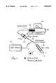
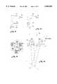
- FIG. 1is a schematic diagram showing an embodiment of the apparatus of the present invention.
- FIG. 2is a detail view of light coupler arrangement of the present invention that connects a white light source and a laser light source.
- FIG. 3is a detail section view of the probe member according to a preferred embodiment of the present invention.
- FIG. 4a to 4hare fluorescence and reflectance spectral measurements of various skin diseases that illustrate how viewing both spectral measurements together assists a user in identifying a particular skin disease.
- FIG. 5is a fluorescence spectrum of a psoriasis lesion.
- FIG. 6is a fluorescence spectrum of a squamous cell carcinoma (SCC).
- FIG. 7ashows the fluorescence spectra of normal skin and seborrheic keratosis.
- FIG. 7bshows the fluorescence ratio spectrum of seborrheic keratosis equal to the lesion fluorescence spectrum divided by the normal skin fluorescence spectrum.
- FIGS. 7c and 7dshow the reflectance spectra and reflectance ratio spectrum, respectively, of seborrheic keratosis.
- FIG. 8ashows the fluorescence spectra of spider angioma and normal skin.
- FIG. 8bshows the fluorescence ratio spectrum of spider angioma equal to the lesion fluorescence spectrum divided by the normal skin fluorescence spectrum.
- FIG. 8c and 8dshow the reflectance spectra and reflectance ratio spectrum, respectively, of spider angioma.
- FIG. 9is a schematic diagram depicting an embodiment of the present invention comprising a system for connecting a proximally-located LED light source to an illumination fiber.
- FIG. 10is a schematic diagram depicting another embodiment of the present invention comprising a system for connecting a proximally-located LED light source to an illumination fiber.
- FIG. 11depicts an end view of an array of illumination fibers disposed around a collection fiber.
- FIG. 12is a schematic diagram depicting a side view of an array of illumination and collection fibers.
- FIG. 13is a schematic diagram depicting an embodiment of the present invention comprising a system for inducing fluorescence wherein an LED is disposed at the distal end of the apparatus.
- FIG. 14is a schematic diagram depicting another embodiment of the present invention comprising an illumination apparatus wherein an LED is disposed at the distal end of the apparatus.
- FIG. 15depicts an end view of an array of illumination LEDs disposed around a collection fiber.
- FIG. 16is a schematic diagram depicting a side view of an array as set forth in FIG. 15.
- FIG. 17is a schematic diagram showing an embodiment of the apparatus of the present invention that is similar to the apparatus set forth in FIG. 1 except that the proximally located light source has been removed.
- FIG. 18is a schematic diagram showing an embodiment of the apparatus of the present invention that is similar to the apparatus set forth in FIG. 1 wherein that the proximally located light source is a tunable light source.
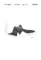
- FIG. 19depicts a 3-D autofluorescence spectrum from a normal skin site.
- the present inventionprovides methods and apparatus relating to the induction and analysis of optical responses, such as fluorescence, reflectance, and Raman scattering, in skin in order to evaluate and diagnose diseases.
- the present inventionprovides simple and easy to use methods and apparatus for the diagnosis of such diseases, and is superior to visual examination of such disease using only white (or room) light, although the present invention is preferably used in combination with such visual examination using white light.
- the present inventionprovides details about distinguishing features that are found in a variety of skin diseases, including actinic keratosis, basal cell carcinoma, seborrheic keratosis, compound nevus, spider angioma, psoriasis and squamous cell carcinoma.
- the apparatusincludes a light source 4 and a probe 6 optically connected via optical fiber 7 to light source 4 for exposing a skin disease site 8 to light.
- the light from source 4generates fluorescence, reflectance light and/or Raman spectra, at disease site 8 that is collected by probe 6 and transmitted by optical fiber 10 via light filter 12 to a spectral analyzer in the form of a spectrometer 14.
- Spectrometer 14generates and displays spectral measurement of the fluorescence and reflectance light on the monitor 18 of a computer 16 for viewing by a user to assist in the diagnosis of the disease site.
- the fluorescence, reflectance and/or Raman lightis acquired and analyzed sequentially.
- light source 4preferably comprises a white light source 20 for reflectance measurements as best shown in FIG. 2.
- Excitation light source 22can be a Helium Cadmium (442 nm) laser and white light source 20 can be a quartz tungsten halogen lamp, where it is desired to induce a Raman scattering, the excitation light source will typically comprise an IR or near IR light source.
- Raman spectroscopydetects the vibrational signatures of particular molecules inside a selected tissue, thereby providing chemical, structural and other information about the tissue. Mahavedan-Jansen, A and Richards-Kortum, R., J Biomed Optics 1(1): 31-70, 1996; U.S. Pat. No. 5,261,410.
- Raman spectroscopy of the target tissuecan be used to identify particular conditions, such as those discussed herein.
- the wavelength bands used for determination of Raman spectraare specific to the molecules in a given tissue and provide direct information about such tissue comprising such molecules.
- a computer-implemented programmakes use of the peak position(s) of the Raman spectrum, the relative intensities of different peaks to identify specific molecules and their relative concentrations relative to different tissue states in order to identify and distinguish one disease state from another.
- At least one excitation or illunmination light guidesuch as an optical fiber, conducts the illumination light through the probe to the target tissue.
- the light sourcecan be disposed at the distal end of the probe.
- At least one collection light guidepicks up the scattered light caused by the illumination light (which light includes Raman shifted photons) and then transmits the collected light to an analyzer such as a spectrometer-CCD system for spectral analysis.
- an analyzersuch as a spectrometer-CCD system for spectral analysis.
- two filtersare located at the tip of each of the illumination light guide and the collection light guide.
- a band pass filteris disposed at or near the distal tip of the illumination light guide (or otherwise between the light source and the target), which filter eliminates any Raman signals generated by the illumination fiber and transmits only the excitation light to the target tissue.
- a long pass filteris preferably disposed at or near the tip of the collection fiber and blocks elastically scattered laser light, and passes only the Raman shifted scattered light.
- the collected lightis then transmitted to a high resolution grating which is used to disperse the Raman scattered light, and then a detector such as a back-thinned CCD array is used to acquire a spectral curve.
- a computerpreferably a PC computer, is then used for process control and spectral data analysis.
- Optical coupler 24comprises a sealed enclosure 26 housing an angled reflecting surface or mirror 28 having a small aperture 29.
- a laser light port 30is formed in enclosure 26 to admit laser light from laser light source 22. Port 30 is aligned with aperture 29 to permit passage of laser light directly through mirror 28 to a light exit port 32 which is connectable to optical fiber bundle 7 and probe unit 6.
- a white light port 34is formed in enclosure 26 and positioned with respect to mirror 28 such that white light is reflected through an angle to be incident upon light exit port 32.
- White light port 34is optically connectable to white light source 20 via a short length of optical fiber 36.
- Lenses 38 and 40are also provided within enclosure 26 to collimate white light from port 34 onto the mirror surface and to focus reflected light from the mirror onto light exit port 32.
- shutter systems 41 and 42Associated with each light source are shutter systems 41 and 42 to permit switching between excitation laser light source 22 and white light source 20, respectively, such that the disease site is illuminated sequentially by only one of the light sources at a time.
- Shutter systems 41 and 42are preferably electronic and controlled by computer 16 (FIG. 1).
- Computer 16is programmed to close shutter 41 while shutter 42 is open to obtain reflectance light measurement of the disease site and vice versa to obtain fluorescence light measurements.
- the light sourceis one or more LEDs, which means a light emitting diode, including a laser-light emitting diode (laser diode) or a non-laser-light emitting diode.
- LEDsas light source(s) provides the ability to use alternative structures for the optical probe as well as alternative connections of the probe to the PC computer. It can also significantly reduce the cost of the system and can make the system more portable and more convenient to use.
- the LEDsare used as a light source: 1) coupling the LEDs to one or more optical fibers where the LEDs are disposed at a proximal end of the fiber(s), preferably where the probe has six fibers for illumination and one fiber for collection or pick-up; and, 2) placing the LEDs into the probe head (i.e., the distal end of the probe), thereby eliminating any illumination fibers but keeping the collection fiber.
- FIG. 9is a schematic diagram showing the coupling and filtering of a blue light LED to an illumination fiber for fluorescence excitation.
- blue LED 70emits light peaked at 450 nm.
- the lightis transmitted through a collimating lens or microlens 71, short pass filter 72 (which only passes light with wavelengths below 450 nm), and then focusing lens 73.
- Resister 74is used to set the proper working current for the LED.
- 5V power supply 76can be from a PC computer or a battery power source or other source.
- FIG. 10depicts a schematic diagram that is similar to FIG. 9 and that is useful for a variety of LEDs. Thus, the diagram in FIG.
- short pass filter 72which filter could be incompatible with the light emitted by certain LEDs (of course, other filters, such as long pass filters or band pass filters, can be placed in the position of short pass filter 72 if desired).
- Multiple LEDscan be coupled to multiple fibers in this manner in order to increase the illumination power and/or provide multiple wavelengths of illumination light. For example, a blue LED, a green LED, and a red LED can be coupled to three fibers to provide full spectral illumination for reflectance measurements or other desired measurements, such as Raman responses.
- FIG. 11is a schematic diagram showing an end view of one arrangement for illumination and collection fibers at the distal end of the optical probe.
- Rindicates an illumination fiber that is transmitting light from red LED
- Gindicates an illumination fiber that is transmitting light from green LED
- Bindicates an illumination fiber that is transmitting light from blue LED
- B 1 , B 2 , B 3indicates three illumination fibers that are transmitting light from filtered blue LEDs.
- the central fiberis the pick-up or collection fiber.
- FIG. 12is a schematic diagram showing that the illumination fibers 78 can be slightly tilted toward each other as they near the distal end 52 so that light emitted from the fibers will be co-centered at a desired point of the potential skin disease site 8. This arrangement assists each illumination fiber 78 to launch its light onto the same area as much as possible. This can be particularly helpful in systems where each illumination fiber carries light having different wavelengths.
- FIG. Ishows an arrangement suitable for use with the modified spectrometer system shown in FIGS. 9-12, except that the light source would be an LED light source. The switching between the LED illumination for fluorescence excitation, reflectance measurement and/or Raman response induction or measurement can be effected by turning the power on or off with respect to the various LEDs.
- FIG. 13is a schematic diagram showing a system wherein the light is emitted by a distally-located blue LED 70, which light is collimated by a collimating microlens 71 and then filtered by a short pass filter 72 to transmit the illumination to the skin for fluorescence excitation.
- FIG. 14is a schematic diagram showing light from a distally-located LED that can be other than a blue LED, and thus lacking short pass filter 72 which could be incompatible with the light emitted by a given LED (of course, other filters, such as long pass filters or band pass filters, can be placed in the position of short pass filter 72 if desired).
- Multiple LEDscan be used in order to increase the illumination power and/or provide multiple wavelengths of illumination light.
- a blue LED, a green LED, and a red LEDcan be used to provide full spectral illumination for reflectance measurements or other desired measurements, such as Raman responses.
- FIG. 15shows an arrangement of the LEDs relative to the collection fiber that is similar to the arrangement set forth in FIG. 11, except that the LEDs are located at the distal end of the probe.
- FIG. 16is a schematic diagram showing an LED assembly wherein the LEDs 70 are slightly tilted toward each other and a collection fiber 80 at the distal end 52 of an optical probe 6 and co-centered at a central point of a potential skin disease site 8. This arrangement enhances the ability of the LEDs to illuminate the same area of potential skin disease site 8.
- FIG. 17is a schematic diagram showing a system that is similar to that set forth in FIG. 1, except that light source 4 has been eliminated because the LEDs are disposed at the distal end of probe 6.
- FIG. 3is a detailed view of a probe unit 6 used to illuminate disease site 8 with light from the light sources and collect fluorescence, reflectance or other light from the site to transmit to spectrometer 14.
- Probe unit 6comprises a generally cylindrical member 50 that houses combined optical fiber bundles 7 and 10 that extend to a distal end 52 of the cylindrical member.
- An opaque sleeve member 55is telescopically mounted on cylindrical member 50 for slidable movement.
- the overlap of sleeve member 55 with distal end 52 of cylindrical member 50defines a chamber 56 that is positionable over skin disease site 8 of interest.
- Fiber bundle 7communicates with chamber 56 to transmit illumination light into the chamber and fiber bundle 10 communicates with the chamber to transmit fluorescence, reflectance or other light from the chamber to the spectrometer 14.
- Slidable movement of sleeve member 55 with respect to cylindrical member 50adjusts the length of chamber 56 and hence the distance between the fiber bundles at distal end 52 to vary the size of the illumination spot at disease site 8.
- By adjusting the length of chamber 56different sizes and areas of disease sites can be examined and diagnosed.
- Free end 62 of sleeve member 56is preferably cut at a 45 degree angle to the longitudinal axis of the sleeve. This avoids the collection of specular reflected light from the skin surface.
- sleeve member 55is preferably formed with a window 58 to allow observation inside the chamber when positioned over a disease site to ensure proper positioning.
- the design of probe unit 6is such that the apparatus of the present invention can be used in a normally lighted room since no ambient light is able to enter chamber 56 and interfere with spectral measurement when chamber 56 is positioned over a disease site and cover 60 is closed over window 58.
- the small cross-sectional area of sleeve member 55is all that actually contacts the skin at disease site 8 in order to minimize the pressure on the disease site. Any excessive pressure on the skin at the disease site will affect the blood content in the skin tissue at the disease site which will affect the resulting spectral measurements.
- Light collected from the sample sitewhich can be, for example, fluorescence light, reflectance light or Raman scattering
- shutter 41is open to transmit excitation laser light to the disease site
- filter 12is used to pass fluorescence light or Raman scattering to the spectrometer and block any reflected excitation light.
- a He-Cd laseris used to generate an excitation light of 442 nm wavelength
- a 470 nm long pass filter 12will pass fluorescence light with wavelengths larger than 470 nm but will block reflected excitation light.
- spectrometer 14is a computer interface card installable in an interface slot of computer 16 which is preferably a standard personal computer (PC) having an I/O bus appropriate to receive the interface card.
- PCpersonal computer
- Such a PC based plug-in spectrometeris available from Ocean Optics, Inc. (Dunedin, Fla.) under the name Model PC 1000.
- the plug-in spectrometeris adapted to connect to optic fiber 10 and includes a miniature monochromator, a CCD linear array detector, and all data acquisition and controlling electronics on the interface card.
- a spectrometer version for laptop computers using a PCMCIA portmay also be used.
- computer 16controls shutter systems 41 and 42.
- the computercan also control the LEDs by turning them on or off
- Computer 16also controls spectrometer 14 in order to acquire spectral measurements of the fluorescence, reflectance or other light from the disease site. In prototype testing, the necessary spectral measurements of fluorescence and reflectance light were accomplished in a few seconds.
- FIGS. 4a to 4hillustrate typical fluorescence and reflectance spectra of particular skin diseases acquired using the apparatus and methods of the present invention.
- reflectance spectraalone or visual inspection of white light illumination, it can be difficult to differentiate between various skin conditions such as seborrheic keratosis (FIG. 4f) and compound nevus (FIG. 4h), or between actinic keratosis (FIG. 4b) and basal cell carcinoma (FIG. 4d) since each pair of skin diseases have similar reflectance spectra.
- FIG. 4fseborrheic keratosis
- compound nevusFIG. 4h
- actinic keratosisFIG. 4b
- basal cell carcinomaFIG. 4d
- fluorescence spectrato differentiate between actinic keratosis (FIG. 4a) with a fluorescence intensity higher than normal skin and basal cell carcinoma (FIG. 4c) having a fluorescence intensity lower than normal skin.
- he apparatus and method of the present inventionrely on using both reflectance and fluorescence spectral measurements in combination to assist in diagnosis.
- comparing only one type of spectral measurementsresults in difficulties in distinguishing between certain skin diseases.
- using only fluorescence spectra for each skin diseasemakes it difficult to distinguish between actinic keratosis (FIG. 4a) and seborrheic keratosis (FIG. 4e) or between basal cell carcinoma (FIG. 4c) and compound nevus (FIG. 4g) as each of these particular pairs of skin diseases have similar fluorescence spectra.
- the fact that the above pairs of skin diseases have different reflectance spectraallow them to be distinguished clearly from each other.
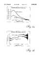
- FIG. 5is the fluorescence spectrum of a psoriasis lesion, showing a unique spectral peak at around 635 nm that allows psoriasis to be differentiated from other skin lesions.
- FIG. 6shows fluorescence spectra of a squamous cell carcinoma (SCC). The fluorescence intensity across a SCC lesion is not uniformly distributed. As shown, in some places within the lesion the fluorescence intensity is higher than normal skin, while in other locations the fluorescence intensity is lower than normal. This feature for SCC lesions is different from that of basal cell carcinomas (BCC), where the fluorescence intensity is uniformly lower than found in surrounding normal skin.
- BCCbasal cell carcinomas
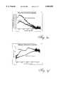
- ratio analysis techniquelesional spectra (reflectance or fluorescence) are divided by the corresponding spectra of the surrounding normal skin. In this way, the relative spectral intensity changes are quantified, and the spectral shape changes are enhanced and more easily visualized on the spectral curves.
- FIG. 7shows the original spectra and the ratio spectra of a seborrheic keratosis.
- FIG. 7ais the original fluorescence spectra, the lesion has higher fluorescence intensity than the normal, but the shape of the two curves are visually about the same.
- FIG. 7bis the fluorescence ratio spectrum which equals the lesion fluorescence spectrum divided by the normal fluorescence spectrum. The absolute value of the ratio spectrum is larger than 1, indicating that the lesion has higher fluorescence intensity than the normal and it indicating quantitatively how many times higher the lesion signal is than the normal at each wavelengths.
- FIG. 7cis the reflectance spectra of the same seborrheic keratosis lesion and its surrounding normal skin.
- FIG. 7dis the reflectance ratio spectrum which equals the lesion reflectance spectrum divided by the normal reflectance spectrum.
- the reflectance ratio spectrum of seborrheic keratosisshows lower values at short wavelengths and higher values at longer wavelengths and is a quantitative representation of the slope changes described in Table 1.
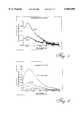
- FIG. 8ais the fluorescence spectra of a spider angioma and its surrounding normal skin. The lesion has lower fluorescence intensity than normal skin.
- FIG. 8bis the fluorescence ratio spectrum of the same lesion. The ratios with values less than 1 indicate that the lesional fluorescence intensity is lower than its surrounding normal skin while the numerical ratio values quantify this difference as a function of wavelength.
- FIG. 8cshows the reflectance spectra of the same spider angioma
- FIG. 8dthe reflectance ratio spectrum.
- the reflectance ratio spectra of spider angiomashave a unique "saw-tooth" shape with a minimum at around 600 nm. This unique spectral feature can be used to differentiate spider angioma from other skin lesions.
- autofluorescence spectra collected at many different excitation wavelengthscan be used to build a 3-D spectral diagram.
- the X-axisis the excitation wavelength
- the Y-axisis the emission wavelength
- the Z-axisis the fluorescence intensity.
- 3-D spectraprovide more information than a single spectral curve obtained with a single excitation wavelength. The specific patterns of 3-D spectra permit accurate diagnosis of skin diseases.
- FIG. 18is a schematic diagram showing a spectrometer system for such 3-D measurements. It is similar to the system set forth in FIG. 1 except that a tunable light source 4a is used to provide different excitation wavelengths for fluorescence excitation (or other desired response such as reflectance or Raman scattering), and the long pass filter in the in-line filter holder is replaced with a filter wheel 82 holding multiple, different long pass filters (or other filters for non-fluorescent uses). When the excitation wavelength changes, the wheel switches to different long pass filters.
- the tunable light source 4acan be, for example, a Xenon arc lamp-monochromator combination, a nitrogen dye laser system, or an OPO (optical parameter oscillator).
- FIG. 19shows an example of a 3-D autofluorescence spectrum obtained from a normal skin site. There are maximum peaks at the excitation-emission wavelengths (i.e., 380 nm, 470 nm). This type of 3-D spectrum shows different spectral characteristics for different skin diseases including skin cancer, and therefore aids the dermatology diagnosis.
- Two preferred methods of analyzing the 3-D datainclude (1) making a two-dimensional contour plot of FIG. 19 to generate a so-called fluorescence excitation-emission matrix (EEM), and/or (2) sectioning the 3-D graph at a fixed emission wavelength to generate a 2-D plot called a fluorescence excitation spectrum. From the excitation spectrum, one can observe the absorption properties of specific fluorophores.
- EEMfluorescence excitation-emission matrix
- the spectracan be analyzed using a 3-D fluorescence matrix wherein derivatives of the "raw" 3-D matrix are generated.
- derivativesinclude "ratio" matrices generated by dividing a spectrum from a lesion versus a spectrum from (preferably surrounding) normal skin, and "subtractive" matrices generated by subtracting a spectrum a lesion from a spectrum from (preferably surrounding) normal skin.
Landscapes
- Health & Medical Sciences (AREA)
- Life Sciences & Earth Sciences (AREA)
- Physics & Mathematics (AREA)
- General Health & Medical Sciences (AREA)
- Pathology (AREA)
- Biomedical Technology (AREA)
- Chemical & Material Sciences (AREA)
- General Physics & Mathematics (AREA)
- Biochemistry (AREA)
- Molecular Biology (AREA)
- Engineering & Computer Science (AREA)
- Analytical Chemistry (AREA)
- Immunology (AREA)
- Surgery (AREA)
- Veterinary Medicine (AREA)
- Nuclear Medicine, Radiotherapy & Molecular Imaging (AREA)
- Public Health (AREA)
- Animal Behavior & Ethology (AREA)
- Medical Informatics (AREA)
- Heart & Thoracic Surgery (AREA)
- Biophysics (AREA)
- Dermatology (AREA)
- Investigating, Analyzing Materials By Fluorescence Or Luminescence (AREA)
Abstract
Description
This application is a continuation-in-part of U.S. patent application No. 08/843,605, filed Apr. 16, 1997, now U.S. Pat. No. 6,008,889.
This invention relates to apparatus and methods for assisting in diagnosis of skin diseases by making measurements of fluorescence light and/or reflectance light and/or Raman scattering from the skin disease sites.
Currently, clinical diagnosis of skin disease is generally accomplished by visual inspection under white light illumination. In this process, the reflectance light of a skin lesion is examined. Visual diagnosis alone may not be particularly accurate for early detection of skin cancer since many skin conditions have a similar appearance under white light. Therefore, when a suspect lesion is identified by visual examination, a biopsy is often performed for a definitive diagnosis. This is because it is crucial to diagnose skin pre-cancer or cancer at an early stage when it is curable. Thus, it is important to improve the clinical diagnosis of suspected skin lesions so as to avoid unnecessary skin biopsies.
Several approaches have been tried to improve dermatologic diagnosis. Digital processing of reflectance images has been extensively investigated recently. Although reflectance imaging has led to improvements in the registration, recording, and documentation of skin lesions, there has been little improvement in the diagnostic accuracy. The foregoing approach does not provide any additional data to the physician making the visual assessment because it is still based on the reflectance pattern of a lesion under white light illumination which is essentially the same pattern a human observer sees.
An alternative approach is ultraviolet (UV) or infrared (IR) photography which does extend visual perception of a physician to the UV or IR reflectance patterns. However, the inconvenience due to delays in processing of film images renders this technique impractical for everyday use.
A further alternative approach that is already in widespread medical use involves a "Wood's lamp," which consists of a mercury discharge lamp associated with a filter that transmits UVA light with a 365 nanometer peak while absorbing visible light. When this device is used to assist in skin diagnosis, the eye serves as both the detector and the long pass filter. The eye is not sensitive to UV light, but is sensitive to visible fluorescence light when the "Wood's lamp" is used in a darkened room, where the physician sees an image of a fluorescing disease site. The "Wood's lamp" is useful for the diagnosis of some skin conditions such as tinea capitis, tinea versicolor, erythrasma, and some pseudomonas infections, as well as aiding in the detection and diagnosis of hypopigmented skin. It is of no value in conditions where the emitted fluorescence is not in the visible spectrum because the human eye cannot detect such fluorescence. It is also incapable of detecting Raman scattering. Thus, there has gone unmet a need for apparatus and methods that are able to detect and analyze fluorescence both within and beyond the visible spectrum, and that can use fluorescence, reflectance and/or Raman scattering to identify, and distinguish between, a variety of skin diseases.
The present invention provides these and other related advantages.
The apparatus and methods of the present invention use spectral measurements of fluorescence light, reflectance light, and/or Raman scattering from a disease site to aid in the medical assessment of skin conditions and the diagnosis of cutaneous diseases. The apparatus and methods can, if desired, be used in a normally lit room, and can distinguish between a variety of skin diseases, including actinic keratosis, basal cell carcinoma, seborrheic keratosis, compound nevus, spider angioma, psoriasis and squamous cell carcinoma.
Thus, in one aspect the present invention provides apparatus able to diagnose a disease selected from the group consisting of actinic keratosis, seborrheic keratosis, basal cell carcinoma and compound nevus at a skin disease site using spectral analysis comprising: a light source for generating light to illuminate the disease site; a probe means optically connected to the light source to expose the disease site to light to generate fluorescence and reflectance light; spectral analysis means optically connected to the probe means for generating and displaying spectral measurements of the fluorescence light and the reflectance light to assist the user in diagnosing the disease site; and a plurality of comparison spectra corresponding to, and able to distinguish between, each of actinic keratosis, seborrheic keratosis, basal cell carcinoma and compound nevus. Further preferably, in this and other aspects of the invention, the apparatus is also able to identifiably detect and diagnose spider angioma, psoriasis and/or squamous cell carcinoma. Also preferably, the apparatus is able to distinguish such diseases from actinic keratosis, seborrheic keratosis, basal cell carcinoma and compound nevus, and is further preferably able to distinguish all such diseases one from another.
In a preferred embodiment for this and other aspects of the invention, the light source comprises an excitation laser light source or other narrow band light source and a white light source, further preferably the excitation light source is a Helium-Cadmium laser and the white light source is a quartz tungsten halogen lamp.
In another preferred embodiment, the apparatus further comprises a shutter system to switch between the excitation light source and the white light source such that the disease site is illuminated sequentially by one of the light sources at a time, and a computer control means to control the shutter system by switching between light sources and to control the spectral analysis means in order to acquire spectral measurements of the fluorescence and reflectance light. The computer control means preferably comprises a personal computer and the spectral analysis means preferably comprises a computer interface card installable in an interface slot of the computer to function as a spectrometer, which computer interface card preferably includes a miniature monochromator, a CCD linear array detector, means for optically connecting to the probe means, and data acquisition and controlling circuitry.
The apparatus also preferably includes a light filter selected to pass fluorescence light and block reflected excitation light, and the light source is preferably optically connected to the probe means and the probe means is optically connected to the spectral analysis means by optical fiber bundles, further preferably including an optical coupler for connecting the white light source and the excitation light source to deliver light via the same optical fiber bundle. Still further preferably, the optical coupler comprises: an enclosure housing, an angled reflecting surface having an aperture; a laser light port in the enclosure aligned with the aperture to permit passage of laser light directly through the angled reflecting surface, the laser light port being optically connectable to a laser light source; a light exit port from the enclosure aligned with the laser light port and the aperture through the angled reflecting surface, the light exit port being optically connectable to the probe means; and, a white light port in the enclosure positioned with respect to the angled reflecting surface such that white light is reflected through an angle to be incident upon the light exit port, the white light opening being optically connectable to the white light source.
In still another preferred embodiment, the probe means comprises an elongate member having an open end defining a chamber with opaque walls that is positionable over the disease site to exclude external light from the chamber, the chamber being connected to the optical fiber bundles for receiving light from the light sources and transmitting fluorescence light and reflectance light to the spectral analysis means. Preferably, the elongate member comprises: a generally cylindrical member housing the optical fiber bundles that extend to a distal end of the cylindrical member, and a sleeve member that telescopes over the generally cylindrical housing, the overlap of the sleeve member with the distal end of the cylindrical member defining the chamber whereby slidable movement of the sleeve member with respect to the cylindrical member adjusts the length of the chamber to vary the size of the illumination spot at the disease site. Also preferably, the sleeve member is formed with a window for viewing inside the chamber to ensure proper positioning of the chamber over the disease site and a slidable cover to seal the window when the apparatus is in use, and the free end of the sleeve member is cut at substantially 45 degrees to the longitudinal axis of the sleeve to avoid specular reflection.
In another aspect, the present invention provides methods for diagnosis of a disease selected from the group consisting of actinic keratosis, seborrheic keratosis, basal cell carcinoma, compound nevus and spider angioma at a skin disease site using spectral analysis comprising the steps of: illuminating the disease site with a light source to generate fluorescence and reflectance light at the disease site; collecting the generated fluorescence and reflectance light; conducting a spectral analysis of the collected light using a spectrometer, determining spectral measurements of the fluorescence light and the reflectance light; and, examining the spectral measurements together to make a diagnosis of the disease site, wherein the step of examining comprises comparing the spectral measurements with known spectral measurements. If the reflectance curve slope is similar to normal and the fluorescence intensity is higher than normal, then determining that the disease is actinic keratosis. If the reflectance curve slope is similar to normal and the fluorescence intensity is lower than normal, then determining that the disease is basal cell carcinoma. If the reflectance curve slope is larger than normal and the fluorescence intensity is higher than normal, then determining that the disease is seborrheic keratosis. If the reflectance curve slope is larger than normal and the fluorescence intensity is lower than normal, then determining that the disease is compound nevus. If the fluorescence intensity is lower than normal and the reflectance ratio spectrum comprises a saw-tooth shape with a minimum at about 600 nm, then determining that the disease is spider angioma. If the fluorescence spectrum comprises a peak at about 630 nm-640 nm, then determining that the disease is psoriasis. If the fluorescence intensity is lower in some locations in the lesion and higher than normal other locations in the lesion, then determining that the disease is squamous cell carcinoma.
In a preferred embodiment, the steps of illuminating the disease site and collecting generated light involves sequentially illuminating the site with white light and collecting reflectance light and then illuminating the site with excitation light and collecting fluorescence light or vice versa. Also preferably, the methods include filtering the collected fluorescence light to filter out the excitation light, and a spectrometer card mounted in a personal computer is used to conduct a spectral analysis of the generated fluorescence and reflected light and display spectral measurements.
In another preferred embodiment, the step of collecting the generated fluorescence and reflectance light involves collecting light from the disease site and the surrounding normal skin. Further preferably, the step of examining the spectral measurements involves comparing the disease site spectrum to the spectrum of the surrounding normal skin, further preferably comparing the intensities of the autofluorescence spectra of the disease site and the surrounding normal skin.
In still another preferred embodiment, the step of examining the spectral measurements involves comparing the slope of the reflectance spectra of the disease site and the surrounding normal skin and/or generating a ratio spectrum by dividing the disease site spectrum by the spectrum of the adjacent normal skin.
In all aspects and embodiments of the invention, the comparison spectra are stored in a computer and the steps of comparing the various spectra or other responses are performed by a computer.
In still yet another aspect, the present invention provides systems able to diagnose a disease selected from the group consisting of actinic keratosis, seborrheic keratosis, basal cell carcinoma and compound nevus at a skin disease site using spectral analysis comprising: a light source for generating light to illuminate the disease site; an optical probe optically connected to the light source to expose the disease site to light to generate fluorescence and reflectance light; a spectral analyzer optically connected to the optical probe for generating and displaying spectral measurements of the fluorescence light and the reflectance light to assist the user in diagnosing the disease site; and a plurality of comparison spectra corresponding to, and able to distinguish between, each of actinic keratosis, seborrheic keratosis, basal cell carcinoma and compound nevus.
In still a further aspect, the present invention provides optical probes able to induce at least one of fluorescence light, reflectance light and Raman scattering in vivo in a target tissue, the optical probe comprising a proximal end, a distal end, at least one light emitting diode disposed at the distal end of the optical probe and operatively connected to a power source able to provide power to the light emitting diode such that the light emitting diode can emit excitation light able to induce at least one of fluorescence light, reflectance light and Raman scattering in the target tissue, and at least one collection fiber that transmits light from the distal end of the optical probe to the proximal end of the optical probe.
Preferably, the collection fiber is operatively connected to a spectrometer able to analyze the spectra transmitted by the collection fiber, which spectrometer is operatively connected to a memory readable by a computer that contains a plurality of comparison spectra corresponding to, and able to distinguish between, one or more of actinic keratosis, seborrheic keratosis, basal cell carcinoma and compound nevus, spider angioma, psoriasis and/or squamous cell carcinoma. Also preferably, the distal end of the optical probe comprises at least one of a plurality of light emitting diodes that emit different wavelengths of excitation light and a plurality of light filters that transmit different wavelengths of excitation light to provide a plurality of light sources, and an on and off switch to switch the light emitting diodes on and off such that the target tissue is illuminated sequentially by one of the light sources at a time. Further preferably, the probes comprise a computer that controls the on and off switch and the spectrometer in order to acquire sequential spectral measurements of at least two of fluorescence light, reflectance light and Raman scattering, which computer is typically a personal computer and the spectrometer typically comprises a computer interface card installable in an interface slot of the computer.
In still a another further aspect, the present invention provides methods for diagnosis of a skin disease using analysis of Raman scattering at a suspected skin disease site comprising the steps of: illuminating the disease site with an infrared or near-infrared light source to generate Raman scattering at the disease site; collecting the generated Raman scattering; conducting an analysis of the collected Raman scattering using a spectrometer; determining measurements of the Raman scattering; and, examining the spectral measurements together to make a diagnosis of the disease site by comparing the spectral measurements of the suspected skin disease site against measurements from a known site. Preferably, the methods comprise including filtering the collected Raman scattering to filter out the excitation light. Also preferably, the methods comprise using a spectrometer card mounted in a personal computer is used to conduct a spectral analysis of the generated Raman scattering and display spectral measurements.
In preferred embodiments, the step of collecting the generated Raman scattering involves collecting Raman scattering from the disease site and the surrounding normal skin. Further preferably, the step of examining the Raman scattering comparing the disease site Raman scattering to the Raman scattering of the surrounding normal skin, which can include comparing the intensities of the Raman scattering of the disease site and the surrounding normal skin.
In other preferred embodiments, the methods include the step of generating a ratio spectrum by dividing the disease site Raman scattering by the Raman scattering of the adjacent normal skin.
In still further aspects, the methods of the present invention comprise determining spectral measurements a 3-D spectrum of the fluorescence light. Such determination can include determining a 3-D spectrum of the fluorescence light further comprises generating a two-dimensional contour plot from the 3-D spectrum to generate a fluorescence excitation-emission matrix (EM), determining a 3-D spectrum of the fluorescence light further comprises sectioning the 3-D spectrum at a fixed emission wavelength to generate a 2-D plot of fluorescence excitation spectrum, generating a ratio matrix by dividing a 3-D spectrum from a lesion versus a 3-D spectrum from surrounding normal skin, and/or generating a subtractive matrix by subtracting a 3-D spectrum a lesion from a 3-D spectrum from surrounding normal skin.
Accordingly, the apparatus of the present invention provides apparatus for diagnosis of a skin disease site using spectral analysis comprising:
a light source for generating light to illuminate the disease site;
a probe means to conduct the illumination light from the light source to the disease site and to collect the reflected and fluorescence light and conduct said light to be analyzed; and
spectral analysis means optically connected to the probe means for generating and displaying spectral measurements of the fluorescence light and the reflectance light to assist the user in diagnosing the disease site.
In a further aspect, the present invention provides a method for diagnosis of a skin disease site using spectral analysis comprising the steps of:
illuminating the disease site with a light source to generate fluorescence and reflectance light at the disease site;
collecting the generated fluorescence and reflectance light;
conducting a spectral analysis of the collected light using the spectrometer;
displaying spectral measurements of the fluorescence light and the reflectance light; and
analyzing the measured fluorescence and reflectance spectra together to make a diagnosis of the disease site.
In a preferred embodiment, the apparatus of the present invention includes a compact spectrometer connected to a personal computer, a fluorescence excitation light source with a shutter, a white light source with a shutter, a bifurcated fiber bundle, a light coupler, a skin probe, and controlling electronics. The system is designed to automatically switch between the fluorescence excitation light and the white light sources and complete fluorescence and reflectance spectral measurements of a skin disease site sequentially in a few seconds. The system exploits the spectral differences of different skin diseases to aid in the dermatologic diagnosis. In particular, the apparatus provides a low costs, compact system that is capable of quickly and efficiently performing combined fluorescence and reflectance spectral analysis.
These and other aspects of the present invention will become evident upon reference to the discussion herein and the attached drawings. In addition, various references are set forth herein that describe in more detail certain procedures or apparatus, etc. (e.g., bioptomes, fluorescence technology, etc.); all such references are incorporated herein by reference in their entirety.
Aspects of the present invention are illustrated, merely by way of example, in the accompanying drawings in which:
FIG. 1 is a schematic diagram showing an embodiment of the apparatus of the present invention.
FIG. 2 is a detail view of light coupler arrangement of the present invention that connects a white light source and a laser light source.
FIG. 3 is a detail section view of the probe member according to a preferred embodiment of the present invention.
FIG. 4a to 4h are fluorescence and reflectance spectral measurements of various skin diseases that illustrate how viewing both spectral measurements together assists a user in identifying a particular skin disease.
FIG. 5 is a fluorescence spectrum of a psoriasis lesion.
FIG. 6 is a fluorescence spectrum of a squamous cell carcinoma (SCC).
FIG. 7a shows the fluorescence spectra of normal skin and seborrheic keratosis.
FIG. 7b shows the fluorescence ratio spectrum of seborrheic keratosis equal to the lesion fluorescence spectrum divided by the normal skin fluorescence spectrum.
FIGS. 7c and 7d show the reflectance spectra and reflectance ratio spectrum, respectively, of seborrheic keratosis.
FIG. 8a shows the fluorescence spectra of spider angioma and normal skin.
FIG. 8b shows the fluorescence ratio spectrum of spider angioma equal to the lesion fluorescence spectrum divided by the normal skin fluorescence spectrum.
FIG. 8c and 8d show the reflectance spectra and reflectance ratio spectrum, respectively, of spider angioma.
FIG. 9 is a schematic diagram depicting an embodiment of the present invention comprising a system for connecting a proximally-located LED light source to an illumination fiber.
FIG. 10 is a schematic diagram depicting another embodiment of the present invention comprising a system for connecting a proximally-located LED light source to an illumination fiber.
FIG. 11 depicts an end view of an array of illumination fibers disposed around a collection fiber.
FIG. 12 is a schematic diagram depicting a side view of an array of illumination and collection fibers.
FIG. 13 is a schematic diagram depicting an embodiment of the present invention comprising a system for inducing fluorescence wherein an LED is disposed at the distal end of the apparatus.
FIG. 14 is a schematic diagram depicting another embodiment of the present invention comprising an illumination apparatus wherein an LED is disposed at the distal end of the apparatus.
FIG. 15 depicts an end view of an array of illumination LEDs disposed around a collection fiber.
FIG. 16 is a schematic diagram depicting a side view of an array as set forth in FIG. 15.
FIG. 17 is a schematic diagram showing an embodiment of the apparatus of the present invention that is similar to the apparatus set forth in FIG. 1 except that the proximally located light source has been removed.
FIG. 18 is a schematic diagram showing an embodiment of the apparatus of the present invention that is similar to the apparatus set forth in FIG. 1 wherein that the proximally located light source is a tunable light source.
FIG. 19 depicts a 3-D autofluorescence spectrum from a normal skin site.
The present invention provides methods and apparatus relating to the induction and analysis of optical responses, such as fluorescence, reflectance, and Raman scattering, in skin in order to evaluate and diagnose diseases. The present invention provides simple and easy to use methods and apparatus for the diagnosis of such diseases, and is superior to visual examination of such disease using only white (or room) light, although the present invention is preferably used in combination with such visual examination using white light. In addition, the present invention provides details about distinguishing features that are found in a variety of skin diseases, including actinic keratosis, basal cell carcinoma, seborrheic keratosis, compound nevus, spider angioma, psoriasis and squamous cell carcinoma.
Referring to FIG. 1, there is a schematic view of the apparatus of the present invention for diagnosing skin diseases. The apparatus includes a light source 4 and aprobe 6 optically connected viaoptical fiber 7 to light source 4 for exposing askin disease site 8 to light. The light from source 4 generates fluorescence, reflectance light and/or Raman spectra, atdisease site 8 that is collected byprobe 6 and transmitted byoptical fiber 10 vialight filter 12 to a spectral analyzer in the form of aspectrometer 14.Spectrometer 14 generates and displays spectral measurement of the fluorescence and reflectance light on themonitor 18 of acomputer 16 for viewing by a user to assist in the diagnosis of the disease site.
In a preferred embodiment of the present invention, the fluorescence, reflectance and/or Raman light is acquired and analyzed sequentially. In order to accomplish this where the detected light is fluorescence and reflectance, light source 4 preferably comprises awhite light source 20 for reflectance measurements as best shown in FIG. 2.Excitation light source 22 can be a Helium Cadmium (442 nm) laser andwhite light source 20 can be a quartz tungsten halogen lamp, where it is desired to induce a Raman scattering, the excitation light source will typically comprise an IR or near IR light source.
Turning generally to Raman spectroscopy, it detects the vibrational signatures of particular molecules inside a selected tissue, thereby providing chemical, structural and other information about the tissue. Mahavedan-Jansen, A and Richards-Kortum, R., J Biomed Optics 1(1): 31-70, 1996; U.S. Pat. No. 5,261,410. Raman spectroscopy of the target tissue can be used to identify particular conditions, such as those discussed herein. The wavelength bands used for determination of Raman spectra are specific to the molecules in a given tissue and provide direct information about such tissue comprising such molecules. In a preferred embodiment, similar to the gathering and comparison of fluorescence and reflectance spectra, a computer-implemented program makes use of the peak position(s) of the Raman spectrum, the relative intensities of different peaks to identify specific molecules and their relative concentrations relative to different tissue states in order to identify and distinguish one disease state from another.
In a preferred embodiment, a near NIR diode laser (e.g., λ=785 nm, 830 nm) is used for the excitation illumination. At least one excitation or illunmination light guide, such as an optical fiber, conducts the illumination light through the probe to the target tissue. Alternatively, the light source can be disposed at the distal end of the probe. At least one collection light guide then picks up the scattered light caused by the illumination light (which light includes Raman shifted photons) and then transmits the collected light to an analyzer such as a spectrometer-CCD system for spectral analysis. Preferably, two filters are located at the tip of each of the illumination light guide and the collection light guide. A band pass filter is disposed at or near the distal tip of the illumination light guide (or otherwise between the light source and the target), which filter eliminates any Raman signals generated by the illumination fiber and transmits only the excitation light to the target tissue. A long pass filter is preferably disposed at or near the tip of the collection fiber and blocks elastically scattered laser light, and passes only the Raman shifted scattered light. In a preferred embodiment, the collected light is then transmitted to a high resolution grating which is used to disperse the Raman scattered light, and then a detector such as a back-thinned CCD array is used to acquire a spectral curve. A computer, preferably a PC computer, is then used for process control and spectral data analysis.
The twolight sources optical coupler 24 that allows light from either source to be transmitted via the sameoptical fiber bundle 7.Optical coupler 24 comprises a sealed enclosure 26 housing an angled reflecting surface or mirror 28 having asmall aperture 29. Alaser light port 30 is formed in enclosure 26 to admit laser light fromlaser light source 22.Port 30 is aligned withaperture 29 to permit passage of laser light directly through mirror 28 to alight exit port 32 which is connectable tooptical fiber bundle 7 andprobe unit 6. Awhite light port 34 is formed in enclosure 26 and positioned with respect to mirror 28 such that white light is reflected through an angle to be incident uponlight exit port 32. Whitelight port 34 is optically connectable towhite light source 20 via a short length of optical fiber 36.Lenses port 34 onto the mirror surface and to focus reflected light from the mirror ontolight exit port 32.
Associated with each light source areshutter systems laser light source 22 andwhite light source 20, respectively, such that the disease site is illuminated sequentially by only one of the light sources at a time.Shutter systems Computer 16 is programmed to closeshutter 41 whileshutter 42 is open to obtain reflectance light measurement of the disease site and vice versa to obtain fluorescence light measurements.
In an alternative embodiment, the light source is one or more LEDs, which means a light emitting diode, including a laser-light emitting diode (laser diode) or a non-laser-light emitting diode. Using LEDs as light source(s) provides the ability to use alternative structures for the optical probe as well as alternative connections of the probe to the PC computer. It can also significantly reduce the cost of the system and can make the system more portable and more convenient to use. There are two preferred embodiments to use the LEDs as a light source: 1) coupling the LEDs to one or more optical fibers where the LEDs are disposed at a proximal end of the fiber(s), preferably where the probe has six fibers for illumination and one fiber for collection or pick-up; and, 2) placing the LEDs into the probe head (i.e., the distal end of the probe), thereby eliminating any illumination fibers but keeping the collection fiber.
FIG. 9 is a schematic diagram showing the coupling and filtering of a blue light LED to an illumination fiber for fluorescence excitation. In particular,blue LED 70 emits light peaked at 450 nm. The light is transmitted through a collimating lens ormicrolens 71, short pass filter 72 (which only passes light with wavelengths below 450 nm), and then focusinglens 73.Resister 74 is used to set the proper working current for the LED.5V power supply 76 can be from a PC computer or a battery power source or other source. FIG. 10 depicts a schematic diagram that is similar to FIG. 9 and that is useful for a variety of LEDs. Thus, the diagram in FIG. 10 is lackingshort pass filter 72, which filter could be incompatible with the light emitted by certain LEDs (of course, other filters, such as long pass filters or band pass filters, can be placed in the position ofshort pass filter 72 if desired). Multiple LEDs can be coupled to multiple fibers in this manner in order to increase the illumination power and/or provide multiple wavelengths of illumination light. For example, a blue LED, a green LED, and a red LED can be coupled to three fibers to provide full spectral illumination for reflectance measurements or other desired measurements, such as Raman responses.
FIG. 11 is a schematic diagram showing an end view of one arrangement for illumination and collection fibers at the distal end of the optical probe. R indicates an illumination fiber that is transmitting light from red LED, G indicates an illumination fiber that is transmitting light from green LED, B indicates an illumination fiber that is transmitting light from blue LED, and B1, B2, B3 indicates three illumination fibers that are transmitting light from filtered blue LEDs. The central fiber is the pick-up or collection fiber.
FIG. 12 is a schematic diagram showing that theillumination fibers 78 can be slightly tilted toward each other as they near thedistal end 52 so that light emitted from the fibers will be co-centered at a desired point of the potentialskin disease site 8. This arrangement assists eachillumination fiber 78 to launch its light onto the same area as much as possible. This can be particularly helpful in systems where each illumination fiber carries light having different wavelengths. FIG. I shows an arrangement suitable for use with the modified spectrometer system shown in FIGS. 9-12, except that the light source would be an LED light source. The switching between the LED illumination for fluorescence excitation, reflectance measurement and/or Raman response induction or measurement can be effected by turning the power on or off with respect to the various LEDs.
Turning to another embodiment, FIG. 13 is a schematic diagram showing a system wherein the light is emitted by a distally-locatedblue LED 70, which light is collimated by a collimatingmicrolens 71 and then filtered by ashort pass filter 72 to transmit the illumination to the skin for fluorescence excitation. FIG. 14 is a schematic diagram showing light from a distally-located LED that can be other than a blue LED, and thus lackingshort pass filter 72 which could be incompatible with the light emitted by a given LED (of course, other filters, such as long pass filters or band pass filters, can be placed in the position ofshort pass filter 72 if desired). Multiple LEDs can be used in order to increase the illumination power and/or provide multiple wavelengths of illumination light. Thus, a blue LED, a green LED, and a red LED can be used to provide full spectral illumination for reflectance measurements or other desired measurements, such as Raman responses.
FIG. 15 shows an arrangement of the LEDs relative to the collection fiber that is similar to the arrangement set forth in FIG. 11, except that the LEDs are located at the distal end of the probe. FIG. 16 is a schematic diagram showing an LED assembly wherein theLEDs 70 are slightly tilted toward each other and acollection fiber 80 at thedistal end 52 of anoptical probe 6 and co-centered at a central point of a potentialskin disease site 8. This arrangement enhances the ability of the LEDs to illuminate the same area of potentialskin disease site 8. FIG. 17 is a schematic diagram showing a system that is similar to that set forth in FIG. 1, except that light source 4 has been eliminated because the LEDs are disposed at the distal end ofprobe 6.
FIG. 3 is a detailed view of aprobe unit 6 used to illuminatedisease site 8 with light from the light sources and collect fluorescence, reflectance or other light from the site to transmit tospectrometer 14.Probe unit 6 comprises a generallycylindrical member 50 that houses combinedoptical fiber bundles distal end 52 of the cylindrical member. Anopaque sleeve member 55 is telescopically mounted oncylindrical member 50 for slidable movement. The overlap ofsleeve member 55 withdistal end 52 ofcylindrical member 50 defines achamber 56 that is positionable overskin disease site 8 of interest.Fiber bundle 7 communicates withchamber 56 to transmit illumination light into the chamber andfiber bundle 10 communicates with the chamber to transmit fluorescence, reflectance or other light from the chamber to thespectrometer 14. Slidable movement ofsleeve member 55 with respect tocylindrical member 50 adjusts the length ofchamber 56 and hence the distance between the fiber bundles atdistal end 52 to vary the size of the illumination spot atdisease site 8. By adjusting the length ofchamber 56, different sizes and areas of disease sites can be examined and diagnosed.
The small cross-sectional area ofsleeve member 55 is all that actually contacts the skin atdisease site 8 in order to minimize the pressure on the disease site. Any excessive pressure on the skin at the disease site will affect the blood content in the skin tissue at the disease site which will affect the resulting spectral measurements.
Light collected from the sample site, which can be, for example, fluorescence light, reflectance light or Raman scattering, is transmitted viaoptical fiber 10 tospectrometer 14. When it is desired to collect fluorescence light or Raman scattering,shutter 41 is open to transmit excitation laser light to the disease site, filter 12 is used to pass fluorescence light or Raman scattering to the spectrometer and block any reflected excitation light. For example, if a He-Cd laser is used to generate an excitation light of 442 nm wavelength, a 470 nmlong pass filter 12 will pass fluorescence light with wavelengths larger than 470 nm but will block reflected excitation light.
Preferably,spectrometer 14 is a computer interface card installable in an interface slot ofcomputer 16 which is preferably a standard personal computer (PC) having an I/O bus appropriate to receive the interface card. Such a PC based plug-in spectrometer is available from Ocean Optics, Inc. (Dunedin, Fla.) under the name Model PC 1000. The plug-in spectrometer is adapted to connect tooptic fiber 10 and includes a miniature monochromator, a CCD linear array detector, and all data acquisition and controlling electronics on the interface card. A spectrometer version for laptop computers using a PCMCIA port may also be used.
As previously described,computer 16 controls shuttersystems Computer 16 also controlsspectrometer 14 in order to acquire spectral measurements of the fluorescence, reflectance or other light from the disease site. In prototype testing, the necessary spectral measurements of fluorescence and reflectance light were accomplished in a few seconds.
FIGS. 4a to 4h illustrate typical fluorescence and reflectance spectra of particular skin diseases acquired using the apparatus and methods of the present invention. Using reflectance spectra alone or visual inspection of white light illumination, it can be difficult to differentiate between various skin conditions such as seborrheic keratosis (FIG. 4f) and compound nevus (FIG. 4h), or between actinic keratosis (FIG. 4b) and basal cell carcinoma (FIG. 4d) since each pair of skin diseases have similar reflectance spectra. However, when a user also considers the corresponding fluorescence spectrum for the particular skin disease, it is possible to differentiate between seborrheic keratosis (FIG. 4e) with a fluorescence intensity higher than normal skin and compound nevus (FIG. 4g) with fluorescence intensity much lower than normal skin. In a similar manner, it is possible to use fluorescence spectra to differentiate between actinic keratosis (FIG. 4a) with a fluorescence intensity higher than normal skin and basal cell carcinoma (FIG. 4c) having a fluorescence intensity lower than normal skin.
It is important to note that he apparatus and method of the present invention rely on using both reflectance and fluorescence spectral measurements in combination to assist in diagnosis. In general, comparing only one type of spectral measurements results in difficulties in distinguishing between certain skin diseases. For example, using only fluorescence spectra for each skin disease makes it difficult to distinguish between actinic keratosis (FIG. 4a) and seborrheic keratosis (FIG. 4e) or between basal cell carcinoma (FIG. 4c) and compound nevus (FIG. 4g) as each of these particular pairs of skin diseases have similar fluorescence spectra. The fact that the above pairs of skin diseases have different reflectance spectra allow them to be distinguished clearly from each other.
TABLE 1 ______________________________________ Distinguishing between skin conditions by considering fluorescence and reflectance spectral characteristics ______________________________________ ##STR1## ______________________________________
More sample spectra are shown in FIGS. 5-8. FIG. 5 is the fluorescence spectrum of a psoriasis lesion, showing a unique spectral peak at around 635 nm that allows psoriasis to be differentiated from other skin lesions. FIG. 6 shows fluorescence spectra of a squamous cell carcinoma (SCC). The fluorescence intensity across a SCC lesion is not uniformly distributed. As shown, in some places within the lesion the fluorescence intensity is higher than normal skin, while in other locations the fluorescence intensity is lower than normal. This feature for SCC lesions is different from that of basal cell carcinomas (BCC), where the fluorescence intensity is uniformly lower than found in surrounding normal skin.
It is important to compare lesional spectra to the spectra of the surrounding normal skin in order to assess the spectral features of diseased skin. This comparison compensates for the regional variations in skin optical properties and spectral features. It also adjusts for intersubject variability.
To further standardize the method of the present invention, we have developed a ratio technique to aid spectra interpretation. In the ratio analysis technique, lesional spectra (reflectance or fluorescence) are divided by the corresponding spectra of the surrounding normal skin. In this way, the relative spectral intensity changes are quantified, and the spectral shape changes are enhanced and more easily visualized on the spectral curves.
FIG. 7 shows the original spectra and the ratio spectra of a seborrheic keratosis. FIG. 7a is the original fluorescence spectra, the lesion has higher fluorescence intensity than the normal, but the shape of the two curves are visually about the same. FIG. 7b is the fluorescence ratio spectrum which equals the lesion fluorescence spectrum divided by the normal fluorescence spectrum. The absolute value of the ratio spectrum is larger than 1, indicating that the lesion has higher fluorescence intensity than the normal and it indicating quantitatively how many times higher the lesion signal is than the normal at each wavelengths. Interestingly, a valley around 510 nm is shown on the ratio spectrum curve, which valley is unique to seborrheic keratosis and cannot be discerned by only looking at the original spectra shown in FIG. 7a. Therefore, using the ratio technique, new spectral features have been revealed from the original spectra and new diagnostic information has been obtained.
FIG. 7c is the reflectance spectra of the same seborrheic keratosis lesion and its surrounding normal skin. FIG. 7d is the reflectance ratio spectrum which equals the lesion reflectance spectrum divided by the normal reflectance spectrum. The reflectance ratio spectrum of seborrheic keratosis shows lower values at short wavelengths and higher values at longer wavelengths and is a quantitative representation of the slope changes described in Table 1.
FIG. 8a is the fluorescence spectra of a spider angioma and its surrounding normal skin. The lesion has lower fluorescence intensity than normal skin. FIG. 8b is the fluorescence ratio spectrum of the same lesion. The ratios with values less than 1 indicate that the lesional fluorescence intensity is lower than its surrounding normal skin while the numerical ratio values quantify this difference as a function of wavelength. FIG. 8c shows the reflectance spectra of the same spider angioma, and FIG. 8d the reflectance ratio spectrum. The reflectance ratio spectra of spider angiomas have a unique "saw-tooth" shape with a minimum at around 600 nm. This unique spectral feature can be used to differentiate spider angioma from other skin lesions.
In another embodiment, autofluorescence spectra collected at many different excitation wavelengths can be used to build a 3-D spectral diagram. In this diagram, the X-axis is the excitation wavelength, the Y-axis is the emission wavelength, and the Z-axis is the fluorescence intensity. 3-D spectra provide more information than a single spectral curve obtained with a single excitation wavelength. The specific patterns of 3-D spectra permit accurate diagnosis of skin diseases.
FIG. 18 is a schematic diagram showing a spectrometer system for such 3-D measurements. It is similar to the system set forth in FIG. 1 except that a tunable light source 4a is used to provide different excitation wavelengths for fluorescence excitation (or other desired response such as reflectance or Raman scattering), and the long pass filter in the in-line filter holder is replaced with afilter wheel 82 holding multiple, different long pass filters (or other filters for non-fluorescent uses). When the excitation wavelength changes, the wheel switches to different long pass filters. The tunable light source 4a can be, for example, a Xenon arc lamp-monochromator combination, a nitrogen dye laser system, or an OPO (optical parameter oscillator).
FIG. 19 shows an example of a 3-D autofluorescence spectrum obtained from a normal skin site. There are maximum peaks at the excitation-emission wavelengths (i.e., 380 nm, 470 nm). This type of 3-D spectrum shows different spectral characteristics for different skin diseases including skin cancer, and therefore aids the dermatology diagnosis. Two preferred methods of analyzing the 3-D data include (1) making a two-dimensional contour plot of FIG. 19 to generate a so-called fluorescence excitation-emission matrix (EEM), and/or (2) sectioning the 3-D graph at a fixed emission wavelength to generate a 2-D plot called a fluorescence excitation spectrum. From the excitation spectrum, one can observe the absorption properties of specific fluorophores. In addition, the spectra can be analyzed using a 3-D fluorescence matrix wherein derivatives of the "raw" 3-D matrix are generated. Examples of such derivatives include "ratio" matrices generated by dividing a spectrum from a lesion versus a spectrum from (preferably surrounding) normal skin, and "subtractive" matrices generated by subtracting a spectrum a lesion from a spectrum from (preferably surrounding) normal skin.
Although the present invention has been described in some detail by way of example for purposes of clarity and understanding, it will be apparent that certain changes and modifications may be practiced within the scope of the appended claims.
Claims (21)
1. An optical probe able to induce at least one of fluorescence light, reflectance light and Raman scattering in vivo in a target tissue, the optical probe comprising a proximal end, a distal end, at least one light emitting diode disposed at the distal end of the optical probe and operatively connected to a power source able to provide power to the light emitting diode such that the light emitting diode can emit excitation light able to induce at least one of fluorescence light, reflectance light and Raman scattering in the target tissue, and at least one collection fiber that transmits light from the distal end of the optical probe to the proximal end of the optical probe, the distal end of the optical probe comprising at least one of a plurality of light emitting diodes that emit different wavelengths of excitation light and a plurality of light filters that transmit different wavelengths of excitation light to provide a plurality of light sources, and an on and off switch to switch the light emitting diodes on and off such that the target tissue is illuminated sequentially by one of the light sources at a time.
2. An optical probe as claimed in claim 1 in which the collection fiber is operatively connected to a spectrometer able to analyze the spectra transmitted by the collection fiber, which spectrometer is operatively connected to a memory readable by a computer that contains a plurality of comparison spectra corresponding to, and able to distinguish between, each of actinic keratosis, seborrheic keratosis, basal cell carcinoma and compound nevus.
3. An optical probe as claimed in claim 2 further comprising a computer that controls the on and off switch and the spectrometer in order to acquire sequential spectral measurements of at least two of fluorescence light, reflectance light and Raman scattering.
4. An optical probe as claimed in claim 3 in which the computer comprises a personal computer and the spectrometer comprises a computer interface card installable in an interface slot of the computer.
5. An optical probe as claimed in claim 4 in which the spectrometer computer interface card includes a miniature monochromator, a CCD linear array detector, and data acquisition and controlling circuitry.
6. An optical probe as claimed in claim 1 in which the collection light guide comprises a light filter selected to pass fluorescence light and block reflected excitation light.
7. An optical probe as claimed in claim 1 in which the optical probe is optically connected to the spectrometer by optical fiber bundles.
8. An optical probe as claimed in claim 1 in which the optical probe comprises an elongate member having an open end defining a chamber with opaque walls that is positionable over the disease site to exclude external light from the chamber, the chamber containing therein the at least one light emitting diode.
9. An optical probe as claimed in claim 8 in which the elongate member comprises:
a generally cylindrical member housing the optical fiber bundles that extend to a distal end of the cylindrical member; and
a sleeve member that telescopes over the generally cylindrical housing, the overlap of the sleeve member with the distal end of the cylindrical member defining the chamber wherein slidable movement of the sleeve member with respect to the cylindrical member adjusts the length of the chamber to vary the size of the illumination spot at the disease site.
10. An optical probe as claimed in claim 9 in which the sleeve member is formed with a window for viewing inside the chamber to ensure proper positioning of the chamber over the disease site and a slidable cover to seal the window when the apparatus is in use.
11. An optical probe as claimed in claim 1 in which the free end of the sleeve member is cut at substantially 45 degrees to the longitudinal axis of the sleeve to avoid specular reflection.
12. A method for diagnosis of a disease selected from the group consisting of actinic keratosis, seborrheic keratosis, basal cell carcinoma, compound nevus and spider angioma at a skin disease site using spectral analysis comprising the steps of:
illuminating the disease site with a light source to generate fluorescence and reflectance light at the disease site;
collecting the generated fluorescence light;
conducting a spectral analysis of the collected light using a spectrometer;
determining spectral measurements of the fluorescence light and therefrom determining a 3-D spectrum of the fluorescence light; and
examining the spectral measurements to make a diagnosis of the disease site.
13. A method as claimed in claim 12 including filtering the collected fluorescence light to filter out the excitation light.
14. A method as claimed in claim 12 in which a spectrometer card mounted in a personal computer is used to conduct a spectral analysis of the generated fluorescence and reflected light and display spectral measurements.
15. A method as claimed in claim 12 in which the step of collecting the generated fluorescence light involves collecting light from the disease site and the surrounding normal skin.
16. A method as claimed in claim 15 in which the step of examining the spectral measurements involves comparing the disease site spectrum to the spectrum of the surrounding normal skin.
17. A method as claimed in claim 16 including the step of generating a ratio spectrum by dividing the disease site spectrum by the spectrum of the adjacent normal skin.
18. A method as claimed in claim 12 in which the step of determining a 3-D spectrum of the fluorescence light further comprises generating a two-dimensional contour plot from the 3-D spectrum to generate a fluorescence excitation-emission matrix (EEM).
19. A method as claimed in claim 12 in which the step of determining a 3-D spectrum of the fluorescence light further comprises sectioning the 3-D spectrum at a fixed emission wavelength to generate a 2-D plot of fluorescence excitation spectrum.
20. A method as claimed in claim 12 in which the method further comprises generating a ratio matrix by dividing a 3-D spectrum from a lesion versus a 3-D spectrum from surrounding normal skin.
21. A method as claimed in claim 12 in which the method further comprises generating a subtractive matrix by subtracting a 3-D spectrum a lesion from a 3-D spectrum from surrounding normal skin.
Priority Applications (1)
| Application Number | Priority Date | Filing Date | Title |
|---|---|---|---|
| US09/059,885US6069689A (en) | 1997-04-16 | 1998-04-14 | Apparatus and methods relating to optical systems for diagnosis of skin diseases |
Applications Claiming Priority (2)
| Application Number | Priority Date | Filing Date | Title |
|---|---|---|---|
| US08/843,605US6008889A (en) | 1997-04-16 | 1997-04-16 | Spectrometer system for diagnosis of skin disease |
| US09/059,885US6069689A (en) | 1997-04-16 | 1998-04-14 | Apparatus and methods relating to optical systems for diagnosis of skin diseases |
Related Parent Applications (1)
| Application Number | Title | Priority Date | Filing Date |
|---|---|---|---|
| US08/843,605Continuation-In-PartUS6008889A (en) | 1997-04-16 | 1997-04-16 | Spectrometer system for diagnosis of skin disease |
Publications (1)
| Publication Number | Publication Date |
|---|---|
| US6069689Atrue US6069689A (en) | 2000-05-30 |
Family
ID=25290498
Family Applications (2)
| Application Number | Title | Priority Date | Filing Date |
|---|---|---|---|
| US08/843,605Expired - Fee RelatedUS6008889A (en) | 1997-04-16 | 1997-04-16 | Spectrometer system for diagnosis of skin disease |
| US09/059,885Expired - Fee RelatedUS6069689A (en) | 1997-04-16 | 1998-04-14 | Apparatus and methods relating to optical systems for diagnosis of skin diseases |
Family Applications Before (1)
| Application Number | Title | Priority Date | Filing Date |
|---|---|---|---|
| US08/843,605Expired - Fee RelatedUS6008889A (en) | 1997-04-16 | 1997-04-16 | Spectrometer system for diagnosis of skin disease |
Country Status (3)
| Country | Link |
|---|---|
| US (2) | US6008889A (en) |
| AU (1) | AU7021198A (en) |
| WO (1) | WO1998046133A1 (en) |
Cited By (187)
| Publication number | Priority date | Publication date | Assignee | Title |
|---|---|---|---|---|
| EP1182425A1 (en)* | 2000-08-26 | 2002-02-27 | Robert Bosch Gmbh | Material testing device and use thereof |
| US6377841B1 (en)* | 2000-03-31 | 2002-04-23 | Vanderbilt University | Tumor demarcation using optical spectroscopy |
| US20020107668A1 (en)* | 2000-12-15 | 2002-08-08 | Costa Peter J. | System for normalizing spectra |
| WO2002067773A1 (en)* | 2001-02-28 | 2002-09-06 | Siemens Aktiengesellschaft | Device for examining tissue using light |
| US20020127735A1 (en)* | 1999-12-15 | 2002-09-12 | Howard Kaufman | Methods of monitoring effects of chemical agents on a sample |
| WO2002032351A3 (en)* | 2000-10-20 | 2002-10-03 | Asclepion Meditec Ag | Method and device for identifying an eye that is to be treated in operations |
| US20020165444A1 (en)* | 2001-04-20 | 2002-11-07 | Whitman Michael P. | Imaging device |
| WO2002061405A3 (en)* | 2001-01-31 | 2002-11-14 | Roy H Pottier | Method and hand-held device for fluorescence detection |
| US20020183624A1 (en)* | 2001-06-05 | 2002-12-05 | Rio Grande Medical Technologies, Inc. | Apparatus and method of biometric determination using specialized optical spectroscopy systems |
| US6560352B2 (en) | 1999-10-08 | 2003-05-06 | Lumidigm, Inc. | Apparatus and method of biometric identification or verification of individuals using optical spectroscopy |
| WO2003043492A1 (en) | 2001-11-20 | 2003-05-30 | University Health Network | Optical transillumination and reflectance spectroscopy to quantify disease risk |
| US6603552B1 (en) | 1999-12-22 | 2003-08-05 | Xillix Technologies Corp. | Portable system for detecting skin abnormalities based on characteristic autofluorescence |
| US20030147254A1 (en)* | 2001-12-04 | 2003-08-07 | Kenji Yoneda | Light radiation device, light source device, light radiation unit, and light connection mechanism |
| US6604847B2 (en)* | 2000-12-28 | 2003-08-12 | Robert A. Lehrer | Portable reading light device |
| US20030153825A1 (en)* | 2002-02-12 | 2003-08-14 | Science & Engineering Associates, Inc. | Cancer detection and adaptive dose optimization treatment system |
| US6628809B1 (en) | 1999-10-08 | 2003-09-30 | Lumidigm, Inc. | Apparatus and method for identification of individuals by near-infrared spectrum |
| US20030193817A1 (en)* | 2002-04-16 | 2003-10-16 | Kenji Yoneda | Lighting unit |
| WO2003104744A1 (en)* | 2002-06-07 | 2003-12-18 | Gelikonov Grigory Valentinovic | Method for obtaining the image of an object, device for carrying out said method and device for delivering low coherent optical radiation |
| US20040010204A1 (en)* | 2002-03-28 | 2004-01-15 | Pearl Technology Holdings, Llc | Electronic/fiberoptic tissue differentiation instrumentation |
| US20040023406A1 (en)* | 2002-07-09 | 2004-02-05 | Schomacker Kevin T. | Optimal windows for obtaining optical data for characterization of tissue samples |
| US6690024B1 (en)* | 1999-07-21 | 2004-02-10 | Mitsubishi Denki Kabushiki Kaisha | Laser inspection apparatus |
| US20040044287A1 (en)* | 2000-03-31 | 2004-03-04 | Wei-Chiang Lin | Identification of human tissue using optical spectroscopy |
| WO2004023991A1 (en) | 2002-09-16 | 2004-03-25 | Joule Microsystems Canada Inc. | Optical system and use thereof for detecting patterns in biological tissue |
| US20040068193A1 (en)* | 2002-08-02 | 2004-04-08 | Barnes Russell H. | Optical devices for medical diagnostics |
| US20040111012A1 (en)* | 2002-09-30 | 2004-06-10 | Whitman Michael P. | Self-contained sterilizable surgical system |
| US20040124366A1 (en)* | 2002-10-02 | 2004-07-01 | Haishan Zeng | Apparatus and methods relating to high speed spectroscopy and excitation-emission matrices |
| US6760613B2 (en) | 1998-12-23 | 2004-07-06 | Medispectra, Inc. | Substantially monostatic, substantially confocal optical systems for examination of samples |
| US6768918B2 (en) | 2002-07-10 | 2004-07-27 | Medispectra, Inc. | Fluorescent fiberoptic probe for tissue health discrimination and method of use thereof |
| US6771368B1 (en)* | 1998-09-04 | 2004-08-03 | Generation Technology Research Pty Ltd. | Laser-induced ionisation spectroscopy, particularly for coal |
| US20040152992A1 (en)* | 2003-01-21 | 2004-08-05 | Haishan Zeng | In vivo raman endoscopic probe and methods of use |
| US20040179196A1 (en)* | 2003-03-14 | 2004-09-16 | Hart Sean J. | Light emitting diode (LED) array for excitation emission matrix (EEM) fluorescence spectroscopy |
| US20040186382A1 (en)* | 1997-01-13 | 2004-09-23 | Medispectra, Inc. | Spectral volume microprobe arrays |
| US20040208385A1 (en)* | 2003-04-18 | 2004-10-21 | Medispectra, Inc. | Methods and apparatus for visually enhancing images |
| US6816605B2 (en) | 1999-10-08 | 2004-11-09 | Lumidigm, Inc. | Methods and systems for biometric identification of individuals using linear optical spectroscopy |
| US6818903B2 (en) | 2002-07-09 | 2004-11-16 | Medispectra, Inc. | Method and apparatus for identifying spectral artifacts |
| US6821245B2 (en) | 2000-07-14 | 2004-11-23 | Xillix Technologies Corporation | Compact fluorescence endoscopy video system |
| US20040240712A1 (en)* | 2003-04-04 | 2004-12-02 | Lumidigm, Inc. | Multispectral biometric sensor |
| USD500134S1 (en) | 2003-09-04 | 2004-12-21 | Medispectra, Inc. | Sheath for cervical optical probe |
| WO2004110265A1 (en)* | 2003-06-13 | 2004-12-23 | Atos Medical Ab | Device for measuring physical properties of the tympanic membrane |
| US20050007582A1 (en)* | 2003-07-07 | 2005-01-13 | Lumidigm, Inc. | Methods and apparatus for collection of optical reference measurements for monolithic sensors |
| US6847490B1 (en) | 1997-01-13 | 2005-01-25 | Medispectra, Inc. | Optical probe accessory device for use in vivo diagnostic procedures |
| US20050033389A1 (en)* | 2003-07-28 | 2005-02-10 | Auld Michael D. | Coaxial illuminated laser endoscopic probe and active numerical aperture control |
| US20050047172A1 (en)* | 2003-08-28 | 2005-03-03 | Ulrich Sander | Light-emitting diode illumination system for an optical observation device, in particular a stereomicroscope or stereo surgical microscope |
| US20050046836A1 (en)* | 2003-08-27 | 2005-03-03 | Leica Microsystems Heidelberg Gmbh | Method for separating fluorescence spectra of dyes present in a sample |
| US6899675B2 (en) | 2002-01-15 | 2005-05-31 | Xillix Technologies Corp. | Fluorescence endoscopy video systems with no moving parts in the camera |
| US20050122513A1 (en)* | 1999-06-30 | 2005-06-09 | Masten Opto-Diagnostics Co. | Digital spectral identifier-controller and related methods |
| WO2005052558A1 (en)* | 2003-11-28 | 2005-06-09 | Bc Cancer Agency | Multimodal detection of tissue abnormalities based on raman and background fluorescence spectroscopy |
| USD507349S1 (en) | 2003-09-04 | 2005-07-12 | Medispectra, Inc. | Sheath for cervical optical probe |
| US20050176053A1 (en)* | 1999-05-05 | 2005-08-11 | Azurtec, Inc. | Method of in situ diagnosis by spectroscopic analysis of biological stain compositions |
| US20050203419A1 (en)* | 2004-02-24 | 2005-09-15 | Nirmala Ramanujam | Side-firing probe for performing optical spectroscopy during core needle biopsy |
| US20050205667A1 (en)* | 2003-04-04 | 2005-09-22 | Lumidigm, Inc. | Combined total-internal-reflectance and tissue imaging systems and methods |
| EP1304955A4 (en)* | 2000-07-13 | 2005-09-28 | Univ Virginia Commonwealth | USE OF ULTRAVIOLET, NAHULTRAVIOLET, AND NEAR-FRAROT RESONANT RMS SPECTROSCOPY AND FLUORESIS SPECTROSCOPY FOR TISSUE STUDY OF SHOCK, CRITICAL DISEASES OR OTHER ILLNESS STATES |
| US20050248759A1 (en)* | 2004-04-30 | 2005-11-10 | Peidong Wang | Method and apparatus for conducting Raman spectroscopy |
| US20050265585A1 (en)* | 2004-06-01 | 2005-12-01 | Lumidigm, Inc. | Multispectral liveness determination |
| US20050271258A1 (en)* | 2004-06-01 | 2005-12-08 | Lumidigm, Inc. | Multispectral imaging biometrics |
| US20060020213A1 (en)* | 2004-07-09 | 2006-01-26 | Whitman Michael P | Surgical imaging device |
| US20060033911A1 (en)* | 2004-08-12 | 2006-02-16 | Brown Lawrence B | Measuring head for planar measurement of a sample |
| WO2006021933A1 (en)* | 2004-08-27 | 2006-03-02 | Koninklijke Philips Electronics N.V. | Transmission based imaging for spectroscopic analysis |
| US20060045151A1 (en)* | 2004-08-30 | 2006-03-02 | Daryoosh Vakhshoori | External cavity wavelength stabilized Raman lasers insensitive to temperature and/or external mechanical stresses, and Raman analyzer utilizing the same |
| US20060062438A1 (en)* | 2003-04-04 | 2006-03-23 | Lumidigm, Inc. | Comparative texture analysis of tissue for biometric spoof detection |
| US20060088069A1 (en)* | 2004-08-30 | 2006-04-27 | Daryoosh Vakhshoori | Uncooled, low profile, external cavity wavelength stabilized laser, and portable Raman analyzer utilizing the same |
| US20060110015A1 (en)* | 2003-04-04 | 2006-05-25 | Lumidigm, Inc. | Systems and methods for improved biometric feature definition |
| US20060170917A1 (en)* | 2004-08-30 | 2006-08-03 | Daryoosh Vakhshoori | Use of free-space coupling between laser assembly, optical probe head assembly, spectrometer assembly and/or other optical elements for portable optical applications such as Raman instruments |
| US7103401B2 (en) | 2002-07-10 | 2006-09-05 | Medispectra, Inc. | Colonic polyp discrimination by tissue fluorescence and fiberoptic probe |
| US7127282B2 (en) | 1998-12-23 | 2006-10-24 | Medispectra, Inc. | Optical methods and systems for rapid screening of the cervix |
| US20060241496A1 (en)* | 2002-01-15 | 2006-10-26 | Xillix Technologies Corp. | Filter for use with imaging endoscopes |
| US20060245700A1 (en)* | 2003-07-17 | 2006-11-02 | Draka Comteq B.V. | Groove cable |
| US7136518B2 (en) | 2003-04-18 | 2006-11-14 | Medispectra, Inc. | Methods and apparatus for displaying diagnostic data |
| WO2007014212A1 (en) | 2005-07-25 | 2007-02-01 | Massachusetts Institute Of Technology | Multi modal spectroscopy |
| US20070024848A1 (en)* | 2004-04-16 | 2007-02-01 | Knopp Kevin J | Method and apparatus for conducting RAMAN spectroscopy using a remote optical probe |
| US20070030475A1 (en)* | 2003-04-04 | 2007-02-08 | Lumidigm, Inc. | White-light spectral biometric sensors |
| US20070037135A1 (en)* | 2005-08-08 | 2007-02-15 | Barnes Russell H | System and method for the identification and quantification of a biological sample suspended in a liquid |
| US20070049831A1 (en)* | 2005-08-31 | 2007-03-01 | The Procter & Gamble Company | Confocal Raman Spectroscopy for dermatological studies |
| US7187810B2 (en) | 1999-12-15 | 2007-03-06 | Medispectra, Inc. | Methods and systems for correcting image misalignment |
| US20070064985A1 (en)* | 2005-09-20 | 2007-03-22 | Rajeshwar Chhibber | Method and system for analyzing skin conditions using digital images |
| US20070073156A1 (en)* | 2005-08-16 | 2007-03-29 | Yafim Smoliak | Combined visual-optic and passive infra-red technologies and the corresponding systems for detection and identification of skin cancer precursors, nevi and tumors for early diagnosis |
| US20070078349A1 (en)* | 2005-10-04 | 2007-04-05 | Scott Ferguson | Raman instrument for measuring weak signals in the presence of strong background fluorescence |
| EP1775577A1 (en)* | 2005-10-12 | 2007-04-18 | Taiwan Unison Biotech Inc. | Portable, scanning and analyzing apparatus |
| US20070116069A1 (en)* | 2005-11-08 | 2007-05-24 | Peidong Wang | Uncooled external cavity laser operating over an extended temperature range |
| US20070116331A1 (en)* | 2004-06-01 | 2007-05-24 | Lumidigm, Inc. | System and method for robust fingerprint acquisition |
| US20070139950A1 (en)* | 2004-07-27 | 2007-06-21 | Easley James C | Coaxial illuminated laser endoscopic probe and active numerical aperture control |
| US20070173736A1 (en)* | 2005-10-07 | 2007-07-26 | Femspec Llc | Apparatus and methods for endometrial biopsies |
| KR100749299B1 (en) | 2006-01-18 | 2007-08-14 | 한국전기연구원 | Fluorescence Video System for Skin Diagnosis |
| US7260248B2 (en) | 1999-12-15 | 2007-08-21 | Medispectra, Inc. | Image processing using measures of similarity |
| US7263213B2 (en) | 2003-12-11 | 2007-08-28 | Lumidigm, Inc. | Methods and systems for estimation of personal characteristics from biometric measurements |
| US7282723B2 (en) | 2002-07-09 | 2007-10-16 | Medispectra, Inc. | Methods and apparatus for processing spectral data for use in tissue characterization |
| US7309867B2 (en) | 2003-04-18 | 2007-12-18 | Medispectra, Inc. | Methods and apparatus for characterization of tissue samples |
| US20080007716A1 (en)* | 2005-09-05 | 2008-01-10 | Olympus Medical Systems Corp. | Raman scattering light observation apparatus and endoscope apparatus |
| US20080144013A1 (en)* | 2006-12-01 | 2008-06-19 | Institute For Technology Development | System and method for co-registered hyperspectral imaging |
| US7394919B2 (en) | 2004-06-01 | 2008-07-01 | Lumidigm, Inc. | Multispectral biometric imaging |
| US20080170223A1 (en)* | 2004-08-30 | 2008-07-17 | Daryoosh Vakhshoori | Low Profile Spectrometer and Raman Analyzer Utilizing the Same |
| US20080177140A1 (en)* | 2007-01-23 | 2008-07-24 | Xillix Technologies Corp. | Cameras for fluorescence and reflectance imaging |
| WO2008095075A1 (en)* | 2007-02-01 | 2008-08-07 | Ls Biopath, Inc. | Optical system for identification and characterization of abnormal tissue and cells |
| US20080192988A1 (en)* | 2006-07-19 | 2008-08-14 | Lumidigm, Inc. | Multibiometric multispectral imager |
| US7459696B2 (en) | 2003-04-18 | 2008-12-02 | Schomacker Kevin T | Methods and apparatus for calibrating spectral data |
| US7469160B2 (en)* | 2003-04-18 | 2008-12-23 | Banks Perry S | Methods and apparatus for evaluating image focus |
| US20090033928A1 (en)* | 2006-08-22 | 2009-02-05 | Masud Azimi | Raman spectrometry assembly |
| US20090051938A1 (en)* | 2007-08-20 | 2009-02-26 | Marc Miousset | Multi-beam optical probe and system for dimensional measurement |
| US7545963B2 (en) | 2003-04-04 | 2009-06-09 | Lumidigm, Inc. | Texture-biometrics sensor |
| US7548311B2 (en) | 2005-04-29 | 2009-06-16 | Ahura Corporation | Method and apparatus for conducting Raman spectroscopy |
| US20090155770A1 (en)* | 2007-12-12 | 2009-06-18 | Kimberly-Clark Worldwide, Inc. | Implantable devices for fiber optic based detection of nosocomial infection |
| US20090156943A1 (en)* | 2007-12-12 | 2009-06-18 | Kimberly-Clark World Inc. | Fiber optic based detection of autofluorescent bacterial pathogens |
| US20090245591A1 (en)* | 2006-07-19 | 2009-10-01 | Lumidigm, Inc. | Contactless Multispectral Biometric Capture |
| US7620212B1 (en) | 2002-08-13 | 2009-11-17 | Lumidigm, Inc. | Electro-optical sensor |
| US20100036260A1 (en)* | 2008-08-07 | 2010-02-11 | Remicalm Llc | Oral cancer screening device |
| US20100063402A1 (en)* | 2007-02-22 | 2010-03-11 | Sheinis Andrew I | Imaging spectrometer for early detection of skin cancer |
| US20100106047A1 (en)* | 2007-02-01 | 2010-04-29 | Ls Biopath, Inc. | Electrical methods for detection and characterization of abnormal tissue and cells |
| USD617457S1 (en) | 2009-04-30 | 2010-06-08 | Noble Marketing, Inc. | Medical instrument |
| US7751870B2 (en) | 2002-01-30 | 2010-07-06 | Power Medical Interventions, Llc | Surgical imaging device |
| US20100198025A1 (en)* | 2007-10-01 | 2010-08-05 | Olof Lindahl | Method and arrangement for unaffected material analyse |
| US20100208050A1 (en)* | 2007-09-24 | 2010-08-19 | Koninklijke Philips Electronics N.V. | Apparatus and method for observing the surface of a sample |
| US7801339B2 (en) | 2006-07-31 | 2010-09-21 | Lumidigm, Inc. | Biometrics with spatiospectral spoof detection |
| US7801338B2 (en) | 2005-04-27 | 2010-09-21 | Lumidigm, Inc. | Multispectral biometric sensors |
| US7804984B2 (en) | 2006-07-31 | 2010-09-28 | Lumidigm, Inc. | Spatial-spectral fingerprint spoof detection |
| US20100245823A1 (en)* | 2009-03-27 | 2010-09-30 | Rajeshwar Chhibber | Methods and Systems for Imaging Skin Using Polarized Lighting |
| US20100249607A1 (en)* | 2008-09-26 | 2010-09-30 | Massachusetts Institute Of Technology | Quantitative spectroscopic imaging |
| US7826043B1 (en)* | 2008-05-15 | 2010-11-02 | Photonix Technologies, Inc. | Optical leak detection instrument |
| US20100280393A1 (en)* | 2009-05-04 | 2010-11-04 | Battelle Memorial Institute | Optical Reflectance Spectroscopy for Evaluation of Radiation Injury |
| US20100284004A1 (en)* | 2007-09-24 | 2010-11-11 | Universtat Potsdam | Measuring arrangement for an optical spectrometer |
| US20100316296A1 (en)* | 2005-09-20 | 2010-12-16 | Rajeshwar Chhibber | Method and system for analyzing lip conditions using digital images |
| WO2011048596A1 (en)* | 2009-10-23 | 2011-04-28 | Boris Melnik | System and method for noninvasive tissue examination |
| US20110117025A1 (en)* | 2008-05-20 | 2011-05-19 | Ralph Sebastian Dacosta | Device and method for fluorescence-based imaging and monitoring |
| US20110211047A1 (en)* | 2009-03-27 | 2011-09-01 | Rajeshwar Chhibber | Methods and Systems for Imaging and Modeling Skin Using Polarized Lighting |
| US8175346B2 (en) | 2006-07-19 | 2012-05-08 | Lumidigm, Inc. | Whole-hand multispectral biometric imaging |
| US8229185B2 (en) | 2004-06-01 | 2012-07-24 | Lumidigm, Inc. | Hygienic biometric sensors |
| US8285010B2 (en) | 2007-03-21 | 2012-10-09 | Lumidigm, Inc. | Biometrics based on locally consistent features |
| US8292888B2 (en) | 2001-04-20 | 2012-10-23 | Tyco Healthcare Group Lp | Bipolar or ultrasonic surgical device |
| US20130012794A1 (en)* | 2010-03-17 | 2013-01-10 | Haishan Zeng | Rapid multi-spectral imaging methods and apparatus and applications for cancer detection and localization |
| US8355545B2 (en) | 2007-04-10 | 2013-01-15 | Lumidigm, Inc. | Biometric detection using spatial, temporal, and/or spectral techniques |
| US8380268B2 (en) | 2001-01-19 | 2013-02-19 | Massachusetts Institute Of Technology | System and methods of fluorescence, reflectance and light scattering spectroscopy for measuring tissue characteristics |
| US8532736B1 (en)* | 2005-07-29 | 2013-09-10 | Hewlett-Packard Development Company, L.P. | Apparatus and a method for quantifying properties of skin |
| WO2013148666A1 (en) | 2012-03-27 | 2013-10-03 | Innovative Science Tools, Inc. | Optical analyzer for identification of materials using reflectance spectroscopy |
| US8570149B2 (en) | 2010-03-16 | 2013-10-29 | Lumidigm, Inc. | Biometric imaging using an optical adaptive interface |
| US20130338438A1 (en)* | 2012-06-04 | 2013-12-19 | Olympus Corporation | Fluorescence endoscope apparatus |
| EP2512337A4 (en)* | 2009-12-15 | 2014-05-07 | Univ Emory | SYSTEM AND METHOD FOR PROVIDING REAL-TIME ANATOMICAL GUIDANCE DURING A DIAGNOSTIC OR THERAPEUTIC ACT |
| US8731250B2 (en) | 2009-08-26 | 2014-05-20 | Lumidigm, Inc. | Multiplexed biometric imaging |
| US8787630B2 (en) | 2004-08-11 | 2014-07-22 | Lumidigm, Inc. | Multispectral barcode imaging |
| CN103932673A (en)* | 2014-04-24 | 2014-07-23 | 福建师范大学 | Arthroscope system based on multi-mode spectrum technology |
| US8804122B2 (en) | 2011-09-22 | 2014-08-12 | Brightex Bio-Photonics Llc | Systems and methods for determining a surface profile using a plurality of light sources |
| CN104020150A (en)* | 2014-03-19 | 2014-09-03 | 苏州久王环保科技有限公司 | Device and method for measuring concentration of ammonium salt based on laser Raman spectrum technology |
| US20140336482A1 (en)* | 2012-03-09 | 2014-11-13 | Fujifilm Corporation | Laser device and photoacoustic measurement device |
| WO2013103475A3 (en)* | 2011-12-09 | 2015-06-18 | Massachusetts Institute Of Technology | Portable optical fiber probe-based spectroscopic scanner for rapid cancer diagnosis |
| US9204115B1 (en)* | 2013-08-27 | 2015-12-01 | George Yu | Color sensing system, method and device having defined optical channels |
| US20150377787A1 (en)* | 2013-02-14 | 2015-12-31 | British Columbia Cancer Agency Branch | Integrated spectral probe for raman, reflectance and fluorescence spectral measurements |
| CN105380609A (en)* | 2015-12-07 | 2016-03-09 | 江苏鼎云信息科技有限公司 | Multi-spectrum based skin detection method and system |
| CN105473051A (en)* | 2013-03-14 | 2016-04-06 | 卢米切拉有限公司 | Medical imaging device and methods of use |
| US20160177366A1 (en)* | 2013-08-07 | 2016-06-23 | Seraph Biosciences, Llc | Hand-held micro-raman based detection instrument and method of detection |
| US9386909B2 (en) | 2006-07-28 | 2016-07-12 | Novadaq Technologies Inc. | System and method for deposition and removal of an optical element on an endoscope objective |
| US20160202124A1 (en)* | 2015-01-09 | 2016-07-14 | California Institute Of Technology | Context imaging raman spectrometer |
| US9610021B2 (en) | 2008-01-25 | 2017-04-04 | Novadaq Technologies Inc. | Method for evaluating blush in myocardial tissue |
| US9642532B2 (en) | 2008-03-18 | 2017-05-09 | Novadaq Technologies Inc. | Imaging system for combined full-color reflectance and near-infrared imaging |
| US9662047B2 (en) | 2010-08-05 | 2017-05-30 | Massachusetts Institute Of Technology | Portable raman diagnostic system |
| US9814378B2 (en) | 2011-03-08 | 2017-11-14 | Novadaq Technologies Inc. | Full spectrum LED illuminator having a mechanical enclosure and heatsink |
| US9816930B2 (en) | 2014-09-29 | 2017-11-14 | Novadaq Technologies Inc. | Imaging a target fluorophore in a biological material in the presence of autofluorescence |
| US20170347883A1 (en)* | 2006-11-10 | 2017-12-07 | Doheny Eye Institute | Enhanced visualization illumination system |
| US9877654B2 (en) | 2006-02-07 | 2018-01-30 | Novadaq Technologies Inc. | Near infrared imaging |
| US9883790B2 (en) | 2009-09-04 | 2018-02-06 | Olympus Winter & Ibe Gmbh | Medical luminaire for background light and excitation light |
| US10041042B2 (en) | 2008-05-02 | 2018-08-07 | Novadaq Technologies ULC | Methods for production and use of substance-loaded erythrocytes (S-IEs) for observation and treatment of microvascular hemodynamics |
| US10039603B2 (en) | 2010-12-08 | 2018-08-07 | Lumicell, Inc. | Methods and system for image guided cell ablation |
| PL422465A1 (en)* | 2017-08-07 | 2019-02-11 | Uniwersytet Śląski W Katowicach | Device and method for skin neoplasms imaging |
| USD841499S1 (en)* | 2017-06-09 | 2019-02-26 | Ocean Optics, Inc. | In-line filter holder |
| US10219742B2 (en) | 2008-04-14 | 2019-03-05 | Novadaq Technologies ULC | Locating and analyzing perforator flaps for plastic and reconstructive surgery |
| US10265419B2 (en) | 2005-09-02 | 2019-04-23 | Novadaq Technologies ULC | Intraoperative determination of nerve location |
| US10278585B2 (en) | 2012-06-21 | 2019-05-07 | Novadaq Technologies ULC | Quantification and analysis of angiography and perfusion |
| US10293122B2 (en) | 2016-03-17 | 2019-05-21 | Novadaq Technologies ULC | Endoluminal introducer with contamination avoidance |
| CN110006865A (en)* | 2019-04-10 | 2019-07-12 | 南京诺源医疗器械有限公司 | Hand-held laser fluorescence spectrum probe assembly |
| US10438356B2 (en) | 2014-07-24 | 2019-10-08 | University Health Network | Collection and analysis of data for diagnostic purposes |
| US10434190B2 (en) | 2006-09-07 | 2019-10-08 | Novadaq Technologies ULC | Pre-and-intra-operative localization of penile sentinel nodes |
| WO2019213484A1 (en) | 2018-05-04 | 2019-11-07 | Siemens Healthcare Diagnostics Inc. | Illumination unit with multiple light sources for generating a uniform illumination spot |
| US10492671B2 (en) | 2009-05-08 | 2019-12-03 | Novadaq Technologies ULC | Near infra red fluorescence imaging for visualization of blood vessels during endoscopic harvest |
| WO2020064389A1 (en)* | 2018-09-24 | 2020-04-02 | Siemens Healthcare Diagnostics Products Gmbh | Apparatus for illuminating a sample and method for illuminating a sample by means of the apparatus |
| US10631746B2 (en) | 2014-10-09 | 2020-04-28 | Novadaq Technologies ULC | Quantification of absolute blood flow in tissue using fluorescence-mediated photoplethysmography |
| US10694152B2 (en) | 2006-12-22 | 2020-06-23 | Novadaq Technologies ULC | Imaging systems and methods for displaying fluorescence and visible images |
| US10869645B2 (en) | 2016-06-14 | 2020-12-22 | Stryker European Operations Limited | Methods and systems for adaptive imaging for low light signal enhancement in medical visualization |
| WO2021038109A1 (en) | 2019-08-30 | 2021-03-04 | Metralabs Gmbh Neue Technologien Und Systeme | System for capturing sequences of movements and/or vital parameters of a person |
| USD916294S1 (en) | 2016-04-28 | 2021-04-13 | Stryker European Operations Limited | Illumination and imaging device |
| US10980420B2 (en) | 2016-01-26 | 2021-04-20 | Stryker European Operations Limited | Configurable platform |
| US10992848B2 (en) | 2017-02-10 | 2021-04-27 | Novadaq Technologies ULC | Open-field handheld fluorescence imaging systems and methods |
| AU2020202082A1 (en)* | 2020-02-07 | 2021-08-26 | Casio Computer Co., Ltd. | Identification apparatus, identifier training method, identification method, and program |
| US11266313B2 (en) | 2008-05-27 | 2022-03-08 | Massachusetts Institute Of Technology | System and method for large field of view, single cell analysis |
| US11426075B1 (en) | 2017-08-23 | 2022-08-30 | Lumicell, Inc. | System and method for residual cancer cell detection |
| US11592396B2 (en) | 2009-05-27 | 2023-02-28 | Lumicell, Inc. | Methods and systems for spatially identifying abnormal cells |
| US11698304B2 (en) | 2019-02-15 | 2023-07-11 | Wayne State University | Apparatuses, systems, and methods for detecting materials based on Raman spectroscopy |
| US11707186B2 (en) | 2019-06-14 | 2023-07-25 | Canon U.S.A., Inc. | Fluorescence or auto-fluorescence trigger or triggers |
| EP3077792B1 (en)* | 2013-12-04 | 2023-08-16 | Implen GmbH | Measuring head with lighting |
| US11930278B2 (en) | 2015-11-13 | 2024-03-12 | Stryker Corporation | Systems and methods for illumination and imaging of a target |
| WO2024248633A1 (en)* | 2023-05-31 | 2024-12-05 | Luminoma Diagnostics Limited | Analysis system and probe assembly |
Families Citing this family (46)
| Publication number | Priority date | Publication date | Assignee | Title |
|---|---|---|---|---|
| EP0927060B1 (en)* | 1996-09-23 | 2009-04-01 | Best Vascular, Inc. | Intraluminal radiation treatment system |
| US6317624B1 (en) | 1997-05-05 | 2001-11-13 | The General Hospital Corporation | Apparatus and method for demarcating tumors |
| US20050171408A1 (en)* | 1997-07-02 | 2005-08-04 | Parker Jeffery R. | Light delivery systems and applications thereof |
| WO2000053261A1 (en) | 1999-03-08 | 2000-09-14 | Asah Medico A/S | An apparatus for tissue treatment and having a monitor for display of tissue features |
| US6074382A (en) | 1997-08-29 | 2000-06-13 | Asah Medico A/S | Apparatus for tissue treatment |
| IL141864A0 (en) | 1998-09-11 | 2002-03-10 | Spectrx Inc | Multi-modal optical tissue diagnostic system |
| DE19844045A1 (en)* | 1998-09-25 | 2000-03-30 | Panz Ulla Monika | Arrangement and method for illuminating a skin area of a test subject |
| US6166804A (en)* | 1999-09-20 | 2000-12-26 | General Electric Company | Method and apparatus for obtaining fluorescence data |
| WO2001024699A2 (en)* | 1999-10-06 | 2001-04-12 | National Research Council Of Canada | Non-invasive screening of skin diseases by visible/near-infrared spectroscopy |
| US7280866B1 (en) | 1999-10-06 | 2007-10-09 | National Research Council Of Canada | Non-invasive screening of skin diseases by visible/near-infrared spectroscopy |
| CA2389177C (en)* | 1999-11-05 | 2012-05-29 | Shabbir B. Bambot | Multi-modal optical tissue diagnostic system |
| AU2001229916A1 (en) | 2000-01-27 | 2001-08-07 | National Research Council Of Canada | Visible-near infrared spectroscopy in burn injury assessment |
| DE10015480A1 (en)* | 2000-03-29 | 2001-10-11 | M U T Gmbh | Device for conducting optical in vivo measurements on surfaces of living beings has shutter/light waveguide unit, no thermal bridge between stimulation point, detector |
| US6826424B1 (en) | 2000-12-19 | 2004-11-30 | Haishan Zeng | Methods and apparatus for fluorescence and reflectance imaging and spectroscopy and for contemporaneous measurements of electromagnetic radiation with multiple measuring devices |
| US20050119548A1 (en)* | 2002-07-05 | 2005-06-02 | Vanderbilt University | Method and apparatus for optical spectroscopic detection of cell and tissue death |
| US20040048842A1 (en)* | 2002-09-10 | 2004-03-11 | Mcmillan Kathleen | Method of treating skin disorders |
| DE10252313B9 (en)* | 2002-11-11 | 2006-10-19 | Carl Zeiss | Investigation system for the simultaneous direct visualization of a fluorescent label and a tissue region surrounding the fluorescent label and method of investigation therefor |
| US8634607B2 (en) | 2003-09-23 | 2014-01-21 | Cambridge Research & Instrumentation, Inc. | Spectral imaging of biological samples |
| US7321791B2 (en)* | 2003-09-23 | 2008-01-22 | Cambridge Research And Instrumentation, Inc. | Spectral imaging of deep tissue |
| US8583216B2 (en)* | 2004-04-27 | 2013-11-12 | University Of Utah Research Foundation | Skin type assessment and nevi screening for skin cancer with a noninvasive, portable reflectance spectrophotometer |
| US7270543B2 (en)* | 2004-06-29 | 2007-09-18 | Therametric Technologies, Inc. | Handpiece for caries detection |
| WO2006069443A1 (en)* | 2004-12-27 | 2006-07-06 | Bc Cancer Agency | Surface roughness measurement methods and apparatus |
| ATE527619T1 (en) | 2005-01-27 | 2011-10-15 | Cambridge Res & Instrumentation Inc | CLASSIFICATION OF IMAGE PROPERTIES |
| WO2007014213A2 (en)* | 2005-07-25 | 2007-02-01 | Massachusetts Institute Of Technology | Tri modal spectroscopic imaging |
| US8360771B2 (en)* | 2006-12-28 | 2013-01-29 | Therametric Technologies, Inc. | Handpiece for detection of dental demineralization |
| KR100785279B1 (en)* | 2007-01-26 | 2007-12-13 | 한국전기연구원 | Optical diagnostic device improves illumination uniformity |
| CN102015020A (en)* | 2008-05-09 | 2011-04-13 | 休·贝克曼 | Medical device for diagnosing and treating abnormal tissue and method of use thereof |
| JP5303015B2 (en)* | 2011-08-29 | 2013-10-02 | 富士フイルム株式会社 | Endoscopic diagnosis device |
| WO2014036470A1 (en) | 2012-08-31 | 2014-03-06 | Sloan-Kettering Institute For Cancer Research | Particles, methods and uses thereof |
| WO2014100380A2 (en) | 2012-12-19 | 2014-06-26 | Sloan-Kettering Institute For Cancer Research | Multimodal particles, methods and uses thereof |
| CN105377108B (en) | 2013-02-20 | 2017-08-15 | 索隆-基特林癌症研究协会 | Wide visual field Raman image equipment and correlation technique |
| US20140350534A1 (en)* | 2013-02-20 | 2014-11-27 | Sloan-Kettering Institute For Cancer Research | Raman based ablation/resection systems and methods |
| KR200474471Y1 (en)* | 2013-03-05 | 2014-09-18 | (주)아모레퍼시픽 | A Skin Diagnosis Unit based Vibration |
| US20160296158A1 (en)* | 2013-11-21 | 2016-10-13 | Drexel University | Non-invasive multi-frequency oxygenation spectroscopy device using nir diffuse photon density waves for measurement and pressure gauges for prediction of pressure ulcers |
| US10912947B2 (en) | 2014-03-04 | 2021-02-09 | Memorial Sloan Kettering Cancer Center | Systems and methods for treatment of disease via application of mechanical force by controlled rotation of nanoparticles inside cells |
| EP3180038A4 (en) | 2014-07-28 | 2018-04-04 | Memorial Sloan-Kettering Cancer Center | Metal(loid) chalcogen nanoparticles as universal binders for medical isotopes |
| US9717417B2 (en) | 2014-10-29 | 2017-08-01 | Spectral Md, Inc. | Reflective mode multi-spectral time-resolved optical imaging methods and apparatuses for tissue classification |
| WO2016069788A1 (en) | 2014-10-29 | 2016-05-06 | Spectral Md, Inc. | Reflective mode multi-spectral time-resolved optical imaging methods and apparatuses for tissue classification |
| CA2990223A1 (en) | 2015-07-01 | 2017-01-05 | Memorial Sloan Kettering Cancer Center | Anisotropic particles, methods and uses thereof |
| WO2018160963A1 (en) | 2017-03-02 | 2018-09-07 | Spectral Md, Inc. | Machine learning systems and techniques for multispectral amputation site analysis |
| CN109363768A (en)* | 2018-10-10 | 2019-02-22 | 南京诺源医疗器械有限公司 | 785nm wavelength light source near-infrared fluorescence angiography surgery guidance system |
| EP3899463A4 (en) | 2018-12-14 | 2022-12-21 | Spectral MD, Inc. | System and method for high precision multi-aperture spectral imaging |
| CN113260303B (en) | 2018-12-14 | 2024-07-05 | 光谱Md公司 | Machine learning system and method for assessing, healing predicting and treating wounds |
| US10740884B2 (en) | 2018-12-14 | 2020-08-11 | Spectral Md, Inc. | System and method for high precision multi-aperture spectral imaging |
| US10783632B2 (en) | 2018-12-14 | 2020-09-22 | Spectral Md, Inc. | Machine learning systems and method for assessment, healing prediction, and treatment of wounds |
| WO2024020219A1 (en)* | 2022-07-21 | 2024-01-25 | Arizona Board Of Regents On Behalf Of The University Of Arizona | Multispectral cancer imaging instrument |
Citations (35)
| Publication number | Priority date | Publication date | Assignee | Title |
|---|---|---|---|---|
| US4718417A (en)* | 1985-03-22 | 1988-01-12 | Massachusetts Institute Of Technology | Visible fluorescence spectral diagnostic for laser angiosurgery |
| US4768513A (en)* | 1986-04-21 | 1988-09-06 | Agency Of Industrial Science And Technology | Method and device for measuring and processing light |
| US4847198A (en)* | 1987-10-07 | 1989-07-11 | The Board Of Governors For Higher Education, State Of Rhode Island And Providence Plantations | Detection and indentification of bacteria by means of ultra-violet excited resonance Raman spectra |
| US4927222A (en)* | 1986-06-16 | 1990-05-22 | Shiley Incorporated | Dual optical fiber device |
| US4930516A (en)* | 1985-11-13 | 1990-06-05 | Alfano Robert R | Method for detecting cancerous tissue using visible native luminescence |
| US4957114A (en)* | 1985-04-01 | 1990-09-18 | Kun Zeng | Diagnostic apparatus for intrinsic fluorescence of malignant tumor |
| US5042494A (en)* | 1985-11-13 | 1991-08-27 | Alfano Robert R | Method and apparatus for detecting cancerous tissue using luminescence excitation spectra |
| US5131398A (en)* | 1990-01-22 | 1992-07-21 | Mediscience Technology Corp. | Method and apparatus for distinguishing cancerous tissue from benign tumor tissue, benign tissue or normal tissue using native fluorescence |
| US5261410A (en)* | 1991-02-07 | 1993-11-16 | Alfano Robert R | Method for determining if a tissue is a malignant tumor tissue, a benign tumor tissue, or a normal or benign tissue using Raman spectroscopy |
| US5280788A (en)* | 1991-02-26 | 1994-01-25 | Massachusetts Institute Of Technology | Devices and methods for optical diagnosis of tissue |
| US5348018A (en)* | 1991-11-25 | 1994-09-20 | Alfano Robert R | Method for determining if tissue is malignant as opposed to non-malignant using time-resolved fluorescence spectroscopy |
| US5413108A (en)* | 1993-04-21 | 1995-05-09 | The Research Foundation Of City College Of New York | Method and apparatus for mapping a tissue sample for and distinguishing different regions thereof based on luminescence measurements of cancer-indicative native fluorophor |
| US5439000A (en)* | 1992-11-18 | 1995-08-08 | Spectrascience, Inc. | Method of diagnosing tissue with guidewire |
| US5452723A (en)* | 1992-07-24 | 1995-09-26 | Massachusetts Institute Of Technology | Calibrated spectrographic imaging |
| US5456260A (en)* | 1994-04-05 | 1995-10-10 | The General Hospital Corporation | Fluorescence detection of cell proliferation |
| US5467767A (en)* | 1991-11-25 | 1995-11-21 | Alfano; Robert R. | Method for determining if tissue is malignant as opposed to non-malignant using time-resolved fluorescence spectroscopy |
| US5507287A (en)* | 1991-05-08 | 1996-04-16 | Xillix Technologies Corporation | Endoscopic imaging system for diseased tissue |
| US5522868A (en)* | 1994-08-23 | 1996-06-04 | Sisters Of Providence In Oregon | Method and apparatus for determination of psoralen concentrations in biological tissues |
| US5556612A (en)* | 1994-03-15 | 1996-09-17 | The General Hospital Corporation | Methods for phototherapeutic treatment of proliferative skin diseases |
| US5590660A (en)* | 1994-03-28 | 1997-01-07 | Xillix Technologies Corp. | Apparatus and method for imaging diseased tissue using integrated autofluorescence |
| US5615673A (en)* | 1995-03-27 | 1997-04-01 | Massachusetts Institute Of Technology | Apparatus and methods of raman spectroscopy for analysis of blood gases and analytes |
| US5647368A (en)* | 1996-02-28 | 1997-07-15 | Xillix Technologies Corp. | Imaging system for detecting diseased tissue using native fluorsecence in the gastrointestinal and respiratory tract |
| US5687730A (en)* | 1991-01-22 | 1997-11-18 | Pdt Systems, Inc. | Apparatus for detecting the presence of abnormal tissue within a target tissue beneath the skin of a patient |
| US5693043A (en)* | 1985-03-22 | 1997-12-02 | Massachusetts Institute Of Technology | Catheter for laser angiosurgery |
| US5697373A (en)* | 1995-03-14 | 1997-12-16 | Board Of Regents, The University Of Texas System | Optical method and apparatus for the diagnosis of cervical precancers using raman and fluorescence spectroscopies |
| US5699795A (en)* | 1995-03-31 | 1997-12-23 | Board Of Regents, The University Of Texas System | Optical probe for the detection of cervical neoplasia using fluorescence spectroscopy and apparatus incorporating same |
| US5701902A (en)* | 1994-09-14 | 1997-12-30 | Cedars-Sinai Medical Center | Spectroscopic burn injury evaluation apparatus and method |
| US5705518A (en)* | 1992-11-20 | 1998-01-06 | University Of British Columbia | Method of activating photosensitive agents |
| US5738101A (en)* | 1996-01-18 | 1998-04-14 | The Regents Of The University Of California | Optical imaging through turbid media with a degenerate four-wave mixing correlation time gate |
| US5751415A (en)* | 1996-05-13 | 1998-05-12 | Process Instruments, Inc. | Raman spectroscopy apparatus and method for continuous chemical analysis of fluid streams |
| US5753511A (en)* | 1996-05-06 | 1998-05-19 | Lion Laboratories, Inc. | Automated fingerprint methods and chemistry for product authentication and monitoring |
| US5769081A (en)* | 1996-03-18 | 1998-06-23 | The Research Foundation Of City College Of New York | Method for detecting cancerous tissue using optical spectroscopy and fourier analysis |
| US5778016A (en)* | 1994-04-01 | 1998-07-07 | Imra America, Inc. | Scanning temporal ultrafast delay methods and apparatuses therefor |
| US5784162A (en)* | 1993-08-18 | 1998-07-21 | Applied Spectral Imaging Ltd. | Spectral bio-imaging methods for biological research, medical diagnostics and therapy |
| US5786893A (en)* | 1993-04-15 | 1998-07-28 | Board Of Regents, The University Of Texas System | Raman spectrometer |
Family Cites Families (4)
| Publication number | Priority date | Publication date | Assignee | Title |
|---|---|---|---|---|
| EP0003015B1 (en)* | 1978-01-03 | 1982-09-29 | Howard Maurice Shapiro | Apparatus for non-invasive detection of zinc protoporphyrin in erythrocytes |
| WO1989002718A1 (en)* | 1987-09-24 | 1989-04-06 | Massachusetts Institute Of Technology | Catheter system for imaging |
| US4894547A (en)* | 1987-09-28 | 1990-01-16 | Yale University | Optical method and apparatus for detecting and measuring aging, photoaging, dermal disease and pigmentation in skin |
| GB2293648B (en)* | 1994-09-27 | 1998-10-28 | Atomic Energy Authority Uk | Pulsed ultra-violet fluoroscopy |
- 1997
- 1997-04-16USUS08/843,605patent/US6008889A/ennot_activeExpired - Fee Related
- 1998
- 1998-04-14USUS09/059,885patent/US6069689A/ennot_activeExpired - Fee Related
- 1998-04-15WOPCT/CA1998/000365patent/WO1998046133A1/enactiveApplication Filing
- 1998-04-15AUAU70211/98Apatent/AU7021198A/ennot_activeAbandoned
Patent Citations (37)
| Publication number | Priority date | Publication date | Assignee | Title |
|---|---|---|---|---|
| US5693043A (en)* | 1985-03-22 | 1997-12-02 | Massachusetts Institute Of Technology | Catheter for laser angiosurgery |
| US4718417A (en)* | 1985-03-22 | 1988-01-12 | Massachusetts Institute Of Technology | Visible fluorescence spectral diagnostic for laser angiosurgery |
| US4957114A (en)* | 1985-04-01 | 1990-09-18 | Kun Zeng | Diagnostic apparatus for intrinsic fluorescence of malignant tumor |
| US4930516B1 (en)* | 1985-11-13 | 1998-08-04 | Laser Diagnostic Instr Inc | Method for detecting cancerous tissue using visible native luminescence |
| US4930516A (en)* | 1985-11-13 | 1990-06-05 | Alfano Robert R | Method for detecting cancerous tissue using visible native luminescence |
| US5042494A (en)* | 1985-11-13 | 1991-08-27 | Alfano Robert R | Method and apparatus for detecting cancerous tissue using luminescence excitation spectra |
| US4768513A (en)* | 1986-04-21 | 1988-09-06 | Agency Of Industrial Science And Technology | Method and device for measuring and processing light |
| US4927222A (en)* | 1986-06-16 | 1990-05-22 | Shiley Incorporated | Dual optical fiber device |
| US4847198A (en)* | 1987-10-07 | 1989-07-11 | The Board Of Governors For Higher Education, State Of Rhode Island And Providence Plantations | Detection and indentification of bacteria by means of ultra-violet excited resonance Raman spectra |
| US5131398A (en)* | 1990-01-22 | 1992-07-21 | Mediscience Technology Corp. | Method and apparatus for distinguishing cancerous tissue from benign tumor tissue, benign tissue or normal tissue using native fluorescence |
| US5687730A (en)* | 1991-01-22 | 1997-11-18 | Pdt Systems, Inc. | Apparatus for detecting the presence of abnormal tissue within a target tissue beneath the skin of a patient |
| US5261410A (en)* | 1991-02-07 | 1993-11-16 | Alfano Robert R | Method for determining if a tissue is a malignant tumor tissue, a benign tumor tissue, or a normal or benign tissue using Raman spectroscopy |
| US5280788A (en)* | 1991-02-26 | 1994-01-25 | Massachusetts Institute Of Technology | Devices and methods for optical diagnosis of tissue |
| US5507287A (en)* | 1991-05-08 | 1996-04-16 | Xillix Technologies Corporation | Endoscopic imaging system for diseased tissue |
| US5348018A (en)* | 1991-11-25 | 1994-09-20 | Alfano Robert R | Method for determining if tissue is malignant as opposed to non-malignant using time-resolved fluorescence spectroscopy |
| US5467767A (en)* | 1991-11-25 | 1995-11-21 | Alfano; Robert R. | Method for determining if tissue is malignant as opposed to non-malignant using time-resolved fluorescence spectroscopy |
| US5452723A (en)* | 1992-07-24 | 1995-09-26 | Massachusetts Institute Of Technology | Calibrated spectrographic imaging |
| US5439000A (en)* | 1992-11-18 | 1995-08-08 | Spectrascience, Inc. | Method of diagnosing tissue with guidewire |
| US5601087A (en)* | 1992-11-18 | 1997-02-11 | Spectrascience, Inc. | System for diagnosing tissue with guidewire |
| US5705518A (en)* | 1992-11-20 | 1998-01-06 | University Of British Columbia | Method of activating photosensitive agents |
| US5786893A (en)* | 1993-04-15 | 1998-07-28 | Board Of Regents, The University Of Texas System | Raman spectrometer |
| US5413108A (en)* | 1993-04-21 | 1995-05-09 | The Research Foundation Of City College Of New York | Method and apparatus for mapping a tissue sample for and distinguishing different regions thereof based on luminescence measurements of cancer-indicative native fluorophor |
| US5784162A (en)* | 1993-08-18 | 1998-07-21 | Applied Spectral Imaging Ltd. | Spectral bio-imaging methods for biological research, medical diagnostics and therapy |
| US5556612A (en)* | 1994-03-15 | 1996-09-17 | The General Hospital Corporation | Methods for phototherapeutic treatment of proliferative skin diseases |
| US5590660A (en)* | 1994-03-28 | 1997-01-07 | Xillix Technologies Corp. | Apparatus and method for imaging diseased tissue using integrated autofluorescence |
| US5778016A (en)* | 1994-04-01 | 1998-07-07 | Imra America, Inc. | Scanning temporal ultrafast delay methods and apparatuses therefor |
| US5456260A (en)* | 1994-04-05 | 1995-10-10 | The General Hospital Corporation | Fluorescence detection of cell proliferation |
| US5522868A (en)* | 1994-08-23 | 1996-06-04 | Sisters Of Providence In Oregon | Method and apparatus for determination of psoralen concentrations in biological tissues |
| US5701902A (en)* | 1994-09-14 | 1997-12-30 | Cedars-Sinai Medical Center | Spectroscopic burn injury evaluation apparatus and method |
| US5697373A (en)* | 1995-03-14 | 1997-12-16 | Board Of Regents, The University Of Texas System | Optical method and apparatus for the diagnosis of cervical precancers using raman and fluorescence spectroscopies |
| US5615673A (en)* | 1995-03-27 | 1997-04-01 | Massachusetts Institute Of Technology | Apparatus and methods of raman spectroscopy for analysis of blood gases and analytes |
| US5699795A (en)* | 1995-03-31 | 1997-12-23 | Board Of Regents, The University Of Texas System | Optical probe for the detection of cervical neoplasia using fluorescence spectroscopy and apparatus incorporating same |
| US5738101A (en)* | 1996-01-18 | 1998-04-14 | The Regents Of The University Of California | Optical imaging through turbid media with a degenerate four-wave mixing correlation time gate |
| US5647368A (en)* | 1996-02-28 | 1997-07-15 | Xillix Technologies Corp. | Imaging system for detecting diseased tissue using native fluorsecence in the gastrointestinal and respiratory tract |
| US5769081A (en)* | 1996-03-18 | 1998-06-23 | The Research Foundation Of City College Of New York | Method for detecting cancerous tissue using optical spectroscopy and fourier analysis |
| US5753511A (en)* | 1996-05-06 | 1998-05-19 | Lion Laboratories, Inc. | Automated fingerprint methods and chemistry for product authentication and monitoring |
| US5751415A (en)* | 1996-05-13 | 1998-05-12 | Process Instruments, Inc. | Raman spectroscopy apparatus and method for continuous chemical analysis of fluid streams |
Non-Patent Citations (58)
| Title |
|---|
| Andrew J. Berger, et al., Rapid, noninvasive concentration measurements of aqueous biological analytes by near infrared Raman spectroscopy, Applied Optics, Jan. 1, 1996, vol. 35, No. 1, pp. 209 212.* |
| Andrew J. Berger, et al., Rapid, noninvasive concentration measurements of aqueous biological analytes by near-infrared Raman spectroscopy, Applied Optics, Jan. 1, 1996, vol. 35, No. 1, pp. 209-212. |
| Barry BW, et al., "Fourier Transform Raman and Infrared Vibrational Study of Human Skin: Assignment of Spectral Bands", Journal of Raman Spectroscopy, 23:641-645 (1992). |
| Barry BW, et al., Fourier Transform Raman and Infrared Vibrational Study of Human Skin: Assignment of Spectral Bands , Journal of Raman Spectroscopy, 23:641 645 (1992).* |
| Bissonnette R.et al., "Detection of Autofluorescence Due to Protoporphyrin IX in the Stratum Corneum of Ps oriasis Plaques," 25th Annual Meeting of the American Society for Photobiology, St. Louis, Missouri, Jul. 5-10, 1997. |
| Bissonnette R.et al., Detection of Autofluorescence Due to Protoporphyrin IX in the Stratum Corneum of Ps oriasis Plaques, 25 th Annual Meeting of the American Society for Photobiology, St. Louis, Missouri, Jul. 5 10, 1997.* |
| Lui, et al., "Optical Spectroscopy as a Potential Diagnostic Aid for Dermatology," Clinical Dermatology 2000--International Congress, Vancouver, BC, May 28-31, 1996, Programme and Abstracts, Abstract 584, p. 176. |
| Lui, et al., "Ratio Analysis of Reflectance and Fluorescence Spectra of Diseased Skin," 24th Annual Meeting of the American Society for Photobiology, Atlanta, Georgia, Jun. 15-20, 1996. |
| Lui, et al., Optical Spectroscopy as a Potential Diagnostic Aid for Dermatology, Clinical Dermatology 2000 International Congress, Vancouver, BC, May 28 31, 1996, Programme and Abstracts, Abstract 584, p. 176.* |
| Lui, et al., Ratio Analysis of Reflectance and Fluorescence Spectra of Diseased Skin, 24 th Annual Meeting of the American Society for Photobiology, Atlanta, Georgia, Jun. 15 20, 1996.* |
| Mahadevan Jansen A. et al., Raman Spectroscopy for the Detection of Cancers and Precancers, Journal of Biomedical Optics 1(1), 31 70 (Jan. 1996).* |
| Mahadevan-Jansen A. et al., "Raman Spectroscopy for the Detection of Cancers and Precancers," Journal of Biomedical Optics 1(1), 31-70 (Jan. 1996). |
| Manoharan R.et al., Histochemical analysis of biological tissues using Raman spectroscopy, Spectrochimica Acta Part A 52:215 249 (1996).* |
| Manoharan R.et al., Histochemical analysis of biological tissues using Raman spectroscopy, Spectrochimica Acta Part A 52:215-249 (1996). |
| Mizuno A.et al., "Near-Infrared Fourier Transform Raman Spectroscopic Study of Human Brain Tissues and Tumours", Journal of Raman Spectroscopy, 25:25-29 (1994). |
| Mizuno A.et al., Near Infrared Fourier Transform Raman Spectroscopic Study of Human Brain Tissues and Tumours , Journal of Raman Spectroscopy, 25:25 29 (1994).* |
| Ono I. et al., "Magnetic Resonance Imaging for Diagnosing Skin Tumors", Clinics in Dermatology, 13:393-399 (1995). |
| Ono I. et al., Magnetic Resonance Imaging for Diagnosing Skin Tumors , Clinics in Dermatology, 13:393 399 (1995).* |
| Redd D. CB et al., Raman Spectroscopic Charaterization of Human Breast Tissues: Implications for Breast Cancer Diagnosis, Applied Spectroscopy, 47:6: 787 791 (1993).* |
| Redd D. CB et al., Raman Spectroscopic Charaterization of Human Breast Tissues: Implications for Breast Cancer Diagnosis, Applied Spectroscopy, 47:6: 787-791 (1993). |
| Richter et al., "Preliminary Studies on a More Effective Phototoxic Agent Than Hematophorphyrin," JNCI. vol. 79. No. 6, Dec., 1987, pp. 1327-1332. |
| Richter et al., Preliminary Studies on a More Effective Phototoxic Agent Than Hematophorphyrin, JNCI. vol. 79. No. 6, Dec., 1987, pp. 1327 1332.* |
| Richter, et al., "Photosensitising potency of structural analogues of benzoporphyrin derivative (BPD) in a mouse tumour model," Cancer (1991) 63, 87-93. |
| Richter, et al., Photosensitising potency of structural analogues of benzoporphyrin derivative (BPD) in a mouse tumour model, Cancer (1991) 63, 87 93.* |
| Tanaka K. et al., "Compound parabolic concentrator probe for efficient light collection in spectroscopy of biological tissue", Applied Optics, 35(4):758-763 (1996). |
| Tanaka K. et al., Compound parabolic concentrator probe for efficient light collection in spectroscopy of biological tissue , Applied Optics, 35(4):758 763 (1996).* |
| Williams AC et al., "A Critical Comparison of Some Raman Spectroscopic Techniques for Studies of Human Stratum Corneum", Pharmaceutical Research, 10 (11): 1642-1647 (1993). |
| Williams AC et al., "Comparison of Fourier Transform Raman Spectra of Mammalian and Reptilian Skin", Analyst, vol. 119:563-566 (1994). |
| Williams AC et al., A Critical Comparison of Some Raman Spectroscopic Techniques for Studies of Human Stratum Corneum , Pharmaceutical Research, 10 (11): 1642 1647 (1993).* |
| Williams AC et al., Comparison of Fourier Transform Raman Spectra of Mammalian and Reptilian Skin , Analyst, vol. 119:563 566 (1994).* |
| Zeng et al., "Miniature spectrometer and multi-spectral imager as a potential diagnostic aid in dermatology," SPIE vol. 2387 / 57-61 (1995). |
| Zeng et al., "Optical spectroscopy studies of diseased skin--preliminary results," SPIE vol. 2628 / 281-285 (1995). |
| Zeng et al., "Update on fluorescence spectroscopy studies of diseased skin," Conference: Lasers in Dermatology and Plastic Surgery, BiOS '96, Jan. 27-Feb. 2, 1996, San Jose, CA. |
| Zeng et al., Miniature spectrometer and multi spectral imager as a potential diagnostic aid in dermatology, SPIE vol. 2387 / 57 61 (1995).* |
| Zeng et al., Optical spectroscopy studies of diseased skin preliminary results, SPIE vol. 2628 / 281 285 (1995).* |
| Zeng et al., Update on fluorescence spectroscopy studies of diseased skin, Conference: Lasers in Dermatology and Plastic Surgery, BiOS 96, Jan. 27 Feb. 2, 1996, San Jose, CA.* |
| Zeng H et al., "A computerized autofluorescence and diffuse reflectance spectroanalyser system for in vivo skin studies", Phys. Med. Biol. vol. 38:231-240 (1993). |
| Zeng H et al., "Monte Carlo modeling of tissue autofluorescence measurement and imaging", from Proceedings of Advances in Laser and Light Spectroscopy to Diagnose Cancer and Other Diseases; SPIE vol. 2135:94-104 (1994). |
| Zeng H et al., A computerized autofluorescence and diffuse reflectance spectroanalyser system for in vivo skin studies , Phys. Med. Biol. vol. 38:231 240 (1993).* |
| Zeng H et al., Monte Carlo modeling of tissue autofluorescence measurement and imaging , from Proceedings of Advances in Laser and Light Spectroscopy to Diagnose Cancer and Other Diseases; SPIE vol. 2135:94 104 (1994).* |
| Zeng, et al., "Laser-induced changes in autofluorescence of in vivo skin", Laser-Tissue Interaction IV, SPIE vol. 1882:278-290 (1993). |
| Zeng, et al., "Miniature Spectrometer and Multi-Spectral Imager for Skin Diagnoses," Laser in Dermatology, Plastic Surgery and Burn Treatment at Biomedical Optics '95, San Jose, CA, Feb. 4-10, 1995, In: SPiE 238709:17, 1995. |
| Zeng, et al., "Non-invasive, Bedside Autofluorescence Spectroscopy of Benign and Malignant Skin Lesions," Fourth Meeting of the Western Canadian Society for Clinical and Investigative Dermatology, Jasper, AB, Mar. 24-26, 1995. |
| Zeng, et al., "Novel Microspectrophotometer and its Biomedical Applications", Optical Engineering, SPIE vol. 32: No. 8, 1809-1814 (1993). |
| Zeng, et al., "Optical spectroscopy studies of diseased skin," European BioMedical Optics-BiOS Europe '95, Barcelona, Spain, Sep. 12-16, 1995 (p. 145). |
| Zeng, et al., "Quantative Analysis of Laser Induced Autofluorescence Spectra of Diseased Skin," Photonics China '96, Lasers in Medicine and Dentistry: Dignostics and Treatment, Bejing, China, Nov. 4-7, 1996. |
| Zeng, et al., "Reconstruction of in vivo skin autofluorescence spectrum from microscopic properties by Monte Carlo simulation", Journal of Photochemistry and Photobiology B: Biology, 234-240 (1996). |
| Zeng, et al., "Spectroscopic and Microscopic Characteristics of Human Skin Autofluorescence Emission", Photochemistry and Photobiology, 61:639-645, 1995. |
| Zeng, et al., "Update on Fluorescence Spectroscopy Studies of Diseased Skin," SPIE BiOS '96, San Jose, CA, Jan. 27-Feb. 2, 1996, Paper 2671E-42, Session 8, p. 12. |
| Zeng, et al., Laser induced changes in autofluorescence of in vivo skin , Laser Tissue Interaction IV, SPIE vol. 1882:278 290 (1993).* |
| Zeng, et al., Miniature Spectrometer and Multi Spectral Imager for Skin Diagnoses, Laser in Dermatology, Plastic Surgery and Burn Treatment at Biomedical Optics 95, San Jose, CA, Feb. 4 10, 1995, In: SPiE 238709:17, 1995.* |
| Zeng, et al., Non invasive, Bedside Autofluorescence Spectroscopy of Benign and Malignant Skin Lesions, Fourth Meeting of the Western Canadian Society for Clinical and Investigative Dermatology, Jasper, AB, Mar. 24 26, 1995.* |
| Zeng, et al., Novel Microspectrophotometer and its Biomedical Applications , Optical Engineering, SPIE vol. 32: No. 8, 1809 1814 (1993).* |
| Zeng, et al., Optical spectroscopy studies of diseased skin, European BioMedical Optics BiOS Europe 95, Barcelona, Spain, Sep. 12 16, 1995 (p. 145).* |
| Zeng, et al., Quantative Analysis of Laser Induced Autofluorescence Spectra of Diseased Skin, Photonics China 96, Lasers in Medicine and Dentistry: Dignostics and Treatment, Bejing, China, Nov. 4 7, 1996.* |
| Zeng, et al., Reconstruction of in vivo skin autofluorescence spectrum from microscopic properties by Monte Carlo simulation , Journal of Photochemistry and Photobiology B: Biology, 234 240 (1996).* |
| Zeng, et al., Spectroscopic and Microscopic Characteristics of Human Skin Autofluorescence Emission , Photochemistry and Photobiology, 61:639 645, 1995.* |
| Zeng, et al., Update on Fluorescence Spectroscopy Studies of Diseased Skin, SPIE BiOS 96, San Jose, CA, Jan. 27 Feb. 2, 1996, Paper 2671E 42, Session 8, p. 12.* |
Cited By (370)
| Publication number | Priority date | Publication date | Assignee | Title |
|---|---|---|---|---|
| US6847490B1 (en) | 1997-01-13 | 2005-01-25 | Medispectra, Inc. | Optical probe accessory device for use in vivo diagnostic procedures |
| US20040186382A1 (en)* | 1997-01-13 | 2004-09-23 | Medispectra, Inc. | Spectral volume microprobe arrays |
| US6826422B1 (en) | 1997-01-13 | 2004-11-30 | Medispectra, Inc. | Spectral volume microprobe arrays |
| US9487398B2 (en) | 1997-06-09 | 2016-11-08 | Hid Global Corporation | Apparatus and method of biometric determination using specialized optical spectroscopy systems |
| US20080304712A1 (en)* | 1997-06-09 | 2008-12-11 | Lumidigm, Inc. | Apparatus and method of biometric determination using specialized optical spectroscopy systems |
| US6771368B1 (en)* | 1998-09-04 | 2004-08-03 | Generation Technology Research Pty Ltd. | Laser-induced ionisation spectroscopy, particularly for coal |
| US7127282B2 (en) | 1998-12-23 | 2006-10-24 | Medispectra, Inc. | Optical methods and systems for rapid screening of the cervix |
| US6760613B2 (en) | 1998-12-23 | 2004-07-06 | Medispectra, Inc. | Substantially monostatic, substantially confocal optical systems for examination of samples |
| US20050176053A1 (en)* | 1999-05-05 | 2005-08-11 | Azurtec, Inc. | Method of in situ diagnosis by spectroscopic analysis of biological stain compositions |
| US9662514B2 (en) | 1999-06-02 | 2017-05-30 | Covidien Lp | Bipolar or ultrasonic surgical device |
| US7099004B2 (en)* | 1999-06-30 | 2006-08-29 | Masten Opto-Diagnostics Co. | Digital spectral identifier-controller and related methods |
| US20050122513A1 (en)* | 1999-06-30 | 2005-06-09 | Masten Opto-Diagnostics Co. | Digital spectral identifier-controller and related methods |
| US6690024B1 (en)* | 1999-07-21 | 2004-02-10 | Mitsubishi Denki Kabushiki Kaisha | Laser inspection apparatus |
| US7203345B2 (en) | 1999-10-08 | 2007-04-10 | Lumidigm, Inc. | Apparatus and method for identification of individuals by near-infrared spectrum |
| US6816605B2 (en) | 1999-10-08 | 2004-11-09 | Lumidigm, Inc. | Methods and systems for biometric identification of individuals using linear optical spectroscopy |
| US6628809B1 (en) | 1999-10-08 | 2003-09-30 | Lumidigm, Inc. | Apparatus and method for identification of individuals by near-infrared spectrum |
| US6560352B2 (en) | 1999-10-08 | 2003-05-06 | Lumidigm, Inc. | Apparatus and method of biometric identification or verification of individuals using optical spectroscopy |
| US7260248B2 (en) | 1999-12-15 | 2007-08-21 | Medispectra, Inc. | Image processing using measures of similarity |
| US20020127735A1 (en)* | 1999-12-15 | 2002-09-12 | Howard Kaufman | Methods of monitoring effects of chemical agents on a sample |
| US7187810B2 (en) | 1999-12-15 | 2007-03-06 | Medispectra, Inc. | Methods and systems for correcting image misalignment |
| US20020197728A1 (en)* | 1999-12-15 | 2002-12-26 | Howard Kaufman | Methods of monitoring effects of chemical agents on a sample |
| US6902935B2 (en) | 1999-12-15 | 2005-06-07 | Medispectra, Inc. | Methods of monitoring effects of chemical agents on a sample |
| US20040196463A1 (en)* | 1999-12-22 | 2004-10-07 | Xillix Technologies Corporation | Portable system for detecting skin abnormalities based on characteristic autofluorescence |
| US6603552B1 (en) | 1999-12-22 | 2003-08-05 | Xillix Technologies Corp. | Portable system for detecting skin abnormalities based on characteristic autofluorescence |
| US20030206301A1 (en)* | 1999-12-22 | 2003-11-06 | Xillix Technologies Corporation | Portable system for detecting skin abnormalities based on characteristic autofluorescence |
| US20040044287A1 (en)* | 2000-03-31 | 2004-03-04 | Wei-Chiang Lin | Identification of human tissue using optical spectroscopy |
| US6377841B1 (en)* | 2000-03-31 | 2002-04-23 | Vanderbilt University | Tumor demarcation using optical spectroscopy |
| EP1304955A4 (en)* | 2000-07-13 | 2005-09-28 | Univ Virginia Commonwealth | USE OF ULTRAVIOLET, NAHULTRAVIOLET, AND NEAR-FRAROT RESONANT RMS SPECTROSCOPY AND FLUORESIS SPECTROSCOPY FOR TISSUE STUDY OF SHOCK, CRITICAL DISEASES OR OTHER ILLNESS STATES |
| US6821245B2 (en) | 2000-07-14 | 2004-11-23 | Xillix Technologies Corporation | Compact fluorescence endoscopy video system |
| US20050065406A1 (en)* | 2000-07-14 | 2005-03-24 | Xillix Technologies Corporation | Compact fluorescence endoscopy video system |
| US7722534B2 (en) | 2000-07-14 | 2010-05-25 | Novadaq Technologies, Inc. | Compact fluorescence endoscopy video system |
| US20100198010A1 (en)* | 2000-07-14 | 2010-08-05 | Novadaq Technologies Inc. | Compact fluorescence endoscopy video system |
| US7341557B2 (en) | 2000-07-14 | 2008-03-11 | Novadaq Technologies Inc. | Compact fluorescence endoscopy video system |
| US8961403B2 (en) | 2000-07-14 | 2015-02-24 | Novadaq Technologies Inc. | Compact fluorescence endoscopy video system |
| US20100210904A1 (en)* | 2000-07-14 | 2010-08-19 | Novadaq Technologies Inc. | Compact fluorescence endoscopy video system |
| US9968244B2 (en) | 2000-07-14 | 2018-05-15 | Novadaq Technologies ULC | Compact fluorescence endoscopy video system |
| EP1182425A1 (en)* | 2000-08-26 | 2002-02-27 | Robert Bosch Gmbh | Material testing device and use thereof |
| US7320685B2 (en) | 2000-10-20 | 2008-01-22 | Carl Zeiss Meditec Ag | Method and device for identifying an eye that is to be treated in operations |
| WO2002032351A3 (en)* | 2000-10-20 | 2002-10-03 | Asclepion Meditec Ag | Method and device for identifying an eye that is to be treated in operations |
| US20080115206A1 (en)* | 2000-10-20 | 2008-05-15 | Carl Zeiss Meditec Ag | Method and device for identifying an eye that is to be treated in operations |
| US20020107668A1 (en)* | 2000-12-15 | 2002-08-08 | Costa Peter J. | System for normalizing spectra |
| US6839661B2 (en) | 2000-12-15 | 2005-01-04 | Medispectra, Inc. | System for normalizing spectra |
| US6604847B2 (en)* | 2000-12-28 | 2003-08-12 | Robert A. Lehrer | Portable reading light device |
| US8380268B2 (en) | 2001-01-19 | 2013-02-19 | Massachusetts Institute Of Technology | System and methods of fluorescence, reflectance and light scattering spectroscopy for measuring tissue characteristics |
| WO2002061405A3 (en)* | 2001-01-31 | 2002-11-14 | Roy H Pottier | Method and hand-held device for fluorescence detection |
| WO2002067773A1 (en)* | 2001-02-28 | 2002-09-06 | Siemens Aktiengesellschaft | Device for examining tissue using light |
| US20040152976A1 (en)* | 2001-02-28 | 2004-08-05 | Arne Hengerer | Device for examining tissue using light |
| US8523890B2 (en) | 2001-04-20 | 2013-09-03 | Covidien Lp | Bipolar or ultrasonic surgical device |
| US20020165444A1 (en)* | 2001-04-20 | 2002-11-07 | Whitman Michael P. | Imaging device |
| US8262560B2 (en) | 2001-04-20 | 2012-09-11 | Tyco Healthcare Group Lp | Imaging device for use with a surgical device |
| US8292888B2 (en) | 2001-04-20 | 2012-10-23 | Tyco Healthcare Group Lp | Bipolar or ultrasonic surgical device |
| US8845665B2 (en) | 2001-04-20 | 2014-09-30 | Covidien Lp | Bipolar or ultrasonic surgical device |
| US7890158B2 (en) | 2001-06-05 | 2011-02-15 | Lumidigm, Inc. | Apparatus and method of biometric determination using specialized optical spectroscopy systems |
| US20020183624A1 (en)* | 2001-06-05 | 2002-12-05 | Rio Grande Medical Technologies, Inc. | Apparatus and method of biometric determination using specialized optical spectroscopy systems |
| WO2002099393A3 (en)* | 2001-06-05 | 2003-02-27 | Rio Grande Medical Tech Inc | Apparatus and method of biometric determination on the basis of spectral optical measurements |
| US7613504B2 (en) | 2001-06-05 | 2009-11-03 | Lumidigm, Inc. | Spectroscopic cross-channel method and apparatus for improved optical measurements of tissue |
| CN100415164C (en)* | 2001-06-05 | 2008-09-03 | 光谱辨识公司 | Device and method for bioassay identification based on spectroscopic optical measurements |
| WO2003043492A1 (en) | 2001-11-20 | 2003-05-30 | University Health Network | Optical transillumination and reflectance spectroscopy to quantify disease risk |
| US6832849B2 (en)* | 2001-12-04 | 2004-12-21 | Ccs, Inc. | Light radiation device, light source device, light radiation unit, and light connection mechanism |
| US20030147254A1 (en)* | 2001-12-04 | 2003-08-07 | Kenji Yoneda | Light radiation device, light source device, light radiation unit, and light connection mechanism |
| US6899675B2 (en) | 2002-01-15 | 2005-05-31 | Xillix Technologies Corp. | Fluorescence endoscopy video systems with no moving parts in the camera |
| US20050143627A1 (en)* | 2002-01-15 | 2005-06-30 | Xillix Technologies Corporation | Fluorescence endoscopy video systems with no moving parts in the camera |
| US10182709B2 (en) | 2002-01-15 | 2019-01-22 | Novadaq Technologies ULC | Filter for use with imaging endoscopes |
| US20060241496A1 (en)* | 2002-01-15 | 2006-10-26 | Xillix Technologies Corp. | Filter for use with imaging endoscopes |
| US8812086B2 (en) | 2002-01-30 | 2014-08-19 | Covidien Lp | Surgical imaging device |
| US7751870B2 (en) | 2002-01-30 | 2010-07-06 | Power Medical Interventions, Llc | Surgical imaging device |
| US9867523B2 (en) | 2002-01-30 | 2018-01-16 | Covidien Lp | Surgical imaging device |
| US7328060B2 (en) | 2002-02-12 | 2008-02-05 | Science & Engineering Associates, Inc. | Cancer detection and adaptive dose optimization treatment system |
| US20030153825A1 (en)* | 2002-02-12 | 2003-08-14 | Science & Engineering Associates, Inc. | Cancer detection and adaptive dose optimization treatment system |
| US20040010204A1 (en)* | 2002-03-28 | 2004-01-15 | Pearl Technology Holdings, Llc | Electronic/fiberoptic tissue differentiation instrumentation |
| US20030193817A1 (en)* | 2002-04-16 | 2003-10-16 | Kenji Yoneda | Lighting unit |
| US6964508B2 (en)* | 2002-04-16 | 2005-11-15 | Ccs, Inc. | Fiber optic bundle lighting units providing focused illumination |
| WO2003104744A1 (en)* | 2002-06-07 | 2003-12-18 | Gelikonov Grigory Valentinovic | Method for obtaining the image of an object, device for carrying out said method and device for delivering low coherent optical radiation |
| JP2005529322A (en)* | 2002-06-07 | 2005-09-29 | イマラックス・コーポレーション | Object imaging method, apparatus realizing the same, and apparatus for delivering low-coherence optical radiation |
| US7515274B2 (en) | 2002-06-07 | 2009-04-07 | Imalux Corporation | Method for obtaining the image of an object, device for carrying out said method and device for delivering low coherent optical radiation |
| US20060165350A1 (en)* | 2002-06-07 | 2006-07-27 | Gelikonov Grigory V | Method for obtaining the image of an object, device for carrying out said method and device for delivering low coherent optical radiation |
| US6933154B2 (en) | 2002-07-09 | 2005-08-23 | Medispectra, Inc. | Optimal windows for obtaining optical data for characterization of tissue samples |
| US7282723B2 (en) | 2002-07-09 | 2007-10-16 | Medispectra, Inc. | Methods and apparatus for processing spectral data for use in tissue characterization |
| US6818903B2 (en) | 2002-07-09 | 2004-11-16 | Medispectra, Inc. | Method and apparatus for identifying spectral artifacts |
| US20040023406A1 (en)* | 2002-07-09 | 2004-02-05 | Schomacker Kevin T. | Optimal windows for obtaining optical data for characterization of tissue samples |
| US7310547B2 (en) | 2002-07-10 | 2007-12-18 | Medispectra, Inc. | Fluorescent fiberoptic probe for tissue health discrimination |
| US6768918B2 (en) | 2002-07-10 | 2004-07-27 | Medispectra, Inc. | Fluorescent fiberoptic probe for tissue health discrimination and method of use thereof |
| US8005527B2 (en) | 2002-07-10 | 2011-08-23 | Luma Imaging Corporation | Method of determining a condition of a tissue |
| US7103401B2 (en) | 2002-07-10 | 2006-09-05 | Medispectra, Inc. | Colonic polyp discrimination by tissue fluorescence and fiberoptic probe |
| US20040068193A1 (en)* | 2002-08-02 | 2004-04-08 | Barnes Russell H. | Optical devices for medical diagnostics |
| US7620212B1 (en) | 2002-08-13 | 2009-11-17 | Lumidigm, Inc. | Electro-optical sensor |
| WO2004023991A1 (en) | 2002-09-16 | 2004-03-25 | Joule Microsystems Canada Inc. | Optical system and use thereof for detecting patterns in biological tissue |
| US8696552B2 (en) | 2002-09-30 | 2014-04-15 | Covidien Lp | Self-contained sterilizable surgical system |
| US20040111012A1 (en)* | 2002-09-30 | 2004-06-10 | Whitman Michael P. | Self-contained sterilizable surgical system |
| US20040124366A1 (en)* | 2002-10-02 | 2004-07-01 | Haishan Zeng | Apparatus and methods relating to high speed spectroscopy and excitation-emission matrices |
| US7383077B2 (en) | 2003-01-21 | 2008-06-03 | British Colombia Cancer Agency | IN Vivo raman endoscopic probe |
| WO2004064627A1 (en)* | 2003-01-21 | 2004-08-05 | British Columbia Cancer Agency | In vivo raman endoscopic probe and methods of use |
| US20040152992A1 (en)* | 2003-01-21 | 2004-08-05 | Haishan Zeng | In vivo raman endoscopic probe and methods of use |
| US6985224B2 (en)* | 2003-03-14 | 2006-01-10 | The United States Of America As Represented By The Secretary Of The Navy | Light emitting diode (LED) array for excitation emission matrix (EEM) fluorescence spectroscopy |
| US20040179196A1 (en)* | 2003-03-14 | 2004-09-16 | Hart Sean J. | Light emitting diode (LED) array for excitation emission matrix (EEM) fluorescence spectroscopy |
| US7347365B2 (en) | 2003-04-04 | 2008-03-25 | Lumidigm, Inc. | Combined total-internal-reflectance and tissue imaging systems and methods |
| US7668350B2 (en) | 2003-04-04 | 2010-02-23 | Lumidigm, Inc. | Comparative texture analysis of tissue for biometric spoof detection |
| US20070030475A1 (en)* | 2003-04-04 | 2007-02-08 | Lumidigm, Inc. | White-light spectral biometric sensors |
| US7440597B2 (en) | 2003-04-04 | 2008-10-21 | Rowe Robert K | Liveness sensor |
| US20060110015A1 (en)* | 2003-04-04 | 2006-05-25 | Lumidigm, Inc. | Systems and methods for improved biometric feature definition |
| US20060002598A1 (en)* | 2003-04-04 | 2006-01-05 | Lumidigm, Inc. | Noninvasive alcohol sensor |
| US8184873B2 (en) | 2003-04-04 | 2012-05-22 | Lumidigm, Inc. | White-light spectral biometric sensors |
| US7627151B2 (en) | 2003-04-04 | 2009-12-01 | Lumidigm, Inc. | Systems and methods for improved biometric feature definition |
| US7386152B2 (en) | 2003-04-04 | 2008-06-10 | Lumidigm, Inc. | Noninvasive alcohol sensor |
| US20040240712A1 (en)* | 2003-04-04 | 2004-12-02 | Lumidigm, Inc. | Multispectral biometric sensor |
| US7147153B2 (en) | 2003-04-04 | 2006-12-12 | Lumidigm, Inc. | Multispectral biometric sensor |
| US20050205667A1 (en)* | 2003-04-04 | 2005-09-22 | Lumidigm, Inc. | Combined total-internal-reflectance and tissue imaging systems and methods |
| US7819311B2 (en) | 2003-04-04 | 2010-10-26 | Lumidigm, Inc. | Multispectral biometric sensor |
| US7545963B2 (en) | 2003-04-04 | 2009-06-09 | Lumidigm, Inc. | Texture-biometrics sensor |
| US7735729B2 (en) | 2003-04-04 | 2010-06-15 | Lumidigm, Inc. | Biometric sensor |
| US7751594B2 (en) | 2003-04-04 | 2010-07-06 | Lumidigm, Inc. | White-light spectral biometric sensors |
| US20060062438A1 (en)* | 2003-04-04 | 2006-03-23 | Lumidigm, Inc. | Comparative texture analysis of tissue for biometric spoof detection |
| US7459696B2 (en) | 2003-04-18 | 2008-12-02 | Schomacker Kevin T | Methods and apparatus for calibrating spectral data |
| US20040208385A1 (en)* | 2003-04-18 | 2004-10-21 | Medispectra, Inc. | Methods and apparatus for visually enhancing images |
| US7469160B2 (en)* | 2003-04-18 | 2008-12-23 | Banks Perry S | Methods and apparatus for evaluating image focus |
| US20110110567A1 (en)* | 2003-04-18 | 2011-05-12 | Chunsheng Jiang | Methods and Apparatus for Visually Enhancing Images |
| US7309867B2 (en) | 2003-04-18 | 2007-12-18 | Medispectra, Inc. | Methods and apparatus for characterization of tissue samples |
| US7136518B2 (en) | 2003-04-18 | 2006-11-14 | Medispectra, Inc. | Methods and apparatus for displaying diagnostic data |
| WO2004110265A1 (en)* | 2003-06-13 | 2004-12-23 | Atos Medical Ab | Device for measuring physical properties of the tympanic membrane |
| US20060282009A1 (en)* | 2003-06-13 | 2006-12-14 | Ake Oberg | Device for measuring physical properties of the tympanic membrane |
| US20050007582A1 (en)* | 2003-07-07 | 2005-01-13 | Lumidigm, Inc. | Methods and apparatus for collection of optical reference measurements for monolithic sensors |
| US20060245700A1 (en)* | 2003-07-17 | 2006-11-02 | Draka Comteq B.V. | Groove cable |
| WO2005016118A2 (en) | 2003-07-28 | 2005-02-24 | Auld Michael D | Coaxial illuminated laser endoscopic probe and active numerical aperture control |
| US7189226B2 (en)* | 2003-07-28 | 2007-03-13 | Synergetics, Inc. | Coaxial illuminated laser endoscopic probe and active numerical aperture control |
| WO2005016118A3 (en)* | 2003-07-28 | 2006-02-16 | Michael D Auld | Coaxial illuminated laser endoscopic probe and active numerical aperture control |
| US20050033389A1 (en)* | 2003-07-28 | 2005-02-10 | Auld Michael D. | Coaxial illuminated laser endoscopic probe and active numerical aperture control |
| EP2298212A1 (en)* | 2003-07-28 | 2011-03-23 | Synergetics, Inc. | Illumination and laser source |
| US20050046836A1 (en)* | 2003-08-27 | 2005-03-03 | Leica Microsystems Heidelberg Gmbh | Method for separating fluorescence spectra of dyes present in a sample |
| US7319520B2 (en)* | 2003-08-27 | 2008-01-15 | Leica Microsystems Cms Gmbh | Method for separating fluorescence spectra of dyes present in a sample |
| US20050047172A1 (en)* | 2003-08-28 | 2005-03-03 | Ulrich Sander | Light-emitting diode illumination system for an optical observation device, in particular a stereomicroscope or stereo surgical microscope |
| US7229202B2 (en)* | 2003-08-28 | 2007-06-12 | Leica Microsystems (Schweiz) Ag | Light-emitting diode illumination system for an optical observation device, in particular a stereomicroscope or stereo surgical microscope |
| USD500134S1 (en) | 2003-09-04 | 2004-12-21 | Medispectra, Inc. | Sheath for cervical optical probe |
| USD507349S1 (en) | 2003-09-04 | 2005-07-12 | Medispectra, Inc. | Sheath for cervical optical probe |
| US20080221457A1 (en)* | 2003-11-28 | 2008-09-11 | Bc Cancer Agency | Multimodal Detection of Tissue Abnormalities Based on Raman and Background Fluorescence Spectroscopy |
| WO2005052558A1 (en)* | 2003-11-28 | 2005-06-09 | Bc Cancer Agency | Multimodal detection of tissue abnormalities based on raman and background fluorescence spectroscopy |
| US8326404B2 (en) | 2003-11-28 | 2012-12-04 | British Columbia Cancer Agency Branch | Multimodal detection of tissue abnormalities based on raman and background fluorescence spectroscopy |
| US7263213B2 (en) | 2003-12-11 | 2007-08-28 | Lumidigm, Inc. | Methods and systems for estimation of personal characteristics from biometric measurements |
| US20050203419A1 (en)* | 2004-02-24 | 2005-09-15 | Nirmala Ramanujam | Side-firing probe for performing optical spectroscopy during core needle biopsy |
| US20070024848A1 (en)* | 2004-04-16 | 2007-02-01 | Knopp Kevin J | Method and apparatus for conducting RAMAN spectroscopy using a remote optical probe |
| US7499159B2 (en) | 2004-04-16 | 2009-03-03 | Ahura Corporation | Method and apparatus for conducting Raman spectroscopy using a remote optical probe |
| US7636157B2 (en) | 2004-04-30 | 2009-12-22 | Ahura Corporation | Method and apparatus for conducting Raman spectroscopy |
| US7420672B2 (en) | 2004-04-30 | 2008-09-02 | Ahura Corporation | Method and apparatus for conducting Raman spectroscopy |
| US20090251694A1 (en)* | 2004-04-30 | 2009-10-08 | Ahura Scientific Inc. | Method and Apparatus for Conducting Raman Spectroscopy |
| US8107069B2 (en) | 2004-04-30 | 2012-01-31 | Ahura Scientific Inc. | Method and apparatus for conducting Raman spectroscopy |
| US20080024777A1 (en)* | 2004-04-30 | 2008-01-31 | Peidong Wang | Method and apparatus for conducting Raman spectroscopy |
| US20050248759A1 (en)* | 2004-04-30 | 2005-11-10 | Peidong Wang | Method and apparatus for conducting Raman spectroscopy |
| US7508965B2 (en) | 2004-06-01 | 2009-03-24 | Lumidigm, Inc. | System and method for robust fingerprint acquisition |
| US20050265585A1 (en)* | 2004-06-01 | 2005-12-01 | Lumidigm, Inc. | Multispectral liveness determination |
| US7394919B2 (en) | 2004-06-01 | 2008-07-01 | Lumidigm, Inc. | Multispectral biometric imaging |
| US20070116331A1 (en)* | 2004-06-01 | 2007-05-24 | Lumidigm, Inc. | System and method for robust fingerprint acquisition |
| US20050271258A1 (en)* | 2004-06-01 | 2005-12-08 | Lumidigm, Inc. | Multispectral imaging biometrics |
| US20090092290A1 (en)* | 2004-06-01 | 2009-04-09 | Lumidigm, Inc. | Multispectral Imaging Biometrics |
| US7539330B2 (en) | 2004-06-01 | 2009-05-26 | Lumidigm, Inc. | Multispectral liveness determination |
| US8229185B2 (en) | 2004-06-01 | 2012-07-24 | Lumidigm, Inc. | Hygienic biometric sensors |
| US7831072B2 (en) | 2004-06-01 | 2010-11-09 | Lumidigm, Inc. | Multispectral imaging biometrics |
| US7460696B2 (en) | 2004-06-01 | 2008-12-02 | Lumidigm, Inc. | Multispectral imaging biometrics |
| US7835554B2 (en) | 2004-06-01 | 2010-11-16 | Lumidigm, Inc. | Multispectral imaging biometrics |
| US8913800B2 (en) | 2004-06-01 | 2014-12-16 | Lumidigm, Inc. | Optical biometrics imaging with films |
| US8165357B2 (en) | 2004-06-01 | 2012-04-24 | Lumidigm, Inc. | Two camera biometric imaging |
| US20060020213A1 (en)* | 2004-07-09 | 2006-01-26 | Whitman Michael P | Surgical imaging device |
| US8862209B2 (en) | 2004-07-09 | 2014-10-14 | Covidien Lp | Surgical imaging device |
| US8229549B2 (en) | 2004-07-09 | 2012-07-24 | Tyco Healthcare Group Lp | Surgical imaging device |
| US20070139950A1 (en)* | 2004-07-27 | 2007-06-21 | Easley James C | Coaxial illuminated laser endoscopic probe and active numerical aperture control |
| US20070139924A1 (en)* | 2004-07-27 | 2007-06-21 | Easley James C | Coaxial illuminated laser endoscopic probe and active numerical aperture control |
| US8787630B2 (en) | 2004-08-11 | 2014-07-22 | Lumidigm, Inc. | Multispectral barcode imaging |
| US7268871B2 (en)* | 2004-08-12 | 2007-09-11 | Datacolor Holding Ag | Measuring head for planar measurement of a sample |
| WO2006020728A3 (en)* | 2004-08-12 | 2006-12-21 | Datacolor Holding Ag | Measuring head for planar measurement of a sample |
| US20060033911A1 (en)* | 2004-08-12 | 2006-02-16 | Brown Lawrence B | Measuring head for planar measurement of a sample |
| WO2006021933A1 (en)* | 2004-08-27 | 2006-03-02 | Koninklijke Philips Electronics N.V. | Transmission based imaging for spectroscopic analysis |
| JP2008510559A (en)* | 2004-08-27 | 2008-04-10 | コーニンクレッカ フィリップス エレクトロニクス エヌ ヴィ | Transmission imaging for spectroscopic analysis |
| US20070293766A1 (en)* | 2004-08-27 | 2007-12-20 | Koninklijke Philips Electronics N.V. | Transmission Based Imaging for Spectroscopic Analysis |
| US20060088069A1 (en)* | 2004-08-30 | 2006-04-27 | Daryoosh Vakhshoori | Uncooled, low profile, external cavity wavelength stabilized laser, and portable Raman analyzer utilizing the same |
| US7595877B2 (en) | 2004-08-30 | 2009-09-29 | Ahura Corporation | Low profile spectrometer and raman analyzer utilizing the same |
| US20060045151A1 (en)* | 2004-08-30 | 2006-03-02 | Daryoosh Vakhshoori | External cavity wavelength stabilized Raman lasers insensitive to temperature and/or external mechanical stresses, and Raman analyzer utilizing the same |
| US20100290042A1 (en)* | 2004-08-30 | 2010-11-18 | Ahura Corporation, A Massachusetts Corporation | Use of Free-space Coupling Between Laser Assembly, Optical Probe Head Assembly, Spectrometer Assembly and/or Other Optical Elements for Portable Optical Applications Such as Raman Instruments |
| US20080170223A1 (en)* | 2004-08-30 | 2008-07-17 | Daryoosh Vakhshoori | Low Profile Spectrometer and Raman Analyzer Utilizing the Same |
| US20060170917A1 (en)* | 2004-08-30 | 2006-08-03 | Daryoosh Vakhshoori | Use of free-space coupling between laser assembly, optical probe head assembly, spectrometer assembly and/or other optical elements for portable optical applications such as Raman instruments |
| US7801338B2 (en) | 2005-04-27 | 2010-09-21 | Lumidigm, Inc. | Multispectral biometric sensors |
| US7548311B2 (en) | 2005-04-29 | 2009-06-16 | Ahura Corporation | Method and apparatus for conducting Raman spectroscopy |
| US8630698B2 (en) | 2005-05-04 | 2014-01-14 | Novadaq Technologies, Inc. | Filter for use with imaging endoscopes |
| US20070015963A1 (en)* | 2005-05-04 | 2007-01-18 | Xillix Technologies Corp. | Filter for use with imaging endoscopes |
| WO2007014212A1 (en) | 2005-07-25 | 2007-02-01 | Massachusetts Institute Of Technology | Multi modal spectroscopy |
| US8532736B1 (en)* | 2005-07-29 | 2013-09-10 | Hewlett-Packard Development Company, L.P. | Apparatus and a method for quantifying properties of skin |
| US20070037135A1 (en)* | 2005-08-08 | 2007-02-15 | Barnes Russell H | System and method for the identification and quantification of a biological sample suspended in a liquid |
| US20070073156A1 (en)* | 2005-08-16 | 2007-03-29 | Yafim Smoliak | Combined visual-optic and passive infra-red technologies and the corresponding systems for detection and identification of skin cancer precursors, nevi and tumors for early diagnosis |
| US7862507B2 (en) | 2005-08-31 | 2011-01-04 | The Procter & Gamble Company | Confocal Raman Spectroscopy for dermatological studies |
| US8114022B2 (en) | 2005-08-31 | 2012-02-14 | The Procter & Gamble Company | Business method for generating advertising claims |
| US20070049831A1 (en)* | 2005-08-31 | 2007-03-01 | The Procter & Gamble Company | Confocal Raman Spectroscopy for dermatological studies |
| US20070050202A1 (en)* | 2005-08-31 | 2007-03-01 | Crowther Jonathan M | Business method for generating advertising claims |
| US20110082352A1 (en)* | 2005-08-31 | 2011-04-07 | Jonathan Mark Crowther | Confocal Raman Spectroscopy for Dermatological Studies |
| US10265419B2 (en) | 2005-09-02 | 2019-04-23 | Novadaq Technologies ULC | Intraoperative determination of nerve location |
| US20080007716A1 (en)* | 2005-09-05 | 2008-01-10 | Olympus Medical Systems Corp. | Raman scattering light observation apparatus and endoscope apparatus |
| US20070064985A1 (en)* | 2005-09-20 | 2007-03-22 | Rajeshwar Chhibber | Method and system for analyzing skin conditions using digital images |
| US20100316296A1 (en)* | 2005-09-20 | 2010-12-16 | Rajeshwar Chhibber | Method and system for analyzing lip conditions using digital images |
| US8155413B2 (en)* | 2005-09-20 | 2012-04-10 | Brightex Bio-Photonics Llc | Method and system for analyzing skin conditions using digital images |
| US7454046B2 (en)* | 2005-09-20 | 2008-11-18 | Brightex Bio-Photonics, Llc | Method and system for analyzing skin conditions using digital images |
| US8861863B2 (en) | 2005-09-20 | 2014-10-14 | Brightex Bio-Photonics Llc | Method and system for analyzing lip conditions using digital images |
| US20090136101A1 (en)* | 2005-09-20 | 2009-05-28 | Rajeshwar Chhibber | Method and System for Analyzing Skin Conditions Using Digital Images |
| US20070078349A1 (en)* | 2005-10-04 | 2007-04-05 | Scott Ferguson | Raman instrument for measuring weak signals in the presence of strong background fluorescence |
| US8160681B2 (en) | 2005-10-04 | 2012-04-17 | Nse Products, Inc. | Raman instrument for measuring weak signals in the presence of strong background fluorescence |
| US20100042001A1 (en)* | 2005-10-04 | 2010-02-18 | Scott Ferguson | Raman Instrument for Measuring Weak Signals in the Presence of Strong Background Fluorescence |
| US7558619B2 (en) | 2005-10-04 | 2009-07-07 | Nu Skin International, Inc. | Raman instrument for measuring weak signals in the presence of strong background fluorescence |
| US20070173736A1 (en)* | 2005-10-07 | 2007-07-26 | Femspec Llc | Apparatus and methods for endometrial biopsies |
| EP1775577A1 (en)* | 2005-10-12 | 2007-04-18 | Taiwan Unison Biotech Inc. | Portable, scanning and analyzing apparatus |
| US7773645B2 (en) | 2005-11-08 | 2010-08-10 | Ahura Scientific Inc. | Uncooled external cavity laser operating over an extended temperature range |
| US20070116069A1 (en)* | 2005-11-08 | 2007-05-24 | Peidong Wang | Uncooled external cavity laser operating over an extended temperature range |
| KR100749299B1 (en) | 2006-01-18 | 2007-08-14 | 한국전기연구원 | Fluorescence Video System for Skin Diagnosis |
| US9877654B2 (en) | 2006-02-07 | 2018-01-30 | Novadaq Technologies Inc. | Near infrared imaging |
| US8175346B2 (en) | 2006-07-19 | 2012-05-08 | Lumidigm, Inc. | Whole-hand multispectral biometric imaging |
| US7995808B2 (en) | 2006-07-19 | 2011-08-09 | Lumidigm, Inc. | Contactless multispectral biometric capture |
| US8781181B2 (en) | 2006-07-19 | 2014-07-15 | Lumidigm, Inc. | Contactless multispectral biometric capture |
| US8831297B2 (en) | 2006-07-19 | 2014-09-09 | Lumidigm, Inc. | Contactless multispectral biometric capture |
| US20080192988A1 (en)* | 2006-07-19 | 2008-08-14 | Lumidigm, Inc. | Multibiometric multispectral imager |
| US7899217B2 (en) | 2006-07-19 | 2011-03-01 | Lumidign, Inc. | Multibiometric multispectral imager |
| US20090245591A1 (en)* | 2006-07-19 | 2009-10-01 | Lumidigm, Inc. | Contactless Multispectral Biometric Capture |
| US9386909B2 (en) | 2006-07-28 | 2016-07-12 | Novadaq Technologies Inc. | System and method for deposition and removal of an optical element on an endoscope objective |
| US7801339B2 (en) | 2006-07-31 | 2010-09-21 | Lumidigm, Inc. | Biometrics with spatiospectral spoof detection |
| US7804984B2 (en) | 2006-07-31 | 2010-09-28 | Lumidigm, Inc. | Spatial-spectral fingerprint spoof detection |
| US7701571B2 (en) | 2006-08-22 | 2010-04-20 | Ahura Scientific Inc. | Raman spectrometry assembly |
| US20090033928A1 (en)* | 2006-08-22 | 2009-02-05 | Masud Azimi | Raman spectrometry assembly |
| US20100296085A1 (en)* | 2006-08-22 | 2010-11-25 | Ahura Scientific Inc. | Raman spectrometry assembly |
| US10434190B2 (en) | 2006-09-07 | 2019-10-08 | Novadaq Technologies ULC | Pre-and-intra-operative localization of penile sentinel nodes |
| US20170347883A1 (en)* | 2006-11-10 | 2017-12-07 | Doheny Eye Institute | Enhanced visualization illumination system |
| US10219695B2 (en)* | 2006-11-10 | 2019-03-05 | Doheny Eye Institute | Enhanced visualization illumination system |
| US20080144013A1 (en)* | 2006-12-01 | 2008-06-19 | Institute For Technology Development | System and method for co-registered hyperspectral imaging |
| US11770503B2 (en) | 2006-12-22 | 2023-09-26 | Stryker European Operations Limited | Imaging systems and methods for displaying fluorescence and visible images |
| US11025867B2 (en) | 2006-12-22 | 2021-06-01 | Stryker European Operations Limited | Imaging systems and methods for displaying fluorescence and visible images |
| US10694151B2 (en) | 2006-12-22 | 2020-06-23 | Novadaq Technologies ULC | Imaging system with a single color image sensor for simultaneous fluorescence and color video endoscopy |
| US10694152B2 (en) | 2006-12-22 | 2020-06-23 | Novadaq Technologies ULC | Imaging systems and methods for displaying fluorescence and visible images |
| US20080177140A1 (en)* | 2007-01-23 | 2008-07-24 | Xillix Technologies Corp. | Cameras for fluorescence and reflectance imaging |
| US20100106047A1 (en)* | 2007-02-01 | 2010-04-29 | Ls Biopath, Inc. | Electrical methods for detection and characterization of abnormal tissue and cells |
| US8417328B2 (en) | 2007-02-01 | 2013-04-09 | Ls Biopath, Inc. | Electrical systems for detection and characterization of abnormal tissue and cells |
| US8437845B2 (en) | 2007-02-01 | 2013-05-07 | Ls Biopath, Inc. | Electrical methods for detection and characterization of abnormal tissue and cells |
| US8865076B2 (en) | 2007-02-01 | 2014-10-21 | Ls Biopath, Inc. | Methods for detection and characterization of abnormal tissue and cells using an electrical system |
| WO2008095075A1 (en)* | 2007-02-01 | 2008-08-07 | Ls Biopath, Inc. | Optical system for identification and characterization of abnormal tissue and cells |
| US9554743B2 (en) | 2007-02-01 | 2017-01-31 | Ls Biopath, Inc. | Methods for optical identification and characterization of abnormal tissue and cells |
| US20100179436A1 (en)* | 2007-02-01 | 2010-07-15 | Moshe Sarfaty | Optical system for detection and characterization of abnormal tissue and cells |
| US9566030B2 (en) | 2007-02-01 | 2017-02-14 | Ls Biopath, Inc. | Optical system for detection and characterization of abnormal tissue and cells |
| US20100106025A1 (en)* | 2007-02-01 | 2010-04-29 | Ls Biopath, Inc., A Delaware Corporation | Methods for optical identification and characterization of abnormal tissue and cells |
| US20100121173A1 (en)* | 2007-02-01 | 2010-05-13 | Moshe Sarfaty | Electrical systems for detection and characterization of abnormal tissue and cells |
| US8315692B2 (en)* | 2007-02-22 | 2012-11-20 | Sheinis Andrew I | Multi-spectral imaging spectrometer for early detection of skin cancer |
| US20100063402A1 (en)* | 2007-02-22 | 2010-03-11 | Sheinis Andrew I | Imaging spectrometer for early detection of skin cancer |
| US8285010B2 (en) | 2007-03-21 | 2012-10-09 | Lumidigm, Inc. | Biometrics based on locally consistent features |
| US8355545B2 (en) | 2007-04-10 | 2013-01-15 | Lumidigm, Inc. | Biometric detection using spatial, temporal, and/or spectral techniques |
| US7903245B2 (en) | 2007-08-20 | 2011-03-08 | Marc Miousset | Multi-beam optical probe and system for dimensional measurement |
| US20090051938A1 (en)* | 2007-08-20 | 2009-02-26 | Marc Miousset | Multi-beam optical probe and system for dimensional measurement |
| US8462356B2 (en)* | 2007-09-24 | 2013-06-11 | Koninnklijke Philips Electronics N.V. | Apparatus and method for observing the surface of a sample |
| US8339599B2 (en)* | 2007-09-24 | 2012-12-25 | Universitaet Potsdam | Measuring arrangement for an optical spectrometer |
| US20100284004A1 (en)* | 2007-09-24 | 2010-11-11 | Universtat Potsdam | Measuring arrangement for an optical spectrometer |
| US20100208050A1 (en)* | 2007-09-24 | 2010-08-19 | Koninklijke Philips Electronics N.V. | Apparatus and method for observing the surface of a sample |
| US20100198025A1 (en)* | 2007-10-01 | 2010-08-05 | Olof Lindahl | Method and arrangement for unaffected material analyse |
| US20090156943A1 (en)* | 2007-12-12 | 2009-06-18 | Kimberly-Clark World Inc. | Fiber optic based detection of autofluorescent bacterial pathogens |
| US20090155770A1 (en)* | 2007-12-12 | 2009-06-18 | Kimberly-Clark Worldwide, Inc. | Implantable devices for fiber optic based detection of nosocomial infection |
| US8280471B2 (en) | 2007-12-12 | 2012-10-02 | Kimberly-Clark Worldwide, Inc. | Fiber optic based detection of autofluorescent bacterial pathogens |
| US9936887B2 (en) | 2008-01-25 | 2018-04-10 | Novadaq Technologies ULC | Method for evaluating blush in myocardial tissue |
| US11564583B2 (en) | 2008-01-25 | 2023-01-31 | Stryker European Operations Limited | Method for evaluating blush in myocardial tissue |
| US9610021B2 (en) | 2008-01-25 | 2017-04-04 | Novadaq Technologies Inc. | Method for evaluating blush in myocardial tissue |
| US10835138B2 (en) | 2008-01-25 | 2020-11-17 | Stryker European Operations Limited | Method for evaluating blush in myocardial tissue |
| US9642532B2 (en) | 2008-03-18 | 2017-05-09 | Novadaq Technologies Inc. | Imaging system for combined full-color reflectance and near-infrared imaging |
| US10779734B2 (en) | 2008-03-18 | 2020-09-22 | Stryker European Operations Limited | Imaging system for combine full-color reflectance and near-infrared imaging |
| US10219742B2 (en) | 2008-04-14 | 2019-03-05 | Novadaq Technologies ULC | Locating and analyzing perforator flaps for plastic and reconstructive surgery |
| US10041042B2 (en) | 2008-05-02 | 2018-08-07 | Novadaq Technologies ULC | Methods for production and use of substance-loaded erythrocytes (S-IEs) for observation and treatment of microvascular hemodynamics |
| US7826043B1 (en)* | 2008-05-15 | 2010-11-02 | Photonix Technologies, Inc. | Optical leak detection instrument |
| US11284800B2 (en) | 2008-05-20 | 2022-03-29 | University Health Network | Devices, methods, and systems for fluorescence-based endoscopic imaging and collection of data with optical filters with corresponding discrete spectral bandwidth |
| US9042967B2 (en) | 2008-05-20 | 2015-05-26 | University Health Network | Device and method for wound imaging and monitoring |
| US12251191B2 (en) | 2008-05-20 | 2025-03-18 | University Health Network | Diagnostic method and system with optical and temperature sensors for imaging and mapping fluorescence intensities of tissue |
| US11154198B2 (en) | 2008-05-20 | 2021-10-26 | University Health Network | Method and system for imaging and collection of data for diagnostic purposes |
| US11375898B2 (en) | 2008-05-20 | 2022-07-05 | University Health Network | Method and system with spectral filtering and thermal mapping for imaging and collection of data for diagnostic purposes from bacteria |
| US20110117025A1 (en)* | 2008-05-20 | 2011-05-19 | Ralph Sebastian Dacosta | Device and method for fluorescence-based imaging and monitoring |
| US12226186B2 (en) | 2008-05-20 | 2025-02-18 | University Health Network | Devices, methods, and systems with spectral filtering for detecting wound and identifying bacteria based on fluorescence signature |
| US11730371B2 (en) | 2008-05-27 | 2023-08-22 | Massachusetts Institute Of Technology | System and method for large field of view, single cell analysis |
| US11266313B2 (en) | 2008-05-27 | 2022-03-08 | Massachusetts Institute Of Technology | System and method for large field of view, single cell analysis |
| US9125610B2 (en) | 2008-08-07 | 2015-09-08 | Remicalm, Llc | Oral screening device |
| US20100036260A1 (en)* | 2008-08-07 | 2010-02-11 | Remicalm Llc | Oral cancer screening device |
| US20100249607A1 (en)* | 2008-09-26 | 2010-09-30 | Massachusetts Institute Of Technology | Quantitative spectroscopic imaging |
| US8373859B2 (en) | 2009-03-27 | 2013-02-12 | Brightex Bio-Photonics Llc | Methods and systems for imaging skin using polarized lighting |
| US20100245823A1 (en)* | 2009-03-27 | 2010-09-30 | Rajeshwar Chhibber | Methods and Systems for Imaging Skin Using Polarized Lighting |
| US20110211047A1 (en)* | 2009-03-27 | 2011-09-01 | Rajeshwar Chhibber | Methods and Systems for Imaging and Modeling Skin Using Polarized Lighting |
| US8823934B2 (en) | 2009-03-27 | 2014-09-02 | Brightex Bio-Photonics Llc | Methods and systems for imaging and modeling skin using polarized lighting |
| USD617457S1 (en) | 2009-04-30 | 2010-06-08 | Noble Marketing, Inc. | Medical instrument |
| US20100280393A1 (en)* | 2009-05-04 | 2010-11-04 | Battelle Memorial Institute | Optical Reflectance Spectroscopy for Evaluation of Radiation Injury |
| US10492671B2 (en) | 2009-05-08 | 2019-12-03 | Novadaq Technologies ULC | Near infra red fluorescence imaging for visualization of blood vessels during endoscopic harvest |
| US12163887B2 (en) | 2009-05-27 | 2024-12-10 | Lumicell, Inc. | Methods and systems for spatially identifying abnormal cells |
| US11592396B2 (en) | 2009-05-27 | 2023-02-28 | Lumicell, Inc. | Methods and systems for spatially identifying abnormal cells |
| US8872908B2 (en) | 2009-08-26 | 2014-10-28 | Lumidigm, Inc | Dual-imager biometric sensor |
| US8731250B2 (en) | 2009-08-26 | 2014-05-20 | Lumidigm, Inc. | Multiplexed biometric imaging |
| US9883790B2 (en) | 2009-09-04 | 2018-02-06 | Olympus Winter & Ibe Gmbh | Medical luminaire for background light and excitation light |
| US8862208B2 (en) | 2009-10-23 | 2014-10-14 | Medespel Ltd | System and method for noninvasive tissue examination |
| WO2011048596A1 (en)* | 2009-10-23 | 2011-04-28 | Boris Melnik | System and method for noninvasive tissue examination |
| EP2512337A4 (en)* | 2009-12-15 | 2014-05-07 | Univ Emory | SYSTEM AND METHOD FOR PROVIDING REAL-TIME ANATOMICAL GUIDANCE DURING A DIAGNOSTIC OR THERAPEUTIC ACT |
| US8570149B2 (en) | 2010-03-16 | 2013-10-29 | Lumidigm, Inc. | Biometric imaging using an optical adaptive interface |
| US9654745B2 (en)* | 2010-03-17 | 2017-05-16 | Haishan Zeng | Rapid multi-spectral imaging methods and apparatus and applications for cancer detection and localization |
| US20130012794A1 (en)* | 2010-03-17 | 2013-01-10 | Haishan Zeng | Rapid multi-spectral imaging methods and apparatus and applications for cancer detection and localization |
| US9662047B2 (en) | 2010-08-05 | 2017-05-30 | Massachusetts Institute Of Technology | Portable raman diagnostic system |
| US10039603B2 (en) | 2010-12-08 | 2018-08-07 | Lumicell, Inc. | Methods and system for image guided cell ablation |
| US11446088B2 (en) | 2010-12-08 | 2022-09-20 | Lumicell, Inc. | Methods and system for image guided cell ablation |
| US12318139B2 (en) | 2010-12-08 | 2025-06-03 | Lumicell, Inc. | Methods and system for image guided cell ablation |
| US10285759B2 (en) | 2010-12-08 | 2019-05-14 | Lumicell, Inc. | Methods and system for image guided cell ablation with microscopic resolution |
| US9814378B2 (en) | 2011-03-08 | 2017-11-14 | Novadaq Technologies Inc. | Full spectrum LED illuminator having a mechanical enclosure and heatsink |
| US8804122B2 (en) | 2011-09-22 | 2014-08-12 | Brightex Bio-Photonics Llc | Systems and methods for determining a surface profile using a plurality of light sources |
| WO2013103475A3 (en)* | 2011-12-09 | 2015-06-18 | Massachusetts Institute Of Technology | Portable optical fiber probe-based spectroscopic scanner for rapid cancer diagnosis |
| US20140336482A1 (en)* | 2012-03-09 | 2014-11-13 | Fujifilm Corporation | Laser device and photoacoustic measurement device |
| WO2013148666A1 (en) | 2012-03-27 | 2013-10-03 | Innovative Science Tools, Inc. | Optical analyzer for identification of materials using reflectance spectroscopy |
| EP2831565A4 (en)* | 2012-03-27 | 2015-04-15 | Innovative Science Tools Inc | OPTICAL ANALYZER FOR IDENTIFICATION OF MATERIALS USING SPECTROSCOPY BY REFLECTION |
| US20130338438A1 (en)* | 2012-06-04 | 2013-12-19 | Olympus Corporation | Fluorescence endoscope apparatus |
| US9271635B2 (en)* | 2012-06-04 | 2016-03-01 | Olympus Corporation | Fluorescence endoscope apparatus |
| US10278585B2 (en) | 2012-06-21 | 2019-05-07 | Novadaq Technologies ULC | Quantification and analysis of angiography and perfusion |
| US11284801B2 (en) | 2012-06-21 | 2022-03-29 | Stryker European Operations Limited | Quantification and analysis of angiography and perfusion |
| US12186055B2 (en) | 2012-06-21 | 2025-01-07 | Stryker Corporation | Quantification and analysis of angiography and perfusion |
| CN105358947A (en)* | 2013-02-14 | 2016-02-24 | 曾海山 | Integrated spectral probe for raman, reflectance and fluorescence spectral measurements |
| US20150377787A1 (en)* | 2013-02-14 | 2015-12-31 | British Columbia Cancer Agency Branch | Integrated spectral probe for raman, reflectance and fluorescence spectral measurements |
| US9804092B2 (en)* | 2013-02-14 | 2017-10-31 | British Columbia Cancer Agency Branch | Integrated spectral probe for raman, reflectance and fluorescence spectral measurements |
| US11471056B2 (en) | 2013-03-14 | 2022-10-18 | Lumicell, Inc. | Medical imaging device and methods of use |
| EP2967280A4 (en)* | 2013-03-14 | 2016-09-28 | Lumicell Inc | MEDICAL IMAGING DEVICE AND METHODS OF USE |
| JP2016520339A (en)* | 2013-03-14 | 2016-07-14 | ルミセル, インコーポレーテッドLumicell, Inc. | Medical imaging device and method of use |
| US10813554B2 (en) | 2013-03-14 | 2020-10-27 | Lumicell, Inc. | Medical imaging device and methods of use |
| AU2014236561B2 (en)* | 2013-03-14 | 2018-08-16 | Lumicell, Inc. | Medical imaging device and methods of use |
| CN105473051A (en)* | 2013-03-14 | 2016-04-06 | 卢米切拉有限公司 | Medical imaging device and methods of use |
| CN105473051B (en)* | 2013-03-14 | 2019-04-30 | 卢米切拉有限公司 | Medical imaging devices and application method |
| US10791937B2 (en) | 2013-03-14 | 2020-10-06 | Lumicell, Inc. | Medical imaging device and methods of use |
| US10253346B2 (en)* | 2013-08-07 | 2019-04-09 | Seraph Biosciences, Inc. | Hand-held micro-raman based detection instrument and method of detection |
| US20160177366A1 (en)* | 2013-08-07 | 2016-06-23 | Seraph Biosciences, Llc | Hand-held micro-raman based detection instrument and method of detection |
| US11236379B2 (en) | 2013-08-07 | 2022-02-01 | Seraph Biosciences, Inc. | Raman based detection instrument and method of detection |
| US9204115B1 (en)* | 2013-08-27 | 2015-12-01 | George Yu | Color sensing system, method and device having defined optical channels |
| EP3077792B1 (en)* | 2013-12-04 | 2023-08-16 | Implen GmbH | Measuring head with lighting |
| CN104020150A (en)* | 2014-03-19 | 2014-09-03 | 苏州久王环保科技有限公司 | Device and method for measuring concentration of ammonium salt based on laser Raman spectrum technology |
| CN103932673A (en)* | 2014-04-24 | 2014-07-23 | 福建师范大学 | Arthroscope system based on multi-mode spectrum technology |
| CN103932673B (en)* | 2014-04-24 | 2016-05-04 | 福建师范大学 | A kind of arthroscope system based on multimode spectral technique |
| US12169935B2 (en) | 2014-07-24 | 2024-12-17 | University Health Network | Systems, devices, and methods for visualization of tissue and collection and analysis of data regarding same |
| US11961236B2 (en) | 2014-07-24 | 2024-04-16 | University Health Network | Collection and analysis of data for diagnostic purposes |
| US11676276B2 (en) | 2014-07-24 | 2023-06-13 | University Health Network | Collection and analysis of data for diagnostic purposes |
| US11954861B2 (en) | 2014-07-24 | 2024-04-09 | University Health Network | Systems, devices, and methods for visualization of tissue and collection and analysis of data regarding same |
| US10438356B2 (en) | 2014-07-24 | 2019-10-08 | University Health Network | Collection and analysis of data for diagnostic purposes |
| US12387335B2 (en) | 2014-07-24 | 2025-08-12 | University Health Network | Systems, devices, and methods for visualization of tissue and collection and analysis of data regarding same |
| US9816930B2 (en) | 2014-09-29 | 2017-11-14 | Novadaq Technologies Inc. | Imaging a target fluorophore in a biological material in the presence of autofluorescence |
| US10488340B2 (en) | 2014-09-29 | 2019-11-26 | Novadaq Technologies ULC | Imaging a target fluorophore in a biological material in the presence of autofluorescence |
| US10631746B2 (en) | 2014-10-09 | 2020-04-28 | Novadaq Technologies ULC | Quantification of absolute blood flow in tissue using fluorescence-mediated photoplethysmography |
| US20160202124A1 (en)* | 2015-01-09 | 2016-07-14 | California Institute Of Technology | Context imaging raman spectrometer |
| US10048130B2 (en)* | 2015-01-09 | 2018-08-14 | California Institute Of Technology | Context imaging raman spectrometer |
| US11930278B2 (en) | 2015-11-13 | 2024-03-12 | Stryker Corporation | Systems and methods for illumination and imaging of a target |
| CN105380609A (en)* | 2015-12-07 | 2016-03-09 | 江苏鼎云信息科技有限公司 | Multi-spectrum based skin detection method and system |
| US11298024B2 (en) | 2016-01-26 | 2022-04-12 | Stryker European Operations Limited | Configurable platform |
| US10980420B2 (en) | 2016-01-26 | 2021-04-20 | Stryker European Operations Limited | Configurable platform |
| US10293122B2 (en) | 2016-03-17 | 2019-05-21 | Novadaq Technologies ULC | Endoluminal introducer with contamination avoidance |
| USD916294S1 (en) | 2016-04-28 | 2021-04-13 | Stryker European Operations Limited | Illumination and imaging device |
| USD977480S1 (en) | 2016-04-28 | 2023-02-07 | Stryker European Operations Limited | Device for illumination and imaging of a target |
| USD1065550S1 (en) | 2016-04-28 | 2025-03-04 | Stryker Corporation | Device for illumination and imaging of a target |
| US11756674B2 (en) | 2016-06-14 | 2023-09-12 | Stryker European Operations Limited | Methods and systems for adaptive imaging for low light signal enhancement in medical visualization |
| US10869645B2 (en) | 2016-06-14 | 2020-12-22 | Stryker European Operations Limited | Methods and systems for adaptive imaging for low light signal enhancement in medical visualization |
| US10992848B2 (en) | 2017-02-10 | 2021-04-27 | Novadaq Technologies ULC | Open-field handheld fluorescence imaging systems and methods |
| US11140305B2 (en) | 2017-02-10 | 2021-10-05 | Stryker European Operations Limited | Open-field handheld fluorescence imaging systems and methods |
| US12028600B2 (en) | 2017-02-10 | 2024-07-02 | Stryker Corporation | Open-field handheld fluorescence imaging systems and methods |
| USD841499S1 (en)* | 2017-06-09 | 2019-02-26 | Ocean Optics, Inc. | In-line filter holder |
| PL422465A1 (en)* | 2017-08-07 | 2019-02-11 | Uniwersytet Śląski W Katowicach | Device and method for skin neoplasms imaging |
| US11426075B1 (en) | 2017-08-23 | 2022-08-30 | Lumicell, Inc. | System and method for residual cancer cell detection |
| US11771324B1 (en) | 2017-08-23 | 2023-10-03 | Lumicell, Inc. | System and method for residual cancer cell detection |
| EP4398021A3 (en)* | 2018-05-04 | 2024-09-11 | Siemens Healthcare Diagnostics, Inc. | Illumination unit with multiple light sources for generating a uniform illumination spot |
| WO2019213484A1 (en) | 2018-05-04 | 2019-11-07 | Siemens Healthcare Diagnostics Inc. | Illumination unit with multiple light sources for generating a uniform illumination spot |
| US11262294B2 (en) | 2018-05-04 | 2022-03-01 | Siemens Healthcare Diagnostics Inc. | Illumination unit with multiple light sources for generating a uniform illumination spot |
| US11668647B2 (en) | 2018-05-04 | 2023-06-06 | Siemens Healthcare Diagnostics Inc. | Illumination unit with multiple light sources for generating a uniform illumination spot |
| EP3788330A4 (en)* | 2018-05-04 | 2021-07-14 | Siemens Healthcare Diagnostics, Inc. | LIGHTING UNIT WITH MULTIPLE LIGHT SOURCES TO CREATE A UNIFORM LIGHTING SPOT |
| WO2020064389A1 (en)* | 2018-09-24 | 2020-04-02 | Siemens Healthcare Diagnostics Products Gmbh | Apparatus for illuminating a sample and method for illuminating a sample by means of the apparatus |
| US11698304B2 (en) | 2019-02-15 | 2023-07-11 | Wayne State University | Apparatuses, systems, and methods for detecting materials based on Raman spectroscopy |
| CN110006865A (en)* | 2019-04-10 | 2019-07-12 | 南京诺源医疗器械有限公司 | Hand-held laser fluorescence spectrum probe assembly |
| US11707186B2 (en) | 2019-06-14 | 2023-07-25 | Canon U.S.A., Inc. | Fluorescence or auto-fluorescence trigger or triggers |
| WO2021038109A1 (en) | 2019-08-30 | 2021-03-04 | Metralabs Gmbh Neue Technologien Und Systeme | System for capturing sequences of movements and/or vital parameters of a person |
| AU2020202082A1 (en)* | 2020-02-07 | 2021-08-26 | Casio Computer Co., Ltd. | Identification apparatus, identifier training method, identification method, and program |
| AU2020202082B2 (en)* | 2020-02-07 | 2022-01-13 | Casio Computer Co., Ltd. | Identification apparatus, identifier training method, identification method, and program |
| US11295443B2 (en) | 2020-02-07 | 2022-04-05 | Casio Computer Co., Ltd. | Identification apparatus, identifier training method, identification method, and recording medium |
| WO2024248633A1 (en)* | 2023-05-31 | 2024-12-05 | Luminoma Diagnostics Limited | Analysis system and probe assembly |
Also Published As
| Publication number | Publication date |
|---|---|
| WO1998046133A1 (en) | 1998-10-22 |
| US6008889A (en) | 1999-12-28 |
| AU7021198A (en) | 1998-11-11 |
Similar Documents
| Publication | Publication Date | Title |
|---|---|---|
| US6069689A (en) | Apparatus and methods relating to optical systems for diagnosis of skin diseases | |
| US7515952B2 (en) | System for characterization and mapping of tissue lesions | |
| US7257437B2 (en) | Autofluorescence detection and imaging of bladder cancer realized through a cystoscope | |
| US5842995A (en) | Spectroscopic probe for in vivo measurement of raman signals | |
| US6411835B1 (en) | Spectral volume microprobe arrays | |
| US6104945A (en) | Spectral volume microprobe arrays | |
| CN100533127C (en) | Use of high wavenumber raman spectroscopy for determining tissue | |
| JP3923080B2 (en) | Optical fine probe and spectral analysis method of material | |
| US9198579B2 (en) | Method and device for the optical spectroscopic identification of cervical cancer | |
| EP2814375B1 (en) | Photonic probe apparatus with integrated tissue marking facility | |
| US20040068193A1 (en) | Optical devices for medical diagnostics | |
| WO2011066149A1 (en) | Method and apparatus for cervical cancer screening | |
| US6256530B1 (en) | Optical instrument and technique for cancer diagnosis using in-vivo fluorescence emission of test tissue | |
| AU2001244423B2 (en) | Method and system for characterization and mapping of tissue lesions | |
| Richards-Kortum et al. | Spectroscopic probe for in vivo measurement of raman signals | |
| HK1056108B (en) | Method and system for characterization and mapping of tissue lesions | |
| AU2001244423A1 (en) | Method and system for characterization and mapping of tissue lesions |
Legal Events
| Date | Code | Title | Description |
|---|---|---|---|
| AS | Assignment | Owner name:DERMA TECHNOLOGIES, INC., CANADA Free format text:ASSIGNMENT OF ASSIGNORS INTEREST;ASSIGNORS:ZENG, HAISHAN;LUI, HARVEY;MACAULAY, CALUM;AND OTHERS;REEL/FRAME:009260/0100 Effective date:19980529 | |
| FEPP | Fee payment procedure | Free format text:PAYOR NUMBER ASSIGNED (ORIGINAL EVENT CODE: ASPN); ENTITY STATUS OF PATENT OWNER: SMALL ENTITY Free format text:PAYER NUMBER DE-ASSIGNED (ORIGINAL EVENT CODE: RMPN); ENTITY STATUS OF PATENT OWNER: SMALL ENTITY | |
| REMI | Maintenance fee reminder mailed | ||
| FPAY | Fee payment | Year of fee payment:4 | |
| SULP | Surcharge for late payment | ||
| REMI | Maintenance fee reminder mailed | ||
| LAPS | Lapse for failure to pay maintenance fees | ||
| STCH | Information on status: patent discontinuation | Free format text:PATENT EXPIRED DUE TO NONPAYMENT OF MAINTENANCE FEES UNDER 37 CFR 1.362 | |
| FP | Lapsed due to failure to pay maintenance fee | Effective date:20080530 |