US6063629A - Microinjection process for introducing an injection substance particularly foreign, genetic material, into procaryotic and eucaryotic cells, as well as cell compartments of the latter (plastids, cell nuclei), as well as nanopipette for the same - Google Patents
Microinjection process for introducing an injection substance particularly foreign, genetic material, into procaryotic and eucaryotic cells, as well as cell compartments of the latter (plastids, cell nuclei), as well as nanopipette for the sameDownload PDFInfo
- Publication number
- US6063629A US6063629AUS09/325,450US32545099AUS6063629AUS 6063629 AUS6063629 AUS 6063629AUS 32545099 AUS32545099 AUS 32545099AUS 6063629 AUS6063629 AUS 6063629A
- Authority
- US
- United States
- Prior art keywords
- nanopipette
- cell
- substance
- pipette
- tip
- Prior art date
- Legal status (The legal status is an assumption and is not a legal conclusion. Google has not performed a legal analysis and makes no representation as to the accuracy of the status listed.)
- Expired - Lifetime
Links
Images


Classifications
- C—CHEMISTRY; METALLURGY
- C12—BIOCHEMISTRY; BEER; SPIRITS; WINE; VINEGAR; MICROBIOLOGY; ENZYMOLOGY; MUTATION OR GENETIC ENGINEERING
- C12N—MICROORGANISMS OR ENZYMES; COMPOSITIONS THEREOF; PROPAGATING, PRESERVING, OR MAINTAINING MICROORGANISMS; MUTATION OR GENETIC ENGINEERING; CULTURE MEDIA
- C12N15/00—Mutation or genetic engineering; DNA or RNA concerning genetic engineering, vectors, e.g. plasmids, or their isolation, preparation or purification; Use of hosts therefor
- C12N15/09—Recombinant DNA-technology
- C12N15/87—Introduction of foreign genetic material using processes not otherwise provided for, e.g. co-transformation
- C12N15/89—Introduction of foreign genetic material using processes not otherwise provided for, e.g. co-transformation using microinjection
- C—CHEMISTRY; METALLURGY
- C12—BIOCHEMISTRY; BEER; SPIRITS; WINE; VINEGAR; MICROBIOLOGY; ENZYMOLOGY; MUTATION OR GENETIC ENGINEERING
- C12M—APPARATUS FOR ENZYMOLOGY OR MICROBIOLOGY; APPARATUS FOR CULTURING MICROORGANISMS FOR PRODUCING BIOMASS, FOR GROWING CELLS OR FOR OBTAINING FERMENTATION OR METABOLIC PRODUCTS, i.e. BIOREACTORS OR FERMENTERS
- C12M35/00—Means for application of stress for stimulating the growth of microorganisms or the generation of fermentation or metabolic products; Means for electroporation or cell fusion
Definitions
- the present inventionrelates to a microinjection process for introducing an injection substance, particularly foreign, genetic material, into procaryotic and eucaryotic cells, as well as cell compartments of the latter (plastids, cell nuclei), as well as to a nanopipette for performing this process.
- Intracellular microinjection of fluorescent dyes(Kempers & van Bel 1997), antibodies (Kamei et al, 1996; Oka et al, 1990; Honer et al, 1988), other proteins (Rose et al, 1992; Staiger et al, 1994; Walton et al, 1992) and genetic material (Kost et al, 1995; Nguyen et al, 1996; Heinzel et al, 1997) is a frequently used method, despite several disadvantages associated with sticking or piercing a microelectrode into a cell. The clear disadvantages of the prior art are associated with damage (cytoplasm loss) to the cell through the glass pipette.
- the plasma membrane surrounding the cellmust close round the stuck in tip of the pipette, so as to prevent cell content leakage.
- This problemfrequently arises on sticking micropipettes (tip diameter 0.5 to 1 ⁇ m) into small cells (diameter 10 to 20 ⁇ m).
- many cellshave a high internal pressure, i.e. the turgor pressure (all plant cells up to 4 MPa [40 bar], procaryotic cells and some animal cells), which clearly worsens the problems. Following piercing there is a pressure discharge round the pipette and via the pipette tip into the pipette interior.
- DE-C2-37 38 874uses a microneedle with a tip diameter of approx. 1 ⁇ m, in order to perform a process for producing genetically transformed plant objects by introducing a transforming factor into the recipient or receptor object, namely into a plant protoplast and subsequent selection of cell lines and plants from said object, which have new, hereditary characteristics, macromolecules being used as the transforming factor and the latter is introduced into the recipient object by microinjection, which is characterized in that the transforming factor is either a DNA molecule or an autonomously replicating organelle and the recipient object is either a single cell or one cell in the cell union.
- the problem of the inventionis therefore to improve the aforementioned process or nanopipette in such a way that it prevents the disadvantages described and permits an easy, rapid and non-destructive introduction of injection substance into procaryotic and eucaryotic cells, as well as cell compartments of the latter.
- the nanopipette according to the inventionis based on the fact that the heat-induced expansion of a substance or substance mixture with which the pipette is filled, forces the substance to be injected out of the pipette tip, which has a diameter of only 0.025 to 0.3 ⁇ m, particularly 0.05 to 0.2 ⁇ m and, as a function of the filling substance or substance mixture, it is possible to build up the desired pressures in stepwise manner.
- the ideal, preferred filling materialproved to be a newly developed gallium-indium-tin alloy (Galinstan, trademark of Geraberger Thermometerwerke, Geschwenda, DE [cf. EP-B1-657 023]).
- This metal alloyis liquid up to 20° C. According to present scientific knowledge it is completely non-toxic and at 11.5 ⁇ 10 -5 K -1 has a volume expansion coefficient of approximately 63% compared with that of mercury (18.1 ⁇ 10 -5 K -1 ).
- the low expansion coefficientmeans that when Galinstan is used alone the pressure is built up very slowly.
- what is important for biological samplesis that there is no rise above or drop below certain temperature ranges.
- all glass typescan be used for producing the glass pipettes, which are normally used for the production of pipettes or microelectrodes (e.g. borosilicate glass, quartz glass).
- borosilicate glasse.g. borosilicate glass, quartz glass.
- quartz glassit is advantageous for certain applications if specific glass types are used. For example, for piercing in small cell compartments (chloroplasts, etc.) and the subsequent heating of the pipette by expansion of the glass there is a migration through the compartment. It is advantageous in such cases to use quartz glass, because it has a 10 to 20 times lower expansion coefficient than other glass types.
- the pipetteis filled in the following way. Capillaries with an inner filament are used. After drawing out the capillary to the pipette, the tip is firstly filled with the substance to be injected by means of the capillary forces acting on the filament. Then Galinstan and silicone oil follow in the corresponding quantities. When the pipette is completely filled in bubble-free manner, its end is sealed in pressure-tight form. Sealing advantageously takes place with the aid of an approximately 1 cm long glass cap, whose internal diameter is slightly larger than the external diameter of the pipette. The glass cap is filled with a two-component adhesive and inverted over the back of the pipette.
- the pipetteis sealed in pressure-tight manner by the small gap between cap and pipette and the shear forces acting on the adhesive in the longitudinal direction.
- the back of the pipettecan also be drawn out to a tip.
- the pressure acts per surface areathe decrease in the surface area leads to a reduction of the pressure acting on the cap.
- a pressure-tight sealis ensured even under maximum pressures.
- the adhesiveshould be a two-component or polymerization adhesive, because on heating they are only subject to limited shrinkage. Adhesives with solvents have a much greater shrinkage, because the solvent volatilizes.
- a heaterIt is important that the heater is not connected to the pipette, so as to prevent vibrations.
- an air flow provided by a pumpis blown by means of a hose and a glass capillary onto the pipette (FIG. 3), the air flow being directed away from the product.
- a constant wireheating wire
- Heating of the heating wireleads to the air flow being heated and ultimately the pipette.
- the pipetteis filled with metal, induction heating systems and the like could also be used.
- microinjection possibilitiesare available to the microbiologist.
- the injection of macromoleculeswhich are essential in certain spheres of genetics, as well as for cellular and medical research, can be performed with reduced cell damage and through the injection of smaller quantities. In general, more precise working is possible.
- the preheating of the pipette and cooling after piercingoffer the possibility of extracting cellular fluids or juices from compartments.
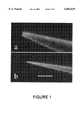
- FIG. 1aA scanning electron micrograph of the front part of a conventional pipette (tip diameter approx. 0.7 ⁇ m).
- FIG. 1bA temperature-controlled nanopipette according to the invention (tip diameter approx. 0.1 ⁇ m).
- FIG. 2A diagrammatic representation of an inventive, completely filled, temperature-controlled nanopipette (10), after filling it with injection substance (1), Galinstan (2) and silicone oil (3) the capillary is sealed with an adhesive-filled glass cap (4).
- FIG. 3A diagrammatic representation of the inventive heater, an air flow produced by a pump (5) being blown via a hose (6) and glass capillary (7) onto the pipette (10), the air being heated by a constantan wire (8) wound round the glass capillary (7) and connected to a continuously variable power supply.
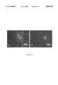
- FIGS. 5a and 5bConfocal laser scanning micrographs of microinjections with the inventive temperature-controlled nanopipette in the nuclei of cells of xenopus distal renal tube A6 cell line cultures: (a) Lucifer Yellow (approx. 0.5 kDa) injected into the lower nucleus and rapidly leaving the latter via the nuclei pores and also accumulated in the other nucleus; (b) whereas a 70 kDa dextran Lucifer Yellow conjugate only leaves the nucleus very slowly.
- Lucifer Yellowapproximately 0.5 kDa
Landscapes
- Health & Medical Sciences (AREA)
- Genetics & Genomics (AREA)
- Life Sciences & Earth Sciences (AREA)
- Engineering & Computer Science (AREA)
- Wood Science & Technology (AREA)
- Bioinformatics & Cheminformatics (AREA)
- Organic Chemistry (AREA)
- Biotechnology (AREA)
- Zoology (AREA)
- Chemical & Material Sciences (AREA)
- Biomedical Technology (AREA)
- General Engineering & Computer Science (AREA)
- General Health & Medical Sciences (AREA)
- Microbiology (AREA)
- Biochemistry (AREA)
- Sustainable Development (AREA)
- Cell Biology (AREA)
- Physics & Mathematics (AREA)
- Biophysics (AREA)
- Molecular Biology (AREA)
- Plant Pathology (AREA)
- Apparatus Associated With Microorganisms And Enzymes (AREA)
Abstract
Description
Claims (9)
Applications Claiming Priority (2)
| Application Number | Priority Date | Filing Date | Title |
|---|---|---|---|
| EP98110294AEP0992577B1 (en) | 1998-06-05 | 1998-06-05 | Process for microinjection and nanopipette for introducing an injection product, particularly foreign genetic material in procaryotic or eucaryotic cells or cellcomparments thereof (plastides, cell nucleus) |
| EP98110294 | 1998-06-05 |
Publications (1)
| Publication Number | Publication Date |
|---|---|
| US6063629Atrue US6063629A (en) | 2000-05-16 |
Family
ID=8232068
Family Applications (1)
| Application Number | Title | Priority Date | Filing Date |
|---|---|---|---|
| US09/325,450Expired - LifetimeUS6063629A (en) | 1998-06-05 | 1999-06-03 | Microinjection process for introducing an injection substance particularly foreign, genetic material, into procaryotic and eucaryotic cells, as well as cell compartments of the latter (plastids, cell nuclei), as well as nanopipette for the same |
Country Status (3)
| Country | Link |
|---|---|
| US (1) | US6063629A (en) |
| EP (1) | EP0992577B1 (en) |
| DE (1) | DE59800215D1 (en) |
Cited By (24)
| Publication number | Priority date | Publication date | Assignee | Title |
|---|---|---|---|---|
| US6661575B1 (en) | 2000-10-31 | 2003-12-09 | Sergey A. Yakovenko | Methods and apparata for micromanipulation of micro-and nanoparticles |
| US20040023372A1 (en)* | 2002-05-28 | 2004-02-05 | The Trustees Of The University Of Pennsylvania | Tubular nanostructures |
| US20040023903A1 (en)* | 1997-12-19 | 2004-02-05 | Davis Brian Ronald | Single-stranded end-capped oligonucleotide mediated targeted gene repair and modification and uses thereof |
| US20040063100A1 (en)* | 2002-09-30 | 2004-04-01 | Wang Chung Lin | Nanoneedle chips and the production thereof |
| WO2004092369A1 (en)* | 2003-04-11 | 2004-10-28 | Riken | Method of microinjection and device therefor |
| US20050101019A1 (en)* | 2003-11-10 | 2005-05-12 | Mcclelland Paul H. | Method and device for targeted delivery of materials to selected single cells |
| US20080199399A1 (en)* | 2007-02-21 | 2008-08-21 | Xing Chen | Interfacing Nanostructures to Biological Cells |
| WO2009009610A3 (en)* | 2007-07-09 | 2009-03-12 | Univ Brigham Young | Methods and devices for charged molecule manipulation |
| US20090085426A1 (en)* | 2007-09-28 | 2009-04-02 | Davis Robert C | Carbon nanotube mems assembly |
| US20100178650A1 (en)* | 2006-08-11 | 2010-07-15 | Karsten Stanislav L | Capillary-based cell and tissue acquisition system (ctas) |
| US20100239828A1 (en)* | 2009-03-19 | 2010-09-23 | Cornaby Sterling W | Resistively heated small planar filament |
| WO2010124177A1 (en)* | 2009-04-24 | 2010-10-28 | The Trustees Of The University Of Pennsylvania | Multiple-electrode and metal-coated probes |
| US20110121179A1 (en)* | 2007-06-01 | 2011-05-26 | Liddiard Steven D | X-ray window with beryllium support structure |
| US20110150184A1 (en)* | 2009-12-17 | 2011-06-23 | Krzysztof Kozaczek | Multiple wavelength x-ray source |
| US8247971B1 (en) | 2009-03-19 | 2012-08-21 | Moxtek, Inc. | Resistively heated small planar filament |
| WO2012149166A1 (en)* | 2011-04-27 | 2012-11-01 | Brigham Young University | Delivery of biological materials into cellular organelles |
| WO2013059824A1 (en)* | 2011-10-21 | 2013-04-25 | Nanoinjection Technologies, L.L.C. | Lance device and associated methods for delivering a biological material into a biological structure |
| US8498381B2 (en) | 2010-10-07 | 2013-07-30 | Moxtek, Inc. | Polymer layer on X-ray window |
| US8750458B1 (en) | 2011-02-17 | 2014-06-10 | Moxtek, Inc. | Cold electron number amplifier |
| US8785177B2 (en) | 2011-11-04 | 2014-07-22 | The Board Of Trustees Of The University Of Illinois, A Body Corporate And Politic Of The State Of Illinois | Methods for nano-mechanoporation |
| US8804910B1 (en) | 2011-01-24 | 2014-08-12 | Moxtek, Inc. | Reduced power consumption X-ray source |
| US8929515B2 (en) | 2011-02-23 | 2015-01-06 | Moxtek, Inc. | Multiple-size support for X-ray window |
| US9305735B2 (en) | 2007-09-28 | 2016-04-05 | Brigham Young University | Reinforced polymer x-ray window |
| US12233406B2 (en) | 2021-08-04 | 2025-02-25 | City University Of Hong Kong | Automated system for high-throughput microinjection of adherent cells |
Families Citing this family (3)
| Publication number | Priority date | Publication date | Assignee | Title |
|---|---|---|---|---|
| DE10334164A1 (en)* | 2003-07-26 | 2005-02-17 | Eppendorf Ag | Glass capillary for microinjection and method for making a glass capillary for microinjection |
| DE102004026087A1 (en)* | 2004-05-25 | 2005-12-15 | "Stiftung Caesar" (Center Of Advanced European Studies And Research) | Nano-cannula |
| FR2983092B1 (en)* | 2011-11-30 | 2014-05-02 | Centre Nat Rech Scient | METHOD AND DEVICE FOR FILLING NANOPIPETTES BY DYNAMIC MICRODISTILLATION |
Citations (8)
| Publication number | Priority date | Publication date | Assignee | Title |
|---|---|---|---|---|
| US3720354A (en)* | 1970-09-24 | 1973-03-13 | Drummond Instr Co | Dispensing micropipette apparatus having disposable parts |
| US4625677A (en)* | 1985-05-23 | 1986-12-02 | Max-Planck-Gesellschaft Zur Foerderung Der Wissenschaften E.V. | Apparatus for coating and polishing a micropipette |
| EP0292899A2 (en)* | 1987-05-29 | 1988-11-30 | Firma Carl Zeiss | Process for microinjection into cells, possibly for suction out of isolated cells or entire cells out of cell cultures |
| GB2211111A (en)* | 1987-10-21 | 1989-06-28 | Saxon Micro Limited | Micropipette and method of operation |
| US5225750A (en)* | 1989-10-02 | 1993-07-06 | Prima Meat Packers, Ltd. | Microinjection apparatus, and method of controlling microinjection |
| DE4423267A1 (en)* | 1994-07-04 | 1996-01-25 | Wildanger Hans Joerg | Pipette to draws in or eject liq. by electrical heating of gas in enclosed space |
| DE19629143A1 (en)* | 1996-07-19 | 1998-01-22 | Bayer Ag | Device for separating micro objects |
| DE29801523U1 (en)* | 1997-02-18 | 1998-04-23 | Inst Physikalische Hochtech Ev | Micropipette or micro actuator |
Family Cites Families (1)
| Publication number | Priority date | Publication date | Assignee | Title |
|---|---|---|---|---|
| JPH05133851A (en)* | 1991-11-13 | 1993-05-28 | Nagano Japan Radio Co | Dispensing device |
- 1998
- 1998-06-05EPEP98110294Apatent/EP0992577B1/ennot_activeExpired - Lifetime
- 1998-06-05DEDE59800215Tpatent/DE59800215D1/ennot_activeExpired - Lifetime
- 1999
- 1999-06-03USUS09/325,450patent/US6063629A/ennot_activeExpired - Lifetime
Patent Citations (8)
| Publication number | Priority date | Publication date | Assignee | Title |
|---|---|---|---|---|
| US3720354A (en)* | 1970-09-24 | 1973-03-13 | Drummond Instr Co | Dispensing micropipette apparatus having disposable parts |
| US4625677A (en)* | 1985-05-23 | 1986-12-02 | Max-Planck-Gesellschaft Zur Foerderung Der Wissenschaften E.V. | Apparatus for coating and polishing a micropipette |
| EP0292899A2 (en)* | 1987-05-29 | 1988-11-30 | Firma Carl Zeiss | Process for microinjection into cells, possibly for suction out of isolated cells or entire cells out of cell cultures |
| GB2211111A (en)* | 1987-10-21 | 1989-06-28 | Saxon Micro Limited | Micropipette and method of operation |
| US5225750A (en)* | 1989-10-02 | 1993-07-06 | Prima Meat Packers, Ltd. | Microinjection apparatus, and method of controlling microinjection |
| DE4423267A1 (en)* | 1994-07-04 | 1996-01-25 | Wildanger Hans Joerg | Pipette to draws in or eject liq. by electrical heating of gas in enclosed space |
| DE19629143A1 (en)* | 1996-07-19 | 1998-01-22 | Bayer Ag | Device for separating micro objects |
| DE29801523U1 (en)* | 1997-02-18 | 1998-04-23 | Inst Physikalische Hochtech Ev | Micropipette or micro actuator |
Non-Patent Citations (1)
| Title |
|---|
| Patent Abstracts of Japan, Higuchi Ryuichi, Dispensing Apparatus 05133851, May 28, 1993.* |
Cited By (39)
| Publication number | Priority date | Publication date | Assignee | Title |
|---|---|---|---|---|
| US20040023903A1 (en)* | 1997-12-19 | 2004-02-05 | Davis Brian Ronald | Single-stranded end-capped oligonucleotide mediated targeted gene repair and modification and uses thereof |
| US6661575B1 (en) | 2000-10-31 | 2003-12-09 | Sergey A. Yakovenko | Methods and apparata for micromanipulation of micro-and nanoparticles |
| US20040023372A1 (en)* | 2002-05-28 | 2004-02-05 | The Trustees Of The University Of Pennsylvania | Tubular nanostructures |
| US20040063100A1 (en)* | 2002-09-30 | 2004-04-01 | Wang Chung Lin | Nanoneedle chips and the production thereof |
| US20090286319A1 (en)* | 2003-04-11 | 2009-11-19 | Olympus Corporation | Microinjection method and device |
| WO2004092369A1 (en)* | 2003-04-11 | 2004-10-28 | Riken | Method of microinjection and device therefor |
| JPWO2004092369A1 (en)* | 2003-04-11 | 2006-07-06 | 独立行政法人理化学研究所 | Microinjection method and apparatus |
| US20070087436A1 (en)* | 2003-04-11 | 2007-04-19 | Atsushi Miyawaki | Microinjection method and device |
| US8304240B2 (en) | 2003-04-11 | 2012-11-06 | Olympus Corporation | Microinjection method and device |
| JP4530991B2 (en)* | 2003-04-11 | 2010-08-25 | 独立行政法人理化学研究所 | Microinjection method and apparatus |
| US20050101019A1 (en)* | 2003-11-10 | 2005-05-12 | Mcclelland Paul H. | Method and device for targeted delivery of materials to selected single cells |
| US7132242B2 (en) | 2003-11-10 | 2006-11-07 | Hewlett-Packard Development Company, L.P. | Method and device for targeted delivery of materials to selected single cells |
| US8797644B2 (en) | 2006-08-11 | 2014-08-05 | The Regents Of The University Of California | Capillary-based cell and tissue acquisition system (CTAS) |
| US20100178650A1 (en)* | 2006-08-11 | 2010-07-15 | Karsten Stanislav L | Capillary-based cell and tissue acquisition system (ctas) |
| US8257932B2 (en) | 2007-02-21 | 2012-09-04 | The Regents Of The University Of California | Interfacing nanostructures to biological cells |
| US20080199399A1 (en)* | 2007-02-21 | 2008-08-21 | Xing Chen | Interfacing Nanostructures to Biological Cells |
| US20110121179A1 (en)* | 2007-06-01 | 2011-05-26 | Liddiard Steven D | X-ray window with beryllium support structure |
| US10119151B2 (en) | 2007-07-09 | 2018-11-06 | Brigham Young University | Methods and devices for charged molecule manipulation |
| US20100248343A1 (en)* | 2007-07-09 | 2010-09-30 | Aten Quentin T | Methods and Devices for Charged Molecule Manipulation |
| WO2009009610A3 (en)* | 2007-07-09 | 2009-03-12 | Univ Brigham Young | Methods and devices for charged molecule manipulation |
| US20100323419A1 (en)* | 2007-07-09 | 2010-12-23 | Aten Quentin T | Methods and Devices for Charged Molecule Manipulation |
| US20100285271A1 (en)* | 2007-09-28 | 2010-11-11 | Davis Robert C | Carbon nanotube assembly |
| US8736138B2 (en) | 2007-09-28 | 2014-05-27 | Brigham Young University | Carbon nanotube MEMS assembly |
| US20090085426A1 (en)* | 2007-09-28 | 2009-04-02 | Davis Robert C | Carbon nanotube mems assembly |
| US9305735B2 (en) | 2007-09-28 | 2016-04-05 | Brigham Young University | Reinforced polymer x-ray window |
| US8247971B1 (en) | 2009-03-19 | 2012-08-21 | Moxtek, Inc. | Resistively heated small planar filament |
| US20100239828A1 (en)* | 2009-03-19 | 2010-09-23 | Cornaby Sterling W | Resistively heated small planar filament |
| WO2010124177A1 (en)* | 2009-04-24 | 2010-10-28 | The Trustees Of The University Of Pennsylvania | Multiple-electrode and metal-coated probes |
| US8702927B2 (en) | 2009-04-24 | 2014-04-22 | The Trustees Of The University Of Pennsylvania | Multiple-electrode and metal-coated probes |
| US7983394B2 (en) | 2009-12-17 | 2011-07-19 | Moxtek, Inc. | Multiple wavelength X-ray source |
| US20110150184A1 (en)* | 2009-12-17 | 2011-06-23 | Krzysztof Kozaczek | Multiple wavelength x-ray source |
| US8498381B2 (en) | 2010-10-07 | 2013-07-30 | Moxtek, Inc. | Polymer layer on X-ray window |
| US8804910B1 (en) | 2011-01-24 | 2014-08-12 | Moxtek, Inc. | Reduced power consumption X-ray source |
| US8750458B1 (en) | 2011-02-17 | 2014-06-10 | Moxtek, Inc. | Cold electron number amplifier |
| US8929515B2 (en) | 2011-02-23 | 2015-01-06 | Moxtek, Inc. | Multiple-size support for X-ray window |
| WO2012149166A1 (en)* | 2011-04-27 | 2012-11-01 | Brigham Young University | Delivery of biological materials into cellular organelles |
| WO2013059824A1 (en)* | 2011-10-21 | 2013-04-25 | Nanoinjection Technologies, L.L.C. | Lance device and associated methods for delivering a biological material into a biological structure |
| US8785177B2 (en) | 2011-11-04 | 2014-07-22 | The Board Of Trustees Of The University Of Illinois, A Body Corporate And Politic Of The State Of Illinois | Methods for nano-mechanoporation |
| US12233406B2 (en) | 2021-08-04 | 2025-02-25 | City University Of Hong Kong | Automated system for high-throughput microinjection of adherent cells |
Also Published As
| Publication number | Publication date |
|---|---|
| DE59800215D1 (en) | 2000-08-31 |
| EP0992577B1 (en) | 2000-07-26 |
| EP0992577A1 (en) | 2000-04-12 |
Similar Documents
| Publication | Publication Date | Title |
|---|---|---|
| US6063629A (en) | Microinjection process for introducing an injection substance particularly foreign, genetic material, into procaryotic and eucaryotic cells, as well as cell compartments of the latter (plastids, cell nuclei), as well as nanopipette for the same | |
| Ansorge | Improved system for capillary microinjection into living cells | |
| Preston et al. | Rapid polymer transport in concentrated solutions through the formation of ordered structures | |
| US5610010A (en) | Process and apparatus for fragmenting biomaterials | |
| US20170252744A1 (en) | Method for handling microdrops which include samples | |
| US20240050914A1 (en) | Method for Generating Solid Capsules | |
| Dong et al. | Preparation of 10 μm scale monodispersed particles by jetting flow in coaxial microfluidic devices | |
| EP1966367A1 (en) | Bioreactor for cell and tissue culture | |
| WO2014005690A1 (en) | Substrate unit, preservation device and method for the cryopreservation of a biological sample | |
| Guevorkian et al. | Flow dynamics of 3D multicellular systems into capillaries | |
| CN111269834B (en) | 3D voxel printing method based on cell soft sphere | |
| CN108315389A (en) | A kind of micro-volume cellular nucleic acid amplification method | |
| CN115646567A (en) | Microfluidic chip with integrated droplet on-line culture and high-throughput screening functions and its application | |
| CN113634208A (en) | Method for preparing porous calcium alginate microspheres by using microfluidic double-aqueous-phase emulsion as template | |
| CN209222159U (en) | A kind of compound micro-fluidic chip of array PDMS- paper base for single cell analysis | |
| JPH01143647A (en) | Micropipette | |
| Chen et al. | Robust fabrication of ultra-soft tunable PDMS microcapsules as a biomimetic model for red blood cells | |
| Gernert et al. | A simple apparatus for controlling nucleation and size in protein crystal growth | |
| JP2003125750A (en) | Micropipette, injection apparatus and method of microinjection | |
| Kleine‐Brüggeney et al. | A Macro‐to‐Micro Interface for Performing Comprehensive Microfluidic Cell Culture Assays | |
| CN114383755A (en) | Method for accurately measuring uniform temperature of inclusion of melt rich in volatile components | |
| CN1954064A (en) | Biosample manipulation device | |
| CN106401901A (en) | Self-driven micro pump based on solvent volatilization effect | |
| CN104328106B (en) | Microcapsule preparation apparatus and method | |
| JP4621942B2 (en) | Microinjection method using liquid expansion pressure by laser heating |
Legal Events
| Date | Code | Title | Description |
|---|---|---|---|
| AS | Assignment | Owner name:LUMMEL, WOLFGANG, SWITZERLAND Free format text:ASSIGNMENT OF ASSIGNORS INTEREST;ASSIGNOR:KNOBLAUCH, MICHAEL;REEL/FRAME:010720/0546 Effective date:20000220 | |
| STCF | Information on status: patent grant | Free format text:PATENTED CASE | |
| FEPP | Fee payment procedure | Free format text:PAYOR NUMBER ASSIGNED (ORIGINAL EVENT CODE: ASPN); ENTITY STATUS OF PATENT OWNER: LARGE ENTITY | |
| AS | Assignment | Owner name:FRAUNHOFER-GESELLSCHAFT ZUR FORDERUNG DER ANGEWAND Free format text:ASSIGNMENT OF ASSIGNORS INTEREST;ASSIGNOR:LUMMEL, WOLFGANG;REEL/FRAME:011356/0103 Effective date:20001026 | |
| FEPP | Fee payment procedure | Free format text:PAT HOLDER NO LONGER CLAIMS SMALL ENTITY STATUS, ENTITY STATUS SET TO UNDISCOUNTED (ORIGINAL EVENT CODE: STOL); ENTITY STATUS OF PATENT OWNER: LARGE ENTITY | |
| FPAY | Fee payment | Year of fee payment:4 | |
| FEPP | Fee payment procedure | Free format text:PAYOR NUMBER ASSIGNED (ORIGINAL EVENT CODE: ASPN); ENTITY STATUS OF PATENT OWNER: LARGE ENTITY Free format text:PAYER NUMBER DE-ASSIGNED (ORIGINAL EVENT CODE: RMPN); ENTITY STATUS OF PATENT OWNER: LARGE ENTITY | |
| FPAY | Fee payment | Year of fee payment:8 | |
| FPAY | Fee payment | Year of fee payment:12 | |
| FEPP | Fee payment procedure | Free format text:PAYOR NUMBER ASSIGNED (ORIGINAL EVENT CODE: ASPN); ENTITY STATUS OF PATENT OWNER: LARGE ENTITY Free format text:PAYER NUMBER DE-ASSIGNED (ORIGINAL EVENT CODE: RMPN); ENTITY STATUS OF PATENT OWNER: LARGE ENTITY |