US6063093A - Systems and methods for guiding a medical instrument through a body - Google Patents
Systems and methods for guiding a medical instrument through a bodyDownload PDFInfo
- Publication number
- US6063093A US6063093AUS09/275,623US27562399AUS6063093AUS 6063093 AUS6063093 AUS 6063093AUS 27562399 AUS27562399 AUS 27562399AUS 6063093 AUS6063093 AUS 6063093A
- Authority
- US
- United States
- Prior art keywords
- medical instrument
- optic fiber
- accordance
- catheter
- tissue
- Prior art date
- Legal status (The legal status is an assumption and is not a legal conclusion. Google has not performed a legal analysis and makes no representation as to the accuracy of the status listed.)
- Expired - Lifetime
Links
Images

Classifications
- A—HUMAN NECESSITIES
- A61—MEDICAL OR VETERINARY SCIENCE; HYGIENE
- A61B—DIAGNOSIS; SURGERY; IDENTIFICATION
- A61B18/00—Surgical instruments, devices or methods for transferring non-mechanical forms of energy to or from the body
- A61B18/18—Surgical instruments, devices or methods for transferring non-mechanical forms of energy to or from the body by applying electromagnetic radiation, e.g. microwaves
- A61B18/20—Surgical instruments, devices or methods for transferring non-mechanical forms of energy to or from the body by applying electromagnetic radiation, e.g. microwaves using laser
- A61B18/22—Surgical instruments, devices or methods for transferring non-mechanical forms of energy to or from the body by applying electromagnetic radiation, e.g. microwaves using laser the beam being directed along or through a flexible conduit, e.g. an optical fibre; Couplings or hand-pieces therefor
- A61B18/24—Surgical instruments, devices or methods for transferring non-mechanical forms of energy to or from the body by applying electromagnetic radiation, e.g. microwaves using laser the beam being directed along or through a flexible conduit, e.g. an optical fibre; Couplings or hand-pieces therefor with a catheter
- A61B18/245—Surgical instruments, devices or methods for transferring non-mechanical forms of energy to or from the body by applying electromagnetic radiation, e.g. microwaves using laser the beam being directed along or through a flexible conduit, e.g. an optical fibre; Couplings or hand-pieces therefor with a catheter for removing obstructions in blood vessels or calculi
- A—HUMAN NECESSITIES
- A61—MEDICAL OR VETERINARY SCIENCE; HYGIENE
- A61B—DIAGNOSIS; SURGERY; IDENTIFICATION
- A61B1/00—Instruments for performing medical examinations of the interior of cavities or tubes of the body by visual or photographical inspection, e.g. endoscopes; Illuminating arrangements therefor
- A61B1/04—Instruments for performing medical examinations of the interior of cavities or tubes of the body by visual or photographical inspection, e.g. endoscopes; Illuminating arrangements therefor combined with photographic or television appliances
- A61B1/042—Instruments for performing medical examinations of the interior of cavities or tubes of the body by visual or photographical inspection, e.g. endoscopes; Illuminating arrangements therefor combined with photographic or television appliances characterised by a proximal camera, e.g. a CCD camera
- A—HUMAN NECESSITIES
- A61—MEDICAL OR VETERINARY SCIENCE; HYGIENE
- A61B—DIAGNOSIS; SURGERY; IDENTIFICATION
- A61B17/00—Surgical instruments, devices or methods
- A61B2017/00017—Electrical control of surgical instruments
- A61B2017/00022—Sensing or detecting at the treatment site
- A61B2017/00057—Light
- A—HUMAN NECESSITIES
- A61—MEDICAL OR VETERINARY SCIENCE; HYGIENE
- A61B—DIAGNOSIS; SURGERY; IDENTIFICATION
- A61B17/00—Surgical instruments, devices or methods
- A61B2017/00017—Electrical control of surgical instruments
- A61B2017/00022—Sensing or detecting at the treatment site
- A61B2017/00106—Sensing or detecting at the treatment site ultrasonic
- A—HUMAN NECESSITIES
- A61—MEDICAL OR VETERINARY SCIENCE; HYGIENE
- A61B—DIAGNOSIS; SURGERY; IDENTIFICATION
- A61B17/00—Surgical instruments, devices or methods
- A61B17/22—Implements for squeezing-off ulcers or the like on inner organs of the body; Implements for scraping-out cavities of body organs, e.g. bones; for invasive removal or destruction of calculus using mechanical vibrations; for removing obstructions in blood vessels, not otherwise provided for
- A61B2017/22072—Implements for squeezing-off ulcers or the like on inner organs of the body; Implements for scraping-out cavities of body organs, e.g. bones; for invasive removal or destruction of calculus using mechanical vibrations; for removing obstructions in blood vessels, not otherwise provided for with an instrument channel, e.g. for replacing one instrument by the other
- A61B2017/22074—Implements for squeezing-off ulcers or the like on inner organs of the body; Implements for scraping-out cavities of body organs, e.g. bones; for invasive removal or destruction of calculus using mechanical vibrations; for removing obstructions in blood vessels, not otherwise provided for with an instrument channel, e.g. for replacing one instrument by the other the instrument being only slidable in a channel, e.g. advancing optical fibre through a channel
- A61B2017/22075—Implements for squeezing-off ulcers or the like on inner organs of the body; Implements for scraping-out cavities of body organs, e.g. bones; for invasive removal or destruction of calculus using mechanical vibrations; for removing obstructions in blood vessels, not otherwise provided for with an instrument channel, e.g. for replacing one instrument by the other the instrument being only slidable in a channel, e.g. advancing optical fibre through a channel with motorized advancing or retracting means
- A—HUMAN NECESSITIES
- A61—MEDICAL OR VETERINARY SCIENCE; HYGIENE
- A61B—DIAGNOSIS; SURGERY; IDENTIFICATION
- A61B18/00—Surgical instruments, devices or methods for transferring non-mechanical forms of energy to or from the body
- A61B18/18—Surgical instruments, devices or methods for transferring non-mechanical forms of energy to or from the body by applying electromagnetic radiation, e.g. microwaves
- A61B18/20—Surgical instruments, devices or methods for transferring non-mechanical forms of energy to or from the body by applying electromagnetic radiation, e.g. microwaves using laser
- A61B18/22—Surgical instruments, devices or methods for transferring non-mechanical forms of energy to or from the body by applying electromagnetic radiation, e.g. microwaves using laser the beam being directed along or through a flexible conduit, e.g. an optical fibre; Couplings or hand-pieces therefor
- A61B2018/2205—Characteristics of fibres
- A61B2018/2211—Plurality of fibres
- A—HUMAN NECESSITIES
- A61—MEDICAL OR VETERINARY SCIENCE; HYGIENE
- A61B—DIAGNOSIS; SURGERY; IDENTIFICATION
- A61B90/00—Instruments, implements or accessories specially adapted for surgery or diagnosis and not covered by any of the groups A61B1/00 - A61B50/00, e.g. for luxation treatment or for protecting wound edges
- A61B90/36—Image-producing devices or illumination devices not otherwise provided for
- A61B90/361—Image-producing devices, e.g. surgical cameras
- A61B2090/3614—Image-producing devices, e.g. surgical cameras using optical fibre
- A—HUMAN NECESSITIES
- A61—MEDICAL OR VETERINARY SCIENCE; HYGIENE
- A61B—DIAGNOSIS; SURGERY; IDENTIFICATION
- A61B90/00—Instruments, implements or accessories specially adapted for surgery or diagnosis and not covered by any of the groups A61B1/00 - A61B50/00, e.g. for luxation treatment or for protecting wound edges
- A61B90/36—Image-producing devices or illumination devices not otherwise provided for
- A61B90/37—Surgical systems with images on a monitor during operation
- A61B2090/373—Surgical systems with images on a monitor during operation using light, e.g. by using optical scanners
- A—HUMAN NECESSITIES
- A61—MEDICAL OR VETERINARY SCIENCE; HYGIENE
- A61M—DEVICES FOR INTRODUCING MEDIA INTO, OR ONTO, THE BODY; DEVICES FOR TRANSDUCING BODY MEDIA OR FOR TAKING MEDIA FROM THE BODY; DEVICES FOR PRODUCING OR ENDING SLEEP OR STUPOR
- A61M25/00—Catheters; Hollow probes
- A61M25/01—Introducing, guiding, advancing, emplacing or holding catheters
Definitions
- This inventionrelates generally to medical instruments and, more particularly, to systems and methods for guiding medical instruments through a body or a portion of the body, such as a blood vessel.
- Disease processese.g., tumors, inflammation of lymph nodes, and plaque build-up in arteries, often afflict the human body.
- a medical deviceinto the body, and to guide the medical device to the diseased site. Once the medical device is adjacent the diseased site, the medical device typically is used to photoablate or otherwise remove or reduce the diseased tissue.
- Atherosclerotic plaqueis known to build-up on the walls of arteries in the human body. Such plaque build-up restricts circulation and often causes cardiovascular problems, especially when the build-up occurs in coronary arteries. Accordingly, it is desirable to detect plaque build-up and remove or otherwise reduce such plaque build-up.
- One known catheterimplement laser energy to remove plaque build up on artery walls.
- One known catheterincludes a laser source and a catheter body.
- the catheter bodyhas a first end and a second end, or head, and several optical fibers extend between the first end and the second end.
- the laser sourceis coupled to each of the optical fibers adjacent the catheter body first end and is configured to transmit laser energy simultaneously through the optical fibers.
- the catheter bodyis positioned in the artery so that the second end of the catheter body is adjacent a region of plaque build-up.
- the laser sourceis then energized so that laser energy travels through each of the optical fibers and substantially photoablates the plaque adjacent the second end of the catheter body.
- the catheter bodyis then advanced through the region to photoablate the plaque in such region.
- a guide wiretypically is required to properly position the catheter in the artery.
- the guide wireis advanced through the artery and region of plaque build-up so that it forms a path through the artery and plaque build-up.
- the catheteris then guided through the artery using the guide wire.
- One known catheterincludes ultrasound sensors positioned at its distal end for displaying images of the artery while the catheter is advanced.
- Known ultrasound sensorsare coupled to an outer perimeter of the catheter and emit sound waves substantially radially from the catheter distal end toward the artery wall. The sound waves then are reflected by the surrounding tissue, e.g., the artery wall and plaque, and toward the ultrasound sensors. The reflected sound waves are then compared to the transmitted sound waves to generate an ultrasound image of the tissue radially sounding the distal end.
- an operatorTo advance the catheter, an operator first positions the catheter at a first location in the artery. Sound waves are then emitted from and received by the ultrasound sensors, and an image is then displayed showing the artery tissue adjacent the circumference of the catheter at such first location. The catheter is then advanced to a second location in the artery, and a second image is displayed showing the artery at such location. This process is then continued until the catheter is advanced through the artery and the plaque-build up.
- Utilizing known ultrasound sensors as described aboveresults in displaying images of the portions of the arterial wall which are radially disposed about the catheter, but does not provide images of the arterial wall or plaque positioned immediately forward the catheter.
- the sensorsmust be aligned within the artery so that the sound waves projected toward the artery wall are substantially perpendicular to the artery wall when reflected to the sensors. Sound waves that are not perpendicular to the artery wall may provide inaccurate signals, which may result in the display of inaccurate images, which is undesirable.
- Inaccurate imagesmay result in improperly guiding the catheter through the blood vessel, which is undesirable.
- known cathetersmust be manually inserted and guided through the blood vessel.
- a surgeon or other operatorutilizes the displayed images to guide the catheter through the vessel and avoid damaging healthy tissue, i.e., the artery wall. If an inaccurate image displays plaque even though such tissue actually is an artery wall, it is possible that the operator may photoablate the artery wall, which is undesirable.
- a catheterwhich, in one embodiment, includes a catheter body and at least one interferometric guidance system.
- the catheter bodyincludes a bundle of optic fibers, each having a first end and a second end, and the second ends of the respective optic fibers form a substantially rounded catheter head.
- Each interferometric guidance systemis coupled to the catheter body and includes a first optic fiber, a second optic fiber, and a detecting element.
- the first optic fiber of each guidance systemincludes a first end and a second end, and is coupled to the catheter body so that the second end is adjacent the catheter head.
- the second optic fiber of each guidance systemsimilarly includes a first end and a second end, and a reference mirror is positioned adjacent the second optic fiber second end.
- the detecting element of each guidance systemis communicatively coupled to both the first optic fiber and the second optic fiber of such guidance system.
- the first optic fiber first endis communicatively coupled to the detecting element and the second optic fiber first end is communicatively coupled to the detecting element.
- the detecting elementis configured to determine interference between substantially equal light beams which are emitted from the same source and which are split to propagate through the first optic fiber and through the second optic fiber. The interference is then utilized to determine the density and type of tissue adjacent the catheter head, and to guide the catheter head through the tissue.
- the catheter headis inserted at least partially into a blood vessel so that the catheter head and the first optic fiber second end of each guidance system is positioned in the blood vessel.
- the second optic fiber of each guidance systemis positioned outside the blood vessel.
- the reference mirror of each guidance systemis positioned a desired, or measuring, distance from its respective second optic fiber second end. The distances between the respective reference mirrors and optic fiber second ends may either be the same or different.
- a light beamis split into first and second substantially equal light beams which are then transmitted through the first and second optic fibers of each guidance system, from their respective first ends to their respective second ends.
- the first light beam transmitted through the first optic fiberexits from the first optic fiber second end, is at least partially reflected by the tissue, re-enters the first optic fiber second end and propagates toward the first optic fiber first end.
- the second light beam transmitted through the second optic fiberexits from the second optic fiber second end, is at least partially reflected by the reference mirror, re-enters the second optic fiber second end and propagates toward the second optic fiber first end.
- Each detecting elementdetects interference between the respective reflected first light beam and the reflected second light beam and transmits interference data to a computer.
- the computerthen utilizes the interference data to determine the density and the type of the tissue to be examined adjacent the catheter head.
- the interference datais representative of the density and type of tissue located at the measuring distance from the second optic fiber second end, and the computer utilizes such data to generate an image of such tissue at such location.
- the computeralso utilizes the interference data to control subsequent advancement of the catheter through the artery.
- the above described guidance systemsfacilitate obtaining more accurate images than obtained using ultrasound.
- such systemsare believed to be substantially easy to implement in connection with medical apparatus other than catheters.
- such systemsare believed to facilitate automatic control and advancement of the catheter through the body.
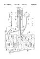
- FIG. 1is a pictorial illustration of a catheter including two guidance systems in accordance with one embodiment of the present invention inserted into a blood vessel.
- FIG. 2is a front cross section view of the catheter body shown in FIG. 1.
- FIG. 3is a schematic illustration of the catheter control element shown in FIG. 1.
- FIG. 4is a schematic illustration of one of the guidance systems shown in FIG. 1.
- FIG. 1is a pictorial illustration of a catheter assembly 20 including two guidance systems 22A and 22B in accordance with one embodiment of the present invention inserted into a blood vessel 24 of a body.
- Catheter assembly 20includes a control element 26 and a catheter body 28.
- Catheter body 28has a first end 30 and a rounded, or hemispherical, second end, or head, 32, and includes a plurality of optic fibers (not shown in FIG. 1).
- Catheter body first end 30is communicatively coupled to control element 26 and catheter body second end 32 is positioned within an interior 34 of blood vessel 24 adjacent tissue to be imaged, e.g., plaque 36.
- Each guidance system 22A and 22Bincludes a respective control element 40A and 40B, a respective first, or measuring, optic fiber 42A and 42B, and a respective second, or reference, optic fiber 44A and 44B.
- First optic fibers 42A and 42Binclude respective first ends 46A and 46B and respective second ends 48A and 48B, and are coupled to catheter body 28 so that second ends 48A and 48B are adjacent catheter head 32 and are positioned in blood vessel interior 34.
- Second optic fibers 44A and 44Balso include respective first ends 50A and 50B and respective second ends 52A and 52B.
- First optic fiber first end 46A and second optic fiber first end 50Aare communicatively coupled to system control element 40A
- first optic fiber first end 46B and second optic fiber first end 50Bare communicatively coupled to system control element 40B.
- First system first optic fiber 42Bis configured to emit energy waves substantially coaxially with respect to catheter head 32.
- Second system first optic fiber 42Bis configured to emit energy waves substantially radially with respect to catheter head 32.
- second end 48B of optic fiber 42Bincludes a prism (not shown in FIG. 1) configured to emit an energy beam at an angle with respect to catheter head 32, e.g., perpendicularly with respect to optic fiber 42A.
- Each guidance system control element 40A and 40Bincludes a respective diagnostic light beam source 54A and 54B, a respective beam splitter 56A and 56B, and a respective detecting element 58A and 58B.
- Beam splitters 56A and 56Bare communicatively coupled to first optic fiber first ends 46A and 46B, respectively.
- beam splitters 56A and 56Bare communicatively coupled to second optic fiber first ends 50A and 50B, respectively.
- Beam splitters 56A and 56Balso are coupled to respective diagnostic light beam sources 54A and 54B and detecting elements 58A and 58B via optic fibers 64A and 64B, respectively.
- Detecting elements 58A and 58Bare coupled to an image screen 38 and are configured to transmit image data to image screen 38 for displaying an image of the tissue to be imaged. Detecting elements 58A and 58B also are configured to transmit control data to catheter control element 26. Particularly, detecting element 58A is configured to determine interference between a light beam propagating through first optic fiber 42A and a light beam propagating through second optic fiber 44A, and to generate interference data representative of such interference. For example, detecting element 58A may include a detector, a demodulator and an analog digitizer which cooperate in a known manner to generate such interference data. Such interference data is transmitted to a computer 66A, which generates image data for display on image screen 38 and generates control data for transmission to catheter control element 26.
- detecting element 58Bis configured to determine interference between a light beam propagating through first optic fiber 42B and a light beam propagating through second optic fiber 44B, and to generate interference data representative of such interference.
- interference datais transmitted to a computer 66B, which generates image data for display on image screen 38 and generates control data for transmission to catheter control element 26.
- catheter body 28includes several optic fibers 68 extending through a housing, or casing, 70.
- Second system first optic fiber 42Bis coupled to housing 70 so that housing 70 extends between such second system first optic fiber 42B and catheter body optic fibers 68.
- First system first optic fiber 42Aextends through and is substantially centered within housing 70.
- second system first optic fiber 42Bmay be positioned within housing 70 and first system optic fiber 42A may be positioned outside housing 70.
- both first system optic fibers 42A and 42Bmay be positioned either within housing 70 or outside housing 70.
- catheter control element 26includes a therapeutic laser source 72 substantially aligned with catheter body optic fibers 68.
- Laser source 70is configured to transmit a therapeutic laser beam through catheter body optic fibers 68 for photoablating plaque 36 (FIG. 1), or other tissue.
- guidance system 22Afurther includes a reference mirror 74A positioned adjacent second fiber second end 52A.
- Reference mirror 74Ais movable with respect to second fiber second end 52A and is controlled, for example, by computer 66A.
- guidance system 22Bincludes a reference mirror 74B positioned adjacent second fiber second end 52B.
- Reference mirror 74Bis movable with respect to second fiber second end 52B and is controlled, for example, by computer 66B.
- each guidance system 22A and 22BPrior to inserting catheter assembly 20 into blood vessel 24, each guidance system 22A and 22B is calibrated. Particularly, reference mirror 74A is positioned a distance D 1 from second fiber second end 52A and guidance system 22A is calibrated so that interference data obtained by detecting element 58A is representative of tissue located approximately the same distance D 1 from first optic fiber second end 48A. Similarly, reference mirror 74A is positioned a distance D 2 from second fiber second end 52B and guidance system 22B is calibrated so that interference data obtained by detecting element 58B is representative of tissue located approximately the same distance D 2 from first optic fiber second end 48B.
- catheter assembly 20is inserted into blood vessel 24 so that catheter head 32 and first optic fiber second ends 48A and 48B are positioned within blood vessel 24, and second optic fiber second ends 52A and 52B are positioned outside blood vessel 24, and outside the body.
- First reference mirror 74Ais positioned distance D 1 from second optic fiber second end 52A
- second reference mirror 74Bis positioned distance D 2 from second optic fiber second end 52B.
- Light beam source 54Atransmits a diagnostic light beam to beam splitter 56A, which splits the light beam into first and second substantially equal light beams 76A and 78A, respectively.
- First light beam 76Ais then transmitted through first optic fiber 42A and second light beam 78A is transmitted through second optic fiber 44A.
- First light beam 76Aexits from first optic fiber second end 48A substantially coaxially with respect to catheter head 32, is at least partially reflected by the tissue, re-enters first optic fiber second end 48A and propagates toward first optic fiber first end 46A.
- Detecting element 58Adetects light interference patterns, e.g., interferences, between the reflected first light beam 76A and reflected second light beam 78A, and transmits interference data representative of such interferences to computer 66A.
- Computer 66Autilizes the interference data to determine the type and depth of the tissue located at a distance D 3 from first optic fiber second end 48A. Particularly, computer 66A utilizes the interference data to determine what type of tissue, if any, is located at a distance D 3 from first fiber second end 48A, where distance D 3 is substantially the same as distance D 1 .
- computer 66Amay include a memory, and representative interference signals for different types of tissues, e.g., plaque, artery walls, healthy tissue, cancerous tissue, may be stored in such memory.
- Computer 66Acompares the interference data received from detecting element 58A to such stored representative interference signals to determine the type of tissue located distance D 3 from first fiber second end 48A.
- Distances D 1 and D 3may, for example, be less than or equal to 1 millimeter, e.g., one quarter of a millimeter. Of course, distances D 1 and D 3 may be larger than 1 millimeter.
- reference mirror 74Amay be moved with respect to second fiber second end 48A to recalibrate guidance system 22A while it is positioned in a blood vessel 24. Particularly, if detecting element 58A generates interference data representative of a loss of signal through first optic fiber 42A, reference mirror 74A may be moved to reestablish a signal at a distance D 4 (not shown in FIG. 1) which is different from distance D 1 .
- reference mirror 74Amay be moved with respect to second fiber second end 48A to determine the type and depth of the tissue located at a varying distances from second fiber second end 48A. Particularly, reference mirror 74 may be moved between a point immediately adjacent second fiber second end 48A and a point distance D 1 from second fiber second end 48A to determine the type and depth of the tissue located at each point between such two points. Accordingly, reference mirror 74A may be moved to determine tissue type at multiple different distances from second fiber second end 48A.
- Computer 66Agenerates image data of such tissue and displays the image of such tissue on image screen 38. Particularly, computer 66A utilizes the interference data generated at various points in the tissue to generate image data representative of a substantially linear image profile of the examined tissue. Computer 66A also utilizes the interference data to generate and transmit control signals to catheter control element 26, as is described in more detail below.
- light beam source 54Btransmits a diagnostic light beam to beam splitter 56B, which splits the light beam into first and second substantially equal light beams 76B and 78B, respectively.
- First light beam 76Bis then transmitted through first optic fiber 42B and second light beam 78B is transmitted through second optic fiber 44B.
- First light beam 76Bexits from first optic fiber second end 48B substantially radially with respect to catheter head 32, is at least partially reflected by the tissue, re-enters first optic fiber second end 48B and propagates toward first optic fiber first end 46B.
- Detecting element 58Bdetects interference between the reflected first light beam 76B and reflected second light beam 78B, and transmits interference data representative of such interference to computer 66B.
- Computer 66Butilizes the interference data, as described above, to determine the type of tissue located a distance D 5 between the tissue and first optic fiber second end 48B, where distance D 5 is substantially the same as distance D 2 .
- Computer 66Butilizing the interference data, generates image data of such tissue, as described above, and displays the image on image screen 38.
- Computer 66Balso utilizes the interference data to generate and transmit control signals to catheter control element 26, as is described in more detail below.
- catheter assembly 20may be utilized to photoablate plaque 36.
- computers 66A and 66Bmay transmit control signals to control element 26 so that control element 26 energizes laser source 72 to transmit a laser beam through catheter body optic fibers 68.
- the laser beampropagates through catheter body optic fibers 68 and photoablates the plaque 36 in a known manner.
- computers 66A and 66Bmay transmit control signals to control element 26 so that control element 26 energizes laser source 72 to transmit a laser beam through only selected catheter body optic fibers 68.
- control elementmay transmit a laser beam only through optic fibers 68 adjacent first system first optic fiber 42B, and not through optic fibers 68 adjacent second system first optic fiber 42A.
- guidance systems 22A and 22Bmay be configured to determine tissue type and density at only periodic intervals.
- computers 66A and 66Bmay be configured to sample interference data from respective detecting elements 58A and 58B at a same period of time of the cardiac cycle.
- computers 66A and 66Bmay be communicatively coupled to an EKG and configured to sample interference data only at the top of the R wave.
- computers 66A and 66Bmay be communicatively coupled to an EKG and configured to sample interference data only at the middle of the T wave.
- computers 66A and 66Bmay be configured to sample interference data at other periodic intervals.
- catheter and guidance systemsfacilitate obtaining higher resolution images than obtained using ultrasound.
- Such guidance systemsalso are believed to be substantially easy to fabricate and utilize in connection with a catheter such as catheter assembly 20.
- the second optic fiber second end prismmay be configured to emit first light beam 76B angularly with respect to an axis of first optic fiber 42B but not perpendicularly with respect to such axis. Accordingly, images may be obtained of tissue about a circumference of catheter head 32, rather than merely the tissue positioned coaxially with catheter head 32 or radially with respect to catheter head 32.
- a cathetermay be utilized in connection with several, e.g., five, guidance systems 22.
- the guidance systems 22may be positioned so that respective measuring, or first optic fibers, are positioned to emit light beams coaxially with respect to the catheter head, as well as substantially about the entire circumference of the catheter head.
- measuring fibers 42A and 42Bare configured to transmit both diagnostic light beams from respective diagnostic light beam sources 54A and 54B and therapeutic laser beams from therapeutic laser source 72.
- measuring fiber 42Ais communicatively coupled to both light beam source 54A and laser source 72.
- measuring fiber 42Bis communicatively coupled to both light beam source 54B and laser source 72.
- Laser source 72 and light beam sources 54A and 54Bmay be configured to transmits beams having different wave lengths to facilitate simultaneous transmission of both the therapeutic laser beam and diagnostic light beams through measuring fibers 42A and 42B.
- Guidance systems 22A and 22Bmay also be implemented in connection with medical apparatus other than catheters.
- guidance systems 22A and 22Bmay be coupled to a medical apparatus such as an angioplasty balloon or an atherectomy device.
- guidance systems 22A and 22Bmay be utilized in connection with hollow tubes configured to facilitate localized treatment.
- guidance systems 22A and 22Bmay be utilized to position a hollow tube adjacent a region so that medicine, radiation, or energy may be transmitted directly to such region.
- guidance systems 22A and 22Bmay be utilized to facilitate positioning biopsy devices proximate desired sites.
- Guidance systems 22A and 22Balso facilitate automatic control of the advancement of catheter assembly 20 through blood vessel 24.
- guidance systems 22A and 22Bare coupled to a motor (not shown) which is coupled to catheter body 28.
- the motoris configured to advance catheter body 28 through the body and to receive control signals from respective computers 66A and 66B. If respective computers 66A and 66B transmit control signals indicating that the tissue adjacent catheter head 32 is, for example, plaque, then the motor advances catheter head 32 through the plaque. If, however, computers 66A and 66B transmit control signals indicating that the tissue adjacent catheter head 32 is, for example, a normal artery wall, then the motor stops advancing catheter head 32.
Landscapes
- Health & Medical Sciences (AREA)
- Life Sciences & Earth Sciences (AREA)
- Surgery (AREA)
- Physics & Mathematics (AREA)
- Animal Behavior & Ethology (AREA)
- Veterinary Medicine (AREA)
- Nuclear Medicine, Radiotherapy & Molecular Imaging (AREA)
- Public Health (AREA)
- Optics & Photonics (AREA)
- Engineering & Computer Science (AREA)
- Biomedical Technology (AREA)
- Heart & Thoracic Surgery (AREA)
- Medical Informatics (AREA)
- Molecular Biology (AREA)
- General Health & Medical Sciences (AREA)
- Vascular Medicine (AREA)
- Electromagnetism (AREA)
- Otolaryngology (AREA)
- Biophysics (AREA)
- Pathology (AREA)
- Radiology & Medical Imaging (AREA)
- Endoscopes (AREA)
- Investigating Or Analysing Materials By Optical Means (AREA)
- Ultra Sonic Daignosis Equipment (AREA)
- Media Introduction/Drainage Providing Device (AREA)
- Instruments For Measurement Of Length By Optical Means (AREA)
- Length Measuring Devices By Optical Means (AREA)
- Measuring And Recording Apparatus For Diagnosis (AREA)
- Measuring Pulse, Heart Rate, Blood Pressure Or Blood Flow (AREA)
- Surgical Instruments (AREA)
Abstract
Description
Claims (20)
Priority Applications (1)
| Application Number | Priority Date | Filing Date | Title |
|---|---|---|---|
| US09/275,623US6063093A (en) | 1997-07-09 | 1999-03-24 | Systems and methods for guiding a medical instrument through a body |
Applications Claiming Priority (2)
| Application Number | Priority Date | Filing Date | Title |
|---|---|---|---|
| US08/890,631US6048349A (en) | 1997-07-09 | 1997-07-09 | Systems and methods for guiding a medical instrument through a body |
| US09/275,623US6063093A (en) | 1997-07-09 | 1999-03-24 | Systems and methods for guiding a medical instrument through a body |
Related Parent Applications (1)
| Application Number | Title | Priority Date | Filing Date |
|---|---|---|---|
| US08/890,631DivisionUS6048349A (en) | 1997-07-09 | 1997-07-09 | Systems and methods for guiding a medical instrument through a body |
Publications (1)
| Publication Number | Publication Date |
|---|---|
| US6063093Atrue US6063093A (en) | 2000-05-16 |
Family
ID=25396925
Family Applications (4)
| Application Number | Title | Priority Date | Filing Date |
|---|---|---|---|
| US08/890,631Expired - LifetimeUS6048349A (en) | 1997-07-09 | 1997-07-09 | Systems and methods for guiding a medical instrument through a body |
| US09/275,623Expired - LifetimeUS6063093A (en) | 1997-07-09 | 1999-03-24 | Systems and methods for guiding a medical instrument through a body |
| US09/276,379Expired - LifetimeUS6463313B1 (en) | 1997-07-09 | 1999-03-25 | Systems for guiding a medical instrument through a body |
| US10/265,801Expired - LifetimeUS6970732B2 (en) | 1997-07-09 | 2002-10-07 | Method for guiding a medical instrument through a body |
Family Applications Before (1)
| Application Number | Title | Priority Date | Filing Date |
|---|---|---|---|
| US08/890,631Expired - LifetimeUS6048349A (en) | 1997-07-09 | 1997-07-09 | Systems and methods for guiding a medical instrument through a body |
Family Applications After (2)
| Application Number | Title | Priority Date | Filing Date |
|---|---|---|---|
| US09/276,379Expired - LifetimeUS6463313B1 (en) | 1997-07-09 | 1999-03-25 | Systems for guiding a medical instrument through a body |
| US10/265,801Expired - LifetimeUS6970732B2 (en) | 1997-07-09 | 2002-10-07 | Method for guiding a medical instrument through a body |
Country Status (8)
| Country | Link |
|---|---|
| US (4) | US6048349A (en) |
| EP (1) | EP0999812B1 (en) |
| AT (1) | ATE321487T1 (en) |
| AU (1) | AU733705B2 (en) |
| CA (1) | CA2298590A1 (en) |
| DE (1) | DE69834039T2 (en) |
| NZ (1) | NZ502706A (en) |
| WO (1) | WO1999002113A1 (en) |
Cited By (101)
| Publication number | Priority date | Publication date | Assignee | Title |
|---|---|---|---|---|
| US20020022788A1 (en)* | 1999-08-19 | 2002-02-21 | Tim Corvi | Apparatus and methods for material capture and removal |
| US20020077642A1 (en)* | 2000-12-20 | 2002-06-20 | Fox Hollow Technologies, Inc. | Debulking catheter |
| US6475210B1 (en)* | 2000-02-11 | 2002-11-05 | Medventure Technology Corp | Light treatment of vulnerable atherosclerosis plaque |
| US6522911B1 (en)* | 1998-11-20 | 2003-02-18 | Fuji Photo Film Co., Ltd. | Apparatus for imaging a blood vessel |
| US20030120295A1 (en)* | 2000-12-20 | 2003-06-26 | Fox Hollow Technologies, Inc. | Debulking catheters and methods |
| US20030125758A1 (en)* | 2000-12-20 | 2003-07-03 | Fox Hollow Technologies, Inc. | Debulking catheters and methods |
| US20030125757A1 (en)* | 2000-12-20 | 2003-07-03 | Fox Hollow Technologies, Inc. | Debulking catheters and methods |
| US6663621B1 (en)* | 1997-07-09 | 2003-12-16 | Intraluminal Therapeutics, Inc. | Systems and methods for steering a catheter through body tissue |
| US20040116851A1 (en)* | 2002-12-16 | 2004-06-17 | Intraluminal Therapeutics, Inc. | Deflecting catheter |
| US20040162490A1 (en)* | 2003-02-13 | 2004-08-19 | Soltz Barbara Ann | Dual fiber-optic surgical apparatus |
| US20040167553A1 (en)* | 2000-12-20 | 2004-08-26 | Fox Hollow Technologies, Inc. | Methods and devices for cutting tissue |
| US20040167554A1 (en)* | 2000-12-20 | 2004-08-26 | Fox Hollow Technologies, Inc. | Methods and devices for reentering a true lumen from a subintimal space |
| US6842639B1 (en)* | 1997-10-03 | 2005-01-11 | Intraluminal Therapeutics, Inc. | Method and apparatus for determining neovascular flow through tissue in a vessel |
| US20050131289A1 (en)* | 2002-01-08 | 2005-06-16 | Bio Scan Ltd | Ultrasonic transducer probe |
| US20050154407A1 (en)* | 2000-12-20 | 2005-07-14 | Fox Hollow Technologies, Inc. | Method of evaluating drug efficacy for treating atherosclerosis |
| US20050177068A1 (en)* | 2000-12-20 | 2005-08-11 | Fox Hollow Technologies, Inc. | Debulking catheters and methods |
| US20050261607A1 (en)* | 2003-04-10 | 2005-11-24 | Intraluminal Therapeutics, Inc. | Shapeable intraluminal device and method therefor |
| US20060036164A1 (en)* | 2001-06-19 | 2006-02-16 | The Trustees Of The University Of Pennsylvania | Optically guided system for precise placement of a medical catheter in a patient |
| US20060235366A1 (en)* | 2000-12-20 | 2006-10-19 | Fox Hollow Technologies, Inc. | Method of evaluating a treatment for vascular disease |
| US20060239982A1 (en)* | 2000-12-20 | 2006-10-26 | Fox Hollow Technologies, Inc. | Debulking catheters and methods |
| US20070038173A1 (en)* | 2005-07-27 | 2007-02-15 | Fox Hollow Technologies, Inc. | Methods affecting markers in patients having vascular disease |
| US20070078469A1 (en)* | 2000-12-20 | 2007-04-05 | Fox Hollow Technologies, Inc | Testing a patient population having a cardiovascular condition for drug efficacy |
| US20070179487A1 (en)* | 2006-02-01 | 2007-08-02 | The General Hospital Corporation | Apparatus for applying a plurality of electro-magnetic radiations to a sample |
| US20070196926A1 (en)* | 2006-02-17 | 2007-08-23 | Fox Hollow Technologies, Inc. | Testing lumenectomy samples for Markers of non-vascular diseases |
| US7273056B2 (en) | 2001-06-19 | 2007-09-25 | The Trustees Of The University Of Pennsylvania | Optical guidance system for invasive catheter placement |
| US20070285669A1 (en)* | 2006-05-26 | 2007-12-13 | Ajgaonkar Mahesh U | Polarization insensitive multiple probe |
| US20080039715A1 (en)* | 2004-11-04 | 2008-02-14 | Wilson David F | Three-dimensional optical guidance for catheter placement |
| US20080058789A1 (en)* | 2006-09-06 | 2008-03-06 | Cardiofirst | Guidance system used in treating chronic occlusion |
| US20080194973A1 (en)* | 2005-09-13 | 2008-08-14 | Imam Farhad B | Light-guided transluminal catheter |
| US20090044799A1 (en)* | 2007-08-15 | 2009-02-19 | Chunyuan Qiu | Systems and methods for intubation |
| US20090105639A1 (en)* | 2001-02-15 | 2009-04-23 | Hansen Medical, Inc. | Catheter driver system |
| US7708749B2 (en) | 2000-12-20 | 2010-05-04 | Fox Hollow Technologies, Inc. | Debulking catheters and methods |
| US7794413B2 (en) | 2005-04-19 | 2010-09-14 | Ev3, Inc. | Libraries and data structures of materials removed by debulking catheters |
| US8078261B2 (en) | 2005-09-13 | 2011-12-13 | Children's Medical Center Corporation | Light-guided transluminal catheter |
| US8092450B2 (en) | 2003-01-21 | 2012-01-10 | Baylis Medical Company Inc. | Magnetically guidable energy delivery apparatus and method of using same |
| US8192452B2 (en) | 2009-05-14 | 2012-06-05 | Tyco Healthcare Group Lp | Easily cleaned atherectomy catheters and methods of use |
| US8246640B2 (en) | 2003-04-22 | 2012-08-21 | Tyco Healthcare Group Lp | Methods and devices for cutting tissue at a vascular location |
| US20120253122A1 (en)* | 2011-04-01 | 2012-10-04 | Fujifilm Corporation | Endoscope system and calibration method |
| US8328829B2 (en) | 1999-08-19 | 2012-12-11 | Covidien Lp | High capacity debulking catheter with razor edge cutting window |
| US8414604B2 (en) | 2008-10-13 | 2013-04-09 | Covidien Lp | Devices and methods for manipulating a catheter shaft |
| US8414505B1 (en) | 2001-02-15 | 2013-04-09 | Hansen Medical, Inc. | Catheter driver system |
| US20130178706A1 (en)* | 2011-03-31 | 2013-07-11 | Olympus Medical Systems Corp. | Scanning endoscope apparatus |
| US8496677B2 (en) | 2009-12-02 | 2013-07-30 | Covidien Lp | Methods and devices for cutting tissue |
| US8597315B2 (en) | 1999-08-19 | 2013-12-03 | Covidien Lp | Atherectomy catheter with first and second imaging devices |
| US20140081126A1 (en)* | 2012-09-19 | 2014-03-20 | Electronics And Telecommunications Research Institute | Apparatus for guiding endoscope and method therefor |
| US8784440B2 (en) | 2008-02-25 | 2014-07-22 | Covidien Lp | Methods and devices for cutting tissue |
| US8808186B2 (en) | 2010-11-11 | 2014-08-19 | Covidien Lp | Flexible debulking catheters with imaging and methods of use and manufacture |
| US8894569B2 (en) | 2010-04-21 | 2014-11-25 | Chunyuan Qiu | Intubation systems and methods based on airway pattern identification |
| US8920450B2 (en) | 2010-10-28 | 2014-12-30 | Covidien Lp | Material removal device and method of use |
| US8992717B2 (en) | 2011-09-01 | 2015-03-31 | Covidien Lp | Catheter with helical drive shaft and methods of manufacture |
| US9028512B2 (en) | 2009-12-11 | 2015-05-12 | Covidien Lp | Material removal device having improved material capture efficiency and methods of use |
| US9119662B2 (en) | 2010-06-14 | 2015-09-01 | Covidien Lp | Material removal device and method of use |
| US9532844B2 (en) | 2012-09-13 | 2017-01-03 | Covidien Lp | Cleaning device for medical instrument and method of use |
| US9687266B2 (en) | 2009-04-29 | 2017-06-27 | Covidien Lp | Methods and devices for cutting and abrading tissue |
| US9795753B2 (en) | 2012-03-07 | 2017-10-24 | Chunyuan Qiu | Intubation delivery systems and methods |
| US10045778B2 (en) | 2008-09-23 | 2018-08-14 | Ethicon Llc | Robotically-controlled motorized surgical instrument with an end effector |
| US10213224B2 (en) | 2014-06-27 | 2019-02-26 | Covidien Lp | Cleaning device for catheter and catheter including the same |
| US10292721B2 (en) | 2015-07-20 | 2019-05-21 | Covidien Lp | Tissue-removing catheter including movable distal tip |
| US10314664B2 (en) | 2015-10-07 | 2019-06-11 | Covidien Lp | Tissue-removing catheter and tissue-removing element with depth stop |
| US10314667B2 (en) | 2015-03-25 | 2019-06-11 | Covidien Lp | Cleaning device for cleaning medical instrument |
| US10772683B2 (en) | 2014-05-18 | 2020-09-15 | Eximo Medical Ltd. | System for tissue ablation using pulsed laser |
| CN113677295A (en)* | 2019-02-14 | 2021-11-19 | 泌尿医学公司 | Apparatus for monitoring an implantable device for urinary continence |
| US11576724B2 (en) | 2011-02-24 | 2023-02-14 | Eximo Medical Ltd. | Hybrid catheter for vascular intervention |
| US11660137B2 (en) | 2006-09-29 | 2023-05-30 | Boston Scientific Medical Device Limited | Connector system for electrosurgical device |
| US11684420B2 (en) | 2016-05-05 | 2023-06-27 | Eximo Medical Ltd. | Apparatus and methods for resecting and/or ablating an undesired tissue |
| US11684447B2 (en) | 2012-05-31 | 2023-06-27 | Boston Scientific Medical Device Limited | Radiofrequency perforation apparatus |
| US11724070B2 (en) | 2019-12-19 | 2023-08-15 | Boston Scientific Medical Device Limited | Methods for determining a position of a first medical device with respect to a second medical device, and related systems and medical devices |
| US11744638B2 (en) | 2006-09-29 | 2023-09-05 | Boston Scientific Medical Device Limited | Electrosurgical device |
| US11759190B2 (en) | 2019-10-18 | 2023-09-19 | Boston Scientific Medical Device Limited | Lock for medical devices, and related systems and methods |
| US11766290B2 (en) | 2015-09-09 | 2023-09-26 | Boston Scientific Medical Device Limited | Epicardial access system and methods |
| US11793446B2 (en) | 2020-06-17 | 2023-10-24 | Boston Scientific Medical Device Limited | Electroanatomical mapping system with visualization of energy-delivery and elongated needle assemblies |
| US11801087B2 (en) | 2019-11-13 | 2023-10-31 | Boston Scientific Medical Device Limited | Apparatus and methods for puncturing tissue |
| US11819243B2 (en) | 2020-03-19 | 2023-11-21 | Boston Scientific Medical Device Limited | Medical sheath and related systems and methods |
| US11826075B2 (en) | 2020-04-07 | 2023-11-28 | Boston Scientific Medical Device Limited | Elongated medical assembly |
| US11878131B2 (en) | 2017-12-05 | 2024-01-23 | Boston Scientific Medical Device Limited | Transseptal guide wire puncture system |
| US11931098B2 (en) | 2020-02-19 | 2024-03-19 | Boston Scientific Medical Device Limited | System and method for carrying out a medical procedure |
| US11938285B2 (en) | 2020-06-17 | 2024-03-26 | Boston Scientific Medical Device Limited | Stop-movement device for elongated medical assembly |
| US11937796B2 (en) | 2020-06-18 | 2024-03-26 | Boston Scientific Medical Device Limited | Tissue-spreader assembly |
| US11937873B2 (en) | 2013-03-12 | 2024-03-26 | Boston Scientific Medical Device Limited | Electrosurgical device having a lumen |
| US11980412B2 (en) | 2020-09-15 | 2024-05-14 | Boston Scientific Medical Device Limited | Elongated medical sheath |
| US11986209B2 (en) | 2020-02-25 | 2024-05-21 | Boston Scientific Medical Device Limited | Methods and devices for creation of communication between aorta and left atrium |
| US11998238B2 (en) | 2013-08-07 | 2024-06-04 | Boston Scientific Medical Device Limited | Methods and devices for puncturing tissue |
| US12005202B2 (en) | 2020-08-07 | 2024-06-11 | Boston Scientific Medical Device Limited | Catheter having tissue-engaging device |
| US12011210B2 (en) | 2013-03-15 | 2024-06-18 | Boston Scientific Medical Device Limited | Electrosurgical device having a distal aperture |
| US12011279B2 (en) | 2020-04-07 | 2024-06-18 | Boston Scientific Medical Device Limited | Electro-anatomic mapping system |
| US12038322B2 (en) | 2022-06-21 | 2024-07-16 | Eximo Medical Ltd. | Devices and methods for testing ablation systems |
| US12042178B2 (en) | 2020-07-21 | 2024-07-23 | Boston Scientific Medical Device Limited | System of medical devices and method for pericardial puncture |
| US12082792B2 (en) | 2020-02-25 | 2024-09-10 | Boston Scientific Medical Device Limited | Systems and methods for creating a puncture between aorta and the left atrium |
| US12128199B2 (en) | 2016-01-07 | 2024-10-29 | Boston Scientific Medical Device Limited | Hybrid transseptal dilator and methods of using the same |
| US12156642B2 (en) | 2019-04-29 | 2024-12-03 | Boston Scientific Medical Device Limited | Transseptal systems, devices and methods |
| US12171622B2 (en) | 2017-08-10 | 2024-12-24 | Boston Scientific Medical Device Limited | Heat exchange and temperature sensing device and method of use |
| US12207836B2 (en) | 2016-11-01 | 2025-01-28 | Boston Scientific Medical Device Limited | Methods and devices for puncturing tissue |
| US12220543B2 (en) | 2020-09-10 | 2025-02-11 | Boston Scientific Medical Device Limited | Elongated medical catheter including marker band |
| US12251159B2 (en) | 2013-03-12 | 2025-03-18 | Boston Scientific Medical Device Limited | Medical device having a support structure |
| US12257401B2 (en) | 2013-12-20 | 2025-03-25 | Boston Scientific Medical Device Limited | Steerable medical device handle |
| US12343042B2 (en) | 2020-07-16 | 2025-07-01 | Boston Scientific Medical Device Limited | Pericardial puncture device and method |
| US12370354B2 (en) | 2018-05-08 | 2025-07-29 | Boston Scientific Medical Device Limited | Coupling mechanisms for medical devices |
| US12376904B1 (en) | 2020-09-08 | 2025-08-05 | Angiodynamics, Inc. | Dynamic laser stabilization and calibration system |
| US12396785B2 (en) | 2020-08-12 | 2025-08-26 | Boston Scientific Medical Device Limited | System of medical devices and method for pericardial puncture |
| US12420067B2 (en) | 2020-05-12 | 2025-09-23 | Boston Scientific Medical Device Limited | Guidewire assembly |
| US12440266B2 (en) | 2022-04-08 | 2025-10-14 | Boston Scientific Medical Device Limited | Transvascular electrosurgical devices and systems and methods of using the same |
Families Citing this family (133)
| Publication number | Priority date | Publication date | Assignee | Title |
|---|---|---|---|---|
| US6048349A (en)* | 1997-07-09 | 2000-04-11 | Intraluminal Therapeutics, Inc. | Systems and methods for guiding a medical instrument through a body |
| US6015414A (en)* | 1997-08-29 | 2000-01-18 | Stereotaxis, Inc. | Method and apparatus for magnetically controlling motion direction of a mechanically pushed catheter |
| US7231243B2 (en) | 2000-10-30 | 2007-06-12 | The General Hospital Corporation | Optical methods for tissue analysis |
| US9295391B1 (en) | 2000-11-10 | 2016-03-29 | The General Hospital Corporation | Spectrally encoded miniature endoscopic imaging probe |
| AU2002235159A1 (en)* | 2000-12-05 | 2002-06-18 | Lumend, Inc. | Catheter system for vascular re-entry from a sub-intimal space |
| WO2002088684A1 (en) | 2001-04-30 | 2002-11-07 | The General Hospital Corporation | Method and apparatus for improving image clarity and sensitivity in optical coherence tomography using dynamic feedback to control focal properties and coherence gating |
| AT503309B1 (en)* | 2001-05-01 | 2011-08-15 | Gen Hospital Corp | DEVICE FOR DETERMINING ATHEROSCLEROTIC BEARING BY MEASURING OPTICAL TISSUE PROPERTIES |
| US7162292B2 (en)* | 2001-05-21 | 2007-01-09 | Olympus Corporation | Beam scanning probe system for surgery |
| US6980299B1 (en) | 2001-10-16 | 2005-12-27 | General Hospital Corporation | Systems and methods for imaging a sample |
| EP1468245B1 (en) | 2002-01-11 | 2011-03-30 | The General Hospital Corporation | Apparatus for OCT imaging with axial line focus for improved resolution and depth of field |
| US7355716B2 (en) | 2002-01-24 | 2008-04-08 | The General Hospital Corporation | Apparatus and method for ranging and noise reduction of low coherence interferometry LCI and optical coherence tomography OCT signals by parallel detection of spectral bands |
| US7033347B2 (en)* | 2002-12-11 | 2006-04-25 | Angiodynamics, Inc. | Endovascular laser treatment device |
| CA2514189A1 (en) | 2003-01-24 | 2004-08-12 | The General Hospital Corporation | System and method for identifying tissue using low-coherence interferometry |
| US8054468B2 (en) | 2003-01-24 | 2011-11-08 | The General Hospital Corporation | Apparatus and method for ranging and noise reduction of low coherence interferometry LCI and optical coherence tomography OCT signals by parallel detection of spectral bands |
| EP2436307B1 (en) | 2003-03-31 | 2015-10-21 | The General Hospital Corporation | Speckle reduction in optical coherence tomography by path length encoded angular compounding |
| EP1637061A4 (en)* | 2003-05-01 | 2010-07-14 | Univ Keio | DIAGNOSTIC OR INTRAVASCULAR TREATMENT APPARATUS USING HIGH INTENSITY PULSE LIGHT |
| KR101386971B1 (en) | 2003-06-06 | 2014-04-18 | 더 제너럴 하스피탈 코포레이션 | Process and apparatus for a wavelength tunning source |
| KR100548044B1 (en)* | 2003-07-14 | 2006-01-31 | 학교법인 한양학원 | Cochlear implant assist device and method |
| EP2280256B1 (en) | 2003-10-27 | 2016-11-16 | The General Hospital Corporation | Method and apparatus for performing optical imaging using frequency-domain interferometry |
| JP5214883B2 (en) | 2003-11-28 | 2013-06-19 | ザ ジェネラル ホスピタル コーポレイション | Method and apparatus for three-dimensional spectrally encoded imaging |
| US20050171437A1 (en)* | 2004-01-14 | 2005-08-04 | Neptec Optical Solutions, Inc. | Optical switching system for catheter-based analysis and treatment |
| KR101239250B1 (en) | 2004-05-29 | 2013-03-05 | 더 제너럴 하스피탈 코포레이션 | Process, system and software arrangement for a chromatic dispersion compensation using reflective layers in optical coherence tomography (oct) imaging |
| AU2005270037B2 (en) | 2004-07-02 | 2012-02-09 | The General Hospital Corporation | Endoscopic imaging probe comprising dual clad fibre |
| EP1782020B1 (en) | 2004-08-06 | 2012-10-03 | The General Hospital Corporation | Process, system and software arrangement for determining at least one location in a sample using an optical coherence tomography |
| WO2006024014A2 (en) | 2004-08-24 | 2006-03-02 | The General Hospital Corporation | Process, system and software arrangement for measuring a mechanical strain and elastic properties of a sample |
| EP2272421A1 (en) | 2004-08-24 | 2011-01-12 | The General Hospital Corporation | Method and apparatus for imaging of vessel segments |
| US7365859B2 (en) | 2004-09-10 | 2008-04-29 | The General Hospital Corporation | System and method for optical coherence imaging |
| KR101257100B1 (en) | 2004-09-29 | 2013-04-22 | 더 제너럴 하스피탈 코포레이션 | System and Method for Optical Coherence Imaging |
| WO2006050453A1 (en) | 2004-11-02 | 2006-05-11 | The General Hospital Corporation | Fiber-optic rotational device, optical system and method for imaging a sample |
| WO2006058049A1 (en) | 2004-11-24 | 2006-06-01 | The General Hospital Corporation | Common-path interferometer for endoscopic oct |
| WO2006058346A1 (en) | 2004-11-29 | 2006-06-01 | The General Hospital Corporation | Arrangements, devices, endoscopes, catheters and methods for performing optical imaging by simultaneously illuminating and detecting multiple points on a sample |
| ES2337497T3 (en) | 2005-04-28 | 2010-04-26 | The General Hospital Corporation | EVALUATION OF CHARACTERISTICS OF THE IMAGE OF AN ANATOMICAL STRUCTURE IN IMAGES OF TOMOGRAPHY OF OPTICAL COHERENCE. |
| US7859679B2 (en) | 2005-05-31 | 2010-12-28 | The General Hospital Corporation | System, method and arrangement which can use spectral encoding heterodyne interferometry techniques for imaging |
| US9060689B2 (en) | 2005-06-01 | 2015-06-23 | The General Hospital Corporation | Apparatus, method and system for performing phase-resolved optical frequency domain imaging |
| EP2267404B1 (en) | 2005-08-09 | 2016-10-05 | The General Hospital Corporation | Apparatus and method for performing polarization-based quadrature demodulation in optical coherence tomography |
| US7843572B2 (en) | 2005-09-29 | 2010-11-30 | The General Hospital Corporation | Method and apparatus for optical imaging via spectral encoding |
| US7889348B2 (en) | 2005-10-14 | 2011-02-15 | The General Hospital Corporation | Arrangements and methods for facilitating photoluminescence imaging |
| EP1971848B1 (en) | 2006-01-10 | 2019-12-04 | The General Hospital Corporation | Systems and methods for generating data based on one or more spectrally-encoded endoscopy techniques |
| DK1973466T3 (en) | 2006-01-19 | 2021-02-01 | Massachusetts Gen Hospital | BALLOON IMAGING CATHETER |
| US8145018B2 (en) | 2006-01-19 | 2012-03-27 | The General Hospital Corporation | Apparatus for obtaining information for a structure using spectrally-encoded endoscopy techniques and methods for producing one or more optical arrangements |
| JP2009537024A (en) | 2006-02-01 | 2009-10-22 | ザ ジェネラル ホスピタル コーポレイション | Apparatus for controlling at least one of at least two sites of at least one fiber |
| WO2007149602A2 (en) | 2006-02-01 | 2007-12-27 | The General Hospital Corporation | Methods and systems for providing electromagnetic radiation to at least one portion of a sample using conformal laser therapy procedures |
| US9777053B2 (en) | 2006-02-08 | 2017-10-03 | The General Hospital Corporation | Methods, arrangements and systems for obtaining information associated with an anatomical sample using optical microscopy |
| EP2982929A1 (en) | 2006-02-24 | 2016-02-10 | The General Hospital Corporation | Methods and systems for performing angle-resolved fourier-domain optical coherence tomography |
| JP5135324B2 (en) | 2006-04-05 | 2013-02-06 | ザ ジェネラル ホスピタル コーポレイション | Method, arrangement and system for polarization sensitive optical frequency domain imaging of samples |
| WO2007133961A2 (en) | 2006-05-10 | 2007-11-22 | The General Hospital Corporation | Processes, arrangements and systems for providing frequency domain imaging of a sample |
| US7782464B2 (en)* | 2006-05-12 | 2010-08-24 | The General Hospital Corporation | Processes, arrangements and systems for providing a fiber layer thickness map based on optical coherence tomography images |
| US20070276419A1 (en) | 2006-05-26 | 2007-11-29 | Fox Hollow Technologies, Inc. | Methods and devices for rotating an active element and an energy emitter on a catheter |
| US8125648B2 (en)* | 2006-06-05 | 2012-02-28 | Board Of Regents, The University Of Texas System | Polarization-sensitive spectral interferometry |
| US7920271B2 (en) | 2006-08-25 | 2011-04-05 | The General Hospital Corporation | Apparatus and methods for enhancing optical coherence tomography imaging using volumetric filtering techniques |
| US8838213B2 (en)* | 2006-10-19 | 2014-09-16 | The General Hospital Corporation | Apparatus and method for obtaining and providing imaging information associated with at least one portion of a sample, and effecting such portion(s) |
| AU2007329469A1 (en)* | 2006-12-01 | 2008-06-12 | The Board Of Trustees Of The Leland Stanford Junior University | Devices and methods for accessing the epidural space |
| WO2008089406A2 (en) | 2007-01-19 | 2008-07-24 | The General Hospital Corporation | Apparatus and method for simultaneous inspection at different depths based on the principle of frequency domain optical coherence tomography |
| EP2104968A1 (en) | 2007-01-19 | 2009-09-30 | The General Hospital Corporation | Rotating disk reflection for fast wavelength scanning of dispersed broadband light |
| US9176319B2 (en) | 2007-03-23 | 2015-11-03 | The General Hospital Corporation | Methods, arrangements and apparatus for utilizing a wavelength-swept laser using angular scanning and dispersion procedures |
| US10534129B2 (en) | 2007-03-30 | 2020-01-14 | The General Hospital Corporation | System and method providing intracoronary laser speckle imaging for the detection of vulnerable plaque |
| US8045177B2 (en) | 2007-04-17 | 2011-10-25 | The General Hospital Corporation | Apparatus and methods for measuring vibrations using spectrally-encoded endoscopy |
| WO2008137637A2 (en) | 2007-05-04 | 2008-11-13 | The General Hospital Corporation | Methods, arrangements and systems for obtaining information associated with a sample using brillouin microscopy |
| EP2160217A1 (en) | 2007-06-08 | 2010-03-10 | Prescient Medical, Inc. | Optical catheter configurations combining raman spectroscopy with optical fiber-based low coherence reflectometry |
| US9375158B2 (en) | 2007-07-31 | 2016-06-28 | The General Hospital Corporation | Systems and methods for providing beam scan patterns for high speed doppler optical frequency domain imaging |
| WO2009029843A1 (en) | 2007-08-31 | 2009-03-05 | The General Hospital Corporation | System and method for self-interference fluoresence microscopy, and computer-accessible medium associated therewith |
| US7933021B2 (en) | 2007-10-30 | 2011-04-26 | The General Hospital Corporation | System and method for cladding mode detection |
| US11123047B2 (en) | 2008-01-28 | 2021-09-21 | The General Hospital Corporation | Hybrid systems and methods for multi-modal acquisition of intravascular imaging data and counteracting the effects of signal absorption in blood |
| US9332942B2 (en) | 2008-01-28 | 2016-05-10 | The General Hospital Corporation | Systems, processes and computer-accessible medium for providing hybrid flourescence and optical coherence tomography imaging |
| US8696695B2 (en) | 2009-04-28 | 2014-04-15 | Avinger, Inc. | Guidewire positioning catheter |
| US8062316B2 (en) | 2008-04-23 | 2011-11-22 | Avinger, Inc. | Catheter system and method for boring through blocked vascular passages |
| US9125562B2 (en) | 2009-07-01 | 2015-09-08 | Avinger, Inc. | Catheter-based off-axis optical coherence tomography imaging system |
| US9788790B2 (en) | 2009-05-28 | 2017-10-17 | Avinger, Inc. | Optical coherence tomography for biological imaging |
| EP2274572A4 (en) | 2008-05-07 | 2013-08-28 | Gen Hospital Corp | SYSTEM, METHOD AND COMPUTER MEDIUM FOR MONITORING THE MOVEMENT OF VESSELS DURING A THREE-DIMENSIONAL MICROSCOPY EXAMINATION OF CORONARY ARTERIES |
| US8861910B2 (en) | 2008-06-20 | 2014-10-14 | The General Hospital Corporation | Fused fiber optic coupler arrangement and method for use thereof |
| WO2010009136A2 (en) | 2008-07-14 | 2010-01-21 | The General Hospital Corporation | Apparatus and methods for color endoscopy |
| JP5731394B2 (en) | 2008-12-10 | 2015-06-10 | ザ ジェネラル ホスピタル コーポレイション | System, apparatus and method for extending imaging depth range of optical coherence tomography through optical subsampling |
| US8241273B2 (en) | 2009-01-09 | 2012-08-14 | Ncontact Surgical, Inc. | Method and devices for coagulation of tissue |
| US8097864B2 (en) | 2009-01-26 | 2012-01-17 | The General Hospital Corporation | System, method and computer-accessible medium for providing wide-field superresolution microscopy |
| CA2749670A1 (en) | 2009-02-04 | 2010-08-12 | The General Hospital Corporation | Apparatus and method for utilization of a high-speed optical wavelength tuning source |
| WO2010105197A2 (en) | 2009-03-12 | 2010-09-16 | The General Hospital Corporation | Non-contact optical system, computer-accessible medium and method for measuring at least one mechanical property of tissue using coherent speckle techniques(s) |
| US20100256483A1 (en)* | 2009-04-03 | 2010-10-07 | Insite Medical Technologies, Inc. | Devices and methods for tissue navigation |
| WO2011003006A2 (en) | 2009-07-01 | 2011-01-06 | Avinger, Inc. | Atherectomy catheter with laterally-displaceable tip |
| JP5819823B2 (en) | 2009-07-14 | 2015-11-24 | ザ ジェネラル ホスピタル コーポレイション | Device for measuring the flow and pressure inside a blood vessel and method of operating the device |
| WO2011072068A2 (en) | 2009-12-08 | 2011-06-16 | Avinger, Inc. | Devices and methods for predicting and preventing restenosis |
| KR20130004326A (en) | 2010-03-05 | 2013-01-09 | 더 제너럴 하스피탈 코포레이션 | Systems, methods and computer-accessible medium which provide microscopic images of at least one anatomical structure at a particular resolution |
| US9069130B2 (en) | 2010-05-03 | 2015-06-30 | The General Hospital Corporation | Apparatus, method and system for generating optical radiation from biological gain media |
| EP2575598A2 (en) | 2010-05-25 | 2013-04-10 | The General Hospital Corporation | Apparatus, systems, methods and computer-accessible medium for spectral analysis of optical coherence tomography images |
| EP2575597B1 (en) | 2010-05-25 | 2022-05-04 | The General Hospital Corporation | Apparatus for providing optical imaging of structures and compositions |
| JP6066901B2 (en) | 2010-06-03 | 2017-01-25 | ザ ジェネラル ホスピタル コーポレイション | Method for apparatus and device for imaging structures in or in one or more luminal organs |
| US9345510B2 (en) | 2010-07-01 | 2016-05-24 | Avinger, Inc. | Atherectomy catheters with longitudinally displaceable drive shafts |
| US10548478B2 (en) | 2010-07-01 | 2020-02-04 | Avinger, Inc. | Balloon atherectomy catheters with imaging |
| US11382653B2 (en) | 2010-07-01 | 2022-07-12 | Avinger, Inc. | Atherectomy catheter |
| WO2014039096A1 (en) | 2012-09-06 | 2014-03-13 | Avinger, Inc. | Re-entry stylet for catheter |
| WO2012058381A2 (en) | 2010-10-27 | 2012-05-03 | The General Hospital Corporation | Apparatus, systems and methods for measuring blood pressure within at least one vessel |
| EP2691038B1 (en) | 2011-03-28 | 2016-07-20 | Avinger, Inc. | Occlusion-crossing devices, imaging, and atherectomy devices |
| US9949754B2 (en) | 2011-03-28 | 2018-04-24 | Avinger, Inc. | Occlusion-crossing devices |
| JP6240064B2 (en) | 2011-04-29 | 2017-11-29 | ザ ジェネラル ホスピタル コーポレイション | Method for determining depth-resolved physical and / or optical properties of a scattering medium |
| WO2013002050A1 (en)* | 2011-06-29 | 2013-01-03 | 大研医器株式会社 | Laser light irradiation device |
| US9330092B2 (en) | 2011-07-19 | 2016-05-03 | The General Hospital Corporation | Systems, methods, apparatus and computer-accessible-medium for providing polarization-mode dispersion compensation in optical coherence tomography |
| EP3835718B1 (en) | 2011-08-25 | 2023-07-26 | The General Hospital Corporation | Apparatus for providing micro-optical coherence tomography inside a respiratory system |
| EP3653151A1 (en) | 2011-10-17 | 2020-05-20 | Avinger, Inc. | Atherectomy catheters and non-contact actuation mechanism for catheters |
| JP2015502562A (en) | 2011-10-18 | 2015-01-22 | ザ ジェネラル ホスピタル コーポレイション | Apparatus and method for generating and / or providing recirculating optical delay |
| US9345406B2 (en) | 2011-11-11 | 2016-05-24 | Avinger, Inc. | Occlusion-crossing devices, atherectomy devices, and imaging |
| WO2013148306A1 (en) | 2012-03-30 | 2013-10-03 | The General Hospital Corporation | Imaging system, method and distal attachment for multidirectional field of view endoscopy |
| WO2013172970A1 (en) | 2012-05-14 | 2013-11-21 | Avinger, Inc. | Atherectomy catheters with imaging |
| US9557156B2 (en) | 2012-05-14 | 2017-01-31 | Avinger, Inc. | Optical coherence tomography with graded index fiber for biological imaging |
| EP2849660B1 (en) | 2012-05-14 | 2021-08-25 | Avinger, Inc. | Atherectomy catheter drive assemblies |
| JP2015517387A (en) | 2012-05-21 | 2015-06-22 | ザ ジェネラル ホスピタル コーポレイション | Apparatus, device and method for capsule microscopy |
| JP6227652B2 (en) | 2012-08-22 | 2017-11-08 | ザ ジェネラル ホスピタル コーポレイション | System, method, and computer-accessible medium for fabricating a miniature endoscope using soft lithography |
| US11284916B2 (en) | 2012-09-06 | 2022-03-29 | Avinger, Inc. | Atherectomy catheters and occlusion crossing devices |
| US9498247B2 (en) | 2014-02-06 | 2016-11-22 | Avinger, Inc. | Atherectomy catheters and occlusion crossing devices |
| US9943329B2 (en) | 2012-11-08 | 2018-04-17 | Covidien Lp | Tissue-removing catheter with rotatable cutter |
| WO2014120791A1 (en) | 2013-01-29 | 2014-08-07 | The General Hospital Corporation | Apparatus, systems and methods for providing information regarding the aortic valve |
| US11179028B2 (en) | 2013-02-01 | 2021-11-23 | The General Hospital Corporation | Objective lens arrangement for confocal endomicroscopy |
| CN105228514B (en) | 2013-03-15 | 2019-01-22 | 阿维格公司 | Optical Pressure Sensor Assembly |
| WO2014143064A1 (en) | 2013-03-15 | 2014-09-18 | Avinger, Inc. | Chronic total occlusion crossing devices with imaging |
| US11096717B2 (en) | 2013-03-15 | 2021-08-24 | Avinger, Inc. | Tissue collection device for catheter |
| US10478072B2 (en) | 2013-03-15 | 2019-11-19 | The General Hospital Corporation | Methods and system for characterizing an object |
| EP2997354A4 (en) | 2013-05-13 | 2017-01-18 | The General Hospital Corporation | Detecting self-interefering fluorescence phase and amplitude |
| EP3019096B1 (en) | 2013-07-08 | 2023-07-05 | Avinger, Inc. | System for identification of elastic lamina to guide interventional therapy |
| WO2015009932A1 (en) | 2013-07-19 | 2015-01-22 | The General Hospital Corporation | Imaging apparatus and method which utilizes multidirectional field of view endoscopy |
| EP3021735A4 (en) | 2013-07-19 | 2017-04-19 | The General Hospital Corporation | Determining eye motion by imaging retina. with feedback |
| WO2015013651A2 (en) | 2013-07-26 | 2015-01-29 | The General Hospital Corporation | System, apparatus and method utilizing optical dispersion for fourier-domain optical coherence tomography |
| WO2015105870A1 (en) | 2014-01-08 | 2015-07-16 | The General Hospital Corporation | Method and apparatus for microscopic imaging |
| US10736494B2 (en) | 2014-01-31 | 2020-08-11 | The General Hospital Corporation | System and method for facilitating manual and/or automatic volumetric imaging with real-time tension or force feedback using a tethered imaging device |
| MX2016010141A (en) | 2014-02-06 | 2017-04-06 | Avinger Inc | Atherectomy catheters and occlusion crossing devices. |
| WO2015153982A1 (en) | 2014-04-04 | 2015-10-08 | The General Hospital Corporation | Apparatus and method for controlling propagation and/or transmission of electromagnetic radiation in flexible waveguide(s) |
| US10357277B2 (en) | 2014-07-08 | 2019-07-23 | Avinger, Inc. | High speed chronic total occlusion crossing devices |
| US10912462B2 (en) | 2014-07-25 | 2021-02-09 | The General Hospital Corporation | Apparatus, devices and methods for in vivo imaging and diagnosis |
| US10568520B2 (en) | 2015-07-13 | 2020-02-25 | Avinger, Inc. | Micro-molded anamorphic reflector lens for image guided therapeutic/diagnostic catheters |
| JP6927986B2 (en) | 2016-01-25 | 2021-09-01 | アビンガー・インコーポレイテッドAvinger, Inc. | OCT imaging catheter with delay compensation |
| EP3435892B1 (en) | 2016-04-01 | 2024-04-03 | Avinger, Inc. | Atherectomy catheter with serrated cutter |
| US11344327B2 (en) | 2016-06-03 | 2022-05-31 | Avinger, Inc. | Catheter device with detachable distal end |
| WO2018006041A1 (en) | 2016-06-30 | 2018-01-04 | Avinger, Inc. | Atherectomy catheter with shapeable distal tip |
| US12167867B2 (en) | 2018-04-19 | 2024-12-17 | Avinger, Inc. | Occlusion-crossing devices |
| JP6987724B2 (en)* | 2018-09-13 | 2022-01-05 | Hoya株式会社 | Optical distribution connector, and endoscope system |
| CN114746033B (en) | 2019-10-18 | 2025-01-10 | 阿维格公司 | Blocking crossing device |
Citations (22)
| Publication number | Priority date | Publication date | Assignee | Title |
|---|---|---|---|---|
| US3608547A (en)* | 1967-07-29 | 1971-09-28 | Olympus Optical Co | Method for determining the distance of an object from an edoscope |
| US4402569A (en)* | 1980-05-31 | 1983-09-06 | Barr & Stroud Limited | Optical fibre light guides for use with lasers |
| US4830460A (en)* | 1987-05-19 | 1989-05-16 | Advanced Interventional Systems, Inc. | Guidance system and method for delivery system for high-energy pulsed ultraviolet laser light |
| US4842390A (en)* | 1987-07-17 | 1989-06-27 | Consiglio Nazionale Delle Ricerche | Fiber optic device for angioplasty |
| US4887605A (en)* | 1988-02-18 | 1989-12-19 | Angelsen Bjorn A J | Laser catheter delivery system for controlled atheroma ablation combining laser angioplasty and intra-arterial ultrasonic imagining |
| EP0358203A1 (en)* | 1988-09-09 | 1990-03-14 | Sumitomo Electric Industries, Ltd. | Fiber optic prove for measuring reflectance spectrum |
| EP0362466A2 (en)* | 1988-10-06 | 1990-04-11 | Messerschmitt-Bölkow-Blohm Gesellschaft mit beschränkter Haftung | Light-guide and irradiation device |
| US4958932A (en)* | 1988-08-18 | 1990-09-25 | Mcdonnell Douglas Corporation | Optical measuring apparatus |
| US4994059A (en)* | 1986-05-09 | 1991-02-19 | Gv Medical, Inc. | Laser catheter feedback system |
| US5029588A (en)* | 1989-06-15 | 1991-07-09 | Cardiovascular Imaging Systems, Inc. | Laser catheter with imaging capability |
| JPH04215737A (en)* | 1990-12-12 | 1992-08-06 | Olympus Optical Co Ltd | Endoscope position detecting device |
| US5163935A (en)* | 1991-02-20 | 1992-11-17 | Reliant Laser Corporation | Surgical laser endoscopic focusing guide with an optical fiber link |
| US5176674A (en)* | 1990-03-05 | 1993-01-05 | Schneider (Europe) Ag | Angioplasty light guide catheter for the removal of stenoses using laser light energy |
| US5217456A (en)* | 1992-02-24 | 1993-06-08 | Pdt Cardiovascular, Inc. | Device and method for intra-vascular optical radial imaging |
| US5293872A (en)* | 1991-04-03 | 1994-03-15 | Alfano Robert R | Method for distinguishing between calcified atherosclerotic tissue and fibrous atherosclerotic tissue or normal cardiovascular tissue using Raman spectroscopy |
| US5350377A (en)* | 1992-10-26 | 1994-09-27 | Ultrasonic Sensing & Monitoring Systems, Inc. | Medical catheter using optical fibers that transmit both laser energy and ultrasonic imaging signals |
| US5470330A (en)* | 1984-12-07 | 1995-11-28 | Advanced Interventional Systems, Inc. | Guidance and delivery system for high-energy pulsed laser light |
| US5514128A (en)* | 1992-08-18 | 1996-05-07 | Spectranetics Corporation | Fiber optic guide wire and support catheter therefor |
| US5593405A (en)* | 1994-07-16 | 1997-01-14 | Osypka; Peter | Fiber optic endoscope |
| US5608520A (en)* | 1994-07-11 | 1997-03-04 | The United States Of America As Represented By He Department Of Energy | Plasma emission spectroscopy method of tumor therapy |
| US5730700A (en)* | 1992-05-14 | 1998-03-24 | The United States Of America As Represented By The Department Of Health And Human Service | Method for measuring incident light in a body cavity |
| US5865828A (en)* | 1997-08-08 | 1999-02-02 | Jeng; James C. | Coaxial dual laser |
Family Cites Families (14)
| Publication number | Priority date | Publication date | Assignee | Title |
|---|---|---|---|---|
| US5057099A (en)* | 1987-02-27 | 1991-10-15 | Xintec Corporation | Method for laser surgery |
| US5217018A (en)* | 1989-05-16 | 1993-06-08 | Hewlett-Packard Company | Acoustic transmission through cladded core waveguide |
| US5280788A (en)* | 1991-02-26 | 1994-01-25 | Massachusetts Institute Of Technology | Devices and methods for optical diagnosis of tissue |
| JPH06510439A (en)* | 1991-03-21 | 1994-11-24 | ウィンストン,トーマス・アール | Catheters for laser treatment of atherosclerotic plaques and other tissue abnormalities |
| US6134003A (en)* | 1991-04-29 | 2000-10-17 | Massachusetts Institute Of Technology | Method and apparatus for performing optical measurements using a fiber optic imaging guidewire, catheter or endoscope |
| US5441053A (en)* | 1991-05-03 | 1995-08-15 | University Of Kentucky Research Foundation | Apparatus and method for multiple wavelength of tissue |
| US5208699A (en)* | 1991-12-20 | 1993-05-04 | Hughes Aircraft Company | Compensated, SBS-free optical beam amplification and delivery apparatus and method |
| US5383467A (en)* | 1992-11-18 | 1995-01-24 | Spectrascience, Inc. | Guidewire catheter and apparatus for diagnostic imaging |
| TW275570B (en)* | 1994-05-05 | 1996-05-11 | Boehringer Mannheim Gmbh | |
| US5573531A (en)* | 1994-06-20 | 1996-11-12 | Gregory; Kenton W. | Fluid core laser angioscope |
| US5582171A (en)* | 1994-07-08 | 1996-12-10 | Insight Medical Systems, Inc. | Apparatus for doppler interferometric imaging and imaging guidewire |
| US6048349A (en)* | 1997-07-09 | 2000-04-11 | Intraluminal Therapeutics, Inc. | Systems and methods for guiding a medical instrument through a body |
| US5921926A (en)* | 1997-07-28 | 1999-07-13 | University Of Central Florida | Three dimensional optical imaging colposcopy |
| US5951482A (en)* | 1997-10-03 | 1999-09-14 | Intraluminal Therapeutics, Inc. | Assemblies and methods for advancing a guide wire through body tissue |
- 1997
- 1997-07-09USUS08/890,631patent/US6048349A/ennot_activeExpired - Lifetime
- 1998
- 1998-07-08WOPCT/US1998/014499patent/WO1999002113A1/enactiveIP Right Grant
- 1998-07-08NZNZ502706Apatent/NZ502706A/enunknown
- 1998-07-08DEDE69834039Tpatent/DE69834039T2/ennot_activeExpired - Lifetime
- 1998-07-08ATAT98934496Tpatent/ATE321487T1/ennot_activeIP Right Cessation
- 1998-07-08EPEP98934496Apatent/EP0999812B1/ennot_activeExpired - Lifetime
- 1998-07-08CACA002298590Apatent/CA2298590A1/ennot_activeAbandoned
- 1998-07-08AUAU84004/98Apatent/AU733705B2/ennot_activeCeased
- 1999
- 1999-03-24USUS09/275,623patent/US6063093A/ennot_activeExpired - Lifetime
- 1999-03-25USUS09/276,379patent/US6463313B1/ennot_activeExpired - Lifetime
- 2002
- 2002-10-07USUS10/265,801patent/US6970732B2/ennot_activeExpired - Lifetime
Patent Citations (22)
| Publication number | Priority date | Publication date | Assignee | Title |
|---|---|---|---|---|
| US3608547A (en)* | 1967-07-29 | 1971-09-28 | Olympus Optical Co | Method for determining the distance of an object from an edoscope |
| US4402569A (en)* | 1980-05-31 | 1983-09-06 | Barr & Stroud Limited | Optical fibre light guides for use with lasers |
| US5470330A (en)* | 1984-12-07 | 1995-11-28 | Advanced Interventional Systems, Inc. | Guidance and delivery system for high-energy pulsed laser light |
| US4994059A (en)* | 1986-05-09 | 1991-02-19 | Gv Medical, Inc. | Laser catheter feedback system |
| US4830460A (en)* | 1987-05-19 | 1989-05-16 | Advanced Interventional Systems, Inc. | Guidance system and method for delivery system for high-energy pulsed ultraviolet laser light |
| US4842390A (en)* | 1987-07-17 | 1989-06-27 | Consiglio Nazionale Delle Ricerche | Fiber optic device for angioplasty |
| US4887605A (en)* | 1988-02-18 | 1989-12-19 | Angelsen Bjorn A J | Laser catheter delivery system for controlled atheroma ablation combining laser angioplasty and intra-arterial ultrasonic imagining |
| US4958932A (en)* | 1988-08-18 | 1990-09-25 | Mcdonnell Douglas Corporation | Optical measuring apparatus |
| EP0358203A1 (en)* | 1988-09-09 | 1990-03-14 | Sumitomo Electric Industries, Ltd. | Fiber optic prove for measuring reflectance spectrum |
| EP0362466A2 (en)* | 1988-10-06 | 1990-04-11 | Messerschmitt-Bölkow-Blohm Gesellschaft mit beschränkter Haftung | Light-guide and irradiation device |
| US5029588A (en)* | 1989-06-15 | 1991-07-09 | Cardiovascular Imaging Systems, Inc. | Laser catheter with imaging capability |
| US5176674A (en)* | 1990-03-05 | 1993-01-05 | Schneider (Europe) Ag | Angioplasty light guide catheter for the removal of stenoses using laser light energy |
| JPH04215737A (en)* | 1990-12-12 | 1992-08-06 | Olympus Optical Co Ltd | Endoscope position detecting device |
| US5163935A (en)* | 1991-02-20 | 1992-11-17 | Reliant Laser Corporation | Surgical laser endoscopic focusing guide with an optical fiber link |
| US5293872A (en)* | 1991-04-03 | 1994-03-15 | Alfano Robert R | Method for distinguishing between calcified atherosclerotic tissue and fibrous atherosclerotic tissue or normal cardiovascular tissue using Raman spectroscopy |
| US5217456A (en)* | 1992-02-24 | 1993-06-08 | Pdt Cardiovascular, Inc. | Device and method for intra-vascular optical radial imaging |
| US5730700A (en)* | 1992-05-14 | 1998-03-24 | The United States Of America As Represented By The Department Of Health And Human Service | Method for measuring incident light in a body cavity |
| US5514128A (en)* | 1992-08-18 | 1996-05-07 | Spectranetics Corporation | Fiber optic guide wire and support catheter therefor |
| US5350377A (en)* | 1992-10-26 | 1994-09-27 | Ultrasonic Sensing & Monitoring Systems, Inc. | Medical catheter using optical fibers that transmit both laser energy and ultrasonic imaging signals |
| US5608520A (en)* | 1994-07-11 | 1997-03-04 | The United States Of America As Represented By He Department Of Energy | Plasma emission spectroscopy method of tumor therapy |
| US5593405A (en)* | 1994-07-16 | 1997-01-14 | Osypka; Peter | Fiber optic endoscope |
| US5865828A (en)* | 1997-08-08 | 1999-02-02 | Jeng; James C. | Coaxial dual laser |
Cited By (172)
| Publication number | Priority date | Publication date | Assignee | Title |
|---|---|---|---|---|
| US6663621B1 (en)* | 1997-07-09 | 2003-12-16 | Intraluminal Therapeutics, Inc. | Systems and methods for steering a catheter through body tissue |
| US6842639B1 (en)* | 1997-10-03 | 2005-01-11 | Intraluminal Therapeutics, Inc. | Method and apparatus for determining neovascular flow through tissue in a vessel |
| US6522911B1 (en)* | 1998-11-20 | 2003-02-18 | Fuji Photo Film Co., Ltd. | Apparatus for imaging a blood vessel |
| US8328829B2 (en) | 1999-08-19 | 2012-12-11 | Covidien Lp | High capacity debulking catheter with razor edge cutting window |
| US8597315B2 (en) | 1999-08-19 | 2013-12-03 | Covidien Lp | Atherectomy catheter with first and second imaging devices |
| US8784333B2 (en) | 1999-08-19 | 2014-07-22 | Covidien Lp | Apparatus and methods for material capture and removal |
| US8911459B2 (en) | 1999-08-19 | 2014-12-16 | Covidien Lp | Debulking catheters and methods |
| US8998937B2 (en) | 1999-08-19 | 2015-04-07 | Covidien Lp | Methods and devices for cutting tissue |
| US9615850B2 (en) | 1999-08-19 | 2017-04-11 | Covidien Lp | Atherectomy catheter with aligned imager |
| US20020022788A1 (en)* | 1999-08-19 | 2002-02-21 | Tim Corvi | Apparatus and methods for material capture and removal |
| US6475210B1 (en)* | 2000-02-11 | 2002-11-05 | Medventure Technology Corp | Light treatment of vulnerable atherosclerosis plaque |
| US20100121360A9 (en)* | 2000-12-20 | 2010-05-13 | Fox Hollow Technologies, Inc | Testing a patient population having a cardiovascular condition for drug efficacy |
| US20030125758A1 (en)* | 2000-12-20 | 2003-07-03 | Fox Hollow Technologies, Inc. | Debulking catheters and methods |
| US20020077642A1 (en)* | 2000-12-20 | 2002-06-20 | Fox Hollow Technologies, Inc. | Debulking catheter |
| US20050154407A1 (en)* | 2000-12-20 | 2005-07-14 | Fox Hollow Technologies, Inc. | Method of evaluating drug efficacy for treating atherosclerosis |
| US20050177068A1 (en)* | 2000-12-20 | 2005-08-11 | Fox Hollow Technologies, Inc. | Debulking catheters and methods |
| US8052704B2 (en) | 2000-12-20 | 2011-11-08 | Foxhollow Technologies, Inc. | High capacity debulking catheter with distal driven cutting wheel |
| US8469979B2 (en) | 2000-12-20 | 2013-06-25 | Covidien Lp | High capacity debulking catheter with distal driven cutting wheel |
| US7927784B2 (en) | 2000-12-20 | 2011-04-19 | Ev3 | Vascular lumen debulking catheters and methods |
| US20060235366A1 (en)* | 2000-12-20 | 2006-10-19 | Fox Hollow Technologies, Inc. | Method of evaluating a treatment for vascular disease |
| US20060239982A1 (en)* | 2000-12-20 | 2006-10-26 | Fox Hollow Technologies, Inc. | Debulking catheters and methods |
| US20040167553A1 (en)* | 2000-12-20 | 2004-08-26 | Fox Hollow Technologies, Inc. | Methods and devices for cutting tissue |
| US20070078469A1 (en)* | 2000-12-20 | 2007-04-05 | Fox Hollow Technologies, Inc | Testing a patient population having a cardiovascular condition for drug efficacy |
| US7887556B2 (en) | 2000-12-20 | 2011-02-15 | Fox Hollow Technologies, Inc. | Debulking catheters and methods |
| US20030120295A1 (en)* | 2000-12-20 | 2003-06-26 | Fox Hollow Technologies, Inc. | Debulking catheters and methods |
| US9241733B2 (en) | 2000-12-20 | 2016-01-26 | Covidien Lp | Debulking catheter |
| US7771444B2 (en) | 2000-12-20 | 2010-08-10 | Fox Hollow Technologies, Inc. | Methods and devices for removing material from a body lumen |
| US8226674B2 (en) | 2000-12-20 | 2012-07-24 | Tyco Healthcare Group Lp | Debulking catheters and methods |
| US7713279B2 (en) | 2000-12-20 | 2010-05-11 | Fox Hollow Technologies, Inc. | Method and devices for cutting tissue |
| US7708749B2 (en) | 2000-12-20 | 2010-05-04 | Fox Hollow Technologies, Inc. | Debulking catheters and methods |
| US20030125757A1 (en)* | 2000-12-20 | 2003-07-03 | Fox Hollow Technologies, Inc. | Debulking catheters and methods |
| US7699790B2 (en) | 2000-12-20 | 2010-04-20 | Ev3, Inc. | Debulking catheters and methods |
| US20040167554A1 (en)* | 2000-12-20 | 2004-08-26 | Fox Hollow Technologies, Inc. | Methods and devices for reentering a true lumen from a subintimal space |
| US8684952B2 (en) | 2001-02-15 | 2014-04-01 | Hansen Medical, Inc. | Catheter driver system |
| US20090105639A1 (en)* | 2001-02-15 | 2009-04-23 | Hansen Medical, Inc. | Catheter driver system |
| US8414505B1 (en) | 2001-02-15 | 2013-04-09 | Hansen Medical, Inc. | Catheter driver system |
| US10695536B2 (en) | 2001-02-15 | 2020-06-30 | Auris Health, Inc. | Catheter driver system |
| US7992573B2 (en) | 2001-06-19 | 2011-08-09 | The Trustees Of The University Of Pennsylvania | Optically guided system for precise placement of a medical catheter in a patient |
| US20080027408A1 (en)* | 2001-06-19 | 2008-01-31 | The Trustees Of The University Of Pennsylvania | Method for catheter placement |
| US7757695B2 (en) | 2001-06-19 | 2010-07-20 | The Trustees Of The University Of Pennsylvania | Method for catheter placement |
| US20060036164A1 (en)* | 2001-06-19 | 2006-02-16 | The Trustees Of The University Of Pennsylvania | Optically guided system for precise placement of a medical catheter in a patient |
| US7273056B2 (en) | 2001-06-19 | 2007-09-25 | The Trustees Of The University Of Pennsylvania | Optical guidance system for invasive catheter placement |
| US20050131289A1 (en)* | 2002-01-08 | 2005-06-16 | Bio Scan Ltd | Ultrasonic transducer probe |
| US20040116851A1 (en)* | 2002-12-16 | 2004-06-17 | Intraluminal Therapeutics, Inc. | Deflecting catheter |
| US6951554B2 (en) | 2002-12-16 | 2005-10-04 | Intraluminal Therapeutics Inc. | Deflecting catheter |
| US8092450B2 (en) | 2003-01-21 | 2012-01-10 | Baylis Medical Company Inc. | Magnetically guidable energy delivery apparatus and method of using same |
| US20040162490A1 (en)* | 2003-02-13 | 2004-08-19 | Soltz Barbara Ann | Dual fiber-optic surgical apparatus |
| US7704247B2 (en)* | 2003-02-13 | 2010-04-27 | Barbara Ann Soltz | Dual fiber-optic surgical apparatus |
| US7303533B2 (en) | 2003-04-10 | 2007-12-04 | Intraluminal Therapeutics, Inc. | Shapeable intraluminal device and method therefor |
| US7951094B2 (en) | 2003-04-10 | 2011-05-31 | The Spectranetics Corporation | Shapeable intraluminal device and method therefor |
| US20050261607A1 (en)* | 2003-04-10 | 2005-11-24 | Intraluminal Therapeutics, Inc. | Shapeable intraluminal device and method therefor |
| US20080077051A1 (en)* | 2003-04-10 | 2008-03-27 | Johansen Jerald A | Shapeable intraluminal device and method therefor |
| US8961546B2 (en) | 2003-04-22 | 2015-02-24 | Covidien Lp | Methods and devices for cutting tissue at a vascular location |
| US8246640B2 (en) | 2003-04-22 | 2012-08-21 | Tyco Healthcare Group Lp | Methods and devices for cutting tissue at a vascular location |
| US20080039715A1 (en)* | 2004-11-04 | 2008-02-14 | Wilson David F | Three-dimensional optical guidance for catheter placement |
| US7794413B2 (en) | 2005-04-19 | 2010-09-14 | Ev3, Inc. | Libraries and data structures of materials removed by debulking catheters |
| US20070038173A1 (en)* | 2005-07-27 | 2007-02-15 | Fox Hollow Technologies, Inc. | Methods affecting markers in patients having vascular disease |
| US20080194973A1 (en)* | 2005-09-13 | 2008-08-14 | Imam Farhad B | Light-guided transluminal catheter |
| US8078261B2 (en) | 2005-09-13 | 2011-12-13 | Children's Medical Center Corporation | Light-guided transluminal catheter |
| US8954134B2 (en) | 2005-09-13 | 2015-02-10 | Children's Medical Center Corporation | Light-guided transluminal catheter |
| US9186067B2 (en)* | 2006-02-01 | 2015-11-17 | The General Hospital Corporation | Apparatus for applying a plurality of electro-magnetic radiations to a sample |
| US9186066B2 (en)* | 2006-02-01 | 2015-11-17 | The General Hospital Corporation | Apparatus for applying a plurality of electro-magnetic radiations to a sample |
| US20070179487A1 (en)* | 2006-02-01 | 2007-08-02 | The General Hospital Corporation | Apparatus for applying a plurality of electro-magnetic radiations to a sample |
| US7989207B2 (en) | 2006-02-17 | 2011-08-02 | Tyco Healthcare Group Lp | Testing lumenectomy samples for markers of non-vascular diseases |
| US20070196926A1 (en)* | 2006-02-17 | 2007-08-23 | Fox Hollow Technologies, Inc. | Testing lumenectomy samples for Markers of non-vascular diseases |
| US20070285669A1 (en)* | 2006-05-26 | 2007-12-13 | Ajgaonkar Mahesh U | Polarization insensitive multiple probe |
| US7869054B2 (en) | 2006-05-26 | 2011-01-11 | Medeikon Corporation | Polarization insensitive multiple probe |
| US20080058789A1 (en)* | 2006-09-06 | 2008-03-06 | Cardiofirst | Guidance system used in treating chronic occlusion |
| US11660137B2 (en) | 2006-09-29 | 2023-05-30 | Boston Scientific Medical Device Limited | Connector system for electrosurgical device |
| US11744638B2 (en) | 2006-09-29 | 2023-09-05 | Boston Scientific Medical Device Limited | Electrosurgical device |
| US11666377B2 (en) | 2006-09-29 | 2023-06-06 | Boston Scientific Medical Device Limited | Electrosurgical device |
| US12161390B2 (en) | 2006-09-29 | 2024-12-10 | Boston Scientific Medical Device Limited | Connector system for electrosurgical device |
| US20110178369A1 (en)* | 2007-08-15 | 2011-07-21 | Chunyuan Qiu | Systems and methods for intubation |
| US8166967B2 (en) | 2007-08-15 | 2012-05-01 | Chunyuan Qiu | Systems and methods for intubation |
| US8882682B2 (en) | 2007-08-15 | 2014-11-11 | Chunyuan Qiu | Systems and methods for intubation |
| US20090044799A1 (en)* | 2007-08-15 | 2009-02-19 | Chunyuan Qiu | Systems and methods for intubation |
| US9700693B2 (en) | 2007-08-15 | 2017-07-11 | Chunyuan Qiu | Systems and methods for intubation |
| US9445834B2 (en) | 2008-02-25 | 2016-09-20 | Covidien Lp | Methods and devices for cutting tissue |
| US10219824B2 (en) | 2008-02-25 | 2019-03-05 | Covidien Lp | Methods and devices for cutting tissue |
| US8784440B2 (en) | 2008-02-25 | 2014-07-22 | Covidien Lp | Methods and devices for cutting tissue |
| US10045778B2 (en) | 2008-09-23 | 2018-08-14 | Ethicon Llc | Robotically-controlled motorized surgical instrument with an end effector |
| US10507037B2 (en) | 2008-10-13 | 2019-12-17 | Covidien Lp | Method for manipulating catheter shaft |
| US8414604B2 (en) | 2008-10-13 | 2013-04-09 | Covidien Lp | Devices and methods for manipulating a catheter shaft |
| US9192406B2 (en) | 2008-10-13 | 2015-11-24 | Covidien Lp | Method for manipulating catheter shaft |
| US9687266B2 (en) | 2009-04-29 | 2017-06-27 | Covidien Lp | Methods and devices for cutting and abrading tissue |
| US10555753B2 (en) | 2009-04-29 | 2020-02-11 | Covidien Lp | Methods and devices for cutting and abrading tissue |
| US9220530B2 (en) | 2009-05-14 | 2015-12-29 | Covidien Lp | Easily cleaned atherectomy catheters and methods of use |
| US8192452B2 (en) | 2009-05-14 | 2012-06-05 | Tyco Healthcare Group Lp | Easily cleaned atherectomy catheters and methods of use |
| US8574249B2 (en) | 2009-05-14 | 2013-11-05 | Covidien Lp | Easily cleaned atherectomy catheters and methods of use |
| US8496677B2 (en) | 2009-12-02 | 2013-07-30 | Covidien Lp | Methods and devices for cutting tissue |
| US9913659B2 (en) | 2009-12-11 | 2018-03-13 | Covidien Lp | Material removal device having improved material capture efficiency and methods of use |
| US9028512B2 (en) | 2009-12-11 | 2015-05-12 | Covidien Lp | Material removal device having improved material capture efficiency and methods of use |
| US10751082B2 (en) | 2009-12-11 | 2020-08-25 | Covidien Lp | Material removal device having improved material capture efficiency and methods of use |
| US9486596B2 (en) | 2010-04-21 | 2016-11-08 | Chunyuan Qiu | Intubation systems and methods based on airway pattern identification |
| US10010688B2 (en) | 2010-04-21 | 2018-07-03 | Chunyuan Qiu | Intubation systems and methods based on airway pattern identification |
| US8894569B2 (en) | 2010-04-21 | 2014-11-25 | Chunyuan Qiu | Intubation systems and methods based on airway pattern identification |
| US9855072B2 (en) | 2010-06-14 | 2018-01-02 | Covidien Lp | Material removal device and method of use |
| US9119662B2 (en) | 2010-06-14 | 2015-09-01 | Covidien Lp | Material removal device and method of use |
| US8920450B2 (en) | 2010-10-28 | 2014-12-30 | Covidien Lp | Material removal device and method of use |
| US9717520B2 (en) | 2010-10-28 | 2017-08-01 | Covidien Lp | Material removal device and method of use |
| US10952762B2 (en) | 2010-10-28 | 2021-03-23 | Covidien Lp | Material removal device and method of use |
| US9326789B2 (en) | 2010-11-11 | 2016-05-03 | Covidien Lp | Flexible debulking catheters with imaging and methods of use and manufacture |
| US8808186B2 (en) | 2010-11-11 | 2014-08-19 | Covidien Lp | Flexible debulking catheters with imaging and methods of use and manufacture |
| US11576724B2 (en) | 2011-02-24 | 2023-02-14 | Eximo Medical Ltd. | Hybrid catheter for vascular intervention |
| US12042223B2 (en) | 2011-02-24 | 2024-07-23 | Eximo Medical Ltd. | Hybrid catheter for vascular intervention |
| US20130178706A1 (en)* | 2011-03-31 | 2013-07-11 | Olympus Medical Systems Corp. | Scanning endoscope apparatus |
| US8663098B2 (en)* | 2011-03-31 | 2014-03-04 | Olympus Medical Systems Corp. | Scanning endoscope apparatus |
| US20120253122A1 (en)* | 2011-04-01 | 2012-10-04 | Fujifilm Corporation | Endoscope system and calibration method |
| US10335188B2 (en) | 2011-09-01 | 2019-07-02 | Covidien Lp | Methods of manufacture of catheter with helical drive shaft |
| US8992717B2 (en) | 2011-09-01 | 2015-03-31 | Covidien Lp | Catheter with helical drive shaft and methods of manufacture |
| US9770259B2 (en) | 2011-09-01 | 2017-09-26 | Covidien Lp | Catheter with helical drive shaft and methods of manufacture |
| US9795753B2 (en) | 2012-03-07 | 2017-10-24 | Chunyuan Qiu | Intubation delivery systems and methods |
| US11684447B2 (en) | 2012-05-31 | 2023-06-27 | Boston Scientific Medical Device Limited | Radiofrequency perforation apparatus |
| US9579157B2 (en) | 2012-09-13 | 2017-02-28 | Covidien Lp | Cleaning device for medical instrument and method of use |
| US9532844B2 (en) | 2012-09-13 | 2017-01-03 | Covidien Lp | Cleaning device for medical instrument and method of use |
| US10406316B2 (en) | 2012-09-13 | 2019-09-10 | Covidien Lp | Cleaning device for medical instrument and method of use |
| US10434281B2 (en) | 2012-09-13 | 2019-10-08 | Covidien Lp | Cleaning device for medical instrument and method of use |
| US20140081126A1 (en)* | 2012-09-19 | 2014-03-20 | Electronics And Telecommunications Research Institute | Apparatus for guiding endoscope and method therefor |
| US12262942B2 (en) | 2013-03-12 | 2025-04-01 | Boston Scientific Medical Device Limited | Electrosurgical device having a lumen |
| US11937873B2 (en) | 2013-03-12 | 2024-03-26 | Boston Scientific Medical Device Limited | Electrosurgical device having a lumen |
| US12251159B2 (en) | 2013-03-12 | 2025-03-18 | Boston Scientific Medical Device Limited | Medical device having a support structure |
| US12011210B2 (en) | 2013-03-15 | 2024-06-18 | Boston Scientific Medical Device Limited | Electrosurgical device having a distal aperture |
| US12433639B2 (en) | 2013-08-07 | 2025-10-07 | Boston Scientific Medical Device Limited | Methods and devices for puncturing tissue |
| US11998238B2 (en) | 2013-08-07 | 2024-06-04 | Boston Scientific Medical Device Limited | Methods and devices for puncturing tissue |
| US12257401B2 (en) | 2013-12-20 | 2025-03-25 | Boston Scientific Medical Device Limited | Steerable medical device handle |
| US11116573B2 (en) | 2014-05-18 | 2021-09-14 | Eximo Medical Ltd | System for tissue ablation using pulsed laser |
| US10792103B2 (en) | 2014-05-18 | 2020-10-06 | Eximo Medical Ltd. | System for tissue ablation using pulsed laser |
| US10772683B2 (en) | 2014-05-18 | 2020-09-15 | Eximo Medical Ltd. | System for tissue ablation using pulsed laser |
| US12048453B2 (en) | 2014-06-27 | 2024-07-30 | Covidien Lp | Cleaning device for catheter and catheter including the same |
| US10213224B2 (en) | 2014-06-27 | 2019-02-26 | Covidien Lp | Cleaning device for catheter and catheter including the same |
| US10314667B2 (en) | 2015-03-25 | 2019-06-11 | Covidien Lp | Cleaning device for cleaning medical instrument |
| US10292721B2 (en) | 2015-07-20 | 2019-05-21 | Covidien Lp | Tissue-removing catheter including movable distal tip |
| US11766290B2 (en) | 2015-09-09 | 2023-09-26 | Boston Scientific Medical Device Limited | Epicardial access system and methods |
| US12369970B2 (en) | 2015-09-09 | 2025-07-29 | Boston Scientific Medical Device Limited | Epicardial access system and methods |
| US10314664B2 (en) | 2015-10-07 | 2019-06-11 | Covidien Lp | Tissue-removing catheter and tissue-removing element with depth stop |
| US12128199B2 (en) | 2016-01-07 | 2024-10-29 | Boston Scientific Medical Device Limited | Hybrid transseptal dilator and methods of using the same |
| US11684420B2 (en) | 2016-05-05 | 2023-06-27 | Eximo Medical Ltd. | Apparatus and methods for resecting and/or ablating an undesired tissue |
| US12207836B2 (en) | 2016-11-01 | 2025-01-28 | Boston Scientific Medical Device Limited | Methods and devices for puncturing tissue |
| US12396820B2 (en) | 2017-08-10 | 2025-08-26 | Boston Scientific Medical Device Limited | Heat exchange and temperature sensing device and method of use |
| US12171622B2 (en) | 2017-08-10 | 2024-12-24 | Boston Scientific Medical Device Limited | Heat exchange and temperature sensing device and method of use |
| US11878131B2 (en) | 2017-12-05 | 2024-01-23 | Boston Scientific Medical Device Limited | Transseptal guide wire puncture system |
| US12370354B2 (en) | 2018-05-08 | 2025-07-29 | Boston Scientific Medical Device Limited | Coupling mechanisms for medical devices |
| US11510766B2 (en)* | 2019-02-14 | 2022-11-29 | Uromedica, Inc. | Method and apparatus for monitoring implantable device for urinary continence |
| US11690704B2 (en) | 2019-02-14 | 2023-07-04 | Uromedica, Inc. | Method and apparatus for monitoring implantable device for urinary continence |
| CN113677295A (en)* | 2019-02-14 | 2021-11-19 | 泌尿医学公司 | Apparatus for monitoring an implantable device for urinary continence |
| US11690703B2 (en) | 2019-02-14 | 2023-07-04 | Uromedica, Inc. | Method and apparatus for monitoring implantable device for urinary continence |
| CN113677295B (en)* | 2019-02-14 | 2024-11-12 | 泌尿医学公司 | Apparatus for monitoring an implantable device for urinary continence |
| US12156642B2 (en) | 2019-04-29 | 2024-12-03 | Boston Scientific Medical Device Limited | Transseptal systems, devices and methods |
| US11759190B2 (en) | 2019-10-18 | 2023-09-19 | Boston Scientific Medical Device Limited | Lock for medical devices, and related systems and methods |
| US11801087B2 (en) | 2019-11-13 | 2023-10-31 | Boston Scientific Medical Device Limited | Apparatus and methods for puncturing tissue |
| US11724070B2 (en) | 2019-12-19 | 2023-08-15 | Boston Scientific Medical Device Limited | Methods for determining a position of a first medical device with respect to a second medical device, and related systems and medical devices |
| US12246148B2 (en) | 2019-12-19 | 2025-03-11 | Boston Scientific Medical Device Limited | Methods for determining a position of a first medical device with respect to a second medical device, and related systems and medical devices |
| US11931098B2 (en) | 2020-02-19 | 2024-03-19 | Boston Scientific Medical Device Limited | System and method for carrying out a medical procedure |
| US12369969B2 (en) | 2020-02-19 | 2025-07-29 | Boston Scientific Medical Device Limited | System and method for carrying out a medical procedure |
| US12082792B2 (en) | 2020-02-25 | 2024-09-10 | Boston Scientific Medical Device Limited | Systems and methods for creating a puncture between aorta and the left atrium |
| US11986209B2 (en) | 2020-02-25 | 2024-05-21 | Boston Scientific Medical Device Limited | Methods and devices for creation of communication between aorta and left atrium |
| US11819243B2 (en) | 2020-03-19 | 2023-11-21 | Boston Scientific Medical Device Limited | Medical sheath and related systems and methods |
| US11826075B2 (en) | 2020-04-07 | 2023-11-28 | Boston Scientific Medical Device Limited | Elongated medical assembly |
| US12011279B2 (en) | 2020-04-07 | 2024-06-18 | Boston Scientific Medical Device Limited | Electro-anatomic mapping system |
| US12420067B2 (en) | 2020-05-12 | 2025-09-23 | Boston Scientific Medical Device Limited | Guidewire assembly |
| US11793446B2 (en) | 2020-06-17 | 2023-10-24 | Boston Scientific Medical Device Limited | Electroanatomical mapping system with visualization of energy-delivery and elongated needle assemblies |
| US11938285B2 (en) | 2020-06-17 | 2024-03-26 | Boston Scientific Medical Device Limited | Stop-movement device for elongated medical assembly |
| US11937796B2 (en) | 2020-06-18 | 2024-03-26 | Boston Scientific Medical Device Limited | Tissue-spreader assembly |
| US12343042B2 (en) | 2020-07-16 | 2025-07-01 | Boston Scientific Medical Device Limited | Pericardial puncture device and method |
| US12042178B2 (en) | 2020-07-21 | 2024-07-23 | Boston Scientific Medical Device Limited | System of medical devices and method for pericardial puncture |
| US12005202B2 (en) | 2020-08-07 | 2024-06-11 | Boston Scientific Medical Device Limited | Catheter having tissue-engaging device |
| US12396785B2 (en) | 2020-08-12 | 2025-08-26 | Boston Scientific Medical Device Limited | System of medical devices and method for pericardial puncture |
| US12376904B1 (en) | 2020-09-08 | 2025-08-05 | Angiodynamics, Inc. | Dynamic laser stabilization and calibration system |
| US12220543B2 (en) | 2020-09-10 | 2025-02-11 | Boston Scientific Medical Device Limited | Elongated medical catheter including marker band |
| US11980412B2 (en) | 2020-09-15 | 2024-05-14 | Boston Scientific Medical Device Limited | Elongated medical sheath |
| US12440266B2 (en) | 2022-04-08 | 2025-10-14 | Boston Scientific Medical Device Limited | Transvascular electrosurgical devices and systems and methods of using the same |
| US12038322B2 (en) | 2022-06-21 | 2024-07-16 | Eximo Medical Ltd. | Devices and methods for testing ablation systems |
Also Published As
| Publication number | Publication date |
|---|---|
| AU733705B2 (en) | 2001-05-24 |
| ATE321487T1 (en) | 2006-04-15 |
| AU8400498A (en) | 1999-02-08 |
| EP0999812A1 (en) | 2000-05-17 |
| US20030050562A1 (en) | 2003-03-13 |
| HK1026600A1 (en) | 2000-12-22 |
| US6048349A (en) | 2000-04-11 |
| EP0999812A4 (en) | 2003-07-23 |
| US6463313B1 (en) | 2002-10-08 |
| DE69834039T2 (en) | 2006-08-31 |
| EP0999812B1 (en) | 2006-03-29 |
| NZ502706A (en) | 2003-02-28 |
| US6970732B2 (en) | 2005-11-29 |
| DE69834039D1 (en) | 2006-05-18 |
| CA2298590A1 (en) | 1999-01-21 |
| WO1999002113A1 (en) | 1999-01-21 |
Similar Documents
| Publication | Publication Date | Title |
|---|---|---|
| US6063093A (en) | Systems and methods for guiding a medical instrument through a body | |
| US5951482A (en) | Assemblies and methods for advancing a guide wire through body tissue | |
| US6663621B1 (en) | Systems and methods for steering a catheter through body tissue | |
| US20060229591A1 (en) | Intravascular catheter | |
| US5131397A (en) | Imaging system for producing ultrasonic images and insonifier for such systems | |
| KR100623212B1 (en) | Systems and Methods for Controlling Tissue Ablation | |
| US7577471B2 (en) | Optical coherence tomography system | |
| US4821731A (en) | Acoustic image system and method | |
| EP0987980A1 (en) | Anatomical probe | |
| US20100056904A1 (en) | Image guided intervention | |
| JP2010042182A (en) | Laser treatment device | |
| EP3932287B1 (en) | Microlens array optical coherence tomography (mla-oct) imaging system | |
| JP2002113017A (en) | Laser treatment device | |
| CN114469333B (en) | Ablation catheter, laser ablation system and intravascular laser plaque ablation method | |
| US12408837B2 (en) | Range finder for OCT luminal clearance | |
| CN112842524A (en) | Intravascular photoacoustic imaging laser ablation catheter | |
| JP3684025B2 (en) | Endoscope shape detection device | |
| US20140180168A1 (en) | Guidewire with touch sensor | |
| HK1026600B (en) | Systems for guiding a medical instrument through a body | |
| US20250195103A1 (en) | Methods, systems and apparatuses for transseptal procedures | |
| CN118976185A (en) | Fiber optic balloon catheter device and balloon expansion state monitoring method | |
| JPH06319726A (en) | Device for measuring oxygen metabolism of bio tissue | |
| JPH08164146A (en) | Laser medical treatment device | |
| CA2639303A1 (en) | Image guided intervention | |
| JP2007037723A (en) | Intracelom diagnostic device |
Legal Events
| Date | Code | Title | Description |
|---|---|---|---|
| AS | Assignment | Owner name:INTRALUMINAL THERAPEUTICS, INC., CALIFORNIA Free format text:ASSIGNMENT OF ASSIGNORS INTEREST;ASSIGNORS:WINSTON, THOMAS R.;NEET, JOHN M.;REEL/FRAME:010700/0410 Effective date:20000329 | |
| STCF | Information on status: patent grant | Free format text:PATENTED CASE | |
| CC | Certificate of correction | ||
| FPAY | Fee payment | Year of fee payment:4 | |
| FPAY | Fee payment | Year of fee payment:8 | |
| AS | Assignment | Owner name:THE SPECTRANETICS CORPORATION, COLORADO Free format text:ASSIGNMENT OF ASSIGNORS INTEREST;ASSIGNOR:KENSEY NASH CORPORATION;REEL/FRAME:022694/0773 Effective date:20080530 Owner name:KENSEY NASH CORPORATION, PENNSYLVANIA Free format text:ASSIGNMENT OF ASSIGNORS INTEREST;ASSIGNOR:INTRALUMINAL THERAPEUTICS, INC.;REEL/FRAME:022694/0749 Effective date:20060523 | |
| FEPP | Fee payment procedure | Free format text:PAT HOLDER NO LONGER CLAIMS SMALL ENTITY STATUS, ENTITY STATUS SET TO UNDISCOUNTED (ORIGINAL EVENT CODE: STOL); ENTITY STATUS OF PATENT OWNER: LARGE ENTITY | |
| FEPP | Fee payment procedure | Free format text:PAYOR NUMBER ASSIGNED (ORIGINAL EVENT CODE: ASPN); ENTITY STATUS OF PATENT OWNER: LARGE ENTITY | |
| AS | Assignment | Owner name:WELLS FARGO BANK, NATIONAL ASSOCIATION, COLORADO Free format text:SECURITY AGREEMENT;ASSIGNOR:THE SPECTRANETICS CORPORATION;REEL/FRAME:026100/0647 Effective date:20110225 | |
| FPAY | Fee payment | Year of fee payment:12 | |
| AS | Assignment | Owner name:THE SPECTRANETICS CORPORATION, COLORADO Free format text:RELEASE BY SECURED PARTY;ASSIGNOR:WELLS FARGO BANK, NATIONAL ASSOCIATION;REEL/FRAME:037261/0819 Effective date:20151208 | |
| AS | Assignment | Owner name:MIDCAP FINANCIAL TRUST, AS AGENT, MARYLAND Free format text:SECURITY INTEREST (REVOLVER);ASSIGNOR:THE SPECTRANETICS CORPORATION;REEL/FRAME:037269/0425 Effective date:20151207 Owner name:MIDCAP FINANCIAL TRUST, AS AGENT, MARYLAND Free format text:SECURITY INTEREST (TERM);ASSIGNOR:THE SPECTRANETICS CORPORATION;REEL/FRAME:037269/0506 Effective date:20151207 | |
| AS | Assignment | Owner name:THE SPECTRANETICS CORPORATION, COLORADO Free format text:RELEASE BY SECURED PARTY;ASSIGNOR:MIDCAP FINANCIAL TRUST;REEL/FRAME:043518/0142 Effective date:20170809 Owner name:ANGIOSCORE INC., CALIFORNIA Free format text:RELEASE BY SECURED PARTY;ASSIGNOR:MIDCAP FINANCIAL TRUST;REEL/FRAME:043518/0066 Effective date:20170809 Owner name:ANGIOSCORE INC., CALIFORNIA Free format text:RELEASE BY SECURED PARTY;ASSIGNOR:MIDCAP FINANCIAL TRUST;REEL/FRAME:043518/0142 Effective date:20170809 Owner name:THE SPECTRANETICS CORPORATION, COLORADO Free format text:RELEASE BY SECURED PARTY;ASSIGNOR:MIDCAP FINANCIAL TRUST;REEL/FRAME:043518/0066 Effective date:20170809 |