US6060288A - Method for performing amplification of nucleic acid on supports - Google Patents
Method for performing amplification of nucleic acid on supportsDownload PDFInfo
- Publication number
- US6060288A US6060288AUS08/800,840US80084097AUS6060288AUS 6060288 AUS6060288 AUS 6060288AUS 80084097 AUS80084097 AUS 80084097AUS 6060288 AUS6060288 AUS 6060288A
- Authority
- US
- United States
- Prior art keywords
- optical
- nucleic acid
- amplification product
- amplification
- immobilized
- Prior art date
- Legal status (The legal status is an assumption and is not a legal conclusion. Google has not performed a legal analysis and makes no representation as to the accuracy of the status listed.)
- Expired - Fee Related
Links
- 150000007523nucleic acidsChemical class0.000titleclaimsabstractdescription210
- 230000003321amplificationEffects0.000titleclaimsabstractdescription192
- 238000003199nucleic acid amplification methodMethods0.000titleclaimsabstractdescription192
- 102000039446nucleic acidsHuman genes0.000titleclaimsabstractdescription152
- 108020004707nucleic acidsProteins0.000titleclaimsabstractdescription152
- 238000000034methodMethods0.000titleclaimsabstractdescription129
- 239000000835fiberSubstances0.000claimsdescription145
- 108091034117OligonucleotideProteins0.000claimsdescription114
- 230000003287optical effectEffects0.000claimsdescription112
- 239000000523sampleSubstances0.000claimsdescription110
- 239000013307optical fiberSubstances0.000claimsdescription93
- 238000001514detection methodMethods0.000claimsdescription68
- 238000012360testing methodMethods0.000claimsdescription59
- 108091028043Nucleic acid sequenceProteins0.000claimsdescription48
- 230000000295complement effectEffects0.000claimsdescription41
- JLCPHMBAVCMARE-UHFFFAOYSA-N[3-[[3-[[3-[[3-[[3-[[3-[[3-[[3-[[3-[[3-[[3-[[5-(2-amino-6-oxo-1H-purin-9-yl)-3-[[3-[[3-[[3-[[3-[[3-[[5-(2-amino-6-oxo-1H-purin-9-yl)-3-[[5-(2-amino-6-oxo-1H-purin-9-yl)-3-hydroxyoxolan-2-yl]methoxy-hydroxyphosphoryl]oxyoxolan-2-yl]methoxy-hydroxyphosphoryl]oxy-5-(5-methyl-2,4-dioxopyrimidin-1-yl)oxolan-2-yl]methoxy-hydroxyphosphoryl]oxy-5-(6-aminopurin-9-yl)oxolan-2-yl]methoxy-hydroxyphosphoryl]oxy-5-(6-aminopurin-9-yl)oxolan-2-yl]methoxy-hydroxyphosphoryl]oxy-5-(6-aminopurin-9-yl)oxolan-2-yl]methoxy-hydroxyphosphoryl]oxy-5-(6-aminopurin-9-yl)oxolan-2-yl]methoxy-hydroxyphosphoryl]oxyoxolan-2-yl]methoxy-hydroxyphosphoryl]oxy-5-(5-methyl-2,4-dioxopyrimidin-1-yl)oxolan-2-yl]methoxy-hydroxyphosphoryl]oxy-5-(4-amino-2-oxopyrimidin-1-yl)oxolan-2-yl]methoxy-hydroxyphosphoryl]oxy-5-(5-methyl-2,4-dioxopyrimidin-1-yl)oxolan-2-yl]methoxy-hydroxyphosphoryl]oxy-5-(5-methyl-2,4-dioxopyrimidin-1-yl)oxolan-2-yl]methoxy-hydroxyphosphoryl]oxy-5-(6-aminopurin-9-yl)oxolan-2-yl]methoxy-hydroxyphosphoryl]oxy-5-(6-aminopurin-9-yl)oxolan-2-yl]methoxy-hydroxyphosphoryl]oxy-5-(4-amino-2-oxopyrimidin-1-yl)oxolan-2-yl]methoxy-hydroxyphosphoryl]oxy-5-(4-amino-2-oxopyrimidin-1-yl)oxolan-2-yl]methoxy-hydroxyphosphoryl]oxy-5-(4-amino-2-oxopyrimidin-1-yl)oxolan-2-yl]methoxy-hydroxyphosphoryl]oxy-5-(6-aminopurin-9-yl)oxolan-2-yl]methoxy-hydroxyphosphoryl]oxy-5-(4-amino-2-oxopyrimidin-1-yl)oxolan-2-yl]methyl [5-(6-aminopurin-9-yl)-2-(hydroxymethyl)oxolan-3-yl] hydrogen phosphatePolymersCc1cn(C2CC(OP(O)(=O)OCC3OC(CC3OP(O)(=O)OCC3OC(CC3O)n3cnc4c3nc(N)[nH]c4=O)n3cnc4c3nc(N)[nH]c4=O)C(COP(O)(=O)OC3CC(OC3COP(O)(=O)OC3CC(OC3COP(O)(=O)OC3CC(OC3COP(O)(=O)OC3CC(OC3COP(O)(=O)OC3CC(OC3COP(O)(=O)OC3CC(OC3COP(O)(=O)OC3CC(OC3COP(O)(=O)OC3CC(OC3COP(O)(=O)OC3CC(OC3COP(O)(=O)OC3CC(OC3COP(O)(=O)OC3CC(OC3COP(O)(=O)OC3CC(OC3COP(O)(=O)OC3CC(OC3COP(O)(=O)OC3CC(OC3COP(O)(=O)OC3CC(OC3COP(O)(=O)OC3CC(OC3COP(O)(=O)OC3CC(OC3CO)n3cnc4c(N)ncnc34)n3ccc(N)nc3=O)n3cnc4c(N)ncnc34)n3ccc(N)nc3=O)n3ccc(N)nc3=O)n3ccc(N)nc3=O)n3cnc4c(N)ncnc34)n3cnc4c(N)ncnc34)n3cc(C)c(=O)[nH]c3=O)n3cc(C)c(=O)[nH]c3=O)n3ccc(N)nc3=O)n3cc(C)c(=O)[nH]c3=O)n3cnc4c3nc(N)[nH]c4=O)n3cnc4c(N)ncnc34)n3cnc4c(N)ncnc34)n3cnc4c(N)ncnc34)n3cnc4c(N)ncnc34)O2)c(=O)[nH]c1=OJLCPHMBAVCMARE-UHFFFAOYSA-N0.000claimsdescription39
- 239000003153chemical reaction reagentSubstances0.000claimsdescription34
- 238000000137annealingMethods0.000claimsdescription32
- 239000002773nucleotideSubstances0.000claimsdescription31
- 125000003729nucleotide groupChemical group0.000claimsdescription28
- 230000008439repair processEffects0.000claimsdescription27
- 108091033319polynucleotideProteins0.000claimsdescription26
- 102000040430polynucleotideHuman genes0.000claimsdescription26
- 239000002157polynucleotideSubstances0.000claimsdescription26
- 239000001226triphosphateSubstances0.000claimsdescription20
- 235000011178triphosphateNutrition0.000claimsdescription20
- -1deoxyribonucleotide triphosphatesChemical class0.000claimsdescription19
- 230000003902lesionEffects0.000claimsdescription19
- 102000004190EnzymesHuman genes0.000claimsdescription18
- 108090000790EnzymesProteins0.000claimsdescription18
- 230000015572biosynthetic processEffects0.000claimsdescription18
- 230000005855radiationEffects0.000claimsdescription16
- 239000003795chemical substances by applicationSubstances0.000claimsdescription15
- 238000010348incorporationMethods0.000claimsdescription14
- 239000000203mixtureSubstances0.000claimsdescription14
- 239000005547deoxyribonucleotideSubstances0.000claimsdescription12
- 230000000694effectsEffects0.000claimsdescription12
- 239000006166lysateSubstances0.000claimsdescription11
- 239000007850fluorescent dyeSubstances0.000claimsdescription9
- 239000002246antineoplastic agentSubstances0.000claimsdescription8
- 229940127089cytotoxic agentDrugs0.000claimsdescription8
- 238000007834ligase chain reactionMethods0.000claimsdescription8
- 238000005259measurementMethods0.000claimsdescription7
- 238000003752polymerase chain reactionMethods0.000claimsdescription7
- 230000002285radioactive effectEffects0.000claimsdescription6
- 102000003960LigasesHuman genes0.000claimsdescription5
- 108090000364LigasesProteins0.000claimsdescription5
- 238000000149argon plasma sinteringMethods0.000claimsdescription5
- 210000004027cellAnatomy0.000claimsdescription5
- 239000000138intercalating agentSubstances0.000claimsdescription5
- 230000027455bindingEffects0.000claimsdescription4
- 230000006378damageEffects0.000claimsdescription4
- 238000001914filtrationMethods0.000claimsdescription4
- 108020004711Nucleic Acid ProbesProteins0.000claimsdescription3
- 238000003491arrayMethods0.000claimsdescription3
- 239000002738chelating agentSubstances0.000claimsdescription3
- 239000003814drugSubstances0.000claimsdescription3
- 102000044158nucleic acid binding proteinHuman genes0.000claimsdescription3
- 108700020942nucleic acid binding proteinProteins0.000claimsdescription3
- 239000002853nucleic acid probeSubstances0.000claimsdescription3
- 125000002637deoxyribonucleotide groupChemical group0.000claims2
- 210000005170neoplastic cellAnatomy0.000claims2
- 229940124597therapeutic agentDrugs0.000claims1
- 238000006243chemical reactionMethods0.000abstractdescription73
- 238000002405diagnostic procedureMethods0.000abstract1
- 238000007899nucleic acid hybridizationMethods0.000abstract1
- 230000002194synthesizing effectEffects0.000abstract1
- 238000002560therapeutic procedureMethods0.000abstract1
- 239000000047productSubstances0.000description135
- 239000013615primerSubstances0.000description96
- 239000011324beadSubstances0.000description56
- 239000013626chemical specieSubstances0.000description51
- 230000005284excitationEffects0.000description42
- 108020004414DNAProteins0.000description33
- 239000007787solidSubstances0.000description27
- 239000000243solutionSubstances0.000description24
- 239000004816latexSubstances0.000description23
- 229920000126latexPolymers0.000description23
- 238000004925denaturationMethods0.000description21
- 230000036425denaturationEffects0.000description21
- 239000000463materialSubstances0.000description21
- 238000003556assayMethods0.000description19
- 239000000975dyeSubstances0.000description16
- KWYUFKZDYYNOTN-UHFFFAOYSA-MPotassium hydroxideChemical compound[OH-].[K+]KWYUFKZDYYNOTN-UHFFFAOYSA-M0.000description15
- VYPSYNLAJGMNEJ-UHFFFAOYSA-NSilicium dioxideChemical compoundO=[Si]=OVYPSYNLAJGMNEJ-UHFFFAOYSA-N0.000description15
- 239000000872bufferSubstances0.000description15
- 239000011521glassSubstances0.000description15
- 238000009396hybridizationMethods0.000description15
- 239000011541reaction mixtureSubstances0.000description14
- 238000010438heat treatmentMethods0.000description12
- 239000004005microsphereSubstances0.000description11
- 230000008569processEffects0.000description10
- 229920002873PolyethyleniminePolymers0.000description9
- 238000004458analytical methodMethods0.000description9
- 239000000126substanceSubstances0.000description9
- 206010028980NeoplasmDiseases0.000description8
- DQLATGHUWYMOKM-UHFFFAOYSA-LcisplatinChemical compoundN[Pt](N)(Cl)ClDQLATGHUWYMOKM-UHFFFAOYSA-L0.000description8
- 239000002777nucleosideSubstances0.000description8
- XLYOFNOQVPJJNP-UHFFFAOYSA-NwaterSubstancesOXLYOFNOQVPJJNP-UHFFFAOYSA-N0.000description8
- QKNYBSVHEMOAJP-UHFFFAOYSA-N2-amino-2-(hydroxymethyl)propane-1,3-diol;hydron;chlorideChemical compoundCl.OCC(N)(CO)COQKNYBSVHEMOAJP-UHFFFAOYSA-N0.000description7
- 239000007795chemical reaction productSubstances0.000description7
- 238000001816coolingMethods0.000description7
- GNBHRKFJIUUOQI-UHFFFAOYSA-NfluoresceinChemical compoundO1C(=O)C2=CC=CC=C2C21C1=CC=C(O)C=C1OC1=CC(O)=CC=C21GNBHRKFJIUUOQI-UHFFFAOYSA-N0.000description7
- 239000013642negative controlSubstances0.000description7
- 239000002245particleSubstances0.000description7
- 239000012071phaseSubstances0.000description7
- FAPWRFPIFSIZLT-UHFFFAOYSA-MSodium chlorideChemical compound[Na+].[Cl-]FAPWRFPIFSIZLT-UHFFFAOYSA-M0.000description6
- 210000001106artificial yeast chromosomeAnatomy0.000description6
- 230000001419dependent effectEffects0.000description6
- 125000003835nucleoside groupChemical group0.000description6
- 108090000623proteins and genesProteins0.000description6
- 239000000377silicon dioxideSubstances0.000description6
- KCXVZYZYPLLWCC-UHFFFAOYSA-NEDTAChemical compoundOC(=O)CN(CC(O)=O)CCN(CC(O)=O)CC(O)=OKCXVZYZYPLLWCC-UHFFFAOYSA-N0.000description5
- 229920001213Polysorbate 20Polymers0.000description5
- 239000007984Tris EDTA bufferSubstances0.000description5
- 230000001580bacterial effectEffects0.000description5
- 229960004316cisplatinDrugs0.000description5
- 230000001351cycling effectEffects0.000description5
- 238000003745diagnosisMethods0.000description5
- 239000012634fragmentSubstances0.000description5
- 238000013507mappingMethods0.000description5
- 239000004033plasticSubstances0.000description5
- 229920003023plasticPolymers0.000description5
- 239000000256polyoxyethylene sorbitan monolaurateSubstances0.000description5
- 235000010486polyoxyethylene sorbitan monolaurateNutrition0.000description5
- 239000013641positive controlSubstances0.000description5
- 125000002264triphosphate groupChemical class[H]OP(=O)(O[H])OP(=O)(O[H])OP(=O)(O[H])O*0.000description5
- 108091032973(ribonucleotides)n+mProteins0.000description4
- HZAXFHJVJLSVMW-UHFFFAOYSA-N2-Aminoethan-1-olChemical compoundNCCOHZAXFHJVJLSVMW-UHFFFAOYSA-N0.000description4
- CSCPPACGZOOCGX-UHFFFAOYSA-NAcetoneChemical compoundCC(C)=OCSCPPACGZOOCGX-UHFFFAOYSA-N0.000description4
- 102000053602DNAHuman genes0.000description4
- 102000012410DNA LigasesHuman genes0.000description4
- 108010061982DNA LigasesProteins0.000description4
- 230000033616DNA repairEffects0.000description4
- 239000004593EpoxySubstances0.000description4
- ZHNUHDYFZUAESO-UHFFFAOYSA-NFormamideChemical compoundNC=OZHNUHDYFZUAESO-UHFFFAOYSA-N0.000description4
- BLRPTPMANUNPDV-UHFFFAOYSA-NSilaneChemical compound[SiH4]BLRPTPMANUNPDV-UHFFFAOYSA-N0.000description4
- 239000007864aqueous solutionSubstances0.000description4
- 238000005119centrifugationMethods0.000description4
- MGNCLNQXLYJVJD-UHFFFAOYSA-Ncyanuric chlorideChemical compoundClC1=NC(Cl)=NC(Cl)=N1MGNCLNQXLYJVJD-UHFFFAOYSA-N0.000description4
- 238000003384imaging methodMethods0.000description4
- 239000002244precipitateSubstances0.000description4
- 150000003141primary aminesChemical group0.000description4
- 229910000077silaneInorganic materials0.000description4
- 239000000758substrateSubstances0.000description4
- 238000003786synthesis reactionMethods0.000description4
- 239000003155DNA primerSubstances0.000description3
- IAZDPXIOMUYVGZ-UHFFFAOYSA-NDimethylsulphoxideChemical compoundCS(C)=OIAZDPXIOMUYVGZ-UHFFFAOYSA-N0.000description3
- 101000899111Homo sapiens Hemoglobin subunit betaProteins0.000description3
- 238000002835absorbanceMethods0.000description3
- 238000010521absorption reactionMethods0.000description3
- 125000003277amino groupChemical group0.000description3
- 210000004369bloodAnatomy0.000description3
- 239000008280bloodSubstances0.000description3
- 238000003776cleavage reactionMethods0.000description3
- 150000001875compoundsChemical class0.000description3
- 239000008367deionised waterSubstances0.000description3
- 229910021641deionized waterInorganic materials0.000description3
- 201000010099diseaseDiseases0.000description3
- 208000037265diseases, disorders, signs and symptomsDiseases0.000description3
- 230000007613environmental effectEffects0.000description3
- 238000002474experimental methodMethods0.000description3
- 239000012530fluidSubstances0.000description3
- 239000000499gelSubstances0.000description3
- 238000011534incubationMethods0.000description3
- 238000004519manufacturing processMethods0.000description3
- 239000012528membraneSubstances0.000description3
- 238000012544monitoring processMethods0.000description3
- 238000012545processingMethods0.000description3
- 150000003839saltsChemical class0.000description3
- 230000007017scissionEffects0.000description3
- 239000011780sodium chlorideSubstances0.000description3
- 125000006850spacer groupChemical group0.000description3
- 230000003612virological effectEffects0.000description3
- FALRKNHUBBKYCC-UHFFFAOYSA-N2-(chloromethyl)pyridine-3-carbonitrileChemical compoundClCC1=NC=CC=C1C#NFALRKNHUBBKYCC-UHFFFAOYSA-N0.000description2
- IJGRMHOSHXDMSA-UHFFFAOYSA-NAtomic nitrogenChemical compoundN#NIJGRMHOSHXDMSA-UHFFFAOYSA-N0.000description2
- 108091003079Bovine Serum AlbuminProteins0.000description2
- 108091033380Coding strandProteins0.000description2
- 201000003883Cystic fibrosisDiseases0.000description2
- 108010017826DNA Polymerase IProteins0.000description2
- 102000004594DNA Polymerase IHuman genes0.000description2
- 230000006820DNA synthesisEffects0.000description2
- 108010069091DystrophinProteins0.000description2
- 102000001039DystrophinHuman genes0.000description2
- 108091005904Hemoglobin subunit betaProteins0.000description2
- 102100021519Hemoglobin subunit betaHuman genes0.000description2
- TWRXJAOTZQYOKJ-UHFFFAOYSA-LMagnesium chlorideChemical compound[Mg+2].[Cl-].[Cl-]TWRXJAOTZQYOKJ-UHFFFAOYSA-L0.000description2
- 238000012408PCR amplificationMethods0.000description2
- 240000004808Saccharomyces cerevisiaeSpecies0.000description2
- XUIMIQQOPSSXEZ-UHFFFAOYSA-NSiliconChemical compound[Si]XUIMIQQOPSSXEZ-UHFFFAOYSA-N0.000description2
- 238000009825accumulationMethods0.000description2
- 230000004520agglutinationEffects0.000description2
- 238000013459approachMethods0.000description2
- 230000005540biological transmissionEffects0.000description2
- 238000001574biopsyMethods0.000description2
- 230000000903blocking effectEffects0.000description2
- 210000001124body fluidAnatomy0.000description2
- 239000010839body fluidSubstances0.000description2
- 229910021538boraxInorganic materials0.000description2
- 229940098773bovine serum albuminDrugs0.000description2
- 201000011510cancerDiseases0.000description2
- 230000000711cancerogenic effectEffects0.000description2
- 231100000357carcinogenToxicity0.000description2
- 239000003183carcinogenic agentSubstances0.000description2
- 238000000576coating methodMethods0.000description2
- 239000000084colloidal systemSubstances0.000description2
- 238000011109contaminationMethods0.000description2
- 230000000875corresponding effectEffects0.000description2
- 125000004122cyclic groupChemical group0.000description2
- 230000007423decreaseEffects0.000description2
- 238000011161developmentMethods0.000description2
- 230000029087digestionEffects0.000description2
- 230000005670electromagnetic radiationEffects0.000description2
- 239000000839emulsionSubstances0.000description2
- 150000002118epoxidesChemical class0.000description2
- 239000000284extractSubstances0.000description2
- 230000002068genetic effectEffects0.000description2
- 239000003365glass fiberSubstances0.000description2
- 238000005286illuminationMethods0.000description2
- 208000015181infectious diseaseDiseases0.000description2
- 230000010354integrationEffects0.000description2
- 230000003993interactionEffects0.000description2
- 230000001678irradiating effectEffects0.000description2
- 230000001404mediated effectEffects0.000description2
- 229910052751metalInorganic materials0.000description2
- 239000002184metalSubstances0.000description2
- 238000002156mixingMethods0.000description2
- 238000012986modificationMethods0.000description2
- 230000004048modificationEffects0.000description2
- 230000035772mutationEffects0.000description2
- 244000052769pathogenSpecies0.000description2
- 239000008363phosphate bufferSubstances0.000description2
- 229920000642polymerPolymers0.000description2
- 102000004169proteins and genesHuman genes0.000description2
- 210000001747pupilAnatomy0.000description2
- 238000000746purificationMethods0.000description2
- 239000000376reactantSubstances0.000description2
- 230000035945sensitivityEffects0.000description2
- 238000000926separation methodMethods0.000description2
- 239000010703siliconSubstances0.000description2
- 229910052710siliconInorganic materials0.000description2
- 238000002791soakingMethods0.000description2
- 235000010339sodium tetraborateNutrition0.000description2
- 239000002689soilSubstances0.000description2
- 229940014800succinic anhydrideDrugs0.000description2
- 239000000725suspensionSubstances0.000description2
- 210000001519tissueAnatomy0.000description2
- 239000012780transparent materialSubstances0.000description2
- BSVBQGMMJUBVOD-UHFFFAOYSA-Ntrisodium borateChemical compound[Na+].[Na+].[Na+].[O-]B([O-])[O-]BSVBQGMMJUBVOD-UHFFFAOYSA-N0.000description2
- 238000005406washingMethods0.000description2
- WYTZZXDRDKSJID-UHFFFAOYSA-N(3-aminopropyl)triethoxysilaneChemical compoundCCO[Si](OCC)(OCC)CCCNWYTZZXDRDKSJID-UHFFFAOYSA-N0.000description1
- HRPVXLWXLXDGHG-UHFFFAOYSA-NAcrylamideChemical compoundNC(=O)C=CHRPVXLWXLXDGHG-UHFFFAOYSA-N0.000description1
- 102000036365BRCA1Human genes0.000description1
- 108700020463BRCA1Proteins0.000description1
- 101150072950BRCA1 geneProteins0.000description1
- 241000894006BacteriaSpecies0.000description1
- 101100268670Caenorhabditis elegans acc-3 geneProteins0.000description1
- OKTJSMMVPCPJKN-UHFFFAOYSA-NCarbonChemical group[C]OKTJSMMVPCPJKN-UHFFFAOYSA-N0.000description1
- 108091026890Coding regionProteins0.000description1
- 230000004544DNA amplificationEffects0.000description1
- 239000012623DNA damaging agentSubstances0.000description1
- 108010008286DNA nucleotidylexotransferaseProteins0.000description1
- 230000004568DNA-bindingEffects0.000description1
- 108010014303DNA-directed DNA polymeraseProteins0.000description1
- 102000016928DNA-directed DNA polymeraseHuman genes0.000description1
- 102100029764DNA-directed DNA/RNA polymerase muHuman genes0.000description1
- 241000588724Escherichia coliSpecies0.000description1
- SXRSQZLOMIGNAQ-UHFFFAOYSA-NGlutaraldehydeChemical compoundO=CCCCC=OSXRSQZLOMIGNAQ-UHFFFAOYSA-N0.000description1
- 101001053946Homo sapiens DystrophinProteins0.000description1
- 208000026350Inborn Genetic diseaseDiseases0.000description1
- UGQMRVRMYYASKQ-KQYNXXCUSA-NInosineChemical compoundO[C@@H]1[C@H](O)[C@@H](CO)O[C@H]1N1C2=NC=NC(O)=C2N=C1UGQMRVRMYYASKQ-KQYNXXCUSA-N0.000description1
- 229930010555InosineNatural products0.000description1
- 239000004677NylonSubstances0.000description1
- 108020005187Oligonucleotide ProbesProteins0.000description1
- BPQQTUXANYXVAA-UHFFFAOYSA-NOrthosilicateChemical compound[O-][Si]([O-])([O-])[O-]BPQQTUXANYXVAA-UHFFFAOYSA-N0.000description1
- 102000004160Phosphoric Monoester HydrolasesHuman genes0.000description1
- 108090000608Phosphoric Monoester HydrolasesProteins0.000description1
- 239000004743PolypropyleneSubstances0.000description1
- 239000004793PolystyreneSubstances0.000description1
- 238000010240RT-PCR analysisMethods0.000description1
- 108020004682Single-Stranded DNAProteins0.000description1
- 238000002105Southern blottingMethods0.000description1
- 108091081024Start codonProteins0.000description1
- NINIDFKCEFEMDL-UHFFFAOYSA-NSulfurChemical compound[S]NINIDFKCEFEMDL-UHFFFAOYSA-N0.000description1
- 208000022292Tay-Sachs diseaseDiseases0.000description1
- 101710136739Teichoic acid poly(glycerol phosphate) polymeraseProteins0.000description1
- 108010020713Tth polymeraseProteins0.000description1
- 230000005856abnormalityEffects0.000description1
- 239000007825activation reagentSubstances0.000description1
- 238000007818agglutination assayMethods0.000description1
- 150000001412aminesChemical class0.000description1
- 238000004873anchoringMethods0.000description1
- 239000006286aqueous extractSubstances0.000description1
- 238000000376autoradiographyMethods0.000description1
- 239000011230binding agentSubstances0.000description1
- 239000013060biological fluidSubstances0.000description1
- 239000012472biological sampleSubstances0.000description1
- 231100000315carcinogenicToxicity0.000description1
- 230000032823cell divisionEffects0.000description1
- 239000000919ceramicSubstances0.000description1
- 239000013043chemical agentSubstances0.000description1
- 238000007385chemical modificationMethods0.000description1
- 230000000973chemotherapeutic effectEffects0.000description1
- 239000011248coating agentSubstances0.000description1
- 238000004624confocal microscopyMethods0.000description1
- 230000001276controlling effectEffects0.000description1
- 230000002596correlated effectEffects0.000description1
- 239000003431cross linking reagentSubstances0.000description1
- 238000012258culturingMethods0.000description1
- SUYVUBYJARFZHO-RRKCRQDMSA-NdATPChemical compoundC1=NC=2C(N)=NC=NC=2N1[C@H]1C[C@H](O)[C@@H](COP(O)(=O)OP(O)(=O)OP(O)(O)=O)O1SUYVUBYJARFZHO-RRKCRQDMSA-N0.000description1
- SUYVUBYJARFZHO-UHFFFAOYSA-NdATPNatural productsC1=NC=2C(N)=NC=NC=2N1C1CC(O)C(COP(O)(=O)OP(O)(=O)OP(O)(O)=O)O1SUYVUBYJARFZHO-UHFFFAOYSA-N0.000description1
- RGWHQCVHVJXOKC-SHYZEUOFSA-JdCTP(4-)Chemical compoundO=C1N=C(N)C=CN1[C@@H]1O[C@H](COP([O-])(=O)OP([O-])(=O)OP([O-])([O-])=O)[C@@H](O)C1RGWHQCVHVJXOKC-SHYZEUOFSA-J0.000description1
- HAAZLUGHYHWQIW-KVQBGUIXSA-NdGTPChemical compoundC1=NC=2C(=O)NC(N)=NC=2N1[C@H]1C[C@H](O)[C@@H](COP(O)(=O)OP(O)(=O)OP(O)(O)=O)O1HAAZLUGHYHWQIW-KVQBGUIXSA-N0.000description1
- NHVNXKFIZYSCEB-XLPZGREQSA-NdTTPChemical compoundO=C1NC(=O)C(C)=CN1[C@@H]1O[C@H](COP(O)(=O)OP(O)(=O)OP(O)(O)=O)[C@@H](O)C1NHVNXKFIZYSCEB-XLPZGREQSA-N0.000description1
- 238000010217densitometric analysisMethods0.000description1
- 238000013461designMethods0.000description1
- 239000003599detergentSubstances0.000description1
- 238000007598dipping methodMethods0.000description1
- 229940079593drugDrugs0.000description1
- 239000005091electrochemiluminescent agentSubstances0.000description1
- 238000005516engineering processMethods0.000description1
- 238000006872enzymatic polymerization reactionMethods0.000description1
- 238000012869ethanol precipitationMethods0.000description1
- ZMMJGEGLRURXTF-UHFFFAOYSA-Nethidium bromideChemical compound[Br-].C12=CC(N)=CC=C2C2=CC=C(N)C=C2[N+](CC)=C1C1=CC=CC=C1ZMMJGEGLRURXTF-UHFFFAOYSA-N0.000description1
- 229960005542ethidium bromideDrugs0.000description1
- 238000011156evaluationMethods0.000description1
- 238000001704evaporationMethods0.000description1
- 230000008020evaporationEffects0.000description1
- 230000006846excision repairEffects0.000description1
- 238000001917fluorescence detectionMethods0.000description1
- 238000000799fluorescence microscopyMethods0.000description1
- 238000001215fluorescent labellingMethods0.000description1
- 125000000524functional groupChemical group0.000description1
- 238000001502gel electrophoresisMethods0.000description1
- 230000009395genetic defectEffects0.000description1
- 208000016361genetic diseaseDiseases0.000description1
- 238000010559graft polymerization reactionMethods0.000description1
- IIRDTKBZINWQAW-UHFFFAOYSA-Nhexaethylene glycolChemical compoundOCCOCCOCCOCCOCCOCCOIIRDTKBZINWQAW-UHFFFAOYSA-N0.000description1
- 238000000338in vitroMethods0.000description1
- 238000001727in vivoMethods0.000description1
- 230000000977initiatory effectEffects0.000description1
- 229960003786inosineDrugs0.000description1
- 239000013461intermediate chemicalSubstances0.000description1
- 238000001990intravenous administrationMethods0.000description1
- 238000004255ion exchange chromatographyMethods0.000description1
- 238000005304joiningMethods0.000description1
- 238000012933kinetic analysisMethods0.000description1
- 238000002372labellingMethods0.000description1
- 210000000265leukocyteAnatomy0.000description1
- 239000007788liquidSubstances0.000description1
- 229910001629magnesium chlorideInorganic materials0.000description1
- 239000000696magnetic materialSubstances0.000description1
- 239000003550markerSubstances0.000description1
- 230000000873masking effectEffects0.000description1
- 108020004999messenger RNAProteins0.000description1
- 150000002739metalsChemical class0.000description1
- 230000001035methylating effectEffects0.000description1
- 230000000813microbial effectEffects0.000description1
- 244000000010microbial pathogenSpecies0.000description1
- 238000002715modification methodMethods0.000description1
- 238000010369molecular cloningMethods0.000description1
- 201000006938muscular dystrophyDiseases0.000description1
- 238000006386neutralization reactionMethods0.000description1
- 229910052757nitrogenInorganic materials0.000description1
- 230000009871nonspecific bindingEffects0.000description1
- 238000001668nucleic acid synthesisMethods0.000description1
- 229920001778nylonPolymers0.000description1
- 239000003921oilSubstances0.000description1
- 239000002751oligonucleotide probeSubstances0.000description1
- 150000001282organosilanesChemical class0.000description1
- 244000045947parasiteSpecies0.000description1
- 230000003071parasitic effectEffects0.000description1
- LCCNCVORNKJIRZ-UHFFFAOYSA-NparathionChemical compoundCCOP(=S)(OCC)OC1=CC=C([N+]([O-])=O)C=C1LCCNCVORNKJIRZ-UHFFFAOYSA-N0.000description1
- 230000000704physical effectEffects0.000description1
- INAAIJLSXJJHOZ-UHFFFAOYSA-NpibenzimolChemical compoundC1CN(C)CCN1C1=CC=C(N=C(N2)C=3C=C4NC(=NC4=CC=3)C=3C=CC(O)=CC=3)C2=C1INAAIJLSXJJHOZ-UHFFFAOYSA-N0.000description1
- 229920000515polycarbonatePolymers0.000description1
- 239000004417polycarbonateSubstances0.000description1
- 238000006116polymerization reactionMethods0.000description1
- 229920001155polypropylenePolymers0.000description1
- 229920002223polystyrenePolymers0.000description1
- 238000001556precipitationMethods0.000description1
- 125000002924primary amino groupChemical group[H]N([H])*0.000description1
- 230000001915proofreading effectEffects0.000description1
- 230000000644propagated effectEffects0.000description1
- 238000011002quantificationMethods0.000description1
- 239000010453quartzSubstances0.000description1
- 239000000700radioactive tracerSubstances0.000description1
- 238000010223real-time analysisMethods0.000description1
- 230000001105regulatory effectEffects0.000description1
- 230000004044responseEffects0.000description1
- 108091008146restriction endonucleasesProteins0.000description1
- 230000002441reversible effectEffects0.000description1
- 238000012552reviewMethods0.000description1
- 210000003296salivaAnatomy0.000description1
- 238000012216screeningMethods0.000description1
- 210000000582semenAnatomy0.000description1
- 208000007056sickle cell anemiaDiseases0.000description1
- 239000007790solid phaseSubstances0.000description1
- 238000010530solution phase reactionMethods0.000description1
- 230000003595spectral effectEffects0.000description1
- 239000003381stabilizerSubstances0.000description1
- 230000000638stimulationEffects0.000description1
- 230000035322succinylationEffects0.000description1
- 238000010613succinylation reactionMethods0.000description1
- 229910052717sulfurInorganic materials0.000description1
- 239000011593sulfurSubstances0.000description1
- 239000006228supernatantSubstances0.000description1
- 231100000606suspected carcinogenToxicity0.000description1
- 229940113082thymineDrugs0.000description1
- 238000012546transferMethods0.000description1
- BPSIOYPQMFLKFR-UHFFFAOYSA-Ntrimethoxy-[3-(oxiran-2-ylmethoxy)propyl]silaneChemical compoundCO[Si](OC)(OC)CCCOCC1CO1BPSIOYPQMFLKFR-UHFFFAOYSA-N0.000description1
- 210000004881tumor cellAnatomy0.000description1
- 238000000108ultra-filtrationMethods0.000description1
- 210000002700urineAnatomy0.000description1
- 244000052613viral pathogenSpecies0.000description1
- 230000000007visual effectEffects0.000description1
- 239000006226wash reagentSubstances0.000description1
- 239000012130whole-cell lysateSubstances0.000description1
Images


Classifications
- C—CHEMISTRY; METALLURGY
- C12—BIOCHEMISTRY; BEER; SPIRITS; WINE; VINEGAR; MICROBIOLOGY; ENZYMOLOGY; MUTATION OR GENETIC ENGINEERING
- C12Q—MEASURING OR TESTING PROCESSES INVOLVING ENZYMES, NUCLEIC ACIDS OR MICROORGANISMS; COMPOSITIONS OR TEST PAPERS THEREFOR; PROCESSES OF PREPARING SUCH COMPOSITIONS; CONDITION-RESPONSIVE CONTROL IN MICROBIOLOGICAL OR ENZYMOLOGICAL PROCESSES
- C12Q1/00—Measuring or testing processes involving enzymes, nucleic acids or microorganisms; Compositions therefor; Processes of preparing such compositions
- C12Q1/68—Measuring or testing processes involving enzymes, nucleic acids or microorganisms; Compositions therefor; Processes of preparing such compositions involving nucleic acids
- C12Q1/6844—Nucleic acid amplification reactions
- C12Q1/6862—Ligase chain reaction [LCR]
- C—CHEMISTRY; METALLURGY
- C12—BIOCHEMISTRY; BEER; SPIRITS; WINE; VINEGAR; MICROBIOLOGY; ENZYMOLOGY; MUTATION OR GENETIC ENGINEERING
- C12Q—MEASURING OR TESTING PROCESSES INVOLVING ENZYMES, NUCLEIC ACIDS OR MICROORGANISMS; COMPOSITIONS OR TEST PAPERS THEREFOR; PROCESSES OF PREPARING SUCH COMPOSITIONS; CONDITION-RESPONSIVE CONTROL IN MICROBIOLOGICAL OR ENZYMOLOGICAL PROCESSES
- C12Q1/00—Measuring or testing processes involving enzymes, nucleic acids or microorganisms; Compositions therefor; Processes of preparing such compositions
- C12Q1/68—Measuring or testing processes involving enzymes, nucleic acids or microorganisms; Compositions therefor; Processes of preparing such compositions involving nucleic acids
- C12Q1/6844—Nucleic acid amplification reactions
- C12Q1/686—Polymerase chain reaction [PCR]
Definitions
- Molecular diagnosis of genetic defects and diseasesrequires techniques that are capable of detecting minute quantities of DNA and RNA in a sample, or techniques that are extremely sensitive to detect mutations in DNA.
- techniquessuch as southern blot, polymerase chain reaction, reverse transcriptase-polymerase chain reaction and ligase chain reaction have been extensively used to detect microbial and viral pathogens, such as HIV, and to diagnosis cancers and genetic diseases, such as cystic fibrosis and muscular dystrophy.
- techniquessuch as polymerase chain reaction, or PCR, amplify small quantities of the target nucleotide sequence to obtain detectable quantities.
- PCRhas a high rate of sample-to-sample contamination which decreases the accuracy of the procedure.
- the present inventionis based on the demonstration that amplification and optical detection of target nucleic acid sequences can be achieved on a solid support, which greatly facilitates detection of the target sequence in a rapid and reproducible manner.
- the methods, apparatus and kits described hereincan be used to detect minute quantities of a target nucleic acid sequence in a wide variety of test samples, and are particularly useful to assess levels of DNA repair following exposure to agents which induce lesions in DNA.
- a methodfor the detection of the presence of (or the absence of) a target nucleic acid sequence in a test sample using a solid support and an amplification reaction, such as polymerase chain reaction, or ligase chain reaction.
- an amplification reactionsuch as polymerase chain reaction, or ligase chain reaction.
- a test sample to be assessed for the presence or absence of a target nucleic acid sequenceis provided.
- a solid supportsuch as an optical fiber.
- the optical fiberhas a proximal end and a distal end.
- an oligonucleotidealso referred to herein as a polynucleotide
- Attachment of the oligonucleotide to the supportcan be accomplished in a number of ways, as described herein, and as well known to those of skill in the art. Typically, attachment is accomplished via an intermediate, or chemical reagent. Such attachment of the oligonucleotide to the support immobilizes the oligonucleotide on the support.
- the oligonucleotideis covalently attached to the support using techniques well-known to those of skill in the art.
- the nucleotide sequence of the oligonucleotideis complementary to a region, or segment, of the target nucleic acid sequence.
- the target nucleic acid sequenceis also referred to herein as a template, which serves as a substrate for the enzymatic polymerization of a complementary nucleic acid strand.
- the oligonucleotide sequenceneed not have 100% identity with the target sequence, but must be of sufficient identity with a region of the target sequence so that it anneals to, or hybridizes with the target.
- the oligonucleotideis also referred to herein as a primer, or oligonucleotide primer.
- a primerrefers to an oligonucleotide which anneals to, or hybridizes with, a nucleic acid such as DNA or RNA and is capable of acting as a site of initiation of the synthesis or polymerization of a nucleic acid sequence complementary to a template sequence, in the presence of deoxynucleotide substrates and appropriate enzyme.
- the oligonucleotide in the PCR amplification reactionis typically between about five and about one hundred nucleotides in length and more typically about 10 to about 50 nucleotides in length.
- the oligonucleotidemust have sufficient length to form a stable hybrid molecule (i.e., complex, or duplex of oligonucleotide annealed to target sequence).
- the oligonucleotide in the ligase chain reactionis longer, typically approximately one-half of the length of the target nucleic acid sequence.
- the distal end of the optical fiber with the oligonucleotide attachedis contacted with the test sample containing the target nucleic acid sequence and contact is maintained under conditions suitable for the amplification of the target sequence.
- the amplification reactioncan be either polymerase chain reaction, or ligase chain reaction. In both embodiments, the amplification reaction typically encompasses the following steps: denaturing double-stranded nucleic acid molecules to produce single-stranded nucleic acid molecules; annealing the single-stranded nucleic acid molecules to oligonucleotides; and finally, producing double-stranded target nucleic acid molecules, also referred to herein as the amplification product. (In the ligase chain reaction, the annealing and producing of double-stranded target sequence occur substantially simultaneously). These steps are repeated, or cycled, a sufficient number of times to result in detectable quantities of amplification product.
- the target nucleic acid in the test sampleis maintained under conditions resulting in the production of single-stranded target nucleic acid molecules. Typical denaturing conditions are described herein and are also well-known to those of skill in the art.
- the single-stranded target nucleic acid sequence in the test sampleanneals to the immobilized oligonucleotide in a sequence-specific manner to form a stable hybrid molecule.
- Typical annealing conditionsare described herein and are also well known to those of skill in the art. (See, for example, MOLECULAR CLONING, Sambrook, J. et al., Cold Spring Harbor Laboratory Press, 1989; CURRENT PROTOCOLS IN MOLECULAR BIOLOGY, eds. Ausuble, F. M., et al. Wiley and Sons, Inc. (1993), the teachings of which are incorporated herein by reference).
- the nucleotide sequence of the oligonucleotide primertypically is complementary to a region of nucleotide sequence that flanks, or borders the target nucleic acid sequence (the sequence to be amplified). Pairs of primers can be used in the polymerase chain reaction amplification where each primer of the primer pair is complementary to a different region of the target nucleic acid sequence. Typically, one primer of the pair is directed to the positive strand (coding strand) of a double-stranded target nucleic acid, and the other primer of the primer pair is directed to the negative strand (anti-coding strand) of the double stranded target.
- the hybrid moleculeis maintained in the presence of one or more polymerase enzymes and deoxyribonucleotide triphosphates under conditions suitable for the polymerase to enzymatically extend, or elongate, the oligonucleotide sequence along the length of the target nucleic acid (also referred to as a template), resulting in a double-stranded target nucleic acid molecule.
- one or more polymerase enzymes and deoxyribonucleotide triphosphatesunder conditions suitable for the polymerase to enzymatically extend, or elongate, the oligonucleotide sequence along the length of the target nucleic acid (also referred to as a template), resulting in a double-stranded target nucleic acid molecule.
- the oligonucleotide (referred to herein as the first oligonucleotide) immobilized on the optical fiberis typically complementary to a region of the target nucleic acid sequence, and is more typically complementary to about one-half of the target nucleic acid sequence (e.g., the left half of the target sequence).
- the test sampleis maintained in the presence of a second oligonucleotide.
- the sequence of the second oligonucleotideis typically complementary to a region of the target nucleic acid sequence that is immediately contiguous with, or adjacent to, the sequence complementary to the first oligonucleotide (e.g., the right half of the target).
- the contact of the target nucleic acidis maintained under conditions suitable for the target sequence to anneal to the first oligonucleotide and for the second oligonucleotide to anneal to the target, resulting in hybrid molecules.
- Also presentare one, or more, ligases that covalently link the adjacent first and second oligonucleotides, resulting in a double-stranded target nucleic acid molecule. In both embodiments, these steps are repeated for a sufficient number of cycles to obtain a detectable quantity of amplification product.
- a pair of oligonucleotidesis immobilized on the solid support.
- a pair of oligonucleotide primerse.g., primer (a) and primer (b)
- primer (a) and primer (b)can be used, with the nucleotide sequence of each primer complementary to a different region of the target nucleic acid sequence.
- the different regions of the target sequenceare at opposite ends of the target sequence.
- a single-stranded target nucleic acid moleculewhich has been formed by the elongation of primer (a), comprises a region of sequence (b) at the opposite end of the strand.
- this single-stranded target sequenceis still immobilized on the solid support, and if a second primer is present on the solid support with a sequence complementary to sequence (b), the end of this target sequence will anneal to primer b, and a target molecule will form that is attached to the solid support at both ends.
- This moleculeessentially forms a "bridge" between primer (a) and primer (b).
- the methodfurther comprises the step of monitoring the support for the presence of one or more amplification products in which one or more amplification products are indicative of the presence of one or more target sequences and in which absence of an amplification product is indicative of the absence of a target sequence.
- the formation of a plurality of amplification productsallows the detection of a plurality of target nucleic acid sequences.
- Optical fibersprovide a preferred support. Use of optical fibers allows for the optical detection of detectably labeled amplified target nucleic acids, as well as providing solid support.
- the optical fibermay optimally be coated with a material that has light-altering properties such as, for example, light scattering, light absorbing, light reflecting or light filtering properties. Examples of such material would be glass, silica or plastic.
- Light-altering properties of the optical fibercan include light scattering with distributed reflective/absorptive particles; light absorbing with opaque materials or absorbtive dyes; light reflecting with metal films or particles, high refractive index discriminators, opaque materials or reflective coatings; and light filtering with dichraic mirrors, optical dyes or gels/colloids with wavelength-sensitive scattering.
- optical fibers of the inventionemploys layering the amplified product with the materials described immediately above to facilitate signal detection.
- These materialsmay have the light-altering properties described above, such as light reflecting properties or focusing properties.
- an additional layersuch as a membrane or filter can cover (overlay) or reside underneath (underlay) the immobilized amplification product which can alter, or modify, the accessibility of chemical reagents in solution to the distal surface of the support, and/or the amplified product.
- the layercan alter, or modify, the rate of formation of the amplification product.
- the surface of the optical fibermay be contoured or shaped in such a way as to facilitate optical focusing properties.
- the light refracting/light reflecting properties of concave and convex surfacesis well known to those of skill in the art.
- the amplification productincorporates a label capable of detection.
- labelsinclude agents such as radioisotopes, chemiluminescent, luminescent, photoabsorbing, electrochemiluminescent and fluorescent agents.
- agentssuch as radioisotopes, chemiluminescent, luminescent, photoabsorbing, electrochemiluminescent and fluorescent agents.
- agentsare used in a broad sense in reference to labels, and includes any molecular moiety which participates in reactions which lead to a detectable response. Where the amplification product participates in hybridization reactions to form a further hybridization product, such product can be detected with intercalating agents.
- dNTPssuch as dATP, dGTP, dCTP and dTTP.
- dNTPsdetectably labeled deoxynucleoside triphosphates
- a detectably labeled moietywhich stably binds to, or hybridizes with the amplification product, either during the amplification reaction cycles, or after cessation of the amplification reaction (typically immediately after cessation) can be used.
- the detectable labelcan be as described above.
- the moietycan be any substance that binds to or hybridizes with nucleic acids.
- Such substancesare well known to those of skill in the art and can include, e.g., fluorescent dyes, intercalating agents, nucleic acid probes (defined herein as a nucleic acid sequence having a sequence complementary to a target nucleotide sequence), nucleic acid analogs (e.g., inosine), nucleic acid binding proteins, antibodies and chelating agents.
- nucleic acid probesdefined herein as a nucleic acid sequence having a sequence complementary to a target nucleotide sequence
- nucleic acid analogse.g., inosine
- nucleic acid binding proteinse.g., antibodies and chelating agents.
- a supportis used with oligonucleotides directed to different target nucleic acid sequences, for the substantially simultaneous detection of more than one target nucleic acid sequence.
- each oligonucleotide(pairs of oligonucleotides can also be used) is positioned in a discrete area of the support.
- the configuration of the supportcan be, for example, a solid support with a planar surface.
- Each area of the supportmay contain a plurality of primer pairs for amplification of a plurality of target sequences contained in a test sample.
- This embodiment of the present inventioncan be used to provide rapid and accurate detection of a panel, or group of target sequences important for diagnosis of a disease.
- a single test sample from a patientcan contain different pathogenic organisms, each of which can require different reagents for detection.
- the target sequencescan comprise nucleic acid sequences from different types, or strains of bacteria or parasites, to diagnose infection.
- Multiplex amplificationpermits the substantially simultaneous amplification of nucleic acids from different organisms in a single assay and obviates the need to do multiple, individual assays.
- the multiplex embodiment of the present inventionis particularly convenient in doctor's offices and small clinics when rapid diagnosis is required.
- At least one pair of oligonucleotidesis a "nonsense" sequence pair.
- the nonsense sequenceis not complementary to any target sequence and does not generate an amplification product, thus serving as a negative control for the reaction.
- at least one oligonucleotide pairis a "positive control" pair having a sequence which is known to be present in the test sample, thus serving as a positive control.
- amplification products formedwill span overlapping sequences of the target nucleic acid. These overlapping sequences can be correlated to produce a map of the target nucleic acid.
- the present inventionmay be used preferentially to replace the use of sequence tag sites by using an array of amplification products.
- a methodwhich facilitates the formation of a precipitate or agglutination product following the amplification of a target sequence.
- This methodfeatures a first support with one oligonucleotide attached to it, and a second support with a different oligonucleotide attached to it.
- the methodcomprises forming contacting the first and second supports with the test sample under conditions suitable for amplification of the target nucleic acid sequence, if present in the test sample.
- the amplification processpromotes an agglutination or precipitation of the amplification product on the solid supports.
- This embodiment of the present inventionpreferably used beads, or particles as the solid support.
- the methods of the present inventioncan be used to detect genetic abnormalities, such as mutations that are associated with specific diseases such as cystic fibrosis, Tay Sachs disease, sickle cell anemia, or a genetic marker for cancer such as mutated BRCA1. These methods can also be used to detect viral, bacterial and yeast nucleic acids from pathogenic organisms indicative of infection. They can also be used in forensic medicine and to assay the purity of solutions and compounds, e.g., intravenous solutions or drugs. The methods of the present invention can also be used to detect the presence of DNA/RNA damage or repair. For example, DNA covalently attached to an optical fiber is exposed to a suspected carcinogen for a sufficient period of time for the carcinogen to induce lesions in the DNA. The damaged DNA is then exposed to repair enzymes and the amount of incorporation of detectably labeled nucleotides is directly assessed on the tip of the fiber.
- genetic abnormalitiessuch as mutations that are associated with specific diseases such as cystic fibrosis, Tay Sachs disease, sickle cell
- kits for detecting the presence of a target nucleic acid sequence in a test samplecomprising a support surface, an optical fiber, or bundle of fibers, with one, or more oligonucleotides attached to the fiber which can be used to produce detectable amplification products containing the target sequence.
- the kitcan comprise a solid support, for example, for multiplex amplification reactions.
- the kitcan comprise a carrier such as a compartmentalized box, which holds one or more containers.
- the containerscan be vials or receptacles to hold lyophilized or solution materials, e.g., detectably labeled deoxynucleoside triphosphates, detectably labeled moieties, enzymes such as polymerases or ligases, buffers and optical fibers with immobilized oligonucleotides.
- the kitcould also include a sheet of paper, or a booklet containing instructions regarding the use of the kit components. Such a kit would be suited for multiple assays, e.g., a 50 test kit, for target nucleic acid sequences (i.e., a multiple use kit). Alternatively, the kit can comprise only enough reagent for a single assay (i.e., a single use kit).
- kitscomprising a support surface where the surface has already been treated, or prepared to facilitate attachment of the oligonucleotide.
- a flat support surfacecan be chemically treated to attach an intermediate chemical that is used to covalently attach the oligonucleotide to the surface.
- the end-usercan easily attach specific oligonucleotides and customize the amplification reaction.
- a further embodiment of the present inventionfeatures an apparatus, or instrument for detecting a target nucleic acid sequence in a test sample.
- the apparatuscomprises means for receiving a solid support having an immobilized oligonucleotide with a nucleotide sequence complementary to a region of sequence of the target as described above.
- the apparatusfurther comprises means for contacting the test sample with the support.
- the apparatusfurther comprises means for forming an amplification product (e.g., providing conditions suitable for denaturing, annealing and amplifying nucleic acids, either by polymerase chain reaction or ligase chain reaction, and detection of the amplification product.
- Means for producing amplification productscomprise devices such as, for example, dispensing orifices, pipettes for contacting reagents with the test sample.
- Typical reagentscan include, for example, polymerases, ligases, nucleotides and buffers.
- the present inventionprovides methods and apparatus for the detection of target nucleic acid sequences without using solution based oligonucleotides/primers, which eliminates the need for an electrophoretic gel-based system for the analysis of amplified products and facilitates the analysis of the assay results, thereby greatly reducing the time to generate data necessary to detect nucleic acids.
- FIGS. 1A through 1Mdepict schematically a method and apparatus for detecting a target nucleic acid sequence when the solid support is in a bead configuration.
- FIGS. 2A though 2Ldepict schematically a method and apparatus for detecting a target nucleic acid sequence when the solid support is a surface.
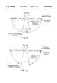
- FIG. 3depicts an apparatus for mapping regions of a nucleic acid.
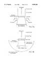
- FIG. 4is a graph depicting the kinetics of the amplification process in accordance with the present invention.
- FIG. 5is a graph illustrating the performance of an optic fiber detector.
- FIGS. 6A-6Rdepict amplification product detection schemes when the solid support is a surface.
- FIGS. 7A-7Mdepict amplification product detection schemes when the solid support is an optical fiber.
- the present inventionrelates to methods, apparatus and kits for detecting the presence of a target nucleic acid sequence in a test sample.
- the methods and apparatus described hereinencompass the use of a solid support for the amplification and optical detection of a target sequence.
- the use of a solid support in the amplification reactions of the present inventionresults in an amplification product that remains captured on a support such as an optical fiber or surface.
- a number of advantagesresult from the use of a solid support.
- the amplification reactionis localized to a small area of the support resulting in a greater signal to noise ratio and better detection than with conventional solution based amplification reactions.
- Separation of amplification productsis not required in order to detect a specific amplified target sequence in the present invention, as is required with conventional amplification reactions.
- the detection of multiple target sequencesis facilitated because multiple amplification reactions can take place in parallel on discreet areas of a surface.
- detection of amplification productsis also facilitated.
- the term supportencompasses, for example, beads, particles, dipsticks, filters, membranes, silicon, silane or silicate supports such as glass and fibers.
- the supportcomprises optical fibers.
- supports useful in the present inventioncomprise inert or inactive materials that do not react with components of the amplification reaction, or interfere with the amplification reaction.
- materialsinclude, for example, epoxy silane, polystyrene, polycarbonate, polypropylene or other plastics, derivatized silica, nylon or latex.
- a materialcan be treated, or coated with inert materials.
- the form, or configuration of the supportcan be, for example, in addition to fibers, a sphere, such as a bead or particle.
- the supportis a surface which can be flat, or planar, or can contain concave or convex areas. Such configuration would be particularly suited to provide for the analysis of multiple samples.
- a particular support encompassed by the present inventioncomprises a sheet which has surfaces with alignment features to facilitate the precise positioning of nucleic acid sequences.
- This type of supportallows for the delineation of areas of the support directed to two, or more distinct target sequences, e.g., a first target sequence and a second target sequence. These areas are preferably arranged in a grid type pattern of pixels.
- the sample containing one or more target nucleic acid sequencesi.e., the nucleic acid sequences to be detected, is referred to herein as the test sample.
- the test sampleencompasses any sample containing a nucleic acid sequence (DNA or RNA, double stranded or single stranded) capable of being amplified.
- the target nucleic acid sequencecan be of mammalian, specifically a human, origin such as a gene, gene fragment or gene product.
- the target nucleic acid sequencecan also be of bacterial, viral, parasitic or yeast origin.
- the test samplecan comprise any sample that contains nucleic acid sequences, for example, biological fluids such as blood, urine, cereberal spinal fluid, semen, saliva, stool or perspiration.
- test samplecan also comprise whole or lysed cells, or tissue such as biopsy material.
- test sampleswhich are to be tested for the presence of nucleic acid sequences as contaminates, such as nucleic acid sequences resulting from bacterial contamination in, for example, chemical extracts and distillates and other suspensions or colloids.
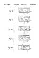
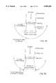
- FIGS. 1A through 1Mdepict an article of manufacture, a plurality of carboxylated latex beads, generally designated by the numerals 11a and 11b, for making an amplification product.
- the presence of an amplification productwill be used to indicate the presence of complementary target sequences of a first nucleic acid.
- first nucleic acidrefers to the target nucleic acid sequence to be detected.
- Latex beads 11a and 11bhave at least one second nucleic acid, (also referred to herein as an oligonucleotide and as a primer) and preferably, a plurality of copies of second nucleic acids which will act as primers in an amplification reaction.
- each latex bead 11a and 11bhas a second nucleic acid 13 and a third nucleic acid 15 for purposes of simplicity, with the understanding that many more second and third nucleic acids 13 and 15 may be present on each support.
- the representations of the latex beads 11a and 11b and second and third nucleic acids 13 and 15are for illustrative purposes and are not drawn to scale.
- FIG. 1A to 1Mrepresents a stage of the amplification reaction.
- latex beads 11a and 11bare depicted as being suspended in an aqueous solution 19 contained within a vessel 17.
- Solution 19 and/or beads 11a and 11bare dispensed into vessel 17 by a dispensing orifice 21 or may be prepackaged in vessel 17.
- FIG. 1Billustrates the addition of a first nucleic acid 23 derived from a sample, to vessel 17.
- First nucleic acid 23may be placed in vessel 17 prior to beads 11a and 11b or after as illustrated.
- First nucleic acid 23may be placed in vessel 17 by means of any suitable dispenser, such as orifice 21 depicted in FIG. 1A.
- First nucleic acid 23is double stranded DNA, comprising a first strand 25 and a second strand 27. Each strand has two complementary copies of the target sequence, a and b.
- Second nucleic acid 13is complementary to target sequence a of strand 25 and homologous to sequence a of strand 27.
- Third nucleic acid 15is homologous to target sequence b of strand 25 and complementary to sequence b of strand 27.
- First nucleic acid 23 and latex beads 11a and 11bform a reaction product.
- FIG. 1Cdepicts the reaction product, latex beads 11a and 11b and first nucleic acid undergoing denaturation conditions. Denaturation conditions are imposed at this stage by suitable means such as controlling temperature, and/or ionic strength, and/or the pH of solution 19 contained in vessel 17.
- the reaction productcomprising the first nucleic acid and the latex beads 11a and 11b, is next subjected to annealing conditions as represented in FIG. 1D.
- Annealing conditionsare achieved by adjusting one or more factors influencing annealing, including temperature, and/or ionic strength and pH.
- FIG. 1Ddepicts an annealed, or hybridization product comprising first nucleic acid strand 25 and second nucleic acid 13 of latex bead 11a.
- First nucleic acid strand 27may also have target areas [not shown] which interact with further primers [not shown]. For purposes of simplicity and clarity, this discussion will focus on strand 25 and target sequence a and b.
- the annealed productcomprising first nucleic acid stand 25 and second nucleic acid 13 of latex bead 11a, is next subjected to elongation, or extension, conditions, as represented in FIG. 1E.
- Elongation conditionsare preferably imposed by adding suitable reagents for elongation of a nucleic acid, including a thermal-stable polymerase, such as Tag polymerase (other polymerases are well-known to those of skill in the art), nucleotides and other necessary reagents, such as buffers.
- a thermal-stable polymerasesuch as Tag polymerase (other polymerases are well-known to those of skill in the art)
- nucleotides and other necessary reagentssuch as buffers.
- the elongation reactioncan take place in a vessel 17 and suitable reagents can be added through orifices such as orifice 21 depicted in FIG. 1A.
- FIG. 1Edepicts a first elongation product as 31 covalently extended from first nucleic acid 13. This product is complementary to the target sequence of first nucleic acid strand 25. Thus elongation product 31 has a target sequence b which is complementary to third nucleic acid 15 of latex beads 11a or 11b.
- the first elongation productis next subjected to denaturation conditions, as illustrated in FIG. 1F.
- a denaturation productis formed comprising first nucleic acid strands 25 and 27; second and third nucleic acids 13 and 15 of latex beads 11a and 11b; and a first elongation product 31 as illustrated in FIG. 1F.
- Denaturation conditionscomprise elevated temperatures, higher salt concentrations and/or lower pH.
- An orifice 21 depicted in FIG. 1Acan be used for adding reagents or heating elements [not shown].
- the denaturation productis next subjected to annealing conditions as illustrated in FIG. 1G.
- annealing conditionsas illustrated in FIG. 1G.
- an annealed productis formed.
- the annealed productcomprises the first elongation product 31 and third nucleic acid 15 of latex bead 11a; and the first nucleic acid strand 25 and second nucleic acid 13 of latex bead 21b.
- a second elongation product 33is formed.
- the second elongation productextends from third nucleic acid 15 of latex bead 11a.
- a second elongation product 33is formed extending from third nucleic acid 15 of latex bead 11b.
- a further first elongation product 31is formed from first nucleic acid 13 of latex bead 11b.
- First and second elongation products 31 and 33anneal to each other and facilitate annealing between adjacent latex particles, forming a bridge 11a and 11b, as depicted in FIG. 1M.
- the annealing of first and second elongation products 31 and 33 on adjacent latex beads 11a and 11bdisrupts the suspension and the beads 11a and 11b precipitate or agglutinate into a detectable mass.
- this detectable massis detected by monitoring equipment [not shown].
- the formation of the detectable massis indicative of the presence of the first nucleic acid and, in particular, target sequences a and b of strand 25.
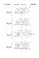
- the apparatuscan comprise an epoxy silane derivatized support 113.
- the supporthas an upper surface 117 with two primer anchoring areas 121 and 123.
- Areas 121 and 123each contain a primer pair comprising a second and a third nucleic acid.
- the second and third nucleic acids of area designated 121are designated 125 and 127 respectively.
- the second and third nucleic acids of area 123are designated 125' and 127' respectively.
- the representations of the nucleic acids and areas 121 and 123are for illustrative purposes only and are not drawn to scale.
- the areasare preferably pixel sized. These areas are preferably areas of 10 ⁇ 2 to 1 mm 2 .
- the support 113may take many different forms, such as sheets of glass, beads or fibers. Individuals skilled in the art can readily modify the shape and size of the support in order to fit individual needs.
- the entire support 113may be any convenient size. For example, it can be shaped to present a planar upper surface 117 of approximately 1 cm 2 .
- a sampleis contacted with the support 113 forming a reaction product.
- the test samplecontacts the nucleic acids immobilized on areas 121 and 123.
- Sample 131has a first nucleic acid 133 having target sequences complementary to the second nucleic acid of region 121 and 123.
- means for applying the sample 131 to support 113comprise a sample dispensing orifice 135.
- the methods of the present inventioncan be performed manually or in an automated instrument.
- the methods of the present inventioncan be performed in a self contained reaction cartridge.
- the cartridgecontains all of the necessary reagents needed to perform the assay.
- the cartridgewill have a port for introduction of the sample and separate isolated chambers for buffers, enzymes, and detection agents, e.g., dyes or labeled oligonucleotides.
- Microfabrication techniquesfacilitate production of supports for use in a cartridge, and in other configurations.
- annealing conditionson imposed on the reaction product.
- an annealed productis formed in area 121 comprising a first nucleic acid 133 and a second nucleic acid 125.
- Annealing conditionsmay comprise altering the ionic strength or pH of solutions, or lowering temperature in order to effect the hybridization of the first and second nucleic acids.
- Means for imposing annealing conditionsare depicted by reagent dispensing orifice 137 and cooling fan 139.
- the first elongation product 145comprises a nucleic acid extending from the second nucleic acid 125 corresponding to the first nucleic acid 133.
- Elongation conditionsmay comprise the addition of polymerases and proof-reading enzymes, nucleoside triphosphates, buffers and other reagents necessary to effect an elongation reaction.
- Reagents to form a first elongation product 145are dispensed through a dispensing orifice 143, or may already be present.
- FIG. 2Edepicts a stage where one or more functions may be performed.
- the nucleosides incorporated into the elongation productcan be labeled in order to effect detection.
- this stagemay comprise detection means [not shown] to monitor the support 113 for the presence of the elongation product.
- detection means[not shown] to monitor the support 113 for the presence of the elongation product.
- denaturation conditionsare imposed on the elongation product 145 to allow first nucleic acid strand 133 to disassociate from second nucleic acid 125 and first elongation product 145.
- Denaturation reagentscan be dispersed on support 113 through orifice 147.
- Additional signalscan be obtained by again forming additional hybridization products.
- FIG. 2Fa second annealed product is formed.
- the first nucleic acid 133may still remain to anneal with nucleic acid 125' of area 123 to effect a further first annealed product.
- a second hybridization productis formed between the first amplification product 145 and third nucleic acid 127. Means for imposing annealing conditions have been described previously.
- FIG. 2Gdepicts forming a second elongation product 147 in area 121 and a further first elongation product 145' in area 123.
- a second elongation product 147is formed in the first region 121.
- the second elongation product 147comprises a nucleic acid which is complementary to the first elongation product 145.
- the second elongation product 147extends from the third nucleic acid 127.
- a further first elongation product 145'is formed in the second area 123 extending from second nucleic acid 125'.
- Amplification reagentscan be applied to support 13 by dispensing orifice 153.
- a first and second elongation product 145 and 147extend from the second and third nucleic acid 125 and 127 of area 121, and a first elongation product 145' extends from second nucleic acid 125' of region 123.
- Means for imposing denaturation conditionsare depicted generally by dispensing orifice 155 and by heating elements [not shown].
- annealing conditionsare imposed on the support 113.
- the first and second elongation products 145 and 147 of area 121hybridize to each other; and, the first elongation product 145' of region 123 hybridizes to the third nucleic acid 127'.
- elongation conditionsare imposed upon the support 113.
- a second elongation product 147'is formed comprising a nucleic acid extending from third nucleic acid 127' which is complementary to the first elongation product extending from second nucleic acid 125'.
- Means for imposing amplification conditionscomprise amplification reagents applied through dispensing orifice 174.
- Amplification reagentscomprise buffers, salts, enzymes, nucleotides and the like.
- washescan optionally be applied to remove unincorporated nucleosides and extraneous matter which may interfere with signal.
- a wash dispensing orifice 173applies wash reagents and solutions to the support 113.
- FIG. 2Lrepresents a detection step, in the event the method is used for diagnostic or detection purposes rather than for the synthesis of nucleic acid.
- Detection means 175detects labelled nucleosides, if such nucleosides are used to form elongation products 145, 145', 147 and 147'.
- Detection meanscan comprise photosensors to detect chemiluminescent, luminescent and fluorescent or radioactive labels. Additional reagents to develop the signal are applied to the support 113.
- first and second elongation productsare made with labeled nucleosides
- detection conditionssuch as the addition of cofactors or light of a wavelength to which the label is sensitive
- a signalcan be developed indicating the presence of the first nucleic acid
- annealing conditionscan be applied to the support in the presence of intercalating agents to develop a signal in the presence of the first and second elongation products.
- a fourth labeled oligonucleotide [not shown] complementary to the first or second elongation productcan be used as a probe for detecting the presence of the target first oligonucleotide.
- area 121 and 123have identical second and third nucleic acids 125 and 127 or 125' and 127'.
- support 113preferably has a plurality of areas which are directed to a plurality of targets.
- at least one areacomprises a second and third nucleic acid which have nonsense sequences. This area is not intended to produce a signal, but to serve as a negative control. The presence of a signal from such second and third nucleic acids defining nonsense sequences indicates a system error.
- At least one areahas a second and third nucleic acid which have sequences that correspond to a first nucleic acid, the presence of which is confirmed as being universally present or which is added to the sample.
- This areais intended to produce a signal in each instance as a positive control. The absence of a signal indicates a system error.
- the first nucleic acid 211has areas a through f located along its length.
- the device 213, for mapping regions of a first nucleic acidhas a flat surface 215.
- the surface 215has areas 217, 219, 221, 223, 225 and 227.
- Each area 217 through 227has a second nucleic acid 231a-f respectively and a third nucleic acid 233a-f respectively.
- the second and third nucleic acids 231a-f and 233a-f of each areacorrespond to an area a-f of nucleic acid 211.
- the detectable signal of a particular area on support 215will depend on the extent to which an area a-f of nucleic acid 211 presents itself.
- a nucleic acid 211comprising segments b, c and d, will be detected on areas 219, 221 and 223.
- the device 213is processed generally in accordance with the method described with respect to FIGS. 1A-1L. That is, a first nucleic acid 211, or alternatively, fragments of nucleic acid 211, are applied to one or more devices 213. The devices are monitored to detect the presence of an elongation product in areas 217, 219, 221, 223, 225 and 227.
- target nucleic acid sequence in a test sampleis amplified, detected, and can be quantified, using pairs of primers attached to a surface contacting the sample and, optionally, other chemical reagents. Each pair of primers is homologous to complementary ends of the length of target sequence.
- amplified target nucleic acid sequencealso referred to herein as polynucleotide
- polynucleotideis formed and attached to said surface by extension from the primers so attached. Because the primer pair is specific to the test sample target sequence, surface bound amplificate forms only if the target sequence is present in the test sample. The amplificate so formed can be detected conveniently by optical means if the amplificate is so labeled.
- Labeling techniquesinclude: using labeled polynucleotides in the PCR mixture, including in the mixture a probe which is specific to the amplificate of the target sequence, and is detectable after being hybridized to said amplificate, or adding such a detectable probe after the amplification phase of the analysis is complete.
- labeled probesfor such purposes is known within the art.
- the optical signal if presentcan be detected by a variety of known optical detection techniques, including photodiodes, photomultipliers, television cameras, CCD arrays, etc.
- the detection schemeis fluorescence of a fluorogenic substance
- fluorescencecan be induced by irradiating the surface bound amplificate with excitation radiation, including from an incandescent lamp, a discharge lamp, a laser, or other irradiation means known within the art.
- excitation radiationincluding from an incandescent lamp, a discharge lamp, a laser, or other irradiation means known within the art.
- the amplification processmay be localized to a given area by attaching the primer pair only at a given area, such that the surface bound amplificate only forms there, and the resultant optical signal is localized and may be detected at the predetermined location.
- multiple target sequences within the test samplecan be tested for by independently attaching multiple primer pairs in different areas of the said surface.
- Each primer pairis homologous to a given target sequence within a single length of sample polynucleotide, or to target sequences in two or more lengths of polynucleotide in the test sample.
- Means of detecting such multiple optical signals in parallelinclude imaging the optical pattern onto an area sensitive optical detector such as a television camera, or CCD array, or other scheme known within the art.
- the pattern of multiple optically detectable signalsmay be detected sequentially, such as by individually imaging each signal in turn onto a detector also including detectors such as photodiodes, photomultipliers and other known detection means.
- thismay be achieved by masking the optical signals with one or more spatial filters, and sequentially permitting each individually to be detected by the optical detector.
- the surface to which the primer pairs are boundcan advantageously be flat, or cylindrical, or spherical for ease of optical detection, or can conform to any other shape as my be chosen.
- said surfacemay be transparent, such that the optical signals may be detected through the transparent materials, and/or a fluorescent excitation signal may be applied through the transparent material.
- the ends of individual glass fibers, or of a bundle of glass fibers,may also serve as the surface to which said primer pairs may be attached.
- the surface or the optical fibercan be modified in various ways to enhance optical properties and signal detection.
- layers or membranescan overlay or underlay the support surface resulting in light-altering effects.
- the fibercan be shaped or contoured to enhance signal detection.
- the surface to which the primer pairs are boundmay be heated and/or cooled to effect thermocycling as part of the amplification conditions.
- Heatingmay be effected by known techniques such as applying heated material to the material whose exposed surface has primers attached, applying electrical joule heating, applying electrical peltier heating, applying electromagnetic radiation, and other known techniques.
- Coolingmay be effected by applying cooled material, by applying electrical peltier cooling, by permitting the adiabatic expansion or evaporation of a liquid, conduction of heat away from the surface into the test sample, and other known techniques.
- meansmay be included for detecting the temperature of the surface, and/or of test sample within the vicinity of the surface, with such means including on the surface or its vicinity temperature sensing means, including thermocouples, thermistors, resistance thermometers, semiconducting devices, temperature sensitive optical elements, temperature sensitive magnetic materials, thermal expansion devices, and other known temperature sensing devices.
- Such temperature sensing meansmay be combined with aforesaid heating and cooling means to effect temperature control by means well known to those of skill in the art.
- the support surface, or optical fiber itselfcan control temperature, e.g., heating and cooling, in a manner sufficient to achieve the required denaturation, annealing and extension conditions for the amplification reaction.
- Materials to which the primers may be attachedinclude glasses, quartz, plastics, metals, ceramics, and other materials that are compatible with the amplification chemistry, including inert materials that do not chemically interact or release chemicals into solution.
- the surfacemay be coated with a material that modifies its properties in advantageous ways for attaching primers, or for permitting the amplification reaction to proceed without chemical modification or interference.
- Said test samplemay be an aqueous solution, or a gel, or an emulsion, or a colloidal solution, or a biological sample such as blood or any body fluid, or a sample derived from a body fluid, or biological tissue, or a soil extract, or a collected water sample, or generally any type of sample capable of containing one or more lengths of polynucleotide.
- optical fibers and optical fiber strandsin combination with light energy absorbing dyes for medical, biochemical and chemical analytical determinations has undergone rapid development, particularly within the last decade.
- the optical fiber strands employedare often glass or plastic extended rods having a relatively small cross-sectional diameter. When light energy is projected into one end of the fiber strand, the angles at which the various light energy rays strike the internal surface are greater than the critical angle; and such rays are "piped" through the strands length by successive internal reflections and eventually emerge from the opposite end of the strand.
- bundles of these strandscan be used collectively as optical fibers in a variety of different applications.
- light energyis photoenergy and is defined as electromagnetic radiation of any wavelength. Accordingly, the terms "light energy” and photoenergy” include at least infrared, visible, and ultraviolet wavelengths conventionally employed in most optical instruments and apparatus.
- light from an appropriate energy sourceis used to illuminate one end of an optical fiber or a fiber bundle.
- the lightpropagates along the length of the optical fiber; and a portion of this propagated light energy exits the opposite end of the optical fiber and is absorbed by one or more light energy absorbing dyes.
- the light energy absorbing dyemay or may not be immobilized; may or may not be directly attached to the optical fiber itself; may or may not be suspended in a fluid sample containing one or more analytes of interest to be detected; and may or may not be retainable for subsequent use in a second optical determination.
- reactantsmay be immobilized on the distal end of the fiber, (i.e., the end opposite that initially illuminated) where a reaction takes place resulting in the incorporation of a fluorophore which generates light energy which can be transmitted in the direction of the proximal end of the fiber (i.e., the end which is initially illuminated).
- These immobilized reactantsmay also contain light altering properties,. e.g., light reflecting, light scattering, light refracting and/or light absorbing. Materials such as, for example, glass, silica or plastic can be used in the present invention.
- the surface of the optical fibermay be either convex or concave in order to enhance optical focusing properties.
- some light energy of varying wavelength and intensitytypically travels through the optical fiber and is then conveyed through either the same fiber or a collection of fibers to a detection system where the emerging light energy is observed and measured, or, alternatively, is detected elsewhere.
- the interactions between light energy and light absorbing dyes both in the presence of a fluid sample containing one or more nucleic acids of interestprovide an optical basis for both qualitative and quantitative spectral determinations.
- these systemsprovide components for image capture, data acquisition, data processing and analysis and visual presentation to the user.
- Several systemsare commercially available from sources such as Quantex, Inc. and Spex Industries, Inc. Each of these systems may be combined with microscopes, cameras, and/or television monitors for automatic processing of all light energy determinations.
- a methodthat utilizes a solid-phase primer-directed amplification process with fiber optic detection format.
- an optical fiberhaving a distal end and a proximal end, is utilized with the amplification reaction being performed on the distal end of the fiber and transmission of the resultant signal occurring through the fiber, the proximal end of which is engaged with the detector.
- nucleic acid primerswhich anneal to the target nucleic acid to be amplified are immobilized on the distal end of the optical fiber. The formation of elongation products the on the distal end of the fiber indicates that the sample contained target nucleic acid sequences.
- an optical fiberas the amplification support facilitates optical detection of amplified products, thus enabling analysis of amplification progress as the reaction proceeds using fluorescence or absorption techniques. Concurrent real-time analysis of amplification progress facilitates accurate quantification of target concentration in a sample.
- the optical fiberalso serves as an integral component of the product detection system. Thus, the use of an optical fiber performs a three-fold function as the support for the amplification reaction, as a transmission means for the resultant signal and as a component of the detection system by transmitting this signal to the detector.
- One end of the optical fiber(referred to hereinafter as the distal end) is cleaved, polished, and then chemically modified to provide a surface having attachment sites for nucleic acid primers.
- a number of surface modification methods suitable for this purposeare known to those of skill in the art. For example, organosilane coating of glass and silica surfaces, graft polymerization on polymer surfaces, and/or high voltage gas-plasma discharges may be used to effect modification of glass, silica or polymer surfaces.
- the surface of the fibermay also be modified to have a convex or concave curvature to facilitate optical focusing.
- oligonucleotidesare then attached to the surface of the distal end of the fiber. This process usually involves several steps, which may include one or more of the following:
- the specificity of the amplification assayis governed by the sequences of the oligonucleotides that are immobilized on the face of the distal end of the fiber.
- a mixture of two specific synthetic oligonucleotide primerswill be immobilized on the distal surface of the optical fiber, preferably by formation of covalent bonds with groups on the fiber surface.
- These olignucleotideswill ordinarily be chosen to amplify a specific subsequence, or region, of the target nucleic acid, using primer design strategies similar to those used for conventional, solution phase PCR, and related techniques.
- Primer attachmentis carried out so that the 3' terminal sequences of the oligonucleotides (for most applications between 5 and 50 bases of sequence) are accessible and capable of serving as primers for template-dependent polynucleotide synthesis by a suitable polymerase enzyme.
- attachmentis carried out so that the distance between adjacent oligonucleotides of opposite polarity on the surface of the support is less than the length of the target nucleic acid.
- test mixtureis prepared containing the sample, template and primer dependent polymerase, and other reagents used for polymerase dependent nucleic acid synthesis.
- the latter categorywould include buffers, salts, polymerase cofactors, nucleoside triphosphates, and possibly additional polymerase stabilizing agents such as detergents and/or blocking proteins (i.e., bovine serum albumin).
- c3Imposition of conditions that allow polymerase extension of the primer-template complexes formed in step 2 above. These three steps are repeated as often as necessary to achieve the desired degree of amplification.
- the distal surface of the fiberis then assayed for the presence of amplified product using optical detection methods that employ light conveyed through the optical fiber support, or excitation through the optical fiber and detection elsewhere.
- Methods for amplification reactionsi.e., repeated cycles of nucleic acid denaturation, annealing/reannealing and polymerase extension fall into two broad categories, i.e., those that operate on the entire contents of the reaction vessel, using a conventional programmable thermal cycler as commonly used in PCR; and/or modulation of the reaction pH to effect nucleic acid denaturation and rehybridization. Cyclic addition of base to the reaction vessel is used to raise pH above the denaturation point, followed by neutralization to a pH below this point.
- the second category of amplification reactionentails treatment of the distal face of the fiber only. In this case, the regulated heating and cooling of the distal face of the fiber is mediated by fiber-based apparatus and/or processes.
- modulation of the temperature of the distal fiber surface by light energy passing through the fibermay be employed.
- modulation of the temperature of the distal fiber surface by light energy passing through the fibermay be employed.
- theseinclude 1) direct stimulation of water vibrational absorption by IR light; and 2) indirect heating caused by visible irradiation of dye molecules that undergo efficient radiationless decay to generate heat.
- Thesecould be present in a layer on the fiber surface, or they could be present in the solution containing the reaction mixture.
- resistive electrical heatingcaused by current passing through an electrically conducive layer deposited on the surface of the fiber may be employed.
- modulation of electrical potential (voltage) of the distal fiber surface to alternately effect denaturation and hybridization of the DNAmay be used, such as with a thermocoupler.
- Elongation productswill be detected by means of light signals transmitted through the fiber.
- the proximal end of the optical fiberis connected to apparatus that allows optical detection of elongation products.
- the preferred apparatuswill include some or all of the following features:
- c)Means to allow simultaneous or substantially simultaneous operation of the detection and illumination steps described in (a) and (b) above.
- a dichroic mirrormay be employed to separate and filter the fluorescent signal returning from the sample from the excitation beam.
- Epifluorescence microscopesemploy dichroic mirrors in a similar fashion.
- a variety of optical detection strategiescan be used in implementing the present invention.
- Productcan be quantitated during or after amplification.
- Kinetic analysis of amplification progressmay be performed concurrently with cycling, thus quantifying initial DNA target levels.
- Elongation productscan be labeled via the inclusion of fluorescently tagged nucleotide triphosphates in the reaction mixture. As polymerase mediated extension of DNA strands proceeds, fluorescently tagged nucleotides are incorporated. Therefore, the amount of product synthesized should be proportional to the fluorescent signal generated at the distal end of the fiber. Elongation products may also be detected using fluorescent DNA-specific dyes, i.e., ethidium bromide. Detection can be performed after amplification or concurrently with cycling. During concurrent detection, the dye is included in the reaction mixture.
- Fluorescenceis recorded at the same point (and temperature) during each cycle (near the end of the amplification stage). A cycle dependent increase in fluorescent intensity indicates the accumulation of DNA on the fiber tip.
- amplificationmay be detected using fluorescently labeled nucleic acid probes which hybridize specifically to amplified products attached to the distal end of the optical fiber. Fluorescence energy transfer probes that fluoresce only when hybridized may also be used to monitor product formation during cycling.
- absorbance-based measurements of product accumulationmay also be made using the optical fiber.
- the preferred apparatuswould involve an external light source and follow the increase in absorbance during or after the amplification reaction.
- the absorbance of DNA bases at 260 nm or the absorption of nonspecific DNA-binding agentscould be used to detect product on the distal end of the fiber since amplification of product would lead to the concentration of those agents on the fiber.
- optical configurationscan be used to detect surface-attached optically detectable oligonucleotides (also referred to herein as polynucleotides).
- the configurations described hereincan be used more generally for any chemical species which may be surface-attached and optically detected.
- FIGS. 6A-6R And FIGS. 7A-7M included herewithindicate configurations possible with surfaces with attached chemical species
- FIGS. 7A-7Millustrate more specifically additional configurations applicable to optical fibers and bundles of optical fibers.
- FIG. 6Adepicts the case where the surface with chemical species attached is illuminated by excitation optical energy, and the resultant optical signal is optically detected with a detector positioned such as to be able to capture such signal from the surface.
- FIG. 6Bdepicts the case where the surface with chemical species attached is illuminated by excitation optical energy, and the resultant optical signal is optically detected with one or more detectors positioned such as to be able to capture such signal from the surface, but positioned such that all or much of reflected optical energy is prevented from entering the detector.
- FIG. 6Cdepicts the case where the surface with chemical species attached is illuminated by excitation optical energy, and the resultant optical signal is optically detected with one or more detectors positioned such as to be able to capture such signal from the surface, but a mask or spatial filter is positioned to prevent all or much of the reflected optical energy from entering the detector.
- FIG. 6Ddepicts the case where the surface is transparent, such that it is optically accessible from both the side of the test sample where chemical species is attached, and also through the surface. Optical excitation is applied through the transparent surface and the detected signal is detected from the side to which the amplified chemical species is attached.
- FIG. 6Edepicts the case of a transparent surface where the optical excitation is applied to the surface attached chemical species and the detected signal is detected through the transparent surface.
- FIG. 6Fdepicts the case where the optical excitation is applied to the attached chemical species through the transparent surface and the optical signal is detected through the transparent surface.
- FIG. 6Gdepicts the case where the optical excitation is applied to the attached chemical species through the transparent surface and the optical signal is detected through the transparent surface, with the optical capture range of the detector being limited such that all or much of the reflected excitation energy does not enter the detector.
- FIG. 6Hdepicts the case where the optical excitation is applied to the attached chemical species through the transparent surface and the optical signal is detected through the transparent surface, with a mask or spatial filter positioned to block all or much of the reflected excitation energy from entering the detector.
- FIG. 6Idepicts the case where the optical excitation is directly applied to the attached chemical species, and the detected signal from the attached chemical species is detected through the transparent surface, with a mask or spatial filter positioned to block all or much of the transmitted optical energy from entering the detector.
- FIG. 6Jdepicts the case where the optical excitation is directly applied to the attached chemical species, and the detected signal from the attached chemical species is detected through the transparent surface, with the optical capture range of the detector being limited such that all or much of the transmitted optical energy does not enter the detector.
- FIG. 6Kdepicts the case where the optical excitation is applied to the attached chemical species through the transparent surface, and the signal is detected directly from the attached chemical species, with a mask or spatial filter positioned to block all or much of the transmitted optical energy from entering the detector.
- FIG. 6Ldepicts the case where the optical excitation is applied to the attached chemical species through the transparent surface, and the signal is detected directly from the attached chemical species, with the optical capture range of the detector being limited such that all or much of the transmitted optical energy does not enter the detector.
- FIG. 6Mdepicts the case where multiple pixels of attached chemical species are positioned on the surface with a spatial relationship, and a lens or other imaging means forms an image of the multiple pixels on a spatially sensitive detector able to individually distinguish the imaged pixels such that the presence and image intensity of each pixel can be detected in parallel.
- FIG. 6Ndepicts the case where multiple pixels of attached chemical species are positioned on the surface with a spatial relationship and the detectable signal passes through the transparent surface to a lens or other imaging means that forms an image of the multiple pixels on a spatially sensitive detector able to individually distinguish the imaged pixels such that the presence and image intensity of each pixel can be detected in parallel.
- FIG. 6Odepicts the case where the surface with attached chemical species is not flat, but is curved in some manner such as cylindrical, spherical, parabaloid or by other mathematical shape or is randomly shaped, and any of the optical excitation and detection configurations herein described are utilized.
- FIG. 6Pdepicts the case where the surface with attached chemical species is not flat, but has a physical pattern impressed upon it, such as e.g. to positively or negatively indent the surface, such as to provide enhanced locations for concentrating applied sample or chemical species to improve their optical detection by any of the optical excitation and detection configurations herein described.
- FIG. 6Qdepicts the case where the surface with attached chemical species has a pattern impressed upon it such that applied sample or chemical species may be conveniently localized and kept separate and detected by any of the optical excitation and detection configurations herein described.
- FIG. 6Rdepicts the case where the surface with attached chemical species is constructed from two or more separate physical elements which when assembled together topologically represent a single surface of shape as described herein whereon chemical species may be detected by any optical excitation and detection configuration as herein described.
- FIG. 7Adepicts the case where the optical excitation energy is transmitted from the fiber's proximal end to the distal end to illuminate the attached chemical species on the fiber tip and the detectable signal is transmitted from the distal end to the proximal end where the detector is positioned.
- FIG. 7Bdepicts the case where the optical excitation energy is transmitted from the fiber's proximal end to the distal end to illuminate the attached chemical species on the fiber tip, and the detectable signal is detected from the surface attached chemical species.
- FIG. 7Cdepicts the case where the optical excitation is directly applied to the attached chemical species on the fiber surface, and the detectable signal is transmitted along the fiber from the distal end to the proximal end where the detector is positioned.
- FIG. 7Ddepicts the case where the optical excitation is directly applied to the attached chemical species on the fiber surface, being limited to such angles as will not be transmitted along the fiber nor therefore captured by the detector, and the detectable signal is transmitted along the fiber from the distal end to the proximal end where the detector is positioned.
- FIG. 7Edepicts the case where the optical excitation is directly applied to the attached chemical species on the fiber surface, but with a mask or spatial filter being positioned to prevent excitation from irradiating the distal end from such angles as would be transmitted along the fiber and therefore captured by the detector, and the detectable signal is transmitted along the fiber from the distal end to the proximal end where the detector is positioned.
- FIG. 7Fdepicts the case where optical excitation energy is transmitted along the fiber from the proximal end to the distal end to illuminate the attached chemical species on the fiber tip, and the detectable signal is detected from the surface attached chemical species, with a mask or spatial filter positioned to prevent transmitted excitation from the exit pupil of the fiber tip from entering the detector.
- FIG. 7Gdepicts the case where optical excitation energy is transmitted along the fiber from the proximal end to the distal end to illuminate the attached chemical species on the fiber tip, and the detectable signal is detected from the surface attached chemical species, with the detector or detectors being positioned such as to prevent transmitted excitation from the exit pupil of the fiber tip from entering the detectors.
- FIG. 7Hdepicts the case where two or more fibers, individually or within a bundle, are positioned adjacently with attached chemical species positioned at their joint distal end, and optical excitation energy is transmitted from the proximal end to the distal end of one fiber to illuminate the attached chemical species, and the optical signal from the attached chemical species is transmitted from the distal end to the proximal end of the other fiber where it enters a detector.
- FIG. 7Idepicts the case where a fiber is used either to transmit optical excitation energy from the proximal end to the distal end, or to transmit an optical detection signal from the distal end to the proximal end, or to do both, and adjacent fibers individually placed or within a bundle are unused, to prevent optical interference between said fiber and another adjacent fiber similarly used.
- FIG. 7Jdepicts the case where two or more sets of fibers are placed with at least one unused fiber between adjacent such pair of fibers to prevent optical interference between such sets of fibers, where in each set of fibers one is used to transmit excitation energy from the proximal to the distal end and the other is used to transmit a detection signal from the distal end to the proximal end.
- FIG. 7Kdepicts the case where two or more optical fibers have excitation energy transmitted from their proximal ends to their distal ends to illuminate chemical species attached to the end of each such fiber, and unused fibers are positioned to prevent said fibers from being positioned adjacently and causing optical interference, and the detectable signals from the fibers with chemical species attached are detected by any detection means described herein.
- FIG. 7Ldepicts the case where two or more optical fibers have excitation energy applied to attached chemical species at their distal ends, and detectable signals are transmitted from their distal ends to their proximal ends to one or more detectors, and optionally unused fibers are positioned to prevent said fibers from being positioned adjacently and causing optical interference, and chemical species attached to the end of said fibers are illuminated by any excitation means described herein.
- FIG. 7Mdepicts the case where two or more optical fibers have excitation energy transmitted from their proximal ends to their distal ends to illuminate chemical species attached to the end of each such fiber, and optionally unused fibers are positioned to prevent said fibers from being positioned adjacently and causing optical interference, with the ends of the fibers with detectable chemical species attached being positioned in a spatially significant way, and the detectable signals from said spatially arranged fiber tips with chemical species attached being optically imaged onto a spatially sensitive detector such that all such signals can be detected and optionally quantified simultaneously.
- This exampledescribes an amplification reaction which results in the formation of an elongation product containing a target nucleic acid sequence.
- the test samplecontains nucleic acid which has been sonicated to produce sequences with an approximate length of 1 kb.
- a primeris synthesized with a nucleotide sequence complementary to a region of the target sequence and the primer is immobilized on an epoxy silane derivatized solid support by a 5' amino group. Spacer groups of hexaethylene-glycol are included during synthesis of the primer to eliminate stearic hindrance during the annealing reaction. The spacer region is introduced into the synthesized primer prior to amino group addition, resulting in a calculated spacer region length of 25 angstroms.
- the primeris allowed to anneal to the target sequence of the test sample in the presence of thermally stable polymerase, enzyme buffer, 32 P-labeled and unlabeled dNTP to form a reaction mixture.
- the reaction mixtureis heated to 94° C. for one minute for denaturation; cooled to 55° C. for one minute for annealing; and warmed to 75° C. for 5 minutes for extension to form an elongation product extending from the immobilized primer and which is complementary to the target nucleic acid sequence.
- This cycle of denaturing, annealing and extensioncan be performed numerous times, e.g., 30, 50 or more than 100 times, to increase the amount of elongation product to be detected.
- the reaction mixtureis washed from the reaction surface and the immobilized amplification product detected. To eliminate random background noise, the reaction surface is washed using high stringency conditions.
- Radiolabeled elongation productsare detected using photographic film placed with the emulsion in contact with the solid support.
- Biotinylated elongation productsare detected by analyzing the conversion of a chemiluminescent substrate by a strepavidin-alkaline phosphatase conjugate using x-ray film.
- Other forms of signal detectioncan include fluorescence microscopy, fiberoptic devices, confocal microscopy, scintillation detection, piezoelectric material and silicon based systems (i.e., charged coupled devices).
- This exampledescribes a method of detecting a tartget nucleic acid sequence from HIV.
- Two oligonucleotide primersof approximately 20 nucleotides in length, and complementary to two different regions of a HIV gene sequence, are immobilized onto a population of derivatized glass beads.
- One half of the glass beadscontain one primer and the other half contains the other primer.
- carboxylated derivatized latex particlesmay be substituted for the derivatized glass beads.
- a test sample potentially containing target HIV viral nucleic acidis introduced into a reaction chamber vessel containing the glass beads to form a reaction mixture.
- the reaction mixturealso contains appropriate buffers and enzymes for amplification of target nucleic acid sequence.
- the reaction mixtureis subjected to one or more cycles of denaturing, annealing and extension, thereby amplifying the target nucleic acid.
- the primer-coated beadswill agglutinate or precipitate.
- the aggregated or agglutinated complexwill be observed spectrophometrically.
- a decrease in optical densityis indicative of the formation of a precipitate and thus the presence of the target nucleic acid.
- the turbidity of the reaction solutionis a function of assay sensitivity and target specificity.
- a planar derivatized glass supportreceives pairs of first and second primers. Each primer is approximately 20 nucleotides long. Each pair of primers is complementary to a different sequence corresponding to STS markers along a region of the first nucleic acid. Each set is also positioned in a predetermined area of the glass support and is comprised of approximately 100,000 5' amino linked, second nucleic acids.
- a yeast artificial chromosome (YAC) library containing first nucleic acidis divided into pools for screening.
- the contents of each YAC poolis applied to the support having the second and third nucleic acid primer array along with a reaction mixture of polymerase, buffer, and fluorescein-labeled deoxynucleoside triphosphates.
- the reactionproceeds for 30 cycles of denaturation, annealing and elongation.
- the supportis washed to remove unincorporated nucleosides and YACs.
- the supportis monitored by detecting the presence of amplification products that correspond to a particular YAC pool.
- Amplification products formed in the presence of a YAC pool in two arrayssuggest that the YAC pool contains an adjacent sequence.
- This Examplehighlights the formation of an amplification product on silica microspheres. Rather than forming interlinking beads or microspheres, the amplified product forms a "bridged" product on each sphere analogous to the process of FIG. 2.
- (4-OmeT) 8indicates an 8 nucleotide stretch containing 4-0-methyl-thymine bases.
- -5'-NH 2 -(C6-linker)indicates that the primers carry a primary amine group linked by a six carbon chain at their 5' ends.
- the initial target used for the bridge amplification reaction shown in the examplewas a 268 base pair double-stranded PCR product that was purified from a solution phase amplification reaction.
- the solution phase reactionused the Bglo-(+) and Bglo-(-) primers and a human genomic DNA sample.
- the specific target fragment used in the examplewas not sequenced, but it can be assumed to be virtually identical to other previously sequenced human beta-globin genes.
- the target sequence shown below in Table 1was deduced from GenBank sequence Accession #26462, using the sequence of the Bglo-(+) and Bglo-(-) primers above.
- the target regionoverlaps the 5'-end of the coding sequence of the human beta-globin gene.
- the ATG initiation codon of exon 1is underlined.
- the strand with the same sequence as the mRNAis shown.
- Solid silica microspheres(0.4 micron diameter) were purchased commercially (Bangs Laboratories, Carmel, Ind., USA). A surface epoxide layer was deposited on the microspheres using the method of Chang, Gooding and Regnier, 1976, J. Chromat. 120, 321-333, as described below.
- a 10% aqueous solution of 3-glycidoxypropyltrimethoxysilane (3-OPTS)was prepared and adjusted to pH 5.7 with 1 millimolar potassium hydroxide, 0.5 milliliters of the 10% 3-OPTS solution were mixed with 100 milligrams of the microspheres suspended in 0.5 milliliters of deionized water. The mixture was held at 88 to 90° C. for 30 minutes.
- the tubewas mixed briefly on a vortex mixer at 5 minute intervals during the incubation. After heating, the beads were washed twice by centrifugation and resuspension in deionized water (1.5 milliliters per wash, 2000 g, 2 minutes).
- Epoxide-silica microspheres(50 mg) were washed once in 1.5 milliliters of 0.1 molar potassium hydroxide. The microspheres were centrifuged as described above and resuspended in 75 microliters of 0.1 molar potassium hydroxide containing Bglo-(+) and Bglo-(-) primers each at 29 micromolar concentration. Oligonucleotide D-13-R, 3'-end-labeled with ddATP-alpha- 35 S and terminal transferase, was included at a concentration of 0.2 nanomolar as a tracer to monitor the level of oligonucleotide binding.
- the derivizationwas carried out for 8 hours at 37° C., with intermittent vortex mixing to resuspend the microspheres.
- the microsphereswere washed three times by centrifugation and resuspended in 0.1 M potassium hydroxide and twice in deionized water (0.5 milliliters per wash).
- the microsphereswere then resuspended in 200 microliters of 20% ethanolamine (w/v), pH 8.2, and incubated for 8 hours at 37° C. with intermittent mixing.
- the microsphereswere then washed three times by centrifugation and resuspension in an aqueous solution of 0.5% Tween-20 (v/v), 100 micrograms/milliliter bovine serum albumin, (0.5 milliliters/wash). From the level of bound 35 S-labeled-D-13-R primer, the estimated total primer concentration (equimolar (+) and (-) primer) on the microspheres was 2.1-2.2 picomoles per milligram of microspheres
- the spacing between adjacent primersis estimated to be 767 angstroms. This distance is equivalent to the length of a 225 bp fragment of double-stranded DNA.
- a 2 mg amount of primer-modified beadswas cycled in 100 ⁇ l reactions containing: 10 mM Tris HCl (pH 8.3 at 25° C.), 100 ⁇ g/ml BSA, 0.5% Tween 20, 5 U Tth polymerase, 200 ⁇ M each dNTP, and 0.25 ⁇ M dCTP-alpha- 32 P (800 Ci/mmole).
- the initial target usedwas 0.45 pmole of the 269 bp beta-globin PCR product, purified from a solution phase PCR reaction by Centricon-100 ultrafiltration. Cycling was carried out for 35 cycles using 1 minute at 94° C., followed by 5 minutes at 60° C.
- the datademonstrate target-and-primer-dependent incorporation of radioactive DNTP into bead-bound form. These data are described in FIG. 4 as counts per minute.
- open circles and trianglesrepresent reactions of beads carrying the (+) and (-) primers and reactions in the presence of target. Closed circles represent a reaction of beads carrying the (+) and (-) primers in the absence of target. Closed squares represent beads without the (+) and (-) primers in the presence of target, and closed triangles represented beads without the (+) and (-) primers in the absence of target.
- No incorporation signalis obtained from primer-modified beads in the absence of target (closed 10 circles), or unmodified beads in the presence or absence of target (closed squares and closed triangles respectively).
- incorporationincreases 6.5 fold for the 5 cycles following cycle 20. This rate is greater than that expected for primer extension using only solution phase target as template, which would be expected to increase by 5-fold at most reasonably indicating that amplification is taking place on the surface of the beads. Assuming an exponential amplification reaction, the increase in incorporation per cycle is approximately 1.4-fold for cycles 21-25.
- primers directed toward a human dystrophin gene fragmentwere modified to include restriction enzyme sites.
- the (+) primercontains an XbaI site and the (-) primer contains a ClaI site.
- Amplificationswere carried out with epoxy-silica beads modified with the dystrophin primers, using the purified 545 bp dystrophin PCR product as target. After 35 cycles, the beads were washed to remove unbound radioactivity, and split into four equal portions. One portion was left in restriction buffer without enzyme, two portions were singly digested with ClaI or XbaI, and one portion was digested simultaneously with both enzymes. After digestion the beads were pelleted by centrifugation, and the supernatants were analyzed by acrylamide gel electrophoresis and autoradiography.
- Fiber optic sensorshave been constructed for monitoring hybridization of fluorescently labeled DNA molecules to oligonucleotide probes immobilized on the tip of an optical fiber.
- Glass optical fiber(0.25 mm diameter) was cleaved and polished according to known procedures.
- Fiber tipswere silanized by soaking in 10% (v/v) aminopropyltriethoxysilane in acetone for 2 hours. The fiber tips were washed in acetone and air dried. The silanized fibers were then soaked in 1.35% glutaraldehyde in 0.02M phosphate buffer (pH 6.8) for 30 minutes. The fiber tips were then rinsed in water and placed in 3% polyethyleneimine (PEI) (2 kd average mol. wt.) in 0.02M phosphate buffer (pH 6.8) for 1-2 hours. The tips of the fibers thus treated were washed in water and 0.1M sodium borate buffer (pH 8.3) (SBB).
- PEIpolyethyleneimine
- a synthetic oligonucleotide designed to recognize human beta-hemoglobin sequenceswas used as a primer.
- This oligonucleotide primerwas obtained commercially with a 5' terminal primary amine group (5'(NH 2 --(CH 2 ) 6 --) tt ttt ttt tca act tca tcc acg ttc acc-3', SEQ ID NO:5).
- the primary amine groupwas activated with the homobifunctional crosslinking agent, cyanuric chloride, as described by Van Ness et al. (Nucleic Acids Res., 1991, 19:3345-3350).
- the hybridization primerwas coupled to the PEI-coated fiber by soaking the fiber in a 50-100 ⁇ M solution of the activated Bglo probe in SBB for 1-2 hours. Subsequently, unreacted amines were capped by reacting the fiber tips with succinic anhydride (1 hour soak in 0.1 M succinic anhydride, 0.1M sodium borate (pH 8.3), 50% DMSO). Noncovalently bound oligonucleotide was removed following succinylation by several washes with TE Buffer (10 mM Tris-HCl, pH 8.3, 1 mM EDTA).
- Primer surface densitywas not determined directly. However, in analogous experiments using PEI coated microsphere supports, primer densities of 1-10 fmoles primer per mm 2 are routinely achieved, as assessed by hybridization with 32 P-labeled complementary oligonucleotides and as described in the previous examples.
- the primer-modified end of the fiberwas annealed at room temperature by immersing it in a microcentrifuge tube containing labeled oligonucleotide (0.1-1.0 ⁇ M) in buffer (TE buffer with 0.2M NaCl and 0.5% SDS) for 15 minutes. Following annealing, the fiber tip was washed by dipping several times in buffer without target oligonucleotide. Fluorescence intensity data were collected using an epifluorescence microscope modified as described in Bronk et al., Anal. Chem. 1995, 67:2750-2757. Briefly, the microscope is modified by replacing the condenser with a fiber holder. The sensor-distal end of the fiber is placed in the holder and imaged using epifluorescence illumination. Fluorescence intensity of the fiber is determined using a microcomputer-controlled CCD camera and image analysis software.
- the left panel of FIG. 5illustrates the performance of the sensor.
- a Bglo-modified fiberwas sequentially hybridized to complementary and noncomplementary fluorescein tagged oligonucleotide targets.
- High hybridization signalswere obtained from reactions using a fluorescein-labeled complementary target (Bglo-CF target, 5'-fluorescein-tg aac gtg gat gaa gtt g-3') (SEQ ID NO:6) at concentrations of 0.1 ⁇ M and 1.0 ⁇ M.
- the signalswere completely eliminated by a denaturing wash in 90% buffered formamide, demonstrating that the signal is completely reversible.
- the right-hand panel of FIG. 5shows a control experiment, where fluorescent beta-globin target was hybridized to an underivatized fiber (no attached primers). Non-specific binding was very low.
- Amplification of target DNA sequencesmay be detected during, or after the amplification reaction.
- PEI-coated optical fibersare prepared as described in Example 7 above.
- Amplification primers carrying 5' terminal aminesare covalently attached to the PEI layer using the cyanuric chloride method of Van Ness et al. (Nucleic Acids Res., 1991, 19:3345-3350).
- two amplification primers, mixed in a 1:1 molar ratio,are attached to each fiber.
- Reactions(50 ⁇ l) are carried out using 10 mM Tris-HCl (pH 8.3 at 25° C.), 50 mM KCl, 2.5 mM MgCl 2 , 200 ⁇ M each dNTP, 100 ⁇ g/ml BSA, 0.5% Tween 20, 0.05 U/ ⁇ l AmpliTaq DNA polymerase (Perkin Elmer).
- the sample to be testedis added to the reaction mixture and the distal tip of the fiber is immersed in the mixture.
- the surface of the reaction mixtureis overlaid with oil, and the reactions are thermocycled in standard programmable instruments (for instance, Perkin-Elmer 2400).
- Typical PCR cycling profilesare used: 10 seconds at 94° C. to denature, 10 seconds at temperatures between 50° C. and 72° C. to allow primer annealing (primer sequence-dependent), and 30-60 seconds at 72° C. to allow polymerase extension.
- Negative control amplification reactions without added sampleare carried out in parallel with the experimental samples to assess the level of target-independent background signal. Positive control reactions are also carried out using known amounts of previously characterized target nucleic acid to ensure that the amplification reagents and instruments are functioning properly.
- the fibers bearing the bridge amplification reaction productare removed from the amplification mixture, washed with 10 mM Tris-HCl pH 8.3, 1 mM EDTA, 0.1% Tween 20 (TE/Tw), and the tips are immersed in a solution of 0.1 ⁇ M Hoechst 33258 (H33258) in TE/Tw for 5 minutes.
- H33258is a DNA stain that fluoresces brightly when bound to double-stranded DNA.
- the dyeis particularly suitable for use in the present assay as free H33258 dye, or dye bound to single-stranded DNA has a 25-fold lower fluorescence yield (Eggleston et al. 1996, Nucleic Acids Res. 24:1179-1186).
- Fluorescence signalis quantified using a fiber optic fluorimeter with capabilities similar to that described by Bronk et al., Anal. Chem. 1995, 67:2750-2757. Elevated H33258 fluorescence signals, defined relative to the values obtained from the negative control reactions, are indicative of the presence of amplified product on the fiber tip. This, in turn, indicates the presence of the nucleic acid target in the original sample.
- fiber-based bridge amplificationis carried out as described above, except that 0.1 ⁇ M H33258 dye is included in the reaction mixture.
- H33258 fluorescent signalis measured at the end of each extension cycle. In this way, the progress of the amplification reaction can be followed in a cycle-by-cycle fashion. This capability is particularly useful for quantifying initial target DNA concentrations in experimental samples (Higuchi R, et al. 1993. Bio/technology 11, 1026-1030).
- oligonucleotide ligation assaytwo oligonucleotides are designed to hybridize in exact juxtaposition to a target DNA sequence, permitting their covalent joining by DNA ligase.
- a first oligonucleotideis attached to the surface of the fiber as described above in Example 5.
- the fiberis then immersed in a test sample suspected of containing the target DNA sequence.
- the test samplefurther comprises a second detectably labeled oligonucleotide and DNA ligase.
- the test sampleis subjected to conditions that permit hybridization of the first and second oligonucleotides to the target DNA sequence.
- a suitable substrate for DNA ligaseis created. Ligation of the second detectably labeled oligonucleotide to the first oligonucleotide affixed to the optical fiber is then detected via the signal sent through the fiber to the detector at the proximal end.
- the following exampleillustrates the use of a fiber optic approach to measure the DNA repair resulting from damage caused by the chemotherapeutic agent, cis-diamminedichloroplatinum (cisplatin).
- Two 70 base oligonucleotidesare synthesized with complementary base sequences. One is synthesized with a 5' terminal primary amine group to provide a site for attachment to the fiber optic tip.
- the two oligonucleotidesare mixed in an equimolar mixture and hybridized in 10 mM Tris-HCl pH 8.3, 0.2M NaCl, 1 mM EDTA by heating the mixture to 94° C. and cooling to room temperature over a 1 hour period to form a double-stranded 70 bp oligonucleotide.
- the double-stranded oligonucleotideis purified from unhybridized single stranded oligonucleotides by ion-exchange chromatography.
- lesionsare then introduced into the DNA as follows.
- the double stranded 70-meris treated with cisplatin in 10 mM Tris-HCl pH 8.3, 1 mM EDTA (TE buffer) to achieve adduct formation in the range of 1 to 3 cisplatin adducts per oligonucleotide on average.
- the cisplatin-modified oligonucleotidesare then purified by ethanol precipitation from solutions containing 0.5 M NaCl.
- the cisplatin-modified oligonucleotidesare coupled to PEI-coated optical fibers using the cyanuric chloride method of Van Ness et al. (Nucleic Acids Res. 1991, 19:3345-3350) as generally described in Example 5 above.
- a parallel set of fibersare coupled with untreated, adduct-free 70 base pair test duplex oligonucleotide.
- a blood sampleis collected and white blood cells are purified. From these cells, a whole-cell lysate is prepared by the method of Manley et al. (Proc. Natl. Acad. Sci. USA, 1980, 77, 3855-3860). Fluorescein-tagged nucleotide triphosphates are added to the lysate (final concentration approximately 500 ⁇ M) and the tips of two fibers, carrying cisplatin-treated and control duplexes, respectively, are immersed in the sample. After incubation for 1 hour at 37° C.
- the fibersare removed and washed in TE buffer.
- a fiber fluorimeterwith capabilities similar to that described by Bronk et al. Anal. Chem. 1995, 67:2750-2757, is used to measure the presence of fluorescein-tagged nucleotide on the fiber tips. If the patient is proficient in repair of cisplatin adducts, the fiber with the cisplatin-modified duplex will show a significantly greater fluorescein signal than the fiber with the unmodified control duplex.
- a biopsy sample of a tumoris obtained from a patient.
- Tumors cellsare grown in culture using standard cell culturing techniques known to those of skill in the art. After approximately 10-20 cell divisions, a lysate is prepared as described above.
- 70-mer oligonucleotidesare also prepared as described above.
- a series of the oligomersare treated separately with a variety of known chemotherapeutic agents to induce lesions in the DNA. These agents may include but are not limited to cisplatin, sulfur, nitrogen mustards, enediene compounds, methylating compounds, or the like.
- the oligonucleotidesare then purified as described above.
- the modified oligomersare then coupled to PEI-coated optical fibers using the cyanuric chloride method of Van Ness et al. (Nucleic Acids Res. 1991, 19:3345-3350).
- a parallel set of fibersare coupled with untreated, lesion-free 70 base pair test duplex oligonucleotide.
- fluorescein-tagged nucleotide triphosphatesare added to the lysate (final concentration approximately 500 ⁇ M) and the tips of two fibers, carrying the chemotherapeutic agent-treated and control duplexes, respectively, are immersed in the lysate sample. After incubation for 1 hour at 37° C. to allow repair of the DNA lesions and incorporation of fluorescein-labeled nucleotide during repair DNA synthesis, the fibers are removed and washed in TE buffer.
- a fiber fluorimeterwith capabilities similar to that described by Bronk et al. Anal. Chem.
- chemotherapeutic agents mentioned abovewould be identified that induce lesions in DNA that are inefficiently repaired by the tumor lysate whereas other agents may induce lesions that are readily repaired by the tumor lysate.
- the above described methodfacilitates analysis of the repair of lesions induced by the different chemotherapeutic agents and enables an assessment ex vivo of the most efficacious chemotherapeutic agent to employ in treating a patient's cancer.
- DNA repair assays on optical fibersmay also be used to detect the presence or quantity of unknown DNA damaging agents in aqueous samples from environmental or food sources.
- a 70 base pair double stranded oligonucleotideis coupled to the tips of two PEI-coated optical fibers as described in the previous example.
- One of the fiber tipsis immersed in the test sample and incubated to allow damage to occur.
- the test samplecan be an environmental water sample, or aqueous extracts of solid samples such as soil or food.
- the pH of the sampleis controlled in the range of 6-8.5 by addition of appropriate buffers to ensure that the DNA remains in the duplex state and is not depurinated.
- the fibersare removed from the sample and washed in TE buffer.
- the fibersare incubated in a reaction mixture containing bacterial enzymes and cofactors required for excision repair, including the E.coli proteins UvrA, UvrB, UvrC, UvrD, DNA polymerase I, DNA ligase, and the cofactors NAD, ATP, and all four deoxynucleoside triphosphates. Additionally, one or more of the deoxynucleoside triphosphates can be fluorescently labeled.
- the bacterial Uvr proteinsare capable of excising damaged DNA sites caused by a broad spectrum of chemical agents that distort the backbone geometry of the DNA double helix. As a result, if the DNA were damaged by chemicals in the environmental sample, the Uvr proteins will cleave and excise the damaged region. Subsequent resynthesis by DNA polymerase I will incorporate fluorescent nucleotides into the DNA. Fluorescent incorporation is detected using a fiber fluorimeter with capabilities similar to that described by Bronk et al., Anal. Chem. 1995, 67:2750-2757. If damaging agents are present in the sample, the fiber, after contact with the sample, will show much higher fluorescent incorporation than the untreated fiber, thus, confirming the presence of carcinogens in the sample.
Landscapes
- Chemical & Material Sciences (AREA)
- Organic Chemistry (AREA)
- Life Sciences & Earth Sciences (AREA)
- Zoology (AREA)
- Wood Science & Technology (AREA)
- Proteomics, Peptides & Aminoacids (AREA)
- Health & Medical Sciences (AREA)
- Engineering & Computer Science (AREA)
- Chemical Kinetics & Catalysis (AREA)
- Biophysics (AREA)
- Immunology (AREA)
- Microbiology (AREA)
- Molecular Biology (AREA)
- Biotechnology (AREA)
- Analytical Chemistry (AREA)
- Physics & Mathematics (AREA)
- Biochemistry (AREA)
- Bioinformatics & Cheminformatics (AREA)
- General Engineering & Computer Science (AREA)
- General Health & Medical Sciences (AREA)
- Genetics & Genomics (AREA)
- Measuring Or Testing Involving Enzymes Or Micro-Organisms (AREA)
Abstract
Description
Bglo-(-) 5 '-NH.sub.2 -(C6-linker)-(4-OmeT).sub.8 -GAAGAGCCAAGGACAGGTAC-3' (Seq. I.D. No. 2) - Bglo-(+) 5'-NH.sub.2 -(C6-linker)-(4-OmeT).sub.8 -CCACCTCATCCACGTTCACC-3' (Seq. I.D. No. 3) - D-13-R 5'-NH.sub.2 -(C6-linker)-CTGACCTTAAGTTGTTCTTCAGAAGCAG-3' (Seq. I.D. No.4)
TABLE 1 ______________________________________ 5'-GAAGAGCCAA GGACAGGTAC GGCTGTCATC - ACTTAGACCT CACCCTGTGG AGCCACACCC TAGGGTTGGC - CAATCTACTC CCAGGAGCAG GGAGGGCAGG AGCCAGGGCT - GGGCATAAAA GTCAGGGCAG AGCCATCTAT TGCTTACATT - TGCTTCTGAC ACAACTGTGT TCACTAGCCA CCTCAAACAG - ACACCATGGT GCATCTGACT CCTGAGGAGA AGTCTGCCGT - TACTGCCCTG TGGGGCAAGG TGAACGTGGA TGAAGTTG-3' ______________________________________
__________________________________________________________________________# SEQUENCE LISTING - - - - (1) GENERAL INFORMATION: - - (iii) NUMBER OF SEQUENCES: 7 - - - - (2) INFORMATION FOR SEQ ID NO:1: - - (i) SEQUENCE CHARACTERISTICS: (A) LENGTH: 268 base - #pairs (B) TYPE: nucleic acid (C) STRANDEDNESS: double (D) TOPOLOGY: linear - - (xi) SEQUENCE DESCRIPTION: SEQ ID NO:1: - - GAAGAGCCAA GGACAGGTAC GGCTGTCATC ACTTAGACCT CACCCTGTGG AG - #CCACACCC 60 - - TAGGGTTGGC CAATCTACTC CCAGGAGCAG GGAGGGCAGG AGCCAGGGCT GG - #GCATAAAA 120 - - GTCAGGGCAG AGCCATCTAT TGCTTACATT TGCTTCTGAC ACAACTGTGT TC - #ACTAGCCA 180 - - CCTCAAACAG ACACCATGGT GCATCTGACT CCTGAGGAGA AGTCTGCCGT TA - #CTGCCCTG 240 - - TGGGGCAAGG TGAACGTGGA TGAAGTTG - # - # 268 - - - - (2) INFORMATION FOR SEQ ID NO:2: - - (i) SEQUENCE CHARACTERISTICS: (A) LENGTH: 20 base - #pairs (B) TYPE: nucleic acid (C) STRANDEDNESS: single (D) TOPOLOGY: linear - - (xi) SEQUENCE DESCRIPTION: SEQ ID NO:2: - - GAAGAGCCAA GGACAGGTAC - # - # - # 20 - - - - (2) INFORMATION FOR SEQ ID NO:3: - - (i) SEQUENCE CHARACTERISTICS: (A) LENGTH: 20 base - #pairs (B) TYPE: nucleic acid (C) STRANDEDNESS: single (D) TOPOLOGY: linear - - (xi) SEQUENCE DESCRIPTION: SEQ ID NO:3: - - CCACCTCATC CACGTTCACC - # - # - # 20 - - - - (2) INFORMATION FOR SEQ ID NO:4: - - (i) SEQUENCE CHARACTERISTICS: (A) LENGTH: 28 base - #pairs (B) TYPE: nucleic acid (C) STRANDEDNESS: single (D) TOPOLOGY: linear - - (xi) SEQUENCE DESCRIPTION: SEQ ID NO:4: - - CTGACCTTAA GTTGTTCTTC AGAAGCAG - # - # 28 - - - - (2) INFORMATION FOR SEQ ID NO:5: - - (i) SEQUENCE CHARACTERISTICS: (A) LENGTH: 29 base - #pairs (B) TYPE: nucleic acid (C) STRANDEDNESS: single (D) TOPOLOGY: linear - - (ii) MOLECULE TYPE: other nucleic acid - - (xi) SEQUENCE DESCRIPTION: SEQ ID NO:5: - - TTTTTTTTTC AACTTCATCC ACGTTCACC - # - # 29 - - - - (2) INFORMATION FOR SEQ ID NO:6: - - (i) SEQUENCE CHARACTERISTICS: (A) LENGTH: 18 base - #pairs (B) TYPE: nucleic acid (C) STRANDEDNESS: single (D) TOPOLOGY: linear - - (xi) SEQUENCE DESCRIPTION: SEQ ID NO:6: - - TGAACGTGGA TGAAGTTG - # - # - # 18 - - - - (2) INFORMATION FOR SEQ ID NO:7: - - (i) SEQUENCE CHARACTERISTICS: (A) LENGTH: 22 base - #pairs (B) TYPE: nucleic acid (C) STRANDEDNESS: single (D) TOPOLOGY: linear - - (ii) MOLECULE TYPE: other nucleic acid - - (xi) SEQUENCE DESCRIPTION: SEQ ID NO:7: - - CCCAGAGGTT CTTTGAGTCC TT - # - # 22 __________________________________________________________________________
Claims (58)
Priority Applications (4)
| Application Number | Priority Date | Filing Date | Title |
|---|---|---|---|
| US08/800,840US6060288A (en) | 1994-08-03 | 1997-02-14 | Method for performing amplification of nucleic acid on supports |
| PCT/US1998/002225WO1998036094A1 (en) | 1997-02-14 | 1998-02-09 | Method and apparatus for performing amplification of nucleic acid on supports |
| AU61460/98AAU6146098A (en) | 1997-02-14 | 1998-02-09 | Method and apparatus for performing amplification of nucleic acid on supports |
| US09/969,287US20020061532A1 (en) | 1997-02-14 | 2001-10-02 | Method and apparatus for performing amplification of nucleic acids on supports |
Applications Claiming Priority (2)
| Application Number | Priority Date | Filing Date | Title |
|---|---|---|---|
| US08/285,385US5641658A (en) | 1994-08-03 | 1994-08-03 | Method for performing amplification of nucleic acid with two primers bound to a single solid support |
| US08/800,840US6060288A (en) | 1994-08-03 | 1997-02-14 | Method for performing amplification of nucleic acid on supports |
Related Parent Applications (1)
| Application Number | Title | Priority Date | Filing Date |
|---|---|---|---|
| US08/776,859Continuation-In-PartUS6090592A (en) | 1994-08-03 | 1995-08-03 | Method for performing amplification of nucleic acid on supports |
Related Child Applications (1)
| Application Number | Title | Priority Date | Filing Date |
|---|---|---|---|
| US31705499ADivision | 1997-02-14 | 1999-05-24 |
Publications (1)
| Publication Number | Publication Date |
|---|---|
| US6060288Atrue US6060288A (en) | 2000-05-09 |
Family
ID=25179510
Family Applications (1)
| Application Number | Title | Priority Date | Filing Date |
|---|---|---|---|
| US08/800,840Expired - Fee RelatedUS6060288A (en) | 1994-08-03 | 1997-02-14 | Method for performing amplification of nucleic acid on supports |
Country Status (3)
| Country | Link |
|---|---|
| US (1) | US6060288A (en) |
| AU (1) | AU6146098A (en) |
| WO (1) | WO1998036094A1 (en) |
Cited By (90)
| Publication number | Priority date | Publication date | Assignee | Title |
|---|---|---|---|---|
| WO2001009607A1 (en)* | 1999-07-30 | 2001-02-08 | Large Scale Proteomics, Corp. | Microarrays and their manufacture |
| US6218126B1 (en)* | 1998-06-11 | 2001-04-17 | Hitachi, Ltd. | Polynucleotide separation method and apparatus therefor |
| US6274351B1 (en)* | 1994-10-28 | 2001-08-14 | Genset | Solid support for solid phase amplification and sequencing and method for preparing the same nucleic acid |
| DE10030452A1 (en)* | 2000-06-21 | 2002-01-24 | Max Planck Gesellschaft | Assay for the detection of primary DNA damage |
| US6387632B2 (en) | 1998-06-11 | 2002-05-14 | Hitachi, Ltd. | Polynucleotide separation method and apparatus therefor |
| EP1207207A1 (en)* | 2000-11-20 | 2002-05-22 | Sony International (Europe) GmbH | Process for immobilization of nucleic acid molecules on a substrate |
| US20020192650A1 (en)* | 2001-05-30 | 2002-12-19 | Amorese Douglas A. | Composite arrays |
| US6500620B2 (en)* | 1999-12-29 | 2002-12-31 | Mergen Ltd. | Methods for amplifying and detecting multiple polynucleotides on a solid phase support |
| US20030077580A1 (en)* | 1997-05-02 | 2003-04-24 | Biomerieux Vitek | Nucleic acid assays |
| US20030082602A1 (en)* | 2000-10-18 | 2003-05-01 | Nobuko Yamamoto | Method for analyzing base sequence of nucleic acid |
| US6573089B1 (en) | 1999-01-08 | 2003-06-03 | Applera Corporation | Method for using and making a fiber array |
| US20030119020A1 (en)* | 2001-07-17 | 2003-06-26 | Stevens Priscilla Wilkins | Solid-phase reactions |
| US6586234B1 (en)* | 1997-05-02 | 2003-07-01 | Biomerieux Vitek, Inc. | Device for automated detection of nucleic acids |
| US20030138774A1 (en)* | 2000-01-19 | 2003-07-24 | Jones Chris L. | Methods and apparatus for separating and detecting nucleic acid |
| US20030143585A1 (en)* | 2001-07-17 | 2003-07-31 | Northwestern University | Solid-phase reactions |
| US20030143575A1 (en)* | 2000-03-07 | 2003-07-31 | Caria Mario Raimondo | Method and system for the simultaneous and multiple detection and quantification of the hybridization of molecular compounds such as nucleic acids, dna rna, pna, and proteins |
| US20030143592A1 (en)* | 2001-10-25 | 2003-07-31 | Fuji Photo Film Co., Ltd. | DNA chip |
| WO2003069333A1 (en)* | 2002-02-14 | 2003-08-21 | Illumina, Inc. | Automated information processing in randomly ordered arrays |
| US6653151B2 (en) | 1999-07-30 | 2003-11-25 | Large Scale Proteomics Corporation | Dry deposition of materials for microarrays using matrix displacement |
| US20030219804A1 (en)* | 1999-04-12 | 2003-11-27 | Nanogen, Inc. | Anchored strand displacement amplification on an electronically addressable microchip |
| US20040014078A1 (en)* | 2002-02-06 | 2004-01-22 | James Xia | Compositions and methods for rolling circle amplification |
| US20040023284A1 (en)* | 2002-07-31 | 2004-02-05 | Browne Kenneth A | Composite arrays |
| WO2004027361A1 (en)* | 2002-09-17 | 2004-04-01 | University Of Virginia Patent Foundation | Remote temperature sensing of small volume and related apparatus thereof |
| US20040086932A1 (en)* | 1998-06-19 | 2004-05-06 | Boles T. Christian | Universal gel and methods for use thereof |
| US20040096853A1 (en)* | 2000-12-08 | 2004-05-20 | Pascal Mayer | Isothermal amplification of nucleic acids on a solid support |
| US6783734B1 (en)* | 1997-06-27 | 2004-08-31 | The United States Of America As Represented By The Department Of Health And Human Services | Convex geometry adhesive film system for laser capture microdissection |
| US20040185530A1 (en)* | 2000-03-28 | 2004-09-23 | Replidyne, Inc. | Novel thermophilic polymerase III holoenzyme |
| US6803238B1 (en)* | 1996-12-31 | 2004-10-12 | Sigma Genosys, L.P. | Methods for multiplexed biochemical analysis |
| US20040214174A1 (en)* | 1996-11-29 | 2004-10-28 | Neri Bruce P. | Reactions on a solid surface |
| US20050026209A1 (en)* | 1999-01-08 | 2005-02-03 | Vann Charles S. | Optical fiber bundle for detecting binding of chemical species |
| US20050042651A1 (en)* | 1999-01-08 | 2005-02-24 | Vann Charles S. | Integrated optics fiber array |
| US20050079519A1 (en)* | 1997-05-16 | 2005-04-14 | Boles Truett C. | Electrophoretic analysis of molecules using immobilized probes |
| US20050112625A1 (en)* | 2003-08-22 | 2005-05-26 | Konica Minolta Medical & Graphic, Inc. | DNA amplification method |
| US20050136441A1 (en)* | 1999-04-12 | 2005-06-23 | Carrino John J. | Primer extension detection methods on active electronic microarrays |
| US20050164177A1 (en)* | 1996-11-29 | 2005-07-28 | Third Wave Technologies, Inc. | Reactions on a solid surface |
| US20060088868A1 (en)* | 2004-10-21 | 2006-04-27 | New England Biolabs, Inc. | Repair of nucleic acids for improved amplification |
| US20060110754A1 (en)* | 1999-04-12 | 2006-05-25 | Nanogen, Inc. | Amplification and separation of nucleic acid sequences using strand displacement amplification and bioelectronic microchip technology |
| US20060151493A1 (en)* | 2002-12-18 | 2006-07-13 | Biotage Ab | Method and apparatus for control of chemical reactions |
| US20060172293A1 (en)* | 2002-05-31 | 2006-08-03 | Kankyo Engineering Co, Ltd. | Novel method of assaying nucleic acid using labeled nucleotide |
| US20060177867A1 (en)* | 2004-10-21 | 2006-08-10 | New England Biolabs, Inc. | Repair of nucleic acids for improved amplification |
| US20060194216A1 (en)* | 2004-03-05 | 2006-08-31 | Willey James C | Methods and compositions for assessing nucleic acids and alleles |
| US20060194308A1 (en)* | 2005-01-18 | 2006-08-31 | Roche Diagnostics Operations, Inc. | Imaging fluorescence signals using telecentricity |
| US7115400B1 (en)* | 1998-09-30 | 2006-10-03 | Solexa Ltd. | Methods of nucleic acid amplification and sequencing |
| US7135278B1 (en) | 2000-09-29 | 2006-11-14 | University Of Rochester | Method of screening for therapeutics for infectious diseases |
| US20070026444A1 (en)* | 2005-07-27 | 2007-02-01 | Allan Heff | Thermal cycling in polymerase chain reactions by thermodynamic methods |
| US7179638B2 (en) | 1999-07-30 | 2007-02-20 | Large Scale Biology Corporation | Microarrays and their manufacture by slicing |
| US20070128624A1 (en)* | 2005-11-01 | 2007-06-07 | Gormley Niall A | Method of preparing libraries of template polynucleotides |
| US20070154942A1 (en)* | 2003-03-28 | 2007-07-05 | Qianjin Hu | Multi-array systems and methods of use thereof |
| US20070254284A1 (en)* | 2006-05-01 | 2007-11-01 | Biwei Zhao | A nucleic acid detection method |
| WO2007035881A3 (en)* | 2005-09-21 | 2007-11-15 | Univ Michigan State | Apparatus and method for solid-phase kinetic analysis of templated elongation reactions |
| US20080009420A1 (en)* | 2006-03-17 | 2008-01-10 | Schroth Gary P | Isothermal methods for creating clonal single molecule arrays |
| US20080176311A1 (en)* | 2000-06-26 | 2008-07-24 | Nurith Kurn | Methods and compositions for transcription-based nucleic acid amplification |
| US20080182300A1 (en)* | 2000-06-26 | 2008-07-31 | Nurith Kurn | Methods And Compositions For Transcription-Based Nucleic Acid Amplification |
| US20080188375A1 (en)* | 1996-01-24 | 2008-08-07 | Third Wave Technologies, Inc. | Reactions on a solid surface |
| US20080286795A1 (en)* | 1997-04-01 | 2008-11-20 | Solexa Limited | Method of nucleic acid amplification |
| US20090036663A1 (en)* | 2001-03-09 | 2009-02-05 | Nurith Kurn | Kits for amplification of RNA sequence using composite primers |
| US20090062132A1 (en)* | 2007-08-29 | 2009-03-05 | Borner Scott R | Alternative nucleic acid sequencing methods |
| US20090068709A1 (en)* | 2003-04-14 | 2009-03-12 | Nurith Kurn | Global Amplification Using A Randomly Primed Composite Primer |
| US20090093378A1 (en)* | 2007-08-29 | 2009-04-09 | Helen Bignell | Method for sequencing a polynucleotide template |
| US20090117573A1 (en)* | 2007-09-14 | 2009-05-07 | Affymetrix, Inc. | Locus specific amplification using array probes |
| US20090130721A1 (en)* | 2005-09-07 | 2009-05-21 | Nurith Kurn | Improved nucleic acid amplification procedure |
| US20090181370A1 (en)* | 2005-07-20 | 2009-07-16 | Geoffrey Paul Smith | Method for Sequencing a Polynucleotide Template |
| US20090203531A1 (en)* | 2008-02-12 | 2009-08-13 | Nurith Kurn | Method for Archiving and Clonal Expansion |
| US20090233802A1 (en)* | 2007-02-02 | 2009-09-17 | Helen Bignell | Methods for indexing samples and sequencing multiple polynucleotide templates |
| US20090239232A1 (en)* | 2008-03-21 | 2009-09-24 | Nugen Technologies Inc | Methods of rna amplification in the presence of dna |
| US20090264298A1 (en)* | 2007-11-06 | 2009-10-22 | Ambergen, Inc. | Methods for enriching subpopulations |
| US20100041561A1 (en)* | 2005-11-25 | 2010-02-18 | Niall Anthony Gormley | Preparation of Nucleic Acid Templates for Solid Phase Amplification |
| US20100075374A1 (en)* | 2007-11-08 | 2010-03-25 | Ambergen, Inc. | Methods for capturing nascent proteins |
| US20100167954A1 (en)* | 2006-07-31 | 2010-07-01 | Solexa Limited | Method of library preparation avoiding the formation of adaptor dimers |
| US7754429B2 (en) | 2006-10-06 | 2010-07-13 | Illumina Cambridge Limited | Method for pair-wise sequencing a plurity of target polynucleotides |
| DE102009012169B3 (en)* | 2009-03-06 | 2010-11-04 | Albert-Ludwigs-Universität Freiburg | Apparatus and method for making a replica or derivative from an array of molecules and applications thereof |
| US20100311597A1 (en)* | 2005-07-20 | 2010-12-09 | Harold Philip Swerdlow | Methods for sequence a polynucleotide template |
| US20110065607A1 (en)* | 2009-09-15 | 2011-03-17 | Alan Kersey | Centroid markers for image analysis of high denisty clusters in complex polynucleotide sequencing |
| US20110105366A1 (en)* | 2007-06-18 | 2011-05-05 | Illumina, Inc. | Microfabrication methods for the optimal patterning of substrates |
| WO2011071923A2 (en) | 2009-12-07 | 2011-06-16 | Illumina, Inc. | Multi-sample indexing for multiplex genotyping |
| US20110172119A1 (en)* | 2008-10-02 | 2011-07-14 | Illumina Cambridge Limited | Nucleic acid sample enrichment for sequencing applications |
| US20110195410A1 (en)* | 2009-12-07 | 2011-08-11 | Gene Check, Inc. | Compositions and methods related to solid phase sequence detection and genotyping |
| US8192930B2 (en) | 2006-02-08 | 2012-06-05 | Illumina Cambridge Limited | Method for sequencing a polynucleotide template |
| US8334116B2 (en) | 2000-12-13 | 2012-12-18 | Nugen Technologies, Inc. | Methods and compositions for generation of multiple copies of nucleic acid sequences and methods of detection thereof |
| US8716190B2 (en) | 2007-09-14 | 2014-05-06 | Affymetrix, Inc. | Amplification and analysis of selected targets on solid supports |
| WO2014150435A1 (en) | 2013-03-15 | 2014-09-25 | Illumina, Inc. | METHODS FOR PRODUCING STRANDED cDNA LIBRARIES |
| US20140296109A1 (en)* | 2008-07-02 | 2014-10-02 | Illumina Cambridge Limited | Using populations of beads for the fabrication of arrays on surfaces |
| US8999642B2 (en) | 2008-03-10 | 2015-04-07 | Illumina, Inc. | Methods for selecting and amplifying polynucleotides |
| US9029103B2 (en) | 2010-08-27 | 2015-05-12 | Illumina Cambridge Limited | Methods for sequencing polynucleotides |
| US9092401B2 (en) | 2012-10-31 | 2015-07-28 | Counsyl, Inc. | System and methods for detecting genetic variation |
| US10093954B2 (en) | 2011-09-30 | 2018-10-09 | Albert-Ludwigs-Universitaet Freiburg | Method for the spatial arrangement of sample fragments for amplification and immobilization for further derivatizations |
| US12060554B2 (en) | 2008-03-10 | 2024-08-13 | Illumina, Inc. | Method for selecting and amplifying polynucleotides |
| US20240425919A1 (en)* | 2021-10-26 | 2024-12-26 | Singular Genomics Systems, Inc | Multiplexed targeted amplification of polynucleotides |
| US12303892B2 (en) | 2021-01-08 | 2025-05-20 | Cellanome, Inc. | Devices and methods for analyzing biological samples |
| US12440837B2 (en) | 2025-04-11 | 2025-10-14 | Cellanome, Inc. | Devices and methods for analyzing biological samples |
Families Citing this family (11)
| Publication number | Priority date | Publication date | Assignee | Title |
|---|---|---|---|---|
| US6300070B1 (en)* | 1999-06-04 | 2001-10-09 | Mosaic Technologies, Inc. | Solid phase methods for amplifying multiple nucleic acids |
| WO2001056691A2 (en)* | 2000-02-03 | 2001-08-09 | Deutsches Krebsforschungszentrum Stiftung des öffentlichen Rechts | Method and device for the synthesis and the analysis of support-bound arrays of oligomers, especially of primer pairs for pcr, as well as oligomer-carrying supports |
| AU2001241723A1 (en)* | 2000-02-25 | 2001-09-03 | Affymetrix, Inc. | Methods for multi-stage solid phase amplification of nucleic acids |
| US6376191B1 (en)* | 2000-03-22 | 2002-04-23 | Mergen, Ltd. | Microarray-based analysis of polynucleotide sequence variations |
| DE10027040A1 (en)* | 2000-06-02 | 2001-12-06 | Basf Lynx Bioscience Ag | Method of making DNA arrays |
| DE10033091A1 (en)* | 2000-07-07 | 2002-01-31 | Max Planck Gesellschaft | Polymer chip |
| DE50012114D1 (en) | 2000-09-05 | 2006-04-13 | Zeltz Patrick | Method for the specific determination of DNA sequences by means of parallel amplification |
| EP1207209A3 (en)* | 2000-11-09 | 2002-09-11 | Agilent Technologies, Inc. (a Delaware corporation) | Methods using arrays for detection of single nucleotide polymorphisms |
| US20090215050A1 (en)* | 2008-02-22 | 2009-08-27 | Robert Delmar Jenison | Systems and methods for point-of-care amplification and detection of polynucleotides |
| GB2495909A (en)* | 2011-10-19 | 2013-05-01 | Genome Analysis Ct | Arrays comprising immobilized primer pair spots for amplifying target nucleic acids |
| WO2014116813A1 (en)* | 2013-01-25 | 2014-07-31 | Douglas Scientific | Silica-based biological material isolation |
Citations (23)
| Publication number | Priority date | Publication date | Assignee | Title |
|---|---|---|---|---|
| WO1990002205A1 (en)* | 1988-08-25 | 1990-03-08 | Angenics, Inc. | Detection of nucleic acid sequences using particle agglutination |
| WO1990006042A2 (en)* | 1988-11-21 | 1990-06-14 | Dynal As | Detection and quantitative determination of rna and dna |
| EP0374665A2 (en)* | 1988-12-23 | 1990-06-27 | Miles Inc. | Assay of sequences using amplified genes |
| WO1990009455A1 (en)* | 1989-02-13 | 1990-08-23 | Geneco Pty Ltd | Detection of a nucleic acid sequence or a change therein |
| GB2233654A (en)* | 1989-07-07 | 1991-01-16 | Nat Res Dev | Immobilised polynucleotides |
| WO1992004469A2 (en)* | 1990-09-06 | 1992-03-19 | Imperial Chemical Industries Plc | Nucleic acid detection method using particle agglutination |
| EP0487104A1 (en)* | 1990-11-22 | 1992-05-27 | Sumitomo Electric Industries, Ltd | Method of immobilizing single-stranded DNA on carrier at terminal |
| WO1993004199A2 (en)* | 1991-08-20 | 1993-03-04 | Scientific Generics Limited | Methods of detecting or quantitating nucleic acids and of producing labelled immobilised nucleic acids |
| WO1993009250A1 (en)* | 1991-11-01 | 1993-05-13 | Adelaide Children's Hospital | Solid phase amplification process |
| WO1994005414A1 (en)* | 1992-08-31 | 1994-03-17 | The Regents Of The University Of California | Microfabricated reactor |
| US5302509A (en)* | 1989-08-14 | 1994-04-12 | Beckman Instruments, Inc. | Method for sequencing polynucleotides |
| WO1994024312A2 (en)* | 1993-04-19 | 1994-10-27 | Beckman Instruments, Inc. | Inverse linkage oligonucleotides for chemical and enzymatic processes |
| WO1995012416A1 (en)* | 1993-11-02 | 1995-05-11 | Neurobiological Technologies, Inc. | Treatment for rheumatoid arthritis |
| US5474796A (en)* | 1991-09-04 | 1995-12-12 | Protogene Laboratories, Inc. | Method and apparatus for conducting an array of chemical reactions on a support surface |
| WO1996004404A1 (en)* | 1994-08-03 | 1996-02-15 | Mosaic Technologies, Inc. | Method and apparatus for performing amplification of nucleic acid on supports |
| EP0701001A2 (en)* | 1994-09-07 | 1996-03-13 | Hitachi, Ltd. | DNA separating, fractionating and analyzing method and system therefor |
| WO1996024688A1 (en)* | 1995-02-07 | 1996-08-15 | Mosaic Technologies | Diagnostics based on dna duplex repair |
| US5641568A (en)* | 1992-12-28 | 1997-06-24 | Shin-Etsu Chemical Co., Ltd. | Fixing belt |
| US5645801A (en)* | 1993-10-21 | 1997-07-08 | Abbott Laboratories | Device and method for amplifying and detecting target nucleic acids |
| US5683872A (en)* | 1991-10-31 | 1997-11-04 | University Of Pittsburgh | Polymers of oligonucleotide probes as the bound ligands for use in reverse dot blots |
| WO1997041256A2 (en)* | 1996-04-26 | 1997-11-06 | Abbott Laboratories | Method and reagent for detecting multiple nucleic acid sequences in a test sample |
| US5690894A (en)* | 1995-05-23 | 1997-11-25 | The Regents Of The University Of California | High density array fabrication and readout method for a fiber optic biosensor |
| WO1997045554A2 (en)* | 1996-05-31 | 1997-12-04 | Nuclyx Limited | Capture of single stranded nucleic acids |
Family Cites Families (1)
| Publication number | Priority date | Publication date | Assignee | Title |
|---|---|---|---|---|
| WO1995026416A1 (en)* | 1994-03-25 | 1995-10-05 | Research Corporation Technologies, Inc. | Nucleic acid biosensor diagnostics |
- 1997
- 1997-02-14USUS08/800,840patent/US6060288A/ennot_activeExpired - Fee Related
- 1998
- 1998-02-09WOPCT/US1998/002225patent/WO1998036094A1/enactiveApplication Filing
- 1998-02-09AUAU61460/98Apatent/AU6146098A/ennot_activeAbandoned
Patent Citations (23)
| Publication number | Priority date | Publication date | Assignee | Title |
|---|---|---|---|---|
| WO1990002205A1 (en)* | 1988-08-25 | 1990-03-08 | Angenics, Inc. | Detection of nucleic acid sequences using particle agglutination |
| WO1990006042A2 (en)* | 1988-11-21 | 1990-06-14 | Dynal As | Detection and quantitative determination of rna and dna |
| EP0374665A2 (en)* | 1988-12-23 | 1990-06-27 | Miles Inc. | Assay of sequences using amplified genes |
| WO1990009455A1 (en)* | 1989-02-13 | 1990-08-23 | Geneco Pty Ltd | Detection of a nucleic acid sequence or a change therein |
| GB2233654A (en)* | 1989-07-07 | 1991-01-16 | Nat Res Dev | Immobilised polynucleotides |
| US5302509A (en)* | 1989-08-14 | 1994-04-12 | Beckman Instruments, Inc. | Method for sequencing polynucleotides |
| WO1992004469A2 (en)* | 1990-09-06 | 1992-03-19 | Imperial Chemical Industries Plc | Nucleic acid detection method using particle agglutination |
| EP0487104A1 (en)* | 1990-11-22 | 1992-05-27 | Sumitomo Electric Industries, Ltd | Method of immobilizing single-stranded DNA on carrier at terminal |
| WO1993004199A2 (en)* | 1991-08-20 | 1993-03-04 | Scientific Generics Limited | Methods of detecting or quantitating nucleic acids and of producing labelled immobilised nucleic acids |
| US5474796A (en)* | 1991-09-04 | 1995-12-12 | Protogene Laboratories, Inc. | Method and apparatus for conducting an array of chemical reactions on a support surface |
| US5683872A (en)* | 1991-10-31 | 1997-11-04 | University Of Pittsburgh | Polymers of oligonucleotide probes as the bound ligands for use in reverse dot blots |
| WO1993009250A1 (en)* | 1991-11-01 | 1993-05-13 | Adelaide Children's Hospital | Solid phase amplification process |
| WO1994005414A1 (en)* | 1992-08-31 | 1994-03-17 | The Regents Of The University Of California | Microfabricated reactor |
| US5641568A (en)* | 1992-12-28 | 1997-06-24 | Shin-Etsu Chemical Co., Ltd. | Fixing belt |
| WO1994024312A2 (en)* | 1993-04-19 | 1994-10-27 | Beckman Instruments, Inc. | Inverse linkage oligonucleotides for chemical and enzymatic processes |
| US5645801A (en)* | 1993-10-21 | 1997-07-08 | Abbott Laboratories | Device and method for amplifying and detecting target nucleic acids |
| WO1995012416A1 (en)* | 1993-11-02 | 1995-05-11 | Neurobiological Technologies, Inc. | Treatment for rheumatoid arthritis |
| WO1996004404A1 (en)* | 1994-08-03 | 1996-02-15 | Mosaic Technologies, Inc. | Method and apparatus for performing amplification of nucleic acid on supports |
| EP0701001A2 (en)* | 1994-09-07 | 1996-03-13 | Hitachi, Ltd. | DNA separating, fractionating and analyzing method and system therefor |
| WO1996024688A1 (en)* | 1995-02-07 | 1996-08-15 | Mosaic Technologies | Diagnostics based on dna duplex repair |
| US5690894A (en)* | 1995-05-23 | 1997-11-25 | The Regents Of The University Of California | High density array fabrication and readout method for a fiber optic biosensor |
| WO1997041256A2 (en)* | 1996-04-26 | 1997-11-06 | Abbott Laboratories | Method and reagent for detecting multiple nucleic acid sequences in a test sample |
| WO1997045554A2 (en)* | 1996-05-31 | 1997-12-04 | Nuclyx Limited | Capture of single stranded nucleic acids |
Non-Patent Citations (27)
| Title |
|---|
| Abel, A.P., et al., "Fiber-Optic Evanescent Wave Biosensor for the Detection of Oligonucleotides," Anal. Chem., 68:2905-2912 (1996). |
| Abel, A.P., et al., Fiber Optic Evanescent Wave Biosensor for the Detection of Oligonucleotides, Anal. Chem., 68:2905 2912 (1996).* |
| Babic et al, "MutS interaction with mismatch and alkylated base containing DNA molecules detected by optical biosensor", Mutation research 372:87-96, Nov. 1996. |
| Babic et al, MutS interaction with mismatch and alkylated base containing DNA molecules detected by optical biosensor , Mutation research 372:87 96, Nov. 1996.* |
| Bronk, K.S., et al., "Combined Imaging and Chemical Sensing Using a Single Optical Imaging Fiber," Anal. Chem., 67:2750-2757 (1995). |
| Bronk, K.S., et al., Combined Imaging and Chemical Sensing Using a Single Optical Imaging Fiber, Anal. Chem., 67:2750 2757 (1995).* |
| Eggleston, A.K., et al., "A Helicase Assay Based on the Displacement of Fluorescent, Nucleic Acid-Binding Ligands," Nucleic Acids Res., 24(7):1179-1186 (1996). |
| Eggleston, A.K., et al., A Helicase Assay Based on the Displacement of Fluorescent, Nucleic Acid Binding Ligands, Nucleic Acids Res., 24(7):1179 1186 (1996).* |
| Ferguson, J., et al., "A Fiber-Optic DNA Biosensor Microarray for the Analysis of Gene Expression," Nature Biotechnology, 14:1681-1684 (1996). |
| Ferguson, J., et al., A Fiber Optic DNA Biosensor Microarray for the Analysis of Gene Expression, Nature Biotechnology, 14:1681 1684 (1996).* |
| Higuchi, R., et al., "Kinetic PCR Analysis: Real-Time Monitoring of DNA Amplification Reactions," Bio/Technology, 11:1026-1030 (1993). |
| Higuchi, R., et al., Kinetic PCR Analysis: Real Time Monitoring of DNA Amplification Reactions, Bio/Technology, 11:1026 1030 (1993).* |
| Kulp, T.J. et al., "Polymer Immobilized Enzyme Optrodes for the Detection of Penicillin," Anal. Chem., 59:2849-2853 (1987). |
| Kulp, T.J. et al., Polymer Immobilized Enzyme Optrodes for the Detection of Penicillin, Anal. Chem., 59:2849 2853 (1987).* |
| Manley, J.L., et al., "DNA-Dependent Transcription of Adenovirus Genes in a Soluble Whole-Cell Extract," Proc. Natl. Acad. Sci. USA, 77(7) :3855-3859 (1980). |
| Manley, J.L., et al., DNA Dependent Transcription of Adenovirus Genes in a Soluble Whole Cell Extract, Proc. Natl. Acad. Sci. USA, 77(7) :3855 3859 (1980).* |
| Maskos, U. and Southern, E.M., "Parallel Analysis of Oligodeoxyribonucleotide (Oligonucleotide) Interactions. 1. Analysis of Factors Influencing Oligonucleotide Duplex Formation," Nulc. Acids Res., 1 (7):1675-1678 (1992). |
| Maskos, U. and Southern, E.M., Parallel Analysis of Oligodeoxyribonucleotide (Oligonucleotide) Interactions. 1. Analysis of Factors Influencing Oligonucleotide Duplex Formation, Nulc. Acids Res., 1 (7):1675 1678 (1992).* |
| Monkholm, C., et al., "Polymer Modification of Fiber Optic Chemical Sensors as a Method of Enhancing Fluorescence Signal for pH Measurement," Anal. Chem., 58:1427-1430 (1986). |
| Monkholm, C., et al., Polymer Modification of Fiber Optic Chemical Sensors as a Method of Enhancing Fluorescence Signal for pH Measurement, Anal. Chem., 58:1427 1430 (1986).* |
| Piunno, P.A.E., and Krull, U.J., "Fiber-Optic DNA Sensor for Fluorometric Nucleic Acid Determination," Anal. Chem., 67:2635-2643 (1995). |
| Piunno, P.A.E., and Krull, U.J., Fiber Optic DNA Sensor for Fluorometric Nucleic Acid Determination, Anal. Chem., 67:2635 2643 (1995).* |
| Stratagene catalog, p. 39, 1988.* |
| VanNess, J., et al., "A Versatile Solid Support System for Oligodeoxynucleotide Probe-Based Hybridization Assays," Nucleic Acids Res., 19(12):3345-3350 (1991). |
| VanNess, J., et al., A Versatile Solid Support System for Oligodeoxynucleotide Probe Based Hybridization Assays, Nucleic Acids Res., 19(12):3345 3350 (1991).* |
| Winn Deen et al, Non radioactive detection of mycobacterium tuberculosis LCR products in a microtiter plate format , Mol. Cell. Probes 7:179 186, 1993.* |
| Winn-Deen et al, "Non-radioactive detection of mycobacterium tuberculosis LCR products in a microtiter plate format", Mol. Cell. Probes 7:179-186, 1993. |
Cited By (223)
| Publication number | Priority date | Publication date | Assignee | Title |
|---|---|---|---|---|
| US6274351B1 (en)* | 1994-10-28 | 2001-08-14 | Genset | Solid support for solid phase amplification and sequencing and method for preparing the same nucleic acid |
| US7820387B2 (en)* | 1996-01-24 | 2010-10-26 | Third Wave Technologies, Inc. | Reactions on a solid surface |
| US20080188375A1 (en)* | 1996-01-24 | 2008-08-07 | Third Wave Technologies, Inc. | Reactions on a solid surface |
| US7432048B2 (en)* | 1996-11-29 | 2008-10-07 | Third Wave Technologies, Inc. | Reactions on a solid surface |
| US20040214174A1 (en)* | 1996-11-29 | 2004-10-28 | Neri Bruce P. | Reactions on a solid surface |
| US7527928B2 (en)* | 1996-11-29 | 2009-05-05 | Third Wave Technologies, Inc. | Reactions on a solid surface |
| US20050164177A1 (en)* | 1996-11-29 | 2005-07-28 | Third Wave Technologies, Inc. | Reactions on a solid surface |
| US6803238B1 (en)* | 1996-12-31 | 2004-10-12 | Sigma Genosys, L.P. | Methods for multiplexed biochemical analysis |
| US7985565B2 (en)* | 1997-04-01 | 2011-07-26 | Illumina, Inc. | Method of nucleic acid amplification |
| US8993271B2 (en) | 1997-04-01 | 2015-03-31 | Illumina, Inc. | Method of nucleic acid amplification |
| US8476044B2 (en) | 1997-04-01 | 2013-07-02 | Illumina, Inc. | Method of nucleic acid amplification |
| US20080286795A1 (en)* | 1997-04-01 | 2008-11-20 | Solexa Limited | Method of nucleic acid amplification |
| US9902951B2 (en) | 1997-04-01 | 2018-02-27 | Illumina, Inc. | Method of nucleic acid amplification |
| US9593328B2 (en) | 1997-04-01 | 2017-03-14 | Illumina, Inc. | Method of nucleic acid amplification |
| US8143008B2 (en) | 1997-04-01 | 2012-03-27 | Illumina, Inc. | Method of nucleic acid amplification |
| US20110045541A1 (en)* | 1997-04-01 | 2011-02-24 | Illumina Cambridge, Ltd. | Method of nucleic acid amplification |
| US20080240985A1 (en)* | 1997-05-02 | 2008-10-02 | Biomerieux, Inc. | Nucleic Acid Assays |
| US6586234B1 (en)* | 1997-05-02 | 2003-07-01 | Biomerieux Vitek, Inc. | Device for automated detection of nucleic acids |
| US7309588B2 (en) | 1997-05-02 | 2007-12-18 | Biomerieux, Inc. | Nucleic acid assays |
| US20040248087A1 (en)* | 1997-05-02 | 2004-12-09 | Burg J. Lawrence | Nucleic acid assays |
| US7262008B2 (en) | 1997-05-02 | 2007-08-28 | Biomerieux, Inc. | Nucleic acid assays |
| US20030077580A1 (en)* | 1997-05-02 | 2003-04-24 | Biomerieux Vitek | Nucleic acid assays |
| US20090152116A1 (en)* | 1997-05-16 | 2009-06-18 | Boles Truett C | Electrophoretic analysis of molecules using immobilized probes |
| US20050079519A1 (en)* | 1997-05-16 | 2005-04-14 | Boles Truett C. | Electrophoretic analysis of molecules using immobilized probes |
| US7452668B2 (en) | 1997-05-16 | 2008-11-18 | Exact Sciences Corporation | Electrophoretic analysis of molecules using immobilized probes |
| US6783734B1 (en)* | 1997-06-27 | 2004-08-31 | The United States Of America As Represented By The Department Of Health And Human Services | Convex geometry adhesive film system for laser capture microdissection |
| US6218126B1 (en)* | 1998-06-11 | 2001-04-17 | Hitachi, Ltd. | Polynucleotide separation method and apparatus therefor |
| US20020055119A1 (en)* | 1998-06-11 | 2002-05-09 | Kenji Yasuda | Polynucleotide separation method and apparatus therefor |
| US6787314B2 (en) | 1998-06-11 | 2004-09-07 | Hitachi, Ltd. | Polynucleotide separation method and apparatus therefor |
| US6361953B1 (en) | 1998-06-11 | 2002-03-26 | Hitachi, Ltd. | Cell component recovery method |
| US6387632B2 (en) | 1998-06-11 | 2002-05-14 | Hitachi, Ltd. | Polynucleotide separation method and apparatus therefor |
| US20020048771A1 (en)* | 1998-06-11 | 2002-04-25 | Kenji Yasuda | Polynucleotide separation meyhod and apparatus therefor |
| US6716584B2 (en) | 1998-06-11 | 2004-04-06 | Hitachi, Ltd. | Polynucleotide separation method and apparatus therefor |
| US20020090641A1 (en)* | 1998-06-11 | 2002-07-11 | Hitachi, Ltd. | Polynucleotide separation method and apparatus therefor |
| US6893823B2 (en) | 1998-06-11 | 2005-05-17 | Hitachi, Ltd. | Polynucleotide separation method and apparatus therefor |
| US20040086932A1 (en)* | 1998-06-19 | 2004-05-06 | Boles T. Christian | Universal gel and methods for use thereof |
| US7115400B1 (en)* | 1998-09-30 | 2006-10-03 | Solexa Ltd. | Methods of nucleic acid amplification and sequencing |
| US10370652B2 (en) | 1998-09-30 | 2019-08-06 | Illumina, Inc. | Methods of nucleic acid amplification and sequencing |
| US8652810B2 (en) | 1998-09-30 | 2014-02-18 | Illumina, Inc. | Methods of nucleic acid amplification and sequencing |
| US9297006B2 (en) | 1998-09-30 | 2016-03-29 | Illumina, Inc. | Methods of nucleic acid amplification and sequencing |
| US20080160580A1 (en)* | 1998-09-30 | 2008-07-03 | Solexa, Inc | Methods of nucleic acid amplification and sequencing |
| US20050042651A1 (en)* | 1999-01-08 | 2005-02-24 | Vann Charles S. | Integrated optics fiber array |
| US6982149B2 (en) | 1999-01-08 | 2006-01-03 | Applera Corporation | Fiber array and methods for using and making same |
| US6573089B1 (en) | 1999-01-08 | 2003-06-03 | Applera Corporation | Method for using and making a fiber array |
| US20050026209A1 (en)* | 1999-01-08 | 2005-02-03 | Vann Charles S. | Optical fiber bundle for detecting binding of chemical species |
| US6635470B1 (en) | 1999-01-08 | 2003-10-21 | Applera Corporation | Fiber array and methods for using and making same |
| US6649404B1 (en)* | 1999-01-08 | 2003-11-18 | Applera Corporation | Method for using and making a fiber array |
| US20030232381A1 (en)* | 1999-01-08 | 2003-12-18 | Vann Charles S. | Fiber array and methods for using and making same |
| US7595189B2 (en) | 1999-01-08 | 2009-09-29 | Applied Biosystems, Llc | Integrated optics fiber array |
| US20060110754A1 (en)* | 1999-04-12 | 2006-05-25 | Nanogen, Inc. | Amplification and separation of nucleic acid sequences using strand displacement amplification and bioelectronic microchip technology |
| US20050136441A1 (en)* | 1999-04-12 | 2005-06-23 | Carrino John J. | Primer extension detection methods on active electronic microarrays |
| US20030219804A1 (en)* | 1999-04-12 | 2003-11-27 | Nanogen, Inc. | Anchored strand displacement amplification on an electronically addressable microchip |
| US6887701B2 (en) | 1999-07-30 | 2005-05-03 | Large Scale Proteomics Corporation | Microarrays and their manufacture |
| US6846635B1 (en) | 1999-07-30 | 2005-01-25 | Large Scale Proteomics Corp. | Microarrays and their manufacture |
| WO2001009607A1 (en)* | 1999-07-30 | 2001-02-08 | Large Scale Proteomics, Corp. | Microarrays and their manufacture |
| US7179638B2 (en) | 1999-07-30 | 2007-02-20 | Large Scale Biology Corporation | Microarrays and their manufacture by slicing |
| US6713309B1 (en) | 1999-07-30 | 2004-03-30 | Large Scale Proteomics Corporation | Microarrays and their manufacture |
| US6653151B2 (en) | 1999-07-30 | 2003-11-25 | Large Scale Proteomics Corporation | Dry deposition of materials for microarrays using matrix displacement |
| US6500620B2 (en)* | 1999-12-29 | 2002-12-31 | Mergen Ltd. | Methods for amplifying and detecting multiple polynucleotides on a solid phase support |
| US20030138774A1 (en)* | 2000-01-19 | 2003-07-24 | Jones Chris L. | Methods and apparatus for separating and detecting nucleic acid |
| US20030143575A1 (en)* | 2000-03-07 | 2003-07-31 | Caria Mario Raimondo | Method and system for the simultaneous and multiple detection and quantification of the hybridization of molecular compounds such as nucleic acids, dna rna, pna, and proteins |
| US20040185530A1 (en)* | 2000-03-28 | 2004-09-23 | Replidyne, Inc. | Novel thermophilic polymerase III holoenzyme |
| DE10030452A1 (en)* | 2000-06-21 | 2002-01-24 | Max Planck Gesellschaft | Assay for the detection of primary DNA damage |
| US20080176311A1 (en)* | 2000-06-26 | 2008-07-24 | Nurith Kurn | Methods and compositions for transcription-based nucleic acid amplification |
| US20080182300A1 (en)* | 2000-06-26 | 2008-07-31 | Nurith Kurn | Methods And Compositions For Transcription-Based Nucleic Acid Amplification |
| US7846733B2 (en) | 2000-06-26 | 2010-12-07 | Nugen Technologies, Inc. | Methods and compositions for transcription-based nucleic acid amplification |
| US7135278B1 (en) | 2000-09-29 | 2006-11-14 | University Of Rochester | Method of screening for therapeutics for infectious diseases |
| US20030082602A1 (en)* | 2000-10-18 | 2003-05-01 | Nobuko Yamamoto | Method for analyzing base sequence of nucleic acid |
| US20050266403A9 (en)* | 2000-10-18 | 2005-12-01 | Nobuko Yamamoto | Method for analyzing base sequence of nucleic acid |
| US6811980B2 (en) | 2000-11-20 | 2004-11-02 | Sony International (Europe) Gmbh | Process for immobilization of nucleic acid molecules on a substrate |
| EP1207207A1 (en)* | 2000-11-20 | 2002-05-22 | Sony International (Europe) GmbH | Process for immobilization of nucleic acid molecules on a substrate |
| US7972820B2 (en) | 2000-12-08 | 2011-07-05 | Illumina Cambridge Limited | Isothermal amplification of nucleic acids on a solid support |
| US20110008781A1 (en)* | 2000-12-08 | 2011-01-13 | Illumina Cambridge, Ltd. | Isothermal amplification of nucleic acids on a solid support |
| US7790418B2 (en) | 2000-12-08 | 2010-09-07 | Illumina Cambridge Limited | Isothermal amplification of nucleic acids on a solid support |
| US20040096853A1 (en)* | 2000-12-08 | 2004-05-20 | Pascal Mayer | Isothermal amplification of nucleic acids on a solid support |
| US8334116B2 (en) | 2000-12-13 | 2012-12-18 | Nugen Technologies, Inc. | Methods and compositions for generation of multiple copies of nucleic acid sequences and methods of detection thereof |
| US20100167354A1 (en)* | 2001-03-09 | 2010-07-01 | Nurith Kurn | Methods and compositions for amplification of rna sequences |
| US9181582B2 (en) | 2001-03-09 | 2015-11-10 | Nugen Technologies, Inc. | Compositions for amplification of RNA sequences using composite primers |
| US8071311B2 (en) | 2001-03-09 | 2011-12-06 | Nugen Technologies, Inc. | Methods and compositions for amplification of RNA sequences |
| US20090036663A1 (en)* | 2001-03-09 | 2009-02-05 | Nurith Kurn | Kits for amplification of RNA sequence using composite primers |
| US7771946B2 (en) | 2001-03-09 | 2010-08-10 | Nugen Technologies, Inc. | Methods, kits and compositions for single primer linear isothermal amplification of nucleic acid sequences |
| US20020192650A1 (en)* | 2001-05-30 | 2002-12-19 | Amorese Douglas A. | Composite arrays |
| US20060008836A1 (en)* | 2001-05-30 | 2006-01-12 | Amorese Douglas A | Composite arrays |
| US20030119020A1 (en)* | 2001-07-17 | 2003-06-26 | Stevens Priscilla Wilkins | Solid-phase reactions |
| US20030143585A1 (en)* | 2001-07-17 | 2003-07-31 | Northwestern University | Solid-phase reactions |
| US7303869B2 (en)* | 2001-07-17 | 2007-12-04 | Northwestern University | Solid-phase reactions |
| US7485443B2 (en)* | 2001-07-17 | 2009-02-03 | Northwestern University | Solid-phase reactions |
| US20030143592A1 (en)* | 2001-10-25 | 2003-07-31 | Fuji Photo Film Co., Ltd. | DNA chip |
| WO2003066817A3 (en)* | 2002-02-06 | 2004-02-05 | Amersham Biosciences Ab | Compositions and methods for rolling circle amplification |
| US20040014078A1 (en)* | 2002-02-06 | 2004-01-22 | James Xia | Compositions and methods for rolling circle amplification |
| WO2003069333A1 (en)* | 2002-02-14 | 2003-08-21 | Illumina, Inc. | Automated information processing in randomly ordered arrays |
| US7499806B2 (en) | 2002-02-14 | 2009-03-03 | Illumina, Inc. | Image processing in microsphere arrays |
| US20060041384A1 (en)* | 2002-02-14 | 2006-02-23 | Kermani Bahram G | Automated information processing in randomly ordered arrays |
| US20060172293A1 (en)* | 2002-05-31 | 2006-08-03 | Kankyo Engineering Co, Ltd. | Novel method of assaying nucleic acid using labeled nucleotide |
| US20040023284A1 (en)* | 2002-07-31 | 2004-02-05 | Browne Kenneth A | Composite arrays |
| US20050287591A1 (en)* | 2002-07-31 | 2005-12-29 | Gen-Probe Incorporated | Nucleic acid amplification and detection method |
| US7582470B2 (en) | 2002-07-31 | 2009-09-01 | Gen-Probe Incorporated | Device for amplifying and detecting a target nucleic acid |
| WO2004027361A1 (en)* | 2002-09-17 | 2004-04-01 | University Of Virginia Patent Foundation | Remote temperature sensing of small volume and related apparatus thereof |
| US20040131504A1 (en)* | 2002-09-17 | 2004-07-08 | Landers James P. | Remote temperature sensing of small volume and related apparatus thereof |
| US20060151493A1 (en)* | 2002-12-18 | 2006-07-13 | Biotage Ab | Method and apparatus for control of chemical reactions |
| US20070154942A1 (en)* | 2003-03-28 | 2007-07-05 | Qianjin Hu | Multi-array systems and methods of use thereof |
| US20100159559A1 (en)* | 2003-04-14 | 2010-06-24 | Nurith Kurn | Global amplification using random priming by a composite primer |
| US9175325B2 (en) | 2003-04-14 | 2015-11-03 | Nugen Technologies, Inc. | Global amplification using a randomly primed composite primer |
| US8465950B2 (en) | 2003-04-14 | 2013-06-18 | Nugen Technologies, Inc. | Global amplification using a randomly primed composite primer |
| US20090068709A1 (en)* | 2003-04-14 | 2009-03-12 | Nurith Kurn | Global Amplification Using A Randomly Primed Composite Primer |
| US20050112625A1 (en)* | 2003-08-22 | 2005-05-26 | Konica Minolta Medical & Graphic, Inc. | DNA amplification method |
| US20060194216A1 (en)* | 2004-03-05 | 2006-08-31 | Willey James C | Methods and compositions for assessing nucleic acids and alleles |
| US8304192B2 (en)* | 2004-03-05 | 2012-11-06 | University Of Toledo | Methods and compositions for assessing nucleic acids and alleles |
| US7700283B2 (en)* | 2004-10-21 | 2010-04-20 | New England Biolabs, Inc. | Repair of nucleic acids for improved amplification |
| US20060177867A1 (en)* | 2004-10-21 | 2006-08-10 | New England Biolabs, Inc. | Repair of nucleic acids for improved amplification |
| US8158388B2 (en)* | 2004-10-21 | 2012-04-17 | New England Biolabs, Inc. | Repair of nucleic acids for improved amplification |
| US20110201056A1 (en)* | 2004-10-21 | 2011-08-18 | New England Biolabs, Inc. | Repair of Nucleic Acids for Improved Amplification |
| US20060088868A1 (en)* | 2004-10-21 | 2006-04-27 | New England Biolabs, Inc. | Repair of nucleic acids for improved amplification |
| US20060194308A1 (en)* | 2005-01-18 | 2006-08-31 | Roche Diagnostics Operations, Inc. | Imaging fluorescence signals using telecentricity |
| US7369227B2 (en)* | 2005-01-18 | 2008-05-06 | Roche Diagnostics Operations, Inc. | Imaging fluorescence signals using telecentricity |
| US9017945B2 (en) | 2005-07-20 | 2015-04-28 | Illumina Cambridge Limited | Method for sequencing a polynucleotide template |
| US10793904B2 (en) | 2005-07-20 | 2020-10-06 | Illumina Cambridge Limited | Methods for sequencing a polynucleotide template |
| US9637786B2 (en) | 2005-07-20 | 2017-05-02 | Illumina Cambridge Limited | Method for sequencing a polynucleotide template |
| US20100311597A1 (en)* | 2005-07-20 | 2010-12-09 | Harold Philip Swerdlow | Methods for sequence a polynucleotide template |
| US8247177B2 (en) | 2005-07-20 | 2012-08-21 | Illumina Cambridge Limited | Method for sequencing a polynucleotide template |
| US20090181370A1 (en)* | 2005-07-20 | 2009-07-16 | Geoffrey Paul Smith | Method for Sequencing a Polynucleotide Template |
| US9765391B2 (en) | 2005-07-20 | 2017-09-19 | Illumina Cambridge Limited | Methods for sequencing a polynucleotide template |
| US11781184B2 (en) | 2005-07-20 | 2023-10-10 | Illumina Cambridge Limited | Method for sequencing a polynucleotide template |
| US11542553B2 (en) | 2005-07-20 | 2023-01-03 | Illumina Cambridge Limited | Methods for sequencing a polynucleotide template |
| US9297043B2 (en) | 2005-07-20 | 2016-03-29 | Illumina Cambridge Limited | Method for sequencing a polynucleotide template |
| US8017335B2 (en) | 2005-07-20 | 2011-09-13 | Illumina Cambridge Limited | Method for sequencing a polynucleotide template |
| US10563256B2 (en) | 2005-07-20 | 2020-02-18 | Illumina Cambridge Limited | Method for sequencing a polynucleotide template |
| US20070026444A1 (en)* | 2005-07-27 | 2007-02-01 | Allan Heff | Thermal cycling in polymerase chain reactions by thermodynamic methods |
| US7939258B2 (en) | 2005-09-07 | 2011-05-10 | Nugen Technologies, Inc. | Nucleic acid amplification procedure using RNA and DNA composite primers |
| US20090130721A1 (en)* | 2005-09-07 | 2009-05-21 | Nurith Kurn | Improved nucleic acid amplification procedure |
| US8852867B2 (en) | 2005-09-07 | 2014-10-07 | Nugen Technologies, Inc. | Nucleic acid amplification procedure using RNA and DNA composite primers |
| WO2007035881A3 (en)* | 2005-09-21 | 2007-11-15 | Univ Michigan State | Apparatus and method for solid-phase kinetic analysis of templated elongation reactions |
| US9376678B2 (en) | 2005-11-01 | 2016-06-28 | Illumina Cambridge Limited | Method of preparing libraries of template polynucleotides |
| US20100273662A1 (en)* | 2005-11-01 | 2010-10-28 | Illumina Cambridge Ltd | Method of preparing libraries of template polynucleotides |
| US12071711B2 (en) | 2005-11-01 | 2024-08-27 | Illumina Cambridge Limited | Method of preparing libraries of template polynucleotides |
| US10253359B2 (en) | 2005-11-01 | 2019-04-09 | Illumina Cambridge Limited | Method of preparing libraries of template polynucleotides |
| US7741463B2 (en) | 2005-11-01 | 2010-06-22 | Illumina Cambridge Limited | Method of preparing libraries of template polynucleotides |
| US8563478B2 (en) | 2005-11-01 | 2013-10-22 | Illumina Cambridge Limited | Method of preparing libraries of template polynucleotides |
| US11142789B2 (en) | 2005-11-01 | 2021-10-12 | Illumina Cambridge Limited | Method of preparing libraries of template polynucleotides |
| US20070128624A1 (en)* | 2005-11-01 | 2007-06-07 | Gormley Niall A | Method of preparing libraries of template polynucleotides |
| US20100041561A1 (en)* | 2005-11-25 | 2010-02-18 | Niall Anthony Gormley | Preparation of Nucleic Acid Templates for Solid Phase Amplification |
| US8168388B2 (en) | 2005-11-25 | 2012-05-01 | Illumina Cambridge Ltd | Preparation of nucleic acid templates for solid phase amplification |
| US9994896B2 (en) | 2006-02-08 | 2018-06-12 | Illumina Cambridge Limited | Method for sequencing a polynucelotide template |
| US8192930B2 (en) | 2006-02-08 | 2012-06-05 | Illumina Cambridge Limited | Method for sequencing a polynucleotide template |
| US8945835B2 (en) | 2006-02-08 | 2015-02-03 | Illumina Cambridge Limited | Method for sequencing a polynucleotide template |
| US10876158B2 (en) | 2006-02-08 | 2020-12-29 | Illumina Cambridge Limited | Method for sequencing a polynucleotide template |
| US20080009420A1 (en)* | 2006-03-17 | 2008-01-10 | Schroth Gary P | Isothermal methods for creating clonal single molecule arrays |
| US20070254284A1 (en)* | 2006-05-01 | 2007-11-01 | Biwei Zhao | A nucleic acid detection method |
| US20100167954A1 (en)* | 2006-07-31 | 2010-07-01 | Solexa Limited | Method of library preparation avoiding the formation of adaptor dimers |
| US9328378B2 (en) | 2006-07-31 | 2016-05-03 | Illumina Cambridge Limited | Method of library preparation avoiding the formation of adaptor dimers |
| US8431348B2 (en) | 2006-10-06 | 2013-04-30 | Illumina Cambridge Limited | Method for pairwise sequencing of target polynucleotides |
| US8236505B2 (en) | 2006-10-06 | 2012-08-07 | Illumina Cambridge Limited | Method for pairwise sequencing of target polynucleotides |
| US10221452B2 (en) | 2006-10-06 | 2019-03-05 | Illumina Cambridge Limited | Method for pairwise sequencing of target polynucleotides |
| US8105784B2 (en) | 2006-10-06 | 2012-01-31 | Illumina Cambridge Limited | Method for pairwise sequencing of target polynucleotides |
| US20110223601A1 (en)* | 2006-10-06 | 2011-09-15 | Illumina Cambridge Limited | Method for pairwise sequencing of target polynucleotides |
| US7960120B2 (en) | 2006-10-06 | 2011-06-14 | Illumina Cambridge Ltd. | Method for pair-wise sequencing a plurality of double stranded target polynucleotides |
| US9267173B2 (en) | 2006-10-06 | 2016-02-23 | Illumina Cambridge Limited | Method for pairwise sequencing of target polynucleotides |
| US8765381B2 (en) | 2006-10-06 | 2014-07-01 | Illumina Cambridge Limited | Method for pairwise sequencing of target polynucleotides |
| US7754429B2 (en) | 2006-10-06 | 2010-07-13 | Illumina Cambridge Limited | Method for pair-wise sequencing a plurity of target polynucleotides |
| US20110014657A1 (en)* | 2006-10-06 | 2011-01-20 | Illumina Cambridge Ltd. | Method for sequencing a polynucleotide template |
| US8182989B2 (en) | 2007-02-02 | 2012-05-22 | Illumina Cambridge Ltd. | Methods for indexing samples and sequencing multiple polynucleotide templates |
| US10988806B2 (en) | 2007-02-02 | 2021-04-27 | Illumina Cambridge Limited | Methods for indexing samples and sequencing multiple polynucleotide templates |
| US10457985B2 (en) | 2007-02-02 | 2019-10-29 | Illumina Cambridge Limited | Methods for indexing samples and sequencing multiple polynucleotide templates |
| US8822150B2 (en) | 2007-02-02 | 2014-09-02 | Illumina Cambridge Limited | Methods for indexing samples and sequencing multiple polynucleotide templates |
| US9512478B2 (en) | 2007-02-02 | 2016-12-06 | Illumina Cambridge Limited | Methods for indexing samples and sequencing multiple polynucleotide templates |
| US11634768B2 (en) | 2007-02-02 | 2023-04-25 | Illumina Cambridge Limited | Methods for indexing samples and sequencing multiple polynucleotide templates |
| US8053192B2 (en) | 2007-02-02 | 2011-11-08 | Illumina Cambridge Ltd. | Methods for indexing samples and sequencing multiple polynucleotide templates |
| US20090233802A1 (en)* | 2007-02-02 | 2009-09-17 | Helen Bignell | Methods for indexing samples and sequencing multiple polynucleotide templates |
| US20110105366A1 (en)* | 2007-06-18 | 2011-05-05 | Illumina, Inc. | Microfabrication methods for the optimal patterning of substrates |
| US9677194B2 (en) | 2007-06-18 | 2017-06-13 | Illumina, Inc. | Microfabrication methods for the optimal patterning of substrates |
| US9404155B2 (en) | 2007-08-29 | 2016-08-02 | Applied Biosystems, Llc | Alternative nucleic acid sequencing methods |
| US20090062132A1 (en)* | 2007-08-29 | 2009-03-05 | Borner Scott R | Alternative nucleic acid sequencing methods |
| EP2657869A2 (en) | 2007-08-29 | 2013-10-30 | Applied Biosystems, LLC | Alternative nucleic acid sequencing methods |
| US20090093378A1 (en)* | 2007-08-29 | 2009-04-09 | Helen Bignell | Method for sequencing a polynucleotide template |
| US10920269B2 (en) | 2007-09-14 | 2021-02-16 | Affymetrix, Inc. | Amplification and analysis of selected targets on solid supports |
| US8716190B2 (en) | 2007-09-14 | 2014-05-06 | Affymetrix, Inc. | Amplification and analysis of selected targets on solid supports |
| US10329600B2 (en) | 2007-09-14 | 2019-06-25 | Affymetrix, Inc. | Locus specific amplification using array probes |
| US9388457B2 (en) | 2007-09-14 | 2016-07-12 | Affymetrix, Inc. | Locus specific amplification using array probes |
| US11408094B2 (en) | 2007-09-14 | 2022-08-09 | Affymetrix, Inc. | Locus specific amplification using array probes |
| US20090117573A1 (en)* | 2007-09-14 | 2009-05-07 | Affymetrix, Inc. | Locus specific amplification using array probes |
| US12139817B2 (en) | 2007-09-14 | 2024-11-12 | Affymetrix, Inc. | Locus specific amplification using array probes |
| US20090264298A1 (en)* | 2007-11-06 | 2009-10-22 | Ambergen, Inc. | Methods for enriching subpopulations |
| US20100075374A1 (en)* | 2007-11-08 | 2010-03-25 | Ambergen, Inc. | Methods for capturing nascent proteins |
| US20090203531A1 (en)* | 2008-02-12 | 2009-08-13 | Nurith Kurn | Method for Archiving and Clonal Expansion |
| US8034568B2 (en) | 2008-02-12 | 2011-10-11 | Nugen Technologies, Inc. | Isothermal nucleic acid amplification methods and compositions |
| US20090203085A1 (en)* | 2008-02-12 | 2009-08-13 | Nurith Kurn | Isothermal Nucleic Acid Amplification Methods and Compositions |
| US12060554B2 (en) | 2008-03-10 | 2024-08-13 | Illumina, Inc. | Method for selecting and amplifying polynucleotides |
| US9624489B2 (en) | 2008-03-10 | 2017-04-18 | Illumina, Inc. | Methods for selecting and amplifying polynucleotides |
| US11142759B2 (en) | 2008-03-10 | 2021-10-12 | Illumina, Inc. | Method for selecting and amplifying polynucleotides |
| US8999642B2 (en) | 2008-03-10 | 2015-04-07 | Illumina, Inc. | Methods for selecting and amplifying polynucleotides |
| US10597653B2 (en) | 2008-03-10 | 2020-03-24 | Illumina, Inc. | Methods for selecting and amplifying polynucleotides |
| US20090239232A1 (en)* | 2008-03-21 | 2009-09-24 | Nugen Technologies Inc | Methods of rna amplification in the presence of dna |
| US7846666B2 (en) | 2008-03-21 | 2010-12-07 | Nugen Technologies, Inc. | Methods of RNA amplification in the presence of DNA |
| US10287577B2 (en) | 2008-07-02 | 2019-05-14 | Illumina Cambridge Ltd. | Nucleic acid arrays of spatially discrete features on a surface |
| US20140296109A1 (en)* | 2008-07-02 | 2014-10-02 | Illumina Cambridge Limited | Using populations of beads for the fabrication of arrays on surfaces |
| US9079148B2 (en)* | 2008-07-02 | 2015-07-14 | Illumina Cambridge Limited | Using populations of beads for the fabrication of arrays on surfaces |
| US9677069B2 (en) | 2008-07-02 | 2017-06-13 | Illumina Cambridge Limited | Nucleic acid arrays of spatially discrete features on a surface |
| US20110172119A1 (en)* | 2008-10-02 | 2011-07-14 | Illumina Cambridge Limited | Nucleic acid sample enrichment for sequencing applications |
| US11866780B2 (en) | 2008-10-02 | 2024-01-09 | Illumina Cambridge Limited | Nucleic acid sample enrichment for sequencing applications |
| US8728764B2 (en) | 2008-10-02 | 2014-05-20 | Illumina Cambridge Limited | Nucleic acid sample enrichment for sequencing applications |
| US9702002B2 (en) | 2008-10-02 | 2017-07-11 | Illumina, Inc. | Nucleic acid sample enrichment for sequencing applications |
| DE102009012169B3 (en)* | 2009-03-06 | 2010-11-04 | Albert-Ludwigs-Universität Freiburg | Apparatus and method for making a replica or derivative from an array of molecules and applications thereof |
| US9725758B2 (en) | 2009-03-06 | 2017-08-08 | Albert-Ludwigs-Universitaet Freiburg | Device and method for producing a replicate or derivative from an array of molecules, and applications thereof |
| US8541172B2 (en) | 2009-09-15 | 2013-09-24 | Illumina Cambridge Limited | Method for sequencing a polynucelotide template |
| US8795971B2 (en) | 2009-09-15 | 2014-08-05 | Illumina Cambridge Limited | Centroid markers for image analysis of high density clusters in complex polynucleotide sequencing |
| US9758825B2 (en) | 2009-09-15 | 2017-09-12 | Illumina Cambridge Limited | Centroid markers for image analysis of high density clusters in complex polynucleotide sequencing |
| US20110065607A1 (en)* | 2009-09-15 | 2011-03-17 | Alan Kersey | Centroid markers for image analysis of high denisty clusters in complex polynucleotide sequencing |
| US8182994B2 (en) | 2009-09-15 | 2012-05-22 | Illumina Cambridge Limited | Centroid markers for image analysis of high denisty clusters in complex polynucleotide sequencing |
| US20110195410A1 (en)* | 2009-12-07 | 2011-08-11 | Gene Check, Inc. | Compositions and methods related to solid phase sequence detection and genotyping |
| WO2011071923A2 (en) | 2009-12-07 | 2011-06-16 | Illumina, Inc. | Multi-sample indexing for multiplex genotyping |
| US9029103B2 (en) | 2010-08-27 | 2015-05-12 | Illumina Cambridge Limited | Methods for sequencing polynucleotides |
| US11279975B2 (en) | 2010-08-27 | 2022-03-22 | Illumina Cambridge Limited | Methods for sequencing polynucleotides |
| US10329613B2 (en) | 2010-08-27 | 2019-06-25 | Illumina Cambridge Limited | Methods for sequencing polynucleotides |
| US12319962B2 (en) | 2010-08-27 | 2025-06-03 | Illumina Cambridge Limited | Methods for sequencing polynucleotides |
| US10093954B2 (en) | 2011-09-30 | 2018-10-09 | Albert-Ludwigs-Universitaet Freiburg | Method for the spatial arrangement of sample fragments for amplification and immobilization for further derivatizations |
| US9092401B2 (en) | 2012-10-31 | 2015-07-28 | Counsyl, Inc. | System and methods for detecting genetic variation |
| US9255265B2 (en) | 2013-03-15 | 2016-02-09 | Illumina, Inc. | Methods for producing stranded cDNA libraries |
| US10047359B2 (en) | 2013-03-15 | 2018-08-14 | Illumina, Inc. | Methods for producing stranded cDNA libraries |
| WO2014150435A1 (en) | 2013-03-15 | 2014-09-25 | Illumina, Inc. | METHODS FOR PRODUCING STRANDED cDNA LIBRARIES |
| US12303892B2 (en) | 2021-01-08 | 2025-05-20 | Cellanome, Inc. | Devices and methods for analyzing biological samples |
| US20240425919A1 (en)* | 2021-10-26 | 2024-12-26 | Singular Genomics Systems, Inc | Multiplexed targeted amplification of polynucleotides |
| US12188088B1 (en)* | 2021-10-26 | 2025-01-07 | Singular Genomics Systems, Inc. | Multiplexed targeted amplification of polynucleotides |
| US12440837B2 (en) | 2025-04-11 | 2025-10-14 | Cellanome, Inc. | Devices and methods for analyzing biological samples |
Also Published As
| Publication number | Publication date |
|---|---|
| AU6146098A (en) | 1998-09-08 |
| WO1998036094A1 (en) | 1998-08-20 |
Similar Documents
| Publication | Publication Date | Title |
|---|---|---|
| US6060288A (en) | Method for performing amplification of nucleic acid on supports | |
| US5641658A (en) | Method for performing amplification of nucleic acid with two primers bound to a single solid support | |
| EP0728218B1 (en) | Hybridization probes for nucleic acid detection, universal stems, methods and kits | |
| US6251660B1 (en) | Devices and methods for detecting target molecules in biological samples | |
| US6406845B1 (en) | Fiber optic biosensor for selectively detecting oligonucleotide species in a mixed fluid sample | |
| EP2182077B1 (en) | A method for single nucleotide polymorphism and mutation detection using real time polymerase chain reaction microarray | |
| EP0457824B2 (en) | Detection of a nucleic acid sequence or a change therein | |
| US20020061532A1 (en) | Method and apparatus for performing amplification of nucleic acids on supports | |
| JP2802125B2 (en) | Nucleic acid detection method | |
| US20120040853A1 (en) | Real time multiplex pcr detection on solid surfaces using double stranded nucleic acid specific dyes | |
| JPH05237000A (en) | Method for detecting and measuring nucleic acid | |
| WO1990002205A1 (en) | Detection of nucleic acid sequences using particle agglutination | |
| US5545528A (en) | Rapid screening method of gene amplification products in polypropylene plates | |
| JP2009261406A (en) | beta2 ADRENERGIC POLYMORPHISM DETECTION | |
| KR20100075524A (en) | Method and kit for detection/quantification of target rna | |
| Vo-Dinh et al. | Development of a multiarray biosensor for DNA diagnostics | |
| WO1997027328A1 (en) | Method for analyzing repeat nucleic acid sequences | |
| CA2511829A1 (en) | Method for hybridisation of immobilized genomic dna | |
| JP2727625B2 (en) | Nucleic acid detection method | |
| Hanson et al. | Multilabel hybridization probes for sequence-specific detection of sepsis-related drug resistance genes in plasmids | |
| JP3705499B2 (en) | Target nucleic acid fragment analysis method and target nucleic acid fragment analysis kit | |
| KR20230038926A (en) | Method of salt mediated immobilization of nucleic acids, the solid support including nucleic acids immobilized thereby the method, lateral flow assay strips including the solid support and method of detection of target nucleic acid | |
| JP5258755B2 (en) | A reaction chamber for real-time PCR that includes a capture probe and allows detection of PCR products by hybridization without opening the PCR chamber | |
| US20030124560A1 (en) | Method for analyzing a target nucleic acid fragment and a kit for analyzing a target nucleic acid fragment | |
| EP3677690A1 (en) | Method and device for analysing nucleic acids |
Legal Events
| Date | Code | Title | Description |
|---|---|---|---|
| AS | Assignment | Owner name:MOSAIC TECHNOLOGIES, MASSACHUSETTS Free format text:ASSIGNMENT OF ASSIGNORS INTEREST;ASSIGNORS:KRON, STEPHEN J.;ADAMS, CHRISTOPHER P.;BOLES, TRUETT C.;AND OTHERS;REEL/FRAME:008588/0784;SIGNING DATES FROM 19970610 TO 19970701 | |
| AS | Assignment | Owner name:SOLSTICE CAPITAL LIMITED PARTNERSHIP, MASSACHUSETT Free format text:SECURITY AGREEMENT;ASSIGNOR:MOSAIC TECHNOLOGIES, INC.;REEL/FRAME:009015/0577 Effective date:19980205 Owner name:VOGEL, ELDA FRANKY, MASSACHUSETTS Free format text:SECURITY AGREEMENT;ASSIGNOR:MOSAIC TECHNOLOGIES, INC.;REEL/FRAME:009015/0577 Effective date:19980205 Owner name:THOMAS, P., MASSACHUSETTS Free format text:SECURITY AGREEMENT;ASSIGNOR:MOSAIC TECHNOLOGIES, INC.;REEL/FRAME:009015/0577 Effective date:19980205 Owner name:ZERO STAGE CAPITAL V LIMITED PARTNERSHIP, MASSACHU Free format text:SECURITY AGREEMENT;ASSIGNOR:MOSAIC TECHNOLOGIES, INC.;REEL/FRAME:009015/0577 Effective date:19980205 | |
| AS | Assignment | Owner name:MOSAIC TECHNOLOGIES, INC., MASSACHUSETTS Free format text:TERMINATION OF SECURITY;ASSIGNORS:ZERO STAGE CAPITAL V LIMITED PARTNERSHIP;SOLSTICE CAPITAL LIMITED PARTNERSHIP;THOMAS, P.;AND OTHERS;REEL/FRAME:009808/0118 Effective date:19981009 | |
| AS | Assignment | Owner name:MMC/GATX PARTNERSHIP NO. 1, CALIFORNIA Free format text:SECURITY AGREEMENT;ASSIGNOR:MOSAIC TECHNOLOGIES, INC.;REEL/FRAME:011170/0377 Effective date:20000731 Owner name:TRANSAMERICA BUSINESS CREDIT CORPORATION, CONNECTI Free format text:SECURITY AGREEMENT;ASSIGNOR:MOSAIC TECHNOLOGIES, INC.;REEL/FRAME:011170/0377 Effective date:20000731 | |
| REMI | Maintenance fee reminder mailed | ||
| LAPS | Lapse for failure to pay maintenance fees | ||
| FP | Lapsed due to failure to pay maintenance fee | Effective date:20040509 | |
| AS | Assignment | Owner name:SMITH, BRUCE F., MASSACHUSETTS Free format text:ASSIGNMENT OF ASSIGNORS INTEREST;ASSIGNOR:MT TECHNOLOGY, INCORPORATED;REEL/FRAME:018109/0630 Effective date:20020417 Owner name:SMITH, AS ASSIGNEE FOR THE BENEFIT OF THE CREDITOR Free format text:SETTLEMENT AGREEMENT AND MUTUAL RELEASE;ASSIGNORS:MMC/GATX PARTNERSHIP NO. 1;TRANSAMERICA TECHNOLOGY FINANCE CORPORATION, SUCCESSOR IN INTEREST TO TRANSAMERICA BUSINESS CREDIT CORPORATION;REEL/FRAME:018075/0422 Effective date:20021211 Owner name:MT TECHNOLOGY, INC. FORMERLY KNOWN AS MOSAIC TECHN Free format text:SETTLEMENT AGREEMENT AND MUTUAL RELEASE;ASSIGNORS:MMC/GATX PARTNERSHIP NO. 1;TRANSAMERICA TECHNOLOGY FINANCE CORPORATION, SUCCESSOR IN INTEREST TO TRANSAMERICA BUSINESS CREDIT CORPORATION;REEL/FRAME:018075/0422 Effective date:20021211 | |
| AS | Assignment | Owner name:SOLEXA, INC., CALIFORNIA Free format text:ASSIGNMENT OF ASSIGNORS INTEREST;ASSIGNOR:SMITH, IN HIS CAPACITY AS ASSIGNEE FOR THE BENEFIT OF THE CREDITORS, BRUCE F.;REEL/FRAME:018099/0317 Effective date:20060807 | |
| STCH | Information on status: patent discontinuation | Free format text:PATENT EXPIRED DUE TO NONPAYMENT OF MAINTENANCE FEES UNDER 37 CFR 1.362 |