US6056700A - Biopsy marker assembly and method of use - Google Patents
Biopsy marker assembly and method of useDownload PDFInfo
- Publication number
- US6056700A US6056700AUS09/170,610US17061098AUS6056700AUS 6056700 AUS6056700 AUS 6056700AUS 17061098 AUS17061098 AUS 17061098AUS 6056700 AUS6056700 AUS 6056700A
- Authority
- US
- United States
- Prior art keywords
- biopsy
- cavity
- needle
- inner needle
- marker
- Prior art date
- Legal status (The legal status is an assumption and is not a legal conclusion. Google has not performed a legal analysis and makes no representation as to the accuracy of the status listed.)
- Expired - Lifetime
Links
- 238000001574biopsyMethods0.000titleclaimsabstractdescription182
- 239000003550markerSubstances0.000titleclaimsabstractdescription144
- 238000000034methodMethods0.000titleclaimsabstractdescription38
- 238000000151depositionMethods0.000claimsabstractdescription9
- 229910001285shape-memory alloyInorganic materials0.000claimsdescription6
- 230000006641stabilisationEffects0.000claimsdescription6
- 238000011105stabilizationMethods0.000claimsdescription6
- 238000010304firingMethods0.000claimsdescription5
- 229910052751metalInorganic materials0.000claimsdescription3
- 239000002184metalSubstances0.000claimsdescription3
- 239000000523sampleSubstances0.000description45
- 230000003902lesionEffects0.000description17
- 239000000463materialSubstances0.000description13
- 210000000481breastAnatomy0.000description12
- 229910001566austeniteInorganic materials0.000description11
- 229910000734martensiteInorganic materials0.000description10
- 229910045601alloyInorganic materials0.000description9
- 239000000956alloySubstances0.000description9
- 230000009471actionEffects0.000description8
- 230000007704transitionEffects0.000description7
- 238000000429assemblyMethods0.000description5
- 230000000712assemblyEffects0.000description5
- RTAQQCXQSZGOHL-UHFFFAOYSA-NTitaniumChemical compound[Ti]RTAQQCXQSZGOHL-UHFFFAOYSA-N0.000description4
- 230000008901benefitEffects0.000description4
- 230000036760body temperatureEffects0.000description4
- 238000005516engineering processMethods0.000description4
- 230000007246mechanismEffects0.000description4
- 239000010936titaniumSubstances0.000description4
- 229910052719titaniumInorganic materials0.000description4
- 238000013459approachMethods0.000description3
- 238000007387excisional biopsyMethods0.000description3
- 208000014674injuryDiseases0.000description3
- 230000004048modificationEffects0.000description3
- 238000012986modificationMethods0.000description3
- 229910001000nickel titaniumInorganic materials0.000description3
- 230000008733traumaEffects0.000description3
- GVJHHUAWPYXKBD-UHFFFAOYSA-N(±)-α-TocopherolChemical compoundOC1=C(C)C(C)=C2OC(CCCC(C)CCCC(C)CCCC(C)C)(C)CCC2=C1CGVJHHUAWPYXKBD-UHFFFAOYSA-N0.000description2
- XEEYBQQBJWHFJM-UHFFFAOYSA-NIronChemical compound[Fe]XEEYBQQBJWHFJM-UHFFFAOYSA-N0.000description2
- 206010028980NeoplasmDiseases0.000description2
- PXHVJJICTQNCMI-UHFFFAOYSA-NNickelChemical compound[Ni]PXHVJJICTQNCMI-UHFFFAOYSA-N0.000description2
- 208000012287ProlapseDiseases0.000description2
- 230000008859changeEffects0.000description2
- 230000002380cytological effectEffects0.000description2
- 238000011161developmentMethods0.000description2
- 230000018109developmental processEffects0.000description2
- 201000010099diseaseDiseases0.000description2
- 208000037265diseases, disorders, signs and symptomsDiseases0.000description2
- 230000010102embolizationEffects0.000description2
- 238000003384imaging methodMethods0.000description2
- 230000003211malignant effectEffects0.000description2
- 230000005012migrationEffects0.000description2
- 238000013508migrationMethods0.000description2
- 239000010935stainless steelSubstances0.000description2
- 229910001220stainless steelInorganic materials0.000description2
- 238000002604ultrasonographyMethods0.000description2
- 239000011800void materialSubstances0.000description2
- 241000379199Abies holophyllaSpecies0.000description1
- 206010006187Breast cancerDiseases0.000description1
- 206010006272Breast massDiseases0.000description1
- 208000026310Breast neoplasmDiseases0.000description1
- VYZAMTAEIAYCRO-UHFFFAOYSA-NChromiumChemical compound[Cr]VYZAMTAEIAYCRO-UHFFFAOYSA-N0.000description1
- 229930003427Vitamin ENatural products0.000description1
- HZEWFHLRYVTOIW-UHFFFAOYSA-N[Ti].[Ni]Chemical compound[Ti].[Ni]HZEWFHLRYVTOIW-UHFFFAOYSA-N0.000description1
- 230000004075alterationEffects0.000description1
- 230000009286beneficial effectEffects0.000description1
- 239000000560biocompatible materialSubstances0.000description1
- 238000006065biodegradation reactionMethods0.000description1
- 230000000903blocking effectEffects0.000description1
- 201000011510cancerDiseases0.000description1
- 229910052804chromiumInorganic materials0.000description1
- 239000011651chromiumSubstances0.000description1
- 230000003111delayed effectEffects0.000description1
- 238000003745diagnosisMethods0.000description1
- 239000003814drugSubstances0.000description1
- 238000005530etchingMethods0.000description1
- 238000011156evaluationMethods0.000description1
- 239000012634fragmentSubstances0.000description1
- WIGCFUFOHFEKBI-UHFFFAOYSA-Ngamma-tocopherolNatural productsCC(C)CCCC(C)CCCC(C)CCCC1CCC2C(C)C(O)C(C)C(C)C2O1WIGCFUFOHFEKBI-UHFFFAOYSA-N0.000description1
- 238000010438heat treatmentMethods0.000description1
- 238000010562histological examinationMethods0.000description1
- 230000006872improvementEffects0.000description1
- 238000007386incisional biopsyMethods0.000description1
- 238000003780insertionMethods0.000description1
- 230000037431insertionEffects0.000description1
- 230000002452interceptive effectEffects0.000description1
- 229910052742ironInorganic materials0.000description1
- 230000004807localizationEffects0.000description1
- 238000003754machiningMethods0.000description1
- WPBNNNQJVZRUHP-UHFFFAOYSA-Lmanganese(2+);methyl n-[[2-(methoxycarbonylcarbamothioylamino)phenyl]carbamothioyl]carbamate;n-[2-(sulfidocarbothioylamino)ethyl]carbamodithioateChemical compound[Mn+2].[S-]C(=S)NCCNC([S-])=S.COC(=O)NC(=S)NC1=CC=CC=C1NC(=S)NC(=O)OCWPBNNNQJVZRUHP-UHFFFAOYSA-L0.000description1
- 238000005259measurementMethods0.000description1
- 238000000465mouldingMethods0.000description1
- 238000013188needle biopsyMethods0.000description1
- 229910052759nickelInorganic materials0.000description1
- HLXZNVUGXRDIFK-UHFFFAOYSA-Nnickel titaniumChemical compound[Ti].[Ti].[Ti].[Ti].[Ti].[Ti].[Ti].[Ti].[Ti].[Ti].[Ti].[Ni].[Ni].[Ni].[Ni].[Ni].[Ni].[Ni].[Ni].[Ni].[Ni].[Ni].[Ni].[Ni].[Ni]HLXZNVUGXRDIFK-UHFFFAOYSA-N0.000description1
- 239000003921oilSubstances0.000description1
- 238000011275oncology therapyMethods0.000description1
- 239000012188paraffin waxSubstances0.000description1
- 229920000642polymerPolymers0.000description1
- 230000001737promoting effectEffects0.000description1
- 230000005855radiationEffects0.000description1
- 229920005989resinPolymers0.000description1
- 239000011347resinSubstances0.000description1
- 230000004044responseEffects0.000description1
- 238000005070samplingMethods0.000description1
- 230000037390scarringEffects0.000description1
- 239000012781shape memory materialSubstances0.000description1
- 229910052710siliconInorganic materials0.000description1
- 239000010703siliconSubstances0.000description1
- 238000001356surgical procedureMethods0.000description1
- 238000002560therapeutic procedureMethods0.000description1
- 230000009466transformationEffects0.000description1
- 230000000472traumatic effectEffects0.000description1
- 238000012800visualizationMethods0.000description1
- 229940046009vitamin EDrugs0.000description1
- 235000019165vitamin ENutrition0.000description1
- 239000011709vitamin ESubstances0.000description1
Images


Classifications
- A—HUMAN NECESSITIES
- A61—MEDICAL OR VETERINARY SCIENCE; HYGIENE
- A61B—DIAGNOSIS; SURGERY; IDENTIFICATION
- A61B90/00—Instruments, implements or accessories specially adapted for surgery or diagnosis and not covered by any of the groups A61B1/00 - A61B50/00, e.g. for luxation treatment or for protecting wound edges
- A61B90/39—Markers, e.g. radio-opaque or breast lesions markers
- A—HUMAN NECESSITIES
- A61—MEDICAL OR VETERINARY SCIENCE; HYGIENE
- A61B—DIAGNOSIS; SURGERY; IDENTIFICATION
- A61B10/00—Instruments for taking body samples for diagnostic purposes; Other methods or instruments for diagnosis, e.g. for vaccination diagnosis, sex determination or ovulation-period determination; Throat striking implements
- A61B10/02—Instruments for taking cell samples or for biopsy
- A61B10/0233—Pointed or sharp biopsy instruments
- A—HUMAN NECESSITIES
- A61—MEDICAL OR VETERINARY SCIENCE; HYGIENE
- A61B—DIAGNOSIS; SURGERY; IDENTIFICATION
- A61B90/00—Instruments, implements or accessories specially adapted for surgery or diagnosis and not covered by any of the groups A61B1/00 - A61B50/00, e.g. for luxation treatment or for protecting wound edges
- A61B90/39—Markers, e.g. radio-opaque or breast lesions markers
- A61B2090/3904—Markers, e.g. radio-opaque or breast lesions markers specially adapted for marking specified tissue
- A61B2090/3908—Soft tissue, e.g. breast tissue
Definitions
- the present inventionrelates generally to biopsy systems. Specifically, the invention concerns devices and methods for obtaining a biopsy sample and marking the site of the biopsy sample for later identification, repeat biopsy or surgical access.
- Biopsiescan be useful in diagnosing and treating various forms of cancer, as well as other diseases in which a localized area of affected tissue can be identified.
- Biopsymay be performed using an open or percutaneous technique.
- Open techniquesare either excisional, removing the entire lesion, or incisional, removing a portion of the mass. Such techniques are expensive and traumatic. Therefore, the trend in recent years has been toward percutaneous procedures.
- Percutaneous biopsymay be accomplished via fine needle aspiration or automated core biopsy.
- fine needle aspirationindividual cells or clusters of cells are obtained through a hollow needle under suction for cytological examination.
- core biopsya core or fragment of tissue is obtained for histological examination via a frozen section or paraffin section.
- Many automated devicesare commercially available for obtaining a core needle biopsy sample. Both fine needle aspiration and core biopsy procedures are not generally excisional biopsy methods but in some cases the entire lesion is removed.
- Vacuum assisted biopsy methodsobtain large specimens so use of this technique may result in an excisional biopsy, whether intentional or not.
- removal of all visible signs of a lesion on imaging examinationdoes not guarantee complete histological excision of the lesion.
- the lesionis malignant, it can be difficult to locate the exact biopsy site at a later time. Also, the biopsy site may be difficult to locate for follow-up during the next several years if the lesion is benign or if additional sampling is required.
- biopsy technologyhas progressed to reduce the trauma associated with a biopsy so much so that the exact location of the biopsy site may not be easily located at a later time.
- changes in the breast caused by a minimally invasive biopsymay be minimal in the first place and any changes that do occur may resolve quickly.
- MicroMarkTM clipwhich can be used to mark a breast biopsy site after vacuum-assisted biopsy if post-biopsy stereotactic images suggest that the lesion has been removed.
- the biopsy sitecan be easily located for treatment if the lesion is malignant or follow-up if it is deemed benign.
- Endovascular embolization microcoilshave also been used as breast biopsy marker devices after biopsy as disclosed in Fajardo et al.
- Radiographically visible clipsare also disclosed in Solin, L J et al., "Determination of Depth for Electron Breast Boosts,” Int. J. Radiation Oncology Biol. Phys. 1987, 13:1915-1919.
- radiographically visible clipsaddresses the need to mark the location of a biopsy site for future reference, however it is difficult to deliver the clip to the biopsy site with precision.
- a clipis delivered with an introducer device after the biopsy sample is taken.
- inserting the clip after the biopsypresents some difficulties.
- the surrounding tissuewill generally shift to fill the void from the sample. This is also aggravated if the tissue is dense, particularly in procedures in which the tissue is compressed. There is no guarantee that delivery to the exact location will be achieved with this approach.
- Precise placement of a localizing clip in combination with all types of biopsy methodsmay also be essential for further developments in the treatment of cancers, such as breast cancer.
- Liberman et al.(1997) suggest that improvements in the placement of markers may be required for the development of guiding therapies such as percutaneous ablative procedures.
- Accurate marker placementis also required for use as a target in wire localization surgeries as discussed by Burbank and Forcier.
- Markersmay also be beneficial in incisional biopsy procedures for many of the same reasons as for vacuum assisted biopsies that result in excision of the mass. Since most breast masses are deemed benign it would be useful to have a permanent marker at the biopsy site for follow up over a period of years. If patients are examined by a different radiologist or if records become unavailable, the marker would serve as a definitive locator of the site. The marker also may be used to confirm excision of the mass if the tissue is removed as part of a cancer therapy.
- a biopsy-marking assemblyfor simultaneously obtaining a biopsy sample from a biopsy site in a patient and marking the biopsy site.
- the biopsy marking assemblyincludes cutting means for cutting tissue to obtain a tissue sample, receiving means for receiving a tissue sample and marking means for marking the biopsy site.
- marking meansis provided to automatically mark the biopsy site before the tissue sample is taken.
- the receiving meansincludes a cavity both for receiving a tissue sample and for carrying the marking element for delivery to the biopsy site.
- the biopsy deviceincludes an outer hollow needle defining a lumen therethrough and an inner needle defining the cavity for receiving a tissue sample slidingly engaged within the lumen.
- the marker elementis a V-shaped rod including two legs and a joint between the two legs. The rod can be compressed into a tissue sample cavity for delivery to a biopsy site.
- the inventionalso includes methods for obtaining a biopsy sample and marking the biopsy site.
- the methodsinclude loading a marker element into a biopsy device to obtain a biopsy-marker assembly, inserting the biopsy-marker assembly into a patient, depositing the marker element in the biopsy site and then obtaining a biopsy sample.
- the methodsinclude rotating the biopsy device after depositing the marker element and before obtaining the biopsy sample.
- One benefit of this inventionis that it provides devices, assemblies and methods for marking a biopsy site with precision.
- Another advantage of this inventionis that devices of this invention overcome the pressure of dense tissue and resist migration after they are deposited in the tissue.
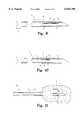
- FIG. 1is a partial side sectional view of a biopsy-marking assembly of the present invention.
- FIG. 2is a side sectional view of a proximal end of a biopsy device.
- FIG. 3is a side sectional view of the proximal end of a biopsy-marking assembly of this invention partially inserted into tissue.
- FIG. 4is a side sectional view of the proximal end of a biopsy-marking assembly that has deposited a marker into tissue.
- FIG. 5is a side sectional view of the assembly of FIG. 4 showing the marker deposited in the tissue and a sample trapped within the cavity.
- FIG. 6is a view of the biopsy device withdrawing from the tissue with the marker deposited in the tissue and a sample trapped within the cavity.
- FIG. 7is a side sectional view of the assembly of FIG. 4 with the inner needle rotated within the tissue.
- FIG. 8is a side elevational view of one embodiment of a marker of this invention.
- FIG. 9is a partial side sectional view of a biopsy marking assembly of this invention including a marker of FIG. 8 compressed within the cavity.
- FIG. 10is a partial side sectional view of a biopsy marking assembly of this invention including a marker of FIG. 8 folded within the cavity.
- FIG. 11is a partial side sectional view of a biopsy device within tissue with the marker of FIG. 8 released within the tissue.
- FIG. 12is a side elevational view of another embodiment of a marker of this invention.
- FIG. 13is a side sectional view of the proximal end of a biopsy-marking assembly according to one embodiment of this invention including a marker of FIG. 12 compressed within the cavity.
- FIG. 14is a partial side sectional view of the assembly of FIG. 13 inserted into tissue with the marker released in the tissue.
- FIG. 15is a side elevational view of another embodiment of a marker of this invention.
- FIG. 16is a side elevational view of another embodiment of a marker of this invention.
- FIG. 17is a side elevational view of another embodiment of a marker of this invention.
- FIG. 18is a side elevational view of another embodiment of a marker of this invention.
- FIG. 19is a side elevational view of a biopsy marker composed of a shape memory alloy and in the austenite phase.
- FIG. 20is a side elevational view of the marker depicted in FIG. 19 in the martensite phase.
- FIG. 21is a partial side elevational view of a biopsy-marker assembly including the marker depicted in FIG. 20.
- FIG. 22is a side elevational view of the biopsy-marker assembly shown in FIG. 21 showing the marker in the austenite phase and expelled from the cavity.
- FIG. 23is a side elevational view of another embodiment of a shape memory marker in the martensite phase.
- FIG. 24is a side elevational view of a biopsy-marker assembly showing the marker of FIG. 23 in the austenite phase and expelled from the cavity.
- FIG. 25is a side elevational view of another embodiment of a marker of this invention.
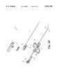
- FIG. 26is an exploded view of a biopsy-marking assembly according to one embodiment of this invention.
- FIG. 27is a partial side sectional view of a biopsy marking assembly having a marker chamber separate from the tissue sample cavity.
- the present inventionprovides devices, assemblies and methods that precisely mark a biopsy site so that the site may be later located and accessed if necessary.
- the markerscan be automatically delivered to the exact biopsy site during the biopsy procedure using the biopsy device.
- the biopsy markerscan be delivered at the same time as the biopsy sample is removed. Therefore, this invention solves the problem of attempting to locate the biopsy site after the biopsy device has been removed and tissue has shifted to fill the void left by the sample.
- FIG. 1A biopsy-marking assembly 10 in accordance with a preferred embodiment of the invention is depicted in FIG. 1.
- This embodimentincludes a biopsy device 11 and a marker 20 of this invention loaded into a cavity 16 of device 11.
- device 11is a two piece biopsy needle as is commonly employed for obtaining tissue core specimens.
- the biopsy-marking device 11 depicted in FIG. 1comprises an outer hollow needle 12 defining a lumen 13 therethrough.
- An inner needle 14is slidingly engaged inside lumen 13 and is moveable relative to outer needle 12.
- the inner needle 14defines a first end 15 having a tissue cutting point 15a and a cavity 16 adjacent first end 15 for receiving a tissue sample.
- the inner needle 14is slidingly engaged within lumen 13.
- the inner needle 14is slideable relative to outer needle 12 between a first position shown in FIG. 1 and a second position shown in FIG. 2. In the first position, inner needle 14 is retracted within lumen 13 so that outer needle 12 covers cavity 16. In the second position, the first end 15 of inner needle 14 is extended away from outer needle 12 to expose cavity 16 to tissue in the biopsy site.
- Biopsy device 11also includes means for moving inner needle 14 and outer needle 12 with respect to each other. Such means are known in the art and commercially available. Biopsy devices of this type are available from U.S. Biopsy, Inc., a division of Promex, Inc. 3049 Hudson Street, Franklin, Ind., (317) 736-0128.
- Biopsy-marking assembly 10also includes marking means for marking the biopsy site immediately before or immediately after a biopsy sample is taken at the site.
- marking means or marking element 20is carried in cavity 16 when inner needle 14 is in the first position. Marking element 20 is deposited in tissue T when inner needle 14 is in the second position to expose cavity 16 and release marker 20.
- FIGS. 3-6A preferred method of this invention is depicted in FIGS. 3-6.
- the biopsy assembly 10is inserted through a small incision or puncture made in the skin.
- the assembly 10is then driven into the body in the direction of arrow I to approach lesion L of suspicion in tissue T.
- Tissue-cutting point 15Aenters the tissue T and is positioned near the lesion L.
- inner needle 14is positioned within outer needle 12 in the first position. In this position, no more than proximal end 15 of inner needle 14 is exposed, cavity 16 is covered by outer needle 12 and a marking element 20 is contained within cavity 16.
- inner needle 14is driven into tissue T far enough to expose cavity 16 of inner needle 14 and release marking element 20. After marking element 20 is released, soft body tissues will then prolapse into cavity 16. Of course, it is the aim of the surgeon to obtain lesion L tissue and so assembly 10 will be positioned so that lesion L will be trapped in cavity 16.
- outer needle 12is then advanced along inner needle 14 in order to cover cavity 16. This forward movement of outer needle 12 severs the prolapsed tissue to obtain a tissue sample S, which becomes trapped in cavity 16 of inner needle 14. With outer needle 12 blocking the opening of cavity 16, biopsy assembly 10 may then be withdrawn carefully along arrow W shown in FIG. 6. Biopsy assembly 10 is then withdrawn from the target site carrying sample S in cavity 16 and leaving marker element 20 behind in tissue T. Thereafter, outer needle 12 is once again retracted to expose cavity 16 of inner needle 14, creating access to tissue sample S contained therein.
- a markermay be left at the site as each sample is taken or only at the first and/or last sample site.
- the inventionincludes means and steps for preventing the marker element from interfering with the prolapse of tissue into the cavity.
- the inner needleis rotated relative to the marker delivery site.
- biopsy marker assembly 10is inserted into the tissue T so that proximal end 15 is adjacent the biopsy site or lesion L.
- Inner needle 14is moved to deposit marker element 20 at the marker delivery site.
- Inner needle 14is then preferably rotated as shown in FIG. 7 after depositing marker element 20 and before sliding outer needle 12 to capture the prolapsed tissue.
- Inner needle 14 alone or the entire biopsy needle assembly including both inner needle 14 and outer needle 12may be rotated.
- the particular components of the biopsy device that are rotatedare not critical so long as the cavity 16 is rotated away from the marker delivery site.
- marker element 20will be clear of the action of outer needle 12 as it is actuated to trap the sample within cavity 16 as shown in FIG. 5.
- the needle in the rotating stepwill have an angle of rotation that is between about 5° and about 180°. Most preferably, the angle of rotation would be less than about 90° and most preferably will be about 45°.
- the particular angle of rotationwill be noted by the surgeon for precise location of the marker relative to the lesion L. The angle of rotation that is required will depend on the density of the tissue T and the shape, size, configuration and material of the marker element.
- the marker elements of this inventionwill be of any suitable biocompatible material that can be visualized by a surgeon or radiologist using the visualization means of choice at the time, such as x-ray, ultrasound, stereotactic imaging and the like.
- suitable biocompatible materialsuch as x-ray, ultrasound, stereotactic imaging and the like.
- certain materialsare highly visible under MRI, such as oil and Vitamin E, and certain surface features are better visualized using ultrasound technology. Such surface features include flats and angular features, etching and through-holes.
- marker element 20is radiopaque.
- the materialmust be resistant to biodegradation for at least a suitable period of time. In most embodiments of this invention, it is contemplated that the material will not be biodegradable and will remain in the body for the life of the patient or until it is surgically removed.
- the materialbiodegrade after a suitable period of time after which the marker will no longer be needed, such as a number of years.
- the markersare composed of medical grade stainless steel or titanium.
- marking element 20is a metal rod.
- the particular shape of the marking elementwill depend upon the application. For example, in breast tissue the marking element must overcome the force of the breast tissue, which will be weighted against the marker element as it attempts to escape from cavity 16. Therefore, in this instance, it may be preferable that the marking element be spring-loaded into cavity 16.
- the marker elementis a rod 30 as shown in FIG. 8 that has an initial V-shaped position. Rod 30 includes a pair of legs 32, 34 and a joint 35 between legs 32, 34.
- marker 30is compressed in a flattened state as shown in FIG. 9.
- marker 30is compressed into a folded state with interior faces 31, 33 of legs 32, 34 pressed towards one another.
- marker element 30will be released and will spring back to its initial V-shaped configuration as shown in FIG. 11.
- the material of choice for marker 30will be one that is flexible enough so that it can be compressed into a spring-loaded state into the cavity and resilient enough to return to its preferred initial configuration.
- Preferred materialsinclude stainless steel, titanium and radiopaque polymers or resins.
- marker element 40is a metal rod formed into a continuous oval, such as a micro-coil. As shown in FIG. 13, a circular or oval marker is compressed within cavity 16 with interior wall 42 compressed towards itself for delivery to the biopsy site. As shown in FIG. 14, when biopsy device 10 is moved into the second position, marker 40 is spring released into the biopsy site.
- FIG. 15depicts a marker 50 having a pair of opposite ends 51 and 52 and a curved middle portion 53.
- the curved middle portion 53forms a spring when marker 50 is compressed within a cavity.
- marker 60is a vertically coiled rod 61, which is compressible into a cavity for delivery to a biopsy site. Additional material can be used to coil the marker 70 into a "ball of string" structure as depicted in FIG. 17.
- Horizontally coiled rodsare also contemplated, such as the marker 80 in FIG. 18.
- Marker 80optionally includes an enlarged middle portion 81 and two opposite tapered ends 82, 83.
- the marker 130is a composed of a material that is responsive to a change in temperature to assume either an expanded shape or an altered configuration.
- a material that is responsive to a change in temperature to assume a different shape or sizeis a shape memory alloy.
- Such alloyshave a crystalline structure that can alternate between two phases depending on the temperature of the environment. In providing a shape memory alloy component, the component is first annealed to a specific shape by traditional means. The alloy is then heated to a temperature high enough that the crystalline structure assumes a phase known as the austenite phase.
- the alloyis cooled until it reverts to a second phase called the martensite phase.
- the componentmay be deformed but the component will return to its original austenite phase shape when heated to a temperature above that at which the martensite phase returns to the austenite phase.
- the alloy"remembers" its original annealed shape and will return to that original shape when heated above the austenite phase transition temperature. In doing so, the alloy converts heat energy into mechanical work.
- Marker 130is composed of a shape memory alloy, preferably a nickel-titanium alloy and most preferably nitinol.
- the specific phase transitional temperature at which the phase transition occurscan be controlled by specifying the exact nickel to titanium ratio. In a preferred embodiment, the ratio is chosen to provide austenite transition temperature near body temperature, approximately 37° C. and a martensite transition temperature of about room temperature, approximately 25° C.
- the present inventioncontemplates other nickel-titanium ratios to provide desired austenite and martensite phase transition temperatures.
- the present inventionfurther contemplates the use of other known shape memory alloys for use in constructing the markers of this invention.
- One example of such an alloyis an iron-based alloy including various percentages by weight of manganese, silicon and chromium.
- marker 130is first annealed and contoured to a V-shape while in the austenite phase crystalline configuration as shown in FIG. 19.
- marker 130includes a pair of legs 132, 134 and a joint 135 between legs 132, 134. Interior faces 131, 133 of legs 132, 134 face one another.
- Marker 130is then cooled until the martensite transformation occurs.
- marker 130is deformed such that legs 132, 134 lie in a straight line or 180° from one another as shown in FIG. 20.
- marker 130can be loaded into a cavity of a biopsy gun for delivery to the biopsy site.
- marker 130is loaded into a cavity with interior faces 131, 133 against bed 117 of cavity 116 and opposite opening or mouth 118 of cavity 116 as depicted in FIG. 21.
- marker 130is heated to body temperature, which is sufficient to transform the alloy to the austenite phase configuration. In doing so, marker 130 "remembers" its original configuration, and its returns to its initial V shape.
- ends 132a, 134a of legs 132, 134will be drawn together and push against bed 117 of cavity 116 to be expelled from cavity 116 as shown in FIG. 22.
- additional heating meansas are known in the art are used.
- the markeris annealed and contoured to a V-shape as described above and shown in FIG. 19.
- marker 130'is deformed to be folded at joint 135' with interior faces 131', 133' of legs 132', 134' pressed towards one another as shown in FIG. 23.
- the folded marker 130'can be loaded into a cavity as shown for marker 30 in FIG. 10.
- marker 130'is heated to body temperature. Referring now to FIG. 24, as marker 130' "remembers" its original configuration and returns to its initial V shape, the mechanical action of leg 132' against cavity bed 117 ejects marker 130' from cavity 116.
- the preferred phase transition temperaturesare approximately room temperature and body temperature for ease of handling. Where other alloys are used which have different phase temperatures, the markers will be treated to achieve those temperatures.
- the marker elementinclude stabilization means for grasping tissue to prevent or retard migration of the marker within the tissue.
- marker element 140 depicted in FIG. 25includes a rod 141 defining two sharpened opposite ends 142, 143. Along its length, rod 141 is provided with sharpened surface projections 146 in between flattened valley area 145.
- Other stabilization meansinclude barbs 84 depicted in FIG. 18. Although barbs 84 and surface projections 146 are depicted in FIGS. 18 and 25 as an example, these features may be provided on any of the markers of this invention. Also, any suitable stabilization means is contemplated by this invention. The stabilization means will be formed into the marker how ever is appropriate for the materials, such as by molding or machining.
- the inventioncontemplates markers of any suitable size and shape.
- the markers of this inventionwill be sized and shaped large enough to be visualized yet compact enough to be delivered to the biopsy site in the cavity of a biopsy needle.
- the markerwill have a dimension of at least about 1 mm and no more than about 30 mm for applications such as breast.
- the markerhas a length of 0.6 inches (15 mm) and a width of 0.38 inches (9.5 mm) and is composed of titanium.
- biopsy devicewhich includes means for obtaining a tissue sample, cutting means for cutting tissue and marking means for marking the biopsy site.
- marking meansis provided for marking the biopsy site before a biopsy sample is taken.
- the marking elementis contained within a cavity in a biopsy device element for delivery to the biopsy site.
- the biopsy device meansis a single action biopsy device as is known in the art.
- a single action biopsy devicewhich can be used with this invention is manufactured and marketed by U.S. Biopsy, Inc., a division of Promex, Inc. 3049 Hudson Street, Franklin, Ind. 46131 (317) 736-0128), and is depicted in FIG. 26.
- Device 150includes a housing 151 having finger grips 152 and 153.
- An actuator 155is operatively engaged to both the inner needle 14 and the outer needle 12.
- the actuatorincludes a gripping portion 156 and a drive mechanism 160.
- the drive mechanism 160operates to depress a drive carriage 170 against the action of a spring 180.
- the housing 151includes a resilient latch 175 that engages the underside 172 of the carriage 170 in the retracted position.
- the latch 175is released by forward movement of the drive mechanism 160 so that the spring 180 pushes the carriage 170 outwardly to expose the cavity 16 of the inner needle 14. Further movement propels the outer needle 12 over the inner needle 14 to trap tissue within the cavity.
- the operational relationship between the outer needle 12 and the inner needle 14is described above and depicted in FIGS. 1-6.
- a single action biopsy device of this typeis preferred because it allows rotation of inner needle 14 between the steps of releasing the marker element and obtaining a biopsy sample. This allows the biopsy marker to be ejected clear from the cavity to expose the cavity to the lesion L.
- double action biopsy devicessuch as the device disclosed in U.S. Pat. No. 5,538,010 to Darr and Ireland, may also be used.
- the needle assemblyis sequentially driven by a spring loaded drive mechanism.
- the devicemay be inserted into tissue in the first position as described above.
- the inner needlefires to expose the cavity, and the outer needle is immediately fired upon the firing of the inner needle to trap the tissue sample within the cavity.
- the marker elementcould be delivered in the cavity of such device, but the immediate firing of the outer needle may not provide enough time for the marker element to clear the cavity.
- such double-action biopsy devicescould be modified to address this. For example, the firing of the outer needle could be delayed to allow sufficient time for the marker element to clear the cavity area or to allow rotation of the inner needle before the outer needle covers the cavity to trap the sample within the cavity.
- a double-action biopsy-marker assembly 200is provided with an outer needle 212 and an inner needle 214.
- Inner needle 214defines a sample cavity 216 as described above.
- Inner needle 214also defines a separate and oppositely located marker chamber 219.
- a wall 218 of inner needle 214separates marker chamber 219 and sample cavity 216.
- marker element 220is automatically ejected from marker chamber 219 and delivered approximately 180° from the biopsy sample site.
- Marker chamber 219may be defined in other locations in inner needle 214.
- marker chamber 219could be between proximal end 215 and sample cavity 216 so that the marker will be placed slightly deeper than the biopsy sample site.
- Marker chamber 219could also be placed between sample cavity 216 and the distal end (not shown). Any configuration is contemplated so long as marker chamber 219 will be exposed when inner needle 214 is moved from the first position to the second position.
- meanscan be provided to incrementally feed a length of marker material through a hole defined in the outer needle 212.
- the materialcan be urged out the hole with a ramp built within the marker chamber 219. Discrete markers can be severed and left in the tissue by movement of the outer needle.
- the present inventioneconomically and conveniently provides biopsy-marker assemblies for marking a biopsy sample site and obtaining a biopsy sample.
- Biopsy devicescan be pre-loaded with a marker for automatic marking of the biopsy site.
- biopsy and biopsy sites markingoccurs simultaneously without the need for complex additional steps. Biopsy sites can then be later located and accessed surgically as necessary.
Landscapes
- Health & Medical Sciences (AREA)
- Surgery (AREA)
- Life Sciences & Earth Sciences (AREA)
- Heart & Thoracic Surgery (AREA)
- Pathology (AREA)
- Oral & Maxillofacial Surgery (AREA)
- Engineering & Computer Science (AREA)
- Biomedical Technology (AREA)
- Nuclear Medicine, Radiotherapy & Molecular Imaging (AREA)
- Medical Informatics (AREA)
- Molecular Biology (AREA)
- Animal Behavior & Ethology (AREA)
- General Health & Medical Sciences (AREA)
- Public Health (AREA)
- Veterinary Medicine (AREA)
- Surgical Instruments (AREA)
Abstract
Description
Claims (21)
Priority Applications (2)
| Application Number | Priority Date | Filing Date | Title |
|---|---|---|---|
| US09/170,610US6056700A (en) | 1998-10-13 | 1998-10-13 | Biopsy marker assembly and method of use |
| US09/526,412US6261243B1 (en) | 1998-10-13 | 2000-03-16 | Biopsy marker assembly and method of use |
Applications Claiming Priority (1)
| Application Number | Priority Date | Filing Date | Title |
|---|---|---|---|
| US09/170,610US6056700A (en) | 1998-10-13 | 1998-10-13 | Biopsy marker assembly and method of use |
Related Child Applications (1)
| Application Number | Title | Priority Date | Filing Date |
|---|---|---|---|
| US09/526,412ContinuationUS6261243B1 (en) | 1998-10-13 | 2000-03-16 | Biopsy marker assembly and method of use |
Publications (1)
| Publication Number | Publication Date |
|---|---|
| US6056700Atrue US6056700A (en) | 2000-05-02 |
Family
ID=22620586
Family Applications (2)
| Application Number | Title | Priority Date | Filing Date |
|---|---|---|---|
| US09/170,610Expired - LifetimeUS6056700A (en) | 1998-10-13 | 1998-10-13 | Biopsy marker assembly and method of use |
| US09/526,412Expired - LifetimeUS6261243B1 (en) | 1998-10-13 | 2000-03-16 | Biopsy marker assembly and method of use |
Family Applications After (1)
| Application Number | Title | Priority Date | Filing Date |
|---|---|---|---|
| US09/526,412Expired - LifetimeUS6261243B1 (en) | 1998-10-13 | 2000-03-16 | Biopsy marker assembly and method of use |
Country Status (1)
| Country | Link |
|---|---|
| US (2) | US6056700A (en) |
Cited By (118)
| Publication number | Priority date | Publication date | Assignee | Title |
|---|---|---|---|---|
| EP1163888A1 (en)* | 2000-06-16 | 2001-12-19 | Inrad, Inc. | Apparatus for the percutaneous marking of a lesion |
| US6336904B1 (en)* | 1998-04-07 | 2002-01-08 | Pro Duct Health, Inc. | Methods and devices for the localization of lesions in solid tissue |
| US6347241B2 (en)* | 1999-02-02 | 2002-02-12 | Senorx, Inc. | Ultrasonic and x-ray detectable biopsy site marker and apparatus for applying it |
| US6350244B1 (en)* | 2000-02-21 | 2002-02-26 | Biopsy Sciences, Llc | Bioabsorable markers for use in biopsy procedures |
| US20020026201A1 (en)* | 1994-09-16 | 2002-02-28 | Foerster Seth A. | Methods for defining and marking tissue |
| US6356782B1 (en)* | 1998-12-24 | 2002-03-12 | Vivant Medical, Inc. | Subcutaneous cavity marking device and method |
| US6355275B1 (en) | 2000-06-23 | 2002-03-12 | Carbon Medical Technologies, Inc. | Embolization using carbon coated microparticles |
| WO2002022015A2 (en) | 2000-09-18 | 2002-03-21 | John Hopkins University School Of Medicine | Methods and systems for image-guided surgical interventions |
| US6371904B1 (en) | 1998-12-24 | 2002-04-16 | Vivant Medical, Inc. | Subcutaneous cavity marking device and method |
| US20020058882A1 (en)* | 1998-06-22 | 2002-05-16 | Artemis Medical, Incorporated | Biopsy localization method and device |
| US6394965B1 (en) | 2000-08-15 | 2002-05-28 | Carbon Medical Technologies, Inc. | Tissue marking using biocompatible microparticles |
| US6419641B1 (en) | 2000-11-28 | 2002-07-16 | Promex, Llc | Flexible tip medical instrument |
| US6427081B1 (en)* | 1999-02-02 | 2002-07-30 | Senorx, Inc. | Methods and chemical preparations for time-limited marking of biopsy sites |
| EP1249209A1 (en)* | 2001-04-09 | 2002-10-16 | Ethicon Endo-Surgery, Inc. | Biopsy instrument with tissue marking element |
| US6471700B1 (en) | 1998-04-08 | 2002-10-29 | Senorx, Inc. | Apparatus and method for accessing biopsy site |
| US6497706B1 (en) | 1998-03-03 | 2002-12-24 | Senorx, Inc. | Biopsy device and method of use |
| US6540695B1 (en) | 1998-04-08 | 2003-04-01 | Senorx, Inc. | Biopsy anchor device with cutter |
| US6605047B2 (en) | 2001-09-10 | 2003-08-12 | Vivant Medical, Inc. | Biopsy marker delivery system |
| US6626903B2 (en) | 1997-07-24 | 2003-09-30 | Rex Medical, L.P. | Surgical biopsy device |
| US6638234B2 (en) | 1998-03-03 | 2003-10-28 | Senorx, Inc. | Sentinel node location and biopsy |
| US20030220640A1 (en)* | 2002-05-22 | 2003-11-27 | Rubicor Medical , Inc. | Methods and systems for in situ tissue marking and orientation stabilization |
| US20030233101A1 (en)* | 2002-06-17 | 2003-12-18 | Senorx, Inc. | Plugged tip delivery tube for marker placement |
| US6679851B2 (en) | 1998-09-01 | 2004-01-20 | Senorx, Inc. | Tissue accessing and anchoring device and method |
| US6725083B1 (en)* | 1999-02-02 | 2004-04-20 | Senorx, Inc. | Tissue site markers for in VIVO imaging |
| US20040097981A1 (en)* | 2002-08-01 | 2004-05-20 | Selis James E. | Biopsy devices and methods |
| WO2004026347A3 (en)* | 2002-09-17 | 2004-06-03 | Iscience Surgical Corp | Apparatus and method for surgical bypass of aqueous humor |
| US6758848B2 (en) | 1998-03-03 | 2004-07-06 | Senorx, Inc. | Apparatus and method for accessing a body site |
| US20040204660A1 (en)* | 1998-06-22 | 2004-10-14 | Artemis Medical, Inc. | Biopsy localization method and device |
| US20040236212A1 (en)* | 2003-05-23 | 2004-11-25 | Senorx, Inc. | Fibrous marker and intracorporeal delivery thereof |
| US20040243023A1 (en)* | 2003-05-30 | 2004-12-02 | Grigoryants Sergey S | Transbronchial needle aspiration device |
| US6846320B2 (en)* | 1998-05-01 | 2005-01-25 | Sub-Q, Inc. | Device and method for facilitating hemostasis of a biopsy tract |
| US20050038355A1 (en)* | 2003-08-13 | 2005-02-17 | Gellman Barry N. | Marking biopsy sites |
| US20050038462A1 (en)* | 1998-04-08 | 2005-02-17 | Senorx, Inc. | Dilation devices and methods for removing tissue specimens |
| US6862470B2 (en) | 1999-02-02 | 2005-03-01 | Senorx, Inc. | Cavity-filling biopsy site markers |
| US20050065453A1 (en)* | 2003-02-24 | 2005-03-24 | Senorx, Inc. | Biopsy device with selectable tissue receiving aperture orientation and site illumination |
| US6875182B2 (en) | 1998-03-03 | 2005-04-05 | Senorx, Inc. | Electrosurgical specimen-collection system |
| US20050119562A1 (en)* | 2003-05-23 | 2005-06-02 | Senorx, Inc. | Fibrous marker formed of synthetic polymer strands |
| US20050119652A1 (en)* | 1998-09-03 | 2005-06-02 | Rubicor Medical, Inc. | Devices and methods for performing procedures on a breast |
| US20050149166A1 (en)* | 2003-11-08 | 2005-07-07 | Schaeffer Darin G. | Branch vessel prosthesis with anchoring device and method |
| US20050159677A1 (en)* | 2003-12-23 | 2005-07-21 | Shabaz Martin V. | Biopsy device with aperture orientation and improved tip |
| US20050222669A1 (en)* | 2004-03-31 | 2005-10-06 | Purdy James D | Fenestrated intraluminal stent system |
| US20050234336A1 (en)* | 2004-03-26 | 2005-10-20 | Beckman Andrew T | Apparatus and method for marking tissue |
| US20050277871A1 (en)* | 2004-06-11 | 2005-12-15 | Selis James E | Biopsy devices and methods |
| US20050283113A1 (en)* | 2004-06-22 | 2005-12-22 | Thomas Brinz | Metering device and method for operating such |
| US20060009696A1 (en)* | 2004-04-08 | 2006-01-12 | Techniscan, Inc. | Method for imaging and treating a breast |
| US20060079805A1 (en)* | 2004-10-13 | 2006-04-13 | Miller Michael E | Site marker visable under multiple modalities |
| US20060116573A1 (en)* | 2003-11-17 | 2006-06-01 | Inrad, Inc. | Self Contained, Self Piercing, Side-Expelling Marking Apparatus |
| US20060153774A1 (en)* | 2004-12-08 | 2006-07-13 | Cook Incorporated | Contrast agent coated medical device |
| US20060173296A1 (en)* | 2004-10-13 | 2006-08-03 | Miller Michael E | Site marker visable under multiple modalities |
| US20060235298A1 (en)* | 2005-03-31 | 2006-10-19 | Robert Kotmel | Internal biopsy marking |
| US20060258933A1 (en)* | 2005-05-10 | 2006-11-16 | Advanced Clinical Solutions, Inc. | Method of defining a biological target for treatment |
| US20070038146A1 (en)* | 2005-08-05 | 2007-02-15 | Quick Richard L | Biopsy device with fluid delivery to tissue specimens |
| US20070038145A1 (en)* | 2004-11-22 | 2007-02-15 | Inrad, Inc. | Post Decompression Marker Introducer System |
| US7189206B2 (en) | 2003-02-24 | 2007-03-13 | Senorx, Inc. | Biopsy device with inner cutter |
| US20070087026A1 (en)* | 2005-10-07 | 2007-04-19 | Inrad, Inc. | Drug-Eluting Tissue Marker |
| US20070093726A1 (en)* | 2004-10-13 | 2007-04-26 | Leopold Phillip M | Site marker visible under multiple modalities |
| US20070118034A1 (en)* | 2005-11-22 | 2007-05-24 | Mark Joseph L | Surgical site marker delivery system |
| US20080021313A1 (en)* | 2006-07-06 | 2008-01-24 | Boston Scientific Scimed, Inc. | Electroactive polymer radiopaque marker |
| US20080039819A1 (en)* | 2006-08-04 | 2008-02-14 | Senorx, Inc. | Marker formed of starch or other suitable polysaccharide |
| US20080058672A1 (en)* | 2004-12-16 | 2008-03-06 | Senorx, Inc. | Biopsy device with aperture orientation and improved tip |
| US20080058769A1 (en)* | 2004-07-05 | 2008-03-06 | Ingemar Naslund | Marker for Positioning in Body Tissue |
| US20080097199A1 (en)* | 2004-08-20 | 2008-04-24 | David Mullen | Tissue Marking Devices and Systems |
| US20080254298A1 (en)* | 2006-02-23 | 2008-10-16 | Meadwestvaco Corporation | Method for treating a substrate |
| US20080269603A1 (en)* | 2004-10-13 | 2008-10-30 | Nicoson Zachary R | Site marker visible under multiple modalities |
| US20080319318A1 (en)* | 2007-05-15 | 2008-12-25 | Johnson Steven A | Breast scanning system |
| US20090030309A1 (en)* | 2007-07-26 | 2009-01-29 | Senorx, Inc. | Deployment of polysaccharide markers |
| US20090043321A1 (en)* | 2004-04-29 | 2009-02-12 | Iscience Interventional Corporation | Apparatus And Method For Surgical Enhancement Of Aqueous Humor Drainage |
| US20090112118A1 (en)* | 2005-08-05 | 2009-04-30 | Senorx, Inc. | Biopsy device with fluid delivery to tissue specimens |
| US20090182245A1 (en)* | 2008-01-16 | 2009-07-16 | Roberto Zambelli | Guide device for localising a neoplasia to be removed during a surgical procedure |
| US20090204021A1 (en)* | 2004-12-16 | 2009-08-13 | Senorx, Inc. | Apparatus and method for accessing a body site |
| US20090209853A1 (en)* | 2008-02-19 | 2009-08-20 | Parihar Shailendra K | Biopsy site marker applier |
| US20090222078A1 (en)* | 2007-12-21 | 2009-09-03 | Greenberg Roy K | Prosthesis for Implantation in Aorta and Method of Using Same |
| US20090287078A1 (en)* | 2003-05-23 | 2009-11-19 | Senorx, Inc. | Marker or filler forming fluid |
| US20100030149A1 (en)* | 2006-10-23 | 2010-02-04 | C.R. Bard, Inc. | Breast marker |
| US20100173866A1 (en)* | 2004-04-29 | 2010-07-08 | Iscience Interventional Corporation | Apparatus and method for ocular treatment |
| US20100191177A1 (en)* | 2009-01-23 | 2010-07-29 | Iscience Interventional Corporation | Device for aspirating fluids |
| AU2005201304B2 (en)* | 2004-03-31 | 2010-07-29 | Ethicon Endo-Surgery, Inc. | Marker device and method of deploying a cavity marker using a surgical biopsy device |
| WO2010091360A1 (en)* | 2009-02-06 | 2010-08-12 | Senorx, Inc | Anchor markers |
| US20110071391A1 (en)* | 2009-09-24 | 2011-03-24 | Speeg Trevor W V | Biopsy marker delivery device with positioning component |
| US8157862B2 (en) | 1997-10-10 | 2012-04-17 | Senorx, Inc. | Tissue marking implant |
| US8311610B2 (en) | 2008-01-31 | 2012-11-13 | C. R. Bard, Inc. | Biopsy tissue marker |
| US8361082B2 (en) | 1999-02-02 | 2013-01-29 | Senorx, Inc. | Marker delivery device with releasable plug |
| US8401622B2 (en) | 2006-12-18 | 2013-03-19 | C. R. Bard, Inc. | Biopsy marker with in situ-generated imaging properties |
| US8425473B2 (en) | 2009-01-23 | 2013-04-23 | Iscience Interventional Corporation | Subretinal access device |
| US8498693B2 (en) | 1999-02-02 | 2013-07-30 | Senorx, Inc. | Intracorporeal marker and marker delivery device |
| US8634899B2 (en) | 2003-11-17 | 2014-01-21 | Bard Peripheral Vascular, Inc. | Multi mode imaging marker |
| US20140024945A1 (en)* | 2012-07-23 | 2014-01-23 | ClariTrac Inc. | Ultrasound device for needle procedures |
| US8641640B2 (en) | 2005-05-23 | 2014-02-04 | Senorx, Inc. | Tissue cutting member for a biopsy device |
| US8668737B2 (en) | 1997-10-10 | 2014-03-11 | Senorx, Inc. | Tissue marking implant |
| US8670818B2 (en) | 2008-12-30 | 2014-03-11 | C. R. Bard, Inc. | Marker delivery device for tissue marker placement |
| US8718745B2 (en) | 2000-11-20 | 2014-05-06 | Senorx, Inc. | Tissue site markers for in vivo imaging |
| USD715442S1 (en) | 2013-09-24 | 2014-10-14 | C. R. Bard, Inc. | Tissue marker for intracorporeal site identification |
| USD715942S1 (en) | 2013-09-24 | 2014-10-21 | C. R. Bard, Inc. | Tissue marker for intracorporeal site identification |
| USD716451S1 (en) | 2013-09-24 | 2014-10-28 | C. R. Bard, Inc. | Tissue marker for intracorporeal site identification |
| USD716450S1 (en) | 2013-09-24 | 2014-10-28 | C. R. Bard, Inc. | Tissue marker for intracorporeal site identification |
| CN104605897A (en)* | 2015-01-04 | 2015-05-13 | 中国人民解放军第四军医大学 | Mechanism capable of marking tumor boundary used for biopsy needle |
| US9327061B2 (en) | 2008-09-23 | 2016-05-03 | Senorx, Inc. | Porous bioabsorbable implant |
| US9358141B2 (en) | 2004-03-31 | 2016-06-07 | Cook Medical Technologies Llc | Stent deployment device |
| US20160302881A1 (en)* | 2008-01-29 | 2016-10-20 | Covidien Lp | Target identification tool for intra-body localization |
| US20160374649A1 (en)* | 2013-02-08 | 2016-12-29 | Radu Kramer | Biopsy Method and Apparatus |
| US9579077B2 (en) | 2006-12-12 | 2017-02-28 | C.R. Bard, Inc. | Multiple imaging mode tissue marker |
| US9669113B1 (en)* | 1998-12-24 | 2017-06-06 | Devicor Medical Products, Inc. | Device and method for safe location and marking of a biopsy cavity |
| US9820824B2 (en) | 1999-02-02 | 2017-11-21 | Senorx, Inc. | Deployment of polysaccharide markers for treating a site within a patent |
| US9993232B2 (en) | 2014-05-22 | 2018-06-12 | Andrew N. Ellingson | Biopsy with marker device and method |
| US20180206866A1 (en)* | 2014-07-28 | 2018-07-26 | Shaw P. Wan | Suction evacuation device |
| US10342635B2 (en) | 2005-04-20 | 2019-07-09 | Bard Peripheral Vascular, Inc. | Marking device with retractable cannula |
| US20200229862A1 (en)* | 2017-02-27 | 2020-07-23 | Avent, Inc. | Method and System for Improving Location Accuracy of a Radiofrequency Ablation Procedure Via Fiducial Marking |
| US20210015509A1 (en)* | 2017-01-12 | 2021-01-21 | Shaw P. Wan | Suction evacuation device |
| US20210022757A1 (en)* | 2014-07-28 | 2021-01-28 | Shaw P. Wan | Suction evacuation device |
| US20210022759A1 (en)* | 2017-01-12 | 2021-01-28 | Shaw P. Wan | Suction evacuation device |
| US11058494B2 (en) | 2013-09-06 | 2021-07-13 | The Brigham And Women's Hospital, Inc. | System and method for a tissue resection margin measurement device |
| US20210378784A1 (en)* | 2018-11-07 | 2021-12-09 | Neotract, Inc. | System for delivery of a fiducial marker |
| JP2022084923A (en)* | 2014-10-08 | 2022-06-07 | デヴィコア メディカル プロダクツ,インク. | Biopsy site marker |
| US20220265125A1 (en)* | 2020-09-15 | 2022-08-25 | Raytrx, Llc | Wireless swivel camera laparoscopic instrument with a virtual mapping and guidance system |
| JP2022541842A (en)* | 2019-07-24 | 2022-09-27 | ビーアイピー バイオメド. インストゥルメンテ ウント プロダクテ ゲーエムベーハー | embeddable marker |
| WO2024039560A1 (en)* | 2022-08-16 | 2024-02-22 | Devicor Medical Products, Inc. | Biopsy site marker having expandable portion |
| WO2024039561A1 (en)* | 2022-08-16 | 2024-02-22 | Devicor Medical Products, Inc. | Biopsy site marker having movable portions |
| US12440301B2 (en)* | 2019-10-30 | 2025-10-14 | Teleflex Life Sciences Llc | System for delivery of a fiducial marker |
Families Citing this family (37)
| Publication number | Priority date | Publication date | Assignee | Title |
|---|---|---|---|---|
| AU756080B2 (en)* | 1998-06-04 | 2003-01-02 | New York University | Endovascular thin film devices and methods for treating and preventing stroke |
| US8251946B2 (en)* | 2000-08-24 | 2012-08-28 | Cardiac Science, Inc. | Method for constructing an instrument with a two-part plunger for subcutaneous implantation |
| US7736330B2 (en)* | 2000-08-24 | 2010-06-15 | Bardy Gust H | Subcutaneous implantation instrument with dissecting tool and method of construction |
| US6436068B1 (en)* | 2000-08-24 | 2002-08-20 | Gust H. Bardy | Instrument for implanting sensors and solid materials in a subcutaneous location and method thereof |
| US8323232B2 (en)* | 2000-08-24 | 2012-12-04 | Cardiac Science Corporation | Instrument with a two-part plunger for subcutaneous implantation |
| US8454552B2 (en)* | 2000-08-24 | 2013-06-04 | Cardiac Science Corporation | Method for constructing an instrument with a covered bore for subcutaneous implantation |
| US8348882B2 (en)* | 2000-08-24 | 2013-01-08 | Cardiac Science Corporation | Instrument with a covered bore for subcutaneous implantation |
| US7280865B2 (en)* | 2001-12-20 | 2007-10-09 | Accuray Incorporated | Anchored fiducial apparatus and method |
| WO2003077730A2 (en) | 2002-03-11 | 2003-09-25 | Wardle John L | Surgical coils and methods of deploying |
| WO2003079907A1 (en)* | 2002-03-20 | 2003-10-02 | Board Of Regents, The University Of Texas System | Biopsy needle |
| US8027712B2 (en)* | 2002-10-11 | 2011-09-27 | Ion Beam Applications S.A. | Elongated markers for soft tissue volume identification |
| US20050159676A1 (en)* | 2003-08-13 | 2005-07-21 | Taylor James D. | Targeted biopsy delivery system |
| US9638770B2 (en)* | 2004-05-21 | 2017-05-02 | Devicor Medical Products, Inc. | MRI biopsy apparatus incorporating an imageable penetrating portion |
| US7708751B2 (en) | 2004-05-21 | 2010-05-04 | Ethicon Endo-Surgery, Inc. | MRI biopsy device |
| ATE444712T1 (en)* | 2004-05-21 | 2009-10-15 | Ethicon Endo Surgery Inc | MRI BIOPSY DEVICE WITH A DISPLAYABLE PENETRATION PART |
| US8932233B2 (en) | 2004-05-21 | 2015-01-13 | Devicor Medical Products, Inc. | MRI biopsy device |
| BRPI0515007A (en) | 2004-08-12 | 2008-07-01 | Navotek Medical Ltd | computerized system for tracking and tracing of irradiated ionization source, sensor for targeting located on an ionized radiation source, method for determining device location, method of locating device manufacturing, and use of ionizing radiation shield |
| WO2007017846A2 (en)* | 2005-08-11 | 2007-02-15 | Navotek Medical Ltd. | Localization of a radioactive source |
| US20080262473A1 (en)* | 2004-10-19 | 2008-10-23 | Navotek Medical Ltd. | Locating a Catheter Tip Using a Tracked Guide |
| US7731705B2 (en)* | 2005-01-10 | 2010-06-08 | Wardle John L | Eluting coils and methods of deploying and retrieving |
| US20060241385A1 (en)* | 2005-04-12 | 2006-10-26 | Ethicon Endo-Surgery, Inc. | Guided disposable fiducial for breast biopsy localization fixture |
| US7947076B2 (en)* | 2005-06-03 | 2011-05-24 | Medtronic Xomed, Inc. | Nasal valve treatment method and apparatus |
| US20070016101A1 (en)* | 2005-07-13 | 2007-01-18 | Feldman Dennis D | Core Biopsy Device |
| ATE555737T1 (en)* | 2005-08-11 | 2012-05-15 | Navotek Medical Ltd | LOCALIZATION OF A RADIOACTIVE SOURCE |
| EP2158940A3 (en)* | 2005-08-11 | 2010-06-02 | Navotek Medical Ltd. | Medical treatment system and method using radioactivity based position sensor |
| US20080097286A1 (en)* | 2006-08-04 | 2008-04-24 | 0696578 B.C. Ltd Incorporation | Anal ointment applicator |
| US8979803B2 (en)* | 2007-04-05 | 2015-03-17 | Allan J. Darr | Stylet for bilumenal flexible medical device |
| US20100152663A1 (en)* | 2007-04-05 | 2010-06-17 | Darr Allan J | Stylet for bilumenal flexible medical device |
| US7942843B2 (en)* | 2008-08-18 | 2011-05-17 | Navotek Medical Ltd. | Implantation device for soft tissue markers and other implants |
| WO2011130216A1 (en)* | 2010-04-14 | 2011-10-20 | Cook Incorporated | Full core biopsy needle with secondary cutting cannula |
| US9044216B2 (en) | 2010-07-12 | 2015-06-02 | Best Medical International, Inc. | Biopsy needle assembly |
| US8758256B2 (en) | 2010-07-12 | 2014-06-24 | Best Medical International, Inc. | Apparatus for brachytherapy that uses a scanning probe for treatment of malignant tissue |
| USD643120S1 (en)* | 2010-07-14 | 2011-08-09 | Bardy Gust H | Implantation instrument |
| USD643119S1 (en)* | 2010-07-14 | 2011-08-09 | Bardy Gust H | Implantation instrument |
| US8882681B2 (en)* | 2011-06-29 | 2014-11-11 | Cook Medical Technologies Llc | Through-cradle soft tissue biopsy device |
| WO2016201115A1 (en)* | 2015-06-11 | 2016-12-15 | Radvation, Llc | Device and method for marking a location of a tissue biopsy |
| CN109009344B (en)* | 2018-07-24 | 2020-10-30 | 西安卓恰医疗器械有限公司 | Puncture locator with replaceable needle |
Citations (21)
| Publication number | Priority date | Publication date | Assignee | Title |
|---|---|---|---|---|
| US3744493A (en)* | 1972-01-10 | 1973-07-10 | Syntex Corp | Implanter having an improved cartridge ejector |
| US4576163A (en)* | 1983-08-08 | 1986-03-18 | Bliss Robert J | Skin marker for use in biopsy excisions |
| US4693237A (en)* | 1986-01-21 | 1987-09-15 | Hoffman Richard B | Radiopaque coded ring markers for use in identifying surgical grafts |
| US4776346A (en)* | 1984-02-10 | 1988-10-11 | Dan Beraha | Biopsy instrument |
| US4781198A (en)* | 1986-09-08 | 1988-11-01 | Kanabrocki Eugene L | Biopsy tracer needle |
| US5034005A (en)* | 1990-07-09 | 1991-07-23 | Appling William M | Radiopaque marker |
| US5083570A (en)* | 1990-06-18 | 1992-01-28 | Mosby Richard A | Volumetric localization/biopsy/surgical device |
| US5195533A (en)* | 1992-05-08 | 1993-03-23 | Boston Scientific Corporation | Biopsy needle instrument for storing multiple specimens |
| US5197482A (en)* | 1989-06-15 | 1993-03-30 | Research Corporation Technologies, Inc. | Helical-tipped lesion localization needle device and method of using the same |
| US5234426A (en)* | 1989-06-15 | 1993-08-10 | Research Corporation Technologies, Inc. | Helical-tipped lesion localization needle device and method of using the same |
| US5290289A (en)* | 1990-05-22 | 1994-03-01 | Sanders Albert E | Nitinol spinal instrumentation and method for surgically treating scoliosis |
| US5487392A (en)* | 1993-11-15 | 1996-01-30 | Haaga; John R. | Biopxy system with hemostatic insert |
| US5538010A (en)* | 1994-10-05 | 1996-07-23 | Proact Ltd. | Biopsy needle device |
| US5562613A (en)* | 1991-07-02 | 1996-10-08 | Intermed, Inc. | Subcutaneous drug delivery device |
| US5595193A (en)* | 1993-02-12 | 1997-01-21 | Walus; Richard L. | Tool for implanting a fiducial marker |
| US5649547A (en)* | 1994-03-24 | 1997-07-22 | Biopsys Medical, Inc. | Methods and devices for automated biopsy and collection of soft tissue |
| US5782775A (en)* | 1995-10-20 | 1998-07-21 | United States Surgical Corporation | Apparatus and method for localizing and removing tissue |
| US5800445A (en)* | 1995-10-20 | 1998-09-01 | United States Surgical Corporation | Tissue tagging device |
| US5853366A (en)* | 1996-07-08 | 1998-12-29 | Kelsey, Inc. | Marker element for interstitial treatment and localizing device and method using same |
| US5879357A (en)* | 1995-10-20 | 1999-03-09 | United States Surgical Corporation | Apparatus for marking tissue location |
| US5902310A (en)* | 1996-08-12 | 1999-05-11 | Ethicon Endo-Surgery, Inc. | Apparatus and method for marking tissue |
Family Cites Families (4)
| Publication number | Priority date | Publication date | Assignee | Title |
|---|---|---|---|---|
| US5358474A (en) | 1991-07-02 | 1994-10-25 | Intermed, Inc. | Subcutaneous drug delivery device |
| CA2202613C (en)* | 1994-10-31 | 2006-06-06 | Michael S. H. Chu | Biopsy needle |
| IT1285549B1 (en)* | 1996-01-26 | 1998-06-18 | Alberto Bauer | TISSUE COLLECTION SYSTEM (BIOPSY) USING A BIOPSY NEEDLE APPLIANCE AND A TESO A GETTING STARTED GUIDE |
| IT1292837B1 (en)* | 1997-04-03 | 1999-02-11 | Alberto Bauer | SURGICAL APPARATUS FOR BIOPSY. |
- 1998
- 1998-10-13USUS09/170,610patent/US6056700A/ennot_activeExpired - Lifetime
- 2000
- 2000-03-16USUS09/526,412patent/US6261243B1/ennot_activeExpired - Lifetime
Patent Citations (21)
| Publication number | Priority date | Publication date | Assignee | Title |
|---|---|---|---|---|
| US3744493A (en)* | 1972-01-10 | 1973-07-10 | Syntex Corp | Implanter having an improved cartridge ejector |
| US4576163A (en)* | 1983-08-08 | 1986-03-18 | Bliss Robert J | Skin marker for use in biopsy excisions |
| US4776346A (en)* | 1984-02-10 | 1988-10-11 | Dan Beraha | Biopsy instrument |
| US4693237A (en)* | 1986-01-21 | 1987-09-15 | Hoffman Richard B | Radiopaque coded ring markers for use in identifying surgical grafts |
| US4781198A (en)* | 1986-09-08 | 1988-11-01 | Kanabrocki Eugene L | Biopsy tracer needle |
| US5234426A (en)* | 1989-06-15 | 1993-08-10 | Research Corporation Technologies, Inc. | Helical-tipped lesion localization needle device and method of using the same |
| US5197482A (en)* | 1989-06-15 | 1993-03-30 | Research Corporation Technologies, Inc. | Helical-tipped lesion localization needle device and method of using the same |
| US5290289A (en)* | 1990-05-22 | 1994-03-01 | Sanders Albert E | Nitinol spinal instrumentation and method for surgically treating scoliosis |
| US5083570A (en)* | 1990-06-18 | 1992-01-28 | Mosby Richard A | Volumetric localization/biopsy/surgical device |
| US5034005A (en)* | 1990-07-09 | 1991-07-23 | Appling William M | Radiopaque marker |
| US5562613A (en)* | 1991-07-02 | 1996-10-08 | Intermed, Inc. | Subcutaneous drug delivery device |
| US5195533A (en)* | 1992-05-08 | 1993-03-23 | Boston Scientific Corporation | Biopsy needle instrument for storing multiple specimens |
| US5595193A (en)* | 1993-02-12 | 1997-01-21 | Walus; Richard L. | Tool for implanting a fiducial marker |
| US5487392A (en)* | 1993-11-15 | 1996-01-30 | Haaga; John R. | Biopxy system with hemostatic insert |
| US5649547A (en)* | 1994-03-24 | 1997-07-22 | Biopsys Medical, Inc. | Methods and devices for automated biopsy and collection of soft tissue |
| US5538010A (en)* | 1994-10-05 | 1996-07-23 | Proact Ltd. | Biopsy needle device |
| US5782775A (en)* | 1995-10-20 | 1998-07-21 | United States Surgical Corporation | Apparatus and method for localizing and removing tissue |
| US5800445A (en)* | 1995-10-20 | 1998-09-01 | United States Surgical Corporation | Tissue tagging device |
| US5879357A (en)* | 1995-10-20 | 1999-03-09 | United States Surgical Corporation | Apparatus for marking tissue location |
| US5853366A (en)* | 1996-07-08 | 1998-12-29 | Kelsey, Inc. | Marker element for interstitial treatment and localizing device and method using same |
| US5902310A (en)* | 1996-08-12 | 1999-05-11 | Ethicon Endo-Surgery, Inc. | Apparatus and method for marking tissue |
Non-Patent Citations (14)
| Title |
|---|
| "Prostate Seeding Set and P.N.C.F. Prostate Seeding Set", P.N.C.F., Oct. 1995. |
| F. Burbank, MD and N. Forcier, MD, "Tissue Marking Clip for Stereotactic Breast Biopsy: Initial Placement Accuracy, Long-term Stability, and Usefulness as a Guide for Wire Localization," Radiology, vol. 205 (No. 2), p. 9, (Nov. 4, 1997). |
| F. Burbank, MD and N. Forcier, MD, Tissue Marking Clip for Stereotactic Breast Biopsy: Initial Placement Accuracy, Long term Stability, and Usefulness as a Guide for Wire Localization, Radiology, vol. 205 (No. 2), p. 9, (Nov. 4, 1997).* |
| L. Fajardo, MD et. al., "Placement of Endovascular Embolization Microcoils to Localize the Site of Breast Lesions Removed at Stereotactic Core Biopsy," Radiology, p. 4, (Jan 4, 1998). |
| L. Fajardo, MD et. al., Placement of Endovascular Embolization Microcoils to Localize the Site of Breast Lesions Removed at Stereotactic Core Biopsy, Radiology, p. 4, (Jan 4, 1998).* |
| L. Liberman, MD et. al., "Clip Placement after Stereotactic Vacuum-assisted Breast Biopsy," Radiology, vol. 205 (No. 2), p. 6, (Nov. 4, 1997). |
| L. Liberman, MD et. al., Clip Placement after Stereotactic Vacuum assisted Breast Biopsy, Radiology, vol. 205 (No. 2), p. 6, (Nov. 4, 1997).* |
| L. Liberman, MD, Excision with Biopsy Needle Leaves Residual Carcinoma, F D C Reports, Inc., p.2 (Dec. 15, 1997).* |
| L. Liberman, MD, Excision with Biopsy Needle Leaves Residual Carcinoma, F-D-C Reports, Inc., p.2 (Dec. 15, 1997). |
| L. Solin MD et. al., "Determination of Depth for Electron Breast Boosts," Radiation Oncology Biol. Phys., Pergamon Journals Ltd. (USA), p. 6, (Dec. 4, 1987). |
| L. Solin MD et. al., Determination of Depth for Electron Breast Boosts, Radiation Oncology Biol. Phys., Pergamon Journals Ltd. (USA), p. 6, (Dec. 4, 1987).* |
| Prostate Seeding Set and P.N.C.F. Prostate Seeding Set , P.N.C.F., Oct. 1995.* |
| S. Parker, MD and A. Klaus, MD, "Performing a Breast Biopsy with a Directional, Vacuum-assisted Biopsy Instrument," RSNA Refresher Courses, vol. 17 (No. 5), p. 20, (Oct. 4, 1997). |
| S. Parker, MD and A. Klaus, MD, Performing a Breast Biopsy with a Directional, Vacuum assisted Biopsy Instrument, RSNA Refresher Courses, vol. 17 (No. 5), p. 20, (Oct. 4, 1997).* |
Cited By (341)
| Publication number | Priority date | Publication date | Assignee | Title |
|---|---|---|---|---|
| US7044957B2 (en)* | 1994-09-16 | 2006-05-16 | Ethicon Endo-Surgery, Inc. | Devices for defining and marking tissue |
| US20020026201A1 (en)* | 1994-09-16 | 2002-02-28 | Foerster Seth A. | Methods for defining and marking tissue |
| US7229417B2 (en) | 1994-09-16 | 2007-06-12 | Ethicon Endo-Surgery, Inc. | Methods for marking a biopsy site |
| US7625397B2 (en)* | 1994-09-16 | 2009-12-01 | Ethicon Endo-Surgery, Inc. | Methods for defining and marking tissue |
| US8277391B2 (en) | 1994-09-16 | 2012-10-02 | Devicor Medical Products, Inc. | Methods and devices for defining and marking tissue |
| US6626903B2 (en) | 1997-07-24 | 2003-09-30 | Rex Medical, L.P. | Surgical biopsy device |
| US6960172B2 (en) | 1997-07-24 | 2005-11-01 | Rex Medical, L.P. | Surgical biopsy device |
| US20060030847A1 (en)* | 1997-07-24 | 2006-02-09 | Rex Medical | Surgical biopsy device |
| US8157862B2 (en) | 1997-10-10 | 2012-04-17 | Senorx, Inc. | Tissue marking implant |
| US9039763B2 (en) | 1997-10-10 | 2015-05-26 | Senorx, Inc. | Tissue marking implant |
| US8668737B2 (en) | 1997-10-10 | 2014-03-11 | Senorx, Inc. | Tissue marking implant |
| US6875182B2 (en) | 1998-03-03 | 2005-04-05 | Senorx, Inc. | Electrosurgical specimen-collection system |
| US6716179B2 (en) | 1998-03-03 | 2004-04-06 | Senorx, Inc. | Sentinel node location and biopsy |
| US20070232955A1 (en)* | 1998-03-03 | 2007-10-04 | Senorx, Inc. | Apparatus and method for accessing a body site |
| US6758848B2 (en) | 1998-03-03 | 2004-07-06 | Senorx, Inc. | Apparatus and method for accessing a body site |
| US20050187489A1 (en)* | 1998-03-03 | 2005-08-25 | Wardle John L. | Electrosurgical specimen-collection system |
| US8147487B2 (en) | 1998-03-03 | 2012-04-03 | Senorx, Inc. | Apparatus and method for accessing a body site |
| US6497706B1 (en) | 1998-03-03 | 2002-12-24 | Senorx, Inc. | Biopsy device and method of use |
| US7229439B2 (en) | 1998-03-03 | 2007-06-12 | Senorx, Inc. | Apparatus and method for accessing a body site |
| US6638234B2 (en) | 1998-03-03 | 2003-10-28 | Senorx, Inc. | Sentinel node location and biopsy |
| US6336904B1 (en)* | 1998-04-07 | 2002-01-08 | Pro Duct Health, Inc. | Methods and devices for the localization of lesions in solid tissue |
| US20080287828A1 (en)* | 1998-04-08 | 2008-11-20 | Fred Burbank | Biopsy anchor device with cutter |
| US7377902B2 (en) | 1998-04-08 | 2008-05-27 | Senorx, Inc. | Biopsy anchor device with cutter |
| US7651467B2 (en) | 1998-04-08 | 2010-01-26 | Senorx, Inc | Dilation devices and methods for removing tissue specimens |
| US6540695B1 (en) | 1998-04-08 | 2003-04-01 | Senorx, Inc. | Biopsy anchor device with cutter |
| US20030144605A1 (en)* | 1998-04-08 | 2003-07-31 | Senorx, Inc. | Biopsy anchor device with cutter |
| US6471700B1 (en) | 1998-04-08 | 2002-10-29 | Senorx, Inc. | Apparatus and method for accessing biopsy site |
| US20050038462A1 (en)* | 1998-04-08 | 2005-02-17 | Senorx, Inc. | Dilation devices and methods for removing tissue specimens |
| US6997885B2 (en) | 1998-04-08 | 2006-02-14 | Senorx, Inc. | Dilation devices and methods for removing tissue specimens |
| US6846320B2 (en)* | 1998-05-01 | 2005-01-25 | Sub-Q, Inc. | Device and method for facilitating hemostasis of a biopsy tract |
| US20050045192A1 (en)* | 1998-06-22 | 2005-03-03 | Artemis Medical, Inc. | Biopsy localization method and device |
| US8292822B2 (en) | 1998-06-22 | 2012-10-23 | Devicor Medical Products, Inc. | Biopsy localization method and device |
| US20050033195A1 (en)* | 1998-06-22 | 2005-02-10 | Fulton Richard E. | Biopsy localization method and device |
| US20040267155A1 (en)* | 1998-06-22 | 2004-12-30 | Fulton Richard Eustis | Biopsy localization method and device |
| US10010380B2 (en) | 1998-06-22 | 2018-07-03 | Devicor Medical Products, Inc. | Biopsy localization method and device |
| US20040210160A1 (en)* | 1998-06-22 | 2004-10-21 | Fulton Richard E. | Biopsy localization method and device |
| US20040204660A1 (en)* | 1998-06-22 | 2004-10-14 | Artemis Medical, Inc. | Biopsy localization method and device |
| US20060079829A1 (en)* | 1998-06-22 | 2006-04-13 | Fulton Richard E | Biopsy localization method and device |
| US20020058882A1 (en)* | 1998-06-22 | 2002-05-16 | Artemis Medical, Incorporated | Biopsy localization method and device |
| US7282034B2 (en) | 1998-09-01 | 2007-10-16 | Senorx, Inc. | Tissue accessing and anchoring device and method |
| US20040117652A1 (en)* | 1998-09-01 | 2004-06-17 | Senorx, Inc. | Tissue accessing and anchoring device and method |
| US20050197594A1 (en)* | 1998-09-01 | 2005-09-08 | Senorx, Inc. | Tissue accessing and anchoring device and method |
| US6679851B2 (en) | 1998-09-01 | 2004-01-20 | Senorx, Inc. | Tissue accessing and anchoring device and method |
| US7517348B2 (en) | 1998-09-03 | 2009-04-14 | Rubicor Medical, Inc. | Devices and methods for performing procedures on a breast |
| US20050119652A1 (en)* | 1998-09-03 | 2005-06-02 | Rubicor Medical, Inc. | Devices and methods for performing procedures on a breast |
| US20070197934A1 (en)* | 1998-09-03 | 2007-08-23 | Rubicor Medical, Inc. | Devices and methods for performing procedures on a breast |
| US20070203428A1 (en)* | 1998-09-03 | 2007-08-30 | Rubicor Medical, Inc. | Devices and methods for performing procedures on a breast |
| US20070203427A1 (en)* | 1998-09-03 | 2007-08-30 | Rubicor Medical, Inc. | Devices and methods for performing procedures on a breast |
| US9986974B2 (en) | 1998-12-24 | 2018-06-05 | Devicor Medical Products, Inc. | Biopsy cavity marking device |
| US6356782B1 (en)* | 1998-12-24 | 2002-03-12 | Vivant Medical, Inc. | Subcutaneous cavity marking device and method |
| US20060036159A1 (en)* | 1998-12-24 | 2006-02-16 | Sirimanne D L | Biopsy cavity marking device |
| US8306602B2 (en)* | 1998-12-24 | 2012-11-06 | Devicor Medical Products, Inc. | Biopsy cavity marking device |
| US20020107437A1 (en)* | 1998-12-24 | 2002-08-08 | Sirimanne D. Laksen | Subcutaneous cavity marking device and method |
| US8600481B2 (en) | 1998-12-24 | 2013-12-03 | Devicor Medical Products, Inc. | Subcutaneous cavity marking device |
| US20020035324A1 (en)* | 1998-12-24 | 2002-03-21 | Sirimanne D. Laksen | Subcutaneous cavity marking device and method |
| US9380998B2 (en) | 1998-12-24 | 2016-07-05 | Devicor Medical Products, Inc. | Subcutaneous cavity marking device and method |
| US20050059888A1 (en)* | 1998-12-24 | 2005-03-17 | Sirimanne D. Laksen | Biopsy cavity marking device and method |
| US9669113B1 (en)* | 1998-12-24 | 2017-06-06 | Devicor Medical Products, Inc. | Device and method for safe location and marking of a biopsy cavity |
| US8320993B2 (en) | 1998-12-24 | 2012-11-27 | Devicor Medical Products, Inc. | Subcutaneous cavity marking device |
| US6371904B1 (en) | 1998-12-24 | 2002-04-16 | Vivant Medical, Inc. | Subcutaneous cavity marking device and method |
| US20050080338A1 (en)* | 1998-12-24 | 2005-04-14 | Sirimanne D. Laksen | Biopsy cavity marking device and method |
| US20050080339A1 (en)* | 1998-12-24 | 2005-04-14 | Vivant Medical, Inc. | Biopsy cavity marking device |
| US20050080337A1 (en)* | 1998-12-24 | 2005-04-14 | Vivant Medical, Inc. | Biopsy site marker |
| US20050085724A1 (en)* | 1998-12-24 | 2005-04-21 | Vivant Medical, Inc. | Biopsy cavity marking device and method |
| US20100234726A1 (en)* | 1998-12-24 | 2010-09-16 | Sirimanne D Laksen | Device and method for safe location and marking of a biopsy cavity |
| US8320994B2 (en)* | 1998-12-24 | 2012-11-27 | Devicor Medical Products, Inc. | Biopsy cavity marking device and method |
| US20060079770A1 (en)* | 1998-12-24 | 2006-04-13 | Sirimanne D L | Biopsy site marker |
| US7668582B2 (en) | 1998-12-24 | 2010-02-23 | Ethicon Endo-Surgery, Inc. | Biopsy site marker |
| US9492570B2 (en)* | 1998-12-24 | 2016-11-15 | Devicor Medical Products, Inc. | Device and method for safe location and marking of a biopsy cavity |
| US6725083B1 (en)* | 1999-02-02 | 2004-04-20 | Senorx, Inc. | Tissue site markers for in VIVO imaging |
| US20100010342A1 (en)* | 1999-02-02 | 2010-01-14 | Senorx, Inc. | Tissue site markers for in vivo imaging |
| US6567689B2 (en)* | 1999-02-02 | 2003-05-20 | Senorx, Inc. | Methods and chemical preparations for time-limited marking of biopsy sites |
| US20050143656A1 (en)* | 1999-02-02 | 2005-06-30 | Senorx, Inc. | Cavity-filling biopsy site markers |
| US9149341B2 (en) | 1999-02-02 | 2015-10-06 | Senorx, Inc | Deployment of polysaccharide markers for treating a site within a patient |
| US9044162B2 (en) | 1999-02-02 | 2015-06-02 | Senorx, Inc. | Marker delivery device with releasable plug |
| US9649093B2 (en) | 1999-02-02 | 2017-05-16 | Senorx, Inc. | Cavity-filling biopsy site markers |
| US20050063908A1 (en)* | 1999-02-02 | 2005-03-24 | Senorx, Inc. | Tissue site markers for in vivo imaging |
| US8965486B2 (en) | 1999-02-02 | 2015-02-24 | Senorx, Inc. | Cavity filling biopsy site markers |
| US8219182B2 (en) | 1999-02-02 | 2012-07-10 | Senorx, Inc. | Cavity-filling biopsy site markers |
| US6347241B2 (en)* | 1999-02-02 | 2002-02-12 | Senorx, Inc. | Ultrasonic and x-ray detectable biopsy site marker and apparatus for applying it |
| US20090131825A1 (en)* | 1999-02-02 | 2009-05-21 | Senorx, Inc. | Imageable biopsy site marker |
| US6993375B2 (en) | 1999-02-02 | 2006-01-31 | Senorx, Inc. | Tissue site markers for in vivo imaging |
| US8224424B2 (en) | 1999-02-02 | 2012-07-17 | Senorx, Inc. | Tissue site markers for in vivo imaging |
| US6996433B2 (en) | 1999-02-02 | 2006-02-07 | Senorx, Inc. | Imageable biopsy site marker |
| US6862470B2 (en) | 1999-02-02 | 2005-03-01 | Senorx, Inc. | Cavity-filling biopsy site markers |
| US9820824B2 (en) | 1999-02-02 | 2017-11-21 | Senorx, Inc. | Deployment of polysaccharide markers for treating a site within a patent |
| US20060036165A1 (en)* | 1999-02-02 | 2006-02-16 | Senorx, Inc. | Tissue site markers for in vivo imaging |
| US7565191B2 (en)* | 1999-02-02 | 2009-07-21 | Senorx, Inc. | Tissue site markers for in vivo imaging |
| US9861294B2 (en) | 1999-02-02 | 2018-01-09 | Senorx, Inc. | Marker delivery device with releasable plug |
| US6662041B2 (en)* | 1999-02-02 | 2003-12-09 | Senorx, Inc. | Imageable biopsy site marker |
| US8626270B2 (en) | 1999-02-02 | 2014-01-07 | Senorx, Inc. | Cavity-filling biopsy site markers |
| US20040193044A1 (en)* | 1999-02-02 | 2004-09-30 | Senorx, Inc. | Tissue site markers for in vivo imaging |
| US9237937B2 (en) | 1999-02-02 | 2016-01-19 | Senorx, Inc. | Cavity-filling biopsy site markers |
| US20060084865A1 (en)* | 1999-02-02 | 2006-04-20 | Burbank Fred H | Imageable biopsy site marker |
| US20040116806A1 (en)* | 1999-02-02 | 2004-06-17 | Senorx, Inc. | Biopsy site marker and process and apparatus for applying it |
| US7047063B2 (en) | 1999-02-02 | 2006-05-16 | Senorx, Inc. | Tissue site markers for in vivo imaging |
| US10172674B2 (en) | 1999-02-02 | 2019-01-08 | Senorx, Inc. | Intracorporeal marker and marker delivery device |
| US7792569B2 (en) | 1999-02-02 | 2010-09-07 | Senorx, Inc. | Cavity-filling biopsy site markers |
| US8498693B2 (en) | 1999-02-02 | 2013-07-30 | Senorx, Inc. | Intracorporeal marker and marker delivery device |
| US20060122503A1 (en)* | 1999-02-02 | 2006-06-08 | Senorx, Inc. | Imageable biopsy site marker |
| US20060155190A1 (en)* | 1999-02-02 | 2006-07-13 | Senorx, Inc. | Imageable biopsy site marker |
| US8361082B2 (en) | 1999-02-02 | 2013-01-29 | Senorx, Inc. | Marker delivery device with releasable plug |
| US20040101479A1 (en)* | 1999-02-02 | 2004-05-27 | Senorx, Inc. | Biopsy site marker and process and apparatus for applying it |
| US6427081B1 (en)* | 1999-02-02 | 2002-07-30 | Senorx, Inc. | Methods and chemical preparations for time-limited marking of biopsy sites |
| US10463446B2 (en) | 1999-06-17 | 2019-11-05 | Bard Peripheral Vascular, Inc. | Apparatus for the percutaneous marking of a lesion |
| US8052708B2 (en) | 1999-06-17 | 2011-11-08 | Bard Peripheral Vascular, Inc. | Apparatus for the percutaneous marking of a lesion |
| US8579931B2 (en)* | 1999-06-17 | 2013-11-12 | Bard Peripheral Vascular, Inc. | Apparatus for the percutaneous marking of a lesion |
| US7569065B2 (en) | 1999-06-17 | 2009-08-04 | Bard Peripheral Vascular, Inc. | Apparatus for the percutaneous marking of a lesion |
| US20140058258A1 (en)* | 1999-06-17 | 2014-02-27 | Bard Peripheral Vascular, Inc. and Bard Shannon Limited | Apparatus for the percutaneous marking of a lesion |
| US20060025795A1 (en)* | 1999-06-17 | 2006-02-02 | Inrad, Inc. | Apparatus for the percutaneous marking of a lesion |
| US20090093714A1 (en)* | 1999-06-17 | 2009-04-09 | Bard Peripheral Vascular, Inc | Apparatus for the percutaneous marking of a lesion |
| US9579159B2 (en)* | 1999-06-17 | 2017-02-28 | Bard Peripheral Vascular, Inc. | Apparatus for the percutaneous marking of a lesion |
| US6575991B1 (en) | 1999-06-17 | 2003-06-10 | Inrad, Inc. | Apparatus for the percutaneous marking of a lesion |
| US20030204137A1 (en)* | 1999-06-17 | 2003-10-30 | Inrad, Inc. | Apparatus for the percutaneous marking of a lesion |
| WO2001062135A3 (en)* | 2000-02-21 | 2003-01-30 | John S Fisher | Bioabsorbable markers for use in biopsy procedures |
| US6350244B1 (en)* | 2000-02-21 | 2002-02-26 | Biopsy Sciences, Llc | Bioabsorable markers for use in biopsy procedures |
| EP1163888A1 (en)* | 2000-06-16 | 2001-12-19 | Inrad, Inc. | Apparatus for the percutaneous marking of a lesion |
| US6355275B1 (en) | 2000-06-23 | 2002-03-12 | Carbon Medical Technologies, Inc. | Embolization using carbon coated microparticles |
| US6394965B1 (en) | 2000-08-15 | 2002-05-28 | Carbon Medical Technologies, Inc. | Tissue marking using biocompatible microparticles |
| WO2002022015A2 (en) | 2000-09-18 | 2002-03-21 | John Hopkins University School Of Medicine | Methods and systems for image-guided surgical interventions |
| US20140243675A1 (en)* | 2000-11-20 | 2014-08-28 | Senorx, Inc. | Tissue site markers for in vivo imaging |
| US8718745B2 (en) | 2000-11-20 | 2014-05-06 | Senorx, Inc. | Tissue site markers for in vivo imaging |
| US20060184153A1 (en)* | 2000-11-28 | 2006-08-17 | Promex Technologies, Llc | Flexible tip medical instrument |
| US6419641B1 (en) | 2000-11-28 | 2002-07-16 | Promex, Llc | Flexible tip medical instrument |
| US7841990B2 (en) | 2000-11-28 | 2010-11-30 | Promex Technologies, Llc | Flexible tip medical instrument |
| EP1249209A1 (en)* | 2001-04-09 | 2002-10-16 | Ethicon Endo-Surgery, Inc. | Biopsy instrument with tissue marking element |
| US20040049126A1 (en)* | 2001-09-10 | 2004-03-11 | Vivant Medical, Inc. | Biopsy marker delivery system |
| US7083576B2 (en) | 2001-09-10 | 2006-08-01 | Sascha Zarins | Biopsy marker delivery system |
| WO2003022133A3 (en)* | 2001-09-10 | 2003-11-06 | Vivant Medical Inc | Biopsy marker delivery system |
| US6605047B2 (en) | 2001-09-10 | 2003-08-12 | Vivant Medical, Inc. | Biopsy marker delivery system |
| US20050027291A1 (en)* | 2002-05-22 | 2005-02-03 | Rubicor Medical, Inc. | Methods and systems for in situ tissue marking and orientation stabilization |
| US6780179B2 (en)* | 2002-05-22 | 2004-08-24 | Rubicor Medical, Inc. | Methods and systems for in situ tissue marking and orientation stabilization |
| US7553310B2 (en) | 2002-05-22 | 2009-06-30 | Rubicor Medical, Inc. | Methods and systems for in situ tissue marking and orientation stabilization |
| US20070198010A1 (en)* | 2002-05-22 | 2007-08-23 | Rubicor Medical, Inc. | Methods and systems for in situ tissue marking and orientation stabilization |
| US20030220640A1 (en)* | 2002-05-22 | 2003-11-27 | Rubicor Medical , Inc. | Methods and systems for in situ tissue marking and orientation stabilization |
| US7651505B2 (en) | 2002-06-17 | 2010-01-26 | Senorx, Inc. | Plugged tip delivery for marker placement |
| US20030233101A1 (en)* | 2002-06-17 | 2003-12-18 | Senorx, Inc. | Plugged tip delivery tube for marker placement |
| US8177792B2 (en) | 2002-06-17 | 2012-05-15 | Senorx, Inc. | Plugged tip delivery tube for marker placement |
| US8784433B2 (en) | 2002-06-17 | 2014-07-22 | Senorx, Inc. | Plugged tip delivery tube for marker placement |
| US20100130887A1 (en)* | 2002-08-01 | 2010-05-27 | Selis James E | Biopsy devices and methods |
| US8414602B2 (en) | 2002-08-01 | 2013-04-09 | James E. Selis | Biopsy devices and methods |
| US8758369B2 (en) | 2002-08-01 | 2014-06-24 | James E. Selis | Biopsy devices and methods |
| US20100106168A1 (en)* | 2002-08-01 | 2010-04-29 | Selis James E | Biopsy devices and methods |
| US20040097981A1 (en)* | 2002-08-01 | 2004-05-20 | Selis James E. | Biopsy devices and methods |
| US7670350B2 (en) | 2002-08-01 | 2010-03-02 | Selis James E | Biopsy devices and methods |
| WO2004026347A3 (en)* | 2002-09-17 | 2004-06-03 | Iscience Surgical Corp | Apparatus and method for surgical bypass of aqueous humor |
| US20060155300A1 (en)* | 2002-09-17 | 2006-07-13 | Iscience Surgical, Inc. | Apparatus and method for surgical bypass of aqueous humor |
| US7699882B2 (en) | 2002-09-17 | 2010-04-20 | Iscience Interventional Corporation | Apparatus and method for surgical bypass of aqueous humor |
| AU2003273339B2 (en)* | 2002-09-17 | 2008-08-14 | Iscience Interventional Corporation | Apparatus and method for surgical bypass of aqueous humor |
| US10813716B2 (en) | 2002-11-18 | 2020-10-27 | Bard Peripheral Vascular, Inc. | Self-contained, self-piercing, side-expelling marking apparatus |
| US9848956B2 (en) | 2002-11-18 | 2017-12-26 | Bard Peripheral Vascular, Inc. | Self-contained, self-piercing, side-expelling marking apparatus |
| US9204866B2 (en) | 2003-02-24 | 2015-12-08 | Senorx, Inc. | Biopsy device with selectable tissue receiving aperture orientation and site illumination |
| US9044215B2 (en) | 2003-02-24 | 2015-06-02 | Senorx, Inc. | Biopsy device with selectable tissue receiving aperature orientation and site illumination |
| US10172595B2 (en) | 2003-02-24 | 2019-01-08 | Senorx, Inc. | Biopsy device with selectable tissue receiving aperture orientation and site illumination |
| US20100268117A1 (en)* | 2003-02-24 | 2010-10-21 | Senorx, Inc. | Biopsy device with selectable tissue receiving aperture orientation and site illumination |
| US11589849B2 (en) | 2003-02-24 | 2023-02-28 | Senorx, Inc. | Biopsy device with selectable tissue receiving aperature orientation and site illumination |
| US8460204B2 (en) | 2003-02-24 | 2013-06-11 | Senorx, Inc. | Biopsy device with inner cutting member |
| US20080319468A1 (en)* | 2003-02-24 | 2008-12-25 | Senorx, Inc. | Biopsy device with selectable tissue receiving aperature orientation and site illumination |
| US8282573B2 (en) | 2003-02-24 | 2012-10-09 | Senorx, Inc. | Biopsy device with selectable tissue receiving aperture orientation and site illumination |
| US7819819B2 (en) | 2003-02-24 | 2010-10-26 | Senorx, Inc. | Biopsy device with inner cutting member |
| US7189206B2 (en) | 2003-02-24 | 2007-03-13 | Senorx, Inc. | Biopsy device with inner cutter |
| US10231715B2 (en) | 2003-02-24 | 2019-03-19 | Senorx, Inc. | Biopsy device with inner cutting member |
| US11534147B2 (en) | 2003-02-24 | 2022-12-27 | Senorx, Inc. | Biopsy device with a removable sample recieving cartridge |
| US20050065453A1 (en)* | 2003-02-24 | 2005-03-24 | Senorx, Inc. | Biopsy device with selectable tissue receiving aperture orientation and site illumination |
| US20070161925A1 (en)* | 2003-02-24 | 2007-07-12 | Senorx, Inc. | Biopsy device with inner cutter |
| US10335127B2 (en) | 2003-02-24 | 2019-07-02 | Senorx, Inc. | Biopsy device with selectable tissue receiving aperature orientation and site illumination |
| US20090287078A1 (en)* | 2003-05-23 | 2009-11-19 | Senorx, Inc. | Marker or filler forming fluid |
| US8639315B2 (en) | 2003-05-23 | 2014-01-28 | Senorx, Inc. | Marker or filler forming fluid |
| US8880154B2 (en) | 2003-05-23 | 2014-11-04 | Senorx, Inc. | Fibrous marker and intracorporeal delivery thereof |
| US7877133B2 (en) | 2003-05-23 | 2011-01-25 | Senorx, Inc. | Marker or filler forming fluid |
| US20050119562A1 (en)* | 2003-05-23 | 2005-06-02 | Senorx, Inc. | Fibrous marker formed of synthetic polymer strands |
| US9801688B2 (en) | 2003-05-23 | 2017-10-31 | Senorx, Inc. | Fibrous marker and intracorporeal delivery thereof |
| US7983734B2 (en) | 2003-05-23 | 2011-07-19 | Senorx, Inc. | Fibrous marker and intracorporeal delivery thereof |
| US20110237943A1 (en)* | 2003-05-23 | 2011-09-29 | Senorx, Inc. | Fibrous marker and intracorporeal delivery thereof |
| US10299881B2 (en) | 2003-05-23 | 2019-05-28 | Senorx, Inc. | Marker or filler forming fluid |
| US8626269B2 (en) | 2003-05-23 | 2014-01-07 | Senorx, Inc. | Fibrous marker and intracorporeal delivery thereof |
| US7970454B2 (en) | 2003-05-23 | 2011-06-28 | Senorx, Inc. | Marker delivery device with releasable plug |
| US20040236213A1 (en)* | 2003-05-23 | 2004-11-25 | Senorx, Inc. | Marker delivery device with releasable plug |
| US8447386B2 (en) | 2003-05-23 | 2013-05-21 | Senorx, Inc. | Marker or filler forming fluid |
| US20040236212A1 (en)* | 2003-05-23 | 2004-11-25 | Senorx, Inc. | Fibrous marker and intracorporeal delivery thereof |
| US10045832B2 (en) | 2003-05-23 | 2018-08-14 | Senorx, Inc. | Marker or filler forming fluid |
| US7625346B2 (en) | 2003-05-30 | 2009-12-01 | Boston Scientific Scimed, Inc. | Transbronchial needle aspiration device |
| US7758514B2 (en) | 2003-05-30 | 2010-07-20 | Boston Scientific Scimed, Inc. | Transbronchial needle aspiration device |
| US20040243023A1 (en)* | 2003-05-30 | 2004-12-02 | Grigoryants Sergey S | Transbronchial needle aspiration device |
| US20060111646A1 (en)* | 2003-08-13 | 2006-05-25 | Gellman Barry N | Marking biopsy sites |
| US7001341B2 (en)* | 2003-08-13 | 2006-02-21 | Scimed Life Systems, Inc. | Marking biopsy sites |
| US20090054806A1 (en)* | 2003-08-13 | 2009-02-26 | Boston Scientific Scimed, Inc. | Marking biopsy sites |
| US7416533B2 (en) | 2003-08-13 | 2008-08-26 | Scimed Life Systems, Inc. | Marking biopsy sites |
| US7946998B2 (en) | 2003-08-13 | 2011-05-24 | Boston Scientific Scimed, Inc. | Marking biopsy sites |
| US20050038355A1 (en)* | 2003-08-13 | 2005-02-17 | Gellman Barry N. | Marking biopsy sites |
| US9974674B2 (en)* | 2003-11-08 | 2018-05-22 | Cook Medical Technologies Llc | Branch vessel prothesis with positional indicator system and method |
| US20050149166A1 (en)* | 2003-11-08 | 2005-07-07 | Schaeffer Darin G. | Branch vessel prosthesis with anchoring device and method |
| US9078780B2 (en) | 2003-11-08 | 2015-07-14 | Cook Medical Technologies Llc | Balloon flareable branch vessel prosthesis and method |
| US9095461B2 (en) | 2003-11-08 | 2015-08-04 | Cook Medical Technologies Llc | Aorta and branch vessel stent grafts and method |
| US8287586B2 (en) | 2003-11-08 | 2012-10-16 | Cook Medical Technologies Llc | Flareable branch vessel prosthesis and method |
| US20050222668A1 (en)* | 2003-11-08 | 2005-10-06 | Schaeffer Darin G | Flareable branch vessel prosthesis and method |
| US20070179592A1 (en)* | 2003-11-08 | 2007-08-02 | Schaeffer Darin G | Branch vessel prosthesis with positional indicator system and method |
| US20050171598A1 (en)* | 2003-11-08 | 2005-08-04 | Schaeffer Darin G. | Aorta and branch vessel stent grafts and method |
| US20060058864A1 (en)* | 2003-11-08 | 2006-03-16 | Schaeffer Darin G | Balloon flareable branch vessel prosthesis and method |
| US20050171597A1 (en)* | 2003-11-08 | 2005-08-04 | Boatman Scott E. | Helical stent for branched vessel prosthesis |
| US20060116573A1 (en)* | 2003-11-17 | 2006-06-01 | Inrad, Inc. | Self Contained, Self Piercing, Side-Expelling Marking Apparatus |
| US8634899B2 (en) | 2003-11-17 | 2014-01-21 | Bard Peripheral Vascular, Inc. | Multi mode imaging marker |
| US7819820B2 (en) | 2003-11-17 | 2010-10-26 | Bard Peripheral Vascular, Inc. | Self contained, self piercing, side-expelling marking apparatus |
| US9408592B2 (en) | 2003-12-23 | 2016-08-09 | Senorx, Inc. | Biopsy device with aperture orientation and improved tip |
| US20050159677A1 (en)* | 2003-12-23 | 2005-07-21 | Shabaz Martin V. | Biopsy device with aperture orientation and improved tip |
| US20050234336A1 (en)* | 2004-03-26 | 2005-10-20 | Beckman Andrew T | Apparatus and method for marking tissue |
| US9358141B2 (en) | 2004-03-31 | 2016-06-07 | Cook Medical Technologies Llc | Stent deployment device |
| US8048140B2 (en) | 2004-03-31 | 2011-11-01 | Cook Medical Technologies Llc | Fenestrated intraluminal stent system |
| US8523934B2 (en) | 2004-03-31 | 2013-09-03 | Cook Medical Technologies Llc | Fenestrated intraluminal stent system |
| AU2005201304B2 (en)* | 2004-03-31 | 2010-07-29 | Ethicon Endo-Surgery, Inc. | Marker device and method of deploying a cavity marker using a surgical biopsy device |
| US20050222669A1 (en)* | 2004-03-31 | 2005-10-06 | Purdy James D | Fenestrated intraluminal stent system |
| US20060009696A1 (en)* | 2004-04-08 | 2006-01-12 | Techniscan, Inc. | Method for imaging and treating a breast |
| US7699783B2 (en)* | 2004-04-08 | 2010-04-20 | Techniscan, Inc. | Method for imaging and treating a breast |
| US20060009693A1 (en)* | 2004-04-08 | 2006-01-12 | Techniscan, Inc. | Apparatus for imaging and treating a breast |
| US20100173866A1 (en)* | 2004-04-29 | 2010-07-08 | Iscience Interventional Corporation | Apparatus and method for ocular treatment |
| US20090043321A1 (en)* | 2004-04-29 | 2009-02-12 | Iscience Interventional Corporation | Apparatus And Method For Surgical Enhancement Of Aqueous Humor Drainage |
| US20050277871A1 (en)* | 2004-06-11 | 2005-12-15 | Selis James E | Biopsy devices and methods |
| US8075569B2 (en) | 2004-06-11 | 2011-12-13 | Selis James E | Biopsy devices and methods |
| US20100145363A1 (en)* | 2004-06-11 | 2010-06-10 | Selis James E | Biopsy devices and methods |
| US8454629B2 (en) | 2004-06-11 | 2013-06-04 | James E. Selis | Biopsy devices and methods |
| US20100292711A2 (en)* | 2004-06-11 | 2010-11-18 | James Selis | Biopsy devices and methods |
| US8075568B2 (en) | 2004-06-11 | 2011-12-13 | Selis James E | Biopsy devices and methods |
| US20050283113A1 (en)* | 2004-06-22 | 2005-12-22 | Thomas Brinz | Metering device and method for operating such |
| US20080058769A1 (en)* | 2004-07-05 | 2008-03-06 | Ingemar Naslund | Marker for Positioning in Body Tissue |
| EP1773229A4 (en)* | 2004-07-05 | 2010-12-29 | Ingemar Naeslund | MARKER FOR POSITIONING IN BODILY TISSUES |
| US8406854B2 (en) | 2004-07-05 | 2013-03-26 | Ingemar Näslund | Marker for positioning in body tissue |
| US20080097199A1 (en)* | 2004-08-20 | 2008-04-24 | David Mullen | Tissue Marking Devices and Systems |
| US20070093726A1 (en)* | 2004-10-13 | 2007-04-26 | Leopold Phillip M | Site marker visible under multiple modalities |
| US20060079805A1 (en)* | 2004-10-13 | 2006-04-13 | Miller Michael E | Site marker visable under multiple modalities |
| US8280486B2 (en)* | 2004-10-13 | 2012-10-02 | Suros Surgical Systems, Inc. | Site marker visable under multiple modalities |
| US8442623B2 (en) | 2004-10-13 | 2013-05-14 | Suros Surgical Systems, Inc. | Site marker visible under multiple modalities |
| US8352014B2 (en) | 2004-10-13 | 2013-01-08 | Suros Surgical Systems, Inc. | Site marker visible under multiple modalities |
| US20060173296A1 (en)* | 2004-10-13 | 2006-08-03 | Miller Michael E | Site marker visable under multiple modalities |
| US8060183B2 (en) | 2004-10-13 | 2011-11-15 | Suros Surgical Systems, Inc. | Site marker visible under multiple modalities |
| WO2006044132A1 (en)* | 2004-10-13 | 2006-04-27 | Suros Surgical Systems, Inc. | Site marker visible under multiple modalities |
| US20080269603A1 (en)* | 2004-10-13 | 2008-10-30 | Nicoson Zachary R | Site marker visible under multiple modalities |
| US8419656B2 (en) | 2004-11-22 | 2013-04-16 | Bard Peripheral Vascular, Inc. | Post decompression marker introducer system |
| US20070038145A1 (en)* | 2004-11-22 | 2007-02-15 | Inrad, Inc. | Post Decompression Marker Introducer System |
| US20060153774A1 (en)* | 2004-12-08 | 2006-07-13 | Cook Incorporated | Contrast agent coated medical device |
| EP2508213A1 (en) | 2004-12-08 | 2012-10-10 | Cook Medical Technologies LLC | Contrast agent coated medical device |
| US7943161B2 (en) | 2004-12-08 | 2011-05-17 | Cook Incorporated | Contrast agent coated medical device |
| US10105125B2 (en) | 2004-12-16 | 2018-10-23 | Senorx, Inc. | Biopsy device with aperture orientation and improved tip |
| US8360990B2 (en) | 2004-12-16 | 2013-01-29 | Senorx, Inc. | Biopsy device with aperture orientation and improved tip |
| US20080058672A1 (en)* | 2004-12-16 | 2008-03-06 | Senorx, Inc. | Biopsy device with aperture orientation and improved tip |
| US20080058675A1 (en)* | 2004-12-16 | 2008-03-06 | Senorx, Inc. | Biopsy device with aperture orientation and improved tip |
| US20090204021A1 (en)* | 2004-12-16 | 2009-08-13 | Senorx, Inc. | Apparatus and method for accessing a body site |
| US11246574B2 (en) | 2004-12-16 | 2022-02-15 | Senorx, Inc. | Biopsy device with aperture orientation and improved tip |
| US8343071B2 (en) | 2004-12-16 | 2013-01-01 | Senorx, Inc. | Biopsy device with aperture orientation and improved tip |
| WO2006105353A3 (en)* | 2005-03-31 | 2007-02-08 | Cytyc Corp | Internal biopsy marking |
| US20060235298A1 (en)* | 2005-03-31 | 2006-10-19 | Robert Kotmel | Internal biopsy marking |
| US10357328B2 (en) | 2005-04-20 | 2019-07-23 | Bard Peripheral Vascular, Inc. and Bard Shannon Limited | Marking device with retractable cannula |
| US11278370B2 (en) | 2005-04-20 | 2022-03-22 | Bard Peripheral Vascular, Inc. | Marking device with retractable cannula |
| US10342635B2 (en) | 2005-04-20 | 2019-07-09 | Bard Peripheral Vascular, Inc. | Marking device with retractable cannula |
| WO2006121763A3 (en)* | 2005-05-10 | 2007-10-11 | Advanced Clinical Solutions In | Method of defining a biological target for treatment |
| US20060258933A1 (en)* | 2005-05-10 | 2006-11-16 | Advanced Clinical Solutions, Inc. | Method of defining a biological target for treatment |
| US7831293B2 (en) | 2005-05-10 | 2010-11-09 | Advanced Clinical Solutions, Inc. | Method of defining a biological target for treatment |
| US10478161B2 (en) | 2005-05-23 | 2019-11-19 | Senorx, Inc. | Tissue cutting member for a biopsy device |
| US9750487B2 (en) | 2005-05-23 | 2017-09-05 | Senorx, Inc. | Tissue cutting member for a biopsy device |
| US8641640B2 (en) | 2005-05-23 | 2014-02-04 | Senorx, Inc. | Tissue cutting member for a biopsy device |
| US9095325B2 (en) | 2005-05-23 | 2015-08-04 | Senorx, Inc. | Tissue cutting member for a biopsy device |
| US11426149B2 (en) | 2005-05-23 | 2022-08-30 | SenoRx., Inc. | Tissue cutting member for a biopsy device |
| US7981051B2 (en) | 2005-08-05 | 2011-07-19 | Senorx, Inc. | Biopsy device with fluid delivery to tissue specimens |
| US20070038146A1 (en)* | 2005-08-05 | 2007-02-15 | Quick Richard L | Biopsy device with fluid delivery to tissue specimens |
| US8317725B2 (en) | 2005-08-05 | 2012-11-27 | Senorx, Inc. | Biopsy device with fluid delivery to tissue specimens |
| US10874381B2 (en) | 2005-08-05 | 2020-12-29 | Senorx, Inc. | Biopsy device with fluid delivery to tissue specimens |
| US8915864B2 (en) | 2005-08-05 | 2014-12-23 | Senorx, Inc. | Biopsy device with fluid delivery to tissue specimens |
| US20090112118A1 (en)* | 2005-08-05 | 2009-04-30 | Senorx, Inc. | Biopsy device with fluid delivery to tissue specimens |
| US10064609B2 (en) | 2005-08-05 | 2018-09-04 | Senorx, Inc. | Method of collecting one or more tissue specimens |
| US7572236B2 (en) | 2005-08-05 | 2009-08-11 | Senorx, Inc. | Biopsy device with fluid delivery to tissue specimens |
| WO2007039857A3 (en)* | 2005-10-03 | 2007-09-13 | Suros Surgical Systems Inc | Site marker visible under multiple modalities |
| US20070087026A1 (en)* | 2005-10-07 | 2007-04-19 | Inrad, Inc. | Drug-Eluting Tissue Marker |
| US8486028B2 (en) | 2005-10-07 | 2013-07-16 | Bard Peripheral Vascular, Inc. | Tissue marking apparatus having drug-eluting tissue marker |
| US8052658B2 (en) | 2005-10-07 | 2011-11-08 | Bard Peripheral Vascular, Inc. | Drug-eluting tissue marker |
| US8688198B2 (en)* | 2005-11-22 | 2014-04-01 | Suros Surgical Sytems, Inc. | Surgical site marker delivery system |
| US20070118034A1 (en)* | 2005-11-22 | 2007-05-24 | Mark Joseph L | Surgical site marker delivery system |
| US20080254298A1 (en)* | 2006-02-23 | 2008-10-16 | Meadwestvaco Corporation | Method for treating a substrate |
| US8694076B2 (en) | 2006-07-06 | 2014-04-08 | Boston Scientific Scimed, Inc. | Electroactive polymer radiopaque marker |
| US20080021313A1 (en)* | 2006-07-06 | 2008-01-24 | Boston Scientific Scimed, Inc. | Electroactive polymer radiopaque marker |
| US20080058640A1 (en)* | 2006-08-04 | 2008-03-06 | Senoxrx, Inc. | Marker formed of starch or other suitable polysaccharide |
| US20080039819A1 (en)* | 2006-08-04 | 2008-02-14 | Senorx, Inc. | Marker formed of starch or other suitable polysaccharide |
| US8437834B2 (en) | 2006-10-23 | 2013-05-07 | C. R. Bard, Inc. | Breast marker |
| US8064987B2 (en) | 2006-10-23 | 2011-11-22 | C. R. Bard, Inc. | Breast marker |
| US20100030149A1 (en)* | 2006-10-23 | 2010-02-04 | C.R. Bard, Inc. | Breast marker |
| US9901415B2 (en) | 2006-12-12 | 2018-02-27 | C. R. Bard, Inc. | Multiple imaging mode tissue marker |
| US10682200B2 (en) | 2006-12-12 | 2020-06-16 | C. R. Bard, Inc. | Multiple imaging mode tissue marker |
| US11471244B2 (en) | 2006-12-12 | 2022-10-18 | C.R. Bard, Inc. | Multiple imaging mode tissue marker |
| US9579077B2 (en) | 2006-12-12 | 2017-02-28 | C.R. Bard, Inc. | Multiple imaging mode tissue marker |
| US8401622B2 (en) | 2006-12-18 | 2013-03-19 | C. R. Bard, Inc. | Biopsy marker with in situ-generated imaging properties |
| US9042965B2 (en) | 2006-12-18 | 2015-05-26 | C. R. Bard, Inc. | Biopsy marker with in situ-generated imaging properties |
| US20080319318A1 (en)* | 2007-05-15 | 2008-12-25 | Johnson Steven A | Breast scanning system |
| US8366617B2 (en) | 2007-05-15 | 2013-02-05 | CVUS Clinical Trials, LLC | Breast scanning system |
| US20090030309A1 (en)* | 2007-07-26 | 2009-01-29 | Senorx, Inc. | Deployment of polysaccharide markers |
| US20090222078A1 (en)* | 2007-12-21 | 2009-09-03 | Greenberg Roy K | Prosthesis for Implantation in Aorta and Method of Using Same |
| US8002816B2 (en) | 2007-12-21 | 2011-08-23 | Cleveland Clinic Foundation | Prosthesis for implantation in aorta and method of using same |
| US20090182245A1 (en)* | 2008-01-16 | 2009-07-16 | Roberto Zambelli | Guide device for localising a neoplasia to be removed during a surgical procedure |
| EP2080487A1 (en)* | 2008-01-16 | 2009-07-22 | Roberto Zambelli | Guide device for localising a neoplasia to be removed during a surgical procedure |
| US10238464B2 (en)* | 2008-01-29 | 2019-03-26 | Covidien Lp | Target identification tool for intra-body localization |
| US20160302881A1 (en)* | 2008-01-29 | 2016-10-20 | Covidien Lp | Target identification tool for intra-body localization |
| US8311610B2 (en) | 2008-01-31 | 2012-11-13 | C. R. Bard, Inc. | Biopsy tissue marker |
| US20090209854A1 (en)* | 2008-02-19 | 2009-08-20 | Parihar Shailendra K | Biopsy method |
| US20090209853A1 (en)* | 2008-02-19 | 2009-08-20 | Parihar Shailendra K | Biopsy site marker applier |
| US11833275B2 (en) | 2008-09-23 | 2023-12-05 | Senorx, Inc. | Porous bioabsorbable implant |
| US10786604B2 (en) | 2008-09-23 | 2020-09-29 | Senorx, Inc. | Porous bioabsorbable implant |
| US9327061B2 (en) | 2008-09-23 | 2016-05-03 | Senorx, Inc. | Porous bioabsorbable implant |
| US10258428B2 (en) | 2008-12-30 | 2019-04-16 | C. R. Bard, Inc. | Marker delivery device for tissue marker placement |
| US8670818B2 (en) | 2008-12-30 | 2014-03-11 | C. R. Bard, Inc. | Marker delivery device for tissue marker placement |
| US11779431B2 (en) | 2008-12-30 | 2023-10-10 | C. R. Bard, Inc. | Marker delivery device for tissue marker placement |
| US20100191177A1 (en)* | 2009-01-23 | 2010-07-29 | Iscience Interventional Corporation | Device for aspirating fluids |
| US8425473B2 (en) | 2009-01-23 | 2013-04-23 | Iscience Interventional Corporation | Subretinal access device |
| WO2010091360A1 (en)* | 2009-02-06 | 2010-08-12 | Senorx, Inc | Anchor markers |
| US20110071391A1 (en)* | 2009-09-24 | 2011-03-24 | Speeg Trevor W V | Biopsy marker delivery device with positioning component |
| US20140024945A1 (en)* | 2012-07-23 | 2014-01-23 | ClariTrac Inc. | Ultrasound device for needle procedures |
| US9289185B2 (en)* | 2012-07-23 | 2016-03-22 | ClariTrac, Inc. | Ultrasound device for needle procedures |
| US9724075B2 (en)* | 2013-02-08 | 2017-08-08 | Variable Guage Catheter Inc. | Biopsy method and apparatus |
| US20160374649A1 (en)* | 2013-02-08 | 2016-12-29 | Radu Kramer | Biopsy Method and Apparatus |
| US12023108B2 (en) | 2013-09-06 | 2024-07-02 | The Brigham And Women's Hospital, Inc. | System and method for a tissue resection margin measurement device |
| US11058494B2 (en) | 2013-09-06 | 2021-07-13 | The Brigham And Women's Hospital, Inc. | System and method for a tissue resection margin measurement device |
| US11246666B2 (en) | 2013-09-06 | 2022-02-15 | The Brigham And Women's Hospital, Inc. | System and method for a tissue resection margin measurement device |
| USD715442S1 (en) | 2013-09-24 | 2014-10-14 | C. R. Bard, Inc. | Tissue marker for intracorporeal site identification |
| USD716450S1 (en) | 2013-09-24 | 2014-10-28 | C. R. Bard, Inc. | Tissue marker for intracorporeal site identification |
| USD716451S1 (en) | 2013-09-24 | 2014-10-28 | C. R. Bard, Inc. | Tissue marker for intracorporeal site identification |
| USD715942S1 (en) | 2013-09-24 | 2014-10-21 | C. R. Bard, Inc. | Tissue marker for intracorporeal site identification |
| US9993232B2 (en) | 2014-05-22 | 2018-06-12 | Andrew N. Ellingson | Biopsy with marker device and method |
| US20180206866A1 (en)* | 2014-07-28 | 2018-07-26 | Shaw P. Wan | Suction evacuation device |
| US10828050B2 (en)* | 2014-07-28 | 2020-11-10 | Shaw P. Wan | Suction evacuation device |
| US12178459B2 (en)* | 2014-07-28 | 2024-12-31 | Well Lead Co, LTD | Suction evacuation device |
| US20210022757A1 (en)* | 2014-07-28 | 2021-01-28 | Shaw P. Wan | Suction evacuation device |
| JP2024074877A (en)* | 2014-10-08 | 2024-05-31 | デビコー・メディカル・プロダクツ・インコーポレイテッド | Biopsy site marker |
| JP2022084923A (en)* | 2014-10-08 | 2022-06-07 | デヴィコア メディカル プロダクツ,インク. | Biopsy site marker |
| CN104605897A (en)* | 2015-01-04 | 2015-05-13 | 中国人民解放军第四军医大学 | Mechanism capable of marking tumor boundary used for biopsy needle |
| US11944329B2 (en)* | 2017-01-12 | 2024-04-02 | Well Lead Co, LTD | Suction evacuation device |
| US20210015509A1 (en)* | 2017-01-12 | 2021-01-21 | Shaw P. Wan | Suction evacuation device |
| US20210022759A1 (en)* | 2017-01-12 | 2021-01-28 | Shaw P. Wan | Suction evacuation device |
| US20200229862A1 (en)* | 2017-02-27 | 2020-07-23 | Avent, Inc. | Method and System for Improving Location Accuracy of a Radiofrequency Ablation Procedure Via Fiducial Marking |
| US20210378784A1 (en)* | 2018-11-07 | 2021-12-09 | Neotract, Inc. | System for delivery of a fiducial marker |
| JP7447239B2 (en) | 2019-07-24 | 2024-03-11 | ビーアイピー バイオメド. インストゥルメンテ ウント プロダクテ ゲーエムベーハー | embeddable marker |
| JP2022541842A (en)* | 2019-07-24 | 2022-09-27 | ビーアイピー バイオメド. インストゥルメンテ ウント プロダクテ ゲーエムベーハー | embeddable marker |
| US12440301B2 (en)* | 2019-10-30 | 2025-10-14 | Teleflex Life Sciences Llc | System for delivery of a fiducial marker |
| US20220265125A1 (en)* | 2020-09-15 | 2022-08-25 | Raytrx, Llc | Wireless swivel camera laparoscopic instrument with a virtual mapping and guidance system |
| WO2024039560A1 (en)* | 2022-08-16 | 2024-02-22 | Devicor Medical Products, Inc. | Biopsy site marker having expandable portion |
| WO2024039561A1 (en)* | 2022-08-16 | 2024-02-22 | Devicor Medical Products, Inc. | Biopsy site marker having movable portions |
Also Published As
| Publication number | Publication date |
|---|---|
| US6261243B1 (en) | 2001-07-17 |
Similar Documents
| Publication | Publication Date | Title |
|---|---|---|
| US6056700A (en) | Biopsy marker assembly and method of use | |
| Liberman et al. | Clip placement after stereotactic vacuum-assisted breast biopsy. | |
| US5954670A (en) | Mandrel-guided tandem multiple channel biopsy guide device and method of use | |
| CA2443966C (en) | Surgical biopsy device | |
| US7946998B2 (en) | Marking biopsy sites | |
| US8277391B2 (en) | Methods and devices for defining and marking tissue | |
| US6019733A (en) | Biopsy apparatus and method | |
| EP2550927B1 (en) | Fibrous marker and intracorporeal delivery thereof | |
| WO2000012010A1 (en) | Percutaneous tissue removal device | |
| US20050119562A1 (en) | Fibrous marker formed of synthetic polymer strands | |
| JP2011518639A (en) | Assembly with hemostatic and radiation detectable pellets | |
| AU2002258866A1 (en) | Surgical biopsy device | |
| US20220071608A1 (en) | Shape memory marker deployment device | |
| US20250235290A1 (en) | Biopsy device with end deployment for marker delivery | |
| US20240058093A1 (en) | Biopsy site marker having movable portions | |
| US20240058092A1 (en) | Biopsy site marker having expandable portion | |
| WO1996010953A1 (en) | Specification of amendment a percutaneous multicannula guide for biopsies and other percutaneous interventions | |
| US20240130828A1 (en) | Fluid deployment mechanism for biopsy site marker | |
| EP1811915A1 (en) | Fibrous marker formed of synthetic polymer strands |
Legal Events
| Date | Code | Title | Description |
|---|---|---|---|
| AS | Assignment | Owner name:EMX, INC., INDIANA Free format text:ASSIGNMENT OF ASSIGNORS INTEREST;ASSIGNORS:BURNEY, BRYAN T.;MILLER, MICHAEL E.;MARK, JOSEPH L.;REEL/FRAME:010347/0533 Effective date:19991001 | |
| STCF | Information on status: patent grant | Free format text:PATENTED CASE | |
| FPAY | Fee payment | Year of fee payment:4 | |
| AS | Assignment | Owner name:PROMEX TECHNOLOGIES, LLC, INDIANA Free format text:ASSIGNMENT OF ASSIGNORS INTEREST;ASSIGNOR:EMX, INC.;REEL/FRAME:017897/0639 Effective date:20060621 | |
| FPAY | Fee payment | Year of fee payment:8 | |
| SULP | Surcharge for late payment | Year of fee payment:7 | |
| REMI | Maintenance fee reminder mailed | ||
| REMI | Maintenance fee reminder mailed | ||
| FPAY | Fee payment | Year of fee payment:12 | |
| SULP | Surcharge for late payment | Year of fee payment:11 | |
| AS | Assignment | Owner name:HSVIT, LLC, INDIANA Free format text:ASSIGNMENT OF ASSIGNORS INTEREST;ASSIGNOR:PROMEX TECHNOLOGIES, LLC;REEL/FRAME:033988/0544 Effective date:20140909 | |
| AS | Assignment | Owner name:ENGINEERED MEDICAL SYSTEMS, INC., INDIANA Free format text:ASSIGNMENT OF ASSIGNORS INTEREST;ASSIGNOR:PAVE, LLC;REEL/FRAME:041146/0974 Effective date:20161028 |