US6050970A - Method and apparatus for inserting a glaucoma implant in an anterior and posterior segment of the eye - Google Patents
Method and apparatus for inserting a glaucoma implant in an anterior and posterior segment of the eyeDownload PDFInfo
- Publication number
- US6050970A US6050970AUS08/853,076US85307697AUS6050970AUS 6050970 AUS6050970 AUS 6050970AUS 85307697 AUS85307697 AUS 85307697AUS 6050970 AUS6050970 AUS 6050970A
- Authority
- US
- United States
- Prior art keywords
- eye
- formation device
- bleb
- plate
- implant
- Prior art date
- Legal status (The legal status is an assumption and is not a legal conclusion. Google has not performed a legal analysis and makes no representation as to the accuracy of the status listed.)
- Expired - Lifetime
Links
- 239000007943implantSubstances0.000titleclaimsabstractdescription125
- 238000000034methodMethods0.000titleclaimsabstractdescription55
- 208000010412GlaucomaDiseases0.000titleclaimsabstractdescription46
- 208000002352blisterDiseases0.000claimsabstractdescription138
- 210000003786scleraAnatomy0.000claimsabstractdescription92
- 239000012530fluidSubstances0.000claimsabstractdescription66
- 210000002159anterior chamberAnatomy0.000claimsabstractdescription46
- 210000001760tenon capsuleAnatomy0.000claimsabstractdescription35
- 231100000241scarToxicity0.000claimsabstractdescription25
- 238000004891communicationMethods0.000claimsabstractdescription8
- 230000015572biosynthetic processEffects0.000claimsdescription76
- 210000001519tissueAnatomy0.000claimsdescription36
- 210000003205muscleAnatomy0.000claimsdescription34
- 238000003780insertionMethods0.000claimsdescription24
- 230000037431insertionEffects0.000claimsdescription24
- 210000004087corneaAnatomy0.000abstractdescription10
- 210000001508eyeAnatomy0.000description190
- 238000001356surgical procedureMethods0.000description21
- 238000007789sealingMethods0.000description15
- 230000004410intraocular pressureEffects0.000description11
- 238000011282treatmentMethods0.000description8
- 239000003814drugSubstances0.000description6
- 229940079593drugDrugs0.000description6
- 230000000694effectsEffects0.000description6
- 238000002513implantationMethods0.000description6
- 229920002379silicone rubberPolymers0.000description5
- 239000004677NylonSubstances0.000description4
- 239000000853adhesiveSubstances0.000description4
- 230000001070adhesive effectEffects0.000description4
- 210000001742aqueous humorAnatomy0.000description4
- 210000000795conjunctivaAnatomy0.000description4
- 230000003247decreasing effectEffects0.000description4
- QSHDDOUJBYECFT-UHFFFAOYSA-NmercuryChemical compound[Hg]QSHDDOUJBYECFT-UHFFFAOYSA-N0.000description4
- 229910052753mercuryInorganic materials0.000description4
- 229920001778nylonPolymers0.000description4
- 235000010469Glycine maxNutrition0.000description3
- 244000068988Glycine maxSpecies0.000description3
- 238000010521absorption reactionMethods0.000description3
- 201000010099diseaseDiseases0.000description3
- 208000037265diseases, disorders, signs and symptomsDiseases0.000description3
- 238000002483medicationMethods0.000description3
- 239000012528membraneSubstances0.000description3
- 210000004279orbitAnatomy0.000description3
- 230000003014reinforcing effectEffects0.000description3
- -1surgerySubstances0.000description3
- LCSKNASZPVZHEG-UHFFFAOYSA-N3,6-dimethyl-1,4-dioxane-2,5-dione;1,4-dioxane-2,5-dioneChemical groupO=C1COC(=O)CO1.CC1OC(=O)C(C)OC1=OLCSKNASZPVZHEG-UHFFFAOYSA-N0.000description2
- 206010012565Developmental glaucomaDiseases0.000description2
- 102000029749MicrotubuleHuman genes0.000description2
- 108091022875MicrotubuleProteins0.000description2
- 239000004743PolypropyleneSubstances0.000description2
- TZCXTZWJZNENPQ-UHFFFAOYSA-Lbarium sulfateChemical compound[Ba+2].[O-]S([O-])(=O)=OTZCXTZWJZNENPQ-UHFFFAOYSA-L0.000description2
- 210000005252bulbus oculiAnatomy0.000description2
- 210000003161choroidAnatomy0.000description2
- 210000004240ciliary bodyAnatomy0.000description2
- 210000002808connective tissueAnatomy0.000description2
- 230000007423decreaseEffects0.000description2
- 238000013461designMethods0.000description2
- 239000013536elastomeric materialSubstances0.000description2
- 230000012010growthEffects0.000description2
- 230000007774longtermEffects0.000description2
- 238000004519manufacturing processMethods0.000description2
- 239000000463materialSubstances0.000description2
- 210000004688microtubuleAnatomy0.000description2
- 210000001328optic nerveAnatomy0.000description2
- 230000002093peripheral effectEffects0.000description2
- 229920001155polypropylenePolymers0.000description2
- 210000001747pupilAnatomy0.000description2
- 230000004044responseEffects0.000description2
- 229940024463silicone emollient and protective productDrugs0.000description2
- 239000004945silicone rubberSubstances0.000description2
- 230000009772tissue formationEffects0.000description2
- 210000001585trabecular meshworkAnatomy0.000description2
- 210000005166vasculatureAnatomy0.000description2
- 210000003462veinAnatomy0.000description2
- 201000002862Angle-Closure GlaucomaDiseases0.000description1
- 201000004569BlindnessDiseases0.000description1
- 206010052122DellenDiseases0.000description1
- 201000006336Juvenile glaucomaDiseases0.000description1
- 208000031481Pathologic ConstrictionDiseases0.000description1
- 208000025865UlcerDiseases0.000description1
- 230000002411adverseEffects0.000description1
- 210000003484anatomyAnatomy0.000description1
- 238000004873anchoringMethods0.000description1
- 239000000560biocompatible materialSubstances0.000description1
- 230000000903blocking effectEffects0.000description1
- 239000002775capsuleSubstances0.000description1
- 238000010276constructionMethods0.000description1
- 201000007717corneal ulcerDiseases0.000description1
- 238000004090dissolutionMethods0.000description1
- 210000001951dura materAnatomy0.000description1
- 210000001723extracellular spaceAnatomy0.000description1
- 210000000109fascia lataAnatomy0.000description1
- 230000006870functionEffects0.000description1
- 208000014674injuryDiseases0.000description1
- 230000002452interceptive effectEffects0.000description1
- 230000007794irritationEffects0.000description1
- 208000028507juvenile open angle glaucomaDiseases0.000description1
- 208000018769loss of visionDiseases0.000description1
- 231100000864loss of visionToxicity0.000description1
- 238000012986modificationMethods0.000description1
- 230000004048modificationEffects0.000description1
- 238000012544monitoring processMethods0.000description1
- 238000011458pharmacological treatmentMethods0.000description1
- 239000004033plasticSubstances0.000description1
- 208000037821progressive diseaseDiseases0.000description1
- 230000002035prolonged effectEffects0.000description1
- 210000001525retinaAnatomy0.000description1
- 230000028327secretionEffects0.000description1
- 125000006850spacer groupChemical group0.000description1
- 230000036262stenosisEffects0.000description1
- 208000037804stenosisDiseases0.000description1
- 150000003431steroidsChemical class0.000description1
- 230000008467tissue growthEffects0.000description1
- 230000008733traumaEffects0.000description1
- 231100000397ulcerToxicity0.000description1
- 230000002792vascularEffects0.000description1
- 230000004393visual impairmentEffects0.000description1
Images


Classifications
- A—HUMAN NECESSITIES
- A61—MEDICAL OR VETERINARY SCIENCE; HYGIENE
- A61F—FILTERS IMPLANTABLE INTO BLOOD VESSELS; PROSTHESES; DEVICES PROVIDING PATENCY TO, OR PREVENTING COLLAPSING OF, TUBULAR STRUCTURES OF THE BODY, e.g. STENTS; ORTHOPAEDIC, NURSING OR CONTRACEPTIVE DEVICES; FOMENTATION; TREATMENT OR PROTECTION OF EYES OR EARS; BANDAGES, DRESSINGS OR ABSORBENT PADS; FIRST-AID KITS
- A61F9/00—Methods or devices for treatment of the eyes; Devices for putting in contact-lenses; Devices to correct squinting; Apparatus to guide the blind; Protective devices for the eyes, carried on the body or in the hand
- A61F9/007—Methods or devices for eye surgery
- A61F9/00781—Apparatus for modifying intraocular pressure, e.g. for glaucoma treatment
Definitions
- the inventionrelates to ocular implants, and, in particular, to an implant and method used in the treatment of glaucoma.
- Intraocular pressure in the eyeis maintained by the formation and drainage of aqueous, a clear, colorless fluid that fills the anterior and posterior chambers of the eye.
- Aqueousnormally flows from the anterior chamber of the eye out through an aqueous outflow channel at a rate of 2 to 5 microliters per minute.
- Glaucomais a progressive disease of the eye characterized by a gradual increase of intraocular pressure. This increase in pressure is most commonly caused by stenosis or blockage of the aqueous outflow channel, resulting in excessive buildup of aqueous fluid in the eyeball, Other causes include increase in venous pressure outside the eye which is reflected back through the aqueous drainage channels and increased production of aqueous.
- intraocular pressureranges from 8 to 21 mm mercury. In an eye with glaucoma, this pressure can range between the so called normal pressures and pressures up to as much as 50 mm mercury. This increase in intraocular pressure produces gradual and permanent loss of vision in the afflicted eye.
- Surgical procedureshave been developed in an effort to treat victims of glaucoma.
- An iridectomyremoval of a portion of the iris, is often used in angle-closure glaucoma wherein there is an occlusion of the trabecular meshwork by iris contact. Removal of a piece of the iris then gives the aqueous free passage from the posterior to the anterior chambers in the eye.
- a trabeculectomyopening the inner wall of Schlemm's canal is often performed in cases of developmental or juvenile glaucoma so as to increase the outflow of the aqueous, thereby decreasing intraocular pressure.
- a trabeculotomyshunts fluid through a trap-door flap in the eye that performs a valve-like function for the first few weeks after surgery. While often successful, these surgical techniques possess inherent risks associated with invasive surgery on an already afflicted eye. Furthermore, the tissue of the eye can scar over this small area and the eye reverts to the pre-operative condition, thereby necessitating the need for further treatment.
- Ocular implantsare often used in long-term glaucoma treatment.
- One early implantwas invented by Dr. Anthony Molteno and is described in the paper entitled "Use of Molteno Implants to Treat Secondary Glaucoma" by A. C. B. Molteno and published by Grune & Stratton, Ltd, 1986, pp 211-238, which is hereby incorporated by reference in its entirety.
- the implantwas a small circular plate with a rigid translimbal drainage tube attached thereto. The plate was 8.5 mm in diameter and formed a surface area of 48 mm 2 .
- This early Molteno implantwas sutured to the sclera in the anterior segment of the eye at the limbus and the drainage tube was inserted into the anterior chamber of the eye.
- the redesigned Molteno implantis disclosed in U.S. Pat. No. 4,457,757 entitled "Device for Draining Aqueous Humor,” which is hereby incorporated by reference in its entirety.
- This implantis commercially available as the MoltenoTM Seton Implant and also referred to as the long tube molteno implant.
- the long tube Molteno implanthas been used exclusively by Dr. Molteno since 1973.
- the implantcomprises a flexible drainage tube connected to one or more rigid plate reservoirs.
- the platesare shaped to conform to the curvature of the eye.
- the long tube Molteno implantis disadvantageous as the plates are formed of a rigid plastic which makes insertion beneath the eye tissue difficult and time-consuming.
- the platesare 13 mm in diameter and therefore the bleb formation area is at least 134 mm 2 .
- the reservoir plateis placed under Tenon's capsule in the posterior segment of the eye and sutured to the sclera.
- the drainage tubeis implanted into the anterior chamber through a scleral flap.
- a second platecan be passed under or over the superior rectus muscle also in the posterior segment of the eye and sutured to the sclera.
- Moltenonot only moved the bleb forming part, or plate, of the implant back to the posterior segment of the eye, he also increased the bleb formation area from at least 48 to at least 134 mm 2 , because it was believed that there was not enough room to form a large bleb in the anterior segment of the eye.
- U.S. Pat. No. 4,750,901 issued to Moltenowhich is hereby incorporated herein by reference, discloses another glaucoma implant with an elevated peripheral ridge, a subsidiary elevated ridge on the upper surface of the implant and a drainage tube which leads from the upper surface of the plate to the anterior chamber of the eye.
- This deviceis also implanted in the posterior segment of the eye under Tenon's tissue, i.e., Tenon's capsule.
- This Molteno patentdiscloses that the tube enters the peripheral ridge to a position above the upper surface of the plate and the subsidiary ridge is located around the entrance of the tube.
- the subsidiary ridgeis forced against Tenon's capsule, to create an initial bleb cavity much smaller in area than the total bleb cavity, but both cavities are formed in the posterior segment of the eye.
- This Molteno patentdiscloses that the addition of the subsidiary ridge to the upper surface of the plate around the exit of the tube has the effect of providing a pressure sensitive one-way valve effect.
- the Molteno patentalso discloses that, once the eye recovers from the operation, the increased production of aqueous fluid by the eye raises the pressure in the eye and also within the small bleb cavity causing the overlying Tenon's capsule to be lifted slightly, thereby allowing fluid to flow into the entire bleb cavity. In practice however, the Molteno device fails to provide an effective sealing surface with Tenon's capsule and the desired one-way valve effect does not occur.
- UK Patent Application 2,160,778 entitled “Aqueous humor drainage device”discloses a similar type of implant device comprising a drainage tube and a drainage body.
- the tubeis fixed to and opens directly onto a surface of the body.
- the deviceis sutured to the selera of the eye in the posterior segment of the eye and the tube positioned within the anterior chamber to provide outflow for the aqueous contained therein.
- U.S. Pat. No. 4,729,761discloses a Glaucoma implant with a plate, a separate fluid reservoir, a first tube between the plate and the fluid reservoir and a second tube between the fluid reservoir and the anterior chamber of the eye.
- the plateis attached to the sclera in the posterior segment of the eye, and a bleb forms around the plate.
- the housing around which the bleb is formedis located in the posterior segment of the eye.
- Optimed Glaucoma Implantmade by Optimed, Inc. of Santa Barbara, Calif.
- This implantcomprises a box valve connected to a drainage tube which extends into the anterior chamber of the eye.
- the box valvehas a dimension of approximately 3 mm ⁇ 2 mm ⁇ 2 mm and has a maximum top surface area of 18 mm 2 .
- the boxhas a small extension with holes therein to form suture locations to attach the box to the eye.
- the boxcontains approximately 180-200 microtubules which are attached to one end of the drainage tube.
- the microtubulesact like a valve to limit the flow of aqueous humor from the anterior chamber.
- the drainage tubeis implanted into the anterior chamber of the eye.
- the box and housingis sutured to the sclera in the anterior segment close to the limbus.
- the housingdoes not constitute a plate or a drainage surface.
- the present inventionprovides an implant for the treatment of glaucoma having a bleb formation device and a draining tube, wherein at least a portion of the bleb formation device can be implanted in the anterior segment of the eye, thereby causing a portion of the scar tissue bleb to be formed in the anterior segment of the eye. The remainder of the bleb is formed in the posterior segment of the eye.
- the implantcreates a temporary seal to restrict the flow of fluid from the anterior chamber of the eye and after a period of time provides flow between the larger surface around the implant and the anterior chamber of the eye.
- the implantcomprises a single plate formed of a pliable, elastomeric material having a non-valved tube attached to and opening onto a surface of the plate.
- the plateis sutured to the scleral tissue in the anterior segment of the eye at the forward portion of the plate utilizing permanent sutures to keep the plate from migrating and impinging on the eye socket tissue or extruding from the eye tissue.
- the plateis covered by a thick flap of Tenon's capsule so that it is encapsulated within and forms a drainage bleb.
- the attached tubeis tunneled through the sclera and the cornea and positioned within the anterior chamber to provide a drain for aqueous fluid. Because of the pliable construction, the device can be implanted with greater ease than previous implants. This substantially shortens the time required to perform the surgical procedure and to implant such large surface area implants.
- the method of treating glaucoma in an eyeis performed utilizing an implant.
- the implantcomprises a bleb formation device and an elastomeric drainage tube, wherein a first end of the elastomeric drainage tube is open to a surface of the bleb formation device.
- the bleb formation deviceis positioned in the eye.
- a scar tissue blebforms around the bleb formation device, such that at least a portion of the bleb is formed in the anterior segment of the eye.
- the second end of the drainage tubeis positioned within the anterior chamber of the eye thus providing fluid communication between the anterior chamber and the scar tissue bleb.
- the bleb formation deviceis positioned over the sclera and/or beneath Tenon's capsule of the eye.
- the bleb formation deviceis positioned in the eye, such that at least a portion of the bleb formation device is anterior to the muscle insertions of the eye.
- the second end of the drainage tubeis positioned within the anterior chamber of the eye, and fluid communication is provided between the anterior chamber and a scar tissue bleb which forms around the implant.
- the bleb formation deviceis positioned over the sclera and/or beneath Tenon's capsule of the eye.
- the bleb formation deviceis an elastomeric plate, wherein the first end of the tube is open to a surface of the elastomeric plate.
- the bleb formation devicemay be sutured to the eye and more preferably to the sclera of the eye.
- the bleb formation deviceis positioned such that between 5% and 100% of the bleb formation device is anterior to the muscle insertions of the eye. More preferably, the bleb formation device is positioned such that between 5% and 15% of the bleb formation device is anterior to the muscle insertions of the eye.
- the implant for draining aqueous fluid from a first region of an eye to a second region of the eye which includes the scleracomprises an elastomeric plate and a drainage tube.
- the first region of the eyeis an anterior chamber of the eye.
- the elastomeric platehas first and second surfaces to conform to the second region of the eye.
- the drainage tubecomprises a first end and a second end.
- the drainage tubeis attached to the plate such that the first end of the drainage tube opens onto the second surface of the elastomeric plate.
- the second end of the drainage tubeis in communication with the first region of the eye.
- the drainage tubeis less than 10 mm in length. More preferably, the drainage tube is less than 8 mm in length.
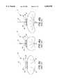
- FIG. 1 and FIG. 2are, respectively, a cross section and cut-away view which illustrate a prior art implant in a human eye, such that the entire plate of the implant is in the posterior segment of the eye;
- FIGS. 3a-3bare perspective views illustrating one embodiment of the implant of the present invention.
- FIG. 3cis a top-plan view of another configuration of the implant of the present invention.
- FIG. 4ais a top-plan view of another configuration of the implant of the present invention.
- FIG. 4bis a top-plan view of still another configuration of the implant of the present invention.
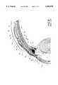
- FIG. 5is a cut-away view which illustrates the implant of FIGS. 3a-3b implanted in a human eye, such that a portion of the plate of the implant extends into the anterior segment of the eye;
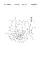
- FIG. 6is a top plan view illustrating the preferred embodiment of the implant
- FIG. 7ais a bottom plan view illustrating the preferred embodiment of the implant.
- FIG. 7bis a cross-sectional view taken along the line 7b--7b of FIG. 7a;
- FIG. 8is a cross-sectional view of the implant of FIG. 6 and FIGS. 7a-7b implanted in the posterior segment of the eye immediately after surgery where both the implant and its placement are prior art;
- FIG. 9is a cross-sectional view of the implant of FIG. 6 and FIGS. 7a-7b implanted as in the prior art in the posterior segment of the eye after bleb formation occurs;
- FIG. 10is a cut-away view which illustrates the implant of FIG. 6 and FIGS. 7a-7b implanted in accordance with the present invention in a human eye, such that a portion of the plate of the implant extends into the anterior segment of the eye;
- FIG. 10ais a cross-sectional view of the implant of FIG. 6 and FIGS. 7a-7b implanted in accordance with the present invention in a human eye, such that a portion of the plate of the implant extends into the anterior segment of the eye after bleb formation occurs;
- FIG. 11is a cut-away view which illustrates an implant similar to the implant of FIG. 6, but with a larger surface area implanted in a human eye, such that a portion of the plate of the implant extends into the anterior segment of the eye;
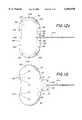
- FIGS. 12a through 12care perspective views illustrating various configurations of the implant of FIG. 6;
- FIG. 12dis a cross-sectional view illustrating another configuration of the implant of FIG. 6 implanted in the posterior segment of the eye immediately after surgery;
- FIG. 12eis a cross-sectional view of the implant of FIG. 12d implanted in the posterior segment of the eye after bleb formation occurs;
- FIG. 13is a perspective view of an additional alternative embodiment of the implant of the present invention.
- FIG. 1 and FIG. 2illustrate an implant 10 positioned within the posterior segment 35 of an eye 12, as known in the prior art.
- the relevant structure of the eye 12will be described briefly below to provide background for the anatomical terms incorporated herein, however, it should be realized that several anatomical details have been omitted for clarity of understanding.
- the tough outer membrane known as the sclera 14covers all of the eye 12 except that portion covered by the cornea 16, the thin, transparent membrane which covers the iris 18 and the pupil 20.
- the cornea 16merges into the sclera 14 at a juncture referred to as the sulcus of the sclera or as the limbus 22.
- a portion of the sclera 14is covered by a thin tissue called the conjunctiva 24.
- the ciliary body 26begins at the limbus 22 and extends along the interior of the sclera 14 and becomes the choroid 28.
- the choroid 28is a brown vascular membrane which extends along the retina back toward the optic nerve.
- the eyeincludes six extraocular eye muscles which control the movement of the eye in the socket.
- the eye musclesinclude the rectus muscles 29 which include lateral, medial, superior, oblique and inferior muscles (the superior and lateral are shown).
- the muscle insertion 27is the point at which the rectus muscles 29 attach to the globe of the eye.
- the dotted-line 31indicates the boundary between the anterior portion of the globe of the eye, also referred to as the anterior segment 34, and the posterior portion of the globe of the eye, also referred to as the posterior segment 35.
- the anterior segment 34is the portion of the globe of the eye which is anterior to the muscle insertions 27.
- the remainder of the globe which is posterior to the muscle insertions 27is considered the posterior segment 35.
- aqueousis produced by the ciliary body 26 and reaches the anterior chamber 30 formed between the iris 18 and the cornea 16 through the pupil 20.
- the aqueousis removed through the trabecular meshwork 32.
- Schlemm's canal 36There the aqueous passes through Schlemm's canal 36 and through veins which merge with blood-carrying veins and into venous circulation.
- Intraocular pressureis maintained in the eye 12 by the intricate balance of secretion and absorption or outflow of the aqueous in the manner described above. Glaucoma results from excessive buildup of aqueous fluid in the anterior chamber 30 which produces an increase in intraocular pressure.
- Implants for treatment of glaucomafacilitate the outflow of the aqueous from the anterior chamber 30 of the eye 12.
- the implant 10comprises a pliable plate or spacer 38, also referred to as a pliable seton in the ophthalmic field, having oppositely disposed first 39 and second 40 curved surfaces, connected to a drainage tube 41 which extends into a first region 42 of the eye 12.
- the seton 38is implanted in a second region 43 of the eye 12 beneath a layer of Tenon's capsule 44 and sutured to the sclera 14. More specifically, the implant illustrated in FIGS. 1-2 is implanted in the posterior segment 35 of the eye.
- the discharge tube 41comprises a first end 46 and a second end 48 wherein the first end 46 is attached to the plate 38 adjacent the first surface 39 of the plate 38.
- the second end 48 of the tube 41extends through the layer of Tenon's capsule 44 and through the cornea 16 into a first region 42 of the eye 12, such as the anterior chamber 30 of the eye 12, to provide fluid communication between the first region 42 and the second region 43 of the eye 12.
- a scleral reinforcing element 50such as a connective tissue graft, i.e., a sclera graft, dura mater graft, fascia lata graft, or a graft formed from other biocompatible materials, covers the exposed portion of the tube 41 located between the Tenon's capsule 44 and the cornea 16.
- a large drainage bleb 52surrounds the seton 38 and lifts the layer of Tenon's capsule 44 above the sclera 14.
- the plate 38acts as a permanent bleb controlling stent to inhibit the tendency of the body to heal itself which would eliminate the bleb. Therefore, the plate 38 is referred to as a bleb formation device or as the bleb formation portion of the implant 10.
- the plate 38is generally spherical in shape and has a perimeter which is elliptical.
- the surface area of the plate 38is preferably in the range of approximately 100 to 600 mm 2 depending on glaucoma conditions and the radius of curvature of the plate 38 is preferably 12 to 14 mm. More preferably, the surface area of the plate 38 is between 250 mm 2 and 450 mm 2 .
- the plate 38When the plate 38 is pressed flat, the plate has a length of between 20-40 mm and a width of 15-20 mm. In the configuration of the plate illustrated in FIG.
- the dimensions of the plate 38 when pressed flatare: a length of 32 mm and a width of 14 mm and the resulting surface area of the plate 38 is 343 mm 2 (+/-7 mm 2 ).
- the length of the plate 38includes a raised ridge 56 formed adjacent one of the larger-radius perimeter edges of the plate 38, on the first curved spherical surface 39 of the plate 38.
- the rounded edge of the plate 38 extending on either side of the raised ridge 56, not including that portion of the plate 38 adjacent the ridge 56,is entirely radiused, tapered and blended so as to facilitate insertion as described below.
- the rounded edge of the plate 38is tapered and blended to discourage the unwanted growth of scar tissue on the plate 38 which may lock the plate 38 into an unwanted position.
- the rounded edge of the plate 38provides a smooth surface from which the scar tissue preferably slides off and is therefore unable to completely anchor onto the plate 38.
- the second surface 40 of the plate 38is curved to conform to the curvature of the eye 12 and the curvature of the ridge 56 matches the curvature of the sclera 14.
- An extension 58 of the plate 38is formed adjacent the ridge 56 in the plate 38 and includes two small suture holes 60, 62.
- the thickness of the plate 38is preferably in the range of 0.5 to 2.0 mm.
- the platemay include fenestrations, or small holes, on the surface of the plate.
- the fenestrations 61are shown in the configurations of the implant shown in FIGS. 3c, 4a and 4b.
- the drainage tube 41is coimected to the plate 38 with adhesive, such as Clear Silicone Rubber Adhesive RTV-118 manufactured by General Electric Silicone Products of Waterford, N.Y., via a small hole 64 formed in the ridge 56 and is bonded to the plate 38 using well-known bonding techniques.
- the first end 46 of the tube 41thus drains into the recess formed at the junction of the ridge 56 and the smooth first surface 39 of the plate 38.
- the plate 38is preferably formed of silicone elastomer, such as SILASTICTM, Medical Grade Q7-4765, 65 Shore A, manufactured by Dow Corning Corporation of Midland, Mich. or Nusil Corp.
- the drainage tube 41is preferably a 1.0 to 3.0 French flow tube, approximately 10 mm to 15 mm in length, formed of SILASTICTM, Medical Grade RX-50, also available from Dow Corning Corporation or Nusil Corp. of Santa Barbara. More preferably, the drainage tube has an inner diameter of 0.30 mm and an outer diameter of 0.60 mm.
- FIGS. 4a-4billustrate various configurations of the plate 38 of the implant 10 of the present invention.
- the dimensions of the plate 38 when pressed flatare: a length of 22 mm and a width of 15 mm and the resulting surface area of the plate 38 is 260 mm 2 (+/-5 mm 2 ).
- the dimensions of the plate 38 when pressed flatare: a length of 36 mm and a width of 16 mm, and the resulting surface area of the plate 38 is 440 mm 2 (+/-9 mm 2 ).
- the implant 10can be inserted into the posterior segment 35 of the eye 12 using known ophthalmological surgical techniques and, with reference to FIG. 1 and FIG. 2, the surgical implant procedure will be briefly described.
- An initial incision 59is made in the conjunctiva 24 and Tenon's capsule 44 parallel to the limbus 22, the incision is stretched to enable the insertion of the implant 10.
- the plate 38is inserted into the second region 43 of the eye 12 through the initial incision 59 and positioned in the posterior segment 35 of the eye beneath the Tenon's capsule 44 and a portion of the superior and lateral rectus muscle 29 or extending totally under one or more of the rectus muscles, thus covering the sclera 14.
- the plate 38can be sutured to the sclera 14, or alternatively, to the rectus muscles 29 if the sclera 14 is thinned by disease, with the suture holes 60,62.
- nonabsorbable nylon suturesare used in the suture holes 60, 62 to secure the plate 38, such as a 7-O or 8-O nylon or polypropylene sutures.
- the drainage tube 41is tunneled out through the sclcra 14 and the cornea 16 beneath Tenon's capsule 44 and in through an incision 65 in the region of the limbus 22 such that the second end 48 of the tube 41 extends into a first region 42, such as the anterior chamber 30, of the eye 12.
- the exposed portion of the drainage tube 41is then covered with a scleral reinforcing element 50.
- the drainage tube 41is sutured closed with a temporary suture(s) 63, 67 at a location on either side of the sclera reinforcing element 50 to prevent any drainage of aqueous prior to formation of the bleb tissue 52 over the plate 38.
- aqueous fluidwill weep through a space formed between the yet to be healed incision 65 and the drainage tube 41. This weeping of the aqueous fluid through the incision 65 relieves some of the fluid pressure until the bleb 52 has formed and the temporary suture(s) 63, 67 is/are removed or absorbed by the body.
- the temporary suture(s) 63is a dissolvable suture while suture 67 is nonabsorbable.
- the temporary sutures 63, 67are removed during a secondary procedure, such as a surgical procedure or an ophthalmic laser procedure. Both procedures are known to those of skill in the art.
- the formation of the bleb 52occurs in response to the introduction of the plate 38 into the tissue of the second region 43 of the eye 12.
- the bleb 52comprises a thin layer of connective tissue which encapsulates the plate 38, and substantially all of the surfaces of the plate 38 contact the tissues in the second region 43 of the eye 12, thus lifting the Tenon's capsule 44 above the sclera 14 as shown.
- bleb 52 formationoccurs in the range of 1 to 8 weeks postoperatively.
- an additional surgerycan be performed at this time to remove the suture(s) 63, 67 from the drainage tube 41 and allow flow of aqueous from the anterior chamber 30 to the bleb 52 via the drainage tube 41.
- a dissolving suturecan be used to seal the drainage tube 41.
- the aqueous flow between the tube 41 and bleb 52is advantageously a patent flow, allowing both flow from the anterior chamber 30 to the bleb 52 and vice versa. This ensures that retrograde non-valved flow from the bleb 52 to the anterior chamber 30, occurring in response to pressure on the eye 12 from the outside, for example, when the lid is forced closed or when the eyeball is pressed on with a finger, does not adversely or harmfully affect intraocular pressure within the eye 12.
- the fluid contained in the bleb 52seeps through the bleb into intercellular spaces within the eye 12 and is then removed through surrounding capillaries or lymphatics.
- the implant 10 of FIG. 4ais implanted in the eye 12 such that a portion of the plate of the implant extends into the anterior segment of the eye.
- An initial incision 59is made in the conjunctiva 24 and Tenon's capsule 44 parallel to the limbus 22.
- the plate 38is inserted into the second region 43 of the eye 12 through the initial incision 59 and placed between Tenon' Capsule 44 and the sclera 14.
- the implant 10is positioned such that at least a portion of the plate 38 extends into the anterior segment 34 of the eye. The remainder of the plate 38, if any, extends into the posterior segment 35 of the eye.
- the surface area of the plate 38 which extends into the anterior segment 34 of the eye 12is between 10 mm 2 and 400 mm 2 depending upon the size of the plate 38. More preferably, the surface area of the plate 38 which extends into the anterior segment 34 of the eye 12 is between 28 mm 2 and 60 mm 2 . Anywhere from 5% to 99% of the surface area of the plate 38 can extend into the anterior segment 34 of the eye 12. Preferably, between 5% and 30% of the surface area of the plate 38 can extend into the anterior segment 34 of the eye 12. A portion of the plate 38 is placed below the superior and lateral rectus muscles 29 or extending under one or more of the rectus muscles 29.
- the plate 38can be sutured to the sclera 14, or alternatively, to the rectus muscles 29 if the sclera 14 is thinned by disease, with the suture holes 60,62.
- nonabsorbable nylon suturesare used in the suture holes 60, 62 to secure the plate 38, such as a 7-O or 8-O nylon or polypropylene sutures.
- the drainage tube 41ais tunneled out through the sclera 14 and the cornea 16 beneath Tenon's capsule 44 and in through an incision 65 in the region of the limbus 22 such that the second end 48 of the tube 41a extends into a first region 42, such as the anterior chamber 30, of the eye 12.
- the drainage tube 41ais sutured closed with a temporary suture(s) 63, 67 (as shown in FIGS. 1 and 2) to prevent any drainage of aqueous prior to formation of the bleb tissue over the plate 38.
- aqueous fluidwill weep through a space formed between the yet to be healed incision 65 and the drainage tube 41a.
- the temporary sutureis a dissolvable suture.
- the temporary sutureis removed during a secondary procedure, such as a surgical procedure or an ophthalmic laser procedure. Both procedures are known to those of skill in the art.
- the insertion of the implantprovides for a simpler insertion procedure.
- the surface area of the plate 38can be increased.
- a plate 38can be formed to cover the sclera 14 in the anterior segment 34 and posterior segment 35 of the eye 12.
- the surface area of the bleb that forms around the plate 38increases. It has been shown that by increasing the surface are of the bleb that covers the sclera 14, the intraocular pressure can be decreased more significantly. Thus, these implants with an increased surface area will be more effective at treating the most severe cases of glaucoma without requiring additional medications.
- the flexible elastomeric material used to form of the present invention, and the size and elliptical shape of the plate 38allows the implant 10 to be inserted much more easily than previously realized with other glaucoma treatment implants.
- the plate 38can be "folded” in half about the axis of the tube 41 and then inserted through the incision 59. Once placed through the incision 59, the plate 38 will return to its original shape and can be positioned to cover the sclera 14, as described above.
- the flexible material from which the plate 38 is formedis soft and pliable which results in much less trauma and irritation to the surrounding tissues and vasculature than experienced with a rigid plate device.
- the plate 38can be folded, a smaller incision can be made in the conjunctive 24 and Tenon's capsule 44.
- the pliable plate 38significantly decreases the surgical procedure length while also minimizing tissue and vasculature damage which can occur in the insertion process.
- the plate 38has a profile shape that is generally spherical and conforms to the contour of the eye.
- the plate 38is shaped like the profile of an elongated soybean.
- the soybean shapeis similar to the elliptical shape of FIGS. 3a-3c with a rearward portion of the plate 38 removed.
- the soybean shapeis preferred as the removed rearward portion of the plate 38 prevents the plate 38 from interfering with the optic nerve.
- the surface area of the plate 38is preferably in the range of approximately 100 to 600 mm 2 depending on glaucoma conditions.
- a sloped wall 66extends from the second surface 40 of the plate 38 in a forward portion 68 of the plate 38.
- the dimensions of the forward portion 68 of the plate 38 in which the sloped wall 66 extendsis from 2 to 20 mm in length and from 2 to 20 mm in width. In the preferred embodiment, the dimensions of the forward portion 68 of the plate are 6 mm in length and 4 mm in width. The dimension of the forward portion 68 of the plate 38 could be larger or smaller than those described above to accommodate a cavity 82 of the desired dimensions.
- the sloped wall 66has a blended and rounded edge to provide a smooth surface from which scar tissue preferably slides off instead of connecting to the wall 66.
- the sloped, blended rounded edge of the wall 66prevents scar tissue from anchoring onto the implant 10 and permanently tethering the plate 38 to the sclera in an undesired position.
- the sloped wall 66is preferably 25 microns to 5 mm thick, i.e., wide and 50 microns to 4 mm in height.
- the tip of the sloped wall 66forms a sealing surface 69 which seals against the sclera 14 upon implantation.
- the drainage tube 41ais connected to the plate 38 of the preferred embodiment along a first notch 70 formed in the first surface 39 of the plate 38, and then through an opening or small hole 72 formed in the plate 38 which opens into the second surface 40 of the plate 38.
- the first end 46 of the tube 41ais bonded to the plate 38 along the notch 70 with adhesive, such as Clcar Silicone Rubber Adhesive RTV-118 manufactured by General Electric Silicone Products of Waterford, N.Y., and using well-known bonding techniques.
- the tube 41ais bonded to the hole 72 in the plate 38 such that the first end 46 of the tube 41a is open to the second surface 40 of the plate 38.
- the drainage tube 41aas shown in FIGS.
- 5-10is preferably a 1.0 to 3.0 French flow tube, approximately 10 mm or less in length, formed of SILASTICTM, Medical Grade RX-50, also available from Dow Corning Corporation or Nusil Corp. of Santa Barbara. More preferably, the drainage tube has an inner diameter of 0.30 mm and an outer diameter of 0.60 mm.
- the drainage tube 41a of the preferred embodimentis less than 8 mm in length, i.e., shorter than the draining tube 41 illustrated in FIGS. 1-4c which is approximately 10 mm to 15 mm in length.
- the drainage tube 41is sized for a plate 38 which is implanted in the posterior segment 35 of the eye 12, while the 41a is sized for implantation such that at least a portion of the plate 38 is positioned in the anterior segment 34 of the eye 12 and therefore does not need to be as long.
- the sloped wall 66completely surrounds the opening 72 in the second surface 40 of the plate 38.
- the sloped wall 66surrounds an area of approximately 0.08 mm 2 -75 mm 2 around the opening 72 in the second surface 40 of the plate 38. More preferably, the sloped wall 66 surrounds an area of approximately 2 mm 2 -14 mm 2 around the opening 72 in the second surface 40 of the plate 38.
- the sloped wall 66is annular in shape to evenly surround the opening 72 and the diameter of the annular shape covered by the wall is preferably between approximately 1 mm to 4 mm, but could be as large as 15 mm.
- the annular sloped wallforms a concave cupped cavity 82 as defined by the sloped wall 66 and the second surface 40 of the plate 38.
- the sloped wall 66may take on a variety of shapes, such as oval, heart shaped, square, rectangular, triangular, etc., in most cases the shape surrounds the opening 72 in the second surface 40 of the plate 38.
- a first plurality of suture holes 74-77are provided around the sloped wall 66. In the preferred embodiment, the first plurality of sutures holes 74-77 are evenly spaced around the perimeter of the forward portion 68 of the plate 38.
- a second plurality of suture holes 78, 80are provided in an intermediate portion 81, proximal to the forward portion 68, of the plate 38 to enable a surgeon to further secure the plate 38 to the sclera 14 (FIG. 8).
- the plate 38is inserted into the second region 43 of the eye and positioned beneath the Tenon's capsule 44 and a portion of the lateral and superior rectus muscles (FIG. 2), thus covering the sclera 14.
- the tip of the sloped wall 66creates a sealing surface 69 against the sclera 14.
- the sealis similar to an o-ring seal.
- the plate 38may not extend under any of the rectus muscles or it may extend under one or more of the rectus muscles (FIG. 2).
- the implant 10lies between the sclera 14 and Tenon's capsule 44.
- Tenon's Capsule 44lies on the upper surface 39 of the plate 38, but Tenon's Capsule 44 does not create a portion of the desired seal.
- the plate 38is sutured to the sclera 14 utilizing both absorbable and nonabsorbable sutures in a first plurality of suture holes 74-77 and absorbable or nonabsorbable sutures in a second plurality of suture holes 78, 80.
- the first plurality of suture holes 74-77are spaced around the sloped wall 66 to provide an enhanced seal of the wall 66 against the sclera 14.
- the second plurality of sutures 78,80 on the immediate portion 81 of the plate 38assist in tethering the plate 38 to the sclera 14.
- the aqueous fluid from the first region 42 of the eye 12drains through the drainage tube 41a into the concave cupped cavity 82 which is sealed against the sclera 14.
- the sutures 74-77assist in sealing the cupped cavity 82 against the sclera 14 at the sealing surface 69; therefore, the aqueous fluid is unable to escape from the cavity 82.
- the fluid pressure within the scaled cavity 82prevents additional fluid from draining from the first region 42 of the eye 12 into the cavity 82, thereby preventing low pressure from occurring in the second region 42 of the eye 12.
- the dimensions of the cavity 82are selected to define the flow of fluid that can exit the first region 42 of the eye 12, before the flow is restricted, without creating a pressure drop in the eye 12. Therefore, the sealing of the wall 66 of the plate 38 against the sclera 14 creates a sealed cavity 82 which temporarily occludes the drainage of aqueous fluid from the first region 42 of the eye 12.
- the o-ring seal effectis achieved both by the design of the implant 10 and the tension produced by the absorbable and nonabsorbable sutures placed by the surgeon in the first plurality of suture holes 74-77 and/or the second plurality of suture holes 78, 80.
- the sutureshold the implant 10 against the sclera 14 with the necessary tension desired by the surgeon.
- the tension level of the suturesis set to withstand pressures of up to 10-20 mm mercury.
- the fluid pressure in the first region 42 of the eye 12reaches an equilibrium pressure with the pressure in the cavity 82 while scar tissue in the eye forms a bleb 52. This equilibrium pressure is less than the pressure applied by the sutures.
- some fluid in the cavity 82may be absorbed by the sclera tissue below the cavity 82. This absorption of fluid will help maintain the desired equilibrium pressure.
- the pressure in the cavity 82 and the first region 42 of the eye 12exceed the tension of the sutures as applied by the surgeon, fluid will leak around the o-ring seal and maintain the eye pressure determined by the surgeon. This prevents the eye pressure from exceeding a specific value, as determined by the tension of the sutures as applied by the surgeon, which could cause damage to the eye. If the fluid pressure does not exceed the tension of the sutures, the o-ring type seal will maintain the fluid pressure within the cavity 82.
- the temporary suturesare dissolvable sutures.
- the temporary suturesare removed during a secondary procedure, such as a surgical procedure or a laser procedure, after scar tissue formation.
- the temporary suturesare selected such that the sutures dissolve after the formation of the scar tissue bleb.
- the suturescan be absorbed at any time postoperatively from 1 day up to 8 weeks.
- the suturesare dissolved in 1 to 6 weeks postoperatively.
- the dissolving suturesare 7-O or 8-O Vicryl. As illustrated in FIG.
- the temporary suturesare removed or absorbed by the body, the fluid pressure in the cupped cavity 82 pushes the plate 38 off the surface of the sclera 14 and the seal formed by the sealing surface 69 of the sloped wall 66 against the sclera 14 is broken.
- the bleb 52eventually fills with fluid and the seton 38 floats within the fluid in the bleb 52 and maintains the bleb shape.
- permanent suturesare utilized in the suture holes 78, 80 on the plate 38 to keep the intermediate end 81 of the seton 38 tethered to the sclera 14 thus preventing the plate 14 from impinging on the eye socket and other tissues in the eye 12 or from extruding.
- a rearward end 84 of the plate 38pivots off of the sclera 14.
- the tethered plate 38acts like a leaflet valve.
- a preferred insertion technique of the implant 10 of the preferred embodimentis used together with known ophthalmological surgical techniques and is briefly described.
- An initial incision 59is made in the conjunctiva 24 and Tenon's capsule 44 parallel to the limbus 22, the incision is stretched slightly to enable the insertion of the implant 10.
- the plate 38is inserted into the second region 43 of the eye and positioned between Tenon's capsule 44 and the sclera 14.
- the implant 10is positioned such that at least a portion of the plate 38 extends into the anterior segment 34 of the eye. The remainder of the plate 38, if any, extends into the posterior segment 35 of the eye.
- the surface area of the plate 38 which extends into the anterior segment 34 of the eye 12is between 10 mm 2 and 400 mm 2 depending upon the size of the plate 38. More preferably, the surface area of the plate 38 which extends into the anterior segment 34 of the eye 12 is between 28 mm 2 and 60 mm 2 . Anywhere from 5% to 99% of the surface area of the plate 38 can extend into the anterior segment 34 of the eye 12. Preferably, between 5% and 30% of the surface area of the plate 38 can extend into the anterior segment 34 of the eye 12. A portion of the plate 38 may be placed below the superior and lateral rectus muscles 29 or extending under one or more of the rectus muscles 29. However, depending upon the surface area of the plate 38, it is possible that no portion of the plate 38 will extend below any of the rectus muscles 29.
- the drainage tube 41ais tunneled out through the sclera 14 and the cornea 16 beneath Tenon's capsule 44 and in through an incision 65 in the region of the limbus 22 such that the second end 48 of the tube 41a extends into a first region 42, such as the anterior chamber 30, of the eye 12.
- the drainage tube 41is sutured closed with a temporary suture(s) to prevent any drainage of aqueous prior to formation of the bleb tissue 52 over the plate 38.
- aqueous fluidwill weep through a space formed between the yet to be healed incision 65 and the drainage tube 41a.
- the temporary suture(s)is/are a dissolvable suture.
- the temporary suturesare removed during a secondary procedure, such as a surgical procedure or an ophthalmic laser procedure. Both procedures are known to those of skill in the art.
- the drainage tube 41ais tunneled through the pars plana which is located 1-3.5 mm behind the limbus into the vitreous cavity (the area behind the lens of the eye).
- the tip of the sloped wall 66creates a sealing surface 69 against the sclera 14.
- the implant 10lies between the sclera 14 and Tenon's capsule 44.
- Tenon's Capsule 44lies on the upper surface 39 of the plate 38, but Tenon's Capsule 44 does not create a portion of the desired seal.
- the plate 38is sutured to the sclera 14 utilizing both absorbable and nonabsorbable sutures in a first plurality of suture holes 74-77 and absorbable or nonabsorbable sutures in a second plurality of suture holes 78, 80.
- the first plurality of suture holes 74-77are spaced around the sloped wall 66 to provide an enhanced seal of the wall 66 against the sclera 14.
- the second plurality of sutures 78,80 on the immediate portion 81 of the plate 38assist in tethering the plate 38 to the sclera 14.
- the aqueous fluid from the first region 42 of the eye 12drains through the drainage tube 41a into the concave cupped cavity 82 which is sealed against the sclera 14.
- the sutures 74-77assist in sealing the cupped cavity 82 against the sclera 14 at the sealing surface 69; therefore, the aqueous fluid is unable to escape from the cavity 82.
- the fluid pressure within the sealed cavity 82prevents additional fluid from draining from the first region 42 of the eye 12 into the cavity 82, thereby preventing low pressure from occurring in the second region 42 of the eye 12.
- the dimensions of the cavity 82are selected to define the flow of fluid that can exit the first region 42 of the eye 12, before the flow is restricted, without creating a pressure drop in the eye 12. Therefore, the sealing of the wall 66 of the plate 38 against the sclera 14 creates a sealed cavity 82 which temporarily occludes the drainage of aqueous fluid from the first region 42 of the eye 12.
- the o-ring seal effectis achieved both by the design of the implant 10 and the tension produced by the absorbable and nonabsorbable sutures placed by the surgeon in the first plurality of suture holes 74-77 and/or the second plurality of suture holes 78, 80.
- the sutureshold the implant 10 against the sclera 14 with the necessary tension desired by the surgeon.
- the tension level of the suturesis set to withstand pressures of up to 10-20 mm mercury.
- the fluid pressure in the first region 42 of the eye 12reaches an equilibrium pressure with the pressure in the cavity 82 while scar tissue in the eye forms a bleb 52. This equilibrium pressure is less than the pressure applied by the sutures.
- some fluid in the cavity 82may be absorbed by the sclera tissue below the cavity 82. This absorption of fluid will help maintain the desired equilibrium pressure.
- the pressure in the cavity 82 and the first region 42 of the eye 12exceed the tension of the sutures as applied by the surgeon, fluid will leak around the o-ring seal and maintain the eye pressure determined by the surgeon. This prevents the eye pressure from exceeding a specific value as determined by the tension of the sutures as applied by the surgeon, which could cause damage to the eye. If the fluid pressure does not exceed the tension of the sutures, the o-ring type seal will maintain the fluid pressure within the cavity 82.
- the temporary sutures of the preferred embodimentare dissolvable sutures.
- the temporary suturesare removed during a secondary procedure, such as a surgical procedure or a laser procedure, after scar tissue formation.
- the temporary suturesare selected such that the sutures dissolve after the formation of the scar tissue bleb.
- the suturescan be absorbed at any time postoperatively from 1 day up to 8 weeks.
- the suturesare dissolved in 1 to 6 weeks postoperatively.
- the dissolving suturesare 7-O or 8-O Vicryl.
- the temporary suturesare removed or absorbed by the body, the fluid pressure in the cupped cavity 82 pushes the plate 38 off the surface of the sclera 14 and the seal formed by the sealing surface 69 of the sloped wall 66 against the sclera 14 is broken.
- the bleb 52eventually fills with fluid and the seton 38 floats within the fluid in the bleb 52 and maintains the bleb shape.
- permanent suturesare utilized in the suture holes 78, 80 on the plate 38 to keep the intermediate end 81 of the seton 38 tethered to the sclera 14 thus preventing the plate 14 from impinging on the eye socket and other tissues in the eye 12 or from extruding.
- a rearward end 84 of the plate 38pivots off of the sclera 14.
- the tethered plate 38acts like a leaflet valve.
- the insertion of the implant 10 such that a portion of the plate 38 extends into the anterior segment 34 of the eyeprovides for a simpler insertion procedure.
- the surface area of the plate 38can be increased.
- a plate 38can be formed to cover a portion of the sclera 14 in the anterior segment 34 and posterior segment 35 of the eye 12.
- the surface area of the bleb that forms around the plate 38increases. It has been shown that by increasing the surface are of the bleb that covers the sclera 14, the intraocular pressure can be decreased more significantly. Thus, these implants with an increased surface area will be more effective at treating the most severe cases of glaucoma without requiring additional medications.
- FIG. 11is a cut-away view which illustrates an implant similar to the implant of FIGS. 6 and 13, but having a plate 38 with a larger surface area implanted in a human eye.
- the plate 38 illustrated in FIG. 11has a surface area of 425 mm 2 .
- the implant 10is positioned in the eye, such that a portion of the plate 38 of the implant 10 extends into the anterior segment 34 of the eye 12.
- a larger surface are of the plate 38extends into the anterior segment 34 of the eye.
- a larger surface are of the plate 38extends below the lateral and superior rectus muscles 29.
- at least 80 mm 2 of the surface area of the plate 38extends into the anterior segment 35 of the eye.
- FIGS. 12a through 12ca variety of configurations of the sloped wall 66 are possible. Although four configurations are illustrated, one skilled in the art will recognize that various other embodiments could be constructed.
- the various configurations of the implant 10 illustrated in FIGS. 12a through 12ccan be implanted in the anterior segment 34 of the eye 12 as described in association with FIGS. 10 and 11 above or in the posterior segment 35 of the eye 12 as described in association with FIGS. 8 and 9 above.
- the concave cavity 82 formed by the sloped wall 66 and the second surface 40 of the plate 38is located in the rearward end 84 of the plate 38.
- the tube 41ais attached along the second surface 40 of the plate 38.
- the tube 41apreferably enters the cavity 82 through a hole (not shown) in the sloped wall 66.
- the first end 46 of the tube 41ais open to the cupped cavity 82 formed by the sloped wall 66 and the second surface 40 of the plate 38.
- the second plurality of suture holes 78-80are located on the perimeter of the forward portion 68 of the plate 38, while the first plurality of suture holes 74-77 are located on a rearward portion 84 of the plate 38 surrounding the sloped wall 66.
- the plate 38is attached to the sclera by temporary sutures in suture holes 74-77 and nonabsorbable sutures in the suture holes 78, 80.
- the temporary sutures in the rearward portion 84 of the plate 38dissolve and the rearward portion 68 of the plate 38 floats in the bleb while the forward portion of the plate 38 remains attached to the sclera.
- a plurality of cavities defined by the sloped walls 66are formed on the second surface 40 of the plate 38.
- First 86 and second 88 cavitiesare connected by a second flexible elastomeric connection tube 90.
- the first end 46 of the drainage tube 41ais open to the first cavity 86.
- a first end 92 of the connection tube 90is open to a hole in the first cavity 86.
- a second end 94 of the connection tube 90is open to a hole in the second cavity 88.
- a third plurality of suture holes 91, 93are located on the perimeter of the rearward portion 84 of the plate 38.
- the plate 38is attached to the sclera by temporary sutures in the first and third plurality of suture holes 74-77 and 91, 93 and nonabsorbable sutures in the second plurality of suture holes 78, 80.
- the temporary suturesare absorbed by the body, or alternatively removed, and the rearward portion 84 and a portion of the forward portion 68 of the plate 38 float while the intermediate portion 81 of the plate 38 remains attached to the sclera.
- one large cavity 96is formed below substantially the entire second surface 40 of the plate 38 by the addition of the sloped wall 66 around the entire perimeter of the second surface 40 of the plate 38.
- Permanent suturesare utilized in the suture holes 98 to affix the forward end 68 of the plate 38 to the sclera.
- a plurality of suture holes 100are evenly dispersed around the remaining perimeter of the plate 38.
- Temporary suturesare used in suture holes 100 to temporarily seal the majority of the perimeter of the plate 33. When the temporary sutures 100 dissolve or alternatively are removed, the rearward portion 84 of the plate 38 lifts off of the sclera 14 to enable patent flow of the aqueous humor from the first region 42 of the eye 12 into the bleb 52.
- the shape of the implant 110is modified to form a cavity 122 within the plate, or seton, 114, rather than including a sloped wall extending from a second surface 116 of the plate 114.
- the plate 114is shown implanted in the posterior 35 segment of the eye 12, but can be implanted in the anterior segment 34 of the eye 12 as described in association with FIGS. 10 and 11 above.
- the plate 114preferably is spherically shaped to match the convex anatomy of the eye and has a matching surface 118 around the perimeter of the concave second surface 116 of the plate 114.
- the plate 114is shaped such that a hemispherical cupped concave cavity 122 is formed around an opening 124 of the tube 41a in the second surface 116 of the plate 114.
- the plate 114does not have to include a hemispherical concave cavity 122 formed in the plate 114. Rather, since the plate 114 is pliable, the plate 114 bends to accommodate the physical anatomical convex shape of the patient's eye 12 such that the matching surface 118 around the perimeter of plate is flush with the surface of the sclera 14.
- a permanent sutureis used in suture hole 125 and a temporary suture is used in suture hole 126 to hold the sealing surface 127 of the seton 114 against the surface of the sclera 14.
- a sealing surface 127 on the matching surface 118 of the plateis formed against the surface of the sclera 14 which temporarily occludes the flow of the aqueous out of the cavity 122.
- the pliable nature of the sutured plate 114will naturally form a cavity 122 of sufficient size to hold the desired amount of fluid. As illustrated in FIG.
- FIG. 13Another alternative embodiment of the present invention is illustrated in FIG. 13 which incorporates a plurality of holes, sometimes referred to as fenestrations, 130 in the rearward portion 84 of the plate 38 which does not contain the cavity 82.
- the plate 38has essentially the same shape as the embodiments illustrated in FIGS. 6, 7a and 7b, with the addition of a plurality of aligned holes 130 in the rearward portion 84 of the plate 38.
- the holes 130do not interfere with the sealed cavity 82 in the forward portion 68 of the plate 38.
- each of the holes 130will form a dimple in the bleb by permitting scar tissue growth through each of the holes 130.
- the overall height of the blebwill be significantly decreased in both the upper and lower directions as the bleb is pulled towards the plate 38 by the growth of scar tissue through each of the holes 130.
- the holes 130are between 50 microns and 10 mm in diameter.
- the diameter of the holes 130decreases proportionally, but will remain within the above range of preferable diameters.
- the number of dimples in the resulting drainage blebwill increase proportionately until the horizontal area of the plate 38 and the diameter of the holes 130 limit the addition of the other holes 130.
- the implant 10 with fenestrations 130 illustrated in FIG. 13can be implanted in the anterior segment 34 of the eye 12 as described in association with FIGS. 10 and 11 above or in the posterior segment 35 of the eye 12 as described in association with FIGS. 8 and 9 above.
Landscapes
- Health & Medical Sciences (AREA)
- Ophthalmology & Optometry (AREA)
- Heart & Thoracic Surgery (AREA)
- Surgery (AREA)
- Engineering & Computer Science (AREA)
- Biomedical Technology (AREA)
- Nuclear Medicine, Radiotherapy & Molecular Imaging (AREA)
- Vascular Medicine (AREA)
- Life Sciences & Earth Sciences (AREA)
- Animal Behavior & Ethology (AREA)
- General Health & Medical Sciences (AREA)
- Public Health (AREA)
- Veterinary Medicine (AREA)
- Prostheses (AREA)
Abstract
Description
Claims (21)
Priority Applications (3)
| Application Number | Priority Date | Filing Date | Title |
|---|---|---|---|
| US08/853,076US6050970A (en) | 1997-05-08 | 1997-05-08 | Method and apparatus for inserting a glaucoma implant in an anterior and posterior segment of the eye |
| PCT/US1998/008951WO1998050092A1 (en) | 1997-05-08 | 1998-05-04 | Method and apparatus for inserting a glaucoma implant in an anterior and posterior segment of the eye |
| AU71751/98AAU7175198A (en) | 1997-05-08 | 1998-05-04 | Method and apparatus for inserting a glaucoma implant in an anterior and posterior segment of the eye |
Applications Claiming Priority (1)
| Application Number | Priority Date | Filing Date | Title |
|---|---|---|---|
| US08/853,076US6050970A (en) | 1997-05-08 | 1997-05-08 | Method and apparatus for inserting a glaucoma implant in an anterior and posterior segment of the eye |
Publications (1)
| Publication Number | Publication Date |
|---|---|
| US6050970Atrue US6050970A (en) | 2000-04-18 |
Family
ID=25314977
Family Applications (1)
| Application Number | Title | Priority Date | Filing Date |
|---|---|---|---|
| US08/853,076Expired - LifetimeUS6050970A (en) | 1997-05-08 | 1997-05-08 | Method and apparatus for inserting a glaucoma implant in an anterior and posterior segment of the eye |
Country Status (3)
| Country | Link |
|---|---|
| US (1) | US6050970A (en) |
| AU (1) | AU7175198A (en) |
| WO (1) | WO1998050092A1 (en) |
Cited By (191)
| Publication number | Priority date | Publication date | Assignee | Title |
|---|---|---|---|---|
| US20020026200A1 (en)* | 2000-08-22 | 2002-02-28 | Savage James A. | Method and apparatus for treatment of glaucoma |
| WO2002032343A2 (en) | 2000-10-18 | 2002-04-25 | Wilcox Michael J | C-shaped cross section tubular ophthalmic implant for reduction of intraocular pressure and method of use |
| WO2002036052A1 (en) | 2000-11-01 | 2002-05-10 | Glaukos Corporation | Glaucoma treatment device |
| US6450984B1 (en) | 1999-04-26 | 2002-09-17 | Gmp Vision Solutions, Inc. | Shunt device and method for treating glaucoma |
| US20020143284A1 (en)* | 2001-04-03 | 2002-10-03 | Hosheng Tu | Drug-releasing trabecular implant for glaucoma treatment |
| US20020169130A1 (en)* | 2001-05-03 | 2002-11-14 | Hosheng Tu | Medical device and methods of use for glaucoma treatment |
| US20020188308A1 (en)* | 2001-04-07 | 2002-12-12 | Hosheng Tu | Glaucoma stent and methods thereof for glaucoma treatment |
| US6510600B2 (en) | 1997-11-20 | 2003-01-28 | Optonol, Ltd. | Method for manufacturing a flow regulating implant |
| US6533768B1 (en) | 2000-04-14 | 2003-03-18 | The Regents Of The University Of California | Device for glaucoma treatment and methods thereof |
| US20030055372A1 (en)* | 1999-04-26 | 2003-03-20 | Lynch Mary G. | Shunt device and method for treating glaucoma |
| US6544208B2 (en) | 2000-12-29 | 2003-04-08 | C. Ross Ethier | Implantable shunt device |
| US20030088260A1 (en)* | 2001-11-08 | 2003-05-08 | Smedley Gregory T. | Combined treatment for cataract and glaucoma treatment |
| US20030097151A1 (en)* | 2001-10-25 | 2003-05-22 | Smedley Gregory T. | Apparatus and mitochondrial treatment for glaucoma |
| US6589203B1 (en)* | 2000-01-26 | 2003-07-08 | Peter Mitrev | Glaucoma drainage device implant |
| US6595945B2 (en) | 2001-01-09 | 2003-07-22 | J. David Brown | Glaucoma treatment device and method |
| US20030153863A1 (en)* | 2002-02-13 | 2003-08-14 | Patel Anilbhai S. | Implant system for glaucoma surgery |
| US20030187385A1 (en)* | 2000-04-14 | 2003-10-02 | Bergheim Olav B. | Implant with anchor |
| US6666841B2 (en) | 2001-05-02 | 2003-12-23 | Glaukos Corporation | Bifurcatable trabecular shunt for glaucoma treatment |
| US20040024345A1 (en)* | 2002-04-19 | 2004-02-05 | Morteza Gharib | Glaucoma implant with valveless flow bias |
| US20040050392A1 (en)* | 2001-08-28 | 2004-03-18 | Hosheng Tu | Glaucoma stent for treating glaucoma and methods of use |
| US20040073156A1 (en)* | 2001-01-09 | 2004-04-15 | Brown J. David | Glaucoma treatment device and method |
| US6726664B2 (en) | 1999-06-02 | 2004-04-27 | Optonol Ltd. | Flow control device, introducer and method of implanting |
| US20040088048A1 (en)* | 1995-05-14 | 2004-05-06 | Jacob Richter | Intraocular implant, delivery device, and method of implantation |
| US20040102729A1 (en)* | 2002-04-08 | 2004-05-27 | David Haffner | Devices and methods for glaucoma treatment |
| US20040111050A1 (en)* | 2000-04-14 | 2004-06-10 | Gregory Smedley | Implantable ocular pump to reduce intraocular pressure |
| US20040127843A1 (en)* | 2000-04-14 | 2004-07-01 | Hosheng Tu | Glaucoma implant with therapeutic agents |
| US20040147870A1 (en)* | 2002-04-08 | 2004-07-29 | Burns Thomas W. | Glaucoma treatment kit |
| US20040193095A1 (en)* | 2003-03-29 | 2004-09-30 | Shadduck John H. | Implants for treating ocular hypertension, methods of use and methods of fabrication |
| US20040254521A1 (en)* | 2003-06-16 | 2004-12-16 | Solx, Inc. | Shunt for the treatment of glaucoma |
| US20040260227A1 (en)* | 2002-12-19 | 2004-12-23 | Lisk James R. | Article and method for ocular aqueous drainage |
| US20050049578A1 (en)* | 2000-04-14 | 2005-03-03 | Hosheng Tu | Implantable ocular pump to reduce intraocular pressure |
| US20050107734A1 (en)* | 2003-11-14 | 2005-05-19 | Coroneo Minas T. | Ocular pressure regulation |
| US20050119636A1 (en)* | 2001-05-02 | 2005-06-02 | David Haffner | Implant with intraocular pressure sensor for glaucoma treatment |
| US20050125059A1 (en)* | 2003-12-05 | 2005-06-09 | Leonard Pinchuk | Ocular lens |
| US20050125003A1 (en)* | 2003-12-05 | 2005-06-09 | Leonard Pinchuk | Glaucoma implant device |
| US20050165385A1 (en)* | 2004-01-22 | 2005-07-28 | Solx, Inc. | Glaucoma treatment method |
| US20050192527A1 (en)* | 2001-05-02 | 2005-09-01 | Morteza Gharib | Glaucoma implant with extending members |
| US20050250788A1 (en)* | 2004-01-30 | 2005-11-10 | Hosheng Tu | Aqueous outflow enhancement with vasodilated aqueous cavity |
| US20050267398A1 (en)* | 2004-05-27 | 2005-12-01 | Dimitri Protopsaltis | Glaucoma shunt |
| US20050266047A1 (en)* | 2002-04-08 | 2005-12-01 | Hosheng Tu | Injectable glaucoma implants with multiple openings |
| US20050271704A1 (en)* | 2002-04-08 | 2005-12-08 | Hosheng Tu | Injectable glaucoma implants with multiple openings |
| US20050277864A1 (en)* | 2000-04-14 | 2005-12-15 | David Haffner | Injectable gel implant for glaucoma treatment |
| US20050288617A1 (en)* | 2004-06-25 | 2005-12-29 | Ira Yaron | Flow regulating implants |
| US20060069340A1 (en)* | 2003-06-16 | 2006-03-30 | Solx, Inc. | Shunt for the treatment of glaucoma |
| US20060155300A1 (en)* | 2002-09-17 | 2006-07-13 | Iscience Surgical, Inc. | Apparatus and method for surgical bypass of aqueous humor |
| US7186232B1 (en) | 2002-03-07 | 2007-03-06 | Glaukoa Corporation | Fluid infusion methods for glaucoma treatment |
| US20070106200A1 (en)* | 2005-11-08 | 2007-05-10 | Brian Levy | Intraocular shunt device and method |
| US20070118066A1 (en)* | 2004-12-03 | 2007-05-24 | Leonard Pinchuk | Glaucoma Implant Device |
| US20070118065A1 (en)* | 2004-12-03 | 2007-05-24 | Leonard Pinchuk | Glaucoma Implant Device |
| US20070123812A1 (en)* | 2004-12-03 | 2007-05-31 | Leonard Pinchuk | Glaucoma Implant Device |
| US20070141116A1 (en)* | 2004-12-03 | 2007-06-21 | Leonard Pinchuk | Glaucoma Implant Device |
| US20070149915A1 (en)* | 2003-05-05 | 2007-06-28 | Judith Yablonski | Internal shunt and method for treating glaucoma |
| US20070156079A1 (en)* | 2005-09-16 | 2007-07-05 | Bg Implant, Inc. | Glaucoma Treatment Devices and Methods |
| US20070191863A1 (en)* | 2006-01-17 | 2007-08-16 | De Juan Eugene Jr | Glaucoma Treatment Device |
| US20070233037A1 (en)* | 2006-01-17 | 2007-10-04 | Gifford Hanson S Iii | Drug Delivery Treatment Device |
| US20070293807A1 (en)* | 2006-05-01 | 2007-12-20 | Lynch Mary G | Dual drainage pathway shunt device and method for treating glaucoma |
| US20070298068A1 (en)* | 2006-06-26 | 2007-12-27 | Badawi David Y | Intraocular implants and methods and kits therefor |
| US20080108933A1 (en)* | 2006-06-30 | 2008-05-08 | Dao-Yi Yu | Methods, Systems and Apparatus for Relieving Pressure in an Organ |
| US20080172204A1 (en)* | 2007-01-15 | 2008-07-17 | Fujitsu Limited | Step counter and method of counting steps |
| US20080277332A1 (en)* | 2007-05-11 | 2008-11-13 | Becton, Dickinson And Company | Micromachined membrane filter device for a glaucoma implant and method for making the same |
| US7488303B1 (en) | 2002-09-21 | 2009-02-10 | Glaukos Corporation | Ocular implant with anchor and multiple openings |
| US20090043321A1 (en)* | 2004-04-29 | 2009-02-12 | Iscience Interventional Corporation | Apparatus And Method For Surgical Enhancement Of Aqueous Humor Drainage |
| US20090082860A1 (en)* | 2007-09-24 | 2009-03-26 | Schieber Andrew T | Ocular Implants with Asymmetric Flexibility |
| US20090132040A1 (en)* | 2007-11-20 | 2009-05-21 | Ivantis, Inc. | Ocular Implant Delivery System and Method |
| US20090177138A1 (en)* | 2007-11-07 | 2009-07-09 | Brown Reay H | Shunt Device for Glaucoma Treatment |
| US20100114006A1 (en)* | 2008-11-05 | 2010-05-06 | Advanced Medical Optics, Inc. | Glaucoma drainage shunts and methods of use |
| US20100121342A1 (en)* | 2007-11-20 | 2010-05-13 | Schieber Andrew T | Methods and Apparatus for Delivering Ocular Implants Into the Eye |
| US20100125237A1 (en)* | 2008-11-20 | 2010-05-20 | Schocket Stanley S | Implant for use in surgery for glaucoma and a method |
| US20100137981A1 (en)* | 2008-06-25 | 2010-06-03 | Silvestrini Thomas A | Ocular implant with shape change capabilities |
| US7740604B2 (en) | 2007-09-24 | 2010-06-22 | Ivantis, Inc. | Ocular implants for placement in schlemm's canal |
| US20100173866A1 (en)* | 2004-04-29 | 2010-07-08 | Iscience Interventional Corporation | Apparatus and method for ocular treatment |
| US20100241046A1 (en)* | 2006-09-06 | 2010-09-23 | Innfocus, Llc | Apparatus, methods and devices for treatment of ocular disorders |
| US20100249691A1 (en)* | 2009-03-26 | 2010-09-30 | Abbott Medical Optics Inc. | Glaucoma shunts with flow management and improved surgical performance |
| US20100274258A1 (en)* | 2009-01-28 | 2010-10-28 | Silvestrini Thomas A | Ocular implant with stiffness qualities, methods of implantation and system |
| US20110009874A1 (en)* | 2009-07-09 | 2011-01-13 | John Wardle | Single Operator Device for Delivering an Ocular Implant |
| US20110009958A1 (en)* | 2009-07-09 | 2011-01-13 | John Wardle | Ocular Implants and Methods for Delivering Ocular Implants Into the Eye |
| US20110029074A1 (en)* | 2009-08-03 | 2011-02-03 | Abbott Medical Optics Inc. | Fixation of ophthalmic implants |
| US20110054601A1 (en)* | 2009-08-27 | 2011-03-03 | Abbott Medical Optics Inc. | Fixation of opthalmic implants |
| US20110105990A1 (en)* | 2009-11-04 | 2011-05-05 | Silvestrini Thomas A | Zonal drug delivery device and method |
| US7951155B2 (en) | 2002-03-15 | 2011-05-31 | Glaukos Corporation | Combined treatment for cataract and glaucoma treatment |
| WO2011085349A2 (en) | 2010-01-11 | 2011-07-14 | Abbott Medical Optics Inc. | Fixation of opthalmic implants |
| US20110196487A1 (en)* | 2010-02-05 | 2011-08-11 | Sight Sciences, Inc. | Intraocular implants and related kits and methods |
| US20110238075A1 (en)* | 2009-12-23 | 2011-09-29 | Luke Clauson | Drug delivery devices and methods |
| US8109896B2 (en) | 2008-02-11 | 2012-02-07 | Optonol Ltd. | Devices and methods for opening fluid passageways |
| US20120184892A1 (en)* | 2011-01-14 | 2012-07-19 | Ecole Polytechnique Federale De Lausanne (Epfl) | Apparatus and methods for treating excess intraocular fluid |
| US8267882B2 (en) | 2008-03-05 | 2012-09-18 | Ivantis, Inc. | Methods and apparatus for treating glaucoma |
| US8308701B2 (en) | 2010-11-15 | 2012-11-13 | Aquesys, Inc. | Methods for deploying intraocular shunts |
| US8313454B2 (en) | 1997-11-20 | 2012-11-20 | Optonol Ltd. | Fluid drainage device, delivery device, and associated methods of use and manufacture |
| US8372026B2 (en) | 2007-09-24 | 2013-02-12 | Ivantis, Inc. | Ocular implant architectures |
| US8425473B2 (en) | 2009-01-23 | 2013-04-23 | Iscience Interventional Corporation | Subretinal access device |
| US8506515B2 (en) | 2006-11-10 | 2013-08-13 | Glaukos Corporation | Uveoscleral shunt and methods for implanting same |
| WO2013155252A1 (en)* | 2012-04-11 | 2013-10-17 | Baylor College Of Medicine | Ophthalmic implant |
| US8585629B2 (en) | 2010-11-15 | 2013-11-19 | Aquesys, Inc. | Systems for deploying intraocular shunts |
| US20130317413A1 (en)* | 2012-05-23 | 2013-11-28 | Leslie A. Field | Pre-Biased Membrane Valve |
| US8657776B2 (en) | 2011-06-14 | 2014-02-25 | Ivantis, Inc. | Ocular implants for delivery into the eye |
| US8663150B2 (en) | 2011-12-19 | 2014-03-04 | Ivantis, Inc. | Delivering ocular implants into the eye |
| US8663303B2 (en) | 2010-11-15 | 2014-03-04 | Aquesys, Inc. | Methods for deploying an intraocular shunt from a deployment device and into an eye |
| US8672870B2 (en) | 2007-07-17 | 2014-03-18 | Transcend Medical, Inc. | Ocular implant with hydrogel expansion capabilities |
| US8721702B2 (en) | 2010-11-15 | 2014-05-13 | Aquesys, Inc. | Intraocular shunt deployment devices |
| US8758290B2 (en) | 2010-11-15 | 2014-06-24 | Aquesys, Inc. | Devices and methods for implanting a shunt in the suprachoroidal space |
| US8765210B2 (en) | 2011-12-08 | 2014-07-01 | Aquesys, Inc. | Systems and methods for making gelatin shunts |
| US8771220B2 (en) | 2011-12-07 | 2014-07-08 | Alcon Research, Ltd. | Glaucoma active pressure regulation shunt |
| US8801766B2 (en) | 2010-11-15 | 2014-08-12 | Aquesys, Inc. | Devices for deploying intraocular shunts |
| US8808222B2 (en) | 2007-11-20 | 2014-08-19 | Ivantis, Inc. | Methods and apparatus for delivering ocular implants into the eye |
| US8828070B2 (en) | 2010-11-15 | 2014-09-09 | Aquesys, Inc. | Devices for deploying intraocular shunts |
| US8852136B2 (en) | 2011-12-08 | 2014-10-07 | Aquesys, Inc. | Methods for placing a shunt into the intra-scleral space |
| US8852137B2 (en) | 2010-11-15 | 2014-10-07 | Aquesys, Inc. | Methods for implanting a soft gel shunt in the suprachoroidal space |
| US8852256B2 (en) | 2010-11-15 | 2014-10-07 | Aquesys, Inc. | Methods for intraocular shunt placement |
| US8888734B2 (en) | 2012-06-05 | 2014-11-18 | Alcon Research, Ltd. | Functionally graded material tube and method for use of the same in implantation |
| US8894603B2 (en) | 2012-03-20 | 2014-11-25 | Sight Sciences, Inc. | Ocular delivery systems and methods |
| US20150018746A1 (en)* | 2012-01-12 | 2015-01-15 | Geuder Ag | Device for use in glaucoma surgery |
| US8974511B2 (en) | 2010-11-15 | 2015-03-10 | Aquesys, Inc. | Methods for treating closed angle glaucoma |
| US9011532B2 (en) | 2009-06-26 | 2015-04-21 | Abbott Medical Optics Inc. | Accommodating intraocular lenses |
| US9017276B2 (en) | 2010-11-15 | 2015-04-28 | Aquesys, Inc. | Shunt placement through the sclera |
| US9039760B2 (en) | 2006-12-29 | 2015-05-26 | Abbott Medical Optics Inc. | Pre-stressed haptic for accommodating intraocular lens |
| US9095411B2 (en) | 2010-11-15 | 2015-08-04 | Aquesys, Inc. | Devices for deploying intraocular shunts |
| US9101444B2 (en) | 2012-01-12 | 2015-08-11 | Innfocus, Inc. | Method, surgical kit and device for treating glaucoma |
| US9125723B2 (en) | 2013-02-19 | 2015-09-08 | Aquesys, Inc. | Adjustable glaucoma implant |
| US9155656B2 (en) | 2012-04-24 | 2015-10-13 | Transcend Medical, Inc. | Delivery system for ocular implant |
| US9198752B2 (en) | 2003-12-15 | 2015-12-01 | Abbott Medical Optics Inc. | Intraocular lens implant having posterior bendable optic |
| US9226851B2 (en) | 2013-08-24 | 2016-01-05 | Novartis Ag | MEMS check valve chip and methods |
| US9271830B2 (en) | 2002-12-05 | 2016-03-01 | Abbott Medical Optics Inc. | Accommodating intraocular lens and method of manufacture thereof |
| US20160067093A1 (en)* | 2014-09-10 | 2016-03-10 | Novartis Ag | Devices, systems and methods for posterior segment drainage |
| US20160067092A1 (en)* | 2014-09-10 | 2016-03-10 | Novartis Ag | Intraocular pressure sensing system for posterior segment drainage |
| US9295389B2 (en) | 2012-12-17 | 2016-03-29 | Novartis Ag | Systems and methods for priming an intraocular pressure sensor in an intraocular implant |
| US9339187B2 (en) | 2011-12-15 | 2016-05-17 | Alcon Research, Ltd. | External pressure measurement system and method for an intraocular implant |
| US9358156B2 (en) | 2012-04-18 | 2016-06-07 | Invantis, Inc. | Ocular implants for delivery into an anterior chamber of the eye |
| US9480598B2 (en) | 2012-09-17 | 2016-11-01 | Novartis Ag | Expanding ocular implant devices and methods |
| US9504560B2 (en) | 2002-01-14 | 2016-11-29 | Abbott Medical Optics Inc. | Accommodating intraocular lens with outer support structure |
| US9510973B2 (en) | 2010-06-23 | 2016-12-06 | Ivantis, Inc. | Ocular implants deployed in schlemm's canal of the eye |
| US9528633B2 (en) | 2012-12-17 | 2016-12-27 | Novartis Ag | MEMS check valve |
| US9572712B2 (en) | 2012-12-17 | 2017-02-21 | Novartis Ag | Osmotically actuated fluidic valve |
| US9579234B2 (en) | 2009-10-23 | 2017-02-28 | Ivantis, Inc. | Ocular implant system and method |
| US9585790B2 (en) | 2013-11-14 | 2017-03-07 | Aquesys, Inc. | Intraocular shunt inserter |
| US9592151B2 (en) | 2013-03-15 | 2017-03-14 | Glaukos Corporation | Systems and methods for delivering an ocular implant to the suprachoroidal space within an eye |
| US9603703B2 (en) | 2009-08-03 | 2017-03-28 | Abbott Medical Optics Inc. | Intraocular lens and methods for providing accommodative vision |
| US9610195B2 (en) | 2013-02-27 | 2017-04-04 | Aquesys, Inc. | Intraocular shunt implantation methods and devices |
| US9615970B2 (en) | 2009-09-21 | 2017-04-11 | Alcon Research, Ltd. | Intraocular pressure sensor with external pressure compensation |
| US9636213B2 (en) | 2005-09-30 | 2017-05-02 | Abbott Medical Optics Inc. | Deformable intraocular lenses and lens systems |
| US9730638B2 (en) | 2013-03-13 | 2017-08-15 | Glaukos Corporation | Intraocular physiological sensor |
| US9763829B2 (en) | 2012-11-14 | 2017-09-19 | Novartis Ag | Flow promoting ocular implant |
| US9808373B2 (en) | 2013-06-28 | 2017-11-07 | Aquesys, Inc. | Intraocular shunt implantation |
| US9814570B2 (en) | 1999-04-30 | 2017-11-14 | Abbott Medical Optics Inc. | Ophthalmic lens combinations |
| US9968441B2 (en) | 2008-03-28 | 2018-05-15 | Johnson & Johnson Surgical Vision, Inc. | Intraocular lens having a haptic that includes a cap |
| US9987125B2 (en) | 2012-05-02 | 2018-06-05 | Johnson & Johnson Surgical Vision, Inc. | Intraocular lens with shape changing capability to provide enhanced accomodation and visual acuity |
| US9987163B2 (en) | 2013-04-16 | 2018-06-05 | Novartis Ag | Device for dispensing intraocular substances |
| US10080682B2 (en) | 2011-12-08 | 2018-09-25 | Aquesys, Inc. | Intrascleral shunt placement |
| US10085884B2 (en) | 2006-06-30 | 2018-10-02 | Aquesys, Inc. | Intraocular devices |
| US10085633B2 (en) | 2012-04-19 | 2018-10-02 | Novartis Ag | Direct visualization system for glaucoma treatment |
| US10159601B2 (en) | 2000-05-19 | 2018-12-25 | Ivantis, Inc. | Delivery system and method of use for the eye |
| US10159600B2 (en) | 2013-02-19 | 2018-12-25 | Aquesys, Inc. | Adjustable intraocular flow regulation |
| US10271989B2 (en) | 2012-03-26 | 2019-04-30 | Glaukos Corporation | System and method for delivering multiple ocular implants |
| US10299958B2 (en) | 2015-03-31 | 2019-05-28 | Sight Sciences, Inc. | Ocular delivery systems and methods |
| US10463537B2 (en) | 2015-06-03 | 2019-11-05 | Aquesys Inc. | Ab externo intraocular shunt placement |
| US10517759B2 (en) | 2013-03-15 | 2019-12-31 | Glaukos Corporation | Glaucoma stent and methods thereof for glaucoma treatment |
| US10596035B2 (en) | 2016-06-06 | 2020-03-24 | Ecole Polytechnique Federale De Lausanne (Epfl) | Apparatus for treating excess intraocular fluid |
| US10603214B2 (en) | 2011-01-14 | 2020-03-31 | Ecole Polytechnique Federale De Lausanne (Epfl) | Apparatus and methods for treating excess intraocular fluid |
| US10617558B2 (en) | 2012-11-28 | 2020-04-14 | Ivantis, Inc. | Apparatus for delivering ocular implants into an anterior chamber of the eye |
| WO2020083824A1 (en) | 2018-10-25 | 2020-04-30 | Amo Groningen B.V. | Bleb control glaucoma shunts |
| CN111132731A (en)* | 2017-09-07 | 2020-05-08 | 光辉疗法公司 | Methods, systems, and compositions for maintaining functional drainage bubbles associated with foreign bodies |
| US10667947B2 (en) | 2016-06-02 | 2020-06-02 | Aquesys, Inc. | Intraocular drug delivery |
| US10709547B2 (en) | 2014-07-14 | 2020-07-14 | Ivantis, Inc. | Ocular implant delivery system and method |
| US10736778B2 (en) | 2014-12-31 | 2020-08-11 | Microoptx Inc. | Glaucoma treatment devices and methods |
| US10842671B2 (en) | 2010-11-15 | 2020-11-24 | Aquesys, Inc. | Intraocular shunt placement in the suprachoroidal space |
| US10952898B2 (en) | 2018-03-09 | 2021-03-23 | Aquesys, Inc. | Intraocular shunt inserter |
| US10959941B2 (en) | 2014-05-29 | 2021-03-30 | Glaukos Corporation | Implants with controlled drug delivery features and methods of using same |
| US10980667B2 (en) | 2015-09-30 | 2021-04-20 | Microoptx Inc. | Eye treatment devices and methods |
| US11116625B2 (en) | 2017-09-28 | 2021-09-14 | Glaukos Corporation | Apparatus and method for controlling placement of intraocular implants |
| US11135089B2 (en) | 2018-03-09 | 2021-10-05 | Aquesys, Inc. | Intraocular shunt inserter |
| US11197779B2 (en) | 2015-08-14 | 2021-12-14 | Ivantis, Inc. | Ocular implant with pressure sensor and delivery system |
| US11246753B2 (en) | 2017-11-08 | 2022-02-15 | Aquesys, Inc. | Manually adjustable intraocular flow regulation |
| US11273325B2 (en) | 2018-11-29 | 2022-03-15 | Radlance Therapeutics, Inc. | Ophthalmic brachytherapy systems and devices for application of beta radiation |
| US11363951B2 (en) | 2011-09-13 | 2022-06-21 | Glaukos Corporation | Intraocular physiological sensor |
| US11504270B1 (en) | 2019-09-27 | 2022-11-22 | Sight Sciences, Inc. | Ocular delivery systems and methods |
| US11540940B2 (en) | 2021-01-11 | 2023-01-03 | Alcon Inc. | Systems and methods for viscoelastic delivery |
| US11666780B2 (en) | 2017-09-07 | 2023-06-06 | Radiance Therapeutics, Inc. | Methods, systems, and compositions for maintaining functioning drainage blebs associated with minimally invasive micro sclerostomy |
| WO2023135547A1 (en) | 2022-01-14 | 2023-07-20 | Liqid Medical Proprietary Limited | Shunt and method for treating glaucoma |
| US11707354B2 (en) | 2017-09-11 | 2023-07-25 | Amo Groningen B.V. | Methods and apparatuses to increase intraocular lenses positional stability |
| US11717440B2 (en) | 2018-01-23 | 2023-08-08 | Avisi Technologies, Inc. | Method and device for treating eye disease |
| US11744734B2 (en) | 2007-09-24 | 2023-09-05 | Alcon Inc. | Method of implanting an ocular implant |
| US11779489B2 (en) | 2020-03-06 | 2023-10-10 | Ecole Polytechnique Federale De Lausanne (Epfl) | Apparatus for treating excess intraocular fluid having an elastic membrane |
| US11925578B2 (en) | 2015-09-02 | 2024-03-12 | Glaukos Corporation | Drug delivery implants with bi-directional delivery capacity |
| US11938058B2 (en) | 2015-12-15 | 2024-03-26 | Alcon Inc. | Ocular implant and delivery system |
| US12005000B2 (en) | 2018-09-06 | 2024-06-11 | Ecole Polytechnique Federale De Lausanne (Epfl) | Apparatus for treating excess intraocular fluid having an elastic membrane |
| US12029683B2 (en) | 2018-02-22 | 2024-07-09 | Alcon Inc. | Ocular implant and delivery system |
| US12150897B2 (en) | 2017-03-17 | 2024-11-26 | W. L. Gore & Associates, Inc. | Delivery aids for glaucoma shunts |
| US12226309B2 (en) | 2013-03-15 | 2025-02-18 | Alcon Inc. | Intraocular lens storage and loading devices and methods of use |
| US12239573B2 (en) | 2018-08-29 | 2025-03-04 | W. L. Gore & Associates, Inc. | Drug therapy delivery systems and methods |
| USD1076086S1 (en) | 2021-11-23 | 2025-05-20 | Radiance Therapeutics, Inc. | Opthalmic brachytherapy device |
| USD1076085S1 (en) | 2021-11-23 | 2025-05-20 | Radiance Therapeutics, Inc. | Opthalmic brachytherapy device |
| US12318279B2 (en) | 2007-07-23 | 2025-06-03 | Alcon Inc. | Lens delivery system |
Families Citing this family (3)
| Publication number | Priority date | Publication date | Assignee | Title |
|---|---|---|---|---|
| WO2005092260A1 (en)* | 2004-03-26 | 2005-10-06 | Molteno Ophthalmic Ltd | Ophthalmic implant for treating glaucoma |
| AR054647A1 (en) | 2005-02-21 | 2007-07-11 | Maldonado Bas Arturo | DEVICE FOR WATER HUMOR DRAINAGE IN GLAUCOMA CASES |
| EP3060180B1 (en)* | 2014-02-24 | 2024-10-09 | National University of Singapore | Ocular drainage device and method of manufacturing thereof |
Citations (37)
| Publication number | Priority date | Publication date | Assignee | Title |
|---|---|---|---|---|
| US2969066A (en)* | 1956-10-02 | 1961-01-24 | Holter Company | Device for draining ventricular fluid in cases of hydrocephalus |
| US3109429A (en)* | 1962-01-30 | 1963-11-05 | Schwartz Samuel | Ventriculo-venous shunt device for treatment of hydrocephalus |
| US3159161A (en)* | 1962-11-14 | 1964-12-01 | Ness Richard Alton | Fistulizing canaliculus |
| US3527226A (en)* | 1966-02-03 | 1970-09-08 | Cordis Corp | Ventricular catheter with valve and pump flushing means |
| US3788327A (en)* | 1971-03-30 | 1974-01-29 | H Donowitz | Surgical implant device |
| FR2233028A1 (en)* | 1973-06-12 | 1975-01-10 | Bonnaud Philippe | Drainer for secretions from wounds - is of rubber strip with channels for plastics tubes |
| US3860008A (en)* | 1973-10-03 | 1975-01-14 | Dow Corning | Flat drain |
| US3915172A (en)* | 1970-05-27 | 1975-10-28 | Ceskoslovenska Akademie Ved | Capillary drain for glaucoma |
| US4030480A (en)* | 1976-05-13 | 1977-06-21 | Ernst Jochen Meyer | Ocular decompression process |
| US4240434A (en)* | 1978-10-10 | 1980-12-23 | Newkirk John B | Peritoneo-venous shunt |
| US4298994A (en)* | 1979-10-26 | 1981-11-10 | Clayman Henry M | Posterior chamber intra-ocular transplant device |
| SU906561A1 (en)* | 1979-11-11 | 1982-02-23 | Военно-Медицинская Ордена Ленина Краснознаменная Академия Им.С.М.Кирова | Apparatus for draining eye anterior chamber |
| GB2101891A (en)* | 1981-07-18 | 1983-01-26 | Anthony Christopher Be Molteno | Device for draining aqueous humour |
| US4373218A (en)* | 1980-11-17 | 1983-02-15 | Schachar Ronald A | Variable power intraocular lens and method of implanting into the posterior chamber |
| US4402681A (en)* | 1980-08-23 | 1983-09-06 | Haas Joseph S | Artificial implant valve for the regulation of intraocular pressure |
| US4428746A (en)* | 1981-07-29 | 1984-01-31 | Antonio Mendez | Glaucoma treatment device |
| EP0102747A1 (en)* | 1982-07-28 | 1984-03-14 | Thomas C. White | Ocular pressure relief device |
| US4457757A (en)* | 1981-07-20 | 1984-07-03 | Molteno Anthony C B | Device for draining aqueous humour |
| US4521210A (en)* | 1982-12-27 | 1985-06-04 | Wong Vernon G | Eye implant for relieving glaucoma, and device and method for use therewith |
| GB2160778A (en)* | 1984-06-28 | 1986-01-02 | Neil Howard Joseph | Aqueous humour drainage device |
| GB2187963A (en)* | 1986-03-07 | 1987-09-23 | Anthony Christopher Be Molteno | Implant for drainage of aqueous humour in glaucoma |
| US4722724A (en)* | 1986-06-23 | 1988-02-02 | Stanley Schocket | Anterior chamber tube shunt to an encircling band, and related surgical procedure |
| US4729761A (en)* | 1985-11-27 | 1988-03-08 | White Thomas C | Tissue-implantable, fluid-dissipating device |
| US4863457A (en)* | 1986-11-24 | 1989-09-05 | Lee David A | Drug delivery device |
| US4886488A (en)* | 1987-08-06 | 1989-12-12 | White Thomas C | Glaucoma drainage the lacrimal system and method |
| US4902292A (en)* | 1987-03-30 | 1990-02-20 | Joseph Neil H | Vitreous body prosthesis device |
| US4915684A (en)* | 1988-06-21 | 1990-04-10 | Mackeen Donald L | Method and apparatus for modulating the flow of lacrimal fluid through a punctum and associated canaliculus |
| US4936825A (en)* | 1988-04-11 | 1990-06-26 | Ungerleider Bruce A | Method for reducing intraocular pressure caused by glaucoma |
| US4946436A (en)* | 1989-11-17 | 1990-08-07 | Smith Stewart G | Pressure-relieving device and process for implanting |
| US4968296A (en)* | 1989-12-20 | 1990-11-06 | Robert Ritch | Transscleral drainage implant device for the treatment of glaucoma |
| US5092837A (en)* | 1989-12-20 | 1992-03-03 | Robert Ritch | Method for the treatment of glaucoma |
| US5300020A (en)* | 1991-05-31 | 1994-04-05 | Medflex Corporation | Surgically implantable device for glaucoma relief |
| US5397300A (en)* | 1990-05-31 | 1995-03-14 | Iovision, Inc. | Glaucoma implant |
| US5476445A (en)* | 1990-05-31 | 1995-12-19 | Iovision, Inc. | Glaucoma implant with a temporary flow restricting seal |
| US5558629A (en)* | 1990-05-31 | 1996-09-24 | Iovision, Inc. | Glaucoma implant |
| US5704907A (en)* | 1994-07-22 | 1998-01-06 | Wound Healing Of Oklahoma | Method and apparatus for lowering the intraocular pressure of an eye |
| US5725493A (en)* | 1994-12-12 | 1998-03-10 | Avery; Robert Logan | Intravitreal medicine delivery |
- 1997
- 1997-05-08USUS08/853,076patent/US6050970A/ennot_activeExpired - Lifetime
- 1998
- 1998-05-04WOPCT/US1998/008951patent/WO1998050092A1/enactiveApplication Filing
- 1998-05-04AUAU71751/98Apatent/AU7175198A/ennot_activeAbandoned
Patent Citations (39)
| Publication number | Priority date | Publication date | Assignee | Title |
|---|---|---|---|---|
| US2969066A (en)* | 1956-10-02 | 1961-01-24 | Holter Company | Device for draining ventricular fluid in cases of hydrocephalus |
| US3109429A (en)* | 1962-01-30 | 1963-11-05 | Schwartz Samuel | Ventriculo-venous shunt device for treatment of hydrocephalus |
| US3159161A (en)* | 1962-11-14 | 1964-12-01 | Ness Richard Alton | Fistulizing canaliculus |
| US3527226A (en)* | 1966-02-03 | 1970-09-08 | Cordis Corp | Ventricular catheter with valve and pump flushing means |
| US3915172A (en)* | 1970-05-27 | 1975-10-28 | Ceskoslovenska Akademie Ved | Capillary drain for glaucoma |
| US3788327A (en)* | 1971-03-30 | 1974-01-29 | H Donowitz | Surgical implant device |
| FR2233028A1 (en)* | 1973-06-12 | 1975-01-10 | Bonnaud Philippe | Drainer for secretions from wounds - is of rubber strip with channels for plastics tubes |
| US3860008A (en)* | 1973-10-03 | 1975-01-14 | Dow Corning | Flat drain |
| US4030480A (en)* | 1976-05-13 | 1977-06-21 | Ernst Jochen Meyer | Ocular decompression process |
| US4240434A (en)* | 1978-10-10 | 1980-12-23 | Newkirk John B | Peritoneo-venous shunt |
| US4298994A (en)* | 1979-10-26 | 1981-11-10 | Clayman Henry M | Posterior chamber intra-ocular transplant device |
| US4298994B1 (en)* | 1979-10-26 | 1991-08-06 | M Clayman Henry | |
| SU906561A1 (en)* | 1979-11-11 | 1982-02-23 | Военно-Медицинская Ордена Ленина Краснознаменная Академия Им.С.М.Кирова | Apparatus for draining eye anterior chamber |
| US4402681A (en)* | 1980-08-23 | 1983-09-06 | Haas Joseph S | Artificial implant valve for the regulation of intraocular pressure |
| US4373218A (en)* | 1980-11-17 | 1983-02-15 | Schachar Ronald A | Variable power intraocular lens and method of implanting into the posterior chamber |
| GB2101891A (en)* | 1981-07-18 | 1983-01-26 | Anthony Christopher Be Molteno | Device for draining aqueous humour |
| US4457757A (en)* | 1981-07-20 | 1984-07-03 | Molteno Anthony C B | Device for draining aqueous humour |
| US4428746A (en)* | 1981-07-29 | 1984-01-31 | Antonio Mendez | Glaucoma treatment device |
| EP0102747A1 (en)* | 1982-07-28 | 1984-03-14 | Thomas C. White | Ocular pressure relief device |
| US4521210A (en)* | 1982-12-27 | 1985-06-04 | Wong Vernon G | Eye implant for relieving glaucoma, and device and method for use therewith |
| EP0168201A1 (en)* | 1984-06-28 | 1986-01-15 | Neil Howard Joseph | Aqueous humour drainage device |
| GB2160778A (en)* | 1984-06-28 | 1986-01-02 | Neil Howard Joseph | Aqueous humour drainage device |
| US4729761A (en)* | 1985-11-27 | 1988-03-08 | White Thomas C | Tissue-implantable, fluid-dissipating device |
| GB2187963A (en)* | 1986-03-07 | 1987-09-23 | Anthony Christopher Be Molteno | Implant for drainage of aqueous humour in glaucoma |
| US4722724A (en)* | 1986-06-23 | 1988-02-02 | Stanley Schocket | Anterior chamber tube shunt to an encircling band, and related surgical procedure |
| US4863457A (en)* | 1986-11-24 | 1989-09-05 | Lee David A | Drug delivery device |
| US4902292A (en)* | 1987-03-30 | 1990-02-20 | Joseph Neil H | Vitreous body prosthesis device |
| US4886488A (en)* | 1987-08-06 | 1989-12-12 | White Thomas C | Glaucoma drainage the lacrimal system and method |
| US4936825A (en)* | 1988-04-11 | 1990-06-26 | Ungerleider Bruce A | Method for reducing intraocular pressure caused by glaucoma |
| US4915684A (en)* | 1988-06-21 | 1990-04-10 | Mackeen Donald L | Method and apparatus for modulating the flow of lacrimal fluid through a punctum and associated canaliculus |
| US4946436A (en)* | 1989-11-17 | 1990-08-07 | Smith Stewart G | Pressure-relieving device and process for implanting |
| US4968296A (en)* | 1989-12-20 | 1990-11-06 | Robert Ritch | Transscleral drainage implant device for the treatment of glaucoma |
| US5092837A (en)* | 1989-12-20 | 1992-03-03 | Robert Ritch | Method for the treatment of glaucoma |
| US5397300A (en)* | 1990-05-31 | 1995-03-14 | Iovision, Inc. | Glaucoma implant |
| US5476445A (en)* | 1990-05-31 | 1995-12-19 | Iovision, Inc. | Glaucoma implant with a temporary flow restricting seal |
| US5558629A (en)* | 1990-05-31 | 1996-09-24 | Iovision, Inc. | Glaucoma implant |
| US5300020A (en)* | 1991-05-31 | 1994-04-05 | Medflex Corporation | Surgically implantable device for glaucoma relief |
| US5704907A (en)* | 1994-07-22 | 1998-01-06 | Wound Healing Of Oklahoma | Method and apparatus for lowering the intraocular pressure of an eye |
| US5725493A (en)* | 1994-12-12 | 1998-03-10 | Avery; Robert Logan | Intravitreal medicine delivery |
Non-Patent Citations (20)
| Title |
|---|
| "Experience with Molteno-type shunts . . . ," Ocular Surgery News, Jun. 1, 1989, pp. 27-29. |
| "Intraocular Pressure," Alder's Physiology of the Eye, Chapter 5, pp. 249-277. |
| "Molteno Seton Implant, for Management of Refractory Glaucoma," Staar Surgical Company, Monrovia, California, one page brochure. |
| Bickford, "Molteno Implant System, for Patient with Previously Unsuccessful Glaucoms Surgery," Journal of Ophthalmic Nursing & Technology, vol. 6, No. 6, 1987, pp. 224-229. |
| Bickford, Molteno Implant System, for Patient with Previously Unsuccessful Glaucoms Surgery, Journal of Ophthalmic Nursing & Technology , vol. 6, No. 6, 1987, pp. 224 229.* |
| Davidovski, et al., "Long-Term Results with the White Glaucoma Pump-Stunt," Ophthalmic Surgery, vol. 21, No. 4 1990, pp. 288-293. |
| Davidovski, et al., Long Term Results with the White Glaucoma Pump Stunt, Ophthalmic Surgery , vol. 21, No. 4 1990, pp. 288 293.* |
| Experience with Molteno type shunts . . . , Ocular Surgery News , Jun. 1, 1989, pp. 27 29.* |
| Intraocular Pressure, Alder s Physiology of the Eye , Chapter 5, pp. 249 277.* |
| Krupin, et al., "Filtering Valve Implant Surgery for Eyes with Neovascular Glaucoma." American Journal of Ophthalmology, vol. 89, No. 3, 1980, pp. 338-343. |
| Krupin, et al., Filtering Valve Implant Surgery for Eyes with Neovascular Glaucoma. American Journal of Ophthalmology , vol. 89, No. 3, 1980, pp. 338 343.* |
| Lee, et al., "Aqueous-Venous Shunt for Glaucoma," Arch Ophthalmol, vol. 99, Nov. 1981, pp. 2007-2012. |
| Lee, et al., Aqueous Venous Shunt for Glaucoma, Arch Ophthalmol , vol. 99, Nov. 1981, pp. 2007 2012.* |
| Minckler, et al., "Clinical Experience with the Single-plate Molteno Implant in Complicated Glaucomas," Ophthalmology, vol. 95, No. 9, Sep. 1988, pp. 1181-1188. |
| Minckler, et al., Clinical Experience with the Single plate Molteno Implant in Complicated Glaucomas, Ophthalmology , vol. 95, No. 9, Sep. 1988, pp. 1181 1188.* |
| Molteno Seton Implant, for Management of Refractory Glaucoma, Staar Surgical Company, Monrovia, California, one page brochure.* |
| Molteno, "Use of Molteno Implants to Treat Secondary Glaucoma," Glaucoma, Grune & Stratton, Ltd., 1986, pp. 211-238. |
| Molteno, Use of Molteno Implants to Treat Secondary Glaucoma, Glaucoma , Grune & Stratton, Ltd., 1986, pp. 211 238.* |
| White, "A New Implantable Ocular Pressure Relief Device," University of South Dakota Medical School, Sioux Falls, San Diego, one page. |
| White, A New Implantable Ocular Pressure Relief Device, University of South Dakota Medical School, Sioux Falls, San Diego, one page.* |
Cited By (513)
| Publication number | Priority date | Publication date | Assignee | Title |
|---|---|---|---|---|
| US7481816B2 (en) | 1995-05-14 | 2009-01-27 | Optonol Ltd. | Intraocular implant, delivery device, and method of implantation |
| US20040088048A1 (en)* | 1995-05-14 | 2004-05-06 | Jacob Richter | Intraocular implant, delivery device, and method of implantation |
| US20030079329A1 (en)* | 1997-11-20 | 2003-05-01 | Ira Yaron | Flow regulating implant, method of manufacture, and delivery device |
| US20080125691A1 (en)* | 1997-11-20 | 2008-05-29 | Optonol Ltd. | Flow regulating implants |
| US8313454B2 (en) | 1997-11-20 | 2012-11-20 | Optonol Ltd. | Fluid drainage device, delivery device, and associated methods of use and manufacture |
| US8486086B2 (en) | 1997-11-20 | 2013-07-16 | Optonol, Ltd | Flow regulating implant, method of manufacture, and delivery device |
| US20100185138A1 (en)* | 1997-11-20 | 2010-07-22 | Optonol Ltd. | Flow regulating implant, method of manufacture, and delivery device |
| US7670310B2 (en) | 1997-11-20 | 2010-03-02 | Optonol Ltd | Flow regulating implants |
| US6510600B2 (en) | 1997-11-20 | 2003-01-28 | Optonol, Ltd. | Method for manufacturing a flow regulating implant |
| US9827143B2 (en) | 1999-04-26 | 2017-11-28 | Glaukos Corporation | Shunt device and method for treating ocular disorders |
| US20050090806A1 (en)* | 1999-04-26 | 2005-04-28 | Gmp Vision Solutions Inc. | Shunt device and method for treating glaucoma |
| US20030055372A1 (en)* | 1999-04-26 | 2003-03-20 | Lynch Mary G. | Shunt device and method for treating glaucoma |
| US10492950B2 (en) | 1999-04-26 | 2019-12-03 | Glaukos Corporation | Shunt device and method for treating ocular disorders |
| US6524275B1 (en) | 1999-04-26 | 2003-02-25 | Gmp Vision Solutions, Inc. | Inflatable device and method for treating glaucoma |
| US8771217B2 (en) | 1999-04-26 | 2014-07-08 | Glaukos Corporation | Shunt device and method for treating ocular disorders |
| US7850637B2 (en) | 1999-04-26 | 2010-12-14 | Glaukos Corporation | Shunt device and method for treating glaucoma |
| US6783544B2 (en) | 1999-04-26 | 2004-08-31 | Gmp Vision Solutions, Inc. | Stent device and method for treating glaucoma |
| US9492320B2 (en) | 1999-04-26 | 2016-11-15 | Glaukos Corporation | Shunt device and method for treating ocular disorders |
| US20050119601A9 (en)* | 1999-04-26 | 2005-06-02 | Lynch Mary G. | Shunt device and method for treating glaucoma |
| US6464724B1 (en) | 1999-04-26 | 2002-10-15 | Gmp Vision Solutions, Inc. | Stent device and method for treating glaucoma |
| US6626858B2 (en) | 1999-04-26 | 2003-09-30 | Gmp Vision Solutions, Inc. | Shunt device and method for treating glaucoma |
| US6450984B1 (en) | 1999-04-26 | 2002-09-17 | Gmp Vision Solutions, Inc. | Shunt device and method for treating glaucoma |
| US10568762B2 (en) | 1999-04-26 | 2020-02-25 | Glaukos Corporation | Stent for treating ocular disorders |
| US8388568B2 (en) | 1999-04-26 | 2013-03-05 | Glaukos Corporation | Shunt device and method for treating ocular disorders |
| US20030236484A1 (en)* | 1999-04-26 | 2003-12-25 | Gmp Vision Solutions, Inc. | Inflatable device and method for treating glaucoma |
| US20050090807A1 (en)* | 1999-04-26 | 2005-04-28 | Gmp Vision Solutions, Inc. | Shunt device and method for treating glaucoma |
| US20110196281A1 (en)* | 1999-04-26 | 2011-08-11 | Glaukos Corporation | Shunt device and method for treating ocular disorders |
| US8152752B2 (en) | 1999-04-26 | 2012-04-10 | Glaukos Corporation | Shunt device and method for treating glaucoma |
| US6827700B2 (en) | 1999-04-26 | 2004-12-07 | Gmp Vision Solutions, Inc. | Shunt device and method for treating glaucoma |
| US6827699B2 (en) | 1999-04-26 | 2004-12-07 | Gmp Vision Solutions, Inc. | Shunt device and method for treating glaucoma |
| US20100004580A1 (en)* | 1999-04-26 | 2010-01-07 | Glaukos Corporation | Shunt device and method for treating ocular disorders |
| US9814570B2 (en) | 1999-04-30 | 2017-11-14 | Abbott Medical Optics Inc. | Ophthalmic lens combinations |
| US6726664B2 (en) | 1999-06-02 | 2004-04-27 | Optonol Ltd. | Flow control device, introducer and method of implanting |
| US6589203B1 (en)* | 2000-01-26 | 2003-07-08 | Peter Mitrev | Glaucoma drainage device implant |
| US20030187385A1 (en)* | 2000-04-14 | 2003-10-02 | Bergheim Olav B. | Implant with anchor |
| US9066782B2 (en) | 2000-04-14 | 2015-06-30 | Dose Medical Corporation | Ocular implant with therapeutic agents and methods thereof |
| US6780164B2 (en) | 2000-04-14 | 2004-08-24 | Glaukos Corporation | L-shaped implant with bi-directional flow |
| US20040127843A1 (en)* | 2000-04-14 | 2004-07-01 | Hosheng Tu | Glaucoma implant with therapeutic agents |
| US20040111050A1 (en)* | 2000-04-14 | 2004-06-10 | Gregory Smedley | Implantable ocular pump to reduce intraocular pressure |
| US20040210185A1 (en)* | 2000-04-14 | 2004-10-21 | Hosheng Tu | Glaucoma implant kit |
| US6736791B1 (en) | 2000-04-14 | 2004-05-18 | Glaukos Corporation | Glaucoma treatment device |
| US20070282245A1 (en)* | 2000-04-14 | 2007-12-06 | Glaukos Corporation | Glaucoma implant with valve |
| US20040249333A1 (en)* | 2000-04-14 | 2004-12-09 | Bergheim Olav B. | Glaucoma implant with bi-directional flow |
| US8333742B2 (en) | 2000-04-14 | 2012-12-18 | Glaukos Corporation | Method of delivering an implant for treating an ocular disorder |
| US20040254519A1 (en)* | 2000-04-14 | 2004-12-16 | Hosheng Tu | Glaucoma treatment device |
| US20080234624A2 (en)* | 2000-04-14 | 2008-09-25 | Glaukos Corporation | Ocular implant with anchor and therapeutic agent |
| US20050049578A1 (en)* | 2000-04-14 | 2005-03-03 | Hosheng Tu | Implantable ocular pump to reduce intraocular pressure |
| US20090137983A1 (en)* | 2000-04-14 | 2009-05-28 | Glaukos Corporation | Implant delivery device and methods thereof for treatment of ocular disorders |
| US8348877B2 (en) | 2000-04-14 | 2013-01-08 | Dose Medical Corporation | Ocular implant with therapeutic agents and methods thereof |
| US6638239B1 (en) | 2000-04-14 | 2003-10-28 | Glaukos Corporation | Apparatus and method for treating glaucoma |
| US9789001B2 (en) | 2000-04-14 | 2017-10-17 | Dose Medical Corporation | Ocular implant with therapeutic agents and methods thereof |
| US7297130B2 (en) | 2000-04-14 | 2007-11-20 | Glaukos Corporation | Implant with anchor |
| US20100010414A1 (en)* | 2000-04-14 | 2010-01-14 | Glaukos Corporation | Method of delivering an implant for treating an ocular disorder |
| US8273050B2 (en) | 2000-04-14 | 2012-09-25 | Glaukos Corporation | Ocular implant with anchor and therapeutic agent |
| US9993368B2 (en) | 2000-04-14 | 2018-06-12 | Glaukos Corporation | System and method for treating an ocular disorder |
| US6533768B1 (en) | 2000-04-14 | 2003-03-18 | The Regents Of The University Of California | Device for glaucoma treatment and methods thereof |
| US7867205B2 (en) | 2000-04-14 | 2011-01-11 | Glaukos Corporation | Method of delivering an implant for treating an ocular disorder |
| US20050209549A1 (en)* | 2000-04-14 | 2005-09-22 | Bergheim Olav B | Glaucoma implant with multiple openings |
| US20050209550A1 (en)* | 2000-04-14 | 2005-09-22 | Bergheim Olav B | Method of treating glaucoma using an implant having a uniform diameter between the anterior chamber and Schlemm's canal |
| US6955656B2 (en) | 2000-04-14 | 2005-10-18 | Glaukos Corporation | Apparatus and method for treating glaucoma |
| US8814820B2 (en) | 2000-04-14 | 2014-08-26 | Glaukos Corporation | Ocular implant with therapeutic agent and methods thereof |
| US20100234790A1 (en)* | 2000-04-14 | 2010-09-16 | Glaukos Corporation | Ocular implant with therapeutic agents and methods thereof |
| US7708711B2 (en) | 2000-04-14 | 2010-05-04 | Glaukos Corporation | Ocular implant with therapeutic agents and methods thereof |
| US8808219B2 (en) | 2000-04-14 | 2014-08-19 | Glaukos Corporation | Implant delivery device and methods thereof for treatment of ocular disorders |
| US20050277864A1 (en)* | 2000-04-14 | 2005-12-15 | David Haffner | Injectable gel implant for glaucoma treatment |
| US8801648B2 (en) | 2000-04-14 | 2014-08-12 | Glaukos Corporation | Ocular implant with anchor and methods thereof |
| US20100056979A1 (en)* | 2000-04-14 | 2010-03-04 | Glaukos Corporation | Implantable ocular pump to reduce intraocular pressure |
| US10485702B2 (en) | 2000-04-14 | 2019-11-26 | Glaukos Corporation | System and method for treating an ocular disorder |
| US10390993B1 (en) | 2000-05-19 | 2019-08-27 | Ivantis, Inc. | Delivery system and method of use for the eye |
| US10335314B2 (en) | 2000-05-19 | 2019-07-02 | Ivantis, Inc. | Delivery system and method of use for the eye |
| US10159601B2 (en) | 2000-05-19 | 2018-12-25 | Ivantis, Inc. | Delivery system and method of use for the eye |
| US10687978B2 (en) | 2000-05-19 | 2020-06-23 | Ivantis, Inc. | Delivery system and method of use for the eye |
| US20020026200A1 (en)* | 2000-08-22 | 2002-02-28 | Savage James A. | Method and apparatus for treatment of glaucoma |
| US6699211B2 (en)* | 2000-08-22 | 2004-03-02 | James A. Savage | Method and apparatus for treatment of glaucoma |
| WO2002032343A2 (en) | 2000-10-18 | 2002-04-25 | Wilcox Michael J | C-shaped cross section tubular ophthalmic implant for reduction of intraocular pressure and method of use |
| WO2002036052A1 (en) | 2000-11-01 | 2002-05-10 | Glaukos Corporation | Glaucoma treatment device |
| US6544208B2 (en) | 2000-12-29 | 2003-04-08 | C. Ross Ethier | Implantable shunt device |
| US20060276739A1 (en)* | 2001-01-09 | 2006-12-07 | Brown J D | Glaucoma treatment device and method |
| US20060079828A1 (en)* | 2001-01-09 | 2006-04-13 | Brown J D | Glaucoma treatment device and method |
| US20100168644A1 (en)* | 2001-01-09 | 2010-07-01 | Brown J David | Glaucoma Treatment Device and Method |
| US6595945B2 (en) | 2001-01-09 | 2003-07-22 | J. David Brown | Glaucoma treatment device and method |
| US6881198B2 (en) | 2001-01-09 | 2005-04-19 | J. David Brown | Glaucoma treatment device and method |
| US20040073156A1 (en)* | 2001-01-09 | 2004-04-15 | Brown J. David | Glaucoma treatment device and method |
| US20020143284A1 (en)* | 2001-04-03 | 2002-10-03 | Hosheng Tu | Drug-releasing trabecular implant for glaucoma treatment |
| US20090138022A1 (en)* | 2001-04-07 | 2009-05-28 | Glaukos Corporation | Ocular implant delivery system and method thereof |
| US20070112292A1 (en)* | 2001-04-07 | 2007-05-17 | Hosheng Tu | Glaucoma stent and methods thereof for glaucoma treatment |
| US7135009B2 (en) | 2001-04-07 | 2006-11-14 | Glaukos Corporation | Glaucoma stent and methods thereof for glaucoma treatment |
| US10406029B2 (en) | 2001-04-07 | 2019-09-10 | Glaukos Corporation | Ocular system with anchoring implant and therapeutic agent |
| US7563241B2 (en) | 2001-04-07 | 2009-07-21 | Glaukos Corporation | Implant and methods thereof for treatment of ocular disorders |
| US10828473B2 (en) | 2001-04-07 | 2020-11-10 | Glaukos Corporation | Ocular implant delivery system and methods thereof |
| US8579846B2 (en) | 2001-04-07 | 2013-11-12 | Glaukos Corporation | Ocular implant systems |
| US20020188308A1 (en)* | 2001-04-07 | 2002-12-12 | Hosheng Tu | Glaucoma stent and methods thereof for glaucoma treatment |
| US8118768B2 (en) | 2001-04-07 | 2012-02-21 | Dose Medical Corporation | Drug eluting ocular implant with anchor and methods thereof |
| US9572963B2 (en) | 2001-04-07 | 2017-02-21 | Glaukos Corporation | Ocular disorder treatment methods and systems |
| US9987472B2 (en) | 2001-04-07 | 2018-06-05 | Glaukos Corporation | Ocular implant delivery systems |
| US7857782B2 (en) | 2001-04-07 | 2010-12-28 | Glaukos Corporation | Ocular implant delivery system and method thereof |
| US8075511B2 (en) | 2001-04-07 | 2011-12-13 | Glaukos Corporation | System for treating ocular disorders and methods thereof |
| US8062244B2 (en) | 2001-04-07 | 2011-11-22 | Glaukos Corporation | Self-trephining implant and methods thereof for treatment of ocular disorders |
| US20090036819A1 (en)* | 2001-04-07 | 2009-02-05 | Glaukos Corporation | Drug eluting ocular implant with anchor and methods thereof |
| EP2982354A1 (en) | 2001-04-07 | 2016-02-10 | Glaukos Corporation | System for treating ocular disorders |
| EP2263621A1 (en) | 2001-04-07 | 2010-12-22 | Glaukos Corporation | System for treating ocular disorders |
| US9155654B2 (en) | 2001-04-07 | 2015-10-13 | Glaukos Corporation | Ocular system with anchoring implant and therapeutic agent |
| EP1977724A1 (en) | 2001-04-07 | 2008-10-08 | Glaukos Corporation | System for treating ocular disorders |
| US8142364B2 (en) | 2001-05-02 | 2012-03-27 | Dose Medical Corporation | Method of monitoring intraocular pressure and treating an ocular disorder |
| US20090076436A2 (en)* | 2001-05-02 | 2009-03-19 | Glaukos Corporation | Ocular implants with deployable structure |
| US20050119636A1 (en)* | 2001-05-02 | 2005-06-02 | David Haffner | Implant with intraocular pressure sensor for glaucoma treatment |
| US6666841B2 (en) | 2001-05-02 | 2003-12-23 | Glaukos Corporation | Bifurcatable trabecular shunt for glaucoma treatment |
| US7678065B2 (en) | 2001-05-02 | 2010-03-16 | Glaukos Corporation | Implant with intraocular pressure sensor for glaucoma treatment |
| US20050192527A1 (en)* | 2001-05-02 | 2005-09-01 | Morteza Gharib | Glaucoma implant with extending members |
| US6981958B1 (en) | 2001-05-02 | 2006-01-03 | Glaukos Corporation | Implant with pressure sensor for glaucoma treatment |
| US20100106073A1 (en)* | 2001-05-02 | 2010-04-29 | Glaukos Corporation | Method of monitoring intraocular pressure and treating an ocular disorder |
| US7273475B2 (en) | 2001-05-03 | 2007-09-25 | Glaukos Corporation | Medical device and methods of use for glaucoma treatment |
| US7094225B2 (en) | 2001-05-03 | 2006-08-22 | Glaukos Corporation | Medical device and methods of use of glaucoma treatment |
| US8337445B2 (en) | 2001-05-03 | 2012-12-25 | Glaukos Corporation | Ocular implant with double anchor mechanism |
| US20020169130A1 (en)* | 2001-05-03 | 2002-11-14 | Hosheng Tu | Medical device and methods of use for glaucoma treatment |
| US20080015488A1 (en)* | 2001-05-03 | 2008-01-17 | Glaukos Corporation | Glaucoma implant with double anchor mechanism |
| US10285856B2 (en) | 2001-08-28 | 2019-05-14 | Glaukos Corporation | Implant delivery system and methods thereof for treating ocular disorders |
| US20040050392A1 (en)* | 2001-08-28 | 2004-03-18 | Hosheng Tu | Glaucoma stent for treating glaucoma and methods of use |
| US20070010827A1 (en)* | 2001-08-28 | 2007-01-11 | Hosheng Tu | Glaucoma stent system |
| US20060241749A1 (en)* | 2001-08-28 | 2006-10-26 | Hosheng Tu | Glaucoma stent system |
| US9561131B2 (en) | 2001-08-28 | 2017-02-07 | Glaukos Corporation | Implant delivery system and methods thereof for treating ocular disorders |
| US7331984B2 (en) | 2001-08-28 | 2008-02-19 | Glaukos Corporation | Glaucoma stent for treating glaucoma and methods of use |
| US7879079B2 (en) | 2001-08-28 | 2011-02-01 | Glaukos Corporation | Implant delivery system and methods thereof for treating ocular disorders |
| US20030097151A1 (en)* | 2001-10-25 | 2003-05-22 | Smedley Gregory T. | Apparatus and mitochondrial treatment for glaucoma |
| US7163543B2 (en) | 2001-11-08 | 2007-01-16 | Glaukos Corporation | Combined treatment for cataract and glaucoma treatment |
| US20030088260A1 (en)* | 2001-11-08 | 2003-05-08 | Smedley Gregory T. | Combined treatment for cataract and glaucoma treatment |
| EP1310222A2 (en) | 2001-11-08 | 2003-05-14 | Glaukos Corporation | Drug-releasing trabecular implant for glaucoma treatment |
| US9504560B2 (en) | 2002-01-14 | 2016-11-29 | Abbott Medical Optics Inc. | Accommodating intraocular lens with outer support structure |
| US20030153863A1 (en)* | 2002-02-13 | 2003-08-14 | Patel Anilbhai S. | Implant system for glaucoma surgery |
| US9220632B2 (en) | 2002-03-07 | 2015-12-29 | Glaukos Corporation | Fluid infusion methods for ocular disorder treatment |
| US7186232B1 (en) | 2002-03-07 | 2007-03-06 | Glaukoa Corporation | Fluid infusion methods for glaucoma treatment |
| US8617094B2 (en) | 2002-03-07 | 2013-12-31 | Glaukos Corporation | Fluid infusion methods for glaucoma treatment |
| US8882781B2 (en) | 2002-03-15 | 2014-11-11 | Glaukos Corporation | Combined treatment for cataract and glaucoma treatment |
| US7951155B2 (en) | 2002-03-15 | 2011-05-31 | Glaukos Corporation | Combined treatment for cataract and glaucoma treatment |
| US9597230B2 (en) | 2002-04-08 | 2017-03-21 | Glaukos Corporation | Devices and methods for glaucoma treatment |
| US7867186B2 (en) | 2002-04-08 | 2011-01-11 | Glaukos Corporation | Devices and methods for treatment of ocular disorders |
| US20040147870A1 (en)* | 2002-04-08 | 2004-07-29 | Burns Thomas W. | Glaucoma treatment kit |
| US20040102729A1 (en)* | 2002-04-08 | 2004-05-27 | David Haffner | Devices and methods for glaucoma treatment |
| US20050271704A1 (en)* | 2002-04-08 | 2005-12-08 | Hosheng Tu | Injectable glaucoma implants with multiple openings |
| US9301875B2 (en) | 2002-04-08 | 2016-04-05 | Glaukos Corporation | Ocular disorder treatment implants with multiple opening |
| US20050266047A1 (en)* | 2002-04-08 | 2005-12-01 | Hosheng Tu | Injectable glaucoma implants with multiple openings |
| US10485701B2 (en) | 2002-04-08 | 2019-11-26 | Glaukos Corporation | Devices and methods for glaucoma treatment |
| US7431710B2 (en) | 2002-04-08 | 2008-10-07 | Glaukos Corporation | Ocular implants with anchors and methods thereof |
| US7879001B2 (en) | 2002-04-08 | 2011-02-01 | Glaukos Corporation | Devices and methods for treatment of ocular disorders |
| US20040024345A1 (en)* | 2002-04-19 | 2004-02-05 | Morteza Gharib | Glaucoma implant with valveless flow bias |
| US7699882B2 (en) | 2002-09-17 | 2010-04-20 | Iscience Interventional Corporation | Apparatus and method for surgical bypass of aqueous humor |
| US20060155300A1 (en)* | 2002-09-17 | 2006-07-13 | Iscience Surgical, Inc. | Apparatus and method for surgical bypass of aqueous humor |
| US7488303B1 (en) | 2002-09-21 | 2009-02-10 | Glaukos Corporation | Ocular implant with anchor and multiple openings |
| US8007459B2 (en) | 2002-09-21 | 2011-08-30 | Glaukos Corporation | Ocular implant with anchoring mechanism and multiple outlets |
| US9271830B2 (en) | 2002-12-05 | 2016-03-01 | Abbott Medical Optics Inc. | Accommodating intraocular lens and method of manufacture thereof |
| US10206773B2 (en) | 2002-12-05 | 2019-02-19 | Johnson & Johnson Surgical Vision, Inc. | Accommodating intraocular lens and method of manufacture thereof |
| US20040260227A1 (en)* | 2002-12-19 | 2004-12-23 | Lisk James R. | Article and method for ocular aqueous drainage |
| US7160264B2 (en) | 2002-12-19 | 2007-01-09 | Medtronic-Xomed, Inc. | Article and method for ocular aqueous drainage |
| US20040193095A1 (en)* | 2003-03-29 | 2004-09-30 | Shadduck John H. | Implants for treating ocular hypertension, methods of use and methods of fabrication |
| US9844462B2 (en) | 2003-05-05 | 2017-12-19 | Novartis Ag | Internal shunt and method for treating glaucoma |
| US8444588B2 (en) | 2003-05-05 | 2013-05-21 | Transcend Medical, Inc. | Internal shunt and method for treating glaucoma |
| US8945038B2 (en) | 2003-05-05 | 2015-02-03 | Transcend Medical, Inc. | Internal shunt and method for treating glaucoma |
| US20070149915A1 (en)* | 2003-05-05 | 2007-06-28 | Judith Yablonski | Internal shunt and method for treating glaucoma |
| US7207965B2 (en) | 2003-06-16 | 2007-04-24 | Solx, Inc. | Shunt for the treatment of glaucoma |
| US20060069340A1 (en)* | 2003-06-16 | 2006-03-30 | Solx, Inc. | Shunt for the treatment of glaucoma |
| US20040254521A1 (en)* | 2003-06-16 | 2004-12-16 | Solx, Inc. | Shunt for the treatment of glaucoma |
| US20050107734A1 (en)* | 2003-11-14 | 2005-05-19 | Coroneo Minas T. | Ocular pressure regulation |
| US9351873B2 (en) | 2003-11-14 | 2016-05-31 | Transcend Medical, Inc. | Ocular pressure regulation |
| US20110028884A1 (en)* | 2003-11-14 | 2011-02-03 | Minas Theodore Coroneo | Ocular pressure regulation |
| US7850638B2 (en) | 2003-11-14 | 2010-12-14 | Transcend Medical, Inc. | Ocular pressure regulation |
| US8486000B2 (en) | 2003-11-14 | 2013-07-16 | Transcend Medical, Inc. | Ocular pressure regulation |
| US8771218B2 (en) | 2003-11-14 | 2014-07-08 | Transcend Medical, Inc. | Ocular pressure regulation |
| US20110087149A1 (en)* | 2003-11-14 | 2011-04-14 | Minas Theodore Coroneo | Ocular pressure regulation |
| US20110087151A1 (en)* | 2003-11-14 | 2011-04-14 | Minas Theodore Coroneo | Ocular pressure regulation |
| US8128588B2 (en) | 2003-11-14 | 2012-03-06 | Transcend Medical, Inc. | Ocular pressure regulation |
| US20070106235A1 (en)* | 2003-11-14 | 2007-05-10 | Coroneo Minas T | Ocular Pressure Regulation |
| US8808220B2 (en) | 2003-11-14 | 2014-08-19 | Transcend Medical, Inc. | Ocular pressure regulation |
| US10226380B2 (en) | 2003-11-14 | 2019-03-12 | Novartis Ag | Ocular pressure regulation |
| US8758289B2 (en) | 2003-11-14 | 2014-06-24 | Transcend Medical, Inc. | Ocular pressure regulation |
| US8728021B2 (en) | 2003-11-14 | 2014-05-20 | Transcend Medical, Inc. | Ocular pressure regulation |
| US7291125B2 (en) | 2003-11-14 | 2007-11-06 | Transcend Medical, Inc. | Ocular pressure regulation |
| US7815592B2 (en) | 2003-11-14 | 2010-10-19 | Transcend Medical, Inc. | Ocular pressure regulation |
| US7794498B2 (en)* | 2003-12-05 | 2010-09-14 | Innolene Llc | Ocular lens |
| US20050125059A1 (en)* | 2003-12-05 | 2005-06-09 | Leonard Pinchuk | Ocular lens |
| US20050125003A1 (en)* | 2003-12-05 | 2005-06-09 | Leonard Pinchuk | Glaucoma implant device |
| US20090312836A1 (en)* | 2003-12-05 | 2009-12-17 | Leonard Pinchuk | Ocular Lens |
| US9198752B2 (en) | 2003-12-15 | 2015-12-01 | Abbott Medical Optics Inc. | Intraocular lens implant having posterior bendable optic |
| US20050165385A1 (en)* | 2004-01-22 | 2005-07-28 | Solx, Inc. | Glaucoma treatment method |
| US7282046B2 (en) | 2004-01-22 | 2007-10-16 | Peter M. Adams, Doug P. Adams, and John Sullivan, Collectively as the Stockholder Representative Committee | Glaucoma treatment method |
| US20050250788A1 (en)* | 2004-01-30 | 2005-11-10 | Hosheng Tu | Aqueous outflow enhancement with vasodilated aqueous cavity |
| US20090043321A1 (en)* | 2004-04-29 | 2009-02-12 | Iscience Interventional Corporation | Apparatus And Method For Surgical Enhancement Of Aqueous Humor Drainage |
| US20100173866A1 (en)* | 2004-04-29 | 2010-07-08 | Iscience Interventional Corporation | Apparatus and method for ocular treatment |
| US20050267398A1 (en)* | 2004-05-27 | 2005-12-01 | Dimitri Protopsaltis | Glaucoma shunt |
| US20080077071A1 (en)* | 2004-06-25 | 2008-03-27 | Optonol Ltd. | Flow Regulating Implants |
| US8034016B2 (en) | 2004-06-25 | 2011-10-11 | Optonol, Ltd. | Flow regulating implants and methods of implanting |
| US7862531B2 (en) | 2004-06-25 | 2011-01-04 | Optonol Ltd. | Flow regulating implants |
| US20050288617A1 (en)* | 2004-06-25 | 2005-12-29 | Ira Yaron | Flow regulating implants |
| US7837644B2 (en) | 2004-12-03 | 2010-11-23 | Innfocus, Llc | Glaucoma implant device |
| US20070123812A1 (en)* | 2004-12-03 | 2007-05-31 | Leonard Pinchuk | Glaucoma Implant Device |
| US20070118065A1 (en)* | 2004-12-03 | 2007-05-24 | Leonard Pinchuk | Glaucoma Implant Device |
| US7594899B2 (en) | 2004-12-03 | 2009-09-29 | Innfocus, Llc | Glaucoma implant device |
| US20070118066A1 (en)* | 2004-12-03 | 2007-05-24 | Leonard Pinchuk | Glaucoma Implant Device |
| US20070141116A1 (en)* | 2004-12-03 | 2007-06-21 | Leonard Pinchuk | Glaucoma Implant Device |
| US20070156079A1 (en)* | 2005-09-16 | 2007-07-05 | Bg Implant, Inc. | Glaucoma Treatment Devices and Methods |
| US9636213B2 (en) | 2005-09-30 | 2017-05-02 | Abbott Medical Optics Inc. | Deformable intraocular lenses and lens systems |
| US20070106200A1 (en)* | 2005-11-08 | 2007-05-10 | Brian Levy | Intraocular shunt device and method |
| US8801649B2 (en) | 2006-01-17 | 2014-08-12 | Transcend Medical, Inc. | Glaucoma treatment device |
| US9421130B2 (en) | 2006-01-17 | 2016-08-23 | Novartis Ag. | Glaucoma treatment device |
| US8734378B2 (en) | 2006-01-17 | 2014-05-27 | Transcend Medical, Inc. | Glaucoma treatment device |
| US11786402B2 (en) | 2006-01-17 | 2023-10-17 | Alcon Inc. | Glaucoma treatment device |
| US20110028883A1 (en)* | 2006-01-17 | 2011-02-03 | Juan Jr Eugene De | Glaucoma treatment device |
| US10905590B2 (en) | 2006-01-17 | 2021-02-02 | Alcon Inc. | Glaucoma treatment device |
| US8721656B2 (en) | 2006-01-17 | 2014-05-13 | Transcend Medical, Inc. | Glaucoma treatment device |
| US9084662B2 (en) | 2006-01-17 | 2015-07-21 | Transcend Medical, Inc. | Drug delivery treatment device |
| US12303430B2 (en) | 2006-01-17 | 2025-05-20 | Alcon Inc. | Glaucoma treatment device |
| US20070191863A1 (en)* | 2006-01-17 | 2007-08-16 | De Juan Eugene Jr | Glaucoma Treatment Device |
| US9668917B2 (en) | 2006-01-17 | 2017-06-06 | Novartis Ag | Drug delivery treatment device |
| US9398977B2 (en) | 2006-01-17 | 2016-07-26 | Transcend Medical, Inc. | Glaucoma treatment device |
| US20070233037A1 (en)* | 2006-01-17 | 2007-10-04 | Gifford Hanson S Iii | Drug Delivery Treatment Device |
| US9789000B2 (en) | 2006-01-17 | 2017-10-17 | Novartis Ag | Glaucoma treatment device |
| US8814819B2 (en) | 2006-01-17 | 2014-08-26 | Transcend Medical, Inc. | Glaucoma treatment device |
| US20070293807A1 (en)* | 2006-05-01 | 2007-12-20 | Lynch Mary G | Dual drainage pathway shunt device and method for treating glaucoma |
| US20070298068A1 (en)* | 2006-06-26 | 2007-12-27 | Badawi David Y | Intraocular implants and methods and kits therefor |
| US20110130831A1 (en)* | 2006-06-26 | 2011-06-02 | Badawi David Y | Intraocular implants and methods and kits therefor |
| US10398597B2 (en) | 2006-06-26 | 2019-09-03 | Sight Sciences, Inc. | Intraocular implants and methods and kits therefor |
| US11389328B2 (en) | 2006-06-26 | 2022-07-19 | Sight Sciences, Inc. | Intraocular implants and methods and kits therefor |
| US20100191329A1 (en)* | 2006-06-26 | 2010-07-29 | Badawi David Y | Intraocular implants and methods and kits therefor |
| US9370443B2 (en) | 2006-06-26 | 2016-06-21 | Sight Sciences, Inc. | Intraocular implants and methods and kits therefor |
| US7909789B2 (en) | 2006-06-26 | 2011-03-22 | Sight Sciences, Inc. | Intraocular implants and methods and kits therefor |
| US11865041B2 (en) | 2006-06-26 | 2024-01-09 | Sight Sciences, Inc. | Intraocular implants and methods and kits therefor |
| US8287482B2 (en) | 2006-06-26 | 2012-10-16 | Sight Sciences, Inc. | Intraocular implants and methods and kits therefor |
| US10314742B2 (en) | 2006-06-26 | 2019-06-11 | Sight Sciences, Inc. | Intraocular implants and methods and kits therefor |
| US9486361B2 (en) | 2006-06-26 | 2016-11-08 | Sight Sciences, Inc. | Intraocular implants and methods and kits therefor |
| US20100121248A1 (en)* | 2006-06-30 | 2010-05-13 | Aquesys, Inc. | Apparatus for reducing pressure in an organ |
| US20080108933A1 (en)* | 2006-06-30 | 2008-05-08 | Dao-Yi Yu | Methods, Systems and Apparatus for Relieving Pressure in an Organ |
| US9636254B2 (en) | 2006-06-30 | 2017-05-02 | Aquesys, Inc. | Systems for reducing pressure in an organ |
| US20100119696A1 (en)* | 2006-06-30 | 2010-05-13 | Aquesys, Inc. | Manufacture of an organ implant |
| US10085884B2 (en) | 2006-06-30 | 2018-10-02 | Aquesys, Inc. | Intraocular devices |
| US20100241046A1 (en)* | 2006-09-06 | 2010-09-23 | Innfocus, Llc | Apparatus, methods and devices for treatment of ocular disorders |
| US9962290B2 (en) | 2006-11-10 | 2018-05-08 | Glaukos Corporation | Uveoscleral shunt and methods for implanting same |
| US8506515B2 (en) | 2006-11-10 | 2013-08-13 | Glaukos Corporation | Uveoscleral shunt and methods for implanting same |
| US10828195B2 (en) | 2006-11-10 | 2020-11-10 | Glaukos Corporation | Uveoscleral shunt and methods for implanting same |
| US12186237B2 (en) | 2006-11-10 | 2025-01-07 | Glaukos Corporation | Uveoscleral shunt and methods for implanting same |
| US9039760B2 (en) | 2006-12-29 | 2015-05-26 | Abbott Medical Optics Inc. | Pre-stressed haptic for accommodating intraocular lens |
| US20080172204A1 (en)* | 2007-01-15 | 2008-07-17 | Fujitsu Limited | Step counter and method of counting steps |
| US20080277332A1 (en)* | 2007-05-11 | 2008-11-13 | Becton, Dickinson And Company | Micromachined membrane filter device for a glaucoma implant and method for making the same |
| US9585789B2 (en) | 2007-07-17 | 2017-03-07 | Novartis Ag | Ocular implant with hydrogel expansion capabilities |
| US8672870B2 (en) | 2007-07-17 | 2014-03-18 | Transcend Medical, Inc. | Ocular implant with hydrogel expansion capabilities |
| US12318279B2 (en) | 2007-07-23 | 2025-06-03 | Alcon Inc. | Lens delivery system |
| US9402767B2 (en) | 2007-09-24 | 2016-08-02 | Ivantis, Inc. | Ocular implant architectures |
| US9610196B2 (en) | 2007-09-24 | 2017-04-04 | Ivantis, Inc. | Ocular implants with asymmetric flexibility |
| US12016796B2 (en) | 2007-09-24 | 2024-06-25 | Alcon Inc. | Methods and devices for increasing aqueous humor outflow |
| US20090082860A1 (en)* | 2007-09-24 | 2009-03-26 | Schieber Andrew T | Ocular Implants with Asymmetric Flexibility |
| US8372026B2 (en) | 2007-09-24 | 2013-02-12 | Ivantis, Inc. | Ocular implant architectures |
| US8414518B2 (en) | 2007-09-24 | 2013-04-09 | Ivantis, Inc. | Glaucoma treatment method |
| US11744734B2 (en) | 2007-09-24 | 2023-09-05 | Alcon Inc. | Method of implanting an ocular implant |
| US8282592B2 (en) | 2007-09-24 | 2012-10-09 | Ivantis, Inc. | Glaucoma treatment method |
| US8734377B2 (en) | 2007-09-24 | 2014-05-27 | Ivantis, Inc. | Ocular implants with asymmetric flexibility |
| US7740604B2 (en) | 2007-09-24 | 2010-06-22 | Ivantis, Inc. | Ocular implants for placement in schlemm's canal |
| US9039650B2 (en) | 2007-09-24 | 2015-05-26 | Ivantis, Inc. | Ocular implants with asymmetric flexibility |
| US20100222733A1 (en)* | 2007-09-24 | 2010-09-02 | Schieber Andrew T | Glaucoma Treatment Method |
| US8961447B2 (en) | 2007-09-24 | 2015-02-24 | Ivantis, Inc. | Glaucoma treatment method |
| US20090177138A1 (en)* | 2007-11-07 | 2009-07-09 | Brown Reay H | Shunt Device for Glaucoma Treatment |
| US9351874B2 (en) | 2007-11-20 | 2016-05-31 | Ivantis, Inc. | Methods and apparatus for delivering ocular implants into the eye |
| US8337509B2 (en) | 2007-11-20 | 2012-12-25 | Ivantis, Inc. | Methods and apparatus for delivering ocular implants into the eye |
| US20090132040A1 (en)* | 2007-11-20 | 2009-05-21 | Ivantis, Inc. | Ocular Implant Delivery System and Method |
| US8551166B2 (en) | 2007-11-20 | 2013-10-08 | Ivantis, Inc. | Methods and apparatus for delivering ocular implants into the eye |
| US8512404B2 (en) | 2007-11-20 | 2013-08-20 | Ivantis, Inc. | Ocular implant delivery system and method |
| US20100121342A1 (en)* | 2007-11-20 | 2010-05-13 | Schieber Andrew T | Methods and Apparatus for Delivering Ocular Implants Into the Eye |
| US9226852B2 (en) | 2007-11-20 | 2016-01-05 | Ivantis, Inc. | Methods and apparatus for delivering ocular implants into the eye |
| US9050169B2 (en) | 2007-11-20 | 2015-06-09 | Ivantis, Inc. | Methods and apparatus for delivering ocular implants into the eye |
| US8808222B2 (en) | 2007-11-20 | 2014-08-19 | Ivantis, Inc. | Methods and apparatus for delivering ocular implants into the eye |
| US8109896B2 (en) | 2008-02-11 | 2012-02-07 | Optonol Ltd. | Devices and methods for opening fluid passageways |
| US8529494B2 (en) | 2008-03-05 | 2013-09-10 | Ivantis, Inc. | Methods and apparatus for treating glaucoma |
| US8267882B2 (en) | 2008-03-05 | 2012-09-18 | Ivantis, Inc. | Methods and apparatus for treating glaucoma |
| US9693902B2 (en) | 2008-03-05 | 2017-07-04 | Ivantis, Inc. | Methods and apparatus for treating glaucoma |
| US10537474B2 (en) | 2008-03-05 | 2020-01-21 | Ivantis, Inc. | Methods and apparatus for treating glaucoma |
| US9066783B2 (en) | 2008-03-05 | 2015-06-30 | Ivantis, Inc. | Methods and apparatus for treating glaucoma |
| US9968441B2 (en) | 2008-03-28 | 2018-05-15 | Johnson & Johnson Surgical Vision, Inc. | Intraocular lens having a haptic that includes a cap |
| US20100137981A1 (en)* | 2008-06-25 | 2010-06-03 | Silvestrini Thomas A | Ocular implant with shape change capabilities |
| US8617139B2 (en) | 2008-06-25 | 2013-12-31 | Transcend Medical, Inc. | Ocular implant with shape change capabilities |
| US10016301B2 (en) | 2008-06-25 | 2018-07-10 | Novartis Ag | Ocular implant with shape change capabilities |
| WO2010054035A1 (en)* | 2008-11-05 | 2010-05-14 | Abbott Medical Optics Inc. | Glaucoma drainage shunts and methods of use |
| US9468558B2 (en) | 2008-11-05 | 2016-10-18 | Abbott Medical Optics Inc. | Glaucoma drainage shunts and methods of use |
| US20170020731A1 (en)* | 2008-11-05 | 2017-01-26 | Abbott Medical Optics Inc. | Glaucoma drainage shunts and methods of use |
| US8353856B2 (en) | 2008-11-05 | 2013-01-15 | Abbott Medical Optics Inc. | Glaucoma drainage shunts and methods of use |
| US20100114006A1 (en)* | 2008-11-05 | 2010-05-06 | Advanced Medical Optics, Inc. | Glaucoma drainage shunts and methods of use |
| US8920357B2 (en) | 2008-11-05 | 2014-12-30 | Abbott Medical Optics Inc. | Glaucoma drainage shunts and methods of use |
| US10492948B2 (en) | 2008-11-05 | 2019-12-03 | Johnson & Johnson Surgical Vision, Inc. | Glaucoma drainage shunts and methods of use |
| WO2010059178A1 (en)* | 2008-11-20 | 2010-05-27 | Schocket Stanley S | An implant for use in surgery for glaucoma and a method |
| US20100125237A1 (en)* | 2008-11-20 | 2010-05-20 | Schocket Stanley S | Implant for use in surgery for glaucoma and a method |
| US8079972B2 (en)* | 2008-11-20 | 2011-12-20 | Schocket Stanley S | Implant for use in surgery for glaucoma and a method |
| US8425473B2 (en) | 2009-01-23 | 2013-04-23 | Iscience Interventional Corporation | Subretinal access device |
| US20110028983A1 (en)* | 2009-01-28 | 2011-02-03 | Silvestrini Thomas A | Ocular implant with stiffness qualities, methods of implantation and system |
| US12233004B2 (en) | 2009-01-28 | 2025-02-25 | Alcon Inc. | Ocular implant with stiffness qualities, methods of implantation and system |
| US8574294B2 (en) | 2009-01-28 | 2013-11-05 | Transcend Medical, Inc. | Ocular implant with stiffness qualities, methods of implantation and system |
| US20110087148A1 (en)* | 2009-01-28 | 2011-04-14 | Silvestrini Thomas A | Ocular implant with stiffness qualities, methods of implantation and system |
| US10531983B2 (en) | 2009-01-28 | 2020-01-14 | Novartis Ag | Ocular implant with stiffness qualities, methods of implantation and system |
| US20100274258A1 (en)* | 2009-01-28 | 2010-10-28 | Silvestrini Thomas A | Ocular implant with stiffness qualities, methods of implantation and system |
| US8167939B2 (en) | 2009-01-28 | 2012-05-01 | Transcend Medical, Inc. | Ocular implant with stiffness qualities, methods of implantation and system |
| US11839571B2 (en) | 2009-01-28 | 2023-12-12 | Alcon Inc. | Ocular implant with stiffness qualities, methods of implantation and system |
| US8377122B2 (en) | 2009-01-28 | 2013-02-19 | Transcend Medical, Inc. | Ocular implant with stiffness qualities, methods of implantation and system |
| US9763828B2 (en) | 2009-01-28 | 2017-09-19 | Novartis Ag | Ocular implant with stiffness qualities, methods of implantation and system |
| US11344448B2 (en) | 2009-01-28 | 2022-05-31 | Alcon Inc. | Ocular implant with stiffness qualities, methods of implantation and system |
| US8262726B2 (en) | 2009-01-28 | 2012-09-11 | Transcend Medical, Inc. | Ocular implant with stiffness qualities, methods of implantation and system |
| US8172899B2 (en) | 2009-01-28 | 2012-05-08 | Transcend Medical, Inc. | Ocular implant with stiffness qualities, methods of implantation and system |
| US8702639B2 (en) | 2009-03-26 | 2014-04-22 | Abbott Medical Optics Inc. | Glaucoma shunts with flow management and improved surgical performance |
| EP2789318A2 (en) | 2009-03-26 | 2014-10-15 | Abbott Medical Optics Inc. | Glaucoma shunts with flow management and improved surgical performance |
| US20100249691A1 (en)* | 2009-03-26 | 2010-09-30 | Abbott Medical Optics Inc. | Glaucoma shunts with flow management and improved surgical performance |
| US10052194B2 (en) | 2009-06-26 | 2018-08-21 | Johnson & Johnson Surgical Vision, Inc. | Accommodating intraocular lenses |
| US9011532B2 (en) | 2009-06-26 | 2015-04-21 | Abbott Medical Optics Inc. | Accommodating intraocular lenses |
| US8425449B2 (en) | 2009-07-09 | 2013-04-23 | Ivantis, Inc. | Ocular implants and methods for delivering ocular implants into the eye |
| US20110009958A1 (en)* | 2009-07-09 | 2011-01-13 | John Wardle | Ocular Implants and Methods for Delivering Ocular Implants Into the Eye |
| US11596546B2 (en) | 2009-07-09 | 2023-03-07 | Alcon Inc. | Ocular implants and methods for delivering ocular implants into the eye |
| US9211213B2 (en) | 2009-07-09 | 2015-12-15 | Ivantis, Inc. | Ocular implants and methods for delivering ocular implants into the eye |
| US10492949B2 (en) | 2009-07-09 | 2019-12-03 | Ivantis, Inc. | Single operator device for delivering an ocular implant |
| US9693899B2 (en) | 2009-07-09 | 2017-07-04 | Ivantis, Inc. | Single operator device for delivering an ocular implant |
| US10406025B2 (en) | 2009-07-09 | 2019-09-10 | Ivantis, Inc. | Ocular implants and methods for delivering ocular implants into the eye |
| US11464675B2 (en) | 2009-07-09 | 2022-10-11 | Alcon Inc. | Single operator device for delivering an ocular implant |
| US12409067B2 (en) | 2009-07-09 | 2025-09-09 | Alcon Inc. | Single operator device for delivering an ocular implant |
| US11918514B2 (en) | 2009-07-09 | 2024-03-05 | Alcon Inc. | Single operator device for delivering an ocular implant |
| US20110009874A1 (en)* | 2009-07-09 | 2011-01-13 | John Wardle | Single Operator Device for Delivering an Ocular Implant |
| US20110029074A1 (en)* | 2009-08-03 | 2011-02-03 | Abbott Medical Optics Inc. | Fixation of ophthalmic implants |
| US9603703B2 (en) | 2009-08-03 | 2017-03-28 | Abbott Medical Optics Inc. | Intraocular lens and methods for providing accommodative vision |
| US10105215B2 (en) | 2009-08-03 | 2018-10-23 | Johnson & Johnson Surgical Vision, Inc. | Intraocular lens and methods for providing accommodative vision |
| WO2011031557A1 (en) | 2009-08-27 | 2011-03-17 | Abbott Medical Optics Inc. | Fixation of opthalmic implants |
| US9072599B2 (en) | 2009-08-27 | 2015-07-07 | Abbott Medical Optics Inc. | Fixation of ophthalmic implants |
| US20110054601A1 (en)* | 2009-08-27 | 2011-03-03 | Abbott Medical Optics Inc. | Fixation of opthalmic implants |
| US9615970B2 (en) | 2009-09-21 | 2017-04-11 | Alcon Research, Ltd. | Intraocular pressure sensor with external pressure compensation |
| US9579234B2 (en) | 2009-10-23 | 2017-02-28 | Ivantis, Inc. | Ocular implant system and method |
| US20110105990A1 (en)* | 2009-11-04 | 2011-05-05 | Silvestrini Thomas A | Zonal drug delivery device and method |
| US9549846B2 (en) | 2009-12-23 | 2017-01-24 | Novartis Ag | Drug delivery devices and methods |
| US9089392B2 (en) | 2009-12-23 | 2015-07-28 | Transcend Medical, Inc. | Drug delivery devices and methods |
| US8529492B2 (en) | 2009-12-23 | 2013-09-10 | Trascend Medical, Inc. | Drug delivery devices and methods |
| US20110238075A1 (en)* | 2009-12-23 | 2011-09-29 | Luke Clauson | Drug delivery devices and methods |
| WO2011085349A2 (en) | 2010-01-11 | 2011-07-14 | Abbott Medical Optics Inc. | Fixation of opthalmic implants |
| US12171689B2 (en) | 2010-02-05 | 2024-12-24 | Sight Sciences, Inc. | Intraocular implants and related kits and methods |
| US20110196487A1 (en)* | 2010-02-05 | 2011-08-11 | Sight Sciences, Inc. | Intraocular implants and related kits and methods |
| US8529622B2 (en) | 2010-02-05 | 2013-09-10 | Sight Sciences, Inc. | Intraocular implants and related kits and methods |
| US11166847B2 (en) | 2010-02-05 | 2021-11-09 | Sight Sciences, Inc. | Intraocular implants and related kits and methods |
| US8876898B2 (en) | 2010-02-05 | 2014-11-04 | Sight Sciences, Inc. | Intraocular implants and related kits and methods |
| US8439972B2 (en) | 2010-02-05 | 2013-05-14 | Sight Sciences, Inc. | Intraocular implants and related kits and methods |
| US10406030B2 (en) | 2010-02-05 | 2019-09-10 | Sight Sciences, Inc. | Intraocular implants and related kits and methods |
| US9173774B2 (en) | 2010-03-26 | 2015-11-03 | Optonol Ltd. | Fluid drainage device, delivery device, and associated methods of use and manufacture |
| US9510973B2 (en) | 2010-06-23 | 2016-12-06 | Ivantis, Inc. | Ocular implants deployed in schlemm's canal of the eye |
| US8828070B2 (en) | 2010-11-15 | 2014-09-09 | Aquesys, Inc. | Devices for deploying intraocular shunts |
| US9980854B2 (en) | 2010-11-15 | 2018-05-29 | Aquesys, Inc. | Shunt placement through the sclera |
| US10307293B2 (en) | 2010-11-15 | 2019-06-04 | Aquesys, Inc. | Methods for intraocular shunt placement |
| US9693901B2 (en) | 2010-11-15 | 2017-07-04 | Aquesys, Inc. | Shunt placement through the sclera |
| US9326891B2 (en) | 2010-11-15 | 2016-05-03 | Aquesys, Inc. | Methods for deploying intraocular shunts |
| US9283116B2 (en) | 2010-11-15 | 2016-03-15 | Aquesys, Inc. | Intraocular shunt deployment device |
| US8974511B2 (en) | 2010-11-15 | 2015-03-10 | Aquesys, Inc. | Methods for treating closed angle glaucoma |
| US8663303B2 (en) | 2010-11-15 | 2014-03-04 | Aquesys, Inc. | Methods for deploying an intraocular shunt from a deployment device and into an eye |
| US8585629B2 (en) | 2010-11-15 | 2013-11-19 | Aquesys, Inc. | Systems for deploying intraocular shunts |
| US8801766B2 (en) | 2010-11-15 | 2014-08-12 | Aquesys, Inc. | Devices for deploying intraocular shunts |
| US9393153B2 (en) | 2010-11-15 | 2016-07-19 | Aquesys, Inc. | Methods for intraocular shunt placement |
| US8721702B2 (en) | 2010-11-15 | 2014-05-13 | Aquesys, Inc. | Intraocular shunt deployment devices |
| US8758290B2 (en) | 2010-11-15 | 2014-06-24 | Aquesys, Inc. | Devices and methods for implanting a shunt in the suprachoroidal space |
| US10842671B2 (en) | 2010-11-15 | 2020-11-24 | Aquesys, Inc. | Intraocular shunt placement in the suprachoroidal space |
| US9192516B2 (en) | 2010-11-15 | 2015-11-24 | Aquesys, Inc. | Intraocular shunt placement |
| US9877866B2 (en) | 2010-11-15 | 2018-01-30 | Aquesys, Inc. | Intraocular shunt placement |
| US9017276B2 (en) | 2010-11-15 | 2015-04-28 | Aquesys, Inc. | Shunt placement through the sclera |
| US8852256B2 (en) | 2010-11-15 | 2014-10-07 | Aquesys, Inc. | Methods for intraocular shunt placement |
| US10940040B2 (en) | 2010-11-15 | 2021-03-09 | Aquesys, Inc. | Intraocular shunt placement |
| US8308701B2 (en) | 2010-11-15 | 2012-11-13 | Aquesys, Inc. | Methods for deploying intraocular shunts |
| US10004638B2 (en) | 2010-11-15 | 2018-06-26 | Aquesys, Inc. | Intraocular shunt delivery |
| US9095411B2 (en) | 2010-11-15 | 2015-08-04 | Aquesys, Inc. | Devices for deploying intraocular shunts |
| US8852137B2 (en) | 2010-11-15 | 2014-10-07 | Aquesys, Inc. | Methods for implanting a soft gel shunt in the suprachoroidal space |
| CN103327940B (en)* | 2011-01-14 | 2015-10-14 | 洛桑联邦理工学院 | Treat equipment and the method for excessive ocular fluids |
| CN103327940A (en)* | 2011-01-14 | 2013-09-25 | 洛桑联邦理工学院 | Apparatus and methods for treating excess intraocular fluid |
| US9101445B2 (en)* | 2011-01-14 | 2015-08-11 | Ecole Polytechnique Federale De Lausanne (Epfl) | Apparatus and methods for treating excess intraocular fluid |
| US10603214B2 (en) | 2011-01-14 | 2020-03-31 | Ecole Polytechnique Federale De Lausanne (Epfl) | Apparatus and methods for treating excess intraocular fluid |
| US9655779B2 (en) | 2011-01-14 | 2017-05-23 | Ecole Polytechnique Federale De Lausanne (Epfl) | Apparatus and methods for treating excess intraocular fluid |
| US11712371B2 (en) | 2011-01-14 | 2023-08-01 | Ecole Polytechnique Federale De Lausanne (Epfl) | Apparatus and methods for treating excess intraocular fluid |
| US20120184892A1 (en)* | 2011-01-14 | 2012-07-19 | Ecole Polytechnique Federale De Lausanne (Epfl) | Apparatus and methods for treating excess intraocular fluid |
| US10363168B2 (en) | 2011-06-14 | 2019-07-30 | Ivantis, Inc. | Ocular implants for delivery into the eye |
| US8657776B2 (en) | 2011-06-14 | 2014-02-25 | Ivantis, Inc. | Ocular implants for delivery into the eye |
| US9155655B2 (en) | 2011-06-14 | 2015-10-13 | Ivantis, Inc. | Ocular implants for delivery into the eye |
| US11363951B2 (en) | 2011-09-13 | 2022-06-21 | Glaukos Corporation | Intraocular physiological sensor |
| US8771220B2 (en) | 2011-12-07 | 2014-07-08 | Alcon Research, Ltd. | Glaucoma active pressure regulation shunt |
| US8765210B2 (en) | 2011-12-08 | 2014-07-01 | Aquesys, Inc. | Systems and methods for making gelatin shunts |
| US10080682B2 (en) | 2011-12-08 | 2018-09-25 | Aquesys, Inc. | Intrascleral shunt placement |
| US9883969B2 (en) | 2011-12-08 | 2018-02-06 | Aquesys, Inc. | Intrascleral shunt placement |
| US9095413B2 (en) | 2011-12-08 | 2015-08-04 | Aquesys, Inc. | Intraocular shunt manufacture |
| US8852136B2 (en) | 2011-12-08 | 2014-10-07 | Aquesys, Inc. | Methods for placing a shunt into the intra-scleral space |
| US9271869B2 (en) | 2011-12-08 | 2016-03-01 | Aquesys, Inc. | Intrascleral shunt placement |
| US10314743B2 (en) | 2011-12-08 | 2019-06-11 | Aquesys, Inc. | Intraocular shunt manufacture |
| US9592154B2 (en) | 2011-12-08 | 2017-03-14 | Aquesys, Inc. | Intraocular shunt manufacture |
| US9113994B2 (en) | 2011-12-08 | 2015-08-25 | Aquesys, Inc. | Intraocular shunt manufacture |
| US9339187B2 (en) | 2011-12-15 | 2016-05-17 | Alcon Research, Ltd. | External pressure measurement system and method for an intraocular implant |
| US11135088B2 (en) | 2011-12-19 | 2021-10-05 | Ivantis Inc. | Delivering ocular implants into the eye |
| US8663150B2 (en) | 2011-12-19 | 2014-03-04 | Ivantis, Inc. | Delivering ocular implants into the eye |
| US9931243B2 (en) | 2011-12-19 | 2018-04-03 | Ivantis, Inc. | Delivering ocular implants into the eye |
| US9066750B2 (en) | 2011-12-19 | 2015-06-30 | Ivantis, Inc. | Delivering ocular implants into the eye |
| US12076273B2 (en) | 2011-12-19 | 2024-09-03 | Alcon Inc. | Delivering ocular implants into the eye |
| US9101444B2 (en) | 2012-01-12 | 2015-08-11 | Innfocus, Inc. | Method, surgical kit and device for treating glaucoma |
| US10596036B2 (en) | 2012-01-12 | 2020-03-24 | Innfocus, Inc. | Device for treating glaucoma |
| US11717441B2 (en) | 2012-01-12 | 2023-08-08 | Innfocus, Inc. | Device for treating glaucoma |
| US10729583B2 (en) | 2012-01-12 | 2020-08-04 | Innfocus, Inc. | Device for treating glaucoma |
| US10772762B2 (en) | 2012-01-12 | 2020-09-15 | Innfocus, Inc. | Device for treating glaucoma |
| EP3329884A1 (en) | 2012-01-12 | 2018-06-06 | Innfocus, Inc. | Implantable device for treating glaucoma |
| US20150018746A1 (en)* | 2012-01-12 | 2015-01-15 | Geuder Ag | Device for use in glaucoma surgery |
| US9889042B2 (en) | 2012-01-12 | 2018-02-13 | Innfocus, Inc. | Method, surgical kit and device for treating glaucoma |
| EP3919030A1 (en) | 2012-01-12 | 2021-12-08 | Innfocus, Inc. | Implantable device for treating glaucoma |
| US11389327B2 (en) | 2012-03-20 | 2022-07-19 | Sight Sciences, Inc. | Ocular delivery systems and methods |
| US8894603B2 (en) | 2012-03-20 | 2014-11-25 | Sight Sciences, Inc. | Ocular delivery systems and methods |
| US10888453B2 (en) | 2012-03-20 | 2021-01-12 | Sight Sciences, Inc. | Ocular delivery systems and methods |
| US10857027B2 (en) | 2012-03-20 | 2020-12-08 | Sight Sciences, Inc. | Ocular delivery systems and methods |
| US9895258B2 (en) | 2012-03-20 | 2018-02-20 | Sight Sciences, Inc. | Ocular delivery systems and methods |
| US11116660B2 (en) | 2012-03-20 | 2021-09-14 | Sight Sciences, Inc. | Ocular delivery systems and methods |
| US9855167B2 (en) | 2012-03-20 | 2018-01-02 | Sight Sciences, Inc. | Ocular delivery systems and methods |
| US9095412B2 (en) | 2012-03-20 | 2015-08-04 | Sight Sciences, Inc. | Ocular delivery systems and methods |
| US12042428B2 (en) | 2012-03-20 | 2024-07-23 | Sight Sciences, Inc. | Ocular delivery systems and methods |
| US11344447B2 (en) | 2012-03-20 | 2022-05-31 | Sight Sciences, Inc. | Ocular delivery systems and methods |
| US12127974B2 (en) | 2012-03-20 | 2024-10-29 | Sight Sciences, Inc. | Ocular delivery systems and methods |
| US11617679B2 (en) | 2012-03-20 | 2023-04-04 | Sight Sciences, Inc. | Ocular delivery systems and methods |
| US11951037B2 (en) | 2012-03-20 | 2024-04-09 | Sight Sciences, Inc. | Ocular delivery systems and methods |
| US10179066B2 (en) | 2012-03-20 | 2019-01-15 | Sight Sciences, Inc. | Ocular delivery systems and methods |
| US12127973B2 (en) | 2012-03-20 | 2024-10-29 | Sight Sciences, Inc. | Ocular delivery systems and methods |
| US9216109B2 (en) | 2012-03-20 | 2015-12-22 | Sight Sciences, Inc. | Ocular delivery systems and methods |
| US11471324B2 (en) | 2012-03-20 | 2022-10-18 | Sight Sciences, Inc. | Ocular delivery systems and methods |
| US12350192B2 (en) | 2012-03-20 | 2025-07-08 | Sight Sciences, Inc. | Ocular delivery systems and methods |
| US12343288B2 (en) | 2012-03-26 | 2025-07-01 | Glaukos Corporation | System and method for delivering multiple ocular implants |
| US11944573B2 (en) | 2012-03-26 | 2024-04-02 | Glaukos Corporation | System and method for delivering multiple ocular implants |
| US11197780B2 (en) | 2012-03-26 | 2021-12-14 | Glaukos Corporation | System and method for delivering multiple ocular implants |
| US10271989B2 (en) | 2012-03-26 | 2019-04-30 | Glaukos Corporation | System and method for delivering multiple ocular implants |
| WO2013155252A1 (en)* | 2012-04-11 | 2013-10-17 | Baylor College Of Medicine | Ophthalmic implant |
| US11026836B2 (en) | 2012-04-18 | 2021-06-08 | Ivantis, Inc. | Ocular implants for delivery into an anterior chamber of the eye |
| US11992437B2 (en) | 2012-04-18 | 2024-05-28 | Alcon Inc. | Ocular implants for delivery into an anterior chamber of the eye |
| US9358156B2 (en) | 2012-04-18 | 2016-06-07 | Invantis, Inc. | Ocular implants for delivery into an anterior chamber of the eye |
| US10085633B2 (en) | 2012-04-19 | 2018-10-02 | Novartis Ag | Direct visualization system for glaucoma treatment |
| US9155656B2 (en) | 2012-04-24 | 2015-10-13 | Transcend Medical, Inc. | Delivery system for ocular implant |
| US9907697B2 (en) | 2012-04-24 | 2018-03-06 | Novartis Ag | Delivery system for ocular implant |
| US9241832B2 (en) | 2012-04-24 | 2016-01-26 | Transcend Medical, Inc. | Delivery system for ocular implant |
| US10912676B2 (en) | 2012-04-24 | 2021-02-09 | Alcon Inc. | Delivery system for ocular implant |
| US9987125B2 (en) | 2012-05-02 | 2018-06-05 | Johnson & Johnson Surgical Vision, Inc. | Intraocular lens with shape changing capability to provide enhanced accomodation and visual acuity |
| US20130317413A1 (en)* | 2012-05-23 | 2013-11-28 | Leslie A. Field | Pre-Biased Membrane Valve |
| US8858491B2 (en)* | 2012-05-23 | 2014-10-14 | Alcon Research, Ltd. | Pre-biased membrane valve |
| US8888734B2 (en) | 2012-06-05 | 2014-11-18 | Alcon Research, Ltd. | Functionally graded material tube and method for use of the same in implantation |
| US9480598B2 (en) | 2012-09-17 | 2016-11-01 | Novartis Ag | Expanding ocular implant devices and methods |
| US9763829B2 (en) | 2012-11-14 | 2017-09-19 | Novartis Ag | Flow promoting ocular implant |
| US11712369B2 (en) | 2012-11-28 | 2023-08-01 | Alcon Inc. | Apparatus for delivering ocular implants into an anterior chamber of the eye |
| US12376988B2 (en) | 2012-11-28 | 2025-08-05 | Alcon Inc. | Apparatus for delivering ocular implants into an anterior chamber of the eye |
| US10617558B2 (en) | 2012-11-28 | 2020-04-14 | Ivantis, Inc. | Apparatus for delivering ocular implants into an anterior chamber of the eye |
| US9528633B2 (en) | 2012-12-17 | 2016-12-27 | Novartis Ag | MEMS check valve |
| US9572712B2 (en) | 2012-12-17 | 2017-02-21 | Novartis Ag | Osmotically actuated fluidic valve |
| US9295389B2 (en) | 2012-12-17 | 2016-03-29 | Novartis Ag | Systems and methods for priming an intraocular pressure sensor in an intraocular implant |
| US10195078B2 (en) | 2013-02-19 | 2019-02-05 | Aquesys, Inc. | Adjustable intraocular flow regulation |
| US10195079B2 (en) | 2013-02-19 | 2019-02-05 | Aquesys, Inc. | Adjustable intraocular implant |
| US9125723B2 (en) | 2013-02-19 | 2015-09-08 | Aquesys, Inc. | Adjustable glaucoma implant |
| US10159600B2 (en) | 2013-02-19 | 2018-12-25 | Aquesys, Inc. | Adjustable intraocular flow regulation |
| US9610195B2 (en) | 2013-02-27 | 2017-04-04 | Aquesys, Inc. | Intraocular shunt implantation methods and devices |
| US10524959B2 (en) | 2013-02-27 | 2020-01-07 | Aquesys, Inc. | Intraocular shunt implantation methods and devices |
| US10849558B2 (en) | 2013-03-13 | 2020-12-01 | Glaukos Corporation | Intraocular physiological sensor |
| US9730638B2 (en) | 2013-03-13 | 2017-08-15 | Glaukos Corporation | Intraocular physiological sensor |
| US12226309B2 (en) | 2013-03-15 | 2025-02-18 | Alcon Inc. | Intraocular lens storage and loading devices and methods of use |
| US9592151B2 (en) | 2013-03-15 | 2017-03-14 | Glaukos Corporation | Systems and methods for delivering an ocular implant to the suprachoroidal space within an eye |
| US10517759B2 (en) | 2013-03-15 | 2019-12-31 | Glaukos Corporation | Glaucoma stent and methods thereof for glaucoma treatment |
| US10188551B2 (en) | 2013-03-15 | 2019-01-29 | Glaukos Corporation | Systems and methods for delivering an ocular implant to the suprachoroidal space within an eye |
| US11559430B2 (en) | 2013-03-15 | 2023-01-24 | Glaukos Corporation | Glaucoma stent and methods thereof for glaucoma treatment |
| US9987163B2 (en) | 2013-04-16 | 2018-06-05 | Novartis Ag | Device for dispensing intraocular substances |
| US11298264B2 (en) | 2013-06-28 | 2022-04-12 | Aquesys, Inc. | Intraocular shunt implantation |
| US9808373B2 (en) | 2013-06-28 | 2017-11-07 | Aquesys, Inc. | Intraocular shunt implantation |
| US10369048B2 (en) | 2013-06-28 | 2019-08-06 | Aquesys, Inc. | Intraocular shunt implantation |
| US9226851B2 (en) | 2013-08-24 | 2016-01-05 | Novartis Ag | MEMS check valve chip and methods |
| US10470928B2 (en) | 2013-11-14 | 2019-11-12 | Aquesys, Inc. | Intraocular shunt inserter |
| US11938059B2 (en) | 2013-11-14 | 2024-03-26 | Aquesys, Inc. | Intraocular shunt insertion techniques |
| US9585790B2 (en) | 2013-11-14 | 2017-03-07 | Aquesys, Inc. | Intraocular shunt inserter |
| US10653555B2 (en) | 2013-11-14 | 2020-05-19 | Aquesys, Inc. | Intraocular shunt insertion techniques |
| US10959941B2 (en) | 2014-05-29 | 2021-03-30 | Glaukos Corporation | Implants with controlled drug delivery features and methods of using same |
| US11992551B2 (en) | 2014-05-29 | 2024-05-28 | Glaukos Corporation | Implants with controlled drug delivery features and methods of using same |
| US10709547B2 (en) | 2014-07-14 | 2020-07-14 | Ivantis, Inc. | Ocular implant delivery system and method |
| US20160067092A1 (en)* | 2014-09-10 | 2016-03-10 | Novartis Ag | Intraocular pressure sensing system for posterior segment drainage |
| US9345619B2 (en)* | 2014-09-10 | 2016-05-24 | Novartis Ag | Devices, systems and methods for posterior segment drainage |
| US9572713B2 (en)* | 2014-09-10 | 2017-02-21 | Novartis Ag | Intraocular pressure sensing system for posterior segment drainage |
| US20160067093A1 (en)* | 2014-09-10 | 2016-03-10 | Novartis Ag | Devices, systems and methods for posterior segment drainage |
| US10736778B2 (en) | 2014-12-31 | 2020-08-11 | Microoptx Inc. | Glaucoma treatment devices and methods |
| US10299958B2 (en) | 2015-03-31 | 2019-05-28 | Sight Sciences, Inc. | Ocular delivery systems and methods |
| US12213914B2 (en) | 2015-03-31 | 2025-02-04 | Sight Sciences, Inc. | Ocular delivery systems and methods |
| US11872158B2 (en) | 2015-03-31 | 2024-01-16 | Sight Sciences, Inc. | Ocular delivery systems and methods |
| US11090188B2 (en) | 2015-03-31 | 2021-08-17 | Sight Sciences, Inc. | Ocular delivery systems and methods |
| US10470927B2 (en) | 2015-06-03 | 2019-11-12 | Aquesys, Inc. | AB externo intraocular shunt placement |
| US10463537B2 (en) | 2015-06-03 | 2019-11-05 | Aquesys Inc. | Ab externo intraocular shunt placement |
| US11612517B2 (en) | 2015-06-03 | 2023-03-28 | Aquesys, Inc. | Ab externo intraocular shunt placement |
| US11197779B2 (en) | 2015-08-14 | 2021-12-14 | Ivantis, Inc. | Ocular implant with pressure sensor and delivery system |
| US11925578B2 (en) | 2015-09-02 | 2024-03-12 | Glaukos Corporation | Drug delivery implants with bi-directional delivery capacity |
| US12350193B2 (en) | 2015-09-30 | 2025-07-08 | Microoptx Inc. | Dry eye treatment devices and methods |
| US10980667B2 (en) | 2015-09-30 | 2021-04-20 | Microoptx Inc. | Eye treatment devices and methods |
| US11938058B2 (en) | 2015-12-15 | 2024-03-26 | Alcon Inc. | Ocular implant and delivery system |
| US10667947B2 (en) | 2016-06-02 | 2020-06-02 | Aquesys, Inc. | Intraocular drug delivery |
| US10596035B2 (en) | 2016-06-06 | 2020-03-24 | Ecole Polytechnique Federale De Lausanne (Epfl) | Apparatus for treating excess intraocular fluid |
| US12150897B2 (en) | 2017-03-17 | 2024-11-26 | W. L. Gore & Associates, Inc. | Delivery aids for glaucoma shunts |
| US11666780B2 (en) | 2017-09-07 | 2023-06-06 | Radiance Therapeutics, Inc. | Methods, systems, and compositions for maintaining functioning drainage blebs associated with minimally invasive micro sclerostomy |
| US11628310B2 (en) | 2017-09-07 | 2023-04-18 | Radiance Therapeutics, Inc. | Methods, systems, and compositions for maintaining functioning drainage blebs associated with foreign bodies |
| CN111132731A (en)* | 2017-09-07 | 2020-05-08 | 光辉疗法公司 | Methods, systems, and compositions for maintaining functional drainage bubbles associated with foreign bodies |
| US11707354B2 (en) | 2017-09-11 | 2023-07-25 | Amo Groningen B.V. | Methods and apparatuses to increase intraocular lenses positional stability |
| US11116625B2 (en) | 2017-09-28 | 2021-09-14 | Glaukos Corporation | Apparatus and method for controlling placement of intraocular implants |
| US12226308B2 (en) | 2017-09-28 | 2025-02-18 | Glaukos Corporation | Method for controlling placement of intraocular implants |
| US11246753B2 (en) | 2017-11-08 | 2022-02-15 | Aquesys, Inc. | Manually adjustable intraocular flow regulation |
| US11717440B2 (en) | 2018-01-23 | 2023-08-08 | Avisi Technologies, Inc. | Method and device for treating eye disease |
| US12029683B2 (en) | 2018-02-22 | 2024-07-09 | Alcon Inc. | Ocular implant and delivery system |
| US10952898B2 (en) | 2018-03-09 | 2021-03-23 | Aquesys, Inc. | Intraocular shunt inserter |
| US11135089B2 (en) | 2018-03-09 | 2021-10-05 | Aquesys, Inc. | Intraocular shunt inserter |
| US12239573B2 (en) | 2018-08-29 | 2025-03-04 | W. L. Gore & Associates, Inc. | Drug therapy delivery systems and methods |
| US12005000B2 (en) | 2018-09-06 | 2024-06-11 | Ecole Polytechnique Federale De Lausanne (Epfl) | Apparatus for treating excess intraocular fluid having an elastic membrane |
| US12274643B2 (en) | 2018-09-06 | 2025-04-15 | Ecole Polytechnique Federale De Lausanne (Epfl) | Apparatus for treating excess intraocular fluid having an elastic membrane |
| US11672701B2 (en) | 2018-10-25 | 2023-06-13 | Amo Groningen B.V. | Bleb control glaucoma shunts |
| EP4438015A2 (en) | 2018-10-25 | 2024-10-02 | Amo Groningen B.V. | Bleb control glaucoma shunts |
| WO2020083824A1 (en) | 2018-10-25 | 2020-04-30 | Amo Groningen B.V. | Bleb control glaucoma shunts |
| US11273325B2 (en) | 2018-11-29 | 2022-03-15 | Radlance Therapeutics, Inc. | Ophthalmic brachytherapy systems and devices for application of beta radiation |
| US11504270B1 (en) | 2019-09-27 | 2022-11-22 | Sight Sciences, Inc. | Ocular delivery systems and methods |
| US11857460B2 (en) | 2019-09-27 | 2024-01-02 | Sight Sciences, Inc. | Ocular delivery systems and methods |
| US11779489B2 (en) | 2020-03-06 | 2023-10-10 | Ecole Polytechnique Federale De Lausanne (Epfl) | Apparatus for treating excess intraocular fluid having an elastic membrane |
| US11540940B2 (en) | 2021-01-11 | 2023-01-03 | Alcon Inc. | Systems and methods for viscoelastic delivery |
| US12336933B2 (en) | 2021-01-11 | 2025-06-24 | Alcon Inc. | Systems and methods for viscoelastic delivery |
| USD1076086S1 (en) | 2021-11-23 | 2025-05-20 | Radiance Therapeutics, Inc. | Opthalmic brachytherapy device |
| USD1076085S1 (en) | 2021-11-23 | 2025-05-20 | Radiance Therapeutics, Inc. | Opthalmic brachytherapy device |
| WO2023135547A1 (en) | 2022-01-14 | 2023-07-20 | Liqid Medical Proprietary Limited | Shunt and method for treating glaucoma |
Also Published As
| Publication number | Publication date |
|---|---|
| AU7175198A (en) | 1998-11-27 |
| WO1998050092A1 (en) | 1998-11-12 |
Similar Documents
| Publication | Publication Date | Title |
|---|---|---|
| US6050970A (en) | Method and apparatus for inserting a glaucoma implant in an anterior and posterior segment of the eye | |
| EP0773759B1 (en) | Glaucoma implant with a temporary flow-restricting seal | |
| US5178604A (en) | Glaucoma implant | |
| US5397300A (en) | Glaucoma implant | |
| US10492948B2 (en) | Glaucoma drainage shunts and methods of use | |
| US4402681A (en) | Artificial implant valve for the regulation of intraocular pressure | |
| US20030236483A1 (en) | Dual drainage ocular shunt for glaucoma | |
| US4604087A (en) | Aqueous humor drainage device | |
| EP0228185B1 (en) | Tissue-implantable fluid-dissipating device | |
| US20190046356A1 (en) | Methods Materials Assemblies Apparatuses and Implants for Surgical Reduction of Intraocular Pressure to Suprachoidal Space Ab Externo and Subconjunctival Space | |
| US4729761A (en) | Tissue-implantable, fluid-dissipating device | |
| US7357778B2 (en) | Aqueous drainage and flow regulating implant | |
| US4886488A (en) | Glaucoma drainage the lacrimal system and method | |
| CA1295907C (en) | Glaucoma drainage in the lacrimal system | |
| US20210275356A1 (en) | Apparatus for treating excess intraocular fluid having an elastic membrane | |
| WO1994002081A9 (en) | Eye implant suitable for relief of glaucoma | |
| WO1996020742A1 (en) | Improve eye implant for relief of glaucoma | |
| WO1994002081A1 (en) | Eye implant suitable for relief of glaucoma | |
| WO1993020783A1 (en) | Glaucoma implant | |
| WO2013155252A1 (en) | Ophthalmic implant | |
| CA1238833A (en) | Aqueous humor drainage device |
Legal Events
| Date | Code | Title | Description |
|---|---|---|---|
| AS | Assignment | Owner name:PHARMACIA IOVISION, INC., CALIFORNIA Free format text:ASSIGNMENT OF ASSIGNORS INTEREST;ASSIGNOR:BAERVELDT, GEORGE;REEL/FRAME:008778/0805 Effective date:19971009 | |
| AS | Assignment | Owner name:PHARMACIA IOVISION, INC., CALIFORNIA Free format text:CHANGE OF NAME;ASSIGNOR:IOVISION, INC.;REEL/FRAME:010074/0398 Effective date:19950411 | |
| AS | Assignment | Owner name:PHARMACIA & UPJOHN COMPANY, CALIFORNIA Free format text:ASSIGNMENT OF ASSIGNORS INTEREST;ASSIGNOR:PHARMACIA IOVISION INC;REEL/FRAME:010088/0899 Effective date:19971217 | |
| STCF | Information on status: patent grant | Free format text:PATENTED CASE | |
| FPAY | Fee payment | Year of fee payment:4 | |
| AS | Assignment | Owner name:BANK OF AMERICA, N.A., AS ADMINISTRATIVE AGENT, CA Free format text:SECURITY AGREEMENT;ASSIGNOR:ADVANCED MEDICAL OPTICS, INC.;REEL/FRAME:014918/0001 Effective date:20040625 | |
| AS | Assignment | Owner name:ADVANCED MEDICAL OPTICS, INC., CALIFORNIA Free format text:ASSIGNMENT OF ASSIGNORS INTEREST;ASSIGNOR:PFIZER, INC.;REEL/FRAME:016630/0902 Effective date:20040626 | |
| AS | Assignment | Owner name:ADVANCED MEDICAL OPTICS, INC., CALIFORNIA Free format text:ASSIGNMENT OF ASSIGNORS INTEREST;ASSIGNOR:PHARMACIA & UPJOHN COMPANY;REEL/FRAME:018239/0501 Effective date:20040626 | |
| AS | Assignment | Owner name:ADVANCED MEDICAL OPTICS, INC., CALIFORNIA Free format text:RELEASE OF SECURITY INTEREST AT REEL/FRAME NO. 14918/0001;ASSIGNOR:BANK OF AMERICA, N.A.;REEL/FRAME:019111/0935 Effective date:20070402 | |
| AS | Assignment | Owner name:BANK OF AMERICA, N.A., AS ADMINISTRATIVE AGENT,NOR Free format text:INTELLECTUAL PROPERTY SECURITY AGREEMENT;ASSIGNOR:ADVANCED MEDICAL OPTICS, INC.;REEL/FRAME:019501/0069 Effective date:20070402 Owner name:BANK OF AMERICA, N.A., AS ADMINISTRATIVE AGENT, NORTH CAROLINA Free format text:INTELLECTUAL PROPERTY SECURITY AGREEMENT;ASSIGNOR:ADVANCED MEDICAL OPTICS, INC.;REEL/FRAME:019501/0069 Effective date:20070402 Owner name:BANK OF AMERICA, N.A., AS ADMINISTRATIVE AGENT, NO Free format text:INTELLECTUAL PROPERTY SECURITY AGREEMENT;ASSIGNOR:ADVANCED MEDICAL OPTICS, INC.;REEL/FRAME:019501/0069 Effective date:20070402 | |
| FPAY | Fee payment | Year of fee payment:8 | |
| AS | Assignment | Owner name:ADVANCED MEDICAL OPTICS, INC., CALIFORNIA Free format text:RELEASE BY SECURED PARTY;ASSIGNOR:BANK OF AMERICA, N.A. AS ADMINISTRATIVE AGENT;REEL/FRAME:022320/0427 Effective date:20090225 Owner name:ADVANCED MEDICAL OPTICS, INC.,CALIFORNIA Free format text:RELEASE BY SECURED PARTY;ASSIGNOR:BANK OF AMERICA, N.A. AS ADMINISTRATIVE AGENT;REEL/FRAME:022320/0427 Effective date:20090225 | |
| AS | Assignment | Owner name:ABBOTT MEDICAL OPTICS INC., CALIFORNIA Free format text:MERGER;ASSIGNOR:ADVANCED MEDICAL OPTICS, INC.;REEL/FRAME:023234/0277 Effective date:20090226 Owner name:ABBOTT MEDICAL OPTICS INC.,CALIFORNIA Free format text:MERGER;ASSIGNOR:ADVANCED MEDICAL OPTICS, INC.;REEL/FRAME:023234/0277 Effective date:20090226 | |
| FPAY | Fee payment | Year of fee payment:12 |