US6048320A - Inner ear diagnostic apparatus - Google Patents
Inner ear diagnostic apparatusDownload PDFInfo
- Publication number
- US6048320A US6048320AUS08/978,197US97819797AUS6048320AUS 6048320 AUS6048320 AUS 6048320AUS 97819797 AUS97819797 AUS 97819797AUS 6048320 AUS6048320 AUS 6048320A
- Authority
- US
- United States
- Prior art keywords
- ear
- acoustic energy
- signal
- wave guide
- acoustic
- Prior art date
- Legal status (The legal status is an assumption and is not a legal conclusion. Google has not performed a legal analysis and makes no representation as to the accuracy of the status listed.)
- Expired - Lifetime
Links
- 210000003027ear innerAnatomy0.000titledescription3
- 230000004044responseEffects0.000claimsabstractdescription48
- 239000000523sampleSubstances0.000claimsabstractdescription36
- 210000000613ear canalAnatomy0.000claimsabstractdescription34
- 239000000463materialSubstances0.000claimsabstractdescription8
- 238000001514detection methodMethods0.000claimsabstractdescription5
- 238000012545processingMethods0.000claimsdescription7
- 230000003287optical effectEffects0.000claimsdescription3
- 238000012360testing methodMethods0.000abstractdescription25
- 238000005259measurementMethods0.000abstractdescription14
- 238000000034methodMethods0.000abstractdescription10
- 210000003454tympanic membraneAnatomy0.000description11
- 238000004458analytical methodMethods0.000description10
- 238000013461designMethods0.000description8
- 210000000959ear middleAnatomy0.000description7
- 210000005069earsAnatomy0.000description6
- 238000010998test methodMethods0.000description5
- 230000006870functionEffects0.000description4
- 206010033078Otitis mediaDiseases0.000description3
- 230000005856abnormalityEffects0.000description3
- 238000012076audiometryMethods0.000description3
- 238000010586diagramMethods0.000description3
- 238000005286illuminationMethods0.000description3
- 230000007170pathologyEffects0.000description3
- 208000000781Conductive Hearing LossDiseases0.000description2
- 206010010280Conductive deafnessDiseases0.000description2
- 206010062545Middle ear effusionDiseases0.000description2
- 238000010521absorption reactionMethods0.000description2
- 238000005094computer simulationMethods0.000description2
- 208000023563conductive hearing loss diseaseDiseases0.000description2
- 238000002405diagnostic procedureMethods0.000description2
- 230000004577ear developmentEffects0.000description2
- 239000012530fluidSubstances0.000description2
- 230000001771impaired effectEffects0.000description2
- 239000013307optical fiberSubstances0.000description2
- 208000005923otitis media with effusionDiseases0.000description2
- 230000035945sensitivityEffects0.000description2
- 206010050337Cerumen impactionDiseases0.000description1
- 241000209140TriticumSpecies0.000description1
- 235000021307TriticumNutrition0.000description1
- 230000002159abnormal effectEffects0.000description1
- NIXOWILDQLNWCW-UHFFFAOYSA-Nacrylic acid groupChemical groupC(C=C)(=O)ONIXOWILDQLNWCW-UHFFFAOYSA-N0.000description1
- 230000003466anti-cipated effectEffects0.000description1
- 230000008901benefitEffects0.000description1
- 230000005540biological transmissionEffects0.000description1
- 230000015572biosynthetic processEffects0.000description1
- 238000004364calculation methodMethods0.000description1
- 239000003990capacitorSubstances0.000description1
- 235000013339cerealsNutrition0.000description1
- 210000002939cerumenAnatomy0.000description1
- 230000008859changeEffects0.000description1
- 238000010276constructionMethods0.000description1
- 230000000593degrading effectEffects0.000description1
- 238000011161developmentMethods0.000description1
- 230000018109developmental processEffects0.000description1
- 230000008030eliminationEffects0.000description1
- 238000003379elimination reactionMethods0.000description1
- 238000011156evaluationMethods0.000description1
- 230000036039immunityEffects0.000description1
- 230000006872improvementEffects0.000description1
- 230000002452interceptive effectEffects0.000description1
- 238000011835investigationMethods0.000description1
- 229910052743kryptonInorganic materials0.000description1
- DNNSSWSSYDEUBZ-UHFFFAOYSA-Nkrypton atomChemical compound[Kr]DNNSSWSSYDEUBZ-UHFFFAOYSA-N0.000description1
- 238000004519manufacturing processMethods0.000description1
- 239000012528membraneSubstances0.000description1
- 238000012986modificationMethods0.000description1
- 230000004048modificationEffects0.000description1
- 238000012544monitoring processMethods0.000description1
- 238000005457optimizationMethods0.000description1
- 230000010363phase shiftEffects0.000description1
- 230000000750progressive effectEffects0.000description1
- 230000001681protective effectEffects0.000description1
- 238000002310reflectometryMethods0.000description1
- 238000000611regression analysisMethods0.000description1
- 238000010561standard procedureMethods0.000description1
Images


Classifications
- A—HUMAN NECESSITIES
- A61—MEDICAL OR VETERINARY SCIENCE; HYGIENE
- A61B—DIAGNOSIS; SURGERY; IDENTIFICATION
- A61B1/00—Instruments for performing medical examinations of the interior of cavities or tubes of the body by visual or photographical inspection, e.g. endoscopes; Illuminating arrangements therefor
- A61B1/227—Instruments for performing medical examinations of the interior of cavities or tubes of the body by visual or photographical inspection, e.g. endoscopes; Illuminating arrangements therefor for ears, i.e. otoscopes
- A—HUMAN NECESSITIES
- A61—MEDICAL OR VETERINARY SCIENCE; HYGIENE
- A61B—DIAGNOSIS; SURGERY; IDENTIFICATION
- A61B5/00—Measuring for diagnostic purposes; Identification of persons
- A61B5/12—Audiometering
- A—HUMAN NECESSITIES
- A61—MEDICAL OR VETERINARY SCIENCE; HYGIENE
- A61B—DIAGNOSIS; SURGERY; IDENTIFICATION
- A61B5/00—Measuring for diagnostic purposes; Identification of persons
- A61B5/68—Arrangements of detecting, measuring or recording means, e.g. sensors, in relation to patient
- A61B5/6801—Arrangements of detecting, measuring or recording means, e.g. sensors, in relation to patient specially adapted to be attached to or worn on the body surface
- A61B5/6813—Specially adapted to be attached to a specific body part
- A61B5/6814—Head
- A61B5/6815—Ear
- A61B5/6817—Ear canal
Definitions
- This inventionrelates to an apparatus for testing and evaluating an individual's capacity to hear and more particularly to an apparatus to monitor middle ear effusion and other ear abnormalities in infants, children and adults.
- One method for testing and evaluating auditory responsesis impedance audiometry. This method uses acoustical measurements made within the test subject's ear canal. A known acoustic test signal having a predefined frequency is inserted into the ear canal and allowed to propagate to the tympanic membrane. A sensor measures the signal reflected back from the membrane. The impedance of the inner ear is thus computed by comparing the test signal with the reflected signal.
- Prior impedance audiometry systemsrelied on a test probe that sealed to the ear canal. creating a sealed chamber and an air source for selectively pressurizing the chamber. The pressure present in the chamber prestresses the tympanic membrane. At each of a variety of known chamber pressures, these systems would insert the test signal into the ear canal and measure the reflected signal. In this way the inner ear would be characterized by the reflected signals for each of the predetermined pressures.
- test probeSince the systems require the formation of a sealed chamber between the test probe and the tympanic membrane, the test probe must be adaptable to closely fit a variety of ear canal sizes. In addition, since the system requires the chamber to be pressurized to a pressure value different from atmospheric pressure, the subject can suffer discomfort during testing.
- test methodsutilize acoustic reflectometry, a test method in which the tool sweeps a range of frequencies at atmospheric pressure without a seal to the ear. The signal reflected from the ear is analyzed and used to generate a test result indicative of the ear condition.
- the disadvantage with this test methodis that measurement of the reflected signal maybe inaccurate due to poor acoustic reflection. In addition, alignment may be off, further degrading instrument performance.
- the apparatusincludes a wave guide having a proximal and distal end.
- the distal endis connected to a probe tip, which may be fabricated of a semi-rigid material or rigid material and may further be a material transmissive to light.
- the probe tipis sized so as to fit at least a portion of the probe tip within the ear canal. In use, the tip may form a seal with the ear canal.
- the wave guidefurther includes an acoustic energy source disposed substantially at the proximal end for generating a test acoustic wave in response to a source signal.
- the wave guidefurther includes an acoustic energy detector, for example a microphone, for detection of reflected acoustic energy and generation of an electrical signal in response.
- the apparatusis controlled by a processor.
- the processoris adapted to generate the source signal which will preferably contain pseudo-random noise.
- the processoris further adapted to receive the detected signal from the acoustic energy detector and to generate a condition signal related to the ear condition in response to the detected signal and the source signal.
- the condition signalmay be utilized to control a video display, a printout device, or any other device capable of conveying to the operator the ear condition.
- the apparatusmay include a lens at the proximal end and a light source toward the distal end of the wave guide.
- the light sourceilluminates the ear canal and tympanic membrane either by direct illumination or by transmission of light via the light transmissive probe tip.
- the lensfocuses the light for the operator.
- the probe tipmay have the portion disposed within the ear canal and have a substantially tapered cross-section. This allows the use of single probe tip over a variety of ear canal sizes.
- the apparatusmay be contained in a single, hand-held portable device.
- the processor, display devices and power sourcemay be external to the housing.
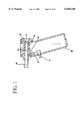
- FIG. 1is a cross-sectional view of an embodiment of the present invention
- FIG. 2is a block diagram of the major components of an embodiment of the present invention.
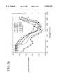
- FIG. 3are graphs illustrating the acoustic measurements utilizing a method of the present invention and indicates the typical response for a normal child ear;
- FIG. 3ais a cross-sectional view of the an embodiment of the present invention indicating the dimensions of a portion of the probe assembly
- FIG. 3bshows cross-sectional views of a variety of embodiments for the probe tip of the present invention:
- FIG. 3cis a cross-sectional view of an additional embodiment for the probe tip of the present invention:
- FIG. 3dshows cross-sectional views of additional embodiments for the probe tip of the present invention.
- FIG. 3eis a graph illustrating the third-octave averaged energy reflectance versus center frequency (Hz) for adult group and infants of ages 1, 3, 6, 12 and 24 months;
- FIG. 3fis a table disclosing initial MLSSA parameters.
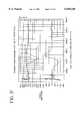
- FIG. 4is a graph illustrating the computer model output for an embodiment of the present invention.
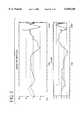
- FIG. 5are graphs illustrating the measurements of a variety of healthy ears in response to a test method of the present invention.
- FIG. 6are graphs illustrating the response of a child ear having an abnormal condition in response to a test method of the present invention
- FIG. 7are graphs illustrating the response of the child ear of FIG. 6 after removing the typical response formed from the measurements illustrated in FIG. 5;
- FIG. 8are graphs illustrating the response of a child ear having a conductive hearing loss condition in response to a test method of the present invention.
- the present inventionis an instrument and method that provides information about the function and integrity of the ear.
- the instrumentmonitors middle ear effusion and other ear abnormalities in infants, children and adults. It uses a test probe which couples to the ear at atmospheric pressure and delivers a known test signal.
- FIG. 1illustrates a cross-sectional view of an embodiment of the present invention.
- the instrumentcomprises a housing 10 sized to be held by the clinician.
- the housing 10contains a switch 12 for operation of the instrument by the clinician.
- the switch 12is adapted to actuate the instrument, wherein a test signal generated by sound source 14 is carried via wave guide 16 to probe tip 18.
- Probe tip 18is sized appropriately to fit the test subject.
- Microphone 20measures the reflected signal received back from the test subject via probe tip 18 and wave guide 16.
- the housingalso optionally may include a lens 22 for the clinician to utilize to examine the subject's tympanic membrane.
- the light source 24may be positioned as needed and may provide direct illumination, or alternatively, may provide indirect illumination that is then carried to the wave guide 16 via optical fiber 26.
- Housing 10also may hold the electronics for performing signal analysis.
- the control electronicsmay be exterior to the housing 10 with the control signal for the sound source 14 and the signal detected by the microphone 20 provided externally via wires or optical fibers.
- FIG. 2illustrates a block diagram for an embodiment of the present invention.
- the systemutilizes a computer 30 which may be a digital microcomputer or analog computer.
- the computer 30is adapted to control a signal generator 32 to create a test signal.
- the test signalis preferably pseudo-random noise.
- the test signalis provided to a power amplifier 34 which amplifies and conditions the signal properly for sound source 14.
- the sound source 14creates acoustical energy in response to the input signal.
- the sound source 14is placed within the wave guide 16 so that the acoustic energy is passed through the wave guide into the ear canal 36 and onto the tympanic membrane 38.
- a waveis reflected from the tympanic membrane 38 back through the ear canal 38 and into the wave guide 16.
- This reflected waveis detected by microphone 20 and, in response to the detected reflected wave, generates a signal.
- the signalis passed through an amplifier 40 and back to the computer 30 through a data acquisition circuit 41 for analysis.
- the deviceis first operated in air, the results of which are then subtracted from subsequent readings to provide zeroing at all frequencies.
- the computer 30is further adapted to perform signal analysis on the reflected wave signal with the respect to test signal. For instance, as illustrated, a Fourier Transform function 42 may be performed on the reflected wave signal and plotted on a hard copy device 44 or display on a screen 43. Other forms of signal analysis are possible and known in the art.
- the graphs of FIG. 3illustrate a typical response from a subject after the instrument has been zeroed in air without any further processing.
- FIG. 3billustrates the design and sizes for a variety of probe tips 18.
- FIG. 3cillustrates an alternative embodiment of the probe tip 18 wherein the probe tip 18 is designed to be used for a variety of ear canal diameters.
- the outside diameter of the tip 18is selected to penetrate into the ear of a subject and provide an airtight seal so that the instrument and ear canal will be sealed at atmospheric pressure.
- the unitoperates by forming a resonant closed end traveling wave tube to provide greatest selectively and noise rejection.
- Typical Q for the systemis four. This calculation is derived in the following way:
- Xquality factor
- Xreactance of either coil or capacitor in ohms
- Rseries resistance in ohms.
- High Qmeans higher selectivity and elimination of noise.
- the instrument designhas been assisted by utilizing a computer model which allows evaluation of different designs for the optimization of the instrument.
- An acoustic modelwhich is sold under the name of AkAbak by Panzer and Partner of Kunststoff, Germany has been utilized.
- the instrument, ear canal and tympanumhave been modeled as three separate acoustic entities, namely 1) lens to microphone, 2) microphone to end of tip and 3) end of tip to the tympanum including the ear canal.
- the graphs of FIG. 4show a typical output from a model.
- the dotted curveis for the instrument when it is operated in air not sealed to the ear.
- the solid lineis for the output when it is operated sealed to the ear.
- the difference in the outputs shown in the shaded areasis the response when the instrument is operated after zeroing in air.
- the objectiveis to construct the instrument to have as much difference in response between the two conditions as possible which will provide the greatest sensitivity for detecting ear abnormalities.
- the interior of the acoustic section of the instrument, wave guide 16 and probe tip 18,is a straight tube which is blocked off at the end away from the ear canal with a lens 22.
- the probe tip 18is made of a transparent clear material which will easily transmit light.
- the light source 24, such as a grain of wheat light or a krypton mini lamp,is mounted in the font of the instrument so that its light will be directed onto the clear tip 18.
- the tip 18transmits the light into the ear canal 36 and, in combination with the lens 22, provides a convenient and inexpensive means to view the ear canal 36 and tympanic membrane 38.
- the featuremay ensure that there is no blockage of the acoustic energy which is projected into the car, and that the ear canal 36 is straightened to allow for the unimpeded resonance of the ear canal 36, tympanic membrane 38 and the instrument.
- FIG. 3aillustrates an embodiment for the portion of the housing assembly for the instrument and gives measurements for the spacing of the earphone (Acoustic Driver) 14, microscope 20 and probe tip 18.
- the lens 22is mounted at the right end of this assembly.
- FIG. 3bshows a series of probe tip 18 designs used in the instrument. The two tips at the right hand of the drawing have worked the best since they seal in the ear canal with a good seal.
- the tipsare made of acrylic or any other rigid and clear plastic which will transmit light.
- the assemblymay protected with a protective covering.
- FIG. 3cis a diagram of a probe tip 18 which is capable of fitting ear canals of individuals of all ages, from neonate to older child. It is the conical shape in the upper part of the drawing. FIG. 3c further contains the dimensions for ear canals from 1 month of age to adults. The dimensions for children seem to follow a proportionally progressive deepening and widening with age, thus, the distance of the instrument assembly housing to the tympanum is constant for all ages using the same tip. As a result, the resonant frequency of the total traveling wave tube will be the same for all ages. This frequency has been selected to be in the range of 2100 Hz since the ear absorbs most sound energy in this frequency range and pathologies of the ear are most easily detected at these frequencies. For example, the following table illustrates the relationships of age, and the frequency at which maximum absorption occurs:
- FIG. 3dshows additional embodiments of tips and their dimensions.
- FIG. 3eshows the null obtained when monitoring ears of children to adults tor acoustic reflection. Designing an instrument to operate in this null region will function well but it is desirable to use the lowest frequency to have an instrument of suitable length for both the acoustic and optical requirements of the application.
- the acoustic driver 14is preferably located adjacent to the lens 22.
- the acoustic input to the driver 14may be pseudo-random series of pulses, commonly referred to as a Maximum Length Sequence (MLS), created either in software or using a hard wire shift register and XOR gate driven by clock pulses.
- MLSMaximum Length Sequence
- the square wave pulsesare selected to generate the acoustic frequencies between 100 to 10,000 Hz.
- the reception rate, pulse length, number of pulses, and spacingcan be selected to fit the design of the unit and meet certain established criteria of frequency domain analysis for desired lowest frequency, maximum frequency and frequency resolution for the unit.
- the total time required for the pulse trainis typically 100 msec.
- the length of the traveling wave guide and the ear canaldetermine the 1/4 wave length path of the unit at the desired resonant frequency.
- the focal length of the lensaffects the length of the resonant chamber.
- the frequency response of the acoustic driver and microphonealso must be considered when optimizing the acoustic chamber length.
- the tip and instrument designare selected to give greatest sensitivity between 1,000 to 6,000 Hz with resonance falling between 1,000 to 3,500 Hz depending on the age and ear development of the subject.
- MLSSAMaximum-Length Sequence System Analyzer
- Valuable response datais also available at the third and fifth octaves above the resonant frequencies stated above which provide an additional potential operational frequency range for the instrument.
- the lower frequencieshave been selected since the human ear has the greatest absorption of acoustic energy in this frequency range and thus any pathology of the ear will have the greatest chance of detection due to the potential percentage change which will occur between a healthy and impaired ear.
- the signal generator 32preferably a pseudo-random noise generator such as a maximum length sequence (MLS) generator, drives an acoustic driver 14 through a power amplifier 34.
- the MLS noiseenters the acoustic chamber, formed by wave guide 16 and probe tip 18, adjacent to the lens 22 and excites the chamber which includes the ear canal 36. If there is fluid in the middle ear, acoustic energy will be reflected off the tympanum 38 and back to the lens 22.
- the chamber and the ear canal 36thus form a resonant closed end tube which resonates at the 1/2 wave length.
- a null 180 degrees phase shiftwill be detected at the microphone 20 which is easy to distinguish with processing by computer 30 in software, with discrete circuitry or visually from a plot of amplitude and phase.
- the output of the microphone 20is fed to a computer 30.
- the computer 30controls the MLS noise generator. It also performs cross-correlation of the original MLS noise and the output of the microphone, providing the impulse response of the total system.
- a Fast Fourier TransformFFT
- amplitude and phase of the returned signal picked up by the microphoneis derived for each analysis frequency.
- the amplitude and phase of the FFTmay be equalized using a library of averaged response characteristics based on many subjects of similar age and ear responses with healthy ears.
- the output FFTshould be flat in amplitude and phase for a healthy ear.
- the outputcan be a plot, digital listing or other suitable output which is convenient for use by the medical practitioner.
- the MLS techniquehas a high immunity to noise interfering with the analysis. A child can cry and useful data will still be obtained.
- the calibration of the instrumentis based on the signal achieved when using a tube of selected dimensions to represent the worst case reflection obtainable with the instrument. The minimum signal is based on the response of healthy ears as obtained from the library referred to above. Thus, the calibration of the instrument is based on repeatable response and is not arbitrarily selected as in acoustic reflection type instruments of the past.
- the instrumentshould be designed to resonate over a narrow frequency range so that the design and construction of the instrument can be as simple as possible to reduce manufacturing costs.
- FIG. 5shows the response of the library for a group of children with similar healthy ear responses. All the responses have been averaged together and stored in the processing computer. Such techniques as using the 95 confidence limits of the averaged library data can be displayed during analysis of the data to assist the practitioner in evaluating the data.
- FIG. 6shows the response of a child with the right ear affected by otitis media. This ear shows greater reflected energy than a healthy ear but the immediate observation is less obvious than after more processing.
- FIG. 7shows the dramatic improvement of the plot after the library for healthy children has been subtracted from the response as shown in FIG. 6.
- acoustic energywill be present at frequencies just above the expected resonance of the ear.
- the frequency of resonancewill fall within an anticipated range for given age groups and ear development. This is used as a check on the reading. For example, when a correct tip is used, the null should appear at about 2100 Hz. An uncharacteristically high frequency at the null may indicate that the wrong tip was used.
- phase response of the instrument at resonanceis a very good indicator of the quality of the measurement.
- a regression analysis of the phase curvemay provide a measure of the slope and coefficient of variation for this purpose. This is used for detecting a poor reading of the instrument.
- FIG. 8shows the response of a child suffering conductive hearing loss. This data has not had the library for healthy children subtracted from it. Notice the flat response in the range of 1,000 to 6,000 Hz where normally one would expect the characteristic null response. This data would show a dramatic positive response if the library for healthy children had been subtracted from it.
- the present inventionis measuring ear characteristics at only atmospheric pressure. Since the child's ear is operating at this pressure, it is the correct pressure to determine the response of the ear and potential impaired hearing problems.
- the present inventiondoes not preclude conducting further investigations, and it may be desired to run a series of measurements over a differential pressure range of +200 to -200 mm for certain situations. For instance, if the ear is operating at other than atmospheric pressure due to otitis media, the tympanum can be pressure unloaded and tested. This method will aid in detailed analysis of the child's problem.
- the present inventionmay be further adapted to seal to the ear canal and pressurized to unload or load the tympanic membrane as desired.
- the response of the device in amplitude and phasealso may be flattened, in both amplitude and phase, over the operating range of the device using standard techniques.
- the wave guide designalso may be used at ultrasonic frequencies into the megahertz region.
- the out of phase or in phase response of the ear canal and middle earmay be examined using ultrasonic frequencies to delineate features not detectable at lower frequencies, such as the delineation of air/fluid interfaces, which may provide more information to determine the pathology of the ear system.
Landscapes
- Health & Medical Sciences (AREA)
- Life Sciences & Earth Sciences (AREA)
- Engineering & Computer Science (AREA)
- Physics & Mathematics (AREA)
- Surgery (AREA)
- General Health & Medical Sciences (AREA)
- Animal Behavior & Ethology (AREA)
- Biophysics (AREA)
- Pathology (AREA)
- Biomedical Technology (AREA)
- Heart & Thoracic Surgery (AREA)
- Medical Informatics (AREA)
- Molecular Biology (AREA)
- Veterinary Medicine (AREA)
- Public Health (AREA)
- Otolaryngology (AREA)
- Acoustics & Sound (AREA)
- Multimedia (AREA)
- Audiology, Speech & Language Pathology (AREA)
- Nuclear Medicine, Radiotherapy & Molecular Imaging (AREA)
- Optics & Photonics (AREA)
- Radiology & Medical Imaging (AREA)
- Measurement Of The Respiration, Hearing Ability, Form, And Blood Characteristics Of Living Organisms (AREA)
Abstract
Description
______________________________________ Null Average Energy Age Frequency (Hz) Reflectance (%) ______________________________________ 1 mo. 2000 .105 3 mo. 2000 .120 6 mo. 2500 .130 l2 mo. 4000 .160 24 mo. 3000 .120 Adult 3000 .350 ______________________________________
Claims (9)
Priority Applications (1)
| Application Number | Priority Date | Filing Date | Title |
|---|---|---|---|
| US08/978,197US6048320A (en) | 1996-11-25 | 1997-11-25 | Inner ear diagnostic apparatus |
Applications Claiming Priority (2)
| Application Number | Priority Date | Filing Date | Title |
|---|---|---|---|
| US3203096P | 1996-11-25 | 1996-11-25 | |
| US08/978,197US6048320A (en) | 1996-11-25 | 1997-11-25 | Inner ear diagnostic apparatus |
Publications (1)
| Publication Number | Publication Date |
|---|---|
| US6048320Atrue US6048320A (en) | 2000-04-11 |
Family
ID=21862731
Family Applications (1)
| Application Number | Title | Priority Date | Filing Date |
|---|---|---|---|
| US08/978,197Expired - LifetimeUS6048320A (en) | 1996-11-25 | 1997-11-25 | Inner ear diagnostic apparatus |
Country Status (6)
| Country | Link |
|---|---|
| US (1) | US6048320A (en) |
| EP (1) | EP1009282A1 (en) |
| JP (1) | JP2001517105A (en) |
| AU (1) | AU5453798A (en) |
| CA (1) | CA2272580A1 (en) |
| WO (1) | WO1998023205A1 (en) |
Cited By (24)
| Publication number | Priority date | Publication date | Assignee | Title |
|---|---|---|---|---|
| US6491644B1 (en)* | 1998-10-23 | 2002-12-10 | Aleksandar Vujanic | Implantable sound receptor for hearing aids |
| US20030053646A1 (en)* | 2001-09-07 | 2003-03-20 | Jakob Nielsen | Listening device |
| US20040138561A1 (en)* | 2002-12-06 | 2004-07-15 | Jan Lewandowski | Ultrasonic detection of ear disorders |
| US20060262950A1 (en)* | 2005-05-19 | 2006-11-23 | Burns Thomas H | System for testing hearing assistance devices using a planar waveguide |
| US20070261494A1 (en)* | 2006-04-28 | 2007-11-15 | Biomec, Inc. | Ultrasonic transducer devices and detection apparatus |
| FR2928823A1 (en)* | 2008-03-19 | 2009-09-25 | Jackie Firion | Part e.g. heart, investigating device for carrying diagnosis on toddler, has positioning units positioning optical system in body such that system defines chambers in body, and reception units receiving sounds gathered in one of chambers |
| US20090321177A1 (en)* | 2008-06-26 | 2009-12-31 | Welch Allyn, Inc. | Acoustic measurement tip |
| US20100069752A1 (en)* | 2002-12-06 | 2010-03-18 | Otosonics, Inc. | Ultrasonic detection of ear disorders |
| US20100142719A1 (en)* | 2008-12-09 | 2010-06-10 | Kabushiki Kaisha Toshiba | Acoustic apparatus and method of controlling an acoustic apparatus |
| US20140236043A1 (en)* | 2011-09-21 | 2014-08-21 | Jacoti Bvba | Method and Device for Conducting a Pure Tone Audiometry Screening |
| US20150065803A1 (en)* | 2013-09-05 | 2015-03-05 | Erik Scott DOUGLAS | Apparatuses and methods for mobile imaging and analysis |
| US9155494B2 (en) | 2011-01-24 | 2015-10-13 | Etymotic Research, Inc. | Hearing testing probe apparatus with digital interface |
| RU2572156C1 (en)* | 2014-08-27 | 2015-12-27 | Сергей Павлович Драган | Instrument for measuring acoustic impedance of middle ear |
| WO2017011035A1 (en)* | 2015-07-13 | 2017-01-19 | Otonexus Medical Technologies, Inc | Apparatus and method for characterization of acute otitis media |
| US10029068B2 (en) | 2016-11-01 | 2018-07-24 | Polyvagal Science LLC | Methods and systems for reducing sound sensitivities and improving auditory processing, behavioral state regulation and social engagement behaviors |
| EP3512404A4 (en)* | 2016-09-16 | 2020-04-22 | Throat Scope Pty Ltd | An otoscope |
| US10675001B2 (en) | 2016-06-04 | 2020-06-09 | Otonexus Medical Technologies, Inc. | Apparatus and method for characterization of a ductile membrane, surface, and sub-surface properties |
| US10702154B2 (en) | 2018-03-01 | 2020-07-07 | Polyvagal Science LLC | Systems and methods for modulating physiological state |
| US20210186426A1 (en)* | 2018-09-07 | 2021-06-24 | University Of Washington | System and method for detection of middle ear fluids |
| CN114615919A (en)* | 2019-08-28 | 2022-06-10 | 沃德诺希斯医疗技术有限公司 | Ultrasound transducer apparatus and method |
| US12349097B2 (en) | 2010-12-30 | 2025-07-01 | St Famtech, Llc | Information processing using a population of data acquisition devices |
| US12363223B2 (en) | 2013-09-22 | 2025-07-15 | ST R&DTech LLC | Real-time voice paging voice augmented caller ID/ring tone alias |
| US12374332B2 (en) | 2008-09-22 | 2025-07-29 | ST Fam Tech, LLC | Personalized sound management and method |
| US12389154B2 (en) | 2012-12-17 | 2025-08-12 | St Famtech, Llc | Shared earpiece communication |
Families Citing this family (10)
| Publication number | Priority date | Publication date | Assignee | Title |
|---|---|---|---|---|
| DE19918288A1 (en)* | 1999-04-22 | 2000-10-26 | Braun Gmbh | Ear reflectometer; has measuring point to fit in ear canal and device to observe ear canal, which includes light conductor coupled between lens at measuring point and eye piece or viewing device |
| FR2802084B1 (en)* | 1999-12-08 | 2002-07-12 | Duret Inventeur | HEARING AID, METHOD FOR IMPLEMENTING SAME, AND MATERIAL FOR ENABLING THE SAME |
| US20030171655A1 (en)* | 2002-03-08 | 2003-09-11 | Newman Richard W. | Combination otoscope |
| US7976474B2 (en)* | 2009-01-23 | 2011-07-12 | Path Medical Gmbh | Ear canal obstruction detecting acoustical stimulation ear probe |
| SG11201505461PA (en) | 2013-02-04 | 2015-08-28 | Helen Of Troy Ltd | Otoscope |
| HK1219856A1 (en) | 2013-02-04 | 2017-04-21 | Helen Of Troy Limited | Otoscope |
| EP2950696B1 (en) | 2013-02-04 | 2020-04-29 | Helen of Troy Limited | Method for identifying objects in a subject's ear |
| MX363569B (en) | 2013-02-04 | 2019-03-27 | Helen Of Troy Ltd | Ear inspection device and method of determining a condition of a subject's ear. |
| CN108810787B (en)* | 2018-05-28 | 2021-08-31 | Oppo广东移动通信有限公司 | Foreign object detection method, device and terminal based on audio equipment |
| EP4051107A4 (en)* | 2019-10-28 | 2023-11-01 | The Research Foundation for The State University of New York | DEVICES, SYSTEMS AND METHODS FOR MONITORING ARTERIAL CARBON DIOXIDE |
Citations (10)
| Publication number | Priority date | Publication date | Assignee | Title |
|---|---|---|---|---|
| US3757769A (en)* | 1971-11-01 | 1973-09-11 | Grason Stadler Comp Inc | Acoustic admittance testing apparatus |
| US4374526A (en)* | 1978-02-10 | 1983-02-22 | National Research Development Corporation | Hearing faculty testing and apparatus therefor |
| US4459996A (en)* | 1982-03-16 | 1984-07-17 | Teele John H | Ear pathology diagnosis apparatus and method |
| US4567881A (en)* | 1983-03-31 | 1986-02-04 | Welch Allyn Inc. | Combination otoscope and audiometer |
| US4601295A (en)* | 1982-03-16 | 1986-07-22 | Teele John H | Ear pathology diagnosis apparatus and method |
| US4688582A (en)* | 1986-03-06 | 1987-08-25 | Welch Allyn, Inc. | Portable hand-held tympanometer |
| US5546956A (en)* | 1992-04-04 | 1996-08-20 | Medical Research Council | Testing hearing |
| US5699809A (en)* | 1985-11-17 | 1997-12-23 | Mdi Instruments, Inc. | Device and process for generating and measuring the shape of an acoustic reflectance curve of an ear |
| US5738633A (en)* | 1993-12-10 | 1998-04-14 | Madsen Electronics A/S | Oto-acoustic emission analyser |
| US5792072A (en)* | 1994-06-06 | 1998-08-11 | University Of Washington | System and method for measuring acoustic reflectance |
Family Cites Families (4)
| Publication number | Priority date | Publication date | Assignee | Title |
|---|---|---|---|---|
| US2240402A (en)* | 1939-03-09 | 1941-04-29 | Plastic Process Company | Illuminated microscope |
| US3203097A (en) | 1963-04-25 | 1965-08-31 | Denver Chemical Mfg Company | Germicidal dental pad |
| US5105822A (en)* | 1988-02-16 | 1992-04-21 | Sensimetrics Corporation | Apparatus for and method of performing high frequency audiometry |
| JPH05220111A (en)* | 1992-02-10 | 1993-08-31 | Machida Endscope Co Ltd | Medical deep part observing device |
- 1997
- 1997-11-25EPEP97948469Apatent/EP1009282A1/ennot_activeWithdrawn
- 1997-11-25CACA002272580Apatent/CA2272580A1/ennot_activeAbandoned
- 1997-11-25AUAU54537/98Apatent/AU5453798A/ennot_activeAbandoned
- 1997-11-25JPJP52477498Apatent/JP2001517105A/enactivePending
- 1997-11-25WOPCT/US1997/021420patent/WO1998023205A1/ennot_activeApplication Discontinuation
- 1997-11-25USUS08/978,197patent/US6048320A/ennot_activeExpired - Lifetime
Patent Citations (10)
| Publication number | Priority date | Publication date | Assignee | Title |
|---|---|---|---|---|
| US3757769A (en)* | 1971-11-01 | 1973-09-11 | Grason Stadler Comp Inc | Acoustic admittance testing apparatus |
| US4374526A (en)* | 1978-02-10 | 1983-02-22 | National Research Development Corporation | Hearing faculty testing and apparatus therefor |
| US4459996A (en)* | 1982-03-16 | 1984-07-17 | Teele John H | Ear pathology diagnosis apparatus and method |
| US4601295A (en)* | 1982-03-16 | 1986-07-22 | Teele John H | Ear pathology diagnosis apparatus and method |
| US4567881A (en)* | 1983-03-31 | 1986-02-04 | Welch Allyn Inc. | Combination otoscope and audiometer |
| US5699809A (en)* | 1985-11-17 | 1997-12-23 | Mdi Instruments, Inc. | Device and process for generating and measuring the shape of an acoustic reflectance curve of an ear |
| US4688582A (en)* | 1986-03-06 | 1987-08-25 | Welch Allyn, Inc. | Portable hand-held tympanometer |
| US5546956A (en)* | 1992-04-04 | 1996-08-20 | Medical Research Council | Testing hearing |
| US5738633A (en)* | 1993-12-10 | 1998-04-14 | Madsen Electronics A/S | Oto-acoustic emission analyser |
| US5792072A (en)* | 1994-06-06 | 1998-08-11 | University Of Washington | System and method for measuring acoustic reflectance |
Cited By (40)
| Publication number | Priority date | Publication date | Assignee | Title |
|---|---|---|---|---|
| US6491644B1 (en)* | 1998-10-23 | 2002-12-10 | Aleksandar Vujanic | Implantable sound receptor for hearing aids |
| US20030053646A1 (en)* | 2001-09-07 | 2003-03-20 | Jakob Nielsen | Listening device |
| US7558390B2 (en)* | 2001-09-07 | 2009-07-07 | Ami Semiconductor, Inc. | Listening device |
| US20100069752A1 (en)* | 2002-12-06 | 2010-03-18 | Otosonics, Inc. | Ultrasonic detection of ear disorders |
| US20040138561A1 (en)* | 2002-12-06 | 2004-07-15 | Jan Lewandowski | Ultrasonic detection of ear disorders |
| US7632232B2 (en)* | 2002-12-06 | 2009-12-15 | Otosonics Inc. | Ultrasonic detection of ear disorders |
| US20060262950A1 (en)* | 2005-05-19 | 2006-11-23 | Burns Thomas H | System for testing hearing assistance devices using a planar waveguide |
| US7769185B2 (en) | 2005-05-19 | 2010-08-03 | Starkey Laboratories, Inc. | System for testing hearing assistance devices using a planar waveguide |
| US20070261494A1 (en)* | 2006-04-28 | 2007-11-15 | Biomec, Inc. | Ultrasonic transducer devices and detection apparatus |
| FR2928823A1 (en)* | 2008-03-19 | 2009-09-25 | Jackie Firion | Part e.g. heart, investigating device for carrying diagnosis on toddler, has positioning units positioning optical system in body such that system defines chambers in body, and reception units receiving sounds gathered in one of chambers |
| WO2009158557A3 (en)* | 2008-06-26 | 2010-04-01 | Welch Allyn, Inc. | Acoustic measurement tip |
| US20090321177A1 (en)* | 2008-06-26 | 2009-12-31 | Welch Allyn, Inc. | Acoustic measurement tip |
| US7882928B2 (en) | 2008-06-26 | 2011-02-08 | Welch Allyn, Inc. | Acoustic measurement tip |
| US12374332B2 (en) | 2008-09-22 | 2025-07-29 | ST Fam Tech, LLC | Personalized sound management and method |
| US7957549B2 (en)* | 2008-12-09 | 2011-06-07 | Kabushiki Kaisha Toshiba | Acoustic apparatus and method of controlling an acoustic apparatus |
| US20100142719A1 (en)* | 2008-12-09 | 2010-06-10 | Kabushiki Kaisha Toshiba | Acoustic apparatus and method of controlling an acoustic apparatus |
| US12349097B2 (en) | 2010-12-30 | 2025-07-01 | St Famtech, Llc | Information processing using a population of data acquisition devices |
| US9155494B2 (en) | 2011-01-24 | 2015-10-13 | Etymotic Research, Inc. | Hearing testing probe apparatus with digital interface |
| US10624562B2 (en) | 2011-01-24 | 2020-04-21 | Etymotic Research, Inc. | Hearing testing probe apparatus with digital interface |
| US20140236043A1 (en)* | 2011-09-21 | 2014-08-21 | Jacoti Bvba | Method and Device for Conducting a Pure Tone Audiometry Screening |
| US10292626B2 (en)* | 2011-09-21 | 2019-05-21 | Jacoti Bvba | Method and device for conducting a pure tone audiometry sceening |
| US12389154B2 (en) | 2012-12-17 | 2025-08-12 | St Famtech, Llc | Shared earpiece communication |
| US20150065803A1 (en)* | 2013-09-05 | 2015-03-05 | Erik Scott DOUGLAS | Apparatuses and methods for mobile imaging and analysis |
| US9445713B2 (en)* | 2013-09-05 | 2016-09-20 | Cellscope, Inc. | Apparatuses and methods for mobile imaging and analysis |
| US12363223B2 (en) | 2013-09-22 | 2025-07-15 | ST R&DTech LLC | Real-time voice paging voice augmented caller ID/ring tone alias |
| RU2572156C1 (en)* | 2014-08-27 | 2015-12-27 | Сергей Павлович Драган | Instrument for measuring acoustic impedance of middle ear |
| WO2017011035A1 (en)* | 2015-07-13 | 2017-01-19 | Otonexus Medical Technologies, Inc | Apparatus and method for characterization of acute otitis media |
| US11627935B2 (en) | 2015-07-13 | 2023-04-18 | Otonexus Medical Technologies, Inc. | Apparatus and method for characterization of acute otitis media |
| EP3322339A4 (en)* | 2015-07-13 | 2019-03-06 | Otonexus Medical Technologies, Inc. | APPARATUS AND METHOD FOR CHARACTERIZING ACUTE MEDIUM OTITIS |
| US10660604B2 (en) | 2015-07-13 | 2020-05-26 | Otonexus Medical Technologies, Inc. | Apparatus and method for characterization of acute otitis media |
| US12167934B2 (en) | 2015-07-13 | 2024-12-17 | Otonexus Medical Technologies, Inc. | Apparatus and method for characterization of acute otitis media |
| US11660074B2 (en) | 2016-06-04 | 2023-05-30 | Otonexus Medical Technologies, Inc. | Apparatus and method for characterization of a ductile membrane, surface, and sub-surface properties |
| US12167936B2 (en) | 2016-06-04 | 2024-12-17 | Otonexus Medical Technologies, Inc. | Apparatus and method for characterization of a ductile membrane, surface and sub-surface properties |
| US10675001B2 (en) | 2016-06-04 | 2020-06-09 | Otonexus Medical Technologies, Inc. | Apparatus and method for characterization of a ductile membrane, surface, and sub-surface properties |
| EP3512404A4 (en)* | 2016-09-16 | 2020-04-22 | Throat Scope Pty Ltd | An otoscope |
| US10661046B2 (en) | 2016-11-01 | 2020-05-26 | Polyvagal Science LLC | Methods and systems for reducing sound sensitivities and improving auditory processing, behavioral state regulation and social engagement behaviors |
| US10029068B2 (en) | 2016-11-01 | 2018-07-24 | Polyvagal Science LLC | Methods and systems for reducing sound sensitivities and improving auditory processing, behavioral state regulation and social engagement behaviors |
| US10702154B2 (en) | 2018-03-01 | 2020-07-07 | Polyvagal Science LLC | Systems and methods for modulating physiological state |
| US20210186426A1 (en)* | 2018-09-07 | 2021-06-24 | University Of Washington | System and method for detection of middle ear fluids |
| CN114615919A (en)* | 2019-08-28 | 2022-06-10 | 沃德诺希斯医疗技术有限公司 | Ultrasound transducer apparatus and method |
Also Published As
| Publication number | Publication date |
|---|---|
| WO1998023205A1 (en) | 1998-06-04 |
| JP2001517105A (en) | 2001-10-02 |
| AU5453798A (en) | 1998-06-22 |
| EP1009282A1 (en) | 2000-06-21 |
| CA2272580A1 (en) | 1998-06-04 |
Similar Documents
| Publication | Publication Date | Title |
|---|---|---|
| US6048320A (en) | Inner ear diagnostic apparatus | |
| WO1998023205A9 (en) | Inner ear diagnostic apparatus and method | |
| US12167934B2 (en) | Apparatus and method for characterization of acute otitis media | |
| US5868682A (en) | Device and process for generating and measuring the shape of an acoustic reflectance curve of an ear | |
| US4601295A (en) | Ear pathology diagnosis apparatus and method | |
| US5699809A (en) | Device and process for generating and measuring the shape of an acoustic reflectance curve of an ear | |
| US6126614A (en) | Apparatus and method for analysis of ear pathologies by detecting fluid in the ear, measuring body temperature and/or determining a characteristic of a fluid | |
| Teele et al. | Detection of middle ear effusion by acoustic reflectometry | |
| KR100235170B1 (en) | A device and process for generating and measuring the shape of an acoustic reflectance curve of an ear | |
| US5951486A (en) | Apparatus and method for analysis of ear pathologies using combinations of acoustic reflectance, temperature and chemical response | |
| WO1983003192A1 (en) | Ear pathology diagnosis apparatus and method | |
| WO1996023293A9 (en) | A device and process for generating and measuring the shape of an acoustic reflectance curve of an ear | |
| US5919143A (en) | Apparatus and method for analysis of acoustic reflectance and thermal radiation of an ear | |
| US4930507A (en) | Double chamber acoustical tonometer | |
| US9867572B2 (en) | Method and system for rapidly determining and displaying the depth of ear tip placement to improve the reliability of hearing tests | |
| CA1219330A (en) | Ear pathology diagnosis apparatus and method | |
| JPH08508900A (en) | Tonometer | |
| Pellett et al. | Use of acoustic reflectometry in the detection of middle ear effusion. | |
| Jaffer | Wideband acoustic immittance: Instrument, ethnicity, and gender specific normative data | |
| RU2332164C2 (en) | Device of tympanum state evaluation |
Legal Events
| Date | Code | Title | Description |
|---|---|---|---|
| AS | Assignment | Owner name:MDI INSTRUMENTS, INC., MASSACHUSETTS Free format text:ASSIGNMENT OF ASSIGNORS INTEREST;ASSIGNOR:BRAINARD, EDWARD C., II;REEL/FRAME:008845/0662 Effective date:19971125 | |
| STCF | Information on status: patent grant | Free format text:PATENTED CASE | |
| FEPP | Fee payment procedure | Free format text:PAYOR NUMBER ASSIGNED (ORIGINAL EVENT CODE: ASPN); ENTITY STATUS OF PATENT OWNER: SMALL ENTITY | |
| FPAY | Fee payment | Year of fee payment:4 | |
| AS | Assignment | Owner name:EARCHECK ACQUISITION, LLC, KANSAS Free format text:ASSIGNMENT OF ASSIGNORS INTEREST;ASSIGNOR:MDI INSTRUMENTS, INC.;REEL/FRAME:019773/0506 Effective date:20050415 Owner name:INNOVIA MEDICAL, LLC, KANSAS Free format text:CHANGE OF NAME;ASSIGNOR:EARCHECK ACQUISITION, LLC;REEL/FRAME:019767/0739 Effective date:20050801 | |
| FPAY | Fee payment | Year of fee payment:8 | |
| REMI | Maintenance fee reminder mailed | ||
| AS | Assignment | Owner name:INNOVIA MEDICAL, LLC, NEBRASKA Free format text:ASSIGNMENT OF ASSIGNORS INTEREST;ASSIGNOR:INNOVIA MEDICAL, LLC;REEL/FRAME:025554/0071 Effective date:20101207 | |
| FPAY | Fee payment | Year of fee payment:12 |