US6033420A - Trocar introducer system and methods of use - Google Patents
Trocar introducer system and methods of useDownload PDFInfo
- Publication number
- US6033420A US6033420AUS09/146,216US14621698AUS6033420AUS 6033420 AUS6033420 AUS 6033420AUS 14621698 AUS14621698 AUS 14621698AUS 6033420 AUS6033420 AUS 6033420A
- Authority
- US
- United States
- Prior art keywords
- cannula
- trocar
- distal end
- lumen
- proximal end
- Prior art date
- Legal status (The legal status is an assumption and is not a legal conclusion. Google has not performed a legal analysis and makes no representation as to the accuracy of the status listed.)
- Expired - Lifetime
Links
- 238000000034methodMethods0.000titleclaimsabstractdescription20
- 230000007246mechanismEffects0.000claimsabstractdescription16
- 210000000709aortaAnatomy0.000claimsdescription12
- 239000008280bloodSubstances0.000claimsdescription12
- 210000004369bloodAnatomy0.000claimsdescription12
- 230000008439repair processEffects0.000claimsdescription7
- 210000004351coronary vesselAnatomy0.000claimsdescription6
- 230000002792vascularEffects0.000claimsdescription6
- 230000002612cardiopulmonary effectEffects0.000claimsdescription5
- 210000004204blood vesselAnatomy0.000claimsdescription4
- 230000002093peripheral effectEffects0.000claimsdescription4
- 238000001356surgical procedureMethods0.000claimsdescription3
- 230000003213activating effectEffects0.000claimsdescription2
- 210000003709heart valveAnatomy0.000claims1
- 238000013160medical therapyMethods0.000abstractdescription8
- 210000001519tissueAnatomy0.000description21
- 210000005245right atriumAnatomy0.000description6
- 241001631457CannulaSpecies0.000description5
- 206010058178Aortic occlusionDiseases0.000description4
- 208000010125myocardial infarctionDiseases0.000description4
- 239000012530fluidSubstances0.000description3
- 238000003780insertionMethods0.000description3
- 230000037431insertionEffects0.000description3
- 238000002324minimally invasive surgeryMethods0.000description3
- 210000000779thoracic wallAnatomy0.000description3
- 206010002329AneurysmDiseases0.000description2
- 208000032170Congenital AbnormalitiesDiseases0.000description2
- 208000031481Pathologic ConstrictionDiseases0.000description2
- 238000013132cardiothoracic surgeryMethods0.000description2
- 210000000038chestAnatomy0.000description2
- 208000029078coronary artery diseaseDiseases0.000description2
- 238000012937correctionMethods0.000description2
- 208000025339heart septal defectDiseases0.000description2
- 238000002955isolationMethods0.000description2
- 230000000250revascularizationEffects0.000description2
- 230000006641stabilisationEffects0.000description2
- 238000011105stabilizationMethods0.000description2
- 230000036262stenosisEffects0.000description2
- 208000037804stenosisDiseases0.000description2
- 210000000115thoracic cavityAnatomy0.000description2
- 206010002383Angina PectorisDiseases0.000description1
- 201000001320AtherosclerosisDiseases0.000description1
- 208000010496Heart ArrestDiseases0.000description1
- 206010042434Sudden deathDiseases0.000description1
- 208000024248Vascular System injuryDiseases0.000description1
- 208000012339Vascular injuryDiseases0.000description1
- 206010047281Ventricular arrhythmiaDiseases0.000description1
- 230000004913activationEffects0.000description1
- 210000000577adipose tissueAnatomy0.000description1
- 238000013459approachMethods0.000description1
- 230000001746atrial effectEffects0.000description1
- 230000017531blood circulationEffects0.000description1
- 230000000747cardiac effectEffects0.000description1
- 238000007675cardiac surgeryMethods0.000description1
- 229940100084cardioplegia solutionDrugs0.000description1
- 230000001101cardioplegic effectEffects0.000description1
- 238000007887coronary angioplastyMethods0.000description1
- 230000006837decompressionEffects0.000description1
- 238000003745diagnosisMethods0.000description1
- 230000004217heart functionEffects0.000description1
- 208000014674injuryDiseases0.000description1
- 230000003601intercostal effectEffects0.000description1
- 230000007774longtermEffects0.000description1
- 238000012986modificationMethods0.000description1
- 230000004048modificationEffects0.000description1
- 208000009091myxomaDiseases0.000description1
- 230000004083survival effectEffects0.000description1
- 230000001732thrombotic effectEffects0.000description1
- 230000008733traumaEffects0.000description1
- 210000001631vena cava inferiorAnatomy0.000description1
- 210000002620vena cava superiorAnatomy0.000description1
- 230000002861ventricularEffects0.000description1
Images


Classifications
- A—HUMAN NECESSITIES
- A61—MEDICAL OR VETERINARY SCIENCE; HYGIENE
- A61B—DIAGNOSIS; SURGERY; IDENTIFICATION
- A61B17/00—Surgical instruments, devices or methods
- A61B17/34—Trocars; Puncturing needles
- A61B17/3417—Details of tips or shafts, e.g. grooves, expandable, bendable; Multiple coaxial sliding cannulas, e.g. for dilating
- A—HUMAN NECESSITIES
- A61—MEDICAL OR VETERINARY SCIENCE; HYGIENE
- A61B—DIAGNOSIS; SURGERY; IDENTIFICATION
- A61B17/00—Surgical instruments, devices or methods
- A61B17/32—Surgical cutting instruments
- A61B17/3209—Incision instruments
- A61B17/3211—Surgical scalpels, knives; Accessories therefor
- A—HUMAN NECESSITIES
- A61—MEDICAL OR VETERINARY SCIENCE; HYGIENE
- A61B—DIAGNOSIS; SURGERY; IDENTIFICATION
- A61B17/00—Surgical instruments, devices or methods
- A61B17/34—Trocars; Puncturing needles
- A61B17/3417—Details of tips or shafts, e.g. grooves, expandable, bendable; Multiple coaxial sliding cannulas, e.g. for dilating
- A61B17/3421—Cannulas
- A—HUMAN NECESSITIES
- A61—MEDICAL OR VETERINARY SCIENCE; HYGIENE
- A61B—DIAGNOSIS; SURGERY; IDENTIFICATION
- A61B17/00—Surgical instruments, devices or methods
- A61B17/00234—Surgical instruments, devices or methods for minimally invasive surgery
- A61B2017/00238—Type of minimally invasive operation
- A61B2017/00243—Type of minimally invasive operation cardiac
- A—HUMAN NECESSITIES
- A61—MEDICAL OR VETERINARY SCIENCE; HYGIENE
- A61B—DIAGNOSIS; SURGERY; IDENTIFICATION
- A61B17/00—Surgical instruments, devices or methods
- A61B17/22—Implements for squeezing-off ulcers or the like on inner organs of the body; Implements for scraping-out cavities of body organs, e.g. bones; for invasive removal or destruction of calculus using mechanical vibrations; for removing obstructions in blood vessels, not otherwise provided for
- A61B2017/22051—Implements for squeezing-off ulcers or the like on inner organs of the body; Implements for scraping-out cavities of body organs, e.g. bones; for invasive removal or destruction of calculus using mechanical vibrations; for removing obstructions in blood vessels, not otherwise provided for with an inflatable part, e.g. balloon, for positioning, blocking, or immobilisation
- A61B2017/22065—Functions of balloons
- A61B2017/22067—Blocking; Occlusion
Definitions
- the present inventionrelates generally to a trocar introducer system which provides access to a body tissue, including a patient's vascular system, and serves as a conduit to apply other medical therapies, such as delivery of oxygenated blood.
- Myocardial infarctionis one of the most common diagnosis occurring in hospitalized patients in Western countries. In the United States, approximately 1.5 million myocardial infarctions occur each year. Myocardial infarction generally occurs with abrupt decrease in coronary blood flow that follows a thrombotic occlusion of a coronary artery previously narrowed by atherosclerosis. Once severe stenosis of a coronary artery has reduced the cross-sectional area by more than approximately seventy percent, a patient is likely to develop clinical manifestation of coronary artery disease, which includes angina pectoris, myocardial infarction, ventricular arrhythmia, and sudden death.
- coronary artery diseasewhich includes angina pectoris, myocardial infarction, ventricular arrhythmia, and sudden death.
- the inventionprovides less-invasive devices and methods for cannulating a body tissue or body cavity and infusing a fluid herein. More particularly, the invention provides a trocar introducer system which includes an access mechanism for insertion of cannulas.
- the methods and devices of the inventioneliminate the need for a median sternotomy or thoracotomy to obtain access into thoracic cavity. The present invention is therefore useful when peripheral vascular access is unavailable due to inadequate vessel diameter, vessel stenosis, vascular injury or other conditions.
- the trocar introducer systemmakes use of a cannula which comprises a proximal end, a distal end, and a lumen therebetween.
- a rigid trocarwhich comprises an elongate member having a proximal end and a distal end, is slideably inserted into the lumen of the cannula.
- the elongate memberhas an actuating mechanism mounted on its proximal end and a surgical blade mounted on its distal end. The surgical blade is operable by manipulating the actuating mechanism.
- the rigid trocarhas a handle mounted on the proximal end of the elongate member.
- the handlemay be distal the actuating mechanism, and facilitates operation of the actuating mechanism.
- the actuating mechanismincludes a shaft carried by a lumen of the trocar.
- the shaftis connected at its distal end to the surgical blade.
- the shaftmay have a knob at its proximal end and may operate against the force of a spring.
- the surgical blademay have a protecting sleeve.
- the rigid trocarcan be inserted into the lumen of a variety of cannulas to provide vascular access in minimally invasive procedures.
- the cannulamay include venous drainage ports and a lumen adapted for drainage of deoxygenated blood from the right atrium to a bypass oxygenator machine.
- a balloon occluderis mounted at the distal end of the cannula to provide aortic occlusion for circulatory isolation of the heart and coronary blood vessels from the peripheral vascular system.
- the cannulahas threads at its distal end to provide better stabilization between the cannula and the body tissue.
- the methods of present inventionrelate to cannulation of a body tissue, including a patient's blood vessel, using the trocar introducer system described above.
- the rigid trocarwhich comprises an elongate member having a proximal end and a distal end, is inserted in the lumen of a cannula.
- an incision on a patient's intercostal spaceis made for access into the thoracic cavity, thereby reducing trauma to the chest wall as compared to the traditional open chest approach.
- the distal end of the cannulais inserted through the chest wall incision.
- an incision on the aortais made by the surgical blade mounted on the distal end of the trocar.
- the cannulais then introduced through the incision and advanced distally into the aorta. After placement of the cannula, the elongate member is withdrawn from the lumen of the cannula. The lumen of the cannula is now available for delivery of oxygenated blood from a bypass oxygenator machine or for delivery of cardioplegia solution to the heart to arrest heart function. After cardiac arrest is achieved and cardiopulmonary bypass is initiated for circulatory support, a variety of cardiothoracic surgeries, including coronary artery bypass grafting, valvular repair and replacement, septal defect repair, aneurysm repair, removal of atrial myxoma, and correction of congenital defects, can then be performed.
- cardiothoracic surgeriesincluding coronary artery bypass grafting, valvular repair and replacement, septal defect repair, aneurysm repair, removal of atrial myxoma, and correction of congenital defects, can then be performed.
- the trocar introducer systemprovides an access mechanism for cannulation of a body tissue, thereby obviating the need for an additional access device.
- the trocar introducer systemcan be employed in various minimally invasive surgeries.
- the trocar introducer systemcan be utilized to deliver fluid and blood to a body tissue, and to apply other medical therapy such as aspirators, filters, pressure monitors, atherectomy devices, and dilatation catheters.
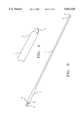
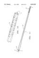
- FIG. 1depicts a rigid trocar according to a first embodiment.
- FIG. 2depicts a distal end of the trocar shown in FIG. 1.
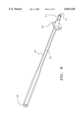
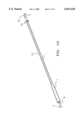
- FIG. 3depicts a rigid trocar according to another embodiment having a longer trocar.
- FIG. 4depicts a distal end of the trocar shown in FIG. 2.
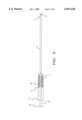
- FIG. 5depicts a rigid trocar according to another embodiment having a spring in a handle.
- FIG. 6depicts an embodiment of a cannula having a helical thread.
- FIG. 7depicts a distal end of the cannula shown in FIG. 6.
- FIG. 8depicts another embodiment of a cannula having a suture flange.
- FIG. 9depicts another embodiment of a cannula having a slideable suture flange.
- FIG. 10depicts a trocar introducer system utilizing the cannula shown in FIG. 9.
- FIG. 11depicts a trocar introducer system according to another embodiment in which the cannula has drainage ports.
- FIG. 12depicts a distal end of the trocar introducer system shown in FIG. 11.
- FIG. 13depicts another embodiment of a cannula having venous drainage ports.
- FIG. 14depicts a distal end of the cannula shown in FIG. 13.
- FIG. 15depicts another embodiment of a cannula having a balloon occluder.
- FIG. 16depicts a rigid trocar inserted in the lumen of the cannula shown in FIG. 15.
- the devices and methods of the inventionfacilitate delivery of medical therapy to a body tissue and cannulation of the body tissue by providing an access mechanism.
- the inventionworks best in minimally invasive cardiothoracic surgeries, it will be understood that the devices and methods disclosed herein can also be utilized in open chest procedures.
- FIG. 1 and FIG. 2depict a trocar according to a first embodiment.
- the trocarhas elongate member 1, proximal end 2, and distal end 4.
- Surgical blade 5is mounted on distal end 4, and is operable by manipulating an actuating mechanism at the proximal end of the trocar.
- Shaft 8connects surgical blade 5 at the distal end, and knob 9 at the proximal end of the trocar.
- Handle 7 at the proximal end of the trocarfacilitates operation of the surgical blade. In use, knob 9 is pushed distally against handle 7 to advance shaft 8 distally in order to expose surgical blade 5 for incising a body tissue.
- FIG. 2depicts a detailed view of surgical blade 5 mounted at distal end 4 of the trocar.
- FIG. 3 and FIG. 4depict a trocar according to another embodiment. Similar to the trocar depicted in FIG. 1, the trocar in FIG. 3 has elongate member 1, proximal end 2, and distal end 4. Proximal end 2 has knob 9, shaft 8, and handle 7 for activating surgical blade 5 mounted at distal end 4.
- the length of elongate member 1 in this trocaris longer than that of the trocar depicted in FIG. 1. A longer trocar is needed to fit within the lumen of a long cannula for incising, for example, an aorta in patients who may have excessive body fat between the chest wall and the great vessels.
- FIG. 5depicts a trocar according to another embodiment having spring 10 contained in handle 7. Again shaft 8 connects to knob 9 at proximal end 2 and surgical blade 5 at distal end 4 of the trocar.
- surgical blade 5is advanced distally by pushing knob 9 against handle 7, thereby operating shaft 8 against the force of spring 10. After a body tissue is incised by the surgical blade, knob 9 is released from handle 7 and spring 10 uncoils against stopper 11 to withdraw the surgical blade from the body tissue.
- the trocar introducer systemcan be used with a variety of cannulas mentioned above.
- One embodiment of the cannulais shown in FIG. 6 and FIG. 7.
- Cannula 20has proximal end 22, distal end 21, and lumen 24 therebetween.
- Helical thread 25is disposed about distal end 21 of the cannula, the thread providing better contact and therefore stabilization between the cannula and a body tissue.
- FIG. 7depicts a detailed view of distal end 4 of the cannula. Fluid, blood, or surgical instruments can be delivered to a body tissue through lumen 24 and port 15 of the cannula.
- FIG. 8, FIG. 9, and FIG. 10depict another embodiment of a cannula having a suture flange mounted at distal end 21.
- Suture flange 26is adapted for holding surgical sutures to secure the cannula onto a body tissue. This cannula may be useful for delivery of oxygenated blood through lumen 24 and port 15 to an aorta from a bypass oxygenator machine.
- Proximal end 22 of the cannulamay be shaped for attachment to a bypass oxygenator machine.
- Suture flange 26may be slideable on distal end 21 of the cannula as shown in FIG. 9 so that the length of the cannula inserted inside a body tissue is adjustable.
- the cannula of FIG. 9is equipped with a longer lumen 24 than the cannula in FIG. 8.
- FIG. 10depicts a trocar introducer system in which a rigid trocar having elongate member 1 is inserted in a lumen of cannula 20.
- the proximal end of the trocarhas knob 9 and shaft 8 which, upon activation, will advance surgical blade 5 at the distal end of the trocar to incise a body tissue, thereby providing access for cannula 20.
- suturescan be placed between suture flange 26 and the body tissue to secure the cannula.
- the trocaris then withdrawn from the lumen of the cannula, thereby leaving the lumen available to enable medical therapies.
- distal region 40 of the cannulacan easily be adjusted to accommodate changing surgical conditions or medical therapy.
- FIG. 11 and FIG. 12depict a trocar introducer system according to another embodiment in which cannula 20 has drainage ports 30 at its distal end.
- the trocar having elongate member 1is shown positioned within a lumen of cannula 20.
- the proximal end of the trocaragain has knob 9 and shaft 8 which connects to surgical blade 5 at the distal end of the trocar.
- knob 9When knob 9 is pushed distally, surgical blade 5 will advance distally to incise a body tissue to provide access for insertion of cannula 20.
- Proximal end 22 of the cannulais also adapted for attachment to a bypass oxygenator machine.
- FIG. 12A detailed view of the distal end of the trocar introducer system is depicted in FIG. 12.
- This embodiment of the trocar introducer systemcan be used to provide drainage of deoxygenated blood from the right atrium to a bypass oxygenator machine.
- the right atriumis incised by surgical blade 5 upon manipulating the actuating mechanism at the proximal end of the trocar.
- the distal end of the cannulais then advanced into the right atrium and the trocar is removed from the lumen of the cannula.
- Proximal end 22 of the cannulais connected by an attachment to a bypass oxygenator machine, and deoxygenated blood can then be drained from the right atrium through drainage ports 30 to the bypass oxygenator machine.
- FIG. 13 and FIG. 14depict another embodiment of a venous drainage cannula.
- a plurality of venous drainage ports 30are located at the distal end of cannula 20 and communicate with lumen 24 of the cannula.
- Proximal end 22is adapted for attachment to a bypass oxygenator machine.
- venous bloodis delivered from drainage ports 30 and lumen 24 to a bypass oxygenator machine.
- FIG. 15depicts another embodiment of a cannula having balloon occluder 35 mounted at distal end 21 of the cannula and communicating with inflation lumen 36 and inflation port 37.
- Distal end 21is angulated relative to proximal end 22 of the cannula.
- the cannulacan be utilized to provide aortic occlusion for circulatory isolation of the heart and coronary blood vessels from the peripheral vascular system through inflating balloon occluder 35, and to deliver oxygenated blood from a bypass oxygenator machine through lumen 24 and port 38.
- Proximal end 22is adapted for attachment to a bypass oxygenator machine.
- FIG. 16depicts a trocar introducer system utilizing the cannula shown in FIG. 15.
- the distal end of the cannulawhich can be flexible, has a linear configuration relative to the proximal end when the trocar is inserted in the lumen of the cannula.
- the trocarprovides access for cannulation of the aorta by surgical blade 5 at the proximal end of the trocar, comprising knob 9 and shaft 8.
- the trocaris removed from the lumen of the cannula, thereby allowing the distal end of the cannula to regain its preformed angulated configuration as depicted in FIG. 15.
- a significant feature of using the trocar introducer system hereis that the cannula can be more easily inserted in the aorta in a linear configuration than an angulated configuration.
- the balloon occludercan then be inflated to provide aortic occlusion, and the lumen of the cannula is available to deliver oxygenated blood from a bypass oxygenator machine to the aorta.
- the length of the elongate member of the trocaris generally between 3 and 20 inches, preferably approximately 5 inches and 12 inches in a short and long trocar as depicted in FIG. 1 and FIG. 3, respectively.
- the length of the handlewhich comprises a slope joining the handle and the elongate member, is generally between 0.8 and 2 inches, preferably approximately 1.5 inches.
- the outer diameter of the elongate memberis generally between 0.1 and 0.3 inches, preferably approximately 0.17 inches.
- the length of the proximal end of the cannula which accommodates the handle of the trocaris generally between 2 and 6 inches, preferably approximately 4.5 inches.
- the length of the cannula which accommodates the slope of the trocaris generally between 0.8 and 2 inches, preferably approximately 1.5 inches.
- the outer diameter of the proximal end of the cannulais generally between 0.3 and 0.7 inches, preferably approximately 0.55 inches.
- the inner diameter of the proximal end of the cannulais generally between 0.2 and 0.6 inches, preferably approximately 0.37 inches.
- the outer diameter of the distal end of the cannulais generally between 0.1 and 0.4 inches, preferably approximately 0.26 inches.
- the inner diameter of the distal end of the cannulais generally between 0.08 and 0.35 inches, preferably approximately 0.18 inches.
- the length of the suture flangeis generally between 0.2 and 1 inches, preferably approximately 0.5 inches.
- the angle at which the suture flange tapers to fit the distal end of the cannulais generally between 1 and 5 degrees, preferably approximately 3 degrees.
- the balloon occluder, when expanded,will generally have a diameter between 1 and 5 centimeters, preferably between about 2.5 and 4.0 centimeters.
Landscapes
- Health & Medical Sciences (AREA)
- Surgery (AREA)
- Life Sciences & Earth Sciences (AREA)
- Biomedical Technology (AREA)
- Nuclear Medicine, Radiotherapy & Molecular Imaging (AREA)
- Engineering & Computer Science (AREA)
- Pathology (AREA)
- Heart & Thoracic Surgery (AREA)
- Medical Informatics (AREA)
- Molecular Biology (AREA)
- Animal Behavior & Ethology (AREA)
- General Health & Medical Sciences (AREA)
- Public Health (AREA)
- Veterinary Medicine (AREA)
- Surgical Instruments (AREA)
Abstract
Description
Claims (7)
Priority Applications (2)
| Application Number | Priority Date | Filing Date | Title |
|---|---|---|---|
| US09/146,216US6033420A (en) | 1998-09-02 | 1998-09-02 | Trocar introducer system and methods of use |
| US09/478,151US6146400A (en) | 1998-09-02 | 2000-01-05 | Trocar introducer system and methods of use |
Applications Claiming Priority (1)
| Application Number | Priority Date | Filing Date | Title |
|---|---|---|---|
| US09/146,216US6033420A (en) | 1998-09-02 | 1998-09-02 | Trocar introducer system and methods of use |
Related Child Applications (1)
| Application Number | Title | Priority Date | Filing Date |
|---|---|---|---|
| US09/478,151ContinuationUS6146400A (en) | 1998-09-02 | 2000-01-05 | Trocar introducer system and methods of use |
Publications (1)
| Publication Number | Publication Date |
|---|---|
| US6033420Atrue US6033420A (en) | 2000-03-07 |
Family
ID=22516330
Family Applications (2)
| Application Number | Title | Priority Date | Filing Date |
|---|---|---|---|
| US09/146,216Expired - LifetimeUS6033420A (en) | 1998-09-02 | 1998-09-02 | Trocar introducer system and methods of use |
| US09/478,151Expired - LifetimeUS6146400A (en) | 1998-09-02 | 2000-01-05 | Trocar introducer system and methods of use |
Family Applications After (1)
| Application Number | Title | Priority Date | Filing Date |
|---|---|---|---|
| US09/478,151Expired - LifetimeUS6146400A (en) | 1998-09-02 | 2000-01-05 | Trocar introducer system and methods of use |
Country Status (1)
| Country | Link |
|---|---|
| US (2) | US6033420A (en) |
Cited By (45)
| Publication number | Priority date | Publication date | Assignee | Title |
|---|---|---|---|---|
| US20020022860A1 (en)* | 2000-08-18 | 2002-02-21 | Borillo Thomas E. | Expandable implant devices for filtering blood flow from atrial appendages |
| US20020198554A1 (en)* | 2001-03-14 | 2002-12-26 | Whitman Michael P. | Trocar device |
| US20030023262A1 (en)* | 2001-07-18 | 2003-01-30 | Jeffrey Welch | Cardiac implant device tether system and method |
| US6537290B2 (en) | 2001-03-05 | 2003-03-25 | Edwards Lifesciences Corporation | Sealing access cannula system |
| US20030057156A1 (en)* | 2001-03-08 | 2003-03-27 | Dean Peterson | Atrial filter implants |
| US6551303B1 (en) | 1999-10-27 | 2003-04-22 | Atritech, Inc. | Barrier device for ostium of left atrial appendage |
| US6652556B1 (en) | 1999-10-27 | 2003-11-25 | Atritech, Inc. | Filter apparatus for ostium of left atrial appendage |
| US6652555B1 (en) | 1999-10-27 | 2003-11-25 | Atritech, Inc. | Barrier device for covering the ostium of left atrial appendage |
| US20040097958A1 (en)* | 2002-07-31 | 2004-05-20 | Whitman Michael P. | Orifice introducer device |
| US20040138702A1 (en)* | 2001-05-31 | 2004-07-15 | Kenneth Peartree | Balloon cannula with over-center clamp |
| US20040230222A1 (en)* | 1999-11-08 | 2004-11-18 | Van Der Burg Erik J. | System for left atrial appendage occlusion |
| US20050004652A1 (en)* | 1998-11-06 | 2005-01-06 | Van Der Burg Eric J. | Method for left atrial appendage occlusion |
| US20050004641A1 (en)* | 2001-06-04 | 2005-01-06 | Ramesh Pappu | Cardiac stimulating apparatus having a blood clot filter and atrial pacer |
| US20060206148A1 (en)* | 1999-11-08 | 2006-09-14 | Khairkhahan Alexander K | Method of implanting an adjustable occlusion device |
| US7169164B2 (en) | 2000-09-21 | 2007-01-30 | Atritech, Inc. | Apparatus for implanting devices in atrial appendages |
| US20070066993A1 (en)* | 2005-09-16 | 2007-03-22 | Kreidler Marc S | Intracardiac cage and method of delivering same |
| US7549983B2 (en) | 1999-09-20 | 2009-06-23 | Atritech, Inc. | Method of closing an opening in a wall of the heart |
| US20090254095A1 (en)* | 2008-03-18 | 2009-10-08 | Blake Levine | Subcutaneous tunneling device |
| US8992579B1 (en) | 2011-03-08 | 2015-03-31 | Nuvasive, Inc. | Lateral fixation constructs and related methods |
| US9060815B1 (en) | 2012-03-08 | 2015-06-23 | Nuvasive, Inc. | Systems and methods for performing spine surgery |
| US9186177B2 (en) | 2001-03-14 | 2015-11-17 | Covidien Lp | Trocar device |
| US9474516B2 (en) | 2011-11-08 | 2016-10-25 | Boston Scientific Scimed, Inc. | Handle assembly for a left atrial appendage occlusion device |
| US9517089B1 (en) | 2013-10-08 | 2016-12-13 | Nuvasive, Inc. | Bone anchor with offset rod connector |
| US9730701B2 (en) | 2014-01-16 | 2017-08-15 | Boston Scientific Scimed, Inc. | Retrieval wire centering device |
| US9883936B2 (en) | 2002-01-25 | 2018-02-06 | Boston Scientific Scimed, Inc | Atrial appendage blood filtration systems |
| US10525179B2 (en) | 2016-03-31 | 2020-01-07 | Heartware, Inc. | Crenellated inflow cannula |
| US10667896B2 (en) | 2015-11-13 | 2020-06-02 | Cardiac Pacemakers, Inc. | Bioabsorbable left atrial appendage closure with endothelialization promoting surface |
| US10952741B2 (en) | 2017-12-18 | 2021-03-23 | Boston Scientific Scimed, Inc. | Occlusive device with expandable member |
| US11123079B2 (en) | 2018-06-08 | 2021-09-21 | Boston Scientific Scimed, Inc. | Occlusive device with actuatable fixation members |
| US11241239B2 (en) | 2018-05-15 | 2022-02-08 | Boston Scientific Scimed, Inc. | Occlusive medical device with charged polymer coating |
| US11331104B2 (en) | 2018-05-02 | 2022-05-17 | Boston Scientific Scimed, Inc. | Occlusive sealing sensor system |
| US11382635B2 (en) | 2018-07-06 | 2022-07-12 | Boston Scientific Scimed, Inc. | Occlusive medical device |
| US11413048B2 (en) | 2018-01-19 | 2022-08-16 | Boston Scientific Scimed, Inc. | Occlusive medical device with delivery system |
| US11432809B2 (en) | 2017-04-27 | 2022-09-06 | Boston Scientific Scimed, Inc. | Occlusive medical device with fabric retention barb |
| US11540838B2 (en) | 2019-08-30 | 2023-01-03 | Boston Scientific Scimed, Inc. | Left atrial appendage implant with sealing disk |
| US11596533B2 (en) | 2018-08-21 | 2023-03-07 | Boston Scientific Scimed, Inc. | Projecting member with barb for cardiovascular devices |
| US11672541B2 (en) | 2018-06-08 | 2023-06-13 | Boston Scientific Scimed, Inc. | Medical device with occlusive member |
| US11903589B2 (en) | 2020-03-24 | 2024-02-20 | Boston Scientific Scimed, Inc. | Medical system for treating a left atrial appendage |
| US11944314B2 (en) | 2019-07-17 | 2024-04-02 | Boston Scientific Scimed, Inc. | Left atrial appendage implant with continuous covering |
| US12023036B2 (en) | 2020-12-18 | 2024-07-02 | Boston Scientific Scimed, Inc. | Occlusive medical device having sensing capabilities |
| US12318092B2 (en) | 2021-06-22 | 2025-06-03 | Boston Scientific Scimed, Inc. | Left atrial appendage implant |
| US12329500B2 (en) | 2020-11-30 | 2025-06-17 | Boston Scientific Scimed, Inc. | Implantable passive mean pressure sensor |
| US12349918B2 (en) | 2021-09-08 | 2025-07-08 | Boston Scientific Scimed, Inc. | Multi-sharpness split top soft tissue anchors |
| US12383278B2 (en) | 2021-07-08 | 2025-08-12 | Boston Scientific Scimed, Inc. | Left atrial appendage closure device |
| US12383201B2 (en) | 2021-02-03 | 2025-08-12 | Boston Scientific Scimed, Inc. | Medical system for treating a left atrial appendage |
Families Citing this family (8)
| Publication number | Priority date | Publication date | Assignee | Title |
|---|---|---|---|---|
| US6440120B1 (en)* | 1998-09-02 | 2002-08-27 | Embol-X, Inc. | Bendable shape-retaining cannula |
| US6811546B1 (en) | 2000-08-25 | 2004-11-02 | Origin Medsystems, Inc. | Endoscopic surgical access port and method |
| EP1418850B1 (en) | 2001-08-01 | 2010-10-06 | Tyco Healthcare Group LP | Apparatus for providing percutaneous access and medicament to a target surgical site |
| US7896897B2 (en) | 2002-11-22 | 2011-03-01 | Tyco Healthcare Group Lp | Sheath introduction apparatus and method |
| US8475476B2 (en)* | 2004-06-01 | 2013-07-02 | Cook Medical Technologies Llc | System and method for accessing a body cavity |
| US7811251B2 (en)* | 2005-10-13 | 2010-10-12 | Tyco Healthcare Group Lp | Trocar anchor |
| CA2594239A1 (en)* | 2006-08-02 | 2008-02-02 | Tyco Healthcare Group Lp | Stabilization assist device for trocar |
| CN104248464B (en)* | 2014-09-24 | 2016-08-24 | 浙江大学 | A kind of Mutiple Targets orientation suction trocar |
Citations (3)
| Publication number | Priority date | Publication date | Assignee | Title |
|---|---|---|---|---|
| US5445645A (en)* | 1989-02-03 | 1995-08-29 | Debbas; Elie | Apparatus for locating a breast mass |
| US5490843A (en)* | 1992-06-30 | 1996-02-13 | Ethicon, Inc. | Flexible endoscopic surgical port |
| US5924424A (en)* | 1993-02-22 | 1999-07-20 | Heartport, Inc. | Method and apparatus for thoracoscopic intracardiac procedures |
Family Cites Families (11)
| Publication number | Priority date | Publication date | Assignee | Title |
|---|---|---|---|---|
| DE69123982T2 (en)* | 1990-11-20 | 1997-12-04 | Innerdyne Medical Inc | STRETCH MAINTENANCE GUIDE ELEMENT AND DILATATOR |
| US5993470A (en)* | 1992-09-15 | 1999-11-30 | Yoon; Inbae | Universal handle for medical instruments |
| US5501698A (en)* | 1994-02-14 | 1996-03-26 | Heartport, Inc. | Endoscopic microsurgical instruments and methods |
| US5454790A (en)* | 1994-05-09 | 1995-10-03 | Innerdyne, Inc. | Method and apparatus for catheterization access |
| US5700269A (en)* | 1995-06-06 | 1997-12-23 | Corvita Corporation | Endoluminal prosthesis deployment device for use with prostheses of variable length and having retraction ability |
| US5817062A (en)* | 1996-03-12 | 1998-10-06 | Heartport, Inc. | Trocar |
| US5814026A (en)* | 1996-03-19 | 1998-09-29 | Yoon; Inbae | Endoscopic portal having a universal seal and methods for introducing instruments therethrough |
| US5910134A (en)* | 1997-03-05 | 1999-06-08 | Fussman; Arie | Device for dilating a puncture hole in a body and for guiding the insertion of an elongated member into the body |
| US5836913A (en)* | 1997-05-02 | 1998-11-17 | Innerdyne, Inc. | Device and method for accessing a body cavity |
| US6080175A (en)* | 1998-07-29 | 2000-06-27 | Corvascular, Inc. | Surgical cutting instrument and method of use |
| US5916145A (en)* | 1998-08-07 | 1999-06-29 | Scimed Life Systems, Inc. | Device and method of using a surgical assembly with mesh sheath |
- 1998
- 1998-09-02USUS09/146,216patent/US6033420A/ennot_activeExpired - Lifetime
- 2000
- 2000-01-05USUS09/478,151patent/US6146400A/ennot_activeExpired - Lifetime
Patent Citations (3)
| Publication number | Priority date | Publication date | Assignee | Title |
|---|---|---|---|---|
| US5445645A (en)* | 1989-02-03 | 1995-08-29 | Debbas; Elie | Apparatus for locating a breast mass |
| US5490843A (en)* | 1992-06-30 | 1996-02-13 | Ethicon, Inc. | Flexible endoscopic surgical port |
| US5924424A (en)* | 1993-02-22 | 1999-07-20 | Heartport, Inc. | Method and apparatus for thoracoscopic intracardiac procedures |
Cited By (88)
| Publication number | Priority date | Publication date | Assignee | Title |
|---|---|---|---|---|
| US20050203568A1 (en)* | 1998-11-06 | 2005-09-15 | Burg Erik J.V. | Filter mesh for preventing passage of embolic material form an atrial appendage |
| US20050004652A1 (en)* | 1998-11-06 | 2005-01-06 | Van Der Burg Eric J. | Method for left atrial appendage occlusion |
| US7722641B2 (en) | 1998-11-06 | 2010-05-25 | Atritech, Inc. | Filter mesh for preventing passage of embolic material form an atrial appendage |
| US8535343B2 (en) | 1998-11-06 | 2013-09-17 | Atritech, Inc. | Method for left atrial appendage occlusion |
| US8523897B2 (en) | 1998-11-06 | 2013-09-03 | Atritech, Inc. | Device for left atrial appendage occlusion |
| US9421004B2 (en) | 1999-09-20 | 2016-08-23 | Atritech Inc. | Method of closing an opening in a wall of the heart |
| US7549983B2 (en) | 1999-09-20 | 2009-06-23 | Atritech, Inc. | Method of closing an opening in a wall of the heart |
| US6551303B1 (en) | 1999-10-27 | 2003-04-22 | Atritech, Inc. | Barrier device for ostium of left atrial appendage |
| US6652556B1 (en) | 1999-10-27 | 2003-11-25 | Atritech, Inc. | Filter apparatus for ostium of left atrial appendage |
| US20040049210A1 (en)* | 1999-10-27 | 2004-03-11 | Vantassel Robert A. | Filter apparatus for ostium of left atrial appendage |
| US6730108B2 (en) | 1999-10-27 | 2004-05-04 | Atritech, Inc. | Barrier device for ostium of left atrial appendage |
| US6652555B1 (en) | 1999-10-27 | 2003-11-25 | Atritech, Inc. | Barrier device for covering the ostium of left atrial appendage |
| US20040127935A1 (en)* | 1999-10-27 | 2004-07-01 | Atritech, Inc. | Filter apparatus for ostium of left atrial appendage |
| US9132000B2 (en) | 1999-10-27 | 2015-09-15 | Atritech Inc. | Filter apparatus for ostium of left atrial appendage |
| US6689150B1 (en) | 1999-10-27 | 2004-02-10 | Atritech, Inc. | Filter apparatus for ostium of left atrial appendage |
| US7727189B2 (en) | 1999-10-27 | 2010-06-01 | Atritech, Inc. | Filter apparatus for ostium of left atrial appendage |
| US8221445B2 (en) | 1999-10-27 | 2012-07-17 | Atritech, Inc. | Barrier device for ostium of left atrial appendage |
| US20050049573A1 (en)* | 1999-10-27 | 2005-03-03 | Atritech, Inc. | Barrier device for ostium of left atrial appendage |
| US8685055B2 (en) | 1999-10-27 | 2014-04-01 | Atritech, Inc. | Filter apparatus for ostium of left atrial appendage |
| US10893926B2 (en) | 1999-10-27 | 2021-01-19 | Atritech, Inc. | Filter apparatus for ostium of left atrial appendage |
| US6949113B2 (en) | 1999-10-27 | 2005-09-27 | Atritech, Inc. | Barrier device for ostium of left atrial appendage |
| US20040230222A1 (en)* | 1999-11-08 | 2004-11-18 | Van Der Burg Erik J. | System for left atrial appendage occlusion |
| US20060206148A1 (en)* | 1999-11-08 | 2006-09-14 | Khairkhahan Alexander K | Method of implanting an adjustable occlusion device |
| US8663273B2 (en) | 1999-11-08 | 2014-03-04 | Atritech, Inc. | Method of implanting an adjustable occlusion device |
| US8043329B2 (en) | 1999-11-08 | 2011-10-25 | Atritech, Inc. | Method of implanting an adjustable occlusion device |
| US9943299B2 (en) | 1999-11-08 | 2018-04-17 | Atritech, Inc. | Method of implanting an adjustable occlusion device |
| US20020022860A1 (en)* | 2000-08-18 | 2002-02-21 | Borillo Thomas E. | Expandable implant devices for filtering blood flow from atrial appendages |
| US7169164B2 (en) | 2000-09-21 | 2007-01-30 | Atritech, Inc. | Apparatus for implanting devices in atrial appendages |
| US6537290B2 (en) | 2001-03-05 | 2003-03-25 | Edwards Lifesciences Corporation | Sealing access cannula system |
| US20030057156A1 (en)* | 2001-03-08 | 2003-03-27 | Dean Peterson | Atrial filter implants |
| US9055971B2 (en) | 2001-03-14 | 2015-06-16 | Covidien Lp | Trocar device |
| US20020198554A1 (en)* | 2001-03-14 | 2002-12-26 | Whitman Michael P. | Trocar device |
| US7905897B2 (en) | 2001-03-14 | 2011-03-15 | Tyco Healthcare Group Lp | Trocar device |
| US9192410B2 (en) | 2001-03-14 | 2015-11-24 | Covidien Lp | Trocar device |
| US9186177B2 (en) | 2001-03-14 | 2015-11-17 | Covidien Lp | Trocar device |
| US20040138702A1 (en)* | 2001-05-31 | 2004-07-15 | Kenneth Peartree | Balloon cannula with over-center clamp |
| US6941169B2 (en) | 2001-06-04 | 2005-09-06 | Albert Einstein Healthcare Network | Cardiac stimulating apparatus having a blood clot filter and atrial pacer |
| US20050004641A1 (en)* | 2001-06-04 | 2005-01-06 | Ramesh Pappu | Cardiac stimulating apparatus having a blood clot filter and atrial pacer |
| US7011671B2 (en) | 2001-07-18 | 2006-03-14 | Atritech, Inc. | Cardiac implant device tether system and method |
| US20030023262A1 (en)* | 2001-07-18 | 2003-01-30 | Jeffrey Welch | Cardiac implant device tether system and method |
| US10751158B2 (en) | 2002-01-25 | 2020-08-25 | Atritech, Inc. | Atrial appendage blood filtration systems |
| US9883936B2 (en) | 2002-01-25 | 2018-02-06 | Boston Scientific Scimed, Inc | Atrial appendage blood filtration systems |
| US20040097958A1 (en)* | 2002-07-31 | 2004-05-20 | Whitman Michael P. | Orifice introducer device |
| US20110082342A1 (en)* | 2002-07-31 | 2011-04-07 | Tyco Healthcare Group Lp | Orifice introducer device |
| US8814785B2 (en) | 2002-07-31 | 2014-08-26 | Covidien Lp | Orifice introducer device |
| US7874981B2 (en) | 2002-07-31 | 2011-01-25 | Tyco Healthcare Group Lp | Orifice introducer device |
| US9554824B2 (en) | 2002-07-31 | 2017-01-31 | Covidien Lp | Orifice introducer device |
| US10143458B2 (en) | 2005-09-16 | 2018-12-04 | Atritech, Inc. | Intracardiac cage and method of delivering same |
| US9445895B2 (en) | 2005-09-16 | 2016-09-20 | Atritech, Inc. | Intracardiac cage and method of delivering same |
| US7972359B2 (en) | 2005-09-16 | 2011-07-05 | Atritech, Inc. | Intracardiac cage and method of delivering same |
| US20070066993A1 (en)* | 2005-09-16 | 2007-03-22 | Kreidler Marc S | Intracardiac cage and method of delivering same |
| JP2011515159A (en)* | 2008-03-18 | 2011-05-19 | レイク リージョン マニュファクチュアリング インコーポレイテッド | Subcutaneous tunnel device |
| CN101980666A (en)* | 2008-03-18 | 2011-02-23 | 湖区制造公司 | Subcutaneous tunneling device |
| US20090254095A1 (en)* | 2008-03-18 | 2009-10-08 | Blake Levine | Subcutaneous tunneling device |
| US8992579B1 (en) | 2011-03-08 | 2015-03-31 | Nuvasive, Inc. | Lateral fixation constructs and related methods |
| US9474516B2 (en) | 2011-11-08 | 2016-10-25 | Boston Scientific Scimed, Inc. | Handle assembly for a left atrial appendage occlusion device |
| US9579131B1 (en) | 2012-03-08 | 2017-02-28 | Nuvasive, Inc. | Systems and methods for performing spine surgery |
| US9060815B1 (en) | 2012-03-08 | 2015-06-23 | Nuvasive, Inc. | Systems and methods for performing spine surgery |
| US9517089B1 (en) | 2013-10-08 | 2016-12-13 | Nuvasive, Inc. | Bone anchor with offset rod connector |
| US9730701B2 (en) | 2014-01-16 | 2017-08-15 | Boston Scientific Scimed, Inc. | Retrieval wire centering device |
| US12193678B2 (en) | 2014-01-16 | 2025-01-14 | Boston Scientific Scimed, Inc. | Retrieval wire centering device |
| US10463377B2 (en) | 2014-01-16 | 2019-11-05 | Boston Scientific Scimed, Inc. | Retrieval wire centering device |
| US11413047B2 (en) | 2014-01-16 | 2022-08-16 | Cardiac Pacemakers, Inc. | Occlusive medical implant |
| US10667896B2 (en) | 2015-11-13 | 2020-06-02 | Cardiac Pacemakers, Inc. | Bioabsorbable left atrial appendage closure with endothelialization promoting surface |
| US10525179B2 (en) | 2016-03-31 | 2020-01-07 | Heartware, Inc. | Crenellated inflow cannula |
| US11432809B2 (en) | 2017-04-27 | 2022-09-06 | Boston Scientific Scimed, Inc. | Occlusive medical device with fabric retention barb |
| US12082797B2 (en) | 2017-04-27 | 2024-09-10 | Boston Scientific Scimed, Inc. | Occlusive medical device with fabric retention barb |
| US11925356B2 (en) | 2017-12-18 | 2024-03-12 | Boston Scientific Scimed, Inc. | Occlusive device with expandable member |
| US10952741B2 (en) | 2017-12-18 | 2021-03-23 | Boston Scientific Scimed, Inc. | Occlusive device with expandable member |
| US11413048B2 (en) | 2018-01-19 | 2022-08-16 | Boston Scientific Scimed, Inc. | Occlusive medical device with delivery system |
| US11331104B2 (en) | 2018-05-02 | 2022-05-17 | Boston Scientific Scimed, Inc. | Occlusive sealing sensor system |
| US11241239B2 (en) | 2018-05-15 | 2022-02-08 | Boston Scientific Scimed, Inc. | Occlusive medical device with charged polymer coating |
| US11672541B2 (en) | 2018-06-08 | 2023-06-13 | Boston Scientific Scimed, Inc. | Medical device with occlusive member |
| US11890018B2 (en) | 2018-06-08 | 2024-02-06 | Boston Scientific Scimed, Inc. | Occlusive device with actuatable fixation members |
| US11123079B2 (en) | 2018-06-08 | 2021-09-21 | Boston Scientific Scimed, Inc. | Occlusive device with actuatable fixation members |
| US11382635B2 (en) | 2018-07-06 | 2022-07-12 | Boston Scientific Scimed, Inc. | Occlusive medical device |
| US12232736B2 (en) | 2018-07-06 | 2025-02-25 | Boston Scientific Scimed, Inc | Occlusive medical device |
| US11596533B2 (en) | 2018-08-21 | 2023-03-07 | Boston Scientific Scimed, Inc. | Projecting member with barb for cardiovascular devices |
| US11944314B2 (en) | 2019-07-17 | 2024-04-02 | Boston Scientific Scimed, Inc. | Left atrial appendage implant with continuous covering |
| US11540838B2 (en) | 2019-08-30 | 2023-01-03 | Boston Scientific Scimed, Inc. | Left atrial appendage implant with sealing disk |
| US11903589B2 (en) | 2020-03-24 | 2024-02-20 | Boston Scientific Scimed, Inc. | Medical system for treating a left atrial appendage |
| US12329500B2 (en) | 2020-11-30 | 2025-06-17 | Boston Scientific Scimed, Inc. | Implantable passive mean pressure sensor |
| US12023036B2 (en) | 2020-12-18 | 2024-07-02 | Boston Scientific Scimed, Inc. | Occlusive medical device having sensing capabilities |
| US12383201B2 (en) | 2021-02-03 | 2025-08-12 | Boston Scientific Scimed, Inc. | Medical system for treating a left atrial appendage |
| US12318092B2 (en) | 2021-06-22 | 2025-06-03 | Boston Scientific Scimed, Inc. | Left atrial appendage implant |
| US12336715B2 (en) | 2021-06-22 | 2025-06-24 | Boston Scientific Scimed, Inc. | Left atrial appendage implant |
| US12383278B2 (en) | 2021-07-08 | 2025-08-12 | Boston Scientific Scimed, Inc. | Left atrial appendage closure device |
| US12349918B2 (en) | 2021-09-08 | 2025-07-08 | Boston Scientific Scimed, Inc. | Multi-sharpness split top soft tissue anchors |
Also Published As
| Publication number | Publication date |
|---|---|
| US6146400A (en) | 2000-11-14 |
Similar Documents
| Publication | Publication Date | Title |
|---|---|---|
| US6033420A (en) | Trocar introducer system and methods of use | |
| US6129713A (en) | Slidable cannula and method of use | |
| US6168586B1 (en) | Inflatable cannula and method of using same | |
| US6626866B1 (en) | Cardioplegia access view probe and method of use | |
| US6767323B2 (en) | Suction support and method of use | |
| US5980503A (en) | Endoscopic cardioplegia infusion cannula and method of use | |
| US6461327B1 (en) | Atrial isolator and method of use | |
| US10518011B2 (en) | Systems for establishing supplemental blood flow in the circulatory system | |
| US6706033B1 (en) | Modular access port for device delivery | |
| US7427287B2 (en) | Medical device introducer and obturator and methods of use | |
| AU760885B2 (en) | Percutaneous filtration catheter for valve repair surgery and methods of use | |
| US5425705A (en) | Thoracoscopic devices and methods for arresting the heart | |
| US7131447B2 (en) | Methods and systems for performing thoracoscopic coronary bypass and other procedures | |
| US6024755A (en) | Suture-free clamp and sealing port and methods of use | |
| US6027476A (en) | Methods and systems for performing thoracoscopic coronary bypass and other procedures | |
| JPH08511694A (en) | Thoracoscopic cardiac bypass procedure | |
| WO2000018303A1 (en) | Minimally invasive cardiac surgery procedure | |
| CN108348666A (en) | Inflow Cannula and Flow Assist Systems | |
| US20020100482A1 (en) | Methods and systems for performing thoracoscopic coronary bypass and other procedures | |
| Terzić et al. | Current Topic/Актуелна тема | |
| Cremer et al. | MIDCAB—small access bypass surgery |
Legal Events
| Date | Code | Title | Description |
|---|---|---|---|
| AS | Assignment | Owner name:EMBOL-X, INC., CALIFORNIA Free format text:ASSIGNMENT OF ASSIGNORS INTEREST;ASSIGNOR:HAHNEN, KEVIN;REEL/FRAME:009438/0721 Effective date:19980817 | |
| AS | Assignment | Owner name:MMC/GATX PARTNERSHIP NO.1, CALIFORNIA Free format text:SECURITY INTEREST;ASSIGNOR:EMBOL-X, INC.;REEL/FRAME:010152/0958 Effective date:19990923 Owner name:SILICON VALLEY BANK, CALIFORNIA Free format text:SECURITY INTEREST;ASSIGNOR:EMBOL-X, INC.;REEL/FRAME:010152/0958 Effective date:19990923 | |
| STCF | Information on status: patent grant | Free format text:PATENTED CASE | |
| AS | Assignment | Owner name:EMBOL-X, INC., CALIFORNIA Free format text:TERMINATION OF SECURITY INTEREST;ASSIGNORS:MMC/GATX PARNERSHIP NO. I;SILICON VALLEY BANK;REEL/FRAME:013029/0415 Effective date:20020809 | |
| AS | Assignment | Owner name:EDWARDS LIFESCIENCES CORPORATION, CALIFORNIA Free format text:ASSIGNMENT OF ASSIGNORS INTEREST;ASSIGNOR:EMBOL-X, INC.;REEL/FRAME:013998/0632 Effective date:20030417 | |
| REFU | Refund | Free format text:REFUND - SURCHARGE, PETITION TO ACCEPT PYMT AFTER EXP, UNINTENTIONAL (ORIGINAL EVENT CODE: R2551); ENTITY STATUS OF PATENT OWNER: LARGE ENTITY | |
| FEPP | Fee payment procedure | Free format text:PAYOR NUMBER ASSIGNED (ORIGINAL EVENT CODE: ASPN); ENTITY STATUS OF PATENT OWNER: LARGE ENTITY Free format text:PAYER NUMBER DE-ASSIGNED (ORIGINAL EVENT CODE: RMPN); ENTITY STATUS OF PATENT OWNER: LARGE ENTITY | |
| FPAY | Fee payment | Year of fee payment:4 | |
| SULP | Surcharge for late payment | ||
| FPAY | Fee payment | Year of fee payment:8 | |
| REMI | Maintenance fee reminder mailed | ||
| FPAY | Fee payment | Year of fee payment:12 |