US6022362A - Excisional biopsy devices and methods - Google Patents
Excisional biopsy devices and methodsDownload PDFInfo
- Publication number
- US6022362A US6022362AUS09/146,743US14674398AUS6022362AUS 6022362 AUS6022362 AUS 6022362AUS 14674398 AUS14674398 AUS 14674398AUS 6022362 AUS6022362 AUS 6022362A
- Authority
- US
- United States
- Prior art keywords
- cutting tool
- tubular member
- tissue
- window
- biopsy device
- Prior art date
- Legal status (The legal status is an assumption and is not a legal conclusion. Google has not performed a legal analysis and makes no representation as to the accuracy of the status listed.)
- Expired - Lifetime
Links
- 238000007387excisional biopsyMethods0.000titleclaimsabstractdescription54
- 238000000034methodMethods0.000titleabstractdescription60
- 238000005520cutting processMethods0.000claimsabstractdescription229
- 210000001519tissueAnatomy0.000claimsabstractdescription198
- 210000004872soft tissueAnatomy0.000claimsabstractdescription38
- 238000001574biopsyMethods0.000claimsabstractdescription30
- 238000002604ultrasonographyMethods0.000claimsdescription45
- 239000012530fluidSubstances0.000claimsdescription17
- 238000004891communicationMethods0.000claimsdescription8
- 238000012545processingMethods0.000claimsdescription6
- 238000003384imaging methodMethods0.000abstractdescription11
- 230000003902lesionEffects0.000description102
- 210000000481breastAnatomy0.000description96
- 230000003444anaesthetic effectEffects0.000description14
- 206010028980NeoplasmDiseases0.000description12
- 238000009607mammographyMethods0.000description9
- 208000032843HemorrhageDiseases0.000description8
- 230000000740bleeding effectEffects0.000description8
- 230000006835compressionEffects0.000description8
- 238000007906compressionMethods0.000description8
- 238000013188needle biopsyMethods0.000description7
- 238000010899nucleationMethods0.000description7
- 230000000087stabilizing effectEffects0.000description7
- 201000011510cancerDiseases0.000description6
- 239000000463materialSubstances0.000description6
- 231100000241scarToxicity0.000description6
- 206010071051Soft tissue massDiseases0.000description5
- 210000004072lungAnatomy0.000description4
- 208000004434CalcinosisDiseases0.000description3
- 206010027476MetastasesDiseases0.000description3
- 230000002159abnormal effectEffects0.000description3
- 230000009471actionEffects0.000description3
- 238000004873anchoringMethods0.000description3
- 230000003115biocidal effectEffects0.000description3
- 239000008280bloodSubstances0.000description3
- 210000004369bloodAnatomy0.000description3
- 238000007796conventional methodMethods0.000description3
- 230000003247decreasing effectEffects0.000description3
- 230000001419dependent effectEffects0.000description3
- 238000003745diagnosisMethods0.000description3
- 230000007774longtermEffects0.000description3
- 239000012528membraneSubstances0.000description3
- 238000012986modificationMethods0.000description3
- 230000004048modificationEffects0.000description3
- 239000004033plasticSubstances0.000description3
- 229920003023plasticPolymers0.000description3
- 230000004044responseEffects0.000description3
- 230000006641stabilisationEffects0.000description3
- 238000011105stabilizationMethods0.000description3
- 206010006294Breast oedemaDiseases0.000description2
- 208000032544CicatrixDiseases0.000description2
- 206010014080EcchymosisDiseases0.000description2
- 206010018852HaematomaDiseases0.000description2
- 208000002193PainDiseases0.000description2
- 210000004204blood vesselAnatomy0.000description2
- 239000004020conductorSubstances0.000description2
- 238000010304firingMethods0.000description2
- 238000003780insertionMethods0.000description2
- 230000037431insertionEffects0.000description2
- 210000005228liver tissueAnatomy0.000description2
- 238000002690local anesthesiaMethods0.000description2
- 229910001000nickel titaniumInorganic materials0.000description2
- 229920001778nylonPolymers0.000description2
- 230000036407painEffects0.000description2
- 230000035515penetrationEffects0.000description2
- 238000001959radiotherapyMethods0.000description2
- 238000005070samplingMethods0.000description2
- 230000037387scarsEffects0.000description2
- 238000012216screeningMethods0.000description2
- 238000001356surgical procedureMethods0.000description2
- 229910000851Alloy steelInorganic materials0.000description1
- 206010006187Breast cancerDiseases0.000description1
- 206010064753Breast haematomaDiseases0.000description1
- 208000026310Breast neoplasmDiseases0.000description1
- 206010073306Exposure to radiationDiseases0.000description1
- NNJVILVZKWQKPM-UHFFFAOYSA-NLidocaineChemical compoundCCN(CC)CC(=O)NC1=C(C)C=CC=C1CNNJVILVZKWQKPM-UHFFFAOYSA-N0.000description1
- 208000002911Neoplasm SeedingDiseases0.000description1
- 239000004677NylonSubstances0.000description1
- 208000035965Postoperative ComplicationsDiseases0.000description1
- 208000004550Postoperative PainDiseases0.000description1
- RTAQQCXQSZGOHL-UHFFFAOYSA-NTitaniumChemical compound[Ti]RTAQQCXQSZGOHL-UHFFFAOYSA-N0.000description1
- HZEWFHLRYVTOIW-UHFFFAOYSA-N[Ti].[Ni]Chemical compound[Ti].[Ni]HZEWFHLRYVTOIW-UHFFFAOYSA-N0.000description1
- 239000000853adhesiveSubstances0.000description1
- 230000001070adhesive effectEffects0.000description1
- 210000004883areolaAnatomy0.000description1
- 210000001099axillaAnatomy0.000description1
- 230000015572biosynthetic processEffects0.000description1
- 208000034158bleedingDiseases0.000description1
- 201000007476breast mucinous carcinomaDiseases0.000description1
- 230000001112coagulating effectEffects0.000description1
- 238000012790confirmationMethods0.000description1
- 238000012937correctionMethods0.000description1
- 239000002537cosmeticSubstances0.000description1
- 230000001934delayEffects0.000description1
- 238000001514detection methodMethods0.000description1
- 230000000694effectsEffects0.000description1
- 238000000605extractionMethods0.000description1
- 229920002457flexible plasticPolymers0.000description1
- 238000002695general anesthesiaMethods0.000description1
- 230000035876healingEffects0.000description1
- 230000023597hemostasisEffects0.000description1
- 206010020718hyperplasiaDiseases0.000description1
- 238000011065in-situ storageMethods0.000description1
- 238000001802infusionMethods0.000description1
- 208000014674injuryDiseases0.000description1
- 229960004194lidocaineDrugs0.000description1
- 210000004185liverAnatomy0.000description1
- 208000014018liver neoplasmDiseases0.000description1
- 230000004807localizationEffects0.000description1
- 208000020816lung neoplasmDiseases0.000description1
- 230000036210malignancyEffects0.000description1
- 229910052751metalInorganic materials0.000description1
- 239000002184metalSubstances0.000description1
- 238000005555metalworkingMethods0.000description1
- 238000012544monitoring processMethods0.000description1
- 210000002445nippleAnatomy0.000description1
- 239000013307optical fiberSubstances0.000description1
- 210000000056organAnatomy0.000description1
- 230000002093peripheral effectEffects0.000description1
- 230000002980postoperative effectEffects0.000description1
- 238000002360preparation methodMethods0.000description1
- 238000003825pressingMethods0.000description1
- 230000008569processEffects0.000description1
- 230000005855radiationEffects0.000description1
- 230000008439repair processEffects0.000description1
- 230000037390scarringEffects0.000description1
- 229910001285shape-memory alloyInorganic materials0.000description1
- 239000010935stainless steelSubstances0.000description1
- 229910001220stainless steelInorganic materials0.000description1
- 239000010959steelSubstances0.000description1
- 230000004083survival effectEffects0.000description1
- 230000008685targetingEffects0.000description1
- 210000000779thoracic wallAnatomy0.000description1
- 210000001685thyroid glandAnatomy0.000description1
- 239000010936titaniumSubstances0.000description1
- 229910052719titaniumInorganic materials0.000description1
- 230000007704transitionEffects0.000description1
- 230000008733traumaEffects0.000description1
- 238000012285ultrasound imagingMethods0.000description1
- 238000012800visualizationMethods0.000description1
Images


Classifications
- A—HUMAN NECESSITIES
- A61—MEDICAL OR VETERINARY SCIENCE; HYGIENE
- A61B—DIAGNOSIS; SURGERY; IDENTIFICATION
- A61B10/00—Instruments for taking body samples for diagnostic purposes; Other methods or instruments for diagnosis, e.g. for vaccination diagnosis, sex determination or ovulation-period determination; Throat striking implements
- A61B10/02—Instruments for taking cell samples or for biopsy
- A61B10/0233—Pointed or sharp biopsy instruments
- A61B10/0266—Pointed or sharp biopsy instruments means for severing sample
- A—HUMAN NECESSITIES
- A61—MEDICAL OR VETERINARY SCIENCE; HYGIENE
- A61B—DIAGNOSIS; SURGERY; IDENTIFICATION
- A61B8/00—Diagnosis using ultrasonic, sonic or infrasonic waves
- A61B8/08—Clinical applications
- A61B8/0833—Clinical applications involving detecting or locating foreign bodies or organic structures
- A61B8/085—Clinical applications involving detecting or locating foreign bodies or organic structures for locating body or organic structures, e.g. tumours, calculi, blood vessels, nodules
- A—HUMAN NECESSITIES
- A61—MEDICAL OR VETERINARY SCIENCE; HYGIENE
- A61B—DIAGNOSIS; SURGERY; IDENTIFICATION
- A61B10/00—Instruments for taking body samples for diagnostic purposes; Other methods or instruments for diagnosis, e.g. for vaccination diagnosis, sex determination or ovulation-period determination; Throat striking implements
- A61B10/02—Instruments for taking cell samples or for biopsy
- A61B10/0233—Pointed or sharp biopsy instruments
- A61B10/0266—Pointed or sharp biopsy instruments means for severing sample
- A61B10/0275—Pointed or sharp biopsy instruments means for severing sample with sample notch, e.g. on the side of inner stylet
- A—HUMAN NECESSITIES
- A61—MEDICAL OR VETERINARY SCIENCE; HYGIENE
- A61B—DIAGNOSIS; SURGERY; IDENTIFICATION
- A61B10/00—Instruments for taking body samples for diagnostic purposes; Other methods or instruments for diagnosis, e.g. for vaccination diagnosis, sex determination or ovulation-period determination; Throat striking implements
- A61B10/02—Instruments for taking cell samples or for biopsy
- A61B10/04—Endoscopic instruments, e.g. catheter-type instruments
- A—HUMAN NECESSITIES
- A61—MEDICAL OR VETERINARY SCIENCE; HYGIENE
- A61B—DIAGNOSIS; SURGERY; IDENTIFICATION
- A61B17/00—Surgical instruments, devices or methods
- A61B17/32—Surgical cutting instruments
- A61B17/3205—Excision instruments
- A61B17/3207—Atherectomy devices working by cutting or abrading; Similar devices specially adapted for non-vascular obstructions
- A61B17/320725—Atherectomy devices working by cutting or abrading; Similar devices specially adapted for non-vascular obstructions with radially expandable cutting or abrading elements
- A—HUMAN NECESSITIES
- A61—MEDICAL OR VETERINARY SCIENCE; HYGIENE
- A61B—DIAGNOSIS; SURGERY; IDENTIFICATION
- A61B18/00—Surgical instruments, devices or methods for transferring non-mechanical forms of energy to or from the body
- A61B18/04—Surgical instruments, devices or methods for transferring non-mechanical forms of energy to or from the body by heating
- A61B18/12—Surgical instruments, devices or methods for transferring non-mechanical forms of energy to or from the body by heating by passing a current through the tissue to be heated, e.g. high-frequency current
- A61B18/14—Probes or electrodes therefor
- A61B18/1482—Probes or electrodes therefor having a long rigid shaft for accessing the inner body transcutaneously in minimal invasive surgery, e.g. laparoscopy
- A—HUMAN NECESSITIES
- A61—MEDICAL OR VETERINARY SCIENCE; HYGIENE
- A61B—DIAGNOSIS; SURGERY; IDENTIFICATION
- A61B10/00—Instruments for taking body samples for diagnostic purposes; Other methods or instruments for diagnosis, e.g. for vaccination diagnosis, sex determination or ovulation-period determination; Throat striking implements
- A61B10/02—Instruments for taking cell samples or for biopsy
- A—HUMAN NECESSITIES
- A61—MEDICAL OR VETERINARY SCIENCE; HYGIENE
- A61B—DIAGNOSIS; SURGERY; IDENTIFICATION
- A61B17/00—Surgical instruments, devices or methods
- A61B17/00234—Surgical instruments, devices or methods for minimally invasive surgery
- A61B2017/00287—Bags for minimally invasive surgery
- A—HUMAN NECESSITIES
- A61—MEDICAL OR VETERINARY SCIENCE; HYGIENE
- A61B—DIAGNOSIS; SURGERY; IDENTIFICATION
- A61B18/00—Surgical instruments, devices or methods for transferring non-mechanical forms of energy to or from the body
- A61B2018/00005—Cooling or heating of the probe or tissue immediately surrounding the probe
- A61B2018/00011—Cooling or heating of the probe or tissue immediately surrounding the probe with fluids
- A—HUMAN NECESSITIES
- A61—MEDICAL OR VETERINARY SCIENCE; HYGIENE
- A61B—DIAGNOSIS; SURGERY; IDENTIFICATION
- A61B18/00—Surgical instruments, devices or methods for transferring non-mechanical forms of energy to or from the body
- A61B18/04—Surgical instruments, devices or methods for transferring non-mechanical forms of energy to or from the body by heating
- A61B18/12—Surgical instruments, devices or methods for transferring non-mechanical forms of energy to or from the body by heating by passing a current through the tissue to be heated, e.g. high-frequency current
- A61B18/14—Probes or electrodes therefor
- A61B2018/1405—Electrodes having a specific shape
- A61B2018/1407—Loop
- A—HUMAN NECESSITIES
- A61—MEDICAL OR VETERINARY SCIENCE; HYGIENE
- A61B—DIAGNOSIS; SURGERY; IDENTIFICATION
- A61B18/00—Surgical instruments, devices or methods for transferring non-mechanical forms of energy to or from the body
- A61B18/04—Surgical instruments, devices or methods for transferring non-mechanical forms of energy to or from the body by heating
- A61B18/12—Surgical instruments, devices or methods for transferring non-mechanical forms of energy to or from the body by heating by passing a current through the tissue to be heated, e.g. high-frequency current
- A61B18/14—Probes or electrodes therefor
- A61B2018/1475—Electrodes retractable in or deployable from a housing
- A—HUMAN NECESSITIES
- A61—MEDICAL OR VETERINARY SCIENCE; HYGIENE
- A61B—DIAGNOSIS; SURGERY; IDENTIFICATION
- A61B90/00—Instruments, implements or accessories specially adapted for surgery or diagnosis and not covered by any of the groups A61B1/00 - A61B50/00, e.g. for luxation treatment or for protecting wound edges
- A61B90/36—Image-producing devices or illumination devices not otherwise provided for
- A61B90/37—Surgical systems with images on a monitor during operation
- A61B2090/378—Surgical systems with images on a monitor during operation using ultrasound
- A61B2090/3782—Surgical systems with images on a monitor during operation using ultrasound transmitter or receiver in catheter or minimal invasive instrument
- A61B2090/3784—Surgical systems with images on a monitor during operation using ultrasound transmitter or receiver in catheter or minimal invasive instrument both receiver and transmitter being in the instrument or receiver being also transmitter
- A—HUMAN NECESSITIES
- A61—MEDICAL OR VETERINARY SCIENCE; HYGIENE
- A61B—DIAGNOSIS; SURGERY; IDENTIFICATION
- A61B2218/00—Details of surgical instruments, devices or methods for transferring non-mechanical forms of energy to or from the body
- A61B2218/001—Details of surgical instruments, devices or methods for transferring non-mechanical forms of energy to or from the body having means for irrigation and/or aspiration of substances to and/or from the surgical site
- A61B2218/002—Irrigation
- A—HUMAN NECESSITIES
- A61—MEDICAL OR VETERINARY SCIENCE; HYGIENE
- A61B—DIAGNOSIS; SURGERY; IDENTIFICATION
- A61B8/00—Diagnosis using ultrasonic, sonic or infrasonic waves
- A61B8/46—Ultrasonic, sonic or infrasonic diagnostic devices with special arrangements for interfacing with the operator or the patient
- A61B8/461—Displaying means of special interest
Definitions
- the present inventionpertains to the field of soft tissue excisional biopsy devices and methods.
- the present inventionrelates to the field of devices and methods for excising suspicious lesions from soft tissue, such as breast tissue.
- Breast canceris a major threat and concern to women. Early detection and treatment of suspicious or cancerous lesions in the breast has been shown to improve long term survival of the patient. The trend is, therefore, to encourage women not only to perform monthly self-breast examination and obtain a yearly breast examination by a qualified physician, but also to undergo annual screening mammography commencing at age 40. Mammography is the only screening modality available today that can detect small, nonpalpable lesions. These nonpalpable lesions may appear as opaque densities relative to normal breast parenchyma and fat or as clusters of microcalcifications.
- the conventional method for diagnosing, localizing and excising nonpalpable lesions detected by mammographygenerally involves a time-consuming, multi-step process.
- the patientgoes to the radiology department where the radiologist finds and localizes the lesion either using mammography or ultrasound guidance.
- a radio-opaque wireis inserted into the breast.
- the distal end of the wiremay include a small hook or loop. Ideally, this is placed adjacent to the suspicious area to be biopsied.
- the patientis then transported to the operating room. Under general or local anesthesia, the surgeon performs a procedure called a needle-localized breast biopsy.
- the surgeonguided by the wire previously placed in the patient's breast, excises a mass of tissue around the distal end of the wire.
- the specimenis sent to the radiology department where a specimen radiograph is taken to confirm that the suspicious lesion is contained within the excised specimen.
- the surgeon, patient, anesthesiologist and operating room staffwait in the operating room for confirmation of that fact from the radiologist before the operation is completed.
- the suspicious lesionshould ideally be excised in toto with a small margin or rim of normal breast tissue on all sides. Obtaining good margins of normal tissue is extremely dependent upon the skill and experience of the surgeon, and often an excessively large amount of normal breast tissue is removed to ensure that the lesion is located within the specimen. This increases the risk of post-operative complications, including bleeding and permanent breast deformity. As 80% of breast biopsies today are benign, many women unnecessarily suffer from permanent scarring and deformity from such benign breast biopsies.
- Another conventional techniquelocalizes the suspicious lesion using stereotactic digital mammography.
- the patientis placed prone on a special table that includes a hole to allow the designated breast to dangle therethrough.
- the breastis compressed between two mammography plates, which stabilizes the breast to be biopsied and allows the digital mammograms to be taken. At least two images are taken 30 degrees apart to obtain stereotactic views.
- the x, y and z coordinates targeting the lesionare calculated by a computer.
- the physicianthen aligns a special mechanical stage mounted under the table that places the biopsy device into the breast to obtain the sample or samples.
- Fine needle aspirationuses a small gauge needle, usually 20 to 25 gauge, to aspirate a small sample of cells from the lesion or suspicious area.
- the sampleis smeared onto slides that are stained and examined by a cytopathologist.
- individual cells in the smearsare examined, and tissue architecture or histology is generally not preserved.
- Fine needle aspirationis also very dependent upon the skill and experience of the operator and can result in a high non-diagnostic rate (up to about 83%), due to inadequate sample collection or preparation.
- Core needle biopsyuses a larger size needle, usually 14 gauge to sample the lesion. Tissue architecture and histology are preserved with this method.
- a side-cutting deviceconsisting of an inner trough with an outer cutting cannula is attached to a spring-loaded device for a rapid semi-automated firing action. After the lesion is localized, local anaesthetic is instilled and a small incision is made in the skin with a scalpel. The device enters the breast and the needle tip is guided into the breast up to the targeted lesion. The device is fired. First, the inner cannula containing the trough rapidly penetrates the lesion.
- the outer cutting cannularapidly advances over the inner cannula cutting a sample of tissue off in the trough. The whole device is then removed and the sample retrieved. Multiple penetrations of the core needle through the breast and into the lesion are required to obtain an adequate sampling of the lesion. Over 10 samples have been recommended by some.
- the vacuum-assisted breast biopsy systemis a larger semi-automated side-cutting device. It is usually 11 gauge in diameter and is more sophisticated than the core needle biopsy device. Multiple large samples can be obtained from the lesion without having to reinsert the needle each time. A vacuum is added to suck the tissue into the trough. The rapid firing action of the spring-loaded core needle device is replaced with an oscillating outer cannula that cuts the breast tissue off in the trough. The physician controls the speed at which the outer cannula advances over the trough and can rotate the alignment of the trough in a clockwise fashion to obtain multiple samples.
- a fine needle aspirate, needle core biopsy or vacuum-assisted biopsyshows malignancy or a specific benign diagnosis of atypical hyperplasia
- the patientneeds to undergo another procedure, the traditional needle-localized breast biopsy, to fully excise the area with an adequate margin of normal breast tissue.
- the vacuum-assisted deviceremoves the whole targeted lesion. If this occurs, a small titanium clip should be placed in the biopsy field. This clip marks the area if a needle-localized breast biopsy is subsequently required for the previously mentioned reasons.
- Another method of biopsying the suspicious lesionutilizes a large end-cutting core device measuring 0.5 cm to 2.0 cm in diameter. This also uses the stereotactic table for stabilization and localization. After the lesion coordinates are calculated and local anesthesia instilled, an incision large enough is permit entry of the bore is made at the entry site with a scalpel. The breast tissue is cored down to and past the lesion. Once the specimen is retrieved, the patient is turned onto her back and the surgeon cauterizes bleeding vessels under direct vision. The incision, measuring 0.5 to larger than 2.0 cm is sutured closed.
- the stereotactic tablerequires awkward positioning of the patient and may be extremely uncomfortable. The woman must lie prone during the entire procedure, which may be impossible for some patients.
- the lesion to be biopsiedmust be in the center working area of the mammography plates. This may be extremely difficult and uncomfortable for the patient if the lesion is very posterior near the chest wall or high towards the axilla.
- the womanis subjected to increased radiation exposure as multiple radiographs are required throughout the course of the procedure to: (1) confirm that the lesion is within the working area of the mammography plates, (2) obtain the stereotactic coordinates (at least two views), (3) verify the positioning of the biopsy needle prior to obtaining tissue, and (4) verify that the lesion was indeed sampled. If any difficulty is encountered during the procedure, additional radiographic exposures are required to verify correction of the problem.
- the core needle biopsy and vacuum-assisted devicesboth cut into the tumor and carve out multiple samples for examination. While cutting into the tumor, cancerous cells may be dislodged. Cutting across blood vessels at the same time may allow the freed cancerous cells access to the blood stream, thus possibly seeding the tumor beyond its original locus.
- the long-term consequences of tumor seeding with the risk of bloodborne metastasesare unknown at this time, as the techniques are relatively new.
- metastases growing in needle tracks from previous biopsies of a cancerous massMost of these are from lung or liver cancers.
- the large core cutting device(0.5 cm to 2.0 cm) generally eliminates the risk of needle track seeding as it is designed to excise the lesion intact.
- a stereotactic tableis required with the same inherent awkwardness for the patient, as discussed above. Bleeding is controlled, albeit manually, requiring that the patient wait until the end of the procedure to be turned over. Compression is used to stabilize the breast and localize the lesions.
- the breastmay be torqued and distorted between the compression plates such that when the plates are removed after the biopsy, the large core track left behind may not be straight, but actually tortuous. This can result in permanent breast deformity.
- the location of the insertion site into the breastis dictated by the positioning of the breast in the machine and not by the physician.
- the entry siteis usually away from the nipple-areolar complex and is usually located on the more exposed areas of the breast.
- the incisionis usually very small and the scar almost unappreciable.
- the large core biopsy device0.5 to 2.0 cm
- a large incisionis needed. Such a large incision often results in a non-aesthetically placed scar.
- the newer conventional minimally invasive breast biopsy deviceshave improved in some ways the ability to diagnose mammographically detected nonpalpable lesions. These devices give the patient a choice as to how she wants the diagnosis to be made. Moreover, these devices are substantially less expensive than the older traditional needle-localized breast biopsy. They are not, however, the final solution. Due to the above-discussed problems and risks associated with compression, needle-track seeding, blood borne metastases, bleeding, radiation exposure and awkwardness of the stereotactic table, more refined devices and methods are needed to resolve these issues. Also, the conventional biopsy devices do not consider margins in their excisions and if cancer is diagnosed, the patient must undergo a needle-localized breast lumpectomy to ensure that adequate margins are removed around the cancer. Devices and methods, therefore, must address the problem of obtaining adequate margins so that a second procedure is not required. Margins, moreover, cannot be assessed while the breast is being compressed.
- an object of the present inventionto provide devices and methods to efficiently and safely excise suspicious lesions from the breast. It is also an object of the present invention to provide devices and methods that remove the entire lesion intact with the minimum amount of normal tissue surrounding the lesion needed to provide adequate margins. It is a further object of the present invention to provide devices and methods that provide hemostasis in the breast to minimize complications of ecchymosis, hematoma formation, and breast edema. It is another object of the present invention to provide methods and devices to provide intra-tissue ultrasonic guidance to provide real time, in situ monitoring of the procedure. A still further object is to provide devices and methods that allow the physician to minimize the size of the incision though which the procedure is performed and to leave an aesthetically acceptable scar on the breast.
- an embodiment of an excisional biopsy devicecomprises:
- tubular memberhaving a window near a distal tip thereof
- a cutting toola distal end of the cutting tool being attached near the distal tip of the tubular member, at least a distal portion of the cutting tool being configured to selectively bow out of the window and to retract within the window;
- tissue collection deviceexternally attached at least to the tubular member, the tissue collection device being adapted to collect a tissue sample severed by the cutting tool as the biopsy device is rotated and the cutting tool is bowed.
- the distal portion of the cutting toolmay comprise a thin ribbon sharpened on a leading edge thereof.
- the leading edge of the thin ribbonmay be serrated.
- the tubular membermay comprise an internal guide allowing a proximal portion of the cutting tool to slide therein when a proximal end of the cutting tool is pushed in a distal direction or pulled in a proximal direction.
- the cutting toolmay further comprise an interior lumen; and a plurality of through holes in the distal portion thereof, the through holes being in fluid communication with the interior lumen.
- the tissue collection devicemay comprise a bag within which the excised sample of tissue is collected. An opening of the bag may be at least co-extensive with the window in the tubular member.
- the tissue collection devicemay be configured to open and to close as the cutting tool is selectively bowed and retracted, respectively.
- the tissue collection devicemay comprise a bag attached to the tubular member and to a trailing edge of the distal portion of the cutting tool, the bag opening and closing as the cutting tool is bowed and retracted, respectively.
- An ultrasound sensormay be mounted within the distal portion of the tubular member, the ultrasound sensor being disposed within the tubular member so as to image tissue about to be cut by the cutting tool as the biopsy device is rotated.
- the ultrasound sensormay be electrically connected to at one or more data processing and display devices to allow either a real time or a near real time graphical representation of the tissue to be cut.
- the distal portion of the cutting toolmay be electrically connected to an RF or other power source.
- the distal portion of the cutting toolmay comprise a thin wire.
- An invasive interventional device for soft biological tissuecomprises
- a rotatable tubular memberhaving a distal tip adapted to penetrate the tissue
- an ultrasound transducerdisposed near the distal tip of the tubular member and away from the work element, so that the transducer sweeps a plane within the tissue ahead of the work element as the tubular member rotates;
- the ultrasound transducermay be tuned within a range from about 7.5 MHz to about 20 MHz.
- the ultrasound transducermay be disposed within the tubular member at an angle ⁇ relative to the work element, the angle ⁇ being no smaller than that necessary to effectively control the operation of the work element in response to the information gathered from the transducer as the tubular member rotates.
- the angle ⁇is preferably less than about 180 degrees.
- the work elementmay comprise at least one device selected from the group consisting of: an abrasive device, a reciprocating cutting device, a bowing cutting device, an electrosurgical device, a laser device and a vibrating device.
- the ultrasonic transducermay be connected to at least one data processing and display device to allow an operator of the device to ascertain a structure of the tissue and to control the operation of the work element before the tissue comes into contact with the work element as the device rotates.
- the work elementmay comprise a cutting tool, a distal end of the cutting tool being attached near the distal tip of the tubular member, at least a distal portion of the cutting tool being configured to selectively bow out of a window in the tubular member and to retract within the window.
- the controlling meansmay include means for selectively bowing and retracting the cutting tool.
- an excisional biopsy method for soft tissuecomprises the steps of:
- tubular memberincluding a cutting tool adapted to selectively bow away from the tubular member and an external tissue collection device near a distal tip of the tubular member;
- the rotating stepmay be carried out by manually rotating the tubular member.
- the tubular membermay further include an imaging transducer and the method may further include the steps of displaying information received from the transducer on a display device; and varying the degree of bowing of the cutting tool based upon the displayed information from the imaging transducer.
- the cutting toolmay comprise an electrosurgical blade and the method may further comprise the step of varying the power (for example, RF power) applied to the electrosurgical blade based upon information received from the transducer.
- a step of stabilizing the soft tissue in an uncompressed state prior to the inserting stepmay also be carried out.
- a step of controlling the cutting tool to assume a non-extended statemay be carried out prior to the inserting step and before the retraction step.
- the tissue collection deviceassumes a closed configuration when the cutting tool assumes the non-extended state.
- the extension of the cutting toolmay be controlled by selectively and manually pushing and retracting a proximal end of the cutting tool in the distal and proximal directions, respectively.
- the cutting toolmay comprise an interior lumen and a plurality of through holes in fluid communication therewith, and the method may further comprise the step of delivering at least one fluid to the tissue via the plurality of through holes.
- the present inventionmay also be viewed as an imaging and treatment method for soft tissue, comprising the steps of:
- the tubular memberincluding an ultrasonic transducer mounted near a distal end of the tubular member;
- the ultrasonic transducermay be tuned to within a frequency range of between about 7.5 MHz to about 20 MHz.
- the acting stepmay include a step of severing a selectively variable volume of soft tissue from a main tissue mass.
- a step of collecting the severed volume of tissue in a tissue collection device mounted externally to the tubular membermay also be carried out.
- an excisional biopsy devicecomprises:
- tubular memberhaving a first and a second window near a distal tip thereof;
- a cutting toolconfigured to selectively bow out of the first window and to retract within the first window
- the transducer coreincluding an active transducer element configured to face out of the second window when the removable transducer core is fitted within the tubular member.
- the removable coremay be adapted to snap fit within the tubular member.
- the active transducer elementmay, for example, include an ultrasound transducer.
- the removable transducer coremay include a tapered distal tip configured to readily penetrate soft tissue.
- An external tissue collection devicemay be attached to the cutting tool and/or to the tubular member.
- the tubular membermay further comprise a recessed section adjacent a trailing edge of the cutting tool, the recessed section being adapted to receive the external tissue collection device.
- An expandable sheathmay also be included, the expandable sheath being adapted to receive the removable transducer core and the tubular member.
- the present inventionmay also be viewed as a method of excising a lesion from soft biological tissue using an excisional biopsy system including a generally tubular member having a cutting tool, a removable transducer core adapted to fit within the tubular member and an expandable sheath, comprising the steps of:
- a step of stabilizing the breast in one of an uncompressed and a slightly expanded state prior to the inserting stepmay also be carried out.
- the sheathmay remain within the tissue after the retracting step and the method may further comprise the step of re-inserting the transducer core within the sheath and imaging the target site to insure that the lesion has been excised.
- a step of collecting the cut lesion within an external tissue collection device secured to the tubular membermay also be carried out. Both the tubular member and the sheath may be retracted from the incision.
- the present inventionmay also be viewed as an excisional biopsy device, comprising:
- a single use disposable tubular memberhaving a window near a distal tip thereof, the tubular member including a cutting tool, a distal end of the cutting tool being attached near the distal tip of the tubular member, at least a distal portion of the cutting tool being configured to selectively bow out of the window and to retract within the window;
- tissue collection deviceexternally attached at least to the tubular member, the tissue collection device collecting tissue severed by the cutting tool as the biopsy device is rotated and the cutting tool is bowed.
- the present inventionis an excisional biopsy device, comprising:
- a single use disposable tubular memberhaving a first and a second window near a distal tip thereof, the tubular member including a cutting tool configured to selectively bow out of the first window and to retract within the first window;
- the transducer coreincluding an active transducer element configured to face out of the second window when the removable transducer core is fitted within the tubular member.
- FIG. 1Ashows an embodiment of the excisional device according to the present invention with the cutting tool in its flat, retracted configuration.
- FIG. 1Bshows the excisional device of FIG. 1A with its cutting tool in an extended, bowed configuration.
- FIG. 1Cshows another view of the excisional device of FIG. 1A.
- FIG. 2Adepicts the distal region of another embodiment of the excisional device according to the present invention, showing the excisional device together with the external tissue collection attached thereto in the open configuration.
- FIG. 2Bshows the excisional device of FIG. 2A together with the external tissue collection attached thereto in the closed configuration.
- FIG. 2Cshows an embodiment of the proximal region of the excisional device according to the present invention.
- FIG. 3Adepicts the operation of an embodiment of the excisional device and method according to the present invention.
- FIG. 3Bfurther shows the operation of an embodiment of the excisional device and method according to the present invention.
- FIG. 3Cfurther depicts the operation of an embodiment of the excisional device and method according to the present invention.
- FIG. 4shows a detailed view of a cutting tool suitable for use with the excisional device according to the present invention.
- FIG. 5shows a cross section of the cutting tool, taken along line AA' in FIG. 4.
- FIG. 6shows a detailed view of another cutting tool suitable for use with the excisional device according to the present invention.
- FIG. 7shows a cross section of the cutting tool, taken along line BB' in FIG. 6.
- FIG. 8shows another embodiment of a cutting tool suitable for use with the excisional biopsy device according to the present invention.
- FIG. 9is a cross-sectional schematic of the tubular member 110, to illustrate the relative placements of the cutter window 120 and of the transducer 270 about the circumference of the tubular member 110. Unnecessary details have been omitted for clarity.
- FIGS. 10-17show another embodiment of a cutting tool according to the present invention.
- FIGS. 1A, 1B and 1Cshow an embodiment of the distal region 105 of the excisional biopsy device 100 according to the present invention.
- the distal region 105 of the excisional biopsy device 100includes a generally tubular member 110 having a generally tapered distal tip 115.
- the distal tip 115is configured to penetrate soft tissue, such as breast tissue, lung tissue, liver tissue and the like.
- the distal tip 115 and the distal region 105 of the excisional biopsy device 100present a smooth, and relatively atraumatic profile to the soft tissue in which it is designed to penetrate.
- the tip 115may be sharply pointed and/or may include an energy source (not shown) to facilitate cutting through the tissue.
- the tubular member 110may be formed of rigid and hard plastic, or may be made of stainless steel, for example.
- the tubular member 100is used once and disposed of, for both safety and functional reasons.
- a cutter window 120is disposed within the tubular member 110.
- the cutter window 120may be, for example, a shallow trench formed in the tubular member 110. As best seen in FIG. 1C, the cutter window 120 may be a shallow and substantially rectangular trench in the tubular member 110, or may be, for example, a thin, shallow I-shaped trench.
- the excisional biopsy device 125includes a work element, such as a cutting tool 125.
- the distal end of the cutting tool 125is attached to the tubular member 110 near its distal tip 115.
- the distal end of the cutting tool 125may be attached to the distal-most point 121 of the cutter window 120.
- the cutting tool 125may alternatively be attached to other points within the distal region 105.
- the distal portion of the cutting tool 125is exposed through the cutter window 120.
- the remaining portion of the cutting tool 125is disposed within an internal guide or lumen 130 of the generally tubular member 110.
- the internal guide 130constrains the movement of the cutting tool 125 and allows the cutting tool 125 to freely slide therein, parallel to the longitudinal axis of the tubular member 110.
- the proximal portion 225 of the cutting tool 125emerges from the internal lumen 130 near the proximal end 215 of the tubular member 110.
- the proximal end of the cutting tool 125may, for example, include a push or turn knob 226.
- the push or turn knob 226allows the operator of the excisional biopsy device 100 to selectively push the cutting tool 125 in the distal direction (away from the physician and toward the distal tip 115) or retract the cutting tool 125 in the proximal direction (toward the physician and away from the distal tip 115).
- the cutting toolis preferably biased in the proximal direction, as symbolized by the arrow 227 in FIG. 2C. This biasing may be effectuated by means of a spring 228 attached at or near the proximal end 215 of the tubular member 110 and to the proximal portion 225 of the cutting tool 125. In this manner, the default configuration of the cutting tool 125 is the retracted position, wherein the cutting tool 125 lies substantially flat within the cutter window 120 of the tubular member 110.
- the cutting tool 125when pushed in the distal direction by the physician applying pressure in the distal direction on the push or turn knob 226 or equivalent structure, slides within the internal guide 130 of the tubular member 110. As the distal end of the cutting tool 125 is attached near the distal end of the tubular member 110 or to the distal-most point 121 of the cutter window 120, the portion thereof exposed through the cutter window 120 tends to bow outwardly, extending out of the cutter window 120, as shown in FIG. 1B. The extension out of the cutter window 120 and the degree of bowing may be controlled by the physician, by appropriate action on the push or turn knob 226.
- the possible range of extension and bowingis potentially infinite, being limited only by the physician's ability to control the cutting tool 125 by finely pushing and retracting the push or turn knob 226.
- the degree of extension, as well as the shape of the bowed portion of the cutting tooltherefore, may be controlled by selectively sliding the cutting tool within the internal guide 130 of the tubular member 110.
- the shape of the bowed portion and the ease with which the distal portion of the cutting tool 125 bows outwardlymay be varied by varying the physical characteristics of the cutting tool 125.
- the cutting toolis formed of a resilient, readily deformable material that, when unstressed, returns to its original unbiased configuration.
- a nickel titanium alloymay be used for the cutting tool 125, to allow the cutting tool 125 to exhibit shape-memory characteristics.
- the shape of the cutting tool 125 in its bowed and extended configurationmay be further controlled by varying, for example, the thickness of the cutting tool over the portion thereof exposed through the cutter window 120. A locally thicker portion of the cutting tool 125 will not bend as readily as a locally relatively thinner portion thereof. Judiciously varying the thickness, for example, of the cutting tool 125, therefore, allows the curvature of the bowed portion thereof to be controlled.
- pushing on the push or turn knob 226causes the cutting tool 125 to bow outwardly and extend out from the cutter window 120 of the tubular member 110, as shown in FIG. 1B.
- retracting the push or turn knob 226causes the cutting tool 125 to flatten out within the cutter window 120 and to assume a configuration (shown in FIG. 1A) that may be substantially flush with the outer surface of the tubular member 110.
- the tubular member 110may easily penetrate soft tissue, such as breast, lung, liver or other soft body tissue.
- the surgeonmakes an incision into the patient's skin, such as the surface of the breast.
- the excisional biopsy device 100then may be directly introduced into the breast tissue, or an expandable sheath (shown at reference numeral 495 in FIG. 13) may be introduced into the incision and thereafter expanded as the excisional biopsy device 100 is introduced therein.
- the excisional biopsy deviceis introduced into the breast tissue itself and positioned, for example, adjacent to the lesion in the breast or adjacent the target site from which the excision is to take place.
- the cutting tool 125is in its retracted configuration wherein the portion thereof exposed through the cutter window 120 is substantially flat.
- the excisional device 100in this configuration, therefore, exhibits a smooth and tapered profile to the surrounding tissue.
- the deviceis rotated about its longitudinal axis. The rotation may be carried out manually, or the rotation of the device may be carried out by a motorized unit disposed within the proximal region of the device 100.
- the surgeoncauses the cutting tool 125 to bow outwardly and to extend from the cutter window 120.
- the degree of bowing and outward extensionis at least sufficient to include the lesion (such as the targeted microcalcification within the breast) within the space between the cutter window 120 and the cutting tool 125.
- the cutting tool 125cuts the tissue as the device 100 is rotated, thereby severing the lesion from its surrounding breast tissue mass. By completing at least one revolution within the breast tissue, the cutting tool 125 sweeps a volume of revolution of breast tissue and severs that volume from the main tissue mass. Such volume of revolution includes at least the targeted lesion. Preferably, the volume of revolution severed from the main tissue mass not only includes the targeted lesion, but also includes a margin of healthy tissue surrounding the lesion. The degree of extension and bowing of the cutting tool 125 may be varied within a given revolution of the excisional biopsy device 100. In this manner, it is possible to exert fine control over the amount of tissue cut away from the main tissue mass, as well as fine control over the shape of the severed mass.
- the severed tissuemay be removed from the main tissue mass. This removal of the severed tissue may be effectuated by any number of means, including the retraction of the excisional biopsy device 100 from the main tissue mass. Alternately, severed tissue extraction may be carried out by means of the structure and method to be described below.
- the cutting tool 125may, as shown in FIG. 1C, be configured as a thin ribbon.
- the thin ribbon 125 shown in FIG. 1Cis preferably sharpened on its leading edge to facilitate cutting through tissue and sometimes fibrous and calcified masses.
- the leading edge of the cutting tool 125is that edge thereof that first comes into contact with the tissue to be severed as the device 100 is rotated. Such a sharpened leading edge is shown in FIG. 4 at reference numeral 127.
- the width of such a ribbon cutting tool 125is preferably smaller than the width of the cutter window 120 into which it recedes when the cutting tool 125 is retracted in the proximal direction.
- FIG. 8Another embodiment of the cutting tool 125 is shown in FIG. 8.
- the leading edge of the portion thereof exposed through the cutter window 120may be serrated, including a plurality of teeth 127.
- the leading edge of the plurality of teeth 127may include a sharpened edge.
- the cutting blade 125 of FIG. 8is believed to be highly effective in cutting through even relatively dense or fibrous tissue while minimizing the torque to be applied to the excisional biopsy device 100 as it is caused to rotate within the main soft tissue mass.
- the cutting tool 125may further comprise an interior lumen 128 running an entire length or a portion of the length of the cutting tool 125.
- the cutting tool 125may further include a plurality of through holes 126 in the distal portion of the cutting tool 125 exposed through the cutter window 120.
- the plurality of through holes 126are in fluid communication with the internal lumen 128.
- the internal lumen 128may be connected, in the proximal portion of the excisional biopsy device 100, to a fluid reservoir.
- the fluid reservoirwhich may be internal or external to the proximal section of the device 100, supplies the distal portion of the cutting tool 125 with, for example, anaesthetic (such as, for example, lidocaine) and/or antibiotic fluid.
- anaesthetic and/or antibiotic fluidmay be delivered precisely to the tissue surrounding the cutting tool 125 as it rotates.
- a precisely dosed anaestheticmay be delivered to the very site where it is most needed. As such anaesthetic is delivered only where it is needed, the effect thereof is near instantaneous, and the patient feels little or no pain as the excisional biopsy device 100 according to the present invention is rotated within her breast, or other soft tissue.
- FIG. 5shows a cross-section of the cutting tool 125 of FIG. 4, taken along line AA' in FIG. 4.
- the configuration and materials selectedshould allow the cutting tool 125 to bow and extend out of the cutter window 120 of the device 100 without, however, pinching or substantially disrupting the flow of fluid delivered via the internal lumen 128 of the cutting tool 125, if the cutting tool 125 is provided with such.
- the cutting tool 125may be made of a shape-memory metal, such as nickel-titanium and/or the proximal portion of the cutting tool 125 may be formed relatively thicker than other portions thereof.
- the cutting tool 125may be formed by a thin sheet of steel or shape memory alloy.
- the sheetmay include a plurality of through holes 126 to allow the anaesthetic or other fluid to be instilled therethrough.
- a small tube 540may be disposed on the sheet, aligned with the through holes 126.
- the sheetmay be folded in the direction indicated by the arrows 530, thus securing the tube 540 between the two folded sides of the sheet.
- the edges 550 of the sheetmay be sealed together to render them fluid tight.
- the sides 550 of the sheetmay be welded together or secured by other means known to those of skill in the metal working arts.
- the edges 560 between the through holes 126may be sharpened, to allow the cutting tool 125 to efficiently cut through soft tissue.
- the tube 540may deliver anaesthetic or other fluid to the cutting tool 125, which delivers minute amounts thereof precisely where it is needed: where the cutting edges 560 of the cutting tool 125, thereby affording the patient immediate relief and minimizing the amount of anaesthetic that need be delivered.
- the proximal end of the tube 540may be in fluid communication with an anaesthetic reservoir (not shown) and/or an anaesthetic pump (also not shown).
- the cutting tool 125may include a thin wire, such as shown at 125 in FIGS. 6 and 7.
- an external radio frequency (hereafter, RF) power source 240(shown at 240 in FIG. 2C) supplies the cutting tool 125 with RF energy via two bipolar electrodes (not shown) attached to the cutting tool 125 of FIG. 6.
- RFradio frequency
- Other energy sourcesmay also be used within the context of the present invention, RF power being discussed herein for illustrative purposes only.
- the RF power delivered by the RF power source 240allows the cutting tool 125 of FIG. 6 to become an electrosurgical cutting and/or an electrocoagulating tool by selectively varying the power applied to the cutting tool 125.
- Suitable generators for such an electrosurgical cutting device 125are known to those of skill in this art.
- An example of such a suitable generatoris described in U.S. Pat. No. 4,903,696 issued Feb. 27, 1990 and assigned to Everest Medical Corporation, Brooklyn Center, Minn., the disclosure of which is incorporated herewith in its entirety.
- the cutting tool 125 of FIGS. 6 and 7includes an internal lumen 128 and a plurality of through holes 126 to allow anaesthetic or other fluid to be delivered to the surrounding tissue as the cutting tool 125 cuts through the soft tissue as the device 100 is rotated.
- the excisional biopsy device 100cuts out a (not necessarily symmetrical) volume of revolution as it cuts through the soft tissue upon rotation of the generally tubular member 110.
- This severed mass of tissuemay be stabilized using an extendable tissue anchoring device, which anchoring device also assists in the retrieval of the severed tissue sample from the breast.
- the anchoring devicemay, for example, include a suction device or other substantially rigid anchor member to anchor the tissue sample.
- the severed tissue samplemay be collected in a tissue collection device, as shown at reference numeral 260 in FIGS. 2A and 2B.
- the tissue collection device 260is attached externally to the tubular member 110, and preferably also to the trailing edge of the cutting tool 125.
- the tissue collection device 260is preferably formed of a thin and flexible plastic membrane shaped like a bag.
- the opening of the bag-shaped collection device 260is preferably co-extensive with the opening 120 and is preferably attached to the tubular member 110 and to the trailing edge of the cutting tool 125. In this manner, the opening or "mouth" of the bag-shaped collection device 260 opens and closes along with the bowing and retraction, respectively, of the cutting tool 125.
- the "mouth" of the bag-shaped collection device 260is opened when the cutting tool 125 is bowed and extended out of the cutter window 120 and substantially closed when the same is retracted within the cutter window 120, as the two edges (one attached to the tubular member 110 just adjacent to the edge of the cutter window 120 and the other attached to the trailing edge of the cutting tool 125) of the collection device are then pressed together.
- the cutting tool 125may be caused to bow and to extend outwardly from the cutter window 120 and caused to cut tissue coming into contact therewith.
- the tissue between the cutting tool 125 and the tubular member 110tends to advance toward and into the collection device.
- the "mouth" or opening of the bag-shaped collection device 260is also correspondingly open, allowing the severed tissue to collect therein.
- the cutting tool 125may be retracted and caused to assume a configuration wherein it is disposed within the recessed cutter window 120, substantially flush with the outer surface of the tubular member 110, as shown in FIG. 2B.
- the collection device 260is closed, thereby securing the excised tissue sample therein.
- the device 100may then be safely retracted from the main tissue mass, such as the breast.
- the probability of seeding the surrounding tissue with potentially abnormal cellsis markedly decreased. This probability is also further decreased, as the excisional device 100 according to the present invention allows the surgeon to obtain adequate margins of healthy tissue surrounding the target lesion by choosing the degree of bowing and extension of the cutting tool 125. In this manner, the integrity of the lesion itself is not violated, thereby maintaining tissue architecture intact.
- the collection device 260is preferably formed of a thin and flexible membrane, it is able to lay substantially flat against the outer surface of the tubular member 110 or slightly recessed within the cutter window 120 during insertion thereof into the soft tissue.
- the collection device 260offers little additional drag and resistance to the device 100 as it is inserted into the incision made in the patient's skin during or prior to the procedure.
- Suitable materials for the tissue collection device 260include plastics and nylon, for example. Any strong adhesive may be utilized to secure the tissue collection device 260 to the tubular member 110 and to the cutting tool 125. Other means of securing the collection device 260 may also be employed without, however, departing from the scope of the present invention.
- the tissue collection device 260may be formed of a material other than specifically enumerated herein while remaining within the spirit of the present invention.
- the shape and size of the tissue collection device 260are such as to minimize drag on the excisional biopsy device 100 as it is inserted and rotated into the tissue.
- the tissue collection device 260preferably should be only as large as necessary to contain the excised tissue sample.
- the excisional biopsy device 100is preferably accurately positioned adjacent to the lesion within the breast or other organ. Toward that end, the present invention allows the surgeon to gain near real time or real time information as to the internal structure of the soft tissue during the procedure itself.
- the present inventionmay include a transducer 270 mounted within the distal portion of the tubular member 110. This transducer 270 is preferably adapted to image tissue about to be cut by the cutting tool 125 as the excisional biopsy device 100 is rotated within the soft tissue.
- the transducer 270preferably generates information relative to the tissue about to be cut--that is, tissue that that has not yet been brought into contact with the cutting tool 125 as the tubular member 110 rotates about its longitudinal axis.
- the rotational speed of the excisional biopsy device 100is preferably quite slow (the rotation may be manually carried out or may be caused by a slow moving motorized unit attached to the tubular member 110)
- the surgeonmay evaluate the information generated by the transducer 270 and may, based upon this information, vary the degree of bowing and extension of the cutting tool 125.
- the transducer 270when the device 100 is positioned adjacent to the lesion of interest and rotated, the transducer 270 will detect the presence and location of the lesion before the lesion comes into contact with the cutting tool 125. After the lesion has been detected by the transducer 270, the surgeon may push on the push or turn knob 226 or other structure that causes the cutting tool to bow and extend from the cutter window 120. The lesion (and preferably an adequate margin of healthy tissue) will then be severed from the main mass, and optionally collected, for example, in the tissue collection device 260. When the transducer 270 indicates that the rotation of the tubular member has brought the cutting tool 125 past the lesion, the cutting tool 125 may be retracted within the cutter window 120. The cutting, it can be seen, may be specifically tailored to the size and shape of the lesion within the main tissue mass in near real time or in real time, thereby allowing the surgeon to excise all of the tissue required and only the tissue that is necessary to achieve the intended results.
- the transducer 270is an ultrasound sensor mounted substantially flush with the external surface of the tubular member 110.
- the ultrasound sensor 270is preferably electrically connected, via a communication channel such as electrical conductors, to at least one data processing and display device, shown at reference 250 in FIG. 2C.
- the data processing and display device(s) 250allows the surgeon to see, in near real time or in real time, the internal structure of the tissue about to be cut by the cutting tool 125.
- the present inventionis an intra-tissue ultrasound imaging device that may, but need not include a cutting tool, such as referenced by numeral 125 in the figures.
- the (e.g., ultrasound) transducersweeps a plane (graphically shown at 280 in FIG. 3A) within the tissue ahead of the work element, such as cutting tool 125.
- the surgeonmust balance the required resolution (i.e., the smallest feature that must be discernable) with the degree of penetration of the ultrasound waves within the tissue and the intensity of the ultrasonic waves generated. In general, higher frequencies allow better resolution. However, high frequencies do not penetrate the tissue as far as do lower frequency ultrasound waves.
- the ultrasound transducer 270is tuned within the range from about 5 MHz to about 20 MHz. More preferably, the ultrasound transducer 270 is tuned within the range of about 7.5 MHz to about 20 MHz.
- the ultrasound transducermay be tuned within the range of about 10 MHz to about 13 MHz.
- the transducer 270To effectively image the internal tissue structure prior to cutting it with, for example, the cutting tool 125, the transducer 270 must be positioned within the tubular member 110 away from the cutting tool 125. With reference to FIG. 9, the transducer 270 may be disposed within the tubular member at an angle ⁇ relative to the cutting tool 125.
- the angle ⁇is preferably no smaller than that necessary to effectively control the operation of the work element (such as cutting element 125) in response to information gathered from the transducer 125 as the tubular member 110 rotates.
- This angle ⁇therefore, is dependent at least upon the rotational speed imposed upon the tubular member 110 and upon the time necessary for the surgeon to assimilate the information generated by the transducer and to effectively control the cutting tool 125 in response to such information.
- the angle ⁇is less than about 180 degrees.
- the excisional biopsy device 100When used in conjunction with an intra-tissue ultrasound transducer, such as shown at 270, the excisional biopsy device 100 according to the present invention may include a variety of work elements in place of or in addition to the cutting tool 125.
- work elementsinclude, for example, an abrasive device, a reciprocating cutting device, an electrosurgical device or a vibrating device.
- FIGS. 3A, 3B and 3Cillustrate an embodiment of the excisional biopsy method according to the present invention.
- FIGS. 3A-3Cillustrate an embodiment of the present invention within the context of breast surgery, it is to be understood that the present inventive method is equally applicable to other soft tissue masses, such as, for example, lung, thyroid or liver tissue, with only minor modifications which will become apparent to skilled practitioners in this art.
- a small incision 331is made in the breast 310, preferably in the peri-areolar region.
- the portion of the device 100 that remains outside of the soft tissuemay include attachment means (not shown) for clamping the device to a rim structure, for example, to allow stable operation and precise guidance thereof
- the small incisionis preferably made on the border of the areola 330 surrounding the nipple 320, as this provides a better cosmetic scar than on the skin on the side of the breast 310.
- an expandable sheath(an example of which is shown at reference numeral 495 in FIG. 13) may be inserted into the breast tissue.
- the excisional biopsy device 100is inserted into the breast tissue and positioned adjacent the lesion 300, which may be, for example, a microcalcification or other abnormal lesion. Once in position, the device 100 is rotated, for example, in the direction indicated in FIG. 3A. The portion of the excisional biopsy device 100 that remains outside the soft tissue may have a greater diameter than the portion thereof that is designed to penetrate the soft tissue. This aids in manual rotation of the device 100. In the configuration depicted in FIG. 3A, the cutting tool 125 is retracted within the cutter window 120 and the tissue collection device 260, if present, is substantially flat against the external surface of the tubular member 110.
- the device 110is rotated about its longitudinal axis and the transducer 270 is energized, the information therefrom being transmitted to, for example, the display device 250 shown in FIG. 2C.
- the surgeongauges the size, shape and location thereof and controls the bowing and extension of the work element, such as cutting tool 125 based on the information received from the transducer 270 and displayed upon the display 250.
- FIG. 3Bdepicts the situation wherein the lesion 300 has been imaged and the surgeon has extended the cutting tool 125 to sever the lesion 300 from the surrounding breast tissue.
- the severed tissuemay be received and collected in a tissue collection device 260, as the device 100 rotates.
- Anaesthetic and/or antibiotic (or other) fluidsmay be delivered directly to the affected tissue by through holes 126 (best seen in FIGS. 2A, 2B and FIGS. 4-7), greatly decreasing pain during the procedure
- the cutting tool 125is retracted within the cutter window 120. This closes the tissue collection device 260, if present, and allows the entire device 100 to be retracted from the breast in the direction of arrow 350, as shown in FIG. 3C. If the tissue collection device 260 is present, the lesion 300 will be isolated from surrounding tissue by the membrane of the tissue collection device 260, thus minimizing any possibility of seeding potentially abnormal cells to surrounding breast tissue. Moreover, the tissue architecture of the retrieved lesion 300 is substantially preserved, thereby allowing accurate histopathology to be performed upon the entire mass excised from the breast.
- any compression such tissue may undergois believed to be solely due to the retraction of the device back through the entrance track of the device 100 in the uncompressed breast tissue.
- the push or turn knob 226may be acted upon to extend and bow the cutting tool 125, thereby allowing the excised lesion to be retrieved from the tissue collection device 260 for examination.
- conventional suction meansmay be employed to extract the severed lesion from the surrounding breast tissue. Bleeding is controlled by suitably varying the RF or other power source applied to the electrosurgical cutting tool 125, if present, to stem the bleeding by cauterizing the tissue coagulating the blood.
- the transducer 270is replaced by a removable transducer core 400.
- the removable transducer core 400includes an active element 440 configured to perform intra-tissue imaging and of relaying information back to a display device (shown in FIG. 14) via a communication channel, such as shown at reference numeral 460.
- the communication channel 460may be wireless or may include, for example, optical fibers and/or electrical conductors.
- the active element 440may draw power from an internal battery (not shown) or from a power source, such as shown at reference numeral 480.
- the active element 440may include an ultrasound transducer. Other types of transducers may be used instead of or in addition to an ultrasound transducer.
- the removable transducer core 400preferably includes a generally tubular shaft 430.
- a proximal section 450is included near the proximal portion of the transducer core 400.
- the excisional device 100 of FIG. 10includes an internal lumen 420 through which the removable transducer core 400 may be inserted.
- the excisional device 100is used once and disposed of, for safety and functional reasons.
- the removable transducer core 400may either be disposable or re-usable for a limited number of uses.
- the generally tubular member 110 of the excisional device 100includes a transducer window 410.
- the proximal section 450 of the core 400preferably snaps into a locked configuration with the proximal end of the excisional device 100.
- the active element 440 of the transducer core 400is aligned with and faces the transducer window 410, to allow the active element 440 to image the lesion and the surrounding tissue therethrough.
- FIG. 11shows an embodiment of the removable core 400 according to the present invention.
- the removable core 400may advantageously be used independently of the excisional device 100, the removable core 400 includes a distal tapered tip 470, to allow it to easily penetrate soft tissue. Moreover, its thin profile allows the surgeon to insert the removable core 400 within soft tissue without, however, unduly damaging the tissue or making a large incision.
- the removable core 400allows the surgeon to precisely localize the lesion to be excised from within the tissue itself
- the active element 440 of the removable core 400may include an ultrasound transducer having similar characteristics as the sensor 270, and may be used alone or in addition to surface ultrasound to localize the lesion with a great degree of precision.
- FIG. 12shows a cross section of the embodiment of the excisional device 100 of FIG. 10, taken along line AA'.
- the cutting tool 125is exposed through the transducer window 120.
- the window 120may, as shown in FIG. 12, include support guides 122 to support and guide the cutting tool 125 as it is outwardly extended and bowed.
- the tissue collection device 260for clarity, is not shown in either FIGS. 10 or 12.
- the tubular member 110may include a recessed section 131.
- the recessed sectionprovides space for the collected (e.g., bagged) tissue sample in the tissue collection device 260 when the excisional device is removed from the soft tissue mass.
- the collected tissue sample within the tissue collection device 260does not protrude from the generally smooth outer surface of the excisional device 100 upon retraction of the latter from the soft tissue mass from which the tissue sample is excised.
- the internal lumen 420allows the removable core 400 to slide therein and to properly position the active element 440 facing the transducer window 410.
- FIG. 13shows the removable core 400 inserted within an expandable sheath 495.
- the expandable sheathincludes a proximal base section 510. Attached to the proximal base section 510 is a generally cylindrical expandable meshwork 500 of, for example, plastic or nylon fibers.
- the meshwork 500may be somewhat tapered at its distal end 520, to provide a smooth transition between the expandable meshwork 500 and the removable core device 400.
- the proximal section 450 of the core 400may snap-fit to the proximal base section 510 of the expandable sheath 495, so as to be securely and removably attached thereto. As shown in FIG. 13, the expandable meshwork 500 expands just enough to accommodate the removable core 400 inserted therein.
- the expandable sheath 495 and removable core 400 assemblymay be inserted within the soft tissue together, to allow the surgeon to image the lesion prior to inserting the somewhat greater diameter excisional device 100 therein. Thereafter, the surgeon may retract the removable core 400 from the expandable sheath 495, leaving the expandable sheath 495 in place within the soft tissue, such as the breast.
- FIG. 14shows another embodiment of a soft tissue excisional device assembly 600 according to the present invention.
- the removable core 400is inserted and secured within the excisional device 100 so that the active element 440 faces out of the transducer window 410.
- the tissue collection device 260is not shown, for clarity.
- the excisional device 100is shown inserted within the expandable sheath 495. Indeed, the excisional device 100, in FIG. 14, is shown inserted within and past the distal end 520 of the meshwork 500, so the distal portion of the excisional device 100 including the cutting tool 125 and the transducer window 410 extends therethrough.
- the meshwork 500in FIG.
- the proximal portion of the excisional device 100may extend from the proximal base section of the expandable sheath 495. This allows the push or turn knob 226 (a turn knob 226 shown in FIG. 14) to be manually accessible to the surgeon.
- a number of peripheral devicesmay be connected to the assembly 600. Examples of such include a core power source 480, which may be, for example, an electrical source for an ultrasound transducer, one or more data processing and display devices 250 on which the internal structure of the tissue imaged by the active element 440 of the core 400 may be displayed, suction means 490, a cutting tool power source (a variable RF energy source, for example), and/or other devices 590.
- the suction device 490may provide a suction force to the window 120 through an internal lumen to facilitate cutting of the tissue by the cutting tool 125.
- the excisional device assembly 600may be rotated in toto, or the excisional device 100 may be rotated independently of the expandable sheath 495, depending upon the degree of friction between the two.
- the excisional device 100is removable from the expanded sheath 495 shown in FIG. 14, while leaving the expanded sheath 495 in place within the soft tissue. In this manner, after retraction of the excisional device 100 from the sheath 495, the sheath 495 remains in place within the soft tissue to allow other instruments to be inserted therethrough.
- the removable core 400may, after the excisional procedure proper, be re-inserted through the expanded sheath 495 to the excision site.
- the surgeonmay cause the active element 440 of the removable core 400 to become energized, to image the excision site to insure that the complete lesion has been removed from the soft tissue mass.
- the surgeonmay rotate the removable core 400 within the expanded sheath 495 while observing the display or displays for signs of the lesion. If none is found, it is probable that the entire lesion has been successfully removed and the surgeon may then retract the core 400 from the sheath 495 and the sheath from the tissue mass and repair the incision made prior to inserting the assembly therein.
- the surgeonmay choose to remove both the expanded sheath 495 and the core 400 simultaneously.
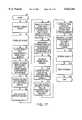
- FIG. 17shows an embodiment of the method of excisional biopsy method according to the present invention.
- the soft tissue from which the lesion is to be excisedis breast tissue and that the active element 440 of the removable core 400 is an ultrasound transducer.
- the removable core 400 and the active element 440, in FIG. 17,are together abbreviated as "US CORE", a shorthand expression for the phrase “ultrasound core” and the word “assembly” is abbreviated to "Ass'y”.
- the steps shown in FIG. 17constitute but a broad outline of one possible embodiment of the present inventive method. Therefore, other additional steps may be inserted between the steps shown in FIG. 17, or other steps may be substituted for some of the displayed steps without, however, departing from the scope of the present invention.
- step S1the lesion within the breast is grossly targeted, using, for example, standard or stereotactic surface ultrasound.
- a rough estimate of the location of the lesion within the breastis obtained.
- the surgeonafter having located the general location of the lesion, may mark the location thereof on the ultrasound display or displays and/or on the corresponding surface of the breast, with an "X", for example.
- the breastis stabilized in step S2.
- the woman's other breastis preferably placed within a counterpart breast stabilizing device, which helps to immobilize the woman during the procedure.
- One of the ultrasound ports of the breast stabilizing deviceis aligned with the lesion, for example, by aligning one of its ultrasound ports with the marked location on the breast.
- an entry site on the breastis chosen.
- the peri-areolar regionis chosen as the incision site, as scars within the peri-areolar region are less visible than scars in more exposed regions of the breast and for other anatomical reasons.
- the incision siteis then anaesthetized, both on the skin surface and subcutaneously.
- a small incisionis made at the chosen incision site.
- the incisionis large enough to accommodate the expandable sheath 495 with the removable core 400 inserted therein.
- the expandable sheath 495, together with the removable core inserted therethrough,is inserted into the incision made in step S3.
- the sheath 495/core 400 assemblyis navigated adjacent to the lesion. If the sheath 495/core 400 assembly can be properly positioned adjacent to the target lesion, the method according to the present invention proceeds to step S5. If the sheath 495/core 400 assembly cannot be properly positioned adjacent to the target lesion, all or a portion of the above-detailed steps are repeated until proper positioning of the sheath 495/core 400 assembly is achieved, adjacent to the target lesion.
- step S4the core 400 is removed from the expandable sheath 495 and the expandable sheath 495 is left in place within the breast, as shown in step S5.
- step S6the removable core 400 is inserted within the internal lumen 420 of the tubular member of the excisional device 100 and locked securely in place, so that the active element 440 (in this case, an ultrasound transducer) is aligned with and faces out of the transducer window 410 of the device 100.
- the excisional device 100is advanced through the expandable sheath 495.
- the sheath 495then expands within the breast tissue to accommodate the somewhat larger diameter of the excisional device 100.
- the excisional device 100is advanced past the tapered distal end 520 of the sheath 495, so the assembly including the sheath 495, the excisional device 100 and the removable core 400 is positioned adjacent to the target lesion within the breast tissue, as shown in step S7.
- step S8the correct position adjacent the target lesion and the correct rotational orientation of the aforementioned assembly (FIG. 14) is confirmed, using surface ultrasound and/or the core ultrasound 400.
- the active element 440 of the core 400is particularly well suited for this task, as the excisional device 100 may be rotated within the tissue, and positioned so the cutting tool 125 is properly positioned to allow it to rotate, extend and bow outwardly in such a manner as to precisely sever the lesion from the surrounding tissue with an adequate margin of healthy tissue.
- the ultrasound transducer 440as it rotates along with the excisional device 100, images the lesion before the cutting tool 125 cuts it, thereby allowing the surgeon to optimally deploy the cutting tool based upon his or her observation of the imaged tissue on a display or displays.
- the surgeonmay activate an anaesthetic infusion, the anaesthetic being delivered by the cutting tool 125 via the plurality of through holes 126, best seen in FIGS. 4 and 6.
- Step S9may be skipped if the cutting tool 125 does not provide for through holes 126 or if the surgeon does not deem it necessary to anaesthetize the tissue during the rotation of the cutting tool 125.
- the tissuemay have been previously anaesthetized.
- the cutting tool 125While rotating at least the excisional device 100 (with the removable core 400 secured therein), the cutting tool 125 is extended using, for example the push or turn knob 226 shown in FIGS. 2C and 14, thereby causing the cutting tool 125 to extend from the window 410 and to bow outwardly, as shown in step S10.
- the sheath 495may rotated along with the excisional device 100. The degree of extension and bowing may be finely controlled by the surgeon as the excisional device 100 is rotated, either manually or by a motorized unit (not shown) coupled thereto.
- the severed tissue sampleis preferably collected (e.g., bagged) in a tissue collection device 260 (FIGS. 2A and 2B), as shown at step S11.
- the blood vesselsmay be coagulated as the cutting tools rotates and cuts the tissue, or afterwards.
- step S12after the excisional device 100 has completed at least one revolution within the breast and has cut a volume of revolution therein, including at least the target lesion and preferably a margin of healthy tissue surrounding the lesion, the excisional biopsy device 100 and removable core 400 assembly are retracted through the sheath 495, leaving the sheath 495 once again in place within the breast.
- the tissue collection device 260 and the tissue sample it encloseslie within the recessed section 131 of the generally tubular member 110.
- the filled collection device 260does not protrude or protrude too much from the surface of the tubular member 110, thereby allowing the retrieved tissue sample to be readily retracted with the excisional device 100 through the sheath 495.
- the core 400may be retracted from the device 100 and re-inserted through the sheath 495 left in place within the breast.
- the core 400is then advanced adjacent to the excision site, and rotated to allow the surgeon to image the excision site to insure that the entire lesion has indeed been removed, as shown in step S13.
- Some or all of the above stepsmay be repeated should the imaging of the excision site by the core 400 within the sheath 495 reveal that a portion of the target lesion was not excised. Assuming that all of the target lesion has been removed, the incision is repaired by, for example, suturing the peri-areolar incision site. The method ends at step S16.
Landscapes
- Health & Medical Sciences (AREA)
- Life Sciences & Earth Sciences (AREA)
- Surgery (AREA)
- Engineering & Computer Science (AREA)
- Veterinary Medicine (AREA)
- Medical Informatics (AREA)
- Molecular Biology (AREA)
- Biomedical Technology (AREA)
- Animal Behavior & Ethology (AREA)
- General Health & Medical Sciences (AREA)
- Public Health (AREA)
- Heart & Thoracic Surgery (AREA)
- Pathology (AREA)
- Nuclear Medicine, Radiotherapy & Molecular Imaging (AREA)
- Physics & Mathematics (AREA)
- Radiology & Medical Imaging (AREA)
- Vascular Medicine (AREA)
- Plasma & Fusion (AREA)
- Otolaryngology (AREA)
- Biophysics (AREA)
- Surgical Instruments (AREA)
- Ultra Sonic Daignosis Equipment (AREA)
- Endoscopes (AREA)
- Length Measuring Devices Characterised By Use Of Acoustic Means (AREA)
- Gyroscopes (AREA)
- Pulleys (AREA)
Abstract
Description
Claims (22)
Priority Applications (31)
| Application Number | Priority Date | Filing Date | Title |
|---|---|---|---|
| US09/146,743US6022362A (en) | 1998-09-03 | 1998-09-03 | Excisional biopsy devices and methods |
| AT99306621TATE295702T1 (en) | 1998-09-03 | 1999-08-20 | DEVICE FOR EXCISION BIOPSY |
| DE69925344TDE69925344T2 (en) | 1998-09-03 | 1999-08-20 | Device for excision biopsy |
| EP99306621AEP0983749B1 (en) | 1998-09-03 | 1999-08-20 | Excisional biopsy device |
| CA002280792ACA2280792C (en) | 1998-09-03 | 1999-08-26 | Excisional biopsy devices and methods |
| CA002413861ACA2413861C (en) | 1998-09-03 | 1999-08-26 | Excisional biopsy devices and methods |
| CA002413859ACA2413859C (en) | 1998-09-03 | 1999-08-26 | Excisional biopsy devices and methods |
| NZ337585ANZ337585A (en) | 1998-09-03 | 1999-09-02 | Devices for excising lesions from soft (breast) tissue for biopsy |
| JP24939099AJP3315951B2 (en) | 1998-09-03 | 1999-09-02 | Incision biopsy device |
| AU45869/99AAU4586999A (en) | 1998-09-03 | 1999-09-02 | Excisional biopsy devices and methods |
| US09/417,520US6423081B1 (en) | 1998-09-03 | 1999-10-13 | Excisional biopsy devices and methods |
| HK00101838.4AHK1022824B (en) | 1998-09-03 | 2000-03-24 | Excisional biopsy device |
| US09/565,611US6440147B1 (en) | 1998-09-03 | 2000-05-04 | Excisional biopsy devices and methods |
| JP2002013543AJP3845023B2 (en) | 1998-09-03 | 2002-01-22 | Incisional biopsy device and method |
| US10/066,462US6702831B2 (en) | 1998-09-03 | 2002-01-31 | Excisional biopsy devices and methods |
| US10/066,437US6849080B2 (en) | 1998-09-03 | 2002-01-31 | Excisional biopsy device and methods |
| US10/066,428US6689145B2 (en) | 1998-09-03 | 2002-01-31 | Excisional biopsy devices and methods |
| US10/066,353US6863676B2 (en) | 1998-09-03 | 2002-01-31 | Excisional biopsy devices and methods |
| US10/098,014US6764495B2 (en) | 1998-09-03 | 2002-03-14 | Excisional biopsy devices and methods |
| AU2002336281AAU2002336281B2 (en) | 1998-09-03 | 2002-12-30 | Excisional Biopsy Devices and Methods |
| AU2002336282AAU2002336282B2 (en) | 1998-09-03 | 2002-12-30 | Excisional Biopsy Devices and Methods |
| US10/796,328US7303531B2 (en) | 1998-09-03 | 2004-03-08 | Excisional biopsy devices and methods |
| US10/923,511US7517348B2 (en) | 1998-09-03 | 2004-08-20 | Devices and methods for performing procedures on a breast |
| US11/045,006US20050182339A1 (en) | 1998-09-03 | 2005-01-26 | Excisional biopsy devices and methods |
| US11/740,858US20070203428A1 (en) | 1998-09-03 | 2007-04-26 | Devices and methods for performing procedures on a breast |
| US11/740,847US20070203427A1 (en) | 1998-09-03 | 2007-04-26 | Devices and methods for performing procedures on a breast |
| US11/740,855US20070197934A1 (en) | 1998-09-03 | 2007-04-26 | Devices and methods for performing procedures on a breast |
| US11/924,568US20080045987A1 (en) | 1998-09-03 | 2007-10-25 | Excisional biopsy device |
| US12/895,698US20110021948A1 (en) | 1998-09-03 | 2010-09-30 | Excisional biopsy devices and methods |
| US13/668,144US20130226028A1 (en) | 1998-09-03 | 2012-11-02 | Devices and methods for performing procedures on a breast |
| US13/771,046US20130231571A1 (en) | 1998-09-03 | 2013-02-19 | Excisional biopsy devices and methods |
Applications Claiming Priority (1)
| Application Number | Priority Date | Filing Date | Title |
|---|---|---|---|
| US09/146,743US6022362A (en) | 1998-09-03 | 1998-09-03 | Excisional biopsy devices and methods |
Related Child Applications (1)
| Application Number | Title | Priority Date | Filing Date |
|---|---|---|---|
| US09/417,520DivisionUS6423081B1 (en) | 1998-09-03 | 1999-10-13 | Excisional biopsy devices and methods |
Publications (1)
| Publication Number | Publication Date |
|---|---|
| US6022362Atrue US6022362A (en) | 2000-02-08 |
Family
ID=22518808
Family Applications (9)
| Application Number | Title | Priority Date | Filing Date |
|---|---|---|---|
| US09/146,743Expired - LifetimeUS6022362A (en) | 1998-09-03 | 1998-09-03 | Excisional biopsy devices and methods |
| US09/417,520Expired - LifetimeUS6423081B1 (en) | 1998-09-03 | 1999-10-13 | Excisional biopsy devices and methods |
| US10/066,437Expired - Fee RelatedUS6849080B2 (en) | 1998-09-03 | 2002-01-31 | Excisional biopsy device and methods |
| US10/066,462Expired - Fee RelatedUS6702831B2 (en) | 1998-09-03 | 2002-01-31 | Excisional biopsy devices and methods |
| US10/098,014Expired - Fee RelatedUS6764495B2 (en) | 1998-09-03 | 2002-03-14 | Excisional biopsy devices and methods |
| US10/796,328Expired - Fee RelatedUS7303531B2 (en) | 1998-09-03 | 2004-03-08 | Excisional biopsy devices and methods |
| US11/924,568AbandonedUS20080045987A1 (en) | 1998-09-03 | 2007-10-25 | Excisional biopsy device |
| US12/895,698AbandonedUS20110021948A1 (en) | 1998-09-03 | 2010-09-30 | Excisional biopsy devices and methods |
| US13/771,046AbandonedUS20130231571A1 (en) | 1998-09-03 | 2013-02-19 | Excisional biopsy devices and methods |
Family Applications After (8)
| Application Number | Title | Priority Date | Filing Date |
|---|---|---|---|
| US09/417,520Expired - LifetimeUS6423081B1 (en) | 1998-09-03 | 1999-10-13 | Excisional biopsy devices and methods |
| US10/066,437Expired - Fee RelatedUS6849080B2 (en) | 1998-09-03 | 2002-01-31 | Excisional biopsy device and methods |
| US10/066,462Expired - Fee RelatedUS6702831B2 (en) | 1998-09-03 | 2002-01-31 | Excisional biopsy devices and methods |
| US10/098,014Expired - Fee RelatedUS6764495B2 (en) | 1998-09-03 | 2002-03-14 | Excisional biopsy devices and methods |
| US10/796,328Expired - Fee RelatedUS7303531B2 (en) | 1998-09-03 | 2004-03-08 | Excisional biopsy devices and methods |
| US11/924,568AbandonedUS20080045987A1 (en) | 1998-09-03 | 2007-10-25 | Excisional biopsy device |
| US12/895,698AbandonedUS20110021948A1 (en) | 1998-09-03 | 2010-09-30 | Excisional biopsy devices and methods |
| US13/771,046AbandonedUS20130231571A1 (en) | 1998-09-03 | 2013-02-19 | Excisional biopsy devices and methods |
Country Status (8)
| Country | Link |
|---|---|
| US (9) | US6022362A (en) |
| EP (1) | EP0983749B1 (en) |
| JP (2) | JP3315951B2 (en) |
| AT (1) | ATE295702T1 (en) |
| AU (1) | AU4586999A (en) |
| CA (1) | CA2280792C (en) |
| DE (1) | DE69925344T2 (en) |
| NZ (1) | NZ337585A (en) |
Cited By (226)
| Publication number | Priority date | Publication date | Assignee | Title |
|---|---|---|---|---|
| US6206844B1 (en)* | 1997-02-28 | 2001-03-27 | Ethicon Endo-Surgery, Inc. | Reusable ultrasonic surgical instrument with removable outer sheath |
| US6277083B1 (en) | 1999-12-27 | 2001-08-21 | Neothermia Corporation | Minimally invasive intact recovery of tissue |
| WO2001060262A1 (en)* | 2000-02-16 | 2001-08-23 | Axiamed, Inc. | Apparatus for forming shaped axial bores through spinal vertebrae |
| US6287304B1 (en) | 1999-10-15 | 2001-09-11 | Neothermia Corporation | Interstitial cauterization of tissue volumes with electrosurgically deployed electrodes |
| US6304770B1 (en)* | 1998-11-25 | 2001-10-16 | Rubicor Medical, Inc. | Breast stabilization devices and imaging and interventional methods using same |
| US20020022788A1 (en)* | 1999-08-19 | 2002-02-21 | Tim Corvi | Apparatus and methods for material capture and removal |
| WO2002045609A1 (en)* | 2000-12-07 | 2002-06-13 | Rubicor Medical, Inc. | Methods and devices for radiofrequency electrosurgery |
| US6405733B1 (en) | 2000-02-18 | 2002-06-18 | Thomas J. Fogarty | Device for accurately marking tissue |
| US20020077648A1 (en)* | 1998-09-03 | 2002-06-20 | Rubicor Medical, Inc. | Excisional biopsy devices and methods |
| US20020077642A1 (en)* | 2000-12-20 | 2002-06-20 | Fox Hollow Technologies, Inc. | Debulking catheter |
| US20020123762A1 (en)* | 1998-09-03 | 2002-09-05 | Rubicor Medical, Inc. | Excisional biopsy device and methods |
| US6471659B2 (en) | 1999-12-27 | 2002-10-29 | Neothermia Corporation | Minimally invasive intact recovery of tissue |
| US6471709B1 (en)* | 1998-10-30 | 2002-10-29 | Vivant Medical, Inc. | Expandable ring percutaneous tissue removal device |
| US6514248B1 (en) | 1999-10-15 | 2003-02-04 | Neothermia Corporation | Accurate cutting about and into tissue volumes with electrosurgically deployed electrodes |
| WO2003022157A2 (en) | 2001-09-12 | 2003-03-20 | Manoa Medical, Inc. | Devices and methods for tissue severing and removal |
| US6540695B1 (en) | 1998-04-08 | 2003-04-01 | Senorx, Inc. | Biopsy anchor device with cutter |
| US20030083656A1 (en)* | 2000-11-07 | 2003-05-01 | George Morrison | Tissue separator assembly and method |
| US6558386B1 (en) | 2000-02-16 | 2003-05-06 | Trans1 Inc. | Axial spinal implant and method and apparatus for implanting an axial spinal implant within the vertebrae of the spine |
| US6558390B2 (en) | 2000-02-16 | 2003-05-06 | Axiamed, Inc. | Methods and apparatus for performing therapeutic procedures in the spine |
| US20030109896A1 (en)* | 1998-02-10 | 2003-06-12 | Artemis Medical, Inc. | Tissue removal methods and apparatus |
| US20030120295A1 (en)* | 2000-12-20 | 2003-06-26 | Fox Hollow Technologies, Inc. | Debulking catheters and methods |
| US20030125757A1 (en)* | 2000-12-20 | 2003-07-03 | Fox Hollow Technologies, Inc. | Debulking catheters and methods |
| US20030125758A1 (en)* | 2000-12-20 | 2003-07-03 | Fox Hollow Technologies, Inc. | Debulking catheters and methods |
| US6589254B2 (en) | 1999-09-14 | 2003-07-08 | Mark G. Fontenot | Breast bracket |
| US6623496B2 (en)* | 1999-08-19 | 2003-09-23 | Fox Hollow Technologies, Inc. | Atherectomy catheter with aligned imager |
| US20030195500A1 (en)* | 1999-06-17 | 2003-10-16 | Moorman Jack W. | Needle kit and method for microwave ablation, track coagulation, and biopsy |
| US20030195499A1 (en)* | 2002-04-16 | 2003-10-16 | Mani Prakash | Microwave antenna having a curved configuration |
| US20030204188A1 (en)* | 2001-11-07 | 2003-10-30 | Artemis Medical, Inc. | Tissue separating and localizing catheter assembly |
| US20030220640A1 (en)* | 2002-05-22 | 2003-11-27 | Rubicor Medical , Inc. | Methods and systems for in situ tissue marking and orientation stabilization |
| US20030229341A1 (en)* | 2002-06-06 | 2003-12-11 | Albrecht Thomas E. | Method of tissue lesion removal |
| US20030229353A1 (en)* | 2000-02-16 | 2003-12-11 | Cragg Andrew H. | Method and apparatus for providing posterior or anterior trans-sacral access to spinal vertebrae |
| US20030229343A1 (en)* | 2002-06-06 | 2003-12-11 | Albrecht Thomas E. | Device for removal of tissue lesions |
| US6663628B2 (en) | 2000-09-24 | 2003-12-16 | Medtronic, Inc. | Surgical micro-resecting instrument with electrocautery feature |
| US20040006338A1 (en)* | 2002-07-03 | 2004-01-08 | Rubicor Medical, Inc. | Methods and devices for cutting and collecting soft tissue |
| US20040006355A1 (en)* | 2002-07-03 | 2004-01-08 | Rubicor Medical, Inc. | Methods and devices for cutting and collecting soft tissue |
| US20040024396A1 (en)* | 1999-12-27 | 2004-02-05 | Eggers Philip E. | Electrosurgical accessing of tissue with controlled collateral thermal phenomena |
| US20040066515A1 (en)* | 2000-12-08 | 2004-04-08 | Gretag-Macbeth Ag | Device for the pixel-by-pixel photoelectric measurement of a planar measured object |
| US20040068172A1 (en)* | 1996-02-27 | 2004-04-08 | Kent Ridge Digital Labs | Curved surgical instruments and method of mapping a curved path for stereotactic surgery |
| US6722371B1 (en) | 2000-02-18 | 2004-04-20 | Thomas J. Fogarty | Device for accurately marking tissue |
| US20040077971A1 (en)* | 2002-10-16 | 2004-04-22 | Rubicor Medical, Inc. | Devices and methods for performing procedures on a breast |
| US20040087872A1 (en)* | 2002-11-06 | 2004-05-06 | Rubicor Medical, Inc. | Excisional devices having selective cutting and atraumatic configurations and methods of using same |
| US20040087942A1 (en)* | 1997-07-24 | 2004-05-06 | Rex Medical | Surgical biopsy device |
| US20040116828A1 (en)* | 2002-11-07 | 2004-06-17 | White Keith M. | Biological specimen collector and container with stand |
| US6752767B2 (en) | 2002-04-16 | 2004-06-22 | Vivant Medical, Inc. | Localization element with energized tip |
| US6752154B2 (en) | 2000-02-18 | 2004-06-22 | Thomas J. Fogarty | Device for accurately marking tissue |
| US6770070B1 (en) | 2000-03-17 | 2004-08-03 | Rita Medical Systems, Inc. | Lung treatment apparatus and method |
| US20040167511A1 (en)* | 2003-02-25 | 2004-08-26 | Artemis Medical, Inc. | Tissue separating catheter assembly and method |
| US20040167554A1 (en)* | 2000-12-20 | 2004-08-26 | Fox Hollow Technologies, Inc. | Methods and devices for reentering a true lumen from a subintimal space |
| US20040167553A1 (en)* | 2000-12-20 | 2004-08-26 | Fox Hollow Technologies, Inc. | Methods and devices for cutting tissue |
| US20040215103A1 (en)* | 2003-04-24 | 2004-10-28 | Mueller Richard L. | Biopsy device |
| US20040255739A1 (en)* | 2003-06-18 | 2004-12-23 | Rubicor Medical, Inc. | Methods and devices for cutting and collecting soft tissue |
| US20050015081A1 (en)* | 2003-07-18 | 2005-01-20 | Roman Turovskiy | Devices and methods for cooling microwave antennas |
| US20050019262A1 (en)* | 2003-07-25 | 2005-01-27 | Rubicor Medical, Inc. | Post-biopsy cavity treatment implants and methods |
| US20050038439A1 (en)* | 2002-01-17 | 2005-02-17 | Concept Matrix, Llc | Diskectomy instrument and method |
| US20050054945A1 (en)* | 2003-09-10 | 2005-03-10 | Scimed Life Systems, Inc | Forceps and collection assembly with accompanying mechanisms and related methods of use |
| US20050096649A1 (en)* | 2003-11-04 | 2005-05-05 | Medtronic, Inc. | Surgical micro-resecting instrument with electrocautery and continuous aspiration features |
| US6899716B2 (en) | 2000-02-16 | 2005-05-31 | Trans1, Inc. | Method and apparatus for spinal augmentation |
| US20050119652A1 (en)* | 1998-09-03 | 2005-06-02 | Rubicor Medical, Inc. | Devices and methods for performing procedures on a breast |
| US20050124986A1 (en)* | 2003-12-09 | 2005-06-09 | Rubicor Medical, Inc. | Suction sleeve and interventional devices having such a suction sleeve |
| US20050130590A1 (en)* | 2001-05-21 | 2005-06-16 | Ashutosh Pande | Distributed data collection of satellite data |
| US20050137605A1 (en)* | 2003-10-23 | 2005-06-23 | Assell Robert L. | Guide pin for guiding instrumentation along a soft tissue tract to a point on the spine |
| US20050143674A1 (en)* | 1998-09-01 | 2005-06-30 | Burbank Fred H. | Methods and apparatus for securing medical instruments to desired locations in a patient's body |
| US20050154407A1 (en)* | 2000-12-20 | 2005-07-14 | Fox Hollow Technologies, Inc. | Method of evaluating drug efficacy for treating atherosclerosis |
| US20050177068A1 (en)* | 2000-12-20 | 2005-08-11 | Fox Hollow Technologies, Inc. | Debulking catheters and methods |
| US6942627B2 (en)* | 2001-07-19 | 2005-09-13 | Ethicon Endo-Surgery, Inc. | Surgical biopsy device having a flexible cutter |
| US20050216019A1 (en)* | 2002-01-17 | 2005-09-29 | Eckman Walter W | Diskectomy instrument with disposable blade head |
| US6955676B2 (en) | 1999-06-22 | 2005-10-18 | Senorx, Inc. | Shaped scalpel |
| US20050261695A1 (en)* | 2000-02-16 | 2005-11-24 | Cragg Andrew H | Method and apparatus for spinal distraction and fusion |
| US6981949B2 (en) | 2002-06-06 | 2006-01-03 | Ethicon Endo-Surgery, Inc. | Perimeter cut biopsy probe |
| US20060036271A1 (en)* | 2004-07-29 | 2006-02-16 | X-Sten, Inc. | Spinal ligament modification devices |
| US20060089633A1 (en)* | 2004-10-15 | 2006-04-27 | Baxano, Inc. | Devices and methods for tissue access |
| US20060089640A1 (en)* | 2004-10-15 | 2006-04-27 | Baxano, Inc. | Devices and methods for tissue modification |
| US20060122458A1 (en)* | 2004-10-15 | 2006-06-08 | Baxano, Inc. | Devices and methods for tissue access |
| US20060129185A1 (en)* | 1999-10-25 | 2006-06-15 | Boston Scientific Scimed, Inc. | Forceps for medical use |
| US7066893B2 (en) | 2002-06-06 | 2006-06-27 | Ethicon Endo-Surgery, Inc. | Biopsy method |
| US20060173525A1 (en)* | 2005-02-02 | 2006-08-03 | Percutaneous Systems, Inc. | Methods and systems for deploying luminal prostheses |
| US20060206106A1 (en)* | 2002-11-13 | 2006-09-14 | Artemis Medical, Inc. | Devices and methods for controlling movement of an electrosurgical electrode |
| US20060236019A1 (en)* | 2005-04-19 | 2006-10-19 | Fox Hollow Technologies, Inc. | Libraries and data structures of materials removed by debulking catheters |
| US20060235366A1 (en)* | 2000-12-20 | 2006-10-19 | Fox Hollow Technologies, Inc. | Method of evaluating a treatment for vascular disease |
| US20060239982A1 (en)* | 2000-12-20 | 2006-10-26 | Fox Hollow Technologies, Inc. | Debulking catheters and methods |
| US20060241648A1 (en)* | 2005-02-04 | 2006-10-26 | Bleich Jeffery L | Methods and apparatus for tissue modification |
| US20060258951A1 (en)* | 2005-05-16 | 2006-11-16 | Baxano, Inc. | Spinal Access and Neural Localization |
| US20060258954A1 (en)* | 2005-05-13 | 2006-11-16 | Tyler Timberlake | Biopsy forceps assemblies |
| US20060264957A1 (en)* | 2000-02-16 | 2006-11-23 | Trans1, Inc. | Apparatus for performing a discectomy through a trans-sacral axial bore within the vertebrae of the spine |
| US20070027464A1 (en)* | 2005-07-29 | 2007-02-01 | X-Sten, Corp. | Device for resecting spinal tissue |
| US20070038173A1 (en)* | 2005-07-27 | 2007-02-15 | Fox Hollow Technologies, Inc. | Methods affecting markers in patients having vascular disease |
| US20070055260A1 (en)* | 2003-06-10 | 2007-03-08 | Cragg Andrew H | Method and apparatus for providing posterior or anterior trans-sacral access to spinal vertebrae |
| US20070055263A1 (en)* | 2005-07-29 | 2007-03-08 | X-Sten Corp. | Tools for Percutaneous Spinal Ligament Decompression and Device for Supporting Same |
| US20070066977A1 (en)* | 2004-10-22 | 2007-03-22 | Assell Robert L | Exchange system for axial spinal procedures |
| US20070073107A1 (en)* | 2005-09-23 | 2007-03-29 | Percutaneous Systems, Inc. | Methods and systems for sheathing tubular medical instruments |
| US20070078469A1 (en)* | 2000-12-20 | 2007-04-05 | Fox Hollow Technologies, Inc | Testing a patient population having a cardiovascular condition for drug efficacy |
| US20070083196A1 (en)* | 2005-10-11 | 2007-04-12 | Evanston Northwestern Healthcare Research Institute | Surgical instrument and method for tissue resection and dissection |
| US20070123888A1 (en)* | 2004-10-15 | 2007-05-31 | Baxano, Inc. | Flexible tissue rasp |
| US20070138915A1 (en)* | 2005-12-16 | 2007-06-21 | Maureen Mulvihill | Piezoelectric micro-device for blockage removal |
| US20070156064A1 (en)* | 1994-03-24 | 2007-07-05 | Ritchart Mark A | Methods and Devices for Automated Biopsy and Collection of Soft Tissue |
| USD549344S1 (en) | 2003-01-16 | 2007-08-21 | Concept Matrix, Llc | Diskectomy tool with convex head |
| US20070213733A1 (en)* | 2004-10-15 | 2007-09-13 | Bleich Jeffery L | Mechanical tissue modification devices and methods |
| US20070213735A1 (en)* | 2004-10-15 | 2007-09-13 | Vahid Saadat | Powered tissue modification devices and methods |
| US20070213734A1 (en)* | 2006-03-13 | 2007-09-13 | Bleich Jeffery L | Tissue modification barrier devices and methods |
| US20070233260A1 (en)* | 2000-02-16 | 2007-10-04 | Trans1 Inc. | Articulating spinal implant |
| US20070255277A1 (en)* | 2006-04-28 | 2007-11-01 | Nobis Rudolph H | Apparatus and method for performing an endoscopic mucosal resection |
| US20070255278A1 (en)* | 2006-04-28 | 2007-11-01 | Nobis Rudolph H | Apparatus and method for deploying a cutting element during an endoscopic mucosal resection |
| US20070255268A1 (en)* | 2006-04-28 | 2007-11-01 | Nobis Rudolph H | Method for performing an endoscopic mucosal resection |
| US20070260104A1 (en)* | 2006-05-08 | 2007-11-08 | Bretz Phillip | Autologous fat pad transfer for use with partial breast irradiation |
| US20070260252A1 (en)* | 2006-05-04 | 2007-11-08 | Baxano, Inc. | Tissue Removal with at Least Partially Flexible Devices |
| US20070270643A1 (en)* | 2006-05-19 | 2007-11-22 | Ifung Lu | Lumen stabilizer for endoscopic mucosal resection |
| US20070276390A1 (en)* | 2006-05-09 | 2007-11-29 | X-Sten, Inc. | Ipsilateral Approach to Minimally Invasive Ligament Decompression Procedure |
| US7318824B2 (en) | 2001-11-02 | 2008-01-15 | Vivant Medical, Inc. | High-strength microwave antenna assemblies |
| US20080051812A1 (en)* | 2006-08-01 | 2008-02-28 | Baxano, Inc. | Multi-Wire Tissue Cutter |
| US20080065076A1 (en)* | 2000-02-16 | 2008-03-13 | Cragg Andrew H | Spinal mobility preservation apparatus |
| US20080082093A1 (en)* | 2006-09-29 | 2008-04-03 | Prakash Mani N | Microwave antenna assembly and method of using the same |
| US20080086034A1 (en)* | 2006-08-29 | 2008-04-10 | Baxano, Inc. | Tissue Access Guidewire System and Method |
| US20080091227A1 (en)* | 2006-08-25 | 2008-04-17 | Baxano, Inc. | Surgical probe and method of making |
| US20080111101A1 (en)* | 2006-11-09 | 2008-05-15 | Jason Keleher | Compositions and methods for CMP of low-k-dielectric materials |
| US20080147084A1 (en)* | 2006-12-07 | 2008-06-19 | Baxano, Inc. | Tissue removal devices and methods |
| US20080146965A1 (en)* | 2003-08-11 | 2008-06-19 | Salvatore Privitera | Surgical Device for The Collection of Soft Tissue |
| US20080161809A1 (en)* | 2006-10-03 | 2008-07-03 | Baxano, Inc. | Articulating Tissue Cutting Device |
| US20080221383A1 (en)* | 2007-02-12 | 2008-09-11 | Vertos Medical, Inc. | Tissue excision devices and methods |
| US7425202B2 (en) | 2004-03-05 | 2008-09-16 | Percutaneous Systems, Inc. | Non-seeding biopsy device and method |
| US20080275458A1 (en)* | 2004-10-15 | 2008-11-06 | Bleich Jeffery L | Guidewire exchange systems to treat spinal stenosis |
| US20080281323A1 (en)* | 1999-01-27 | 2008-11-13 | Burbank Fred H | Tissue specimen isolating and damaging device and method |
| US20080294167A1 (en)* | 2007-05-21 | 2008-11-27 | Brian Schumacher | Articulating cavitation device |
| US20080312660A1 (en)* | 2007-06-15 | 2008-12-18 | Baxano, Inc. | Devices and methods for measuring the space around a nerve root |
| US20090018507A1 (en)* | 2007-07-09 | 2009-01-15 | Baxano, Inc. | Spinal access system and method |
| US20090033553A1 (en)* | 2001-04-05 | 2009-02-05 | Sirf Technology, Inc. | Gps-based positioning system for mobile gps terminals |
| US20090112119A1 (en)* | 2007-10-31 | 2009-04-30 | Kim Stanley I | Rotating biopsy device and biopsy robot |
| US20090118709A1 (en)* | 2007-11-06 | 2009-05-07 | Vertos Medical, Inc. A Delaware Corporation | Tissue Excision Tool, Kits and Methods of Using the Same |
| US20090125036A1 (en)* | 2004-10-15 | 2009-05-14 | Bleich Jeffery L | Devices and methods for selective surgical removal of tissue |
| US20090138005A1 (en)* | 2007-11-27 | 2009-05-28 | Vivant Medical, Inc. | Targeted Cooling of Deployable Microwave Antenna |
| US20090149865A1 (en)* | 2007-12-07 | 2009-06-11 | Schmitz Gregory P | Tissue modification devices |
| US7547317B2 (en) | 2000-02-16 | 2009-06-16 | Trans1 Inc. | Methods of performing procedures in the spine |
| US20090177241A1 (en)* | 2005-10-15 | 2009-07-09 | Bleich Jeffery L | Multiple pathways for spinal nerve root decompression from a single access point |
| US20090182478A1 (en)* | 2008-01-15 | 2009-07-16 | Gm Global Technology Operations, Inc. | Axle torque based cruise control |
| USD606654S1 (en) | 2006-07-31 | 2009-12-22 | Vertos Medical, Inc. | Tissue excision device |
| USD610259S1 (en) | 2008-10-23 | 2010-02-16 | Vertos Medical, Inc. | Tissue modification device |
| USD611146S1 (en) | 2008-10-23 | 2010-03-02 | Vertos Medical, Inc. | Tissue modification device |
| US7708749B2 (en) | 2000-12-20 | 2010-05-04 | Fox Hollow Technologies, Inc. | Debulking catheters and methods |
| US20100121361A1 (en)* | 2008-06-05 | 2010-05-13 | Cardiovascular Systems, Inc. | Directional rotational atherectomy device with offset spinning abrasive element |
| US7744852B2 (en) | 2003-07-25 | 2010-06-29 | Rubicor Medical, Llc | Methods and systems for marking post biopsy cavity sites |
| USD619252S1 (en) | 2008-10-23 | 2010-07-06 | Vertos Medical, Inc. | Tissue modification device |
| USD619253S1 (en) | 2008-10-23 | 2010-07-06 | Vertos Medical, Inc. | Tissue modification device |
| USD620593S1 (en) | 2006-07-31 | 2010-07-27 | Vertos Medical, Inc. | Tissue excision device |
| USD621939S1 (en) | 2008-10-23 | 2010-08-17 | Vertos Medical, Inc. | Tissue modification device |
| US20100321426A1 (en)* | 2007-11-22 | 2010-12-23 | Kazuki Suzuki | Image forming apparatus |
| US20100331883A1 (en)* | 2004-10-15 | 2010-12-30 | Schmitz Gregory P | Access and tissue modification systems and methods |
| US20100331900A1 (en)* | 2009-06-25 | 2010-12-30 | Baxano, Inc. | Surgical tools for treatment of spinal stenosis |
| US20110022042A1 (en)* | 2005-12-29 | 2011-01-27 | Boston Scientific Scimed, Inc. | Tissue ablation probes and methods for treating osteoid osteomas |
| USD635671S1 (en) | 2008-10-23 | 2011-04-05 | Vertos Medical, Inc. | Tissue modification device |
| US20110112539A1 (en)* | 2008-07-14 | 2011-05-12 | Wallace Michael P | Tissue modification devices |
| US20110112373A1 (en)* | 2009-11-10 | 2011-05-12 | Trans1 Inc. | Soft tissue access apparatus and methods for spinal surgery |
| US7942896B2 (en) | 2003-11-25 | 2011-05-17 | Scimed Life Systems, Inc. | Forceps and collection assembly and related methods of use and manufacture |
| US7959577B2 (en) | 2007-09-06 | 2011-06-14 | Baxano, Inc. | Method, system, and apparatus for neural localization |
| US20110160731A1 (en)* | 2004-10-15 | 2011-06-30 | Bleich Jeffery L | Devices and methods for tissue access |
| US7989207B2 (en) | 2006-02-17 | 2011-08-02 | Tyco Healthcare Group Lp | Testing lumenectomy samples for markers of non-vascular diseases |
| US20110224710A1 (en)* | 2004-10-15 | 2011-09-15 | Bleich Jeffery L | Methods, systems and devices for carpal tunnel release |
| US20110282323A1 (en)* | 2003-05-21 | 2011-11-17 | Klein Jeffrey A | Infiltration cannula |
| US8062298B2 (en) | 2005-10-15 | 2011-11-22 | Baxano, Inc. | Flexible tissue removal devices and methods |
| US20120004662A1 (en)* | 2010-06-30 | 2012-01-05 | Paul Alexander Torrie | Resection Instrument |
| US20120004691A1 (en)* | 2010-06-30 | 2012-01-05 | Paul Alexander Torrie | Bone And Tissue Marker |
| US8192452B2 (en) | 2009-05-14 | 2012-06-05 | Tyco Healthcare Group Lp | Easily cleaned atherectomy catheters and methods of use |
| US8221397B2 (en) | 2004-10-15 | 2012-07-17 | Baxano, Inc. | Devices and methods for tissue modification |
| US8246640B2 (en) | 2003-04-22 | 2012-08-21 | Tyco Healthcare Group Lp | Methods and devices for cutting tissue at a vascular location |
| US8328829B2 (en) | 1999-08-19 | 2012-12-11 | Covidien Lp | High capacity debulking catheter with razor edge cutting window |
| US20120316463A1 (en)* | 2007-07-16 | 2012-12-13 | Dune Medical Devices Ltd. | Medical device and method for use in tissue characterization and treatment |
| US8366712B2 (en) | 2005-10-15 | 2013-02-05 | Baxano, Inc. | Multiple pathways for spinal nerve root decompression from a single access point |
| US8398641B2 (en) | 2008-07-01 | 2013-03-19 | Baxano, Inc. | Tissue modification devices and methods |
| US8409206B2 (en) | 2008-07-01 | 2013-04-02 | Baxano, Inc. | Tissue modification devices and methods |
| US8414604B2 (en) | 2008-10-13 | 2013-04-09 | Covidien Lp | Devices and methods for manipulating a catheter shaft |
| US8496677B2 (en) | 2009-12-02 | 2013-07-30 | Covidien Lp | Methods and devices for cutting tissue |
| US20140025056A1 (en)* | 2006-05-24 | 2014-01-23 | Kambiz Dowlatshahi | Image-guided removal and thermal therapy of breast cancer |
| US8685050B2 (en) | 2010-10-06 | 2014-04-01 | Rex Medical L.P. | Cutting wire assembly for use with a catheter |
| US8685049B2 (en) | 2010-11-18 | 2014-04-01 | Rex Medical L.P. | Cutting wire assembly for use with a catheter |
| US8702736B2 (en) | 2010-11-22 | 2014-04-22 | Rex Medical L.P. | Cutting wire assembly for use with a catheter |
| US8784440B2 (en) | 2008-02-25 | 2014-07-22 | Covidien Lp | Methods and devices for cutting tissue |
| US8801626B2 (en) | 2004-10-15 | 2014-08-12 | Baxano Surgical, Inc. | Flexible neural localization devices and methods |
| US8808186B2 (en) | 2010-11-11 | 2014-08-19 | Covidien Lp | Flexible debulking catheters with imaging and methods of use and manufacture |
| US8920450B2 (en) | 2010-10-28 | 2014-12-30 | Covidien Lp | Material removal device and method of use |
| US8992717B2 (en) | 2011-09-01 | 2015-03-31 | Covidien Lp | Catheter with helical drive shaft and methods of manufacture |
| CN104519810A (en)* | 2012-12-12 | 2015-04-15 | 奥林巴斯医疗株式会社 | Puncture tool and ultrasonic endoscope |
| US9028512B2 (en) | 2009-12-11 | 2015-05-12 | Covidien Lp | Material removal device having improved material capture efficiency and methods of use |
| US9101386B2 (en) | 2004-10-15 | 2015-08-11 | Amendia, Inc. | Devices and methods for treating tissue |
| US9119662B2 (en) | 2010-06-14 | 2015-09-01 | Covidien Lp | Material removal device and method of use |
| WO2015197765A1 (en)* | 2014-06-25 | 2015-12-30 | Endox Feinwerktechnik Gmbh | Hf surgical resection snare for flexible endoscopy |
| CN105358036A (en)* | 2013-06-28 | 2016-02-24 | 奥林巴斯株式会社 | Endoscope system |
| US9282991B2 (en) | 2010-10-06 | 2016-03-15 | Rex Medical, L.P. | Cutting wire assembly with coating for use with a catheter |
| US9314253B2 (en) | 2008-07-01 | 2016-04-19 | Amendia, Inc. | Tissue modification devices and methods |
| US9433402B2 (en) | 1998-10-23 | 2016-09-06 | Devicor Medical Products, Inc. | Surgical device for the collection of soft tissue |
| EP2986229A4 (en)* | 2013-04-16 | 2016-09-28 | Transmed7 Llc | Methods, devices and therapeutic platform for automated, selectable, soft tissue resection |
| US9456829B2 (en) | 2004-10-15 | 2016-10-04 | Amendia, Inc. | Powered tissue modification devices and methods |
| US9532844B2 (en) | 2012-09-13 | 2017-01-03 | Covidien Lp | Cleaning device for medical instrument and method of use |
| US9550010B2 (en) | 2010-07-02 | 2017-01-24 | Agnovos Healthcare, Llc | Methods of treating degenerative bone conditions |
| US9687266B2 (en) | 2009-04-29 | 2017-06-27 | Covidien Lp | Methods and devices for cutting and abrading tissue |
| US9707012B2 (en) | 2015-07-31 | 2017-07-18 | Polygon Medical, Inc. | Polypectomy systems, devices, and methods |
| US9801647B2 (en) | 2006-05-26 | 2017-10-31 | Covidien Lp | Catheter including cutting element and energy emitting element |
| US9814598B2 (en) | 2013-03-14 | 2017-11-14 | Quandary Medical, Llc | Spinal implants and implantation system |
| US9826888B2 (en) | 2013-06-28 | 2017-11-28 | Olympus Corporation | Endoscope having power transmission electrode and treatment tool having power reception electrode, and endoscope system |
| US9901362B2 (en) | 2007-07-16 | 2018-02-27 | Dune Medical Devices Ltd. | Medical device and method for use in tissue characterization and treatment |
| US9943329B2 (en) | 2012-11-08 | 2018-04-17 | Covidien Lp | Tissue-removing catheter with rotatable cutter |
| US9999353B2 (en) | 2007-07-16 | 2018-06-19 | Dune Medical Devices Ltd. | Medical device and method for use in tissue characterization and treatment |
| US10080571B2 (en) | 2015-03-06 | 2018-09-25 | Warsaw Orthopedic, Inc. | Surgical instrument and method |
| US10213224B2 (en) | 2014-06-27 | 2019-02-26 | Covidien Lp | Cleaning device for catheter and catheter including the same |
| GB2567000A (en)* | 2017-09-29 | 2019-04-03 | John Milton Trevor | A biopsy device |
| USD847992S1 (en) | 2017-06-27 | 2019-05-07 | Polygon Medical, Inc. | Medical device handle |
| US10285731B2 (en) | 2017-06-14 | 2019-05-14 | Polygon Medical, Inc. | Polypectomy systems, devices, and methods |
| WO2019094948A1 (en) | 2017-11-13 | 2019-05-16 | UVision360, Inc. | Biopsy device and method |
| US10292721B2 (en) | 2015-07-20 | 2019-05-21 | Covidien Lp | Tissue-removing catheter including movable distal tip |
| US10314667B2 (en) | 2015-03-25 | 2019-06-11 | Covidien Lp | Cleaning device for cleaning medical instrument |
| US10314664B2 (en) | 2015-10-07 | 2019-06-11 | Covidien Lp | Tissue-removing catheter and tissue-removing element with depth stop |
| US10372876B2 (en)* | 2017-01-20 | 2019-08-06 | Agfa Healthcare Inc. | System and method for providing breast image data |
| CN110327115A (en)* | 2019-07-23 | 2019-10-15 | 南昌航空大学 | A kind of optical coherent chromatographic imaging operation piloting method |
| CN110381849A (en)* | 2017-01-11 | 2019-10-25 | 卡尔维通医疗有限公司 | Surgical instruments with bending axis |
| US20200038090A1 (en)* | 2018-07-31 | 2020-02-06 | Ethicon, Inc. | Method for removing a tissue lesion |
| US10820825B2 (en) | 2008-10-22 | 2020-11-03 | Cornell University | Method and device for evaluation of local tissue's biological or biomechanical character |
| US20210022723A1 (en)* | 2018-04-26 | 2021-01-28 | Olympus Corporation | Surgical system and expansion device |
| CN112401992A (en)* | 2020-12-09 | 2021-02-26 | 上海睿刀医疗科技有限公司 | Multi-needle puncture guide frame |
| CN113017776A (en)* | 2020-12-24 | 2021-06-25 | 中国人民解放军总医院第三医学中心 | Superficial tumor closed resection device and use method thereof |
| US11160540B2 (en)* | 2017-09-28 | 2021-11-02 | Merit Medical Systems, Inc. | Biopsy needle sample retention system |
| US20220047314A1 (en)* | 2020-08-11 | 2022-02-17 | Ethicon, Inc. | Tissue dilation and resection systems and methods |
| US20220087661A1 (en)* | 2018-04-04 | 2022-03-24 | Praxis Holding Llc | Rotatable syringe device with side cutting biopsy needle |
| US11607205B2 (en)* | 2017-03-03 | 2023-03-21 | Arizona Board Of Regents On Behalf Of The University Of Arizona | Biopsy system for enhanced tissue harvesting |
| CN115836885A (en)* | 2023-02-28 | 2023-03-24 | 吉林大学 | A kind of tumor biopsy forceps |
| US20230190330A1 (en)* | 2020-06-18 | 2023-06-22 | Boston Scientific Medical Device Limited | Energy-emitting devices for elongated medical assembly |
| US12102348B2 (en) | 2016-09-07 | 2024-10-01 | Vertos Medical, Inc. | Percutaneous lateral recess resection methods and instruments |
| US12102372B2 (en) | 2018-07-31 | 2024-10-01 | Prana Thoracic, Inc. | Tissue resection apparatus |
| US12324572B2 (en) | 2022-06-16 | 2025-06-10 | Vertos Medical, Inc. | Integrated instrument assembly |
| US12357366B2 (en) | 2017-01-23 | 2025-07-15 | Neocision Medical, Llc | Minimally invasive diagnostic and therapeutic excision of tissue |
Families Citing this family (279)
| Publication number | Priority date | Publication date | Assignee | Title |
|---|---|---|---|---|
| US6270464B1 (en) | 1998-06-22 | 2001-08-07 | Artemis Medical, Inc. | Biopsy localization method and device |
| US9498604B2 (en) | 1997-11-12 | 2016-11-22 | Genesis Technologies Llc | Medical device and method |
| US6602204B2 (en) | 1998-02-10 | 2003-08-05 | Artemis Medical, Inc | Intraoperative tissue treatment methods |
| US20100030256A1 (en) | 1997-11-12 | 2010-02-04 | Genesis Technologies Llc | Medical Devices and Methods |
| US6602265B2 (en) | 1998-02-10 | 2003-08-05 | Artemis Medical, Inc. | Tissue separation medical device and method |
| WO1999039648A1 (en) | 1998-02-10 | 1999-08-12 | Dubrul William R | Entrapping apparatus and method for use |
| US6331166B1 (en)* | 1998-03-03 | 2001-12-18 | Senorx, Inc. | Breast biopsy system and method |
| US6758848B2 (en) | 1998-03-03 | 2004-07-06 | Senorx, Inc. | Apparatus and method for accessing a body site |
| US6497706B1 (en)* | 1998-03-03 | 2002-12-24 | Senorx, Inc. | Biopsy device and method of use |
| US6471700B1 (en) | 1998-04-08 | 2002-10-29 | Senorx, Inc. | Apparatus and method for accessing biopsy site |
| US6679851B2 (en) | 1998-09-01 | 2004-01-20 | Senorx, Inc. | Tissue accessing and anchoring device and method |
| JP4755745B2 (en)* | 1999-12-22 | 2011-08-24 | 株式会社東芝 | Ultrasonic probe with puncture adapter |
| EP1767159A1 (en)* | 2000-12-20 | 2007-03-28 | Fox Hollow Technologies Inc. | Catheter for removing atheromatous or thrombotic occlusive material |
| WO2002062226A1 (en)* | 2001-02-05 | 2002-08-15 | Tyco Healthcare Group Lp | Biopsy apparatus and method |
| US20030069502A1 (en) | 2001-05-29 | 2003-04-10 | Makin Inder Raj. S. | Ultrasound feedback in medically-treated patients |
| US7846096B2 (en) | 2001-05-29 | 2010-12-07 | Ethicon Endo-Surgery, Inc. | Method for monitoring of medical treatment using pulse-echo ultrasound |
| US11229472B2 (en) | 2001-06-12 | 2022-01-25 | Cilag Gmbh International | Modular battery powered handheld surgical instrument with multiple magnetic position sensors |
| US7166103B2 (en)* | 2001-10-01 | 2007-01-23 | Electrosurgery Associates, Llc | High efficiency electrosurgical ablator with electrode subjected to oscillatory or other repetitive motion |
| US8109885B2 (en) | 2002-03-19 | 2012-02-07 | C. R. Bard, Inc. | Biopsy device for removing tissue specimens using a vacuum |
| EP1524940B1 (en)* | 2002-03-19 | 2011-08-24 | Bard Dublin ITC Limited | Biopsy device and biopsy needle module that can be inserted into the biopsy device |
| US7264592B2 (en)* | 2002-06-28 | 2007-09-04 | Alfred E. Mann Institute For Biomedical Engineering At The University Of Southern California | Scanning devices for three-dimensional ultrasound mammography |
| US7613503B2 (en)* | 2002-08-09 | 2009-11-03 | Boston Scientific Scimed, Inc. | Device with infusion holes for imaging inside a blood vessel |
| JP4713339B2 (en) | 2002-10-10 | 2011-06-29 | ビジュアルソニックス インコーポレイティド | High frequency high frame rate ultrasound imaging system |
| US7008382B2 (en)* | 2002-11-20 | 2006-03-07 | Scimed Life Systems, Inc. | Medical instrument |
| US7022085B2 (en) | 2002-11-20 | 2006-04-04 | Scimed Life Systems, Inc. | Medical instrument |
| US8137288B2 (en)* | 2002-11-20 | 2012-03-20 | Boston Scientific Scimed, Inc. | Medical instrument |
| US7271736B2 (en)* | 2003-01-06 | 2007-09-18 | Michael Aaron Siegel | Emergency vehicle alert system |
| FR2849781B1 (en) | 2003-01-14 | 2005-03-25 | Edap S A | THERAPY PROBE |
| DE10314240B4 (en) | 2003-03-29 | 2025-05-28 | Bard Dublin Itc Ltd. | Pressure generation unit |
| DE20305093U1 (en)* | 2003-03-29 | 2003-09-11 | Heske, Norbert F., 82288 Kottgeisering | Coaxial cannula with sealing element |
| US20040260199A1 (en)* | 2003-06-19 | 2004-12-23 | Wilson-Cook Medical, Inc. | Cytology collection device |
| US20050124914A1 (en)* | 2003-12-04 | 2005-06-09 | Dicarlo Paul | Medical instrument |
| US9408592B2 (en) | 2003-12-23 | 2016-08-09 | Senorx, Inc. | Biopsy device with aperture orientation and improved tip |
| US8182501B2 (en) | 2004-02-27 | 2012-05-22 | Ethicon Endo-Surgery, Inc. | Ultrasonic surgical shears and method for sealing a blood vessel using same |
| US8425539B2 (en) | 2004-04-12 | 2013-04-23 | Xlumena, Inc. | Luminal structure anchoring devices and methods |
| US12303105B2 (en) | 2004-04-12 | 2025-05-20 | Boston Scientific Scimed, Inc. | Luminal structure anchoring devices and methods |
| US7473250B2 (en) | 2004-05-21 | 2009-01-06 | Ethicon Endo-Surgery, Inc. | Ultrasound medical system and method |
| IL162415A0 (en)* | 2004-06-09 | 2005-11-20 | Noam Shamay | A micro-catheter for crossing totalocclusions in blood vessels |
| US7806839B2 (en) | 2004-06-14 | 2010-10-05 | Ethicon Endo-Surgery, Inc. | System and method for ultrasound therapy using grating lobes |
| JP4814229B2 (en) | 2004-07-09 | 2011-11-16 | バード ペリフェラル ヴァスキュラー インコーポレイテッド | Transport device for biopsy device |
| ITBO20040532A1 (en)* | 2004-08-26 | 2004-11-26 | Aticarta S P A | RIGID WRAPPING FOR SMOKING ITEMS WITH HINGED COVER CONNECTED BY GLUING |
| US20060074345A1 (en) | 2004-09-29 | 2006-04-06 | Hibner John A | Biopsy apparatus and method |
| US20060079879A1 (en) | 2004-10-08 | 2006-04-13 | Faller Craig N | Actuation mechanism for use with an ultrasonic surgical instrument |
| JP5111112B2 (en) | 2004-12-08 | 2012-12-26 | エックスルミナ, インコーポレイテッド | Device for performing needle-guided therapy |
| US8343071B2 (en) | 2004-12-16 | 2013-01-01 | Senorx, Inc. | Biopsy device with aperture orientation and improved tip |
| US7517321B2 (en) | 2005-01-31 | 2009-04-14 | C. R. Bard, Inc. | Quick cycle biopsy system |
| JP4745673B2 (en)* | 2005-02-02 | 2011-08-10 | Hoya株式会社 | Endoscopic high-frequency incision tool |
| US20060241566A1 (en)* | 2005-04-11 | 2006-10-26 | Orthox, Llc | Nucleus Extraction from Spine Intervertebral Disc |
| US8784437B2 (en) | 2005-06-09 | 2014-07-22 | Xlumena, Inc. | Methods and devices for endosonography-guided fundoplexy |
| US8777967B2 (en) | 2005-06-09 | 2014-07-15 | Xlumena, Inc. | Methods and devices for anchoring to tissue |
| US7828748B2 (en)* | 2005-08-05 | 2010-11-09 | Devicor Medical Products, Inc. | Vacuum syringe assisted biopsy device |
| USRE46135E1 (en) | 2005-08-05 | 2016-09-06 | Devicor Medical Products, Inc. | Vacuum syringe assisted biopsy device |
| US7662109B2 (en)* | 2006-02-01 | 2010-02-16 | Ethicon Endo-Surgery, Inc. | Biopsy device with replaceable probe incorporating static vacuum source dual valve sample stacking retrieval and saline flush |
| US7854707B2 (en) | 2005-08-05 | 2010-12-21 | Devicor Medical Products, Inc. | Tissue sample revolver drum biopsy device |
| US20080004545A1 (en)* | 2005-08-05 | 2008-01-03 | Garrison William A | Trigger Fired Radial Plate Specimen Retrieval Biopsy Instrument |
| US7867173B2 (en)* | 2005-08-05 | 2011-01-11 | Devicor Medical Products, Inc. | Biopsy device with replaceable probe and incorporating vibration insertion assist and static vacuum source sample stacking retrieval |
| US7896817B2 (en) | 2005-08-05 | 2011-03-01 | Devicor Medical Products, Inc. | Biopsy device with manually rotated sample barrel |
| ES2539578T3 (en) | 2005-08-10 | 2015-07-02 | C.R. Bard, Inc. | Multi-sample biopsy device and single insert with various transport systems |
| EP1921998B8 (en) | 2005-08-10 | 2021-07-07 | C.R.Bard, Inc. | Single-insertion, multiple sampling biopsy device with linear drive |
| JP4991723B2 (en) | 2005-08-10 | 2012-08-01 | シー・アール・バード・インコーポレーテッド | Single insertion multiple sampling biopsy device with integrated marker |
| US20070191713A1 (en) | 2005-10-14 | 2007-08-16 | Eichmann Stephen E | Ultrasonic device for cutting and coagulating |
| US7621930B2 (en) | 2006-01-20 | 2009-11-24 | Ethicon Endo-Surgery, Inc. | Ultrasound medical instrument having a medical ultrasonic blade |
| US7824342B2 (en)* | 2006-02-03 | 2010-11-02 | Olympus Medical Systems Corp. | Tissue cutting device |
| US20070265633A1 (en)* | 2006-05-11 | 2007-11-15 | Moon Jon K | Implement and method to extract nucleus from spine intervertebral disc |
| US20080047861A1 (en)* | 2006-06-05 | 2008-02-28 | West John H | Product Development and Management Methodologies |
| EP3417792B1 (en) | 2006-08-21 | 2022-03-02 | C. R. Bard, Inc. | Self-contained handheld biopsy needle |
| SI2086418T1 (en) | 2006-10-06 | 2011-05-31 | Bard Peripheral Vascular Inc | Tissue handling system with reduced operator exposure |
| US8262586B2 (en) | 2006-10-24 | 2012-09-11 | C. R. Bard, Inc. | Large sample low aspect ratio biopsy needle |
| US7981049B2 (en)* | 2006-12-13 | 2011-07-19 | Devicor Medical Products, Inc. | Engagement interface for biopsy system vacuum module |
| US20120283563A1 (en)* | 2011-05-03 | 2012-11-08 | Moore Kyle P | Biopsy device with manifold alignment feature and tissue sensor |
| US8702623B2 (en)* | 2008-12-18 | 2014-04-22 | Devicor Medical Products, Inc. | Biopsy device with discrete tissue chambers |
| US9345457B2 (en) | 2006-12-13 | 2016-05-24 | Devicor Medical Products, Inc. | Presentation of biopsy sample by biopsy device |
| US20130324882A1 (en) | 2012-05-30 | 2013-12-05 | Devicor Medical Products, Inc. | Control for biopsy device |
| US7938786B2 (en)* | 2006-12-13 | 2011-05-10 | Devicor Medical Products, Inc. | Vacuum timing algorithm for biopsy device |
| US8251916B2 (en)* | 2006-12-13 | 2012-08-28 | Devicor Medical Products, Inc. | Revolving tissue sample holder for biopsy device |
| US8480595B2 (en) | 2006-12-13 | 2013-07-09 | Devicor Medical Products, Inc. | Biopsy device with motorized needle cocking |
| US20140039343A1 (en) | 2006-12-13 | 2014-02-06 | Devicor Medical Products, Inc. | Biopsy system |
| FR2912304B1 (en)* | 2007-02-14 | 2009-05-15 | Scient X Sa | INSTRUMENT FOR REDUCING FRACTURE OF BONE BODY. |
| US20080221478A1 (en)* | 2007-03-07 | 2008-09-11 | Ritchie Paul G | Integrated Imaging and Biopsy System with Integrated Control Interface |
| US8911460B2 (en) | 2007-03-22 | 2014-12-16 | Ethicon Endo-Surgery, Inc. | Ultrasonic surgical instruments |
| US8226675B2 (en) | 2007-03-22 | 2012-07-24 | Ethicon Endo-Surgery, Inc. | Surgical instruments |
| US8057498B2 (en) | 2007-11-30 | 2011-11-15 | Ethicon Endo-Surgery, Inc. | Ultrasonic surgical instrument blades |
| US8142461B2 (en) | 2007-03-22 | 2012-03-27 | Ethicon Endo-Surgery, Inc. | Surgical instruments |
| EP2155069A1 (en)* | 2007-05-14 | 2010-02-24 | Boston Scientific Scimed, Inc. | Biopsy device |
| US8808319B2 (en) | 2007-07-27 | 2014-08-19 | Ethicon Endo-Surgery, Inc. | Surgical instruments |
| US8882791B2 (en) | 2007-07-27 | 2014-11-11 | Ethicon Endo-Surgery, Inc. | Ultrasonic surgical instruments |
| US8523889B2 (en) | 2007-07-27 | 2013-09-03 | Ethicon Endo-Surgery, Inc. | Ultrasonic end effectors with increased active length |
| US9044261B2 (en) | 2007-07-31 | 2015-06-02 | Ethicon Endo-Surgery, Inc. | Temperature controlled ultrasonic surgical instruments |
| US8512365B2 (en) | 2007-07-31 | 2013-08-20 | Ethicon Endo-Surgery, Inc. | Surgical instruments |
| US8430898B2 (en) | 2007-07-31 | 2013-04-30 | Ethicon Endo-Surgery, Inc. | Ultrasonic surgical instruments |
| EP2217157A2 (en) | 2007-10-05 | 2010-08-18 | Ethicon Endo-Surgery, Inc. | Ergonomic surgical instruments |
| EP2224856A4 (en)* | 2007-10-25 | 2017-05-24 | Epitome Pharmaceuticals Limited | Tissue splitting biopsy needle |
| US8454531B2 (en) | 2007-11-20 | 2013-06-04 | Devicor Medical Products, Inc. | Icon-based user interface on biopsy system control module |
| US7806835B2 (en)* | 2007-11-20 | 2010-10-05 | Devicor Medical Products, Inc. | Biopsy device with sharps reduction feature |
| US9039634B2 (en)* | 2007-11-20 | 2015-05-26 | Devicor Medical Products, Inc. | Biopsy device tissue sample holder rotation control |
| US7575556B2 (en)* | 2007-11-20 | 2009-08-18 | Ethicon Endo-Surgery, Inc. | Deployment device interface for biopsy device |
| US7858038B2 (en)* | 2007-11-20 | 2010-12-28 | Devicor Medical Products, Inc. | Biopsy device with illuminated tissue holder |
| US20090131821A1 (en)* | 2007-11-20 | 2009-05-21 | Speeg Trevor W V | Graphical User Interface For Biopsy System Control Module |
| US20090131819A1 (en)* | 2007-11-20 | 2009-05-21 | Ritchie Paul G | User Interface On Biopsy Device |
| WO2009079155A2 (en)* | 2007-11-20 | 2009-06-25 | The Cleveland Clinic Foundation | Method and apparatus for tissue sampling |
| US8052616B2 (en)* | 2007-11-20 | 2011-11-08 | Devicor Medical Products, Inc. | Biopsy device with fine pitch drive train |
| US10010339B2 (en) | 2007-11-30 | 2018-07-03 | Ethicon Llc | Ultrasonic surgical blades |
| US8241225B2 (en) | 2007-12-20 | 2012-08-14 | C. R. Bard, Inc. | Biopsy device |
| US7854706B2 (en) | 2007-12-27 | 2010-12-21 | Devicor Medical Products, Inc. | Clutch and valving system for tetherless biopsy device |
| JP5363020B2 (en) | 2008-04-07 | 2013-12-11 | オリンパスメディカルシステムズ株式会社 | Capsule type medical device and medical system |
| US8454632B2 (en) | 2008-05-12 | 2013-06-04 | Xlumena, Inc. | Tissue anchor for securing tissue layers |
| US9186170B2 (en)* | 2008-06-05 | 2015-11-17 | Cardiovascular Systems, Inc. | Bidirectional expandable head for rotational atherectomy device |
| US9089360B2 (en) | 2008-08-06 | 2015-07-28 | Ethicon Endo-Surgery, Inc. | Devices and techniques for cutting and coagulating tissue |
| US8058771B2 (en) | 2008-08-06 | 2011-11-15 | Ethicon Endo-Surgery, Inc. | Ultrasonic device for cutting and coagulating with stepped output |
| US9186128B2 (en) | 2008-10-01 | 2015-11-17 | Covidien Lp | Needle biopsy device |
| US9332973B2 (en) | 2008-10-01 | 2016-05-10 | Covidien Lp | Needle biopsy device with exchangeable needle and integrated needle protection |
| US9782565B2 (en) | 2008-10-01 | 2017-10-10 | Covidien Lp | Endoscopic ultrasound-guided biliary access system |
| US11298113B2 (en) | 2008-10-01 | 2022-04-12 | Covidien Lp | Device for needle biopsy with integrated needle protection |
| US8968210B2 (en) | 2008-10-01 | 2015-03-03 | Covidien LLP | Device for needle biopsy with integrated needle protection |
| US8936557B2 (en)* | 2009-01-19 | 2015-01-20 | King Saud University | Punch biopsy device |
| US8326437B2 (en)* | 2009-03-04 | 2012-12-04 | W. L. Gore & Associates, Inc. | Atraumatic lead removal sheath |
| WO2010107424A1 (en) | 2009-03-16 | 2010-09-23 | C.R. Bard, Inc. | Biopsy device having rotational cutting |
| AU2009344276B2 (en) | 2009-04-15 | 2014-06-05 | C.R. Bard, Inc. | Biopsy apparatus having integrated fluid management |
| US9364259B2 (en) | 2009-04-21 | 2016-06-14 | Xlumena, Inc. | System and method for delivering expanding trocar through a sheath |
| US9700339B2 (en) | 2009-05-20 | 2017-07-11 | Ethicon Endo-Surgery, Inc. | Coupling arrangements and methods for attaching tools to ultrasonic surgical instruments |
| JP5535313B2 (en) | 2009-05-29 | 2014-07-02 | エックスルミナ, インコーポレイテッド | Device and method for deploying a stent across adjacent tissue layers |
| US8206316B2 (en) | 2009-06-12 | 2012-06-26 | Devicor Medical Products, Inc. | Tetherless biopsy device with reusable portion |
| US8650728B2 (en) | 2009-06-24 | 2014-02-18 | Ethicon Endo-Surgery, Inc. | Method of assembling a transducer for a surgical instrument |
| US8461744B2 (en) | 2009-07-15 | 2013-06-11 | Ethicon Endo-Surgery, Inc. | Rotating transducer mount for ultrasonic surgical instruments |
| US8663220B2 (en) | 2009-07-15 | 2014-03-04 | Ethicon Endo-Surgery, Inc. | Ultrasonic surgical instruments |
| US9173641B2 (en) | 2009-08-12 | 2015-11-03 | C. R. Bard, Inc. | Biopsy apparatus having integrated thumbwheel mechanism for manual rotation of biopsy cannula |
| US8485989B2 (en) | 2009-09-01 | 2013-07-16 | Bard Peripheral Vascular, Inc. | Biopsy apparatus having a tissue sample retrieval mechanism |
| US8283890B2 (en) | 2009-09-25 | 2012-10-09 | Bard Peripheral Vascular, Inc. | Charging station for battery powered biopsy apparatus |
| US8430824B2 (en) | 2009-10-29 | 2013-04-30 | Bard Peripheral Vascular, Inc. | Biopsy driver assembly having a control circuit for conserving battery power |
| US11090104B2 (en) | 2009-10-09 | 2021-08-17 | Cilag Gmbh International | Surgical generator for ultrasonic and electrosurgical devices |
| US10441345B2 (en) | 2009-10-09 | 2019-10-15 | Ethicon Llc | Surgical generator for ultrasonic and electrosurgical devices |
| USRE47996E1 (en) | 2009-10-09 | 2020-05-19 | Ethicon Llc | Surgical generator for ultrasonic and electrosurgical devices |
| US9168054B2 (en) | 2009-10-09 | 2015-10-27 | Ethicon Endo-Surgery, Inc. | Surgical generator for ultrasonic and electrosurgical devices |
| US9050093B2 (en) | 2009-10-09 | 2015-06-09 | Ethicon Endo-Surgery, Inc. | Surgical generator for ultrasonic and electrosurgical devices |
| US8597206B2 (en) | 2009-10-12 | 2013-12-03 | Bard Peripheral Vascular, Inc. | Biopsy probe assembly having a mechanism to prevent misalignment of components prior to installation |
| US9259234B2 (en) | 2010-02-11 | 2016-02-16 | Ethicon Endo-Surgery, Llc | Ultrasonic surgical instruments with rotatable blade and hollow sheath arrangements |
| US8486096B2 (en)* | 2010-02-11 | 2013-07-16 | Ethicon Endo-Surgery, Inc. | Dual purpose surgical instrument for cutting and coagulating tissue |
| US8951272B2 (en) | 2010-02-11 | 2015-02-10 | Ethicon Endo-Surgery, Inc. | Seal arrangements for ultrasonically powered surgical instruments |
| US8469981B2 (en) | 2010-02-11 | 2013-06-25 | Ethicon Endo-Surgery, Inc. | Rotatable cutting implement arrangements for ultrasonic surgical instruments |
| US8579928B2 (en) | 2010-02-11 | 2013-11-12 | Ethicon Endo-Surgery, Inc. | Outer sheath and blade arrangements for ultrasonic surgical instruments |
| US8961547B2 (en) | 2010-02-11 | 2015-02-24 | Ethicon Endo-Surgery, Inc. | Ultrasonic surgical instruments with moving cutting implement |
| US8409235B2 (en)* | 2010-04-30 | 2013-04-02 | Medtronic Xomed, Inc. | Rotary cutting tool with improved cutting and reduced clogging on soft tissue and thin bone |
| GB2480498A (en) | 2010-05-21 | 2011-11-23 | Ethicon Endo Surgery Inc | Medical device comprising RF circuitry |
| US8795327B2 (en) | 2010-07-22 | 2014-08-05 | Ethicon Endo-Surgery, Inc. | Electrosurgical instrument with separate closure and cutting members |
| US9561094B2 (en) | 2010-07-23 | 2017-02-07 | Nfinium Vascular Technologies, Llc | Devices and methods for treating venous diseases |
| US9192431B2 (en) | 2010-07-23 | 2015-11-24 | Ethicon Endo-Surgery, Inc. | Electrosurgical cutting and sealing instrument |
| EP4032486A1 (en) | 2010-11-16 | 2022-07-27 | TVA Medical, Inc. | Devices for forming a fistula |
| EP2673738A4 (en) | 2011-02-11 | 2017-08-23 | E-4 Endeavors, Inc. | System and method for modeling a biopsy specimen |
| US8585725B2 (en) | 2011-02-25 | 2013-11-19 | DSign Surgical Innovations, LLC | Laparoscopic scalpel and method for use |
| US9259265B2 (en) | 2011-07-22 | 2016-02-16 | Ethicon Endo-Surgery, Llc | Surgical instruments for tensioning tissue |
| WO2013119545A1 (en) | 2012-02-10 | 2013-08-15 | Ethicon-Endo Surgery, Inc. | Robotically controlled surgical instrument |
| US9522258B2 (en) | 2012-02-24 | 2016-12-20 | Cook Medical Technologies Llc | Clot removal system and method |
| US9439668B2 (en) | 2012-04-09 | 2016-09-13 | Ethicon Endo-Surgery, Llc | Switch arrangements for ultrasonic surgical instruments |
| US9226766B2 (en) | 2012-04-09 | 2016-01-05 | Ethicon Endo-Surgery, Inc. | Serial communication protocol for medical device |
| US9237921B2 (en) | 2012-04-09 | 2016-01-19 | Ethicon Endo-Surgery, Inc. | Devices and techniques for cutting and coagulating tissue |
| US9724118B2 (en) | 2012-04-09 | 2017-08-08 | Ethicon Endo-Surgery, Llc | Techniques for cutting and coagulating tissue for ultrasonic surgical instruments |
| US9241731B2 (en) | 2012-04-09 | 2016-01-26 | Ethicon Endo-Surgery, Inc. | Rotatable electrical connection for ultrasonic surgical instruments |
| JP6360042B2 (en) | 2012-05-17 | 2018-07-18 | ボストン サイエンティフィック サイムド,インコーポレイテッドBoston Scientific Scimed,Inc. | Method and device for access across adjacent tissue layers |
| US20140005705A1 (en) | 2012-06-29 | 2014-01-02 | Ethicon Endo-Surgery, Inc. | Surgical instruments with articulating shafts |
| US9226767B2 (en) | 2012-06-29 | 2016-01-05 | Ethicon Endo-Surgery, Inc. | Closed feedback control for electrosurgical device |
| US9326788B2 (en) | 2012-06-29 | 2016-05-03 | Ethicon Endo-Surgery, Llc | Lockout mechanism for use with robotic electrosurgical device |
| US9351754B2 (en) | 2012-06-29 | 2016-05-31 | Ethicon Endo-Surgery, Llc | Ultrasonic surgical instruments with distally positioned jaw assemblies |
| US20140005702A1 (en) | 2012-06-29 | 2014-01-02 | Ethicon Endo-Surgery, Inc. | Ultrasonic surgical instruments with distally positioned transducers |
| US9393037B2 (en) | 2012-06-29 | 2016-07-19 | Ethicon Endo-Surgery, Llc | Surgical instruments with articulating shafts |
| US9198714B2 (en) | 2012-06-29 | 2015-12-01 | Ethicon Endo-Surgery, Inc. | Haptic feedback devices for surgical robot |
| US9408622B2 (en) | 2012-06-29 | 2016-08-09 | Ethicon Endo-Surgery, Llc | Surgical instruments with articulating shafts |
| US9283045B2 (en) | 2012-06-29 | 2016-03-15 | Ethicon Endo-Surgery, Llc | Surgical instruments with fluid management system |
| US9820768B2 (en) | 2012-06-29 | 2017-11-21 | Ethicon Llc | Ultrasonic surgical instruments with control mechanisms |
| US9888954B2 (en) | 2012-08-10 | 2018-02-13 | Cook Medical Technologies Llc | Plasma resection electrode |
| EP2900158B1 (en) | 2012-09-28 | 2020-04-15 | Ethicon LLC | Multi-function bi-polar forceps |
| US10201365B2 (en) | 2012-10-22 | 2019-02-12 | Ethicon Llc | Surgeon feedback sensing and display methods |
| US9095367B2 (en) | 2012-10-22 | 2015-08-04 | Ethicon Endo-Surgery, Inc. | Flexible harmonic waveguides/blades for surgical instruments |
| US20140135804A1 (en) | 2012-11-15 | 2014-05-15 | Ethicon Endo-Surgery, Inc. | Ultrasonic and electrosurgical devices |
| ES2980140T3 (en) | 2013-02-21 | 2024-09-30 | Boston Scient Scimed Inc | Devices for forming an anastomosis |
| US10226273B2 (en) | 2013-03-14 | 2019-03-12 | Ethicon Llc | Mechanical fasteners for use with surgical energy devices |
| US9241728B2 (en) | 2013-03-15 | 2016-01-26 | Ethicon Endo-Surgery, Inc. | Surgical instrument with multiple clamping mechanisms |
| CA2902221A1 (en) | 2013-03-20 | 2014-09-25 | Bard Peripheral Vascular, Inc. | Biopsy device |
| US20160151055A1 (en)* | 2013-07-26 | 2016-06-02 | The Royal Institution For The Advacement Of Learning/Mcgill University | Biopsy device and method for obtaining a tomogram of a tissue volume using same |
| EP3038548A4 (en)* | 2013-08-31 | 2017-08-23 | Robert Peliks | Tissue removal device and method of use |
| US9204867B2 (en) | 2013-08-31 | 2015-12-08 | Robert Bilgor Peliks | Tissue removal device and method of use |
| US9814514B2 (en) | 2013-09-13 | 2017-11-14 | Ethicon Llc | Electrosurgical (RF) medical instruments for cutting and coagulating tissue |
| EP3057517B1 (en) | 2013-10-15 | 2020-04-08 | Stryker Corporation | Device for creating a void space in a living tissue, the device including a handle with a control knob that can be set regardless of the orientation of the handle |
| US10524833B2 (en) | 2013-11-04 | 2020-01-07 | Fnapen Llc | Device and methods for precise control of medical procedures |
| ES2726985T3 (en) | 2013-11-05 | 2019-10-11 | Bard Inc C R | Biopsy device that has integrated vacuum |
| US9265926B2 (en) | 2013-11-08 | 2016-02-23 | Ethicon Endo-Surgery, Llc | Electrosurgical devices |
| GB2521229A (en) | 2013-12-16 | 2015-06-17 | Ethicon Endo Surgery Inc | Medical device |
| GB2521228A (en) | 2013-12-16 | 2015-06-17 | Ethicon Endo Surgery Inc | Medical device |
| US9795436B2 (en) | 2014-01-07 | 2017-10-24 | Ethicon Llc | Harvesting energy from a surgical generator |
| EP3102113B1 (en)* | 2014-02-03 | 2019-08-07 | The University Of Western Australia | A medical device for insertion into a material to obtain a material sample |
| US9554854B2 (en) | 2014-03-18 | 2017-01-31 | Ethicon Endo-Surgery, Llc | Detecting short circuits in electrosurgical medical devices |
| US10463421B2 (en) | 2014-03-27 | 2019-11-05 | Ethicon Llc | Two stage trigger, clamp and cut bipolar vessel sealer |
| US10092310B2 (en) | 2014-03-27 | 2018-10-09 | Ethicon Llc | Electrosurgical devices |
| US9737355B2 (en) | 2014-03-31 | 2017-08-22 | Ethicon Llc | Controlling impedance rise in electrosurgical medical devices |
| US9913680B2 (en) | 2014-04-15 | 2018-03-13 | Ethicon Llc | Software algorithms for electrosurgical instruments |
| EP3151904A4 (en) | 2014-06-04 | 2018-02-14 | Nfinium Vascular Technologies, LLC | Low radial force vascular device and method of occlusion |
| US10285724B2 (en) | 2014-07-31 | 2019-05-14 | Ethicon Llc | Actuation mechanisms and load adjustment assemblies for surgical instruments |
| US10639092B2 (en) | 2014-12-08 | 2020-05-05 | Ethicon Llc | Electrode configurations for surgical instruments |
| US10245095B2 (en) | 2015-02-06 | 2019-04-02 | Ethicon Llc | Electrosurgical instrument with rotation and articulation mechanisms |
| US10321950B2 (en) | 2015-03-17 | 2019-06-18 | Ethicon Llc | Managing tissue treatment |
| US10342602B2 (en) | 2015-03-17 | 2019-07-09 | Ethicon Llc | Managing tissue treatment |
| US10595929B2 (en) | 2015-03-24 | 2020-03-24 | Ethicon Llc | Surgical instruments with firing system overload protection mechanisms |
| WO2016178656A1 (en) | 2015-05-01 | 2016-11-10 | C. R. Bard, Inc. | Biopsy device |
| JP6925282B2 (en)* | 2015-06-02 | 2021-08-25 | メリット・メディカル・システムズ・インコーポレイテッドMerit Medical Systems,Inc. | Tissue excision device |
| US10034684B2 (en) | 2015-06-15 | 2018-07-31 | Ethicon Llc | Apparatus and method for dissecting and coagulating tissue |
| US11020140B2 (en) | 2015-06-17 | 2021-06-01 | Cilag Gmbh International | Ultrasonic surgical blade for use with ultrasonic surgical instruments |
| US11129669B2 (en) | 2015-06-30 | 2021-09-28 | Cilag Gmbh International | Surgical system with user adaptable techniques based on tissue type |
| US11141213B2 (en) | 2015-06-30 | 2021-10-12 | Cilag Gmbh International | Surgical instrument with user adaptable techniques |
| US10034704B2 (en) | 2015-06-30 | 2018-07-31 | Ethicon Llc | Surgical instrument with user adaptable algorithms |
| US10898256B2 (en) | 2015-06-30 | 2021-01-26 | Ethicon Llc | Surgical system with user adaptable techniques based on tissue impedance |
| US11051873B2 (en) | 2015-06-30 | 2021-07-06 | Cilag Gmbh International | Surgical system with user adaptable techniques employing multiple energy modalities based on tissue parameters |
| US10357303B2 (en) | 2015-06-30 | 2019-07-23 | Ethicon Llc | Translatable outer tube for sealing using shielded lap chole dissector |
| US10154852B2 (en) | 2015-07-01 | 2018-12-18 | Ethicon Llc | Ultrasonic surgical blade with improved cutting and coagulation features |
| CN105212998A (en)* | 2015-08-24 | 2016-01-06 | 华中科技大学同济医学院附属同济医院 | In-vivo foreign body taking needle and fetching method |
| US10194973B2 (en) | 2015-09-30 | 2019-02-05 | Ethicon Llc | Generator for digitally generating electrical signal waveforms for electrosurgical and ultrasonic surgical instruments |
| US10595930B2 (en) | 2015-10-16 | 2020-03-24 | Ethicon Llc | Electrode wiping surgical device |
| CN108472023B (en) | 2015-11-20 | 2021-11-12 | 弗纳彭有限责任公司 | Aspiration device and method for fine needle penetration |
| US10179022B2 (en) | 2015-12-30 | 2019-01-15 | Ethicon Llc | Jaw position impedance limiter for electrosurgical instrument |
| US10575892B2 (en) | 2015-12-31 | 2020-03-03 | Ethicon Llc | Adapter for electrical surgical instruments |
| US11229471B2 (en) | 2016-01-15 | 2022-01-25 | Cilag Gmbh International | Modular battery powered handheld surgical instrument with selective application of energy based on tissue characterization |
| US12193698B2 (en) | 2016-01-15 | 2025-01-14 | Cilag Gmbh International | Method for self-diagnosing operation of a control switch in a surgical instrument system |
| US11051840B2 (en) | 2016-01-15 | 2021-07-06 | Ethicon Llc | Modular battery powered handheld surgical instrument with reusable asymmetric handle housing |
| US10716615B2 (en) | 2016-01-15 | 2020-07-21 | Ethicon Llc | Modular battery powered handheld surgical instrument with curved end effectors having asymmetric engagement between jaw and blade |
| US11129670B2 (en) | 2016-01-15 | 2021-09-28 | Cilag Gmbh International | Modular battery powered handheld surgical instrument with selective application of energy based on button displacement, intensity, or local tissue characterization |
| US10555769B2 (en) | 2016-02-22 | 2020-02-11 | Ethicon Llc | Flexible circuits for electrosurgical instrument |
| US10646269B2 (en) | 2016-04-29 | 2020-05-12 | Ethicon Llc | Non-linear jaw gap for electrosurgical instruments |
| US10702329B2 (en) | 2016-04-29 | 2020-07-07 | Ethicon Llc | Jaw structure with distal post for electrosurgical instruments |
| US10485607B2 (en) | 2016-04-29 | 2019-11-26 | Ethicon Llc | Jaw structure with distal closure for electrosurgical instruments |
| US10456193B2 (en) | 2016-05-03 | 2019-10-29 | Ethicon Llc | Medical device with a bilateral jaw configuration for nerve stimulation |
| US10245064B2 (en) | 2016-07-12 | 2019-04-02 | Ethicon Llc | Ultrasonic surgical instrument with piezoelectric central lumen transducer |
| US10893883B2 (en) | 2016-07-13 | 2021-01-19 | Ethicon Llc | Ultrasonic assembly for use with ultrasonic surgical instruments |
| US10842522B2 (en) | 2016-07-15 | 2020-11-24 | Ethicon Llc | Ultrasonic surgical instruments having offset blades |
| US10376305B2 (en) | 2016-08-05 | 2019-08-13 | Ethicon Llc | Methods and systems for advanced harmonic energy |
| US10285723B2 (en) | 2016-08-09 | 2019-05-14 | Ethicon Llc | Ultrasonic surgical blade with improved heel portion |
| USD847990S1 (en) | 2016-08-16 | 2019-05-07 | Ethicon Llc | Surgical instrument |
| US10736649B2 (en) | 2016-08-25 | 2020-08-11 | Ethicon Llc | Electrical and thermal connections for ultrasonic transducer |
| US10952759B2 (en) | 2016-08-25 | 2021-03-23 | Ethicon Llc | Tissue loading of a surgical instrument |
| US10945758B1 (en) | 2016-10-07 | 2021-03-16 | Rebound Therapeutics Corporation | Macerating and aspiration tool for intracranial surgery |
| US10603064B2 (en) | 2016-11-28 | 2020-03-31 | Ethicon Llc | Ultrasonic transducer |
| US11266430B2 (en) | 2016-11-29 | 2022-03-08 | Cilag Gmbh International | End effector control and calibration |
| US11197709B2 (en) | 2017-03-13 | 2021-12-14 | Medtronic Advanced Energy Llc | Electrosurgical system |
| US11844500B2 (en) | 2017-05-19 | 2023-12-19 | Merit Medical Systems, Inc. | Semi-automatic biopsy needle device and methods of use |
| US11793498B2 (en) | 2017-05-19 | 2023-10-24 | Merit Medical Systems, Inc. | Biopsy needle devices and methods of use |
| US11116483B2 (en) | 2017-05-19 | 2021-09-14 | Merit Medical Systems, Inc. | Rotating biopsy needle |
| US10820920B2 (en) | 2017-07-05 | 2020-11-03 | Ethicon Llc | Reusable ultrasonic medical devices and methods of their use |
| CN108403155B (en)* | 2018-03-20 | 2020-09-01 | 佳木斯大学 | Digestion scope is excision sample collection device down |
| US10993756B2 (en) | 2018-03-23 | 2021-05-04 | Lenoss Medical LLC | Transversely displacing structures in the body |
| CN108742789B (en)* | 2018-07-01 | 2020-12-04 | 青岛大学附属医院 | A kind of esophagus tumor scalpel for gastroenterology |
| EP3597112A1 (en)* | 2018-07-17 | 2020-01-22 | Erbe Elektromedizin GmbH | Biopsy material recovery device |
| USD925738S1 (en) | 2018-07-25 | 2021-07-20 | Progressive Medical, Inc. | Surgical blade |
| US11849986B2 (en) | 2019-04-24 | 2023-12-26 | Stryker Corporation | Systems and methods for off-axis augmentation of a vertebral body |
| US12295556B2 (en) | 2019-09-27 | 2025-05-13 | Merit Medical Systems, Inc. | Rotation biopsy system and handle |
| US12150627B2 (en) | 2019-12-11 | 2024-11-26 | Merit Medical Systems, Inc. | Bone biopsy device and related methods |
| US12023086B2 (en) | 2019-12-30 | 2024-07-02 | Cilag Gmbh International | Electrosurgical instrument for delivering blended energy modalities to tissue |
| US11452525B2 (en) | 2019-12-30 | 2022-09-27 | Cilag Gmbh International | Surgical instrument comprising an adjustment system |
| US12082808B2 (en) | 2019-12-30 | 2024-09-10 | Cilag Gmbh International | Surgical instrument comprising a control system responsive to software configurations |
| US20210196362A1 (en) | 2019-12-30 | 2021-07-01 | Ethicon Llc | Electrosurgical end effectors with thermally insulative and thermally conductive portions |
| US11812957B2 (en) | 2019-12-30 | 2023-11-14 | Cilag Gmbh International | Surgical instrument comprising a signal interference resolution system |
| US12262937B2 (en) | 2019-12-30 | 2025-04-01 | Cilag Gmbh International | User interface for surgical instrument with combination energy modality end-effector |
| US11779387B2 (en) | 2019-12-30 | 2023-10-10 | Cilag Gmbh International | Clamp arm jaw to minimize tissue sticking and improve tissue control |
| US11911063B2 (en) | 2019-12-30 | 2024-02-27 | Cilag Gmbh International | Techniques for detecting ultrasonic blade to electrode contact and reducing power to ultrasonic blade |
| US11986201B2 (en) | 2019-12-30 | 2024-05-21 | Cilag Gmbh International | Method for operating a surgical instrument |
| US12053224B2 (en) | 2019-12-30 | 2024-08-06 | Cilag Gmbh International | Variation in electrode parameters and deflectable electrode to modify energy density and tissue interaction |
| US12064109B2 (en) | 2019-12-30 | 2024-08-20 | Cilag Gmbh International | Surgical instrument comprising a feedback control circuit |
| US12343063B2 (en) | 2019-12-30 | 2025-07-01 | Cilag Gmbh International | Multi-layer clamp arm pad for enhanced versatility and performance of a surgical device |
| US11786294B2 (en) | 2019-12-30 | 2023-10-17 | Cilag Gmbh International | Control program for modular combination energy device |
| US11786291B2 (en) | 2019-12-30 | 2023-10-17 | Cilag Gmbh International | Deflectable support of RF energy electrode with respect to opposing ultrasonic blade |
| US12336747B2 (en) | 2019-12-30 | 2025-06-24 | Cilag Gmbh International | Method of operating a combination ultrasonic / bipolar RF surgical device with a combination energy modality end-effector |
| US11944366B2 (en) | 2019-12-30 | 2024-04-02 | Cilag Gmbh International | Asymmetric segmented ultrasonic support pad for cooperative engagement with a movable RF electrode |
| US11660089B2 (en) | 2019-12-30 | 2023-05-30 | Cilag Gmbh International | Surgical instrument comprising a sensing system |
| US11684412B2 (en) | 2019-12-30 | 2023-06-27 | Cilag Gmbh International | Surgical instrument with rotatable and articulatable surgical end effector |
| US11937866B2 (en) | 2019-12-30 | 2024-03-26 | Cilag Gmbh International | Method for an electrosurgical procedure |
| US11696776B2 (en) | 2019-12-30 | 2023-07-11 | Cilag Gmbh International | Articulatable surgical instrument |
| US20210196357A1 (en) | 2019-12-30 | 2021-07-01 | Ethicon Llc | Electrosurgical instrument with asynchronous energizing electrodes |
| US12114912B2 (en) | 2019-12-30 | 2024-10-15 | Cilag Gmbh International | Non-biased deflectable electrode to minimize contact between ultrasonic blade and electrode |
| US11779329B2 (en) | 2019-12-30 | 2023-10-10 | Cilag Gmbh International | Surgical instrument comprising a flex circuit including a sensor system |
| US12076006B2 (en) | 2019-12-30 | 2024-09-03 | Cilag Gmbh International | Surgical instrument comprising an orientation detection system |
| US11950797B2 (en) | 2019-12-30 | 2024-04-09 | Cilag Gmbh International | Deflectable electrode with higher distal bias relative to proximal bias |
| US11937863B2 (en) | 2019-12-30 | 2024-03-26 | Cilag Gmbh International | Deflectable electrode with variable compression bias along the length of the deflectable electrode |
| CN116019500B (en) | 2023-03-03 | 2025-01-14 | 北京市肿瘤防治研究所 | CT guided percutaneous puncture lung nodule cutter |
Citations (12)
| Publication number | Priority date | Publication date | Assignee | Title |
|---|---|---|---|---|
| US3732858A (en)* | 1968-09-16 | 1973-05-15 | Surgical Design Corp | Apparatus for removing blood clots, cataracts and other objects from the eye |
| US4903696A (en)* | 1988-10-06 | 1990-02-27 | Everest Medical Corporation | Electrosurgical generator |
| US4966604A (en)* | 1989-01-23 | 1990-10-30 | Interventional Technologies Inc. | Expandable atherectomy cutter with flexibly bowed blades |
| US5152293A (en)* | 1991-07-01 | 1992-10-06 | Northwestern University | Finger-mounted intraoperative imaging device |
| US5217479A (en)* | 1991-02-14 | 1993-06-08 | Linvatec Corporation | Surgical cutting instrument |
| US5224488A (en)* | 1992-08-31 | 1993-07-06 | Neuffer Francis H | Biopsy needle with extendable cutting means |
| US5224945A (en)* | 1992-01-13 | 1993-07-06 | Interventional Technologies, Inc. | Compressible/expandable atherectomy cutter |
| US5441510A (en)* | 1993-09-01 | 1995-08-15 | Technology Development Center | Bi-axial cutter apparatus for catheter |
| US5554163A (en)* | 1995-04-27 | 1996-09-10 | Shturman Cardiology Systems, Inc. | Atherectomy device |
| US5630426A (en)* | 1995-03-03 | 1997-05-20 | Neovision Corporation | Apparatus and method for characterization and treatment of tumors |
| US5672172A (en)* | 1994-06-23 | 1997-09-30 | Vros Corporation | Surgical instrument with ultrasound pulse generator |
| US5895399A (en)* | 1996-07-17 | 1999-04-20 | Embol-X Inc. | Atherectomy device having trapping and excising means for removal of plaque from the aorta and other arteries |
Family Cites Families (98)
| Publication number | Priority date | Publication date | Assignee | Title |
|---|---|---|---|---|
| US1813902A (en) | 1928-01-18 | 1931-07-14 | Liebel Flarsheim Co | Electrosurgical apparatus |
| US2816552A (en) | 1954-06-29 | 1957-12-17 | Roy D Hoffman | Teat bistoury with improved cutter blade adjusting means |
| US2816522A (en)* | 1955-11-14 | 1957-12-17 | Wilbur L Root | Boat anchor |
| US3320957A (en) | 1964-05-21 | 1967-05-23 | Sokolik Edward | Surgical instrument |
| DE1949957A1 (en) | 1969-10-03 | 1971-04-15 | Bosch Gmbh Robert | Connection element for electrical conductors made of different metals |
| US3749085A (en) | 1970-06-26 | 1973-07-31 | J Willson | Vascular tissue removing device |
| JPS5727445Y2 (en)* | 1973-06-20 | 1982-06-15 | ||
| US3895399A (en)* | 1974-04-12 | 1975-07-22 | Gary C Giarrante | Tub shower spray diverter |
| DE7418576U (en) | 1974-05-30 | 1974-09-12 | Richard Wolf Gmbh | Catheter with a loop that can be put under tension |
| US3955578A (en) | 1974-12-23 | 1976-05-11 | Cook Inc. | Rotatable surgical snare |
| US4099518A (en) | 1976-05-10 | 1978-07-11 | Baylis Shelby M | Biopsy apparatus |
| US4245653A (en)* | 1979-01-02 | 1981-01-20 | Kenneth Weaver | Method and apparatus for obtaining specimens of endometrial tissue |
| IT1126526B (en) | 1979-12-07 | 1986-05-21 | Enrico Dormia | SURGICAL EXTRACTOR TO REMOVE FOREIGN BODIES THAT ARE FOUND IN THE NATURAL ROUTES OF THE HUMAN BODY, AS CALCULATIONS AND SIMILAR |
| NZ202965A (en) | 1982-01-07 | 1986-05-09 | Technicare Corp | Exploratory needle with ultrasonic conducting coaxial stylet |
| SU1235497A1 (en) | 1984-04-11 | 1986-06-07 | Ташкентский Ордена Трудового Красного Знамени Государственный Медицинский Институт | Arrangement for dissection of stenosis of tubular organs |
| US4611594A (en) | 1984-04-11 | 1986-09-16 | Northwestern University | Medical instrument for containment and removal of calculi |
| JPS6129733A (en) | 1984-07-23 | 1986-02-10 | Yoichi Yajima | Sampler for vital tissue |
| US4650466A (en) | 1985-11-01 | 1987-03-17 | Angiobrade Partners | Angioplasty device |
| SU1355266A1 (en) | 1985-12-05 | 1987-11-30 | 3. Янгибаев | Arrangement for dissection of constrictions of tubular organs |
| US5000185A (en)* | 1986-02-28 | 1991-03-19 | Cardiovascular Imaging Systems, Inc. | Method for intravascular two-dimensional ultrasonography and recanalization |
| GB2204496B (en) | 1987-05-15 | 1991-09-11 | Syntex Inc | Device for collecting biological material |
| US4890611A (en) | 1988-04-05 | 1990-01-02 | Thomas J. Fogarty | Endarterectomy apparatus and method |
| US5147355A (en) | 1988-09-23 | 1992-09-15 | Brigham And Womens Hospital | Cryoablation catheter and method of performing cryoablation |
| US5156610A (en) | 1989-08-18 | 1992-10-20 | Evi Corporation | Catheter atherotome |
| US5211651A (en) | 1989-08-18 | 1993-05-18 | Evi Corporation | Catheter atherotome |
| US5071424A (en) | 1989-08-18 | 1991-12-10 | Evi Corporation | Catheter atherotome |
| US5282484A (en) | 1989-08-18 | 1994-02-01 | Endovascular Instruments, Inc. | Method for performing a partial atherectomy |
| US5031634A (en)* | 1990-01-19 | 1991-07-16 | Beth Israel Hospital Assoc., Inc. | Adjustable biopsy needle-guide device |
| JPH03280939A (en) | 1990-03-29 | 1991-12-11 | Fujitsu Ltd | Ultrasonic probe |
| US5083570A (en) | 1990-06-18 | 1992-01-28 | Mosby Richard A | Volumetric localization/biopsy/surgical device |
| US5100423A (en) | 1990-08-21 | 1992-03-31 | Medical Engineering & Development Institute, Inc. | Ablation catheter |
| US5916192A (en)* | 1991-01-11 | 1999-06-29 | Advanced Cardiovascular Systems, Inc. | Ultrasonic angioplasty-atherectomy catheter and method of use |
| WO1992020291A1 (en) | 1991-05-24 | 1992-11-26 | Applied Medical Resources, Inc. | Articulating tissue cutter assembly |
| US5176688A (en) | 1991-07-17 | 1993-01-05 | Perinchery Narayan | Stone extractor and method |
| US5201740A (en)* | 1991-11-05 | 1993-04-13 | Nakao Naomi L | Surgical retrieval assembly and related method |
| US5325860A (en)* | 1991-11-08 | 1994-07-05 | Mayo Foundation For Medical Education And Research | Ultrasonic and interventional catheter and method |
| US5192291A (en) | 1992-01-13 | 1993-03-09 | Interventional Technologies, Inc. | Rotationally expandable atherectomy cutter assembly |
| WO1993019679A1 (en) | 1992-04-07 | 1993-10-14 | The Johns Hopkins University | A percutaneous mechanical fragmentation catheter system |
| US5308321A (en) | 1992-05-05 | 1994-05-03 | Castro Donna J | Retainer assisted by vacuum expansion system |
| US5843028A (en)* | 1992-05-11 | 1998-12-01 | Medical Innovations Corporation | Multi-lumen endoscopic catheter |
| US5318576A (en) | 1992-12-16 | 1994-06-07 | Plassche Jr Walter M | Endovascular surgery systems |
| US5527326A (en) | 1992-12-29 | 1996-06-18 | Thomas J. Fogarty | Vessel deposit shearing apparatus |
| US5480404A (en)* | 1993-06-16 | 1996-01-02 | Ethicon, Inc. | Surgical tissue retrieval instrument |
| US5417697A (en)* | 1993-07-07 | 1995-05-23 | Wilk; Peter J. | Polyp retrieval assembly with cauterization loop and suction web |
| GB9314641D0 (en) | 1993-07-15 | 1993-08-25 | Salim Aws S M | Tunnelling umbrella |
| GB9314640D0 (en) | 1993-07-15 | 1993-08-25 | Salim Aws S M | Tunnellimg catheter |
| US5415656A (en) | 1993-09-28 | 1995-05-16 | American Medical Systems, Inc. | Electrosurgical apparatus |
| US5536267A (en) | 1993-11-08 | 1996-07-16 | Zomed International | Multiple electrode ablation apparatus |
| US5526822A (en) | 1994-03-24 | 1996-06-18 | Biopsys Medical, Inc. | Method and apparatus for automated biopsy and collection of soft tissue |
| US6044846A (en) | 1994-06-24 | 2000-04-04 | Edwards; Stuart D. | Method to treat esophageal sphincters |
| US5794626A (en)* | 1994-08-18 | 1998-08-18 | Kieturakis; Maciej J. | Excisional stereotactic apparatus |
| US5954670A (en) | 1994-10-05 | 1999-09-21 | Baker; Gary H. | Mandrel-guided tandem multiple channel biopsy guide device and method of use |
| US5611803A (en)* | 1994-12-22 | 1997-03-18 | Urohealth Systems, Inc. | Tissue segmentation device |
| US5632754A (en)* | 1994-12-23 | 1997-05-27 | Devices For Vascular Intervention | Universal catheter with interchangeable work element |
| US5655542A (en)* | 1995-01-26 | 1997-08-12 | Weilandt; Anders | Instrument and apparatus for biopsy and a method thereof |
| US6106524A (en) | 1995-03-03 | 2000-08-22 | Neothermia Corporation | Methods and apparatus for therapeutic cauterization of predetermined volumes of biological tissue |
| US5947964A (en) | 1995-03-03 | 1999-09-07 | Neothermia Corporation | Methods and apparatus for therapeutic cauterization of predetermined volumes of biological tissue |
| US5868740A (en) | 1995-03-24 | 1999-02-09 | Board Of Regents-Univ Of Nebraska | Method for volumetric tissue ablation |
| US5782771A (en) | 1995-04-17 | 1998-07-21 | Hussman; Karl L. | Dual, fused, and grooved optical localization fibers |
| DE19528440C2 (en) | 1995-08-02 | 1998-09-10 | Harald Dr Med Kuebler | Surgical cutting instrument |
| US5913855A (en) | 1995-08-15 | 1999-06-22 | Rita Medical Systems, Inc. | Multiple antenna ablation apparatus and method |
| US5709697A (en) | 1995-11-22 | 1998-01-20 | United States Surgical Corporation | Apparatus and method for removing tissue |
| DE19706751A1 (en)* | 1996-03-27 | 1997-10-02 | Valleylab Inc | Electrosurgical device for removing tissue in body areas |
| US6096053A (en) | 1996-05-03 | 2000-08-01 | Scimed Life Systems, Inc. | Medical retrieval basket |
| US5779721A (en)* | 1996-07-26 | 1998-07-14 | Kensey Nash Corporation | System and method of use for revascularizing stenotic bypass grafts and other blood vessels |
| US5810806A (en) | 1996-08-29 | 1998-09-22 | Ethicon Endo-Surgery | Methods and devices for collection of soft tissue |
| US5882316A (en) | 1996-08-29 | 1999-03-16 | City Of Hope | Minimally invasive biopsy device |
| NL1004723C2 (en) | 1996-12-06 | 1998-06-09 | Ronald Willem De Haan | Diathermic loop provided with electrically conductive cutting wire |
| US6080151A (en) | 1997-07-21 | 2000-06-27 | Daig Corporation | Ablation catheter |
| JP4255208B2 (en) | 1997-07-24 | 2009-04-15 | レックス メディカル リミテッド パートナーシップ | Device for resecting subcutaneous target tissue mass |
| US6626903B2 (en)* | 1997-07-24 | 2003-09-30 | Rex Medical, L.P. | Surgical biopsy device |
| US6078831A (en)* | 1997-09-29 | 2000-06-20 | Scimed Life Systems, Inc. | Intravascular imaging guidewire |
| US6238389B1 (en) | 1997-09-30 | 2001-05-29 | Boston Scientific Corporation | Deflectable interstitial ablation device |
| US6099534A (en) | 1997-10-01 | 2000-08-08 | Scimed Life Systems, Inc. | Releasable basket |
| US6063082A (en) | 1997-11-04 | 2000-05-16 | Scimed Life Systems, Inc. | Percutaneous myocardial revascularization basket delivery system and radiofrequency therapeutic device |
| US6602204B2 (en) | 1998-02-10 | 2003-08-05 | Artemis Medical, Inc | Intraoperative tissue treatment methods |
| US6080149A (en) | 1998-01-09 | 2000-06-27 | Radiotherapeutics, Corporation | Method and apparatus for monitoring solid tissue heating |
| WO1999039648A1 (en) | 1998-02-10 | 1999-08-12 | Dubrul William R | Entrapping apparatus and method for use |
| US6344026B1 (en) | 1998-04-08 | 2002-02-05 | Senorx, Inc. | Tissue specimen encapsulation device and method thereof |
| US6261241B1 (en) | 1998-03-03 | 2001-07-17 | Senorx, Inc. | Electrosurgical biopsy device and method |
| US6331166B1 (en)* | 1998-03-03 | 2001-12-18 | Senorx, Inc. | Breast biopsy system and method |
| US6659105B2 (en) | 1998-02-26 | 2003-12-09 | Senorx, Inc. | Tissue specimen isolating and damaging device and method |
| US6540693B2 (en) | 1998-03-03 | 2003-04-01 | Senorx, Inc. | Methods and apparatus for securing medical instruments to desired locations in a patients body |
| US6454727B1 (en) | 1998-03-03 | 2002-09-24 | Senorx, Inc. | Tissue acquisition system and method of use |
| US6161034A (en) | 1999-02-02 | 2000-12-12 | Senorx, Inc. | Methods and chemical preparations for time-limited marking of biopsy sites |
| WO1999053851A1 (en) | 1998-04-17 | 1999-10-28 | Wilk And Nakao Medical Technology, Incorporated | Surgical retrieval assembly and associated method |
| US6015390A (en) | 1998-06-12 | 2000-01-18 | D. Krag Llc | System and method for stabilizing and removing tissue |
| US6036708A (en) | 1998-08-13 | 2000-03-14 | Advanced Cardiovascular Systems, Inc. | Cutting stent with flexible tissue extractor |
| US6179860B1 (en) | 1998-08-19 | 2001-01-30 | Artemis Medical, Inc. | Target tissue localization device and method |
| US6187000B1 (en)* | 1998-08-20 | 2001-02-13 | Endius Incorporated | Cannula for receiving surgical instruments |
| US6440147B1 (en)* | 1998-09-03 | 2002-08-27 | Rubicor Medical, Inc. | Excisional biopsy devices and methods |
| US6022362A (en)* | 1998-09-03 | 2000-02-08 | Rubicor Medical, Inc. | Excisional biopsy devices and methods |
| US6007546A (en)* | 1998-10-26 | 1999-12-28 | Boston Scientific Ltd. | Injection snare |
| US6325797B1 (en)* | 1999-04-05 | 2001-12-04 | Medtronic, Inc. | Ablation catheter and method for isolating a pulmonary vein |
| WO2000074561A1 (en) | 1999-06-04 | 2000-12-14 | Artemis Medical, Inc. | Tissue removal methods and apparatus |
| US6287304B1 (en) | 1999-10-15 | 2001-09-11 | Neothermia Corporation | Interstitial cauterization of tissue volumes with electrosurgically deployed electrodes |
| US6514248B1 (en) | 1999-10-15 | 2003-02-04 | Neothermia Corporation | Accurate cutting about and into tissue volumes with electrosurgically deployed electrodes |
| US6605047B2 (en) | 2001-09-10 | 2003-08-12 | Vivant Medical, Inc. | Biopsy marker delivery system |
- 1998
- 1998-09-03USUS09/146,743patent/US6022362A/ennot_activeExpired - Lifetime
- 1999
- 1999-08-20EPEP99306621Apatent/EP0983749B1/ennot_activeExpired - Lifetime
- 1999-08-20ATAT99306621Tpatent/ATE295702T1/ennot_activeIP Right Cessation
- 1999-08-20DEDE69925344Tpatent/DE69925344T2/ennot_activeExpired - Fee Related
- 1999-08-26CACA002280792Apatent/CA2280792C/ennot_activeExpired - Fee Related
- 1999-09-02AUAU45869/99Apatent/AU4586999A/ennot_activeAbandoned
- 1999-09-02NZNZ337585Apatent/NZ337585A/enunknown
- 1999-09-02JPJP24939099Apatent/JP3315951B2/ennot_activeExpired - Fee Related
- 1999-10-13USUS09/417,520patent/US6423081B1/ennot_activeExpired - Lifetime
- 2002
- 2002-01-22JPJP2002013543Apatent/JP3845023B2/ennot_activeExpired - Fee Related
- 2002-01-31USUS10/066,437patent/US6849080B2/ennot_activeExpired - Fee Related
- 2002-01-31USUS10/066,462patent/US6702831B2/ennot_activeExpired - Fee Related
- 2002-03-14USUS10/098,014patent/US6764495B2/ennot_activeExpired - Fee Related
- 2004
- 2004-03-08USUS10/796,328patent/US7303531B2/ennot_activeExpired - Fee Related
- 2007
- 2007-10-25USUS11/924,568patent/US20080045987A1/ennot_activeAbandoned
- 2010
- 2010-09-30USUS12/895,698patent/US20110021948A1/ennot_activeAbandoned
- 2013
- 2013-02-19USUS13/771,046patent/US20130231571A1/ennot_activeAbandoned
Patent Citations (12)
| Publication number | Priority date | Publication date | Assignee | Title |
|---|---|---|---|---|
| US3732858A (en)* | 1968-09-16 | 1973-05-15 | Surgical Design Corp | Apparatus for removing blood clots, cataracts and other objects from the eye |
| US4903696A (en)* | 1988-10-06 | 1990-02-27 | Everest Medical Corporation | Electrosurgical generator |
| US4966604A (en)* | 1989-01-23 | 1990-10-30 | Interventional Technologies Inc. | Expandable atherectomy cutter with flexibly bowed blades |
| US5217479A (en)* | 1991-02-14 | 1993-06-08 | Linvatec Corporation | Surgical cutting instrument |
| US5152293A (en)* | 1991-07-01 | 1992-10-06 | Northwestern University | Finger-mounted intraoperative imaging device |
| US5224945A (en)* | 1992-01-13 | 1993-07-06 | Interventional Technologies, Inc. | Compressible/expandable atherectomy cutter |
| US5224488A (en)* | 1992-08-31 | 1993-07-06 | Neuffer Francis H | Biopsy needle with extendable cutting means |
| US5441510A (en)* | 1993-09-01 | 1995-08-15 | Technology Development Center | Bi-axial cutter apparatus for catheter |
| US5672172A (en)* | 1994-06-23 | 1997-09-30 | Vros Corporation | Surgical instrument with ultrasound pulse generator |
| US5630426A (en)* | 1995-03-03 | 1997-05-20 | Neovision Corporation | Apparatus and method for characterization and treatment of tumors |
| US5554163A (en)* | 1995-04-27 | 1996-09-10 | Shturman Cardiology Systems, Inc. | Atherectomy device |
| US5895399A (en)* | 1996-07-17 | 1999-04-20 | Embol-X Inc. | Atherectomy device having trapping and excising means for removal of plaque from the aorta and other arteries |
Cited By (546)
| Publication number | Priority date | Publication date | Assignee | Title |
|---|---|---|---|---|
| US20110160611A1 (en)* | 1994-03-24 | 2011-06-30 | Devicor Medical Products, Inc. | Methods and devices for biopsy and collection of soft tissue |
| US20070156064A1 (en)* | 1994-03-24 | 2007-07-05 | Ritchart Mark A | Methods and Devices for Automated Biopsy and Collection of Soft Tissue |
| US20080154151A1 (en)* | 1994-03-24 | 2008-06-26 | Ritchart Mark A | Methods and Devices for Automated Biopsy and Collection of Soft Tissue |
| US8591435B2 (en) | 1994-03-24 | 2013-11-26 | Devicor Medical Products, Inc. | Methods and devices for biopsy and collection of soft tissue |
| US8808199B2 (en) | 1994-03-24 | 2014-08-19 | Devicor Medical Products, Inc. | Methods and devices for biopsy and collection of soft tissue |
| US7981050B2 (en)* | 1994-03-24 | 2011-07-19 | Devicor Medical Products, Inc. | Methods and devices for automated biopsy and collection of soft tissue |
| US7794411B2 (en)* | 1994-03-24 | 2010-09-14 | Devicor Medical Products, Inc. | Methods and devices for automated biopsy and collection of soft tissue |
| US8790276B2 (en) | 1995-02-10 | 2014-07-29 | Devicor Medical Products, Inc. | Methods and devices for biopsy and collection of soft tissue |
| US20040068172A1 (en)* | 1996-02-27 | 2004-04-08 | Kent Ridge Digital Labs | Curved surgical instruments and method of mapping a curved path for stereotactic surgery |
| US6206844B1 (en)* | 1997-02-28 | 2001-03-27 | Ethicon Endo-Surgery, Inc. | Reusable ultrasonic surgical instrument with removable outer sheath |
| US6960172B2 (en) | 1997-07-24 | 2005-11-01 | Rex Medical, L.P. | Surgical biopsy device |
| US20040087942A1 (en)* | 1997-07-24 | 2004-05-06 | Rex Medical | Surgical biopsy device |
| US20060030847A1 (en)* | 1997-07-24 | 2006-02-09 | Rex Medical | Surgical biopsy device |
| US20030109896A1 (en)* | 1998-02-10 | 2003-06-12 | Artemis Medical, Inc. | Tissue removal methods and apparatus |
| US20060094983A1 (en)* | 1998-03-03 | 2006-05-04 | Burbank Fred H | Methods and apparatus for securing medical instruments to desired locations in a patient's body |
| US20050245842A1 (en)* | 1998-03-03 | 2005-11-03 | Senorx, Inc. | Methods and apparatus for securing medical instruments to desired locations in a patient's body |
| US7329228B2 (en) | 1998-03-03 | 2008-02-12 | Senorx, Inc. | Methods and apparatus for securing medical instruments to desired locations in a patient's body |
| US20030144605A1 (en)* | 1998-04-08 | 2003-07-31 | Senorx, Inc. | Biopsy anchor device with cutter |
| US6540695B1 (en) | 1998-04-08 | 2003-04-01 | Senorx, Inc. | Biopsy anchor device with cutter |
| US20080287828A1 (en)* | 1998-04-08 | 2008-11-20 | Fred Burbank | Biopsy anchor device with cutter |
| US7377902B2 (en) | 1998-04-08 | 2008-05-27 | Senorx, Inc. | Biopsy anchor device with cutter |
| US20050143674A1 (en)* | 1998-09-01 | 2005-06-30 | Burbank Fred H. | Methods and apparatus for securing medical instruments to desired locations in a patient's body |
| US8229553B2 (en) | 1998-09-01 | 2012-07-24 | Senorx, Inc. | Methods and apparatus for securing medical instruments to desired locations in a patient's body |
| US9216012B2 (en) | 1998-09-01 | 2015-12-22 | Senorx, Inc | Methods and apparatus for securing medical instruments to desired locations in a patient's body |
| US6440147B1 (en) | 1998-09-03 | 2002-08-27 | Rubicor Medical, Inc. | Excisional biopsy devices and methods |
| US20050182339A1 (en)* | 1998-09-03 | 2005-08-18 | Roberta Lee | Excisional biopsy devices and methods |
| US6863676B2 (en) | 1998-09-03 | 2005-03-08 | Rubicor Medical, Inc. | Excisional biopsy devices and methods |
| US7303531B2 (en) | 1998-09-03 | 2007-12-04 | Rubicor Medical, Inc. | Excisional biopsy devices and methods |
| US7517348B2 (en) | 1998-09-03 | 2009-04-14 | Rubicor Medical, Inc. | Devices and methods for performing procedures on a breast |
| US20040176789A1 (en)* | 1998-09-03 | 2004-09-09 | Rubicor Medical, Inc. | Excisional biopsy devices and methods |
| US6764495B2 (en) | 1998-09-03 | 2004-07-20 | Rubicor Medical, Inc. | Excisional biopsy devices and methods |
| US20070203427A1 (en)* | 1998-09-03 | 2007-08-30 | Rubicor Medical, Inc. | Devices and methods for performing procedures on a breast |
| US6849080B2 (en)* | 1998-09-03 | 2005-02-01 | Rubicon Medical, Inc. | Excisional biopsy device and methods |
| US20130231571A1 (en)* | 1998-09-03 | 2013-09-05 | Encapsule Medical, Llc | Excisional biopsy devices and methods |
| US20110021948A1 (en)* | 1998-09-03 | 2011-01-27 | Rubicor Medical Llc | Excisional biopsy devices and methods |
| US20020123762A1 (en)* | 1998-09-03 | 2002-09-05 | Rubicor Medical, Inc. | Excisional biopsy device and methods |
| US20070203428A1 (en)* | 1998-09-03 | 2007-08-30 | Rubicor Medical, Inc. | Devices and methods for performing procedures on a breast |
| US20070197934A1 (en)* | 1998-09-03 | 2007-08-23 | Rubicor Medical, Inc. | Devices and methods for performing procedures on a breast |
| US20050119652A1 (en)* | 1998-09-03 | 2005-06-02 | Rubicor Medical, Inc. | Devices and methods for performing procedures on a breast |
| US20020077648A1 (en)* | 1998-09-03 | 2002-06-20 | Rubicor Medical, Inc. | Excisional biopsy devices and methods |
| US10166010B2 (en) | 1998-10-23 | 2019-01-01 | Devicor Medical Products, Inc. | Surgical device for the collection of soft tissue |
| US9433402B2 (en) | 1998-10-23 | 2016-09-06 | Devicor Medical Products, Inc. | Surgical device for the collection of soft tissue |
| US6471709B1 (en)* | 1998-10-30 | 2002-10-29 | Vivant Medical, Inc. | Expandable ring percutaneous tissue removal device |
| US6304770B1 (en)* | 1998-11-25 | 2001-10-16 | Rubicor Medical, Inc. | Breast stabilization devices and imaging and interventional methods using same |
| US9510809B2 (en) | 1999-01-27 | 2016-12-06 | Senorx, Inc. | Tissue specimen isolating and damaging device and method |
| US20080281323A1 (en)* | 1999-01-27 | 2008-11-13 | Burbank Fred H | Tissue specimen isolating and damaging device and method |
| US8636734B2 (en) | 1999-01-27 | 2014-01-28 | Senorx, Inc. | Tissue specimen isolating and damaging device and method |
| US7160292B2 (en) | 1999-06-17 | 2007-01-09 | Vivant Medical, Inc. | Needle kit and method for microwave ablation, track coagulation, and biopsy |
| US8690868B2 (en) | 1999-06-17 | 2014-04-08 | Covidien Lp | Needle kit and method for microwave ablation, track coagulation, and biopsy |
| US20030195500A1 (en)* | 1999-06-17 | 2003-10-16 | Moorman Jack W. | Needle kit and method for microwave ablation, track coagulation, and biopsy |
| US20070161977A1 (en)* | 1999-06-17 | 2007-07-12 | Moorman Jack W | Needle kit and method for microwave ablation, track coagulation, and biopsy |
| US6955676B2 (en) | 1999-06-22 | 2005-10-18 | Senorx, Inc. | Shaped scalpel |
| US7572256B2 (en) | 1999-06-22 | 2009-08-11 | Senorx, Inc. | Shaped scalpel |
| US9532799B2 (en) | 1999-08-19 | 2017-01-03 | Covidien Lp | Method and devices for cutting tissue |
| US7758599B2 (en) | 1999-08-19 | 2010-07-20 | Fox Hollow Technologies, Inc. | Atherectomy catheter with aligned imager |
| US9486237B2 (en) | 1999-08-19 | 2016-11-08 | Covidien Lp | Methods and devices for cutting tissue |
| US20020022788A1 (en)* | 1999-08-19 | 2002-02-21 | Tim Corvi | Apparatus and methods for material capture and removal |
| US8998937B2 (en) | 1999-08-19 | 2015-04-07 | Covidien Lp | Methods and devices for cutting tissue |
| US8597315B2 (en) | 1999-08-19 | 2013-12-03 | Covidien Lp | Atherectomy catheter with first and second imaging devices |
| US8328829B2 (en) | 1999-08-19 | 2012-12-11 | Covidien Lp | High capacity debulking catheter with razor edge cutting window |
| US8911459B2 (en) | 1999-08-19 | 2014-12-16 | Covidien Lp | Debulking catheters and methods |
| US6623496B2 (en)* | 1999-08-19 | 2003-09-23 | Fox Hollow Technologies, Inc. | Atherectomy catheter with aligned imager |
| US9615850B2 (en) | 1999-08-19 | 2017-04-11 | Covidien Lp | Atherectomy catheter with aligned imager |
| US9788854B2 (en) | 1999-08-19 | 2017-10-17 | Covidien Lp | Debulking catheters and methods |
| US8784333B2 (en) | 1999-08-19 | 2014-07-22 | Covidien Lp | Apparatus and methods for material capture and removal |
| US10022145B2 (en) | 1999-08-19 | 2018-07-17 | Covidien Lp | Methods and devices for cutting tissue |
| US6589254B2 (en) | 1999-09-14 | 2003-07-08 | Mark G. Fontenot | Breast bracket |
| US6287304B1 (en) | 1999-10-15 | 2001-09-11 | Neothermia Corporation | Interstitial cauterization of tissue volumes with electrosurgically deployed electrodes |
| US20050171529A1 (en)* | 1999-10-15 | 2005-08-04 | Eggers Philip E. | Accurate cutting about and into tissue volumes with electrosurgically deployed electrodes |
| US6514248B1 (en) | 1999-10-15 | 2003-02-04 | Neothermia Corporation | Accurate cutting about and into tissue volumes with electrosurgically deployed electrodes |
| US7909850B2 (en) | 1999-10-25 | 2011-03-22 | Boston Scientific Scimed, Inc. | Forceps for medical use |
| US20060129185A1 (en)* | 1999-10-25 | 2006-06-15 | Boston Scientific Scimed, Inc. | Forceps for medical use |
| US8034055B2 (en) | 1999-12-13 | 2011-10-11 | Trans1 Inc. | Method and apparatus for providing access to a presacral space |
| US20090234399A1 (en)* | 1999-12-13 | 2009-09-17 | Trans1 Inc. | Method and apparatus for providing access to a presacral space |
| US20040024396A1 (en)* | 1999-12-27 | 2004-02-05 | Eggers Philip E. | Electrosurgical accessing of tissue with controlled collateral thermal phenomena |
| US7041101B2 (en) | 1999-12-27 | 2006-05-09 | Neothermia Corporation | Electrosurgical accessing of tissue with controlled collateral thermal phenomena |
| US6471659B2 (en) | 1999-12-27 | 2002-10-29 | Neothermia Corporation | Minimally invasive intact recovery of tissue |
| US7828797B2 (en) | 1999-12-27 | 2010-11-09 | Intact Medical Corporation | Electrosurgical accessing of tissue with controlled collateral thermal phenomena |
| US20060095027A1 (en)* | 1999-12-27 | 2006-05-04 | Eggers Philip E | Electrosurgical accessing of tissue with controlled collateral thermal phenomena |
| US6277083B1 (en) | 1999-12-27 | 2001-08-21 | Neothermia Corporation | Minimally invasive intact recovery of tissue |
| US8317867B2 (en) | 2000-02-16 | 2012-11-27 | Trans1 Inc. | Methods and apparatus for performing therapeutic procedures in the spine |
| US7794463B2 (en) | 2000-02-16 | 2010-09-14 | Trans1 Inc. | Methods and apparatus for performing therapeutic procedures in the spine |
| WO2001060262A1 (en)* | 2000-02-16 | 2001-08-23 | Axiamed, Inc. | Apparatus for forming shaped axial bores through spinal vertebrae |
| US8292928B2 (en) | 2000-02-16 | 2012-10-23 | Trans1 Inc. | Method and apparatus for spinal distraction and fusion |
| US6899716B2 (en) | 2000-02-16 | 2005-05-31 | Trans1, Inc. | Method and apparatus for spinal augmentation |
| US20070233260A1 (en)* | 2000-02-16 | 2007-10-04 | Trans1 Inc. | Articulating spinal implant |
| US20070233099A1 (en)* | 2000-02-16 | 2007-10-04 | Trans1 Inc. | Articulating spinal implant |
| US8105365B2 (en) | 2000-02-16 | 2012-01-31 | Trans1 Inc. | Methods and apparatus for performing therapeutic procedures in the spine |
| US7309338B2 (en) | 2000-02-16 | 2007-12-18 | Trans1 Inc. | Methods and apparatus for performing therapeutic procedures in the spine |
| US6921403B2 (en) | 2000-02-16 | 2005-07-26 | Trans1 Inc. | Method and apparatus for spinal distraction and fusion |
| US20080065076A1 (en)* | 2000-02-16 | 2008-03-13 | Cragg Andrew H | Spinal mobility preservation apparatus |
| US8709087B2 (en) | 2000-02-16 | 2014-04-29 | Baxano Surgical, Inc. | Methods and apparatus for performing therapeutic procedures in the spine |
| US20080091199A1 (en)* | 2000-02-16 | 2008-04-17 | Trans1 Inc. | Methods and apparatus for performing therapeutic procedures in the spine |
| US6790210B1 (en) | 2000-02-16 | 2004-09-14 | Trans1, Inc. | Methods and apparatus for forming curved axial bores through spinal vertebrae |
| US6558386B1 (en) | 2000-02-16 | 2003-05-06 | Trans1 Inc. | Axial spinal implant and method and apparatus for implanting an axial spinal implant within the vertebrae of the spine |
| US6558390B2 (en) | 2000-02-16 | 2003-05-06 | Axiamed, Inc. | Methods and apparatus for performing therapeutic procedures in the spine |
| US20060264957A1 (en)* | 2000-02-16 | 2006-11-23 | Trans1, Inc. | Apparatus for performing a discectomy through a trans-sacral axial bore within the vertebrae of the spine |
| US20080188895A1 (en)* | 2000-02-16 | 2008-08-07 | Cragg Andrew H | Spinal mobility preservation apparatus |
| US7547317B2 (en) | 2000-02-16 | 2009-06-16 | Trans1 Inc. | Methods of performing procedures in the spine |
| US7569056B2 (en) | 2000-02-16 | 2009-08-04 | Trans1 Inc. | Methods and apparatus for forming shaped axial bores through spinal vertebrae |
| US20030195518A1 (en)* | 2000-02-16 | 2003-10-16 | Cragg Andrew H. | Methods and apparatus for performing therapeutic procedures in the spine |
| US7608077B2 (en) | 2000-02-16 | 2009-10-27 | Trans1 Inc. | Method and apparatus for spinal distraction and fusion |
| US20090292287A1 (en)* | 2000-02-16 | 2009-11-26 | Trans1 Inc. | Methods and apparatus for tools in axially oriented bores through spinal vertebrae |
| US20050261695A1 (en)* | 2000-02-16 | 2005-11-24 | Cragg Andrew H | Method and apparatus for spinal distraction and fusion |
| US20100004690A1 (en)* | 2000-02-16 | 2010-01-07 | Trans1 Inc. | Method and apparatus for spinal distraction and fusion |
| US7087058B2 (en) | 2000-02-16 | 2006-08-08 | Trans1, Inc. | Method and apparatus for providing posterior or anterior trans-sacral access to spinal vertebrae |
| US6740090B1 (en) | 2000-02-16 | 2004-05-25 | Trans1 Inc. | Methods and apparatus for forming shaped axial bores through spinal vertebrae |
| US7905908B2 (en) | 2000-02-16 | 2011-03-15 | Trans1, Inc. | Spinal mobility preservation method |
| US7905905B2 (en) | 2000-02-16 | 2011-03-15 | Trans1, Inc. | Spinal mobility preservation apparatus |
| US7727263B2 (en) | 2000-02-16 | 2010-06-01 | Trans1, Inc. | Articulating spinal implant |
| US7744599B2 (en) | 2000-02-16 | 2010-06-29 | Trans1 Inc. | Articulating spinal implant |
| US20030204189A1 (en)* | 2000-02-16 | 2003-10-30 | Cragg Andrew H. | Axial spinal implant and method and apparatus for implanting an axial spinal implant within the vertebrae of the spine |
| US20030229353A1 (en)* | 2000-02-16 | 2003-12-11 | Cragg Andrew H. | Method and apparatus for providing posterior or anterior trans-sacral access to spinal vertebrae |
| US6752154B2 (en) | 2000-02-18 | 2004-06-22 | Thomas J. Fogarty | Device for accurately marking tissue |
| US7322360B2 (en) | 2000-02-18 | 2008-01-29 | Thomas J. Fogarty | Device for accurately marking tissue |
| US6564806B1 (en) | 2000-02-18 | 2003-05-20 | Thomas J. Fogarty | Device for accurately marking tissue |
| US20040168692A1 (en)* | 2000-02-18 | 2004-09-02 | Thomas Fogarty | Device for accurately marking tissue |
| US6722371B1 (en) | 2000-02-18 | 2004-04-20 | Thomas J. Fogarty | Device for accurately marking tissue |
| US6405733B1 (en) | 2000-02-18 | 2002-06-18 | Thomas J. Fogarty | Device for accurately marking tissue |
| US6770070B1 (en) | 2000-03-17 | 2004-08-03 | Rita Medical Systems, Inc. | Lung treatment apparatus and method |
| US6663628B2 (en) | 2000-09-24 | 2003-12-16 | Medtronic, Inc. | Surgical micro-resecting instrument with electrocautery feature |
| US7846159B2 (en) | 2000-11-07 | 2010-12-07 | Artemis Medical, Inc. | Tissue separating and localizing catheter assembly |
| US20030083656A1 (en)* | 2000-11-07 | 2003-05-01 | George Morrison | Tissue separator assembly and method |
| US20040049224A1 (en)* | 2000-11-07 | 2004-03-11 | Buehlmann Eric L. | Target tissue localization assembly and method |
| US20060293654A1 (en)* | 2000-11-07 | 2006-12-28 | Artemis Medical, Inc. | Tissue separating and localizing catheter assembly |
| US7150712B2 (en) | 2000-11-07 | 2006-12-19 | Buehlmann Eric L | Target tissue localization assembly and method |
| WO2002076281A3 (en)* | 2000-11-07 | 2003-06-19 | Artemis Medical Inc | Tissue separator assembly and method |
| WO2002045609A1 (en)* | 2000-12-07 | 2002-06-13 | Rubicor Medical, Inc. | Methods and devices for radiofrequency electrosurgery |
| US7198626B2 (en) | 2000-12-07 | 2007-04-03 | Rubicor Medical, Inc. | Methods and devices for radiofrequency electrosurgery |
| AU2002225886B2 (en)* | 2000-12-07 | 2006-11-02 | Rubicor Medical, Inc. | Methods and devices for radiofrequency electrosurgery |
| US20040066515A1 (en)* | 2000-12-08 | 2004-04-08 | Gretag-Macbeth Ag | Device for the pixel-by-pixel photoelectric measurement of a planar measured object |
| US20050177068A1 (en)* | 2000-12-20 | 2005-08-11 | Fox Hollow Technologies, Inc. | Debulking catheters and methods |
| US7713279B2 (en) | 2000-12-20 | 2010-05-11 | Fox Hollow Technologies, Inc. | Method and devices for cutting tissue |
| US20030125758A1 (en)* | 2000-12-20 | 2003-07-03 | Fox Hollow Technologies, Inc. | Debulking catheters and methods |
| US20040167554A1 (en)* | 2000-12-20 | 2004-08-26 | Fox Hollow Technologies, Inc. | Methods and devices for reentering a true lumen from a subintimal space |
| US20060235366A1 (en)* | 2000-12-20 | 2006-10-19 | Fox Hollow Technologies, Inc. | Method of evaluating a treatment for vascular disease |
| US20040167553A1 (en)* | 2000-12-20 | 2004-08-26 | Fox Hollow Technologies, Inc. | Methods and devices for cutting tissue |
| US20030125757A1 (en)* | 2000-12-20 | 2003-07-03 | Fox Hollow Technologies, Inc. | Debulking catheters and methods |
| US20050154407A1 (en)* | 2000-12-20 | 2005-07-14 | Fox Hollow Technologies, Inc. | Method of evaluating drug efficacy for treating atherosclerosis |
| US20060239982A1 (en)* | 2000-12-20 | 2006-10-26 | Fox Hollow Technologies, Inc. | Debulking catheters and methods |
| US20030120295A1 (en)* | 2000-12-20 | 2003-06-26 | Fox Hollow Technologies, Inc. | Debulking catheters and methods |
| US9241733B2 (en) | 2000-12-20 | 2016-01-26 | Covidien Lp | Debulking catheter |
| US7927784B2 (en) | 2000-12-20 | 2011-04-19 | Ev3 | Vascular lumen debulking catheters and methods |
| US8226674B2 (en) | 2000-12-20 | 2012-07-24 | Tyco Healthcare Group Lp | Debulking catheters and methods |
| US7699790B2 (en) | 2000-12-20 | 2010-04-20 | Ev3, Inc. | Debulking catheters and methods |
| US20100121360A9 (en)* | 2000-12-20 | 2010-05-13 | Fox Hollow Technologies, Inc | Testing a patient population having a cardiovascular condition for drug efficacy |
| US7771444B2 (en) | 2000-12-20 | 2010-08-10 | Fox Hollow Technologies, Inc. | Methods and devices for removing material from a body lumen |
| US20020077642A1 (en)* | 2000-12-20 | 2002-06-20 | Fox Hollow Technologies, Inc. | Debulking catheter |
| US20070078469A1 (en)* | 2000-12-20 | 2007-04-05 | Fox Hollow Technologies, Inc | Testing a patient population having a cardiovascular condition for drug efficacy |
| US7708749B2 (en) | 2000-12-20 | 2010-05-04 | Fox Hollow Technologies, Inc. | Debulking catheters and methods |
| US8469979B2 (en) | 2000-12-20 | 2013-06-25 | Covidien Lp | High capacity debulking catheter with distal driven cutting wheel |
| US8052704B2 (en) | 2000-12-20 | 2011-11-08 | Foxhollow Technologies, Inc. | High capacity debulking catheter with distal driven cutting wheel |
| US7887556B2 (en) | 2000-12-20 | 2011-02-15 | Fox Hollow Technologies, Inc. | Debulking catheters and methods |
| US20090033553A1 (en)* | 2001-04-05 | 2009-02-05 | Sirf Technology, Inc. | Gps-based positioning system for mobile gps terminals |
| US20050130590A1 (en)* | 2001-05-21 | 2005-06-16 | Ashutosh Pande | Distributed data collection of satellite data |
| US6942627B2 (en)* | 2001-07-19 | 2005-09-13 | Ethicon Endo-Surgery, Inc. | Surgical biopsy device having a flexible cutter |
| WO2003022157A2 (en) | 2001-09-12 | 2003-03-20 | Manoa Medical, Inc. | Devices and methods for tissue severing and removal |
| US6743228B2 (en) | 2001-09-12 | 2004-06-01 | Manoa Medical, Inc. | Devices and methods for tissue severing and removal |
| US7318824B2 (en) | 2001-11-02 | 2008-01-15 | Vivant Medical, Inc. | High-strength microwave antenna assemblies |
| US10154880B2 (en) | 2001-11-02 | 2018-12-18 | Covidien Lp | High-strength microwave antenna assemblies |
| US20030204188A1 (en)* | 2001-11-07 | 2003-10-30 | Artemis Medical, Inc. | Tissue separating and localizing catheter assembly |
| US20050038439A1 (en)* | 2002-01-17 | 2005-02-17 | Concept Matrix, Llc | Diskectomy instrument and method |
| US7699849B2 (en) | 2002-01-17 | 2010-04-20 | Concept Matrix, Llc | Diskectomy instrument with disposable blade head |
| US6939351B2 (en)* | 2002-01-17 | 2005-09-06 | Concept Matrix, Llc | Diskectomy instrument and method |
| US20050216019A1 (en)* | 2002-01-17 | 2005-09-29 | Eckman Walter W | Diskectomy instrument with disposable blade head |
| US8808282B2 (en) | 2002-04-16 | 2014-08-19 | Covidien Lp | Microwave antenna having a curved configuration |
| US7468042B2 (en) | 2002-04-16 | 2008-12-23 | Vivant Medical, Inc. | Localization element with energized tip |
| US20090149850A1 (en)* | 2002-04-16 | 2009-06-11 | Vivant Medical, Inc. | Localization Element with Energized Tip |
| US11045253B2 (en) | 2002-04-16 | 2021-06-29 | Covidien Lp | Electrosurgical energy channel splitters and systems for delivering electrosurgical energy |
| US20070198006A1 (en)* | 2002-04-16 | 2007-08-23 | Mani Prakash | Microwave antenna having a curved configuration |
| US7846108B2 (en) | 2002-04-16 | 2010-12-07 | Vivant Medical, Inc. | Localization element with energized tip |
| US6752767B2 (en) | 2002-04-16 | 2004-06-22 | Vivant Medical, Inc. | Localization element with energized tip |
| US10363097B2 (en) | 2002-04-16 | 2019-07-30 | Coviden Lp | Ablation system having multiple energy sources |
| US20030195499A1 (en)* | 2002-04-16 | 2003-10-16 | Mani Prakash | Microwave antenna having a curved configuration |
| US20040267156A1 (en)* | 2002-04-16 | 2004-12-30 | Vivant Medical, Inc. | Localization element with energized tip |
| US10143520B2 (en) | 2002-04-16 | 2018-12-04 | Covidien Lp | Microwave antenna guide assembly |
| US10039602B2 (en) | 2002-04-16 | 2018-08-07 | Covidien Lp | Electrosurgical energy channel splitters and systems for delivering electrosurgical energy |
| US7197363B2 (en) | 2002-04-16 | 2007-03-27 | Vivant Medical, Inc. | Microwave antenna having a curved configuration |
| US20030220640A1 (en)* | 2002-05-22 | 2003-11-27 | Rubicor Medical , Inc. | Methods and systems for in situ tissue marking and orientation stabilization |
| US6780179B2 (en)* | 2002-05-22 | 2004-08-24 | Rubicor Medical, Inc. | Methods and systems for in situ tissue marking and orientation stabilization |
| US6855140B2 (en) | 2002-06-06 | 2005-02-15 | Thomas E. Albrecht | Method of tissue lesion removal |
| US7670338B2 (en)* | 2002-06-06 | 2010-03-02 | Ethicon Endo-Surgery, Inc. | Device for tissue removal |
| US6840948B2 (en) | 2002-06-06 | 2005-01-11 | Ethicon-Endo Surgery, Inc. | Device for removal of tissue lesions |
| US6981949B2 (en) | 2002-06-06 | 2006-01-03 | Ethicon Endo-Surgery, Inc. | Perimeter cut biopsy probe |
| US20030229343A1 (en)* | 2002-06-06 | 2003-12-11 | Albrecht Thomas E. | Device for removal of tissue lesions |
| US7066893B2 (en) | 2002-06-06 | 2006-06-27 | Ethicon Endo-Surgery, Inc. | Biopsy method |
| US7510535B2 (en) | 2002-06-06 | 2009-03-31 | Ethicon Endo-Surgery, Inc. | Biopsy method |
| US20060241516A1 (en)* | 2002-06-06 | 2006-10-26 | Hibner John A | Biopsy method |
| US20030229341A1 (en)* | 2002-06-06 | 2003-12-11 | Albrecht Thomas E. | Method of tissue lesion removal |
| US20050256520A1 (en)* | 2002-06-06 | 2005-11-17 | Albrecht Thomas E | Device for tissue removal |
| US20040006338A1 (en)* | 2002-07-03 | 2004-01-08 | Rubicor Medical, Inc. | Methods and devices for cutting and collecting soft tissue |
| US20060229650A1 (en)* | 2002-07-03 | 2006-10-12 | Rubicor Medical, Inc. | Methods and devices for cutting and collecting soft tissue |
| US20070203513A1 (en)* | 2002-07-03 | 2007-08-30 | Rubicor Medical, Inc. | Methods and devices for cutting and collecting soft tissue |
| WO2004005599A3 (en)* | 2002-07-03 | 2005-07-07 | Rubicor Medical Inc | Methods and devices for cutting and collecting soft tissue |
| US8066727B2 (en)* | 2002-07-03 | 2011-11-29 | Rubicor Medical Llc | Methods and devices for cutting and collecting soft tissue |
| US20120035503A1 (en)* | 2002-07-03 | 2012-02-09 | Vetter James W | Methods and devices for cutting and collecting soft tissue |
| US20080140067A1 (en)* | 2002-07-03 | 2008-06-12 | Rubicor Medical, Inc. | Methods and devices for cutting and collecting soft tissue |
| US20040006355A1 (en)* | 2002-07-03 | 2004-01-08 | Rubicor Medical, Inc. | Methods and devices for cutting and collecting soft tissue |
| US7044956B2 (en) | 2002-07-03 | 2006-05-16 | Rubicor Medical, Inc. | Methods and devices for cutting and collecting soft tissue |
| US9603586B2 (en)* | 2002-07-03 | 2017-03-28 | Encapsule Medical, Llc | Methods and devices for cutting and collecting soft tissue |
| WO2004004789A3 (en)* | 2002-07-03 | 2004-09-02 | Rubicor Medical Inc | Methods and devices for cutting and collecting soft tissue |
| US6936014B2 (en) | 2002-10-16 | 2005-08-30 | Rubicor Medical, Inc. | Devices and methods for performing procedures on a breast |
| US20040077971A1 (en)* | 2002-10-16 | 2004-04-22 | Rubicor Medical, Inc. | Devices and methods for performing procedures on a breast |
| US7438693B2 (en) | 2002-10-16 | 2008-10-21 | Rubicor Medical, Inc. | Devices and methods for performing procedures on a breast |
| US20050222521A1 (en)* | 2002-10-16 | 2005-10-06 | Rubicor Medical, Inc. | Devices and methods for performing procedures on a breast |
| US7029451B2 (en) | 2002-11-06 | 2006-04-18 | Rubicor Medical, Inc. | Excisional devices having selective cutting and atraumatic configurations and methods of using same |
| US20040087872A1 (en)* | 2002-11-06 | 2004-05-06 | Rubicor Medical, Inc. | Excisional devices having selective cutting and atraumatic configurations and methods of using same |
| US20040116828A1 (en)* | 2002-11-07 | 2004-06-17 | White Keith M. | Biological specimen collector and container with stand |
| US7189232B2 (en)* | 2002-11-13 | 2007-03-13 | Ethicon Endo-Surgery, Inc. | Devices and methods for controlling movement of an electrosurgical electrode |
| US20070173807A1 (en)* | 2002-11-13 | 2007-07-26 | Artemis Medical, Inc. | Devices and methods for controlling movement of an electrosurgical electrode |
| US20060206106A1 (en)* | 2002-11-13 | 2006-09-14 | Artemis Medical, Inc. | Devices and methods for controlling movement of an electrosurgical electrode |
| USD549344S1 (en) | 2003-01-16 | 2007-08-21 | Concept Matrix, Llc | Diskectomy tool with convex head |
| US20090198180A1 (en)* | 2003-02-25 | 2009-08-06 | Buehlmann Eric L | Tissue Separating Catheter Assembly and Method |
| US20040167511A1 (en)* | 2003-02-25 | 2004-08-26 | Artemis Medical, Inc. | Tissue separating catheter assembly and method |
| US7534242B2 (en) | 2003-02-25 | 2009-05-19 | Artemis Medical, Inc. | Tissue separating catheter assembly and method |
| US8469955B2 (en)* | 2003-02-25 | 2013-06-25 | Artemis Medical, Inc. | Tissue separating catheter assembly and method |
| US8246640B2 (en) | 2003-04-22 | 2012-08-21 | Tyco Healthcare Group Lp | Methods and devices for cutting tissue at a vascular location |
| US9999438B2 (en) | 2003-04-22 | 2018-06-19 | Covidien Lp | Methods and devices for cutting tissue at a vascular location |
| US8961546B2 (en) | 2003-04-22 | 2015-02-24 | Covidien Lp | Methods and devices for cutting tissue at a vascular location |
| US7311673B2 (en)* | 2003-04-24 | 2007-12-25 | Acueity, Inc. | Biopsy device |
| US20040215103A1 (en)* | 2003-04-24 | 2004-10-28 | Mueller Richard L. | Biopsy device |
| US8506551B2 (en)* | 2003-05-21 | 2013-08-13 | Jeffrey A. Klein | Infiltration cannula |
| US20110282323A1 (en)* | 2003-05-21 | 2011-11-17 | Klein Jeffrey A | Infiltration cannula |
| US7641657B2 (en) | 2003-06-10 | 2010-01-05 | Trans1, Inc. | Method and apparatus for providing posterior or anterior trans-sacral access to spinal vertebrae |
| US20070055260A1 (en)* | 2003-06-10 | 2007-03-08 | Cragg Andrew H | Method and apparatus for providing posterior or anterior trans-sacral access to spinal vertebrae |
| US7122011B2 (en) | 2003-06-18 | 2006-10-17 | Rubicor Medical, Inc. | Methods and devices for cutting and collecting soft tissue |
| US20040255739A1 (en)* | 2003-06-18 | 2004-12-23 | Rubicor Medical, Inc. | Methods and devices for cutting and collecting soft tissue |
| US7615013B2 (en) | 2003-06-18 | 2009-11-10 | Rubicor Medical, Inc. | Methods and devices for cutting and collecting soft tissue |
| US20060224083A1 (en)* | 2003-06-18 | 2006-10-05 | Rubicor Medical, Inc. | Methods and devices for cutting and collecting soft tissue |
| US7311703B2 (en) | 2003-07-18 | 2007-12-25 | Vivant Medical, Inc. | Devices and methods for cooling microwave antennas |
| US10405921B2 (en) | 2003-07-18 | 2019-09-10 | Covidien Lp | Devices and methods for cooling microwave antennas |
| US20050015081A1 (en)* | 2003-07-18 | 2005-01-20 | Roman Turovskiy | Devices and methods for cooling microwave antennas |
| US20050019262A1 (en)* | 2003-07-25 | 2005-01-27 | Rubicor Medical, Inc. | Post-biopsy cavity treatment implants and methods |
| US7744852B2 (en) | 2003-07-25 | 2010-06-29 | Rubicor Medical, Llc | Methods and systems for marking post biopsy cavity sites |
| US7537788B2 (en) | 2003-07-25 | 2009-05-26 | Rubicor Medical, Inc. | Post-biopsy cavity treatment implants and methods |
| US7780948B2 (en) | 2003-07-25 | 2010-08-24 | Rubicor Medical, Llc | Post biopsy cavity treatment implants and methods |
| US8092779B2 (en) | 2003-07-25 | 2012-01-10 | Rubicor Medical, Llc | Post-biopsy cavity treatment implants and methods |
| US20080146965A1 (en)* | 2003-08-11 | 2008-06-19 | Salvatore Privitera | Surgical Device for The Collection of Soft Tissue |
| US8460205B2 (en) | 2003-09-10 | 2013-06-11 | Boston Scientific Scimed, Inc. | Forceps and collection assembly with accompanying mechanisms and related methods of use |
| US20050054945A1 (en)* | 2003-09-10 | 2005-03-10 | Scimed Life Systems, Inc | Forceps and collection assembly with accompanying mechanisms and related methods of use |
| US7588545B2 (en) | 2003-09-10 | 2009-09-15 | Boston Scientific Scimed, Inc. | Forceps and collection assembly with accompanying mechanisms and related methods of use |
| US8083686B2 (en) | 2003-09-10 | 2011-12-27 | Boston Scientific Scimed, Inc. | Forceps and collection assembly with accompanying mechanisms and related methods of use |
| US7534452B2 (en) | 2003-10-16 | 2009-05-19 | Rubicor Medical, Inc. | Post-biopsy cavity treatment implants and methods |
| US20080114329A1 (en)* | 2003-10-16 | 2008-05-15 | Rubicor Medical, Inc. | Post-biopsy cavity treatment implants and methods |
| US20080065092A1 (en)* | 2003-10-23 | 2008-03-13 | Trans1 Inc. | Access assembly for guiding instrumentation through soft tissue to a point on the spine |
| US20110060369A1 (en)* | 2003-10-23 | 2011-03-10 | Trans1 Inc. | Method and apparatus for manipulating material in the spine |
| US7799033B2 (en) | 2003-10-23 | 2010-09-21 | Trans1 Inc. | Access kits for enabling axial access and procedures in the spine |
| US20090143809A1 (en)* | 2003-10-23 | 2009-06-04 | Trans1 Inc. | Method and apparatus for manipulating material in the spine |
| US20050149049A1 (en)* | 2003-10-23 | 2005-07-07 | Assell Robert L. | Exchange system for soft tissue access pathway |
| US20090124859A1 (en)* | 2003-10-23 | 2009-05-14 | Trans1 Inc. | Methods of use of an exchange system for soft tissue access pathway |
| US7530993B2 (en) | 2003-10-23 | 2009-05-12 | Trans1 Inc. | Method of spinal fixation |
| US20050165406A1 (en)* | 2003-10-23 | 2005-07-28 | Assell Robert L. | Kits for enabling axial access and procedures in the spine |
| US20050137602A1 (en)* | 2003-10-23 | 2005-06-23 | Assell Robert L. | Method and apparatus for spinal distraction |
| US7588574B2 (en) | 2003-10-23 | 2009-09-15 | Trans1 Inc. | Kits for enabling axial access and procedures in the spine |
| US7799032B2 (en) | 2003-10-23 | 2010-09-21 | Trans1 Inc. | Guide pin introducer for guiding instrumentation through soft tissue to a point on the spine |
| US8052613B2 (en) | 2003-10-23 | 2011-11-08 | Trans1 Inc. | Spinal nucleus extraction tool |
| US7500977B2 (en) | 2003-10-23 | 2009-03-10 | Trans1 Inc. | Method and apparatus for manipulating material in the spine |
| US8308777B2 (en) | 2003-10-23 | 2012-11-13 | Trans1 Inc. | Method and apparatus for removable spinal implant extending between at least two adjacent vertebral bodies |
| US20050137612A1 (en)* | 2003-10-23 | 2005-06-23 | Assell Robert L. | Access assembly for guiding instrumentation through soft tissue to a point on the spine |
| US7473256B2 (en) | 2003-10-23 | 2009-01-06 | Trans1 Inc. | Method and apparatus for spinal distraction |
| US20050137604A1 (en)* | 2003-10-23 | 2005-06-23 | Assell Robert L. | Method and apparatus for introducing material along an access path to a treatment site |
| US7763025B2 (en) | 2003-10-23 | 2010-07-27 | Trans1 Inc. | Spinal fusion kit for guiding instrumentation through soft tissue to a point on the spine |
| US20050137605A1 (en)* | 2003-10-23 | 2005-06-23 | Assell Robert L. | Guide pin for guiding instrumentation along a soft tissue tract to a point on the spine |
| US20080065080A1 (en)* | 2003-10-23 | 2008-03-13 | Trans1 Inc. | Mobility kits for enabling axial access and procedures in the spine |
| US20080071278A1 (en)* | 2003-10-23 | 2008-03-20 | Trans1 Inc. | Disc preparation kits for enabling axial access and procedures in the spine |
| US7740633B2 (en) | 2003-10-23 | 2010-06-22 | Trans1 Inc. | Guide pin for guiding instrumentation along a soft tissue tract to a point on the spine |
| US20050149034A1 (en)* | 2003-10-23 | 2005-07-07 | Assell Robert L. | Method and apparatus for manipulating material in the spine |
| US20080071282A1 (en)* | 2003-10-23 | 2008-03-20 | Trans1 Inc. | Access kits for enabling axial access and procedures in the spine |
| US7914535B2 (en) | 2003-10-23 | 2011-03-29 | Trans1 Inc. | Method and apparatus for manipulating material in the spine |
| US6979332B2 (en) | 2003-11-04 | 2005-12-27 | Medtronic, Inc. | Surgical micro-resecting instrument with electrocautery and continuous aspiration features |
| US20050096649A1 (en)* | 2003-11-04 | 2005-05-05 | Medtronic, Inc. | Surgical micro-resecting instrument with electrocautery and continuous aspiration features |
| US7942896B2 (en) | 2003-11-25 | 2011-05-17 | Scimed Life Systems, Inc. | Forceps and collection assembly and related methods of use and manufacture |
| US7329253B2 (en) | 2003-12-09 | 2008-02-12 | Rubicor Medical, Inc. | Suction sleeve and interventional devices having such a suction sleeve |
| US20050124986A1 (en)* | 2003-12-09 | 2005-06-09 | Rubicor Medical, Inc. | Suction sleeve and interventional devices having such a suction sleeve |
| US7425202B2 (en) | 2004-03-05 | 2008-09-16 | Percutaneous Systems, Inc. | Non-seeding biopsy device and method |
| US20060206115A1 (en)* | 2004-07-29 | 2006-09-14 | X-Sten | Ligament decompression kit with contrast |
| US7896879B2 (en) | 2004-07-29 | 2011-03-01 | Vertos Medical, Inc. | Spinal ligament modification |
| US20060036272A1 (en)* | 2004-07-29 | 2006-02-16 | X-Sten, Inc. | Spinal ligament modification |
| US20060036211A1 (en)* | 2004-07-29 | 2006-02-16 | X-Sten, Inc. | Spinal ligament modification kit |
| US20060036271A1 (en)* | 2004-07-29 | 2006-02-16 | X-Sten, Inc. | Spinal ligament modification devices |
| US20060184175A1 (en)* | 2004-07-29 | 2006-08-17 | X-Sten, Inc. | Spinal ligament modification devices |
| US20060235451A1 (en)* | 2004-07-29 | 2006-10-19 | X-Sten | Ligament Decompression Tool with Tissue Engaging Device |
| US20060235452A1 (en)* | 2004-07-29 | 2006-10-19 | X-Sten | Ligament Decompression Tool with Tissue Removal Device |
| US20060264994A1 (en)* | 2004-07-29 | 2006-11-23 | X-Sten | Spinal Decompression Method Using Tissue Retraction |
| EP1793757A4 (en)* | 2004-08-20 | 2010-02-24 | Rubicor Medical Inc | Devices and methods for performing procedures on a breast |
| US8257356B2 (en) | 2004-10-15 | 2012-09-04 | Baxano, Inc. | Guidewire exchange systems to treat spinal stenosis |
| US8568416B2 (en) | 2004-10-15 | 2013-10-29 | Baxano Surgical, Inc. | Access and tissue modification systems and methods |
| US20060094976A1 (en)* | 2004-10-15 | 2006-05-04 | Baxano, Inc. | Devices and methods for selective surgical removal of tissue |
| US9456829B2 (en) | 2004-10-15 | 2016-10-04 | Amendia, Inc. | Powered tissue modification devices and methods |
| US9463041B2 (en) | 2004-10-15 | 2016-10-11 | Amendia, Inc. | Devices and methods for tissue access |
| US20090125036A1 (en)* | 2004-10-15 | 2009-05-14 | Bleich Jeffery L | Devices and methods for selective surgical removal of tissue |
| US20060095028A1 (en)* | 2004-10-15 | 2006-05-04 | Baxano, Inc. | Devices and methods for tissue access |
| US20060089609A1 (en)* | 2004-10-15 | 2006-04-27 | Baxano, Inc. | Devices and methods for tissue modification |
| US20060095059A1 (en)* | 2004-10-15 | 2006-05-04 | Baxano, Inc. | Devices and methods for tissue modification |
| US11382647B2 (en) | 2004-10-15 | 2022-07-12 | Spinal Elements, Inc. | Devices and methods for treating tissue |
| US20060089640A1 (en)* | 2004-10-15 | 2006-04-27 | Baxano, Inc. | Devices and methods for tissue modification |
| US8652138B2 (en) | 2004-10-15 | 2014-02-18 | Baxano Surgical, Inc. | Flexible tissue rasp |
| US20060089633A1 (en)* | 2004-10-15 | 2006-04-27 | Baxano, Inc. | Devices and methods for tissue access |
| EP1799129B1 (en)* | 2004-10-15 | 2020-11-25 | Baxano, Inc. | Devices for tissue removal |
| US8647346B2 (en) | 2004-10-15 | 2014-02-11 | Baxano Surgical, Inc. | Devices and methods for tissue modification |
| US20100331883A1 (en)* | 2004-10-15 | 2010-12-30 | Schmitz Gregory P | Access and tissue modification systems and methods |
| US20070123888A1 (en)* | 2004-10-15 | 2007-05-31 | Baxano, Inc. | Flexible tissue rasp |
| US8221397B2 (en) | 2004-10-15 | 2012-07-17 | Baxano, Inc. | Devices and methods for tissue modification |
| US8617163B2 (en) | 2004-10-15 | 2013-12-31 | Baxano Surgical, Inc. | Methods, systems and devices for carpal tunnel release |
| US20060100651A1 (en)* | 2004-10-15 | 2006-05-11 | Baxano, Inc. | Devices and methods for tissue access |
| US7740631B2 (en) | 2004-10-15 | 2010-06-22 | Baxano, Inc. | Devices and methods for tissue modification |
| US8613745B2 (en) | 2004-10-15 | 2013-12-24 | Baxano Surgical, Inc. | Methods, systems and devices for carpal tunnel release |
| US9345491B2 (en) | 2004-10-15 | 2016-05-24 | Amendia, Inc. | Flexible tissue rasp |
| US7738968B2 (en) | 2004-10-15 | 2010-06-15 | Baxano, Inc. | Devices and methods for selective surgical removal of tissue |
| US7738969B2 (en) | 2004-10-15 | 2010-06-15 | Baxano, Inc. | Devices and methods for selective surgical removal of tissue |
| US20060122458A1 (en)* | 2004-10-15 | 2006-06-08 | Baxano, Inc. | Devices and methods for tissue access |
| US20070213733A1 (en)* | 2004-10-15 | 2007-09-13 | Bleich Jeffery L | Mechanical tissue modification devices and methods |
| US20060135882A1 (en)* | 2004-10-15 | 2006-06-22 | Baxano, Inc. | Devices and methods for selective surgical removal of tissue |
| US9320618B2 (en) | 2004-10-15 | 2016-04-26 | Amendia, Inc. | Access and tissue modification systems and methods |
| US20070213735A1 (en)* | 2004-10-15 | 2007-09-13 | Vahid Saadat | Powered tissue modification devices and methods |
| US7918849B2 (en) | 2004-10-15 | 2011-04-05 | Baxano, Inc. | Devices and methods for tissue access |
| US8192435B2 (en) | 2004-10-15 | 2012-06-05 | Baxano, Inc. | Devices and methods for tissue modification |
| US20110098708A9 (en)* | 2004-10-15 | 2011-04-28 | Vahid Saadat | Powered tissue modification devices and methods |
| US7938830B2 (en) | 2004-10-15 | 2011-05-10 | Baxano, Inc. | Powered tissue modification devices and methods |
| US10052116B2 (en) | 2004-10-15 | 2018-08-21 | Amendia, Inc. | Devices and methods for treating tissue |
| US9247952B2 (en) | 2004-10-15 | 2016-02-02 | Amendia, Inc. | Devices and methods for tissue access |
| US8579902B2 (en) | 2004-10-15 | 2013-11-12 | Baxano Signal, Inc. | Devices and methods for tissue modification |
| US7553307B2 (en) | 2004-10-15 | 2009-06-30 | Baxano, Inc. | Devices and methods for tissue modification |
| US20110130758A9 (en)* | 2004-10-15 | 2011-06-02 | Baxano, Inc. | Flexible tissue rasp |
| US8801626B2 (en) | 2004-10-15 | 2014-08-12 | Baxano Surgical, Inc. | Flexible neural localization devices and methods |
| US7963915B2 (en) | 2004-10-15 | 2011-06-21 | Baxano, Inc. | Devices and methods for tissue access |
| US7555343B2 (en) | 2004-10-15 | 2009-06-30 | Baxano, Inc. | Devices and methods for selective surgical removal of tissue |
| US20110160731A1 (en)* | 2004-10-15 | 2011-06-30 | Bleich Jeffery L | Devices and methods for tissue access |
| US9101386B2 (en) | 2004-10-15 | 2015-08-11 | Amendia, Inc. | Devices and methods for treating tissue |
| US20080275458A1 (en)* | 2004-10-15 | 2008-11-06 | Bleich Jeffery L | Guidewire exchange systems to treat spinal stenosis |
| US20110224710A1 (en)* | 2004-10-15 | 2011-09-15 | Bleich Jeffery L | Methods, systems and devices for carpal tunnel release |
| US20110224709A1 (en)* | 2004-10-15 | 2011-09-15 | Bleich Jeffery L | Methods, systems and devices for carpal tunnel release |
| US8430881B2 (en) | 2004-10-15 | 2013-04-30 | Baxano, Inc. | Mechanical tissue modification devices and methods |
| US8048080B2 (en) | 2004-10-15 | 2011-11-01 | Baxano, Inc. | Flexible tissue rasp |
| WO2006044727A3 (en)* | 2004-10-15 | 2006-11-02 | Baxano Inc | Devices and methods for tissue removal |
| US20070066977A1 (en)* | 2004-10-22 | 2007-03-22 | Assell Robert L | Exchange system for axial spinal procedures |
| US20060173525A1 (en)* | 2005-02-02 | 2006-08-03 | Percutaneous Systems, Inc. | Methods and systems for deploying luminal prostheses |
| US20060241648A1 (en)* | 2005-02-04 | 2006-10-26 | Bleich Jeffery L | Methods and apparatus for tissue modification |
| US20060236019A1 (en)* | 2005-04-19 | 2006-10-19 | Fox Hollow Technologies, Inc. | Libraries and data structures of materials removed by debulking catheters |
| US7794413B2 (en) | 2005-04-19 | 2010-09-14 | Ev3, Inc. | Libraries and data structures of materials removed by debulking catheters |
| US8672859B2 (en) | 2005-05-13 | 2014-03-18 | Boston Scientific Scimed, Inc. | Biopsy forceps assemblies |
| US20060258954A1 (en)* | 2005-05-13 | 2006-11-16 | Tyler Timberlake | Biopsy forceps assemblies |
| US7762960B2 (en) | 2005-05-13 | 2010-07-27 | Boston Scientific Scimed, Inc. | Biopsy forceps assemblies |
| US8317726B2 (en) | 2005-05-13 | 2012-11-27 | Boston Scientific Scimed, Inc. | Biopsy forceps assemblies |
| US8419653B2 (en) | 2005-05-16 | 2013-04-16 | Baxano, Inc. | Spinal access and neural localization |
| US20100010334A1 (en)* | 2005-05-16 | 2010-01-14 | Bleich Jeffery L | Spinal access and neural localization |
| US20060258951A1 (en)* | 2005-05-16 | 2006-11-16 | Baxano, Inc. | Spinal Access and Neural Localization |
| US7578819B2 (en) | 2005-05-16 | 2009-08-25 | Baxano, Inc. | Spinal access and neural localization |
| US20070038173A1 (en)* | 2005-07-27 | 2007-02-15 | Fox Hollow Technologies, Inc. | Methods affecting markers in patients having vascular disease |
| US8894653B2 (en) | 2005-07-29 | 2014-11-25 | Vertos Medical, Inc. | Percutaneous tissue excision devices and methods |
| US20070055263A1 (en)* | 2005-07-29 | 2007-03-08 | X-Sten Corp. | Tools for Percutaneous Spinal Ligament Decompression and Device for Supporting Same |
| US20070027464A1 (en)* | 2005-07-29 | 2007-02-01 | X-Sten, Corp. | Device for resecting spinal tissue |
| US20070055215A1 (en)* | 2005-07-29 | 2007-03-08 | X-Sten Corp. | Percutaneous Tissue Excision Devices and Methods |
| US8696671B2 (en) | 2005-07-29 | 2014-04-15 | Vertos Medical Inc. | Percutaneous tissue excision devices |
| US8882772B2 (en) | 2005-07-29 | 2014-11-11 | Vertos Medical, Inc. | Percutaneous tissue excision devices and methods |
| US20070073107A1 (en)* | 2005-09-23 | 2007-03-29 | Percutaneous Systems, Inc. | Methods and systems for sheathing tubular medical instruments |
| US7591817B2 (en) | 2005-10-11 | 2009-09-22 | Northshore University Healthsystem Research Institute | Surgical instrument for tissue resection and dissection |
| US20070083196A1 (en)* | 2005-10-11 | 2007-04-12 | Evanston Northwestern Healthcare Research Institute | Surgical instrument and method for tissue resection and dissection |
| WO2007044761A3 (en)* | 2005-10-11 | 2009-04-23 | Evanston Northwestern Healthca | Surgical instrument and method for tissue resection and dissection |
| US9125682B2 (en) | 2005-10-15 | 2015-09-08 | Amendia, Inc. | Multiple pathways for spinal nerve root decompression from a single access point |
| US7887538B2 (en) | 2005-10-15 | 2011-02-15 | Baxano, Inc. | Methods and apparatus for tissue modification |
| US8062298B2 (en) | 2005-10-15 | 2011-11-22 | Baxano, Inc. | Flexible tissue removal devices and methods |
| US9492151B2 (en) | 2005-10-15 | 2016-11-15 | Amendia, Inc. | Multiple pathways for spinal nerve root decompression from a single access point |
| US20090177241A1 (en)* | 2005-10-15 | 2009-07-09 | Bleich Jeffery L | Multiple pathways for spinal nerve root decompression from a single access point |
| US8366712B2 (en) | 2005-10-15 | 2013-02-05 | Baxano, Inc. | Multiple pathways for spinal nerve root decompression from a single access point |
| US8092456B2 (en) | 2005-10-15 | 2012-01-10 | Baxano, Inc. | Multiple pathways for spinal nerve root decompression from a single access point |
| US20070138915A1 (en)* | 2005-12-16 | 2007-06-21 | Maureen Mulvihill | Piezoelectric micro-device for blockage removal |
| US20110022042A1 (en)* | 2005-12-29 | 2011-01-27 | Boston Scientific Scimed, Inc. | Tissue ablation probes and methods for treating osteoid osteomas |
| US7989207B2 (en) | 2006-02-17 | 2011-08-02 | Tyco Healthcare Group Lp | Testing lumenectomy samples for markers of non-vascular diseases |
| US20070213734A1 (en)* | 2006-03-13 | 2007-09-13 | Bleich Jeffery L | Tissue modification barrier devices and methods |
| US20070255277A1 (en)* | 2006-04-28 | 2007-11-01 | Nobis Rudolph H | Apparatus and method for performing an endoscopic mucosal resection |
| US7651491B2 (en) | 2006-04-28 | 2010-01-26 | Ethicon Endo-Surgery, Inc. | Method for performing an endoscopic mucosal resection |
| US7867228B2 (en) | 2006-04-28 | 2011-01-11 | Ethicon Endo-Surgery, Inc. | Apparatus and method for performing an endoscopic mucosal resection |
| US20070255268A1 (en)* | 2006-04-28 | 2007-11-01 | Nobis Rudolph H | Method for performing an endoscopic mucosal resection |
| US20070255278A1 (en)* | 2006-04-28 | 2007-11-01 | Nobis Rudolph H | Apparatus and method for deploying a cutting element during an endoscopic mucosal resection |
| US8062300B2 (en) | 2006-05-04 | 2011-11-22 | Baxano, Inc. | Tissue removal with at least partially flexible devices |
| US8585704B2 (en) | 2006-05-04 | 2013-11-19 | Baxano Surgical, Inc. | Flexible tissue removal devices and methods |
| US20070260252A1 (en)* | 2006-05-04 | 2007-11-08 | Baxano, Inc. | Tissue Removal with at Least Partially Flexible Devices |
| US9351741B2 (en) | 2006-05-04 | 2016-05-31 | Amendia, Inc. | Flexible tissue removal devices and methods |
| US20070260104A1 (en)* | 2006-05-08 | 2007-11-08 | Bretz Phillip | Autologous fat pad transfer for use with partial breast irradiation |
| US8734477B2 (en) | 2006-05-09 | 2014-05-27 | Vertos Medical, Inc. | Translaminar approach to minimally invasive ligament decompression procedure |
| US20070276390A1 (en)* | 2006-05-09 | 2007-11-29 | X-Sten, Inc. | Ipsilateral Approach to Minimally Invasive Ligament Decompression Procedure |
| US7942830B2 (en) | 2006-05-09 | 2011-05-17 | Vertos Medical, Inc. | Ipsilateral approach to minimally invasive ligament decompression procedure |
| US8608762B2 (en) | 2006-05-09 | 2013-12-17 | Vertos Medical, Inc. | Translaminar approach to minimally invasive ligament decompression procedure |
| US20090036936A1 (en)* | 2006-05-09 | 2009-02-05 | Vertos Medical, Inc. | Translaminar approach to minimally invasive ligament decompression procedure |
| US20070270643A1 (en)* | 2006-05-19 | 2007-11-22 | Ifung Lu | Lumen stabilizer for endoscopic mucosal resection |
| US20140025056A1 (en)* | 2006-05-24 | 2014-01-23 | Kambiz Dowlatshahi | Image-guided removal and thermal therapy of breast cancer |
| US9801647B2 (en) | 2006-05-26 | 2017-10-31 | Covidien Lp | Catheter including cutting element and energy emitting element |
| US10588653B2 (en) | 2006-05-26 | 2020-03-17 | Covidien Lp | Catheter including cutting element and energy emitting element |
| US11666355B2 (en) | 2006-05-26 | 2023-06-06 | Covidien Lp | Catheter including cutting element and energy emitting element |
| USD606654S1 (en) | 2006-07-31 | 2009-12-22 | Vertos Medical, Inc. | Tissue excision device |
| USD620593S1 (en) | 2006-07-31 | 2010-07-27 | Vertos Medical, Inc. | Tissue excision device |
| US20080051812A1 (en)* | 2006-08-01 | 2008-02-28 | Baxano, Inc. | Multi-Wire Tissue Cutter |
| US20080091227A1 (en)* | 2006-08-25 | 2008-04-17 | Baxano, Inc. | Surgical probe and method of making |
| AU2007289243B2 (en)* | 2006-08-29 | 2012-06-14 | Baxano, Inc. | Tissue access guidewire system and method |
| US8551097B2 (en) | 2006-08-29 | 2013-10-08 | Baxano Surgical, Inc. | Tissue access guidewire system and method |
| US20110046613A1 (en)* | 2006-08-29 | 2011-02-24 | Gregory Schmitz | Tissue access guidewire system and method |
| US7857813B2 (en) | 2006-08-29 | 2010-12-28 | Baxano, Inc. | Tissue access guidewire system and method |
| US20080086034A1 (en)* | 2006-08-29 | 2008-04-10 | Baxano, Inc. | Tissue Access Guidewire System and Method |
| US8845637B2 (en) | 2006-08-29 | 2014-09-30 | Baxano Surgical, Inc. | Tissue access guidewire system and method |
| US20080086114A1 (en)* | 2006-08-29 | 2008-04-10 | Baxano, Inc. | Tissue Access Guidewire System and Method |
| WO2008027926A3 (en)* | 2006-08-29 | 2008-10-02 | Baxano Inc | Tissue access guidewire system and method |
| US9333032B2 (en) | 2006-09-29 | 2016-05-10 | Covidien Lp | Microwave antenna assembly and method of using the same |
| US20080082093A1 (en)* | 2006-09-29 | 2008-04-03 | Prakash Mani N | Microwave antenna assembly and method of using the same |
| US8068921B2 (en) | 2006-09-29 | 2011-11-29 | Vivant Medical, Inc. | Microwave antenna assembly and method of using the same |
| US20080161809A1 (en)* | 2006-10-03 | 2008-07-03 | Baxano, Inc. | Articulating Tissue Cutting Device |
| US20080111101A1 (en)* | 2006-11-09 | 2008-05-15 | Jason Keleher | Compositions and methods for CMP of low-k-dielectric materials |
| US20080147084A1 (en)* | 2006-12-07 | 2008-06-19 | Baxano, Inc. | Tissue removal devices and methods |
| US20080221383A1 (en)* | 2007-02-12 | 2008-09-11 | Vertos Medical, Inc. | Tissue excision devices and methods |
| US20080294167A1 (en)* | 2007-05-21 | 2008-11-27 | Brian Schumacher | Articulating cavitation device |
| US20080312660A1 (en)* | 2007-06-15 | 2008-12-18 | Baxano, Inc. | Devices and methods for measuring the space around a nerve root |
| US20090018507A1 (en)* | 2007-07-09 | 2009-01-15 | Baxano, Inc. | Spinal access system and method |
| US9757098B2 (en)* | 2007-07-16 | 2017-09-12 | Dune Medical Devices Ltd. | Medical device and method for use in tissue characterization and treatment |
| US9901362B2 (en) | 2007-07-16 | 2018-02-27 | Dune Medical Devices Ltd. | Medical device and method for use in tissue characterization and treatment |
| US20120316463A1 (en)* | 2007-07-16 | 2012-12-13 | Dune Medical Devices Ltd. | Medical device and method for use in tissue characterization and treatment |
| US9999353B2 (en) | 2007-07-16 | 2018-06-19 | Dune Medical Devices Ltd. | Medical device and method for use in tissue characterization and treatment |
| US8303516B2 (en) | 2007-09-06 | 2012-11-06 | Baxano, Inc. | Method, system and apparatus for neural localization |
| US7959577B2 (en) | 2007-09-06 | 2011-06-14 | Baxano, Inc. | Method, system, and apparatus for neural localization |
| US20090112119A1 (en)* | 2007-10-31 | 2009-04-30 | Kim Stanley I | Rotating biopsy device and biopsy robot |
| US20090118709A1 (en)* | 2007-11-06 | 2009-05-07 | Vertos Medical, Inc. A Delaware Corporation | Tissue Excision Tool, Kits and Methods of Using the Same |
| US20100321426A1 (en)* | 2007-11-22 | 2010-12-23 | Kazuki Suzuki | Image forming apparatus |
| US20090138005A1 (en)* | 2007-11-27 | 2009-05-28 | Vivant Medical, Inc. | Targeted Cooling of Deployable Microwave Antenna |
| US8292880B2 (en) | 2007-11-27 | 2012-10-23 | Vivant Medical, Inc. | Targeted cooling of deployable microwave antenna |
| US20090149865A1 (en)* | 2007-12-07 | 2009-06-11 | Schmitz Gregory P | Tissue modification devices |
| US9463029B2 (en) | 2007-12-07 | 2016-10-11 | Amendia, Inc. | Tissue modification devices |
| US8192436B2 (en) | 2007-12-07 | 2012-06-05 | Baxano, Inc. | Tissue modification devices |
| US8663228B2 (en) | 2007-12-07 | 2014-03-04 | Baxano Surgical, Inc. | Tissue modification devices |
| US20090182478A1 (en)* | 2008-01-15 | 2009-07-16 | Gm Global Technology Operations, Inc. | Axle torque based cruise control |
| US10219824B2 (en) | 2008-02-25 | 2019-03-05 | Covidien Lp | Methods and devices for cutting tissue |
| US8784440B2 (en) | 2008-02-25 | 2014-07-22 | Covidien Lp | Methods and devices for cutting tissue |
| US9445834B2 (en) | 2008-02-25 | 2016-09-20 | Covidien Lp | Methods and devices for cutting tissue |
| US20100121361A1 (en)* | 2008-06-05 | 2010-05-13 | Cardiovascular Systems, Inc. | Directional rotational atherectomy device with offset spinning abrasive element |
| US9101387B2 (en) | 2008-06-05 | 2015-08-11 | Cardiovascular Systems, Inc. | Directional rotational atherectomy device with offset spinning abrasive element |
| US8398641B2 (en) | 2008-07-01 | 2013-03-19 | Baxano, Inc. | Tissue modification devices and methods |
| US9314253B2 (en) | 2008-07-01 | 2016-04-19 | Amendia, Inc. | Tissue modification devices and methods |
| US8409206B2 (en) | 2008-07-01 | 2013-04-02 | Baxano, Inc. | Tissue modification devices and methods |
| US20110112539A1 (en)* | 2008-07-14 | 2011-05-12 | Wallace Michael P | Tissue modification devices |
| US8845639B2 (en) | 2008-07-14 | 2014-09-30 | Baxano Surgical, Inc. | Tissue modification devices |
| US9192406B2 (en) | 2008-10-13 | 2015-11-24 | Covidien Lp | Method for manipulating catheter shaft |
| US8414604B2 (en) | 2008-10-13 | 2013-04-09 | Covidien Lp | Devices and methods for manipulating a catheter shaft |
| US10507037B2 (en) | 2008-10-13 | 2019-12-17 | Covidien Lp | Method for manipulating catheter shaft |
| US10820825B2 (en) | 2008-10-22 | 2020-11-03 | Cornell University | Method and device for evaluation of local tissue's biological or biomechanical character |
| USD621939S1 (en) | 2008-10-23 | 2010-08-17 | Vertos Medical, Inc. | Tissue modification device |
| USD611146S1 (en) | 2008-10-23 | 2010-03-02 | Vertos Medical, Inc. | Tissue modification device |
| USD610259S1 (en) | 2008-10-23 | 2010-02-16 | Vertos Medical, Inc. | Tissue modification device |
| USD635671S1 (en) | 2008-10-23 | 2011-04-05 | Vertos Medical, Inc. | Tissue modification device |
| USD619252S1 (en) | 2008-10-23 | 2010-07-06 | Vertos Medical, Inc. | Tissue modification device |
| USD619253S1 (en) | 2008-10-23 | 2010-07-06 | Vertos Medical, Inc. | Tissue modification device |
| USD676964S1 (en) | 2008-10-23 | 2013-02-26 | Vertos Medical, Inc. | Tissue modification device |
| US10555753B2 (en) | 2009-04-29 | 2020-02-11 | Covidien Lp | Methods and devices for cutting and abrading tissue |
| US9687266B2 (en) | 2009-04-29 | 2017-06-27 | Covidien Lp | Methods and devices for cutting and abrading tissue |
| US9220530B2 (en) | 2009-05-14 | 2015-12-29 | Covidien Lp | Easily cleaned atherectomy catheters and methods of use |
| US8192452B2 (en) | 2009-05-14 | 2012-06-05 | Tyco Healthcare Group Lp | Easily cleaned atherectomy catheters and methods of use |
| US8574249B2 (en) | 2009-05-14 | 2013-11-05 | Covidien Lp | Easily cleaned atherectomy catheters and methods of use |
| US8394102B2 (en) | 2009-06-25 | 2013-03-12 | Baxano, Inc. | Surgical tools for treatment of spinal stenosis |
| US20100331900A1 (en)* | 2009-06-25 | 2010-12-30 | Baxano, Inc. | Surgical tools for treatment of spinal stenosis |
| US20110112373A1 (en)* | 2009-11-10 | 2011-05-12 | Trans1 Inc. | Soft tissue access apparatus and methods for spinal surgery |
| US8496677B2 (en) | 2009-12-02 | 2013-07-30 | Covidien Lp | Methods and devices for cutting tissue |
| US10499947B2 (en) | 2009-12-02 | 2019-12-10 | Covidien Lp | Device for cutting tissue |
| US9687267B2 (en) | 2009-12-02 | 2017-06-27 | Covidien Lp | Device for cutting tissue |
| US9913659B2 (en) | 2009-12-11 | 2018-03-13 | Covidien Lp | Material removal device having improved material capture efficiency and methods of use |
| US9028512B2 (en) | 2009-12-11 | 2015-05-12 | Covidien Lp | Material removal device having improved material capture efficiency and methods of use |
| US10751082B2 (en) | 2009-12-11 | 2020-08-25 | Covidien Lp | Material removal device having improved material capture efficiency and methods of use |
| US9119662B2 (en) | 2010-06-14 | 2015-09-01 | Covidien Lp | Material removal device and method of use |
| US9855072B2 (en) | 2010-06-14 | 2018-01-02 | Covidien Lp | Material removal device and method of use |
| US20120004662A1 (en)* | 2010-06-30 | 2012-01-05 | Paul Alexander Torrie | Resection Instrument |
| US20120004691A1 (en)* | 2010-06-30 | 2012-01-05 | Paul Alexander Torrie | Bone And Tissue Marker |
| US10206690B2 (en)* | 2010-06-30 | 2019-02-19 | Smith & Nephew, Inc. | Bone and tissue marker |
| US10245045B2 (en)* | 2010-06-30 | 2019-04-02 | Smith & Nephew, Inc. | Resection instrument |
| US9550010B2 (en) | 2010-07-02 | 2017-01-24 | Agnovos Healthcare, Llc | Methods of treating degenerative bone conditions |
| US9282991B2 (en) | 2010-10-06 | 2016-03-15 | Rex Medical, L.P. | Cutting wire assembly with coating for use with a catheter |
| US9622771B2 (en) | 2010-10-06 | 2017-04-18 | Rex Medical, L.P. | Cutting wire assembly with coating for use with a catheter |
| US9532798B2 (en) | 2010-10-06 | 2017-01-03 | Rex Medical, L.P. | Cutting wire assembly for use with a catheter |
| US10327802B2 (en) | 2010-10-06 | 2019-06-25 | Rex Medical, L.P. | Cutting wire assembly for use with a catheter |
| US8685050B2 (en) | 2010-10-06 | 2014-04-01 | Rex Medical L.P. | Cutting wire assembly for use with a catheter |
| US10952762B2 (en) | 2010-10-28 | 2021-03-23 | Covidien Lp | Material removal device and method of use |
| US9717520B2 (en) | 2010-10-28 | 2017-08-01 | Covidien Lp | Material removal device and method of use |
| US8920450B2 (en) | 2010-10-28 | 2014-12-30 | Covidien Lp | Material removal device and method of use |
| US9326789B2 (en) | 2010-11-11 | 2016-05-03 | Covidien Lp | Flexible debulking catheters with imaging and methods of use and manufacture |
| US8808186B2 (en) | 2010-11-11 | 2014-08-19 | Covidien Lp | Flexible debulking catheters with imaging and methods of use and manufacture |
| US8685049B2 (en) | 2010-11-18 | 2014-04-01 | Rex Medical L.P. | Cutting wire assembly for use with a catheter |
| US9615849B2 (en) | 2010-11-18 | 2017-04-11 | Rex Medical, L.P. | Cutting wire assembly for use with a catheter |
| US10548627B2 (en) | 2010-11-18 | 2020-02-04 | Rex Medical, L.P. | Cutting wire assembly for use with a catheter |
| US9737330B2 (en) | 2010-11-22 | 2017-08-22 | Rex Medical, L.P. | Cutting wire assembly for use with a catheter |
| US8702736B2 (en) | 2010-11-22 | 2014-04-22 | Rex Medical L.P. | Cutting wire assembly for use with a catheter |
| US9770259B2 (en) | 2011-09-01 | 2017-09-26 | Covidien Lp | Catheter with helical drive shaft and methods of manufacture |
| US8992717B2 (en) | 2011-09-01 | 2015-03-31 | Covidien Lp | Catheter with helical drive shaft and methods of manufacture |
| US10335188B2 (en) | 2011-09-01 | 2019-07-02 | Covidien Lp | Methods of manufacture of catheter with helical drive shaft |
| US9532844B2 (en) | 2012-09-13 | 2017-01-03 | Covidien Lp | Cleaning device for medical instrument and method of use |
| US9579157B2 (en) | 2012-09-13 | 2017-02-28 | Covidien Lp | Cleaning device for medical instrument and method of use |
| US10434281B2 (en) | 2012-09-13 | 2019-10-08 | Covidien Lp | Cleaning device for medical instrument and method of use |
| US10406316B2 (en) | 2012-09-13 | 2019-09-10 | Covidien Lp | Cleaning device for medical instrument and method of use |
| US10932811B2 (en) | 2012-11-08 | 2021-03-02 | Covidien Lp | Tissue-removing catheter with rotatable cutter |
| US9943329B2 (en) | 2012-11-08 | 2018-04-17 | Covidien Lp | Tissue-removing catheter with rotatable cutter |
| CN104519810A (en)* | 2012-12-12 | 2015-04-15 | 奥林巴斯医疗株式会社 | Puncture tool and ultrasonic endoscope |
| EP2865345A4 (en)* | 2012-12-12 | 2016-01-13 | Olympus Corp | Puncture tool and ultrasonic endoscope |
| US9814598B2 (en) | 2013-03-14 | 2017-11-14 | Quandary Medical, Llc | Spinal implants and implantation system |
| US9913728B2 (en) | 2013-03-14 | 2018-03-13 | Quandary Medical, Llc | Spinal implants and implantation system |
| EP2986229A4 (en)* | 2013-04-16 | 2016-09-28 | Transmed7 Llc | Methods, devices and therapeutic platform for automated, selectable, soft tissue resection |
| US10835312B2 (en) | 2013-04-16 | 2020-11-17 | Transmed7, Llc | Methods, devices and therapeutic platform for automated, selectable, soft tissue resection |
| US9826888B2 (en) | 2013-06-28 | 2017-11-28 | Olympus Corporation | Endoscope having power transmission electrode and treatment tool having power reception electrode, and endoscope system |
| CN105358036A (en)* | 2013-06-28 | 2016-02-24 | 奥林巴斯株式会社 | Endoscope system |
| EP3954315A1 (en)* | 2014-06-25 | 2022-02-16 | Günter Farin | Hf surgical resection snare for flexible endoscopy |
| US11109908B2 (en) | 2014-06-25 | 2021-09-07 | Endox Feinwerktechnik Gmbh | RF surgical resection snare for flexible endoscopy |
| CN106413615B (en)* | 2014-06-25 | 2020-09-01 | 恩多克斯精密仪器技术有限公司 | High-Frequency Surgical Rings for Flexible Endoscopy |
| WO2015197765A1 (en)* | 2014-06-25 | 2015-12-30 | Endox Feinwerktechnik Gmbh | Hf surgical resection snare for flexible endoscopy |
| CN106413615A (en)* | 2014-06-25 | 2017-02-15 | 恩多克斯精密仪器技术有限公司 | High Frequency Surgical Cutting Ring for Flexible Endoscopy |
| US10213224B2 (en) | 2014-06-27 | 2019-02-26 | Covidien Lp | Cleaning device for catheter and catheter including the same |
| US12048453B2 (en) | 2014-06-27 | 2024-07-30 | Covidien Lp | Cleaning device for catheter and catheter including the same |
| US12433611B2 (en) | 2015-03-06 | 2025-10-07 | Warsaw Orthopedic, Inc. | Surgical instrument and method |
| US10667827B2 (en) | 2015-03-06 | 2020-06-02 | Warsaw Orthopedic, Inc. | Surgical instrument and method |
| US10080571B2 (en) | 2015-03-06 | 2018-09-25 | Warsaw Orthopedic, Inc. | Surgical instrument and method |
| US11653934B2 (en) | 2015-03-06 | 2023-05-23 | Warsaw Orthopedic, Inc. | Surgical instrument and method |
| US10314667B2 (en) | 2015-03-25 | 2019-06-11 | Covidien Lp | Cleaning device for cleaning medical instrument |
| US10292721B2 (en) | 2015-07-20 | 2019-05-21 | Covidien Lp | Tissue-removing catheter including movable distal tip |
| US9707012B2 (en) | 2015-07-31 | 2017-07-18 | Polygon Medical, Inc. | Polypectomy systems, devices, and methods |
| US10314664B2 (en) | 2015-10-07 | 2019-06-11 | Covidien Lp | Tissue-removing catheter and tissue-removing element with depth stop |
| US12102348B2 (en) | 2016-09-07 | 2024-10-01 | Vertos Medical, Inc. | Percutaneous lateral recess resection methods and instruments |
| CN110381849A (en)* | 2017-01-11 | 2019-10-25 | 卡尔维通医疗有限公司 | Surgical instruments with bending axis |
| US10372876B2 (en)* | 2017-01-20 | 2019-08-06 | Agfa Healthcare Inc. | System and method for providing breast image data |
| US12357366B2 (en) | 2017-01-23 | 2025-07-15 | Neocision Medical, Llc | Minimally invasive diagnostic and therapeutic excision of tissue |
| US11607205B2 (en)* | 2017-03-03 | 2023-03-21 | Arizona Board Of Regents On Behalf Of The University Of Arizona | Biopsy system for enhanced tissue harvesting |
| US10285731B2 (en) | 2017-06-14 | 2019-05-14 | Polygon Medical, Inc. | Polypectomy systems, devices, and methods |
| US11278320B2 (en) | 2017-06-14 | 2022-03-22 | Polygon Medical, Inc. | Polypectomy systems, devices, and methods |
| USD847992S1 (en) | 2017-06-27 | 2019-05-07 | Polygon Medical, Inc. | Medical device handle |
| USD947378S1 (en) | 2017-06-27 | 2022-03-29 | Polygon Medical, Inc. | Medical device handle |
| US11160540B2 (en)* | 2017-09-28 | 2021-11-02 | Merit Medical Systems, Inc. | Biopsy needle sample retention system |
| GB2567000A (en)* | 2017-09-29 | 2019-04-03 | John Milton Trevor | A biopsy device |
| GB2567000B (en)* | 2017-09-29 | 2020-03-04 | John Milton Trevor | A slidably operated flexible biopsy blade |
| US10492768B2 (en) | 2017-09-29 | 2019-12-03 | Trevor John Milton | Biopsy device |
| EP3706640A4 (en)* | 2017-11-13 | 2021-08-18 | Uvision 360. Inc. | BIOPSY DEVICE AND PROCEDURE |
| WO2019094948A1 (en) | 2017-11-13 | 2019-05-16 | UVision360, Inc. | Biopsy device and method |
| US20220087661A1 (en)* | 2018-04-04 | 2022-03-24 | Praxis Holding Llc | Rotatable syringe device with side cutting biopsy needle |
| US12070186B2 (en)* | 2018-04-26 | 2024-08-27 | Olympus Corporation | Surgical system and expansion device |
| US20210022723A1 (en)* | 2018-04-26 | 2021-01-28 | Olympus Corporation | Surgical system and expansion device |
| US11723708B2 (en)* | 2018-07-31 | 2023-08-15 | Prana Thoracic, Inc. | Method for removing a tissue lesion |
| US12102372B2 (en) | 2018-07-31 | 2024-10-01 | Prana Thoracic, Inc. | Tissue resection apparatus |
| US20200038090A1 (en)* | 2018-07-31 | 2020-02-06 | Ethicon, Inc. | Method for removing a tissue lesion |
| CN110327115A (en)* | 2019-07-23 | 2019-10-15 | 南昌航空大学 | A kind of optical coherent chromatographic imaging operation piloting method |
| US20230190330A1 (en)* | 2020-06-18 | 2023-06-22 | Boston Scientific Medical Device Limited | Energy-emitting devices for elongated medical assembly |
| US20220047314A1 (en)* | 2020-08-11 | 2022-02-17 | Ethicon, Inc. | Tissue dilation and resection systems and methods |
| CN112401992A (en)* | 2020-12-09 | 2021-02-26 | 上海睿刀医疗科技有限公司 | Multi-needle puncture guide frame |
| CN113017776A (en)* | 2020-12-24 | 2021-06-25 | 中国人民解放军总医院第三医学中心 | Superficial tumor closed resection device and use method thereof |
| US12324572B2 (en) | 2022-06-16 | 2025-06-10 | Vertos Medical, Inc. | Integrated instrument assembly |
| US12342999B2 (en) | 2022-06-16 | 2025-07-01 | Vertos Medical, Inc. | Integrated instrument assembly |
| CN115836885A (en)* | 2023-02-28 | 2023-03-24 | 吉林大学 | A kind of tumor biopsy forceps |
Also Published As
| Publication number | Publication date |
|---|---|
| CA2280792A1 (en) | 2000-03-03 |
| US6849080B2 (en) | 2005-02-01 |
| EP0983749A2 (en) | 2000-03-08 |
| JP3845023B2 (en) | 2006-11-15 |
| US6764495B2 (en) | 2004-07-20 |
| DE69925344T2 (en) | 2006-05-11 |
| US6702831B2 (en) | 2004-03-09 |
| DE69925344D1 (en) | 2005-06-23 |
| HK1022824A1 (en) | 2000-08-25 |
| US20080045987A1 (en) | 2008-02-21 |
| JP3315951B2 (en) | 2002-08-19 |
| US20130231571A1 (en) | 2013-09-05 |
| US20020095100A1 (en) | 2002-07-18 |
| US20040176789A1 (en) | 2004-09-09 |
| US20020099398A1 (en) | 2002-07-25 |
| EP0983749A3 (en) | 2000-08-09 |
| JP2002282254A (en) | 2002-10-02 |
| CA2280792C (en) | 2003-12-30 |
| US20110021948A1 (en) | 2011-01-27 |
| AU4586999A (en) | 2000-03-16 |
| JP2000116657A (en) | 2000-04-25 |
| US7303531B2 (en) | 2007-12-04 |
| EP0983749B1 (en) | 2005-05-18 |
| ATE295702T1 (en) | 2005-06-15 |
| NZ337585A (en) | 2001-05-25 |
| US20020123762A1 (en) | 2002-09-05 |
| US6423081B1 (en) | 2002-07-23 |
Similar Documents
| Publication | Publication Date | Title |
|---|---|---|
| US6022362A (en) | Excisional biopsy devices and methods | |
| US6863676B2 (en) | Excisional biopsy devices and methods | |
| AU2001295188A1 (en) | Excisional biopsy devices and methods | |
| US8066727B2 (en) | Methods and devices for cutting and collecting soft tissue | |
| US20070197934A1 (en) | Devices and methods for performing procedures on a breast | |
| US7029451B2 (en) | Excisional devices having selective cutting and atraumatic configurations and methods of using same | |
| AU2002336281B2 (en) | Excisional Biopsy Devices and Methods | |
| CA2413859C (en) | Excisional biopsy devices and methods | |
| HK1022824B (en) | Excisional biopsy device |
Legal Events
| Date | Code | Title | Description |
|---|---|---|---|
| AS | Assignment | Owner name:RUBICOR MEDICAL, INC., CALIFORNIA Free format text:ASSIGNMENT OF ASSIGNORS INTEREST;ASSIGNORS:LEE, ROBERTA;VETTER, JAMES W.;REEL/FRAME:009476/0164 Effective date:19980902 | |
| STCF | Information on status: patent grant | Free format text:PATENTED CASE | |
| FPAY | Fee payment | Year of fee payment:4 | |
| AS | Assignment | Owner name:DEMA, JOHN K., MARYLAND Free format text:INTELLECTUAL PROPERTY SECURITY AGREEMENT;ASSIGNOR:RUBICOR MEDICAL, INC.;REEL/FRAME:015653/0052 Effective date:20050106 | |
| AS | Assignment | Owner name:RUBICOR MEDICAL, INC., CALIFORNIA Free format text:RELEASE BY SECURED PARTY;ASSIGNOR:DEMA, JOHN K;REEL/FRAME:018711/0587 Effective date:20060824 | |
| FPAY | Fee payment | Year of fee payment:8 | |
| AS | Assignment | Owner name:COMERICA BANK, CALIFORNIA Free format text:SECURITY AGREEMENT;ASSIGNOR:RUBICOR MEDICAL, INC.;REEL/FRAME:020828/0811 Effective date:20080421 Owner name:OXFORD FINANCE CORPORATION, VIRGINIA Free format text:SECURITY AGREEMENT;ASSIGNOR:RUBICOR MEDICAL, INC.;REEL/FRAME:020828/0811 Effective date:20080421 Owner name:COMERICA BANK,CALIFORNIA Free format text:SECURITY AGREEMENT;ASSIGNOR:RUBICOR MEDICAL, INC.;REEL/FRAME:020828/0811 Effective date:20080421 Owner name:OXFORD FINANCE CORPORATION,VIRGINIA Free format text:SECURITY AGREEMENT;ASSIGNOR:RUBICOR MEDICAL, INC.;REEL/FRAME:020828/0811 Effective date:20080421 | |
| AS | Assignment | Owner name:HOLOGIC, INC., MASSACHUSETTS Free format text:SECURITY AGREEMENT;ASSIGNOR:RUBICOR MEDICAL, INC.;REEL/FRAME:020845/0034 Effective date:20080421 Owner name:HOLOGIC, INC.,MASSACHUSETTS Free format text:SECURITY AGREEMENT;ASSIGNOR:RUBICOR MEDICAL, INC.;REEL/FRAME:020845/0034 Effective date:20080421 | |
| AS | Assignment | Owner name:RUBICOR MEDICAL, LLC,CALIFORNIA Free format text:ASSIGNMENT OF ASSIGNORS INTEREST;ASSIGNOR:RM LIQUIDATING F/K/A RUBICOR MEDICAL, INC.;REEL/FRAME:024358/0197 Effective date:20100329 Owner name:RUBICOR MEDICAL, LLC, CALIFORNIA Free format text:ASSIGNMENT OF ASSIGNORS INTEREST;ASSIGNOR:RM LIQUIDATING F/K/A RUBICOR MEDICAL, INC.;REEL/FRAME:024358/0197 Effective date:20100329 | |
| FPAY | Fee payment | Year of fee payment:12 | |
| AS | Assignment | Owner name:ENCAPSULE MEDICAL, LLC, CALIFORNIA Free format text:MERGER;ASSIGNOR:RUBICOR MEDICAL, LLC;REEL/FRAME:029655/0927 Effective date:20120822 | |
| AS | Assignment | Owner name:BECHTEL, DARREN, CALIFORNIA Free format text:LOAN AND SECURITY AGREEMENT;ASSIGNOR:ENCAPSULE MEDICAL, LLC;REEL/FRAME:029732/0360 Effective date:20121226 |