US6007484A - Endoscope having elevation and azimuth control of camera - Google Patents
Endoscope having elevation and azimuth control of cameraDownload PDFInfo
- Publication number
- US6007484A US6007484AUS09/065,116US6511698AUS6007484AUS 6007484 AUS6007484 AUS 6007484AUS 6511698 AUS6511698 AUS 6511698AUS 6007484 AUS6007484 AUS 6007484A
- Authority
- US
- United States
- Prior art keywords
- sheath
- image sensor
- support member
- pivoting
- pivot axis
- Prior art date
- Legal status (The legal status is an assumption and is not a legal conclusion. Google has not performed a legal analysis and makes no representation as to the accuracy of the status listed.)
- Expired - Fee Related
Links
Images


Classifications
- A—HUMAN NECESSITIES
- A61—MEDICAL OR VETERINARY SCIENCE; HYGIENE
- A61B—DIAGNOSIS; SURGERY; IDENTIFICATION
- A61B1/00—Instruments for performing medical examinations of the interior of cavities or tubes of the body by visual or photographical inspection, e.g. endoscopes; Illuminating arrangements therefor
- A61B1/00163—Optical arrangements
- A61B1/00188—Optical arrangements with focusing or zooming features
- A—HUMAN NECESSITIES
- A61—MEDICAL OR VETERINARY SCIENCE; HYGIENE
- A61B—DIAGNOSIS; SURGERY; IDENTIFICATION
- A61B1/00—Instruments for performing medical examinations of the interior of cavities or tubes of the body by visual or photographical inspection, e.g. endoscopes; Illuminating arrangements therefor
- A61B1/00064—Constructional details of the endoscope body
- A61B1/00071—Insertion part of the endoscope body
- A61B1/0008—Insertion part of the endoscope body characterised by distal tip features
- A61B1/00096—Optical elements
- A—HUMAN NECESSITIES
- A61—MEDICAL OR VETERINARY SCIENCE; HYGIENE
- A61B—DIAGNOSIS; SURGERY; IDENTIFICATION
- A61B1/00—Instruments for performing medical examinations of the interior of cavities or tubes of the body by visual or photographical inspection, e.g. endoscopes; Illuminating arrangements therefor
- A61B1/00142—Instruments for performing medical examinations of the interior of cavities or tubes of the body by visual or photographical inspection, e.g. endoscopes; Illuminating arrangements therefor with means for preventing contamination, e.g. by using a sanitary sheath
- A—HUMAN NECESSITIES
- A61—MEDICAL OR VETERINARY SCIENCE; HYGIENE
- A61B—DIAGNOSIS; SURGERY; IDENTIFICATION
- A61B1/00—Instruments for performing medical examinations of the interior of cavities or tubes of the body by visual or photographical inspection, e.g. endoscopes; Illuminating arrangements therefor
- A61B1/00163—Optical arrangements
- A61B1/00174—Optical arrangements characterised by the viewing angles
- A61B1/00183—Optical arrangements characterised by the viewing angles for variable viewing angles
- A—HUMAN NECESSITIES
- A61—MEDICAL OR VETERINARY SCIENCE; HYGIENE
- A61B—DIAGNOSIS; SURGERY; IDENTIFICATION
- A61B1/00—Instruments for performing medical examinations of the interior of cavities or tubes of the body by visual or photographical inspection, e.g. endoscopes; Illuminating arrangements therefor
- A61B1/04—Instruments for performing medical examinations of the interior of cavities or tubes of the body by visual or photographical inspection, e.g. endoscopes; Illuminating arrangements therefor combined with photographic or television appliances
- A61B1/045—Control thereof
- A—HUMAN NECESSITIES
- A61—MEDICAL OR VETERINARY SCIENCE; HYGIENE
- A61B—DIAGNOSIS; SURGERY; IDENTIFICATION
- A61B1/00—Instruments for performing medical examinations of the interior of cavities or tubes of the body by visual or photographical inspection, e.g. endoscopes; Illuminating arrangements therefor
- A61B1/04—Instruments for performing medical examinations of the interior of cavities or tubes of the body by visual or photographical inspection, e.g. endoscopes; Illuminating arrangements therefor combined with photographic or television appliances
- A61B1/05—Instruments for performing medical examinations of the interior of cavities or tubes of the body by visual or photographical inspection, e.g. endoscopes; Illuminating arrangements therefor combined with photographic or television appliances characterised by the image sensor, e.g. camera, being in the distal end portion
Definitions
- This inventionis related to an imaging device for use in interabdominal, interthoracic, and other surgical and diagnostic procedures on the human body.
- Endoscopic surgery and diagnosisare considerably less invasive than the conventional procedures. This results in a lower mortality rate and minimizes both the patient's hospital stay and recovery time.
- endoscopesinclude a rigid elongated member, a lens assembly, and an imaging device mounted either on or within the endoscope. Examples of such endoscopes are described in U.S. Pat. Nos. 4,697,210 (Toyota et al.), 4,791,479 (Ogiu et al.), and 4,989,586 (Furukawa).
- a surgical/diagnostic imaging device embodying the inventionincludes a charge-coupled device ("CCD”) and an associated lens mounted within a camera bore in a camera housing.
- the camera housingis pivotally mounted at the distal end of an elongated camera support.
- High intensity lightsare also mounted within bores in the camera housing that are coaxial with the camera bore and thus with the axis of the CCD.
- the camera housing and camera support tubePrior to use, the camera housing and camera support tube are inserted into a disposable sterile sheath. The distal portion of the sheath is then inserted into the patient through an incision in the patient. Electric stepper motors and associated components are provided to move the camera housing (and thus the CCD) in elevation and azimuth.
- the imaging deviceis electrically connected to a control console.
- the control consoleis in turn electrically connected to a display device and a control assembly.
- the display devicedisplays the image received by the CCD and the control assembly allows the surgeon to control the elevation and azimuth of the camera housing.
- the surgical/diagnostic imaging deviceis easily aimed at the area of interest within the patient by the surgeon.
- surgical/diagnostic imaging deviceneed not be held in position in the patient by a member of the operating team.
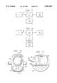
- FIG. 1is a front view of a surgical/diagnostic imaging device in accordance with the invention
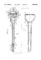
- FIG. 2is a partially cutaway side view of the imaging device of FIG. 1;
- FIG. 3is a cutaway top view of the sheath cap taken through plane 3--3 in FIG. 1;
- FIG. 4is an enlarged cutaway side view of the upper housing and the lower portion of the imaging device
- FIG. 5is a cutaway top view of the upper housing taken through plane 5--5 in FIG. 4;
- FIG. 6is an cutaway top view of the lower portion of the imaging device taken through plane 6--6 in FIG. 4;
- FIG. 7is a block diagram of a system for controlling the imaging device of FIG. 1 and for displaying the images transmitted by the imaging device;
- FIG. 8is a block diagram of a second control and display system for the imaging device of FIG. 1;
- FIG. 9is a cutaway side view of a second imaging device in accordance with the invention.
- FIG. 10is a front view of the camera housing shown in FIG. 10;
- FIG. 11is a cutaway side view of the camera housing taken through plane 11--11 in FIG. 10.
- FIGS. 1-3show a surgical/diagnostic imaging device 1 for use in interabdominal, interthoracic, and other surgical and diagnostic procedures.
- the device 1comprises an upper housing 3, a camera housing 5, and left and right camera housing supports 7, 9.
- the device 1is inserted into a sterile sheath 11.
- the device 1 and sheath 11are then inserted through an incision (not shown) into the patient's body (not shown).
- the camerais inserted so as place the camera housing 5 in a position from which it can be pointed at the surgical site or the area to be diagnosed.
- the incisionis sealed around the camera with a purse string stitch, thereby preventing leakage of the CO 2 gas which is used to distend the patient's abdomen or chest during surgery or diagnosis.
- the sheath 11is constructed of medical-grade plastic is provided in a sterilized condition, and is intended to be disposed of after use.
- the sheath 11can be constructed of heat-resistant materials in order to allow it to be sterilized using an autoclave, then reused. It should be appreciated that the sterile sheath 11 eliminates the need to sterilize the camera.
- the camera housing 5contains a CCD (not shown) and a zoom lens assembly (not shown).
- a plurality of high intensity lights 13are mounted within a light housing 15 which extends about the outer circumference of the camera housing 5.
- the lights 13are aligned with the focal axis 17 of the CCD, and they provide illumination of the area at which the camera housing 5 and hence the CCD are pointed.
- the left and right camera housing supports 7, 9engage complimentary locking keys 19, 21 within a sheath cap 23.
- the camera housing 5is locked into a position in which the CCD's focal axis 17 is aligned perpendicular to an optically-clear window 25.
- the locking keys 19, 21cause the sheath cap 13 to rotate about the longitudinal axis 27 of the camera when the camera housing supports 7, 9 are rotated about that axis.
- a camera cable 29extends between the camera housing 5 and the upper housing 3.
- the camera cable 29contains conductors which carry the CCD's signals to the upper housing 3 and which supply electrical power to the CCD and lights 13.
- An imaging device cable 31is provided to carry control signals and supply electrical power to the device 1 and to carry the CCD's signals to external processing and display devices (not shown).
- the length of the camera housing supports 7, 9 and the length of the sheath 11are varied to accommodate variation in the thickness of the abdominal walls of patients and to allow the camera to be used in thoracic surgery/diagnosis. Three lengths are provided: 3, 6, and 11 inches below the upper housing 3.
- an elevation motor 51drives an elevation shaft 53 by means of gears 55, 57.
- the elevation shaft 53extends downwardly through the hollow left camera support 7.
- a ring and pinon gear arrangement 59 at the lower end of the elevation shaft 53transfers the rotary motion of the elevation shaft 53 to the camera housing 15, thereby causing the camera housing 15 to elevate or depress, depending on the direction of rotation of the elevation motor 51.
- the camera housing 15can be elevated 70 degrees above and depressed 90 degrees below above a plane perpendicular to the longitudinal axis 27 of the camera and passing through intersection of the longitudinal axis 27 and the focal axis 17 of the camera.
- the elevation motor 51is mounted on a plate 63.
- the plate 63is rotably mounted within the upper housing 3 on a bearing 65.
- An azimuth motor 67is also mounted on the plate 63.
- the azimuth motor 67drives an azimuth gear 69.
- the azimuth gear 69engages a housing gear 71 which is attached to the inner surface of the upper housing 3.
- the plate 63rotates within the upper housing 3.
- the plate 63rotates plus or minus 180 degrees in order to minimize the amount the camera cable 21 is twisted. 360 degree rotation can easily be achieved by using conventional slip rings.
- a zoom/focus motor 72drives gears 73, 75, which rotate a zoom/focus shaft 77.
- the zoom/focus shaftextends downwardly through the right camera support 9.
- a ring and pinon arrangement 79transfers the rotary motion of the focus shaft 77 to a zoom lens mechanism (not shown) within the camera housing 5.
- the imaging device 1is connected to a control console 101 by means of the imaging device cable 31. Signals from the CCD of the imaging device 1 are amplified by circuits in the control console 101 and directed to a display device 103.
- the display device 103is a conventional television set.
- a foot pedal control assembly 105allows the surgeon (not shown) to control the imaging device 1.
- the foot pedal control assembly 105includes four controls (not shown): (1) camera housing left and right; (2) camera housing up and down; (3) zoom in and out; and (4) light intensity up and down. Signals from the foot pedal control assembly 105 are routed to the control console 101. Circuits (not shown) in the control console 103 convert the control assembly signals into signals which are suitable to control the imaging device 1, then route the converted signals to the imaging device 1.
- a computer 107is interposed between the control console 101 and the display device 103.
- a plurality of computer programs contained in the computer 107allow operating team personnel to manipulate and/or store the signals from the imaging device 1.
- FIGS. 9-11illustrate a second surgical/diagnostic imaging device in accordance with the invention.
- the imaging devicecomprises two major assemblies: a camera assembly 150 and a disposable sheath assembly 152.
- a rotary stepper motor 154is rigidly mounted in an upper housing 156.
- a linear stepper motor 158 and the distal end of a planetary gear assembly 162are press fitted in a linear stepper motor housing 164.
- the proximal end of the planetary gear assembly 162is attached to the upper housing 156 by screws 168.
- Three planetary gears 170(only two of which are shown in FIG. 9) are rotably mounted on pins 172 within the planetary gear assembly 162.
- the rotary stepper motor 154drives the planetary gears 170 through a sun gear 174.
- the proximal end of a camera support tube 178is press fitted in the linear stepper housing 164.
- a camera housing 180is pivotally mounted between pair of arms 182 (only one of which is shown in FIG. 9) that are integral with and extend from the distal end of the camera support tube 178.
- the linear stepper motor 158acts through a pushrod 186 and a fork 188 to control the elevation of the camera housing 180.
- the disposable sheath assembly 152comprises a sheath 190, a sheath housing 192, and a ring gear 194.
- the distal portion of the sheath 190is optically clear.
- the proximal end of the sheath 190is adhesively attached within the distal end of the sheath housing 192.
- the ring gear 194is adhesively attached within the proximal end of the sheath housing 192.
- the camera assembly 150Prior to use, the camera assembly 150 is inserted into the sheath assembly 152, and the planet gears 170 engage the ring gear. As a result, when the rotary stepper motor 154 is actuated, the camera assembly 150 rotates in relation to the longitudinal axis 202 of the sheath assembly .
- a CCD assembly 204 and a lens 206are mounted within a camera bore 208 in the camera housing 180.
- a pair of high intensity lights 210are mounted in bores that are coaxial with the camera bore 208.
- a multi-conductor flexcable 212provides the necessary connections for the CCD assembly 204, for the camera housing lights 210, and for three high intensity lights 214 that are disposed in bores in the pushrod 186.
- the flexcable 212extends from the camera housing 180 to the upper housing 156.
- the flexcable 212is combined with power and control wires (not shown) for the rotary stepper motor 154 and the linear stepper motor 158 to form the camera assembly cable 218.
- the camera assembly cable 218passes through an orifice 220 in the upper housing 152. As with the embodiment of the invention shown in FIGS. 1-8, the camera assembly cable 218 connects the camera assembly 150 to external display and control devices (not shown).
Landscapes
- Health & Medical Sciences (AREA)
- Life Sciences & Earth Sciences (AREA)
- Surgery (AREA)
- Biomedical Technology (AREA)
- Medical Informatics (AREA)
- Optics & Photonics (AREA)
- Pathology (AREA)
- Radiology & Medical Imaging (AREA)
- Biophysics (AREA)
- Engineering & Computer Science (AREA)
- Physics & Mathematics (AREA)
- Heart & Thoracic Surgery (AREA)
- Nuclear Medicine, Radiotherapy & Molecular Imaging (AREA)
- Molecular Biology (AREA)
- Animal Behavior & Ethology (AREA)
- General Health & Medical Sciences (AREA)
- Public Health (AREA)
- Veterinary Medicine (AREA)
- Endoscopes (AREA)
- Studio Devices (AREA)
Abstract
Description
Claims (23)
Priority Applications (5)
| Application Number | Priority Date | Filing Date | Title |
|---|---|---|---|
| US09/065,116US6007484A (en) | 1995-09-15 | 1998-04-23 | Endoscope having elevation and azimuth control of camera |
| US09/282,021US6413209B1 (en) | 1995-09-15 | 1999-03-29 | Imaging system with condensation control |
| US09/382,496US6398725B1 (en) | 1995-09-15 | 1999-08-25 | Endoscope having elevation and azimuth control of camera |
| US09/382,495US6428470B1 (en) | 1995-09-15 | 1999-08-25 | Imaging system and components thereof |
| US10/144,426US20020128538A1 (en) | 1995-09-15 | 2002-05-13 | Imaging system and components thereof |
Applications Claiming Priority (4)
| Application Number | Priority Date | Filing Date | Title |
|---|---|---|---|
| US380295P | 1995-09-15 | 1995-09-15 | |
| US70804496A | 1996-08-30 | 1996-08-30 | |
| US08/937,238US5762603A (en) | 1995-09-15 | 1997-09-16 | Endoscope having elevation and azimuth control of camera assembly |
| US09/065,116US6007484A (en) | 1995-09-15 | 1998-04-23 | Endoscope having elevation and azimuth control of camera |
Related Parent Applications (1)
| Application Number | Title | Priority Date | Filing Date |
|---|---|---|---|
| US08/937,238ContinuationUS5762603A (en) | 1995-09-15 | 1997-09-16 | Endoscope having elevation and azimuth control of camera assembly |
Related Child Applications (3)
| Application Number | Title | Priority Date | Filing Date |
|---|---|---|---|
| US09/282,021Continuation-In-PartUS6413209B1 (en) | 1995-09-15 | 1999-03-29 | Imaging system with condensation control |
| US09/382,495Continuation-In-PartUS6428470B1 (en) | 1995-09-15 | 1999-08-25 | Imaging system and components thereof |
| US09/382,496DivisionUS6398725B1 (en) | 1995-09-15 | 1999-08-25 | Endoscope having elevation and azimuth control of camera |
Publications (1)
| Publication Number | Publication Date |
|---|---|
| US6007484Atrue US6007484A (en) | 1999-12-28 |
Family
ID=27357492
Family Applications (2)
| Application Number | Title | Priority Date | Filing Date |
|---|---|---|---|
| US09/065,116Expired - Fee RelatedUS6007484A (en) | 1995-09-15 | 1998-04-23 | Endoscope having elevation and azimuth control of camera |
| US09/382,496Expired - Fee RelatedUS6398725B1 (en) | 1995-09-15 | 1999-08-25 | Endoscope having elevation and azimuth control of camera |
Family Applications After (1)
| Application Number | Title | Priority Date | Filing Date |
|---|---|---|---|
| US09/382,496Expired - Fee RelatedUS6398725B1 (en) | 1995-09-15 | 1999-08-25 | Endoscope having elevation and azimuth control of camera |
Country Status (1)
| Country | Link |
|---|---|
| US (2) | US6007484A (en) |
Cited By (52)
| Publication number | Priority date | Publication date | Assignee | Title |
|---|---|---|---|---|
| US6398725B1 (en) | 1995-09-15 | 2002-06-04 | Pinotage, Llc | Endoscope having elevation and azimuth control of camera |
| US6428471B1 (en)* | 1999-02-03 | 2002-08-06 | William E. Durell, Jr. | Variable view arthroscope |
| US6447444B1 (en)* | 1997-11-04 | 2002-09-10 | Sightline Technologies Ltd. | Video rectoscope |
| US20030032863A1 (en)* | 2001-08-09 | 2003-02-13 | Yuri Kazakevich | Endoscope with imaging probe |
| US6529620B2 (en) | 2000-09-11 | 2003-03-04 | Pinotage, L.L.C. | System and method for obtaining and utilizing maintenance information |
| US20040147809A1 (en)* | 2001-09-07 | 2004-07-29 | Smith & Nephew, Inc., A Delaware Corporation | Endoscopic system with a solid-state light source |
| US20050054894A1 (en)* | 2003-09-04 | 2005-03-10 | Amram Aizenfeld | Sleeve for endoscopic tools |
| US20050080318A1 (en)* | 2003-10-09 | 2005-04-14 | Squicciarini John B. | Multi-functional video scope |
| US20050113643A1 (en)* | 2003-11-20 | 2005-05-26 | Hale Eric L. | Method and apparatus for displaying endoscopic images |
| WO2006031897A1 (en)* | 2004-09-10 | 2006-03-23 | Gyntec Medical, Inc. | Flexible video scope extension and methods |
| US7056283B2 (en) | 2000-09-04 | 2006-06-06 | Sightline Technoligies Ltd. | Double sleeve endoscope |
| US20060184223A1 (en)* | 2005-02-04 | 2006-08-17 | Squicciarini John B | Cavity probe with exciter and/or dilator tip |
| US20060189842A1 (en)* | 2005-02-14 | 2006-08-24 | Hoeg Hans D | Method for using variable direction of view endoscopy in conjunction with image guided surgical systems |
| US20060256431A1 (en)* | 2005-01-26 | 2006-11-16 | Hoeg Hans D | Illumination system for variable direction of view instruments |
| US20070073109A1 (en)* | 2005-09-23 | 2007-03-29 | Irion Klaus M | Lighting system for endoscopic examinations |
| US20070213590A1 (en)* | 2003-10-09 | 2007-09-13 | Gyntec Medical, Inc. | Apparatus and methods for examining, visualizing, diagnosing, manipulating, treating and recording of abnormalities within interior regions of body cavities |
| US20080025770A1 (en)* | 2006-07-10 | 2008-01-31 | Xerox Corporation | Planetary dual stepper drives |
| US7691103B2 (en) | 2006-04-29 | 2010-04-06 | Board Of Regents, The University Of Texas System | Devices for use in transluminal and endoluminal surgery |
| US20110292195A1 (en)* | 2010-06-01 | 2011-12-01 | Jan Dahmen | Visual field apparatus for an endoscope |
| CN102988087A (en)* | 2005-07-27 | 2013-03-27 | Tyco医疗健康集团 | Shaft, e.g., for an electro-mechanical surgical device |
| US20130096457A1 (en)* | 2011-10-18 | 2013-04-18 | Qscope, LLC | Oral scope system with image sensor and method for visual examination of oral cavity and upper airway |
| US20130147935A1 (en)* | 2008-06-17 | 2013-06-13 | Fujifilm Corporation | Electronic endoscope |
| US20130296652A1 (en)* | 2004-09-24 | 2013-11-07 | Vivid Medical Inc. | Solid state illumination for endoscopy |
| US20130345503A1 (en)* | 2010-12-08 | 2013-12-26 | Karl Storz Gmbh & Co. Kg | Hand-Operated Endoscope For Medical Purposes |
| US9033871B2 (en) | 2004-04-07 | 2015-05-19 | Karl Storz Imaging, Inc. | Gravity referenced endoscopic image orientation |
| US9033957B2 (en) | 2003-12-02 | 2015-05-19 | Board Of Regents, The University Of Texas System | Surgical anchor and system |
| US20150216399A1 (en)* | 2012-09-05 | 2015-08-06 | Olympus Winter & Ibe Gmbh | Video camera housing for an endoscope |
| US20150223677A1 (en)* | 2012-10-24 | 2015-08-13 | Olympus Winter & Ibe Gmbh | Endoscope with lateral illumination, use and method |
| US20150238071A1 (en)* | 2012-10-18 | 2015-08-27 | The Arizona Board Of Regents On Behalf Of The University Of Arizona | Multi-Resolution Foveated Endoscope/Laparoscope |
| WO2016094559A1 (en)* | 2014-12-10 | 2016-06-16 | Mayo Foundation For Medical Education And Research | Tube thoracostomy using an optical trocar |
| WO2017173524A1 (en)* | 2016-04-07 | 2017-10-12 | Titan Medical Inc. | Camera positioning method and apparatus for capturing images during a medical procedure |
| US10172669B2 (en) | 2009-10-09 | 2019-01-08 | Ethicon Llc | Surgical instrument comprising an energy trigger lockout |
| US10314638B2 (en) | 2015-04-07 | 2019-06-11 | Ethicon Llc | Articulating radio frequency (RF) tissue seal with articulating state sensing |
| US10603117B2 (en) | 2017-06-28 | 2020-03-31 | Ethicon Llc | Articulation state detection mechanisms |
| US10751117B2 (en) | 2016-09-23 | 2020-08-25 | Ethicon Llc | Electrosurgical instrument with fluid diverter |
| US10751109B2 (en) | 2014-12-22 | 2020-08-25 | Ethicon Llc | High power battery powered RF amplifier topology |
| US10779876B2 (en) | 2011-10-24 | 2020-09-22 | Ethicon Llc | Battery powered surgical instrument |
| US10799284B2 (en) | 2017-03-15 | 2020-10-13 | Ethicon Llc | Electrosurgical instrument with textured jaws |
| US10856934B2 (en) | 2016-04-29 | 2020-12-08 | Ethicon Llc | Electrosurgical instrument with electrically conductive gap setting and tissue engaging members |
| US10959806B2 (en) | 2015-12-30 | 2021-03-30 | Ethicon Llc | Energized medical device with reusable handle |
| US10959771B2 (en) | 2015-10-16 | 2021-03-30 | Ethicon Llc | Suction and irrigation sealing grasper |
| US10987156B2 (en) | 2016-04-29 | 2021-04-27 | Ethicon Llc | Electrosurgical instrument with electrically conductive gap setting member and electrically insulative tissue engaging members |
| US11033325B2 (en) | 2017-02-16 | 2021-06-15 | Cilag Gmbh International | Electrosurgical instrument with telescoping suction port and debris cleaner |
| US11033323B2 (en) | 2017-09-29 | 2021-06-15 | Cilag Gmbh International | Systems and methods for managing fluid and suction in electrosurgical systems |
| US11090103B2 (en) | 2010-05-21 | 2021-08-17 | Cilag Gmbh International | Medical device |
| US20210361369A1 (en)* | 2006-06-13 | 2021-11-25 | Intuitive Surgical Operations, Inc. | Side-looking minimally invasive surgery instrument assembly |
| US11484358B2 (en) | 2017-09-29 | 2022-11-01 | Cilag Gmbh International | Flexible electrosurgical instrument |
| US11490951B2 (en) | 2017-09-29 | 2022-11-08 | Cilag Gmbh International | Saline contact with electrodes |
| US11497546B2 (en) | 2017-03-31 | 2022-11-15 | Cilag Gmbh International | Area ratios of patterned coatings on RF electrodes to reduce sticking |
| US20230301504A1 (en)* | 2020-08-12 | 2023-09-28 | Lina Medical International Operations Ag | A surgical camera |
| US11957342B2 (en) | 2021-11-01 | 2024-04-16 | Cilag Gmbh International | Devices, systems, and methods for detecting tissue and foreign objects during a surgical operation |
| US12023010B2 (en) | 2015-05-13 | 2024-07-02 | Atricure, Inc. | Access visualization systems |
Families Citing this family (22)
| Publication number | Priority date | Publication date | Assignee | Title |
|---|---|---|---|---|
| US6379334B1 (en)* | 1997-02-10 | 2002-04-30 | Essex Technology, Inc. | Rotate advance catheterization system |
| AU7720100A (en) | 1999-09-27 | 2001-04-30 | Essex Technology, Inc. | Rotate-to-advance catheterization system |
| US7175593B2 (en)* | 2000-08-30 | 2007-02-13 | Durell & Gitelis, Inc. | Variable view arthroscope with charge coupled device |
| EP1332710B1 (en)* | 2002-02-05 | 2008-12-03 | Kersten Zaar | Endoscope with side-view optics |
| WO2005082226A1 (en)* | 2004-02-27 | 2005-09-09 | Olympus Corporation | Endoscope |
| US7896803B2 (en)* | 2005-02-14 | 2011-03-01 | Karl Storz Imaging, Inc. | Variable direction of view instrument with on-board actuators |
| EP1861133B1 (en) | 2005-02-28 | 2012-11-21 | Spirus Medical Inc. | Rotate-to-advance catheterization system |
| US8317678B2 (en) | 2005-05-04 | 2012-11-27 | Olympus Endo Technology America Inc. | Rotate-to-advance catheterization system |
| US8414477B2 (en) | 2005-05-04 | 2013-04-09 | Olympus Endo Technology America Inc. | Rotate-to-advance catheterization system |
| US8343040B2 (en) | 2005-05-04 | 2013-01-01 | Olympus Endo Technology America Inc. | Rotate-to-advance catheterization system |
| US7780650B2 (en) | 2005-05-04 | 2010-08-24 | Spirus Medical, Inc. | Rotate-to-advance catheterization system |
| US8235942B2 (en) | 2005-05-04 | 2012-08-07 | Olympus Endo Technology America Inc. | Rotate-to-advance catheterization system |
| US8574220B2 (en) | 2006-02-28 | 2013-11-05 | Olympus Endo Technology America Inc. | Rotate-to-advance catheterization system |
| US8435229B2 (en) | 2006-02-28 | 2013-05-07 | Olympus Endo Technology America Inc. | Rotate-to-advance catheterization system |
| US8807414B2 (en) | 2006-10-06 | 2014-08-19 | Covidien Lp | System and method for non-contact electronic articulation sensing |
| US8870755B2 (en) | 2007-05-18 | 2014-10-28 | Olympus Endo Technology America Inc. | Rotate-to-advance catheterization system |
| US20090198099A1 (en)* | 2008-02-05 | 2009-08-06 | Myers Stephen R | In vivo imaging system |
| US20100216086A1 (en)* | 2009-02-23 | 2010-08-26 | John Sylvester | Intra-oral image system having swivel head optic |
| JP6375309B2 (en) | 2013-02-01 | 2018-08-15 | デカ・プロダクツ・リミテッド・パートナーシップ | Endoscope with camera capable of panning |
| US10616491B2 (en) | 2013-02-01 | 2020-04-07 | Deka Products Limited Partnership | Endoscope with pannable camera and related method |
| WO2019210227A1 (en) | 2018-04-26 | 2019-10-31 | Deka Products Limited Partnership | Endoscope with rotatable camera and related methods |
| EP4106604A1 (en) | 2020-03-10 | 2022-12-28 | VTI Medical, Inc. | Multi-angle imaging platform |
Citations (53)
| Publication number | Priority date | Publication date | Assignee | Title |
|---|---|---|---|---|
| DE232524C (en)* | ||||
| US3557780A (en)* | 1967-04-20 | 1971-01-26 | Olympus Optical Co | Mechanism for controlling flexure of endoscope |
| US3896793A (en)* | 1973-06-19 | 1975-07-29 | Olympus Optical Co | Endoscope with view field altering means |
| US3958080A (en)* | 1974-06-11 | 1976-05-18 | Josef Kaiser Ag Fahrzeugwerk | Visual inspection apparatus for conduits |
| DE7833379U1 (en)* | 1978-11-10 | 1979-02-15 | Storz, Karl, 7200 Tuttlingen | |
| US4697210A (en)* | 1984-08-20 | 1987-09-29 | Fuji Photo Optical Co., Ltd. | Endoscope for displaying a normal image |
| US4718417A (en)* | 1985-03-22 | 1988-01-12 | Massachusetts Institute Of Technology | Visible fluorescence spectral diagnostic for laser angiosurgery |
| US4741326A (en)* | 1986-10-01 | 1988-05-03 | Fujinon, Inc. | Endoscope disposable sheath |
| US4791479A (en)* | 1986-06-04 | 1988-12-13 | Olympus Optical Co., Ltd. | Color-image sensing apparatus |
| US4819620A (en)* | 1986-08-16 | 1989-04-11 | Ichiro Okutsu | Endoscope guide pipe |
| US4855838A (en)* | 1988-05-27 | 1989-08-08 | Cues, Inc. | Remotely controlled pan and tilt television camera |
| US4858001A (en)* | 1987-10-08 | 1989-08-15 | High-Tech Medical Instrumentation, Inc. | Modular endoscopic apparatus with image rotation |
| US4905082A (en)* | 1987-05-06 | 1990-02-27 | Olympus Optical Co., Ltd. | Rigid video endoscope having a detachable imaging unit |
| US4947827A (en)* | 1988-12-30 | 1990-08-14 | Opielab, Inc. | Flexible endoscope |
| US4971035A (en)* | 1989-02-28 | 1990-11-20 | Asahi Kogaku Kogyo Kabushiki Kaisha | Insert part of endoscope |
| US4989586A (en)* | 1989-07-21 | 1991-02-05 | Olympus Optical Co., Ltd. | Endoscope having a solid-state image pickup device |
| US5028997A (en)* | 1989-07-20 | 1991-07-02 | Elbex Video Kabushiki Kaisha | Television camera apparatus |
| DE9113080U1 (en)* | 1991-10-21 | 1991-12-05 | Siemens AG, 8000 München | Endoscope for observing a hidden surgical field |
| US5111288A (en)* | 1988-03-02 | 1992-05-05 | Diamond Electronics, Inc. | Surveillance camera system |
| US5166787A (en)* | 1989-06-28 | 1992-11-24 | Karl Storz Gmbh & Co. | Endoscope having provision for repositioning a video sensor to a location which does not provide the same cross-sectionally viewed relationship with the distal end |
| US5217453A (en)* | 1991-03-18 | 1993-06-08 | Wilk Peter J | Automated surgical system and apparatus |
| US5237984A (en)* | 1991-06-24 | 1993-08-24 | Xomed-Treace Inc. | Sheath for endoscope |
| US5243967A (en)* | 1991-03-26 | 1993-09-14 | Olympus Optical Co., Ltd. | Endoscope system providing mutual operative communication between the drive control means and the video signal control means |
| US5251613A (en)* | 1991-05-06 | 1993-10-12 | Adair Edwin Lloyd | Method of cervical videoscope with detachable camera |
| US5267970A (en)* | 1991-11-01 | 1993-12-07 | Origin Medsystems, Inc. | Device for anchoring trocar sleeve |
| US5271381A (en)* | 1991-11-18 | 1993-12-21 | Vision Sciences, Inc. | Vertebrae for a bending section of an endoscope |
| US5290168A (en)* | 1987-03-05 | 1994-03-01 | Optical Systems, Inc. | Electronic video dental camera |
| US5305121A (en)* | 1992-06-08 | 1994-04-19 | Origin Medsystems, Inc. | Stereoscopic endoscope system |
| US5307804A (en)* | 1991-02-21 | 1994-05-03 | Richard Wolf Gmbh | Endoscope having a camera coupled thereto |
| US5308325A (en)* | 1991-01-28 | 1994-05-03 | Corpak, Inc. | Retention balloon for percutaneous catheter |
| US5334150A (en)* | 1992-11-17 | 1994-08-02 | Kaali Steven G | Visually directed trocar for laparoscopic surgical procedures and method of using same |
| US5349941A (en)* | 1993-03-26 | 1994-09-27 | Oktas | Cleanable endoscope |
| US5351678A (en)* | 1992-09-01 | 1994-10-04 | Citation Medical Corporation | Endoscope scope assembly for full hemisphere view |
| US5381784A (en)* | 1992-09-30 | 1995-01-17 | Adair; Edwin L. | Stereoscopic endoscope |
| US5381943A (en)* | 1992-10-09 | 1995-01-17 | Ethicon, Inc. | Endoscopic surgical stapling instrument with pivotable and rotatable staple cartridge |
| US5383859A (en)* | 1992-02-06 | 1995-01-24 | Sewell, Jr.; Frank | Rotatable laparoscopic puncturing instrument |
| US5396879A (en)* | 1992-04-09 | 1995-03-14 | Wilk; Peter J. | Elongate medical instrument with distal end orientation control |
| US5402768A (en)* | 1992-09-01 | 1995-04-04 | Adair; Edwin L. | Endoscope with reusable core and disposable sheath with passageways |
| US5418567A (en)* | 1993-01-29 | 1995-05-23 | Bayport Controls, Inc. | Surveillance camera system |
| US5458132A (en)* | 1993-03-15 | 1995-10-17 | Olympus Optical Co., Ltd. | Endoscope cover-sheathed endoscope system |
| US5483951A (en)* | 1994-02-25 | 1996-01-16 | Vision-Sciences, Inc. | Working channels for a disposable sheath for an endoscope |
| US5489256A (en)* | 1992-09-01 | 1996-02-06 | Adair; Edwin L. | Sterilizable endoscope with separable disposable tube assembly |
| US5508735A (en)* | 1994-07-12 | 1996-04-16 | Northeast Technical Service Co. Inc. | Underdeck inspection device |
| WO1996010947A1 (en)* | 1994-10-07 | 1996-04-18 | Oktas General Partnership | Electronic endoscope with zoom lens system |
| US5531664A (en)* | 1990-12-26 | 1996-07-02 | Olympus Optical Co., Ltd. | Bending actuator having a coil sheath with a fixed distal end and a free proximal end |
| US5538497A (en)* | 1992-10-28 | 1996-07-23 | Oktas | Endoscope having parasitic light elements |
| US5540649A (en)* | 1993-10-08 | 1996-07-30 | Leonard Medical, Inc. | Positioner for medical instruments |
| US5558619A (en)* | 1991-04-23 | 1996-09-24 | Olympus Optical Co., Ltd. | Endoscope system with automatic control according to movement of an operator |
| US5573494A (en)* | 1993-02-23 | 1996-11-12 | Olympus Optical Co., Ltd. | Endoscope cover-sheathed endoscope in which an endoscope-cover coverable endoscope to be sheathed with an endoscope cover is structured to shut out water tightly |
| US5591192A (en)* | 1995-02-01 | 1997-01-07 | Ethicon Endo-Surgery, Inc. | Surgical penetration instrument including an imaging element |
| US5617762A (en)* | 1995-03-10 | 1997-04-08 | Kirsch; Jerry | Miniature positioning device |
| US5626553A (en)* | 1995-06-05 | 1997-05-06 | Vision-Sciences, Inc. | Endoscope articulation system to reduce effort during articulation of an endoscope |
| US5689365A (en)* | 1994-09-13 | 1997-11-18 | Olympus Optical Co., Ltd | Stereoscopic-vision endoscope |
Family Cites Families (10)
| Publication number | Priority date | Publication date | Assignee | Title |
|---|---|---|---|---|
| JPH0644105B2 (en) | 1985-01-14 | 1994-06-08 | オリンパス光学工業株式会社 | Endoscope |
| US4727859A (en) | 1986-12-29 | 1988-03-01 | Welch Allyn, Inc. | Right angle detachable prism assembly for borescope |
| US5253638A (en) | 1992-03-25 | 1993-10-19 | Welch Allyn, Inc. | Right-angle detachable variable-position reflector assembly |
| US5737013A (en) | 1992-09-11 | 1998-04-07 | Williams; Ronald R. | Dental video camera with an adjustable iris |
| US5562602A (en) | 1993-03-15 | 1996-10-08 | Olympus Optical Co., Ltd. | Insert cover portion of endoscope cover, insert cover portion having channels of endoscope cover, endoscope-cover-type endoscope, endoscope-cover-system endoscope and endoscope apparatus |
| US6007484A (en) | 1995-09-15 | 1999-12-28 | Image Technologies Corporation | Endoscope having elevation and azimuth control of camera |
| CA2206156A1 (en) | 1995-09-15 | 1997-04-03 | Robert Lee Thompson | Surgical/diagnostic imaging device |
| US5879289A (en) | 1996-07-15 | 1999-03-09 | Universal Technologies International, Inc. | Hand-held portable endoscopic camera |
| US5908294A (en) | 1997-06-12 | 1999-06-01 | Schick Technologies, Inc | Dental imaging system with lamps and method |
| JP3536282B2 (en) | 2000-02-14 | 2004-06-07 | 富士通株式会社 | Service control device |
- 1998
- 1998-04-23USUS09/065,116patent/US6007484A/ennot_activeExpired - Fee Related
- 1999
- 1999-08-25USUS09/382,496patent/US6398725B1/ennot_activeExpired - Fee Related
Patent Citations (56)
| Publication number | Priority date | Publication date | Assignee | Title |
|---|---|---|---|---|
| DE232524C (en)* | ||||
| US3557780A (en)* | 1967-04-20 | 1971-01-26 | Olympus Optical Co | Mechanism for controlling flexure of endoscope |
| US3896793A (en)* | 1973-06-19 | 1975-07-29 | Olympus Optical Co | Endoscope with view field altering means |
| US3958080A (en)* | 1974-06-11 | 1976-05-18 | Josef Kaiser Ag Fahrzeugwerk | Visual inspection apparatus for conduits |
| DE7833379U1 (en)* | 1978-11-10 | 1979-02-15 | Storz, Karl, 7200 Tuttlingen | |
| US4697210A (en)* | 1984-08-20 | 1987-09-29 | Fuji Photo Optical Co., Ltd. | Endoscope for displaying a normal image |
| US4718417A (en)* | 1985-03-22 | 1988-01-12 | Massachusetts Institute Of Technology | Visible fluorescence spectral diagnostic for laser angiosurgery |
| US4791479A (en)* | 1986-06-04 | 1988-12-13 | Olympus Optical Co., Ltd. | Color-image sensing apparatus |
| US4819620A (en)* | 1986-08-16 | 1989-04-11 | Ichiro Okutsu | Endoscope guide pipe |
| US4741326A (en)* | 1986-10-01 | 1988-05-03 | Fujinon, Inc. | Endoscope disposable sheath |
| US5290168A (en)* | 1987-03-05 | 1994-03-01 | Optical Systems, Inc. | Electronic video dental camera |
| US4905082A (en)* | 1987-05-06 | 1990-02-27 | Olympus Optical Co., Ltd. | Rigid video endoscope having a detachable imaging unit |
| US4858001A (en)* | 1987-10-08 | 1989-08-15 | High-Tech Medical Instrumentation, Inc. | Modular endoscopic apparatus with image rotation |
| US4858001B1 (en)* | 1987-10-08 | 1992-06-30 | High Tech Medical Instrumentat | |
| US5111288A (en)* | 1988-03-02 | 1992-05-05 | Diamond Electronics, Inc. | Surveillance camera system |
| US4855838A (en)* | 1988-05-27 | 1989-08-08 | Cues, Inc. | Remotely controlled pan and tilt television camera |
| US4947827A (en)* | 1988-12-30 | 1990-08-14 | Opielab, Inc. | Flexible endoscope |
| US4971035A (en)* | 1989-02-28 | 1990-11-20 | Asahi Kogaku Kogyo Kabushiki Kaisha | Insert part of endoscope |
| US5166787A (en)* | 1989-06-28 | 1992-11-24 | Karl Storz Gmbh & Co. | Endoscope having provision for repositioning a video sensor to a location which does not provide the same cross-sectionally viewed relationship with the distal end |
| US5028997A (en)* | 1989-07-20 | 1991-07-02 | Elbex Video Kabushiki Kaisha | Television camera apparatus |
| US4989586A (en)* | 1989-07-21 | 1991-02-05 | Olympus Optical Co., Ltd. | Endoscope having a solid-state image pickup device |
| US5531664A (en)* | 1990-12-26 | 1996-07-02 | Olympus Optical Co., Ltd. | Bending actuator having a coil sheath with a fixed distal end and a free proximal end |
| US5308325A (en)* | 1991-01-28 | 1994-05-03 | Corpak, Inc. | Retention balloon for percutaneous catheter |
| US5307804A (en)* | 1991-02-21 | 1994-05-03 | Richard Wolf Gmbh | Endoscope having a camera coupled thereto |
| US5217453A (en)* | 1991-03-18 | 1993-06-08 | Wilk Peter J | Automated surgical system and apparatus |
| US5368015A (en)* | 1991-03-18 | 1994-11-29 | Wilk; Peter J. | Automated surgical system and apparatus |
| US5243967A (en)* | 1991-03-26 | 1993-09-14 | Olympus Optical Co., Ltd. | Endoscope system providing mutual operative communication between the drive control means and the video signal control means |
| US5558619A (en)* | 1991-04-23 | 1996-09-24 | Olympus Optical Co., Ltd. | Endoscope system with automatic control according to movement of an operator |
| US5251613A (en)* | 1991-05-06 | 1993-10-12 | Adair Edwin Lloyd | Method of cervical videoscope with detachable camera |
| US5237984A (en)* | 1991-06-24 | 1993-08-24 | Xomed-Treace Inc. | Sheath for endoscope |
| DE9113080U1 (en)* | 1991-10-21 | 1991-12-05 | Siemens AG, 8000 München | Endoscope for observing a hidden surgical field |
| US5267970A (en)* | 1991-11-01 | 1993-12-07 | Origin Medsystems, Inc. | Device for anchoring trocar sleeve |
| US5271381A (en)* | 1991-11-18 | 1993-12-21 | Vision Sciences, Inc. | Vertebrae for a bending section of an endoscope |
| US5383859A (en)* | 1992-02-06 | 1995-01-24 | Sewell, Jr.; Frank | Rotatable laparoscopic puncturing instrument |
| US5396879A (en)* | 1992-04-09 | 1995-03-14 | Wilk; Peter J. | Elongate medical instrument with distal end orientation control |
| US5305121A (en)* | 1992-06-08 | 1994-04-19 | Origin Medsystems, Inc. | Stereoscopic endoscope system |
| US5351678A (en)* | 1992-09-01 | 1994-10-04 | Citation Medical Corporation | Endoscope scope assembly for full hemisphere view |
| US5489256A (en)* | 1992-09-01 | 1996-02-06 | Adair; Edwin L. | Sterilizable endoscope with separable disposable tube assembly |
| US5402768A (en)* | 1992-09-01 | 1995-04-04 | Adair; Edwin L. | Endoscope with reusable core and disposable sheath with passageways |
| US5381784A (en)* | 1992-09-30 | 1995-01-17 | Adair; Edwin L. | Stereoscopic endoscope |
| US5381943A (en)* | 1992-10-09 | 1995-01-17 | Ethicon, Inc. | Endoscopic surgical stapling instrument with pivotable and rotatable staple cartridge |
| US5538497A (en)* | 1992-10-28 | 1996-07-23 | Oktas | Endoscope having parasitic light elements |
| US5380291A (en)* | 1992-11-17 | 1995-01-10 | Kaali; Steven G. | Visually directed trocar for laparoscopic surgical procedures and method of using same |
| US5334150A (en)* | 1992-11-17 | 1994-08-02 | Kaali Steven G | Visually directed trocar for laparoscopic surgical procedures and method of using same |
| US5418567A (en)* | 1993-01-29 | 1995-05-23 | Bayport Controls, Inc. | Surveillance camera system |
| US5573494A (en)* | 1993-02-23 | 1996-11-12 | Olympus Optical Co., Ltd. | Endoscope cover-sheathed endoscope in which an endoscope-cover coverable endoscope to be sheathed with an endoscope cover is structured to shut out water tightly |
| US5458132A (en)* | 1993-03-15 | 1995-10-17 | Olympus Optical Co., Ltd. | Endoscope cover-sheathed endoscope system |
| US5349941A (en)* | 1993-03-26 | 1994-09-27 | Oktas | Cleanable endoscope |
| US5540649A (en)* | 1993-10-08 | 1996-07-30 | Leonard Medical, Inc. | Positioner for medical instruments |
| US5483951A (en)* | 1994-02-25 | 1996-01-16 | Vision-Sciences, Inc. | Working channels for a disposable sheath for an endoscope |
| US5508735A (en)* | 1994-07-12 | 1996-04-16 | Northeast Technical Service Co. Inc. | Underdeck inspection device |
| US5689365A (en)* | 1994-09-13 | 1997-11-18 | Olympus Optical Co., Ltd | Stereoscopic-vision endoscope |
| WO1996010947A1 (en)* | 1994-10-07 | 1996-04-18 | Oktas General Partnership | Electronic endoscope with zoom lens system |
| US5591192A (en)* | 1995-02-01 | 1997-01-07 | Ethicon Endo-Surgery, Inc. | Surgical penetration instrument including an imaging element |
| US5617762A (en)* | 1995-03-10 | 1997-04-08 | Kirsch; Jerry | Miniature positioning device |
| US5626553A (en)* | 1995-06-05 | 1997-05-06 | Vision-Sciences, Inc. | Endoscope articulation system to reduce effort during articulation of an endoscope |
Cited By (82)
| Publication number | Priority date | Publication date | Assignee | Title |
|---|---|---|---|---|
| US6398725B1 (en) | 1995-09-15 | 2002-06-04 | Pinotage, Llc | Endoscope having elevation and azimuth control of camera |
| US6447444B1 (en)* | 1997-11-04 | 2002-09-10 | Sightline Technologies Ltd. | Video rectoscope |
| US6855107B2 (en) | 1997-11-04 | 2005-02-15 | Sightline Techgnologies Ltd. | Method for insertion of an endoscope into the colon |
| US6428471B1 (en)* | 1999-02-03 | 2002-08-06 | William E. Durell, Jr. | Variable view arthroscope |
| US7056283B2 (en) | 2000-09-04 | 2006-06-06 | Sightline Technoligies Ltd. | Double sleeve endoscope |
| US6529620B2 (en) | 2000-09-11 | 2003-03-04 | Pinotage, L.L.C. | System and method for obtaining and utilizing maintenance information |
| US7068301B2 (en) | 2000-09-11 | 2006-06-27 | Pinotage L.L.C. | System and method for obtaining and utilizing maintenance information |
| US6916286B2 (en) | 2001-08-09 | 2005-07-12 | Smith & Nephew, Inc. | Endoscope with imaging probe |
| US20030032863A1 (en)* | 2001-08-09 | 2003-02-13 | Yuri Kazakevich | Endoscope with imaging probe |
| US20040147809A1 (en)* | 2001-09-07 | 2004-07-29 | Smith & Nephew, Inc., A Delaware Corporation | Endoscopic system with a solid-state light source |
| US7063663B2 (en) | 2001-09-07 | 2006-06-20 | Smith & Nephew, Inc. | Endoscopic system with a solid-state light source |
| US20050054894A1 (en)* | 2003-09-04 | 2005-03-10 | Amram Aizenfeld | Sleeve for endoscopic tools |
| US6908428B2 (en) | 2003-09-04 | 2005-06-21 | Sightline Technologies Ltd. | Sleeve for endoscopic tools |
| US20070213590A1 (en)* | 2003-10-09 | 2007-09-13 | Gyntec Medical, Inc. | Apparatus and methods for examining, visualizing, diagnosing, manipulating, treating and recording of abnormalities within interior regions of body cavities |
| US20050080318A1 (en)* | 2003-10-09 | 2005-04-14 | Squicciarini John B. | Multi-functional video scope |
| US20050113643A1 (en)* | 2003-11-20 | 2005-05-26 | Hale Eric L. | Method and apparatus for displaying endoscopic images |
| US7232409B2 (en) | 2003-11-20 | 2007-06-19 | Karl Storz Development Corp. | Method and apparatus for displaying endoscopic images |
| US9033957B2 (en) | 2003-12-02 | 2015-05-19 | Board Of Regents, The University Of Texas System | Surgical anchor and system |
| US9033871B2 (en) | 2004-04-07 | 2015-05-19 | Karl Storz Imaging, Inc. | Gravity referenced endoscopic image orientation |
| US20060161048A1 (en)* | 2004-09-10 | 2006-07-20 | Squicciarini John B | Flexible video scope extension and methods |
| WO2006031897A1 (en)* | 2004-09-10 | 2006-03-23 | Gyntec Medical, Inc. | Flexible video scope extension and methods |
| US20130296652A1 (en)* | 2004-09-24 | 2013-11-07 | Vivid Medical Inc. | Solid state illumination for endoscopy |
| US9271637B2 (en)* | 2004-09-24 | 2016-03-01 | Vivid Medical Inc. | Solid state illumination for endoscopy |
| US20060256431A1 (en)* | 2005-01-26 | 2006-11-16 | Hoeg Hans D | Illumination system for variable direction of view instruments |
| US10231608B2 (en) | 2005-01-26 | 2019-03-19 | Karl Storz Imaging | Illumination system for variable direction of view instruments |
| US20110046447A1 (en)* | 2005-01-26 | 2011-02-24 | Hans David Hoeg | Illumination System For Variable Direction Of View Instruments |
| US7909756B2 (en)* | 2005-01-26 | 2011-03-22 | Karl Storz Imaging, Inc. | Illumination system for variable direction of view instruments |
| US20060184223A1 (en)* | 2005-02-04 | 2006-08-17 | Squicciarini John B | Cavity probe with exciter and/or dilator tip |
| US8414476B2 (en) | 2005-02-14 | 2013-04-09 | Karl Storz Imaging, Inc. | Method for using variable direction of view endoscopy in conjunction with image guided surgical systems |
| US7967742B2 (en) | 2005-02-14 | 2011-06-28 | Karl Storz Imaging, Inc. | Method for using variable direction of view endoscopy in conjunction with image guided surgical systems |
| US20110230710A1 (en)* | 2005-02-14 | 2011-09-22 | Hans David Hoeg | Method For Using Variable Direction Of View Endoscopy In Conjunction With Image Guided Surgical Systems |
| US20060189842A1 (en)* | 2005-02-14 | 2006-08-24 | Hoeg Hans D | Method for using variable direction of view endoscopy in conjunction with image guided surgical systems |
| CN102988087B (en)* | 2005-07-27 | 2015-09-09 | 柯惠Lp公司 | Such as the axle of electro-mechanical surgical device |
| CN102988087A (en)* | 2005-07-27 | 2013-03-27 | Tyco医疗健康集团 | Shaft, e.g., for an electro-mechanical surgical device |
| US20070073109A1 (en)* | 2005-09-23 | 2007-03-29 | Irion Klaus M | Lighting system for endoscopic examinations |
| US9717398B2 (en)* | 2005-09-23 | 2017-08-01 | Karl Storz Gmbh & Co. Kg | Lighting system for endoscopic examinations |
| US8480668B2 (en) | 2006-04-29 | 2013-07-09 | Board Of Regents Of The University Of Texas System | Devices for use in transluminal and endoluminal surgery |
| US7691103B2 (en) | 2006-04-29 | 2010-04-06 | Board Of Regents, The University Of Texas System | Devices for use in transluminal and endoluminal surgery |
| US20210361369A1 (en)* | 2006-06-13 | 2021-11-25 | Intuitive Surgical Operations, Inc. | Side-looking minimally invasive surgery instrument assembly |
| US7463004B2 (en)* | 2006-07-10 | 2008-12-09 | Xerox Corporation | Planetary dual stepper drives |
| US20080025770A1 (en)* | 2006-07-10 | 2008-01-31 | Xerox Corporation | Planetary dual stepper drives |
| US20130155214A1 (en)* | 2008-06-17 | 2013-06-20 | Fujifilm Corporation | Electronic endoscope |
| US20130147934A1 (en)* | 2008-06-17 | 2013-06-13 | Fujifilm Corporation | Electronic endoscope |
| US20130147935A1 (en)* | 2008-06-17 | 2013-06-13 | Fujifilm Corporation | Electronic endoscope |
| US10172669B2 (en) | 2009-10-09 | 2019-01-08 | Ethicon Llc | Surgical instrument comprising an energy trigger lockout |
| US11090103B2 (en) | 2010-05-21 | 2021-08-17 | Cilag Gmbh International | Medical device |
| US20110292195A1 (en)* | 2010-06-01 | 2011-12-01 | Jan Dahmen | Visual field apparatus for an endoscope |
| US9625700B2 (en)* | 2010-06-01 | 2017-04-18 | Karl Storz Gmbh & Co. Kg | Visual field apparatus and image transmission apparatus for an endoscope |
| US10588490B2 (en)* | 2010-12-08 | 2020-03-17 | Karl Storz Gmbh & Co. Kg | Hand-operated endoscope for medical purposes |
| US20130345503A1 (en)* | 2010-12-08 | 2013-12-26 | Karl Storz Gmbh & Co. Kg | Hand-Operated Endoscope For Medical Purposes |
| US20130096457A1 (en)* | 2011-10-18 | 2013-04-18 | Qscope, LLC | Oral scope system with image sensor and method for visual examination of oral cavity and upper airway |
| US10779876B2 (en) | 2011-10-24 | 2020-09-22 | Ethicon Llc | Battery powered surgical instrument |
| US10085625B2 (en)* | 2012-09-05 | 2018-10-02 | Olympus Winter & Ibe Gmbh | Video camera housing for an endoscope |
| US20150216399A1 (en)* | 2012-09-05 | 2015-08-06 | Olympus Winter & Ibe Gmbh | Video camera housing for an endoscope |
| US10064545B2 (en)* | 2012-10-18 | 2018-09-04 | The Arizona Board Of Regents On Behalf Of The University Of Arizona | Multi-resolution foveated endoscope/laparoscope |
| US20150238071A1 (en)* | 2012-10-18 | 2015-08-27 | The Arizona Board Of Regents On Behalf Of The University Of Arizona | Multi-Resolution Foveated Endoscope/Laparoscope |
| US20150223677A1 (en)* | 2012-10-24 | 2015-08-13 | Olympus Winter & Ibe Gmbh | Endoscope with lateral illumination, use and method |
| WO2016094559A1 (en)* | 2014-12-10 | 2016-06-16 | Mayo Foundation For Medical Education And Research | Tube thoracostomy using an optical trocar |
| US10786148B2 (en) | 2014-12-10 | 2020-09-29 | Mayo Foundation For Medical Education And Research | Tube thoracostomy using an optical trocar |
| US10751109B2 (en) | 2014-12-22 | 2020-08-25 | Ethicon Llc | High power battery powered RF amplifier topology |
| US10314638B2 (en) | 2015-04-07 | 2019-06-11 | Ethicon Llc | Articulating radio frequency (RF) tissue seal with articulating state sensing |
| US12023010B2 (en) | 2015-05-13 | 2024-07-02 | Atricure, Inc. | Access visualization systems |
| US10959771B2 (en) | 2015-10-16 | 2021-03-30 | Ethicon Llc | Suction and irrigation sealing grasper |
| US10959806B2 (en) | 2015-12-30 | 2021-03-30 | Ethicon Llc | Energized medical device with reusable handle |
| US11576562B2 (en)* | 2016-04-07 | 2023-02-14 | Titan Medical Inc. | Camera positioning method and apparatus for capturing images during a medical procedure |
| WO2017173524A1 (en)* | 2016-04-07 | 2017-10-12 | Titan Medical Inc. | Camera positioning method and apparatus for capturing images during a medical procedure |
| US10987156B2 (en) | 2016-04-29 | 2021-04-27 | Ethicon Llc | Electrosurgical instrument with electrically conductive gap setting member and electrically insulative tissue engaging members |
| US10856934B2 (en) | 2016-04-29 | 2020-12-08 | Ethicon Llc | Electrosurgical instrument with electrically conductive gap setting and tissue engaging members |
| US11839422B2 (en) | 2016-09-23 | 2023-12-12 | Cilag Gmbh International | Electrosurgical instrument with fluid diverter |
| US12295644B2 (en) | 2016-09-23 | 2025-05-13 | Cilag Gmbh International | Electrosurgical instrument with fluid diverter |
| US10751117B2 (en) | 2016-09-23 | 2020-08-25 | Ethicon Llc | Electrosurgical instrument with fluid diverter |
| US11033325B2 (en) | 2017-02-16 | 2021-06-15 | Cilag Gmbh International | Electrosurgical instrument with telescoping suction port and debris cleaner |
| US10799284B2 (en) | 2017-03-15 | 2020-10-13 | Ethicon Llc | Electrosurgical instrument with textured jaws |
| US12023087B2 (en) | 2017-03-15 | 2024-07-02 | Cilag Gmbh International | Electrosurgical instrument with textured jaws |
| US11497546B2 (en) | 2017-03-31 | 2022-11-15 | Cilag Gmbh International | Area ratios of patterned coatings on RF electrodes to reduce sticking |
| US10603117B2 (en) | 2017-06-28 | 2020-03-31 | Ethicon Llc | Articulation state detection mechanisms |
| US11490951B2 (en) | 2017-09-29 | 2022-11-08 | Cilag Gmbh International | Saline contact with electrodes |
| US11484358B2 (en) | 2017-09-29 | 2022-11-01 | Cilag Gmbh International | Flexible electrosurgical instrument |
| US11033323B2 (en) | 2017-09-29 | 2021-06-15 | Cilag Gmbh International | Systems and methods for managing fluid and suction in electrosurgical systems |
| US12390264B2 (en) | 2017-09-29 | 2025-08-19 | Cilag Gmbh International | Systems and methods for managing fluid and suction in electrosurgical systems |
| US20230301504A1 (en)* | 2020-08-12 | 2023-09-28 | Lina Medical International Operations Ag | A surgical camera |
| US11957342B2 (en) | 2021-11-01 | 2024-04-16 | Cilag Gmbh International | Devices, systems, and methods for detecting tissue and foreign objects during a surgical operation |
Also Published As
| Publication number | Publication date |
|---|---|
| US6398725B1 (en) | 2002-06-04 |
Similar Documents
| Publication | Publication Date | Title |
|---|---|---|
| US6007484A (en) | Endoscope having elevation and azimuth control of camera | |
| US5762603A (en) | Endoscope having elevation and azimuth control of camera assembly | |
| US7896803B2 (en) | Variable direction of view instrument with on-board actuators | |
| CN104883947B (en) | Decoupled multicamera system for micro-wound surgical operation | |
| TW415834B (en) | Zoom laparoscope | |
| US5817005A (en) | Apparatus and method for simultaneously retracting and viewing bodily tissues | |
| AU664343B2 (en) | Endoscope scope assembly for full hemisphere view | |
| CA2675517C (en) | Cable-free arthroscopy | |
| US6450949B1 (en) | Endoscope | |
| US5329936A (en) | Portable arthroscope with periscope optics | |
| Hu et al. | Insertable surgical imaging device with pan, tilt, zoom, and lighting | |
| US20060206007A1 (en) | Disposable illuminator endoscope | |
| JP2002514448A (en) | Penetrating endoscope and endoscopic surgical instrument with CMOS image sensor and display | |
| US5411500A (en) | Portable arthroscope with disposable probe | |
| CA2054603A1 (en) | Portable arthroscope with periscope optics | |
| US5323767A (en) | Portable arthroscope with periscope optics | |
| EP0549097A1 (en) | Portable arthroscope with periscope optics | |
| JP2023532325A (en) | Endoscope with bendable camera shaft | |
| US20070038117A1 (en) | Multi-spectral imaging endoscope system | |
| EP0979633A1 (en) | Video endoscope with side view optics | |
| CN214157257U (en) | Endoscope with a detachable handle | |
| JPH01293840A (en) | root canal endoscopy | |
| JPH11501850A (en) | Surgical / Diagnostic Imaging Device | |
| MXPA97003590A (en) | Diagnost surgical image formation device | |
| JP3081657B2 (en) | Image display device |
Legal Events
| Date | Code | Title | Description |
|---|---|---|---|
| AS | Assignment | Owner name:ARGO CAPITAL PARTNERS L.L.P., TEXAS Free format text:SECURITY INTEREST;ASSIGNOR:IMAGE TECHNOLOGIES CORPORATION;REEL/FRAME:010206/0965 Effective date:19990827 Owner name:ARGO CAPITAL PARTNERS I.L.P., TEXAS Free format text:SECURITY INTEREST;ASSIGNORS:PINOTAGE, L.L.C.;THOMPSON, ROBERT LEE;REEL/FRAME:010206/0938 Effective date:19990827 | |
| AS | Assignment | Owner name:NMT MEDICAL, INC., MASSACHUSETTS Free format text:SECURITY AGREEMENT;ASSIGNORS:PINOTAGE, L.L.C.;THOMPSON, ROBERT LEE;REEL/FRAME:010226/0817 Effective date:19990827 | |
| FEPP | Fee payment procedure | Free format text:PAYOR NUMBER ASSIGNED (ORIGINAL EVENT CODE: ASPN); ENTITY STATUS OF PATENT OWNER: SMALL ENTITY | |
| AS | Assignment | Owner name:NMT MEDICAL, INC., MASSACHUSETTS Free format text:SECURITY INTEREST;ASSIGNOR:IMAGE TECHNOLOGIES CORPORATION;REEL/FRAME:010395/0232 Effective date:19990827 | |
| FEPP | Fee payment procedure | Free format text:PAYER NUMBER DE-ASSIGNED (ORIGINAL EVENT CODE: RMPN); ENTITY STATUS OF PATENT OWNER: SMALL ENTITY Free format text:PAYOR NUMBER ASSIGNED (ORIGINAL EVENT CODE: ASPN); ENTITY STATUS OF PATENT OWNER: SMALL ENTITY | |
| FEPP | Fee payment procedure | Free format text:PAYER NUMBER DE-ASSIGNED (ORIGINAL EVENT CODE: RMPN); ENTITY STATUS OF PATENT OWNER: SMALL ENTITY Free format text:PAYOR NUMBER ASSIGNED (ORIGINAL EVENT CODE: ASPN); ENTITY STATUS OF PATENT OWNER: SMALL ENTITY | |
| AS | Assignment | Owner name:IMAGE TECHNOLOGIES CORPORATION, MASSACHUSETTS Free format text:ASSIGNMENT OF ASSIGNORS INTEREST;ASSIGNORS:PINOTAGE, L.L.C., LIMITED LIABILITY COMPANY OF ARKANSAS;THOMPSON, ROBERT LEE, AN INDIVIDUAL;REEL/FRAME:011449/0115 Effective date:20001207 | |
| AS | Assignment | Owner name:PINOTAGE, L.L.C., ARKANSAS Free format text:RELEASE OF SECURITY INTEREST;ASSIGNOR:ARGO CAPITAL PARTNERS, L.L.P.;REEL/FRAME:011467/0130 Effective date:20001228 Owner name:THOMPSON, ROBERT LEE, ARKANSAS Free format text:RELEASE OF SECURITY INTEREST;ASSIGNOR:ARGO CAPITAL PARTNERS, L.L.P.;REEL/FRAME:011467/0130 Effective date:20001228 | |
| AS | Assignment | Owner name:MED IMAGES SM, INC., TENNESSEE Free format text:CHANGE OF NAME;ASSIGNOR:IMAGE TECHNOLOGIES CORPORATION;REEL/FRAME:011467/0103 Effective date:20001228 Owner name:ARGO CAPITAL PARTNERS I L.P., TEXAS Free format text:ASSIGNMENT OF SECURITY INTEREST;ASSIGNOR:NMT MEDICAL, INC.;REEL/FRAME:011485/0424 Effective date:20001130 | |
| AS | Assignment | Owner name:MED IMAGES, INC., TENNESSEE Free format text:CHANGE OF NAME;ASSIGNOR:MED IMAGES SM, INC.;REEL/FRAME:012665/0610 Effective date:20011127 | |
| CC | Certificate of correction | ||
| FEPP | Fee payment procedure | Free format text:PAYER NUMBER DE-ASSIGNED (ORIGINAL EVENT CODE: RMPN); ENTITY STATUS OF PATENT OWNER: SMALL ENTITY Free format text:PAT HOLDER CLAIMS SMALL ENTITY STATUS, ENTITY STATUS SET TO SMALL (ORIGINAL EVENT CODE: LTOS); ENTITY STATUS OF PATENT OWNER: SMALL ENTITY Free format text:PAYOR NUMBER ASSIGNED (ORIGINAL EVENT CODE: ASPN); ENTITY STATUS OF PATENT OWNER: SMALL ENTITY | |
| FPAY | Fee payment | Year of fee payment:4 | |
| AS | Assignment | Owner name:PINOTAGE L.L.C., ARKANSAS Free format text:ASSIGNMENT OF ASSIGNORS INTEREST;ASSIGNOR:MED IMAGES, INC.;REEL/FRAME:018847/0529 Effective date:20060216 Owner name:THOMPSON, ROBERT LEE, ARKANSAS Free format text:ASSIGNMENT OF ASSIGNORS INTEREST;ASSIGNOR:MED IMAGES, INC.;REEL/FRAME:018847/0529 Effective date:20060216 | |
| FPAY | Fee payment | Year of fee payment:8 | |
| FEPP | Fee payment procedure | Free format text:PAYER NUMBER DE-ASSIGNED (ORIGINAL EVENT CODE: RMPN); ENTITY STATUS OF PATENT OWNER: SMALL ENTITY Free format text:PAYOR NUMBER ASSIGNED (ORIGINAL EVENT CODE: ASPN); ENTITY STATUS OF PATENT OWNER: SMALL ENTITY | |
| REMI | Maintenance fee reminder mailed | ||
| LAPS | Lapse for failure to pay maintenance fees | ||
| STCH | Information on status: patent discontinuation | Free format text:PATENT EXPIRED DUE TO NONPAYMENT OF MAINTENANCE FEES UNDER 37 CFR 1.362 | |
| FP | Lapsed due to failure to pay maintenance fee | Effective date:20111228 |