US6007481A - Instrument for the penetration of body tissue with cutting and viewing of body structure - Google Patents
Instrument for the penetration of body tissue with cutting and viewing of body structureDownload PDFInfo
- Publication number
- US6007481A US6007481AUS08/500,091US50009195AUS6007481AUS 6007481 AUS6007481 AUS 6007481AUS 50009195 AUS50009195 AUS 50009195AUS 6007481 AUS6007481 AUS 6007481A
- Authority
- US
- United States
- Prior art keywords
- distal end
- instrument
- shaft
- window
- tissue contacting
- Prior art date
- Legal status (The legal status is an assumption and is not a legal conclusion. Google has not performed a legal analysis and makes no representation as to the accuracy of the status listed.)
- Expired - Lifetime
Links
- 230000035515penetrationEffects0.000titleclaimsabstractdescription23
- 230000000694effectsEffects0.000claimsdescription5
- 239000012530fluidSubstances0.000claims2
- 210000001519tissueAnatomy0.000description21
- 238000003780insertionMethods0.000description14
- 230000037431insertionEffects0.000description14
- 210000003815abdominal wallAnatomy0.000description13
- 210000004303peritoneumAnatomy0.000description9
- 210000000683abdominal cavityAnatomy0.000description8
- 210000000569greater omentumAnatomy0.000description8
- 238000005286illuminationMethods0.000description6
- 210000000056organAnatomy0.000description6
- 239000000835fiberSubstances0.000description5
- 210000000936intestineAnatomy0.000description5
- 230000003187abdominal effectEffects0.000description4
- 239000011521glassSubstances0.000description3
- 229920005372Plexiglas®Polymers0.000description2
- VYPSYNLAJGMNEJ-UHFFFAOYSA-NSilicium dioxideChemical compoundO=[Si]=OVYPSYNLAJGMNEJ-UHFFFAOYSA-N0.000description2
- 229910000831SteelInorganic materials0.000description2
- 210000004204blood vesselAnatomy0.000description2
- 239000003365glass fiberSubstances0.000description2
- 239000004926polymethyl methacrylateSubstances0.000description2
- 210000000813small intestineAnatomy0.000description2
- 239000010959steelSubstances0.000description2
- 239000012780transparent materialSubstances0.000description2
- 238000004026adhesive bondingMethods0.000description1
- 210000000577adipose tissueAnatomy0.000description1
- 230000004075alterationEffects0.000description1
- 210000004381amniotic fluidAnatomy0.000description1
- 238000013459approachMethods0.000description1
- 239000010432diamondSubstances0.000description1
- 229910003460diamondInorganic materials0.000description1
- 238000006073displacement reactionMethods0.000description1
- 210000003195fasciaAnatomy0.000description1
- 210000003754fetusAnatomy0.000description1
- 238000002347injectionMethods0.000description1
- 239000007924injectionSubstances0.000description1
- 230000002452interceptive effectEffects0.000description1
- 210000002429large intestineAnatomy0.000description1
- 239000000463materialSubstances0.000description1
- 210000004379membraneAnatomy0.000description1
- 239000012528membraneSubstances0.000description1
- 230000003287optical effectEffects0.000description1
- 230000000149penetrating effectEffects0.000description1
- 238000003793prenatal diagnosisMethods0.000description1
- 238000002310reflectometryMethods0.000description1
- 230000003068static effectEffects0.000description1
- 238000007920subcutaneous administrationMethods0.000description1
- 230000002792vascularEffects0.000description1
- 210000001835visceraAnatomy0.000description1
Images


Classifications
- A—HUMAN NECESSITIES
- A61—MEDICAL OR VETERINARY SCIENCE; HYGIENE
- A61B—DIAGNOSIS; SURGERY; IDENTIFICATION
- A61B17/00—Surgical instruments, devices or methods
- A61B17/34—Trocars; Puncturing needles
- A61B17/3417—Details of tips or shafts, e.g. grooves, expandable, bendable; Multiple coaxial sliding cannulas, e.g. for dilating
- A—HUMAN NECESSITIES
- A61—MEDICAL OR VETERINARY SCIENCE; HYGIENE
- A61B—DIAGNOSIS; SURGERY; IDENTIFICATION
- A61B1/00—Instruments for performing medical examinations of the interior of cavities or tubes of the body by visual or photographical inspection, e.g. endoscopes; Illuminating arrangements therefor
- A61B1/00163—Optical arrangements
- A61B1/00165—Optical arrangements with light-conductive means, e.g. fibre optics
- A—HUMAN NECESSITIES
- A61—MEDICAL OR VETERINARY SCIENCE; HYGIENE
- A61B—DIAGNOSIS; SURGERY; IDENTIFICATION
- A61B1/00—Instruments for performing medical examinations of the interior of cavities or tubes of the body by visual or photographical inspection, e.g. endoscopes; Illuminating arrangements therefor
- A61B1/313—Instruments for performing medical examinations of the interior of cavities or tubes of the body by visual or photographical inspection, e.g. endoscopes; Illuminating arrangements therefor for introducing through surgical openings, e.g. laparoscopes
- A61B1/3132—Instruments for performing medical examinations of the interior of cavities or tubes of the body by visual or photographical inspection, e.g. endoscopes; Illuminating arrangements therefor for introducing through surgical openings, e.g. laparoscopes for laparoscopy
- A—HUMAN NECESSITIES
- A61—MEDICAL OR VETERINARY SCIENCE; HYGIENE
- A61B—DIAGNOSIS; SURGERY; IDENTIFICATION
- A61B17/00—Surgical instruments, devices or methods
- A61B17/34—Trocars; Puncturing needles
- A61B17/3417—Details of tips or shafts, e.g. grooves, expandable, bendable; Multiple coaxial sliding cannulas, e.g. for dilating
- A61B2017/3454—Details of tips
- A61B2017/3458—Details of tips threaded
- A—HUMAN NECESSITIES
- A61—MEDICAL OR VETERINARY SCIENCE; HYGIENE
- A61B—DIAGNOSIS; SURGERY; IDENTIFICATION
- A61B90/00—Instruments, implements or accessories specially adapted for surgery or diagnosis and not covered by any of the groups A61B1/00 - A61B50/00, e.g. for luxation treatment or for protecting wound edges
- A61B90/30—Devices for illuminating a surgical field, the devices having an interrelation with other surgical devices or with a surgical procedure
- A61B2090/306—Devices for illuminating a surgical field, the devices having an interrelation with other surgical devices or with a surgical procedure using optical fibres
- A—HUMAN NECESSITIES
- A61—MEDICAL OR VETERINARY SCIENCE; HYGIENE
- A61B—DIAGNOSIS; SURGERY; IDENTIFICATION
- A61B90/00—Instruments, implements or accessories specially adapted for surgery or diagnosis and not covered by any of the groups A61B1/00 - A61B50/00, e.g. for luxation treatment or for protecting wound edges
- A61B90/36—Image-producing devices or illumination devices not otherwise provided for
- A61B90/361—Image-producing devices, e.g. surgical cameras
- A61B2090/3614—Image-producing devices, e.g. surgical cameras using optical fibre
- A—HUMAN NECESSITIES
- A61—MEDICAL OR VETERINARY SCIENCE; HYGIENE
- A61B—DIAGNOSIS; SURGERY; IDENTIFICATION
- A61B90/00—Instruments, implements or accessories specially adapted for surgery or diagnosis and not covered by any of the groups A61B1/00 - A61B50/00, e.g. for luxation treatment or for protecting wound edges
- A61B90/36—Image-producing devices or illumination devices not otherwise provided for
- A61B90/361—Image-producing devices, e.g. surgical cameras
Definitions
- the inventionrelates to an instrument for the penetration of body tissue with a hollow shaft, ending in a point formed as a conical window, and with optics and lighting units.
- Instruments of this typeserve particularly as trocars to create an artificial access to body cavities or organs, which do not possess any natural communicating passage with the exterior.
- the instrumenthas a point, which serves to pierce the body tissue and to widen the perforation opening to the diameter of the shaft.
- a sleeve surrounding the shaftis pushed together with the trocar into the perforation opening and, after removal of the trocar, represents an artificial access to the body cavity, the said access through which endoscopes, instruments, and the like can be introduced into the interior of the body.
- the insertion of the trocareven with the advantageous selection of an injection site, entails the risk of damaging blood vessels in subcutaneous fatty tissue, in fascia, and in the peritoneum ("abdominal membrane"), thus vessels in the abdominal wall. There is the further risk after penetration of the abdominal wall that the vessels in the abdominal area (abdominal cavity) and organs in the abdominal area, such as the large intestine, small intestine, omentum majus (greater omentum), and retroperitoneally located vessels and structures can be damaged.
- a hollow needlecan be passed through the abdominal wall first while the abdominal wall is lifted, to introduce gas into the abdominal cavity and to distance the abdominal wall from the underlying omentum majus and intestine for the subsequent insertion of the trocar. In that case as well, however, there is a residual risk of damage during the insertion of the hollow needle and the trocar.
- An instrument disclosed in DE [West German Patent] 29 22 239 C2has an outer tube, which is beveled at the anterior distal end thereof, to form an insertion point.
- Two fiber-optic light bundlesare run in the outer tube to the point, with the anterior end faces of the said bundles lying in the plane of the beveled face of the outer tube.
- the light from a light sourceis directed through a fiber-optic light bundle and emerges at the distal point.
- the second fiber-optic light bundlereceives the reflected portion of this emerging light and directs the said portion to an optically sensitive element.
- the measured intensity of the reflected lightprovides information about the anatomic structure in front of the point.
- the alteration in the intensity of reflected lightshows when the point of the instrument approaches an organ in the free abdominal cavity.
- the beveling of the outer tubeproduces an insertion point, which is located near the shell of the outer tube laterally beside the emerging plane of the fiber-optic light bundles and projects beyond the said plane. The insertion point thereby limits the field of vision that can be controlled via the optic system.
- a fiber-optic system functioning as a microscopeis disclosed in DE-A3 [Examined West German Patent Application] 16 16 107, the said system which is placed in an insertion needle.
- the outer tube surrounding the optic system in the needleis beveled at the distal end to form the insertion point.
- the tissue in front of the needle tipcan be observed microscopically via the optic system.
- the needlecannot be employed as a trocar, because the smaller diameter thereof does not cause the widening of the perforation opening.
- the insertion point produced by the beveling of the outer tubeproduces a blind area which restricts the field of vision.
- Endoscopeshave been disclosed in EP [European Patent] 0 369 937 A1, EP 0 369 936 A1, and EP 0 347 140 A1, the said endoscopes which possess an outer tube wherein the fiber-optic systems are run to the point.
- the said endoscopesenable observation in the direction of fiber-optic fibers emerging at the point.
- the endoscopesare not suitable for the penetration of tissue.
- a massive trocaris disclosed in DE-OS [Unexamined West German Patent Application] 22 18 901, the trocar sleeve of which possesses an outer screw thread at the proximal end thereof.
- the screw threadhas the purpose of fixing the placed trocar sleeve more reliably against an axial displacement in the trocar-produced perforation opening.
- the inventionhas as its object the provision of an instrument for the penetration of body tissue, the said instrument which reduces as much as possible the risk of damage to vessels, organs, and the like via improved optic control during insertion.
- the novel instrumenthas a hollow shaft.
- the point or at least the distal end portion of the pointis made as a window from a suitable transparent material, e.g., from glass, quartz glass, Plexiglas, or the like.
- An opticis run through the hollow shaft to the point, e.g., an optic with glass fiber-optic light guide, as is used in endoscopes.
- a lighting unitis run in the hollow shaft to the point.
- the lighting unitcan be integrated into the optic in that fiber-optic fibers employed for producing illumination are placed in the tract of the optic. Likewise it is possible to run the illumination separately from the optic through the shaft to the point or also to combine lighting units integrated in the optic with additional separately introduced lighting units.
- the opticends at an axial distance behind the summit of the point, so that the entire surface area of the conical window can be illuminated by the optic and observed.
- the operatorthus has a view during the advance of the instrument of the structures to be penetrated and lying in front of the instrument point.
- the operatorcan thus recognize blood vessels, for example, before these are struck by the point of the instrument, and avoid the said vessels.
- the important step of the penetration of the peritoneumcan proceed within view.
- the semitransparent peritoneumalmost permits a look into the abdominal cavity before total penetration, so that the underlying omentum majus, intestines, and vascular structures in the peritoneum are discernable and damage thereto can be avoided.
- the operatorcan observe structures penetrated by the point and passing laterally by the surface area of the window, and thus obtains a feeling for the penetration of the point and for the rate of advance.
- the windowis useful as a point, which effects both the penetration of the tissue and also the widening of the perforation opening.
- the special shape of the conical, beveled, or tapered windowis thereby of lesser significance.
- Preferredis a right circular cone, because the said cone is the most simple to fabricate and produces the lowest optic distortion during observation.
- other conical, beveled, or tapered formsare also essentially possible, e.g., with a polygonal base and with a slightly crowned or slightly recessed surface line.
- the lighting unitis placed within the point-forming window and illumination occurs through this window, it can be advantageous to dereflect the inner surface of the window.
- the conical, beveled, or tapered form of the window in a hollow pointcauses an inclination of the inner window surfaces relative to the optical axis of the illumination and the optic, so that interfering reflections are minor in any event.
- the observation of the area lying in front of the point of the instrumentcan be improved still further, so that a combination of the static picture of the area lying in front of the point of the instrument with the dynamic picture of the point's advance is created for the operator.
- a second opticwhich is formed as a thin flexible glass fiber optic, is run to the window-forming point.
- the said second opticis run laterally past the first optic to the window-forming point and ends at the face of the window-forming point.
- the said second optic with the smallest possible diameterthus provides the view of the area in front of the point, whereas the first optic enables the view of the penetrated structures adjacent to the surface area of the window.
- the small diameter of the second fiber opticessentially does not obstruct the observation through the first optic.
- the operatorcan observe both the advance of the instrument's point with the use of two oculars, to obtain a feeling for the path and rate of advance, and also observe the structures lying in front of the point, to avoid damage to vessels, organs, the intestine, or the omentum majus.
- the opticsare formed preferably as a wide-angle optic (fish-eye optic), to offer the operator the largest possible field of vision and to enable observation through the entire surface area of the conical, beveled, or tapered window.
- fish-eye opticfish-eye optic
- the instrumentis fashioned as a trocar-sleeve, then upon the insertion of the trocar within view the possibility of avoiding structures at risk for damage can be employed optimally if the sleeve has an external thread.
- the sleeveis mounted axially stationary but rotatable on the trocar. By turning of the sleeve, the external thread thereof takes hold of the tissue and effects the advance of the trocar. The trocar with its optic does not turn in so doing.
- the advance via the sleeve provided with the threadenables a steady, smooth entry of the trocar point, without the operator needing to exert axial pressure on the trocar. This promotes delicate guidance of the trocar point in the area of structures at risk for damage.
- the said sleevecan be actuated manually via a ratchet. Actuation via an electric motor with appropriate gear reduction is equally possible.
- the novel instrumentis preferably employed as a trocar for the penetration of the abdominal wall and for the introduction of a trocar-sleeve into the perforation opening. Furthermore, the novel instrument can be used as a perforation needle with a thin shaft to penetrate the abdominal wall and to introduce gas into the abdominal area, so that the abdominal wall can be lifted off the internal organs before the insertion of the trocar. Finally, the novel instrument with a very fine shaft can be used in prenatal diagnosis, to pierce the amniotic sac for the removal of amniotic fluid, whereby the view through the point of the instrument safely precludes damage to the fetus.
- FIG. 1a side view of an instrument fashioned as a trocar
- FIG. 2an axial-section of the point of the trocar
- FIG. 3a front view of the point from the back
- FIG. 4a side view of the trocar in another embodiment
- FIG. 5an axial section of the point of the trocar in FIG. 4,
- FIG. 6an axial section, corresponding to FIG. 5, of a modified embodiment
- FIG. 7a side view of the point of the trocar in an additional embodiment.
- the trocar shown in FIG. 1has a hollow cylindrical shaft 10 made of steel, with a point 12, described below, inserted in the distal anterior end thereof.
- an insufflation cock 20is placed, through which for example, CO 2 gas can be delivered in a manner known per se to outlets, not depicted, at the anterior distal end of the shaft.
- a sleeve 22is mounted on the shaft 10 axially stationary but rotatable.
- the sleeve 22has a stub thread 24 on its outer surface.
- the sleeve 22can be actuated to rotate relative to shaft 10 via a ratchet 28 having a toggle handle 26.
- An electric motor drive for the sleeve 22is also possible. If sleeve 22 is turned on the inserted trocar, the thread 24 effects an axial advance of the trocar, whereby the said trocar itself does not turn. With the aid of the sleeve 22 provided with the thread 24, the trocar can be advanced slowly and delicately into the tissue by the operator without the exertion of axial pressure.
- a point 12is mounted, e.g., screwed in or soldered in, in the distal end of hollow shaft 10 and the proximal end of point 12 has approximately the same size and shape as the distal end of hollow shaft 12.
- the optic 32is inserted into an axially central drill hole leading to point 12.
- the optic 32is run axially through the entire shaft 10 and ends at the proximal posterior end in eyepiece 16.
- the distal anterior end of optic 32is cemented or glued in place into the drill hole of point 12.
- Optic 32is a fish-eye optic known per se, as is employed, for example, in endoscopes.
- a fiber-optic light guide opticis used preferentially.
- a lighting unit 38 in the form of a fiber-optic light guideis inserted into the said drill holes in each case.
- the fiber-optic light guides of the lighting units 38are run through the shaft 10. Light is supplied to the lighting units 38 via the fiber-optic light guide 18 and a branching.
- the point 12comprises an end flange 40 made of steel, which is mounted into the distal end of hollow shaft 10.
- An optic window 34 in the form of a hollow imperforate cone, bevel, or taper, made of glass, quartz glass, Plexiglas, or diamondis placed in front of end flange 40 and attached by gluing or cementing.
- the end flange 40has the central, axially passing drill hole, into which the optic 32 is inserted. Furthermore, the four drill holes, arranged around the optic, for the lighting units 38 are provided in the end flange 40.
- the optic 32 and lighting units 38end at the anterior face of the end flange 40. The lighting units 38 thus illuminate the entire cone, bevel, or taper of the window 34 and optic 32 enables observation of the tissue penetrated by point 12 through the entire 360° cone surface of window 34. The interior surface of window 34 can be dereflected if necessary.
- direct illuminationcan also be effected, if a lighting unit, e.g., in the form of a light-conducting optic fiber, is integrated into the optic 32, the said fiber to which light is also supplied via the fiber-optic light guide 18.
- a lighting unite.g., in the form of a light-conducting optic fiber
- the window 34can form a sharp point, having an apex in advance of hollow shaft 10 which enables easy penetration of tissue with the trocar.
- an optic 32 with a larger diametercan also be used, e.g., a typical rod lens system (so-called Hopkins optic), which has an improved light efficiency.
- the lighting unit 38can be integrated into the optic 32.
- the optic 32 in this case as wellis naturally a fish-eye optic.
- FIGS. 4, 5, and 6Another embodiment is shown in FIGS. 4, 5, and 6.
- the optic window 34is formed as a transparent hollow cone, bevel, or taper placed on an end flange 40 of the point 12, as is described with regard to FIGS. 2 and 3.
- the first optic 32is mounted axially in the center, the said optic which permits observation of the entire cone, bevel, or taper surface area of the window 34 preferably as a rod lens system.
- a second optic 42is run through the shaft 10 to the point 12.
- the second optic 42is run eccentrically to the first optic 32 through the end flange 40 and proceeds interiorly along the hollow-cone window 34 to the apex of point 12.
- the second optic 42emerges through the face of the conical, beveled, or tapered window 34 at the apex of the point 12. In so doing, the face can be formed by the distally most anterior part of the second optic 42.
- a window 44can be provided in the face in front of the second optic 42.
- the said window 44can be prepared as a ground surface from the material of the window 34, as is shown in FIG.
- the second optic 42preferably contains an integrated lighting unit.
- the second opticis preferably fashioned as a thin fiber-optic light guide optic with a diameter of 0.2 to 0.8 mm.
- the second optic 42is flexible, so that it can be run interiorly along window 34.
- the proximal posterior end of the second optic 42is brought out laterally from shaft 10 and equipped with a second ocular 46, so that the operator can observe binocularly via both optics 32 and 42.
- the second optic 42run in the conical, beveled, or tapered window 34 to the apex of the point 12, virtually does not obstruct the view of the first optic 32, because the second optic 42 has a small diameter and preferably consists of transparent glass fibers.
- FIGS. 4 to 6gives the operator optimal information upon insertion of the trocar. Via the first optic 32 and the conical, beveled, or tapered window 34, he can observe the tissue structures, which have been passed through at the time, during penetration of the tissue to obtain the necessary information on the position of the point and the rate of advance. Via the second optic 42, he has a view of the tissue structures lying ahead of the point 12 immediately before the penetration thereof.
- the penetration of the peritoneum in particularcan proceed within view, whereby the still intact semitransparent peritoneum directly before the point 12 permits a look into the abdominal cavity via the second optic so that damage to vessels in the peritoneum and the underlying omentum majus and intestines can be avoided during the penetration of the peritoneum.
- a gripping elementis mounted at the point of the trocar, the said gripping element which has the purpose of firmly holding the body tissue during penetration against the pressure of the trocar point.
- the body tissuee.g., peritoneum, cannot thereby avoid the trocar point and cannot be vaulted inwardly into the abdominal cavity.
- the gripping elementconsists of a rotatable spiral 48, mounted externally to the conical, truncated conical, beveled, or tapered window 34, the said spiral which, for example, is made from a wire having surfaces converging to a line which is a cutting edge or cutting element, and is fitted to the conical, beveled, or tapered window 34.
- the spiral 48extends to adjacent to the distal end of window 34, and is bent at its posterior end into an axis-parallel connecting piece 50, which is shaped at its end into a ring, which is not visible in the figure and is mounted rotatably in a circumferential groove of the shaft 10 of the trocar.
- the spiral screw 48is thus mounted rotatably on the conical window 34 and firmly held axially.
- the thread 24 of the sleeve 22is brought out through the anterior distal sleeve end and acts as a driver point 52 upon the posterior end of the spiral 48, to carry it along during the rotation of the sleeve 22.
- the sleeve 22To penetrate the body tissue, e.g., the patient's abdominal wall, the sleeve 22 is caused to rotate, whereby it causes the spiral 48 on the conical, beveled, or tapered window 34 to rotate via the driver point 52. Via the rotating spiral 48 and the attached thereto thread 24, the trocar bores into the tissue like a corkscrew, advancing, cutting and penetrating without the said tissue being able to evade the trocar point. As soon as the tissue is penetrated and the trocar point is, for example, in the abdominal cavity, the trocar together with the spiral 48 held axially on its point can be withdrawn from the sleeve 22. The sleeve 22 can then be used for the insertion of surgical instruments or the like.
Landscapes
- Health & Medical Sciences (AREA)
- Life Sciences & Earth Sciences (AREA)
- Surgery (AREA)
- Animal Behavior & Ethology (AREA)
- Public Health (AREA)
- Engineering & Computer Science (AREA)
- Biomedical Technology (AREA)
- Heart & Thoracic Surgery (AREA)
- Medical Informatics (AREA)
- Molecular Biology (AREA)
- Pathology (AREA)
- General Health & Medical Sciences (AREA)
- Nuclear Medicine, Radiotherapy & Molecular Imaging (AREA)
- Veterinary Medicine (AREA)
- Physics & Mathematics (AREA)
- Optics & Photonics (AREA)
- Biophysics (AREA)
- Radiology & Medical Imaging (AREA)
- Endoscopes (AREA)
- Materials For Medical Uses (AREA)
- Surgical Instruments (AREA)
- Instruments For Viewing The Inside Of Hollow Bodies (AREA)
- Treatment Of Fiber Materials (AREA)
Abstract
Description
This application is a continuation application of U.S. patent application Ser. No. 08/168,213, filed Dec. 17, 1993, now U.S. Pat. No. 5,431,153 which is a continuation application of application Ser. No. 07/779,730 filed Oct. 23, 1991, now U.S. Pat. No. 5,271,380 issued Dec. 21, 1993.
The invention relates to an instrument for the penetration of body tissue with a hollow shaft, ending in a point formed as a conical window, and with optics and lighting units.
Instruments of this type serve particularly as trocars to create an artificial access to body cavities or organs, which do not possess any natural communicating passage with the exterior. The instrument has a point, which serves to pierce the body tissue and to widen the perforation opening to the diameter of the shaft. During use as a trocar, a sleeve surrounding the shaft is pushed together with the trocar into the perforation opening and, after removal of the trocar, represents an artificial access to the body cavity, the said access through which endoscopes, instruments, and the like can be introduced into the interior of the body.
The insertion of the trocar, even with the advantageous selection of an injection site, entails the risk of damaging blood vessels in subcutaneous fatty tissue, in fascia, and in the peritoneum ("abdominal membrane"), thus vessels in the abdominal wall. There is the further risk after penetration of the abdominal wall that the vessels in the abdominal area (abdominal cavity) and organs in the abdominal area, such as the large intestine, small intestine, omentum majus (greater omentum), and retroperitoneally located vessels and structures can be damaged. Especially at risk for damage are the small intestine and the omentum majus, if adhesions and concretions with the anterior abdominal wall are present, so that during penetration of the abdominal wall, structures adhering to the said wall may be pierced at the same time before the trocar enters the free abdominal cavity. To reduce the risk of damage, particularly to the intestine and omentum majus, a hollow needle can be passed through the abdominal wall first while the abdominal wall is lifted, to introduce gas into the abdominal cavity and to distance the abdominal wall from the underlying omentum majus and intestine for the subsequent insertion of the trocar. In that case as well, however, there is a residual risk of damage during the insertion of the hollow needle and the trocar.
An instrument disclosed in DE [West German Patent] 29 22 239 C2 has an outer tube, which is beveled at the anterior distal end thereof, to form an insertion point. Two fiber-optic light bundles are run in the outer tube to the point, with the anterior end faces of the said bundles lying in the plane of the beveled face of the outer tube. The light from a light source is directed through a fiber-optic light bundle and emerges at the distal point. The second fiber-optic light bundle receives the reflected portion of this emerging light and directs the said portion to an optically sensitive element. The measured intensity of the reflected light provides information about the anatomic structure in front of the point. The alteration in the intensity of reflected light shows when the point of the instrument approaches an organ in the free abdominal cavity. During the penetration of successive tissue layers in the abdominal wall or when the point enters adhesions and concretions of organs connected to the abdominal wall, however, the reflectivity of the tissue structures in front of the point remain essentially unchanged, so that the entry of the point cannot be controlled. The beveling of the outer tube produces an insertion point, which is located near the shell of the outer tube laterally beside the emerging plane of the fiber-optic light bundles and projects beyond the said plane. The insertion point thereby limits the field of vision that can be controlled via the optic system.
A fiber-optic system functioning as a microscope is disclosed in DE-A3 [Examined West German Patent Application] 16 16 107, the said system which is placed in an insertion needle. The outer tube surrounding the optic system in the needle is beveled at the distal end to form the insertion point. The tissue in front of the needle tip can be observed microscopically via the optic system. The needle cannot be employed as a trocar, because the smaller diameter thereof does not cause the widening of the perforation opening. The insertion point produced by the beveling of the outer tube produces a blind area which restricts the field of vision.
Endoscopes have been disclosed in EP [European Patent] 0 369 937 A1,EP 0 369 936 A1, andEP 0 347 140 A1, the said endoscopes which possess an outer tube wherein the fiber-optic systems are run to the point. The said endoscopes enable observation in the direction of fiber-optic fibers emerging at the point. The endoscopes are not suitable for the penetration of tissue.
A massive trocar is disclosed in DE-OS [Unexamined West German Patent Application] 22 18 901, the trocar sleeve of which possesses an outer screw thread at the proximal end thereof. The screw thread has the purpose of fixing the placed trocar sleeve more reliably against an axial displacement in the trocar-produced perforation opening.
The invention has as its object the provision of an instrument for the penetration of body tissue, the said instrument which reduces as much as possible the risk of damage to vessels, organs, and the like via improved optic control during insertion.
The above object is achieved with an instrument according to the type described in the invention by means of a hollow shaft with a pointed conical end of transparent material, with optics and lighting such that the entire pointed, transparent end is capable of being illuminated and observed.
Advantageous embodiments of the invention described herein are also claims
The novel instrument has a hollow shaft. The point or at least the distal end portion of the point is made as a window from a suitable transparent material, e.g., from glass, quartz glass, Plexiglas, or the like. An optic is run through the hollow shaft to the point, e.g., an optic with glass fiber-optic light guide, as is used in endoscopes. Furthermore, a lighting unit is run in the hollow shaft to the point. The lighting unit can be integrated into the optic in that fiber-optic fibers employed for producing illumination are placed in the tract of the optic. Likewise it is possible to run the illumination separately from the optic through the shaft to the point or also to combine lighting units integrated in the optic with additional separately introduced lighting units. The optic ends at an axial distance behind the summit of the point, so that the entire surface area of the conical window can be illuminated by the optic and observed. The operator thus has a view during the advance of the instrument of the structures to be penetrated and lying in front of the instrument point. The operator can thus recognize blood vessels, for example, before these are struck by the point of the instrument, and avoid the said vessels. In particular, the important step of the penetration of the peritoneum can proceed within view. The semitransparent peritoneum almost permits a look into the abdominal cavity before total penetration, so that the underlying omentum majus, intestines, and vascular structures in the peritoneum are discernable and damage thereto can be avoided. In addition, during the advance of the instrument the operator can observe structures penetrated by the point and passing laterally by the surface area of the window, and thus obtains a feeling for the penetration of the point and for the rate of advance.
Because of its conical, beveled, or tapered form, the window is useful as a point, which effects both the penetration of the tissue and also the widening of the perforation opening. The special shape of the conical, beveled, or tapered window is thereby of lesser significance. Preferred is a right circular cone, because the said cone is the most simple to fabricate and produces the lowest optic distortion during observation. However, other conical, beveled, or tapered forms are also essentially possible, e.g., with a polygonal base and with a slightly crowned or slightly recessed surface line.
Because the lighting unit is placed within the point-forming window and illumination occurs through this window, it can be advantageous to dereflect the inner surface of the window. The conical, beveled, or tapered form of the window in a hollow point, however, of necessity causes an inclination of the inner window surfaces relative to the optical axis of the illumination and the optic, so that interfering reflections are minor in any event.
In an advantageous embodiment, the observation of the area lying in front of the point of the instrument can be improved still further, so that a combination of the static picture of the area lying in front of the point of the instrument with the dynamic picture of the point's advance is created for the operator. In the said embodiment, in addition to the optic viewing the entire surface area of the window, a second optic, which is formed as a thin flexible glass fiber optic, is run to the window-forming point. The said second optic is run laterally past the first optic to the window-forming point and ends at the face of the window-forming point. The said second optic with the smallest possible diameter thus provides the view of the area in front of the point, whereas the first optic enables the view of the penetrated structures adjacent to the surface area of the window. The small diameter of the second fiber optic essentially does not obstruct the observation through the first optic.
In this embodiment, the operator can observe both the advance of the instrument's point with the use of two oculars, to obtain a feeling for the path and rate of advance, and also observe the structures lying in front of the point, to avoid damage to vessels, organs, the intestine, or the omentum majus.
In all cases, the optics are formed preferably as a wide-angle optic (fish-eye optic), to offer the operator the largest possible field of vision and to enable observation through the entire surface area of the conical, beveled, or tapered window.
If the instrument is fashioned as a trocar-sleeve, then upon the insertion of the trocar within view the possibility of avoiding structures at risk for damage can be employed optimally if the sleeve has an external thread. The sleeve is mounted axially stationary but rotatable on the trocar. By turning of the sleeve, the external thread thereof takes hold of the tissue and effects the advance of the trocar. The trocar with its optic does not turn in so doing. The advance via the sleeve provided with the thread enables a steady, smooth entry of the trocar point, without the operator needing to exert axial pressure on the trocar. This promotes delicate guidance of the trocar point in the area of structures at risk for damage.
To be able to turn the sleeve without great effort, the said sleeve can be actuated manually via a ratchet. Actuation via an electric motor with appropriate gear reduction is equally possible.
The novel instrument is preferably employed as a trocar for the penetration of the abdominal wall and for the introduction of a trocar-sleeve into the perforation opening. Furthermore, the novel instrument can be used as a perforation needle with a thin shaft to penetrate the abdominal wall and to introduce gas into the abdominal area, so that the abdominal wall can be lifted off the internal organs before the insertion of the trocar. Finally, the novel instrument with a very fine shaft can be used in prenatal diagnosis, to pierce the amniotic sac for the removal of amniotic fluid, whereby the view through the point of the instrument safely precludes damage to the fetus.
The invention will be described in greater detail below on the basis of embodiments presented in the figures. Shown are:
FIG. 1 a side view of an instrument fashioned as a trocar,
FIG. 2 an axial-section of the point of the trocar,
FIG. 3 a front view of the point from the back,
FIG. 4 a side view of the trocar in another embodiment,
FIG. 5 an axial section of the point of the trocar in FIG. 4,
FIG. 6 an axial section, corresponding to FIG. 5, of a modified embodiment, and
FIG. 7 a side view of the point of the trocar in an additional embodiment.
The trocar shown in FIG. 1 has a hollowcylindrical shaft 10 made of steel, with apoint 12, described below, inserted in the distal anterior end thereof. Theeyepiece 16, provided with acup 14, of an optic 16, placed coaxially inshaft 10 and described below, is placed at the proximal posterior end of theshaft 10. Furthermore, a fiber-optic light guide 18 of a lighting unit, described below, is introduced laterally into the posterior end ofshaft 10. Finally, at the posterior end ofshaft 10 aninsufflation cock 20 is placed, through which for example, CO2 gas can be delivered in a manner known per se to outlets, not depicted, at the anterior distal end of the shaft.
Asleeve 22 is mounted on theshaft 10 axially stationary but rotatable. Thesleeve 22 has astub thread 24 on its outer surface. Thesleeve 22 can be actuated to rotate relative toshaft 10 via aratchet 28 having atoggle handle 26. An electric motor drive for thesleeve 22 is also possible. Ifsleeve 22 is turned on the inserted trocar, thethread 24 effects an axial advance of the trocar, whereby the said trocar itself does not turn. With the aid of thesleeve 22 provided with thethread 24, the trocar can be advanced slowly and delicately into the tissue by the operator without the exertion of axial pressure.
As is evident from FIGS. 2 and 3, apoint 12 is mounted, e.g., screwed in or soldered in, in the distal end ofhollow shaft 10 and the proximal end ofpoint 12 has approximately the same size and shape as the distal end ofhollow shaft 12.
The optic 32 is inserted into an axially central drill hole leading topoint 12. The optic 32 is run axially through theentire shaft 10 and ends at the proximal posterior end ineyepiece 16. The distal anterior end ofoptic 32 is cemented or glued in place into the drill hole ofpoint 12.
Four additional drill holes, arranged equidistant around the central drill hole, are provided inpoint 12 parallel to the central drill hole incorporating the optic 32. Alighting unit 38 in the form of a fiber-optic light guide is inserted into the said drill holes in each case. The fiber-optic light guides of thelighting units 38 are run through theshaft 10. Light is supplied to thelighting units 38 via the fiber-optic light guide 18 and a branching.
Thepoint 12 comprises anend flange 40 made of steel, which is mounted into the distal end ofhollow shaft 10. Anoptic window 34 in the form of a hollow imperforate cone, bevel, or taper, made of glass, quartz glass, Plexiglas, or diamond is placed in front ofend flange 40 and attached by gluing or cementing.
Theend flange 40 has the central, axially passing drill hole, into which the optic 32 is inserted. Furthermore, the four drill holes, arranged around the optic, for thelighting units 38 are provided in theend flange 40. The optic 32 andlighting units 38 end at the anterior face of theend flange 40. Thelighting units 38 thus illuminate the entire cone, bevel, or taper of thewindow 34 andoptic 32 enables observation of the tissue penetrated bypoint 12 through the entire 360° cone surface ofwindow 34. The interior surface ofwindow 34 can be dereflected if necessary.
In addition or instead of the said indirect illumination, direct illumination can also be effected, if a lighting unit, e.g., in the form of a light-conducting optic fiber, is integrated into the optic 32, the said fiber to which light is also supplied via the fiber-optic light guide 18.
Thewindow 34 can form a sharp point, having an apex in advance ofhollow shaft 10 which enables easy penetration of tissue with the trocar. Because the optic 32 does not emerge on the face of thepoint 12 but rather in the face, forming the base of thepoint 12, of theend flange 40, an optic 32 with a larger diameter can also be used, e.g., a typical rod lens system (so-called Hopkins optic), which has an improved light efficiency. Here as well, thelighting unit 38 can be integrated into the optic 32. The optic 32 in this case as well is naturally a fish-eye optic.
Another embodiment is shown in FIGS. 4, 5, and 6.
In the said embodiment, theoptic window 34 is formed as a transparent hollow cone, bevel, or taper placed on anend flange 40 of thepoint 12, as is described with regard to FIGS. 2 and 3. Thefirst optic 32 is mounted axially in the center, the said optic which permits observation of the entire cone, bevel, or taper surface area of thewindow 34 preferably as a rod lens system.
In addition to the saidfirst optic 32, asecond optic 42 is run through theshaft 10 to thepoint 12. Thesecond optic 42 is run eccentrically to thefirst optic 32 through theend flange 40 and proceeds interiorly along the hollow-cone window 34 to the apex ofpoint 12. Thesecond optic 42 emerges through the face of the conical, beveled, or taperedwindow 34 at the apex of thepoint 12. In so doing, the face can be formed by the distally most anterior part of thesecond optic 42. To protect the second optic 42 from pressure and soiling, awindow 44 can be provided in the face in front of thesecond optic 42. The saidwindow 44 can be prepared as a ground surface from the material of thewindow 34, as is shown in FIG. 5, or can be inserted intowindow 34, and for example, supported axially by a collar, as is shown in FIG. 6. Thus, thepoint 12 is somewhat truncated. Thesecond optic 42 preferably contains an integrated lighting unit. The second optic is preferably fashioned as a thin fiber-optic light guide optic with a diameter of 0.2 to 0.8 mm. Preferably, thesecond optic 42 is flexible, so that it can be run interiorly alongwindow 34. The proximal posterior end of thesecond optic 42 is brought out laterally fromshaft 10 and equipped with asecond ocular 46, so that the operator can observe binocularly via bothoptics
Thesecond optic 42, run in the conical, beveled, or taperedwindow 34 to the apex of thepoint 12, virtually does not obstruct the view of thefirst optic 32, because thesecond optic 42 has a small diameter and preferably consists of transparent glass fibers.
The embodiment in FIGS. 4 to 6 gives the operator optimal information upon insertion of the trocar. Via thefirst optic 32 and the conical, beveled, or taperedwindow 34, he can observe the tissue structures, which have been passed through at the time, during penetration of the tissue to obtain the necessary information on the position of the point and the rate of advance. Via thesecond optic 42, he has a view of the tissue structures lying ahead of thepoint 12 immediately before the penetration thereof. The penetration of the peritoneum in particular can proceed within view, whereby the still intact semitransparent peritoneum directly before thepoint 12 permits a look into the abdominal cavity via the second optic so that damage to vessels in the peritoneum and the underlying omentum majus and intestines can be avoided during the penetration of the peritoneum.
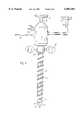
In another embodiment shown in FIG. 7, a gripping element is mounted at the point of the trocar, the said gripping element which has the purpose of firmly holding the body tissue during penetration against the pressure of the trocar point. The body tissue, e.g., peritoneum, cannot thereby avoid the trocar point and cannot be vaulted inwardly into the abdominal cavity.
As FIG. 7 shows, the gripping element consists of arotatable spiral 48, mounted externally to the conical, truncated conical, beveled, or taperedwindow 34, the said spiral which, for example, is made from a wire having surfaces converging to a line which is a cutting edge or cutting element, and is fitted to the conical, beveled, or taperedwindow 34. Thespiral 48 extends to adjacent to the distal end ofwindow 34, and is bent at its posterior end into an axis-parallel connectingpiece 50, which is shaped at its end into a ring, which is not visible in the figure and is mounted rotatably in a circumferential groove of theshaft 10 of the trocar. By means of the said ring, thespiral screw 48 is thus mounted rotatably on theconical window 34 and firmly held axially. Thethread 24 of thesleeve 22 is brought out through the anterior distal sleeve end and acts as adriver point 52 upon the posterior end of the spiral 48, to carry it along during the rotation of thesleeve 22.
To penetrate the body tissue, e.g., the patient's abdominal wall, thesleeve 22 is caused to rotate, whereby it causes thespiral 48 on the conical, beveled, or taperedwindow 34 to rotate via thedriver point 52. Via therotating spiral 48 and the attached theretothread 24, the trocar bores into the tissue like a corkscrew, advancing, cutting and penetrating without the said tissue being able to evade the trocar point. As soon as the tissue is penetrated and the trocar point is, for example, in the abdominal cavity, the trocar together with the spiral 48 held axially on its point can be withdrawn from thesleeve 22. Thesleeve 22 can then be used for the insertion of surgical instruments or the like.
Claims (25)
1. An instrument for the penetration of body tissue comprising:
a rigid shaft having a distal end,
a tissue contacting element at the distal end of said shaft shaped to enlarge an opening in body tissue as the tissue contacting element and said shaft are advanced, said tissue contacting element having a distal end, and at least part of said tissue contacting element being transparent to permit viewing of body tissue therethrough, and
an advancing element at said tissue contacting element, said advancing element having a cutting edge to penetrate the body tissue and extending from adjacent the distal end of said tissue contacting element towards said shaft.
2. The instrument of claim 1, said tissue contacting element being of greater transverse dimension adjacent said distal end of said rigid shaft and of lesser transverse dimension at parts thereof more remote from said distal end of said rigid shaft, said cutting edge extending from outwardly of a part of said tissue contacting element having a greater transverse dimension to outwardly of a part of said tissue contacting element having a lesser transverse dimension.
3. The instrument of claim 2, wherein said transparent part of said tissue contacting element is positioned at the distal end thereof.
4. The instrument of claim 2, said tissue contacting element being truncated.
5. The instrument of claim 2, wherein said tissue contacting element has a surface which is without an opening or openings of sufficient size to permit flow of a substantial amount of fluid therethrough.
6. The instrument of claim 2, wherein the transparent part of said tissue contacting element extends circumferentially through substantially 360°.
7. The instrument of claim 2, said advancing element not extending beyond said distal end of said tissue contacting element.
8. The instrument of claim 2, wherein said cutting element has a proximal end spaced from the distal end of said elongated shaft.
9. The instrument of claim 2, said cutting edge lying outwardly of said tissue contacting element.
10. The instrument of claim 2, wherein said tissue contacting element is in the form of a right circular cone.
11. An instrument for the penetration of body tissue comprising:
a rigid elongated shaft having a distal end,
a tissue contacting element at the distal end of said shaft shaped to penetrate body tissue to enlarge an opening therein as the tissue contacting element and said shaft are advanced, said tissue contacting element having a distal end, and said tissue contacting element being substantially entirely transparent to permit viewing of body tissue therethrough,
an advancing element at said tissue contacting element, said advancing element having a cutting edge lying outwardly of said tissue contacting element and extending from adjacent to the distal end thereof towards said shaft,
said tissue contacting element having a proximal end at and adjacent to the distal end of said shaft, said proximal end of said tissue contacting element being of substantially the same lateral size and shape as the distal end of said shaft.
12. The instrument of claim 11, said advancing element not extending beyond said distal end of said tissue contacting element.
13. An instrument for penetration of body tissue comprising:
an elongated shaft having a longitudinal axis and a distal end,
a window at the distal end of said shaft, said window having a longitudinal axis and a distal end portion spaced from said distal end of said shaft, at least the distal end portion of said window being transparent,
said window having a surface configuration substantially symmetrical about the longitudinal axis thereof,
said longitudinal axis of said window being in alignment with the longitudinal axis of said shaft, and
at least one cutting element having a cutting edge to penetrate the body tissue and extending from adjacent the distal end portion of said window towards said shaft,
whereby light may be transmitted through the transparent portion of said window and said cutting element will effect the advance of said instrument into said body tissue.
14. The instrument of claim 13, said window having a proximal end adjacent the distal end of said shaft, said proximal end of said window being of substantially the same lateral size and shape as the distal end of said shaft.
15. The instrument of claim 13, said cutting edge being outwardly of said window.
16. The instrument of claim 13, wherein said elongated shaft is rigid.
17. The instrument of claim 13, wherein said window is substantially entirely transparent.
18. The instrument of claim 13, wherein said window is of generally tapering configuration from its proximal end to its distal end, and converging to at least one real or virtual point at or adjacent the distal end thereof.
19. The instrument of claim 13, wherein said window is conical.
20. The instrument of claim 19, wherein said conical window is in the form of a right circular cone.
21. The instrument of claim 13, said window having a greater lateral extent at the proximal end thereof than at the distal end thereof, and having at least one surface extending from the proximal end to the distal end thereof.
22. The instrument of claim 13, wherein said surface is a single, generally conical surface.
23. The instrument of claim 13, wherein said window has a surface which is without an opening or openings of sufficient size to permit flow of a substantial amount of fluid therethrough.
24. The instrument of claim 13, wherein said cutting element has a proximal end spaced from of the distal end of said elongated shaft.
25. An instrument for penetration of body tissue comprising:
a rigid elongated shaft having a longitudinal axis and a distal end,
a transparent window at the distal end of said shaft, said window having a longitudinal axis and a distal end portion spaced from said distal end of said shaft,
said transparent window having a surface configuration substantially symmetrical about the longitudinal axis thereof,
said longitudinal axis of said transparent window being in alignment with the longitudinal axis of said shaft,
at least one cutting element having a cutting edge to penetrate the body tissue and extending from adjacent the distal end of said transparent window towards said shaft,
said window having a proximal end at and adjacent to the distal end of said shaft, said proximal end of said window being of substantially the same lateral size and shape as the distal end of said shaft,
whereby light may be transmitted through said transparent window and said cutting element will effect the advance of said instrument into said body tissue.
Priority Applications (1)
| Application Number | Priority Date | Filing Date | Title |
|---|---|---|---|
| US08/500,091US6007481A (en) | 1990-11-06 | 1995-07-10 | Instrument for the penetration of body tissue with cutting and viewing of body structure |
Applications Claiming Priority (5)
| Application Number | Priority Date | Filing Date | Title |
|---|---|---|---|
| DE4035146 | 1990-11-06 | ||
| DE4035146ADE4035146A1 (en) | 1990-11-06 | 1990-11-06 | INSTRUMENT FOR PENETRATING BODY TISSUE |
| US07/779,730US5271380A (en) | 1990-11-06 | 1991-10-23 | Penetration instrument |
| US08/168,213US5431151A (en) | 1990-11-06 | 1993-12-17 | Instrument for the penetration of body tissue |
| US08/500,091US6007481A (en) | 1990-11-06 | 1995-07-10 | Instrument for the penetration of body tissue with cutting and viewing of body structure |
Related Parent Applications (1)
| Application Number | Title | Priority Date | Filing Date |
|---|---|---|---|
| US08/168,213ContinuationUS5431151A (en) | 1990-11-06 | 1993-12-17 | Instrument for the penetration of body tissue |
Publications (1)
| Publication Number | Publication Date |
|---|---|
| US6007481Atrue US6007481A (en) | 1999-12-28 |
Family
ID=6417674
Family Applications (3)
| Application Number | Title | Priority Date | Filing Date |
|---|---|---|---|
| US07/779,730Expired - LifetimeUS5271380A (en) | 1990-11-06 | 1991-10-23 | Penetration instrument |
| US08/168,213Expired - LifetimeUS5431151A (en) | 1990-11-06 | 1993-12-17 | Instrument for the penetration of body tissue |
| US08/500,091Expired - LifetimeUS6007481A (en) | 1990-11-06 | 1995-07-10 | Instrument for the penetration of body tissue with cutting and viewing of body structure |
Family Applications Before (2)
| Application Number | Title | Priority Date | Filing Date |
|---|---|---|---|
| US07/779,730Expired - LifetimeUS5271380A (en) | 1990-11-06 | 1991-10-23 | Penetration instrument |
| US08/168,213Expired - LifetimeUS5431151A (en) | 1990-11-06 | 1993-12-17 | Instrument for the penetration of body tissue |
Country Status (8)
| Country | Link |
|---|---|
| US (3) | US5271380A (en) |
| EP (2) | EP0694280B1 (en) |
| JP (1) | JPH0763477B2 (en) |
| AT (2) | ATE135896T1 (en) |
| DE (4) | DE4035146A1 (en) |
| DK (1) | DK0484725T3 (en) |
| ES (2) | ES2160655T3 (en) |
| GR (1) | GR3020092T3 (en) |
Cited By (67)
| Publication number | Priority date | Publication date | Assignee | Title |
|---|---|---|---|---|
| WO2002051323A1 (en) | 2000-12-27 | 2002-07-04 | Siegfried Riek | Foldable sleeve used as a channel for instruments for minimal invasive surgery |
| US6564087B1 (en)* | 1991-04-29 | 2003-05-13 | Massachusetts Institute Of Technology | Fiber optic needle probes for optical coherence tomography imaging |
| US6656198B2 (en) | 2001-06-01 | 2003-12-02 | Ethicon-Endo Surgery, Inc. | Trocar with reinforced obturator shaft |
| US20050027199A1 (en)* | 2001-04-11 | 2005-02-03 | Clarke Dana S. | Tissue structure identification in advance of instrument |
| US20050065543A1 (en)* | 2001-09-24 | 2005-03-24 | Henry Kahle | Bladeless optical obturator |
| US20060015006A1 (en)* | 2004-06-01 | 2006-01-19 | Laurence Bernard H | System and method for accessing a body cavity |
| US20060169293A1 (en)* | 2003-01-30 | 2006-08-03 | Takeshi Yokoi | Medical device |
| US20060173479A1 (en)* | 2005-01-28 | 2006-08-03 | Smith Robert C | Optical penetrating adapter for surgical portal |
| US20060224174A1 (en)* | 2005-03-31 | 2006-10-05 | Smith Robert C | Optical obturator |
| US20060226655A1 (en)* | 2005-04-12 | 2006-10-12 | Smith Robert C | Optical trocar with scope holding assembly |
| US20070059990A1 (en)* | 2004-05-14 | 2007-03-15 | Olympus Corporation | Insertion device and endoscopic system |
| US20080086160A1 (en)* | 2006-10-06 | 2008-04-10 | Surgiquest, Incorporated | Visualization trocar |
| US20080097474A1 (en)* | 2004-01-12 | 2008-04-24 | Medtreo, Llc | Method of perforating a biological membrane |
| US20080300617A1 (en)* | 2007-06-01 | 2008-12-04 | Tyco Healthcare Group Lp | Obturator tips |
| US20090093677A1 (en)* | 2007-10-05 | 2009-04-09 | Tyco Healthcare Group Lp | Visual obturator |
| US20090192352A1 (en)* | 2008-01-24 | 2009-07-30 | P Regadas F Sergio | Operating Anoscope For Transanal Endoscopic Microsurgery |
| US7708713B2 (en) | 2004-06-29 | 2010-05-04 | Applied Medical Resources Corporation | Insufflating optical surgical instrument |
| US20100137895A1 (en)* | 2007-04-17 | 2010-06-03 | Smith Robert C | Visual obturator with handle |
| US20110098531A1 (en)* | 2009-04-20 | 2011-04-28 | Spine View, Inc. | Dilator with direct visualization |
| US20110263933A1 (en)* | 2007-08-23 | 2011-10-27 | Hansgeorg Schaaf | Resilient support |
| USD654168S1 (en) | 2010-09-21 | 2012-02-14 | Tyco Healthcare Group Lp | Bladeless obturator member |
| USD655005S1 (en) | 2010-09-21 | 2012-02-28 | Tyco Healthcare Group Lp | Bladeless obturator member |
| USD656233S1 (en) | 2010-09-21 | 2012-03-20 | Tyco Healthcare Group Lp | Bladeless obturator member |
| USD663838S1 (en) | 2007-10-05 | 2012-07-17 | Surgiquest, Inc. | Visualization trocar |
| USD667954S1 (en) | 2007-10-05 | 2012-09-25 | Surgiquest, Inc. | Visualization trocar |
| US8377090B2 (en) | 2002-05-16 | 2013-02-19 | Applied Medical Resources Corporation | Blunt tip obturator |
| US20130197395A1 (en)* | 2012-02-01 | 2013-08-01 | Phillibert Jacques JANSSENS | Instrument for taking a tissue sample |
| US8506520B2 (en) | 2008-09-29 | 2013-08-13 | Applied Medical Resources Corporation | Trocar system with laparoscope gas channel |
| US8517977B2 (en) | 2006-10-06 | 2013-08-27 | Applied Medical Resources Corporation | Visual insufflation port |
| WO2013172869A1 (en)* | 2012-05-16 | 2013-11-21 | Jackson Avery M Iii | Illuminated endoscopic pedicle probe with replaceable tip |
| US8636759B2 (en) | 2001-09-24 | 2014-01-28 | Applied Medical Resources Corporation | Bladeless obturator |
| US8663227B2 (en) | 2011-12-03 | 2014-03-04 | Ouroboros Medical, Inc. | Single-unit cutting head systems for safe removal of nucleus pulposus tissue |
| US8795223B2 (en) | 2011-03-08 | 2014-08-05 | Surgiquest, Inc. | Trocar assembly with pneumatic sealing |
| US20140358070A1 (en) | 2007-04-13 | 2014-12-04 | Surgiquest, Inc. | System and method for improved gas recirculation in surgical trocars with pneumatic sealing |
| US8979883B2 (en) | 2009-12-17 | 2015-03-17 | Covidien Lp | Obturator tip |
| US20150080896A1 (en) | 2013-07-19 | 2015-03-19 | Ouroboros Medical, Inc. | Anti-clogging device for a vacuum-assisted, tissue removal system |
| US9216015B2 (en) | 2004-10-28 | 2015-12-22 | Vycor Medical, Inc. | Apparatus and methods for performing brain surgery |
| US9226774B2 (en) | 2009-12-17 | 2016-01-05 | Covidien Lp | Visual obturator with tip openings |
| US9254148B2 (en) | 2011-05-02 | 2016-02-09 | Applied Medical Resources Corporation | Low-profile surgical universal access port |
| US9265899B2 (en) | 2008-01-25 | 2016-02-23 | Applied Medical Resources Corporation | Insufflating access system |
| US9307969B2 (en) | 2005-06-17 | 2016-04-12 | Vycor Medical, Inc. | Tissue retractor apparatus and methods |
| US9439667B2 (en) | 2002-05-31 | 2016-09-13 | Vidacare LLC | Apparatus and methods to install, support and/or monitor performance of intraosseous devices |
| WO2017079662A1 (en) | 2014-11-06 | 2017-05-11 | Asimion Inc. | Visually assisted entry of a veress needle with a tapered videoscope for microlaparoscopy |
| US9693802B2 (en) | 2012-06-06 | 2017-07-04 | Covidien Lp | Obturator tip with insufflation pathway |
| US9737287B2 (en) | 2014-05-13 | 2017-08-22 | Vycor Medical, Inc. | Guidance system mounts for surgical introducers |
| US10022149B2 (en) | 2012-09-28 | 2018-07-17 | Covidien Lp | Optical trocar visualization system and apparatus |
| US10369303B2 (en) | 2003-04-08 | 2019-08-06 | Conmed Corporation | Trocar assembly with pneumatic sealing |
| US10376258B2 (en) | 2016-11-07 | 2019-08-13 | Vycor Medical, Inc. | Surgical introducer with guidance system receptacle |
| US10463492B2 (en) | 2015-11-17 | 2019-11-05 | Edwards Lifesciences Corporation | Systems and devices for setting an anchor |
| US10543016B2 (en) | 2016-11-07 | 2020-01-28 | Vycor Medical, Inc. | Surgical introducer with guidance system receptacle |
| US20200178769A1 (en)* | 2013-03-15 | 2020-06-11 | DePuy Synthes Products, Inc. | Viewing trocar with integrated prism for use with angled endoscope |
| US10827907B2 (en) | 2010-11-29 | 2020-11-10 | Reiner Kunz | Trocar system |
| US11103282B1 (en) | 2002-05-31 | 2021-08-31 | Teleflex Life Sciences Limited | Powered drivers, intraosseous devices and methods to access bone marrow |
| US11135062B2 (en) | 2017-11-20 | 2021-10-05 | Valtech Cardio Ltd. | Cinching of dilated heart muscle |
| US11234683B2 (en) | 2002-05-31 | 2022-02-01 | Teleflex Life Sciences Limited | Assembly for coupling powered driver with intraosseous device |
| US11266441B2 (en) | 2002-05-31 | 2022-03-08 | Teleflex Life Sciences Limited | Penetrator assembly for accessing bone marrow |
| US11324521B2 (en) | 2002-05-31 | 2022-05-10 | Teleflex Life Sciences Limited | Apparatus and method to access bone marrow |
| US11337728B2 (en) | 2002-05-31 | 2022-05-24 | Teleflex Life Sciences Limited | Powered drivers, intraosseous devices and methods to access bone marrow |
| US11357542B2 (en) | 2019-06-21 | 2022-06-14 | Covidien Lp | Valve assembly and retainer for surgical access assembly |
| US11426249B2 (en) | 2006-09-12 | 2022-08-30 | Teleflex Life Sciences Limited | Vertebral access system and methods |
| US11446058B2 (en) | 2020-03-27 | 2022-09-20 | Covidien Lp | Fixture device for folding a seal member |
| US11541218B2 (en) | 2020-03-20 | 2023-01-03 | Covidien Lp | Seal assembly for a surgical access assembly and method of manufacturing the same |
| US11642153B2 (en) | 2020-03-19 | 2023-05-09 | Covidien Lp | Instrument seal for surgical access assembly |
| US11717321B2 (en) | 2020-04-24 | 2023-08-08 | Covidien Lp | Access assembly with retention mechanism |
| US11771439B2 (en) | 2007-04-04 | 2023-10-03 | Teleflex Life Sciences Limited | Powered driver |
| US11812991B2 (en) | 2019-10-18 | 2023-11-14 | Covidien Lp | Seal assemblies for surgical access assemblies |
| US12178469B2 (en) | 2016-11-07 | 2024-12-31 | Vycor Medical Inc. | Surgical introducer with guidance system receptacle |
Families Citing this family (134)
| Publication number | Priority date | Publication date | Assignee | Title |
|---|---|---|---|---|
| DE4035146A1 (en)* | 1990-11-06 | 1992-05-07 | Riek Siegfried | INSTRUMENT FOR PENETRATING BODY TISSUE |
| US5685820A (en)* | 1990-11-06 | 1997-11-11 | Partomed Medizintechnik Gmbh | Instrument for the penetration of body tissue |
| US5350393A (en)* | 1992-01-06 | 1994-09-27 | Inbae Yoon | Safety trocar penetrating instrument |
| US5290276A (en)* | 1992-02-06 | 1994-03-01 | Sewell Jr Frank | Rotatable laparoscopic puncturing instrument |
| US5540648A (en)* | 1992-08-17 | 1996-07-30 | Yoon; Inbae | Medical instrument stabilizer with anchoring system and methods |
| US5653718A (en)* | 1994-05-16 | 1997-08-05 | Yoon; Inbae | Cannula anchoring system |
| US5762609A (en) | 1992-09-14 | 1998-06-09 | Sextant Medical Corporation | Device and method for analysis of surgical tissue interventions |
| US5460182A (en)* | 1992-09-14 | 1995-10-24 | Sextant Medical Corporation | Tissue penetrating apparatus and methods |
| US5772597A (en)* | 1992-09-14 | 1998-06-30 | Sextant Medical Corporation | Surgical tool end effector |
| US5385572A (en)* | 1992-11-12 | 1995-01-31 | Beowulf Holdings | Trocar for endoscopic surgery |
| US5562696A (en)* | 1992-11-12 | 1996-10-08 | Cordis Innovasive Systems, Inc. | Visualization trocar |
| US5334150A (en)* | 1992-11-17 | 1994-08-02 | Kaali Steven G | Visually directed trocar for laparoscopic surgical procedures and method of using same |
| US5987346A (en) | 1993-02-26 | 1999-11-16 | Benaron; David A. | Device and method for classification of tissue |
| US5370640A (en)* | 1993-07-01 | 1994-12-06 | Kolff; Jack | Intracorporeal catheter placement apparatus and method |
| US5470316A (en)* | 1993-09-07 | 1995-11-28 | United States Surgical Corporation | Body tissue penetrating device having a vacuum indicator |
| US5454791A (en)* | 1993-09-07 | 1995-10-03 | United States Surgical Corporation | Trocar with tissue penetration pressure indicator |
| US5441041A (en)* | 1993-09-13 | 1995-08-15 | United States Surgical Corporation | Optical trocar |
| US5467762A (en)* | 1993-09-13 | 1995-11-21 | United States Surgical Corporation | Optical trocar |
| US5429636A (en)* | 1993-10-08 | 1995-07-04 | United States Surgical Corporation | Conductive body tissue penetrating device |
| US5423796A (en)* | 1993-10-08 | 1995-06-13 | United States Surgical Corporation | Trocar with electrical tissue penetration indicator |
| CH687295A5 (en)* | 1993-11-10 | 1996-11-15 | Volpi Ag | Ophthalmoscopic illumination probe. |
| US5720761A (en)* | 1993-11-16 | 1998-02-24 | Worldwide Optical Trocar Licensing Corp. | Visually directed trocar and method |
| DE69433986T2 (en)* | 1993-11-16 | 2005-02-03 | Ethicon Endo-Surgery, Inc., Cincinnati | VISUALLY TAXED TROCAR |
| US5609562A (en)* | 1993-11-16 | 1997-03-11 | Worldwide Optical Trocar Licensing Corporation | Visually directed trocar and method |
| US5591191A (en)* | 1994-01-26 | 1997-01-07 | Kieturakis; Maciej J. | Surgical instrument and method for helically incising a pathway into the interior of the body |
| US5538509A (en)* | 1994-01-31 | 1996-07-23 | Richard-Allan Medical Industries, Inc. | Trocar assembly |
| US5533496A (en)* | 1994-02-15 | 1996-07-09 | Very Inventive Physicians, Inc. | Endoscopic technique particularly suited for exploratory surgery |
| US5448990A (en)* | 1994-02-15 | 1995-09-12 | Very Inventive Physicians, Inc. | Endoscope viewing cannula and surgical techniques |
| US5445142A (en)* | 1994-03-15 | 1995-08-29 | Ethicon Endo-Surgery, Inc. | Surgical trocars having optical tips defining one or more viewing ports |
| US5478329A (en)* | 1994-05-06 | 1995-12-26 | Ternamian; Artin M. | Trocarless rotational entry cannula |
| CA2149290C (en)* | 1994-05-26 | 2006-07-18 | Carl T. Urban | Optical trocar |
| US5569183A (en)* | 1994-06-01 | 1996-10-29 | Archimedes Surgical, Inc. | Method for performing surgery around a viewing space in the interior of the body |
| US6007483A (en)* | 1994-06-01 | 1999-12-28 | Archimedes Surgical, Inc. | Surgical method for developing an anatomic structure |
| US5558665A (en)* | 1994-06-24 | 1996-09-24 | Archimedes Surgical, Inc. | Surgical instrument and method for intraluminal retraction of an anatomic structure |
| US5658306A (en)* | 1994-07-01 | 1997-08-19 | Archimedes Surgical, Inc. | Method for making additional incisions in laparoscopic surgery |
| WO1996001132A1 (en)* | 1994-07-01 | 1996-01-18 | Northgate Technologies Incorporated | High flow insufflation instrument for laparoscopic surgery |
| US5624381A (en)* | 1994-08-09 | 1997-04-29 | Kieturakis; Maciej J. | Surgical instrument and method for retraction of an anatomic structure defining an interior lumen |
| US5794626A (en)* | 1994-08-18 | 1998-08-18 | Kieturakis; Maciej J. | Excisional stereotactic apparatus |
| US5643282A (en)* | 1994-08-22 | 1997-07-01 | Kieturakis; Maciej J. | Surgical instrument and method for removing tissue from an endoscopic workspace |
| US5632717A (en)* | 1994-10-07 | 1997-05-27 | Yoon; Inbae | Penetrating endoscope |
| US5752973A (en)* | 1994-10-18 | 1998-05-19 | Archimedes Surgical, Inc. | Endoscopic surgical gripping instrument with universal joint jaw coupler |
| US5549627A (en)* | 1994-10-21 | 1996-08-27 | Kieturakis; Maciej J. | Surgical instruments and method for applying progressive intracorporeal traction |
| US5653726A (en)* | 1994-11-03 | 1997-08-05 | Archimedes Surgical, Inc. | Retrograde dissector and method for facilitating a TRAM flap |
| US5630813A (en)* | 1994-12-08 | 1997-05-20 | Kieturakis; Maciej J. | Electro-cauterizing dissector and method for facilitating breast implant procedure |
| US5569291A (en)* | 1995-02-01 | 1996-10-29 | Ethicon Endo-Surgery, Inc. | Surgical penetration and dissection instrument |
| US5569292A (en)* | 1995-02-01 | 1996-10-29 | Ethicon Endo-Surgery, Inc. | Surgical penetration instrument with transparent blades and tip cover |
| US5607441A (en)* | 1995-03-24 | 1997-03-04 | Ethicon Endo-Surgery, Inc. | Surgical dissector |
| US5738628A (en)* | 1995-03-24 | 1998-04-14 | Ethicon Endo-Surgery, Inc. | Surgical dissector and method for its use |
| US5662673A (en)* | 1995-04-05 | 1997-09-02 | Kieturakis; Maciej J. | Surgical trocar and method for placing a trocar sleeve in a body wall |
| US5591183A (en)* | 1995-04-12 | 1997-01-07 | Origin Medsystems, Inc. | Dissection apparatus |
| US5980549A (en) | 1995-07-13 | 1999-11-09 | Origin Medsystems, Inc. | Tissue separation cannula with dissection probe and method |
| JPH07323002A (en)* | 1995-06-26 | 1995-12-12 | Olympus Optical Co Ltd | Trocar |
| US5759150A (en) | 1995-07-07 | 1998-06-02 | Olympus Optical Co., Ltd. | System for evulsing subcutaneous tissue |
| US7001404B1 (en) | 1995-07-13 | 2006-02-21 | Origin Medsystems, Inc. | Tissue separation cannula and method |
| US7384423B1 (en) | 1995-07-13 | 2008-06-10 | Origin Medsystems, Inc. | Tissue dissection method |
| US5968065A (en) | 1995-07-13 | 1999-10-19 | Origin Medsystems, Inc. | Tissue separation cannula |
| DE19547246C1 (en)* | 1995-12-18 | 1997-03-20 | Riek Siegfried | Medicinal needle containing spring-loaded guard |
| CA2244164A1 (en) | 1996-01-24 | 1997-07-31 | Albert K. Chin | Tissue separation cannula with dissection probe and method |
| US6630947B1 (en)* | 1996-04-10 | 2003-10-07 | The United States Of America As Represented By The Secretary Of The Navy | Method for examining subsurface environments |
| US5697913A (en) | 1996-08-09 | 1997-12-16 | Ethicon Endo-Surgery, Inc. | Trocar including cannula with stepped region |
| US6379334B1 (en) | 1997-02-10 | 2002-04-30 | Essex Technology, Inc. | Rotate advance catheterization system |
| DE19718086C2 (en)* | 1997-04-29 | 2001-07-05 | Florian Krug | Trocar as an access instrument for minimally invasive surgery |
| US5817061A (en)* | 1997-05-16 | 1998-10-06 | Ethicon Endo-Surgery, Inc. | Trocar assembly |
| US5873889A (en)* | 1997-08-08 | 1999-02-23 | Origin Medsystems, Inc. | Tissue separation cannula with dissection probe and method |
| US6383145B1 (en) | 1997-09-12 | 2002-05-07 | Imagyn Medical Technologies California, Inc. | Incisional breast biopsy device |
| US6080113A (en) | 1998-09-11 | 2000-06-27 | Imagyn Medical Technologies California, Inc. | Incisional breast biopsy device |
| US6551253B2 (en) | 1997-09-12 | 2003-04-22 | Imagyn Medical Technologies | Incisional breast biopsy device |
| US5916233A (en)* | 1998-03-05 | 1999-06-29 | Origin Medsystems, Inc. | Vessel harvesting method and instrument including access port |
| US6447527B1 (en) | 1998-04-23 | 2002-09-10 | Ronald J. Thompson | Apparatus and methods for the penetration of tissue |
| US6030402A (en)* | 1998-04-23 | 2000-02-29 | Thompson; Ronald J. | Apparatus and methods for the penetration of tissue, and the creation of an opening therein |
| US6056766A (en)* | 1998-06-09 | 2000-05-02 | Thompson; Ronald J. | Stabilized trocar, and method of using same |
| US6830546B1 (en) | 1998-06-22 | 2004-12-14 | Origin Medsystems, Inc. | Device and method for remote vessel ligation |
| US7326178B1 (en) | 1998-06-22 | 2008-02-05 | Origin Medsystems, Inc. | Vessel retraction device and method |
| US6976957B1 (en) | 1998-06-22 | 2005-12-20 | Origin Medsystems, Inc. | Cannula-based surgical instrument and method |
| EP0979635A2 (en) | 1998-08-12 | 2000-02-16 | Origin Medsystems, Inc. | Tissue dissector apparatus |
| US20030081310A1 (en)* | 1998-09-09 | 2003-05-01 | Mcmanus Dennis Q. | Microscopy method and apparatus |
| EP1028649A4 (en) | 1998-09-09 | 2004-10-20 | Dennis Q Mcmanus | Microscopy method and apparatus |
| US6206823B1 (en) | 1999-08-02 | 2001-03-27 | Ethicon Endo-Surgery, Inc. | Surgical instrument and method for endoscopic tissue dissection |
| US7398781B1 (en) | 1999-08-10 | 2008-07-15 | Maquet Cardiovascular, Llc | Method for subxiphoid endoscopic access |
| US7264587B2 (en)* | 1999-08-10 | 2007-09-04 | Origin Medsystems, Inc. | Endoscopic subxiphoid surgical procedures |
| US7288096B2 (en) | 2003-01-17 | 2007-10-30 | Origin Medsystems, Inc. | Apparatus for placement of cardiac defibrillator and pacer |
| US7526342B2 (en) | 1999-08-10 | 2009-04-28 | Maquet Cardiovascular Llc | Apparatus for endoscopic cardiac mapping and lead placement |
| US7597698B2 (en) | 1999-08-10 | 2009-10-06 | Maquet Cardiovascular Llc | Apparatus and method for endoscopic encirclement of pulmonary veins for epicardial ablation |
| AU7720100A (en) | 1999-09-27 | 2001-04-30 | Essex Technology, Inc. | Rotate-to-advance catheterization system |
| US6471638B1 (en) | 2000-04-28 | 2002-10-29 | Origin Medsystems, Inc. | Surgical apparatus |
| US6558313B1 (en) | 2000-11-17 | 2003-05-06 | Embro Corporation | Vein harvesting system and method |
| DE10109611A1 (en)* | 2001-02-28 | 2002-09-05 | Bosch Gmbh Robert | Fuel injector |
| EP2422829B1 (en)* | 2001-08-14 | 2013-03-06 | Applied Medical Resources Corporation | Surgical access sealing apparatus |
| US6694715B2 (en)* | 2002-01-22 | 2004-02-24 | H&S Manufacturing Co., Inc. | Inverter shield for a windrow merger |
| US8277411B2 (en) | 2002-01-31 | 2012-10-02 | Boston Scientific Scimed, Inc. | Needle device |
| US20040093000A1 (en)* | 2002-10-23 | 2004-05-13 | Stephen Kerr | Direct vision port site dissector |
| US7056329B2 (en)* | 2002-10-23 | 2006-06-06 | Intellimed Surgical Solutions, Llc | Laparoscopic direct vision dissecting port |
| DE10333956B4 (en)* | 2003-07-25 | 2005-11-03 | Richard Wolf Gmbh | Sichtobturator |
| CA2533204A1 (en) | 2003-08-06 | 2005-02-17 | Applied Medical Resources Corporation | Surgical device with tack-free gel and method of manufacture |
| EP1524542B1 (en)* | 2003-10-17 | 2008-07-02 | Olympus Corporation | Objective lens insertion tool & objective optical system attachment device |
| JP4642769B2 (en)* | 2003-10-17 | 2011-03-02 | タイコ ヘルスケア グループ リミテッド パートナーシップ | Expandable surgical access device |
| US20050149096A1 (en)* | 2003-12-23 | 2005-07-07 | Hilal Said S. | Catheter with conduit traversing tip |
| JP4436833B2 (en)* | 2004-05-14 | 2010-03-24 | オリンパス株式会社 | Insertion device |
| AU2011221382B2 (en)* | 2004-06-29 | 2013-08-22 | Applied Medical Resources Corporation | Insufflating optical surgical instrument |
| EP1861133B1 (en) | 2005-02-28 | 2012-11-21 | Spirus Medical Inc. | Rotate-to-advance catheterization system |
| US7780650B2 (en) | 2005-05-04 | 2010-08-24 | Spirus Medical, Inc. | Rotate-to-advance catheterization system |
| US8414477B2 (en) | 2005-05-04 | 2013-04-09 | Olympus Endo Technology America Inc. | Rotate-to-advance catheterization system |
| US8317678B2 (en) | 2005-05-04 | 2012-11-27 | Olympus Endo Technology America Inc. | Rotate-to-advance catheterization system |
| US8343040B2 (en) | 2005-05-04 | 2013-01-01 | Olympus Endo Technology America Inc. | Rotate-to-advance catheterization system |
| US8235942B2 (en) | 2005-05-04 | 2012-08-07 | Olympus Endo Technology America Inc. | Rotate-to-advance catheterization system |
| DE102005049021B4 (en)* | 2005-10-11 | 2008-08-21 | Richard Wolf Gmbh | endoscope |
| EP1933733A2 (en) | 2005-10-14 | 2008-06-25 | Applied Medical Resources Corporation | Surgical access port |
| US7731710B2 (en) | 2005-10-31 | 2010-06-08 | Alcon, Inc. | Surgical wide-angle illuminator |
| US8157833B2 (en) | 2005-11-09 | 2012-04-17 | Applied Medical Resources Corporation | Trocars with advanced fixation |
| US8574220B2 (en) | 2006-02-28 | 2013-11-05 | Olympus Endo Technology America Inc. | Rotate-to-advance catheterization system |
| US8435229B2 (en) | 2006-02-28 | 2013-05-07 | Olympus Endo Technology America Inc. | Rotate-to-advance catheterization system |
| US8287503B2 (en) | 2006-03-13 | 2012-10-16 | Applied Medical Resources Corporation | Balloon trocar |
| US8147453B2 (en) | 2006-03-13 | 2012-04-03 | Applied Medical Resources Corporation | Balloon trocar |
| US20070260273A1 (en)* | 2006-05-08 | 2007-11-08 | Ethicon Endo-Surgery, Inc. | Endoscopic Translumenal Surgical Systems |
| US7963912B2 (en)* | 2006-05-08 | 2011-06-21 | Ethicon Endo-Surgery, Inc. | Endoscopic translumenal surgical methods using a sheath |
| US20070260121A1 (en)* | 2006-05-08 | 2007-11-08 | Ethicon Endo-Surgery, Inc. | Endoscopic Translumenal Surgical Systems |
| US9770230B2 (en) | 2006-06-01 | 2017-09-26 | Maquet Cardiovascular Llc | Endoscopic vessel harvesting system components |
| AU2007334420B2 (en)* | 2006-12-15 | 2013-03-14 | Covidien Lp | Trocar assembly with obturator design |
| AU2008239409B2 (en)* | 2007-04-11 | 2013-09-19 | Covidien Lp | Visualized entry trocar with moving blade |
| US8870755B2 (en) | 2007-05-18 | 2014-10-28 | Olympus Endo Technology America Inc. | Rotate-to-advance catheterization system |
| US20110028795A1 (en)* | 2009-07-29 | 2011-02-03 | Tyco Healthcare Group Lp | Surgical portal device including textured surface |
| US8888692B1 (en) | 2011-08-26 | 2014-11-18 | Applied Medical Resources Corporation | Trocar cannula assembly and method of manufacture |
| DE102011056705A1 (en)* | 2011-12-20 | 2013-06-20 | Aesculap Ag | Medical obturator and trocar |
| US9186173B2 (en)* | 2012-04-27 | 2015-11-17 | Specialty Care, Inc. | Optical obturator system |
| EP2901913A4 (en)* | 2012-09-28 | 2016-08-31 | Olympus Corp | Endoscope device having cleaning mechanism |
| US10265096B2 (en)* | 2013-03-14 | 2019-04-23 | Medtronic, Inc. | Tunneling tool and method for an implantable medical lead extension |
| KR102301914B1 (en) | 2013-03-15 | 2021-09-15 | 어플라이드 메디컬 리소시스 코포레이션 | Trocar cannula assembly with low profile insertion configuration and method of manufacture |
| DE102014114890A1 (en) | 2014-10-14 | 2016-04-14 | Karl Storz Gmbh & Co. Kg | Trocar sleeve with an asymmetric helix |
| DE102015103214A1 (en) | 2015-03-05 | 2016-09-08 | Karl Storz Gmbh & Co. Kg | trocar |
| WO2016183503A1 (en) | 2015-05-13 | 2016-11-17 | Atricure, Inc. | Access visualization systems |
| USD814034S1 (en)* | 2016-07-26 | 2018-03-27 | Karl Storz Gmbh & Co. Kg | Trocar |
| USD814033S1 (en)* | 2016-07-26 | 2018-03-27 | Karl Storz Gmbh & Co. Kg | Trocar |
| WO2019028458A1 (en) | 2017-08-04 | 2019-02-07 | Brigham And Women's Hospital, Inc. | Veress-type needles with illuminated guidance and safety features |
| US11382662B2 (en) | 2017-08-04 | 2022-07-12 | The Brigham And Women's Hospital, Inc. | Trocars and veress-type needles with illuminated guidance and safety features |
Citations (54)
| Publication number | Priority date | Publication date | Assignee | Title |
|---|---|---|---|---|
| US207932A (en)* | 1878-09-10 | Improvement in surgical dilators | ||
| US1147408A (en)* | 1915-02-08 | 1915-07-20 | Charles Edmund Kells | Trocar. |
| US2699770A (en)* | 1951-05-11 | 1955-01-18 | Centre Nat Rech Scient | Endoscope |
| US2932294A (en)* | 1954-10-13 | 1960-04-12 | Centre Nat Rech Scient | Lighting devices for endoscopes |
| FR1370580A (en)* | 1962-12-04 | 1964-08-28 | Centre Nat Rech Scient | Contact endoscope |
| US3224320A (en)* | 1959-05-05 | 1965-12-21 | Slagteriernes Forskningsinst | Apparatus for determination of the distribution of meat and fat in slaughtered animals or parts thereof |
| US3437747A (en)* | 1964-03-24 | 1969-04-08 | Sheldon Edward E | Devices for inspection using fiberoptic members |
| US3459189A (en)* | 1965-07-28 | 1969-08-05 | Brunswick Corp | Trocar catheter |
| US3556085A (en)* | 1968-02-26 | 1971-01-19 | Olympus Optical Co | Optical viewing instrument |
| US3613684A (en)* | 1969-09-19 | 1971-10-19 | David S Sheridan | Trocar catheters |
| US3653338A (en)* | 1970-09-17 | 1972-04-04 | Flambeau Plastics Corp | Hollow polyethylene rolling pin |
| DE2218901A1 (en)* | 1972-04-19 | 1973-10-31 | Storz Karl Fa | TROCAR FOR INSERTING SURGICAL INSTRUMENTS INTO BODY CAVES |
| US3870036A (en)* | 1972-05-01 | 1975-03-11 | Bio Analytical Labor Inc | Improved speculum barrel member |
| US3961621A (en)* | 1974-02-06 | 1976-06-08 | Akademiet For De Tekniske Videnskaber, Svejsecentralen | Surgical tool for taking biological samples |
| DE2538758A1 (en)* | 1975-08-30 | 1977-03-10 | Eberhard Dr Med Regenbogen | Rectoscope obturator with transparent bolt - is coupled to annular end piece over thin parallel connecting rods |
| US4112932A (en)* | 1977-02-24 | 1978-09-12 | Chiulli Robert D | Laparoscopic cannula |
| DE2929233A1 (en)* | 1978-07-19 | 1980-01-31 | Medtronic Inc | DEVICE FOR FORMING A TISSUE TUNNEL AND / OR FOR POSITIONING A PIPE IN BODY TISSUE |
| US4191191A (en)* | 1978-02-13 | 1980-03-04 | Auburn Robert M | Laproscopic trocar |
| US4222375A (en)* | 1978-03-10 | 1980-09-16 | Miguel Martinez | In vivo illumination system utilizing a cannula with a conical opening allowing a snap-fit with a conical lens and an aperture for flow of fluids and utilizing a housing with a spherical lens for focusing light onto fiber optics |
| DE2922239A1 (en)* | 1979-05-31 | 1980-12-04 | Olympus Optical Co | Vaccination needle penetration sensor - uses reflected light passing through optical fibres to phototransistor to produce visual or audible indication w.r.t. penetration depth |
| US4248214A (en)* | 1979-05-22 | 1981-02-03 | Robert S. Kish | Illuminated urethral catheter |
| US4254762A (en)* | 1979-10-23 | 1981-03-10 | Inbae Yoon | Safety endoscope system |
| US4269192A (en)* | 1977-12-02 | 1981-05-26 | Olympus Optical Co., Ltd. | Stabbing apparatus for diagnosis of living body |
| US4299230A (en)* | 1979-05-09 | 1981-11-10 | Olympus Optical Co., Ltd. | Stabbing apparatus for diagnosis of living body |
| US4311138A (en)* | 1980-03-10 | 1982-01-19 | Sugarman Edward D | Illuminated hypodermic needle |
| US4319563A (en)* | 1977-12-02 | 1982-03-16 | Olympus Optical Co., Ltd. | Endoscope with a smoothly curved distal end face |
| SU942730A1 (en)* | 1980-07-11 | 1982-07-15 | Московский научно-исследовательский институт туберкулеза | Trochar |
| US4356826A (en)* | 1979-05-09 | 1982-11-02 | Olympus Optical Co., Ltd. | Stabbing apparatus for diagnosis of living body |
| US4498902A (en)* | 1982-11-13 | 1985-02-12 | Purdue Research Foundation | Catheter guide |
| EP0135364A2 (en)* | 1983-08-24 | 1985-03-27 | Endotherapeutics Corporation | Trocar assembly |
| US4535773A (en)* | 1982-03-26 | 1985-08-20 | Inbae Yoon | Safety puncturing instrument and method |
| US4537593A (en)* | 1983-06-06 | 1985-08-27 | Becton, Dickinson And Co. | Self-venting, non-coring needle assembly |
| US4567882A (en)* | 1982-12-06 | 1986-02-04 | Vanderbilt University | Method for locating the illuminated tip of an endotracheal tube |
| EP0312787A1 (en)* | 1987-10-21 | 1989-04-26 | Richard Wolf GmbH | Endoscopic instrument set |
| US4895431A (en)* | 1986-11-13 | 1990-01-23 | Olympus Optical Co., Ltd. | Method of processing endoscopic images |
| US4901142A (en)* | 1987-03-23 | 1990-02-13 | Olympus Optical Co., Ltd. | Video scope system |
| EP0369937A1 (en)* | 1988-11-18 | 1990-05-23 | Effner Biomet GmbH | Endoscope, in particular an arthroscope |
| EP0369936A1 (en)* | 1988-11-18 | 1990-05-23 | Effner Biomet GmbH | Endoscope |
| US4972827A (en)* | 1989-02-06 | 1990-11-27 | Fuji Photo Optical Co., Ltd. | Guide device for percutaneous insertion of endoscope |
| DE4020956A1 (en)* | 1989-06-30 | 1991-01-10 | Jaraiz Ricardo Fernandez Blanc | Needle for puncturing spinal column - has tubular body with sliding insert and both parts having conical tips |
| US5057082A (en)* | 1988-11-04 | 1991-10-15 | Plastic Injectors, Inc. | Trocar assembly |
| US5066288A (en)* | 1988-07-06 | 1991-11-19 | Ethicon, Inc. | Safety trocar |
| US5098388A (en)* | 1991-05-02 | 1992-03-24 | Richard Kulkashi | Veress needle assembly |
| US5104388A (en)* | 1990-05-08 | 1992-04-14 | Fbk International Corporation | Membrane splittable tubing |
| DE4133073A1 (en)* | 1990-10-11 | 1992-04-16 | Effner Gmbh | Trocar for producing access for surgery under endoscopic control - has pointed tip at one end of shaft and optical system with objective in vicinity to tip |
| US5147376A (en)* | 1990-05-04 | 1992-09-15 | Francesco Pianetti | Trocar needle laparoscopy with a threaded truncated cone bit |
| US5159920A (en)* | 1990-06-18 | 1992-11-03 | Mentor Corporation | Scope and stent system |
| US5163941A (en)* | 1991-05-07 | 1992-11-17 | California Medical Products | Intubation device |
| US5246425A (en)* | 1992-09-21 | 1993-09-21 | Daniel Hunsberger | Trocar and cannula assembly |
| US5250068A (en)* | 1990-11-30 | 1993-10-05 | Yakuouji Shinkiyu Chiryouin | Optical transmission type acupuncture needle |
| US5256149A (en)* | 1992-02-14 | 1993-10-26 | Ethicon, Inc. | Trocar having transparent cannula and method of using |
| US5269316A (en)* | 1991-08-16 | 1993-12-14 | Spitalny Hans Henning | Surgical instrument |
| US5271380A (en)* | 1990-11-06 | 1993-12-21 | Siegfried Riek | Penetration instrument |
| US5290276A (en)* | 1992-02-06 | 1994-03-01 | Sewell Jr Frank | Rotatable laparoscopic puncturing instrument |
Family Cites Families (10)
| Publication number | Priority date | Publication date | Assignee | Title |
|---|---|---|---|---|
| DE365049C (en)* | 1922-12-07 | Albrecht Meyenberg Dr | Tube speculum for women | |
| DE1616107B1 (en)* | 1968-02-26 | 1971-04-08 | Olympus Optical Co | Microscope for introducing and observing body cavities and an optical instrument with such a microscope |
| US3821956A (en)* | 1973-08-14 | 1974-07-02 | D Gordhamer | Bovine teat dilator and medicament dispenser |
| DE2744222C2 (en) | 1977-09-30 | 1984-10-18 | Siemens AG, 1000 Berlin und 8000 München | Winding arrangement for an electric two-motor drive |
| SU1328658A1 (en)* | 1986-02-04 | 1987-08-07 | Ленинградское Отделение Всесоюзного Государственного Научно-Исследовательского И Проектно-Изыскательского Института "Атомтеплоэлектропроект" | Centrifugal nozzle |
| JPS62183812U (en)* | 1986-05-15 | 1987-11-21 | ||
| JPH01135908U (en)* | 1988-03-14 | 1989-09-18 | ||
| US4846154A (en)* | 1988-06-13 | 1989-07-11 | Macanally Richard B | Dual view endoscope |
| IT1226152B (en)* | 1988-07-05 | 1990-12-19 | Francesco Pianetti | NEEDLE TREQUARTI FOR LAPAROSCOPY. |
| DE4116648A1 (en)* | 1991-05-22 | 1992-11-26 | Helmut Dipl Ing Wurster | Self-cutting trocar insertable in sleeve for penetrating abdominal wall - has manual or motor driven cutting tool so hat power which is normally not used for inserting trocar is considerably reduced and insertion can be controlled |
- 1990
- 1990-11-06DEDE4035146Apatent/DE4035146A1/enactiveGranted
- 1991
- 1991-10-22ATAT91117958Tpatent/ATE135896T1/ennot_activeIP Right Cessation
- 1991-10-22ESES95114514Tpatent/ES2160655T3/ennot_activeExpired - Lifetime
- 1991-10-22EPEP95114514Apatent/EP0694280B1/ennot_activeExpired - Lifetime
- 1991-10-22DEDE9117107Upatent/DE9117107U1/ennot_activeExpired - Lifetime
- 1991-10-22DEDE59109216Tpatent/DE59109216D1/ennot_activeExpired - Lifetime
- 1991-10-22ESES91117958Tpatent/ES2087947T3/ennot_activeExpired - Lifetime
- 1991-10-22DEDE59107614Tpatent/DE59107614D1/ennot_activeExpired - Lifetime
- 1991-10-22DKDK91117958.8Tpatent/DK0484725T3/enactive
- 1991-10-22EPEP91117958Apatent/EP0484725B1/ennot_activeExpired - Lifetime
- 1991-10-22ATAT95114514Tpatent/ATE202913T1/ennot_activeIP Right Cessation
- 1991-10-23USUS07/779,730patent/US5271380A/ennot_activeExpired - Lifetime
- 1991-11-05JPJP3288593Apatent/JPH0763477B2/ennot_activeExpired - Lifetime
- 1993
- 1993-12-17USUS08/168,213patent/US5431151A/ennot_activeExpired - Lifetime
- 1995
- 1995-07-10USUS08/500,091patent/US6007481A/ennot_activeExpired - Lifetime
- 1996
- 1996-05-30GRGR960401448Tpatent/GR3020092T3/enunknown
Patent Citations (58)
| Publication number | Priority date | Publication date | Assignee | Title |
|---|---|---|---|---|
| US207932A (en)* | 1878-09-10 | Improvement in surgical dilators | ||
| US1147408A (en)* | 1915-02-08 | 1915-07-20 | Charles Edmund Kells | Trocar. |
| US2699770A (en)* | 1951-05-11 | 1955-01-18 | Centre Nat Rech Scient | Endoscope |
| US2932294A (en)* | 1954-10-13 | 1960-04-12 | Centre Nat Rech Scient | Lighting devices for endoscopes |
| US3224320A (en)* | 1959-05-05 | 1965-12-21 | Slagteriernes Forskningsinst | Apparatus for determination of the distribution of meat and fat in slaughtered animals or parts thereof |
| US3357433A (en)* | 1962-12-04 | 1967-12-12 | Centre Nat Rech Scient | Endoscope for illumination and observation of contacting distal regions |
| FR1370580A (en)* | 1962-12-04 | 1964-08-28 | Centre Nat Rech Scient | Contact endoscope |
| US3437747A (en)* | 1964-03-24 | 1969-04-08 | Sheldon Edward E | Devices for inspection using fiberoptic members |
| US3459189A (en)* | 1965-07-28 | 1969-08-05 | Brunswick Corp | Trocar catheter |
| US3556085A (en)* | 1968-02-26 | 1971-01-19 | Olympus Optical Co | Optical viewing instrument |
| US3613684A (en)* | 1969-09-19 | 1971-10-19 | David S Sheridan | Trocar catheters |
| US3653338A (en)* | 1970-09-17 | 1972-04-04 | Flambeau Plastics Corp | Hollow polyethylene rolling pin |
| DE2218901A1 (en)* | 1972-04-19 | 1973-10-31 | Storz Karl Fa | TROCAR FOR INSERTING SURGICAL INSTRUMENTS INTO BODY CAVES |
| US3870036A (en)* | 1972-05-01 | 1975-03-11 | Bio Analytical Labor Inc | Improved speculum barrel member |
| US3961621A (en)* | 1974-02-06 | 1976-06-08 | Akademiet For De Tekniske Videnskaber, Svejsecentralen | Surgical tool for taking biological samples |
| DE2538758A1 (en)* | 1975-08-30 | 1977-03-10 | Eberhard Dr Med Regenbogen | Rectoscope obturator with transparent bolt - is coupled to annular end piece over thin parallel connecting rods |
| US4112932A (en)* | 1977-02-24 | 1978-09-12 | Chiulli Robert D | Laparoscopic cannula |
| US4269192A (en)* | 1977-12-02 | 1981-05-26 | Olympus Optical Co., Ltd. | Stabbing apparatus for diagnosis of living body |
| US4319563A (en)* | 1977-12-02 | 1982-03-16 | Olympus Optical Co., Ltd. | Endoscope with a smoothly curved distal end face |
| US4191191A (en)* | 1978-02-13 | 1980-03-04 | Auburn Robert M | Laproscopic trocar |
| US4222375A (en)* | 1978-03-10 | 1980-09-16 | Miguel Martinez | In vivo illumination system utilizing a cannula with a conical opening allowing a snap-fit with a conical lens and an aperture for flow of fluids and utilizing a housing with a spherical lens for focusing light onto fiber optics |
| DE2929233A1 (en)* | 1978-07-19 | 1980-01-31 | Medtronic Inc | DEVICE FOR FORMING A TISSUE TUNNEL AND / OR FOR POSITIONING A PIPE IN BODY TISSUE |
| US4299230A (en)* | 1979-05-09 | 1981-11-10 | Olympus Optical Co., Ltd. | Stabbing apparatus for diagnosis of living body |
| US4356826A (en)* | 1979-05-09 | 1982-11-02 | Olympus Optical Co., Ltd. | Stabbing apparatus for diagnosis of living body |
| US4248214A (en)* | 1979-05-22 | 1981-02-03 | Robert S. Kish | Illuminated urethral catheter |
| DE2922239A1 (en)* | 1979-05-31 | 1980-12-04 | Olympus Optical Co | Vaccination needle penetration sensor - uses reflected light passing through optical fibres to phototransistor to produce visual or audible indication w.r.t. penetration depth |
| US4254762A (en)* | 1979-10-23 | 1981-03-10 | Inbae Yoon | Safety endoscope system |
| US4311138A (en)* | 1980-03-10 | 1982-01-19 | Sugarman Edward D | Illuminated hypodermic needle |
| SU942730A1 (en)* | 1980-07-11 | 1982-07-15 | Московский научно-исследовательский институт туберкулеза | Trochar |
| US4535773A (en)* | 1982-03-26 | 1985-08-20 | Inbae Yoon | Safety puncturing instrument and method |
| US4498902A (en)* | 1982-11-13 | 1985-02-12 | Purdue Research Foundation | Catheter guide |
| US4567882A (en)* | 1982-12-06 | 1986-02-04 | Vanderbilt University | Method for locating the illuminated tip of an endotracheal tube |
| US4537593A (en)* | 1983-06-06 | 1985-08-27 | Becton, Dickinson And Co. | Self-venting, non-coring needle assembly |
| EP0135364A2 (en)* | 1983-08-24 | 1985-03-27 | Endotherapeutics Corporation | Trocar assembly |
| US4601710A (en)* | 1983-08-24 | 1986-07-22 | Endotherapeutics Corporation | Trocar assembly |
| US4601710B1 (en)* | 1983-08-24 | 1998-05-05 | United States Surgical Corp | Trocar assembly |
| US4895431A (en)* | 1986-11-13 | 1990-01-23 | Olympus Optical Co., Ltd. | Method of processing endoscopic images |
| US4901142A (en)* | 1987-03-23 | 1990-02-13 | Olympus Optical Co., Ltd. | Video scope system |
| EP0312787A1 (en)* | 1987-10-21 | 1989-04-26 | Richard Wolf GmbH | Endoscopic instrument set |
| US5066288A (en)* | 1988-07-06 | 1991-11-19 | Ethicon, Inc. | Safety trocar |
| US5057082A (en)* | 1988-11-04 | 1991-10-15 | Plastic Injectors, Inc. | Trocar assembly |
| EP0369936A1 (en)* | 1988-11-18 | 1990-05-23 | Effner Biomet GmbH | Endoscope |
| EP0369937A1 (en)* | 1988-11-18 | 1990-05-23 | Effner Biomet GmbH | Endoscope, in particular an arthroscope |
| US4972827A (en)* | 1989-02-06 | 1990-11-27 | Fuji Photo Optical Co., Ltd. | Guide device for percutaneous insertion of endoscope |
| DE4020956A1 (en)* | 1989-06-30 | 1991-01-10 | Jaraiz Ricardo Fernandez Blanc | Needle for puncturing spinal column - has tubular body with sliding insert and both parts having conical tips |
| US5147376A (en)* | 1990-05-04 | 1992-09-15 | Francesco Pianetti | Trocar needle laparoscopy with a threaded truncated cone bit |
| US5104388A (en)* | 1990-05-08 | 1992-04-14 | Fbk International Corporation | Membrane splittable tubing |
| US5159920A (en)* | 1990-06-18 | 1992-11-03 | Mentor Corporation | Scope and stent system |
| DE4133073A1 (en)* | 1990-10-11 | 1992-04-16 | Effner Gmbh | Trocar for producing access for surgery under endoscopic control - has pointed tip at one end of shaft and optical system with objective in vicinity to tip |
| US5271380A (en)* | 1990-11-06 | 1993-12-21 | Siegfried Riek | Penetration instrument |
| US5431151A (en)* | 1990-11-06 | 1995-07-11 | Partomed Medizintechnik Gmbh | Instrument for the penetration of body tissue |
| US5250068A (en)* | 1990-11-30 | 1993-10-05 | Yakuouji Shinkiyu Chiryouin | Optical transmission type acupuncture needle |
| US5098388A (en)* | 1991-05-02 | 1992-03-24 | Richard Kulkashi | Veress needle assembly |
| US5163941A (en)* | 1991-05-07 | 1992-11-17 | California Medical Products | Intubation device |
| US5269316A (en)* | 1991-08-16 | 1993-12-14 | Spitalny Hans Henning | Surgical instrument |
| US5290276A (en)* | 1992-02-06 | 1994-03-01 | Sewell Jr Frank | Rotatable laparoscopic puncturing instrument |
| US5256149A (en)* | 1992-02-14 | 1993-10-26 | Ethicon, Inc. | Trocar having transparent cannula and method of using |
| US5246425A (en)* | 1992-09-21 | 1993-09-21 | Daniel Hunsberger | Trocar and cannula assembly |
Non-Patent Citations (1)
| Title |
|---|
| Applied Optics, Oct. 1, 1985, vol. 24, No. 19, p. 325, 3126.* |
Cited By (156)
| Publication number | Priority date | Publication date | Assignee | Title |
|---|---|---|---|---|
| US6564087B1 (en)* | 1991-04-29 | 2003-05-13 | Massachusetts Institute Of Technology | Fiber optic needle probes for optical coherence tomography imaging |
| WO2002051323A1 (en) | 2000-12-27 | 2002-07-04 | Siegfried Riek | Foldable sleeve used as a channel for instruments for minimal invasive surgery |
| US20050027199A1 (en)* | 2001-04-11 | 2005-02-03 | Clarke Dana S. | Tissue structure identification in advance of instrument |
| US8046057B2 (en)* | 2001-04-11 | 2011-10-25 | Clarke Dana S | Tissue structure identification in advance of instrument |
| US6656198B2 (en) | 2001-06-01 | 2003-12-02 | Ethicon-Endo Surgery, Inc. | Trocar with reinforced obturator shaft |
| US8267952B2 (en) | 2001-09-24 | 2012-09-18 | Applied Medical Resources Corporation | Bladeless optical obturator |
| US9254125B2 (en) | 2001-09-24 | 2016-02-09 | Applied Medical Resources Corporation | Bladeless obturator |
| US20110190592A1 (en)* | 2001-09-24 | 2011-08-04 | Applied Medical Resources Corporation | Bladeless optical obturator |
| US8608769B2 (en) | 2001-09-24 | 2013-12-17 | Applied Medical Resources Corporation | Bladeless optical obturator |
| US10568658B2 (en) | 2001-09-24 | 2020-02-25 | Applied Medical Resources Corporation | Bladeless optical obturator |
| US7947058B2 (en) | 2001-09-24 | 2011-05-24 | Applied Medical Resources Corporation | Bladeless optical obturator |
| US20050065543A1 (en)* | 2001-09-24 | 2005-03-24 | Henry Kahle | Bladeless optical obturator |
| US8636759B2 (en) | 2001-09-24 | 2014-01-28 | Applied Medical Resources Corporation | Bladeless obturator |
| US8940009B2 (en) | 2001-09-24 | 2015-01-27 | Applied Medical Resources Corporation | Bladeless optical obturator |
| US9655643B2 (en) | 2001-09-24 | 2017-05-23 | Applied Medical Resources Corporation | Bladeless optical obturator |
| US9545248B2 (en) | 2002-05-16 | 2017-01-17 | Applied Medical Resources Corporation | Blunt tip obturator |
| US11207098B2 (en) | 2002-05-16 | 2021-12-28 | Applied Medical Resources Corporation | Blunt tip obturator |
| US8377090B2 (en) | 2002-05-16 | 2013-02-19 | Applied Medical Resources Corporation | Blunt tip obturator |
| US8608768B2 (en) | 2002-05-16 | 2013-12-17 | Applied Medical Resources Corporation | Blunt tip obturator |
| US10368906B2 (en) | 2002-05-16 | 2019-08-06 | Applied Medical Resources Corporation | Blunt tip obturator |
| US11103282B1 (en) | 2002-05-31 | 2021-08-31 | Teleflex Life Sciences Limited | Powered drivers, intraosseous devices and methods to access bone marrow |
| US11266441B2 (en) | 2002-05-31 | 2022-03-08 | Teleflex Life Sciences Limited | Penetrator assembly for accessing bone marrow |
| US10016217B2 (en) | 2002-05-31 | 2018-07-10 | Teleflex Medical Devices S.À.R.L. | Apparatus and methods to install, support and/or monitor performance of intraosseous devices |
| US11291472B2 (en) | 2002-05-31 | 2022-04-05 | Teleflex Life Sciences Limited | Powered drivers, intraosseous devices and methods to access bone marrow |
| US11324521B2 (en) | 2002-05-31 | 2022-05-10 | Teleflex Life Sciences Limited | Apparatus and method to access bone marrow |
| US9439667B2 (en) | 2002-05-31 | 2016-09-13 | Vidacare LLC | Apparatus and methods to install, support and/or monitor performance of intraosseous devices |
| US11103281B2 (en) | 2002-05-31 | 2021-08-31 | Teleflex Life Sciences Limited | Apparatus and methods to install, support and/or monitor performance of intraosseous devices |
| US11234683B2 (en) | 2002-05-31 | 2022-02-01 | Teleflex Life Sciences Limited | Assembly for coupling powered driver with intraosseous device |
| US11337728B2 (en) | 2002-05-31 | 2022-05-24 | Teleflex Life Sciences Limited | Powered drivers, intraosseous devices and methods to access bone marrow |
| US20060169293A1 (en)* | 2003-01-30 | 2006-08-03 | Takeshi Yokoi | Medical device |
| US7637864B2 (en)* | 2003-01-30 | 2009-12-29 | Olympus Corporation | Medical device |
| US10369303B2 (en) | 2003-04-08 | 2019-08-06 | Conmed Corporation | Trocar assembly with pneumatic sealing |
| US10918814B2 (en) | 2003-10-03 | 2021-02-16 | Applied Medical Resources Corporation | Insufflating optical surgical instrument |
| US10226589B2 (en) | 2003-10-03 | 2019-03-12 | Applied Medical Resources Corporation | Insufflating optical surgical instrument |
| US20080097474A1 (en)* | 2004-01-12 | 2008-04-24 | Medtreo, Llc | Method of perforating a biological membrane |
| US20070059990A1 (en)* | 2004-05-14 | 2007-03-15 | Olympus Corporation | Insertion device and endoscopic system |
| US8475476B2 (en) | 2004-06-01 | 2013-07-02 | Cook Medical Technologies Llc | System and method for accessing a body cavity |
| US20060015006A1 (en)* | 2004-06-01 | 2006-01-19 | Laurence Bernard H | System and method for accessing a body cavity |
| US9155558B2 (en) | 2004-06-29 | 2015-10-13 | Applied Medical Resources Corporation | Insufflating optical surgical instrument |
| EP2545870A1 (en)* | 2004-06-29 | 2013-01-16 | Applied Medical Resources Corporation | Insufflating optical surgical instrument |
| US8128590B2 (en) | 2004-06-29 | 2012-03-06 | Applied Medical Resources Corporation | Insufflating optical surgical instruments |
| US7708713B2 (en) | 2004-06-29 | 2010-05-04 | Applied Medical Resources Corporation | Insufflating optical surgical instrument |
| US9216015B2 (en) | 2004-10-28 | 2015-12-22 | Vycor Medical, Inc. | Apparatus and methods for performing brain surgery |
| US9968415B2 (en) | 2004-10-28 | 2018-05-15 | Vycor Medical, Inc. | Apparatus and methods for performing brain surgery |
| US9386974B2 (en) | 2004-10-28 | 2016-07-12 | Vycor Medical, Inc. | Apparatus and methods for performing brain surgery |
| US9968414B2 (en) | 2004-10-28 | 2018-05-15 | Vycor Medical, Inc. | Apparatus and methods for performing brain surgery |
| US10064648B2 (en) | 2005-01-28 | 2018-09-04 | Covidien Lp | Optical penetrating adapter for surgical portal |
| US9226770B2 (en) | 2005-01-28 | 2016-01-05 | Covidien Lp | Optical penetrating adapter for surgical portal |
| US8475486B2 (en) | 2005-01-28 | 2013-07-02 | Covidien Lp | Optical penetrating adapter for surgical portal |
| US8070767B2 (en) | 2005-01-28 | 2011-12-06 | Tyco Healthcare Group Lp | Optical penetrating adapter for surgical portal |
| US20060173479A1 (en)* | 2005-01-28 | 2006-08-03 | Smith Robert C | Optical penetrating adapter for surgical portal |
| US7470230B2 (en) | 2005-03-31 | 2008-12-30 | Tyco Healthcare Group Lp | Optical obturator |
| US10610258B2 (en) | 2005-03-31 | 2020-04-07 | Covidien Lp | Optical obturator |
| US20060224174A1 (en)* | 2005-03-31 | 2006-10-05 | Smith Robert C | Optical obturator |
| US11559327B2 (en) | 2005-03-31 | 2023-01-24 | Covidien Lp | Optical obturator |
| US20090076323A1 (en)* | 2005-03-31 | 2009-03-19 | Tyco Healthcare Group Lp | Optical obturator |
| US20060226655A1 (en)* | 2005-04-12 | 2006-10-12 | Smith Robert C | Optical trocar with scope holding assembly |
| US8517918B2 (en) | 2005-04-12 | 2013-08-27 | Covidien Lp | Optical trocar with scope holding assembly |
| US7824327B2 (en) | 2005-04-12 | 2010-11-02 | Tyco Healthcare Group Llp | Optical trocar with scope holding assembly |
| US20110004061A1 (en)* | 2005-04-12 | 2011-01-06 | Tyco Healthcare Group Lp | Optical trocar with scope holding assembly |
| US10159402B2 (en) | 2005-04-12 | 2018-12-25 | Covidien Lp | Optical trocar with scope holding assembly |
| US9675331B2 (en) | 2005-06-17 | 2017-06-13 | Vycor Medical, Inc. | Tissue retractor apparatus and methods |
| US9782157B2 (en) | 2005-06-17 | 2017-10-10 | Vycor Medical, Inc. | Tissue retractor apparatus and methods |
| US9566052B2 (en) | 2005-06-17 | 2017-02-14 | Vycor Medical, Inc. | Tissue retractor apparatus and methods |
| US9307969B2 (en) | 2005-06-17 | 2016-04-12 | Vycor Medical, Inc. | Tissue retractor apparatus and methods |
| US11426249B2 (en) | 2006-09-12 | 2022-08-30 | Teleflex Life Sciences Limited | Vertebral access system and methods |
| US12089972B2 (en) | 2006-09-12 | 2024-09-17 | Teleflex Life Sciences Limited | Apparatus and methods for biopsy and aspiration of bone marrow |
| US8317815B2 (en) | 2006-10-06 | 2012-11-27 | Surgiquest, Inc. | Visualization trocar |
| US11123105B2 (en) | 2006-10-06 | 2021-09-21 | Applied Medical Resources Corporation | Visual insufflation port |
| US20080086160A1 (en)* | 2006-10-06 | 2008-04-10 | Surgiquest, Incorporated | Visualization trocar |
| US8517977B2 (en) | 2006-10-06 | 2013-08-27 | Applied Medical Resources Corporation | Visual insufflation port |
| US9907569B2 (en) | 2006-10-06 | 2018-03-06 | Conmed Corporation | Trocar assembly with pneumatic sealing |
| US9492062B2 (en) | 2006-10-06 | 2016-11-15 | Applied Medical Resources Corporation | Visual insufflation port |
| US11771439B2 (en) | 2007-04-04 | 2023-10-03 | Teleflex Life Sciences Limited | Powered driver |
| US20140358070A1 (en) | 2007-04-13 | 2014-12-04 | Surgiquest, Inc. | System and method for improved gas recirculation in surgical trocars with pneumatic sealing |
| US10092319B2 (en) | 2007-04-13 | 2018-10-09 | Surgiquest, Inc. | System and method for improved gas recirculation in surgical trocars with pneumatic sealing |
| US20100137895A1 (en)* | 2007-04-17 | 2010-06-03 | Smith Robert C | Visual obturator with handle |
| US8202290B2 (en) | 2007-04-17 | 2012-06-19 | Tyco Healthcare Group Lp | Visual obturator with handle |
| US20080300617A1 (en)* | 2007-06-01 | 2008-12-04 | Tyco Healthcare Group Lp | Obturator tips |
| US9028521B2 (en) | 2007-06-01 | 2015-05-12 | Covidien Lp | Obturator tips |
| US20110263933A1 (en)* | 2007-08-23 | 2011-10-27 | Hansgeorg Schaaf | Resilient support |
| US10863889B2 (en)* | 2007-08-23 | 2020-12-15 | Polydiagnost Entwicklungs—, Produktions—, Vertriebs—, Und Service GmbH | Modular endoscope |
| USD667954S1 (en) | 2007-10-05 | 2012-09-25 | Surgiquest, Inc. | Visualization trocar |
| US20090093677A1 (en)* | 2007-10-05 | 2009-04-09 | Tyco Healthcare Group Lp | Visual obturator |
| US8192353B2 (en) | 2007-10-05 | 2012-06-05 | Tyco Healthcare Group Lp | Visual obturator |
| USD663838S1 (en) | 2007-10-05 | 2012-07-17 | Surgiquest, Inc. | Visualization trocar |
| US20090192352A1 (en)* | 2008-01-24 | 2009-07-30 | P Regadas F Sergio | Operating Anoscope For Transanal Endoscopic Microsurgery |
| US9265899B2 (en) | 2008-01-25 | 2016-02-23 | Applied Medical Resources Corporation | Insufflating access system |
| US10022150B2 (en) | 2008-09-29 | 2018-07-17 | Applied Medical Resources Corporation | First-entry trocar system |
| US8506520B2 (en) | 2008-09-29 | 2013-08-13 | Applied Medical Resources Corporation | Trocar system with laparoscope gas channel |
| US10856906B2 (en) | 2008-09-29 | 2020-12-08 | Applied Medical Resources Corporation | First-entry trocar system |
| US9314266B2 (en) | 2008-09-29 | 2016-04-19 | Applied Medical Resources Corporation | First-entry trocar system |
| US9358040B2 (en) | 2008-09-29 | 2016-06-07 | Applied Medical Resources Corporation | First-entry trocar system |
| US11723689B2 (en) | 2008-09-29 | 2023-08-15 | Applied Medical Resources Corporation | First-entry trocar system |
| US12357346B2 (en) | 2008-10-10 | 2025-07-15 | Conmed Corporation | System and method for improved gas recirculation in surgical trocars with pneumatic sealing |
| US10639071B2 (en) | 2008-10-10 | 2020-05-05 | Conmed Corporation | System and method for improved gas recirculation in surgical trocars with pneumatic sealing |
| US8343035B2 (en) | 2009-04-20 | 2013-01-01 | Spine View, Inc. | Dilator with direct visualization |
| US20110098531A1 (en)* | 2009-04-20 | 2011-04-28 | Spine View, Inc. | Dilator with direct visualization |
| US9226774B2 (en) | 2009-12-17 | 2016-01-05 | Covidien Lp | Visual obturator with tip openings |
| US9301778B2 (en) | 2009-12-17 | 2016-04-05 | Covidien Lp | Obturator tip |
| US8979883B2 (en) | 2009-12-17 | 2015-03-17 | Covidien Lp | Obturator tip |
| USD654168S1 (en) | 2010-09-21 | 2012-02-14 | Tyco Healthcare Group Lp | Bladeless obturator member |
| USD655005S1 (en) | 2010-09-21 | 2012-02-28 | Tyco Healthcare Group Lp | Bladeless obturator member |
| USD656233S1 (en) | 2010-09-21 | 2012-03-20 | Tyco Healthcare Group Lp | Bladeless obturator member |
| US10827907B2 (en) | 2010-11-29 | 2020-11-10 | Reiner Kunz | Trocar system |
| US8795223B2 (en) | 2011-03-08 | 2014-08-05 | Surgiquest, Inc. | Trocar assembly with pneumatic sealing |
| US9254148B2 (en) | 2011-05-02 | 2016-02-09 | Applied Medical Resources Corporation | Low-profile surgical universal access port |
| US10448967B2 (en) | 2011-12-03 | 2019-10-22 | DePuy Synthes Products, Inc. | Discectomy kits with an obturator, guard cannula |
| US8663227B2 (en) | 2011-12-03 | 2014-03-04 | Ouroboros Medical, Inc. | Single-unit cutting head systems for safe removal of nucleus pulposus tissue |
| US9220528B2 (en) | 2011-12-03 | 2015-12-29 | Ouroboros Medical, Inc. | Tubular cutter having a talon with opposing, lateral cutting surfaces |
| US9119659B2 (en) | 2011-12-03 | 2015-09-01 | Ouroboros Medical, Inc. | Safe cutting heads and systems for fast removal of a target tissue |
| US9265521B2 (en) | 2011-12-03 | 2016-02-23 | Ouroboros Medical, Inc. | Tissue removal systems with articulating cutting heads |
| US20130197395A1 (en)* | 2012-02-01 | 2013-08-01 | Phillibert Jacques JANSSENS | Instrument for taking a tissue sample |
| US9050071B2 (en)* | 2012-02-01 | 2015-06-09 | Phillibert Jacques JANSSENS | Instrument for taking a tissue sample |
| WO2013172869A1 (en)* | 2012-05-16 | 2013-11-21 | Jackson Avery M Iii | Illuminated endoscopic pedicle probe with replaceable tip |
| US10939925B2 (en) | 2012-05-16 | 2021-03-09 | Avery M. Jackson, III | Illuminated endoscopic pedicle probe with replaceable tip |
| US11000291B2 (en) | 2012-05-16 | 2021-05-11 | Optical Spine, Llc | Illuminated probe with replaceable tip |
| US10398451B2 (en) | 2012-05-16 | 2019-09-03 | Optical Spine, Llc | Illuminated endoscopic pedicle probe with replaceable tip |
| US9693802B2 (en) | 2012-06-06 | 2017-07-04 | Covidien Lp | Obturator tip with insufflation pathway |
| US10420586B2 (en) | 2012-06-06 | 2019-09-24 | Covidien Lp | Obturator tip with insufflation pathway |
| US11147587B2 (en) | 2012-06-06 | 2021-10-19 | Covidien Lp | Obturator tip with insufflation pathway |
| US10588662B2 (en) | 2012-09-28 | 2020-03-17 | Covidien Lp | Optical trocar visualization system and apparatus |
| US10022149B2 (en) | 2012-09-28 | 2018-07-17 | Covidien Lp | Optical trocar visualization system and apparatus |
| US11529169B2 (en) | 2012-09-28 | 2022-12-20 | Covidien Lp | Optical trocar visualization system and apparatus |
| US20200178769A1 (en)* | 2013-03-15 | 2020-06-11 | DePuy Synthes Products, Inc. | Viewing trocar with integrated prism for use with angled endoscope |
| US11690498B2 (en)* | 2013-03-15 | 2023-07-04 | DePuy Synthes Products, Inc. | Viewing trocar with integrated prism for use with angled endoscope |
| US20150080896A1 (en) | 2013-07-19 | 2015-03-19 | Ouroboros Medical, Inc. | Anti-clogging device for a vacuum-assisted, tissue removal system |
| US10342563B2 (en) | 2013-07-19 | 2019-07-09 | DePuy Synthes Products, Inc. | Anti-clogging device for a vacuum-assisted, tissue removal system |
| US11116487B2 (en) | 2014-05-13 | 2021-09-14 | Vycor Medical, Inc. | Guidance system mounts for surgical introducers |
| US9737287B2 (en) | 2014-05-13 | 2017-08-22 | Vycor Medical, Inc. | Guidance system mounts for surgical introducers |
| US12059144B2 (en) | 2014-05-13 | 2024-08-13 | Vycor Medical, Inc. | Guidance system mounts for surgical introducers |
| US10327748B2 (en) | 2014-05-13 | 2019-06-25 | Vycor Medical, Inc. | Guidance system mounts for surgical introducers |
| US10463399B2 (en) | 2014-11-06 | 2019-11-05 | Asimion Inc. | Visually assisted entry of a Veress needle with a tapered videoscope for microlaparoscopy |
| US11602374B1 (en) | 2014-11-06 | 2023-03-14 | Enable, Inc. | Visually assisted entry of a fluid delivering needle with a tapered videoscope |
| WO2017079662A1 (en) | 2014-11-06 | 2017-05-11 | Asimion Inc. | Visually assisted entry of a veress needle with a tapered videoscope for microlaparoscopy |
| US11446146B2 (en) | 2015-11-17 | 2022-09-20 | Edwards Lifesciences Corporation | Heart reshaping system |
| US10555814B2 (en) | 2015-11-17 | 2020-02-11 | Edwards Lifesciences Corporation | Ultrasound probe for cardiac treatment |
| US11883294B2 (en) | 2015-11-17 | 2024-01-30 | Edwards Lifesciences Corporation | Systems and devices for setting an anchor |
| US11331189B2 (en) | 2015-11-17 | 2022-05-17 | Edwards Lifesciences Corporation | Systems and devices for setting an anchor |
| US10463492B2 (en) | 2015-11-17 | 2019-11-05 | Edwards Lifesciences Corporation | Systems and devices for setting an anchor |
| US10543016B2 (en) | 2016-11-07 | 2020-01-28 | Vycor Medical, Inc. | Surgical introducer with guidance system receptacle |
| US10376258B2 (en) | 2016-11-07 | 2019-08-13 | Vycor Medical, Inc. | Surgical introducer with guidance system receptacle |
| US11517347B2 (en) | 2016-11-07 | 2022-12-06 | Vycor Medical, Inc. | Surgical introducer with guidance system receptacle |
| US12178469B2 (en) | 2016-11-07 | 2024-12-31 | Vycor Medical Inc. | Surgical introducer with guidance system receptacle |
| US11045182B2 (en) | 2016-11-07 | 2021-06-29 | Vycor Medical, Inc. | Surgical introducer with guidance system receptacle |
| US11135062B2 (en) | 2017-11-20 | 2021-10-05 | Valtech Cardio Ltd. | Cinching of dilated heart muscle |
| US12364605B2 (en) | 2017-11-20 | 2025-07-22 | Edwards Lifesciences Innovation (Israel) Ltd. | Cinching of dilated heart muscle |
| US11357542B2 (en) | 2019-06-21 | 2022-06-14 | Covidien Lp | Valve assembly and retainer for surgical access assembly |
| US12127762B2 (en) | 2019-06-21 | 2024-10-29 | Covidien, LP | Valve assembly and retainer for surgical access assembly |
| US11812991B2 (en) | 2019-10-18 | 2023-11-14 | Covidien Lp | Seal assemblies for surgical access assemblies |
| US11642153B2 (en) | 2020-03-19 | 2023-05-09 | Covidien Lp | Instrument seal for surgical access assembly |
| US12127763B2 (en) | 2020-03-19 | 2024-10-29 | Covidien Lp | Instrument seal for surgical access assembly |
| US11541218B2 (en) | 2020-03-20 | 2023-01-03 | Covidien Lp | Seal assembly for a surgical access assembly and method of manufacturing the same |
| US12064141B2 (en) | 2020-03-27 | 2024-08-20 | Covidien Lp | Fixture device for folding a seal member |
| US11446058B2 (en) | 2020-03-27 | 2022-09-20 | Covidien Lp | Fixture device for folding a seal member |
| US11717321B2 (en) | 2020-04-24 | 2023-08-08 | Covidien Lp | Access assembly with retention mechanism |
Also Published As
| Publication number | Publication date |
|---|---|
| EP0484725A1 (en) | 1992-05-13 |
| US5431151A (en) | 1995-07-11 |
| DE4035146A1 (en) | 1992-05-07 |
| JPH04266752A (en) | 1992-09-22 |
| DE4035146C2 (en) | 1992-10-29 |
| DE59107614D1 (en) | 1996-05-02 |
| EP0694280A2 (en) | 1996-01-31 |
| ES2160655T3 (en) | 2001-11-16 |
| ATE135896T1 (en) | 1996-04-15 |
| ES2087947T3 (en) | 1996-08-01 |
| DK0484725T3 (en) | 1996-07-29 |
| DE9117107U1 (en) | 1995-12-21 |
| EP0484725B1 (en) | 1996-03-27 |
| DE59109216D1 (en) | 2001-08-16 |
| EP0694280A3 (en) | 1996-04-17 |
| GR3020092T3 (en) | 1996-08-31 |
| JPH0763477B2 (en) | 1995-07-12 |
| EP0694280B1 (en) | 2001-07-11 |
| US5271380A (en) | 1993-12-21 |
| ATE202913T1 (en) | 2001-07-15 |
Similar Documents
| Publication | Publication Date | Title |
|---|---|---|
| US6007481A (en) | Instrument for the penetration of body tissue with cutting and viewing of body structure | |
| US5685820A (en) | Instrument for the penetration of body tissue | |
| US5323766A (en) | Illuminating endo-photocoagulation probe | |
| EP0726784B1 (en) | Visually directed trocar | |
| US5588952A (en) | Intracardiac illuminator with suction | |
| US5632740A (en) | Illuminated leading probe device | |
| US5121740A (en) | Laser video endoscope | |
| US5720761A (en) | Visually directed trocar and method | |
| JP3662377B2 (en) | Medical needle | |
| JP2002507461A (en) | Illumination device for use in ophthalmic microsurgery | |
| CA2267324A1 (en) | Tip formation for inserting a flexible membrane into an eye | |
| EP0217935A4 (en) | Endoscope. | |
| US5785645A (en) | Beveled tip illuminator for microsurgery | |
| JP7514993B2 (en) | Ophthalmic Microsurgery Instruments | |
| JP2021500198A5 (en) | ||
| EP0746247B1 (en) | Visually directed trocar | |
| US11826033B2 (en) | Illuminated scleral depressor assembly | |
| JPH09164114A (en) | Endoscope | |
| JPH0530406Y2 (en) | ||
| JPH034845A (en) | Insertion supporter | |
| JPH01284233A (en) | Ultra-sonic wave probe | |
| CA2389821A1 (en) | Tip formation for inserting a flexible membrane into an eye |
Legal Events
| Date | Code | Title | Description |
|---|---|---|---|
| STCF | Information on status: patent grant | Free format text:PATENTED CASE | |
| FEPP | Fee payment procedure | Free format text:PAYOR NUMBER ASSIGNED (ORIGINAL EVENT CODE: ASPN); ENTITY STATUS OF PATENT OWNER: LARGE ENTITY | |
| FPAY | Fee payment | Year of fee payment:4 | |
| FPAY | Fee payment | Year of fee payment:8 | |
| FPAY | Fee payment | Year of fee payment:12 |