US6002958A - Method and apparatus for diagnostics of internal organs - Google Patents
Method and apparatus for diagnostics of internal organsDownload PDFInfo
- Publication number
- US6002958A US6002958AUS08/491,865US49186596AUS6002958AUS 6002958 AUS6002958 AUS 6002958AUS 49186596 AUS49186596 AUS 49186596AUS 6002958 AUS6002958 AUS 6002958A
- Authority
- US
- United States
- Prior art keywords
- electromagnetic radiation
- human body
- near infrared
- ultrasound beam
- amplitude modulated
- Prior art date
- Legal status (The legal status is an assumption and is not a legal conclusion. Google has not performed a legal analysis and makes no representation as to the accuracy of the status listed.)
- Expired - Fee Related
Links
Images


Classifications
- A—HUMAN NECESSITIES
- A61—MEDICAL OR VETERINARY SCIENCE; HYGIENE
- A61B—DIAGNOSIS; SURGERY; IDENTIFICATION
- A61B5/00—Measuring for diagnostic purposes; Identification of persons
- A61B5/0059—Measuring for diagnostic purposes; Identification of persons using light, e.g. diagnosis by transillumination, diascopy, fluorescence
- A—HUMAN NECESSITIES
- A61—MEDICAL OR VETERINARY SCIENCE; HYGIENE
- A61B—DIAGNOSIS; SURGERY; IDENTIFICATION
- A61B5/00—Measuring for diagnostic purposes; Identification of persons
- A61B5/0048—Detecting, measuring or recording by applying mechanical forces or stimuli
- A61B5/0051—Detecting, measuring or recording by applying mechanical forces or stimuli by applying vibrations
- G—PHYSICS
- G01—MEASURING; TESTING
- G01N—INVESTIGATING OR ANALYSING MATERIALS BY DETERMINING THEIR CHEMICAL OR PHYSICAL PROPERTIES
- G01N21/00—Investigating or analysing materials by the use of optical means, i.e. using sub-millimetre waves, infrared, visible or ultraviolet light
- G01N21/17—Systems in which incident light is modified in accordance with the properties of the material investigated
- G01N21/47—Scattering, i.e. diffuse reflection
- G01N21/49—Scattering, i.e. diffuse reflection within a body or fluid
Definitions
- the inventionrelates generally to medicine, or more particularly to methods of non-invasive investigations of living organisms, and more specifically, for diagnostics of pathological changes in tissues of human and animal organisms.
- the method closest to the presentis a spectral method of optical diagnostics of internal organs, based on transillumination of the investigated organ by radiation of the near infrared (NIR), 0.6-1.5 um wavelength range and recording of the intensity of the transmitted illumination at several wavelengths.
- NIRnear infrared
- the presence of a "shadow” or a change in the photodetector output signalserves as a criterion of the pathology. This method may be considered similar to the present method and equipment.
- the known apparatus for the realization of the transillumination methodcontains an NIR-radiation source with an optical system and a photodetecting device, coupled to a recorder.
- the investigated organa mammary gland, for example, is placed between two transparent plates, compressing the organ and shaping it into a plane-parallel, trapezoid or other forms.
- the means of moving the "NIR-radiation source--photodetecting device" pair over the organ surfaceare also available.
- the main disadvantage of this apparatusis its low sensitivity in revealing the pathology at an early stage due to the above mentioned strong scattering of NIR-radiation by biological tissues.
- the task of the inventionis elimination of the above mentioned disadvantages of the prototype, i.e. to achieve increasing in the spatial resolution under the conditions of a strong optical NIR-radiation scattering by the investigated tissues.
- the inventionis directed to the method and apparatus of optical diagnostics of human body internal organs, based on the illumination of the investigated organ with near IR (NIR)-radiation of 0.6-1.5 um wavelength range and recording of the transmitted and/or back scattered radiation parameters at several discrete wavelengths, an additional focused beam of amplitude-modulated ultrasound waves, the focal spot of this beam scanning the volume of the body investigated and, simultaneously with, at least one of the parameters of modulation of the intensity of the transmitted and/or of back scattered radiation of the infrared radiation, appearing under these conditions, being recorded.
- NIRnear IR
- the presence and the type of pathologybeing judged by the value and/or the character of the relative change in the above mentioned parameters of modulation during the scanning process.
- a pulse modulation of the intensity of the ultrasound waves beamis employed, and the amplitude of modulation of the infrared radiation appearing under these conditions is recorded.
- the duration and the duty cycle of the pulses of the modulation intensity of the ultrasound waves beamare varied, and at least one of the parameters of the transient process of the intensity modulation of the scattered infrared radiation is recorded.
- An apparatus for realization of the methodincludes a transparent support base both for NIR radiation and ultrasound for a body investigated, illuminators and detectors of the radiation placed from the both sides of the support, a scanning system, a block for signal processing and control, a reflecting device and an oscillator of ultrasound focused waves with an electrical generator, a modulator, illuminators and photodetectors control blocks, an amplifier and a synchronous integrator, the ultrasound focusing oscillator being coupled to the scanning system in the plane of the transparent support and connected with the generator and the modulator.
- Manageable spectrally-selective photodetectorsare connected with the block of signal processing and control via the sequentially connected photodetectors control block, amplifier and synchronous integrator; the modulator output is connected with the synchronizing input of the synchronous integrator, and the illuminators inputs are connected with the corresponding control block outputs, the synchronizing inputs of the illuminators and photodetectors control blocks being connected with the outputs of the signal processing and control block.
- the apparatus supportis in the form of plates, transparent both for infrared and ultrasound radiation.
- the photodetectorscan be placed inside the plates body and turned to present their sensitive surfaces to each other.
- radiating elements of the photodetectors and the illuminatorscan be placed inside the body of at least one plate.
- the ultrasound focusing ocsillatoris placed at a camera, filled with an immersion medium and covered by the (transparent both for NIR and ultrasound) support or base plate, the oscillator acoustic axis crossing the plate plane.
- the oscillatorcan be in the form of a piezo-electric transformer with the electrodes divided into sections and connected via manageable phase rotators to the generator, the control inputs of the latter being connected with the signal processing and control block.
- FIG. 1is a schematic block diagram of the apparatus of this invention
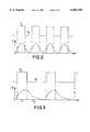
- FIG. 2is a graphic representation of the varying modulation of the ultrasound probe utilized with the invention.
- FIG. 3is a graphic representation of the IR signals utilized with this invention.
- FIG. 4is a schematic, cross-sectional representation showing the ultrasound oscillator and illuminator utilized with this invention
- FIG. 5is a schematic, cross-sectional view of the photodetectors and light -emitting diodes arrayed in one of the plates used in this invention.
- FIG. 6is another embodiment of this invention illustrating in a schematic, cross-sectional view the ultrasound oscillator and photodetector devices when an object under investigation is viewed from one side only this invention.
- the present inventionis based upon the fact, established by the inventor for the first time, that the spatial resolution of the optical transillumination method can be considerably increased by the application of ultrasound waves under the conditions of strong optical inhomogeneity of the subject investigated.
- optical inhomogeneity of the investigated subjectresults in an abrupt deterioration of the metrological characteristics of the transillumination method, since the "shadow" recorded can exceed considerably the size of a pathological growth and this is connected in a rather complex way with the pathology location in the strongly scattering medium.
- the real part(the refraction coefficient) is changed practically non-inertially under pressure application, due to "condensing" of scatterers (see Barabanenkov, Yu. N. On the relative increase in radiation extinction length due to correlation of weak scatterers/USSR Academy of Sci. Proceedings, Physics of atmosphere and ocean, vol. 18, No. 7, pp. 720-726, 1982) and changes in the imaginary part (the absorption coefficient, mainly by blood) take place with a delay of several seconds, as a result of blood expelling from the capillaries (vessels).
- This effectrepresents some kind of the acoustic optical palpation, making up the basis for revealing the pathology via the contrast in compression, the latter being transformed into the amplitude modulation contrast of the transmitted or/and back scattered optical beam.
- a modulated ultrasound beamirritates the biological tissue receptors (blood vessels cell walls, for the first time). It could be mechanical receptors, thermal receptors and non-specific nerve outer sheaths.
- capillary blood flow at the effected zonesis modulated, for example, at the skin areas related to the internal organs--Zakharjina-Ged zones. This produces the corresponding modulation of the optical transparency of the tissue investigated.
- the latter effectis more inertial (from several seconds to minutes), but the necessary intensities of the ultrasound waves are very small and are determined by the reception threshold.
- the contrastin order to reveal a pathological growth in this case, is formed as a result of the differences in the sensitivity thresholds for normal and pathological tissues to the influence of the modulated ultrasound beam. Both effects of the "acoustic optical palpation"--via direct modulation of tissue optical parameters and the sensory ones--could be used in cooperation for pathology revealing and identification.
- a focused ultrasound beamperforms the function of the probe.
- Ultrasound probesi.e. the sources of ultrasound waves, representing ultrasound focusing transformers supplied with the scanning means, are known in biology and medicine (see Vartapetjan M. A. et al. Sensor perception. An investigation experience with the help of focused ultrasound, Leningrad, Nauka, 1985 ( Russian)). Their main object is to provoke local changes in the tissue features from point to point over the scanning direction and to compare the features of the nearby sites. Biological influence of the focused ultrasound waves is caused by the combined action of a number of factors (mechanical, thermal etc.), that could be recorded both via changes in the acoustic characteristics (see Patent application of Great Britain No. 1403241, cl.
- the preferred embodiment of the ultrasound (US) probeis chosen on the basis of the following considerations:
- the density of the ultrasound waves at the focal spot areais to be set up not higher than 1 kW/m 2 of continuous ultrasound radiation, since such "diagnostic" ultrasound waves at the range of frequencies of 0.3-10 MHz and the exposition times from 1 s up to 500 s are known not to be accompanied by any irreversible effects (see Biophysical approach to the problem of safety under the ultrasound diagnostics/Proc. Conf. Ultrasound Biology & Medicine--Ubiomed. YI, Warsaw Jablonna, Sep. 19-23, pp. 95-99, 1983).
- the workshould be performed under the pain threshold of sensitivity, the latter, being dependent on the type of organ investigated, can aggravate the above mentioned restrictions (see Vartapetjan M. A. et al. Ibid.)
- the frequency of the ultrasound wavesis to be chosen to be in the range of 0.3-10 MHz.
- the diameter of the focal spot in the investigated tissuewould be between one millimeter and one tenth of a millimeter, depending on the modulation frequency.
- a pulse modulation of the ultrasound beamis used.
- the frequency of the pulsesis usually chosen to be in the range from about one tenth of a hertz to tens of kilohertz.
- the ultrasound beamis variably modulated, the amplitude of the first harmonic of the scattered infrared radiation modulation, appearing under these conditions, is synchronously recorded.
- the US-beamis pulse modulated with the intervals between pulses exceeding the time constant of the recovery of the tissue's optical characteristics after the deformation provoked by the influence of the US-beam, then not only is the amplitude of the modulation of the scattered NIR-radiation measured, but also the form of the pulses of this radiation is recorded with the help of a strobe-integrator.
- the parameters of the transient processesare measured following the US-beam switching on and off, including time delay between the beginning of changes in the intensity of NIR-radiation in answer to these switchings.
- the following parameters of the pulse modulationare chosen: the pulse duration of the ultrasound waves, from tens of seconds to milliseconds; sequence frequency, from tenths of hertz to tens of kilohertz. The optimal pulse durations and intervals between them are selected for different tissues during special experiments.
- the method of the present inventioncan be realized with the help of the apparatus shown in FIG. 1.
- a body under investigation (or its organ) 1is illuminated from illuminators 2 and 3 by NIR-radiation beams, each of the beam widths being unlimited and could be of the same magnitude as the size of the organ investigated.
- investigated body 1is scanned by focused beam 4 with focal spot 5 from ultrasound oscillator 6.
- Oscillator 6is coupled to a high frequency generator 7 of continuous electrical oscillations, the modulation of the latter being performed with modulator 8.
- the transmitted and back scattered NIR-radiationis recorded with help of photodetectors 9,10, supplied with a lens and a reflector, receiving the scattered light, which is coupled via photodetector 11 control block to amplifier 12.
- the signal from the output of amplifier 12is received by synchronous integrator 13, and then by block 14 of the signal processing and control.
- a synchronizing signalis received by block 13 from modulator 8.
- a synchronous detectoris usually used as the integrator 13.
- a strobe-integrator with an electric controlis used (see Titce, U. and Shenck, K. Semiconductor scheme technology, p. 144, Moscow, Mir, 1982 (Translated from English to Russian)).
- Block 14can be in an analog or a digital form; in the latter case, a personal computer is sufficient for performing the apparatus control via the input-output controller, as well as for -the data processing and their display on a monitor.
- Information from block 14 of the signal processing and controlis received by display 15.
- Ultrasound oscillator 6is supplied with scanner driver 16, connected with block 17 of US-probe scanner.
- Scanner block 17 outputis connected with input of block 14 of signal processing and control.
- the intensity and the spectral composition of NIR-radiationis set up with the help of illuminator control block 18.
- the operation of the inventiontakes place as follows. Investigated body 1, or a part of it, is placed on support elements 19. Simultaneously with illumination of investigated body 1 with the help of the NIR radiation source, the scanning of the area investigated by a focused beam 4 of the ultrasound waves is performed. Focal spot 5 is moved up and down, as well as from right to left, in accordance with a scanning regime, provided by blocks 14 and 17, and at each point scanned, the modulated scattered NIR-radiation (transmitted or back scattered) is received and recorded by photodetectors 9 and 10. To increase the intensity of the light received, light gathering lenses are placed before the photodetector's sensitive plates and a reflector behind them.
- the form of the evoked modulation of the scattered NIR-radiationis close to the sinusoidal wave shown in FIG. 2: the upper curve shows the form of the US-beam modulation, and the lower one that of the NIR-radiation).
- a pulse modulationwith the pulse duration larger than the recovery time of the investigated tissue optical characteristics.
- the transientprocesses parameters--a delay in the beginning of the changes in the NIR-radiation intensity after switching on and off the ultrasound waves, the time constant of these processes, the position at the time axis and the extrema amplitudes etc.--are recorded.
- the form of the modulation pulses of the scattered NIR-radiationis recorded synchronously with the help of the strobe-integrator (see FIG. 3, the disposition of the curves are the same as in FIG. 2).
- a periodical shifting of a normal, non-modulated US-beam from one part of the body investigated to the other oneis performed with the help of scanning block 17, the latter being managed by control block 14, according to the assigned program.
- the input signalwill be characterized by some value of the modulation, typical of normal tissues. If focal spot 5 falls into the zone of a pathological growth 20 and becomes coincident with it, at least one of the parameters of the NIR-modulation, received by photodetectors 9 and 10 (by both or by one of them), will change, indicating the presence of the tissue inhomogeneity and of possible pathology. It is a combined recording of the output signal and the changes in the position of focal spot 5 (via information from scanning block 17) over the investigated area that gives the possibility to localize the pathological growth and to draw the growth contour, or in other words, to perform mapping.
- the focal spot 5is fixed at the pathological growth and spectral dependencies of the amplitude of the output signal are recorded, while varying the spectral composition of the illuminating radiation and/or of the optical radiation recorded.
- the dependence of the parameters of the NIR-radiation modulation on the parameters of the US-beam modulationare recorded with the-same aim.
- the device for realization of the method of this invention in the part that relates to the supplementing the apparatus with the means of the ultrasound beam forming and scanning or the arrangement of US-probecan be accomplished with the use of the elements adopted to the ultrasound diagnostics, see Vartapetjan M. A., et al. Ibid., for example.
- the device shown in FIG. 4represents a hermetic frame 21 filled with an immersion medium 22, of water free of gases, for example.
- Ultrasound oscillator 6connected with the scanning facilities (not shown) is placed at the cavity.
- Oscillator 6 with the facilities for forming focal spot 5can be in the form of an electro-acoustical piezo-transformer, supplied with an acoustical lens. If the latter is omitted, a sectioned piezo-transformer is used, each section being connected via a manageable phase-rotor with the generator of the electrical oscillations (see U.S. Pat. No. 4,207,901, cl. A 61 B 5/00, NKI 128-660, 1980). In the latter case, controlled moving of the focal spot 5 can be performed both in the axial and in the transverse directions without use of any mechanical scanning mechanisms.
- Frame 21is optically (and acoustically) closed at the front surface with transparent cover/support 23 made from an organic glass, for example, or from other transparent plastic material.
- transparent cover/support 23made from an organic glass, for example, or from other transparent plastic material.
- plastic protectorsare widely used in the ultrasound technology, since they are characterized by relatively low decay of ultrasound waves at the frequency of up to 10 MHz.
- Photodetectors 9, 10are placed in cover/support 23 and are in the form of one or several photo-sensitive elements 24, having their sensitive parts adjacent to the outer surface of cover/support 23. They could be arranged in a random manner, however, a uniform distribution over the cover/support area is mostly used.
- a local bath with the immersion medium, together with cover/support 23,can be shaped in the form of a solid state sound guide.
- Illuminating device 2capable of moving up and down is fixed to the other side of cover/support 23 which is in the form of optically transparent plate 26, supplied with an illumination source, an incandescent lamp or light diodes, for example, and is moveable with respect to cover/support 23.
- lamp 27is supplied with an optical system which is in the form of condenser 28 and changeable filters 29, setting up the necessary radiation spectral range.
- a monochromator of any typecan be used.
- the monochromator or filter's operationis controlled by illuminator control 18.
- the photodetectorsare installed, the sensitive elements 24 of the latter being oriented towards the photodetectors placed at the surface of cover/support 23.
- the photodetectors, placed both at cover/support 23 and at plate 26,are coupled to the control block 11, the latter possessing several inputs, each for each photodetector.
- Block 11provides the necessary spectral range of the received radiation with the help of known means: monochromators or changeable filters.
- light diodes 30when used as the NIR-radiation sources, they are disposed in such a manner as shown in FIG. 5 that both sensitive elements 24 of the photodetectors and light diodes 30 are located at the same side of plate 26.
- the base of plate 26is made to be non-transparent for the NIR-radiation, in order to exclude direct illuminating of sensitive elements 24 by light diodes 30, since a working point of the photodetectors can be shifted due to such illumination.
- a configurationcould be given to the working surface of plate 26, so that elements 31 could be properly inserted into it and are capable of correcting the diagram of the radiation directionality. As such elements, lens and photons could be used.
- light diodes 30are divided into groups with different wavelength of radiation, which are switched on alternatively one after another from each group. These groups are connected with illuminator control block 18, providing a choice of the corresponding light diodes groups.
- Plate 26 in the apparatus, working both with the transmitted and reflected (back scattered) light,is moveble into frame 21 so that the investigated organ could be formed into a flat, smooth condition.
- Conventional springscould serve as the means of such movement, as well as pneumatic drivers with a progressive movement, or other known devices of a similar assignment with a smooth action.
- the apparatus of the inventionmay be modified for the investigation of an organ, such as the liver for example, which is accessible from one side.
- an organsuch as the liver for example, which is accessible from one side.
- the NIR-radiation sources--light diodes 30 and sensitive elements 24 of the photodetectorsare arranged directly in the cover/support 23 and their disposition can be random or uniform, as in the case, shown in FIG. 5.
- the method utilizing the apparatus of this inventionis described in the following manner.
- the investigated organ 1, a mammary gland, for example,is placed at the surface of cover/support 23 while plate 26 is moved upward.
- plate 26is moved down under the action of springs or pneumatic driver until the investigated organ forms into a flat and smooth form.
- the system of the IR radiation illumination(lamp 27 or light diodes 30) is switched on and the corresponding radiation spectrum is set up, that for lamp 27 with the help of filters 29 and that for photodiodes 30 by switching on of the corresponding group at illuminator control block 18.
- Ultrasound oscillator 6 and driver 16 of the scan system of the US-probeare then switched on.
- focal spot 5 of the US-probecoincides with pathological growth
- at least one of the parameters of the modulation of the optical signal recorded by the photodetectorsmust change.
- signals representative of the US-probe coordinatesare received by block 14 from scanning block 17 which permits the construction of a map of the dependency of the amplitude of optical radiation modulation on the investigated point coordinate. At such a map, the areas differing (by contrast) from the surrounding tissues can be revealed.
- the set of these datai.e.
- the spectral dependence of at least one of the parameters of the modulation of the scattered NIR-radiationmakes it possible to identify the type of the pathology by comparing this data set with similar characteristics for different pathological tissues.
- the apparatus realizationcan be performed both with the help of an analog means and digital technology.
- the signal processing block 14 and the display 15can be realized on the basis of a computer (see Krenkel, T. E., et al. Personal Computers in Engineering Practice, pp.71-75, Moscow, RiS, 1989).
- the illuminator control 18adjusts, after the computer command, the necessary level of the illumination intensity, as well as the spectral composition either for one common radiation source or for each of the separate sources.
- the principles and the means of such constructionsare well known in the technology and are not the subject of the invention.
- the wavelength of the radiation recorded by the frequency selective photodetectorsis set up via the photodetector control block.
Landscapes
- Health & Medical Sciences (AREA)
- Life Sciences & Earth Sciences (AREA)
- Physics & Mathematics (AREA)
- General Health & Medical Sciences (AREA)
- Pathology (AREA)
- Surgery (AREA)
- Biophysics (AREA)
- Heart & Thoracic Surgery (AREA)
- Medical Informatics (AREA)
- Molecular Biology (AREA)
- Engineering & Computer Science (AREA)
- Animal Behavior & Ethology (AREA)
- Biomedical Technology (AREA)
- Public Health (AREA)
- Veterinary Medicine (AREA)
- Chemical & Material Sciences (AREA)
- Analytical Chemistry (AREA)
- Biochemistry (AREA)
- General Physics & Mathematics (AREA)
- Immunology (AREA)
- Investigating Or Analysing Materials By Optical Means (AREA)
Abstract
Description
Claims (26)
Priority Applications (1)
| Application Number | Priority Date | Filing Date | Title |
|---|---|---|---|
| US08/491,865US6002958A (en) | 1992-12-24 | 1993-12-01 | Method and apparatus for diagnostics of internal organs |
Applications Claiming Priority (4)
| Application Number | Priority Date | Filing Date | Title |
|---|---|---|---|
| RU92014137/14ARU92014137A (en) | 1992-12-24 | METHOD OF OPTICAL DIAGNOSTICS OF INTERNAL BODIES AND DEVICE FOR ITS IMPLEMENTATION | |
| RU92014137 | 1992-12-24 | ||
| US08/491,865US6002958A (en) | 1992-12-24 | 1993-12-01 | Method and apparatus for diagnostics of internal organs |
| PCT/US1993/011655WO1994028795A1 (en) | 1992-12-24 | 1993-12-01 | Method and apparatus for diagnostics of internal organs |
Publications (1)
| Publication Number | Publication Date |
|---|---|
| US6002958Atrue US6002958A (en) | 1999-12-14 |
Family
ID=26653724
Family Applications (1)
| Application Number | Title | Priority Date | Filing Date |
|---|---|---|---|
| US08/491,865Expired - Fee RelatedUS6002958A (en) | 1992-12-24 | 1993-12-01 | Method and apparatus for diagnostics of internal organs |
Country Status (1)
| Country | Link |
|---|---|
| US (1) | US6002958A (en) |
Cited By (57)
| Publication number | Priority date | Publication date | Assignee | Title |
|---|---|---|---|---|
| US6192262B1 (en) | 1994-02-23 | 2001-02-20 | Dobi Medical Systems, Llc | Method of living organism multimodal functional mapping |
| US6272374B1 (en)* | 1995-06-07 | 2001-08-07 | Stephen T. Flock | Method and apparatus for detecting electro-magnetic reflection from biological tissue |
| US6385474B1 (en)* | 1999-03-19 | 2002-05-07 | Barbara Ann Karmanos Cancer Institute | Method and apparatus for high-resolution detection and characterization of medical pathologies |
| US6490470B1 (en)* | 2001-06-19 | 2002-12-03 | Optosonics, Inc. | Thermoacoustic tissue scanner |
| US6524246B1 (en) | 2000-10-13 | 2003-02-25 | Sonocine, Inc. | Ultrasonic cellular tissue screening tool |
| WO2003043492A1 (en) | 2001-11-20 | 2003-05-30 | University Health Network | Optical transillumination and reflectance spectroscopy to quantify disease risk |
| US20030124712A1 (en)* | 2002-01-02 | 2003-07-03 | Bauman Mark A. | Method and apparatus for differentiating articles in a product stream |
| US6609015B2 (en)* | 2001-01-18 | 2003-08-19 | Koninklijke Philips Electronics N.V. | Analysis of a composition |
| US20030187360A1 (en)* | 2001-02-12 | 2003-10-02 | Milton Waner | Infrared assisted monitoring of a catheter |
| US20030210810A1 (en)* | 2002-05-08 | 2003-11-13 | Gee, James W. | Method and apparatus for detecting structures of interest |
| US20040030227A1 (en)* | 2002-05-16 | 2004-02-12 | Barbara Ann Karmanos Cancer Institute | Method and apparatus for combined diagnostic and therapeutic ultrasound system incorporating noninvasive thermometry, ablation control and automation |
| US6738653B1 (en)* | 1999-04-12 | 2004-05-18 | The State Of Israel, Atomic Energy Commission, Soreq Nuclear Research Center | Metabolism monitoring of body organs |
| US20040122322A1 (en)* | 2002-12-18 | 2004-06-24 | Barbara Ann Karmanos Cancer Institute | Electret acoustic transducer array for computerized ultrasound risk evaluation system |
| US20040122325A1 (en)* | 2002-12-18 | 2004-06-24 | Barbara Ann Karmanos Cancer Institute | Diagnostic analysis of ultrasound data |
| US20040122313A1 (en)* | 2002-12-18 | 2004-06-24 | Barbara Ann Karmanos Cancer Institute | Methods and systems for using reference images in acoustic image processing |
| AU2002300219B2 (en)* | 1995-06-07 | 2004-11-25 | University Of Arkansas | Method and Apparatus for Detecting Electro-magnetic Reflection from Biological Tissue |
| US6825838B2 (en) | 2002-10-11 | 2004-11-30 | Sonocine, Inc. | 3D modeling system |
| US20050251042A1 (en)* | 2002-09-06 | 2005-11-10 | Echosens | Device and method for measuring elasticity of a human or animal organ and for two-or three-dimensional representation thereof |
| US20050256403A1 (en)* | 2004-05-12 | 2005-11-17 | Fomitchov Pavel A | Method and apparatus for imaging of tissue using multi-wavelength ultrasonic tagging of light |
| US7006676B1 (en) | 2000-01-21 | 2006-02-28 | Medical Optical Imaging, Inc. | Method and apparatus for detecting an abnormality within a host medium utilizing frequency-swept modulation diffusion tomography |
| US20060106293A1 (en)* | 2002-03-13 | 2006-05-18 | Sergio Fantini | Optical imaging and oximetry of tissue |
| US20060122475A1 (en)* | 2003-09-12 | 2006-06-08 | Or-Nim Medical Ltd. | Method and apparatus for noninvasively monitoring parameters of a region of interest in a human body |
| US20060184049A1 (en)* | 2005-01-26 | 2006-08-17 | Fuji Photo Film Co., Ltd. | Apparatus for acquiring tomographic image formed by ultrasound-modulated fluorescence |
| WO2006097910A1 (en)* | 2005-03-16 | 2006-09-21 | Or-Nim Medical Ltd. | Noninvasive measurements in a human body |
| US20060224053A1 (en)* | 2005-03-30 | 2006-10-05 | Skyline Biomedical, Inc. | Apparatus and method for non-invasive and minimally-invasive sensing of venous oxygen saturation and pH levels |
| US20060254358A1 (en)* | 2004-11-12 | 2006-11-16 | Harald Merkel | Apparatus and a method for determining the spatial distribution of physical parameters in an object |
| US20070055126A1 (en)* | 2005-08-18 | 2007-03-08 | Yan Yu | Acoustically induced blood stasis and in vivo optical spectroscopy |
| US20070265523A1 (en)* | 2006-05-11 | 2007-11-15 | Sten Pahlsson | Method and system for determining process parameters |
| US20070287913A1 (en)* | 2006-06-12 | 2007-12-13 | Regni Gerald J | Detection and diagnostic system and method |
| US20070287890A1 (en)* | 2006-06-12 | 2007-12-13 | Regni Gerald J | Detection and diagnostic system and method |
| US20080110242A1 (en)* | 2004-11-12 | 2008-05-15 | Fmc Foodtech Ab | Apparatus and Method for Determining Physical Parameters in an Object Using Acousto-Electric Interaction |
| US20080218732A1 (en)* | 2005-07-27 | 2008-09-11 | University Of Massachusetts Lowell | Infrared Scanner for Biological Applications |
| US20090069676A1 (en)* | 2007-09-12 | 2009-03-12 | Canon Kabushiki Kaisha | Measurement apparatus |
| US20090124902A1 (en)* | 2005-07-19 | 2009-05-14 | Vera Herrmann | Method for in vivo tissue classification |
| US20090198128A1 (en)* | 2008-02-06 | 2009-08-06 | Canon Kabushiki Kaisha | Biological information imaging apparatus and method for analyzing biological information |
| US20090253989A1 (en)* | 2008-04-03 | 2009-10-08 | Infraredx, Inc. | System and Method for Intravascular Structural Analysis Compensation of Chemical Analysis Modality |
| US20100000330A1 (en)* | 2008-07-06 | 2010-01-07 | Or-Nim Medical Ltd. | Method and system for non-invasively monitoring fluid flow in a subject |
| US20100054543A1 (en)* | 2006-11-27 | 2010-03-04 | Amit Technology Science & Medicine Ltd. | method and system for diagnosing and treating a pest infested body |
| US7929145B2 (en) | 2005-09-30 | 2011-04-19 | Infraredx, Inc. | Arterial probe for OCT |
| US20110201928A1 (en)* | 2010-02-12 | 2011-08-18 | Nebojsa Duric | Method of characterizing the pathological response of tissue to a treatment plan |
| US20110201932A1 (en)* | 2010-02-12 | 2011-08-18 | Nebojsa Duric | Method of characterizing tissue of a patient |
| US20110237956A1 (en)* | 2008-10-13 | 2011-09-29 | Isis Innovation Limited | Investigation of physical properties of an object |
| US20120226272A1 (en)* | 2011-03-04 | 2012-09-06 | Tyco Healthcare Group Lp | System and Methods for Identifying Tissue and Vessels |
| US8870771B2 (en) | 2007-05-04 | 2014-10-28 | Barbara Ann Karmanos Cancer Institute | Method and apparatus for categorizing breast density and assessing cancer risk utilizing acoustic parameters |
| US8917442B2 (en) | 2012-09-20 | 2014-12-23 | Elwha Llc | Focusing electromagnetic radiation within a turbid medium using ultrasonic modulation |
| US9027412B2 (en) | 2008-07-06 | 2015-05-12 | Or-Nim Medical Ltd. | Method and system for non-invasively monitoring fluid flow in a subject |
| US9232896B2 (en) | 2012-09-20 | 2016-01-12 | Elwha Llc | Focusing electromagnetic radiation within a turbid medium using ultrasonic modulation |
| US9488573B2 (en) | 2011-09-23 | 2016-11-08 | Isis Innovation Limited | Acousto-electromagnetic investigation of physical properties of an object |
| US9763641B2 (en) | 2012-08-30 | 2017-09-19 | Delphinus Medical Technologies, Inc. | Method and system for imaging a volume of tissue with tissue boundary detection |
| US10074199B2 (en) | 2013-06-27 | 2018-09-11 | Tractus Corporation | Systems and methods for tissue mapping |
| US10123770B2 (en) | 2013-03-13 | 2018-11-13 | Delphinus Medical Technologies, Inc. | Patient support system |
| US10143443B2 (en) | 2014-05-05 | 2018-12-04 | Delphinus Medical Technologies, Inc. | Method for representing tissue stiffness |
| US10201324B2 (en) | 2007-05-04 | 2019-02-12 | Delphinus Medical Technologies, Inc. | Patient interface system |
| US10226297B2 (en) | 2012-09-06 | 2019-03-12 | Covidien Lp | Medical devices and methods incorporating frustrated total internal reflection for energy-efficient sealing and cutting of tissue using light energy |
| US10231782B2 (en) | 2012-09-06 | 2019-03-19 | Covidien Lp | Medical devices and methods incorporating frustrated total internal reflection for energy-efficient sealing and cutting of tissue using light energy |
| US10285667B2 (en) | 2014-08-05 | 2019-05-14 | Delphinus Medical Technologies, Inc. | Method for generating an enhanced image of a volume of tissue |
| US10743837B2 (en) | 2014-08-04 | 2020-08-18 | Delphinus Medical Technologies, Inc. | Ultrasound waveform tomography method and system |
Citations (61)
| Publication number | Priority date | Publication date | Assignee | Title |
|---|---|---|---|---|
| US3878392A (en)* | 1973-12-17 | 1975-04-15 | Etec Corp | Specimen analysis with ion and electrom beams |
| US3897150A (en)* | 1972-04-03 | 1975-07-29 | Hughes Aircraft Co | Scanned laser imaging and ranging system |
| WO1979000594A1 (en)* | 1978-02-06 | 1979-08-23 | K Schlager | Thermographic apparatus for physical examination of patients |
| US4207901A (en)* | 1976-03-11 | 1980-06-17 | New York Institute Of Technology | Ultrasound reflector |
| US4212306A (en)* | 1978-05-18 | 1980-07-15 | Khalid Mahmud | Breast examination device and method |
| US4281645A (en)* | 1977-06-28 | 1981-08-04 | Duke University, Inc. | Method and apparatus for monitoring metabolism in body organs |
| US4286602A (en)* | 1979-06-20 | 1981-09-01 | Robert Guy | Transillumination diagnostic system |
| US4312357A (en)* | 1976-12-03 | 1982-01-26 | Sinus Medical Equipment Ab | Transillumination diagnostic method and apparatus |
| US4339954A (en)* | 1978-03-09 | 1982-07-20 | National Research Development Corporation | Measurement of small movements |
| US4385634A (en)* | 1981-04-24 | 1983-05-31 | University Of Arizona Foundation | Radiation-induced thermoacoustic imaging |
| EP0099756A2 (en)* | 1982-07-19 | 1984-02-01 | STOLLER, Milton | Diaphanoscopy apparatus |
| US4434799A (en)* | 1982-03-02 | 1984-03-06 | Siemens Ag | Ultrasound apparatus for medical examinations |
| EP0108617A1 (en)* | 1982-11-03 | 1984-05-16 | The University of Aberdeen, University Court | Tele-diaphanography apparatus |
| US4495949A (en)* | 1982-07-19 | 1985-01-29 | Spectrascan, Inc. | Transillumination method |
| US4515165A (en)* | 1980-02-04 | 1985-05-07 | Energy Conversion Devices, Inc. | Apparatus and method for detecting tumors |
| EP0140633A2 (en)* | 1983-10-14 | 1985-05-08 | Somanetics Corporation | Method and apparatus for spectral transmissibility examination and analysis |
| US4536790A (en)* | 1982-11-26 | 1985-08-20 | Thomson-Csf Broadcast, Inc. | Apparatus and method for fluoroscopic imaging of a body |
| US4573472A (en)* | 1981-09-25 | 1986-03-04 | Yoshihiro Ito | Autogenic training and treating apparatus |
| US4576173A (en)* | 1982-06-28 | 1986-03-18 | The Johns Hopkins University | Electro-optical device and method for monitoring instanteous singlet oxygen concentration produced during photoradiation using a CW excitation source |
| US4583869A (en)* | 1981-05-05 | 1986-04-22 | Centre National De La Recherche Scientifique | Method and apparatus for measuring the temperature of a body in microwaves |
| US4649275A (en)* | 1984-06-25 | 1987-03-10 | Nelson Robert S | High resolution breast imaging device utilizing non-ionizing radiation of narrow spectral bandwidth |
| US4767928A (en)* | 1984-06-25 | 1988-08-30 | Nelson Robert S | High resolution breast imaging device utilizing non-ionizing radiation of narrow spectral bandwidth |
| US4774961A (en)* | 1985-11-07 | 1988-10-04 | M/A Com, Inc. | Multiple antennae breast screening system |
| WO1989000278A1 (en)* | 1987-07-03 | 1989-01-12 | General Electric Cgr S.A. | Imaging process and system for transillumination with photon frequency marking |
| US4798209A (en)* | 1986-01-23 | 1989-01-17 | Siemens Aktiengesellschaft | Method and apparatus for non-contacting identification of the temperature distribution in an examination subject |
| US4807637A (en)* | 1984-08-20 | 1989-02-28 | American Science And Engineering, Inc. | Diaphanography method and apparatus |
| US4810875A (en)* | 1987-02-02 | 1989-03-07 | Wyatt Technology Corporation | Method and apparatus for examining the interior of semi-opaque objects |
| US4817038A (en)* | 1977-12-21 | 1989-03-28 | Siemens Gammasonics, Inc. | Radiation signal processing system |
| US4817622A (en)* | 1986-07-22 | 1989-04-04 | Carl Pennypacker | Infrared imager for viewing subcutaneous location of vascular structures and method of use |
| US4829184A (en)* | 1984-06-25 | 1989-05-09 | Nelson Robert S | Reflective, transmissive high resolution imaging apparatus |
| US4834111A (en)* | 1987-01-12 | 1989-05-30 | The Trustees Of Columbia University In The City Of New York | Heterodyne interferometer |
| US4862894A (en)* | 1987-03-03 | 1989-09-05 | Hitoshi Fujii | Apparatus for monitoring bloodstream |
| US4927244A (en)* | 1987-04-07 | 1990-05-22 | Hoechst Aktiengesellschaft | Use of compounds or mixtures of compounds which have a chiral, orthogonal, more highly ordered smectic phase in the range of said phase as switching or indicating medium |
| US4945239A (en)* | 1989-03-29 | 1990-07-31 | Center For Innovative Technology | Early detection of breast cancer using transillumination |
| US4948974A (en)* | 1984-06-25 | 1990-08-14 | Nelson Robert S | High resolution imaging apparatus and method for approximating scattering effects |
| US4955383A (en)* | 1988-12-22 | 1990-09-11 | Biofield Corporation | Discriminant function analysis method and apparatus for disease diagnosis and screening |
| US4995398A (en)* | 1990-04-30 | 1991-02-26 | Turnidge Patrick A | Coronary angiography imaging system |
| US5007428A (en)* | 1987-04-21 | 1991-04-16 | The University Court Of The University Of Aberdeen | Apparatus for examining a body of living tissues |
| WO1991006244A1 (en)* | 1989-10-31 | 1991-05-16 | Gert Nilsson | A system and a method for measurement and presentation of fluid flow movements, particularly the flow of blood through a body organ |
| EP0447708A2 (en)* | 1990-03-17 | 1991-09-25 | Long Xing Mesd Co. Hai Dien Beijing | Apparatus and method for quantitative examination and high-resolution imaging of human tissue |
| US5079698A (en)* | 1989-05-03 | 1992-01-07 | Advanced Light Imaging Technologies Ltd. | Transillumination method apparatus for the diagnosis of breast tumors and other breast lesions by normalization of an electronic image of the breast |
| US5099848A (en)* | 1990-11-02 | 1992-03-31 | University Of Rochester | Method and apparatus for breast imaging and tumor detection using modal vibration analysis |
| US5139025A (en)* | 1983-10-14 | 1992-08-18 | Somanetics Corporation | Method and apparatus for in vivo optical spectroscopic examination |
| US5170119A (en)* | 1990-03-28 | 1992-12-08 | Hitachi, Ltd. | Process and apparatus for determining the biocurrent distribution of a living body when the exact number of field sources is not known |
| US5197470A (en)* | 1990-07-16 | 1993-03-30 | Eastman Kodak Company | Near infrared diagnostic method and instrument |
| US5213105A (en)* | 1990-12-04 | 1993-05-25 | Research Corporation Technologies, Inc. | Frequency domain optical imaging using diffusion of intensity modulated radiation |
| US5222495A (en)* | 1990-02-02 | 1993-06-29 | Angiomedics Ii, Inc. | Non-invasive blood analysis by near infrared absorption measurements using two closely spaced wavelengths |
| US5269325A (en)* | 1989-05-26 | 1993-12-14 | Biomagnetic Technologies, Inc. | Analysis of biological signals using data from arrays of sensors |
| US5293873A (en)* | 1991-08-29 | 1994-03-15 | Siemens Aktiengesellschaft | Measuring arrangement for tissue-optical examination of a subject with visible, NIR or IR light |
| US5303026A (en)* | 1991-02-26 | 1994-04-12 | The Regents Of The University Of California Los Alamos National Laboratory | Apparatus and method for spectroscopic analysis of scattering media |
| US5301681A (en)* | 1991-09-27 | 1994-04-12 | Deban Abdou F | Device for detecting cancerous and precancerous conditions in a breast |
| US5305748A (en)* | 1992-06-05 | 1994-04-26 | Wilk Peter J | Medical diagnostic system and related method |
| US5307807A (en)* | 1991-03-15 | 1994-05-03 | Centro De Neurociencias De Cuba | Method and system for three dimensional tomography of activity and connectivity of brain and heart electromagnetic waves generators |
| US5311018A (en)* | 1993-02-11 | 1994-05-10 | The United States Of America As Represented By The Secretary Of The Air Force | Optical system for obtaining separate and simultaneous near-infrared and visible light images |
| US5309907A (en)* | 1991-09-04 | 1994-05-10 | Siemens Aktiengesellschaft | Measuring arrangement for examining a subject with visible, NIR or IR light |
| US5313941A (en)* | 1993-01-28 | 1994-05-24 | Braig James R | Noninvasive pulsed infrared spectrophotometer |
| US5333610A (en)* | 1991-03-27 | 1994-08-02 | Otsuka Electronics Co., Ltd. | Absorption spectrum determining method and spectrometric measuring apparatus for light-diffusive object using the method |
| US5337745A (en)* | 1992-03-10 | 1994-08-16 | Benaron David A | Device and method for in vivo qualitative or quantative measurement of blood chromophore concentration using blood pulse spectrophotometry |
| US5371368A (en)* | 1992-07-23 | 1994-12-06 | Alfano; Robert R. | Ultrafast optical imaging of objects in a scattering medium |
| US5515847A (en)* | 1993-01-28 | 1996-05-14 | Optiscan, Inc. | Self-emission noninvasive infrared spectrophotometer |
| US5572996A (en)* | 1994-09-19 | 1996-11-12 | Pdt Systems, Inc. | In vivo pharmacokinetics of photosensitive drugs and method |
- 1993
- 1993-12-01USUS08/491,865patent/US6002958A/ennot_activeExpired - Fee Related
Patent Citations (63)
| Publication number | Priority date | Publication date | Assignee | Title |
|---|---|---|---|---|
| US3897150A (en)* | 1972-04-03 | 1975-07-29 | Hughes Aircraft Co | Scanned laser imaging and ranging system |
| US3878392A (en)* | 1973-12-17 | 1975-04-15 | Etec Corp | Specimen analysis with ion and electrom beams |
| US4207901A (en)* | 1976-03-11 | 1980-06-17 | New York Institute Of Technology | Ultrasound reflector |
| US4312357A (en)* | 1976-12-03 | 1982-01-26 | Sinus Medical Equipment Ab | Transillumination diagnostic method and apparatus |
| US4281645A (en)* | 1977-06-28 | 1981-08-04 | Duke University, Inc. | Method and apparatus for monitoring metabolism in body organs |
| US4817038A (en)* | 1977-12-21 | 1989-03-28 | Siemens Gammasonics, Inc. | Radiation signal processing system |
| WO1979000594A1 (en)* | 1978-02-06 | 1979-08-23 | K Schlager | Thermographic apparatus for physical examination of patients |
| US4339954A (en)* | 1978-03-09 | 1982-07-20 | National Research Development Corporation | Measurement of small movements |
| US4212306A (en)* | 1978-05-18 | 1980-07-15 | Khalid Mahmud | Breast examination device and method |
| US4286602A (en)* | 1979-06-20 | 1981-09-01 | Robert Guy | Transillumination diagnostic system |
| US4515165A (en)* | 1980-02-04 | 1985-05-07 | Energy Conversion Devices, Inc. | Apparatus and method for detecting tumors |
| US4385634A (en)* | 1981-04-24 | 1983-05-31 | University Of Arizona Foundation | Radiation-induced thermoacoustic imaging |
| US4583869A (en)* | 1981-05-05 | 1986-04-22 | Centre National De La Recherche Scientifique | Method and apparatus for measuring the temperature of a body in microwaves |
| US4573472A (en)* | 1981-09-25 | 1986-03-04 | Yoshihiro Ito | Autogenic training and treating apparatus |
| US4434799A (en)* | 1982-03-02 | 1984-03-06 | Siemens Ag | Ultrasound apparatus for medical examinations |
| US4576173A (en)* | 1982-06-28 | 1986-03-18 | The Johns Hopkins University | Electro-optical device and method for monitoring instanteous singlet oxygen concentration produced during photoradiation using a CW excitation source |
| US4495949A (en)* | 1982-07-19 | 1985-01-29 | Spectrascan, Inc. | Transillumination method |
| EP0099756A2 (en)* | 1982-07-19 | 1984-02-01 | STOLLER, Milton | Diaphanoscopy apparatus |
| EP0108617A1 (en)* | 1982-11-03 | 1984-05-16 | The University of Aberdeen, University Court | Tele-diaphanography apparatus |
| US4536790A (en)* | 1982-11-26 | 1985-08-20 | Thomson-Csf Broadcast, Inc. | Apparatus and method for fluoroscopic imaging of a body |
| EP0140633A2 (en)* | 1983-10-14 | 1985-05-08 | Somanetics Corporation | Method and apparatus for spectral transmissibility examination and analysis |
| US5139025A (en)* | 1983-10-14 | 1992-08-18 | Somanetics Corporation | Method and apparatus for in vivo optical spectroscopic examination |
| US4570638A (en)* | 1983-10-14 | 1986-02-18 | Somanetics Corporation | Method and apparatus for spectral transmissibility examination and analysis |
| US4649275A (en)* | 1984-06-25 | 1987-03-10 | Nelson Robert S | High resolution breast imaging device utilizing non-ionizing radiation of narrow spectral bandwidth |
| US4767928A (en)* | 1984-06-25 | 1988-08-30 | Nelson Robert S | High resolution breast imaging device utilizing non-ionizing radiation of narrow spectral bandwidth |
| US4948974A (en)* | 1984-06-25 | 1990-08-14 | Nelson Robert S | High resolution imaging apparatus and method for approximating scattering effects |
| US4829184A (en)* | 1984-06-25 | 1989-05-09 | Nelson Robert S | Reflective, transmissive high resolution imaging apparatus |
| US4807637A (en)* | 1984-08-20 | 1989-02-28 | American Science And Engineering, Inc. | Diaphanography method and apparatus |
| US4774961A (en)* | 1985-11-07 | 1988-10-04 | M/A Com, Inc. | Multiple antennae breast screening system |
| US4798209A (en)* | 1986-01-23 | 1989-01-17 | Siemens Aktiengesellschaft | Method and apparatus for non-contacting identification of the temperature distribution in an examination subject |
| US4817622A (en)* | 1986-07-22 | 1989-04-04 | Carl Pennypacker | Infrared imager for viewing subcutaneous location of vascular structures and method of use |
| US4834111A (en)* | 1987-01-12 | 1989-05-30 | The Trustees Of Columbia University In The City Of New York | Heterodyne interferometer |
| US4810875A (en)* | 1987-02-02 | 1989-03-07 | Wyatt Technology Corporation | Method and apparatus for examining the interior of semi-opaque objects |
| US4862894A (en)* | 1987-03-03 | 1989-09-05 | Hitoshi Fujii | Apparatus for monitoring bloodstream |
| US4927244A (en)* | 1987-04-07 | 1990-05-22 | Hoechst Aktiengesellschaft | Use of compounds or mixtures of compounds which have a chiral, orthogonal, more highly ordered smectic phase in the range of said phase as switching or indicating medium |
| US5007428A (en)* | 1987-04-21 | 1991-04-16 | The University Court Of The University Of Aberdeen | Apparatus for examining a body of living tissues |
| WO1989000278A1 (en)* | 1987-07-03 | 1989-01-12 | General Electric Cgr S.A. | Imaging process and system for transillumination with photon frequency marking |
| US5174298A (en)* | 1987-07-03 | 1992-12-29 | General Electric Cgr S.A. | Imaging process and system for transillumination with photon frequency marking |
| US4955383A (en)* | 1988-12-22 | 1990-09-11 | Biofield Corporation | Discriminant function analysis method and apparatus for disease diagnosis and screening |
| US4945239A (en)* | 1989-03-29 | 1990-07-31 | Center For Innovative Technology | Early detection of breast cancer using transillumination |
| US5079698A (en)* | 1989-05-03 | 1992-01-07 | Advanced Light Imaging Technologies Ltd. | Transillumination method apparatus for the diagnosis of breast tumors and other breast lesions by normalization of an electronic image of the breast |
| US5269325A (en)* | 1989-05-26 | 1993-12-14 | Biomagnetic Technologies, Inc. | Analysis of biological signals using data from arrays of sensors |
| WO1991006244A1 (en)* | 1989-10-31 | 1991-05-16 | Gert Nilsson | A system and a method for measurement and presentation of fluid flow movements, particularly the flow of blood through a body organ |
| US5222495A (en)* | 1990-02-02 | 1993-06-29 | Angiomedics Ii, Inc. | Non-invasive blood analysis by near infrared absorption measurements using two closely spaced wavelengths |
| EP0447708A2 (en)* | 1990-03-17 | 1991-09-25 | Long Xing Mesd Co. Hai Dien Beijing | Apparatus and method for quantitative examination and high-resolution imaging of human tissue |
| US5170119A (en)* | 1990-03-28 | 1992-12-08 | Hitachi, Ltd. | Process and apparatus for determining the biocurrent distribution of a living body when the exact number of field sources is not known |
| US4995398A (en)* | 1990-04-30 | 1991-02-26 | Turnidge Patrick A | Coronary angiography imaging system |
| US5197470A (en)* | 1990-07-16 | 1993-03-30 | Eastman Kodak Company | Near infrared diagnostic method and instrument |
| US5099848A (en)* | 1990-11-02 | 1992-03-31 | University Of Rochester | Method and apparatus for breast imaging and tumor detection using modal vibration analysis |
| US5213105A (en)* | 1990-12-04 | 1993-05-25 | Research Corporation Technologies, Inc. | Frequency domain optical imaging using diffusion of intensity modulated radiation |
| US5303026A (en)* | 1991-02-26 | 1994-04-12 | The Regents Of The University Of California Los Alamos National Laboratory | Apparatus and method for spectroscopic analysis of scattering media |
| US5307807A (en)* | 1991-03-15 | 1994-05-03 | Centro De Neurociencias De Cuba | Method and system for three dimensional tomography of activity and connectivity of brain and heart electromagnetic waves generators |
| US5333610A (en)* | 1991-03-27 | 1994-08-02 | Otsuka Electronics Co., Ltd. | Absorption spectrum determining method and spectrometric measuring apparatus for light-diffusive object using the method |
| US5293873A (en)* | 1991-08-29 | 1994-03-15 | Siemens Aktiengesellschaft | Measuring arrangement for tissue-optical examination of a subject with visible, NIR or IR light |
| US5309907A (en)* | 1991-09-04 | 1994-05-10 | Siemens Aktiengesellschaft | Measuring arrangement for examining a subject with visible, NIR or IR light |
| US5301681A (en)* | 1991-09-27 | 1994-04-12 | Deban Abdou F | Device for detecting cancerous and precancerous conditions in a breast |
| US5337745A (en)* | 1992-03-10 | 1994-08-16 | Benaron David A | Device and method for in vivo qualitative or quantative measurement of blood chromophore concentration using blood pulse spectrophotometry |
| US5305748A (en)* | 1992-06-05 | 1994-04-26 | Wilk Peter J | Medical diagnostic system and related method |
| US5371368A (en)* | 1992-07-23 | 1994-12-06 | Alfano; Robert R. | Ultrafast optical imaging of objects in a scattering medium |
| US5313941A (en)* | 1993-01-28 | 1994-05-24 | Braig James R | Noninvasive pulsed infrared spectrophotometer |
| US5515847A (en)* | 1993-01-28 | 1996-05-14 | Optiscan, Inc. | Self-emission noninvasive infrared spectrophotometer |
| US5311018A (en)* | 1993-02-11 | 1994-05-10 | The United States Of America As Represented By The Secretary Of The Air Force | Optical system for obtaining separate and simultaneous near-infrared and visible light images |
| US5572996A (en)* | 1994-09-19 | 1996-11-12 | Pdt Systems, Inc. | In vivo pharmacokinetics of photosensitive drugs and method |
Non-Patent Citations (56)
| Title |
|---|
| "Physics of Image Visualization in Medicine", C. Webb, ed. vol. 2, pp. 241-243.--copy not available. |
| Barabanenkov, Yu. N. On the relative increase in radiation extinction length due to correlation of weak scatterers , USSR Academy of Sci Proceedings, Physics of atmosphere and ocean, vol. 18, No. 7, pp. 720 726, 1982 (copy not available).* |
| Barabanenkov, Yu. N. On the relative increase in radiation extinction length due to correlation of weak scatterers, USSR Academy of Sci Proceedings, Physics of atmosphere and ocean, vol. 18, No. 7, pp. 720-726, 1982 (copy not available). |
| Biophysical approach to the problem of safety under the ultrasound diagnostics , Proc. Conf. Ultrasound Biology & Medicine Ubiomed. YI, Warsaw Jablonna, Sep. 19 23, pp. 95 99, 1983 (copy not available).* |
| Biophysical approach to the problem of safety under the ultrasound diagnostics, Proc. Conf. Ultrasound Biology & Medicine--Ubiomed. YI, Warsaw--Jablonna, Sep. 19-23, pp. 95-99, 1983 (copy not available). |
| de Haller EB and Depeursinge C. Simulation of time resolved breast transillumination. Medical & Biological Engereering & Computing 1993; 31:165 70.* |
| de Haller EB and Depeursinge C. Simulation of time-resolved breast transillumination. Medical & Biological Engereering & Computing 1993; 31:165-70. |
| Dgagupov, R.G. and Erofeev, A.A., Piezo Ceramic Elements in Instrument Designing and Automatics , Leningrad, Izd. Mashinosroenie, 1986, pp. 154 155 (Russian). copy not available.* |
| Dgagupov, R.G. and Erofeev, A.A., Piezo-Ceramic Elements in Instrument Designing and Automatics, Leningrad, Izd. Mashinosroenie, 1986, pp. 154-155 (Russian).--copy not available. |
| Gandjbakche AH, Nossal R, and Bonner RF. Resolution limits for optical transillumination of abnormalities deeply embedded in tissues. Medical Physics 1994; 21:185 91.* |
| Gandjbakche AH, Nossal R, and Bonner RF. Resolution limits for optical transillumination of abnormalities deeply embedded in tissues. Medical Physics 1994; 21:185-91. |
| Godik, E.E., Guljaev, Yu.V. Human and animal physical fields , V mire nauki (Russian version of Scientific American) /1990, No. 5, pp. 74 83 (copy not available).* |
| Godik, E.E., Guljaev, Yu.V. Human and animal physical fields, V mire nauki (Russian version of Scientific American) /1990, No. 5, pp. 74-83 (copy not available). |
| Godik, E.E., Guljaev, Yu.V., "The Human Being Through `Eyes of Radiophysics`", Journal of Radio Engineering (Russian) 1991, No. 8, pp. 51-62. |
| Godik, E.E., Guljaev, Yu.V., The Human Being Through Eyes of Radiophysics , Journal of Radio Engineering (Russian) 1991, No. 8, pp. 51 62.* |
| Godik, Eduard E. and Gulyaev, Uri, V., "Functional Imaging of the Human Body," IEEE Engineering in Medicine and Biology, Dec. 1991, pp. 21-29. |
| Godik, Eduard E. and Gulyaev, Uri, V., Functional Imaging of the Human Body, IEEE Engineering in Medicine and Biology , Dec. 1991, pp. 21 29.* |
| Guljaev, Yu.V., Godik, E.E. et al. On the possibilities of the functional diagnostics of the biological subjects via their temporal dynamics of the infrared images , USSR Academy Nauk Proceedings/Biophysics 1984, vol. 277, pp. 1486 1491 (copy not available).* |
| Guljaev, Yu.V., Godik, E.E. et al. On the possibilities of the functional diagnostics of the biological subjects via their temporal dynamics of the infrared images, USSR Academy Nauk Proceedings/Biophysics--1984, vol. 277, pp. 1486-1491 (copy not available). |
| Hasset, J. Introduction into psycho physiology , Moscow, Mir. 1981 (Translated into Russian) (copy not available).* |
| Hasset, J. Introduction into psycho-physiology,--Moscow, Mir. 1981 (Translated into Russian) (copy not available). |
| Hebden JC and Kruger, RA. Transillumination imaging performance: A time of flight imaging system. Medical Physics 1990; 17:351 6.* |
| Hebden JC and Kruger, RA. Transillumination imaging performance: A time-of-flight imaging system. Medical Physics 1990; 17:351-6. |
| Ichimury, A. Wave propagation and scattering in randomly inhomogeneous media , vol. 1, pp. 74 79, Moscow, Mir, 1981 (Translated from English) (copy not available).* |
| Ichimury, A. Wave propagation and scattering in randomly inhomogeneous media, vol. 1, pp. 74-79, Moscow, Mir, 1981 (Translated from English) (copy not available). |
| Jacquez, J.A. et al, "Spectral Reflectance of Human Skin in the Region 235--1000 nm", Journal of Applied Physiology, 1955, vol. 7, No. 3, pp. 523-528.--copy not available. |
| Jacquez, J.A. et al, Spectral Reflectance of Human Skin in the Region 235 1000 nm , Journal of Applied Physiology , 1955, vol. 7, No. 3, pp. 523 528. copy not available.* |
| Krenkel, T.E. et al. Personal computers in engineering practices , pp. 71 75, Moscow, RiS, 1989 (Russian) (copy not available).* |
| Krenkel, T.E. et al. Personal computers in engineering practices, pp. 71-75, Moscow, RiS, 1989 (Russian) (copy not available). |
| Krenkel, T.E., Kogan, A.G. and Tatatorian, A.M., "Personal Computers in Engineering", Izd. Mir, RiS, (Russian) 1989, p. 71.--copy not available. |
| Krenkel, T.E., Kogan, A.G. and Tatatorian, A.M., Personal Computers in Engineering , Izd. Mir, RiS, (Russian) 1989, p. 71. copy not available.* |
| Legett, Kate, Optical mamography offers promise as alternative to x ray detection , Biophotonics International, Jan./Feb., 1996, pp. 56 57. This publication has been submitted as representative of a recent development in the field of mamography.* |
| Legett, Kate, Optical mamography offers promise as alternative to x-ray detection, Biophotonics International, Jan./Feb., 1996, pp. 56-57. This publication has been submitted as representative of a recent development in the field of mamography. |
| Levin DC, Schapiro RM, Boxt LM, Dunham L, Harrington DP, and Ergun DL. Digital subtraction angiography: principles and pitfalls of image improvement techniques. AJR 1984; 143:447 454.* |
| Levin DC, Schapiro RM, Boxt LM, Dunham L, Harrington DP, and Ergun DL. Digital subtraction angiography: principles and pitfalls of image improvement techniques. AJR 1984; 143:447-454. |
| Marks, Fay A. et al, A comprehensive approach to breast cancer detection using light; photon localization by ultrasound modulation and tissue characterization by spectral discrimination, SPIE vol. 1888, pub. Sep. 1993, pp. 500 510.* |
| Marks, Fay A. et al, A comprehensive approach to breast cancer detection using light; photon localization by ultrasound modulation and tissue characterization by spectral discrimination, SPIE vol. 1888, pub. Sep. 1993, pp. 500-510. |
| Physics of Image Visualization in Medicine , C. Webb, ed. vol. 2, pp. 241 243. copy not available.* |
| Physics of image visualization in medicine , C. Webb, ed., vol. 2, p. 382, Moscow, Mir, 1991 (Translated from English) (copy not available).* |
| Physics of image visualization in medicine, C. Webb, ed., vol. 2, p. 382, Moscow, Mir, 1991 (Translated from English) (copy not available). |
| Platonov, S.A., . . . , Godik, E.E., "Informative Tasks of Functional Mapping of Biological Subjects", Journal of Radio Engineering (Russian) 1991, No. 8, pp. 62-68. |
| Platonov, S.A., . . . , Godik, E.E., Informative Tasks of Functional Mapping of Biological Subjects , Journal of Radio Engineering (Russian) 1991, No. 8, pp. 62 68.* |
| Ring, E.F.J. and Hughes, H. "Real Time Video Thermography", in Recent Developments in Medical and Physiological Imaging a supplement to Journal of Medical Engineering and Technology, 1986, pp. 86-89. |
| Ring, E.F.J. and Hughes, H. Real Time Video Thermography , in Recent Developments in Medical and Physiological Imaging a supplement to Journal of Medical Engineering and Technology , 1986, pp. 86 89.* |
| Sabel M, Horst A. Recent developments in breast imaging (Review). Physics in Medicine and Biology 1996; 41:315 68.* |
| Sabel M, Horst A. Recent developments in breast imaging (Review). Physics in Medicine and Biology 1996; 41:315-68. |
| Sickles EA. Breast CT scanning, heavy ion mammography, NMR imaging, and diaphanography. In Feig SA and McLelland R eds. Breast Carcinoma: Current Diagnosis and Treatment 1983; New York: Masson, 233 50.* |
| Sickles EA. Breast CT scanning, heavy-ion mammography, NMR imaging, and diaphanography. In Feig SA and McLelland R eds. Breast Carcinoma: Current Diagnosis and Treatment 1983; New York: Masson, 233-50. |
| Svechnikov S.V. "Optoelectronics elements", Moscow, Izd, "Sov. Radio" 1971, pp. 250-256.--copy not available. |
| Svechnikov S.V. Optoelectronics elements , Moscow, Izd, Sov. Radio 1971, pp. 250 256. copy not available.* |
| The comparison of the sensitivity of ultrasound echo and shadow methods for determination of calcification of breast tissues , Proc. Conf, Ultrasound Biology & Mecicine Ubiomed. VI, Warsaw Jablonna, Sep. 19 23, 1983, pp. 41 49 (copy not available).* |
| The comparison of the sensitivity of ultrasound echo and shadow methods for determination of calcification of breast tissues, Proc. Conf, Ultrasound Biology & Mecicine--Ubiomed. VI, Warsaw-Jablonna, Sep. 19-23, 1983, pp. 41-49 (copy not available). |
| Titce, U. and Shenck, K. Semiconductor scheme technology , p. 144, Moscow, Mir, 1982 (Translated from English to Russian) (copy not available).* |
| Titce, U. and Shenck, K. Semiconductor scheme technology, p. 144, Moscow, Mir, 1982 (Translated from English to Russian) (copy not available). |
| Vartapetjan, M.A. et al. Sensor perception, An investigation experience with the help of focused ultrasound , Leningrad, Nauka, 1985 (Russian).* |
| Vartapetjan, M.A. et al. Sensor perception, An investigation experience with the help of focused ultrasound, Leningrad, Nauka, 1985 (Russian). |
Cited By (116)
| Publication number | Priority date | Publication date | Assignee | Title |
|---|---|---|---|---|
| US6192262B1 (en) | 1994-02-23 | 2001-02-20 | Dobi Medical Systems, Llc | Method of living organism multimodal functional mapping |
| US6272374B1 (en)* | 1995-06-07 | 2001-08-07 | Stephen T. Flock | Method and apparatus for detecting electro-magnetic reflection from biological tissue |
| US7006861B2 (en) | 1995-06-07 | 2006-02-28 | Board Of Trustees Of The University Of Arkansas | Method and apparatus for detecting electro-magnetic reflection from biological tissue |
| AU2002300219B2 (en)* | 1995-06-07 | 2004-11-25 | University Of Arkansas | Method and Apparatus for Detecting Electro-magnetic Reflection from Biological Tissue |
| US6728567B2 (en) | 1998-03-20 | 2004-04-27 | Barbara Ann Karmanos Cancer Institute | Method and apparatus for high-resolution detection and characterization of medical pathologies |
| US6385474B1 (en)* | 1999-03-19 | 2002-05-07 | Barbara Ann Karmanos Cancer Institute | Method and apparatus for high-resolution detection and characterization of medical pathologies |
| US6738653B1 (en)* | 1999-04-12 | 2004-05-18 | The State Of Israel, Atomic Energy Commission, Soreq Nuclear Research Center | Metabolism monitoring of body organs |
| US7006676B1 (en) | 2000-01-21 | 2006-02-28 | Medical Optical Imaging, Inc. | Method and apparatus for detecting an abnormality within a host medium utilizing frequency-swept modulation diffusion tomography |
| US6932768B2 (en) | 2000-10-13 | 2005-08-23 | Sonocine, Inc. | Ultrasonic cellular tissue screening system |
| US7556603B2 (en) | 2000-10-13 | 2009-07-07 | Sonocine, Inc. | Ultrasonic cellular tissue screening system |
| US20070073149A1 (en)* | 2000-10-13 | 2007-03-29 | Sonocine, Inc. | Ultrasonic Cellular Tissue Screening System |
| US20040015080A1 (en)* | 2000-10-13 | 2004-01-22 | Sonocine, Inc. | Ultrasonic cellular tissue screening system |
| US7445599B2 (en) | 2000-10-13 | 2008-11-04 | Sonocine, Inc. | Ultrasonic cellular tissue screening tool |
| US6808495B2 (en) | 2000-10-13 | 2004-10-26 | Sonocine, Inc. | Ultrasonic cellular tissue screening tool |
| US6524246B1 (en) | 2000-10-13 | 2003-02-25 | Sonocine, Inc. | Ultrasonic cellular tissue screening tool |
| US6733448B2 (en) | 2000-10-13 | 2004-05-11 | Sonocine, Inc. | Method of transmitting ultrasonic scan data |
| US9486181B2 (en) | 2000-10-13 | 2016-11-08 | Sonocine, Inc. | Ultrasonic cellular tissue screening system |
| US8911370B2 (en) | 2000-10-13 | 2014-12-16 | Sonocine, Inc. | Ultrasonic cellular tissue screening system |
| US6609015B2 (en)* | 2001-01-18 | 2003-08-19 | Koninklijke Philips Electronics N.V. | Analysis of a composition |
| US20030187360A1 (en)* | 2001-02-12 | 2003-10-02 | Milton Waner | Infrared assisted monitoring of a catheter |
| US20040019280A1 (en)* | 2001-02-12 | 2004-01-29 | Milton Waner | Infrared assisted monitoring of a catheter |
| US6490470B1 (en)* | 2001-06-19 | 2002-12-03 | Optosonics, Inc. | Thermoacoustic tissue scanner |
| US6633774B2 (en) | 2001-06-19 | 2003-10-14 | Optosonics, Inc. | Thermoacoustic tissue scanner |
| WO2003043492A1 (en) | 2001-11-20 | 2003-05-30 | University Health Network | Optical transillumination and reflectance spectroscopy to quantify disease risk |
| US20030124712A1 (en)* | 2002-01-02 | 2003-07-03 | Bauman Mark A. | Method and apparatus for differentiating articles in a product stream |
| US7962187B2 (en)* | 2002-03-13 | 2011-06-14 | Tufts University | Optical imaging and oximetry of tissue |
| US20060106293A1 (en)* | 2002-03-13 | 2006-05-18 | Sergio Fantini | Optical imaging and oximetry of tissue |
| US20030210810A1 (en)* | 2002-05-08 | 2003-11-13 | Gee, James W. | Method and apparatus for detecting structures of interest |
| US7158660B2 (en) | 2002-05-08 | 2007-01-02 | Gee Jr James W | Method and apparatus for detecting structures of interest |
| US20040030227A1 (en)* | 2002-05-16 | 2004-02-12 | Barbara Ann Karmanos Cancer Institute | Method and apparatus for combined diagnostic and therapeutic ultrasound system incorporating noninvasive thermometry, ablation control and automation |
| US8376946B2 (en) | 2002-05-16 | 2013-02-19 | Barbara Ann Karamanos Cancer Institute | Method and apparatus for combined diagnostic and therapeutic ultrasound system incorporating noninvasive thermometry, ablation control and automation |
| US20050251042A1 (en)* | 2002-09-06 | 2005-11-10 | Echosens | Device and method for measuring elasticity of a human or animal organ and for two-or three-dimensional representation thereof |
| US7553283B2 (en)* | 2002-09-06 | 2009-06-30 | Echosens Sa | Device and method for measuring elasticity of a human or animal organ and for two-or three-dimensional representation thereof |
| US6825838B2 (en) | 2002-10-11 | 2004-11-30 | Sonocine, Inc. | 3D modeling system |
| US20040122325A1 (en)* | 2002-12-18 | 2004-06-24 | Barbara Ann Karmanos Cancer Institute | Diagnostic analysis of ultrasound data |
| US6837854B2 (en) | 2002-12-18 | 2005-01-04 | Barbara Ann Karmanos Cancer Institute | Methods and systems for using reference images in acoustic image processing |
| US20040122322A1 (en)* | 2002-12-18 | 2004-06-24 | Barbara Ann Karmanos Cancer Institute | Electret acoustic transducer array for computerized ultrasound risk evaluation system |
| US20040122313A1 (en)* | 2002-12-18 | 2004-06-24 | Barbara Ann Karmanos Cancer Institute | Methods and systems for using reference images in acoustic image processing |
| US6926672B2 (en) | 2002-12-18 | 2005-08-09 | Barbara Ann Karmanos Cancer Institute | Electret acoustic transducer array for computerized ultrasound risk evaluation system |
| US6984210B2 (en) | 2002-12-18 | 2006-01-10 | Barbara Ann Karmanos Cancer Institute | Diagnostic analysis of ultrasound data |
| US7285092B2 (en) | 2002-12-18 | 2007-10-23 | Barbara Ann Karmanos Cancer Institute | Computerized ultrasound risk evaluation system |
| US20060247506A1 (en)* | 2003-09-12 | 2006-11-02 | Or-Im Medical Ltd. | Method and apparatus for noninvasively monitoring parameters of a region of interest in a human body |
| US8126524B2 (en) | 2003-09-12 | 2012-02-28 | Or-Nim Medical Ltd. | Method and apparatus for noninvasively monitoring parameters of a region of interest in a human body |
| US8108022B2 (en) | 2003-09-12 | 2012-01-31 | Or-Nim Medical Ltd. | Method and apparatus for noninvasively monitoring parameters of a region of interest in a human body |
| US8644900B2 (en) | 2003-09-12 | 2014-02-04 | Or-Nim Medical Ltd. | Method and apparatus for noninvasively monitoring parameters of a region of interest in a human body |
| US20060122475A1 (en)* | 2003-09-12 | 2006-06-08 | Or-Nim Medical Ltd. | Method and apparatus for noninvasively monitoring parameters of a region of interest in a human body |
| US20050256403A1 (en)* | 2004-05-12 | 2005-11-17 | Fomitchov Pavel A | Method and apparatus for imaging of tissue using multi-wavelength ultrasonic tagging of light |
| US7144370B2 (en)* | 2004-05-12 | 2006-12-05 | General Electric Company | Method and apparatus for imaging of tissue using multi-wavelength ultrasonic tagging of light |
| US20060254358A1 (en)* | 2004-11-12 | 2006-11-16 | Harald Merkel | Apparatus and a method for determining the spatial distribution of physical parameters in an object |
| US20080110242A1 (en)* | 2004-11-12 | 2008-05-15 | Fmc Foodtech Ab | Apparatus and Method for Determining Physical Parameters in an Object Using Acousto-Electric Interaction |
| US7620445B2 (en)* | 2005-01-26 | 2009-11-17 | Fujifilm Corporation | Apparatus for acquiring tomographic image formed by ultrasound-modulated fluorescence |
| US20060184049A1 (en)* | 2005-01-26 | 2006-08-17 | Fuji Photo Film Co., Ltd. | Apparatus for acquiring tomographic image formed by ultrasound-modulated fluorescence |
| US20080312533A1 (en)* | 2005-03-16 | 2008-12-18 | Or-Nim Medical Ltd. | Noninvasive Measurements in a Human Body |
| WO2006097910A1 (en)* | 2005-03-16 | 2006-09-21 | Or-Nim Medical Ltd. | Noninvasive measurements in a human body |
| US8423116B2 (en) | 2005-03-16 | 2013-04-16 | Or-Nim Medical Ltd. | Noninvasive measurements in a human body |
| EP1863387A4 (en)* | 2005-03-16 | 2009-07-15 | Or Nim Medical Ltd | Noninvasive measurements in a human body |
| US9131880B2 (en) | 2005-03-16 | 2015-09-15 | Or-Nim Medical Ltd. | Noninvasive measurements in a human body |
| US20060224053A1 (en)* | 2005-03-30 | 2006-10-05 | Skyline Biomedical, Inc. | Apparatus and method for non-invasive and minimally-invasive sensing of venous oxygen saturation and pH levels |
| US20090124902A1 (en)* | 2005-07-19 | 2009-05-14 | Vera Herrmann | Method for in vivo tissue classification |
| US20080218732A1 (en)* | 2005-07-27 | 2008-09-11 | University Of Massachusetts Lowell | Infrared Scanner for Biological Applications |
| US20070055126A1 (en)* | 2005-08-18 | 2007-03-08 | Yan Yu | Acoustically induced blood stasis and in vivo optical spectroscopy |
| US8855738B2 (en)* | 2005-08-18 | 2014-10-07 | University Of Rochester | Acoustically induced blood stasis and in vivo optical spectroscopy |
| WO2007022484A3 (en)* | 2005-08-18 | 2007-06-28 | Univ Rochester | Acoustically induced blood stasis and in vivo optical spectroscopy |
| US8035819B2 (en) | 2005-09-30 | 2011-10-11 | Infraredx, Inc. | Arterial probe for OCT |
| US7929145B2 (en) | 2005-09-30 | 2011-04-19 | Infraredx, Inc. | Arterial probe for OCT |
| US20070265523A1 (en)* | 2006-05-11 | 2007-11-15 | Sten Pahlsson | Method and system for determining process parameters |
| US7520667B2 (en)* | 2006-05-11 | 2009-04-21 | John Bean Technologies Ab | Method and system for determining process parameters |
| US20070287890A1 (en)* | 2006-06-12 | 2007-12-13 | Regni Gerald J | Detection and diagnostic system and method |
| US20070287913A1 (en)* | 2006-06-12 | 2007-12-13 | Regni Gerald J | Detection and diagnostic system and method |
| US7937140B2 (en)* | 2006-06-12 | 2011-05-03 | Regni Jr Gerald J | Detection and diagnostic system and method |
| US20100054543A1 (en)* | 2006-11-27 | 2010-03-04 | Amit Technology Science & Medicine Ltd. | method and system for diagnosing and treating a pest infested body |
| US8391550B2 (en)* | 2006-11-27 | 2013-03-05 | Freddy Pachys | Method and system for diagnosing and treating a pest infested body |
| US10201324B2 (en) | 2007-05-04 | 2019-02-12 | Delphinus Medical Technologies, Inc. | Patient interface system |
| US8870771B2 (en) | 2007-05-04 | 2014-10-28 | Barbara Ann Karmanos Cancer Institute | Method and apparatus for categorizing breast density and assessing cancer risk utilizing acoustic parameters |
| US12161505B2 (en) | 2007-05-04 | 2024-12-10 | Delphinus Medical Technologies, Inc. | Patient interface system |
| US20090069676A1 (en)* | 2007-09-12 | 2009-03-12 | Canon Kabushiki Kaisha | Measurement apparatus |
| EP2036490A1 (en)* | 2007-09-12 | 2009-03-18 | Canon Kabushiki Kaisha | Measurement apparatus |
| US20090198128A1 (en)* | 2008-02-06 | 2009-08-06 | Canon Kabushiki Kaisha | Biological information imaging apparatus and method for analyzing biological information |
| US9131851B2 (en)* | 2008-02-06 | 2015-09-15 | Canon Kabushiki Kaisha | Biological information imaging apparatus and method for analyzing biological information |
| US9713448B2 (en) | 2008-04-03 | 2017-07-25 | Infraredx, Inc. | System and method for intravascular structural analysis compensation of chemical analysis modality |
| US20090253989A1 (en)* | 2008-04-03 | 2009-10-08 | Infraredx, Inc. | System and Method for Intravascular Structural Analysis Compensation of Chemical Analysis Modality |
| US11317868B2 (en) | 2008-04-03 | 2022-05-03 | Infraredx, Inc. | System and method for intravascular structural analysis compensation of chemical analysis modality |
| US8336391B2 (en) | 2008-07-06 | 2012-12-25 | Or-Nim Medical Ltd. | Method and system for non-invasively monitoring fluid flow in a subject |
| US9027412B2 (en) | 2008-07-06 | 2015-05-12 | Or-Nim Medical Ltd. | Method and system for non-invasively monitoring fluid flow in a subject |
| US20100000330A1 (en)* | 2008-07-06 | 2010-01-07 | Or-Nim Medical Ltd. | Method and system for non-invasively monitoring fluid flow in a subject |
| US20110237956A1 (en)* | 2008-10-13 | 2011-09-29 | Isis Innovation Limited | Investigation of physical properties of an object |
| US9164033B2 (en) | 2008-10-13 | 2015-10-20 | Isis Innovation Limited | Investigation of physical properties of an object |
| US9144403B2 (en) | 2010-02-12 | 2015-09-29 | Delphinus Medical Technologies, Inc. | Method of characterizing the pathological response of tissue to a treatment plan |
| US9814441B2 (en) | 2010-02-12 | 2017-11-14 | Delphinus Medical Technologies, Inc. | Method of characterizing tissue of a patient |
| US11399798B2 (en) | 2010-02-12 | 2022-08-02 | Delphinus Medical Technologies, Inc. | Method of characterizing tissue of a patient |
| US10278672B2 (en) | 2010-02-12 | 2019-05-07 | Delphinus Medical Technologies, Inc. | Method of characterizing the pathological response of tissue to a treatment plan |
| US20110201928A1 (en)* | 2010-02-12 | 2011-08-18 | Nebojsa Duric | Method of characterizing the pathological response of tissue to a treatment plan |
| US10231696B2 (en) | 2010-02-12 | 2019-03-19 | Delphinus Medical Technologies, Inc. | Method of characterizing tissue of a patient |
| US8876716B2 (en) | 2010-02-12 | 2014-11-04 | Delphinus Medical Technologies, Inc. | Method of characterizing breast tissue using muliple ultrasound renderings |
| US9101290B2 (en) | 2010-02-12 | 2015-08-11 | Delphinus Medical Technologies, Inc. | Method of characterizing breast tissue using multiple contrast enhanced ultrasound renderings |
| US20110201932A1 (en)* | 2010-02-12 | 2011-08-18 | Nebojsa Duric | Method of characterizing tissue of a patient |
| US20120226272A1 (en)* | 2011-03-04 | 2012-09-06 | Tyco Healthcare Group Lp | System and Methods for Identifying Tissue and Vessels |
| US10413349B2 (en)* | 2011-03-04 | 2019-09-17 | Covidien Lp | System and methods for identifying tissue and vessels |
| US9488573B2 (en) | 2011-09-23 | 2016-11-08 | Isis Innovation Limited | Acousto-electromagnetic investigation of physical properties of an object |
| US9763641B2 (en) | 2012-08-30 | 2017-09-19 | Delphinus Medical Technologies, Inc. | Method and system for imaging a volume of tissue with tissue boundary detection |
| US11793569B2 (en) | 2012-09-06 | 2023-10-24 | Covidien Lp | Medical devices and methods incorporating frustrated total internal reflection for energy-efficient sealing and cutting of tissue using light energy |
| US10226297B2 (en) | 2012-09-06 | 2019-03-12 | Covidien Lp | Medical devices and methods incorporating frustrated total internal reflection for energy-efficient sealing and cutting of tissue using light energy |
| US10231782B2 (en) | 2012-09-06 | 2019-03-19 | Covidien Lp | Medical devices and methods incorporating frustrated total internal reflection for energy-efficient sealing and cutting of tissue using light energy |
| US11786304B2 (en) | 2012-09-06 | 2023-10-17 | Covidien Lp | Medical devices and methods incorporating frustrated total internal reflection for energy-efficient sealing and cutting of tissue using light energy |
| US10893908B2 (en) | 2012-09-06 | 2021-01-19 | Covidien Lp | Medical devices and methods incorporating frustrated total internal reflection for energy-efficient sealing and cutting of tissue using light energy |
| US10925670B2 (en) | 2012-09-06 | 2021-02-23 | Covidien Lp | Medical devices and methods incorporating frustrated total internal reflection for energy-efficient sealing and cutting of tissue using light energy |
| US8917442B2 (en) | 2012-09-20 | 2014-12-23 | Elwha Llc | Focusing electromagnetic radiation within a turbid medium using ultrasonic modulation |
| US9232896B2 (en) | 2012-09-20 | 2016-01-12 | Elwha Llc | Focusing electromagnetic radiation within a turbid medium using ultrasonic modulation |
| US10123770B2 (en) | 2013-03-13 | 2018-11-13 | Delphinus Medical Technologies, Inc. | Patient support system |
| US11064974B2 (en) | 2013-03-13 | 2021-07-20 | Delphinus Medical Technologies, Inc. | Patient interface system |
| US10074199B2 (en) | 2013-06-27 | 2018-09-11 | Tractus Corporation | Systems and methods for tissue mapping |
| US11147537B2 (en) | 2014-05-05 | 2021-10-19 | Delphinus Medical Technologies, Inc. | Method for representing tissue stiffness |
| US10143443B2 (en) | 2014-05-05 | 2018-12-04 | Delphinus Medical Technologies, Inc. | Method for representing tissue stiffness |
| US10743837B2 (en) | 2014-08-04 | 2020-08-18 | Delphinus Medical Technologies, Inc. | Ultrasound waveform tomography method and system |
| US11298111B2 (en) | 2014-08-05 | 2022-04-12 | Delphinus Medical Technologies, Inc. | Method for generating an enhanced image of a volume of tissue |
| US10285667B2 (en) | 2014-08-05 | 2019-05-14 | Delphinus Medical Technologies, Inc. | Method for generating an enhanced image of a volume of tissue |
Similar Documents
| Publication | Publication Date | Title |
|---|---|---|
| US6002958A (en) | Method and apparatus for diagnostics of internal organs | |
| US4653855A (en) | Apparatus and process for object analysis by perturbation of interference fringes | |
| US5213105A (en) | Frequency domain optical imaging using diffusion of intensity modulated radiation | |
| EP2553425B2 (en) | Photoacoustic imaging apparatus and photoacoustic imaging method | |
| US6815694B2 (en) | Apparatus and method for probing light absorbing agents in biological tissues | |
| US5741213A (en) | Apparatus for analyzing blood | |
| US20060184042A1 (en) | Method, system and apparatus for dark-field reflection-mode photoacoustic tomography | |
| Lengenfelder et al. | Remote photoacoustic sensing using speckle-analysis | |
| JPH10504896A (en) | Apparatus and method for optical characterization of light scattering sample structure and composition | |
| US7342652B2 (en) | Biomedical optical device and biomedical optical measuring method | |
| US6212421B1 (en) | Method and apparatus of spectro-acoustically enhanced ultrasonic detection for diagnostics | |
| US20110054294A1 (en) | Tissue Scanner | |
| CN101460091A (en) | Raman analysis of tissue | |
| KR20070004821A (en) | Method and system for tomography imaging using fluorescent protein | |
| JP2010101902A (en) | Nir clinical opti-scan system | |
| US6587578B2 (en) | Dynamic-functional imaging of biological objects using a non-rigid object holder | |
| JPH11514549A (en) | Laser optical acoustic imaging equipment | |
| WO2005012553A2 (en) | Optical in vivo probe of analyte concentration within the sterile matrix under the human nail | |
| EP0714628B1 (en) | Non-invasive blood analyzer | |
| CN110455719A (en) | Three-dimensional photoacoustic imaging system and method | |
| JP6000778B2 (en) | SUBJECT INFORMATION ACQUISITION DEVICE AND METHOD FOR CONTROLLING SUBJECT INFORMATION ACQUISITION DEVICE | |
| WO1996021938A1 (en) | Video-rate confocal scanning laser microscope | |
| WO2019149744A1 (en) | Device and method for mid-infrared microscopy and analysis | |
| CN1792335A (en) | Photoacoustic imaging and chromatographic imaging method based on acoustic lens and apparatus thereof | |
| KR20190013541A (en) | Speckles on image absorptiomete |
Legal Events
| Date | Code | Title | Description |
|---|---|---|---|
| AS | Assignment | Owner name:GODIK, EDUARD E., (ALSO REFERENCED AS EDUARD A. GO Free format text:ASSIGNMENT OF ASSIGNORS INTEREST;ASSIGNORS:RIFKIN, CALMAN H.;RIFKIN, MINDY A.;EVA-HEALTH USA, INC.;AND OTHERS;REEL/FRAME:008502/0073 Effective date:19951017 Owner name:DYNAMICS IMAGING, INC., PENNSYLVANIA Free format text:ASSIGNMENT OF ASSIGNORS INTEREST;ASSIGNOR:GODIK, EDUARD E.;REEL/FRAME:008543/0793 Effective date:19970524 | |
| AS | Assignment | Owner name:GREAT SOUTH BEACH IMPROVEMENT CO., FLORIDA Free format text:SECURITY INTEREST;ASSIGNOR:DYNAMICS IMAGING INC.;REEL/FRAME:010247/0540 Effective date:19990908 | |
| AS | Assignment | Owner name:GREAT SOUTH BEACH IMPROVEMENT CO., FLORIDA Free format text:SECURITY INTEREST;ASSIGNOR:DYNAMICS IMAGING INC.;REEL/FRAME:010272/0774 Effective date:19990908 | |
| AS | Assignment | Owner name:DOBI MEDICAL SYSTEMS, LLC, NEW JERSEY Free format text:ASSIGNMENT OF ASSIGNORS INTEREST;ASSIGNOR:DYNAMICS IMAGING, INC.;REEL/FRAME:010461/0351 Effective date:19991208 | |
| AS | Assignment | Owner name:LAKE WORTH VENTURES, INC., FLORIDA Free format text:SECURITY AGREEMENT;ASSIGNOR:DOBI MEDICAL SYSTEMS, LLC;REEL/FRAME:010514/0265 Effective date:19991220 | |
| AS | Assignment | Owner name:DOBI MEDICAL SYSTEMS, INC., NEW JERSEY Free format text:CHANGE OF NAME;ASSIGNOR:DOBI MEDICAL SYSTEMS, LLC;REEL/FRAME:013949/0463 Effective date:20030101 | |
| FPAY | Fee payment | Year of fee payment:4 | |
| AS | Assignment | Owner name:DOBI MEDICAL SYSTEMS, LLC, NEW JERSEY Free format text:TERMINATION OF SECURITY INTEREST;ASSIGNOR:LAKE WORTH VENTURES, INC.;REEL/FRAME:014615/0690 Effective date:20031017 Owner name:DYNAMICS IMAGING INC., NEW JERSEY Free format text:TERMINATION OF SECURITY INTEREST RECORDED ON REEL 010247/FRAME 0540 AND REEL 010272/FRAME 0774;ASSIGNOR:GREAT SOUTH BEACH IMPROVEMENT CO.;REEL/FRAME:014615/0685 Effective date:20031017 | |
| REMI | Maintenance fee reminder mailed | ||
| LAPS | Lapse for failure to pay maintenance fees | ||
| STCH | Information on status: patent discontinuation | Free format text:PATENT EXPIRED DUE TO NONPAYMENT OF MAINTENANCE FEES UNDER 37 CFR 1.362 | |
| FP | Lapsed due to failure to pay maintenance fee | Effective date:20071214 |