US5997509A - Minimally invasive gene therapy delivery device and method - Google Patents
Minimally invasive gene therapy delivery device and methodDownload PDFInfo
- Publication number
- US5997509A US5997509AUS09/035,892US3589298AUS5997509AUS 5997509 AUS5997509 AUS 5997509AUS 3589298 AUS3589298 AUS 3589298AUS 5997509 AUS5997509 AUS 5997509A
- Authority
- US
- United States
- Prior art keywords
- needle
- tubular member
- tissue
- distal end
- flexible tube
- Prior art date
- Legal status (The legal status is an assumption and is not a legal conclusion. Google has not performed a legal analysis and makes no representation as to the accuracy of the status listed.)
- Expired - Lifetime
Links
- 238000000034methodMethods0.000titleclaimsabstractdescription32
- 238000001415gene therapyMethods0.000titleabstractdescription4
- 239000000126substanceSubstances0.000claimsabstractdescription49
- 238000002347injectionMethods0.000claimsabstractdescription41
- 239000007924injectionSubstances0.000claimsabstractdescription41
- 230000001225therapeutic effectEffects0.000claimsabstractdescription41
- 239000012530fluidSubstances0.000claimsdescription8
- 238000004891communicationMethods0.000claimsdescription4
- 239000000463materialSubstances0.000claimsdescription4
- 230000035515penetrationEffects0.000claimsdescription4
- 230000000717retained effectEffects0.000claimsdescription4
- 230000002745absorbentEffects0.000claimsdescription3
- 239000002250absorbentSubstances0.000claimsdescription3
- 229920006395saturated elastomerPolymers0.000claimsdescription3
- 208000031225myocardial ischemiaDiseases0.000abstractdescription6
- 210000001519tissueAnatomy0.000description63
- 230000033115angiogenesisEffects0.000description10
- 239000003102growth factorSubstances0.000description10
- 108090000623proteins and genesProteins0.000description8
- 210000005003heart tissueAnatomy0.000description6
- 239000000243solutionSubstances0.000description6
- 102000005789Vascular Endothelial Growth FactorsHuman genes0.000description5
- 108010019530Vascular Endothelial Growth FactorsProteins0.000description5
- 230000012010growthEffects0.000description5
- 230000002107myocardial effectEffects0.000description5
- 238000001356surgical procedureMethods0.000description5
- 238000002560therapeutic procedureMethods0.000description5
- 210000004204blood vesselAnatomy0.000description4
- 230000000994depressogenic effectEffects0.000description4
- 230000001737promoting effectEffects0.000description4
- 238000012546transferMethods0.000description4
- 241000701161unidentified adenovirusSpecies0.000description4
- 238000002399angioplastyMethods0.000description3
- 239000000975dyeSubstances0.000description3
- 230000000302ischemic effectEffects0.000description3
- 102000004169proteins and genesHuman genes0.000description3
- 108090000386Fibroblast Growth Factor 1Proteins0.000description2
- 108090000379Fibroblast growth factor 2Proteins0.000description2
- 206010028980NeoplasmDiseases0.000description2
- 239000000560biocompatible materialSubstances0.000description2
- 210000004027cellAnatomy0.000description2
- 230000002950deficientEffects0.000description2
- 238000011161developmentMethods0.000description2
- 230000002068genetic effectEffects0.000description2
- 230000000977initiatory effectEffects0.000description2
- 230000001404mediated effectEffects0.000description2
- 210000004165myocardiumAnatomy0.000description2
- 230000000149penetrating effectEffects0.000description2
- 229920000915polyvinyl chloridePolymers0.000description2
- 239000004800polyvinyl chlorideSubstances0.000description2
- 230000010076replicationEffects0.000description2
- 230000002459sustained effectEffects0.000description2
- 210000003813thumbAnatomy0.000description2
- 210000005166vasculatureAnatomy0.000description2
- 238000012800visualizationMethods0.000description2
- RBTBFTRPCNLSDE-UHFFFAOYSA-N3,7-bis(dimethylamino)phenothiazin-5-iumChemical compoundC1=CC(N(C)C)=CC2=[S+]C3=CC(N(C)C)=CC=C3N=C21RBTBFTRPCNLSDE-UHFFFAOYSA-N0.000description1
- 241000282465CanisSpecies0.000description1
- 206010010356Congenital anomalyDiseases0.000description1
- 102000003971Fibroblast Growth Factor 1Human genes0.000description1
- 102100031706Fibroblast growth factor 1Human genes0.000description1
- 102000003974Fibroblast growth factor 2Human genes0.000description1
- 102100024785Fibroblast growth factor 2Human genes0.000description1
- 241001465754MetazoaSpecies0.000description1
- 239000004698PolyethyleneSubstances0.000description1
- 108010073929Vascular Endothelial Growth Factor AProteins0.000description1
- 230000002491angiogenic effectEffects0.000description1
- 238000013459approachMethods0.000description1
- 230000031018biological processes and functionsEffects0.000description1
- 230000017531blood circulationEffects0.000description1
- 230000036770blood supplyEffects0.000description1
- POIUWJQBRNEFGX-XAMSXPGMSA-NcathelicidinChemical compoundC([C@@H](C(=O)N[C@@H](CCCNC(N)=N)C(=O)N[C@@H](CCCCN)C(=O)N[C@@H](CO)C(=O)N[C@@H](CCCCN)C(=O)N[C@@H](CCC(O)=O)C(=O)N[C@@H](CCCCN)C(=O)N[C@@H]([C@@H](C)CC)C(=O)NCC(=O)N[C@@H](CCCCN)C(=O)N[C@@H](CCC(O)=O)C(=O)N[C@@H](CC=1C=CC=CC=1)C(=O)N[C@@H](CCCCN)C(=O)N[C@@H](CCCNC(N)=N)C(=O)N[C@@H]([C@@H](C)CC)C(=O)N[C@@H](C(C)C)C(=O)N[C@@H](CCC(N)=O)C(=O)N[C@@H](CCCNC(N)=N)C(=O)N[C@@H]([C@@H](C)CC)C(=O)N[C@@H](CCCCN)C(=O)N[C@@H](CC(O)=O)C(=O)N[C@@H](CC=1C=CC=CC=1)C(=O)N[C@@H](CC(C)C)C(=O)N[C@@H](CCCNC(N)=N)C(=O)N[C@@H](CC(N)=O)C(=O)N[C@@H](CC(C)C)C(=O)N[C@@H](C(C)C)C(=O)N1[C@@H](CCC1)C(=O)N[C@@H](CCCNC(N)=N)C(=O)N[C@@H]([C@@H](C)O)C(=O)N[C@@H](CCC(O)=O)C(=O)N[C@@H](CO)C(O)=O)NC(=O)[C@H](CC=1C=CC=CC=1)NC(=O)[C@H](CC(O)=O)NC(=O)CNC(=O)[C@H](CC(C)C)NC(=O)[C@@H](N)CC(C)C)C1=CC=CC=C1POIUWJQBRNEFGX-XAMSXPGMSA-N0.000description1
- 230000001684chronic effectEffects0.000description1
- 239000002299complementary DNASubstances0.000description1
- 150000001875compoundsChemical class0.000description1
- 230000001010compromised effectEffects0.000description1
- 239000002872contrast mediaSubstances0.000description1
- 238000011443conventional therapyMethods0.000description1
- 208000029078coronary artery diseaseDiseases0.000description1
- 210000004351coronary vesselAnatomy0.000description1
- 238000009792diffusion processMethods0.000description1
- 239000006185dispersionSubstances0.000description1
- 210000002889endothelial cellAnatomy0.000description1
- 238000002474experimental methodMethods0.000description1
- 239000000835fiberSubstances0.000description1
- 230000001939inductive effectEffects0.000description1
- 208000014674injuryDiseases0.000description1
- 238000003780insertionMethods0.000description1
- 230000037431insertionEffects0.000description1
- 208000028867ischemiaDiseases0.000description1
- 230000033001locomotionEffects0.000description1
- 210000003141lower extremityAnatomy0.000description1
- 229960000907methylthioninium chlorideDrugs0.000description1
- 238000012986modificationMethods0.000description1
- 230000004048modificationEffects0.000description1
- 210000000056organAnatomy0.000description1
- 230000001575pathological effectEffects0.000description1
- -1polyethylenePolymers0.000description1
- 229920000573polyethylenePolymers0.000description1
- 229920002635polyurethanePolymers0.000description1
- 239000004814polyurethaneSubstances0.000description1
- 238000011084recoveryMethods0.000description1
- 230000010410reperfusionEffects0.000description1
- 210000001525retinaAnatomy0.000description1
- 230000000250revascularizationEffects0.000description1
- 230000008093supporting effectEffects0.000description1
- 230000008733traumaEffects0.000description1
- 210000001835visceraAnatomy0.000description1
- 230000000007visual effectEffects0.000description1
- 230000016776visual perceptionEffects0.000description1
- 230000029663wound healingEffects0.000description1
Images


Classifications
- A—HUMAN NECESSITIES
- A61—MEDICAL OR VETERINARY SCIENCE; HYGIENE
- A61M—DEVICES FOR INTRODUCING MEDIA INTO, OR ONTO, THE BODY; DEVICES FOR TRANSDUCING BODY MEDIA OR FOR TAKING MEDIA FROM THE BODY; DEVICES FOR PRODUCING OR ENDING SLEEP OR STUPOR
- A61M31/00—Devices for introducing or retaining media, e.g. remedies, in cavities of the body
- A—HUMAN NECESSITIES
- A61—MEDICAL OR VETERINARY SCIENCE; HYGIENE
- A61K—PREPARATIONS FOR MEDICAL, DENTAL OR TOILETRY PURPOSES
- A61K48/00—Medicinal preparations containing genetic material which is inserted into cells of the living body to treat genetic diseases; Gene therapy
- A—HUMAN NECESSITIES
- A61—MEDICAL OR VETERINARY SCIENCE; HYGIENE
- A61B—DIAGNOSIS; SURGERY; IDENTIFICATION
- A61B17/00—Surgical instruments, devices or methods
- A61B17/00234—Surgical instruments, devices or methods for minimally invasive surgery
- A—HUMAN NECESSITIES
- A61—MEDICAL OR VETERINARY SCIENCE; HYGIENE
- A61B—DIAGNOSIS; SURGERY; IDENTIFICATION
- A61B17/00—Surgical instruments, devices or methods
- A61B17/00234—Surgical instruments, devices or methods for minimally invasive surgery
- A61B2017/00238—Type of minimally invasive operation
- A61B2017/00243—Type of minimally invasive operation cardiac
- A61B2017/00247—Making holes in the wall of the heart, e.g. laser Myocardial revascularization
- A—HUMAN NECESSITIES
- A61—MEDICAL OR VETERINARY SCIENCE; HYGIENE
- A61B—DIAGNOSIS; SURGERY; IDENTIFICATION
- A61B18/00—Surgical instruments, devices or methods for transferring non-mechanical forms of energy to or from the body
- A61B2018/00315—Surgical instruments, devices or methods for transferring non-mechanical forms of energy to or from the body for treatment of particular body parts
- A61B2018/00345—Vascular system
- A61B2018/00351—Heart
- A61B2018/00392—Transmyocardial revascularisation
- A—HUMAN NECESSITIES
- A61—MEDICAL OR VETERINARY SCIENCE; HYGIENE
- A61B—DIAGNOSIS; SURGERY; IDENTIFICATION
- A61B90/00—Instruments, implements or accessories specially adapted for surgery or diagnosis and not covered by any of the groups A61B1/00 - A61B50/00, e.g. for luxation treatment or for protecting wound edges
- A61B90/39—Markers, e.g. radio-opaque or breast lesions markers
- A61B2090/3937—Visible markers
- A61B2090/395—Visible markers with marking agent for marking skin or other tissue
- A—HUMAN NECESSITIES
- A61—MEDICAL OR VETERINARY SCIENCE; HYGIENE
- A61M—DEVICES FOR INTRODUCING MEDIA INTO, OR ONTO, THE BODY; DEVICES FOR TRANSDUCING BODY MEDIA OR FOR TAKING MEDIA FROM THE BODY; DEVICES FOR PRODUCING OR ENDING SLEEP OR STUPOR
- A61M25/00—Catheters; Hollow probes
- A61M25/0067—Catheters; Hollow probes characterised by the distal end, e.g. tips
- A61M25/0082—Catheter tip comprising a tool
- A61M25/0084—Catheter tip comprising a tool being one or more injection needles
- A61M2025/0089—Single injection needle protruding axially, i.e. along the longitudinal axis of the catheter, from the distal tip
Definitions
- This inventionembodies a therapeutic medical device and a method of treatment.
- the deviceis suitable for treating target tissue of a patient by injection of predetermined quantities of a substance into bodily tissue at successive locations within the patient's body.
- One particular application to which this invention may be appliedis the delivery of therapeutic substances to the heart to induce angiogenesis for improving blood flow in heart tissue.
- Angiogenesisis a complex biological process that results in the growth of new blood vessels within tissue.
- Angiogenesisis an essential process common to several normal and pathologic conditions including embryologic development, wound healing, development of neoplasms, and the like.
- Angiogenesishas also been induced in heart tissue for reperfusion of tissue compromised by myocardial ischemia.
- growth factorshave been identified and are intimately involved in initiating and promoting angiogenesis in tissue within a living body. These growth factors are typically proteins which stimulate endothelial cell reproduction in the target tissue. The tissue must be exposed to the growth factors for a period of time, i.e., a number of days.
- the growth factorshould be limited to the target tissue so that angiogenesis is not induced in sensitive non-diseased organs, such as the retina, or in occult tumors.
- the growth factormay be delivered to the target tissue through the use of indwelling catheters over a period of time.
- a preferred method of delivering the growth factoris in the form of gene transfer by a replication deficient adenoviral vector. Under this method, a quantity of adenovirus having the desired genetic component is delivered to the treatment area by injection in solution.
- an open-chest procedurehas been used to deliver the treatment solution.
- the patient's chestis opened surgically to expose the heart.
- the solution containing the adenovirusis then delivered to the heart tissue by using a syringe to make a number of injections in a grid-like pattern, with the surgeon keeping track of the location of each injection.
- the adenoviruscauses the cells in the target tissue to express the desired growth factor protein, and this protein expression from the treated cells will continue for the desired period of time.
- Previous studieshave shown the feasibility and efficacy of safe, sustained, and localized expression of angiogenesis-promoting growth factors utilizing adenoviral-mediated gene transfer therapy.
- the present inventionsets forth an apparatus and method for providing gene therapy treatment to the heart or other internal organs in a minimally invasive manner.
- the present inventionalso provides an apparatus and method for delivering angiogenesis-promoting substances to an area of diseased tissue with greater ease and efficiency, and with reduced trauma and recovery time for the patient. Accordingly, the subject invention could be potentially helpful to hundreds of thousands of patients with severe ischemic heart disease who are not candidates for surgical bypass or balloon angioplasty.
- the present inventionembodies a novel, minimally invasive injection apparatus and method.
- the inventionis useful, for example, in gene transfer therapy for injecting an angiogenesis-promoting factor into living tissue, such as into the myocardium.
- the injection deviceincludes an elongate flexible tubular body having a proximal end and a distal end.
- a hollow needleis mounted on the distal end of the tubular body. The needle is capable of penetrating the target tissue for delivering a therapeutic substance to the tissue.
- a marking meansis mounted on a platen on the distal end of the tubular member.
- the marking meansleaves a discernable mark on the target tissue so that the surgeon may keep track of which areas of tissue have been treated by viewing through an endoscope, or by other means.
- the platen and needleare fixed relative to each other so that the platen, by contacting the tissue surface, ensures that the needle penetrates the target tissue to the same depth with each injection.
- a control meansmay also be included with the device for controllably positioning the distal end of the tubular member to facilitate proper positioning and insertion of the needle.
- a metering meansmay also be included for controlling the amount of therapeutic substance injected at each successive injection site.
- a therapeutic substancemay be injected into living tissue successively at a plurality of locations inside the body in a minimally invasive manner.
- the injectionsmay be made for a variety of purposes in a variety of bodily tissues.
- the present inventionis especially suitable for delivering gene therapy to the heart.
- an angiogenesis-promoting factormay be introduced into myocardial territories in predetermined quantities at a plurality of points to induce the growth of bypass vessels which may allow the bridging of narrowed or occluded coronary vessels.
- the treatmentmay also be used to induce the growth of new vessels in myocardial territories poorly supplied by the native coronary vasculature.
- the treatment methodmay also be used to deliver therapeutic substances to other bodily tissues in a minimally invasive manner.
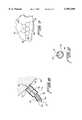
- FIG. 1is an enlarged cross section elevation view of the delivery device of the present invention.
- FIG. 2shows an end view of the device of FIG. 1 as taken along line 2--2.
- FIG. 3shows the delivery device of FIG. 1 with the distal portion flexed transversely.
- FIG. 4shows the delivery device of FIG. 3 with the needle and marking platen extended.
- FIG. 5shows the delivery device of FIG. 4 following injection of a portion of the therapeutic substance.
- FIG. 6shows the delivery device of FIGS. 5 during injection of the therapeutic substance into tissue.
- FIG. 7shows a typical injection pattern for delivery of a therapeutic substance to an area of tissue.
- the inventionis directed to a method and apparatus for delivering therapeutic treatment to body tissues.
- the apparatusincludes a delivery device 10, as illustrated in FIGS. 1 and 2, capable of injecting a therapeutic substance into bodily tissue through a minimally invasive method.
- Device 10may be inserted through a thoracoscopic port (not shown), giving thoracoscopic access to the patient's heart or other tissue.
- the devicemay advantageously be used to inject a substance in a grid-like or other pattern so that by making successive injections of a predetermined quantity of substance having a predictable diffusion mobility, an entire area of tissue can be treated.
- Delivery device 10includes an elongate flexible tubular member 12 having a proximal end 14 and a distal end 16.
- Tubular member 12is constructed of a flexible material such as polyurethane, polyvinyl chloride, polyethylene, or other suitable flexible biocompatible materials.
- Tubular member 12includes a lumen 18 which passes from proximal end 14 to distal end 16, and which has a generally circular cross section.
- a cap 20is mounted on the proximal end 14 of tubular member 12, and includes a circular hole 22 to allow passage of a syringe 24.
- Syringe 24includes a cylindrical body having a plunger shaft 26 and a plunger 28 slidably mounted therein.
- Plunger 28is mounted on the distal end of plunger shaft 26, and a plunger thumb button 30 is mounted on the proximal end of plunger shaft 26.
- Plunger shaft 26may include screw threads or other gradations 29 formed along its length.
- a plunger stop 31is movably mounted on plunger shaft 26 and positionable at a predetermined distance 33 from the proximal end 35 of syringe 24. Plunger stop 31 ensures that when plunger 28 is depressed, only a predetermined amount of therapeutic substance is dispensed from syringe 24. Following delivery of the substance, plunger stop 31 is reset by moving proximally back along plunger shaft 26 the predetermined distance 33. This may be accomplished by turning plunger stop 31 back along threads 29 or the like. Syringe 24 is thereby set to deliver the next predetermined dose of therapeutic substance at the next injection location. Other means for metering the amount of therapeutic substance delivered may also be used.
- a ratchet mechanism(not shown) may be incorporated into the syringe for enabling consistent delivery of successive constant amounts of therapeutic substance in an aliquot manner.
- the physiciandepresses plunger button 30 until the ratchet mechanism stops the forward motion at a predetermined distance.
- the ratchet mechanismwould then reset to enable to the plunger to be depressed an additional predetermined distance, and so forth.
- a cylindrical support 32is located within lumen 18 of tubular member 12. Cylindrical support 32 has a circular opening 34 therethrough for receiving and supporting syringe 24. Syringe 24 is able to slide axially within circular hole 22 in cap 20 and circular opening 34 in cylindrical support 32.
- a spring 36is fixedly mounted concentrically on syringe 24 between cylindrical support 32 and an annular spring stop 38. Spring stop 38 is a disk-shaped member mounted on syringe 24, and is fixed to syringe 24 so that it moves axially as syringe 24 is moved axially.
- a second stop member 40is fixedly mounted on syringe 24 proximal of cap 20.
- Second stop member 40is also a disk-shaped member which is fixed to the exterior of syringe 24. Second stop member 40 prevents syringe 24 from being advanced too far into tubular body 12, when syringe 24 is moved axially in the distal direction relative to tubular body 12.
- a control lever 50is mounted on the side of tubular body 12 for controlling the position of distal end 16 of tubular body 12.
- Control lever 50is pivotally mounted to tubular body 12 at pivot point 51, and is connected to a control wire 52 which is connected to the distal end 16 of tubular body 12 in an offset manner.
- Control wire 52may be retained within eyelets 54 located within lumen 18 of tubular body 12, or may be retained within a separate lumen (not shown), or by other means, as is known in the art.
- control lever 50when control lever 50 is pulled in the proximal direction, it may be seen that control wire 52 will pull on the distal end 16 of tubular body 12. This causes tubular body 12 to bend or flex transversely so that distal end 16 is disposed in an oblique direction relative to the major axis of tubular body 12.
- An adjustment mechanism(not shown) may be included for holding control lever 50 in a particular desired position.
- other control devicesmay be substituted for lever 50, as is known in the art.
- a threaded knob(not shown) may be used in place of lever 50, with the knob being turned in one direction to pull on control wire 52, and being turned in the other direction to release tension on control wire 52.
- a flexible tube 60is connected to the distal end of syringe 24, in fluid communication with syringe 24.
- the distal end of flexible tube 60is connected to a hollow needle 62 of the hypodermic type.
- a therapeutic substance 64 contained within syringe 24may be expelled through needle 62 by pressing syringe plunger button 30. This forces the therapeutic substance from syringe 24, through flexible tube 60, and out the distal end of needle 62.
- Cup 70is mounted on the distal end 16 of tubular body 12 to serve as a guard.
- Cup 70is a hollow cylindrical member, and includes a wall having a distal edge 72 which extends at least as far as the tip of needle 62 when needle 62 is in the retracted position, as illustrated in FIGS. 1 and 3. Accordingly, as long as needle 62 is retracted, cup 70 will prevent needle 62 from contacting non-target areas during placement of device 10 within a patient.
- a cylindrical platen 76is located within cup 70, and is connected to flexible tube 60 and needle 62. Platen 76 moves forward within cup 70 as needle 62 is extended. Because needle 62 and platen 76 are fixed relative to each other, when needle 62 is inserted into target tissue 80, as illustrated in FIG. 6, platen 76 will contact the tissue surface and prevent further penetration by needle 62. This ensures that needle 62 may be inserted to a consistent depth at each successive injection site. Also, if the surface of target tissue 80 is curved, platen 76 will press against target tissue 80 and flatten out the curvature, thereby further assuring that needle 62 penetrates to a consistent depth.
- Platen 76includes a raised marking ring 78 on its distal outer side for making a discernable mark on the tissue surface when needle 62 is inserted into the tissue.
- Marking ring 78is a raised ring that concentrically encircles needle 62.
- Ring 78may be constructed of an absorbent polymeric substance saturated with a visual dye, such as methylene blue. When ring 78 is pressed against tissue, ring 78 will leave a discernable ring-shaped mark on the tissue. The ring-shaped mark will indicate to the surgeon which area of tissue has already receive an injection of therapeutic substance 64.
- Alternative marking substances for use with the present inventionmay include contrast agents or fluoroscopic dyes which will allow the use of echocardiographic or fluoroscopic viewing of dye markings and placement patterns.
- syringe 24may be moved axially forward in the distal direction relative to tubular body 12 to extend needle 22 and platen 76.
- Syringe 24is moved forward against the bias of spring 36, thereby compressing spring 36.
- flexible tube 60, needle 62 and platen 76also move distally so that needle 62 and marking ring 78 extend beyond the distal edge 72 of cup 70.
- Second stop member 40is fixed on syringe 24 in a location which ensures that when second stop member 40 comes into contact with cap 18, needle 62 and marking ring 78 are properly extended beyond the distal edge 72 of cup 70.
- Syringe 24may be retained in the forward position by a latch (not shown), by the surgeon's hand, or by other means. Upon release of syringe 24, spring 36 will force syringe 24 back in the proximal direction, thereby retracting needle 62 and platen 76 back into cup 70. This enables needle 62 to properly retracted following each injection so that needle 62 does not accidentally penetrate non-target areas.
- Flexible tube 60is able to flex in the transverse direction, as illustrated in FIGS. 3-5, but has sufficient column strength so that as syringe 24 is moved forward, flexible tube 60 will push platen 76 and needle 62 distally so that needle 62 is able to penetrate target tissue 80, as illustrated in FIG. 6.
- Suitable materials for use as flexible tube 60include hypotube, polyvinyl chloride tube, or other sufficiently stiff biocompatible materials.
- the clearance between flexible tube 60 and the lumen wall 18 of tubular body 12may be small enough so that lumen wall 18 will support flexible tube 60.
- FIG. 6once needle 62 has penetrated the target tissue 80, marking ring 78 on platen 76 will come into contact with the surface of tissue 80. Then, as illustrated in FIG. 5, plunger thumb button 30 is depressed so that a quantity of therapeutic substance 64 is injected into tissue 80. Plunger stop 31 contacts the proximal end 35 of syringe 24, thereby ensuring that a predetermined dose of therapeutic substance is delivered. Needle 62 is then withdrawn from target tissue 80, and marking ring 78 leaves a discernable mark on target tissue 80. The distal end of device 10 is then repositioned to the next target site for the next injection. In this manner, an area of tissue 80 may be treated with therapeutic substance 64 without overlap. A typical grid-like pattern for such treatment is illustrated in FIG. 7, showing a plurality of ring-shaped marks 82 formed on the surface of tissue 80 by marking ring 78 during successive injections.
- factors taken into considerationinclude the proximity of the injection sites to each other and the mobility of the solution within tissue 80.
- the area covered by ring 78is preferably the same as the estimated dispersion area of the predetermined injection quantity of therapeutic substance within the tissue.
- the amount of fluid deliveredwill vary in correspondence with the type of treatment being administered. Accordingly, the spacing of the injection sites, the volume of therapeutic substance to be delivered, the mobility of the therapeutic substance in tissue, the desired area of coverage, and the depth of delivery are all factors which may be taken into account when determining the spacing of the injection pattern.
- the geometric pattern shown in FIG. 7provides diffuse coverage with minimal overlap, and with minimal uncovered areas within the overall geometry.
- the distance which needle 62 extends beyond platen 76may be adjusted from patient to patient and for particular uses by using needles of different length.
- One reason for controlling the depth of needle penetration when treating ischemic heart diseaseis to ensure that the needle tip is located sub-epicardially, e.g., at approximately 5 mm depth. Platen 76 ensures that the needle tip is always located at the same depth within the tissue during each injection. This ensures a more uniform delivery of the therapeutic substance over the entire treatment area.
- differing amounts of fluidcould be delivered to different areas of tissue by controlling the quantity of fluid dispensed from the syringe during each injection.
- Additional features of device 10may include a fiber optic light source (not shown) located at the distal tip of device 10.
- the light sourcemay emit from platen 76, from the edge of cup 70, or from other locations. The light source would aid in visualization of the target tissue prior to injection, and also in visual perception of the ring-shaped markings 82 following injection and contact of the target tissue with marking ring 78.
- the preferred embodiment of device 10is between 12 and 18 inches in overall length.
- the maximum diameter of the preferred embodimentis less than 12 mm for the portion of device 10 which is inserted into the patient. This enables device 10 to fit through a 12 or 15 mm thoracoscopic port.
- the device of the subject inventionmay be used to inject a variety of fluids into tissue for a variety of therapeutic reasons.
- the delivery deviceis used to deliver an angiogenesis-promoting compound to a portion of the myocardial wall that is suffering from insufficient blood supply.
- the treatmentmay be used independently for promoting the growth of new blood vessels, or may be used in conjunction with other procedures such as angioplasty, bypass surgery, transmyocardial revascularization, or the like.
- the device of the subject inventionenables quick, precise, and effective delivery of controlled quantities of the angiogenesis-promoting substance over a desired coverage area with minimal overlap.
- VEGFvascular endothelial growth factor
- a preferred method of delivering VEGFis in the form of cDNA or gene coding in a replication-deficient adenoviral ("Ad") vector.
- Adreplication-deficient adenoviral
- a quantity of adenovirus carrying the desired genetic componentis delivered to the treatment area by injection in solution.
- Previous studieshave shown the feasibility and efficacy of safe, sustained, and localized expression of VEGF utilizing adenoviral mediated gene transfer therapy.
- a predetermined quantity of angiogenesis-promoting factoris loaded into syringe 24, and plunger stop 31 is set at a predetermined distance 33 for dispensing 0.1 cc of the angiogenesis-promoting factor.
- the patient's heartis accessed via a thoracotomy incision of 7 cm or less, or, in a thoracoscopic approach, via a 15 mm port. Additional ports may be used to provide visualization and manipulation of tissue.
- a pericardial incisionis formed, and stay sutures are emplaced. With the heart appropriately positioned, using retracting devices or the like, cup 76 of device 10 is placed adjacent to the myocardial wall.
- Needle 62is then extended to penetrate the target tissue until ring 78 contacts the tissue. Plunger 28 is then depressed until plunger stop 31 contacts the proximal end 35 of syringe 24 to deliver the angiogenesis-promoting factor to the ischemic myocardial tissue in the manner described above. Needle 62 is then withdrawn and repositioned at the next target site. Plunger stop 31 is moved back along plunger rod 26 a predetermined distance 33 for dispensing a second dose of the angiogenesis-promoting substance.
- a typical proceduremay comprise 10 injections of 0.1 cc each for a total of 1.0 cc of angiogenesis-promoting substance being delivered over a 10 cm 2 area.
- specific amounts of substance delivered, and the area coveredwill be dictated by the specific treatment being implemented, and the above specifics are not intended to be limiting and are only given for describing the preferred best mode of treatment.
- angiogenesis-promoting factorinitiates the complex process of angiogenesis in the treated tissue, thereby inducing the growth of new blood vessels.
- This treatmentis of benefit to heart tissue in which the existing blood vessels are clogged or narrowed, and is also of benefit to heart tissue which is poorly supplied by the native coronary vasculature for congenital reasons.
- the present inventionsets forth a method and apparatus for delivering predetermined quantities of therapeutic substance to biological tissue successively at a plurality locations to achieve therapeutic treatment over a desired area of tissue quickly and accurately.
Landscapes
- Health & Medical Sciences (AREA)
- Life Sciences & Earth Sciences (AREA)
- Animal Behavior & Ethology (AREA)
- Engineering & Computer Science (AREA)
- Veterinary Medicine (AREA)
- Public Health (AREA)
- General Health & Medical Sciences (AREA)
- Surgery (AREA)
- Molecular Biology (AREA)
- Biomedical Technology (AREA)
- Heart & Thoracic Surgery (AREA)
- Medicinal Chemistry (AREA)
- Genetics & Genomics (AREA)
- Biotechnology (AREA)
- Nuclear Medicine, Radiotherapy & Molecular Imaging (AREA)
- Chemical & Material Sciences (AREA)
- Pharmacology & Pharmacy (AREA)
- Epidemiology (AREA)
- Medical Informatics (AREA)
- Anesthesiology (AREA)
- Hematology (AREA)
- Infusion, Injection, And Reservoir Apparatuses (AREA)
- Surgical Instruments (AREA)
Abstract
Description
Claims (20)
Priority Applications (9)
| Application Number | Priority Date | Filing Date | Title |
|---|---|---|---|
| US09/035,892US5997509A (en) | 1998-03-06 | 1998-03-06 | Minimally invasive gene therapy delivery device and method |
| PCT/US1999/003731WO1999044656A1 (en) | 1998-03-06 | 1999-03-05 | Minimally invasive gene therapy delivery device and method |
| BR9908561-5ABR9908561A (en) | 1998-03-06 | 1999-03-05 | Device and method for delivering a therapeutic substance to body tissue in a minimally invasive manner |
| EP99909530AEP1067976A4 (en) | 1998-03-06 | 1999-03-05 | DEVICE AND METHOD FOR DELIVERING GENES ON A MINIMALLY INVASIVE WAY |
| CA002322786ACA2322786A1 (en) | 1998-03-06 | 1999-03-05 | Minimally invasive gene therapy delivery device and method |
| MXPA00008732AMXPA00008732A (en) | 1998-03-06 | 1999-03-05 | Minimally invasive gene therapy delivery device and method. |
| KR1020007009808AKR20010041611A (en) | 1998-03-06 | 1999-03-05 | Minimally invasive gene therapy delivery device and method |
| AU28714/99AAU2871499A (en) | 1998-03-06 | 1999-03-05 | Minimally invasive gene therapy delivery device and method |
| US09/393,873US6322536B1 (en) | 1998-03-06 | 1999-09-10 | Minimally invasive gene therapy delivery and method |
Applications Claiming Priority (1)
| Application Number | Priority Date | Filing Date | Title |
|---|---|---|---|
| US09/035,892US5997509A (en) | 1998-03-06 | 1998-03-06 | Minimally invasive gene therapy delivery device and method |
Related Child Applications (1)
| Application Number | Title | Priority Date | Filing Date |
|---|---|---|---|
| US09/393,873Continuation-In-PartUS6322536B1 (en) | 1998-03-06 | 1999-09-10 | Minimally invasive gene therapy delivery and method |
Publications (1)
| Publication Number | Publication Date |
|---|---|
| US5997509Atrue US5997509A (en) | 1999-12-07 |
Family
ID=21885406
Family Applications (2)
| Application Number | Title | Priority Date | Filing Date |
|---|---|---|---|
| US09/035,892Expired - LifetimeUS5997509A (en) | 1998-03-06 | 1998-03-06 | Minimally invasive gene therapy delivery device and method |
| US09/393,873Expired - LifetimeUS6322536B1 (en) | 1998-03-06 | 1999-09-10 | Minimally invasive gene therapy delivery and method |
Family Applications After (1)
| Application Number | Title | Priority Date | Filing Date |
|---|---|---|---|
| US09/393,873Expired - LifetimeUS6322536B1 (en) | 1998-03-06 | 1999-09-10 | Minimally invasive gene therapy delivery and method |
Country Status (8)
| Country | Link |
|---|---|
| US (2) | US5997509A (en) |
| EP (1) | EP1067976A4 (en) |
| KR (1) | KR20010041611A (en) |
| AU (1) | AU2871499A (en) |
| BR (1) | BR9908561A (en) |
| CA (1) | CA2322786A1 (en) |
| MX (1) | MXPA00008732A (en) |
| WO (1) | WO1999044656A1 (en) |
Cited By (58)
| Publication number | Priority date | Publication date | Assignee | Title |
|---|---|---|---|---|
| US6183444B1 (en) | 1998-05-16 | 2001-02-06 | Microheart, Inc. | Drug delivery module |
| US6322536B1 (en)* | 1998-03-06 | 2001-11-27 | Cornell Research Foundation, Inc. | Minimally invasive gene therapy delivery and method |
| WO2001089606A2 (en) | 2000-05-23 | 2001-11-29 | Cornell Research Foundation, Inc. | Delivery device and method for administering a therapeutic solution to a heart |
| US20020150509A1 (en)* | 2001-04-17 | 2002-10-17 | Houge Erik C. | Laboratory specimen sampler with integrated specimen mount |
| US20030040706A1 (en)* | 2001-07-25 | 2003-02-27 | Kuracina Thomas C. | Method and apparatus for indicating or covering a percutaneous puncture site |
| US6565528B1 (en) | 1999-05-07 | 2003-05-20 | Scimed Life Systems, Inc. | Apparatus and method for delivering therapeutic and diagnostic agents |
| US6613025B1 (en) | 2000-05-25 | 2003-09-02 | Scimed Life Systems, Inc. | Method and apparatus for diagnostic and therapeutic agent delivery |
| US6641553B1 (en) | 1999-06-02 | 2003-11-04 | Boston Scientific Corporation | Devices and methods for delivering a drug |
| US20040059413A1 (en)* | 2002-09-20 | 2004-03-25 | Claudio Argento | Suture template for facilitating implantation of a prosthetic heart valve |
| US6716190B1 (en) | 2000-04-19 | 2004-04-06 | Scimed Life Systems, Inc. | Device and methods for the delivery and injection of therapeutic and diagnostic agents to a target site within a body |
| US6780179B2 (en) | 2002-05-22 | 2004-08-24 | Rubicor Medical, Inc. | Methods and systems for in situ tissue marking and orientation stabilization |
| US20040191225A1 (en)* | 2002-08-06 | 2004-09-30 | Dinsmore Jonathan H. | Injection system |
| US20050191278A1 (en)* | 2002-11-15 | 2005-09-01 | Genvec, Inc. | Coronary artery disease treatment |
| US20050215991A1 (en)* | 1997-03-13 | 2005-09-29 | Altman Peter A | Cardiac drug delivery system and method of use |
| US20050267061A1 (en)* | 2004-04-08 | 2005-12-01 | Sangamo Biosciences, Inc. | Methods and compositions for treating neuropathic and neurodegenerative conditions |
| US20060079475A1 (en)* | 2004-04-08 | 2006-04-13 | Sangamo Biosciences, Inc. | Methods and compositions for modulating cardiac contractility |
| WO2006055845A1 (en)* | 2004-11-15 | 2006-05-26 | Cytyc Corporation | System for drug delivery |
| US7147633B2 (en) | 1999-06-02 | 2006-12-12 | Boston Scientific Scimed, Inc. | Method and apparatus for treatment of atrial fibrillation |
| US20070078435A1 (en)* | 2001-06-14 | 2007-04-05 | Corbett Stone | Tissue augmentation methods using a medical injection apparatus |
| US20070106208A1 (en)* | 2005-11-04 | 2007-05-10 | Medrad, Inc. | Delivery of agents to tissue |
| US20070118077A1 (en)* | 2005-11-21 | 2007-05-24 | Becton, Dickinson And Company | Intradermal delivery device |
| US20070149924A1 (en)* | 2005-10-13 | 2007-06-28 | Becton, Dickinson And Company | Disposable needle and hub assembly |
| US20080086111A1 (en)* | 2006-10-09 | 2008-04-10 | Medrad, Inc. | Fluid delivery systems and volume metering in cell delivery |
| WO2008002515A3 (en)* | 2006-06-27 | 2008-10-02 | Injectimed Inc | Method and apparatus for indicating or covering a percutaneous puncture site |
| US20080294096A1 (en)* | 2005-11-04 | 2008-11-27 | Medrad Inc. | Delivery of Agents Such as Cells to Tissue |
| US7458956B1 (en)* | 1999-11-12 | 2008-12-02 | Boston Scientific Scimed, Inc. | Apparatus for delivery of controlled doses of therapeutic drugs in endoluminal procedures |
| US20090018497A1 (en)* | 2007-05-23 | 2009-01-15 | Birchard Christopher J | Magnetically guided catheter with concentric needle port |
| US20090024009A1 (en)* | 2002-04-19 | 2009-01-22 | Dominique Freeman | Body fluid sampling device with a capacitive sensor |
| US7517348B2 (en) | 1998-09-03 | 2009-04-14 | Rubicor Medical, Inc. | Devices and methods for performing procedures on a breast |
| US20090099514A1 (en)* | 2007-05-23 | 2009-04-16 | Birchard Christopher J | Automated injection catheter device and system |
| US20090099513A1 (en)* | 2007-05-23 | 2009-04-16 | Birchard Christopher J | Extension control handle with adjustable locking mechanism |
| US7588554B2 (en) | 2000-06-26 | 2009-09-15 | Boston Scientific Scimed, Inc. | Method and apparatus for treating ischemic tissue |
| US20100268159A1 (en)* | 2009-04-16 | 2010-10-21 | Engel Rebecca L | Retrograde coronary sinus perfusion cannula and methods of using same |
| US20110046556A1 (en)* | 2009-08-22 | 2011-02-24 | Joseph Wayne Kraft | Rapid Local Anesthesia Injection Cone |
| WO2013090711A1 (en)* | 2011-12-15 | 2013-06-20 | Taylor Robert C | Injection site marking method and apparatus |
| US8808201B2 (en) | 2002-04-19 | 2014-08-19 | Sanofi-Aventis Deutschland Gmbh | Methods and apparatus for penetrating tissue |
| US8845549B2 (en) | 2002-04-19 | 2014-09-30 | Sanofi-Aventis Deutschland Gmbh | Method for penetrating tissue |
| US8845550B2 (en) | 2001-06-12 | 2014-09-30 | Sanofi-Aventis Deutschland Gmbh | Tissue penetration device |
| US8905945B2 (en) | 2002-04-19 | 2014-12-09 | Dominique M. Freeman | Method and apparatus for penetrating tissue |
| US8945910B2 (en) | 2003-09-29 | 2015-02-03 | Sanofi-Aventis Deutschland Gmbh | Method and apparatus for an improved sample capture device |
| US8965476B2 (en) | 2010-04-16 | 2015-02-24 | Sanofi-Aventis Deutschland Gmbh | Tissue penetration device |
| US9034639B2 (en) | 2002-12-30 | 2015-05-19 | Sanofi-Aventis Deutschland Gmbh | Method and apparatus using optical techniques to measure analyte levels |
| US9089294B2 (en) | 2002-04-19 | 2015-07-28 | Sanofi-Aventis Deutschland Gmbh | Analyte measurement device with a single shot actuator |
| US9144401B2 (en) | 2003-06-11 | 2015-09-29 | Sanofi-Aventis Deutschland Gmbh | Low pain penetrating member |
| US9226699B2 (en) | 2002-04-19 | 2016-01-05 | Sanofi-Aventis Deutschland Gmbh | Body fluid sampling module with a continuous compression tissue interface surface |
| US9248267B2 (en) | 2002-04-19 | 2016-02-02 | Sanofi-Aventis Deustchland Gmbh | Tissue penetration device |
| US9261476B2 (en) | 2004-05-20 | 2016-02-16 | Sanofi Sa | Printable hydrogel for biosensors |
| US9314194B2 (en) | 2002-04-19 | 2016-04-19 | Sanofi-Aventis Deutschland Gmbh | Tissue penetration device |
| US9351680B2 (en) | 2003-10-14 | 2016-05-31 | Sanofi-Aventis Deutschland Gmbh | Method and apparatus for a variable user interface |
| US9375169B2 (en) | 2009-01-30 | 2016-06-28 | Sanofi-Aventis Deutschland Gmbh | Cam drive for managing disposable penetrating member actions with a single motor and motor and control system |
| US9427532B2 (en) | 2001-06-12 | 2016-08-30 | Sanofi-Aventis Deutschland Gmbh | Tissue penetration device |
| CN106163594A (en)* | 2014-04-14 | 2016-11-23 | 凸版印刷株式会社 | Injection device |
| US9561000B2 (en) | 2003-12-31 | 2017-02-07 | Sanofi-Aventis Deutschland Gmbh | Method and apparatus for improving fluidic flow and sample capture |
| US9560993B2 (en) | 2001-11-21 | 2017-02-07 | Sanofi-Aventis Deutschland Gmbh | Blood testing apparatus having a rotatable cartridge with multiple lancing elements and testing means |
| US9775553B2 (en) | 2004-06-03 | 2017-10-03 | Sanofi-Aventis Deutschland Gmbh | Method and apparatus for a fluid sampling device |
| US9795747B2 (en) | 2010-06-02 | 2017-10-24 | Sanofi-Aventis Deutschland Gmbh | Methods and apparatus for lancet actuation |
| US9820684B2 (en) | 2004-06-03 | 2017-11-21 | Sanofi-Aventis Deutschland Gmbh | Method and apparatus for a fluid sampling device |
| US20180133442A1 (en)* | 2016-11-17 | 2018-05-17 | Boston Scientific Scimed Inc. | Hydraulic auto crossing balloon/catheter |
Families Citing this family (88)
| Publication number | Priority date | Publication date | Assignee | Title |
|---|---|---|---|---|
| WO2001010315A1 (en) | 1999-08-05 | 2001-02-15 | Cornell Research Foundation, Inc. | Gene therapy platformed needle and method of administering a therapeutic solution to a heart |
| US7526342B2 (en) | 1999-08-10 | 2009-04-28 | Maquet Cardiovascular Llc | Apparatus for endoscopic cardiac mapping and lead placement |
| US7398781B1 (en) | 1999-08-10 | 2008-07-15 | Maquet Cardiovascular, Llc | Method for subxiphoid endoscopic access |
| US7597698B2 (en) | 1999-08-10 | 2009-10-06 | Maquet Cardiovascular Llc | Apparatus and method for endoscopic encirclement of pulmonary veins for epicardial ablation |
| US7264587B2 (en)* | 1999-08-10 | 2007-09-04 | Origin Medsystems, Inc. | Endoscopic subxiphoid surgical procedures |
| US7288096B2 (en) | 2003-01-17 | 2007-10-30 | Origin Medsystems, Inc. | Apparatus for placement of cardiac defibrillator and pacer |
| US20030187460A1 (en)* | 1999-08-10 | 2003-10-02 | Chin Albert K. | Methods and apparatus for endoscopic cardiac surgery |
| US6329348B1 (en) | 1999-11-08 | 2001-12-11 | Cornell Research Foundation, Inc. | Method of inducing angiogenesis |
| KR100406729B1 (en)* | 2000-02-15 | 2003-11-21 | 사회복지법인삼성생명공익재단(삼성서울병원) | synchronized medical device |
| US6893421B1 (en) | 2000-08-08 | 2005-05-17 | Scimed Life Systems, Inc. | Catheter shaft assembly |
| US6595958B1 (en) | 2000-08-08 | 2003-07-22 | Scimed Life Systems, Inc. | Tortuous path injection device and method |
| US6613017B1 (en) | 2000-08-08 | 2003-09-02 | Scimed Life Systems, Inc. | Controlled depth injection device and method |
| JP2003007916A (en)* | 2001-06-19 | 2003-01-10 | Sanyo Electric Co Ltd | Method of manufacturing circuit device |
| US7794707B2 (en) | 2002-06-19 | 2010-09-14 | University Health Network | ACE2 activation for treatment of heart, lung and kidney disease and hypertension |
| US20040019509A1 (en)* | 2002-07-23 | 2004-01-29 | Bekkers Ivan H. | System and method for managing flight information |
| US7811274B2 (en) | 2003-05-07 | 2010-10-12 | Portaero, Inc. | Method for treating chronic obstructive pulmonary disease |
| US7426929B2 (en) | 2003-05-20 | 2008-09-23 | Portaero, Inc. | Intra/extra-thoracic collateral ventilation bypass system and method |
| US7533667B2 (en) | 2003-05-29 | 2009-05-19 | Portaero, Inc. | Methods and devices to assist pulmonary decompression |
| US7252086B2 (en) | 2003-06-03 | 2007-08-07 | Cordis Corporation | Lung reduction system |
| US7377278B2 (en) | 2003-06-05 | 2008-05-27 | Portaero, Inc. | Intra-thoracic collateral ventilation bypass system and method |
| US7682332B2 (en) | 2003-07-15 | 2010-03-23 | Portaero, Inc. | Methods to accelerate wound healing in thoracic anastomosis applications |
| IL157981A (en) | 2003-09-17 | 2014-01-30 | Elcam Medical Agricultural Cooperative Ass Ltd | Auto-injector |
| US20050148935A1 (en)* | 2003-12-29 | 2005-07-07 | Rozalina Dimitrova | Botulinum toxin injection guide |
| IL160891A0 (en) | 2004-03-16 | 2004-08-31 | Auto-mix needle | |
| US7615019B2 (en) | 2004-07-22 | 2009-11-10 | Nordt Development Co., Llc | Potentiating support with side struts spanning hinge joint |
| US7398782B2 (en) | 2004-11-19 | 2008-07-15 | Portaero, Inc. | Method for pulmonary drug delivery |
| US8220460B2 (en) | 2004-11-19 | 2012-07-17 | Portaero, Inc. | Evacuation device and method for creating a localized pleurodesis |
| US7824366B2 (en) | 2004-12-10 | 2010-11-02 | Portaero, Inc. | Collateral ventilation device with chest tube/evacuation features and method |
| US8050746B2 (en) | 2005-02-02 | 2011-11-01 | Voyage Medical, Inc. | Tissue visualization device and method variations |
| US8078266B2 (en) | 2005-10-25 | 2011-12-13 | Voyage Medical, Inc. | Flow reduction hood systems |
| US7918787B2 (en) | 2005-02-02 | 2011-04-05 | Voyage Medical, Inc. | Tissue visualization and manipulation systems |
| US7860555B2 (en) | 2005-02-02 | 2010-12-28 | Voyage Medical, Inc. | Tissue visualization and manipulation system |
| US8137333B2 (en) | 2005-10-25 | 2012-03-20 | Voyage Medical, Inc. | Delivery of biological compounds to ischemic and/or infarcted tissue |
| US7930016B1 (en) | 2005-02-02 | 2011-04-19 | Voyage Medical, Inc. | Tissue closure system |
| US11478152B2 (en) | 2005-02-02 | 2022-10-25 | Intuitive Surgical Operations, Inc. | Electrophysiology mapping and visualization system |
| US9510732B2 (en) | 2005-10-25 | 2016-12-06 | Intuitive Surgical Operations, Inc. | Methods and apparatus for efficient purging |
| US20080015569A1 (en) | 2005-02-02 | 2008-01-17 | Voyage Medical, Inc. | Methods and apparatus for treatment of atrial fibrillation |
| US7860556B2 (en) | 2005-02-02 | 2010-12-28 | Voyage Medical, Inc. | Tissue imaging and extraction systems |
| US10064540B2 (en) | 2005-02-02 | 2018-09-04 | Intuitive Surgical Operations, Inc. | Visualization apparatus for transseptal access |
| US7691086B2 (en)* | 2005-06-14 | 2010-04-06 | Tengiz Tkebuchava | Catheter for introduction of medications to the tissues of a heart or other organ |
| US8104474B2 (en) | 2005-08-23 | 2012-01-31 | Portaero, Inc. | Collateral ventilation bypass system with retention features |
| US8221310B2 (en) | 2005-10-25 | 2012-07-17 | Voyage Medical, Inc. | Tissue visualization device and method variations |
| US7406963B2 (en) | 2006-01-17 | 2008-08-05 | Portaero, Inc. | Variable resistance pulmonary ventilation bypass valve and method |
| US9055906B2 (en) | 2006-06-14 | 2015-06-16 | Intuitive Surgical Operations, Inc. | In-vivo visualization systems |
| US20080097476A1 (en) | 2006-09-01 | 2008-04-24 | Voyage Medical, Inc. | Precision control systems for tissue visualization and manipulation assemblies |
| US10004388B2 (en) | 2006-09-01 | 2018-06-26 | Intuitive Surgical Operations, Inc. | Coronary sinus cannulation |
| WO2008028149A2 (en) | 2006-09-01 | 2008-03-06 | Voyage Medical, Inc. | Electrophysiology mapping and visualization system |
| US10335131B2 (en) | 2006-10-23 | 2019-07-02 | Intuitive Surgical Operations, Inc. | Methods for preventing tissue migration |
| US20080183036A1 (en) | 2006-12-18 | 2008-07-31 | Voyage Medical, Inc. | Systems and methods for unobstructed visualization and ablation |
| US8131350B2 (en) | 2006-12-21 | 2012-03-06 | Voyage Medical, Inc. | Stabilization of visualization catheters |
| US9226648B2 (en) | 2006-12-21 | 2016-01-05 | Intuitive Surgical Operations, Inc. | Off-axis visualization systems |
| US10166066B2 (en)* | 2007-03-13 | 2019-01-01 | University Of Virginia Patent Foundation | Epicardial ablation catheter and method of use |
| WO2008118737A1 (en) | 2007-03-22 | 2008-10-02 | University Of Virginia Patent Foundation | Electrode catheter for ablation purposes and related method thereof |
| US11058354B2 (en) | 2007-03-19 | 2021-07-13 | University Of Virginia Patent Foundation | Access needle with direct visualization and related methods |
| US9468396B2 (en) | 2007-03-19 | 2016-10-18 | University Of Virginia Patent Foundation | Systems and methods for determining location of an access needle in a subject |
| BRPI0809127B8 (en) | 2007-03-19 | 2021-06-22 | Univ Virginia Patent Foundation | devices and methods for accessing one or more locations and detecting pressure at one or more locations |
| WO2011103456A2 (en) | 2010-02-18 | 2011-08-25 | University Of Virginia Patent Foundation | System, method, and computer program product for simulating epicardial electrophysiology procedures |
| EP2148608A4 (en) | 2007-04-27 | 2010-04-28 | Voyage Medical Inc | Complex shape steerable tissue visualization and manipulation catheter |
| US8657805B2 (en) | 2007-05-08 | 2014-02-25 | Intuitive Surgical Operations, Inc. | Complex shape steerable tissue visualization and manipulation catheter |
| WO2008141238A1 (en) | 2007-05-11 | 2008-11-20 | Voyage Medical, Inc. | Visual electrode ablation systems |
| US8163034B2 (en) | 2007-05-11 | 2012-04-24 | Portaero, Inc. | Methods and devices to create a chemically and/or mechanically localized pleurodesis |
| US7931641B2 (en) | 2007-05-11 | 2011-04-26 | Portaero, Inc. | Visceral pleura ring connector |
| US8062315B2 (en) | 2007-05-17 | 2011-11-22 | Portaero, Inc. | Variable parietal/visceral pleural coupling |
| US8235985B2 (en) | 2007-08-31 | 2012-08-07 | Voyage Medical, Inc. | Visualization and ablation system variations |
| US20100241185A1 (en)* | 2007-11-09 | 2010-09-23 | University Of Virginia Patent Foundation | Steerable epicardial pacing catheter system placed via the subxiphoid process |
| EP2077119A1 (en)* | 2007-12-21 | 2009-07-08 | Apeiron Biologics Forschungs- und Entwicklungsgesellschaft M.B.H. | Treatment of fibrosis and liver diseases |
| US8858609B2 (en) | 2008-02-07 | 2014-10-14 | Intuitive Surgical Operations, Inc. | Stent delivery under direct visualization |
| US8336540B2 (en) | 2008-02-19 | 2012-12-25 | Portaero, Inc. | Pneumostoma management device and method for treatment of chronic obstructive pulmonary disease |
| US8475389B2 (en) | 2008-02-19 | 2013-07-02 | Portaero, Inc. | Methods and devices for assessment of pneumostoma function |
| WO2009105432A2 (en) | 2008-02-19 | 2009-08-27 | Portaero, Inc. | Devices and methods for delivery of a therapeutic agent through a pneumostoma |
| EP2722068A1 (en) | 2008-04-08 | 2014-04-23 | Karolinska Institutet Innovations AB | Endoluminal medical access device |
| US9101735B2 (en) | 2008-07-07 | 2015-08-11 | Intuitive Surgical Operations, Inc. | Catheter control systems |
| US8894643B2 (en) | 2008-10-10 | 2014-11-25 | Intuitive Surgical Operations, Inc. | Integral electrode placement and connection systems |
| US8333012B2 (en) | 2008-10-10 | 2012-12-18 | Voyage Medical, Inc. | Method of forming electrode placement and connection systems |
| US8870848B2 (en)* | 2008-10-31 | 2014-10-28 | Medtronic, Inc. | System and method for delivery of biologic agents |
| US9468364B2 (en) | 2008-11-14 | 2016-10-18 | Intuitive Surgical Operations, Inc. | Intravascular catheter with hood and image processing systems |
| US8347881B2 (en) | 2009-01-08 | 2013-01-08 | Portaero, Inc. | Pneumostoma management device with integrated patency sensor and method |
| US8518053B2 (en) | 2009-02-11 | 2013-08-27 | Portaero, Inc. | Surgical instruments for creating a pneumostoma and treating chronic obstructive pulmonary disease |
| US9642534B2 (en) | 2009-09-11 | 2017-05-09 | University Of Virginia Patent Foundation | Systems and methods for determining location of an access needle in a subject |
| US8694071B2 (en) | 2010-02-12 | 2014-04-08 | Intuitive Surgical Operations, Inc. | Image stabilization techniques and methods |
| US9814522B2 (en) | 2010-04-06 | 2017-11-14 | Intuitive Surgical Operations, Inc. | Apparatus and methods for ablation efficacy |
| WO2012112565A2 (en) | 2011-02-15 | 2012-08-23 | Rotation Medical, Inc. | Methods and apparatus for delivering and positioning sheet-like materials |
| SG10201609345QA (en) | 2012-02-07 | 2017-01-27 | Global Bio Therapeutics Inc | Compartmentalized method of nucleic acid delivery and compositions and uses thereof |
| EA032285B1 (en) | 2013-08-08 | 2019-05-31 | Глобал Био Терапьютикс, Инк. | Injection device for minimally invasive procedures and uses thereof |
| WO2015021443A1 (en) | 2013-08-08 | 2015-02-12 | Global Bio Therapeutics Usa, Inc. | Clamp device for minimally invasive procedures and uses thereof |
| CA2945821C (en) | 2014-05-09 | 2018-09-04 | Rotation Medical, Inc. | Medical implant delivery system for sheet-like implant |
| CN112566544B (en)* | 2019-05-08 | 2025-09-19 | 阿特瑞克尔公司 | Biological tissue location positioning and marking |
| US20230355871A1 (en)* | 2020-09-15 | 2023-11-09 | University Of South Carolina | Minimally Invasive and Semi-Automated Myocardial Injection Device |
Citations (31)
| Publication number | Priority date | Publication date | Assignee | Title |
|---|---|---|---|---|
| US2498692A (en)* | 1949-01-04 | 1950-02-28 | Mains Marshall Paul | Gastrointestinal tube |
| US2688329A (en)* | 1953-03-19 | 1954-09-07 | American Cystoscope Makers Inc | Catheter |
| US2700385A (en)* | 1951-07-10 | 1955-01-25 | Ortiz Mariano | Obstetrical needle |
| US3797491A (en)* | 1971-02-11 | 1974-03-19 | Ampoules Inc | Method of performing an intramuscular injection |
| US4222380A (en)* | 1977-12-02 | 1980-09-16 | Olympus Optical Co., Ltd. | Celiac injector |
| US4243035A (en)* | 1979-06-18 | 1981-01-06 | Barrett Howard G | Syringe with integral swab |
| US4578061A (en)* | 1980-10-28 | 1986-03-25 | Lemelson Jerome H | Injection catheter and method |
| US4787891A (en)* | 1987-07-13 | 1988-11-29 | Paul Levin | Syringe holder and applicator |
| US4838854A (en)* | 1988-05-03 | 1989-06-13 | Zivko Kuzmanovich | Method and apparatus for selecting a medicine injection site |
| US4861336A (en)* | 1987-04-01 | 1989-08-29 | Helzel Manfred W | Puncture catheter |
| US4946442A (en)* | 1985-06-28 | 1990-08-07 | Olympus Optical Co., Ltd. | Endoscope treatment device |
| US4976688A (en)* | 1989-02-03 | 1990-12-11 | Rosenblum Jeffrey L | Position-adjustable thoracic catheter |
| US4994041A (en)* | 1988-12-27 | 1991-02-19 | Dombrowski Mitchell P | Needle and catheter assembly |
| US5147307A (en)* | 1991-06-17 | 1992-09-15 | Gluck Seymour M | Anatomical marker device and method |
| US5192270A (en)* | 1990-11-19 | 1993-03-09 | Carswell Jr Donald D | Hypodermic syringe and a method for marking injections |
| US5261889A (en)* | 1992-11-24 | 1993-11-16 | Boston Scientific Corporation | Injection therapy catheter |
| US5269754A (en)* | 1992-01-31 | 1993-12-14 | Everest Medical Corporation | Laparoscopic cholangiogram device |
| US5322510A (en)* | 1989-11-21 | 1994-06-21 | Andreas Lindner | Injection apparatus |
| US5354279A (en)* | 1992-10-21 | 1994-10-11 | Bavaria Medizin Technologie Gmbh | Plural needle injection catheter |
| US5380292A (en)* | 1993-12-22 | 1995-01-10 | Wilson-Cook Medical, Inc. | Gastrointestinal needle mechanism |
| US5417662A (en)* | 1991-09-13 | 1995-05-23 | Pharmacia Ab | Injection needle arrangement |
| US5441499A (en)* | 1993-07-14 | 1995-08-15 | Dekna Elektro-U. Medizinische Apparatebau Gesellschaft Mbh | Bipolar radio-frequency surgical instrument |
| US5464395A (en)* | 1994-04-05 | 1995-11-07 | Faxon; David P. | Catheter for delivering therapeutic and/or diagnostic agents to the tissue surrounding a bodily passageway |
| US5522815A (en)* | 1993-03-29 | 1996-06-04 | Durgin, Jr.; Russell F. | Integrated catheter for diverse in situ tissue therapy |
| US5536251A (en)* | 1993-02-22 | 1996-07-16 | Heartport, Inc. | Thoracoscopic devices and methods for arresting the heart |
| US5567217A (en)* | 1993-05-19 | 1996-10-22 | Kabushiki Kaisya Ohara | Method for manufacturing a crystalized glass magnetic disk substrate |
| US5569237A (en)* | 1995-04-27 | 1996-10-29 | Beckenstein; Michael S. | Marking device for breast surgery |
| US5674197A (en)* | 1994-07-01 | 1997-10-07 | Cordis Corporation | Controlled flexible catheter |
| US5820592A (en)* | 1996-07-16 | 1998-10-13 | Hammerslag; Gary R. | Angiographic and/or guide catheter |
| US5827216A (en)* | 1995-06-07 | 1998-10-27 | Cormedics Corp. | Method and apparatus for accessing the pericardial space |
| US5845646A (en)* | 1996-11-05 | 1998-12-08 | Lemelson; Jerome | System and method for treating select tissue in a living being |
Family Cites Families (77)
| Publication number | Priority date | Publication date | Assignee | Title |
|---|---|---|---|---|
| US510413A (en) | 1893-12-12 | Drainage-tube | ||
| US2402306A (en) | 1943-10-07 | 1946-06-18 | Turkel Henry | Retaining guard guide for needles |
| US2512568A (en) | 1946-08-13 | 1950-06-20 | Jacob A Saffir | Hypodermic injection device |
| US2551902A (en) | 1948-09-10 | 1951-05-08 | Arthur Schaffer | Dehorning fluid ejector |
| US2670673A (en) | 1950-07-17 | 1954-03-02 | Joyce A Gordon | Fluid injecting device |
| US2952256A (en) | 1957-12-06 | 1960-09-13 | Sierra Eng Co | Epidural needle |
| US3467096A (en) | 1966-04-12 | 1969-09-16 | Ferrell S Horn | Multiple hypodermic syringe arrangement |
| US3435824A (en) | 1966-10-27 | 1969-04-01 | Herminio Gamponia | Surgical apparatus and related process |
| US3487837A (en) | 1967-02-06 | 1970-01-06 | Roy A Petersen | Device for holding catheters in position |
| US3530492A (en) | 1967-12-05 | 1970-09-22 | Jack R Ferber | Method and apparatus for administering hypodermic injections |
| FR1584474A (en) | 1968-02-20 | 1969-12-26 | ||
| US3572336A (en) | 1968-04-30 | 1971-03-23 | Daniel R Hershberg | Syringe |
| CA938850A (en) | 1970-07-24 | 1973-12-25 | W. Edwards Donald | Device for venipuncture |
| US3831584A (en) | 1971-06-21 | 1974-08-27 | Investors In Ventures Inc | Devices for controlling fluid flow in living beings |
| US3765420A (en) | 1971-12-16 | 1973-10-16 | Kendall & Co | Eccentric locking device for surgical drainage members |
| US3783876A (en) | 1972-05-04 | 1974-01-08 | Kendall & Co | Tabbed eccentric locking device |
| US3826241A (en) | 1972-10-16 | 1974-07-30 | Investors In Ventures Inc | Implanting method |
| US3951132A (en) | 1973-05-11 | 1976-04-20 | Investors In Ventures, Inc. | Implant and implanting method |
| US3991767A (en) | 1973-11-02 | 1976-11-16 | Cutter Laboratories, Inc. | Tubular unit with vessel engaging cuff structure |
| US4245624A (en) | 1977-01-20 | 1981-01-20 | Olympus Optical Co., Ltd. | Endoscope with flexible tip control |
| US4150669A (en) | 1977-03-16 | 1979-04-24 | Alvaro Latorre | Apparatus for effecting and enhancing an erection |
| US4168708A (en) | 1977-04-20 | 1979-09-25 | Medical Engineering Corp. | Blood vessel occlusion means suitable for use in anastomosis |
| US4167179A (en) | 1977-10-17 | 1979-09-11 | Mark Kirsch | Planar radioactive seed implanter |
| US4230119A (en) | 1978-12-01 | 1980-10-28 | Medical Engineering Corp. | Micro-hemostat |
| US4356826A (en) | 1979-05-09 | 1982-11-02 | Olympus Optical Co., Ltd. | Stabbing apparatus for diagnosis of living body |
| US4299230A (en) | 1979-05-09 | 1981-11-10 | Olympus Optical Co., Ltd. | Stabbing apparatus for diagnosis of living body |
| US4280508A (en) | 1979-12-27 | 1981-07-28 | Barrada M Ismail | Method for positioning a fluid withdrawing means by detecting local temperature |
| US4419094A (en) | 1981-06-08 | 1983-12-06 | The Kendall Company | Suprapubic catheter system |
| US4674506A (en) | 1984-11-29 | 1987-06-23 | Kirk Alcond | Surgical anastomosis stent |
| US4645495A (en) | 1985-06-26 | 1987-02-24 | Vaillancourt Vincent L | Vascular access implant needle patch |
| US4877037A (en) | 1985-11-12 | 1989-10-31 | Minnesota Mining And Manufacturing Company | Tissue or mucus sampling device |
| US4753236A (en) | 1986-04-08 | 1988-06-28 | Healey Maureen A | Temporary anastomotic device |
| US4721109A (en) | 1986-04-08 | 1988-01-26 | Healey Maureen A | Temporary anastomotic device |
| US5335670A (en) | 1986-04-18 | 1994-08-09 | Henry Fishman | Allergy testing method and apparatus |
| AU7782687A (en)* | 1986-08-05 | 1988-02-24 | University Of Wales College Of Medicine | Proximity detector |
| US4760847A (en) | 1986-08-18 | 1988-08-02 | Vincent Vaillancourt | Depth measuring device |
| US5036868A (en) | 1990-01-29 | 1991-08-06 | Unilink Inc. | Anastomosis preparation technique |
| US5323789A (en) | 1986-12-18 | 1994-06-28 | Minnesota Mining And Manufacturing Company | Anastomosis preparation technique with easily insertable member |
| US4798193A (en) | 1987-05-18 | 1989-01-17 | Thomas J. Fogarty | Protective sheath instrument carrier |
| ES2007667A6 (en) | 1987-07-28 | 1989-07-01 | Espejo Martinez Antonio | A device for locating the epidural space. |
| US5195526A (en) | 1988-03-11 | 1993-03-23 | Michelson Gary K | Spinal marker needle |
| US4966589A (en) | 1988-11-14 | 1990-10-30 | Hemedix International, Inc. | Intravenous catheter placement device |
| US4932421A (en) | 1989-01-23 | 1990-06-12 | Steven Kaali | Electrified intrauterine device |
| US4940458A (en) | 1989-02-02 | 1990-07-10 | Cohn Arnold K | Epidural needle placement system |
| US5425739A (en) | 1989-03-09 | 1995-06-20 | Avatar Design And Development, Inc. | Anastomosis stent and stent selection system |
| US5192289A (en) | 1989-03-09 | 1993-03-09 | Avatar Design And Development, Inc. | Anastomosis stent and stent selection system |
| US4946463A (en) | 1989-04-10 | 1990-08-07 | Pioneering Technologies, Inc. | Vessel occluder |
| GB8924946D0 (en) | 1989-11-04 | 1989-12-28 | Shiu Man F | Support system for catheter |
| US5254088A (en) | 1990-02-02 | 1993-10-19 | Ep Technologies, Inc. | Catheter steering mechanism |
| US5820591A (en) | 1990-02-02 | 1998-10-13 | E. P. Technologies, Inc. | Assemblies for creating compound curves in distal catheter regions |
| US5121750A (en)* | 1990-03-02 | 1992-06-16 | Katims Jefferson J | Apparatus for locating a catheter adjacent to a pacemaker node of the heart |
| US5037428A (en) | 1990-06-21 | 1991-08-06 | Applied Medical Technology, Inc. | Vessel approximation and alignment device |
| US5146913A (en) | 1991-03-04 | 1992-09-15 | Asphendiar Khorsandian | Holder and lock for oro-intubation |
| US5376084A (en) | 1991-10-17 | 1994-12-27 | Imagyn Medical, Inc. | Catheter with internal mandrel and method |
| US5259377A (en) | 1992-03-30 | 1993-11-09 | Stephen M. Daugherty | Endotracheal tube stylet |
| FR2691069B1 (en) | 1992-05-14 | 1999-08-20 | Vygon | SURGICAL INSTRUMENT FOR PERIDURAL ANESTHESIA OPERATION. |
| US5290258A (en) | 1992-07-27 | 1994-03-01 | Genesis Industries, Inc. | Syringe for administering sequentially multiple doses of a medicament |
| US5273525A (en) | 1992-08-13 | 1993-12-28 | Btx Inc. | Injection and electroporation apparatus for drug and gene delivery |
| CA2109980A1 (en) | 1992-12-01 | 1994-06-02 | Mir A. Imran | Steerable catheter with adjustable bend location and/or radius and method |
| US5312351A (en) | 1993-01-29 | 1994-05-17 | Gerrone Carmen J | Combined pneumo-needle and trocar apparatus |
| GB9307572D0 (en)* | 1993-04-13 | 1993-06-02 | Gould Derek A | Transintimal recanalisation device |
| US5403283A (en)* | 1993-10-28 | 1995-04-04 | Luther Medical Products, Inc. | Percutaneous port catheter assembly and method of use |
| US5447529A (en)* | 1994-01-28 | 1995-09-05 | Philadelphia Heart Institute | Method of using endocardial impedance for determining electrode-tissue contact, appropriate sites for arrhythmia ablation and tissue heating during ablation |
| US5478309A (en)* | 1994-05-27 | 1995-12-26 | William P. Sweezer, Jr. | Catheter system and method for providing cardiopulmonary bypass pump support during heart surgery |
| US5417683A (en) | 1994-07-13 | 1995-05-23 | Shiao; I-Shen | Mini-graft hair implanting device for implanting multiple clumps of hair follicles at one time |
| US5713890A (en)* | 1994-07-20 | 1998-02-03 | University Of Utah | Marking pen for coloring the skin |
| US5478315A (en) | 1994-08-08 | 1995-12-26 | Brothers Family Investments, L.C. | Local anesthetic injection system |
| US5520650A (en) | 1994-08-25 | 1996-05-28 | Zadini; Filiberto | Self-arresting overpenetration-proof cannula device for body cavities |
| WO1996035469A1 (en)* | 1995-05-10 | 1996-11-14 | Cardiogenesis Corporation | System for treating or diagnosing heart tissue |
| JP3633032B2 (en)* | 1995-05-26 | 2005-03-30 | 佐々木 寛 | Puncture device |
| US5810836A (en)* | 1996-03-04 | 1998-09-22 | Myocardial Stents, Inc. | Device and method for trans myocardial revascularization (TMR) |
| US5989274A (en)* | 1996-10-17 | 1999-11-23 | Ethicon Endo-Surgery, Inc. | Methods and devices for improving blood flow to a heart of a patient |
| US5931810A (en)* | 1996-12-05 | 1999-08-03 | Comedicus Incorporated | Method for accessing the pericardial space |
| US5868764A (en) | 1996-12-12 | 1999-02-09 | Cornell Research Foundation, Inc. | Perfusion and occlusion device and method |
| US5846225A (en)* | 1997-02-19 | 1998-12-08 | Cornell Research Foundation, Inc. | Gene transfer therapy delivery device and method |
| US5911701A (en)* | 1998-01-29 | 1999-06-15 | Sdgi Holidings, Inc. | Surgical cutting instrument |
| US5997509A (en) | 1998-03-06 | 1999-12-07 | Cornell Research Foundation, Inc. | Minimally invasive gene therapy delivery device and method |
- 1998
- 1998-03-06USUS09/035,892patent/US5997509A/ennot_activeExpired - Lifetime
- 1999
- 1999-03-05WOPCT/US1999/003731patent/WO1999044656A1/ennot_activeApplication Discontinuation
- 1999-03-05CACA002322786Apatent/CA2322786A1/ennot_activeAbandoned
- 1999-03-05KRKR1020007009808Apatent/KR20010041611A/ennot_activeWithdrawn
- 1999-03-05AUAU28714/99Apatent/AU2871499A/ennot_activeAbandoned
- 1999-03-05BRBR9908561-5Apatent/BR9908561A/ennot_activeApplication Discontinuation
- 1999-03-05MXMXPA00008732Apatent/MXPA00008732A/enunknown
- 1999-03-05EPEP99909530Apatent/EP1067976A4/ennot_activeWithdrawn
- 1999-09-10USUS09/393,873patent/US6322536B1/ennot_activeExpired - Lifetime
Patent Citations (31)
| Publication number | Priority date | Publication date | Assignee | Title |
|---|---|---|---|---|
| US2498692A (en)* | 1949-01-04 | 1950-02-28 | Mains Marshall Paul | Gastrointestinal tube |
| US2700385A (en)* | 1951-07-10 | 1955-01-25 | Ortiz Mariano | Obstetrical needle |
| US2688329A (en)* | 1953-03-19 | 1954-09-07 | American Cystoscope Makers Inc | Catheter |
| US3797491A (en)* | 1971-02-11 | 1974-03-19 | Ampoules Inc | Method of performing an intramuscular injection |
| US4222380A (en)* | 1977-12-02 | 1980-09-16 | Olympus Optical Co., Ltd. | Celiac injector |
| US4243035A (en)* | 1979-06-18 | 1981-01-06 | Barrett Howard G | Syringe with integral swab |
| US4578061A (en)* | 1980-10-28 | 1986-03-25 | Lemelson Jerome H | Injection catheter and method |
| US4946442A (en)* | 1985-06-28 | 1990-08-07 | Olympus Optical Co., Ltd. | Endoscope treatment device |
| US4861336A (en)* | 1987-04-01 | 1989-08-29 | Helzel Manfred W | Puncture catheter |
| US4787891A (en)* | 1987-07-13 | 1988-11-29 | Paul Levin | Syringe holder and applicator |
| US4838854A (en)* | 1988-05-03 | 1989-06-13 | Zivko Kuzmanovich | Method and apparatus for selecting a medicine injection site |
| US4994041A (en)* | 1988-12-27 | 1991-02-19 | Dombrowski Mitchell P | Needle and catheter assembly |
| US4976688A (en)* | 1989-02-03 | 1990-12-11 | Rosenblum Jeffrey L | Position-adjustable thoracic catheter |
| US5322510A (en)* | 1989-11-21 | 1994-06-21 | Andreas Lindner | Injection apparatus |
| US5192270A (en)* | 1990-11-19 | 1993-03-09 | Carswell Jr Donald D | Hypodermic syringe and a method for marking injections |
| US5147307A (en)* | 1991-06-17 | 1992-09-15 | Gluck Seymour M | Anatomical marker device and method |
| US5417662A (en)* | 1991-09-13 | 1995-05-23 | Pharmacia Ab | Injection needle arrangement |
| US5269754A (en)* | 1992-01-31 | 1993-12-14 | Everest Medical Corporation | Laparoscopic cholangiogram device |
| US5354279A (en)* | 1992-10-21 | 1994-10-11 | Bavaria Medizin Technologie Gmbh | Plural needle injection catheter |
| US5261889A (en)* | 1992-11-24 | 1993-11-16 | Boston Scientific Corporation | Injection therapy catheter |
| US5536251A (en)* | 1993-02-22 | 1996-07-16 | Heartport, Inc. | Thoracoscopic devices and methods for arresting the heart |
| US5522815A (en)* | 1993-03-29 | 1996-06-04 | Durgin, Jr.; Russell F. | Integrated catheter for diverse in situ tissue therapy |
| US5567217A (en)* | 1993-05-19 | 1996-10-22 | Kabushiki Kaisya Ohara | Method for manufacturing a crystalized glass magnetic disk substrate |
| US5441499A (en)* | 1993-07-14 | 1995-08-15 | Dekna Elektro-U. Medizinische Apparatebau Gesellschaft Mbh | Bipolar radio-frequency surgical instrument |
| US5380292A (en)* | 1993-12-22 | 1995-01-10 | Wilson-Cook Medical, Inc. | Gastrointestinal needle mechanism |
| US5464395A (en)* | 1994-04-05 | 1995-11-07 | Faxon; David P. | Catheter for delivering therapeutic and/or diagnostic agents to the tissue surrounding a bodily passageway |
| US5674197A (en)* | 1994-07-01 | 1997-10-07 | Cordis Corporation | Controlled flexible catheter |
| US5569237A (en)* | 1995-04-27 | 1996-10-29 | Beckenstein; Michael S. | Marking device for breast surgery |
| US5827216A (en)* | 1995-06-07 | 1998-10-27 | Cormedics Corp. | Method and apparatus for accessing the pericardial space |
| US5820592A (en)* | 1996-07-16 | 1998-10-13 | Hammerslag; Gary R. | Angiographic and/or guide catheter |
| US5845646A (en)* | 1996-11-05 | 1998-12-08 | Lemelson; Jerome | System and method for treating select tissue in a living being |
Cited By (109)
| Publication number | Priority date | Publication date | Assignee | Title |
|---|---|---|---|---|
| US20050215991A1 (en)* | 1997-03-13 | 2005-09-29 | Altman Peter A | Cardiac drug delivery system and method of use |
| US6322536B1 (en)* | 1998-03-06 | 2001-11-27 | Cornell Research Foundation, Inc. | Minimally invasive gene therapy delivery and method |
| US6183444B1 (en) | 1998-05-16 | 2001-02-06 | Microheart, Inc. | Drug delivery module |
| US7517348B2 (en) | 1998-09-03 | 2009-04-14 | Rubicor Medical, Inc. | Devices and methods for performing procedures on a breast |
| US6565528B1 (en) | 1999-05-07 | 2003-05-20 | Scimed Life Systems, Inc. | Apparatus and method for delivering therapeutic and diagnostic agents |
| US7211041B2 (en) | 1999-05-07 | 2007-05-01 | Boston Scientific Scimed, Inc. | Apparatus and method for delivering therapeutic and diagnostic agents |
| US6641553B1 (en) | 1999-06-02 | 2003-11-04 | Boston Scientific Corporation | Devices and methods for delivering a drug |
| US7147633B2 (en) | 1999-06-02 | 2006-12-12 | Boston Scientific Scimed, Inc. | Method and apparatus for treatment of atrial fibrillation |
| US8187251B2 (en) | 1999-06-02 | 2012-05-29 | Boston Scientific Scimed, Inc. | Methods of treating cardiac arrhythmia |
| US7458956B1 (en)* | 1999-11-12 | 2008-12-02 | Boston Scientific Scimed, Inc. | Apparatus for delivery of controlled doses of therapeutic drugs in endoluminal procedures |
| US20040210188A1 (en)* | 2000-04-19 | 2004-10-21 | Scimed Life Systems, Inc. | Devices and methods for the delivery and injection of therapeutic and diagnostic agents to a target site within a body |
| US6716190B1 (en) | 2000-04-19 | 2004-04-06 | Scimed Life Systems, Inc. | Device and methods for the delivery and injection of therapeutic and diagnostic agents to a target site within a body |
| US6508802B1 (en) | 2000-05-23 | 2003-01-21 | Cornell Research Foundation, Inc. | Remote sensing gene therapy delivery device and method of administering a therapeutic solution to a heart |
| WO2001089606A2 (en) | 2000-05-23 | 2001-11-29 | Cornell Research Foundation, Inc. | Delivery device and method for administering a therapeutic solution to a heart |
| US6613025B1 (en) | 2000-05-25 | 2003-09-02 | Scimed Life Systems, Inc. | Method and apparatus for diagnostic and therapeutic agent delivery |
| US20040015149A1 (en)* | 2000-05-25 | 2004-01-22 | Maria Palasis | Method and apparatus for diagnostic and therapeutic agent delivery |
| US7762996B2 (en) | 2000-05-25 | 2010-07-27 | Boston Scientific Scimed, Inc. | Method and apparatus for diagnostic and therapeutic agent delivery |
| US7588554B2 (en) | 2000-06-26 | 2009-09-15 | Boston Scientific Scimed, Inc. | Method and apparatus for treating ischemic tissue |
| US20020150509A1 (en)* | 2001-04-17 | 2002-10-17 | Houge Erik C. | Laboratory specimen sampler with integrated specimen mount |
| US9937298B2 (en) | 2001-06-12 | 2018-04-10 | Sanofi-Aventis Deutschland Gmbh | Tissue penetration device |
| US8845550B2 (en) | 2001-06-12 | 2014-09-30 | Sanofi-Aventis Deutschland Gmbh | Tissue penetration device |
| US9694144B2 (en) | 2001-06-12 | 2017-07-04 | Sanofi-Aventis Deutschland Gmbh | Sampling module device and method |
| US9802007B2 (en) | 2001-06-12 | 2017-10-31 | Sanofi-Aventis Deutschland Gmbh | Methods and apparatus for lancet actuation |
| US9427532B2 (en) | 2001-06-12 | 2016-08-30 | Sanofi-Aventis Deutschland Gmbh | Tissue penetration device |
| US20070078435A1 (en)* | 2001-06-14 | 2007-04-05 | Corbett Stone | Tissue augmentation methods using a medical injection apparatus |
| US7066908B2 (en)* | 2001-07-25 | 2006-06-27 | Injectimed, Inc. | Method and apparatus for indicating or covering a percutaneous puncture site |
| WO2003009886A3 (en)* | 2001-07-25 | 2003-09-04 | Injectimed Inc | Method and apparatus for indicating or covering a percutaneous puncture site |
| AU2002319694B2 (en)* | 2001-07-25 | 2008-09-25 | B. Braun Melsungen Ag | Method and apparatus for indicating or covering a percutaneous puncture site |
| US20030040706A1 (en)* | 2001-07-25 | 2003-02-27 | Kuracina Thomas C. | Method and apparatus for indicating or covering a percutaneous puncture site |
| AU2002319694B8 (en)* | 2001-07-25 | 2008-10-23 | B. Braun Melsungen Ag | Method and apparatus for indicating or covering a percutaneous puncture site |
| US9560993B2 (en) | 2001-11-21 | 2017-02-07 | Sanofi-Aventis Deutschland Gmbh | Blood testing apparatus having a rotatable cartridge with multiple lancing elements and testing means |
| US9248267B2 (en) | 2002-04-19 | 2016-02-02 | Sanofi-Aventis Deustchland Gmbh | Tissue penetration device |
| US8905945B2 (en) | 2002-04-19 | 2014-12-09 | Dominique M. Freeman | Method and apparatus for penetrating tissue |
| US9498160B2 (en) | 2002-04-19 | 2016-11-22 | Sanofi-Aventis Deutschland Gmbh | Method for penetrating tissue |
| US8784335B2 (en)* | 2002-04-19 | 2014-07-22 | Sanofi-Aventis Deutschland Gmbh | Body fluid sampling device with a capacitive sensor |
| US8808201B2 (en) | 2002-04-19 | 2014-08-19 | Sanofi-Aventis Deutschland Gmbh | Methods and apparatus for penetrating tissue |
| US9226699B2 (en) | 2002-04-19 | 2016-01-05 | Sanofi-Aventis Deutschland Gmbh | Body fluid sampling module with a continuous compression tissue interface surface |
| US9186468B2 (en) | 2002-04-19 | 2015-11-17 | Sanofi-Aventis Deutschland Gmbh | Method and apparatus for penetrating tissue |
| US9907502B2 (en) | 2002-04-19 | 2018-03-06 | Sanofi-Aventis Deutschland Gmbh | Method and apparatus for penetrating tissue |
| US9839386B2 (en) | 2002-04-19 | 2017-12-12 | Sanofi-Aventis Deustschland Gmbh | Body fluid sampling device with capacitive sensor |
| US20090024009A1 (en)* | 2002-04-19 | 2009-01-22 | Dominique Freeman | Body fluid sampling device with a capacitive sensor |
| US8845549B2 (en) | 2002-04-19 | 2014-09-30 | Sanofi-Aventis Deutschland Gmbh | Method for penetrating tissue |
| US9089294B2 (en) | 2002-04-19 | 2015-07-28 | Sanofi-Aventis Deutschland Gmbh | Analyte measurement device with a single shot actuator |
| US9724021B2 (en) | 2002-04-19 | 2017-08-08 | Sanofi-Aventis Deutschland Gmbh | Method and apparatus for penetrating tissue |
| US9089678B2 (en) | 2002-04-19 | 2015-07-28 | Sanofi-Aventis Deutschland Gmbh | Method and apparatus for penetrating tissue |
| US9072842B2 (en) | 2002-04-19 | 2015-07-07 | Sanofi-Aventis Deutschland Gmbh | Method and apparatus for penetrating tissue |
| US9339612B2 (en) | 2002-04-19 | 2016-05-17 | Sanofi-Aventis Deutschland Gmbh | Tissue penetration device |
| US9314194B2 (en) | 2002-04-19 | 2016-04-19 | Sanofi-Aventis Deutschland Gmbh | Tissue penetration device |
| US20070198010A1 (en)* | 2002-05-22 | 2007-08-23 | Rubicor Medical, Inc. | Methods and systems for in situ tissue marking and orientation stabilization |
| US7553310B2 (en) | 2002-05-22 | 2009-06-30 | Rubicor Medical, Inc. | Methods and systems for in situ tissue marking and orientation stabilization |
| US20050027291A1 (en)* | 2002-05-22 | 2005-02-03 | Rubicor Medical, Inc. | Methods and systems for in situ tissue marking and orientation stabilization |
| US6780179B2 (en) | 2002-05-22 | 2004-08-24 | Rubicor Medical, Inc. | Methods and systems for in situ tissue marking and orientation stabilization |
| US20040191225A1 (en)* | 2002-08-06 | 2004-09-30 | Dinsmore Jonathan H. | Injection system |
| WO2004012791A3 (en)* | 2002-08-06 | 2005-01-20 | Genvec Inc | Improved injection system |
| US20040059413A1 (en)* | 2002-09-20 | 2004-03-25 | Claudio Argento | Suture template for facilitating implantation of a prosthetic heart valve |
| US20050191278A1 (en)* | 2002-11-15 | 2005-09-01 | Genvec, Inc. | Coronary artery disease treatment |
| US9034639B2 (en) | 2002-12-30 | 2015-05-19 | Sanofi-Aventis Deutschland Gmbh | Method and apparatus using optical techniques to measure analyte levels |
| US9144401B2 (en) | 2003-06-11 | 2015-09-29 | Sanofi-Aventis Deutschland Gmbh | Low pain penetrating member |
| US10034628B2 (en) | 2003-06-11 | 2018-07-31 | Sanofi-Aventis Deutschland Gmbh | Low pain penetrating member |
| US8945910B2 (en) | 2003-09-29 | 2015-02-03 | Sanofi-Aventis Deutschland Gmbh | Method and apparatus for an improved sample capture device |
| US9351680B2 (en) | 2003-10-14 | 2016-05-31 | Sanofi-Aventis Deutschland Gmbh | Method and apparatus for a variable user interface |
| US9561000B2 (en) | 2003-12-31 | 2017-02-07 | Sanofi-Aventis Deutschland Gmbh | Method and apparatus for improving fluidic flow and sample capture |
| US7534775B2 (en) | 2004-04-08 | 2009-05-19 | Sangamo Biosciences, Inc. | Methods and compositions for modulating cardiac contractility |
| US20050267061A1 (en)* | 2004-04-08 | 2005-12-01 | Sangamo Biosciences, Inc. | Methods and compositions for treating neuropathic and neurodegenerative conditions |
| US20060079475A1 (en)* | 2004-04-08 | 2006-04-13 | Sangamo Biosciences, Inc. | Methods and compositions for modulating cardiac contractility |
| US20100256221A1 (en)* | 2004-04-08 | 2010-10-07 | Sangamo Biosciences, Inc. | Methods and compositions for treating neuropathic and neurodegenerative conditions |
| US9261476B2 (en) | 2004-05-20 | 2016-02-16 | Sanofi Sa | Printable hydrogel for biosensors |
| US9775553B2 (en) | 2004-06-03 | 2017-10-03 | Sanofi-Aventis Deutschland Gmbh | Method and apparatus for a fluid sampling device |
| US9820684B2 (en) | 2004-06-03 | 2017-11-21 | Sanofi-Aventis Deutschland Gmbh | Method and apparatus for a fluid sampling device |
| US7905862B2 (en) | 2004-11-15 | 2011-03-15 | Cytyc Corporation | Method and system for drug delivery |
| US20070185435A1 (en)* | 2004-11-15 | 2007-08-09 | Cytyc Corporation | Method and System for Drug Delivery |
| WO2006055845A1 (en)* | 2004-11-15 | 2006-05-26 | Cytyc Corporation | System for drug delivery |
| US20060129128A1 (en)* | 2004-11-15 | 2006-06-15 | Sampson Russel M | Method and system for drug delivery |
| US8133202B2 (en)* | 2005-10-13 | 2012-03-13 | Becton, Dickinson And Company | Disposable needle and hub assembly |
| US20070149924A1 (en)* | 2005-10-13 | 2007-06-28 | Becton, Dickinson And Company | Disposable needle and hub assembly |
| US20110028908A1 (en)* | 2005-11-04 | 2011-02-03 | Medrad, Inc. | System for processing cells and container for use therewith |
| US8414765B2 (en) | 2005-11-04 | 2013-04-09 | Medrad, Inc. | System for processing cells and container for use therewith |
| US7713232B2 (en) | 2005-11-04 | 2010-05-11 | Medrad, Inc. | System for washing and processing of cells for delivery thereof to tissue |
| US20070106208A1 (en)* | 2005-11-04 | 2007-05-10 | Medrad, Inc. | Delivery of agents to tissue |
| US20080294096A1 (en)* | 2005-11-04 | 2008-11-27 | Medrad Inc. | Delivery of Agents Such as Cells to Tissue |
| US8182444B2 (en) | 2005-11-04 | 2012-05-22 | Medrad, Inc. | Delivery of agents such as cells to tissue |
| US9629780B2 (en) | 2005-11-04 | 2017-04-25 | Bayer Healthcare Llc | System for processing cells and container for use therewith |
| US7842008B2 (en)* | 2005-11-21 | 2010-11-30 | Becton, Dickinson And Company | Intradermal delivery device |
| US9452257B2 (en) | 2005-11-21 | 2016-09-27 | Becton, Dickinson And Company | Intradermal delivery device |
| US20070118077A1 (en)* | 2005-11-21 | 2007-05-24 | Becton, Dickinson And Company | Intradermal delivery device |
| JP2009516572A (en)* | 2005-11-21 | 2009-04-23 | ベクトン・ディキンソン・アンド・カンパニー | Intradermal delivery device |
| WO2008002515A3 (en)* | 2006-06-27 | 2008-10-02 | Injectimed Inc | Method and apparatus for indicating or covering a percutaneous puncture site |
| US20080086111A1 (en)* | 2006-10-09 | 2008-04-10 | Medrad, Inc. | Fluid delivery systems and volume metering in cell delivery |
| US8480653B2 (en) | 2007-05-23 | 2013-07-09 | Biosense Webster, Inc. | Magnetically guided catheter with concentric needle port |
| US20090018497A1 (en)* | 2007-05-23 | 2009-01-15 | Birchard Christopher J | Magnetically guided catheter with concentric needle port |
| US10695535B2 (en) | 2007-05-23 | 2020-06-30 | Biosense Webster, Inc. | Magnetically guided catheter with concentric needle port |
| US8603046B2 (en) | 2007-05-23 | 2013-12-10 | Biosense Webster, Inc. | Automated injection catheter device and system |
| US8192399B2 (en)* | 2007-05-23 | 2012-06-05 | Biosense Webster, Inc. | Extension control handle with adjustable locking mechanism |
| US9919131B2 (en) | 2007-05-23 | 2018-03-20 | Biosense Webster, Inc. | Magnetically guided catheter with concentric needle port |
| US9017283B2 (en) | 2007-05-23 | 2015-04-28 | Biosense Webster, Inc. | Magnetically guided catheter with concentric needle port |
| US20090099513A1 (en)* | 2007-05-23 | 2009-04-16 | Birchard Christopher J | Extension control handle with adjustable locking mechanism |
| US20090099514A1 (en)* | 2007-05-23 | 2009-04-16 | Birchard Christopher J | Automated injection catheter device and system |
| US9375169B2 (en) | 2009-01-30 | 2016-06-28 | Sanofi-Aventis Deutschland Gmbh | Cam drive for managing disposable penetrating member actions with a single motor and motor and control system |
| US20100268159A1 (en)* | 2009-04-16 | 2010-10-21 | Engel Rebecca L | Retrograde coronary sinus perfusion cannula and methods of using same |
| US8500688B2 (en)* | 2009-04-16 | 2013-08-06 | Medtronic, Inc. | Retrograde coronary sinus perfusion cannula and methods of using same |
| US20110046556A1 (en)* | 2009-08-22 | 2011-02-24 | Joseph Wayne Kraft | Rapid Local Anesthesia Injection Cone |
| US8088108B2 (en)* | 2009-08-22 | 2012-01-03 | Joseph Wayne Kraft | Rapid local anesthesia injection cone |
| US8965476B2 (en) | 2010-04-16 | 2015-02-24 | Sanofi-Aventis Deutschland Gmbh | Tissue penetration device |
| US9795747B2 (en) | 2010-06-02 | 2017-10-24 | Sanofi-Aventis Deutschland Gmbh | Methods and apparatus for lancet actuation |
| WO2013090711A1 (en)* | 2011-12-15 | 2013-06-20 | Taylor Robert C | Injection site marking method and apparatus |
| CN106163594B (en)* | 2014-04-14 | 2019-10-25 | 凸版印刷株式会社 | injection device |
| CN106163594A (en)* | 2014-04-14 | 2016-11-23 | 凸版印刷株式会社 | Injection device |
| US20180133442A1 (en)* | 2016-11-17 | 2018-05-17 | Boston Scientific Scimed Inc. | Hydraulic auto crossing balloon/catheter |
| US10864354B2 (en)* | 2016-11-17 | 2020-12-15 | Boston Scientific Scimed Inc | Hydraulic auto crossing balloon/catheter |
Also Published As
| Publication number | Publication date |
|---|---|
| WO1999044656A1 (en) | 1999-09-10 |
| EP1067976A1 (en) | 2001-01-17 |
| MXPA00008732A (en) | 2002-06-04 |
| EP1067976A4 (en) | 2002-05-15 |
| CA2322786A1 (en) | 1999-09-10 |
| KR20010041611A (en) | 2001-05-25 |
| BR9908561A (en) | 2000-11-21 |
| US6322536B1 (en) | 2001-11-27 |
| AU2871499A (en) | 1999-09-20 |
Similar Documents
| Publication | Publication Date | Title |
|---|---|---|
| US5997509A (en) | Minimally invasive gene therapy delivery device and method | |
| US6796963B2 (en) | Flexible tissue injection catheters with controlled depth penetration | |
| US6835193B2 (en) | Methods for controlled depth injections into interior body cavities | |
| US5846225A (en) | Gene transfer therapy delivery device and method | |
| US6322548B1 (en) | Delivery catheter system for heart chamber | |
| US6689121B1 (en) | Systems and methods for treating ischemia | |
| US6989004B2 (en) | Apparatus for delivering ablation fluid to treat lesions | |
| US6251418B1 (en) | Systems and methods for local delivery of an agent | |
| US6508802B1 (en) | Remote sensing gene therapy delivery device and method of administering a therapeutic solution to a heart | |
| US6723082B1 (en) | Delivery catheter system for heart chamber | |
| AU2002355386A1 (en) | Flexible tissue injection catheters with controlled depth penetration | |
| US8012096B2 (en) | Surgical device and method for performing combination revascularization and therapeutic substance delivery to tissue | |
| AU2002320369A1 (en) | Methods for controlled depth injections into interior body cavities | |
| WO1999059663A1 (en) | Drug delivery module | |
| WO2000025850A1 (en) | Medicinal agent administration catheter device | |
| US20100047210A1 (en) | Systems and Methods for Positioning of Needles and Other Devices Within Body Tissue | |
| WO2001010315A1 (en) | Gene therapy platformed needle and method of administering a therapeutic solution to a heart |
Legal Events
| Date | Code | Title | Description |
|---|---|---|---|
| AS | Assignment | Owner name:CORNELL RESEARCH FOUNDATION, INC., NEW YORK Free format text:ASSIGNMENT OF ASSIGNORS INTEREST;ASSIGNORS:ROSENGART, TODD K.;CRYSTAL, RONALD C.;HARTMAN, RAYMOND A.;REEL/FRAME:009025/0189;SIGNING DATES FROM 19980220 TO 19980226 | |
| STCF | Information on status: patent grant | Free format text:PATENTED CASE | |
| FEPP | Fee payment procedure | Free format text:PAYOR NUMBER ASSIGNED (ORIGINAL EVENT CODE: ASPN); ENTITY STATUS OF PATENT OWNER: SMALL ENTITY | |
| REFU | Refund | Free format text:REFUND - SURCHARGE, PETITION TO ACCEPT PYMT AFTER EXP, UNINTENTIONAL (ORIGINAL EVENT CODE: R2551); ENTITY STATUS OF PATENT OWNER: SMALL ENTITY | |
| FPAY | Fee payment | Year of fee payment:4 | |
| REMI | Maintenance fee reminder mailed | ||
| REMI | Maintenance fee reminder mailed | ||
| FPAY | Fee payment | Year of fee payment:8 | |
| SULP | Surcharge for late payment | Year of fee payment:7 | |
| FEPP | Fee payment procedure | Free format text:PAYOR NUMBER ASSIGNED (ORIGINAL EVENT CODE: ASPN); ENTITY STATUS OF PATENT OWNER: SMALL ENTITY Free format text:PAYER NUMBER DE-ASSIGNED (ORIGINAL EVENT CODE: RMPN); ENTITY STATUS OF PATENT OWNER: SMALL ENTITY | |
| FPAY | Fee payment | Year of fee payment:12 |