US5989835A - System for cell-based screening - Google Patents
System for cell-based screeningDownload PDFInfo
- Publication number
- US5989835A US5989835AUS08/810,983US81098397AUS5989835AUS 5989835 AUS5989835 AUS 5989835AUS 81098397 AUS81098397 AUS 81098397AUS 5989835 AUS5989835 AUS 5989835A
- Authority
- US
- United States
- Prior art keywords
- cells
- cell
- reporter molecules
- fluorescent
- changes
- Prior art date
- Legal status (The legal status is an assumption and is not a legal conclusion. Google has not performed a legal analysis and makes no representation as to the accuracy of the status listed.)
- Expired - Lifetime
Links
Images


Classifications
- B—PERFORMING OPERATIONS; TRANSPORTING
- B82—NANOTECHNOLOGY
- B82Y—SPECIFIC USES OR APPLICATIONS OF NANOSTRUCTURES; MEASUREMENT OR ANALYSIS OF NANOSTRUCTURES; MANUFACTURE OR TREATMENT OF NANOSTRUCTURES
- B82Y30/00—Nanotechnology for materials or surface science, e.g. nanocomposites
- C—CHEMISTRY; METALLURGY
- C12—BIOCHEMISTRY; BEER; SPIRITS; WINE; VINEGAR; MICROBIOLOGY; ENZYMOLOGY; MUTATION OR GENETIC ENGINEERING
- C12Q—MEASURING OR TESTING PROCESSES INVOLVING ENZYMES, NUCLEIC ACIDS OR MICROORGANISMS; COMPOSITIONS OR TEST PAPERS THEREFOR; PROCESSES OF PREPARING SUCH COMPOSITIONS; CONDITION-RESPONSIVE CONTROL IN MICROBIOLOGICAL OR ENZYMOLOGICAL PROCESSES
- C12Q1/00—Measuring or testing processes involving enzymes, nucleic acids or microorganisms; Compositions therefor; Processes of preparing such compositions
- C12Q1/68—Measuring or testing processes involving enzymes, nucleic acids or microorganisms; Compositions therefor; Processes of preparing such compositions involving nucleic acids
- C12Q1/6813—Hybridisation assays
- G—PHYSICS
- G01—MEASURING; TESTING
- G01N—INVESTIGATING OR ANALYSING MATERIALS BY DETERMINING THEIR CHEMICAL OR PHYSICAL PROPERTIES
- G01N15/00—Investigating characteristics of particles; Investigating permeability, pore-volume or surface-area of porous materials
- G01N15/10—Investigating individual particles
- G01N15/14—Optical investigation techniques, e.g. flow cytometry
- G01N15/1429—Signal processing
- G01N15/1433—Signal processing using image recognition
- G—PHYSICS
- G01—MEASURING; TESTING
- G01N—INVESTIGATING OR ANALYSING MATERIALS BY DETERMINING THEIR CHEMICAL OR PHYSICAL PROPERTIES
- G01N21/00—Investigating or analysing materials by the use of optical means, i.e. using sub-millimetre waves, infrared, visible or ultraviolet light
- G01N21/62—Systems in which the material investigated is excited whereby it emits light or causes a change in wavelength of the incident light
- G01N21/63—Systems in which the material investigated is excited whereby it emits light or causes a change in wavelength of the incident light optically excited
- G01N21/64—Fluorescence; Phosphorescence
- G01N21/6428—Measuring fluorescence of fluorescent products of reactions or of fluorochrome labelled reactive substances, e.g. measuring quenching effects, using measuring "optrodes"
- G—PHYSICS
- G01—MEASURING; TESTING
- G01N—INVESTIGATING OR ANALYSING MATERIALS BY DETERMINING THEIR CHEMICAL OR PHYSICAL PROPERTIES
- G01N21/00—Investigating or analysing materials by the use of optical means, i.e. using sub-millimetre waves, infrared, visible or ultraviolet light
- G01N21/62—Systems in which the material investigated is excited whereby it emits light or causes a change in wavelength of the incident light
- G01N21/63—Systems in which the material investigated is excited whereby it emits light or causes a change in wavelength of the incident light optically excited
- G01N21/64—Fluorescence; Phosphorescence
- G01N21/645—Specially adapted constructive features of fluorimeters
- G01N21/6452—Individual samples arranged in a regular 2D-array, e.g. multiwell plates
- G—PHYSICS
- G01—MEASURING; TESTING
- G01N—INVESTIGATING OR ANALYSING MATERIALS BY DETERMINING THEIR CHEMICAL OR PHYSICAL PROPERTIES
- G01N21/00—Investigating or analysing materials by the use of optical means, i.e. using sub-millimetre waves, infrared, visible or ultraviolet light
- G01N21/62—Systems in which the material investigated is excited whereby it emits light or causes a change in wavelength of the incident light
- G01N21/63—Systems in which the material investigated is excited whereby it emits light or causes a change in wavelength of the incident light optically excited
- G01N21/64—Fluorescence; Phosphorescence
- G01N21/645—Specially adapted constructive features of fluorimeters
- G01N21/6456—Spatial resolved fluorescence measurements; Imaging
- G01N21/6458—Fluorescence microscopy
- G—PHYSICS
- G01—MEASURING; TESTING
- G01N—INVESTIGATING OR ANALYSING MATERIALS BY DETERMINING THEIR CHEMICAL OR PHYSICAL PROPERTIES
- G01N33/00—Investigating or analysing materials by specific methods not covered by groups G01N1/00 - G01N31/00
- G01N33/48—Biological material, e.g. blood, urine; Haemocytometers
- G01N33/50—Chemical analysis of biological material, e.g. blood, urine; Testing involving biospecific ligand binding methods; Immunological testing
- G01N33/5005—Chemical analysis of biological material, e.g. blood, urine; Testing involving biospecific ligand binding methods; Immunological testing involving human or animal cells
- G01N33/5008—Chemical analysis of biological material, e.g. blood, urine; Testing involving biospecific ligand binding methods; Immunological testing involving human or animal cells for testing or evaluating the effect of chemical or biological compounds, e.g. drugs, cosmetics
- G01N33/502—Chemical analysis of biological material, e.g. blood, urine; Testing involving biospecific ligand binding methods; Immunological testing involving human or animal cells for testing or evaluating the effect of chemical or biological compounds, e.g. drugs, cosmetics for testing non-proliferative effects
- G01N33/5035—Chemical analysis of biological material, e.g. blood, urine; Testing involving biospecific ligand binding methods; Immunological testing involving human or animal cells for testing or evaluating the effect of chemical or biological compounds, e.g. drugs, cosmetics for testing non-proliferative effects on sub-cellular localization
- G—PHYSICS
- G01—MEASURING; TESTING
- G01N—INVESTIGATING OR ANALYSING MATERIALS BY DETERMINING THEIR CHEMICAL OR PHYSICAL PROPERTIES
- G01N33/00—Investigating or analysing materials by specific methods not covered by groups G01N1/00 - G01N31/00
- G01N33/48—Biological material, e.g. blood, urine; Haemocytometers
- G01N33/50—Chemical analysis of biological material, e.g. blood, urine; Testing involving biospecific ligand binding methods; Immunological testing
- G01N33/58—Chemical analysis of biological material, e.g. blood, urine; Testing involving biospecific ligand binding methods; Immunological testing involving labelled substances
- G01N33/582—Chemical analysis of biological material, e.g. blood, urine; Testing involving biospecific ligand binding methods; Immunological testing involving labelled substances with fluorescent label
- G—PHYSICS
- G02—OPTICS
- G02B—OPTICAL ELEMENTS, SYSTEMS OR APPARATUS
- G02B21/00—Microscopes
- G02B21/16—Microscopes adapted for ultraviolet illumination ; Fluorescence microscopes
- G—PHYSICS
- G02—OPTICS
- G02B—OPTICAL ELEMENTS, SYSTEMS OR APPARATUS
- G02B21/00—Microscopes
- G02B21/36—Microscopes arranged for photographic purposes or projection purposes or digital imaging or video purposes including associated control and data processing arrangements
- G02B21/365—Control or image processing arrangements for digital or video microscopes
- B—PERFORMING OPERATIONS; TRANSPORTING
- B01—PHYSICAL OR CHEMICAL PROCESSES OR APPARATUS IN GENERAL
- B01J—CHEMICAL OR PHYSICAL PROCESSES, e.g. CATALYSIS OR COLLOID CHEMISTRY; THEIR RELEVANT APPARATUS
- B01J2219/00—Chemical, physical or physico-chemical processes in general; Their relevant apparatus
- B01J2219/00274—Sequential or parallel reactions; Apparatus and devices for combinatorial chemistry or for making arrays; Chemical library technology
- B01J2219/00277—Apparatus
- B01J2219/00279—Features relating to reactor vessels
- B01J2219/00306—Reactor vessels in a multiple arrangement
- B01J2219/00313—Reactor vessels in a multiple arrangement the reactor vessels being formed by arrays of wells in blocks
- B01J2219/00315—Microtiter plates
- B01J2219/00317—Microwell devices, i.e. having large numbers of wells
- B—PERFORMING OPERATIONS; TRANSPORTING
- B01—PHYSICAL OR CHEMICAL PROCESSES OR APPARATUS IN GENERAL
- B01J—CHEMICAL OR PHYSICAL PROCESSES, e.g. CATALYSIS OR COLLOID CHEMISTRY; THEIR RELEVANT APPARATUS
- B01J2219/00—Chemical, physical or physico-chemical processes in general; Their relevant apparatus
- B01J2219/00274—Sequential or parallel reactions; Apparatus and devices for combinatorial chemistry or for making arrays; Chemical library technology
- B01J2219/0068—Means for controlling the apparatus of the process
- B01J2219/00702—Processes involving means for analysing and characterising the products
- B01J2219/00707—Processes involving means for analysing and characterising the products separated from the reactor apparatus
- B—PERFORMING OPERATIONS; TRANSPORTING
- B01—PHYSICAL OR CHEMICAL PROCESSES OR APPARATUS IN GENERAL
- B01J—CHEMICAL OR PHYSICAL PROCESSES, e.g. CATALYSIS OR COLLOID CHEMISTRY; THEIR RELEVANT APPARATUS
- B01J2219/00—Chemical, physical or physico-chemical processes in general; Their relevant apparatus
- B01J2219/00274—Sequential or parallel reactions; Apparatus and devices for combinatorial chemistry or for making arrays; Chemical library technology
- B01J2219/00718—Type of compounds synthesised
- B01J2219/0072—Organic compounds
- B01J2219/0074—Biological products
- B01J2219/00743—Cells
- B—PERFORMING OPERATIONS; TRANSPORTING
- B01—PHYSICAL OR CHEMICAL PROCESSES OR APPARATUS IN GENERAL
- B01L—CHEMICAL OR PHYSICAL LABORATORY APPARATUS FOR GENERAL USE
- B01L3/00—Containers or dishes for laboratory use, e.g. laboratory glassware; Droppers
- B01L3/50—Containers for the purpose of retaining a material to be analysed, e.g. test tubes
- B01L3/508—Containers for the purpose of retaining a material to be analysed, e.g. test tubes rigid containers not provided for above
- B01L3/5085—Containers for the purpose of retaining a material to be analysed, e.g. test tubes rigid containers not provided for above for multiple samples, e.g. microtitration plates
- C—CHEMISTRY; METALLURGY
- C40—COMBINATORIAL TECHNOLOGY
- C40B—COMBINATORIAL CHEMISTRY; LIBRARIES, e.g. CHEMICAL LIBRARIES
- C40B60/00—Apparatus specially adapted for use in combinatorial chemistry or with libraries
- C40B60/14—Apparatus specially adapted for use in combinatorial chemistry or with libraries for creating libraries
- G—PHYSICS
- G01—MEASURING; TESTING
- G01N—INVESTIGATING OR ANALYSING MATERIALS BY DETERMINING THEIR CHEMICAL OR PHYSICAL PROPERTIES
- G01N15/00—Investigating characteristics of particles; Investigating permeability, pore-volume or surface-area of porous materials
- G01N15/10—Investigating individual particles
- G01N15/14—Optical investigation techniques, e.g. flow cytometry
- G01N2015/1486—Counting the particles
- G—PHYSICS
- G01—MEASURING; TESTING
- G01N—INVESTIGATING OR ANALYSING MATERIALS BY DETERMINING THEIR CHEMICAL OR PHYSICAL PROPERTIES
- G01N15/00—Investigating characteristics of particles; Investigating permeability, pore-volume or surface-area of porous materials
- G01N15/10—Investigating individual particles
- G01N15/14—Optical investigation techniques, e.g. flow cytometry
- G01N2015/1497—Particle shape
- G—PHYSICS
- G01—MEASURING; TESTING
- G01N—INVESTIGATING OR ANALYSING MATERIALS BY DETERMINING THEIR CHEMICAL OR PHYSICAL PROPERTIES
- G01N21/00—Investigating or analysing materials by the use of optical means, i.e. using sub-millimetre waves, infrared, visible or ultraviolet light
- G01N21/62—Systems in which the material investigated is excited whereby it emits light or causes a change in wavelength of the incident light
- G01N21/63—Systems in which the material investigated is excited whereby it emits light or causes a change in wavelength of the incident light optically excited
- G01N21/64—Fluorescence; Phosphorescence
- G01N2021/6417—Spectrofluorimetric devices
- G01N2021/6419—Excitation at two or more wavelengths
- G—PHYSICS
- G01—MEASURING; TESTING
- G01N—INVESTIGATING OR ANALYSING MATERIALS BY DETERMINING THEIR CHEMICAL OR PHYSICAL PROPERTIES
- G01N21/00—Investigating or analysing materials by the use of optical means, i.e. using sub-millimetre waves, infrared, visible or ultraviolet light
- G01N21/62—Systems in which the material investigated is excited whereby it emits light or causes a change in wavelength of the incident light
- G01N21/63—Systems in which the material investigated is excited whereby it emits light or causes a change in wavelength of the incident light optically excited
- G01N21/64—Fluorescence; Phosphorescence
- G01N21/645—Specially adapted constructive features of fluorimeters
- G01N2021/6482—Sample cells, cuvettes
- Y—GENERAL TAGGING OF NEW TECHNOLOGICAL DEVELOPMENTS; GENERAL TAGGING OF CROSS-SECTIONAL TECHNOLOGIES SPANNING OVER SEVERAL SECTIONS OF THE IPC; TECHNICAL SUBJECTS COVERED BY FORMER USPC CROSS-REFERENCE ART COLLECTIONS [XRACs] AND DIGESTS
- Y10—TECHNICAL SUBJECTS COVERED BY FORMER USPC
- Y10S—TECHNICAL SUBJECTS COVERED BY FORMER USPC CROSS-REFERENCE ART COLLECTIONS [XRACs] AND DIGESTS
- Y10S436/00—Chemistry: analytical and immunological testing
- Y10S436/80—Fluorescent dyes, e.g. rhodamine
- Y—GENERAL TAGGING OF NEW TECHNOLOGICAL DEVELOPMENTS; GENERAL TAGGING OF CROSS-SECTIONAL TECHNOLOGIES SPANNING OVER SEVERAL SECTIONS OF THE IPC; TECHNICAL SUBJECTS COVERED BY FORMER USPC CROSS-REFERENCE ART COLLECTIONS [XRACs] AND DIGESTS
- Y10—TECHNICAL SUBJECTS COVERED BY FORMER USPC
- Y10S—TECHNICAL SUBJECTS COVERED BY FORMER USPC CROSS-REFERENCE ART COLLECTIONS [XRACs] AND DIGESTS
- Y10S436/00—Chemistry: analytical and immunological testing
- Y10S436/807—Apparatus included in process claim, e.g. physical support structures
- Y10S436/809—Multifield plates or multicontainer arrays
- Y—GENERAL TAGGING OF NEW TECHNOLOGICAL DEVELOPMENTS; GENERAL TAGGING OF CROSS-SECTIONAL TECHNOLOGIES SPANNING OVER SEVERAL SECTIONS OF THE IPC; TECHNICAL SUBJECTS COVERED BY FORMER USPC CROSS-REFERENCE ART COLLECTIONS [XRACs] AND DIGESTS
- Y10—TECHNICAL SUBJECTS COVERED BY FORMER USPC
- Y10S—TECHNICAL SUBJECTS COVERED BY FORMER USPC CROSS-REFERENCE ART COLLECTIONS [XRACs] AND DIGESTS
- Y10S977/00—Nanotechnology
- Y10S977/70—Nanostructure
- Y10S977/788—Of specified organic or carbon-based composition
- Y10S977/795—Composed of biological material
- Y—GENERAL TAGGING OF NEW TECHNOLOGICAL DEVELOPMENTS; GENERAL TAGGING OF CROSS-SECTIONAL TECHNOLOGIES SPANNING OVER SEVERAL SECTIONS OF THE IPC; TECHNICAL SUBJECTS COVERED BY FORMER USPC CROSS-REFERENCE ART COLLECTIONS [XRACs] AND DIGESTS
- Y10—TECHNICAL SUBJECTS COVERED BY FORMER USPC
- Y10S—TECHNICAL SUBJECTS COVERED BY FORMER USPC CROSS-REFERENCE ART COLLECTIONS [XRACs] AND DIGESTS
- Y10S977/00—Nanotechnology
- Y10S977/84—Manufacture, treatment, or detection of nanostructure
- Y10S977/88—Manufacture, treatment, or detection of nanostructure with arrangement, process, or apparatus for testing
- Y10S977/881—Microscopy or spectroscopy, e.g. sem, tem
- Y—GENERAL TAGGING OF NEW TECHNOLOGICAL DEVELOPMENTS; GENERAL TAGGING OF CROSS-SECTIONAL TECHNOLOGIES SPANNING OVER SEVERAL SECTIONS OF THE IPC; TECHNICAL SUBJECTS COVERED BY FORMER USPC CROSS-REFERENCE ART COLLECTIONS [XRACs] AND DIGESTS
- Y10—TECHNICAL SUBJECTS COVERED BY FORMER USPC
- Y10S—TECHNICAL SUBJECTS COVERED BY FORMER USPC CROSS-REFERENCE ART COLLECTIONS [XRACs] AND DIGESTS
- Y10S977/00—Nanotechnology
- Y10S977/902—Specified use of nanostructure
- Y10S977/904—Specified use of nanostructure for medical, immunological, body treatment, or diagnosis
- Y10S977/92—Detection of biochemical
- Y—GENERAL TAGGING OF NEW TECHNOLOGICAL DEVELOPMENTS; GENERAL TAGGING OF CROSS-SECTIONAL TECHNOLOGIES SPANNING OVER SEVERAL SECTIONS OF THE IPC; TECHNICAL SUBJECTS COVERED BY FORMER USPC CROSS-REFERENCE ART COLLECTIONS [XRACs] AND DIGESTS
- Y10—TECHNICAL SUBJECTS COVERED BY FORMER USPC
- Y10S—TECHNICAL SUBJECTS COVERED BY FORMER USPC CROSS-REFERENCE ART COLLECTIONS [XRACs] AND DIGESTS
- Y10S977/00—Nanotechnology
- Y10S977/902—Specified use of nanostructure
- Y10S977/932—Specified use of nanostructure for electronic or optoelectronic application
- Y10S977/94—Specified use of nanostructure for electronic or optoelectronic application in a logic circuit
- Y10S977/941—Specified use of nanostructure for electronic or optoelectronic application in a logic circuit including dna logic element
- Y—GENERAL TAGGING OF NEW TECHNOLOGICAL DEVELOPMENTS; GENERAL TAGGING OF CROSS-SECTIONAL TECHNOLOGIES SPANNING OVER SEVERAL SECTIONS OF THE IPC; TECHNICAL SUBJECTS COVERED BY FORMER USPC CROSS-REFERENCE ART COLLECTIONS [XRACs] AND DIGESTS
- Y10—TECHNICAL SUBJECTS COVERED BY FORMER USPC
- Y10S—TECHNICAL SUBJECTS COVERED BY FORMER USPC CROSS-REFERENCE ART COLLECTIONS [XRACs] AND DIGESTS
- Y10S977/00—Nanotechnology
- Y10S977/902—Specified use of nanostructure
- Y10S977/932—Specified use of nanostructure for electronic or optoelectronic application
- Y10S977/94—Specified use of nanostructure for electronic or optoelectronic application in a logic circuit
- Y10S977/942—Specified use of nanostructure for electronic or optoelectronic application in a logic circuit including protein logic element
Definitions
- This inventionis in the field of introducing fluorescent reagents into or applying fluorescent reagents to cells and monitoring the fluorescence in the cells.
- Drug discoveryis a long, multiple step process involving the identification of specific disease targets, development of an assay based on a specific target, validation of the assay, optimization and automation of the assay to produce a screen, high throughput screening of compound libraries using the assay, hit validation and hit compound optimization.
- the output of this processis a lead compound that goes into preclinical and eventually clinical trials.
- the screening phaseis distinct from the assay development phases and testing the efficacy of the compounds in living biological systems.
- Standard high throughput screensuse homogeneous mixtures of compounds and biological reagents along with some indicator compound loaded into arrays of wells in standard microtiter plates with 96 or 384 wells.
- the signal measured from each welleither fluorescence emission, optical density, or radioactivity, integrates the signal from all the material in the well giving an overall population average of all the molecules in the well.
- This type of assayis commonly referred to as a homogeneous assay.
- SAICScience Applications International Corporation 130 Fifth Avenue, Seattle, Wash. 98109 describes an imaging plate reader. This system uses a CCD camera to image the whole area of a 96 well plate. The image is analyzed to calculate the total fluorescence per well for homogeneous assays.
- Molecular Devices, Inc.describes a system (FLIPR) which uses low angle laser scanning illumination and a mask to selectively excite fluorescence within approximately 200 microns of the bottoms of the wells in standard 96 well plates in order to reduce background when imaging cell monolayers.
- FLIPRlow angle laser scanning illumination and a mask to selectively excite fluorescence within approximately 200 microns of the bottoms of the wells in standard 96 well plates in order to reduce background when imaging cell monolayers.
- This systemuses a CCD camera to image the whole area of the plate bottom. Although this system measures signals originating from a cell monolayer at the bottom of the well, the signal measured is averaged over the area of the well and is therefore still considered a homogeneous measurement, since it is an average response of a population of cells. The image is analyzed to calculate the total fluorescence per well for cell-based homogeneous assays.
- Fluorescence microscopy of cells and tissuesis well known in the art. A variety of methods have been developed to image fluorescent cells in a microscope and extract information about the spatial distribution and temporal changes occurring in these cells. An article by Taylor, et al. in American Scientist 80 (1992), p. 322-335 describes many of these methods and their applications. These methods have been designed and optimized for the preparation of a few specimens for high spatial and temporal resolution imaging measurements of distribution, amount and biochemical environment of the fluorescent reporter molecules in the cells.
- GFPmodified green fluorescent protein
- the green fluorescent protein (GFP) of the jellyfish Aequorea Victoriais a protein with an excitation maximum at 395 nm and an emission maximum at 510 nm and does not require an exogenous factor.
- Uses of GFP for the study of gene expression and protein localizationare discussed in more detail in papers by Chalfie et al. in Science 263 (1994), p. 12501-12504.
- Some properties of wild-type GFPare disclosed by Morise et al. in Biochemistry 13 (1974), p.
- PCT/DK 96/00052relates to methods of detecting biologically active substances affecting intracellular processes by utilizing a GFP construct having a protein kinase activation site.
- Numerous referencesare related to GFP proteins in biological systems.
- PCT/US94/10165describes a system for isolating cells of interest utilizing the expression of a GFP like protein.
- PCT/GB96/00481describes the expression of GFP in plants.
- PCT/US95/01425describes modified GFP protein expressed in transformed organisms to detect mutagenesis.
- U.S. Pat. Nos. 5,401,629 and 5,436,128describe assays and compositions for detecting and evaluating the intracellular transduction of an extracellular signal.
- Recombinant cells that express cell surface receptors and contain reporter gene constructs that include transcriptional regulatory elements that are responsive to the activity of cell surface receptorsare used.
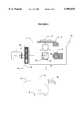
- FIG. 1shows a diagram of the components of the cell-based scanning system.
- FIG. 2shows a schematic of the microscope subassembly
- FIG. 3shows the camera subassembly
- FIG. 4illustrates cell scanning system process
- FIG. 5illustrates a user interface showing major functions to guide the user
- FIG. 6illustrates data presentation on screen
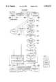
- FIG. 7flow chart of processing step for the cell-based scanning system
- FIGS. 8 A-Jillustrates the strategy of the Nuclear Translocation Assay
- FIG. 9is example data from a known inhibitor of translocation
- FIG. 10is example data from a known stimulator of translocation
- the inventionrelates to a computer controlled optical-mechanical system for rapidly determining the distribution, environment, or activity of fluorescently labeled reporter molecules in cells for the purpose of screening large numbers of compounds for those that specifically affect particular biological functions.
- the inventioninvolves:
- the array of locationsmay be a microtiter plate or a microchip which is a microplate having cells in an array of locations.
- the inventionincludes an apparatus and a computerized method for acquiring data such as a digital frame grabber, processing, displaying and storing the data.
- Standard 96 well microtiter plateswhich are 86 mm by 129 mm, with 6 mm diameter wells on a 9 mm pitch, are used for compatibility with current automated loading and robotic handling systems.
- the microplateis typically 20 mm by 30 mm, with cell locations that are 100-200 microns in dimension on a pitch of about 500 microns. Methods for making microplate are described in U.S. Ser. No. 60/018,696 filed on May 30, 1996, assigned to the same assignee. This application is incorporated herein by reference in its entirety.
- Microplatesmay consist of coplanar layers of materials to which cells adhere patterned with materials to which cells will not adhere, or etched 3-dimensional surfaces of similarly pattered materials.
- ⁇ well ⁇ and ⁇ microwell ⁇refer to a location in an array of any construction to which cells adhere and within which the cells are imaged.
- Microplatesalso include fluid delivery channels in the spaces between the wells. The smaller format of a microplate increases the overall efficiency of the system by minimizing the quantities of the reagents, storage and handling during preparation and the overall movement required for the scanning operation. In addition, the whole area of the microplate can be imaged more efficiently, allowing a second mode of operation for the microplate reader as described later in this document.
- fluorescently labeled biomoleculessuch as proteins, phospholipids and DNA hybridizing probes, as well as fluorescent reagents specifically synthesized with particular chemical properties of binding or association have been used as fluorescent reporter molecules.
- fluorescently labeled antibodiesare particularly useful reporter molecules due to their high degree of specificity for attaching to a single molecular target in a mixture of molecules as complex as a cell, tissue or extract of either.
- Fluorescently labeled reporter moleculesare useful for determining the location, amount and chemical environment of the reporter. For example, whether the reporter is in a lipophilic membrane environment or in a more aqueous environment can be determined. The pH environment of the reporter can be determined. It can be determined whether a reporter having a chelating group is bound to an ion, such as Ca++, or not.
- fluorescent reporter moleculesexhibit a charge in excitation or emission spectra, some exhibit resonance energy transfer where one fluorescent reporter looses fluorescence, while a second gains in fluorescence, some exhibit a loss (quenching) or appearance of fluorescence, while some report rotational movements.
- a cellcan be genetically engineered to express reporter molecules such as GFP coupled to a protein of interest.
- reporter moleculessuch as GFP coupled to a protein of interest.
- FIG. 1is a schematic diagram of the system for measuring the distribution, environment, or activity of fluorescent reporter molecules in cells.
- An inverted fluorescent microscope 1is a Zeiss Axiovert inverted fluorescence microscope which uses standard objectives with magnification of 1-100 ⁇ to the camera, and a white light source (e.g. 100 W mercury-arc lamp or 75W xenon lamp) with power supply 2.
- a white light sourcee.g. 100 W mercury-arc lamp or 75W xenon lamp
- a Z-axis focus drive 5moves the objective in the Z direction for focusing.
- a joystick 6provides for manual movement of the stage in the XYZ direction.
- a high resolution digital camera 7acquires images from each well or location on the plate. 8 is a camera power supply.
- 9is an automation controller and 10 is a central processing unit.
- the PC 11provides a display 12 and has associated software.
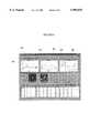
- FIG. 2is a schematic of the microscope assembly 1 showing in more detail the XY stage 3, Z-axis focus drive 5, joystick 6, light source 2, automation controller 9, oculars 14. 15 and 16 are cables to the computer and microscope, respectively.
- FIG. 2shows a 96 well microtiter plate 17 which is moved on the XY stage 3 in the XY direction.
- Light from the light source 2passes through the PC controlled shutter 18 to a motorized filter wheel 19 with excitation filters 20.
- the lightpasses into filter cube 25 which has a dichroic mirror 26 and an emission filter 27. Excitation light reflects off the dichroic mirror to the wells in the microtiter plate 17 and fluorescent light 28 passes through the dichroic mirror 26 and the emission filter 27 and to the digital camera 7.
- FIG. 3shows a schematic drawing of the camera assembly.
- a digital cable 30transports digital signals to the computer.
- 31is the camera power supply.
- FIG. 4illustrates an alternative embodiment of the invention in which cells are in microwells 40 on a microplate 41.
- the microplateis 20 mm by 30 mm as compared to a standard 96 well microtiter plate which is 86 mm by 129 mm.
- the microplate chamber 42serves as a microfluidic delivery system for the addition of compounds to cells.
- the microplate 41 in the microplate chamber 42is placed in an XY microplate reader 43.
- Digital datais processed as described above.
- the small size of this microplate systemincreases throughput, minimizes reagent volume and provides for the ability to control the distribution and placement of cells for fast and precise cell-based analysis.
- This informationcan be displayed on a PC screen 11 and made part of a bioinformatics data base 44.
- This data baseis an important part of the present invention because it not only permits storage and retrieval of data obtained through the methods of this invention, but also permits acquisition and storage of external data relating to cells.
- FIG. 5is a PC display which illustrate
- the higher density array of cells on a microplateallows the microplate reader to image the whole microplate at a low resolution of a few microns per pixel for high throughput and image particular locations on the microplate at a higher resolution of less than 0.5 microns per pixel for higher precision. These two resolution modes improve the overall throughput of the system.
- the operatorenters information 100 that describes the sample, specifies the filter settings and fluorescent channels to match the biological labels being used and the information sought and then adjusts the camera settings to match the sample brightness.
- the softwarenext allows selection of various parameter settings used to identify nuclei, cytoplasm, different fluorescent reagents, cell selection settings and number of cells to be analyzed per well. These parameters are stored in the system's database for easy retrieval for each automated run.
- the system's interactive cell identification modesimplifies the selection of morphological parameter limits such as the range of size, shape, and intensity of cells to be analyzed. The user specifies which wells of the plate the system will scan and how many fields or how many cells to analyze in each well.
- the systemeither automatically pre-focuses the region of the plate to be scanned using an autofocus procedure to "find focus" of the plate 102 or the user interactively pre-focuses 103 the scanning region by selecting three "tag" points which defines the rectangular area to be scanned by the system.
- a least-squares fit "focal plane model”is then calculated by the system from these tag points to estimate the focus of each well during a an automated scan.
- the focus of each wellis estimated by interpolating from the focal plane model during a scan.
- the softwaredynamically displays the status of scan in progress such as the number of cells that have been analyzed, the current well that is being analyzed, and images of each independent wavelength as they are acquired, and the result of the screen for each well as it is acquired.
- the plate 4is scanned in a serpentine style as the software automatically moves the motorized microscope XY stage 3 from well to well and field to field within each well of a 96-well plate.
- Those skilled in the programming artwill recognize how to adapt software for scanning of other microplate formats such as 24, 48, and 384 well plates.
- the scan pattern of the entire plate as well as the scan pattern of fields within each wellare programmed.
- the systemadjusts sample focus with an autofocus procedure 104 through the Z axis focus drive 5, controls filter selection via a motorized filter wheel 19 and acquires and analyzes images of up to four different colors ("channels" or "wavelengths").
- the autofocus procedureis called at a user selected frequency, typically for the first field in each well and then once every 4 to 5 fields within each well.
- the autofocus procedurecalculates the starting Z-axis point by interpolating from the pre-calculated plane focal model. Starting a programmable distance above or below this set point, the procedure moves the mechanical Z-axis through a number of different positions, acquires an image at each, and finds the maximum of a calculated focus score that estimates the contrast of each image. The Z position of the image with the maximum focus score determines the best focus for a particular field.
- Those skilled in the artwill recognize this as a variant of automatic focusing algorithms as described in the prior art in Harms et al. in Cytometry 5 (1984), p. 236-243, Groen et al. in Cytometry 6 (1985), p. 81-91, and Firestone et al. in Cytometry 12 (1991), p. 195-206.
- the camera's 7 exposure timeis separately adjusted for each dye to ensure a high-quality image from each channel.
- Software procedurescan be called, at the user's option, to correct for registration shifts between wavelengths by accounting for linear (X and Y) shifts between wavelengths before making any further measurements.
- the electronic shutter 18is controlled so that sample photo-bleaching is kept to a minimum. Background shading and uneven illumination can also be corrected by the software using algorithms known in the prior art.
- imagesare acquired of a primary marker 105 (typically cell nuclei counterstained with DAPI or PI fluorescent dyes) which are segmented ("identified") using an adaptive thresholding procedure.
- the adaptive thresholding procedure 106is used to dynamically select the threshold of an image for separating cells from the background.
- the staining of cells with fluorescent dyescan vary to an unknown degree across cells in a microtiter plate sample as well as within images of field of cells within each well of a microtiter plate. This variation can occur due to sample preparation and/or as a result of the nature of cell biology.
- a global thresholdis calculated for the complete image to separate the cells from background.
- the global adaptive techniques used in the systemare variants of those described in prior art in Kittler et al.
- the output of the segmentation procedureis a binary mask wherein the objects are white and the background is black.
- This binary imagealso called a mask in the prior art, is used to determine if the field contains objects 107.
- the maskis labeled with a blob labeling algorithm whereby each object (or blob) has a unique number assigned to it. Morphological features, such as area and shape, of the blobs are used to differentiate blobs likely to be cells from those that are considered artifacts.
- the userpre-sets the morphological selection criteria by either typing in known cells morphological features or by using the interactive training utility. If objects of interest are found in the field, images are acquired for all other active channels 108, otherwise the stage is advanced to the next field 109 in the current well.
- Each object of interestis located in the image for further analysis 110.
- the softwaredetermines if the object meets the criteria for a valid cell nucleus 111 by measuring its morphological features (size and shape). For each valid cell, the XYZ stage location is recorded, a small image of the cell is stored, and features are measured 112.
- the cell scanning systemcan perform multiple tests on cellular samples by applying a number of analysis methods simultaneously to measure features at multiple wavelengths including:
- Features 1 through 4are commonly used in a variety of image analysis applications and are well known in prior art.
- Features 5-9have been developed specifically to provide measurements of a cell's fluorescent molecules within the local cytoplasmic region of the cell and the translocation (i.e. movement) of fluorescent molecules from the cytoplasm to the nucleus.
- These screen specific featuresare used for analyzing cells in microplates for the inhibition of nuclear translocation. Inhibition of nuclear translocation of transcription factors provides a novel approach to screening intact cells.
- An automated screen of an inhibitor of NF- ⁇ B translocationhas been successfully performed.
- a specific algorithmmeasures the amount of NF- ⁇ B probe in the nuclear region (feature 4) versus the local cytoplasmic region (feature 7) of each cell. Quantification of the difference between these two sub-cellular compartments provides a measure of cytoplasm-nuclear translocation (feature 9).
- Feature 10is used for counting of DNA or RNA probes within the nuclear region in colors 2-4.
- DNA probesare commercially available for identifying the centromeres of specific chromosomes.
- Cellsare three-dimensional in nature and when examined at a high magnification under a microscope one probe may be in-focus while another may be completely out-of-focus.
- the cell screening systemhas a procedure for detecting three-dimensional probes in nuclei by acquiring images from multiple focal planes.
- the softwaremoves the Z-axis motor drive 5 in small steps where the step distance is user selected to account for a wide range of different nuclear diameters.
- At each of the focal stepsan image is acquired. The maximum gray-level intensity from each pixel in each image is found and stored in a resulting maximum projection image.
- the maximum projection imageis then used to count the probes.
- the above algorithmwork well in counting probes that are not stacked directly above or below another one.
- userscan select an option to improve the counting of probes by analyzing probes in each of the focal planes acquired.
- the scanning systemperforms the maximum plane projection algorithm as discussed above, detects probe regions of interest in this image, then further analyzes these regions in all the focal plane images.
- the systemsAfter measuring cell features 112, the systems checks if there are any unprocessed objects in the current field 113. If there are any unprocessed objects, it locates the next object 110 and checks if it meets the criteria for a valid cell nucleus 111 and measures its features. After it the system has processed all the objects in the current field, it checks if it is done with the current plate 114. If the system is not done with the current plate, it check if it needs to find more cells in the current well 115. If it needs to find more cells in the current well it advances the XYZ stage to the next field within the current well 109 or it advances the stage to the next well 116 of the plate.
- images and datacan be reviewed with the system's image review, data review, and summary review facilities. All images, data, and settings from a scan are archived in the system's database for later review. Users can review the images alone of every cell analyzed by the system with an interactive image review procedure 117. The user can review data on a cell-by-cell basis using a combination of interactive graphs, a data spreadsheet of features measured, and images of all the fluorescent channels of a cell of interest with the interactive cell-by-cell data review procedure 118. Graphical plotting capabilities are provided in which data can be analyzed via interactive graphs such as histograms and scatter plots.
- reportscan be generated on one or more statistics of features measured. Users can generate a graphical report of data summarized on a well-by-well basis for the scanned region of the plate using an interactive report generation procedure 120. This report includes a summary of the statistics by well in tabular and graphical format and identification information on the sample. The report window allows the operator to enter comments about the scan for later retrieval. Multiple reports can be generated on many statistics and be printed with the touch of one button. Reports can be previewed for placement and data before being printed.
- Regulation of transcription of some genesinvolves activation of a transcription factor in the cytoplasm, resulting in that factor being transported into the nucleus where it can initiate transcription of a particular gene or genes.
- This change in transcription factor distributionis the basis of a screen for the cell-based screening system to detect compounds which inhibit or induce transcription of a particular gene or group of genes. A general description of the screen is given followed by a specific example.
- the distribution of the transcription factoris determined by labeling the nuclei with a DNA specific fluorophore like Hoechst 33423 and the transcription factor with a specific fluorescent antibody. After autofocusing on the Hoechst labeled nuclei, an image of the nuclei is acquired in the cell-based screening system at 20 ⁇ magnification and used to create a mask by one of several optional thresholding methods. The morphological descriptors of the regions defined by the mask are compared with the user defined parameters and valid nuclear masks are identified and used with the following algorithm to extract transcription factor distributions. Each valid nuclear mask is eroded to define a slightly smaller nuclear region.
- FIG. 8Aillustrates an unstimulated cell with its nucleus 200 labeled with a blue fluorophore and a transcription factor in the cytoplasm 201 labeled with a green fluorophore.
- FIG. 8Billustrates the nuclear mask 202 derived by the cell-based screening system.
- FIG. 8Cillustrates the cytoplasm 203 of the unstimulated cell imaged at a green wavelength.
- FIG. 8Dillustrates the nuclear mask 202 is eroded (reduced) once to define a nuclear sampling region 204 with minimal cytoplasmic distribution.
- the nucleus boundary 202is dilated (expanded) several times to form a ring that is 2-3 pixels wide that is used to define the cytoplasmic sampling region 205 for the same cell.
- FIG. 8Efurther illustrates a side view which shows the nuclear sampling region 204 and the cytoplasmic sampling region 205. Using these two sampling regions, data on nuclear translocation can be automatically analyzed by the cell-based screening system on a cell by cell basis.
- FIG. 8F-Jillustrates the strategy for determining nuclear translocation in a stimulated cell.
- FIG. 8Fillustrates a stimulated cell with its nucleus 206 labeled with a blue fluorophore and a transcription factor in the cytoplasm 207 labeled with a green fluorophore.
- the nuclear mask 208 in FIG. 8Gis derived by the cell based screening system.
- FIG. 8Hillustrates the cytoplasm 209 of a stimulated cell imaged at a green wavelength.
- FIG. 8Iillustrates the nuclear sampling region 211 and cytoplasmic sampling region 212 of the stimulated cell.
- FIG. 8Jfurther illustrates a side view which shows the nuclear sampling region 211 and the cytoplasmic sampling region 212.
- a specific application of this methodhas been used to validate this method as a screen.
- a human chondrocyte cell linewas plated in 96 well microtiter plates. Some rows of wells were titrated with IL-1 ⁇ , a known inducer of the nuclear transcription factor NF- ⁇ B. The cells were then fixed and stained by standard methods with a fluorescein labeled antibody to NF- ⁇ B, and Hoechst 33423. The cell-based screening system was used to acquire and analyze images from this plate and the NucCyt Difference was found to be strongly correlated with the amount of IL-1 ⁇ added to the wells as illustrated in FIG. 9.
- FIG. 6is a representative display on a PC screen of data which was obtained in accordance with Example 1.
- Graph 1 300plots the difference between the average antibody fluorescence in the nuclear sampling region and cytoplasmic sampling region, NucCyt Difference verses Well #.
- Graph 2 301plots the average fluorescence of the antibody in the nuclear sampling region, NP1 average, versus the Well #.
- Graph 3 302plots the average antibody fluorescence in the cytoplasmic sampling region, LIP1 average, versus Well #.
- the softwarepermits displaying data from each cell.
- FIG. 6shows a screen display, the nuclear image, and the fluorescent antibody image for cell #14.
- NucCyt Difference referred to in graph 1 303 of FIG. 6is the difference between the average cytoplasmic probe (fluorescent reporter molecule) intensity and the average nuclear probe (fluorescent reporter molecule) intensity.
- the inventionprovides a computer means for converting the digital signal from the camera into this parameter and for plotting the parameter verses the well number.
- NP1 average referred to in graph 2 304 of FIG. 6is the average of cyloplasmic probe (fluorescent reporter molecule) intensity within the nuclear sampling region.
- the inventionprovides a computer means for converting the digital signal from the camera into this parameter and for plotting the parameter verses the well number.
- L1P1 average referred to in graph 3 305 of FIG. 6is the average probe (fluorescent reporter molecule) intensity within the cytoplasmic sampling region.
- the inventionprovides a computer means for converting the digital signal from the camera into this parameter and for plotting the parameter verses the well number.
- Hypertrophy in cardiac myocyteshas been associated with a cascade of alterations in gene expression and can be characterized in cell culture by an alteration in cell size, that is clearly visible in adherent cells growing on a coverslip.
- Preliminary experimentsindicate that a screen can be implemented using the following strategy.
- Myocytes cultured in 96 well platescan be treated with various compounds and then fixed and labeled with a fluorescent antibody to a cell surface marker and a DNA label like Hoechst. After focusing on the Hoechst labeled nuclei, two images are acquired, one of the Hoechst labeled nuclei and one of the fluorescent antibody.
- the nucleiare identified by thresholding to create a mask and then comparing the morphological descriptors of the mask with a set of user defined descriptor values. Local regions containing cells are defined around the nuclei. The limits of the cells in those regions are then defined by a local dynamic threshold operation on the same region in the fluorescent antibody image. A sequence of erosions and dilations is used to separate slightly touching cells and a second set of morphological descriptors is used to identify single cells. The area of the individual cells is tabulated in order to define the distribution of cell sizes for comparison with size data from normal and hypertrophic cells.
- a second fluorescent antibody to a particular cellular protein, such as one of the major muscle proteins actin or myosincan included. Images of this antibody can be acquired and stored with the above images, for later review, to identify anomalies in the distribution of these proteins in hypertrophic cells, or algorithms can be developed to automatically analyze the distributions of the labeled proteins in these images.
- the GTP-binding proteins of the Rho familyare maintained as cytoplasmic complexes with RhoGDI in resting cells, but are released and translocate to plasma membrane during cell activation.
- specific organellessuch as components of the cytoskeleton, nuclear envelope, chromatin, golgi apparatus, mitochondria, and endosomes are reorganized in response to specific stimuli.
- the targets for screeningcan themselves be converted into fluorescence-based reagents that report molecular changes including ligand-binding and post-translocational modifications.
Landscapes
- Health & Medical Sciences (AREA)
- Chemical & Material Sciences (AREA)
- Life Sciences & Earth Sciences (AREA)
- Engineering & Computer Science (AREA)
- Immunology (AREA)
- Physics & Mathematics (AREA)
- Analytical Chemistry (AREA)
- General Physics & Mathematics (AREA)
- General Health & Medical Sciences (AREA)
- Biochemistry (AREA)
- Molecular Biology (AREA)
- Pathology (AREA)
- Biomedical Technology (AREA)
- Hematology (AREA)
- Urology & Nephrology (AREA)
- Organic Chemistry (AREA)
- Microbiology (AREA)
- Biotechnology (AREA)
- Zoology (AREA)
- Wood Science & Technology (AREA)
- Proteomics, Peptides & Aminoacids (AREA)
- Cell Biology (AREA)
- Food Science & Technology (AREA)
- Medicinal Chemistry (AREA)
- Bioinformatics & Cheminformatics (AREA)
- Optics & Photonics (AREA)
- Nuclear Medicine, Radiotherapy & Molecular Imaging (AREA)
- Multimedia (AREA)
- Genetics & Genomics (AREA)
- General Engineering & Computer Science (AREA)
- Biophysics (AREA)
- Nanotechnology (AREA)
- Tropical Medicine & Parasitology (AREA)
- Computer Vision & Pattern Recognition (AREA)
- Composite Materials (AREA)
- Condensed Matter Physics & Semiconductors (AREA)
- Materials Engineering (AREA)
- Crystallography & Structural Chemistry (AREA)
- Chemical Kinetics & Catalysis (AREA)
- Toxicology (AREA)
Abstract
Description
Claims (8)
Priority Applications (31)
| Application Number | Priority Date | Filing Date | Title |
|---|---|---|---|
| US08/810,983US5989835A (en) | 1997-02-27 | 1997-02-27 | System for cell-based screening |
| US08/865,341US6103479A (en) | 1996-05-30 | 1997-05-29 | Miniaturized cell array methods and apparatus for cell-based screening |
| DE69839501TDE69839501D1 (en) | 1997-02-27 | 1998-02-27 | System for screening biological cells |
| CA002282658ACA2282658C (en) | 1997-02-27 | 1998-02-27 | A system for cell-based screening |
| EP98908719AEP0983498B1 (en) | 1997-02-27 | 1998-02-27 | A system for cell-based screening |
| EP04009981AEP1439384B1 (en) | 1997-02-27 | 1998-02-27 | A system for screening biological cells |
| DE69824174TDE69824174T2 (en) | 1997-02-27 | 1998-02-27 | A SYSTEM FOR CELL-BASED SERIES TESTING |
| PCT/US1998/003701WO1998038490A1 (en) | 1997-02-27 | 1998-02-27 | A system for cell-based screening |
| AU66678/98AAU730100B2 (en) | 1997-02-27 | 1998-02-27 | A system for cell-based screening |
| DK98908719TDK0983498T3 (en) | 1997-02-27 | 1998-02-27 | Cell-based screening system |
| JP53782198AJP3683591B2 (en) | 1997-02-27 | 1998-02-27 | Cell-based screening system |
| AT98908719TATE268008T1 (en) | 1997-02-27 | 1998-02-27 | A SYSTEM FOR CELL-BASED SERIAL EXAMINATION |
| US09/380,259US6727071B1 (en) | 1997-02-27 | 1998-02-27 | System for cell-based screening |
| CA002410688ACA2410688C (en) | 1997-02-27 | 1998-02-27 | A system for cell-based screening |
| US09/293,210US6620591B1 (en) | 1997-02-27 | 1999-04-16 | System for cell-based screening |
| US09/352,171US6759206B1 (en) | 1997-02-27 | 1999-07-12 | System for cell-based screening |
| US09/430,656US6756207B1 (en) | 1997-02-27 | 1999-10-29 | System for cell-based screening |
| US09/513,783US6416959B1 (en) | 1997-02-27 | 2000-02-25 | System for cell-based screening |
| US09/650,937US6573039B1 (en) | 1997-02-27 | 2000-08-29 | System for cell-based screening |
| US09/721,168US7060445B1 (en) | 1997-02-27 | 2000-11-22 | System for cell-based screening |
| US09/718,770US7565247B1 (en) | 1997-02-27 | 2000-11-22 | Method for acquisition, storage, and retrieval of cell screening data on a computer system |
| US09/723,256US7117098B1 (en) | 1997-02-27 | 2000-11-27 | Machine-readable storage medium for analyzing distribution of macromolecules between the cell membrane and the cell cytoplasm |
| US09/724,376US6671624B1 (en) | 1997-02-27 | 2000-11-27 | Machine readable storage media for detecting distribution of macromolecules between nucleus and cytoplasm in cells |
| JP2002061217AJP4011936B2 (en) | 1997-02-27 | 2002-03-06 | Cell-based screening system |
| US10/100,957US6875578B2 (en) | 1997-02-27 | 2002-03-19 | System for cell-based screening |
| US10/411,635US7235373B2 (en) | 1997-02-27 | 2003-04-11 | System for cell-based screening |
| US10/430,534US6902883B2 (en) | 1997-02-27 | 2003-05-06 | System for cell-based screening |
| US10/685,737US7853411B2 (en) | 1997-02-27 | 2003-10-15 | System for cell-based screening |
| US10/686,161US20040063162A1 (en) | 1997-02-27 | 2003-10-15 | System for cell-based screening |
| US11/809,937US7853409B2 (en) | 1997-02-27 | 2007-06-04 | System for cell-based screening |
| JP2007184678AJP4575403B2 (en) | 1997-02-27 | 2007-07-13 | Cell-based screening system |
Applications Claiming Priority (1)
| Application Number | Priority Date | Filing Date | Title |
|---|---|---|---|
| US08/810,983US5989835A (en) | 1997-02-27 | 1997-02-27 | System for cell-based screening |
Related Parent Applications (1)
| Application Number | Title | Priority Date | Filing Date |
|---|---|---|---|
| US08/865,341Continuation-In-PartUS6103479A (en) | 1996-05-30 | 1997-05-29 | Miniaturized cell array methods and apparatus for cell-based screening |
Related Child Applications (7)
| Application Number | Title | Priority Date | Filing Date |
|---|---|---|---|
| US08/865,341Continuation-In-PartUS6103479A (en) | 1996-05-30 | 1997-05-29 | Miniaturized cell array methods and apparatus for cell-based screening |
| US09/380,259Continuation-In-PartUS6727071B1 (en) | 1997-02-27 | 1998-02-27 | System for cell-based screening |
| US3127198AContinuation-In-Part | 1997-02-27 | 1998-02-27 | |
| US09/293,210ContinuationUS6620591B1 (en) | 1997-02-27 | 1999-04-16 | System for cell-based screening |
| US29320999ADivision | 1997-02-27 | 1999-04-16 | |
| US09/352,171Continuation-In-PartUS6759206B1 (en) | 1997-02-27 | 1999-07-12 | System for cell-based screening |
| US10/685,737Continuation-In-PartUS7853411B2 (en) | 1997-02-27 | 2003-10-15 | System for cell-based screening |
Publications (1)
| Publication Number | Publication Date |
|---|---|
| US5989835Atrue US5989835A (en) | 1999-11-23 |
Family
ID=25205216
Family Applications (9)
| Application Number | Title | Priority Date | Filing Date |
|---|---|---|---|
| US08/810,983Expired - LifetimeUS5989835A (en) | 1996-05-30 | 1997-02-27 | System for cell-based screening |
| US09/293,210Expired - LifetimeUS6620591B1 (en) | 1997-02-27 | 1999-04-16 | System for cell-based screening |
| US09/650,937Expired - LifetimeUS6573039B1 (en) | 1997-02-27 | 2000-08-29 | System for cell-based screening |
| US09/718,770Expired - LifetimeUS7565247B1 (en) | 1997-02-27 | 2000-11-22 | Method for acquisition, storage, and retrieval of cell screening data on a computer system |
| US09/721,168Expired - LifetimeUS7060445B1 (en) | 1997-02-27 | 2000-11-22 | System for cell-based screening |
| US10/411,635Expired - Fee RelatedUS7235373B2 (en) | 1997-02-27 | 2003-04-11 | System for cell-based screening |
| US10/430,534Expired - LifetimeUS6902883B2 (en) | 1997-02-27 | 2003-05-06 | System for cell-based screening |
| US10/686,161AbandonedUS20040063162A1 (en) | 1997-02-27 | 2003-10-15 | System for cell-based screening |
| US11/809,937Expired - Fee RelatedUS7853409B2 (en) | 1997-02-27 | 2007-06-04 | System for cell-based screening |
Family Applications After (8)
| Application Number | Title | Priority Date | Filing Date |
|---|---|---|---|
| US09/293,210Expired - LifetimeUS6620591B1 (en) | 1997-02-27 | 1999-04-16 | System for cell-based screening |
| US09/650,937Expired - LifetimeUS6573039B1 (en) | 1997-02-27 | 2000-08-29 | System for cell-based screening |
| US09/718,770Expired - LifetimeUS7565247B1 (en) | 1997-02-27 | 2000-11-22 | Method for acquisition, storage, and retrieval of cell screening data on a computer system |
| US09/721,168Expired - LifetimeUS7060445B1 (en) | 1997-02-27 | 2000-11-22 | System for cell-based screening |
| US10/411,635Expired - Fee RelatedUS7235373B2 (en) | 1997-02-27 | 2003-04-11 | System for cell-based screening |
| US10/430,534Expired - LifetimeUS6902883B2 (en) | 1997-02-27 | 2003-05-06 | System for cell-based screening |
| US10/686,161AbandonedUS20040063162A1 (en) | 1997-02-27 | 2003-10-15 | System for cell-based screening |
| US11/809,937Expired - Fee RelatedUS7853409B2 (en) | 1997-02-27 | 2007-06-04 | System for cell-based screening |
Country Status (1)
| Country | Link |
|---|---|
| US (9) | US5989835A (en) |
Cited By (272)
| Publication number | Priority date | Publication date | Assignee | Title |
|---|---|---|---|---|
| US6110693A (en)* | 1997-06-05 | 2000-08-29 | Duke University | Methods of assaying receptor activity and constructs useful in such methods |
| WO2000068810A1 (en)* | 1999-05-07 | 2000-11-16 | Tropix, Inc. | Data display software |
| US6169816B1 (en)* | 1997-05-14 | 2001-01-02 | Applied Imaging, Inc. | Identification of objects of interest using multiple illumination schemes and finding overlap of features in corresponding multiple images |
| US6193647B1 (en)* | 1999-04-08 | 2001-02-27 | The Board Of Trustees Of The University Of Illinois | Microfluidic embryo and/or oocyte handling device and method |
| US6232124B1 (en) | 1996-05-06 | 2001-05-15 | Verification Technologies, Inc. | Automated fingerprint methods and chemistry for product authentication and monitoring |
| US6259807B1 (en)* | 1997-05-14 | 2001-07-10 | Applied Imaging Corp. | Identification of objects of interest using multiple illumination schemes and finding overlap of features in corresponding multiple images |
| US6258326B1 (en) | 1997-09-20 | 2001-07-10 | Ljl Biosystems, Inc. | Sample holders with reference fiducials |
| US20010007775A1 (en)* | 1996-04-25 | 2001-07-12 | Michael Seul | System and method for programmable illumination pattern generation |
| US6261815B1 (en) | 1996-09-23 | 2001-07-17 | Duke University | Method of introducing exogenous compounds into cells by electroporation and apparatus for same |
| US6297018B1 (en)* | 1998-04-17 | 2001-10-02 | Ljl Biosystems, Inc. | Methods and apparatus for detecting nucleic acid polymorphisms |
| US20010030770A1 (en)* | 2000-02-01 | 2001-10-18 | Kazuhito Ohashi | Discrepancy correction method and apparatus for correcting difference in levels of image signals obtained by an image sensor having a multiple output channels |
| US6317207B2 (en) | 1999-02-23 | 2001-11-13 | Ljl Biosystems, Inc. | Frequency-domain light detection device |
| US6326605B1 (en) | 1998-02-20 | 2001-12-04 | Ljl Biosystems, Inc. | Broad range light detection system |
| US6365367B1 (en) | 1999-12-06 | 2002-04-02 | Cellomics, Inc. | Environmental chamber for the analysis of live cells |
| WO2002027296A1 (en) | 2000-09-27 | 2002-04-04 | Hyperion, Inc. | Quantitative digital fluorography and related products and methods |
| US20020049544A1 (en)* | 2000-06-23 | 2002-04-25 | Cytokinetics, Inc. | Image analysis for phenotyping sets of mutant cells |
| US6388788B1 (en) | 1998-03-16 | 2002-05-14 | Praelux, Inc. | Method and apparatus for screening chemical compounds |
| US6387707B1 (en) | 1996-04-25 | 2002-05-14 | Bioarray Solutions | Array Cytometry |
| WO2002002226A3 (en)* | 2000-07-05 | 2002-05-23 | Joline Gmbh & Co Kg | Support plate and method for the carrying out of functional tests |
| WO2001094528A3 (en)* | 2000-06-08 | 2002-06-06 | Univ California | Visual-servoing optical microscopy |
| US20020085293A1 (en)* | 2000-11-17 | 2002-07-04 | Stuckey Jeffrey A. | Rapidly changing dichroic beamsplitter in epi-fluorescent microscopes |
| US6416959B1 (en)* | 1997-02-27 | 2002-07-09 | Kenneth Giuliano | System for cell-based screening |
| US20020090613A1 (en)* | 1997-05-23 | 2002-07-11 | Michael Seul | Color-encoding and in-situ interrogation of matrix-coupled chemical compounds |
| US20020119441A1 (en)* | 2000-12-18 | 2002-08-29 | Cytokinetics, Inc., A Delaware Corporation | Method of characterizing potential therapeutics by determining cell-cell interactions |
| US20020118983A1 (en)* | 2001-02-27 | 2002-08-29 | Hiroshi Fuma | Image fixing device and image forming apparatus equipped therewith |
| EP1170591A3 (en)* | 2000-07-07 | 2002-09-18 | CombinatoRx, Incorporated | Methods for identifying combinations of entities as therapeutics |
| WO2002073200A1 (en) | 2001-03-12 | 2002-09-19 | Cellomics, Inc. | Methods to increase the capacity of high content cell-based screening assays |
| US20020141631A1 (en)* | 2001-02-20 | 2002-10-03 | Cytokinetics, Inc. | Image analysis of the golgi complex |
| US6466316B2 (en) | 1998-07-27 | 2002-10-15 | Ljl Biosystems, Inc. | Apparatus and methods for spectroscopic measurements |
| US6468811B1 (en) | 1996-04-25 | 2002-10-22 | Bioarray Solutions | Light-controlled electrokinetic assembly of particles near surfaces |
| US6469311B1 (en) | 1997-07-16 | 2002-10-22 | Molecular Devices Corporation | Detection device for light transmitted from a sensed volume |
| US20020154798A1 (en)* | 2001-02-20 | 2002-10-24 | Ge Cong | Extracting shape information contained in cell images |
| US20020159625A1 (en)* | 2001-04-02 | 2002-10-31 | Cytoprint, Inc. | Method and apparatus for discovering, identifying and comparing biological activity mechanisms |
| US20020168657A1 (en)* | 2001-02-02 | 2002-11-14 | Chen Lan Bo | Rare event detection system |
| US6483582B2 (en) | 1998-07-27 | 2002-11-19 | Ljl Biosystems, Inc. | Apparatus and methods for time-resolved spectroscopic measurements |
| US20020172964A1 (en)* | 2001-02-02 | 2002-11-21 | Cellomics, Inc. | Method for determining a best initial focal position estimate |
| US20020177149A1 (en)* | 2001-04-20 | 2002-11-28 | Rimm David L. | Systems and methods for automated analysis of cells and tissues |
| US6490030B1 (en) | 1999-01-18 | 2002-12-03 | Verification Technologies, Inc. | Portable product authentication device |
| US20020198665A1 (en)* | 2000-09-17 | 2002-12-26 | Michael Seul | System and method for programmable illumination pattern generation |
| US20020198858A1 (en)* | 2000-12-06 | 2002-12-26 | Biosentients, Inc. | System, method, software architecture, and business model for an intelligent object based information technology platform |
| US20020197656A1 (en)* | 1999-12-17 | 2002-12-26 | Ronghao Li | Cell arrays and the uses thereof |
| WO2002019594A3 (en)* | 2000-08-02 | 2002-12-27 | Univ Arizona State | Scanning fluorescence lifetime microscope: directed evolution |
| US6499366B1 (en) | 1997-07-16 | 2002-12-31 | Ljl Biosystems, Inc. | Sample feeder |
| US20030008323A1 (en)* | 1999-04-15 | 2003-01-09 | Ilya Ravkin | Chemical-library composition and method |
| US20030013137A1 (en)* | 2001-03-13 | 2003-01-16 | Barak Larry S. | Automated methods of detecting receptor activity |
| US6512580B1 (en) | 1999-10-27 | 2003-01-28 | Verification Technologies, Inc. | Method and apparatus for portable product authentication |
| US20030022370A1 (en)* | 2001-07-27 | 2003-01-30 | Rocco Casagrande | Magnetic immobilization of cells |
| US20030022271A1 (en)* | 1999-04-09 | 2003-01-30 | John Voneiff | Apparatus and method for automatically producing tissue slides |
| US20030032002A1 (en)* | 2001-07-27 | 2003-02-13 | Evelyn Wang | Cell isolation and screening device and method of using same |
| US20030032071A1 (en)* | 2001-07-27 | 2003-02-13 | Evelyn Wang | Cell isolation and screening device and method of using same |
| US20030036096A1 (en)* | 1999-04-15 | 2003-02-20 | Ilya Ravkin | Chemical-library composition and method |
| US20030036853A1 (en)* | 2000-12-22 | 2003-02-20 | Cellomics, Inc. | Automated assay for identification of individual cells during kinetic assays |
| US20030036855A1 (en)* | 1998-03-16 | 2003-02-20 | Praelux Incorporated, A Corporation Of New Jersey | Method and apparatus for screening chemical compounds |
| US20030059764A1 (en)* | 2000-10-18 | 2003-03-27 | Ilya Ravkin | Multiplexed cell analysis system |
| US20030059093A1 (en)* | 2001-03-26 | 2003-03-27 | Cellomics, Inc. | Methods for determining the organization of a cellular component of interest |
| US6548263B1 (en) | 1997-05-29 | 2003-04-15 | Cellomics, Inc. | Miniaturized cell array methods and apparatus for cell-based screening |
| US20030082564A1 (en)* | 1997-04-07 | 2003-05-01 | Bioimage A/S | Method for extracting quantitative information relating to an influence on a cellular response |
| US20030082551A1 (en)* | 2001-09-28 | 2003-05-01 | Zarling David A. | High-throughput gene cloning and phenotypic screening |
| US20030096309A1 (en)* | 2001-08-29 | 2003-05-22 | Alexis Borisy | Screening system for identifying drug-drug interactions and methods of use thereof |
| US6573039B1 (en)* | 1997-02-27 | 2003-06-03 | Cellomics, Inc. | System for cell-based screening |
| US20030104479A1 (en)* | 2001-08-01 | 2003-06-05 | Cellomics, Inc. | Novel fusion proteins and assays for molecular binding |
| US20030104494A1 (en)* | 2001-10-26 | 2003-06-05 | Ilya Ravkin | Assay systems with adjustable fluid communication |
| US6576476B1 (en) | 1998-09-02 | 2003-06-10 | Ljl Biosystems, Inc. | Chemiluminescence detection method and device |
| US20030113813A1 (en)* | 2001-11-15 | 2003-06-19 | Heidaran Mohammad A. | Methods and devices for the integrated discovery of cell culture environments |
| US20030124505A1 (en)* | 1999-03-22 | 2003-07-03 | Jain Sarita Kumari | High-throughput gene cloning and phenotypic screening |
| US6589626B2 (en) | 2000-06-30 | 2003-07-08 | Verification Technologies, Inc. | Copy-protected optical media and method of manufacture thereof |
| US20030129654A1 (en)* | 1999-04-15 | 2003-07-10 | Ilya Ravkin | Coded particles for multiplexed analysis of biological samples |
| US20030130828A1 (en)* | 2001-11-08 | 2003-07-10 | Dibenedetto Emmanuele | Method for modeling signal transduction in cells |
| US20030129648A1 (en)* | 1998-02-07 | 2003-07-10 | Soheil Shams | Automated DNA array image segmentation and analysis |
| US20030134330A1 (en)* | 1999-04-15 | 2003-07-17 | Ilya Ravkin | Chemical-library composition and method |
| US6615141B1 (en) | 1999-05-14 | 2003-09-02 | Cytokinetics, Inc. | Database system for predictive cellular bioinformatics |
| US20030166028A1 (en)* | 2000-02-11 | 2003-09-04 | Sarah Burroughs-Tencza | Peptide biosensors for anthrax protease |
| US20030166015A1 (en)* | 1999-04-15 | 2003-09-04 | Zarowitz Michael A. | Multiplexed analysis of cell-substrate interactions |
| US20030175821A1 (en)* | 2002-03-14 | 2003-09-18 | Hoover Rex A. | Apparatus and method for microscopic comet assay |
| US6623860B2 (en) | 2000-10-10 | 2003-09-23 | Aclara Biosciences, Inc. | Multilevel flow structures |
| WO2003078965A2 (en) | 2002-03-13 | 2003-09-25 | Q3Dm, Llc | System and method for measurement of a response of localized cellular compartments |
| US20030182669A1 (en)* | 2002-03-19 | 2003-09-25 | Rockman Howard A. | Phosphoinositide 3-kinase mediated inhibition of GPCRs |
| US20030185450A1 (en)* | 2002-02-13 | 2003-10-02 | Garakani Arman M. | Method and apparatus for acquisition, compression, and characterization of spatiotemporal signals |
| WO2003088123A1 (en)* | 2002-04-16 | 2003-10-23 | Evotec Oai Ag | Method for analyzing chemical and/or biological samples by means of particle images |
| US6638593B2 (en) | 2000-06-30 | 2003-10-28 | Verification Technologies, Inc. | Copy-protected optical media and method of manufacture thereof |
| US20030207249A1 (en)* | 1999-04-15 | 2003-11-06 | Beske Oren E. | Connection of cells to substrates using association pairs |
| US20030207327A1 (en)* | 2001-09-27 | 2003-11-06 | Kmiec Eric B. | Coisogenic eukaryotic cell collections |
| US6651008B1 (en) | 1999-05-14 | 2003-11-18 | Cytokinetics, Inc. | Database system including computer code for predictive cellular bioinformatics |
| US20030215941A1 (en)* | 2002-03-12 | 2003-11-20 | Stewart Campbell | Assay device that analyzes the absorption, metabolism, permeability and/or toxicity of a candidate compound |
| US20030219800A1 (en)* | 2001-10-18 | 2003-11-27 | Beske Oren E. | Multiplexed cell transfection using coded carriers |
| US20030224531A1 (en)* | 2002-05-29 | 2003-12-04 | Brennen Reid A. | Microplate with an integrated microfluidic system for parallel processing minute volumes of fluids |
| US20030228610A1 (en)* | 1996-04-25 | 2003-12-11 | Michael Seul | Encoded beads having oligonucleotides attached and formed in arrays |
| US20030228566A1 (en)* | 2002-06-11 | 2003-12-11 | Biotechplex Corporation | Method of and apparatus for screening for drug candidates |
| US6671624B1 (en) | 1997-02-27 | 2003-12-30 | Cellomics, Inc. | Machine readable storage media for detecting distribution of macromolecules between nucleus and cytoplasm in cells |
| US6682927B2 (en) | 1998-10-05 | 2004-01-27 | Duke University | Methods and apparatus for the high through-put detection of binding interactions in vivo |
| US6690399B1 (en) | 1999-05-07 | 2004-02-10 | Tropix, Inc. | Data display software for displaying assay results |
| US20040029213A1 (en)* | 2000-06-08 | 2004-02-12 | Callahan Daniel E. | Visual-servoing optical microscopy |
| US20040047499A1 (en)* | 1998-02-07 | 2004-03-11 | Soheil Shams | Automated DNA array image segmentation and analysis |
| US20040059520A1 (en)* | 2002-09-25 | 2004-03-25 | Soheil Shams | Apparatus, method, and computer program product for determining confidence measures and combined confidence measures for assessing the quality of microarrays |
| US6716588B2 (en) | 1999-12-09 | 2004-04-06 | Cellomics, Inc. | System for cell-based screening |
| US20040071328A1 (en)* | 2001-09-07 | 2004-04-15 | Vaisberg Eugeni A. | Classifying cells based on information contained in cell images |
| US6727071B1 (en) | 1997-02-27 | 2004-04-27 | Cellomics, Inc. | System for cell-based screening |
| US20040091946A1 (en)* | 2000-11-03 | 2004-05-13 | Oakley Robert H. | Methods of screening compositions for G protein -coupled receptor desensitization inhibitory activity |
| US20040096911A1 (en)* | 1999-04-15 | 2004-05-20 | Oleg Siniaguine | Particles with light-polarizing codes |
| US20040101912A1 (en)* | 1997-02-27 | 2004-05-27 | Cellomics, Inc. | System for cell-based screening |
| US20040117124A1 (en)* | 2001-02-20 | 2004-06-17 | Eskinder Kiros | Method and apparatus for automated cellular bioinformatics |
| US6752942B2 (en) | 1996-03-15 | 2004-06-22 | President And Fellows Of Harvard College | Method of forming articles including waveguides via capillary micromolding and microtransfer molding |
| US6756207B1 (en) | 1997-02-27 | 2004-06-29 | Cellomics, Inc. | System for cell-based screening |
| US20040126773A1 (en)* | 2002-05-23 | 2004-07-01 | Beske Oren E. | Assays with coded sensor particles to sense assay conditions |
| US6768982B1 (en) | 2000-09-06 | 2004-07-27 | Cellomics, Inc. | Method and system for creating and using knowledge patterns |
| US6770449B2 (en) | 1997-06-05 | 2004-08-03 | Duke University | Methods of assaying receptor activity and constructs useful in such methods |
| US6773894B1 (en)* | 2000-01-21 | 2004-08-10 | Biochain Institute, Inc. | Isolation and extraction of subcellularly compartmentalized protein |
| WO2004070351A2 (en) | 2003-02-06 | 2004-08-19 | Odyssey Thera, Inc. | Protein fragment complementation assays for high-throughput and high-content screening |
| US20040161797A1 (en)* | 1999-06-14 | 2004-08-19 | Kauvar Lawrence M. | Protein localization assays for toxicity and antidotes thereto |
| US20040213446A1 (en)* | 1999-10-12 | 2004-10-28 | Soheil Shams | System and method for automatically processing microarrays |
| US6821787B2 (en) | 2000-11-17 | 2004-11-23 | Thermogenic Imaging, Inc. | Apparatus and methods for infrared calorimetric measurements |
| US6825921B1 (en) | 1999-11-10 | 2004-11-30 | Molecular Devices Corporation | Multi-mode light detection system |
| US20040253627A1 (en)* | 2000-07-07 | 2004-12-16 | Grant Zimmermann | System and method for multidimensional evaluation of combinations of compositions |
| US6835574B2 (en) | 2000-11-17 | 2004-12-28 | Flir Systems Boston, Inc. | Apparatus and methods for infrared calorimetric measurements |
| US20050002552A1 (en)* | 2003-04-30 | 2005-01-06 | Pfizer Inc | Automated in vitro cellular imaging assays for micronuclei and other target objects |
| US20050009113A1 (en)* | 2000-04-14 | 2005-01-13 | Simon Goldbard | Multiplexed assays of cell migration |
| US6844184B2 (en) | 2000-11-08 | 2005-01-18 | Surface Logix, Inc. | Device for arraying biomolecules and for monitoring cell motility in real-time |
| US20050014217A1 (en)* | 2003-07-18 | 2005-01-20 | Cytokinetics, Inc. | Predicting hepatotoxicity using cell based assays |
| US20050014216A1 (en)* | 2003-07-18 | 2005-01-20 | Cytokinetics, Inc. | Predicting hepatotoxicity using cell based assays |
| US6853454B1 (en) | 2004-01-15 | 2005-02-08 | Alpha Innotech Corporation | Optical analysis systems |
| US6864065B2 (en) | 2000-11-08 | 2005-03-08 | Surface Logix, Inc. | Assays for monitoring cell motility in real-time |
| US20050064385A1 (en)* | 2003-09-23 | 2005-03-24 | Shu-Gui Huang | High-throughput turbidometric assay for screening inhibitors of protein disulfide isomerase activity |
| US20050068614A1 (en)* | 2003-09-29 | 2005-03-31 | Olympus Corporation | Microscope system and microscope focus maintaining device for the same |
| US6893851B2 (en) | 2000-11-08 | 2005-05-17 | Surface Logix, Inc. | Method for arraying biomolecules and for monitoring cell motility in real-time |
| US20050106623A1 (en)* | 2000-11-03 | 2005-05-19 | Oakley Robert H. | Modified G-protein coupled receptors |
| WO2005046707A1 (en)* | 2003-11-10 | 2005-05-26 | Fein Seymour H | Pharmaceutical compositions including low dosages of desmopressin |
| US6902882B2 (en) | 2000-05-23 | 2005-06-07 | Kerong Gu | Methods of monitoring production of gene products and uses thereof |
| US6905881B2 (en) | 2000-11-30 | 2005-06-14 | Paul Sammak | Microbead-based test plates and test methods for fluorescence imaging systems |
| US20050136431A1 (en)* | 2000-11-03 | 2005-06-23 | Oakley Robert H. | Methods of screening compositions for G protein-coupled receptor desensitization inhibitory activity |
| US20050137806A1 (en)* | 2003-07-18 | 2005-06-23 | Cytokinetics, Inc. A Delaware Corporation | Characterizing biological stimuli by response curves |
| US20050136469A1 (en)* | 1997-08-01 | 2005-06-23 | Invitrogen Corporation, A Delaware Corporation | Photon reducing agents for fluorescence assays |
| US20050149994A1 (en)* | 2001-12-19 | 2005-07-07 | The University Of Chicago | Rapidly maturing fluorescent proteins and methods for using the same |
| US20050157299A1 (en)* | 2004-01-15 | 2005-07-21 | Heffelfinger David M. | Optical analysis systems |
| US20050157385A1 (en)* | 2004-01-15 | 2005-07-21 | Heffelfinger David M. | Optical analysis systems |
| WO2005047464A3 (en)* | 2003-11-10 | 2005-08-18 | Advanced Pharmaceutical Scienc | Cell culture tool and method |
| US20050221280A1 (en)* | 1998-02-02 | 2005-10-06 | Odyssey Thera, Inc. | Protein-protein interactions for pharmacological profiling |
| US6958245B2 (en) | 1996-04-25 | 2005-10-25 | Bioarray Solutions Ltd. | Array cytometry |
| US20050272073A1 (en)* | 2000-12-04 | 2005-12-08 | Cytokinetics, Inc., A Delaware Corporation | Ploidy classification method |
| US20050273271A1 (en)* | 2004-04-05 | 2005-12-08 | Aibing Rao | Method of characterizing cell shape |
| US20050283317A1 (en)* | 2001-02-20 | 2005-12-22 | Cytokinetics, Inc., A Delaware Corporation | Characterizing biological stimuli by response curves |
| US6979530B2 (en) | 2001-05-21 | 2005-12-27 | Applera Corporation | Peptide conjugates and fluorescence detection methods for intracellular caspase assay |
| US6982431B2 (en) | 1998-08-31 | 2006-01-03 | Molecular Devices Corporation | Sample analysis systems |
| US20060008878A1 (en)* | 2004-04-07 | 2006-01-12 | The University Of Chicago | Monomeric red fluorescent proteins |
| US6986993B1 (en) | 1999-08-05 | 2006-01-17 | Cellomics, Inc. | System for cell-based screening |
| US20060013031A1 (en)* | 2001-10-26 | 2006-01-19 | Vitra Bioscience, Inc. | Assay systems with adjustable fluid communication |
| US20060014135A1 (en)* | 2004-07-15 | 2006-01-19 | Cytokinetics, Inc. | Assay for distinguishing live and dead cells |
| US6992761B2 (en) | 1997-09-20 | 2006-01-31 | Molecular Devices Corporation | Broad range light detection system |
| US20060039593A1 (en)* | 2004-05-13 | 2006-02-23 | Paul Sammak | Methods and systems for imaging cells |
| US20060050946A1 (en)* | 2002-05-10 | 2006-03-09 | Mitchison Timothy J | Computer-assisted cell analysis |
| US20060050376A1 (en)* | 2004-09-02 | 2006-03-09 | Houston Edward S | Robotic microscopy apparatus for high throughput observation of multicellular organisms |
| US20060072817A1 (en)* | 2004-09-29 | 2006-04-06 | Shih-Jong J. Lee | Method for robust analysis of biological activity in microscopy images |
| US7041510B2 (en) | 1996-04-25 | 2006-05-09 | Bioarray Solutions Ltd. | System and method for programmable illumination pattern generation |
| US20060110829A1 (en)* | 2004-11-22 | 2006-05-25 | Barbara Robertson | Apparatus and system having dry gene silencing pools |
| US20060141539A1 (en)* | 1996-05-30 | 2006-06-29 | Taylor D L | Miniaturized cell array methods and apparatus for cell-based screening |
| US7070921B2 (en) | 2000-04-28 | 2006-07-04 | Molecular Devices Corporation | Molecular modification assays |
| US20060148426A1 (en)* | 2004-12-31 | 2006-07-06 | Meng-Hsi Chuang | Mobile communication device capable of changing its user interface |
| US20060154236A1 (en)* | 2004-11-11 | 2006-07-13 | Altschuler Steven J | Computer-assisted analysis |
| US7079230B1 (en) | 1999-07-16 | 2006-07-18 | Sun Chemical B.V. | Portable authentication device and method of authenticating products or product packaging |
| US20060173630A1 (en)* | 2004-12-09 | 2006-08-03 | Cellomics, Inc. | Automated method and system for setting image analysis parameters to control image analysis operations |
| US20060210962A1 (en)* | 2003-09-29 | 2006-09-21 | Nikon Corporation | Cell observation device and cell observation method |
| US7117098B1 (en) | 1997-02-27 | 2006-10-03 | Cellomics, Inc. | Machine-readable storage medium for analyzing distribution of macromolecules between the cell membrane and the cell cytoplasm |
| US7124944B2 (en) | 2000-06-30 | 2006-10-24 | Verification Technologies, Inc. | Product packaging including digital data |
| US20060239329A1 (en)* | 2005-03-14 | 2006-10-26 | Kabushiki Kaisha Bio Echo Net | Ear-type clinical thermometer |
| US20060291706A1 (en)* | 2005-06-23 | 2006-12-28 | Applera Corporation | Method of extracting intensity data from digitized image |
| US20070003120A1 (en)* | 2003-11-07 | 2007-01-04 | Celogos | Automatic analysis of cellular samples |
| US7160687B1 (en) | 1997-05-29 | 2007-01-09 | Cellomics, Inc. | Miniaturized cell array methods and apparatus for cell-based screening |
| WO2005022362A3 (en)* | 2003-09-02 | 2007-02-01 | Univ Michigan | Chemical address tags |
| US20070031818A1 (en)* | 2004-07-15 | 2007-02-08 | Cytokinetics, Inc., A Delaware Corporation | Assay for distinguishing live and dead cells |
| US20070105196A1 (en)* | 2005-11-04 | 2007-05-10 | Evrogen Joint Stock Company | Modified green fluorescent proteins and methods for using same |
| US20070124085A1 (en)* | 2005-11-30 | 2007-05-31 | Geert Kalusche | Method of processing a biological image |
| WO2007085923A2 (en) | 2006-01-25 | 2007-08-02 | Evrogen, Ip | Novel fluorescent proteins and methods for using same |
| US7272252B2 (en) | 2002-06-12 | 2007-09-18 | Clarient, Inc. | Automated system for combining bright field and fluorescent microscopy |
| US20070231217A1 (en)* | 2005-12-21 | 2007-10-04 | Clinton Charles M | Assay apparatuses, methods and reagents |
| US20080026415A1 (en)* | 2006-07-13 | 2008-01-31 | Rimm David L | Methods for making cancer prognoses based on subcellular localization of biomarkers |
| EP1889056A2 (en)* | 2005-05-20 | 2008-02-20 | University of Greenwich | Device for detection and measurement of a target compound such as a food toxin |
| US20080056589A1 (en)* | 2006-09-05 | 2008-03-06 | Lee Shih-Jong J | Spatial-temporal regulation method for robust model estimation |
| US7351578B2 (en) | 1999-12-10 | 2008-04-01 | Invitrogen Corp. | Use of multiple recombination sites with unique specificity in recombinational cloning |
| US7356416B2 (en) | 2000-01-25 | 2008-04-08 | Cellomics, Inc. | Method and system for automated inference creation of physico-chemical interaction knowledge from databases of co-occurrence data |
| US7374906B2 (en) | 2000-11-08 | 2008-05-20 | Surface Logix, Inc. | Biological assays using gradients formed in microfluidic systems |
| US20080144895A1 (en)* | 2005-11-21 | 2008-06-19 | Edward Hunter | System, method, and kit for processing a magnified image of biological material to identify components of a biological object |
| US20080187949A1 (en)* | 2001-10-26 | 2008-08-07 | Millipore Corporation | Multiplexed assays of cell migration |
| US20080207465A1 (en)* | 2002-10-28 | 2008-08-28 | Millipore Corporation | Assay systems with adjustable fluid communication |
| EP1774330A4 (en)* | 2004-08-02 | 2008-10-15 | Cellumen Inc | Methods for the detection of molecular interactions within cells |
| US20080263468A1 (en)* | 2007-04-17 | 2008-10-23 | Guava Technologies, Inc. | Graphical User Interface for Analysis and Comparison of Location-Specific Multiparameter Data Sets |
| US20080301202A1 (en)* | 1998-11-13 | 2008-12-04 | Cellomics, Inc. | Method for spooling experimental data |
| US20090022425A1 (en)* | 2001-09-20 | 2009-01-22 | International Business Machines Corporation | Alignment of tape cartridge accessor |
| US7485454B1 (en) | 2000-03-10 | 2009-02-03 | Bioprocessors Corp. | Microreactor |
| US7486790B1 (en) | 2000-06-30 | 2009-02-03 | Verification Technologies, Inc. | Method and apparatus for controlling access to storage media |
| US7488451B2 (en) | 2003-09-15 | 2009-02-10 | Millipore Corporation | Systems for particle manipulation |
| US7541151B2 (en) | 1997-06-05 | 2009-06-02 | Duke University | Single-cell biosensor for the measurement of GPCR ligands in a test sample |
| US20090155767A1 (en)* | 2007-06-29 | 2009-06-18 | Rimm David L | Methods for a predictive diagnostic test for tamoxifen |
| US20090170091A1 (en)* | 2006-01-17 | 2009-07-02 | Kenneth Giuliano | Method For Predicting Biological Systems Responses |
| US20090180693A1 (en)* | 2008-01-16 | 2009-07-16 | The Charles Stark Draper Laboratory, Inc. | Systems and methods for analyzing image data using adaptive neighborhooding |
| US20090206234A1 (en)* | 2006-07-12 | 2009-08-20 | Toyo Boseki Kabushiki Kaisha | Analyzer and use thereof |
| EP2100958A1 (en) | 2002-11-12 | 2009-09-16 | Zakrytoe Aktsionernoe Obschestvo 'Evrogen' | Fluorescent proteins and chromoproteins from non-aequorea hydrozoa species and methods for using same |
| US7595387B2 (en) | 2004-04-01 | 2009-09-29 | Dharmacon, Inc. | Modified polynucleotides for reducing off-target effects in RNA interference |
| US20090263904A1 (en)* | 2008-04-11 | 2009-10-22 | Clinton Charles M | Assay apparatuses, methods and reagents |
| US20090269772A1 (en)* | 2008-04-29 | 2009-10-29 | Andrea Califano | Systems and methods for identifying combinations of compounds of therapeutic interest |
| US7632651B2 (en) | 1997-09-15 | 2009-12-15 | Mds Analytical Technologies (Us) Inc. | Molecular modification assays |
| US20100002929A1 (en)* | 2004-05-13 | 2010-01-07 | The Charles Stark Draper Laboratory, Inc. | Image-based methods for measuring global nuclear patterns as epigenetic markers of cell differentiation |
| US20100028900A1 (en)* | 2001-08-03 | 2010-02-04 | University College Cardiff Consultants Limited | Screening methods and sequences relating thereto |
| US20100028928A1 (en)* | 2005-10-11 | 2010-02-04 | Andre Levchenko | Device for High-Throughput Stimulation, Immunostaining, and Visualizaion of Single Cells |
| US7660415B2 (en) | 2000-08-03 | 2010-02-09 | Selinfreund Richard H | Method and apparatus for controlling access to storage media |
| US20100035292A1 (en)* | 2005-10-11 | 2010-02-11 | The Johns Hopkins University | Microfluidic device for high-throughput cellular gradient and dose response studies |
| US20100053211A1 (en)* | 2008-06-27 | 2010-03-04 | Vala Sciences, Inc. | User interface method and system with image viewer for management and control of automated image processing in high content screening or high throughput screening |
| US7678893B2 (en) | 2002-12-26 | 2010-03-16 | Zakrytoe Aktsionernoe Obschestvo “Evrogen” | Fluorescent proteins from Copepoda species and methods for using same |
| US7745142B2 (en) | 1997-09-15 | 2010-06-29 | Molecular Devices Corporation | Molecular modification assays |
| US20100168396A1 (en)* | 2006-09-22 | 2010-07-01 | Arcady Mushegian | Novel Branchiostoma Derived Fluorescent Proteins |
| US20100192084A1 (en)* | 2009-01-06 | 2010-07-29 | Vala Sciences, Inc. | Automated image analysis with gui management and control of a pipeline workflow |
| US20100202679A1 (en)* | 2006-08-14 | 2010-08-12 | Olympus Corporation | Automatic cell analyzer and automatic cell analyzing method |
| WO2010097584A1 (en) | 2009-02-27 | 2010-09-02 | The University Of York | Dna damage repair assay |
| US20100223295A1 (en)* | 2000-12-06 | 2010-09-02 | Io Informatics, Inc. | Applied Semantic Knowledgebases and Applications Thereof |
| US20100234674A1 (en)* | 2009-01-05 | 2010-09-16 | Wheeler Matthew B | Microfluidic systems and methods |
| US20100254590A1 (en)* | 2002-05-14 | 2010-10-07 | Ge Healthcare Niagara Inc. | System and methods for rapid and automated screening of cells |
| US20100264332A1 (en)* | 2007-10-08 | 2010-10-21 | Toximet Limited | Apparatus and method for detection and measurement of target compounds such as a food toxin |
| WO2010136752A1 (en) | 2009-05-26 | 2010-12-02 | University Of The West Of Scotland | Neural differentiation medium |
| US20110020784A1 (en)* | 2007-11-01 | 2011-01-27 | Glick Benjamin S | Red fluorescent proteins with enhanced bacterial expression, increased brightness and reduced aggregation |
| US7923206B2 (en) | 2004-11-22 | 2011-04-12 | Dharmacon, Inc. | Method of determining a cellular response to a biological agent |
| EP2308974A2 (en) | 1999-10-14 | 2011-04-13 | Clontech Laboratories Inc. | Anthozoa derived chromo/fluoroproteins and methods for using the same |
| US7935811B2 (en) | 2004-11-22 | 2011-05-03 | Dharmacon, Inc. | Apparatus and system having dry gene silencing compositions |
| US20110124037A1 (en)* | 2006-09-22 | 2011-05-26 | Aviso Gmbh | Method and Device for Automated Removal of Cells and/or Cell Colonies |
| US7970549B1 (en) | 2002-08-28 | 2011-06-28 | Rigel Pharmaceuticals Inc. | System and method for high-content oncology assay |
| US20110188728A1 (en)* | 2009-12-17 | 2011-08-04 | The Charles Stark Draper Laboratory, Inc. | Methods of generating trophectoderm and neurectoderm from human embryonic stem cells |
| WO2011098757A1 (en) | 2010-02-10 | 2011-08-18 | Tmri Limited | Cell based bioassay |
| US8032346B1 (en) | 2002-08-28 | 2011-10-04 | Rigel Pharmaceuticals, Inc. | System and method for high-content oncology assay |
| US8030066B2 (en) | 2000-12-11 | 2011-10-04 | Life Technologies Corporation | Methods and compositions for synthesis of nucleic acid molecules using multiple recognition sites |
| US8114615B2 (en) | 2006-05-17 | 2012-02-14 | Cernostics, Inc. | Method for automated tissue analysis |
| US20120100532A1 (en)* | 2004-03-16 | 2012-04-26 | Amnis Corporation | Method for imaging and differential analysis of cells |
| US8188060B2 (en) | 2008-02-11 | 2012-05-29 | Dharmacon, Inc. | Duplex oligonucleotides with enhanced functionality in gene regulation |
| EP2486924A2 (en) | 2006-04-22 | 2012-08-15 | Harbor BioSciences, Inc. | Drugs and uses |
| US8252755B2 (en) | 2006-09-22 | 2012-08-28 | Dharmacon, Inc. | Duplex oligonucleotide complexes and methods for gene silencing by RNA interference |
| WO2012123712A1 (en) | 2011-03-17 | 2012-09-20 | University College Cardiff Consultants Limited | Cell-based screening assay |
| US8304189B2 (en) | 2003-12-01 | 2012-11-06 | Life Technologies Corporation | Nucleic acid molecules containing recombination sites and methods of using the same |
| US8367351B2 (en) | 2006-05-05 | 2013-02-05 | Historx, Inc. | Methods for determining signal transduction activity in tumors |
| EP2562266A1 (en)* | 2005-03-30 | 2013-02-27 | Olympus Corporation | Predetermined site luminescence measuring method, predetermined site luminescence measuring apparatus, expression amount measuring method, and measuring apparatus |
| WO2013041843A1 (en) | 2011-09-19 | 2013-03-28 | The University Of York | Cell differentiation |
| US8486720B2 (en) | 2000-06-21 | 2013-07-16 | Bioarray Solutions, Ltd. | Arrays of magnetic particles |
| US8486629B2 (en) | 2005-06-01 | 2013-07-16 | Bioarray Solutions, Ltd. | Creation of functionalized microparticle libraries by oligonucleotide ligation or elongation |
| US8548745B2 (en) | 2000-06-08 | 2013-10-01 | The Regents Of The University Of California | Visual-servoing optical microscopy |
| US8563703B2 (en) | 2006-01-25 | 2013-10-22 | Evrogen IP Joint Stock Company | Fluorescent proteins and methods for using same |
| US8563247B2 (en) | 2003-10-29 | 2013-10-22 | Bioarray Solutions, Ltd. | Kits for multiplexed nucleic acid analysis by capture of single-stranded DNA produced from double-stranded target fragments |
| US8615367B2 (en) | 2003-09-18 | 2013-12-24 | Bioarray Solutions, Ltd. | Number coding for identification of subtypes of coded types of solid phase carriers |
| US8691754B2 (en) | 2003-09-22 | 2014-04-08 | Bioarray Solutions, Ltd. | Microparticles with enhanced covalent binding capacity and their uses |
| US8712123B2 (en) | 2002-11-15 | 2014-04-29 | Bioarray Solutions, Ltd. | Analysis, secure access to, and transmission of array images |
| US8737703B2 (en) | 2008-01-16 | 2014-05-27 | The Charles Stark Draper Laboratory, Inc. | Systems and methods for detecting retinal abnormalities |
| US8795960B2 (en) | 2003-10-28 | 2014-08-05 | Bioarray Solutions, Ltd. | Optimization of gene expression analysis using immobilized capture probes |
| US20140320513A1 (en)* | 2011-12-28 | 2014-10-30 | Hiroshi Ogi | Image display apparatus and image display method |
| US8953866B2 (en) | 2004-03-16 | 2015-02-10 | Amnis Corporation | Method for imaging and differential analysis of cells |
| US9147037B2 (en) | 2004-08-02 | 2015-09-29 | Bioarray Solutions, Ltd. | Automated analysis of multiplexed probe-target interaction patterns: pattern matching and allele identification |
| US20150369739A1 (en)* | 2010-09-29 | 2015-12-24 | Florida State University Research Foundation, Inc. | Semi-synthetic quorum sensors |
| US9377388B2 (en) | 1997-03-14 | 2016-06-28 | Trustees Of Tufts College | Methods for detecting target analytes and enzymatic reactions |
| US9423601B2 (en) | 2013-02-01 | 2016-08-23 | Hamamatsu Photonics K.K. | Image acquisition device, and imaging device that scans an object with illumination light to acquire an image of the object |
| US9436088B2 (en) | 2001-06-21 | 2016-09-06 | Bioarray Solutions, Ltd. | Un-supported polymeric film with embedded microbeads |
| US9643180B2 (en) | 2008-09-24 | 2017-05-09 | First Light Biosciences, Inc. | Method for detecting analytes |
| US9709559B2 (en) | 2000-06-21 | 2017-07-18 | Bioarray Solutions, Ltd. | Multianalyte molecular analysis using application-specific random particle arrays |
| WO2017223147A1 (en) | 2016-06-23 | 2017-12-28 | Life Technologies Corporation | Methods and compositions for detecting or measuring caspases or apoptosis |
| US10000788B2 (en) | 2001-09-06 | 2018-06-19 | First Light Biosciences, Inc. | Rapid and sensitive detection of molecules |
| US10018631B2 (en) | 2011-03-17 | 2018-07-10 | Cernostics, Inc. | Systems and compositions for diagnosing Barrett's esophagus and methods of using the same |
| US10107804B2 (en) | 2001-03-23 | 2018-10-23 | Trustees Of Tufts College | Methods for detecting target analytes and enzymatic reactions |
| WO2019077412A1 (en) | 2017-10-16 | 2019-04-25 | Genomic Vision | DETECTION, MEASUREMENT AND ANALYSIS OF DNA REPLICATION SIGNALS |
| US10324080B2 (en)* | 2015-11-17 | 2019-06-18 | Purdue Research Foundation | Systems and methods for automated image-guided patch-clamp electrophysiology in vitro |
| EP3473998A4 (en)* | 2016-06-15 | 2019-07-03 | Sony Corporation | INFORMATION PROCESSING DEVICE, OBSERVATION SYSTEM, OBSERVATION METHOD, AND PROGRAM |
| US10415081B2 (en) | 2001-10-15 | 2019-09-17 | Bioarray Solutions Ltd. | Multiplexed analysis of polymorphic loci by concurrent interrogation and enzyme-mediated detection |
| US10962544B2 (en) | 2015-11-25 | 2021-03-30 | Cernostics, Inc. | Methods of predicting progression of Barrett's esophagus |
| US10996454B2 (en) | 2018-04-04 | 2021-05-04 | Olympus Corporation | Microscope |
| US20210271062A1 (en)* | 2018-07-06 | 2021-09-02 | Nipro Corporation | Observation Device |
| US20210350927A1 (en)* | 2013-01-28 | 2021-11-11 | Oslo Universitetssykehus Hf | Assessing circulatory failure |
| US20210403853A1 (en)* | 2018-11-16 | 2021-12-30 | Cairn Biosciences, Inc. | Methods for dynamic evolution and monitoring of characteristics in living cells using a microfluidic-enabled multi-well cell culture devices and systems |
| US12031985B2 (en) | 2018-04-19 | 2024-07-09 | First Light Diagnostics, Inc. | Detection of targets |
| US12158478B2 (en) | 2010-04-19 | 2024-12-03 | Meso Scale Technologies, Llc. | Systems of monitoring systems and methods thereof |
| US12287332B2 (en) | 2018-10-04 | 2025-04-29 | First Light Diagnostics, Inc. | Test cartridges |
| US12442819B2 (en) | 2021-03-12 | 2025-10-14 | Cernostics, Inc. | Methods of predicting progression of Barrett's esophagus |
Families Citing this family (108)
| Publication number | Priority date | Publication date | Assignee | Title |
|---|---|---|---|---|
| US6602248B1 (en)* | 1995-06-07 | 2003-08-05 | Arthro Care Corp. | Methods for repairing damaged intervertebral discs |
| US6517781B1 (en)* | 1997-06-02 | 2003-02-11 | Aurora Biosciences Corporation | Low fluorescence assay platforms and related methods for drug discovery |
| US6420108B2 (en)* | 1998-02-09 | 2002-07-16 | Affymetrix, Inc. | Computer-aided display for comparative gene expression |
| US6308170B1 (en)* | 1997-07-25 | 2001-10-23 | Affymetrix Inc. | Gene expression and evaluation system |
| US7155344B1 (en)* | 1998-07-27 | 2006-12-26 | Caliper Life Sciences, Inc. | Distributed database for analytical instruments |
| US20040199544A1 (en)* | 2000-11-02 | 2004-10-07 | Affymetrix, Inc. | Method and apparatus for providing an expression data mining database |
| DE19983691T1 (en)* | 1998-10-29 | 2001-11-29 | Cell Works Inc | Characterization of multiple markers from single cells |
| US7450229B2 (en)* | 1999-01-25 | 2008-11-11 | Amnis Corporation | Methods for analyzing inter-cellular phenomena |
| US8885913B2 (en) | 1999-01-25 | 2014-11-11 | Amnis Corporation | Detection of circulating tumor cells using imaging flow cytometry |
| US8131053B2 (en) | 1999-01-25 | 2012-03-06 | Amnis Corporation | Detection of circulating tumor cells using imaging flow cytometry |
| US8406498B2 (en) | 1999-01-25 | 2013-03-26 | Amnis Corporation | Blood and cell analysis using an imaging flow cytometer |
| US8005314B2 (en) | 2005-12-09 | 2011-08-23 | Amnis Corporation | Extended depth of field imaging for high speed object analysis |
| US20060257884A1 (en)* | 2004-05-20 | 2006-11-16 | Amnis Corporation | Methods for preparing and analyzing cells having chromosomal abnormalities |
| US6937330B2 (en) | 1999-04-23 | 2005-08-30 | Ppd Biomarker Discovery Sciences, Llc | Disposable optical cuvette cartridge with low fluorescence material |
| US20030228565A1 (en)* | 2000-04-26 | 2003-12-11 | Cytokinetics, Inc. | Method and apparatus for predictive cellular bioinformatics |
| US6687395B1 (en)* | 1999-07-21 | 2004-02-03 | Surromed, Inc. | System for microvolume laser scanning cytometry |
| US20060128006A1 (en)* | 1999-11-10 | 2006-06-15 | Gerhardt Antimony L | Hydrodynamic capture and release mechanisms for particle manipulation |
| WO2001085914A2 (en)* | 2000-05-11 | 2001-11-15 | Becton, Dickinson And Company | System for identifying clusters in scatter plots using smoothed polygons with optimal boundaries |
| WO2002031583A1 (en) | 2000-10-12 | 2002-04-18 | Amnis Corporation | System and method for high numeric aperture imaging systems |
| US6787761B2 (en)* | 2000-11-27 | 2004-09-07 | Surromed, Inc. | Median filter for liquid chromatography-mass spectrometry data |
| US20020095260A1 (en)* | 2000-11-28 | 2002-07-18 | Surromed, Inc. | Methods for efficiently mining broad data sets for biological markers |
| WO2002045018A1 (en)* | 2000-12-01 | 2002-06-06 | Japan Science And Technology Corporation | Nuclear area recognizing method and nuclear genealogy creating method |
| EP2089413A4 (en)* | 2001-02-21 | 2009-10-28 | Amnis Corp | Method and apparatus for labeling and analyzing cellular components |
| US20020186875A1 (en)* | 2001-04-09 | 2002-12-12 | Burmer Glenna C. | Computer methods for image pattern recognition in organic material |
| US20040058407A1 (en)* | 2001-04-10 | 2004-03-25 | Miller Scott E. | Reactor systems having a light-interacting component |
| US20050032204A1 (en)* | 2001-04-10 | 2005-02-10 | Bioprocessors Corp. | Microreactor architecture and methods |
| US20040132166A1 (en)* | 2001-04-10 | 2004-07-08 | Bioprocessors Corp. | Determination and/or control of reactor environmental conditions |
| US20060019235A1 (en)* | 2001-07-02 | 2006-01-26 | The Board Of Trustees Of The Leland Stanford Junior University | Molecular and functional profiling using a cellular microarray |
| US6873915B2 (en)* | 2001-08-24 | 2005-03-29 | Surromed, Inc. | Peak selection in multidimensional data |
| US20030078739A1 (en)* | 2001-10-05 | 2003-04-24 | Surromed, Inc. | Feature list extraction from data sets such as spectra |
| EP1463944A4 (en)* | 2001-12-21 | 2006-05-10 | Eastern Virginia Med School | METHOD FOR ANALYZING THE EFFECTS OF MEDICINAL PRODUCTS |
| CA2474509C (en)* | 2002-02-14 | 2012-01-31 | Immunivest Corporation | Methods and algorithms for cell enumeration in a low-cost cytometer |
| US7764821B2 (en)* | 2002-02-14 | 2010-07-27 | Veridex, Llc | Methods and algorithms for cell enumeration in a low-cost cytometer |
| US20050026134A1 (en)* | 2002-04-10 | 2005-02-03 | Bioprocessors Corp. | Systems and methods for control of pH and other reactor environment conditions |
| WO2003095978A2 (en)* | 2002-05-09 | 2003-11-20 | Surromed, Inc. | Methods for time-alignment of liquid chromatography-mass spectrometry data |
| GB0211072D0 (en)* | 2002-05-15 | 2002-06-26 | Amersham Biosciences Uk Ltd | Reagent and method for the determination of changes in a cellular morphological parameter |
| CA2500392C (en) | 2002-09-27 | 2012-11-27 | The General Hospital Corporation | Microfluidic device for cell separation and uses thereof |
| WO2004083405A2 (en)* | 2003-03-19 | 2004-09-30 | Alfa Wassermann, Inc. | Separation and accumulation of subcellular components, and proteins derived therefrom |
| WO2005017160A2 (en)* | 2003-08-13 | 2005-02-24 | Children's Hospital Medical Center | Mobilization of hematopoietic cells |
| WO2005017161A2 (en)* | 2003-08-13 | 2005-02-24 | Children's Hospital Medical Center | Chimeric peptides for the regulation of gtpases |
| US7738682B2 (en)* | 2003-10-22 | 2010-06-15 | Medtrain Technologies, Llc | Tissue engineered construct analytical imaging system and method of obtaining and analyzing images of tissue engineered constructs |
| WO2005056024A1 (en)* | 2003-12-05 | 2005-06-23 | Children's Hospital Medical Center | Methods of enhancing stem cell engraftment |
| US20080044844A1 (en)* | 2004-01-27 | 2008-02-21 | Shimshon Belkin | Populations Of Cells And Devices And Systems Including Same |
| WO2005084374A2 (en)* | 2004-03-03 | 2005-09-15 | The General Hospital Corporation | Magnetic device for isolation of cells and biomolecules in a microfluidic environment |
| EP1800124B1 (en) | 2004-03-16 | 2011-12-21 | Amnis Corporation | Image based quantitation of molecular translocation |
| US7248360B2 (en) | 2004-04-02 | 2007-07-24 | Ppd Biomarker Discovery Sciences, Llc | Polychronic laser scanning system and method of use |
| US20050271560A1 (en)* | 2004-06-07 | 2005-12-08 | Bioprocessors Corp. | Gas control in a reactor |
| EP1758674A2 (en)* | 2004-06-07 | 2007-03-07 | Bioprocessors Corporation | Creation of shear in a reactor |
| US20050287673A1 (en)* | 2004-06-07 | 2005-12-29 | Bioprocessors Corp. | Reactor mixing |
| US8189899B2 (en)* | 2004-07-30 | 2012-05-29 | Veridex, Llc | Methods and algorithms for cell enumeration in a low-cost cytometer |
| US7472576B1 (en) | 2004-11-17 | 2009-01-06 | State Of Oregon Acting By And Through The State Board Of Higher Education On Behalf Of Portland State University | Nanometrology device standards for scanning probe microscopes and processes for their fabrication and use |
| US20060160151A1 (en)* | 2005-01-10 | 2006-07-20 | Baylor College Of Medicine | Method of quantitative immunohistochemistry and in situ hybridization |
| US20070026413A1 (en)* | 2005-07-29 | 2007-02-01 | Mehmet Toner | Devices and methods for enrichment and alteration of circulating tumor cells and other particles |
| US20070026417A1 (en)* | 2005-07-29 | 2007-02-01 | Martin Fuchs | Devices and methods for enrichment and alteration of circulating tumor cells and other particles |
| US20070026414A1 (en)* | 2005-07-29 | 2007-02-01 | Martin Fuchs | Devices and methods for enrichment and alteration of circulating tumor cells and other particles |
| US20070196820A1 (en) | 2005-04-05 | 2007-08-23 | Ravi Kapur | Devices and methods for enrichment and alteration of cells and other particles |
| US20070026415A1 (en)* | 2005-07-29 | 2007-02-01 | Martin Fuchs | Devices and methods for enrichment and alteration of circulating tumor cells and other particles |
| DE102005024063B3 (en)* | 2005-05-25 | 2006-07-06 | Soft Imaging System Gmbh | Method for optical scanning sample involves recording of individual image whereby in each case, it is evaluated by means of adaptive sensor unit with respect to contrast totally or partly |
| US8921102B2 (en) | 2005-07-29 | 2014-12-30 | Gpb Scientific, Llc | Devices and methods for enrichment and alteration of circulating tumor cells and other particles |
| US20070026416A1 (en)* | 2005-07-29 | 2007-02-01 | Martin Fuchs | Devices and methods for enrichment and alteration of circulating tumor cells and other particles |
| WO2007126554A2 (en)* | 2006-04-18 | 2007-11-08 | Millipore Corporation | Systems for characterizing localization in biological cells |
| US20100009352A1 (en)* | 2006-05-24 | 2010-01-14 | Gough Albert H | Method for Modeling a Disease |
| HUP0600435A2 (en)* | 2006-05-26 | 2007-11-28 | 3Dhistech Kft | Method and system for digitizing a specimen having fluorescent target points |
| US20080050739A1 (en) | 2006-06-14 | 2008-02-28 | Roland Stoughton | Diagnosis of fetal abnormalities using polymorphisms including short tandem repeats |
| WO2007147018A1 (en)* | 2006-06-14 | 2007-12-21 | Cellpoint Diagnostics, Inc. | Analysis of rare cell-enriched samples |
| EP2589668A1 (en) | 2006-06-14 | 2013-05-08 | Verinata Health, Inc | Rare cell analysis using sample splitting and DNA tags |
| US7706591B2 (en)* | 2006-07-13 | 2010-04-27 | Cellomics, Inc. | Neuronal profiling |
| US7560711B2 (en)* | 2006-08-09 | 2009-07-14 | King Fahd University Of Petroleum And Minerals | Multiple fingerprinting of petroleum oils using normalized time-resolved laser-induced fluorescence spectral subtractions |
| US8797644B2 (en)* | 2006-08-11 | 2014-08-05 | The Regents Of The University Of California | Capillary-based cell and tissue acquisition system (CTAS) |
| US8050516B2 (en)* | 2006-09-13 | 2011-11-01 | Fluidigm Corporation | Methods and systems for determining a baseline during image processing |
| US8055034B2 (en)* | 2006-09-13 | 2011-11-08 | Fluidigm Corporation | Methods and systems for image processing of microfluidic devices |
| WO2008038271A1 (en)* | 2006-09-28 | 2008-04-03 | Xeround Systems Ltd. | An apparatus and method for a distributed storage global database |
| WO2008060483A2 (en)* | 2006-11-10 | 2008-05-22 | Cellumen, Inc. | Protein-protein interaction biosensors and methods of use thereof |
| US7769219B2 (en)* | 2006-12-11 | 2010-08-03 | Cytyc Corporation | Method for assessing image focus quality |
| US20090093374A1 (en)* | 2007-01-25 | 2009-04-09 | Kahp-Yang Suh | Method of arraying cells at single-cell level inside microfluidic channel and method of analysing cells using the same, and cell analysis chip used for carrying out the same |
| US8084257B2 (en)* | 2007-02-16 | 2011-12-27 | Donndelinger Thomas M | Methods for sorting dimorphic daughter cells |
| WO2008137912A1 (en)* | 2007-05-07 | 2008-11-13 | Ge Healthcare Bio-Sciences Corp. | System and method for the automated analysis of cellular assays and tissues |
| WO2008151278A1 (en)* | 2007-06-05 | 2008-12-11 | Nanopoint, Inc. | Cell tray systems and methods |
| US8326014B2 (en)* | 2007-09-28 | 2012-12-04 | Cytyc Corporation | Methods and systems for processing biological specimens utilizing multiple wavelengths |
| US8679836B2 (en)* | 2007-10-01 | 2014-03-25 | The United States Of America, As Represented By The Secretary, Department Of Health And Human Services | Methods of monitoring angiogenesis and metastasis in three dimensional co-cultures |
| US20090088341A1 (en)* | 2007-10-01 | 2009-04-02 | The Government Of The Usa As Represented By The Secretary Of The Dept. Of Health & Human Services | Multiplex assay method for mixed cell populations |
| JP5446868B2 (en)* | 2007-10-19 | 2014-03-19 | 株式会社ニコン | Program, computer and culture state analysis method |
| JP4839298B2 (en)* | 2007-11-28 | 2011-12-21 | キヤノン株式会社 | Image processing apparatus, image processing apparatus control method, program, and storage medium |
| US7845686B2 (en)* | 2007-12-17 | 2010-12-07 | S & B Technical Products, Inc. | Restrained pipe joining system for plastic pipe |
| GB0800938D0 (en)* | 2008-01-18 | 2008-02-27 | Ge Healthcare Uk Ltd | Multiplex cell signalling assay |
| TW201104253A (en)* | 2008-12-31 | 2011-02-01 | Nat Health Research Institutes | Microarray chip and method of fabricating for the same |
| US8379960B2 (en)* | 2009-03-30 | 2013-02-19 | Ge Healthcare Bio-Sciences Corp. | System and method for distinguishing between biological materials |
| WO2011028818A2 (en) | 2009-09-01 | 2011-03-10 | Trustees Of Boston University | High throughput multichannel reader and uses thereof |
| JP5439096B2 (en)* | 2009-09-08 | 2014-03-12 | 株式会社日立ハイテクノロジーズ | Bacteria classification device and bacteria test pretreatment device |
| US8451524B2 (en) | 2009-09-29 | 2013-05-28 | Amnis Corporation | Modifying the output of a laser to achieve a flat top in the laser's Gaussian beam intensity profile |
| EP2497823B1 (en)* | 2009-11-05 | 2017-08-16 | Hitachi High-Technologies Corporation | Device for harvesting bacterial colony and method therefor |
| CA2728965A1 (en)* | 2010-01-22 | 2011-07-22 | Institut National D'optique | Optical imaging for identifying cells labeled with fluorescent nanoparticles |
| US8817115B1 (en) | 2010-05-05 | 2014-08-26 | Amnis Corporation | Spatial alignment of image data from a multichannel detector using a reference image |
| CN103988084B (en)* | 2011-03-03 | 2017-03-08 | 生命科技公司 | Sampling probe, system, apparatus and method |
| US20120301913A1 (en)* | 2011-05-24 | 2012-11-29 | Cody Youngbull | Microplate Assemblies for Flux Determination by Gradient Monitoring and Methods for Using Same |
| US8577079B2 (en)* | 2011-09-02 | 2013-11-05 | Molecular Devices, Llc | Image processing system providing selective arrangement and configuration for an image analysis sequence |
| CN104904231A (en)* | 2012-12-17 | 2015-09-09 | 英特尔公司 | Embedding thumbnail information into video streams |
| TWI496112B (en)* | 2013-09-13 | 2015-08-11 | Univ Nat Cheng Kung | Cell image segmentation method and a nuclear-to-cytoplasmic ratio evaluation method using the same |
| JP6569672B2 (en) | 2014-06-16 | 2019-09-04 | 株式会社ニコン | Observation apparatus, observation method, observation system, program thereof, and cell manufacturing method |
| JP2017522016A (en) | 2014-06-27 | 2017-08-10 | ザ リージェンツ オブ ザ ユニバーシティ オブ カリフォルニア | Cultured mammalian limbal stem cells, production method thereof and use thereof |
| KR20250112310A (en)* | 2015-02-06 | 2025-07-23 | 라이프 테크놀로지스 코포레이션 | Systems and Methods for Assessing Biological Samples |
| CN112326538B (en) | 2015-03-31 | 2024-12-24 | 希森美康株式会社 | Urine analysis system, imaging device, cell imaging device, urine analysis method, management device and information processing method |
| ES2900644T3 (en) | 2015-08-21 | 2022-03-17 | Univ Arizona | Methods for measuring growth rate in aquatic plant or animal species |
| US11480570B2 (en)* | 2015-10-23 | 2022-10-25 | Novartis Ag | Method of deriving a value for percent biomarker positivity for selected cells present in a field of view |
| TWI595372B (en) | 2016-08-31 | 2017-08-11 | 義守大學 | Microscopic image inspection system and method for inspecting actin filaments |
| JP7248024B2 (en) | 2018-06-28 | 2023-03-29 | 株式会社ニコン | CELL CONDITION EVALUATION DEVICE, MICROSCOPE DEVICE, CELL CONDITION EVALUATION METHOD AND PROGRAM |
| UY38427A (en) | 2018-10-26 | 2020-05-29 | Novartis Ag | METHODS AND COMPOSITIONS FOR EYE CELL THERAPY |
| CN115667504A (en) | 2020-04-27 | 2023-01-31 | 诺华股份有限公司 | Methods and compositions for ocular cell therapy |
Citations (10)
| Publication number | Priority date | Publication date | Assignee | Title |
|---|---|---|---|---|
| WO1987002802A1 (en)* | 1985-11-04 | 1987-05-07 | Cell Analysis Systems, Inc. | Analysis method and apparatus for biological specimens |
| WO1994011841A1 (en)* | 1992-11-06 | 1994-05-26 | Quatro Biosystems Limited | Method and apparatus for image analysis |
| US5355215A (en)* | 1992-09-30 | 1994-10-11 | Environmental Research Institute Of Michigan | Method and apparatus for quantitative fluorescence measurements |
| WO1995007463A1 (en)* | 1993-09-10 | 1995-03-16 | The Trustees Of Columbia University In The City Of New York | Uses of green fluorescent protein |
| US5401629A (en)* | 1990-08-07 | 1995-03-28 | The Salk Institute Biotechnology/Industrial Associates, Inc. | Assay methods and compositions useful for measuring the transduction of an intracellular signal |
| WO1995021191A1 (en)* | 1994-02-04 | 1995-08-10 | William Ward | Bioluminescent indicator based upon the expression of a gene for a modified green-fluorescent protein |
| US5491084A (en)* | 1993-09-10 | 1996-02-13 | The Trustees Of Columbia University In The City Of New York | Uses of green-fluorescent protein |
| WO1996023898A1 (en)* | 1995-01-31 | 1996-08-08 | Novo Nordisk A/S | A method of detecting biologically active substances |
| US5548661A (en)* | 1991-07-12 | 1996-08-20 | Price; Jeffrey H. | Operator independent image cytometer |
| WO1996027675A1 (en)* | 1995-03-06 | 1996-09-12 | Medical Research Council | Jellyfish green fluorescent protein (gfp) expression in plants |
Family Cites Families (124)
| Publication number | Priority date | Publication date | Assignee | Title |
|---|---|---|---|---|
| US4125828A (en)* | 1972-08-04 | 1978-11-14 | Med-El Inc. | Method and apparatus for automated classification and analysis of cells |
| US4344816A (en) | 1980-12-19 | 1982-08-17 | Bell Telephone Laboratories, Incorporated | Selectively etched bodies |
| US4647531A (en) | 1984-02-06 | 1987-03-03 | Ortho Diagnostic Systems, Inc. | Generalized cytometry instrument and methods of use |
| US4942231A (en) | 1984-05-24 | 1990-07-17 | Mallinckrodt, Inc. | Method of preparing a chlorinated, brominated, radio-brominated, iodinated and/or radioiodinated aromatic or heteroaromatic compound |
| JPS6131282A (en) | 1984-07-24 | 1986-02-13 | Fuji Xerox Co Ltd | Serial color thermal transfer printer |
| US5096807A (en) | 1985-03-06 | 1992-03-17 | Murex Corporation | Imaging immunoassay detection system with background compensation and its use |
| US4673988A (en) | 1985-04-22 | 1987-06-16 | E.I. Du Pont De Nemours And Company | Electronic mosaic imaging process |
| US4873633A (en) | 1985-10-18 | 1989-10-10 | Cetus Corporation | User controlled off-center light absorbance reading adjuster in a liquid handling and reaction system |
| JPH083815B2 (en) | 1985-10-25 | 1996-01-17 | 株式会社日立製作所 | Natural language co-occurrence relation dictionary maintenance method |
| US5016283A (en) | 1985-11-04 | 1991-05-14 | Cell Analysis Systems, Inc. | Methods and apparatus for immunoploidy analysis |
| US4998284A (en) | 1987-11-17 | 1991-03-05 | Cell Analysis Systems, Inc. | Dual color camera microscope and methodology for cell staining and analysis |
| US4859584A (en) | 1986-10-31 | 1989-08-22 | Smithkline Beckman Corporation | Cell growth rate determination by measurement of changes in cyanine dye levels in plasma membranes |
| US4783401A (en) | 1986-10-31 | 1988-11-08 | Smithkline Beckman Corporation | Viable cell labelling |
| US4762701A (en) | 1986-10-31 | 1988-08-09 | Smithkline Beckman Corporation | In vivo cellular tracking |
| US5079600A (en) | 1987-03-06 | 1992-01-07 | Schnur Joel M | High resolution patterning on solid substrates |
| JPH0242572A (en) | 1988-08-03 | 1990-02-13 | Hitachi Ltd | How to generate and maintain a co-occurrence relationship dictionary |
| US5025388A (en) | 1988-08-26 | 1991-06-18 | Cramer Richard D Iii | Comparative molecular field analysis (CoMFA) |
| SE462408B (en) | 1988-11-10 | 1990-06-18 | Pharmacia Ab | OPTICAL BIOSENSOR SYSTEM USING SURFACE MONITORING RESONSE FOR THE DETECTION OF A SPECIFIC BIOMOLIC CYCLE, TO CALIBRATE THE SENSOR DEVICE AND TO CORRECT FOUND BASELINE OPERATION IN THE SYSTEM |
| US5031797A (en) | 1988-11-18 | 1991-07-16 | Beckman Instruments, Inc. | Reagent storage and delivery system |
| US4982739A (en) | 1989-02-06 | 1991-01-08 | Board Of Regents For The Univeristy Of Oklahoma | Biosample aspirator |
| US5143854A (en) | 1989-06-07 | 1992-09-01 | Affymax Technologies N.V. | Large scale photolithographic solid phase synthesis of polypeptides and receptor binding screening thereof |
| US5107422A (en) | 1989-10-02 | 1992-04-21 | Kamentsky Louis A | Method and apparatus for measuring multiple optical properties of biological specimens |
| US5072382A (en) | 1989-10-02 | 1991-12-10 | Kamentsky Louis A | Methods and apparatus for measuring multiple optical properties of biological specimens |
| US5276867A (en) | 1989-12-19 | 1994-01-04 | Epoch Systems, Inc. | Digital data storage system with improved data migration |
| US5276860A (en) | 1989-12-19 | 1994-01-04 | Epoch Systems, Inc. | Digital data processor with improved backup storage |
| US5218695A (en) | 1990-02-05 | 1993-06-08 | Epoch Systems, Inc. | File server system having high-speed write execution |
| US5375606A (en) | 1990-02-28 | 1994-12-27 | Zynaxis, Inc. | Bio-analytical separation method |
| JPH0469776A (en) | 1990-07-11 | 1992-03-04 | Omron Corp | Automatic classifying method for red blood corpuscle micronucleus cell and automatic classifying device for red blood corpuscle micronucleus cell and red blood corpuscle micronucleus cell sample generating method |
| US5235522A (en) | 1990-10-10 | 1993-08-10 | Cell Analysis Systems, Inc. | Method and apparatus for automated analysis of biological specimens |
| EP0558639A1 (en) | 1990-11-23 | 1993-09-08 | Coulter Corporation | Method and apparatus for optically screening microscopic cells |
| US5340719A (en) | 1990-11-23 | 1994-08-23 | Corporation Coulter | Method and apparatus for optically screening microscopic cells |
| US5233369A (en) | 1990-12-27 | 1993-08-03 | Xerox Corporation | Method and apparatus for supplying ink to an ink jet printer |
| EP0496902A1 (en) | 1991-01-26 | 1992-08-05 | International Business Machines Corporation | Knowledge-based molecular retrieval system and method |
| JPH04260132A (en) | 1991-02-15 | 1992-09-16 | Hitachi Ltd | problem solving system |
| US5784162A (en) | 1993-08-18 | 1998-07-21 | Applied Spectral Imaging Ltd. | Spectral bio-imaging methods for biological research, medical diagnostics and therapy |
| US5287497A (en) | 1991-03-15 | 1994-02-15 | Unisys Corporation | Image statement printing system with document storage/retrieval using optical media |
| CA2065473C (en) | 1991-04-25 | 1999-08-10 | Kouichi Shibao | Knowledge base management system for an information reasoning apparatus |
| US5790710A (en)* | 1991-07-12 | 1998-08-04 | Jeffrey H. Price | Autofocus system for scanning microscopy |
| IL102486A (en) | 1991-10-04 | 1997-11-20 | Orgenics Ltd | Method and apparatus for detection of nucleic acid sequences with a nucleic acid probe |
| US5418943A (en) | 1991-10-23 | 1995-05-23 | At&T Corp. | Information system with knowledge base and data base |
| US5326691A (en) | 1991-11-21 | 1994-07-05 | John Hozier | Micro-libraries and methods of making and manipulating them methods for generating and analyzing micro-libraries |
| US5384261A (en) | 1991-11-22 | 1995-01-24 | Affymax Technologies N.V. | Very large scale immobilized polymer synthesis using mechanically directed flow paths |
| US5670113A (en) | 1991-12-20 | 1997-09-23 | Sibia Neurosciences, Inc. | Automated analysis equipment and assay method for detecting cell surface protein and/or cytoplasmic receptor function using same |
| US5263126A (en) | 1992-09-10 | 1993-11-16 | Chang Hou Mei H | Automatic expert system |
| US5659724A (en) | 1992-11-06 | 1997-08-19 | Ncr | Interactive data analysis apparatus employing a knowledge base |
| US5511186A (en) | 1992-11-18 | 1996-04-23 | Mdl Information Systems, Inc. | System and methods for performing multi-source searches over heterogeneous databases |
| US5427910A (en)* | 1992-12-09 | 1995-06-27 | Compucyte Corporation | Method of cytogenetic analysis |
| GB9226065D0 (en) | 1992-12-14 | 1993-02-10 | Ici Plc | Peptides |
| US5615112A (en) | 1993-01-29 | 1997-03-25 | Arizona Board Of Regents | Synthesized object-oriented entity-relationship (SOOER) model for coupled knowledge-base/database of image retrieval expert system (IRES) |
| US5434796A (en) | 1993-06-30 | 1995-07-18 | Daylight Chemical Information Systems, Inc. | Method and apparatus for designing molecules with desired properties by evolving successive populations |
| WO1995004281A1 (en)* | 1993-07-27 | 1995-02-09 | THE UNITED STATES GOVERNMENT, represented by THE ADMINISTRATOR OF THE NATIONAL AERONAUTICS AND SPACE ADMINISTRATION | Quantitative method of measuring cancer cell urokinase and metastatic potential |
| US5581487A (en) | 1994-02-23 | 1996-12-03 | Science Applications International Corporation | Method and apparatus for microscopic screening of cytological samples |
| US5537585A (en) | 1994-02-25 | 1996-07-16 | Avail Systems Corporation | Data storage management for network interconnected processors |
| US5502467A (en) | 1994-03-07 | 1996-03-26 | Spectra, Inc. | Ink jet printhead with ink viscosity control |
| US5675819A (en) | 1994-06-16 | 1997-10-07 | Xerox Corporation | Document information retrieval using global word co-occurrence patterns |
| US5577239A (en) | 1994-08-10 | 1996-11-19 | Moore; Jeffrey | Chemical structure storage, searching and retrieval system |
| US5463564A (en) | 1994-09-16 | 1995-10-31 | 3-Dimensional Pharmaceuticals, Inc. | System and method of automatically generating chemical compounds with desired properties |
| US5627908A (en)* | 1994-09-20 | 1997-05-06 | Neopath, Inc. | Method for cytological system dynamic normalization |
| AU3490695A (en) | 1994-09-20 | 1996-04-09 | Neopath, Inc. | Cytological slide scoring apparatus |
| US5571410A (en) | 1994-10-19 | 1996-11-05 | Hewlett Packard Company | Fully integrated miniaturized planar liquid sample handling and analysis device |
| US5500071A (en) | 1994-10-19 | 1996-03-19 | Hewlett-Packard Company | Miniaturized planar columns in novel support media for liquid phase analysis |
| US5556752A (en) | 1994-10-24 | 1996-09-17 | Affymetrix, Inc. | Surface-bound, unimolecular, double-stranded DNA |
| US5585069A (en) | 1994-11-10 | 1996-12-17 | David Sarnoff Research Center, Inc. | Partitioned microelectronic and fluidic device array for clinical diagnostics and chemical synthesis |
| US5980096A (en) | 1995-01-17 | 1999-11-09 | Intertech Ventures, Ltd. | Computer-based system, methods and graphical interface for information storage, modeling and stimulation of complex systems |
| US5930154A (en) | 1995-01-17 | 1999-07-27 | Intertech Ventures, Ltd. | Computer-based system and methods for information storage, modeling and simulation of complex systems organized in discrete compartments in time and space |
| US5914891A (en) | 1995-01-20 | 1999-06-22 | Board Of Trustees, The Leland Stanford Junior University | System and method for simulating operation of biochemical systems |
| US5819266A (en) | 1995-03-03 | 1998-10-06 | International Business Machines Corporation | System and method for mining sequential patterns in a large database |
| US5961923A (en)* | 1995-04-25 | 1999-10-05 | Irori | Matrices with memories and uses thereof |
| US5867118A (en) | 1995-06-07 | 1999-02-02 | Lockheed Martin Corporation | Apparatus for and method of classifying patterns |
| US5732150A (en)* | 1995-09-19 | 1998-03-24 | Ihc Health Services, Inc. | Method and system for multiple wavelength microscopy image analysis |
| US5851786A (en)* | 1995-09-27 | 1998-12-22 | National Jewish Center For Immunology And Respiratory Medicine | Product and process to regulate actin polymerization |
| US5742811A (en) | 1995-10-10 | 1998-04-21 | International Business Machines Corporation | Method and system for mining generalized sequential patterns in a large database |
| US5689698A (en) | 1995-10-20 | 1997-11-18 | Ncr Corporation | Method and apparatus for managing shared data using a data surrogate and obtaining cost parameters from a data dictionary by evaluating a parse tree object |
| JP2960340B2 (en) | 1995-10-20 | 1999-10-06 | 富士ゼロックス株式会社 | Data search method and device |
| US5809499A (en) | 1995-10-20 | 1998-09-15 | Pattern Discovery Software Systems, Ltd. | Computational method for discovering patterns in data sets |
| US5859700A (en)* | 1995-11-22 | 1999-01-12 | Kairos Scientific, Inc. | High resolution imaging microscope (HIRIM) and uses thereof |
| US5567294A (en) | 1996-01-30 | 1996-10-22 | Board Of Governors, University Of Alberta | Multiple capillary biochemical analyzer with barrier member |
| US5970482A (en) | 1996-02-12 | 1999-10-19 | Datamind Corporation | System for data mining using neuroagents |
| US5978804A (en) | 1996-04-11 | 1999-11-02 | Dietzman; Gregg R. | Natural products information system |
| US6103479A (en) | 1996-05-30 | 2000-08-15 | Cellomics, Inc. | Miniaturized cell array methods and apparatus for cell-based screening |
| US5989835A (en) | 1997-02-27 | 1999-11-23 | Cellomics, Inc. | System for cell-based screening |
| WO1997045730A1 (en) | 1996-05-30 | 1997-12-04 | Biodx | Miniaturized cell array methods and apparatus for cell-based screening |
| US5892838A (en) | 1996-06-11 | 1999-04-06 | Minnesota Mining And Manufacturing Company | Biometric recognition using a classification neural network |
| US5804436A (en) | 1996-08-02 | 1998-09-08 | Axiom Biotechnologies, Inc. | Apparatus and method for real-time measurement of cellular response |
| US5751605A (en) | 1996-08-15 | 1998-05-12 | Tripos, Inc. | Molecular hologram QSAR |
| US5873080A (en) | 1996-09-20 | 1999-02-16 | International Business Machines Corporation | Using multiple search engines to search multimedia data |
| EP0931259B1 (en) | 1996-10-07 | 2006-11-29 | Discovery Partners International, Inc. | System for synthesizing a library of compounds and use thereof |
| US6023659A (en) | 1996-10-10 | 2000-02-08 | Incyte Pharmaceuticals, Inc. | Database system employing protein function hierarchies for viewing biomolecular sequence data |
| US5862514A (en) | 1996-12-06 | 1999-01-19 | Ixsys, Inc. | Method and means for synthesis-based simulation of chemicals having biological functions |
| US5970500A (en) | 1996-12-12 | 1999-10-19 | Incyte Pharmaceuticals, Inc. | Database and system for determining, storing and displaying gene locus information |
| US5966712A (en) | 1996-12-12 | 1999-10-12 | Incyte Pharmaceuticals, Inc. | Database and system for storing, comparing and displaying genomic information |
| US5885840A (en) | 1997-02-10 | 1999-03-23 | Compucyte Corp. | Multiple assays of cell specimens |
| US6021220A (en) | 1997-02-11 | 2000-02-01 | Silicon Biology, Inc. | System and method for pattern recognition |
| US6759206B1 (en)* | 1997-02-27 | 2004-07-06 | Cellomics, Inc. | System for cell-based screening |
| US6727071B1 (en)* | 1997-02-27 | 2004-04-27 | Cellomics, Inc. | System for cell-based screening |
| US7117098B1 (en) | 1997-02-27 | 2006-10-03 | Cellomics, Inc. | Machine-readable storage medium for analyzing distribution of macromolecules between the cell membrane and the cell cytoplasm |
| US6416959B1 (en)* | 1997-02-27 | 2002-07-09 | Kenneth Giuliano | System for cell-based screening |
| AU730100B2 (en) | 1997-02-27 | 2001-02-22 | Cellomics, Inc. | A system for cell-based screening |
| US6756207B1 (en) | 1997-02-27 | 2004-06-29 | Cellomics, Inc. | System for cell-based screening |
| US7853411B2 (en)* | 1997-02-27 | 2010-12-14 | Cellomics, Inc. | System for cell-based screening |
| US6548263B1 (en) | 1997-05-29 | 2003-04-15 | Cellomics, Inc. | Miniaturized cell array methods and apparatus for cell-based screening |
| US7160687B1 (en) | 1997-05-29 | 2007-01-09 | Cellomics, Inc. | Miniaturized cell array methods and apparatus for cell-based screening |
| US5891646A (en) | 1997-06-05 | 1999-04-06 | Duke University | Methods of assaying receptor activity and constructs useful in such methods |
| US5977890A (en) | 1997-06-12 | 1999-11-02 | International Business Machines Corporation | Method and apparatus for data compression utilizing efficient pattern discovery |
| US6308170B1 (en) | 1997-07-25 | 2001-10-23 | Affymetrix Inc. | Gene expression and evaluation system |
| JPH1165958A (en) | 1997-08-21 | 1999-03-09 | Matsushita Electric Ind Co Ltd | Information transmitting method and device, information receiving method and device |
| US5857786A (en)* | 1997-09-22 | 1999-01-12 | Illinois Tool Works Inc. | Modular printer system with depleting ribbon supply roll and heated typeholder |
| US6238869B1 (en)* | 1997-12-19 | 2001-05-29 | High Throughput Genomics, Inc. | High throughput assay system |
| US6210910B1 (en)* | 1998-03-02 | 2001-04-03 | Trustees Of Tufts College | Optical fiber biosensor array comprising cell populations confined to microcavities |
| US6388788B1 (en) | 1998-03-16 | 2002-05-14 | Praelux, Inc. | Method and apparatus for screening chemical compounds |
| US5965352A (en) | 1998-05-08 | 1999-10-12 | Rosetta Inpharmatics, Inc. | Methods for identifying pathways of drug action |
| US6094652A (en) | 1998-06-10 | 2000-07-25 | Oracle Corporation | Hierarchical query feedback in an information retrieval system |
| NL1009376C1 (en) | 1998-06-11 | 1998-07-06 | Boardwalk Ag | Data system for providing relationship patterns between people. |
| DE69905049T2 (en) | 1998-07-13 | 2003-11-20 | Cellomics, Inc. | CELL-BASED SCREENING SYSTEM |
| WO2000015847A2 (en) | 1998-09-11 | 2000-03-23 | Gene Logic, Inc. | Genomic knowledge discovery |
| CA2344567A1 (en) | 1998-09-18 | 2000-03-30 | Cellomics, Inc. | A system for cell-based screening |
| AU759180B2 (en) | 1998-10-30 | 2003-04-10 | Cellomics, Inc. | A system for cell-based screening |
| CA2362117C (en) | 1999-02-26 | 2004-11-30 | Cellomics, Inc. | A system for cell-based screening |
| US6743576B1 (en)* | 1999-05-14 | 2004-06-01 | Cytokinetics, Inc. | Database system for predictive cellular bioinformatics |
| WO2000070342A2 (en) | 1999-05-14 | 2000-11-23 | Cellomics, Inc. | Optical system analysis of cells |
| ATE309528T1 (en) | 1999-08-05 | 2005-11-15 | Cellomics Inc | OPTICAL SYSTEM ANALYSIS FOR CELLS |
| US6986993B1 (en) | 1999-08-05 | 2006-01-17 | Cellomics, Inc. | System for cell-based screening |
| AU1656401A (en) | 1999-11-09 | 2001-06-06 | Cellomics, Inc. | A system for cell-based screening |
| US6716588B2 (en)* | 1999-12-09 | 2004-04-06 | Cellomics, Inc. | System for cell-based screening |
- 1997
- 1997-02-27USUS08/810,983patent/US5989835A/ennot_activeExpired - Lifetime
- 1999
- 1999-04-16USUS09/293,210patent/US6620591B1/ennot_activeExpired - Lifetime
- 2000
- 2000-08-29USUS09/650,937patent/US6573039B1/ennot_activeExpired - Lifetime
- 2000-11-22USUS09/718,770patent/US7565247B1/ennot_activeExpired - Lifetime
- 2000-11-22USUS09/721,168patent/US7060445B1/ennot_activeExpired - Lifetime
- 2003
- 2003-04-11USUS10/411,635patent/US7235373B2/ennot_activeExpired - Fee Related
- 2003-05-06USUS10/430,534patent/US6902883B2/ennot_activeExpired - Lifetime
- 2003-10-15USUS10/686,161patent/US20040063162A1/ennot_activeAbandoned
- 2007
- 2007-06-04USUS11/809,937patent/US7853409B2/ennot_activeExpired - Fee Related
Patent Citations (11)
| Publication number | Priority date | Publication date | Assignee | Title |
|---|---|---|---|---|
| WO1987002802A1 (en)* | 1985-11-04 | 1987-05-07 | Cell Analysis Systems, Inc. | Analysis method and apparatus for biological specimens |
| US5401629A (en)* | 1990-08-07 | 1995-03-28 | The Salk Institute Biotechnology/Industrial Associates, Inc. | Assay methods and compositions useful for measuring the transduction of an intracellular signal |
| US5436128A (en)* | 1990-08-07 | 1995-07-25 | Salk Institute Biotechnology/Industrial Associates | Assay methods and compositions for detecting and evaluating the intracellular transduction of an extracellular signal |
| US5548661A (en)* | 1991-07-12 | 1996-08-20 | Price; Jeffrey H. | Operator independent image cytometer |
| US5355215A (en)* | 1992-09-30 | 1994-10-11 | Environmental Research Institute Of Michigan | Method and apparatus for quantitative fluorescence measurements |
| WO1994011841A1 (en)* | 1992-11-06 | 1994-05-26 | Quatro Biosystems Limited | Method and apparatus for image analysis |
| WO1995007463A1 (en)* | 1993-09-10 | 1995-03-16 | The Trustees Of Columbia University In The City Of New York | Uses of green fluorescent protein |
| US5491084A (en)* | 1993-09-10 | 1996-02-13 | The Trustees Of Columbia University In The City Of New York | Uses of green-fluorescent protein |
| WO1995021191A1 (en)* | 1994-02-04 | 1995-08-10 | William Ward | Bioluminescent indicator based upon the expression of a gene for a modified green-fluorescent protein |
| WO1996023898A1 (en)* | 1995-01-31 | 1996-08-08 | Novo Nordisk A/S | A method of detecting biologically active substances |
| WO1996027675A1 (en)* | 1995-03-06 | 1996-09-12 | Medical Research Council | Jellyfish green fluorescent protein (gfp) expression in plants |
Non-Patent Citations (56)
| Title |
|---|
| Bhatia et al., "Fabrication of Surfaces Resistant to Protein Adsorption and Application to Two-Dimensional Protein Patterning," Analytical Biochemistry 208:197-205 (1993). |
| Bhatia et al., Fabrication of Surfaces Resistant to Protein Adsorption and Application to Two Dimensional Protein Patterning, Analytical Biochemistry 208:197 205 (1993).* |
| Calvert et al., "Photoresist Channel-Constrained Deposition of Electroless Metallization on Ligating Self-Assembled Films," Journal of Vacuum Science and Technology B12:3884-3997 (1994). |
| Calvert et al., Photoresist Channel Constrained Deposition of Electroless Metallization on Ligating Self Assembled Films, Journal of Vacuum Science and Technology B12:3884 3997 (1994).* |
| Calvert, "Lithographically Patterned Self-Assembled Films," In Thin Films, vol. 20: Organic Thin Films And Surfaces:Directions For The Nineties, A. Ulman, Ed., Academic Press, Boston, pp. 109-141 (1995). |
| Calvert, Lithographically Patterned Self Assembled Films, In Thin Films, vol. 20: Organic Thin Films And Surfaces:Directions For The Nineties, A. Ulman, Ed., Academic Press, Boston, pp. 109 141 (1995).* |
| Chalfie et al., "Green Fluorescent Protein as a Marker For Gene Expression," Science 26:12501-12504 (1994). |
| Chalfie et al., Green Fluorescent Protein as a Marker For Gene Expression, Science 26:12501 12504 (1994).* |
| Chrisey et al., "Covalent Attachment of Synthetic DNA to Self-Assembled Monolayer Films," Nucleic Acids Research 24:3031-3039 (1996). |
| Chrisey et al., "Fabrication of Patterned DNA Surfaces," Nucleic Acids Research 24:3040-3047 (1996). |
| Chrisey et al., Covalent Attachment of Synthetic DNA to Self Assembled Monolayer Films, Nucleic Acids Research 24:3031 3039 (1996).* |
| Chrisey et al., Fabrication of Patterned DNA Surfaces, Nucleic Acids Research 24:3040 3047 (1996).* |
| Chrisey et al., Selective Attachment of Synthetic DNA to Self Assembled Monolayer Functionalized Surfaces, Proceedings, Materials Research Society 330:179 184 (1994).* |
| Chrisey et al., Selective Attachment of Synthetic DNA to Self-Assembled-Monolayer Functionalized Surfaces, Proceedings, Materials Research Society 330:179-184 (1994). |
| Davis et al., "A Nuclear GFP That Marks Nuclei in Living Drosophila Embryos; Maternal Supply Overcomes a Delay in the Appearance of Zygotic Fluorescence," Dev. Biology 170:726-729 (1995). |
| Davis et al., A Nuclear GFP That Marks Nuclei in Living Drosophila Embryos; Maternal Supply Overcomes a Delay in the Appearance of Zygotic Fluorescence, Dev. Biology 170:726 729 (1995).* |
| Dulcey et al., "Deep UV Photochemistry of Chemisorbed Monolayers: Patterned Coplanar Molecular Assemblies," Science 252:551-554 (1991). |
| Dulcey et al., "Photochemistry and Patterning of Self-Assembled Monolayer Films Containing Aromatic Hydrocarbon Functional Groups," Langmuir 12:1638-1650 (1996). |
| Dulcey et al., Deep UV Photochemistry of Chemisorbed Monolayers: Patterned Coplanar Molecular Assemblies, Science 252:551 554 (1991).* |
| Dulcey et al., Photochemistry and Patterning of Self Assembled Monolayer Films Containing Aromatic Hydrocarbon Functional Groups, Langmuir 12:1638 1650 (1996).* |
| Farkas et al., "Multimode Light Microscopy and the Dynamics of Molecules, Cells, and Tissues," Ann. Rev. Physiol. 55:785-817 (1993). |
| Farkas et al., Multimode Light Microscopy and the Dynamics of Molecules, Cells, and Tissues, Ann. Rev. Physiol. 55:785 817 (1993).* |
| Firestone et al., "Comparison of Autofocus Methods for Automated Microscopy," Cytometry 12:195-206 (1991). |
| Firestone et al., Comparison of Autofocus Methods for Automated Microscopy, Cytometry 12:195 206 (1991).* |
| Groen et al., "A Comparison of Different Focus Functions for Use in Autofocus Algorithms," Cytometry 6:81-91 (1985). |
| Groen et al., A Comparison of Different Focus Functions for Use in Autofocus Algorithms, Cytometry 6:81 91 (1985).* |
| Harms and Aus, "Comparison of Digital Focus Criteria for a TV Microscope System," Cytometry 5:236-243 (1984). |
| Harms and Aus, Comparison of Digital Focus Criteria for a TV Microscope System, Cytometry 5:236 243 (1984).* |
| Hu and Cheng, "Expression of Aequorea Green Fluorescent protein in Plant Cells," FEBS Letters 369:331-334 (1995). |
| Hu and Cheng, Expression of Aequorea Green Fluorescent protein in Plant Cells, FEBS Letters 369:331 334 (1995).* |
| Kaether and Gerdes, "Visualization of Protein Transport Along the Secretory pathway Using Green Fluorescent Protein," FEBS Letters 369:267-271 (1995). |
| Kaether and Gerdes, Visualization of Protein Transport Along the Secretory pathway Using Green Fluorescent Protein, FEBS Letters 369:267 271 (1995).* |
| Kapur et al., "Fabrication and Selective Surface Modification of 3-Dimensionally Textured Biomedical Polymers from Etched Silicon Substrates," Journals of Biomedical Materials Research 33:205-216 (1996). |
| Kapur et al., Fabrication and Selective Surface Modification of 3 Dimensionally Textured Biomedical Polymers from Etched Silicon Substrates, Journals of Biomedical Materials Research 33:205 216 (1996).* |
| Kittler et al., "Threshold Selection Based on a Simple Image Statistic," in Computer Vision, Graphics and Image Processing 30:125-147 (1985). |
| Kittler et al., Threshold Selection Based on a Simple Image Statistic, in Computer Vision, Graphics and Image Processing 30:125 147 (1985).* |
| Morise et al., "Intermolecular Energy Transfer in the Bioluminescent System of Aequorea," Biochemistry 13:2656-2662 (1974). |
| Morise et al., Intermolecular Energy Transfer in the Bioluminescent System of Aequorea, Biochemistry 13:2656 2662 (1974).* |
| Proffitt et al., "A Fluorescence Digital Image Microscopy System for Quantifying Relative Cell Numbers in Tissue Culture Plates," Cytometry 24:204-213 (1996). |
| Proffitt et al., A Fluorescence Digital Image Microscopy System for Quantifying Relative Cell Numbers in Tissue Culture Plates, Cytometry 24:204 213 (1996).* |
| Ridler and Calvard, "Picture Thresholding Using an Iterative Selection method," In IEEE Trans. Systems, Man, And Cybernetics pp. 630-632 (1978). |
| Ridler and Calvard, Picture Thresholding Using an Iterative Selection method, In IEEE Trans. Systems, Man, And Cybernetics pp. 630 632 (1978).* |
| Rizzuto et al., "Chimeric Green Fluorescent Protein as a Tool for Visualizing Subcellular Organelles in Living Cells," Curr. Biology 5:635-642 (1995). |
| Rizzuto et al., Chimeric Green Fluorescent Protein as a Tool for Visualizing Subcellular Organelles in Living Cells, Curr. Biology 5:635 642 (1995).* |
| Spargo et al., "Spatially Controlled Adhesion, Spreading, and Differentiation of Endothelial Cells on Self-Assembled Molecular Monolayers," PNAS 91:11070-11074 (1994). |
| Spargo et al., Spatially Controlled Adhesion, Spreading, and Differentiation of Endothelial Cells on Self Assembled Molecular Monolayers, PNAS 91:11070 11074 (1994).* |
| Stenger et al., "Coplanar Molecular Assemblies of Amino- and Perfluorinated Alkylsilanes: Characterization and Geometric Definition of Mammalian Cell Adhesion and Growth," Journal of the American Chemical Society 114:8435-8442 (1992). |
| Stenger et al., Coplanar Molecular Assemblies of Amino and Perfluorinated Alkylsilanes: Characterization and Geometric Definition of Mammalian Cell Adhesion and Growth, Journal of the American Chemical Society 114:8435 8442 (1992).* |
| Taylor et al., "Automated Interactive Microscopy: Measuring and Manipulating the Chemical and Molecular Dynamics of Cells and Tissues," Intl. Soc. for Optical Engineering 2678: 15-27 (1996). |
| Taylor et al., "Potential of Machine-Vision Light Microscopy in Toxicologic Pathology," Toxicologic Pathology 22:145-159 (1994). |
| Taylor et al., "The New Vision of Light Microscopy," American Scientist 80:322-335 (1992). |
| Taylor et al., Automated Interactive Microscopy: Measuring and Manipulating the Chemical and Molecular Dynamics of Cells and Tissues, Intl. Soc. for Optical Engineering 2678: 15 27 (1996).* |
| Taylor et al., Potential of Machine Vision Light Microscopy in Toxicologic Pathology, Toxicologic Pathology 22:145 159 (1994).* |
| Taylor et al., The New Vision of Light Microscopy, American Scientist 80:322 335 (1992).* |
| Ward et al., "Spectrophotometric Identity of the Energy transfer Chromophores in Renilla and Aequorea Green-Fluorescent Proteins," Photochem. Photobiol. 31:611-615 (1980). |
| Ward et al., Spectrophotometric Identity of the Energy transfer Chromophores in Renilla and Aequorea Green Fluorescent Proteins, Photochem. Photobiol. 31:611 615 (1980).* |
Cited By (531)
| Publication number | Priority date | Publication date | Assignee | Title |
|---|---|---|---|---|
| US6752942B2 (en) | 1996-03-15 | 2004-06-22 | President And Fellows Of Harvard College | Method of forming articles including waveguides via capillary micromolding and microtransfer molding |
| US8012382B2 (en) | 1996-03-15 | 2011-09-06 | President And Fellows Of Harvard College | Molded waveguides |
| US20080116608A1 (en)* | 1996-03-15 | 2008-05-22 | President And Fellows Of Harvard College | Molded waveguides |
| US20040178523A1 (en)* | 1996-03-15 | 2004-09-16 | President And Fellows Of Harvard College | Molded waveguides |
| US20090166903A1 (en)* | 1996-03-15 | 2009-07-02 | President And Fellows Of Harvard College | Molded waveguides |
| US9400259B2 (en) | 1996-04-25 | 2016-07-26 | Bioarray Solutions, Ltd. | Method of making a microbead array with attached biomolecules |
| US7615345B2 (en) | 1996-04-25 | 2009-11-10 | Bio Array Solutions Ltd. | Arrays formed of encoded beads having oligonucleotides attached |
| US20010007775A1 (en)* | 1996-04-25 | 2001-07-12 | Michael Seul | System and method for programmable illumination pattern generation |
| US6958245B2 (en) | 1996-04-25 | 2005-10-25 | Bioarray Solutions Ltd. | Array cytometry |
| US20070082412A9 (en)* | 1996-04-25 | 2007-04-12 | Michael Seul | Light-controlled electrokinetic assembly of particles near surfaces |
| US20040096846A1 (en)* | 1996-04-25 | 2004-05-20 | Michael Seul | Light-controlled electrokinetic assembly of particles near surfaces |
| US6991941B1 (en) | 1996-04-25 | 2006-01-31 | Bioarray Solutions Ltd. | Light-controlled electrokinetic assembly of particles near surfaces |
| US20040037744A1 (en)* | 1996-04-25 | 2004-02-26 | Michael Seul | Arrays formed of encoded beads having ligands attached |
| US20030228610A1 (en)* | 1996-04-25 | 2003-12-11 | Michael Seul | Encoded beads having oligonucleotides attached and formed in arrays |
| US20060275924A1 (en)* | 1996-04-25 | 2006-12-07 | Michael Seul | Light-controlled electrokinetic assembly of particles near surfaces |
| US7144119B2 (en) | 1996-04-25 | 2006-12-05 | Bioarray Solutions Ltd. | System and method for programmable illumination pattern generation |
| US20060040411A1 (en)* | 1996-04-25 | 2006-02-23 | Michael Seul | Light-controlled electrokinetic assembly of particles near surfaces |
| US6387707B1 (en) | 1996-04-25 | 2002-05-14 | Bioarray Solutions | Array Cytometry |
| US8039271B2 (en) | 1996-04-25 | 2011-10-18 | Bioarray Solutions, Ltd. | Assays employing randomly distributed microbeads with attached biomolecules |
| US20030022393A1 (en)* | 1996-04-25 | 2003-01-30 | Michael Seul | Array cytometry |
| US7427512B2 (en) | 1996-04-25 | 2008-09-23 | Bioarray Solutions Ltd. | Light-controlled electrokinetic assembly of particles near surfaces |
| US8691594B2 (en) | 1996-04-25 | 2014-04-08 | Bioarray Solutions, Ltd. | Method of making a microbead array with attached biomolecules |
| US20060228739A1 (en)* | 1996-04-25 | 2006-10-12 | Michael Seul | Light-controlled electrokinetic assembly of particles near surfaces |
| US7291504B2 (en) | 1996-04-25 | 2007-11-06 | Bioarray Solutions Ltd. | Assay involving detection and identification with encoded particles |
| US7749774B2 (en) | 1996-04-25 | 2010-07-06 | Michael Seul | Arrays formed of encoded beads having ligands attached |
| US8309368B2 (en) | 1996-04-25 | 2012-11-13 | Bioarray Solutions, Ltd. | Method of making a microbead array with attached biomolecules |
| US6514771B1 (en) | 1996-04-25 | 2003-02-04 | Bioarray Solutions | Light-controlled electrokinetic assembly of particles near surfaces |
| US8124402B2 (en) | 1996-04-25 | 2012-02-28 | Bioarray Solutions, Ltd. | Encoded beads having oligonucleotides attached in arrays on a patterned surface |
| US8071393B2 (en) | 1996-04-25 | 2011-12-06 | Bioarray Solutions, Ltd. | Method of analyzing nucleic acids using an array of encoded beads |
| US7041510B2 (en) | 1996-04-25 | 2006-05-09 | Bioarray Solutions Ltd. | System and method for programmable illumination pattern generation |
| US7056746B2 (en) | 1996-04-25 | 2006-06-06 | Bioarray Solutions Ltd. | Array cytometry |
| US6468811B1 (en) | 1996-04-25 | 2002-10-22 | Bioarray Solutions | Light-controlled electrokinetic assembly of particles near surfaces |
| US6797524B1 (en) | 1996-04-25 | 2004-09-28 | Bioarray Solutions Ltd. | Light-controlled electrokinetic assembly of particles near surfaces |
| US6458595B1 (en) | 1996-05-06 | 2002-10-01 | Verification Technologies, Inc. | Automated fingerprint methods and chemistry for product authentication and monitoring |
| US6232124B1 (en) | 1996-05-06 | 2001-05-15 | Verification Technologies, Inc. | Automated fingerprint methods and chemistry for product authentication and monitoring |
| US20060141539A1 (en)* | 1996-05-30 | 2006-06-29 | Taylor D L | Miniaturized cell array methods and apparatus for cell-based screening |
| US6261815B1 (en) | 1996-09-23 | 2001-07-17 | Duke University | Method of introducing exogenous compounds into cells by electroporation and apparatus for same |
| US6756207B1 (en) | 1997-02-27 | 2004-06-29 | Cellomics, Inc. | System for cell-based screening |
| US6727071B1 (en) | 1997-02-27 | 2004-04-27 | Cellomics, Inc. | System for cell-based screening |
| US20080040044A1 (en)* | 1997-02-27 | 2008-02-14 | Cellomics, Inc. | System for cell-based screening |
| US6671624B1 (en) | 1997-02-27 | 2003-12-30 | Cellomics, Inc. | Machine readable storage media for detecting distribution of macromolecules between nucleus and cytoplasm in cells |
| US20040009539A1 (en)* | 1997-02-27 | 2004-01-15 | Dunlay R. Terry | System for cell-based screening |
| US20030204316A1 (en)* | 1997-02-27 | 2003-10-30 | Cellomics, Inc. | System for cell-based screening |
| US20040063162A1 (en)* | 1997-02-27 | 2004-04-01 | Cellomics, Inc. | System for cell-based screening |
| US7235373B2 (en)* | 1997-02-27 | 2007-06-26 | Cellomics, Inc. | System for cell-based screening |
| US6620591B1 (en) | 1997-02-27 | 2003-09-16 | Cellomics, Inc. | System for cell-based screening |
| US6902883B2 (en)* | 1997-02-27 | 2005-06-07 | R. Terry Dunlay | System for cell-based screening |
| US7117098B1 (en) | 1997-02-27 | 2006-10-03 | Cellomics, Inc. | Machine-readable storage medium for analyzing distribution of macromolecules between the cell membrane and the cell cytoplasm |
| US20040101912A1 (en)* | 1997-02-27 | 2004-05-27 | Cellomics, Inc. | System for cell-based screening |
| US7853409B2 (en) | 1997-02-27 | 2010-12-14 | Cellomics, Inc. | System for cell-based screening |
| US6875578B2 (en) | 1997-02-27 | 2005-04-05 | Cellomics, Inc. | System for cell-based screening |
| US7853411B2 (en) | 1997-02-27 | 2010-12-14 | Cellomics, Inc. | System for cell-based screening |
| US7060445B1 (en) | 1997-02-27 | 2006-06-13 | Cellomics, Inc. | System for cell-based screening |
| US6573039B1 (en)* | 1997-02-27 | 2003-06-03 | Cellomics, Inc. | System for cell-based screening |
| US7565247B1 (en) | 1997-02-27 | 2009-07-21 | Cellomics, Inc. | Method for acquisition, storage, and retrieval of cell screening data on a computer system |
| US6416959B1 (en)* | 1997-02-27 | 2002-07-09 | Kenneth Giuliano | System for cell-based screening |
| US9377388B2 (en) | 1997-03-14 | 2016-06-28 | Trustees Of Tufts College | Methods for detecting target analytes and enzymatic reactions |
| US10241026B2 (en) | 1997-03-14 | 2019-03-26 | Trustees Of Tufts College | Target analyte sensors utilizing microspheres |
| US8058008B2 (en)* | 1997-04-07 | 2011-11-15 | Fisher Bioimage Aps | Method for extracting quantitative information relating to an influence on a cellular response |
| US20090023598A1 (en)* | 1997-04-07 | 2009-01-22 | Fisher Bioimage Aps | Method for extracting quantitative information relating to an influence on a cellular response |
| US20030082564A1 (en)* | 1997-04-07 | 2003-05-01 | Bioimage A/S | Method for extracting quantitative information relating to an influence on a cellular response |
| US6169816B1 (en)* | 1997-05-14 | 2001-01-02 | Applied Imaging, Inc. | Identification of objects of interest using multiple illumination schemes and finding overlap of features in corresponding multiple images |
| US6259807B1 (en)* | 1997-05-14 | 2001-07-10 | Applied Imaging Corp. | Identification of objects of interest using multiple illumination schemes and finding overlap of features in corresponding multiple images |
| US6633662B2 (en) | 1997-05-14 | 2003-10-14 | Applied Imaging Corporation | Identification of objects of interest using multiple illumination schemes and finding overlap of features in corresponding multiple images |
| US9945051B2 (en) | 1997-05-23 | 2018-04-17 | Rutgers, The State University Of New Jersey | Color-encoding and in-situ interrogation of matrix-coupled chemical compounds |
| US7083914B2 (en) | 1997-05-23 | 2006-08-01 | Bioarray Solutions Ltd. | Color-encoding and in-situ interrogation of matrix-coupled chemical compounds |
| US20060188943A1 (en)* | 1997-05-23 | 2006-08-24 | Bioarray Solutions, Ltd. | Color-encoding and in-situ interrogation of matrix-coupled chemical compounds |
| US20020090613A1 (en)* | 1997-05-23 | 2002-07-11 | Michael Seul | Color-encoding and in-situ interrogation of matrix-coupled chemical compounds |
| US7476510B2 (en) | 1997-05-29 | 2009-01-13 | Cellomics, Inc. | Miniaturized cell array methods and apparatus for cell-based screening |
| US6548263B1 (en) | 1997-05-29 | 2003-04-15 | Cellomics, Inc. | Miniaturized cell array methods and apparatus for cell-based screening |
| US7160687B1 (en) | 1997-05-29 | 2007-01-09 | Cellomics, Inc. | Miniaturized cell array methods and apparatus for cell-based screening |
| US20070166771A1 (en)* | 1997-05-29 | 2007-07-19 | Cellomics, Inc. | Miniaturized cell array methods and apparatus for cell-based screening |
| US6770449B2 (en) | 1997-06-05 | 2004-08-03 | Duke University | Methods of assaying receptor activity and constructs useful in such methods |
| US7541151B2 (en) | 1997-06-05 | 2009-06-02 | Duke University | Single-cell biosensor for the measurement of GPCR ligands in a test sample |
| US7138240B2 (en) | 1997-06-05 | 2006-11-21 | Duke University | Methods of assaying receptor activity |
| US6110693A (en)* | 1997-06-05 | 2000-08-29 | Duke University | Methods of assaying receptor activity and constructs useful in such methods |
| US20080261256A1 (en)* | 1997-06-05 | 2008-10-23 | Duke University | Methods of assaying receptor activity and constructs useful in such methods |
| US20040209308A1 (en)* | 1997-06-05 | 2004-10-21 | Duke University | Methods of assaying receptor activity and constructs useful in such methods |
| US7572888B2 (en) | 1997-06-05 | 2009-08-11 | Duke University | Methods of assaying receptor activity and constructs useful in such methods |
| US20060188944A1 (en)* | 1997-06-05 | 2006-08-24 | Barak Lawrence S | Methods of assaying receptor activity |
| US6469311B1 (en) | 1997-07-16 | 2002-10-22 | Molecular Devices Corporation | Detection device for light transmitted from a sensed volume |
| US6499366B1 (en) | 1997-07-16 | 2002-12-31 | Ljl Biosystems, Inc. | Sample feeder |
| US8518710B2 (en) | 1997-08-01 | 2013-08-27 | Life Technologies Corporation | Photon reducing agents for fluorescence assays |
| US20110076711A1 (en)* | 1997-08-01 | 2011-03-31 | Life Technologies Corporation | Photon reducing agents for fluorescence assays |
| US7067324B2 (en)* | 1997-08-01 | 2006-06-27 | Invitrogen Corporation | Photon reducing agents for fluorescence assays |
| US7785536B2 (en)* | 1997-08-01 | 2010-08-31 | Life Technologies Corporation | Photon reducing agents for fluorescence assays |
| US20090162926A1 (en)* | 1997-08-01 | 2009-06-25 | Life Technologies Corporation | Photon reducing agents for fluorescence assays |
| US20050136469A1 (en)* | 1997-08-01 | 2005-06-23 | Invitrogen Corporation, A Delaware Corporation | Photon reducing agents for fluorescence assays |
| US8163562B2 (en) | 1997-08-01 | 2012-04-24 | Life Technologies Corporation | Photon reducing agents for fluorescence assays |
| US7745142B2 (en) | 1997-09-15 | 2010-06-29 | Molecular Devices Corporation | Molecular modification assays |
| US7632651B2 (en) | 1997-09-15 | 2009-12-15 | Mds Analytical Technologies (Us) Inc. | Molecular modification assays |
| US6992761B2 (en) | 1997-09-20 | 2006-01-31 | Molecular Devices Corporation | Broad range light detection system |
| US6258326B1 (en) | 1997-09-20 | 2001-07-10 | Ljl Biosystems, Inc. | Sample holders with reference fiducials |
| US20050221280A1 (en)* | 1998-02-02 | 2005-10-06 | Odyssey Thera, Inc. | Protein-protein interactions for pharmacological profiling |
| US20030129648A1 (en)* | 1998-02-07 | 2003-07-10 | Soheil Shams | Automated DNA array image segmentation and analysis |
| US20040047499A1 (en)* | 1998-02-07 | 2004-03-11 | Soheil Shams | Automated DNA array image segmentation and analysis |
| US6674882B1 (en)* | 1998-02-07 | 2004-01-06 | Biodiscovery, Inc. | Automated DNA array image segmentation and analysis |
| US6990221B2 (en) | 1998-02-07 | 2006-01-24 | Biodiscovery, Inc. | Automated DNA array image segmentation and analysis |
| US6498335B2 (en) | 1998-02-20 | 2002-12-24 | Ljl Biosystems, Inc. | Broad range light detection system |
| US6326605B1 (en) | 1998-02-20 | 2001-12-04 | Ljl Biosystems, Inc. | Broad range light detection system |
| US7957911B2 (en) | 1998-03-16 | 2011-06-07 | Ge Healthcare Bio-Sciences Corp. | Method and apparatus for screening chemical compounds |
| US20080262741A1 (en)* | 1998-03-16 | 2008-10-23 | Ge Healthcare Bio-Sciences Corp. | Method and apparatus for screening chemical compounds |
| US6388788B1 (en) | 1998-03-16 | 2002-05-14 | Praelux, Inc. | Method and apparatus for screening chemical compounds |
| US6400487B1 (en) | 1998-03-16 | 2002-06-04 | Praelux, Inc. | Method and apparatus for screening chemical compounds |
| US20030036855A1 (en)* | 1998-03-16 | 2003-02-20 | Praelux Incorporated, A Corporation Of New Jersey | Method and apparatus for screening chemical compounds |
| US6297018B1 (en)* | 1998-04-17 | 2001-10-02 | Ljl Biosystems, Inc. | Methods and apparatus for detecting nucleic acid polymorphisms |
| US6466316B2 (en) | 1998-07-27 | 2002-10-15 | Ljl Biosystems, Inc. | Apparatus and methods for spectroscopic measurements |
| US6483582B2 (en) | 1998-07-27 | 2002-11-19 | Ljl Biosystems, Inc. | Apparatus and methods for time-resolved spectroscopic measurements |
| US6982431B2 (en) | 1998-08-31 | 2006-01-03 | Molecular Devices Corporation | Sample analysis systems |
| US6576476B1 (en) | 1998-09-02 | 2003-06-10 | Ljl Biosystems, Inc. | Chemiluminescence detection method and device |
| US6682927B2 (en) | 1998-10-05 | 2004-01-27 | Duke University | Methods and apparatus for the high through-put detection of binding interactions in vivo |
| US8024293B2 (en) | 1998-11-13 | 2011-09-20 | Cellomics, Inc. | Method for spooling experimental data |
| US7467153B2 (en) | 1998-11-13 | 2008-12-16 | Cellomics, Inc. | Method and system for efficient collection and storage of experimental data |
| US20080301202A1 (en)* | 1998-11-13 | 2008-12-04 | Cellomics, Inc. | Method for spooling experimental data |
| US8090748B2 (en) | 1998-11-13 | 2012-01-03 | Cellomics, Inc. | Method for efficient collection and storage of experimental data |
| US20080306989A1 (en)* | 1998-11-13 | 2008-12-11 | Cellomics, Inc. | Method for efficient collection and storage of experimental data |
| US6707539B2 (en) | 1999-01-18 | 2004-03-16 | Verification Technologies, Inc. | Portable product authentication device |
| US6490030B1 (en) | 1999-01-18 | 2002-12-03 | Verification Technologies, Inc. | Portable product authentication device |
| US6317207B2 (en) | 1999-02-23 | 2001-11-13 | Ljl Biosystems, Inc. | Frequency-domain light detection device |
| US20030124505A1 (en)* | 1999-03-22 | 2003-07-03 | Jain Sarita Kumari | High-throughput gene cloning and phenotypic screening |
| US6193647B1 (en)* | 1999-04-08 | 2001-02-27 | The Board Of Trustees Of The University Of Illinois | Microfluidic embryo and/or oocyte handling device and method |
| US6695765B1 (en) | 1999-04-08 | 2004-02-24 | The Board Of Trustees Of The University Of Illinois | Microfluidic channel embryo and/or oocyte handling, analysis and biological evaluation |
| US7600457B2 (en)* | 1999-04-09 | 2009-10-13 | John Voneiff | Apparatus for automatically producing tissue slides |
| US20030022271A1 (en)* | 1999-04-09 | 2003-01-30 | John Voneiff | Apparatus and method for automatically producing tissue slides |
| US20030036096A1 (en)* | 1999-04-15 | 2003-02-20 | Ilya Ravkin | Chemical-library composition and method |
| US20030134330A1 (en)* | 1999-04-15 | 2003-07-17 | Ilya Ravkin | Chemical-library composition and method |
| US20030207249A1 (en)* | 1999-04-15 | 2003-11-06 | Beske Oren E. | Connection of cells to substrates using association pairs |
| US20030129654A1 (en)* | 1999-04-15 | 2003-07-10 | Ilya Ravkin | Coded particles for multiplexed analysis of biological samples |
| US20040096911A1 (en)* | 1999-04-15 | 2004-05-20 | Oleg Siniaguine | Particles with light-polarizing codes |
| US6908737B2 (en) | 1999-04-15 | 2005-06-21 | Vitra Bioscience, Inc. | Systems and methods of conducting multiplexed experiments |
| US20030166015A1 (en)* | 1999-04-15 | 2003-09-04 | Zarowitz Michael A. | Multiplexed analysis of cell-substrate interactions |
| US20030008323A1 (en)* | 1999-04-15 | 2003-01-09 | Ilya Ravkin | Chemical-library composition and method |
| US7253435B2 (en) | 1999-04-15 | 2007-08-07 | Millipore Corporation | Particles with light-polarizing codes |
| US6690399B1 (en) | 1999-05-07 | 2004-02-10 | Tropix, Inc. | Data display software for displaying assay results |
| WO2000068810A1 (en)* | 1999-05-07 | 2000-11-16 | Tropix, Inc. | Data display software |
| US6743576B1 (en) | 1999-05-14 | 2004-06-01 | Cytokinetics, Inc. | Database system for predictive cellular bioinformatics |
| US20060228016A1 (en)* | 1999-05-14 | 2006-10-12 | Cytokinetics, Inc. | Image analysis of the golgi complex |
| US6738716B1 (en) | 1999-05-14 | 2004-05-18 | Cytokinetics, Inc. | Database system for predictive cellular bioinformatics |
| US6651008B1 (en) | 1999-05-14 | 2003-11-18 | Cytokinetics, Inc. | Database system including computer code for predictive cellular bioinformatics |
| US6615141B1 (en) | 1999-05-14 | 2003-09-02 | Cytokinetics, Inc. | Database system for predictive cellular bioinformatics |
| US6631331B1 (en) | 1999-05-14 | 2003-10-07 | Cytokinetics, Inc. | Database system for predictive cellular bioinformatics |
| US20040161797A1 (en)* | 1999-06-14 | 2004-08-19 | Kauvar Lawrence M. | Protein localization assays for toxicity and antidotes thereto |
| US7504230B2 (en)* | 1999-06-14 | 2009-03-17 | Trelolis Bioscience, Inc. | Method of identifying a useful set of signal transduction proteins from a multiplicity of potentially useful proteins |
| US7079230B1 (en) | 1999-07-16 | 2006-07-18 | Sun Chemical B.V. | Portable authentication device and method of authenticating products or product packaging |
| US8062856B2 (en) | 1999-08-05 | 2011-11-22 | Cellomics, Inc. | System for cell-based screening |
| US20060014137A1 (en)* | 1999-08-05 | 2006-01-19 | Ghosh Richik N | System for cell-based screening |
| US6986993B1 (en) | 1999-08-05 | 2006-01-17 | Cellomics, Inc. | System for cell-based screening |
| US7099502B2 (en) | 1999-10-12 | 2006-08-29 | Biodiscovery, Inc. | System and method for automatically processing microarrays |
| US20040213446A1 (en)* | 1999-10-12 | 2004-10-28 | Soheil Shams | System and method for automatically processing microarrays |
| EP2308974A2 (en) | 1999-10-14 | 2011-04-13 | Clontech Laboratories Inc. | Anthozoa derived chromo/fluoroproteins and methods for using the same |
| US6512580B1 (en) | 1999-10-27 | 2003-01-28 | Verification Technologies, Inc. | Method and apparatus for portable product authentication |
| US6825921B1 (en) | 1999-11-10 | 2004-11-30 | Molecular Devices Corporation | Multi-mode light detection system |
| US6365367B1 (en) | 1999-12-06 | 2002-04-02 | Cellomics, Inc. | Environmental chamber for the analysis of live cells |
| US6716588B2 (en) | 1999-12-09 | 2004-04-06 | Cellomics, Inc. | System for cell-based screening |
| US7351578B2 (en) | 1999-12-10 | 2008-04-01 | Invitrogen Corp. | Use of multiple recombination sites with unique specificity in recombinational cloning |
| US20020197656A1 (en)* | 1999-12-17 | 2002-12-26 | Ronghao Li | Cell arrays and the uses thereof |
| US6773894B1 (en)* | 2000-01-21 | 2004-08-10 | Biochain Institute, Inc. | Isolation and extraction of subcellularly compartmentalized protein |
| US7356416B2 (en) | 2000-01-25 | 2008-04-08 | Cellomics, Inc. | Method and system for automated inference creation of physico-chemical interaction knowledge from databases of co-occurrence data |
| US7324236B2 (en) | 2000-02-01 | 2008-01-29 | Canon Kabushiki Kaisha | Discrepancy correction method and apparatus for correcting difference in levels of image signals obtained by an image sensor having a multiple output channels |
| US20010030770A1 (en)* | 2000-02-01 | 2001-10-18 | Kazuhito Ohashi | Discrepancy correction method and apparatus for correcting difference in levels of image signals obtained by an image sensor having a multiple output channels |
| US20030166028A1 (en)* | 2000-02-11 | 2003-09-04 | Sarah Burroughs-Tencza | Peptide biosensors for anthrax protease |
| US7410769B2 (en) | 2000-02-11 | 2008-08-12 | Cellomics Inc. | Peptide biosensors for anthrax protease |
| US7485454B1 (en) | 2000-03-10 | 2009-02-03 | Bioprocessors Corp. | Microreactor |
| US7338773B2 (en) | 2000-04-14 | 2008-03-04 | Millipore Corporation | Multiplexed assays of cell migration |
| US20050009113A1 (en)* | 2000-04-14 | 2005-01-13 | Simon Goldbard | Multiplexed assays of cell migration |
| US7070921B2 (en) | 2000-04-28 | 2006-07-04 | Molecular Devices Corporation | Molecular modification assays |
| US6902882B2 (en) | 2000-05-23 | 2005-06-07 | Kerong Gu | Methods of monitoring production of gene products and uses thereof |
| US7949474B2 (en) | 2000-06-08 | 2011-05-24 | The Regents Of The University Of California | Visual-servoing optical microscopy |
| US7546210B2 (en) | 2000-06-08 | 2009-06-09 | The Regents Of The University Of California | Visual-servoing optical microscopy |
| WO2001094528A3 (en)* | 2000-06-08 | 2002-06-06 | Univ California | Visual-servoing optical microscopy |
| US20090285469A1 (en)* | 2000-06-08 | 2009-11-19 | The Regents Of The University Of California | Visual-servoing optical microscopy |
| EP2325294A3 (en)* | 2000-06-08 | 2011-12-14 | The Regents Of the University of California | Visual-servoing optical microscopy |
| US8548745B2 (en) | 2000-06-08 | 2013-10-01 | The Regents Of The University Of California | Visual-servoing optical microscopy |
| US20040029213A1 (en)* | 2000-06-08 | 2004-02-12 | Callahan Daniel E. | Visual-servoing optical microscopy |
| US8486720B2 (en) | 2000-06-21 | 2013-07-16 | Bioarray Solutions, Ltd. | Arrays of magnetic particles |
| US9709559B2 (en) | 2000-06-21 | 2017-07-18 | Bioarray Solutions, Ltd. | Multianalyte molecular analysis using application-specific random particle arrays |
| US20020049544A1 (en)* | 2000-06-23 | 2002-04-25 | Cytokinetics, Inc. | Image analysis for phenotyping sets of mutant cells |
| US6638593B2 (en) | 2000-06-30 | 2003-10-28 | Verification Technologies, Inc. | Copy-protected optical media and method of manufacture thereof |
| US6589626B2 (en) | 2000-06-30 | 2003-07-08 | Verification Technologies, Inc. | Copy-protected optical media and method of manufacture thereof |
| US7303803B2 (en) | 2000-06-30 | 2007-12-04 | Verification Technologies, Inc. | Copy-protected optical media and method of manufacture thereof |
| US7486790B1 (en) | 2000-06-30 | 2009-02-03 | Verification Technologies, Inc. | Method and apparatus for controlling access to storage media |
| US7124944B2 (en) | 2000-06-30 | 2006-10-24 | Verification Technologies, Inc. | Product packaging including digital data |
| WO2002002226A3 (en)* | 2000-07-05 | 2002-05-23 | Joline Gmbh & Co Kg | Support plate and method for the carrying out of functional tests |
| WO2002004946A3 (en)* | 2000-07-07 | 2003-01-23 | Combinatorx Inc | Methods for identifying combinations of drugs |
| EP1170591A3 (en)* | 2000-07-07 | 2002-09-18 | CombinatoRx, Incorporated | Methods for identifying combinations of entities as therapeutics |
| US20040253627A1 (en)* | 2000-07-07 | 2004-12-16 | Grant Zimmermann | System and method for multidimensional evaluation of combinations of compositions |
| US20060177877A1 (en)* | 2000-07-07 | 2006-08-10 | Stockwell Brent R | Methods for identifying combinations of entities as therapeutics |
| EP1586900A3 (en)* | 2000-07-07 | 2005-12-14 | CombinatoRx, Incorporated | Methods for identifying combinations of entities as therapeutics |
| WO2002019594A3 (en)* | 2000-08-02 | 2002-12-27 | Univ Arizona State | Scanning fluorescence lifetime microscope: directed evolution |
| US7193706B2 (en) | 2000-08-02 | 2007-03-20 | Arizona Board Of Regents, Acting On Behalf Of Arizona State University | Computer interfaced scanning fluorescence lifetime microscope applied to directed evolution |
| US20030148393A1 (en)* | 2000-08-02 | 2003-08-07 | Woodbury Neal W.T. | Computer interfaced scanning fluorescence lifetime microscope applied to directed evolution methodologies and methods for light-mediated patterning in cell selection |
| US7660415B2 (en) | 2000-08-03 | 2010-02-09 | Selinfreund Richard H | Method and apparatus for controlling access to storage media |
| US9309520B2 (en) | 2000-08-21 | 2016-04-12 | Life Technologies Corporation | Methods and compositions for synthesis of nucleic acid molecules using multiple recognition sites |
| US6768982B1 (en) | 2000-09-06 | 2004-07-27 | Cellomics, Inc. | Method and system for creating and using knowledge patterns |
| US20020198665A1 (en)* | 2000-09-17 | 2002-12-26 | Michael Seul | System and method for programmable illumination pattern generation |
| US7057704B2 (en) | 2000-09-17 | 2006-06-06 | Bioarray Solutions Ltd. | System and method for programmable illumination pattern generation |
| WO2002027296A1 (en) | 2000-09-27 | 2002-04-04 | Hyperion, Inc. | Quantitative digital fluorography and related products and methods |
| US6623860B2 (en) | 2000-10-10 | 2003-09-23 | Aclara Biosciences, Inc. | Multilevel flow structures |
| US20030059764A1 (en)* | 2000-10-18 | 2003-03-27 | Ilya Ravkin | Multiplexed cell analysis system |
| US7557070B2 (en) | 2000-10-18 | 2009-07-07 | Millipore Corporation | Multiplexed cell analysis system |
| US20050136431A1 (en)* | 2000-11-03 | 2005-06-23 | Oakley Robert H. | Methods of screening compositions for G protein-coupled receptor desensitization inhibitory activity |
| US7214496B2 (en) | 2000-11-03 | 2007-05-08 | Duke University | Modified G-protein coupled receptors |
| US20050106623A1 (en)* | 2000-11-03 | 2005-05-19 | Oakley Robert H. | Modified G-protein coupled receptors |
| US20040091946A1 (en)* | 2000-11-03 | 2004-05-13 | Oakley Robert H. | Methods of screening compositions for G protein -coupled receptor desensitization inhibitory activity |
| US7018812B2 (en) | 2000-11-03 | 2006-03-28 | Duke University | Modified G-protein coupled receptors |
| US7163800B2 (en) | 2000-11-03 | 2007-01-16 | Molecular Devices Corporation | Methods of screening compositions for G protein-coupled receptor desensitization inhibitory activity |
| US6864065B2 (en) | 2000-11-08 | 2005-03-08 | Surface Logix, Inc. | Assays for monitoring cell motility in real-time |
| US7374906B2 (en) | 2000-11-08 | 2008-05-20 | Surface Logix, Inc. | Biological assays using gradients formed in microfluidic systems |
| US6893851B2 (en) | 2000-11-08 | 2005-05-17 | Surface Logix, Inc. | Method for arraying biomolecules and for monitoring cell motility in real-time |
| US6844184B2 (en) | 2000-11-08 | 2005-01-18 | Surface Logix, Inc. | Device for arraying biomolecules and for monitoring cell motility in real-time |
| US6991765B2 (en) | 2000-11-17 | 2006-01-31 | Flir Systems Boston, Inc. | Apparatus and methods for infrared calorimetric measurements |
| US20040196547A1 (en)* | 2000-11-17 | 2004-10-07 | Universal Imaging Corporation | Rapidly changing dichroic beamsplitter |
| US6927903B2 (en) | 2000-11-17 | 2005-08-09 | Universal Imaging Corporation | Rapidly changing dichroic beamsplitter |
| US6821787B2 (en) | 2000-11-17 | 2004-11-23 | Thermogenic Imaging, Inc. | Apparatus and methods for infrared calorimetric measurements |
| US6835574B2 (en) | 2000-11-17 | 2004-12-28 | Flir Systems Boston, Inc. | Apparatus and methods for infrared calorimetric measurements |
| US20020085293A1 (en)* | 2000-11-17 | 2002-07-04 | Stuckey Jeffrey A. | Rapidly changing dichroic beamsplitter in epi-fluorescent microscopes |
| US20050176056A1 (en)* | 2000-11-30 | 2005-08-11 | Paul Sammak | Microbead-based test plates and test methods for fluorescence imaging systems |
| US7482167B2 (en) | 2000-11-30 | 2009-01-27 | Cellomics, Inc. | Microbead-based test methods for fluorescence imaging systems |
| US6905881B2 (en) | 2000-11-30 | 2005-06-14 | Paul Sammak | Microbead-based test plates and test methods for fluorescence imaging systems |
| US20050272073A1 (en)* | 2000-12-04 | 2005-12-08 | Cytokinetics, Inc., A Delaware Corporation | Ploidy classification method |
| US7218764B2 (en) | 2000-12-04 | 2007-05-15 | Cytokinetics, Inc. | Ploidy classification method |
| US7702639B2 (en) | 2000-12-06 | 2010-04-20 | Io Informatics, Inc. | System, method, software architecture, and business model for an intelligent object based information technology platform |
| US20020198858A1 (en)* | 2000-12-06 | 2002-12-26 | Biosentients, Inc. | System, method, software architecture, and business model for an intelligent object based information technology platform |
| US6988109B2 (en) | 2000-12-06 | 2006-01-17 | Io Informatics, Inc. | System, method, software architecture, and business model for an intelligent object based information technology platform |
| US20040003132A1 (en)* | 2000-12-06 | 2004-01-01 | Biosentients, Inc. | Data pool architecture, system, and method for intelligent object data in heterogeneous data environments |
| US20100223295A1 (en)* | 2000-12-06 | 2010-09-02 | Io Informatics, Inc. | Applied Semantic Knowledgebases and Applications Thereof |
| US20050289166A1 (en)* | 2000-12-06 | 2005-12-29 | Io Informatics | System, method, software architecture, and business model for an intelligent object based information technology platform |
| US8030066B2 (en) | 2000-12-11 | 2011-10-04 | Life Technologies Corporation | Methods and compositions for synthesis of nucleic acid molecules using multiple recognition sites |
| US8945884B2 (en) | 2000-12-11 | 2015-02-03 | Life Technologies Corporation | Methods and compositions for synthesis of nucleic acid molecules using multiplerecognition sites |
| US6599694B2 (en) | 2000-12-18 | 2003-07-29 | Cytokinetics, Inc. | Method of characterizing potential therapeutics by determining cell-cell interactions |
| US20020119441A1 (en)* | 2000-12-18 | 2002-08-29 | Cytokinetics, Inc., A Delaware Corporation | Method of characterizing potential therapeutics by determining cell-cell interactions |
| US6917884B2 (en) | 2000-12-22 | 2005-07-12 | Paul Sammak | Automated assay for identification of individual cells during kinetic assays |
| US20030036853A1 (en)* | 2000-12-22 | 2003-02-20 | Cellomics, Inc. | Automated assay for identification of individual cells during kinetic assays |
| US6970789B2 (en) | 2001-02-02 | 2005-11-29 | Cellomics, Inc. | Method of determining a best initial focal position estimate |
| US20020172964A1 (en)* | 2001-02-02 | 2002-11-21 | Cellomics, Inc. | Method for determining a best initial focal position estimate |
| US20020168657A1 (en)* | 2001-02-02 | 2002-11-14 | Chen Lan Bo | Rare event detection system |
| WO2002062201A3 (en)* | 2001-02-02 | 2003-04-10 | Dana Farber Cancer Inst Inc | Rare event detection system |
| US7151847B2 (en) | 2001-02-20 | 2006-12-19 | Cytokinetics, Inc. | Image analysis of the golgi complex |
| US7657076B2 (en) | 2001-02-20 | 2010-02-02 | Cytokinetics, Inc. | Characterizing biological stimuli by response curves |
| US20060045348A1 (en)* | 2001-02-20 | 2006-03-02 | Cytokinetics, Inc. A Delaware Corporation | Method and apparatus for automated cellular bioinformatics |
| US7016787B2 (en) | 2001-02-20 | 2006-03-21 | Cytokinetics, Inc. | Characterizing biological stimuli by response curves |
| US7269278B2 (en) | 2001-02-20 | 2007-09-11 | Cytokinetics, Inc. | Extracting shape information contained in cell images |
| US20050267690A1 (en)* | 2001-02-20 | 2005-12-01 | Cytokinetics, Inc., A Delaware Corporation | Extracting shape information contained in cell images |
| US20040117124A1 (en)* | 2001-02-20 | 2004-06-17 | Eskinder Kiros | Method and apparatus for automated cellular bioinformatics |
| US20050283317A1 (en)* | 2001-02-20 | 2005-12-22 | Cytokinetics, Inc., A Delaware Corporation | Characterizing biological stimuli by response curves |
| US6999607B2 (en) | 2001-02-20 | 2006-02-14 | Cytokinetics, Inc. | Method and apparatus for automated cellular bioinformatics |
| US20020154798A1 (en)* | 2001-02-20 | 2002-10-24 | Ge Cong | Extracting shape information contained in cell images |
| US6956961B2 (en) | 2001-02-20 | 2005-10-18 | Cytokinetics, Inc. | Extracting shape information contained in cell images |
| US20020141631A1 (en)* | 2001-02-20 | 2002-10-03 | Cytokinetics, Inc. | Image analysis of the golgi complex |
| US20020118983A1 (en)* | 2001-02-27 | 2002-08-29 | Hiroshi Fuma | Image fixing device and image forming apparatus equipped therewith |
| US7085765B2 (en) | 2001-03-12 | 2006-08-01 | Cellomics, Inc. | Methods to increase the capacity of high content cell-based screening assays |
| US20020177174A1 (en)* | 2001-03-12 | 2002-11-28 | Joseph Zock | Methods to increase the capacity of high content cell-based screening assays |
| US20060265137A1 (en)* | 2001-03-12 | 2006-11-23 | Cellomics, Inc. | Methods to increase the capacity of high content cell-based screening assays |
| WO2002073200A1 (en) | 2001-03-12 | 2002-09-19 | Cellomics, Inc. | Methods to increase the capacity of high content cell-based screening assays |
| US20030013137A1 (en)* | 2001-03-13 | 2003-01-16 | Barak Larry S. | Automated methods of detecting receptor activity |
| US7455982B2 (en)* | 2001-03-13 | 2008-11-25 | Molecular Devices Corporation | Automated methods of detecting receptor activity |
| US20050181423A1 (en)* | 2001-03-13 | 2005-08-18 | Barak Larry S. | Automated methods of detecting receptor activity |
| US20040029190A1 (en)* | 2001-03-13 | 2004-02-12 | Duke Univeristy | Automated methods of detecting receptor activity |
| US10107804B2 (en) | 2001-03-23 | 2018-10-23 | Trustees Of Tufts College | Methods for detecting target analytes and enzymatic reactions |
| US8050868B2 (en) | 2001-03-26 | 2011-11-01 | Cellomics, Inc. | Methods for determining the organization of a cellular component of interest |
| US20030059093A1 (en)* | 2001-03-26 | 2003-03-27 | Cellomics, Inc. | Methods for determining the organization of a cellular component of interest |
| US20020159625A1 (en)* | 2001-04-02 | 2002-10-31 | Cytoprint, Inc. | Method and apparatus for discovering, identifying and comparing biological activity mechanisms |
| US20080013816A1 (en)* | 2001-04-20 | 2008-01-17 | Yale University | Systems and methods for automated analysis of cells and tissues |
| US10217219B2 (en) | 2001-04-20 | 2019-02-26 | Yale University | Systems and methods for automated analysis of cells and tissues |
| US20020177149A1 (en)* | 2001-04-20 | 2002-11-28 | Rimm David L. | Systems and methods for automated analysis of cells and tissues |
| US7219016B2 (en) | 2001-04-20 | 2007-05-15 | Yale University | Systems and methods for automated analysis of cells and tissues |
| US20080046190A1 (en)* | 2001-04-20 | 2008-02-21 | Yale University | Systems and methods for automated analysis of cells and tissues |
| US20080056553A1 (en)* | 2001-04-20 | 2008-03-06 | Yale University | Systems and methods for automated analysis of cells and tissues |
| US8121794B2 (en) | 2001-04-20 | 2012-02-21 | Yale University | Systems and methods for automated analysis of cells and tissues |
| US8185320B2 (en) | 2001-04-20 | 2012-05-22 | Yale University | Systems and methods for automated analysis of cells and tissues |
| US11315250B2 (en) | 2001-04-20 | 2022-04-26 | Yale University | Systems and methods for automated analysis of cells and tissues |
| US8036833B2 (en) | 2001-04-20 | 2011-10-11 | Yale University | Systems and methods for automated analysis of cells and tissues |
| US7873480B2 (en) | 2001-04-20 | 2011-01-18 | Yale University | Systems and methods for automated analysis of cells and tissues |
| US8639450B2 (en) | 2001-04-20 | 2014-01-28 | Yale University | Systems and methods for automated analysis of cells and tissues |
| US20080026420A1 (en)* | 2001-04-20 | 2008-01-31 | Yale University | Systems and methods for automated analysis of cells and tissues |
| US6979530B2 (en) | 2001-05-21 | 2005-12-27 | Applera Corporation | Peptide conjugates and fluorescence detection methods for intracellular caspase assay |
| US20060252109A1 (en)* | 2001-05-21 | 2006-11-09 | Applera Corporation | Peptide conjugates and fluorescence detection methods for intracellular caspase assay |
| US8623995B2 (en) | 2001-05-21 | 2014-01-07 | Applied Biosystems, Llc | Peptide conjugates and fluorescence detection methods for intracellular caspase assay |
| US20100273943A1 (en)* | 2001-05-21 | 2010-10-28 | Life Technologies Corporation | Peptide conjugates and fluorescence detection methods for intracellular caspase assay |
| US20110097758A1 (en)* | 2001-05-21 | 2011-04-28 | Life Technologies Corporation | Peptide Conjugates and Fluorescence Detection Methods for Intracellular Caspase Assay |
| US20070009440A1 (en)* | 2001-05-21 | 2007-01-11 | Applera Corporation | Peptide Conjugates and Fluorescence Detection Methods for Intarcellular Caspase Assay |
| US9436088B2 (en) | 2001-06-21 | 2016-09-06 | Bioarray Solutions, Ltd. | Un-supported polymeric film with embedded microbeads |
| US7285412B2 (en) | 2001-07-27 | 2007-10-23 | Surface Logix Inc. | Device for magnetic immobilization of cells |
| US20030022370A1 (en)* | 2001-07-27 | 2003-01-30 | Rocco Casagrande | Magnetic immobilization of cells |
| US20030032002A1 (en)* | 2001-07-27 | 2003-02-13 | Evelyn Wang | Cell isolation and screening device and method of using same |
| US20030032071A1 (en)* | 2001-07-27 | 2003-02-13 | Evelyn Wang | Cell isolation and screening device and method of using same |
| US7169577B2 (en) | 2001-07-27 | 2007-01-30 | Surface Logix, Inc. | Cell isolation and screening device and method of using same |
| US7169578B2 (en) | 2001-07-27 | 2007-01-30 | Surface Logix, Inc. | Cell isolation and screening device and method of using same |
| US20030104479A1 (en)* | 2001-08-01 | 2003-06-05 | Cellomics, Inc. | Novel fusion proteins and assays for molecular binding |
| US7244614B2 (en) | 2001-08-01 | 2007-07-17 | Cellomics, Inc. | Fusion proteins and assays for molecular binding |
| US20100028900A1 (en)* | 2001-08-03 | 2010-02-04 | University College Cardiff Consultants Limited | Screening methods and sequences relating thereto |
| US20030096309A1 (en)* | 2001-08-29 | 2003-05-22 | Alexis Borisy | Screening system for identifying drug-drug interactions and methods of use thereof |
| US10000788B2 (en) | 2001-09-06 | 2018-06-19 | First Light Biosciences, Inc. | Rapid and sensitive detection of molecules |
| US11499176B2 (en) | 2001-09-06 | 2022-11-15 | Rapid Micro Biosystems, Inc. | Rapid detection of replicating cells |
| US20040071328A1 (en)* | 2001-09-07 | 2004-04-15 | Vaisberg Eugeni A. | Classifying cells based on information contained in cell images |
| US20090022425A1 (en)* | 2001-09-20 | 2009-01-22 | International Business Machines Corporation | Alignment of tape cartridge accessor |
| US7907796B2 (en)* | 2001-09-20 | 2011-03-15 | International Business Machines Corporation | Alignment of tape cartridge accessor |
| US20030207327A1 (en)* | 2001-09-27 | 2003-11-06 | Kmiec Eric B. | Coisogenic eukaryotic cell collections |
| US20030082551A1 (en)* | 2001-09-28 | 2003-05-01 | Zarling David A. | High-throughput gene cloning and phenotypic screening |
| US10415081B2 (en) | 2001-10-15 | 2019-09-17 | Bioarray Solutions Ltd. | Multiplexed analysis of polymorphic loci by concurrent interrogation and enzyme-mediated detection |
| US20030219800A1 (en)* | 2001-10-18 | 2003-11-27 | Beske Oren E. | Multiplexed cell transfection using coded carriers |
| US20030104494A1 (en)* | 2001-10-26 | 2003-06-05 | Ilya Ravkin | Assay systems with adjustable fluid communication |
| US20060013031A1 (en)* | 2001-10-26 | 2006-01-19 | Vitra Bioscience, Inc. | Assay systems with adjustable fluid communication |
| WO2003036265A3 (en)* | 2001-10-26 | 2004-07-22 | Virtual Arrays Inc | Assay systems with adjustable fluid communication |
| US7381375B2 (en) | 2001-10-26 | 2008-06-03 | Millipore Corporation | Assay systems with adjustable fluid communication |
| US20080187949A1 (en)* | 2001-10-26 | 2008-08-07 | Millipore Corporation | Multiplexed assays of cell migration |
| US7113898B2 (en)* | 2001-11-08 | 2006-09-26 | Vanderbilt University | Method for modeling signal transduction in cells |
| US20030130828A1 (en)* | 2001-11-08 | 2003-07-10 | Dibenedetto Emmanuele | Method for modeling signal transduction in cells |
| US20030113813A1 (en)* | 2001-11-15 | 2003-06-19 | Heidaran Mohammad A. | Methods and devices for the integrated discovery of cell culture environments |
| US8664471B2 (en) | 2001-12-19 | 2014-03-04 | The University Of Chicago | Rapidly maturing fluorescent proteins and methods for using the same |
| US20050149994A1 (en)* | 2001-12-19 | 2005-07-07 | The University Of Chicago | Rapidly maturing fluorescent proteins and methods for using the same |
| US20100104203A1 (en)* | 2002-02-13 | 2010-04-29 | Reify Corporation | Method and Apparatus for Acquisition, Compression, and Characterization of Spatiotemporal Signals |
| US9001884B2 (en) | 2002-02-13 | 2015-04-07 | Reify Corporation | Method and apparatus for acquisition, compression, and characterization of spatiotemporal signals |
| US20030185450A1 (en)* | 2002-02-13 | 2003-10-02 | Garakani Arman M. | Method and apparatus for acquisition, compression, and characterization of spatiotemporal signals |
| US7672369B2 (en) | 2002-02-13 | 2010-03-02 | Reify Corporation | Method and apparatus for acquisition, compression, and characterization of spatiotemporal signals |
| US20030215941A1 (en)* | 2002-03-12 | 2003-11-20 | Stewart Campbell | Assay device that analyzes the absorption, metabolism, permeability and/or toxicity of a candidate compound |
| US20070166816A1 (en)* | 2002-03-12 | 2007-07-19 | Surface Logix, Inc. | Assay device that analyzes the absorption, metabolism, permeability and/or toxicity of a candidate compound |
| WO2003078965A2 (en) | 2002-03-13 | 2003-09-25 | Q3Dm, Llc | System and method for measurement of a response of localized cellular compartments |
| EP1488229A4 (en)* | 2002-03-13 | 2010-12-08 | Q3Dm Llc | System and method for automatic color segmentation and minimum significant response for measurement of fractional localized intensity of cellular compartments |
| US20030175821A1 (en)* | 2002-03-14 | 2003-09-18 | Hoover Rex A. | Apparatus and method for microscopic comet assay |
| US20030182669A1 (en)* | 2002-03-19 | 2003-09-25 | Rockman Howard A. | Phosphoinositide 3-kinase mediated inhibition of GPCRs |
| USRE44555E1 (en) | 2002-04-16 | 2013-10-22 | Evotec Ag | Method for analyzing chemical and/or biological samples by means of particle images |
| WO2003088123A1 (en)* | 2002-04-16 | 2003-10-23 | Evotec Oai Ag | Method for analyzing chemical and/or biological samples by means of particle images |
| US7376256B2 (en) | 2002-04-16 | 2008-05-20 | Evotec Oai Ag | Method for analyzing chemical and or biological samples by means of particle images |
| US20040248191A1 (en)* | 2002-04-16 | 2004-12-09 | Achim Kirsch | Method for analyzing chemical and or biological samples by means of particle images |
| US20060050946A1 (en)* | 2002-05-10 | 2006-03-09 | Mitchison Timothy J | Computer-assisted cell analysis |
| US9057701B2 (en) | 2002-05-14 | 2015-06-16 | Ge Healthcare Niagara Inc. | System and methods for rapid and automated screening of cells |
| US20100254590A1 (en)* | 2002-05-14 | 2010-10-07 | Ge Healthcare Niagara Inc. | System and methods for rapid and automated screening of cells |
| US20040126773A1 (en)* | 2002-05-23 | 2004-07-01 | Beske Oren E. | Assays with coded sensor particles to sense assay conditions |
| US20030224531A1 (en)* | 2002-05-29 | 2003-12-04 | Brennen Reid A. | Microplate with an integrated microfluidic system for parallel processing minute volumes of fluids |
| US20030228566A1 (en)* | 2002-06-11 | 2003-12-11 | Biotechplex Corporation | Method of and apparatus for screening for drug candidates |
| US7272252B2 (en) | 2002-06-12 | 2007-09-18 | Clarient, Inc. | Automated system for combining bright field and fluorescent microscopy |
| US8412504B2 (en) | 2002-08-28 | 2013-04-02 | Rigel Pharmaceuticals, Inc. | System and method for high-content oncology assay |
| US9702865B2 (en) | 2002-08-28 | 2017-07-11 | Rigel Pharmaceuticals, Inc. | System and method for high-content oncology assay |
| US7970549B1 (en) | 2002-08-28 | 2011-06-28 | Rigel Pharmaceuticals Inc. | System and method for high-content oncology assay |
| US8032346B1 (en) | 2002-08-28 | 2011-10-04 | Rigel Pharmaceuticals, Inc. | System and method for high-content oncology assay |
| US20040059520A1 (en)* | 2002-09-25 | 2004-03-25 | Soheil Shams | Apparatus, method, and computer program product for determining confidence measures and combined confidence measures for assessing the quality of microarrays |
| US7512496B2 (en) | 2002-09-25 | 2009-03-31 | Soheil Shams | Apparatus, method, and computer program product for determining confidence measures and combined confidence measures for assessing the quality of microarrays |
| US20080207465A1 (en)* | 2002-10-28 | 2008-08-28 | Millipore Corporation | Assay systems with adjustable fluid communication |
| EP2100958A1 (en) | 2002-11-12 | 2009-09-16 | Zakrytoe Aktsionernoe Obschestvo 'Evrogen' | Fluorescent proteins and chromoproteins from non-aequorea hydrozoa species and methods for using same |
| US8712123B2 (en) | 2002-11-15 | 2014-04-29 | Bioarray Solutions, Ltd. | Analysis, secure access to, and transmission of array images |
| US9251583B2 (en) | 2002-11-15 | 2016-02-02 | Bioarray Solutions, Ltd. | Analysis, secure access to, and transmission of array images |
| US7678893B2 (en) | 2002-12-26 | 2010-03-16 | Zakrytoe Aktsionernoe Obschestvo “Evrogen” | Fluorescent proteins from Copepoda species and methods for using same |
| WO2004070351A2 (en) | 2003-02-06 | 2004-08-19 | Odyssey Thera, Inc. | Protein fragment complementation assays for high-throughput and high-content screening |
| US7834171B2 (en) | 2003-04-02 | 2010-11-16 | Dharmacon, Inc. | Modified polynucleotides for reducing off-target effects in RNA interference |
| US20050002552A1 (en)* | 2003-04-30 | 2005-01-06 | Pfizer Inc | Automated in vitro cellular imaging assays for micronuclei and other target objects |
| US7235353B2 (en) | 2003-07-18 | 2007-06-26 | Cytokinetics, Inc. | Predicting hepatotoxicity using cell based assays |
| US20050014216A1 (en)* | 2003-07-18 | 2005-01-20 | Cytokinetics, Inc. | Predicting hepatotoxicity using cell based assays |
| US7246012B2 (en) | 2003-07-18 | 2007-07-17 | Cytokinetics, Inc. | Characterizing biological stimuli by response curves |
| US7817840B2 (en) | 2003-07-18 | 2010-10-19 | Cytokinetics, Inc. | Predicting hepatotoxicity using cell based assays |
| US20050014217A1 (en)* | 2003-07-18 | 2005-01-20 | Cytokinetics, Inc. | Predicting hepatotoxicity using cell based assays |
| US20050137806A1 (en)* | 2003-07-18 | 2005-06-23 | Cytokinetics, Inc. A Delaware Corporation | Characterizing biological stimuli by response curves |
| US20060234332A1 (en)* | 2003-07-18 | 2006-10-19 | Cytokinetics, Inc. | Predicting hepatotoxicity using cell based assays |
| US20060228695A1 (en)* | 2003-07-18 | 2006-10-12 | Cytokinetics, Inc. | Predicting hepatotoxicity using cell based assays |
| WO2005022362A3 (en)* | 2003-09-02 | 2007-02-01 | Univ Michigan | Chemical address tags |
| US20070111251A1 (en)* | 2003-09-02 | 2007-05-17 | Rosania Gustavo R | Chemical address tags |
| US7488451B2 (en) | 2003-09-15 | 2009-02-10 | Millipore Corporation | Systems for particle manipulation |
| US8615367B2 (en) | 2003-09-18 | 2013-12-24 | Bioarray Solutions, Ltd. | Number coding for identification of subtypes of coded types of solid phase carriers |
| US8691754B2 (en) | 2003-09-22 | 2014-04-08 | Bioarray Solutions, Ltd. | Microparticles with enhanced covalent binding capacity and their uses |
| US6977142B2 (en)* | 2003-09-23 | 2005-12-20 | Agy Therapeutics, Inc. | High-throughput turbidometric assay for screening inhibitors of protein disulfide isomerase activity |
| US20050064385A1 (en)* | 2003-09-23 | 2005-03-24 | Shu-Gui Huang | High-throughput turbidometric assay for screening inhibitors of protein disulfide isomerase activity |
| US20060210962A1 (en)* | 2003-09-29 | 2006-09-21 | Nikon Corporation | Cell observation device and cell observation method |
| US20080204865A1 (en)* | 2003-09-29 | 2008-08-28 | Olympus Corporation | Microscope system and microscope focus maintaining device for the same |
| US7415144B2 (en)* | 2003-09-29 | 2008-08-19 | Nikon Corporation | Cell observation device and cell observation method |
| US7345814B2 (en)* | 2003-09-29 | 2008-03-18 | Olympus Corporation | Microscope system and microscope focus maintaining device for the same |
| US20050068614A1 (en)* | 2003-09-29 | 2005-03-31 | Olympus Corporation | Microscope system and microscope focus maintaining device for the same |
| US9637777B2 (en) | 2003-10-28 | 2017-05-02 | Bioarray Solutions, Ltd. | Optimization of gene expression analysis using immobilized capture probes |
| US8795960B2 (en) | 2003-10-28 | 2014-08-05 | Bioarray Solutions, Ltd. | Optimization of gene expression analysis using immobilized capture probes |
| US8563247B2 (en) | 2003-10-29 | 2013-10-22 | Bioarray Solutions, Ltd. | Kits for multiplexed nucleic acid analysis by capture of single-stranded DNA produced from double-stranded target fragments |
| US20070003120A1 (en)* | 2003-11-07 | 2007-01-04 | Celogos | Automatic analysis of cellular samples |
| WO2005047464A3 (en)* | 2003-11-10 | 2005-08-18 | Advanced Pharmaceutical Scienc | Cell culture tool and method |
| WO2005046707A1 (en)* | 2003-11-10 | 2005-05-26 | Fein Seymour H | Pharmaceutical compositions including low dosages of desmopressin |
| US9534252B2 (en) | 2003-12-01 | 2017-01-03 | Life Technologies Corporation | Nucleic acid molecules containing recombination sites and methods of using the same |
| US8304189B2 (en) | 2003-12-01 | 2012-11-06 | Life Technologies Corporation | Nucleic acid molecules containing recombination sites and methods of using the same |
| US6853454B1 (en) | 2004-01-15 | 2005-02-08 | Alpha Innotech Corporation | Optical analysis systems |
| US20050157299A1 (en)* | 2004-01-15 | 2005-07-21 | Heffelfinger David M. | Optical analysis systems |
| US20050157385A1 (en)* | 2004-01-15 | 2005-07-21 | Heffelfinger David M. | Optical analysis systems |
| US6995901B2 (en) | 2004-01-15 | 2006-02-07 | Alpha Innotech Corporation | Optical analysis systems |
| US8824770B2 (en) | 2004-03-16 | 2014-09-02 | Amnis Corporation | Method for imaging and differential analysis of cells |
| US8571295B2 (en) | 2004-03-16 | 2013-10-29 | Amnis Corporation | Method for imaging and differential analysis of cells |
| US20120100532A1 (en)* | 2004-03-16 | 2012-04-26 | Amnis Corporation | Method for imaging and differential analysis of cells |
| US8953866B2 (en) | 2004-03-16 | 2015-02-10 | Amnis Corporation | Method for imaging and differential analysis of cells |
| US8571294B2 (en)* | 2004-03-16 | 2013-10-29 | Amnis Corporation | Method for imaging and differential analysis of cells |
| US7595387B2 (en) | 2004-04-01 | 2009-09-29 | Dharmacon, Inc. | Modified polynucleotides for reducing off-target effects in RNA interference |
| US20050273271A1 (en)* | 2004-04-05 | 2005-12-08 | Aibing Rao | Method of characterizing cell shape |
| US7910714B2 (en) | 2004-04-07 | 2011-03-22 | The University Of Chicago | Monomeric red fluorescent proteins |
| US20060008878A1 (en)* | 2004-04-07 | 2006-01-12 | The University Of Chicago | Monomeric red fluorescent proteins |
| US7250298B2 (en) | 2004-04-07 | 2007-07-31 | The University Of Chicago | Monomeric red fluorescent proteins |
| US7671185B2 (en) | 2004-04-07 | 2010-03-02 | The University Of Chicago | Monomeric red fluorescent proteins |
| US20100184954A1 (en)* | 2004-04-07 | 2010-07-22 | The University Of Chicago | Monomeric red fluorescent proteins |
| EP2466504A1 (en) | 2004-04-12 | 2012-06-20 | Odyssey Thera, Inc. | Protein-Protein interactions for pharmacological profiling |
| EP2450708A2 (en) | 2004-04-12 | 2012-05-09 | Odyssey Thera, Inc. | Protein-Protein interactions for pharmacological profiling |
| US20060039593A1 (en)* | 2004-05-13 | 2006-02-23 | Paul Sammak | Methods and systems for imaging cells |
| US8189900B2 (en) | 2004-05-13 | 2012-05-29 | Tha Charles Stark Draper Laboratory, Inc. | Image-based methods for measuring global nuclear patterns as epigenetic markers of cell differentiation |
| US20100260406A1 (en)* | 2004-05-13 | 2010-10-14 | Paul Sammak | Methods and systems for imaging cells |
| US7920736B2 (en) | 2004-05-13 | 2011-04-05 | The Charles Stark Draper Laboratory, Inc. | Methods and systems for imaging cells |
| US7907769B2 (en) | 2004-05-13 | 2011-03-15 | The Charles Stark Draper Laboratory, Inc. | Image-based methods for measuring global nuclear patterns as epigenetic markers of cell differentiation |
| US20100002929A1 (en)* | 2004-05-13 | 2010-01-07 | The Charles Stark Draper Laboratory, Inc. | Image-based methods for measuring global nuclear patterns as epigenetic markers of cell differentiation |
| US7711174B2 (en) | 2004-05-13 | 2010-05-04 | The Charles Stark Draper Laboratory, Inc. | Methods and systems for imaging cells |
| US7323318B2 (en) | 2004-07-15 | 2008-01-29 | Cytokinetics, Inc. | Assay for distinguishing live and dead cells |
| US20060014135A1 (en)* | 2004-07-15 | 2006-01-19 | Cytokinetics, Inc. | Assay for distinguishing live and dead cells |
| US20070031818A1 (en)* | 2004-07-15 | 2007-02-08 | Cytokinetics, Inc., A Delaware Corporation | Assay for distinguishing live and dead cells |
| EP1774330A4 (en)* | 2004-08-02 | 2008-10-15 | Cellumen Inc | Methods for the detection of molecular interactions within cells |
| US9147037B2 (en) | 2004-08-02 | 2015-09-29 | Bioarray Solutions, Ltd. | Automated analysis of multiplexed probe-target interaction patterns: pattern matching and allele identification |
| US20060050376A1 (en)* | 2004-09-02 | 2006-03-09 | Houston Edward S | Robotic microscopy apparatus for high throughput observation of multicellular organisms |
| US7697755B2 (en) | 2004-09-29 | 2010-04-13 | Drvision Technologies Llc | Method for robust analysis of biological activity in microscopy images |
| US20060072817A1 (en)* | 2004-09-29 | 2006-04-06 | Shih-Jong J. Lee | Method for robust analysis of biological activity in microscopy images |
| US20060154236A1 (en)* | 2004-11-11 | 2006-07-13 | Altschuler Steven J | Computer-assisted analysis |
| WO2006060246A2 (en) | 2004-11-22 | 2006-06-08 | Dharmacon, Inc. | Apparatus and system having dry control gene silencing compositions |
| US7923207B2 (en) | 2004-11-22 | 2011-04-12 | Dharmacon, Inc. | Apparatus and system having dry gene silencing pools |
| US7923206B2 (en) | 2004-11-22 | 2011-04-12 | Dharmacon, Inc. | Method of determining a cellular response to a biological agent |
| US7935811B2 (en) | 2004-11-22 | 2011-05-03 | Dharmacon, Inc. | Apparatus and system having dry gene silencing compositions |
| US20060110829A1 (en)* | 2004-11-22 | 2006-05-25 | Barbara Robertson | Apparatus and system having dry gene silencing pools |
| US20110096977A1 (en)* | 2004-12-09 | 2011-04-28 | Cellomics, Inc. | Automated method and system for setting image analysis parameters to control image analysis operations |
| US8649981B2 (en) | 2004-12-09 | 2014-02-11 | Cellomics, Inc. | Automated method and system for setting image analysis parameters to control image analysis operations |
| US20060173630A1 (en)* | 2004-12-09 | 2006-08-03 | Cellomics, Inc. | Automated method and system for setting image analysis parameters to control image analysis operations |
| US8121383B2 (en) | 2004-12-09 | 2012-02-21 | Cellomics, Inc. | Automated method and system for setting image analysis parameters to control image analysis operations |
| US7818130B2 (en) | 2004-12-09 | 2010-10-19 | Cellomics, Inc. | Automated method and system for setting image analysis parameters to control image analysis operations |
| WO2006063216A3 (en)* | 2004-12-09 | 2006-09-08 | Cellomics Inc | Automated method and system for setting image analysis parameters to control image analysis operations |
| US20110096976A1 (en)* | 2004-12-09 | 2011-04-28 | Cellomics, Inc. | Automated method and system for setting image analysis parameters to control image analysis operations |
| US20060148426A1 (en)* | 2004-12-31 | 2006-07-06 | Meng-Hsi Chuang | Mobile communication device capable of changing its user interface |
| US20060239329A1 (en)* | 2005-03-14 | 2006-10-26 | Kabushiki Kaisha Bio Echo Net | Ear-type clinical thermometer |
| EP2562266A1 (en)* | 2005-03-30 | 2013-02-27 | Olympus Corporation | Predetermined site luminescence measuring method, predetermined site luminescence measuring apparatus, expression amount measuring method, and measuring apparatus |
| US7843566B2 (en)* | 2005-05-20 | 2010-11-30 | University Of Greenwich | Device for detection and measurement of a target compound such as a food toxin |
| EP1889056A2 (en)* | 2005-05-20 | 2008-02-20 | University of Greenwich | Device for detection and measurement of a target compound such as a food toxin |
| CN101180537B (en)* | 2005-05-20 | 2013-06-19 | 格林威治大学 | Device for detection and measurement of a target compound such as a food toxin |
| US20080198379A1 (en)* | 2005-05-20 | 2008-08-21 | University Of Greenwich | Device For Detection And Measurement Of A Target Compound Such As A Food Toxin |
| US8486629B2 (en) | 2005-06-01 | 2013-07-16 | Bioarray Solutions, Ltd. | Creation of functionalized microparticle libraries by oligonucleotide ligation or elongation |
| US20060291706A1 (en)* | 2005-06-23 | 2006-12-28 | Applera Corporation | Method of extracting intensity data from digitized image |
| US20100035292A1 (en)* | 2005-10-11 | 2010-02-11 | The Johns Hopkins University | Microfluidic device for high-throughput cellular gradient and dose response studies |
| US20100028928A1 (en)* | 2005-10-11 | 2010-02-04 | Andre Levchenko | Device for High-Throughput Stimulation, Immunostaining, and Visualizaion of Single Cells |
| US8822206B2 (en) | 2005-10-11 | 2014-09-02 | The Johns Hopkins University | Device for high-throughput stimulation, immunostaining, and visualization of single cells |
| US8449837B2 (en) | 2005-10-11 | 2013-05-28 | The Johns Hopkins University | Microfluidic device for high-throughput cellular gradient and dose response studies |
| US7417131B2 (en) | 2005-11-04 | 2008-08-26 | Evrogen Joint Stock Company | Modified green fluorescent proteins and methods for using same |
| US7605230B2 (en) | 2005-11-04 | 2009-10-20 | Evrogen Joint Stock Company | Modified green fluorescent proteins and methods for using same |
| US7888113B2 (en) | 2005-11-04 | 2011-02-15 | Evrogen Joint Stock Company | Modified green fluorescent proteins and methods for using same |
| US20090117650A1 (en)* | 2005-11-04 | 2009-05-07 | Lukyanov Sergey A | Modified green fluorescent proteins and methods for using same |
| US20090017535A1 (en)* | 2005-11-04 | 2009-01-15 | Lukyanov Sergey A | Modified green fluorescent proteins and methods for using same |
| US20070105196A1 (en)* | 2005-11-04 | 2007-05-10 | Evrogen Joint Stock Company | Modified green fluorescent proteins and methods for using same |
| US20080144895A1 (en)* | 2005-11-21 | 2008-06-19 | Edward Hunter | System, method, and kit for processing a magnified image of biological material to identify components of a biological object |
| US7933435B2 (en) | 2005-11-21 | 2011-04-26 | Vala Sciences, Inc. | System, method, and kit for processing a magnified image of biological material to identify components of a biological object |
| US20070124085A1 (en)* | 2005-11-30 | 2007-05-31 | Geert Kalusche | Method of processing a biological image |
| US11300571B2 (en) | 2005-12-21 | 2022-04-12 | Meso Scale Technologies, Llc. | Assay apparatuses, methods and reagents |
| US10302649B2 (en) | 2005-12-21 | 2019-05-28 | Meso Scale Technologies, Llc | Assay apparatuses, methods and reagents |
| US10794914B2 (en) | 2005-12-21 | 2020-10-06 | Meso Scale Technologies, Llc | Assay apparatuses, methods and reagents |
| US11892455B2 (en) | 2005-12-21 | 2024-02-06 | Meso Scale Technologies, Llc. | Assay apparatuses, methods and reagents |
| US20070231217A1 (en)* | 2005-12-21 | 2007-10-04 | Clinton Charles M | Assay apparatuses, methods and reagents |
| US12306190B2 (en) | 2005-12-21 | 2025-05-20 | Meso Scale Technologies, Llc. | Assay apparatuses, methods and reagents |
| US20090170091A1 (en)* | 2006-01-17 | 2009-07-02 | Kenneth Giuliano | Method For Predicting Biological Systems Responses |
| US7638615B1 (en) | 2006-01-25 | 2009-12-29 | Evrogen IP Joint Stock Company | Fluorescent proteins and methods for using same |
| US8563703B2 (en) | 2006-01-25 | 2013-10-22 | Evrogen IP Joint Stock Company | Fluorescent proteins and methods for using same |
| US20110070625A1 (en)* | 2006-01-25 | 2011-03-24 | Evrogen IP Joint Stock Company | Novel fluorescent proteins and methods for using same |
| US8138320B2 (en) | 2006-01-25 | 2012-03-20 | Evrogen IP Joint Stock Company | Fluorescent proteins and methods for using same |
| WO2007085923A2 (en) | 2006-01-25 | 2007-08-02 | Evrogen, Ip | Novel fluorescent proteins and methods for using same |
| EP2486924A2 (en) | 2006-04-22 | 2012-08-15 | Harbor BioSciences, Inc. | Drugs and uses |
| US8497080B2 (en) | 2006-05-05 | 2013-07-30 | Historx, Inc. | Methods for determining signal transduction activity in tumors |
| US8367351B2 (en) | 2006-05-05 | 2013-02-05 | Historx, Inc. | Methods for determining signal transduction activity in tumors |
| US8597899B2 (en) | 2006-05-17 | 2013-12-03 | Cernostics, Inc. | Method for automated tissue analysis |
| US8114615B2 (en) | 2006-05-17 | 2012-02-14 | Cernostics, Inc. | Method for automated tissue analysis |
| US20090206234A1 (en)* | 2006-07-12 | 2009-08-20 | Toyo Boseki Kabushiki Kaisha | Analyzer and use thereof |
| US7968832B2 (en)* | 2006-07-12 | 2011-06-28 | Toyo Boseki Kabushiki Kaisha | Analyzer and use thereof |
| US20080026415A1 (en)* | 2006-07-13 | 2008-01-31 | Rimm David L | Methods for making cancer prognoses based on subcellular localization of biomarkers |
| US7709222B2 (en) | 2006-07-13 | 2010-05-04 | Yale University | Methods for making cancer prognoses based on subcellular localization of biomarkers |
| US20110081665A1 (en)* | 2006-07-13 | 2011-04-07 | Yale University and HistoRx, Inc. | Methods for making cancer prognoses based on subcellular localization of biomarkers |
| US20100202679A1 (en)* | 2006-08-14 | 2010-08-12 | Olympus Corporation | Automatic cell analyzer and automatic cell analyzing method |
| US8175370B2 (en) | 2006-08-14 | 2012-05-08 | Olympus Corporation | Automatic cell analyzer and automatic cell analyzing method |
| US20080056589A1 (en)* | 2006-09-05 | 2008-03-06 | Lee Shih-Jong J | Spatial-temporal regulation method for robust model estimation |
| US7974456B2 (en) | 2006-09-05 | 2011-07-05 | Drvision Technologies Llc | Spatial-temporal regulation method for robust model estimation |
| US10900009B2 (en) | 2006-09-22 | 2021-01-26 | Als Automated Lab Solutions Gmbh | Method and device for automated removal of cells and/or cell colonies |
| US8680235B2 (en) | 2006-09-22 | 2014-03-25 | Stowers Institute For Medical Research | Branchiostoma derived fluorescent proteins |
| US9822331B2 (en)* | 2006-09-22 | 2017-11-21 | Als Automated Lab Solutions Gmbh | Method and device for automated removal of cells and/or cell colonies |
| US20110124037A1 (en)* | 2006-09-22 | 2011-05-26 | Aviso Gmbh | Method and Device for Automated Removal of Cells and/or Cell Colonies |
| US20100168396A1 (en)* | 2006-09-22 | 2010-07-01 | Arcady Mushegian | Novel Branchiostoma Derived Fluorescent Proteins |
| US8252755B2 (en) | 2006-09-22 | 2012-08-28 | Dharmacon, Inc. | Duplex oligonucleotide complexes and methods for gene silencing by RNA interference |
| US20080263468A1 (en)* | 2007-04-17 | 2008-10-23 | Guava Technologies, Inc. | Graphical User Interface for Analysis and Comparison of Location-Specific Multiparameter Data Sets |
| US10140419B2 (en) | 2007-04-17 | 2018-11-27 | Emd Millipore Corporation | Graphical user interface for analysis and comparison of location-specific multiparameter data sets |
| US8959448B2 (en)* | 2007-04-17 | 2015-02-17 | Emd Millipore Corporation | Graphical user interface for analysis and comparison of location-specific multiparameter data sets |
| US20090155767A1 (en)* | 2007-06-29 | 2009-06-18 | Rimm David L | Methods for a predictive diagnostic test for tamoxifen |
| US8344334B2 (en) | 2007-10-08 | 2013-01-01 | Toximet Limited | Apparatus and method for detection and measurement of target compounds such as a food toxin |
| US20100264332A1 (en)* | 2007-10-08 | 2010-10-21 | Toximet Limited | Apparatus and method for detection and measurement of target compounds such as a food toxin |
| US8679749B2 (en) | 2007-11-01 | 2014-03-25 | The University Of Chicago | Red fluorescent proteins with enhanced bacterial expression, increased brightness and reduced aggregation |
| US20110020784A1 (en)* | 2007-11-01 | 2011-01-27 | Glick Benjamin S | Red fluorescent proteins with enhanced bacterial expression, increased brightness and reduced aggregation |
| US8737703B2 (en) | 2008-01-16 | 2014-05-27 | The Charles Stark Draper Laboratory, Inc. | Systems and methods for detecting retinal abnormalities |
| US8718363B2 (en) | 2008-01-16 | 2014-05-06 | The Charles Stark Draper Laboratory, Inc. | Systems and methods for analyzing image data using adaptive neighborhooding |
| US20090180693A1 (en)* | 2008-01-16 | 2009-07-16 | The Charles Stark Draper Laboratory, Inc. | Systems and methods for analyzing image data using adaptive neighborhooding |
| US8188060B2 (en) | 2008-02-11 | 2012-05-29 | Dharmacon, Inc. | Duplex oligonucleotides with enhanced functionality in gene regulation |
| US10203286B2 (en) | 2008-04-11 | 2019-02-12 | Meso Scale Diagnostics, Llc | Continuous interleaved process for conducting an assay in a multi-well plate |
| US20090263904A1 (en)* | 2008-04-11 | 2009-10-22 | Clinton Charles M | Assay apparatuses, methods and reagents |
| US11940385B2 (en) | 2008-04-11 | 2024-03-26 | Meso Scale Technologies, Llc. | Apparatus for conducting continuous interleaved assaying in a multi-well plate |
| US20090269772A1 (en)* | 2008-04-29 | 2009-10-29 | Andrea Califano | Systems and methods for identifying combinations of compounds of therapeutic interest |
| US20100053211A1 (en)* | 2008-06-27 | 2010-03-04 | Vala Sciences, Inc. | User interface method and system with image viewer for management and control of automated image processing in high content screening or high throughput screening |
| US9643180B2 (en) | 2008-09-24 | 2017-05-09 | First Light Biosciences, Inc. | Method for detecting analytes |
| US10384203B2 (en) | 2008-09-24 | 2019-08-20 | First Light Biosciences, Inc. | Kits and devices for detecting analytes |
| US11583853B2 (en) | 2008-09-24 | 2023-02-21 | First Light Diagnostics, Inc. | Kits and devices for detecting analytes |
| US11865534B2 (en) | 2008-09-24 | 2024-01-09 | First Light Diagnostics, Inc. | Imaging analyzer for testing analytes |
| US9157550B2 (en) | 2009-01-05 | 2015-10-13 | The Board Of Trustees Of The University Of Illinois | Microfluidic systems and methods |
| US20100234674A1 (en)* | 2009-01-05 | 2010-09-16 | Wheeler Matthew B | Microfluidic systems and methods |
| US20100192084A1 (en)* | 2009-01-06 | 2010-07-29 | Vala Sciences, Inc. | Automated image analysis with gui management and control of a pipeline workflow |
| US8861810B2 (en) | 2009-01-06 | 2014-10-14 | Vala Sciences, Inc. | Automated image analysis with GUI management and control of a pipeline workflow |
| WO2010097584A1 (en) | 2009-02-27 | 2010-09-02 | The University Of York | Dna damage repair assay |
| WO2010136752A1 (en) | 2009-05-26 | 2010-12-02 | University Of The West Of Scotland | Neural differentiation medium |
| US20110188728A1 (en)* | 2009-12-17 | 2011-08-04 | The Charles Stark Draper Laboratory, Inc. | Methods of generating trophectoderm and neurectoderm from human embryonic stem cells |
| US9607202B2 (en) | 2009-12-17 | 2017-03-28 | University of Pittsburgh—of the Commonwealth System of Higher Education | Methods of generating trophectoderm and neurectoderm from human embryonic stem cells |
| WO2011098757A1 (en) | 2010-02-10 | 2011-08-18 | Tmri Limited | Cell based bioassay |
| US12158478B2 (en) | 2010-04-19 | 2024-12-03 | Meso Scale Technologies, Llc. | Systems of monitoring systems and methods thereof |
| US20150369739A1 (en)* | 2010-09-29 | 2015-12-24 | Florida State University Research Foundation, Inc. | Semi-synthetic quorum sensors |
| US12372527B2 (en) | 2011-03-17 | 2025-07-29 | Cernostics, Inc. | Systems and compositions for diagnosing Barrett's esophagus and methods of using the same |
| US11221333B2 (en) | 2011-03-17 | 2022-01-11 | Cernostics, Inc. | Systems and compositions for diagnosing Barrett's esophagus and methods of using the same |
| US10018631B2 (en) | 2011-03-17 | 2018-07-10 | Cernostics, Inc. | Systems and compositions for diagnosing Barrett's esophagus and methods of using the same |
| WO2012123712A1 (en) | 2011-03-17 | 2012-09-20 | University College Cardiff Consultants Limited | Cell-based screening assay |
| WO2013041843A1 (en) | 2011-09-19 | 2013-03-28 | The University Of York | Cell differentiation |
| US20140320513A1 (en)* | 2011-12-28 | 2014-10-30 | Hiroshi Ogi | Image display apparatus and image display method |
| US20210350927A1 (en)* | 2013-01-28 | 2021-11-11 | Oslo Universitetssykehus Hf | Assessing circulatory failure |
| US10602087B2 (en) | 2013-02-01 | 2020-03-24 | Hamamatsu Photonics K.K | Image acquisition device, and imaging device |
| US10142566B2 (en) | 2013-02-01 | 2018-11-27 | Hamamatsu Photonics K.K. | Rolling readout type camera and imaging method for using the same |
| US10362245B2 (en) | 2013-02-01 | 2019-07-23 | Hamamatsu Photonics K.K. | Imaging device that performs rolling readout of pixel rows to acquire an image of an object |
| US9423601B2 (en) | 2013-02-01 | 2016-08-23 | Hamamatsu Photonics K.K. | Image acquisition device, and imaging device that scans an object with illumination light to acquire an image of the object |
| US10324080B2 (en)* | 2015-11-17 | 2019-06-18 | Purdue Research Foundation | Systems and methods for automated image-guided patch-clamp electrophysiology in vitro |
| US10962544B2 (en) | 2015-11-25 | 2021-03-30 | Cernostics, Inc. | Methods of predicting progression of Barrett's esophagus |
| EP3473998A4 (en)* | 2016-06-15 | 2019-07-03 | Sony Corporation | INFORMATION PROCESSING DEVICE, OBSERVATION SYSTEM, OBSERVATION METHOD, AND PROGRAM |
| US10872414B2 (en)* | 2016-06-15 | 2020-12-22 | Sony Corporation | Information processing apparatus, observation system, observation method, and program |
| WO2017223147A1 (en) | 2016-06-23 | 2017-12-28 | Life Technologies Corporation | Methods and compositions for detecting or measuring caspases or apoptosis |
| WO2019077412A1 (en) | 2017-10-16 | 2019-04-25 | Genomic Vision | DETECTION, MEASUREMENT AND ANALYSIS OF DNA REPLICATION SIGNALS |
| US10996454B2 (en) | 2018-04-04 | 2021-05-04 | Olympus Corporation | Microscope |
| US12031985B2 (en) | 2018-04-19 | 2024-07-09 | First Light Diagnostics, Inc. | Detection of targets |
| US12110480B2 (en)* | 2018-07-06 | 2024-10-08 | Nipro Corporation | Observation device |
| US20210271062A1 (en)* | 2018-07-06 | 2021-09-02 | Nipro Corporation | Observation Device |
| US12287332B2 (en) | 2018-10-04 | 2025-04-29 | First Light Diagnostics, Inc. | Test cartridges |
| US20210403853A1 (en)* | 2018-11-16 | 2021-12-30 | Cairn Biosciences, Inc. | Methods for dynamic evolution and monitoring of characteristics in living cells using a microfluidic-enabled multi-well cell culture devices and systems |
| US12258551B2 (en)* | 2018-11-16 | 2025-03-25 | Cairn Biosciences, Inc. | Methods for dynamic evolution and monitoring of characteristics in living cells using a microfluidic-enabled multi-well cell culture devices and systems |
| US12442819B2 (en) | 2021-03-12 | 2025-10-14 | Cernostics, Inc. | Methods of predicting progression of Barrett's esophagus |
Also Published As
| Publication number | Publication date |
|---|---|
| US7235373B2 (en) | 2007-06-26 |
| US6573039B1 (en) | 2003-06-03 |
| US7565247B1 (en) | 2009-07-21 |
| US20040063162A1 (en) | 2004-04-01 |
| US6620591B1 (en) | 2003-09-16 |
| US20080040044A1 (en) | 2008-02-14 |
| US7060445B1 (en) | 2006-06-13 |
| US20040009539A1 (en) | 2004-01-15 |
| US6902883B2 (en) | 2005-06-07 |
| US7853409B2 (en) | 2010-12-14 |
| US20030204316A1 (en) | 2003-10-30 |
Similar Documents
| Publication | Publication Date | Title |
|---|---|---|
| US5989835A (en) | System for cell-based screening | |
| US6727071B1 (en) | System for cell-based screening | |
| US6671624B1 (en) | Machine readable storage media for detecting distribution of macromolecules between nucleus and cytoplasm in cells | |
| US6103479A (en) | Miniaturized cell array methods and apparatus for cell-based screening | |
| US7117098B1 (en) | Machine-readable storage medium for analyzing distribution of macromolecules between the cell membrane and the cell cytoplasm | |
| US6713264B2 (en) | Process and device for the screening of molecules with regard to their individual binding behaviour towards at least one given ligand | |
| EP0912892B1 (en) | Miniaturized cell array methods and apparatus for cell-based screening | |
| CA2381344C (en) | Optical system analysis of cells | |
| CA2344567A1 (en) | A system for cell-based screening | |
| WO1998038490A9 (en) | A system for cell-based screening | |
| AU4992599A (en) | A system for cell-based screening | |
| US20060141539A1 (en) | Miniaturized cell array methods and apparatus for cell-based screening | |
| EP1439384B1 (en) | A system for screening biological cells | |
| AU773088B2 (en) | A system for cell-based screening |
Legal Events
| Date | Code | Title | Description |
|---|---|---|---|
| AS | Assignment | Owner name:BIODX, INC., PENNSYLVANIA Free format text:ASSIGNMENT OF ASSIGNORS INTEREST;ASSIGNORS:DUNLAY, R. TERRY;TAYLOR, D. LANSING;REEL/FRAME:013574/0321 Effective date:19970731 Owner name:BIODX, INC., PENNSYLVANIA Free format text:ASSIGNMENT OF ASSIGNORS INTEREST;ASSIGNORS:OUNLAY, TERRY R.;TAYLOR, LANSING D.;REEL/FRAME:008649/0730 Effective date:19970731 | |
| AS | Assignment | Owner name:CELLOMICS, INC., PENNSYLVANIA Free format text:MERGER;ASSIGNOR:BIODX, INC.;REEL/FRAME:009879/0459 Effective date:19980116 | |
| AS | Assignment | Owner name:TRANSAMERICA BUSINESS CREDIT CORP., CONNECTICUT Free format text:SECURITY AGREEMENT;ASSIGNOR:CELLOMICS, INC.;REEL/FRAME:010124/0456 Effective date:19990714 | |
| STCF | Information on status: patent grant | Free format text:PATENTED CASE | |
| FEPP | Fee payment procedure | Free format text:PAYER NUMBER DE-ASSIGNED (ORIGINAL EVENT CODE: RMPN); ENTITY STATUS OF PATENT OWNER: LARGE ENTITY Free format text:PAYOR NUMBER ASSIGNED (ORIGINAL EVENT CODE: ASPN); ENTITY STATUS OF PATENT OWNER: LARGE ENTITY | |
| FPAY | Fee payment | Year of fee payment:4 | |
| AS | Assignment | Owner name:AMERSHAM BIOSCIENCES CORP, NEW JERSEY Free format text:MERGER;ASSIGNOR:PRAELUX INCORPORATED;REEL/FRAME:014301/0029 Effective date:20030423 | |
| AS | Assignment | Owner name:CARL ZEISS JENA GMBH, GERMANY Free format text:ASSIGNMENT OF ASSIGNORS INTEREST;ASSIGNOR:CELLOMICS, INC.;REEL/FRAME:014692/0942 Effective date:20031111 | |
| FEPP | Fee payment procedure | Free format text:PAT HOLDER NO LONGER CLAIMS SMALL ENTITY STATUS, ENTITY STATUS SET TO UNDISCOUNTED (ORIGINAL EVENT CODE: STOL); ENTITY STATUS OF PATENT OWNER: LARGE ENTITY | |
| AS | Assignment | Owner name:CELLOMICS, INC., PENNSYLVANIA Free format text:ASSIGNMENT OF ASSIGNORS INTEREST;ASSIGNORS:CARL ZEISS JENA GMBH;CARL ZEISS MICROIMAGING, INC.;REEL/FRAME:016864/0619 Effective date:20050830 | |
| FPAY | Fee payment | Year of fee payment:8 | |
| FEPP | Fee payment procedure | Free format text:PAYER NUMBER DE-ASSIGNED (ORIGINAL EVENT CODE: RMPN); ENTITY STATUS OF PATENT OWNER: LARGE ENTITY Free format text:PAYOR NUMBER ASSIGNED (ORIGINAL EVENT CODE: ASPN); ENTITY STATUS OF PATENT OWNER: LARGE ENTITY | |
| FPAY | Fee payment | Year of fee payment:12 |