US5989269A - Method, instruments and kit for autologous transplantation - Google Patents
Method, instruments and kit for autologous transplantationDownload PDFInfo
- Publication number
- US5989269A US5989269AUS08/857,090US85709097AUS5989269AUS 5989269 AUS5989269 AUS 5989269AUS 85709097 AUS85709097 AUS 85709097AUS 5989269 AUS5989269 AUS 5989269A
- Authority
- US
- United States
- Prior art keywords
- cartilage
- chondrocytes
- graft site
- patch
- covering
- Prior art date
- Legal status (The legal status is an assumption and is not a legal conclusion. Google has not performed a legal analysis and makes no representation as to the accuracy of the status listed.)
- Expired - Lifetime
Links
- 238000000034methodMethods0.000titleclaimsabstractdescription39
- 238000002054transplantationMethods0.000titleclaimsabstractdescription14
- 210000000845cartilageAnatomy0.000claimsabstractdescription70
- 210000001612chondrocyteAnatomy0.000claimsabstractdescription59
- 230000004888barrier functionEffects0.000claimsdescription55
- 230000002439hemostatic effectEffects0.000claimsdescription48
- 239000000463materialSubstances0.000claimsdescription34
- 108010035532CollagenProteins0.000claimsdescription30
- 102000008186CollagenHuman genes0.000claimsdescription30
- 229920001436collagenPolymers0.000claimsdescription30
- 239000003292glueSubstances0.000claimsdescription24
- 239000011159matrix materialSubstances0.000claimsdescription14
- 230000002792vascularEffects0.000claimsdescription8
- 230000008595infiltrationEffects0.000claimsdescription4
- 238000001764infiltrationMethods0.000claimsdescription4
- 230000007547defectEffects0.000abstractdescription35
- 210000004027cellAnatomy0.000description56
- 108010080379Fibrin Tissue AdhesiveProteins0.000description37
- 229940033618tisseelDrugs0.000description25
- 108010009565Bio-GideProteins0.000description22
- 210000000988bone and boneAnatomy0.000description19
- 230000008439repair processEffects0.000description15
- SXRSQZLOMIGNAQ-UHFFFAOYSA-NGlutaraldehydeChemical compoundO=CCCCC=OSXRSQZLOMIGNAQ-UHFFFAOYSA-N0.000description13
- 238000005520cutting processMethods0.000description13
- 210000002966serumAnatomy0.000description11
- 239000002609mediumSubstances0.000description10
- 210000001519tissueAnatomy0.000description9
- 239000001963growth mediumSubstances0.000description8
- 239000000853adhesiveSubstances0.000description7
- 230000001070adhesive effectEffects0.000description7
- 238000002360preparation methodMethods0.000description7
- 239000003104tissue culture mediaSubstances0.000description7
- 108010039627AprotininProteins0.000description6
- 229960004405aprotininDrugs0.000description6
- OGGXGZAMXPVRFZ-UHFFFAOYSA-Ndimethylarsinic acidChemical compoundC[As](C)(O)=OOGGXGZAMXPVRFZ-UHFFFAOYSA-N0.000description6
- ZPNFWUPYTFPOJU-LPYSRVMUSA-NiniprolChemical compoundC([C@H]1C(=O)NCC(=O)NCC(=O)N[C@H]2CSSC[C@H]3C(=O)N[C@@H](CCCCN)C(=O)N[C@@H](C)C(=O)N[C@@H](CCCNC(N)=N)C(=O)N[C@H](C(N[C@H](C(=O)N[C@@H](CCCNC(N)=N)C(=O)N[C@@H](CC=4C=CC(O)=CC=4)C(=O)N[C@@H](CC=4C=CC=CC=4)C(=O)N[C@@H](CC=4C=CC(O)=CC=4)C(=O)N[C@@H](CC(N)=O)C(=O)N[C@@H](C)C(=O)N[C@@H](CCCCN)C(=O)N[C@@H](C)C(=O)NCC(=O)N[C@@H](CC(C)C)C(=O)N[C@@H](CSSC[C@H](NC(=O)[C@H](CC(O)=O)NC(=O)[C@H](CCC(O)=O)NC(=O)[C@H](C)NC(=O)[C@H](CO)NC(=O)[C@H](CCCCN)NC(=O)[C@H](CC=4C=CC=CC=4)NC(=O)[C@H](CC(N)=O)NC(=O)[C@H](CC(N)=O)NC(=O)[C@H](CCCNC(N)=N)NC(=O)[C@H](CCCCN)NC(=O)[C@H](C)NC(=O)[C@H](CCCNC(N)=N)NC2=O)C(=O)N[C@@H](CCSC)C(=O)N[C@@H](CCCNC(N)=N)C(=O)N[C@@H]([C@@H](C)O)C(=O)N[C@@H](CSSC[C@H](NC(=O)[C@H](CC=2C=CC=CC=2)NC(=O)[C@H](CC(O)=O)NC(=O)[C@H]2N(CCC2)C(=O)[C@@H](N)CCCNC(N)=N)C(=O)N[C@@H](CC(C)C)C(=O)N[C@@H](CCC(O)=O)C(=O)N2[C@@H](CCC2)C(=O)N2[C@@H](CCC2)C(=O)N[C@@H](CC=2C=CC(O)=CC=2)C(=O)N[C@@H]([C@@H](C)O)C(=O)NCC(=O)N2[C@@H](CCC2)C(=O)N3)C(=O)NCC(=O)NCC(=O)N[C@@H](C)C(O)=O)C(=O)N[C@@H](CCC(N)=O)C(=O)N[C@H](C(=O)N[C@@H](CC=2C=CC=CC=2)C(=O)N[C@H](C(=O)N1)C(C)C)[C@@H](C)O)[C@@H](C)CC)=O)[C@@H](C)CC)C1=CC=C(O)C=C1ZPNFWUPYTFPOJU-LPYSRVMUSA-N0.000description6
- 210000003127kneeAnatomy0.000description6
- 239000010410layerSubstances0.000description6
- 230000003902lesionEffects0.000description6
- JKMHFZQWWAIEOD-UHFFFAOYSA-N2-[4-(2-hydroxyethyl)piperazin-1-yl]ethanesulfonic acidChemical compoundOCC[NH+]1CCN(CCS([O-])(=O)=O)CC1JKMHFZQWWAIEOD-UHFFFAOYSA-N0.000description5
- 102000016611ProteoglycansHuman genes0.000description5
- 108010067787ProteoglycansProteins0.000description5
- 230000008859changeEffects0.000description5
- 230000006835compressionEffects0.000description5
- 238000007906compressionMethods0.000description5
- 230000006378damageEffects0.000description5
- 239000000834fixativeSubstances0.000description5
- 238000000338in vitroMethods0.000description5
- 238000001356surgical procedureMethods0.000description5
- KCXVZYZYPLLWCC-UHFFFAOYSA-NEDTAChemical compoundOC(=O)CN(CC(O)=O)CCN(CC(O)=O)CC(O)=OKCXVZYZYPLLWCC-UHFFFAOYSA-N0.000description4
- 241000985282SymbionSpecies0.000description4
- GLNADSQYFUSGOU-GPTZEZBUSA-JTrypan blueChemical compound[Na+].[Na+].[Na+].[Na+].C1=C(S([O-])(=O)=O)C=C2C=C(S([O-])(=O)=O)C(/N=N/C3=CC=C(C=C3C)C=3C=C(C(=CC=3)\N=N\C=3C(=CC4=CC(=CC(N)=C4C=3O)S([O-])(=O)=O)S([O-])(=O)=O)C)=C(O)C2=C1NGLNADSQYFUSGOU-GPTZEZBUSA-J0.000description4
- 108090000631TrypsinProteins0.000description4
- 102000004142TrypsinHuman genes0.000description4
- 239000003012bilayer membraneSubstances0.000description4
- 230000015572biosynthetic processEffects0.000description4
- 239000003795chemical substances by applicationSubstances0.000description4
- 238000012258culturingMethods0.000description4
- 230000002950deficientEffects0.000description4
- 238000011161developmentMethods0.000description4
- 230000018109developmental processEffects0.000description4
- 238000001493electron microscopyMethods0.000description4
- 239000012530fluidSubstances0.000description4
- 210000000629knee jointAnatomy0.000description4
- 239000012528membraneSubstances0.000description4
- 229910052751metalInorganic materials0.000description4
- 239000002184metalSubstances0.000description4
- 238000011160researchMethods0.000description4
- 238000010008shearingMethods0.000description4
- 238000010186stainingMethods0.000description4
- 239000000126substanceSubstances0.000description4
- 238000012360testing methodMethods0.000description4
- 239000012588trypsinSubstances0.000description4
- 230000035899viabilityEffects0.000description4
- 241000283690Bos taurusSpecies0.000description3
- 102000000503Collagen Type IIHuman genes0.000description3
- 108010041390Collagen Type IIProteins0.000description3
- 108010073385FibrinProteins0.000description3
- 102000009123FibrinHuman genes0.000description3
- BWGVNKXGVNDBDI-UHFFFAOYSA-NFibrin monomerChemical compoundCNC(=O)CNC(=O)CNBWGVNKXGVNDBDI-UHFFFAOYSA-N0.000description3
- FAPWRFPIFSIZLT-UHFFFAOYSA-MSodium chlorideChemical compound[Na+].[Cl-]FAPWRFPIFSIZLT-UHFFFAOYSA-M0.000description3
- HEMHJVSKTPXQMS-UHFFFAOYSA-MSodium hydroxideChemical compound[OH-].[Na+]HEMHJVSKTPXQMS-UHFFFAOYSA-M0.000description3
- 210000001188articular cartilageAnatomy0.000description3
- 210000004204blood vesselAnatomy0.000description3
- 229950004243cacodylic acidDrugs0.000description3
- 230000001413cellular effectEffects0.000description3
- 229950003499fibrinDrugs0.000description3
- 239000002874hemostatic agentSubstances0.000description3
- VDEGQTCMQUFPFH-UHFFFAOYSA-Nhydroxy-dimethyl-arsineNatural productsC[As](C)OVDEGQTCMQUFPFH-UHFFFAOYSA-N0.000description3
- 238000011534incubationMethods0.000description3
- 210000001503jointAnatomy0.000description3
- 238000004519manufacturing processMethods0.000description3
- 201000008482osteoarthritisDiseases0.000description3
- 102000004169proteins and genesHuman genes0.000description3
- 108090000623proteins and genesProteins0.000description3
- IHQKEDIOMGYHEB-UHFFFAOYSA-Msodium dimethylarsinateChemical compound[Na+].C[As](C)([O-])=OIHQKEDIOMGYHEB-UHFFFAOYSA-M0.000description3
- 159000000000sodium saltsChemical class0.000description3
- 239000007787solidSubstances0.000description3
- 102000012422Collagen Type IHuman genes0.000description2
- 108010022452Collagen Type IProteins0.000description2
- 206010018852HaematomaDiseases0.000description2
- 208000032843HemorrhageDiseases0.000description2
- 108010072582Matrilin ProteinsProteins0.000description2
- 102000055008Matrilin ProteinsHuman genes0.000description2
- 241000906034OrthopsSpecies0.000description2
- 108090000190ThrombinProteins0.000description2
- 208000007536ThrombosisDiseases0.000description2
- 208000027418Wounds and injuryDiseases0.000description2
- 210000001015abdomenAnatomy0.000description2
- 210000003484anatomyAnatomy0.000description2
- 230000000740bleeding effectEffects0.000description2
- 206010061592cardiac fibrillationDiseases0.000description2
- 239000006285cell suspensionSubstances0.000description2
- 238000006243chemical reactionMethods0.000description2
- 239000011248coating agentSubstances0.000description2
- 238000000576coating methodMethods0.000description2
- 210000002808connective tissueAnatomy0.000description2
- 238000010586diagramMethods0.000description2
- 230000004069differentiationEffects0.000description2
- 239000000835fiberSubstances0.000description2
- 230000002600fibrillogenic effectEffects0.000description2
- 210000002950fibroblastAnatomy0.000description2
- 238000002695general anesthesiaMethods0.000description2
- 230000035876healingEffects0.000description2
- 238000005286illuminationMethods0.000description2
- 239000007943implantSubstances0.000description2
- 238000001727in vivoMethods0.000description2
- 230000002401inhibitory effectEffects0.000description2
- 230000005764inhibitory processEffects0.000description2
- 230000009545invasionEffects0.000description2
- 210000003141lower extremityAnatomy0.000description2
- 239000007758minimum essential mediumSubstances0.000description2
- 239000000203mixtureSubstances0.000description2
- 230000003349osteoarthritic effectEffects0.000description2
- 210000004417patellaAnatomy0.000description2
- 230000007170pathologyEffects0.000description2
- 230000008569processEffects0.000description2
- 230000004044responseEffects0.000description2
- 239000011780sodium chlorideSubstances0.000description2
- 239000007779soft materialSubstances0.000description2
- 239000000243solutionSubstances0.000description2
- 210000005065subchondral bone plateAnatomy0.000description2
- 229960004072thrombinDrugs0.000description2
- 230000036346tooth eruptionEffects0.000description2
- 238000012800visualizationMethods0.000description2
- BMUDPLZKKRQECS-UHFFFAOYSA-K3-[18-(2-carboxyethyl)-8,13-bis(ethenyl)-3,7,12,17-tetramethylporphyrin-21,24-diid-2-yl]propanoic acid iron(3+) hydroxideChemical compound[OH-].[Fe+3].[N-]1C2=C(C)C(CCC(O)=O)=C1C=C([N-]1)C(CCC(O)=O)=C(C)C1=CC(C(C)=C1C=C)=NC1=CC(C(C)=C1C=C)=NC1=C2BMUDPLZKKRQECS-UHFFFAOYSA-K0.000description1
- FHVDTGUDJYJELY-UHFFFAOYSA-N6-{[2-carboxy-4,5-dihydroxy-6-(phosphanyloxy)oxan-3-yl]oxy}-4,5-dihydroxy-3-phosphanyloxane-2-carboxylic acidChemical compoundO1C(C(O)=O)C(P)C(O)C(O)C1OC1C(C(O)=O)OC(OP)C(O)C1OFHVDTGUDJYJELY-UHFFFAOYSA-N0.000description1
- ZCYVEMRRCGMTRW-UHFFFAOYSA-N7553-56-2Chemical compound[I]ZCYVEMRRCGMTRW-UHFFFAOYSA-N0.000description1
- 108010067219AggrecansProteins0.000description1
- 102000016284AggrecansHuman genes0.000description1
- -1Austria)Substances0.000description1
- UXVMQQNJUSDDNG-UHFFFAOYSA-LCalcium chlorideChemical compound[Cl-].[Cl-].[Ca+2]UXVMQQNJUSDDNG-UHFFFAOYSA-L0.000description1
- 102000001187Collagen Type IIIHuman genes0.000description1
- 108010069502Collagen Type IIIProteins0.000description1
- 102000030746Collagen Type XHuman genes0.000description1
- 108010022510Collagen Type XProteins0.000description1
- 241000557626Corvus coraxSpecies0.000description1
- 230000006820DNA synthesisEffects0.000description1
- 241001414890DeliaSpecies0.000description1
- 102000010834Extracellular Matrix ProteinsHuman genes0.000description1
- 108010037362Extracellular Matrix ProteinsProteins0.000description1
- 108010071289Factor XIIIProteins0.000description1
- 108010049003FibrinogenProteins0.000description1
- 102000008946FibrinogenHuman genes0.000description1
- 102100037362FibronectinHuman genes0.000description1
- 108010067306FibronectinsProteins0.000description1
- 102000016359FibronectinsHuman genes0.000description1
- 206010020880HypertrophyDiseases0.000description1
- 241001465754MetazoaSpecies0.000description1
- 208000012868OvergrowthDiseases0.000description1
- 241000282320Panthera leoSpecies0.000description1
- 102000013566PlasminogenHuman genes0.000description1
- 108010051456PlasminogenProteins0.000description1
- 241000282887SuidaeSpecies0.000description1
- 238000005299abrasionMethods0.000description1
- 238000010521absorption reactionMethods0.000description1
- 239000011543agarose gelSubstances0.000description1
- 230000004931aggregating effectEffects0.000description1
- 229940072056alginateDrugs0.000description1
- 235000010443alginic acidNutrition0.000description1
- 229920000615alginic acidPolymers0.000description1
- 238000004458analytical methodMethods0.000description1
- 239000004037angiogenesis inhibitorSubstances0.000description1
- 230000000890antigenic effectEffects0.000description1
- 230000002917arthritic effectEffects0.000description1
- 239000011324beadSubstances0.000description1
- 210000000601blood cellAnatomy0.000description1
- 230000022159cartilage developmentEffects0.000description1
- 239000006143cell culture mediumSubstances0.000description1
- 230000032823cell divisionEffects0.000description1
- 230000036978cell physiologyEffects0.000description1
- 239000001913celluloseSubstances0.000description1
- 229920002678cellulosePolymers0.000description1
- 210000003837chick embryoAnatomy0.000description1
- 229940096422collagen type iDrugs0.000description1
- 238000004132cross linkingMethods0.000description1
- 230000001934delayEffects0.000description1
- 230000003111delayed effectEffects0.000description1
- 208000037265diseases, disorders, signs and symptomsDiseases0.000description1
- 239000012153distilled waterSubstances0.000description1
- 238000005553drillingMethods0.000description1
- 230000035194endochondral ossificationEffects0.000description1
- 210000003743erythrocyteAnatomy0.000description1
- 210000002744extracellular matrixAnatomy0.000description1
- 229940012444factor xiiiDrugs0.000description1
- 229940012952fibrinogenDrugs0.000description1
- 229940109738hematinDrugs0.000description1
- 208000014674injuryDiseases0.000description1
- 238000003780insertionMethods0.000description1
- 230000037431insertionEffects0.000description1
- 230000010354integrationEffects0.000description1
- 229910052740iodineInorganic materials0.000description1
- 239000011630iodineSubstances0.000description1
- 238000013150knee replacementMethods0.000description1
- 210000003041ligamentAnatomy0.000description1
- 108700041430linkProteins0.000description1
- 230000033001locomotionEffects0.000description1
- 230000035800maturationEffects0.000description1
- 238000012986modificationMethods0.000description1
- 230000004048modificationEffects0.000description1
- 238000004264monolayer cultureMethods0.000description1
- 230000004660morphological changeEffects0.000description1
- 210000003205muscleAnatomy0.000description1
- 235000015097nutrientsNutrition0.000description1
- 235000016709nutritionNutrition0.000description1
- 230000035764nutritionEffects0.000description1
- 239000011368organic materialSubstances0.000description1
- 210000004409osteocyteAnatomy0.000description1
- 206010033675panniculitisDiseases0.000description1
- 230000035515penetrationEffects0.000description1
- 230000000704physical effectEffects0.000description1
- 239000002504physiological saline solutionSubstances0.000description1
- 230000002980postoperative effectEffects0.000description1
- 230000002265preventionEffects0.000description1
- 230000002035prolonged effectEffects0.000description1
- 230000001737promoting effectEffects0.000description1
- 238000011084recoveryMethods0.000description1
- 230000001172regenerating effectEffects0.000description1
- 230000008929regenerationEffects0.000description1
- 238000011069regeneration methodMethods0.000description1
- 239000000565sealantSubstances0.000description1
- 238000007711solidificationMethods0.000description1
- 230000008023solidificationEffects0.000description1
- 210000002023somiteAnatomy0.000description1
- 238000005507sprayingMethods0.000description1
- 238000004114suspension cultureMethods0.000description1
- 230000002194synthesizing effectEffects0.000description1
- 231100000331toxicToxicity0.000description1
- 230000002588toxic effectEffects0.000description1
- 230000001988toxicityEffects0.000description1
- 231100000419toxicityToxicity0.000description1
- 210000000689upper legAnatomy0.000description1
- 230000006444vascular growthEffects0.000description1
- 230000009790vascular invasionEffects0.000description1
- 238000005406washingMethods0.000description1
- XLYOFNOQVPJJNP-UHFFFAOYSA-NwaterChemical compoundOXLYOFNOQVPJJNP-UHFFFAOYSA-N0.000description1
Images


Classifications
- A—HUMAN NECESSITIES
- A61—MEDICAL OR VETERINARY SCIENCE; HYGIENE
- A61F—FILTERS IMPLANTABLE INTO BLOOD VESSELS; PROSTHESES; DEVICES PROVIDING PATENCY TO, OR PREVENTING COLLAPSING OF, TUBULAR STRUCTURES OF THE BODY, e.g. STENTS; ORTHOPAEDIC, NURSING OR CONTRACEPTIVE DEVICES; FOMENTATION; TREATMENT OR PROTECTION OF EYES OR EARS; BANDAGES, DRESSINGS OR ABSORBENT PADS; FIRST-AID KITS
- A61F2/00—Filters implantable into blood vessels; Prostheses, i.e. artificial substitutes or replacements for parts of the body; Appliances for connecting them with the body; Devices providing patency to, or preventing collapsing of, tubular structures of the body, e.g. stents
- A61F2/02—Prostheses implantable into the body
- A61F2/28—Bones
- A61F2/2846—Support means for bone substitute or for bone graft implants, e.g. membranes or plates for covering bone defects
- A—HUMAN NECESSITIES
- A61—MEDICAL OR VETERINARY SCIENCE; HYGIENE
- A61F—FILTERS IMPLANTABLE INTO BLOOD VESSELS; PROSTHESES; DEVICES PROVIDING PATENCY TO, OR PREVENTING COLLAPSING OF, TUBULAR STRUCTURES OF THE BODY, e.g. STENTS; ORTHOPAEDIC, NURSING OR CONTRACEPTIVE DEVICES; FOMENTATION; TREATMENT OR PROTECTION OF EYES OR EARS; BANDAGES, DRESSINGS OR ABSORBENT PADS; FIRST-AID KITS
- A61F2/00—Filters implantable into blood vessels; Prostheses, i.e. artificial substitutes or replacements for parts of the body; Appliances for connecting them with the body; Devices providing patency to, or preventing collapsing of, tubular structures of the body, e.g. stents
- A61F2/02—Prostheses implantable into the body
- A61F2/30—Joints
- A61F2/30756—Cartilage endoprostheses
- A—HUMAN NECESSITIES
- A61—MEDICAL OR VETERINARY SCIENCE; HYGIENE
- A61B—DIAGNOSIS; SURGERY; IDENTIFICATION
- A61B17/00—Surgical instruments, devices or methods
- A61B17/00491—Surgical glue applicators
- A—HUMAN NECESSITIES
- A61—MEDICAL OR VETERINARY SCIENCE; HYGIENE
- A61F—FILTERS IMPLANTABLE INTO BLOOD VESSELS; PROSTHESES; DEVICES PROVIDING PATENCY TO, OR PREVENTING COLLAPSING OF, TUBULAR STRUCTURES OF THE BODY, e.g. STENTS; ORTHOPAEDIC, NURSING OR CONTRACEPTIVE DEVICES; FOMENTATION; TREATMENT OR PROTECTION OF EYES OR EARS; BANDAGES, DRESSINGS OR ABSORBENT PADS; FIRST-AID KITS
- A61F2/00—Filters implantable into blood vessels; Prostheses, i.e. artificial substitutes or replacements for parts of the body; Appliances for connecting them with the body; Devices providing patency to, or preventing collapsing of, tubular structures of the body, e.g. stents
- A61F2/02—Prostheses implantable into the body
- A61F2/30—Joints
- A61F2/38—Joints for elbows or knees
- A—HUMAN NECESSITIES
- A61—MEDICAL OR VETERINARY SCIENCE; HYGIENE
- A61F—FILTERS IMPLANTABLE INTO BLOOD VESSELS; PROSTHESES; DEVICES PROVIDING PATENCY TO, OR PREVENTING COLLAPSING OF, TUBULAR STRUCTURES OF THE BODY, e.g. STENTS; ORTHOPAEDIC, NURSING OR CONTRACEPTIVE DEVICES; FOMENTATION; TREATMENT OR PROTECTION OF EYES OR EARS; BANDAGES, DRESSINGS OR ABSORBENT PADS; FIRST-AID KITS
- A61F2/00—Filters implantable into blood vessels; Prostheses, i.e. artificial substitutes or replacements for parts of the body; Appliances for connecting them with the body; Devices providing patency to, or preventing collapsing of, tubular structures of the body, e.g. stents
- A61F2/02—Prostheses implantable into the body
- A61F2/28—Bones
- A61F2002/2835—Bone graft implants for filling a bony defect or an endoprosthesis cavity, e.g. by synthetic material or biological material
- A—HUMAN NECESSITIES
- A61—MEDICAL OR VETERINARY SCIENCE; HYGIENE
- A61F—FILTERS IMPLANTABLE INTO BLOOD VESSELS; PROSTHESES; DEVICES PROVIDING PATENCY TO, OR PREVENTING COLLAPSING OF, TUBULAR STRUCTURES OF THE BODY, e.g. STENTS; ORTHOPAEDIC, NURSING OR CONTRACEPTIVE DEVICES; FOMENTATION; TREATMENT OR PROTECTION OF EYES OR EARS; BANDAGES, DRESSINGS OR ABSORBENT PADS; FIRST-AID KITS
- A61F2/00—Filters implantable into blood vessels; Prostheses, i.e. artificial substitutes or replacements for parts of the body; Appliances for connecting them with the body; Devices providing patency to, or preventing collapsing of, tubular structures of the body, e.g. stents
- A61F2/02—Prostheses implantable into the body
- A61F2/30—Joints
- A61F2002/30001—Additional features of subject-matter classified in A61F2/28, A61F2/30 and subgroups thereof
- A61F2002/30003—Material related properties of the prosthesis or of a coating on the prosthesis
- A61F2002/30004—Material related properties of the prosthesis or of a coating on the prosthesis the prosthesis being made from materials having different values of a given property at different locations within the same prosthesis
- A61F2002/30016—Material related properties of the prosthesis or of a coating on the prosthesis the prosthesis being made from materials having different values of a given property at different locations within the same prosthesis differing in hardness, e.g. Vickers, Shore, Brinell
- A—HUMAN NECESSITIES
- A61—MEDICAL OR VETERINARY SCIENCE; HYGIENE
- A61F—FILTERS IMPLANTABLE INTO BLOOD VESSELS; PROSTHESES; DEVICES PROVIDING PATENCY TO, OR PREVENTING COLLAPSING OF, TUBULAR STRUCTURES OF THE BODY, e.g. STENTS; ORTHOPAEDIC, NURSING OR CONTRACEPTIVE DEVICES; FOMENTATION; TREATMENT OR PROTECTION OF EYES OR EARS; BANDAGES, DRESSINGS OR ABSORBENT PADS; FIRST-AID KITS
- A61F2/00—Filters implantable into blood vessels; Prostheses, i.e. artificial substitutes or replacements for parts of the body; Appliances for connecting them with the body; Devices providing patency to, or preventing collapsing of, tubular structures of the body, e.g. stents
- A61F2/02—Prostheses implantable into the body
- A61F2/30—Joints
- A61F2002/30001—Additional features of subject-matter classified in A61F2/28, A61F2/30 and subgroups thereof
- A61F2002/30003—Material related properties of the prosthesis or of a coating on the prosthesis
- A61F2002/3006—Properties of materials and coating materials
- A61F2002/30062—(bio)absorbable, biodegradable, bioerodable, (bio)resorbable, resorptive
- A—HUMAN NECESSITIES
- A61—MEDICAL OR VETERINARY SCIENCE; HYGIENE
- A61F—FILTERS IMPLANTABLE INTO BLOOD VESSELS; PROSTHESES; DEVICES PROVIDING PATENCY TO, OR PREVENTING COLLAPSING OF, TUBULAR STRUCTURES OF THE BODY, e.g. STENTS; ORTHOPAEDIC, NURSING OR CONTRACEPTIVE DEVICES; FOMENTATION; TREATMENT OR PROTECTION OF EYES OR EARS; BANDAGES, DRESSINGS OR ABSORBENT PADS; FIRST-AID KITS
- A61F2/00—Filters implantable into blood vessels; Prostheses, i.e. artificial substitutes or replacements for parts of the body; Appliances for connecting them with the body; Devices providing patency to, or preventing collapsing of, tubular structures of the body, e.g. stents
- A61F2/02—Prostheses implantable into the body
- A61F2/30—Joints
- A61F2002/30001—Additional features of subject-matter classified in A61F2/28, A61F2/30 and subgroups thereof
- A61F2002/30316—The prosthesis having different structural features at different locations within the same prosthesis; Connections between prosthetic parts; Special structural features of bone or joint prostheses not otherwise provided for
- A61F2002/30535—Special structural features of bone or joint prostheses not otherwise provided for
- A—HUMAN NECESSITIES
- A61—MEDICAL OR VETERINARY SCIENCE; HYGIENE
- A61F—FILTERS IMPLANTABLE INTO BLOOD VESSELS; PROSTHESES; DEVICES PROVIDING PATENCY TO, OR PREVENTING COLLAPSING OF, TUBULAR STRUCTURES OF THE BODY, e.g. STENTS; ORTHOPAEDIC, NURSING OR CONTRACEPTIVE DEVICES; FOMENTATION; TREATMENT OR PROTECTION OF EYES OR EARS; BANDAGES, DRESSINGS OR ABSORBENT PADS; FIRST-AID KITS
- A61F2/00—Filters implantable into blood vessels; Prostheses, i.e. artificial substitutes or replacements for parts of the body; Appliances for connecting them with the body; Devices providing patency to, or preventing collapsing of, tubular structures of the body, e.g. stents
- A61F2/02—Prostheses implantable into the body
- A61F2/30—Joints
- A61F2/30756—Cartilage endoprostheses
- A61F2002/30761—Support means for artificial cartilage, e.g. cartilage defect covering membranes
- A—HUMAN NECESSITIES
- A61—MEDICAL OR VETERINARY SCIENCE; HYGIENE
- A61F—FILTERS IMPLANTABLE INTO BLOOD VESSELS; PROSTHESES; DEVICES PROVIDING PATENCY TO, OR PREVENTING COLLAPSING OF, TUBULAR STRUCTURES OF THE BODY, e.g. STENTS; ORTHOPAEDIC, NURSING OR CONTRACEPTIVE DEVICES; FOMENTATION; TREATMENT OR PROTECTION OF EYES OR EARS; BANDAGES, DRESSINGS OR ABSORBENT PADS; FIRST-AID KITS
- A61F2/00—Filters implantable into blood vessels; Prostheses, i.e. artificial substitutes or replacements for parts of the body; Appliances for connecting them with the body; Devices providing patency to, or preventing collapsing of, tubular structures of the body, e.g. stents
- A61F2/02—Prostheses implantable into the body
- A61F2/30—Joints
- A61F2/30756—Cartilage endoprostheses
- A61F2002/30762—Means for culturing cartilage
- A—HUMAN NECESSITIES
- A61—MEDICAL OR VETERINARY SCIENCE; HYGIENE
- A61F—FILTERS IMPLANTABLE INTO BLOOD VESSELS; PROSTHESES; DEVICES PROVIDING PATENCY TO, OR PREVENTING COLLAPSING OF, TUBULAR STRUCTURES OF THE BODY, e.g. STENTS; ORTHOPAEDIC, NURSING OR CONTRACEPTIVE DEVICES; FOMENTATION; TREATMENT OR PROTECTION OF EYES OR EARS; BANDAGES, DRESSINGS OR ABSORBENT PADS; FIRST-AID KITS
- A61F2210/00—Particular material properties of prostheses classified in groups A61F2/00 - A61F2/26 or A61F2/82 or A61F9/00 or A61F11/00 or subgroups thereof
- A61F2210/0004—Particular material properties of prostheses classified in groups A61F2/00 - A61F2/26 or A61F2/82 or A61F9/00 or A61F11/00 or subgroups thereof bioabsorbable
- A—HUMAN NECESSITIES
- A61—MEDICAL OR VETERINARY SCIENCE; HYGIENE
- A61F—FILTERS IMPLANTABLE INTO BLOOD VESSELS; PROSTHESES; DEVICES PROVIDING PATENCY TO, OR PREVENTING COLLAPSING OF, TUBULAR STRUCTURES OF THE BODY, e.g. STENTS; ORTHOPAEDIC, NURSING OR CONTRACEPTIVE DEVICES; FOMENTATION; TREATMENT OR PROTECTION OF EYES OR EARS; BANDAGES, DRESSINGS OR ABSORBENT PADS; FIRST-AID KITS
- A61F2250/00—Special features of prostheses classified in groups A61F2/00 - A61F2/26 or A61F2/82 or A61F9/00 or A61F11/00 or subgroups thereof
- A61F2250/0014—Special features of prostheses classified in groups A61F2/00 - A61F2/26 or A61F2/82 or A61F9/00 or A61F11/00 or subgroups thereof having different values of a given property or geometrical feature, e.g. mechanical property or material property, at different locations within the same prosthesis
- A61F2250/0019—Special features of prostheses classified in groups A61F2/00 - A61F2/26 or A61F2/82 or A61F9/00 or A61F11/00 or subgroups thereof having different values of a given property or geometrical feature, e.g. mechanical property or material property, at different locations within the same prosthesis differing in hardness, e.g. Vickers, Shore, Brinell
- A—HUMAN NECESSITIES
- A61—MEDICAL OR VETERINARY SCIENCE; HYGIENE
- A61F—FILTERS IMPLANTABLE INTO BLOOD VESSELS; PROSTHESES; DEVICES PROVIDING PATENCY TO, OR PREVENTING COLLAPSING OF, TUBULAR STRUCTURES OF THE BODY, e.g. STENTS; ORTHOPAEDIC, NURSING OR CONTRACEPTIVE DEVICES; FOMENTATION; TREATMENT OR PROTECTION OF EYES OR EARS; BANDAGES, DRESSINGS OR ABSORBENT PADS; FIRST-AID KITS
- A61F2250/00—Special features of prostheses classified in groups A61F2/00 - A61F2/26 or A61F2/82 or A61F9/00 or A61F11/00 or subgroups thereof
- A61F2250/0058—Additional features; Implant or prostheses properties not otherwise provided for
- A—HUMAN NECESSITIES
- A61—MEDICAL OR VETERINARY SCIENCE; HYGIENE
- A61F—FILTERS IMPLANTABLE INTO BLOOD VESSELS; PROSTHESES; DEVICES PROVIDING PATENCY TO, OR PREVENTING COLLAPSING OF, TUBULAR STRUCTURES OF THE BODY, e.g. STENTS; ORTHOPAEDIC, NURSING OR CONTRACEPTIVE DEVICES; FOMENTATION; TREATMENT OR PROTECTION OF EYES OR EARS; BANDAGES, DRESSINGS OR ABSORBENT PADS; FIRST-AID KITS
- A61F2310/00—Prostheses classified in A61F2/28 or A61F2/30 - A61F2/44 being constructed from or coated with a particular material
- A61F2310/00005—The prosthesis being constructed from a particular material
- A61F2310/00365—Proteins; Polypeptides; Degradation products thereof
- A—HUMAN NECESSITIES
- A61—MEDICAL OR VETERINARY SCIENCE; HYGIENE
- A61L—METHODS OR APPARATUS FOR STERILISING MATERIALS OR OBJECTS IN GENERAL; DISINFECTION, STERILISATION OR DEODORISATION OF AIR; CHEMICAL ASPECTS OF BANDAGES, DRESSINGS, ABSORBENT PADS OR SURGICAL ARTICLES; MATERIALS FOR BANDAGES, DRESSINGS, ABSORBENT PADS OR SURGICAL ARTICLES
- A61L2430/00—Materials or treatment for tissue regeneration
- A61L2430/06—Materials or treatment for tissue regeneration for cartilage reconstruction, e.g. meniscus
Definitions
- the instant inventionconcerns the field of chondrocyte transplantation, bone and cartilage grafting, healing, joint repair and the prevention of arthritic pathologies.
- methods for the preparation of the graft siteinstruments for such preparation and for the autologous transplantation of cells to the prepared graft site.
- the articular chondrocytesare specialized mesenchymal derived cells found exclusively in cartilage.
- Cartilageis an avascalar tissue whose physical properties depend on the extracellular matrix produced by the chondrocytes.
- endochondral ossification chondrocytesundergo a maturation leading to cellular hypertrophy, characterized by the onset of expression of type X collagen (Upholt, W. B. and Olsen, R. R., In: Cartilage Molecular Aspects (Hall, B & Newman, S, Eds.) CRC Boca Raton 1991, 43; Reichenberger, E. et al., Dev. Biol. 1991, 148, 562; Kirsch, T. et al., Differentiation, 1992, 52, 89; Stephens, M. et al., J. Cell Sci. 1993, 103, 1111).
- the teachings of the instant inventionprovide for effective, and efficient means of promoting the transplantation of cartilage and/or chondrocytes into a defect in an articulating joint or other cartilage covered bone surface, whereby cartilage is regenerated to fix the defect.
- the instant inventionalso provides for surgical instruments which are designed prepare the graft site so as to facilitate the efficient integration of grafted material to the graft site.
- the instant inventionprovides a method for the effective treatment of articulating joint surface cartilage by the transplantation of chondrocytes in a suitable matrix, to a surface to be treated, with a hemostatic barrier and a cell-free covering-patch comprising; first placing a hemostatic barrier proximal to the surface to be treated, placing chondrocytes in a suitable matrix upon the surface to be treated distal to the hemostatic barrier, covering the surface to be treated with a cell-free covering-patch.
- a hemostatic barrieris a barrier which inhibits the penetration of vascularizing cells and tissue into the grafted material.
- the instant methodprovides for a hemostatic barrier that is a resorbable, semi-permeable material which inhibits or prohibits vascular infiltration through the barrier.
- the hemostatic barriercontains collagen.
- Cell-freeis used herein as in the art, and means a material that is substantially free from intact cells which are capable of further cell division, promulgation or biological activty.
- a cell-free materialis free from all intact nucleated cells.
- the instant methodencompasses the use of a cell-free covering patch which contains a semi-permeable collagen matrix.
- the porous surface of the cell-free covering-patchis directed towards the implant material.
- the instant inventionfurther provides for the autologous transplantation of collagen or chondrocytes to a graft site, wherein the graft site has first been prepared by surgical manipulation to better accept the grafted material.
- the graft siteis sculpted such that the walls of the graft site are contoured in an undulating pattern such that the grafted material, when placed within the graft site and expanded to contact the graft site wall, there will be resistance against removal or expulsion of the entire graft from the graft site.
- the instant inventionfurther provides for surgical instruments designed to sculpt the graft site as taught by the method of the invention.
- the inventionfurther provides for a kit for cartilage and/or chondrocyte transplantation onto the surface of an articular joint wherein said kit comprises a hemostatic barrier, cell-free semi-permeable covering-patch, and organic glue.
- the kitcan optionally further provide one or more surgical instruments which can be used to sculpt the graft site in accordance with the methods of the instant invention.
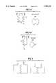
- FIG. 1Ais a drawing showing a typical articulating end of a bone.
- the bone materialis covered on the articulating surface with a cartilaginous cap (shown by cross-hatching).
- FIG. 1Bshows an example of where a defect or injury to the cartilaginous cap occurs (gap in the cross-hatching), and such a defect can be treated directly, enlarged slightly, or sculpted to accept the grafted material by surgical procedures prior to treatment.
- FIG. 1Cshows how the hemostatic barrier (solid black, numbered 1) is placed within the defect in the cartilage cap to inhibit or prevent vascularization into regenerating cartilage, from the underlying bone.
- the chondrocytes to be implanted into the defect cavityare then layered on top of the hemostatic barrier.
- FIG. 2is a drawing showing the treated defect (gap in cross-hatched area) in the cartilaginous cap (cross-hatched area) covered by a cell-free semi-permeable material (solid black, numbered 2) which is used to form a cap/patch or bandage over the defect site.
- This capis fixed in place, either sutured to the edge of the cavity into healthy cartilage, or otherwise attached.
- This capis covering the defective area of the joint into which the cultured chondrocytes/cartilage transplant has been placed.
- FIG. 3Ais a diagram illustrating the differential response to compression and shearing forces by harder and softer cartilage with subsequent zone of demarcation.
- FIG. 3Billustrates the graft site, after the defect has been sculpted to have undulating walls.
- FIG. 3Cillustrates the sculpted graft site with the hemostatic barrier (1), transplanted material (3), and cell-free covering-patch (2) in place within the articular surface cartilage (4).
- FIG. 4Aillustrates one embodiment of the surgical device of the instant invention showing cutting teeth (5) and protruding placement pin (6).
- FIG. 4Billustrates a second embodiment of the surgical device of the instant invention.
- FIG. 5is a diagram illustrating the modified differential response to compression and shearing forces by harder cartilage and softer cartilage after sculpting the graft site.
- FIG. 6Ais an MRI image of a pig knee showing cartilage defect in left (medial) condyle.
- FIG. 6Bis an MRI image of the same pig knee three months after treatment.
- This inventionconcerns the use of certain products that inhibit the formation of vascular tissue, for instance such as capillary loops projecting into the cartilage being established, during the process of autologous transplantation of chondrocytes into defects in the cartilage.
- vascular tissuefor instance such as capillary loops projecting into the cartilage being established, during the process of autologous transplantation of chondrocytes into defects in the cartilage.
- the formation of vascular tissue from the underlying bonewill tend to project into the new cartilage to be formed leading to appearance of cells other than the mesenchymal specialized chondrocytes desired.
- the contaminating cells introduced by the vascularizationmay give rise to encroachment and over-growth into the cartilage to be formed by the implanted chondrocytes.
- One of the types of commercial products which can be used in this inventionis Surgicel® (Ethicon Ltd., UK) which is absorbable after a period of 7-14 days.
- Surgicel®Ethicon Ltd., UK
- the use of this material in the method of the instant inventionis contrary to the normal use of a hemostatic device, such as Surgicel® as it is described in the package insert from Ethicon Ltd.
- a hemostatic material materialwill act like a gel-like artificial coagulate. If red blood cells should be present within the full-thickness defect of articular cartilage that is capped by such a hemostatic barrier, these blood cells will be chemically changed to hematin, and thus rendered unable to induce vascular growth.
- a hemostatic product used as a re-vascularization inhibitor) barrier with or without fibrin adhesivessuch as for example the Surgicel®, is effective for the envisioned method as taught by the instant invention.
- Another part of this inventionis the use of a cell-free component, that is used as a patch covering the defective area of the joint into which the cultured chondrocytes/cartilage are being transplanted, using autologous chondrocytes for the transplantation.
- the method of the inventionalso contemplates the use of suitable allogenic chondrocytes or xenogenic chondrocytes for the repair of a cartilage defect.
- the instant inventionteaches methods for effective repair or treatment of cartilage defects in articular joint bone surfaces which comprises administering an agent or device to block vascular invasion into the cartilage site to be repaired, and also providing for a cell-free barrier which will isolate the repair site and keep transplanted cells in place.
- the instant inventionalso provides for a kit comprising a hemostatic barrier component for insertion into the site to be repaired, such that there is effective inhibition of vascularization into the site to be repaired; and once the chondrocytes to be transplanted are placed into the site to be repaired, a cell-free semi-permeable barrier is capped over the repair site such that the transplanted chondrocytes are held in place, but are still able to gain access to nutrients.
- Certain aspects of the inventionhave been exemplified using an in vitro system to study the behavior of the chondrocytes when in contact with a certain product or a combination of certain products that inhibit the formation of vascular tissue.
- This in vitro testingpredicts the ability of certain tested materials to inhibit vascularization, as will occur in vivo where capillary loops project into the cartilage being established during the process of autologous transplantation of chondrocytes into defects in the cartilage.
- Suitable hemostatic productswill be characterized by having the ability to inhibit the growth, or invasion of vascular tissue, osteocytes, fibroblasts etc. into the developing cartilage.
- a suitable hemostatic materialwill achieve the goal of the method of the instant invention in that vascular and cellular invasion into developing cartilage should be prevented in order to optimize the formation of cartilage and achieve repair of the full-thickness of any defects in the articular cartilage.
- the hemostatic barrierwill be stable for an extended period of time sufficient to allow for full cartilage repair, and then be able to be resorbed or otherwise broken down over time.
- Surgicel® W1912an absorbable hemostat containing oxidized regenerated sterile cellulose; Lot GG3DH, Ethicon Ltd. UK.
- BioGide®a commercially available type I collagen matrix pad; Geistlich Sohne, Switzerland.
- Suitable organic glue materialcan be found commercially, such as for example Tisseel® or Tissucol® (fibrin based adhesive; Immuno AG, Austria), Adhesive Protein (Cat. #A-2707, Sigma Chemical, USA), and Dow Corning Medical Adhesive B (Cat. #895-3, Dow Corning, USA).
- the surgical instruments contemplated by the instant inventioncan be manufactured from metal and/or plastic suitable for making single-use disposable, or multi-use reusable surgical instruments.
- the cutting instrumentmay contain cutting teeth that are fully circular or flat, or anything in between.
- cartilageis a relatively soft material it may be advantageous to manufacture hardened plastic cutting edges which will be able to sculpt cartilage without being able to damage bone.
- Such cutting instrumentscan be manufactured to incorporate openings for administration of fluid, suction removal of cutting debris and fluid, and fiber optic threads for illumination and visualization of the defect site.
- Surgicel®was first treated with a fixative, such as glutaric aldehyde. Briefly, Surgicel® was treated with 0.6% glutaric aldehyde for 1 minute, followed by several washings to eliminate glutaric aldehyde residues that may otherwise be toxic to tissue. Alternatively, the Surgicel® was treated with the fibrin adhesive called Tisseel® prior to treatment with glutaric aldehyde as described in Example 2.
- a fixativesuch as glutaric aldehyde.
- the Surgicel®was also coated with an organic glue, in this example the glue used was Tisseel® but others can also be used. This product, together with the Surgicel® produces a useable barrier for the particular purpose of the invention. Any other hemostat or vascular inhibiting barrier could be used.
- the Tisseel®was mixed as described below.
- the Surgicel®was then coated with Tisseel® by spraying the Surgicel® material on both sides until soaked.
- the Tisseel® (fibrin glue)was then allowed to solidify at room temperature.
- the coated Surgicel®was then placed in 0.6% glutaric aldehyde for 1 minute and then washed with sterile physiological (0.9%) saline.
- the pHwas then adjusted by PBS and/or with NaOH until pH was stable at 7.2 to 7.4. Afterwards the thus treated Surgicel® was then washed in tissue culture medium such as minimum essential medium/F12 with 15 mM Hepes buffer.
- Tisseel®as the fibrin adhesive to coat the Surgicel®.
- fibrin adhesive or gluemay also be applied directly on the bottom of the lesion towards the bone, on which the Surgicel® is glued.
- the in vitro system usedin lieu of in vivo testing, consisted of a NUNCLONTM Delta 6-well sterile disposable plate for cell research work (NUNC, InterMed, Roskilde, Denmark). Each well measures approximately 4 cm in diameter.
- the fibrin adhesivecan be any adhesive which together with the fibrin component will produce a glue that can be tolerated in humans (Ihara, N, et al., Burns Incl. Therm. Inj., 1984, 10, 396).
- the inventionalso anticipates any other glue component that can be used in lieu of the fibrin adhesive.
- Tisseel® or Tissucol®Immuno AG, Vienna, Austria.
- the Tisseel® kitconsists of the following components:
- Tisseel®a lyophilized, virus-inactivated Sealer, containing clottable protein, thereof: fibrinogen, Plasma fibronectin (CIG) and Factor XIII, and Plasminogen.
- the Tisseel® kitcontains a DUPLOJECT® Application System.
- the fibrin adhesive or the two-component sealant using Tisseel® Kitis combined in the following manner according to the Immuno AG product insert sheet:
- Chondrocyteswere grown in minimal essential culture medium containing HAM F12 and 15 mM Hepes buffer and 5 to 7.5% autologous serum in a CO 2 incubator at 37° C. and handled in a Class 100 laboratory at Verigen Europe A/S, Symbion Science Park, Copenhagen, Denmark. Other compositions of culture medium may be used for culturing the chondrocytes.
- the cellswere trypsinized using trypsin EDTA for 5 to 10 minutes and counted using Trypan Blue viability staining in a Burker-Turk chamber. The cell count was adjusted to 7.5 ⁇ 10 5 cells per ml.
- One NUNCLONTM platewas uncovered in the Class 100 laboratory.
- the Surgicel® hemostatic barrierwas cut to a suitable size fitting into the bottom of the well in the NUNCLONTM tissue culture tray. In this case a circle, of a size of approximately 4 cm (but could be of any possible size) and placed under aseptic conditions on the bottom in well in a NUNCLONTM Delia 6-well sterile disposable plate for cell research work (NUNC, InterMed, Roskilde, Denmark).
- the hemostatic barrier to be placed on the bottom of the wellwas pre-treated as described in Example 1. This treatment delays the absorption of the Surgicel significantly.
- This hemostatic barrierwas then washed several times in distilled water and subsequently several times until non-reacted glutaraldehyde was washed out. A small amount of the cell culture medium containing serum was applied to be absorbed into the hemostatic barrier and at the same time keeping the hemostatic barrier wet at the bottom of the well.
- the mediumwas decanted and cold refrigerated 2.5% glutaraldehyde containing 0.1M sodium salt of dimethylarsinic acid, (also called sodium cacodylate, pH is adjusted with HCl to 7.4), was added as fixative for preparation of the cell and supporter (hemostatic barrier) for later preparation for electron microscopy.
- glutaraldehydecontaining 0.1M sodium salt of dimethylarsinic acid, (also called sodium cacodylate, pH is adjusted with HCl to 7.4)
- Chondrocyteswere grown in minimal essential culture medium containing HAM F12 and 15 mM Hepes buffer and 5 to 7.5% autologous serum in a CO 2 incubator at 37° C. and handled in a Class 100 laboratory at Verigen Europe A/S, Symbion Science Park, Copenhagen, Denmark. Other compositions of culture medium may be used for culturing the chondrocytes.
- the cellswere trypsinized using trypsin EDTA for 5 to 10 minutes and counted using Trypan Blue viability staining in a Burker-Turk chamber. The cell count was adjusted to 7.5 ⁇ 10 5 cells per ml.
- One NUNCLONTM platewas uncovered in the Class 100 laboratory.
- the Surgicel®(for use as the hemostatic barrier) was treated with 0.6% glutaric aldehyde for one minute as described in Example 1, and washed with 0.9% sterile sodium chloride solution or, preferably, with a buffer such as a PBS buffer or the culture medium such as MEM/F12, because pH after the glutaric aldehyde treatment is 6.8 and should preferably be 7.0 to 7.5.
- the Tisseel®was applied on both side of the Surgicel® using the DUPLOJECT® system, thus coating both sides of the Surgicel®, the patch intended to be used, with fibrin adhesive.
- the glueis left to dry under aseptic condition for at least 3 to 5 minutes.
- the "coated" hemostatic barrierwas placed on the bottom of the well in a NUNCLONTM Delta 6-well sterile disposable plate for cell research work.
- a small amount of tissue culture medium containing serumwas applied to be absorbed into the hemostatic barrier.
- tissue culture medium containing serumwas placed directly on top of the Hemostat, dispersed over the surface of the hemostatic barrier.
- the platewas then incubated in a CO 2 incubator at 37° C. for 60 minutes.
- An amount of 2 to 5 ml of tissue culture medium containing 5 to 7.5% serumwas carefully added to the well containing the cells avoiding splashing the cells by holding the pipette tip tangential to the side of the well when expelling the medium.
- the platewas incubated for 3 to 7 days with medium change at day 3. At the end of the incubation period the medium was decanted and cold refrigerated 2.5% glutaraldehyde containing 0.1M sodium salt of dimethylarsinic acid, also called sodium cacodylate, pH is adjusted with HCl to 7.4, was added as fixative for preparation of the cell and supporter (hemostatic barrier) for later preparation for electron microscopy.
- Chondrocyteswere grown in minimal essential culture medium containing HAM F12 and 15 mM Hepes buffer and 5 to 7.5% autologous serum in a CO 2 incubator at 37° C. and handled in a Class 100 laboratory at Verigen Europe A/S, Symbion Science Park, Copenhagen, Denmark.
- the cellswere trypsinized using trypsin EDTA for 5 to 10 minutes and counted using Trypan Blue viability staining in a Burker-Turk chamber. The cell count was adjusted to 7.5 ⁇ 10 5 to 2 ⁇ 10 6 cells per ml.
- One NUNCLONTM platewas uncovered in the Class 100 laboratory.
- the Bio-Gide®can be used as a resorbable bilayer membrane which will be used as the patch or bandage covering the defective area of the joint into which the cultured chondrocytes are being transplanted as well as the hemostatic barrier.
- the Bio-Gide®is a pure collagen membrane obtained by standardized, controlled manufacturing processes (by E. D. Geistlich Sohne AG, CH-6110 Wolhusen). The collagen is extracted from veterinary certified pigs and is carefully purified to avoid antigenic reactions, and sterilized in double blisters by ⁇ -irradiation.
- the bilayer membranehas a porous surface and a dense surface. The membrane is made of collagen type I and type III without further cross-linking or chemical treatment.
- the collagenis resorbed within 24 weeks.
- the membraneretains its structural integrity even when wet and it can be fixed by sutures or nails.
- the membranemay also be "glued” using fibrin adhesive such as Tisseel® to the neighboring cartilage or tissue either instead of sutures or together with sutures.
- the Bio-Gide®was un-covered in a class 100 laboratory and placed under aseptic conditions on the bottom of the wells in a NUNCLONTM Delta 6-well sterile disposable plate for cell research work,--either with the porous surface of the bilayer membrane facing up or with the dense surface facing up.
- tissue culture medium containing serumwas placed directly on top of the Bio-Gide®, dispersed either over the porous or the dense surface of the Bio-Gide®.
- the platewas then incubated in a CO 2 incubator at 37° C. for 60 minutes. An amount of 2 to 5 ml of tissue culture medium containing 5 to 7.5% serum was carefully added to the well containing the cells avoiding splashing the cells by holding the pipette tip tangential to the side of the well when expelling the medium.
- the platewas incubated for 3 to 7 days with medium change at day 3. At the end of the incubation period the medium was decanted and cold refrigerated 2.5% glutaraldehyde containing 0.1M sodium salt of dimethylarsinic acid, also called sodium cacodylate, pH is adjusted with HCl to 7.4, was added as fixative for preparation of the cell and the Bio-Gide® supporter with the cells either cultured on the porous surface or the dense surface. The Bio-Gide® patches were then sent for electron microscopy at Department of Pathology, Herlev Hospital, Denmark.
- Chondrocyteswere grown in minimal essential culture medium containing HAM F12 and 15 mM Hepes buffer and 5 to 7.5% autologous serum in a CO 2 incubator at 37° C. and handled in a Class 100 laboratory at Verigen Europe A/S, Symbion Science Park, Copenhagen, Denmark.
- the cellswere trypsinized using trypsin EDTA for 5 to 10 minutes and counted using Trypan Blue viability staining in a Burker-Turk chamber. The cell count was adjusted to 7.5 ⁇ 10 5 to 2 ⁇ 10 6 cells per ml.
- One NUNCLONTM platewas uncovered in the Class 100 laboratory.
- the Bio-Gide® used as a resorbable bilayer membranemay also be used together with an organic glue such as Tisseel® with additional, significantly higher content of Aprotinin than normally found in Tisseel®, as described in the product insert.
- an organic gluesuch as Tisseel® with additional, significantly higher content of Aprotinin than normally found in Tisseel®, as described in the product insert.
- the Tisseel®is applied to the bottom of the well of the NUNCLONTM plate, and allowed to solidify incompletely.
- a collagen patchsuch as a Bio-Gide® is then applied over the Tisseel® and glued to the bottom of the well.
- This combination of Bio-Gide® and Tisseel®is designed to be a hemostatic barrier that will inhibit or prevent development or infiltration of blood vessels into the chondrocyte transplantation area.
- This hybrid collagen patchcan now be used for both as a hemostatic barrier at the bottom of the lesion (most proximal to the surface to be repaired) but also as a support for cartilage formation because the distal surface can be the porous side of the collagen patch and thus encourage infiltration of chondrocytes and cartilage matrix.
- this hybrid collagen patchcan also be used to cover the top of the implant with the collagen porous surface directed down towards the implanted chondrocytes and the barrier forming the top.
- the hybrid collagen patch, with elevated Aprotinin componentmay also be used swithout any organic glue such as Tisseel® and placed within the defect directly, adhering by natural forces.
- the collagen patchcan be used both as the hemostatic barrier, and the cell-free covering of the repair/transplant site, with the porous surfaces of the patches oriented towards the transplanted chondrocytes/cartilage.
- Another variantwould use a collagen patch which consists of type II collagen (ie. from Geistlich Sohne AG, CH-6110 Wolhusen).
- the instant inventionprovides for a hybrid collagen patch where said patch is a collagen matrix with elevated levels of aprotinin component, preferably about 25,000 KIU/ml, in association with an organic matrix glue, where the collagen component is similar to the Bio-Gide® resorbable bilayer material or Type II collagen, and the organic glue is similar to the Tisseel® material.
- the hybrid collagen patchdoes not use any organic glue to adhere to the site of the repair.
- a marginal zonezone of demarcation
- This marginal zonewill be most pronounced if the graft site is prepared for the graft by creating straight, smooth walls cut in a linear fashion. The shearing and compression forces across such a marginal zone (as illustrated in FIG. 3A) will exert great force to dislodge the graft when the graft site is cut in a linear fashion.
- This marginal zone, and differential movement of materials along this zonewill inhibit confluent healing between the grafted material and the surrounding material.
- the graft materialis softer than the surrounding material, however, in some instances of osteoarthritis disease, the surrounding cartilage may in fact be softer than the implanted chondrocytes/cartilage.
- the method of the inventionteaches the use of surgical instruments to sculpt the walls of the graft site such that the walls are non-linear, and thus provide for undulated surface. It is also possible to shape the graft site such that the diameter of the site proximal to the bone surface is of a greater dimension then the opening distal to the bone, and at the surface of the cartilage.
- the preferred embodimentdescribes the sculpting of the walls of the graft site in an fashion similar to a threaded opening for receiving a bolt or screw (as illustrated in FIG. 3B), thus providing mechanical resistance to the compression and or ejection of the grafted material from the graft site which can be described as "male” and "female” threading.
- the surgical instruments contemplated by the instant inventioncan be manufactured from metal and/or plastic suitable for making single-use disposable, or multi-use reusable surgical instruments.
- cartilageis a relatively soft material it may be advantageous to manufacture hardened plastic cutting edges which will be able to sculpt cartilage without being able to damage bone.
- Such cutting instrumentscan be manufactured to incorporate openings for administration of fluid, suction removal of cutting debris and fluid, and fiber optic threads for illumination and visualization of the defect site.
- the base of the instrumentmay have protruding point or pin-like structure which will assist in guiding and placing the instrument in the graft site. Of course such a pin would be designed to minimized damage to the underlying bone.
- the cutting surface of the instrumentmay be single toothed, or multi-toothed, or describe a screw-like pattern such as that in a metal tap used to generate threaded holes in metal parts
- the characteristic required of the cutting instrumentis that the resulting sculpted sides of the graft site is undulated, and non-linear.
- the cutting edge of the instrumentcan be shaped similar to that shown in FIG. 4A, or as in FIG. 4B.
- the cutting edgemaybe flat, or circular in that it wraps around the diameter of the cutting instrument. Many other shapes can be designed to accomplish the purpose of the method of the invention to create an interface which provides for mechanical resistance to differential reaction to compression and shearing forces on the transplanted material and the surrounding material.
- a four month old mixed Yorkshire breed pigwas subjected to general anesthesia and placed on its back. The pig was washed and draped in a surgical suite at Harrington Arthritis Research Center, Phoenix, Ariz. The entire surgical procedure was performed aseptically. The left hind-leg and adjacent abdomen and inguinal area was cleaned with iodine. The knee joint was localized, and the patella localized. A medial incision was performed approximately 3 cm from the posterior part of the patella and the several subcutis, muscle layers and ligaments was cut approximately in order to get access to the medial femoral condyle.
- a lesionwas prepared in the white cartilage on the medial part of the medial condyle, leaving a 0.5 to 1 cm margin to the edge of the cartilage covering the posterior-medial part of the condyle (left condyle, FIG. 6A).
- the 0.5 to 1 cm defectwas placed in a caudal weight bearing part of the medial condyle.
- the entire surgical procedurewas done without tourniquet on the left femur. The different layers and skin was sutured appropriately.
- Adhesive ProteinA-2707, Sigma Chemical, USA
- Adhesive ProteinA-2707, Sigma Chemical, USA
- a second BioGide®was cut somewhat bigger in circumference than the lesion and was placed with dense side up (thus the porous side down towards the graft) as described above.
- This non-cellular covering-padwas then sutured over the cavity, leaving one edge open, where the chondrocyte to be explanted could be injected.
- the surrounding part of the edge of the padwas covered with the second glue, Dow Corning Medical Adhesive B (Cat. #895-3, Dow Corning, USA).
- This second gluedried much faster and more efficiently than the first glue. It was found that during this particular procedure, the first glue had not dried sufficiently to hold the hemostatic barrier in place when suturing of the cap was attempted. Thus the main barrier formed on the proximal surface of the graft site was by the glue itself.
- the chondrocyte cell suspension(about 0.6 ml) was drawn up into the barrel of the syringe.
- a 23 gauge short needlewas switched for the 16 gauge needle, and the cell suspension was injected under the sutured covering-patch into the graft site (about 10 ⁇ 10 6 cells).
- the open edge of the capwas then glued prior to removal of the needle, and the needle carefully withdrawn. No leakage of cells was seen.
- the woundwas sutured and as above, no tourniquet was used, no bleeding was observed.
- the final skin layerswere sutured. No protrusion of the skin occurred after suturing, which indicates that there was no hematoma. Postoperative recovery was uneventful.
- FIG. 6Ais an MRI image of a pig knee showing the cartilage defect created in the knee (left condyle, the medial condyle), and FIG. 6B is an MRI image of the same pig knee three months after treatment showing repair of the defect.
- kitscomprising the components useful for practicing the method of the invention, will allow for the convenient practice of the method of the invention in a surgical setting.
- a kit of the inventionwill provide sterile components suitable for easy use in the surgical environment, and will provide a suitable hemostatic barrier, suitable covering patch, and if needed organic glue.
- a kit of the inventionmay also provide sterile, cell-free matrix material suitable for supporting autologous chondrocytes that are to be implanted into an articular joint surface defect.
- a kit of the inventioncontains a Surgicel® hemostatic barrier and a Bio-Gide® covering patch with suitable coating of Tisseel® organic glue, where the Surgicel® and Bio-Gide® have been treated according to the teachings of the invention to increase the time till resorption.
- Tisseel®is pre-coated
- the Tisseel®is supplemented with additional aprotinin to increase time till resorption.
- the hemostatic barrier and covering-patchare both a semi-permeable collagen matrix which is treated to extend the time till resorption of the material. It is also possible to provide Tisseel® glue in enhanced form as a separate component to be applied as needed because of the inherent variability and unique circumstances every repair/transplantation procedure will encounter.
- a further embodiment of the kitwill include a surgical instrument as described in Example 7 above.
Landscapes
- Health & Medical Sciences (AREA)
- Transplantation (AREA)
- Life Sciences & Earth Sciences (AREA)
- Animal Behavior & Ethology (AREA)
- Oral & Maxillofacial Surgery (AREA)
- Orthopedic Medicine & Surgery (AREA)
- Engineering & Computer Science (AREA)
- Biomedical Technology (AREA)
- Heart & Thoracic Surgery (AREA)
- Vascular Medicine (AREA)
- Veterinary Medicine (AREA)
- Cardiology (AREA)
- General Health & Medical Sciences (AREA)
- Public Health (AREA)
- Rheumatology (AREA)
- Materials For Medical Uses (AREA)
- Prostheses (AREA)
- Medicines Containing Material From Animals Or Micro-Organisms (AREA)
- Surgical Instruments (AREA)
- Pharmaceuticals Containing Other Organic And Inorganic Compounds (AREA)
- Acyclic And Carbocyclic Compounds In Medicinal Compositions (AREA)
- Agricultural Chemicals And Associated Chemicals (AREA)
Abstract
Description
Claims (6)
Priority Applications (70)
| Application Number | Priority Date | Filing Date | Title |
|---|---|---|---|
| US08/857,090US5989269A (en) | 1996-08-30 | 1997-05-15 | Method, instruments and kit for autologous transplantation |
| TR2001/02871TTR200102871T2 (en) | 1996-08-30 | 1997-08-29 | Methods, tools and instrument sets for autologous transplantation |
| CA002264138ACA2264138C (en) | 1996-08-30 | 1997-08-29 | Method, instruments and kit for autologous transplantation |
| AT97939677TATE263524T1 (en) | 1996-08-30 | 1997-08-29 | NIT FOR CHONDROCYTE TRANSPLANTATION IN A JOINT |
| DE69728569TDE69728569T2 (en) | 1996-08-30 | 1997-08-29 | Kit for chondrocyte transplantation in a joint |
| IL128580AIL128580A (en) | 1996-08-30 | 1997-08-29 | Methods for treating an articulating joint surface of a joint surface cartilage of a non-human mammal and kits for chondrocyte transplantation |
| SG200101255ASG119137A1 (en) | 1996-08-30 | 1997-08-29 | Method instruments and kit for autologous transplantation |
| PT97939677TPT1006950E (en) | 1996-08-30 | 1997-08-29 | KIT FOR THE TRANSPLANTATION OF CONDROCITATION IN A JOINT |
| KR1019997001597AKR100686412B1 (en) | 1996-08-30 | 1997-08-29 | Cartilage repair structures |
| TR2001/02883TTR200102883T2 (en) | 1996-08-30 | 1997-08-29 | Methods, tools and instrument sets for autologous transplantation. |
| CZ0058799ACZ297248B6 (en) | 1996-08-30 | 1997-08-29 | Implantable element for repairing defects of joint surface cartilage |
| CNB97199207XACN1200656C (en) | 1996-08-30 | 1997-08-29 | A cartilage repair construct for repairing articular cartilage defects |
| AU41710/97AAU731162B2 (en) | 1996-08-30 | 1997-08-29 | Method, instruments and kit for autologous transplantation |
| CA002419644ACA2419644C (en) | 1996-08-30 | 1997-08-29 | Method and kit for the transplantation of chondrocyte cells |
| DK97939677TDK1006950T3 (en) | 1996-08-30 | 1997-08-29 | Set for chondrocyte transplantation in a joint |
| AT03023272TATE428376T1 (en) | 1996-08-30 | 1997-08-29 | METHOD FOR PRODUCING AN AUTOLOGOUS TRANSPLANTATION KIT |
| CNA031553281ACN1500447A (en) | 1996-08-30 | 1997-08-29 | Method, instruments, and kit for autologous transplantation |
| NZ518474ANZ518474A (en) | 1996-08-30 | 1997-08-29 | Method, instruments and kit for autologous transplantation comprising hemostatic barrier and a covering patch |
| SI9730789TSI1384452T1 (en) | 1996-08-30 | 1997-08-29 | Method for providing a kit for autologous transplantation |
| RU99106519/14ARU2214197C2 (en) | 1996-08-30 | 1997-08-29 | Method and set for performing autotransplantation |
| EP97939677AEP1006950B1 (en) | 1996-08-30 | 1997-08-29 | Kit for chondrocytes transplantation in a joint |
| KR1020027011281AKR20030097603A (en) | 1996-08-30 | 1997-08-29 | Method, instruments and kit for autologous transplantation |
| AT01126359TATE256443T1 (en) | 1996-08-30 | 1997-08-29 | SUPPORT STRUCTURE FOR CHONDROCYTE TRANSPLANTATION TO REPAIR CARTILAGES AND JOINTS |
| SK240-99ASK285075B6 (en) | 1996-08-30 | 1997-08-29 | Kit for chondrocytes transplantation in a joint |
| HU0400358AHU225952B1 (en) | 1996-08-30 | 1997-08-29 | Plaster for treating of joint surface cartilage |
| MXPA99001794AMXPA99001794A (en) | 1996-08-30 | 1997-08-29 | Method, instruments and kit for autologous transplantation. |
| ES01126359TES2211722T3 (en) | 1996-08-30 | 1997-08-29 | SUPPORT FOR THE TRANSFER OF CONDROCITS FOR THE REPAIR OF CARTILAGO AND ARTICULATIONS. |
| ES03023272TES2325206T3 (en) | 1996-08-30 | 1997-08-29 | METHOD FOR THE PROVISION OF A KIT FOR AUTOGENOUS TRANSPLANTATION. |
| PT01126359TPT1181908E (en) | 1996-08-30 | 1997-08-29 | SUPPORT FOR THE TRANSPLANTATION OF CONDOCCITUS IN CARTILAGE AND PREPARATION OF ARTICULACOESC |
| SI9730655TSI1006950T1 (en) | 1996-08-30 | 1997-08-29 | Kit for chondrocytes transplantation in a joint |
| HK00104418.6AHK1025035B (en) | 1996-08-30 | 1997-08-29 | A cartilage repair structure for repair of a defect in articular cartilage |
| DE69726896TDE69726896T2 (en) | 1996-08-30 | 1997-08-29 | Support structure for chondrocyte transplantation for the repair of cartilage and joints |
| NZ334400ANZ334400A (en) | 1996-08-30 | 1997-08-29 | Method, instruments and kit for autologous transplantation comprising hemostatic barrier and a covering patch |
| DE29724585UDE29724585U1 (en) | 1996-08-30 | 1997-08-29 | Patch and cartilage recovery kit |
| PCT/US1997/015258WO1998008469A2 (en) | 1996-08-30 | 1997-08-29 | Method, instruments and kit for autologous transplantation |
| PL97331834APL331834A1 (en) | 1996-08-30 | 1997-08-29 | Method of instruments for and set of means for making transplantation between different portions of the same body |
| JP51196798AJP2002502272A (en) | 1996-08-30 | 1997-08-29 | Methods, devices and kits for autografting |
| EP03023272AEP1384452B1 (en) | 1996-08-30 | 1997-08-29 | Method for providing a kit for autologous transplantation |
| PT03023272TPT1384452E (en) | 1996-08-30 | 1997-08-29 | Method for providing a kit for autologous transplantation |
| TR1999/00437TTR199900437T2 (en) | 1996-08-30 | 1997-08-29 | Set of methods, instruments and instruments for autologous transplantation |
| BR9711967-9ABR9711967A (en) | 1996-08-30 | 1997-08-29 | Processes for the effective treatment of articulating joint surface cartilage and for the transplantation of chondrocytes in a suitable matrix, kit for chondrocyte transplantation and for the practice of processes, and, surgical instrument. |
| DE69739363TDE69739363D1 (en) | 1996-08-30 | 1997-08-29 | Method for producing a kit for autologous transplantation |
| DK01126359TDK1181908T3 (en) | 1996-08-30 | 1997-08-29 | Chondrocyte transplant support structure for cartilage and joint repair |
| EP04008116AEP1459709A1 (en) | 1996-08-30 | 1997-08-29 | Method, instruments and kit for autologous transplantation |
| ES97939677TES2218697T3 (en) | 1996-08-30 | 1997-08-29 | KIT FOR THE TRANSPLANTATION OF CONDROCITS IN AN ARTICULATION. |
| DK03023272TDK1384452T3 (en) | 1996-08-30 | 1997-08-29 | Method of Preparing a Kit for Autologous Transplantation |
| EP01126359AEP1181908B1 (en) | 1996-08-30 | 1997-08-29 | Support for chondrocyte transplantation in cartilage and joint repair |
| HU0002980AHUP0002980A3 (en) | 1996-08-30 | 1997-08-29 | Treatment of joint surface cartilage using condrocyte transplantation grafting |
| NZ508145ANZ508145A (en) | 1996-08-30 | 1997-08-29 | Use of chondrocytes for treating articulating joint surface cartilage using asurgical instrument having a handle with an undulating cutting surface |
| SG200101256ASG120854A1 (en) | 1996-08-30 | 1997-08-29 | Method, instruments and kit for autologous transplantation |
| NO990933ANO990933D0 (en) | 1996-08-30 | 1999-02-26 | Procedure, instruments and equipment for autologous transplantation |
| US09/320,246US6283980B1 (en) | 1996-08-30 | 1999-05-26 | Method, instruments, and kit for autologous transplantation |
| US09/690,252US6379367B1 (en) | 1996-08-30 | 2000-10-17 | Method instruments and kit for autologous transplantation |
| US09/970,323US6569172B2 (en) | 1996-08-30 | 2001-10-03 | Method, instruments, and kit for autologous transplantation |
| US10/055,105US6592598B2 (en) | 1996-08-30 | 2002-01-23 | Method, instruments, and kit for autologous transplantation |
| RU2002103570/14ARU2002103570A (en) | 1996-08-30 | 2002-02-08 | METHOD, DEVICE AND KIT FOR AUTOTRANSPLANTATION |
| US10/091,006US6599300B2 (en) | 1996-08-30 | 2002-03-04 | Method, instruments, and kit for autologous transplantation |
| US10/090,922US6592599B2 (en) | 1996-08-30 | 2002-03-05 | Method, instruments, and kit for autologous transplantation |
| US10/093,129US6599301B2 (en) | 1996-08-30 | 2002-03-06 | Method, instruments, and kit for autologous transplantation |
| US10/121,249US20020173806A1 (en) | 1996-08-30 | 2002-04-12 | Method for autologous transplantation |
| DK200200177UDK200200177U1 (en) | 1996-08-30 | 2002-06-04 | Patches, as well as kits comprising such patches for autologous transplantation |
| DK200200178UDK200200178U1 (en) | 1996-08-30 | 2002-06-04 | Patches, as well as kits comprising such patches for autologous transplantation |
| JP2002316043AJP2003159266A (en) | 1996-08-30 | 2002-10-30 | Method for autotransplantation, instrument and kit |
| US10/397,404US7137989B2 (en) | 1996-08-30 | 2003-03-26 | Method, instruments, and kit for autologous transplantation |
| US10/444,391US7048750B2 (en) | 1996-08-30 | 2003-05-23 | Method, instruments, and kits for autologous transplantation |
| HK04105480.2AHK1063003B (en) | 1996-08-30 | 2004-07-23 | Method for providing a kit for autologous transplantation |
| US10/962,868US20060025786A1 (en) | 1996-08-30 | 2004-10-12 | Method for autologous transplantation |
| US11/375,183US20060167483A1 (en) | 1996-08-30 | 2006-03-14 | Method, instruments, and kit for autologous transplantation |
| US11/375,180US20060195122A1 (en) | 1996-08-30 | 2006-03-14 | Method, instruments, and kit for autologous transplantation |
| JP2007155993AJP2007296371A (en) | 1996-08-30 | 2007-06-13 | Method, instrument and kit for autologous transplantation |
Applications Claiming Priority (2)
| Application Number | Priority Date | Filing Date | Title |
|---|---|---|---|
| US08/704,891US5759190A (en) | 1996-08-30 | 1996-08-30 | Method and kit for autologous transplantation |
| US08/857,090US5989269A (en) | 1996-08-30 | 1997-05-15 | Method, instruments and kit for autologous transplantation |
Related Parent Applications (1)
| Application Number | Title | Priority Date | Filing Date |
|---|---|---|---|
| US08/704,891Continuation-In-PartUS5759190A (en) | 1996-08-30 | 1996-08-30 | Method and kit for autologous transplantation |
Related Child Applications (2)
| Application Number | Title | Priority Date | Filing Date |
|---|---|---|---|
| US09/088,142ContinuationUS6120514A (en) | 1996-08-30 | 1998-06-01 | Method and kit for autologous transplantation |
| US09/320,246ContinuationUS6283980B1 (en) | 1996-08-30 | 1999-05-26 | Method, instruments, and kit for autologous transplantation |
Publications (1)
| Publication Number | Publication Date |
|---|---|
| US5989269Atrue US5989269A (en) | 1999-11-23 |
Family
ID=27107401
Family Applications (9)
| Application Number | Title | Priority Date | Filing Date |
|---|---|---|---|
| US08/857,090Expired - LifetimeUS5989269A (en) | 1996-08-30 | 1997-05-15 | Method, instruments and kit for autologous transplantation |
| US09/320,246Expired - LifetimeUS6283980B1 (en) | 1996-08-30 | 1999-05-26 | Method, instruments, and kit for autologous transplantation |
| US09/690,252Expired - LifetimeUS6379367B1 (en) | 1996-08-30 | 2000-10-17 | Method instruments and kit for autologous transplantation |
| US10/055,105Expired - LifetimeUS6592598B2 (en) | 1996-08-30 | 2002-01-23 | Method, instruments, and kit for autologous transplantation |
| US10/091,006Expired - LifetimeUS6599300B2 (en) | 1996-08-30 | 2002-03-04 | Method, instruments, and kit for autologous transplantation |
| US10/090,922Expired - LifetimeUS6592599B2 (en) | 1996-08-30 | 2002-03-05 | Method, instruments, and kit for autologous transplantation |
| US10/093,129Expired - LifetimeUS6599301B2 (en) | 1996-08-30 | 2002-03-06 | Method, instruments, and kit for autologous transplantation |
| US10/444,391Expired - Fee RelatedUS7048750B2 (en) | 1996-08-30 | 2003-05-23 | Method, instruments, and kits for autologous transplantation |
| US11/375,180AbandonedUS20060195122A1 (en) | 1996-08-30 | 2006-03-14 | Method, instruments, and kit for autologous transplantation |
Family Applications After (8)
| Application Number | Title | Priority Date | Filing Date |
|---|---|---|---|
| US09/320,246Expired - LifetimeUS6283980B1 (en) | 1996-08-30 | 1999-05-26 | Method, instruments, and kit for autologous transplantation |
| US09/690,252Expired - LifetimeUS6379367B1 (en) | 1996-08-30 | 2000-10-17 | Method instruments and kit for autologous transplantation |
| US10/055,105Expired - LifetimeUS6592598B2 (en) | 1996-08-30 | 2002-01-23 | Method, instruments, and kit for autologous transplantation |
| US10/091,006Expired - LifetimeUS6599300B2 (en) | 1996-08-30 | 2002-03-04 | Method, instruments, and kit for autologous transplantation |
| US10/090,922Expired - LifetimeUS6592599B2 (en) | 1996-08-30 | 2002-03-05 | Method, instruments, and kit for autologous transplantation |
| US10/093,129Expired - LifetimeUS6599301B2 (en) | 1996-08-30 | 2002-03-06 | Method, instruments, and kit for autologous transplantation |
| US10/444,391Expired - Fee RelatedUS7048750B2 (en) | 1996-08-30 | 2003-05-23 | Method, instruments, and kits for autologous transplantation |
| US11/375,180AbandonedUS20060195122A1 (en) | 1996-08-30 | 2006-03-14 | Method, instruments, and kit for autologous transplantation |
Country Status (26)
| Country | Link |
|---|---|
| US (9) | US5989269A (en) |
| EP (4) | EP1181908B1 (en) |
| JP (3) | JP2002502272A (en) |
| KR (2) | KR20030097603A (en) |
| CN (2) | CN1200656C (en) |
| AT (3) | ATE256443T1 (en) |
| AU (1) | AU731162B2 (en) |
| BR (1) | BR9711967A (en) |
| CA (2) | CA2264138C (en) |
| CZ (1) | CZ297248B6 (en) |
| DE (4) | DE69726896T2 (en) |
| DK (5) | DK1384452T3 (en) |
| ES (3) | ES2218697T3 (en) |
| HU (2) | HU225952B1 (en) |
| IL (1) | IL128580A (en) |
| MX (1) | MXPA99001794A (en) |
| NO (1) | NO990933D0 (en) |
| NZ (3) | NZ508145A (en) |
| PL (1) | PL331834A1 (en) |
| PT (3) | PT1006950E (en) |
| RU (2) | RU2214197C2 (en) |
| SG (2) | SG119137A1 (en) |
| SI (2) | SI1006950T1 (en) |
| SK (1) | SK285075B6 (en) |
| TR (3) | TR199900437T2 (en) |
| WO (1) | WO1998008469A2 (en) |
Cited By (113)
| Publication number | Priority date | Publication date | Assignee | Title |
|---|---|---|---|---|
| WO2001008610A1 (en) | 1999-08-02 | 2001-02-08 | Verigen Transplantation Service International Ag | Kit for chondrocyte cell transplantation |
| US6283980B1 (en)* | 1996-08-30 | 2001-09-04 | Verigen Transplantation Services Internt'l | Method, instruments, and kit for autologous transplantation |
| US20020045940A1 (en)* | 1998-08-14 | 2002-04-18 | Bruno Giannetti | Methods, instruments and materials for chondrocyte cell transplantation |
| US20020116063A1 (en)* | 1999-08-02 | 2002-08-22 | Bruno Giannetti | Kit for chondrocyte cell transplantation |
| US20020119177A1 (en)* | 2000-12-21 | 2002-08-29 | Bowman Steven M. | Reinforced foam implants with enhanced integrity for soft tissue repair and regeneration |
| US20020127265A1 (en)* | 2000-12-21 | 2002-09-12 | Bowman Steven M. | Use of reinforced foam implants with enhanced integrity for soft tissue repair and regeneration |
| US20020173806A1 (en)* | 1996-08-30 | 2002-11-21 | Verigen Transplantation Service International (Vtsi) Ag | Method for autologous transplantation |
| US20030095994A1 (en)* | 2001-11-20 | 2003-05-22 | Peter Geistlich | Resorbable extracellular matrix containing collagen I and collagen II for reconstruction of cartilage |
| US20030120276A1 (en)* | 2000-05-01 | 2003-06-26 | Tallarida Steven J. | System and method for joint resurface repair |
| US20030135209A1 (en)* | 1999-12-03 | 2003-07-17 | Bahaa Seedhom | Fixation technology |
| US20030144197A1 (en)* | 2001-09-24 | 2003-07-31 | Zheng Ming Hao | Autologous growth factor cocktail composition, method of production and use |
| US20030147935A1 (en)* | 2000-12-21 | 2003-08-07 | Ethicon, Inc. | Use of reinforced foam implants with enhanced integrity for soft tissue repair and regeneration |
| US20030177026A1 (en)* | 1996-05-24 | 2003-09-18 | Purcell Daniel S. | Automated and independently accessible inventory information exchange system |
| US20030180263A1 (en)* | 2002-02-21 | 2003-09-25 | Peter Geistlich | Resorbable extracellular matrix for reconstruction of bone |
| US20030191107A1 (en)* | 2002-01-22 | 2003-10-09 | Pfizer Inc. | 3-(Imidazolyl)-2-aminopropanoic acids |
| US20030193104A1 (en)* | 2000-12-21 | 2003-10-16 | Melican Mora Carolynne | Reinforced tissue implants and methods of manufacture and use |
| US20040037812A1 (en)* | 2002-05-01 | 2004-02-26 | Verigen Ag | Injectable chondrocyte implant |
| US20040039399A1 (en)* | 2002-05-16 | 2004-02-26 | Sverker Norrby | Kit and method in eye surgery |
| US6713085B2 (en) | 2001-04-27 | 2004-03-30 | Ed. Geistlich Soehne Ag Fuer Chemische Industrie | Method and membrane for mucosa regeneration |
| US20040136968A1 (en)* | 2002-09-27 | 2004-07-15 | Verigen Ag | Autologous cells on a support matrix for tissue repair |
| US20040151705A1 (en)* | 2002-03-22 | 2004-08-05 | Shuichi Mizuno | Neo-cartilage constructs and a method for preparation thereof |
| US20040191900A1 (en)* | 2002-03-22 | 2004-09-30 | Shuichi Mizuno | Method for growth and de novo formation of a superficial cartilage layer in situ and for repair of damaged cartilage using neo-cartilage constructs |
| EP1278460A4 (en)* | 2000-05-01 | 2004-10-13 | Arthro Surface Inc | System and method for joint resurface repair |
| US20050186673A1 (en)* | 1995-02-22 | 2005-08-25 | Ed. Geistlich Soehne Ag Fuer Chemistrie Industrie | Collagen carrier of therapeutic genetic material, and method |
| US20050232967A1 (en) | 2004-04-20 | 2005-10-20 | Kladakis Stephanie M | Nonwoven tissue scaffold |
| US20050276760A1 (en)* | 2004-06-09 | 2005-12-15 | Lornamead Brands, Inc. | Tooth for whitening products and methods of making the same |
| US20060025786A1 (en)* | 1996-08-30 | 2006-02-02 | Verigen Transplantation Service International (Vtsi) Ag | Method for autologous transplantation |
| WO2006022671A1 (en)* | 2004-07-30 | 2006-03-02 | Histogenics Corporation | Method for in situ repair of injured, damaged, diseased or aged articular cartilage using neo-cartilage constructs and a method for preparation thereof |
| US20060111778A1 (en)* | 2004-10-29 | 2006-05-25 | Michalow Alexander E | Methods of promoting healing of cartilage defects and method of causing stem cells to differentiate by the articular chondrocyte pathway |
| US20060159668A1 (en)* | 1997-10-10 | 2006-07-20 | Ed. Geistlich Soehne Ag Fuer Chemistrie Industrie | Resorbable extracellular matrix for reconstruction of bone |
| US20060229726A1 (en)* | 2000-05-01 | 2006-10-12 | Ek Steven W | Articular surface implant |
| US7160333B2 (en) | 2000-08-04 | 2007-01-09 | Depuy Orthopaedics, Inc. | Reinforced small intestinal submucosa |
| US7163563B2 (en) | 2001-07-16 | 2007-01-16 | Depuy Products, Inc. | Unitary surgical device and method |
| US20070031388A1 (en)* | 1997-10-10 | 2007-02-08 | Ed. Geistlich Soehne Ag Fuer Chemische Industrie | Cell-charged multi-layer collagen membrane |
| US20070073394A1 (en)* | 2003-10-28 | 2007-03-29 | Xiros Plc | Repair of damaged tissue on a bone site |
| US7201917B2 (en) | 2001-07-16 | 2007-04-10 | Depuy Products, Inc. | Porous delivery scaffold and method |
| US7316822B2 (en) | 2003-11-26 | 2008-01-08 | Ethicon, Inc. | Conformable tissue repair implant capable of injection delivery |
| US7354627B2 (en) | 2004-12-22 | 2008-04-08 | Depuy Products, Inc. | Method for organizing the assembly of collagen fibers and compositions formed therefrom |
| US7361195B2 (en) | 2001-07-16 | 2008-04-22 | Depuy Products, Inc. | Cartilage repair apparatus and method |
| US20080107710A1 (en)* | 1997-10-10 | 2008-05-08 | Ed. Geistlich Soehne Ag Fuer Chemistrie Industrie | Collagen carrier of therapeutic genetic material and method |
| US7488348B2 (en) | 2003-05-16 | 2009-02-10 | Musculoskeletal Transplant Foundation | Cartilage allograft plug |
| US7510558B2 (en) | 2000-05-01 | 2009-03-31 | Arthrosurface, Inc. | System and method for joint resurface repair |
| US7513866B2 (en) | 2004-10-29 | 2009-04-07 | Depuy Products, Inc. | Intestine processing device and associated method |
| US7569233B2 (en) | 2004-05-04 | 2009-08-04 | Depuy Products, Inc. | Hybrid biologic-synthetic bioabsorbable scaffolds |
| US20090214614A1 (en)* | 2005-09-02 | 2009-08-27 | Interface Biotech A/S | Method for Cell Implantation |
| US7595062B2 (en) | 2005-07-28 | 2009-09-29 | Depuy Products, Inc. | Joint resurfacing orthopaedic implant and associated method |
| US7618462B2 (en) | 2000-05-01 | 2009-11-17 | Arthrosurface Incorporated | System and method for joint resurface repair |
| US20100040667A1 (en)* | 2006-09-07 | 2010-02-18 | Ed. Geistlich Soehne Ag Fuer Chemische Industrie | Method of treating bone cancer |
| US7678151B2 (en) | 2000-05-01 | 2010-03-16 | Ek Steven W | System and method for joint resurface repair |
| US7815926B2 (en) | 2005-07-11 | 2010-10-19 | Musculoskeletal Transplant Foundation | Implant for articular cartilage repair |
| US7819918B2 (en) | 2001-07-16 | 2010-10-26 | Depuy Products, Inc. | Implantable tissue repair device |
| US7824701B2 (en) | 2002-10-18 | 2010-11-02 | Ethicon, Inc. | Biocompatible scaffold for ligament or tendon repair |
| US7828853B2 (en) | 2004-11-22 | 2010-11-09 | Arthrosurface, Inc. | Articular surface implant and delivery system |
| US7837740B2 (en) | 2007-01-24 | 2010-11-23 | Musculoskeletal Transplant Foundation | Two piece cancellous construct for cartilage repair |
| US20100322908A1 (en)* | 2008-02-29 | 2010-12-23 | Hanne Everland | Compositions and methods for augmentation and regeneration of living tissue in a subject |
| US7871440B2 (en) | 2006-12-11 | 2011-01-18 | Depuy Products, Inc. | Unitary surgical device and method |
| US20110014267A1 (en)* | 2008-02-29 | 2011-01-20 | Hanne Everland | Biosynthetic cartilaginous matrix and methods for their production |
| US7896883B2 (en) | 2000-05-01 | 2011-03-01 | Arthrosurface, Inc. | Bone resurfacing system and method |
| US7896885B2 (en) | 2002-12-03 | 2011-03-01 | Arthrosurface Inc. | Retrograde delivery of resurfacing devices |
| US7901457B2 (en) | 2003-05-16 | 2011-03-08 | Musculoskeletal Transplant Foundation | Cartilage allograft plug |
| USRE42208E1 (en) | 2003-04-29 | 2011-03-08 | Musculoskeletal Transplant Foundation | Glue for cartilage repair |
| US7901461B2 (en) | 2003-12-05 | 2011-03-08 | Ethicon, Inc. | Viable tissue repair implants and methods of use |
| US7901408B2 (en) | 2002-12-03 | 2011-03-08 | Arthrosurface, Inc. | System and method for retrograde procedure |
| US7914808B2 (en) | 2001-07-16 | 2011-03-29 | Depuy Products, Inc. | Hybrid biologic/synthetic porous extracellular matrix scaffolds |
| US7914545B2 (en) | 2002-12-03 | 2011-03-29 | Arthrosurface, Inc | System and method for retrograde procedure |
| US7951163B2 (en) | 2003-11-20 | 2011-05-31 | Arthrosurface, Inc. | Retrograde excision system and apparatus |
| US8012205B2 (en) | 2001-07-16 | 2011-09-06 | Depuy Products, Inc. | Cartilage repair and regeneration device |
| US20110218635A1 (en)* | 2008-07-10 | 2011-09-08 | Andrew Arthur Amis | Modular knee implants |
| US8016867B2 (en) | 1999-07-23 | 2011-09-13 | Depuy Mitek, Inc. | Graft fixation device and method |
| US8025896B2 (en) | 2001-07-16 | 2011-09-27 | Depuy Products, Inc. | Porous extracellular matrix scaffold and method |
| US8092529B2 (en) | 2001-07-16 | 2012-01-10 | Depuy Products, Inc. | Meniscus regeneration device |
| US8177841B2 (en) | 2000-05-01 | 2012-05-15 | Arthrosurface Inc. | System and method for joint resurface repair |
| US8221780B2 (en) | 2004-04-20 | 2012-07-17 | Depuy Mitek, Inc. | Nonwoven tissue scaffold |
| US8226715B2 (en) | 2003-06-30 | 2012-07-24 | Depuy Mitek, Inc. | Scaffold for connective tissue repair |
| US8292968B2 (en) | 2004-10-12 | 2012-10-23 | Musculoskeletal Transplant Foundation | Cancellous constructs, cartilage particles and combinations of cancellous constructs and cartilage particles |
| US8337537B2 (en) | 2001-07-16 | 2012-12-25 | Depuy Products, Inc. | Device from naturally occurring biologically derived materials |
| US8361159B2 (en) | 2002-12-03 | 2013-01-29 | Arthrosurface, Inc. | System for articular surface replacement |
| US8366787B2 (en) | 2000-08-04 | 2013-02-05 | Depuy Products, Inc. | Hybrid biologic-synthetic bioabsorbable scaffolds |
| US8388624B2 (en) | 2003-02-24 | 2013-03-05 | Arthrosurface Incorporated | Trochlear resurfacing system and method |
| US8435551B2 (en) | 2007-03-06 | 2013-05-07 | Musculoskeletal Transplant Foundation | Cancellous construct with support ring for repair of osteochondral defects |
| US8449561B2 (en) | 1999-07-23 | 2013-05-28 | Depuy Mitek, Llc | Graft fixation device combination |
| US8480757B2 (en) | 2005-08-26 | 2013-07-09 | Zimmer, Inc. | Implants and methods for repair, replacement and treatment of disease |
| US8497121B2 (en) | 2006-12-20 | 2013-07-30 | Zimmer Orthobiologics, Inc. | Method of obtaining viable small tissue particles and use for tissue repair |
| US8518433B2 (en) | 2003-12-11 | 2013-08-27 | Zimmer, Inc. | Method of treating an osteochondral defect |
| US8523872B2 (en) | 2002-12-03 | 2013-09-03 | Arthrosurface Incorporated | Tibial resurfacing system |
| US20130296657A1 (en)* | 2012-05-03 | 2013-11-07 | Covidien Lp | Methods of using light to repair hernia defects |
| US8657881B2 (en) | 2004-04-20 | 2014-02-25 | Depuy Mitek, Llc | Meniscal repair scaffold |
| US8895045B2 (en) | 2003-03-07 | 2014-11-25 | Depuy Mitek, Llc | Method of preparation of bioabsorbable porous reinforced tissue implants and implants thereof |
| US9066716B2 (en) | 2011-03-30 | 2015-06-30 | Arthrosurface Incorporated | Suture coil and suture sheath for tissue repair |
| US9138318B2 (en) | 2007-04-12 | 2015-09-22 | Zimmer, Inc. | Apparatus for forming an implant |
| US9283076B2 (en) | 2009-04-17 | 2016-03-15 | Arthrosurface Incorporated | Glenoid resurfacing system and method |
| US9358029B2 (en) | 2006-12-11 | 2016-06-07 | Arthrosurface Incorporated | Retrograde resection apparatus and method |
| US9421304B2 (en) | 2007-07-03 | 2016-08-23 | Histogenics Corporation | Method for improvement of differentiation of mesenchymal stem cells using a double-structured tissue implant |
| US9468448B2 (en) | 2012-07-03 | 2016-10-18 | Arthrosurface Incorporated | System and method for joint resurfacing and repair |
| US9492200B2 (en) | 2013-04-16 | 2016-11-15 | Arthrosurface Incorporated | Suture system and method |
| US20160331539A1 (en)* | 2015-05-12 | 2016-11-17 | Elwha Llc | Modifiable implants |
| US9511171B2 (en) | 2002-10-18 | 2016-12-06 | Depuy Mitek, Llc | Biocompatible scaffolds with tissue fragments |
| US9662126B2 (en) | 2009-04-17 | 2017-05-30 | Arthrosurface Incorporated | Glenoid resurfacing system and method |
| US9687590B2 (en) | 2007-07-03 | 2017-06-27 | Histogenics Corporation | Double-structured tissue implant and a method for preparation and use thereof |
| US9701940B2 (en) | 2005-09-19 | 2017-07-11 | Histogenics Corporation | Cell-support matrix having narrowly defined uniformly vertically and non-randomly organized porosity and pore density and a method for preparation thereof |
| US9861492B2 (en) | 2014-03-07 | 2018-01-09 | Arthrosurface Incorporated | Anchor for an implant assembly |
| US9993326B2 (en) | 2007-07-03 | 2018-06-12 | Histogenics Corporation | Method for use of a double-structured tissue implant for treatment of tissue defects |
| US10077420B2 (en) | 2014-12-02 | 2018-09-18 | Histogenics Corporation | Cell and tissue culture container |
| US10167447B2 (en) | 2012-12-21 | 2019-01-01 | Zimmer, Inc. | Supports and methods for promoting integration of cartilage tissue explants |
| US10583220B2 (en) | 2003-08-11 | 2020-03-10 | DePuy Synthes Products, Inc. | Method and apparatus for resurfacing an articular surface |
| US10624752B2 (en) | 2006-07-17 | 2020-04-21 | Arthrosurface Incorporated | Tibial resurfacing system and method |
| US10624748B2 (en) | 2014-03-07 | 2020-04-21 | Arthrosurface Incorporated | System and method for repairing articular surfaces |
| US10945743B2 (en) | 2009-04-17 | 2021-03-16 | Arthrosurface Incorporated | Glenoid repair system and methods of use thereof |
| US11160663B2 (en) | 2017-08-04 | 2021-11-02 | Arthrosurface Incorporated | Multicomponent articular surface implant |
| US11395865B2 (en) | 2004-02-09 | 2022-07-26 | DePuy Synthes Products, Inc. | Scaffolds with viable tissue |
| US11478358B2 (en) | 2019-03-12 | 2022-10-25 | Arthrosurface Incorporated | Humeral and glenoid articular surface implant systems and methods |
| US11607319B2 (en) | 2014-03-07 | 2023-03-21 | Arthrosurface Incorporated | System and method for repairing articular surfaces |
| US11712276B2 (en) | 2011-12-22 | 2023-08-01 | Arthrosurface Incorporated | System and method for bone fixation |
Families Citing this family (113)
| Publication number | Priority date | Publication date | Assignee | Title |
|---|---|---|---|---|
| US6569172B2 (en)* | 1996-08-30 | 2003-05-27 | Verigen Transplantation Service International (Vtsi) | Method, instruments, and kit for autologous transplantation |
| US8735773B2 (en) | 2007-02-14 | 2014-05-27 | Conformis, Inc. | Implant device and method for manufacture |
| US7534263B2 (en) | 2001-05-25 | 2009-05-19 | Conformis, Inc. | Surgical tools facilitating increased accuracy, speed and simplicity in performing joint arthroplasty |
| US8556983B2 (en) | 2001-05-25 | 2013-10-15 | Conformis, Inc. | Patient-adapted and improved orthopedic implants, designs and related tools |
| US7618451B2 (en) | 2001-05-25 | 2009-11-17 | Conformis, Inc. | Patient selectable joint arthroplasty devices and surgical tools facilitating increased accuracy, speed and simplicity in performing total and partial joint arthroplasty |
| US8480754B2 (en) | 2001-05-25 | 2013-07-09 | Conformis, Inc. | Patient-adapted and improved articular implants, designs and related guide tools |
| US20070100462A1 (en) | 2001-05-25 | 2007-05-03 | Conformis, Inc | Joint Arthroplasty Devices |
| US8771365B2 (en) | 2009-02-25 | 2014-07-08 | Conformis, Inc. | Patient-adapted and improved orthopedic implants, designs, and related tools |
| US7468075B2 (en) | 2001-05-25 | 2008-12-23 | Conformis, Inc. | Methods and compositions for articular repair |
| US8882847B2 (en) | 2001-05-25 | 2014-11-11 | Conformis, Inc. | Patient selectable knee joint arthroplasty devices |
| US8083745B2 (en) | 2001-05-25 | 2011-12-27 | Conformis, Inc. | Surgical tools for arthroplasty |
| US20030055502A1 (en) | 2001-05-25 | 2003-03-20 | Philipp Lang | Methods and compositions for articular resurfacing |
| US8617242B2 (en) | 2001-05-25 | 2013-12-31 | Conformis, Inc. | Implant device and method for manufacture |
| US9603711B2 (en) | 2001-05-25 | 2017-03-28 | Conformis, Inc. | Patient-adapted and improved articular implants, designs and related guide tools |
| US8545569B2 (en) | 2001-05-25 | 2013-10-01 | Conformis, Inc. | Patient selectable knee arthroplasty devices |
| US20100030340A1 (en)* | 1998-06-30 | 2010-02-04 | Wolfinbarger Jr Lloyd | Plasticized Grafts and Methods of Making and Using Same |
| US20080077251A1 (en)* | 1999-06-07 | 2008-03-27 | Chen Silvia S | Cleaning and devitalization of cartilage |
| US6293970B1 (en) | 1998-06-30 | 2001-09-25 | Lifenet | Plasticized bone and soft tissue grafts and methods of making and using same |
| US8563232B2 (en) | 2000-09-12 | 2013-10-22 | Lifenet Health | Process for devitalizing soft-tissue engineered medical implants, and devitalized soft-tissue medical implants produced |
| EP1656960A1 (en)* | 1998-08-14 | 2006-05-17 | Verigen AG | Methods, instruments and materials for chondrocyte cell transplantation |
| US7239908B1 (en) | 1998-09-14 | 2007-07-03 | The Board Of Trustees Of The Leland Stanford Junior University | Assessing the condition of a joint and devising treatment |
| WO2000035346A2 (en) | 1998-09-14 | 2000-06-22 | Stanford University | Assessing the condition of a joint and preventing damage |
| US20020081732A1 (en) | 2000-10-18 | 2002-06-27 | Bowlin Gary L. | Electroprocessing in drug delivery and cell encapsulation |
| US7615373B2 (en) | 1999-02-25 | 2009-11-10 | Virginia Commonwealth University Intellectual Property Foundation | Electroprocessed collagen and tissue engineering |
| US7476250B1 (en)* | 1999-04-06 | 2009-01-13 | Mansmann Kevin A | Semi-permeable membranes to assist in cartilage repair |
| ES2239608T3 (en)* | 1999-07-28 | 2005-10-01 | Interface Biotech A/S | IN VITRO REPAIR OF BONE AND / OR CARTILAGO DEFECTS. |
| US6626945B2 (en)* | 2000-03-14 | 2003-09-30 | Chondrosite, Llc | Cartilage repair plug |
| MXPA03001914A (en) | 2000-09-01 | 2004-05-24 | Univ Virginia Commonwealth | Electroprocessed fibrin-based matrices and tissues. |
| ATE426357T1 (en) | 2000-09-14 | 2009-04-15 | Univ Leland Stanford Junior | ASSESSING THE CONDITION OF A JOINT AND PLANNING TREATMENT |
| DE60136474D1 (en) | 2000-09-14 | 2008-12-18 | Univ R | ASSESSMENT OF THE CONDITION OF A JOINT AND LOSS OF CARTEL TISSUE |
| US8439926B2 (en) | 2001-05-25 | 2013-05-14 | Conformis, Inc. | Patient selectable joint arthroplasty devices and surgical tools |
| US6482209B1 (en) | 2001-06-14 | 2002-11-19 | Gerard A. Engh | Apparatus and method for sculpting the surface of a joint |
| US6723102B2 (en)* | 2001-06-14 | 2004-04-20 | Alexandria Research Technologies, Llc | Apparatus and method for minimally invasive total joint replacement |
| US6712822B2 (en)* | 2001-10-01 | 2004-03-30 | Scandius Biomedical, Inc. | Apparatus and method for the repair of articular cartilage defects |
| US20040166169A1 (en)* | 2002-07-15 | 2004-08-26 | Prasanna Malaviya | Porous extracellular matrix scaffold and method |
| JP2006501977A (en) | 2002-10-07 | 2006-01-19 | コンフォーミス・インコーポレイテッド | Minimally invasive joint implant with a three-dimensional profile that conforms to the joint surface |
| EP1555963A4 (en) | 2002-10-23 | 2008-12-31 | Mako Surgical Corp | Modular femoral component for a total knee joint replacement for minimally invasive implantation |
| JP2006505366A (en) | 2002-11-07 | 2006-02-16 | コンフォーミス・インコーポレイテッド | Method of determining meniscus size and shape and devised treatment |
| AU2003261497B2 (en) | 2002-11-08 | 2009-02-26 | Howmedica Osteonics Corp. | Laser-produced porous surface |
| US20060147332A1 (en) | 2004-12-30 | 2006-07-06 | Howmedica Osteonics Corp. | Laser-produced porous structure |
| AU2003900620A0 (en)* | 2003-02-12 | 2003-02-27 | Australian Surgical Design And Manufacture Pty Limited | Arthroscopic chondrocyte implantation method and device |
| US20050064042A1 (en)* | 2003-04-29 | 2005-03-24 | Musculoskeletal Transplant Foundation | Cartilage implant plug with fibrin glue and method for implantation |
| AU2004246749A1 (en)* | 2003-06-12 | 2004-12-23 | Interface Biotech A/S | A method for cell implantation |
| US20050046915A1 (en)* | 2003-08-22 | 2005-03-03 | Fuji Photo Film Co., Ltd. | Hologram recording material composition, hologram recording material and hologram recording method |
| DE10349722A1 (en) | 2003-10-23 | 2005-06-16 | Beschorner, Katharina, Dr. | Composition for arthritis / arthritis treatment, in particular of joints |
| RU2242981C1 (en)* | 2003-10-31 | 2004-12-27 | ЗАО "РеМеТэкс" | Biotransplant and method for treating degenerative and traumatic diseases of articular cartilage |
| RU2242980C1 (en)* | 2003-10-31 | 2004-12-27 | ЗАО "РеМеТэкс" | Culture of genetically non-modified human chondroprogenitory cells and method for their preparing |
| RU2259852C1 (en)* | 2003-11-24 | 2005-09-10 | Государственное учреждение "Нижегородский научно-исследовательский институт травматологии и ортопедии Министерства здравоохранения Российской Федерации" | Method for preparing cell-free dermal transplantation matrix in treating wounds of various etiology including burns in particular |
| RU2260379C2 (en)* | 2003-11-26 | 2005-09-20 | Юшков Борис Германович | Method for obtaining autoprostheses for plasty of tissues and organs |
| RU2283050C2 (en)* | 2004-02-09 | 2006-09-10 | Федеральное государственное учреждение "Российский научно-исследовательский институт травматологии и ортопедии им.Р.Р.Вредена Федерального агентства по здравоохранению и социальному развитию" (ФГУ "РНИИТО им.Р.Р.Вредена Росздрава") | Method for treating knee joint cartilage for local injuries |
| EP1576957A1 (en)* | 2004-03-18 | 2005-09-21 | Universiteit Twente | Tissue repair using pluripotent cells |
| RU2265442C1 (en)* | 2004-05-14 | 2005-12-10 | ЗАО "РеМеТэкс" | Biotransplant and method for treating the cases of osteoporosis |
| RU2268738C1 (en)* | 2004-06-18 | 2006-01-27 | Владимир Никитич Ярыгин | Method for reconstructing hyaline articular cartilage |
| RU2270015C1 (en)* | 2004-07-23 | 2006-02-20 | Владимир Александрович Макаров | Hemostatic glue |
| RU2262937C1 (en)* | 2004-07-23 | 2005-10-27 | Макаров Владимир Александрович | Hemostatic glue |
| US8697139B2 (en) | 2004-09-21 | 2014-04-15 | Frank M. Phillips | Method of intervertebral disc treatment using articular chondrocyte cells |
| CN100384390C (en)* | 2005-01-13 | 2008-04-30 | 中国人民解放军第三军医大学第一附属医院 | Tissue engineered bone and cartilage composite tissue and its in vitro construction method |
| DE102005030614B4 (en)* | 2005-06-30 | 2014-05-08 | Biotissue Ag | Cell-free graft, its use, process for its preparation, matrix thus produced with gel and process for the preparation of this matrix with gel |
| RU2295980C1 (en)* | 2005-09-08 | 2007-03-27 | Григорий Федорович Назаренко | Implant for repairing bone and/or cartilage tissue and method for producing it |
| WO2007062079A2 (en) | 2005-11-21 | 2007-05-31 | Philipp Lang | Devices and methods for treating facet joints, uncovertebral joints, costovertebral joints and other joints |
| US8728387B2 (en) | 2005-12-06 | 2014-05-20 | Howmedica Osteonics Corp. | Laser-produced porous surface |
| CA2572095C (en)* | 2005-12-30 | 2009-12-08 | Howmedica Osteonics Corp. | Laser-produced implants |
| CN105030296A (en)* | 2006-02-06 | 2015-11-11 | 康复米斯公司 | Patient selectable joint arthroplasty devices and surgical tools |
| US8623026B2 (en) | 2006-02-06 | 2014-01-07 | Conformis, Inc. | Patient selectable joint arthroplasty devices and surgical tools incorporating anatomical relief |
| US20070288021A1 (en)* | 2006-06-07 | 2007-12-13 | Howmedica Osteonics Corp. | Flexible joint implant |
| US8147861B2 (en)* | 2006-08-15 | 2012-04-03 | Howmedica Osteonics Corp. | Antimicrobial implant |
| KR20080037883A (en)* | 2006-10-27 | 2008-05-02 | 세원셀론텍(주) | Medical kits and how to use them |
| US7758643B2 (en)* | 2007-02-26 | 2010-07-20 | Biomet Sports Medicine, Llc | Stable cartilage defect repair plug |
| US20090117087A1 (en)* | 2007-04-13 | 2009-05-07 | Wake Forest University | Methods and compositions for printing biologically compatible nanotube composites of autologous tissue |
| WO2008157412A2 (en) | 2007-06-13 | 2008-12-24 | Conformis, Inc. | Surgical cutting guide |
| US9125743B2 (en)* | 2007-07-16 | 2015-09-08 | Lifenet Health | Devitalization and recellularization of cartilage |
| US20090024224A1 (en)* | 2007-07-16 | 2009-01-22 | Chen Silvia S | Implantation of cartilage |
| WO2009014718A1 (en) | 2007-07-24 | 2009-01-29 | Porex Corporation | Porous laser sintered articles |
| WO2009111626A2 (en) | 2008-03-05 | 2009-09-11 | Conformis, Inc. | Implants for altering wear patterns of articular surfaces |
| WO2009140294A1 (en) | 2008-05-12 | 2009-11-19 | Conformis, Inc. | Devices and methods for treatment of facet and other joints |
| US9017334B2 (en) | 2009-02-24 | 2015-04-28 | Microport Orthopedics Holdings Inc. | Patient specific surgical guide locator and mount |
| US8808297B2 (en) | 2009-02-24 | 2014-08-19 | Microport Orthopedics Holdings Inc. | Orthopedic surgical guide |
| US12383287B2 (en) | 2009-02-24 | 2025-08-12 | Microport Orthopedics Holdings, Inc. | Systems and methods for installing an orthopedic implant |
| US8808303B2 (en) | 2009-02-24 | 2014-08-19 | Microport Orthopedics Holdings Inc. | Orthopedic surgical guide |
| WO2010099231A2 (en) | 2009-02-24 | 2010-09-02 | Conformis, Inc. | Automated systems for manufacturing patient-specific orthopedic implants and instrumentation |
| SG10201401326SA (en) | 2009-04-16 | 2014-10-30 | Conformis Inc | Patient-specific joint arthroplasty devices for ligament repair |
| CA2782137A1 (en) | 2009-12-11 | 2011-06-16 | Conformis, Inc. | Patient-specific and patient-engineered orthopedic implants |
| RU2428128C1 (en)* | 2010-03-23 | 2011-09-10 | Государственное образовательное учреждение высшего профессионального образования "Санкт-Петербургская государственная медицинская академия им. И.И. Мечникова Федерального агентства по здравоохранению и социальному развитию" | Method of fixation of cartilage plate in treatment of defects of cartilage surface of femur condyles |
| EP2625263B1 (en) | 2010-10-08 | 2020-03-11 | Terumo BCT, Inc. | Configurable methods and systems of growing and harvesting cells in a hollow fiber bioreactor system |
| WO2012112694A2 (en) | 2011-02-15 | 2012-08-23 | Conformis, Inc. | Medeling, analyzing and using anatomical data for patient-adapted implants. designs, tools and surgical procedures |
| WO2013081071A1 (en)* | 2011-12-02 | 2013-06-06 | テルモ株式会社 | Device for cell transplantation |
| US9364896B2 (en) | 2012-02-07 | 2016-06-14 | Medical Modeling Inc. | Fabrication of hybrid solid-porous medical implantable devices with electron beam melting technology |
| US9135374B2 (en) | 2012-04-06 | 2015-09-15 | Howmedica Osteonics Corp. | Surface modified unit cell lattice structures for optimized secure freeform fabrication |
| US9180010B2 (en) | 2012-04-06 | 2015-11-10 | Howmedica Osteonics Corp. | Surface modified unit cell lattice structures for optimized secure freeform fabrication |
| US9486226B2 (en) | 2012-04-18 | 2016-11-08 | Conformis, Inc. | Tibial guides, tools, and techniques for resecting the tibial plateau |
| US9675471B2 (en) | 2012-06-11 | 2017-06-13 | Conformis, Inc. | Devices, techniques and methods for assessing joint spacing, balancing soft tissues and obtaining desired kinematics for joint implant components |
| US10016527B2 (en) | 2012-10-23 | 2018-07-10 | Orthovita, Inc. | Materials and methods for repair of cartilage defects |
| DK2853384T3 (en)* | 2013-09-27 | 2017-02-20 | Skulle Implants Oy | METHOD OF COATING AND COATING SURFACE |
| WO2015073913A1 (en) | 2013-11-16 | 2015-05-21 | Terumo Bct, Inc. | Expanding cells in a bioreactor |
| CN106232800B (en) | 2014-03-25 | 2020-07-03 | 泰尔茂比司特公司 | Passive replacement of media |
| JP6830059B2 (en) | 2014-09-26 | 2021-02-17 | テルモ ビーシーティー、インコーポレーテッド | Scheduled cell feeding |
| CA2985398C (en)* | 2015-05-08 | 2021-08-03 | Reoss Gmbh | Device for covering and/or reconstructing a bone defect site; method for producing a cap of a covering device for a bone defect site |
| WO2017004592A1 (en) | 2015-07-02 | 2017-01-05 | Terumo Bct, Inc. | Cell growth with mechanical stimuli |
| WO2017205667A1 (en) | 2016-05-25 | 2017-11-30 | Terumo Bct, Inc. | Cell expansion |
| US11104874B2 (en) | 2016-06-07 | 2021-08-31 | Terumo Bct, Inc. | Coating a bioreactor |
| US11685883B2 (en) | 2016-06-07 | 2023-06-27 | Terumo Bct, Inc. | Methods and systems for coating a cell growth surface |
| US12234441B2 (en) | 2017-03-31 | 2025-02-25 | Terumo Bct, Inc. | Cell expansion |
| US11624046B2 (en) | 2017-03-31 | 2023-04-11 | Terumo Bct, Inc. | Cell expansion |
| EP3601521A2 (en) | 2017-03-31 | 2020-02-05 | Terumo BCT, Inc. | Cell expansion |
| US11298747B2 (en) | 2017-05-18 | 2022-04-12 | Howmedica Osteonics Corp. | High fatigue strength porous structure |
| GB2567173A (en)* | 2017-10-04 | 2019-04-10 | Univ Oxford Innovation Ltd | Cartilage plug |
| DE102018113580A1 (en)* | 2018-06-07 | 2019-12-12 | Christoph Karl | METHOD AND DEVICE FOR PRODUCING AN IMPLANT |
| EP3975939B1 (en) | 2019-05-29 | 2024-11-13 | Wright Medical Technology, Inc. | Device for preparing a tibia for receiving tibial implant component of a replacement ankle |
| WO2021146015A1 (en) | 2020-01-17 | 2021-07-22 | Wright Medical Technology, Inc. | Guidance tools, systems, and methods |
| EP4314244B1 (en) | 2021-03-23 | 2025-07-23 | Terumo BCT, Inc. | Cell capture and expansion |
| US12152699B2 (en) | 2022-02-28 | 2024-11-26 | Terumo Bct, Inc. | Multiple-tube pinch valve assembly |
| US11523834B1 (en) | 2022-06-20 | 2022-12-13 | University Of Utah Research Foundation | Cartilage and bone harvest and delivery system and methods |
| US11660194B1 (en) | 2022-06-20 | 2023-05-30 | University Of Utah Research Foundation | Cartilage and bone harvest and delivery system and methods |
Citations (8)
| Publication number | Priority date | Publication date | Assignee | Title |
|---|---|---|---|---|
| US4642120A (en)* | 1983-03-23 | 1987-02-10 | Ramot University Authority For Applied Research And Industrial Development Ltd. | Repair of cartilage and bones |
| US4681588A (en)* | 1983-10-20 | 1987-07-21 | Vettivetpillai Ketharanathan | Biomaterial |
| US4789663A (en)* | 1984-07-06 | 1988-12-06 | Collagen Corporation | Methods of bone repair using collagen |
| US4846835A (en)* | 1987-06-15 | 1989-07-11 | Grande Daniel A | Technique for healing lesions in cartilage |
| US4904259A (en)* | 1988-04-29 | 1990-02-27 | Samuel Itay | Compositions and methods for repair of cartilage and bone |
| US5201745A (en)* | 1988-03-15 | 1993-04-13 | Imedex | Visceral surgery patch |
| US5206023A (en)* | 1991-01-31 | 1993-04-27 | Robert F. Shaw | Method and compositions for the treatment and repair of defects or lesions in cartilage |
| US5259835A (en)* | 1991-08-29 | 1993-11-09 | Tri-Point Medical L.P. | Wound closure means and method using flowable adhesive |
Family Cites Families (89)
| Publication number | Priority date | Publication date | Assignee | Title |
|---|---|---|---|---|
| CH632923A5 (en) | 1978-10-06 | 1982-11-15 | Sulzer Ag | Implant for partial replacement of a sliding surface of a human joint |
| SU770479A1 (en)* | 1978-12-21 | 1980-10-15 | Tsnii Travmatologii Ortopedii | Method of recovering articular cartilage |
| DE3160759D1 (en) | 1980-02-21 | 1983-09-22 | Coats Ltd J & P | Device for use in the treatment of damaged articular surfaces of human joints |
| US4649918A (en)* | 1980-09-03 | 1987-03-17 | Custom Medical Devices, Inc. | Bone core removing tool |
| US4553272A (en) | 1981-02-26 | 1985-11-19 | University Of Pittsburgh | Regeneration of living tissues by growth of isolated cells in porous implant and product thereof |
| SU1090362A1 (en)* | 1982-04-19 | 1984-05-07 | Центральный научно-исследовательский институт травматологии и ортопедии им.Н.Н.Приорова | Method of recovery of articular cartilage |
| US4393874A (en) | 1982-04-26 | 1983-07-19 | Telectronics Pty. Ltd. | Bradycardia event counting and reporting pacer |
| US4559936A (en)* | 1983-09-29 | 1985-12-24 | Hill Edward B | Bone plugging apparatus |
| US4620327A (en) | 1984-07-05 | 1986-11-04 | Caplan Arnold I | Process of adapting soluble bone protein for use in stimulating osteoinduction |
| US4877020A (en)* | 1984-11-30 | 1989-10-31 | Vich Jose M O | Apparatus for bone graft |
| US4642117A (en) | 1985-03-22 | 1987-02-10 | Collagen Corporation | Mechanically sheared collagen implant material and method |
| US5902741A (en) | 1986-04-18 | 1999-05-11 | Advanced Tissue Sciences, Inc. | Three-dimensional cartilage cultures |
| US5041138A (en) | 1986-11-20 | 1991-08-20 | Massachusetts Institute Of Technology | Neomorphogenesis of cartilage in vivo from cell culture |
| US5736372A (en) | 1986-11-20 | 1998-04-07 | Massachusetts Institute Of Technology | Biodegradable synthetic polymeric fibrous matrix containing chondrocyte for in vivo production of a cartilaginous structure |
| NL8700113A (en)* | 1987-01-19 | 1988-08-16 | Groningen Science Park | INK, SUITABLE FOR TREATMENT BY RECONSTRUCTIVE SURGERY, WITH TISSUE-SPECIFIC POROSITY, AND METHOD FOR MANUFACTURING THE ENTAGMENT. |
| GB8705985D0 (en) | 1987-03-13 | 1987-04-15 | Geistlich Soehne Ag | Dressings |
| US5158574A (en) | 1987-07-20 | 1992-10-27 | Regen Corporation | Prosthetic meniscus |
| US5306311A (en)* | 1987-07-20 | 1994-04-26 | Regen Corporation | Prosthetic articular cartilage |
| US5258043A (en) | 1987-07-20 | 1993-11-02 | Regen Corporation | Method for making a prosthetic intervertebral disc |
| US5116374A (en) | 1989-03-02 | 1992-05-26 | Regen Corporation | Prosthetic meniscus |
| CA1329089C (en) | 1987-09-02 | 1994-05-03 | Russell Warren | Surgical fastener |
| NZ226170A (en) | 1987-09-18 | 1990-07-26 | Ethicon Inc | Stable freeze-dried pharmaceutical composition containing epidermal growth factor |
| WO1989004646A1 (en) | 1987-11-13 | 1989-06-01 | Jefferies Steven R | Bone repair material and delayed drug delivery |
| JP2645098B2 (en)* | 1988-09-06 | 1997-08-25 | テルモ株式会社 | Antibacterial agent-containing artificial skin and method for producing the same |
| JP2820415B2 (en)* | 1988-03-14 | 1998-11-05 | ティーエイチエム・バイオメディカル・インコーポレイテッド | Biodegradable and osteogenic graft bone graft substitute composition |
| US4975526A (en) | 1989-02-23 | 1990-12-04 | Creative Biomolecules, Inc. | Bone collagen matrix for zenogenic implants |
| US5053050A (en)* | 1988-04-29 | 1991-10-01 | Samuel Itay | Compositions for repair of cartilage and bone |
| US5108436A (en) | 1988-09-29 | 1992-04-28 | Collagen Corporation | Implant fixation |
| IE61346B1 (en) | 1988-11-02 | 1994-11-02 | Genentech Inc | A permeable material to fit around the teeth or gums of a mammal |
| US5162430A (en) | 1988-11-21 | 1992-11-10 | Collagen Corporation | Collagen-polymer conjugates |
| US5510418A (en) | 1988-11-21 | 1996-04-23 | Collagen Corporation | Glycosaminoglycan-synthetic polymer conjugates |
| US5092883A (en)* | 1988-12-28 | 1992-03-03 | Eppley Barry L | Method for promoting soft connective tissue growth and repair in mammals |
| WO1990009783A1 (en) | 1989-02-22 | 1990-09-07 | Massachusetts Institute Of Technology | Delivery system for controlled release of bioactive factors |
| AU5654990A (en) | 1989-04-28 | 1990-11-29 | Brigham And Women's Hospital | Novel materials and methods for guided tissue regeneration |
| US5062845A (en)* | 1989-05-10 | 1991-11-05 | Spine-Tech, Inc. | Method of making an intervertebral reamer |
| US5129906A (en) | 1989-09-08 | 1992-07-14 | Linvatec Corporation | Bioabsorbable tack for joining bodily tissue and in vivo method and apparatus for deploying same |
| US5067964A (en)* | 1989-12-13 | 1991-11-26 | Stryker Corporation | Articular surface repair |
| US5019108A (en)* | 1990-02-02 | 1991-05-28 | Bertin Kim C | Modular implant |
| US6413511B1 (en)* | 1990-12-20 | 2002-07-02 | University Of Pittsburgh Of The Commonwealth System Of Higher Education | Cartilage alterations by administering to joints chondrocytes comprising a heterologous polynucleotide |
| US5853746A (en)* | 1991-01-31 | 1998-12-29 | Robert Francis Shaw | Methods and compositions for the treatment and repair of defects or lesions in cartilage or bone using functional barrier |
| US5206028A (en) | 1991-02-11 | 1993-04-27 | Li Shu Tung | Dense collagen membrane matrices for medical uses |
| SE9101853D0 (en)* | 1991-06-17 | 1991-06-17 | Jonas Wadstroem | IMPROVED TISSUE ASHESIVE |
| FR2679778B1 (en) | 1991-08-02 | 1995-07-07 | Coletica | USE OF CROLAGEN CROSSLINKED BY A CROSSLINKING AGENT FOR THE MANUFACTURE OF A SLOW RESORPTIVE, BIOCOMPATIBLE, SUTURABLE MEMBRANE, AS WELL AS SUCH A MEMBRANE. |
| US5270300A (en)* | 1991-09-06 | 1993-12-14 | Robert Francis Shaw | Methods and compositions for the treatment and repair of defects or lesions in cartilage or bone |
| IL100721A (en)* | 1992-01-21 | 1996-12-05 | Milo Simcha | Punch for opening passages between two compartments |
| AU667877B2 (en)* | 1992-02-14 | 1996-04-18 | Board Of Regents, The University Of Texas System | Multi-phase bioerodible implant/carrier and method of manufacturing and using same |
| US5326357A (en) | 1992-03-18 | 1994-07-05 | Mount Sinai Hospital Corporation | Reconstituted cartridge tissue |
| AU4543193A (en)* | 1992-06-22 | 1994-01-24 | Henry E. Young | Scar inhibitory factor and use thereof |
| WO1994009118A1 (en) | 1992-10-13 | 1994-04-28 | The General Hospital Corporation | Immortalized human chondrocytes |
| US5455597A (en)* | 1992-12-29 | 1995-10-03 | Canon Kabushiki Kaisha | Image-forming apparatus, and designation of electron beam diameter at image-forming member in image-forming apparatus |
| DE4300039C1 (en) | 1993-01-04 | 1994-06-09 | Imz Fertigung Vertrieb | Fixture nail for attachment of cover membrane to bone surrounding defect point filled with bone structural material - has nail head of large dia. and shaft of smaller dia. which has cylindrical retention part and conical point part |
| US5514378A (en)* | 1993-02-01 | 1996-05-07 | Massachusetts Institute Of Technology | Biocompatible polymer membranes and methods of preparation of three dimensional membrane structures |
| DE4306661C2 (en) | 1993-03-03 | 1995-04-20 | Michael Dipl Biol Sittinger | Process for producing an implant from cell cultures |
| DE4431598C2 (en) | 1993-03-03 | 1997-03-20 | Michael Sittinger | Process for producing an implant from cell cultures |
| US5709854A (en)* | 1993-04-30 | 1998-01-20 | Massachusetts Institute Of Technology | Tissue formation by injecting a cell-polymeric solution that gels in vivo |
| DE4317448A1 (en)* | 1993-05-19 | 1994-11-24 | Gross Ulrich | Joint replacement |
| CA2130295A1 (en) | 1993-08-26 | 1995-02-27 | Richard A. Berg | Ionically crosslinked glycosaminoglycan gels for soft tissue augmentation and drug delivery |
| US5423858A (en) | 1993-09-30 | 1995-06-13 | United States Surgical Corporation | Septoplasty fasteners and device for applying same |
| GB9400163D0 (en) | 1994-01-06 | 1994-03-02 | Geistlich Soehne Ag | Membrane |
| US5354283A (en) | 1994-01-07 | 1994-10-11 | Little Rapids Corporation | Trocar retention apparatus |
| CA2142209A1 (en) | 1994-03-29 | 1995-09-30 | George H. Chu | Collagen implants having improved tensile properties |
| AU2435495A (en)* | 1994-05-05 | 1995-11-29 | Genzyme Corporation | Methods and compositions for the repair of articular cartilage defects in mammals |
| US5858781A (en)* | 1994-05-13 | 1999-01-12 | Matyas; John R. | Method of tissue transfer and retrieval |
| EP0692227A1 (en) | 1994-07-11 | 1996-01-17 | SULZER Medizinaltechnik AG | Sheet implant |
| WO1996003093A1 (en) | 1994-07-28 | 1996-02-08 | Ivan Vesely | Bioprosthetic implants and method of making and using same |
| US5769899A (en) | 1994-08-12 | 1998-06-23 | Matrix Biotechnologies, Inc. | Cartilage repair unit |
| DE4425456A1 (en)* | 1994-09-07 | 1996-03-21 | Matthias Dr Med Honl | Bone saw for concentric cylindrical or part spherical cuts |
| US5569252A (en) | 1994-09-27 | 1996-10-29 | Justin; Daniel F. | Device for repairing a meniscal tear in a knee and method |
| US5713374A (en) | 1995-02-10 | 1998-02-03 | The Hospital For Joint Diseases Orthopaedic Institute | Fixation method for the attachment of wound repair materials to cartilage defects |
| US6080194A (en)* | 1995-02-10 | 2000-06-27 | The Hospital For Joint Disease Orthopaedic Institute | Multi-stage collagen-based template or implant for use in the repair of cartilage lesions |
| GB9721585D0 (en) | 1997-10-10 | 1997-12-10 | Geistlich Soehne Ag | Chemical product |
| GB9503492D0 (en) | 1995-02-22 | 1995-04-12 | Ed Geistlich S Hne A G F R Che | Chemical product |
| US6132463A (en)* | 1995-05-19 | 2000-10-17 | Etex Corporation | Cell seeding of ceramic compositions |
| WO1997004665A1 (en) | 1995-07-31 | 1997-02-13 | Alta Spinner Australia Pty. Limited | Surface moisture removal from food products |
| US5659252A (en)* | 1995-08-25 | 1997-08-19 | Sony Corporation | Apparatus and method for detecting arcing in a CRT |
| JP3452366B2 (en) | 1995-10-20 | 2003-09-29 | トランスティッシュウ テクノロジーズ ゲーエムベーハー | New artificial tissue, its production method and its use |
| DE19540487A1 (en) | 1995-10-20 | 1997-04-24 | Olaf Schultz | Cell interaction system for induction of artificial 3-dimensional tissue |
| DE19601477C2 (en) | 1996-01-17 | 1999-12-16 | Axel Kirsch | Fastening nail |
| US5842477A (en) | 1996-02-21 | 1998-12-01 | Advanced Tissue Sciences, Inc. | Method for repairing cartilage |
| DE19654884C2 (en) | 1996-03-04 | 1999-07-29 | Kirsch Axel | Molded body |
| DE59710789D1 (en) | 1996-06-04 | 2003-10-30 | Sulzer Orthopedics Ltd | METHOD FOR PRODUCING CARTILAGE TISSUE AND IMPLANTS |
| US5989269A (en)* | 1996-08-30 | 1999-11-23 | Vts Holdings L.L.C. | Method, instruments and kit for autologous transplantation |
| EP0842670A1 (en) | 1996-11-13 | 1998-05-20 | Katsunari Nishihara | Biomedical materials |
| US6187053B1 (en) | 1996-11-16 | 2001-02-13 | Will Minuth | Process for producing a natural implant |
| DE19648876C2 (en) | 1996-11-16 | 1999-10-07 | Will Minuth | Method of making a natural implant |
| AU6654598A (en) | 1997-02-13 | 1998-09-08 | Benedict, James A. | Implantable collagen-containing putty material |
| RU2142285C1 (en)* | 1997-04-14 | 1999-12-10 | Чайлахян Рубен Карпович | Method for restoring hyalin cartilage integrity |
| DE19957388A1 (en) | 1999-11-24 | 2001-06-13 | Michael Sittinger | Chondroinductive and implantable substrates for cartilage healing and protection |
| DE10006822A1 (en) | 2000-02-08 | 2001-08-23 | Michael Sittinger | Artificial bone chips, processes for their production and their use |
- 1997
- 1997-05-15USUS08/857,090patent/US5989269A/ennot_activeExpired - Lifetime
- 1997-08-29DEDE69726896Tpatent/DE69726896T2/ennot_activeExpired - Lifetime
- 1997-08-29NZNZ508145Apatent/NZ508145A/ennot_activeIP Right Cessation
- 1997-08-29ILIL128580Apatent/IL128580A/ennot_activeIP Right Cessation
- 1997-08-29CACA002264138Apatent/CA2264138C/ennot_activeExpired - Lifetime
- 1997-08-29EPEP01126359Apatent/EP1181908B1/ennot_activeRevoked
- 1997-08-29PTPT97939677Tpatent/PT1006950E/enunknown
- 1997-08-29DKDK03023272Tpatent/DK1384452T3/enactive
- 1997-08-29KRKR1020027011281Apatent/KR20030097603A/ennot_activeCeased
- 1997-08-29DEDE29724585Upatent/DE29724585U1/ennot_activeExpired - Lifetime
- 1997-08-29RURU99106519/14Apatent/RU2214197C2/ennot_activeIP Right Cessation
- 1997-08-29WOPCT/US1997/015258patent/WO1998008469A2/enactiveIP Right Grant
- 1997-08-29AUAU41710/97Apatent/AU731162B2/ennot_activeExpired
- 1997-08-29CZCZ0058799Apatent/CZ297248B6/ennot_activeIP Right Cessation
- 1997-08-29ATAT01126359Tpatent/ATE256443T1/enactive
- 1997-08-29ESES97939677Tpatent/ES2218697T3/ennot_activeExpired - Lifetime
- 1997-08-29DEDE69728569Tpatent/DE69728569T2/ennot_activeExpired - Lifetime
- 1997-08-29CNCNB97199207XApatent/CN1200656C/ennot_activeExpired - Fee Related
- 1997-08-29ESES03023272Tpatent/ES2325206T3/ennot_activeExpired - Lifetime
- 1997-08-29HUHU0400358Apatent/HU225952B1/enunknown
- 1997-08-29PTPT03023272Tpatent/PT1384452E/enunknown
- 1997-08-29ATAT97939677Tpatent/ATE263524T1/enactive
- 1997-08-29SGSG200101255Apatent/SG119137A1/enunknown
- 1997-08-29MXMXPA99001794Apatent/MXPA99001794A/ennot_activeIP Right Cessation
- 1997-08-29BRBR9711967-9Apatent/BR9711967A/ennot_activeApplication Discontinuation
- 1997-08-29CACA002419644Apatent/CA2419644C/ennot_activeExpired - Lifetime
- 1997-08-29TRTR1999/00437Tpatent/TR199900437T2/enunknown
- 1997-08-29DKDK97939677Tpatent/DK1006950T3/enactive
- 1997-08-29KRKR1019997001597Apatent/KR100686412B1/ennot_activeExpired - Fee Related
- 1997-08-29CNCNA031553281Apatent/CN1500447A/enactivePending
- 1997-08-29TRTR2001/02871Tpatent/TR200102871T2/enunknown
- 1997-08-29EPEP03023272Apatent/EP1384452B1/ennot_activeRevoked
- 1997-08-29TRTR2001/02883Tpatent/TR200102883T2/enunknown
- 1997-08-29SISI9730655Tpatent/SI1006950T1/enunknown
- 1997-08-29JPJP51196798Apatent/JP2002502272A/enactivePending
- 1997-08-29PTPT01126359Tpatent/PT1181908E/enunknown
- 1997-08-29DKDK01126359Tpatent/DK1181908T3/enactive
- 1997-08-29SKSK240-99Apatent/SK285075B6/enunknown
- 1997-08-29SGSG200101256Apatent/SG120854A1/enunknown
- 1997-08-29ESES01126359Tpatent/ES2211722T3/ennot_activeExpired - Lifetime
- 1997-08-29EPEP97939677Apatent/EP1006950B1/ennot_activeExpired - Lifetime
- 1997-08-29NZNZ334400Apatent/NZ334400A/ennot_activeIP Right Cessation
- 1997-08-29HUHU0002980Apatent/HUP0002980A3/enunknown
- 1997-08-29ATAT03023272Tpatent/ATE428376T1/enactive
- 1997-08-29DEDE69739363Tpatent/DE69739363D1/ennot_activeExpired - Lifetime
- 1997-08-29EPEP04008116Apatent/EP1459709A1/ennot_activeWithdrawn
- 1997-08-29SISI9730789Tpatent/SI1384452T1/enunknown
- 1997-08-29NZNZ518474Apatent/NZ518474A/ennot_activeIP Right Cessation
- 1997-08-29PLPL97331834Apatent/PL331834A1/enunknown
- 1999
- 1999-02-26NONO990933Apatent/NO990933D0/ennot_activeApplication Discontinuation
- 1999-05-26USUS09/320,246patent/US6283980B1/ennot_activeExpired - Lifetime
- 2000
- 2000-10-17USUS09/690,252patent/US6379367B1/ennot_activeExpired - Lifetime
- 2002
- 2002-01-23USUS10/055,105patent/US6592598B2/ennot_activeExpired - Lifetime
- 2002-02-08RURU2002103570/14Apatent/RU2002103570A/ennot_activeApplication Discontinuation
- 2002-03-04USUS10/091,006patent/US6599300B2/ennot_activeExpired - Lifetime
- 2002-03-05USUS10/090,922patent/US6592599B2/ennot_activeExpired - Lifetime
- 2002-03-06USUS10/093,129patent/US6599301B2/ennot_activeExpired - Lifetime
- 2002-06-04DKDK200200178Upatent/DK200200178U1/enunknown
- 2002-06-04DKDK200200177Upatent/DK200200177U1/enunknown
- 2002-10-30JPJP2002316043Apatent/JP2003159266A/enactivePending
- 2003
- 2003-05-23USUS10/444,391patent/US7048750B2/ennot_activeExpired - Fee Related
- 2006
- 2006-03-14USUS11/375,180patent/US20060195122A1/ennot_activeAbandoned
- 2007
- 2007-06-13JPJP2007155993Apatent/JP2007296371A/enactivePending
Patent Citations (9)
| Publication number | Priority date | Publication date | Assignee | Title |
|---|---|---|---|---|
| US4642120A (en)* | 1983-03-23 | 1987-02-10 | Ramot University Authority For Applied Research And Industrial Development Ltd. | Repair of cartilage and bones |
| US4681588A (en)* | 1983-10-20 | 1987-07-21 | Vettivetpillai Ketharanathan | Biomaterial |
| US4789663A (en)* | 1984-07-06 | 1988-12-06 | Collagen Corporation | Methods of bone repair using collagen |
| US4846835A (en)* | 1987-06-15 | 1989-07-11 | Grande Daniel A | Technique for healing lesions in cartilage |
| US5201745A (en)* | 1988-03-15 | 1993-04-13 | Imedex | Visceral surgery patch |
| US4904259A (en)* | 1988-04-29 | 1990-02-27 | Samuel Itay | Compositions and methods for repair of cartilage and bone |
| US5206023A (en)* | 1991-01-31 | 1993-04-27 | Robert F. Shaw | Method and compositions for the treatment and repair of defects or lesions in cartilage |
| US5259835A (en)* | 1991-08-29 | 1993-11-09 | Tri-Point Medical L.P. | Wound closure means and method using flowable adhesive |
| US5445597A (en)* | 1991-08-29 | 1995-08-29 | Tri-Point Medical L.P. | Wound closure means using flowable adhesive |
Cited By (198)
| Publication number | Priority date | Publication date | Assignee | Title |
|---|---|---|---|---|
| US20050186673A1 (en)* | 1995-02-22 | 2005-08-25 | Ed. Geistlich Soehne Ag Fuer Chemistrie Industrie | Collagen carrier of therapeutic genetic material, and method |
| US20080268053A1 (en)* | 1995-02-22 | 2008-10-30 | Ed. Geistlich Soehne Ag Fuer Chemistrie Industrie | Collagen carrier of therapeutic genetic material, and method |
| US20030177026A1 (en)* | 1996-05-24 | 2003-09-18 | Purcell Daniel S. | Automated and independently accessible inventory information exchange system |
| US20060025786A1 (en)* | 1996-08-30 | 2006-02-02 | Verigen Transplantation Service International (Vtsi) Ag | Method for autologous transplantation |
| US6379367B1 (en) | 1996-08-30 | 2002-04-30 | Verigen Transplantation Service International (Vtsi) Ag | Method instruments and kit for autologous transplantation |
| US20020173806A1 (en)* | 1996-08-30 | 2002-11-21 | Verigen Transplantation Service International (Vtsi) Ag | Method for autologous transplantation |
| US6283980B1 (en)* | 1996-08-30 | 2001-09-04 | Verigen Transplantation Services Internt'l | Method, instruments, and kit for autologous transplantation |
| US20060159668A1 (en)* | 1997-10-10 | 2006-07-20 | Ed. Geistlich Soehne Ag Fuer Chemistrie Industrie | Resorbable extracellular matrix for reconstruction of bone |
| US20070031388A1 (en)* | 1997-10-10 | 2007-02-08 | Ed. Geistlich Soehne Ag Fuer Chemische Industrie | Cell-charged multi-layer collagen membrane |
| US20080107710A1 (en)* | 1997-10-10 | 2008-05-08 | Ed. Geistlich Soehne Ag Fuer Chemistrie Industrie | Collagen carrier of therapeutic genetic material and method |
| US8858981B2 (en) | 1997-10-10 | 2014-10-14 | Ed. Geistlich Soehne Fuer Chemistrie Industrie | Bone healing material comprising matrix carrying bone-forming cells |
| US8911763B2 (en) | 1997-10-10 | 2014-12-16 | Ed. Geistlich Soehne Ag Fuer Chemistrie Industrie | Collagen carrier of therapeutic genetic material and method |
| US9034315B2 (en) | 1997-10-10 | 2015-05-19 | Ed. Geistlich Soehne Ag Fuer Chemische Industrie | Cell-charged multi-layer collagen membrane |
| US6866668B2 (en) | 1998-08-14 | 2005-03-15 | Verigen Transplantation Service International (“VTSL”) AG | Methods, instruments and materials for chondrocyte cell transplantation |
| US20050129668A1 (en)* | 1998-08-14 | 2005-06-16 | Verigen Ag | Methods, instruments and materials for chondrocyte cell transplantation |
| US20020045940A1 (en)* | 1998-08-14 | 2002-04-18 | Bruno Giannetti | Methods, instruments and materials for chondrocyte cell transplantation |
| US8016867B2 (en) | 1999-07-23 | 2011-09-13 | Depuy Mitek, Inc. | Graft fixation device and method |
| US8449561B2 (en) | 1999-07-23 | 2013-05-28 | Depuy Mitek, Llc | Graft fixation device combination |
| WO2001008610A1 (en) | 1999-08-02 | 2001-02-08 | Verigen Transplantation Service International Ag | Kit for chondrocyte cell transplantation |
| US20020116063A1 (en)* | 1999-08-02 | 2002-08-22 | Bruno Giannetti | Kit for chondrocyte cell transplantation |
| EP1719463B1 (en)* | 1999-12-03 | 2009-02-25 | University Of Leeds | Repair of damaged tissue |
| US7427284B2 (en)* | 1999-12-03 | 2008-09-23 | University Of Leeds | Fixation technology |
| US20080183300A1 (en)* | 1999-12-03 | 2008-07-31 | University Of Leeds | Fixation technology |
| US20080147069A1 (en)* | 1999-12-03 | 2008-06-19 | University Of Leeds | Fixation technology |
| US7867232B2 (en) | 1999-12-03 | 2011-01-11 | University Of Leeds | Fixation technology |
| US20030135209A1 (en)* | 1999-12-03 | 2003-07-17 | Bahaa Seedhom | Fixation technology |
| JP2011045753A (en)* | 1999-12-03 | 2011-03-10 | Univ Of Leeds | Repair of damaged tissue |
| US8864827B2 (en) | 2000-05-01 | 2014-10-21 | Arthrosurface Inc. | System and method for joint resurface repair |
| US20060229726A1 (en)* | 2000-05-01 | 2006-10-12 | Ek Steven W | Articular surface implant |
| US8177841B2 (en) | 2000-05-01 | 2012-05-15 | Arthrosurface Inc. | System and method for joint resurface repair |
| US8147559B2 (en) | 2000-05-01 | 2012-04-03 | Arthrosurface Incorporated | System and method for joint resurface repair |
| US9204873B2 (en) | 2000-05-01 | 2015-12-08 | Arthrosurface Incorporated | System and method for joint resurface repair |
| US9357989B2 (en) | 2000-05-01 | 2016-06-07 | Arthrosurface Incorporated | System and method for joint resurface repair |
| US7896883B2 (en) | 2000-05-01 | 2011-03-01 | Arthrosurface, Inc. | Bone resurfacing system and method |
| EP1278460A4 (en)* | 2000-05-01 | 2004-10-13 | Arthro Surface Inc | System and method for joint resurface repair |
| US7713305B2 (en) | 2000-05-01 | 2010-05-11 | Arthrosurface, Inc. | Articular surface implant |
| EP2062541A3 (en)* | 2000-05-01 | 2009-08-12 | ArthroSurface, Inc. | System and method for joint resurface repair |
| US8540717B2 (en) | 2000-05-01 | 2013-09-24 | Arthrosurface Incorporated | System and method for joint resurface repair |
| US7678151B2 (en) | 2000-05-01 | 2010-03-16 | Ek Steven W | System and method for joint resurface repair |
| US20030120276A1 (en)* | 2000-05-01 | 2003-06-26 | Tallarida Steven J. | System and method for joint resurface repair |
| US7857817B2 (en) | 2000-05-01 | 2010-12-28 | Arthrosurface Inc. | System and method for joint resurface repair |
| US7618462B2 (en) | 2000-05-01 | 2009-11-17 | Arthrosurface Incorporated | System and method for joint resurface repair |
| US7510558B2 (en) | 2000-05-01 | 2009-03-31 | Arthrosurface, Inc. | System and method for joint resurface repair |
| US7604641B2 (en) | 2000-05-01 | 2009-10-20 | Arthrosurface Incorporated | System and method for joint resurface repair |
| US9055955B2 (en) | 2000-05-01 | 2015-06-16 | Arthrosurface Inc. | Bone resurfacing system and method |
| US8366787B2 (en) | 2000-08-04 | 2013-02-05 | Depuy Products, Inc. | Hybrid biologic-synthetic bioabsorbable scaffolds |
| US7160333B2 (en) | 2000-08-04 | 2007-01-09 | Depuy Orthopaedics, Inc. | Reinforced small intestinal submucosa |
| US7799089B2 (en) | 2000-08-04 | 2010-09-21 | Depuy Orthopaedics, Inc. | Reinforced small intestinal submucosa |
| US6884428B2 (en) | 2000-12-21 | 2005-04-26 | Depuy Mitek, Inc. | Use of reinforced foam implants with enhanced integrity for soft tissue repair and regeneration |
| US8691259B2 (en) | 2000-12-21 | 2014-04-08 | Depuy Mitek, Llc | Reinforced foam implants with enhanced integrity for soft tissue repair and regeneration |
| US20020127265A1 (en)* | 2000-12-21 | 2002-09-12 | Bowman Steven M. | Use of reinforced foam implants with enhanced integrity for soft tissue repair and regeneration |
| US20030147935A1 (en)* | 2000-12-21 | 2003-08-07 | Ethicon, Inc. | Use of reinforced foam implants with enhanced integrity for soft tissue repair and regeneration |
| US20020119177A1 (en)* | 2000-12-21 | 2002-08-29 | Bowman Steven M. | Reinforced foam implants with enhanced integrity for soft tissue repair and regeneration |
| US6852330B2 (en) | 2000-12-21 | 2005-02-08 | Depuy Mitek, Inc. | Reinforced foam implants with enhanced integrity for soft tissue repair and regeneration |
| US20030193104A1 (en)* | 2000-12-21 | 2003-10-16 | Melican Mora Carolynne | Reinforced tissue implants and methods of manufacture and use |
| US6713085B2 (en) | 2001-04-27 | 2004-03-30 | Ed. Geistlich Soehne Ag Fuer Chemische Industrie | Method and membrane for mucosa regeneration |
| US7163563B2 (en) | 2001-07-16 | 2007-01-16 | Depuy Products, Inc. | Unitary surgical device and method |
| US8012205B2 (en) | 2001-07-16 | 2011-09-06 | Depuy Products, Inc. | Cartilage repair and regeneration device |
| US20080167716A1 (en)* | 2001-07-16 | 2008-07-10 | Schwartz Hebert E | Cartilage repair apparatus and method |
| US8092529B2 (en) | 2001-07-16 | 2012-01-10 | Depuy Products, Inc. | Meniscus regeneration device |
| US7361195B2 (en) | 2001-07-16 | 2008-04-22 | Depuy Products, Inc. | Cartilage repair apparatus and method |
| US7914808B2 (en) | 2001-07-16 | 2011-03-29 | Depuy Products, Inc. | Hybrid biologic/synthetic porous extracellular matrix scaffolds |
| US7819918B2 (en) | 2001-07-16 | 2010-10-26 | Depuy Products, Inc. | Implantable tissue repair device |
| US8337537B2 (en) | 2001-07-16 | 2012-12-25 | Depuy Products, Inc. | Device from naturally occurring biologically derived materials |
| US7201917B2 (en) | 2001-07-16 | 2007-04-10 | Depuy Products, Inc. | Porous delivery scaffold and method |
| US8025896B2 (en) | 2001-07-16 | 2011-09-27 | Depuy Products, Inc. | Porous extracellular matrix scaffold and method |
| US20030144197A1 (en)* | 2001-09-24 | 2003-07-31 | Zheng Ming Hao | Autologous growth factor cocktail composition, method of production and use |
| US20030095994A1 (en)* | 2001-11-20 | 2003-05-22 | Peter Geistlich | Resorbable extracellular matrix containing collagen I and collagen II for reconstruction of cartilage |
| US8354119B2 (en) | 2001-11-20 | 2013-01-15 | Ed. Geistlich Soehne Ag Fuer Chemische Industrie | Resorbable extracellular matrix containing collagen I and collagen II for reconstruction of cartilage |
| US20030191107A1 (en)* | 2002-01-22 | 2003-10-09 | Pfizer Inc. | 3-(Imidazolyl)-2-aminopropanoic acids |
| US20030180263A1 (en)* | 2002-02-21 | 2003-09-25 | Peter Geistlich | Resorbable extracellular matrix for reconstruction of bone |
| US20040151705A1 (en)* | 2002-03-22 | 2004-08-05 | Shuichi Mizuno | Neo-cartilage constructs and a method for preparation thereof |
| US7537780B2 (en) | 2002-03-22 | 2009-05-26 | Histogenics Corporation | Method for preparing and implanting a cartilage construct to treat cartilage lesions |
| US9393195B2 (en) | 2002-03-22 | 2016-07-19 | Histogenics Corporation | Systems for cartilage repair |
| US20040191900A1 (en)* | 2002-03-22 | 2004-09-30 | Shuichi Mizuno | Method for growth and de novo formation of a superficial cartilage layer in situ and for repair of damaged cartilage using neo-cartilage constructs |
| US8906686B2 (en) | 2002-03-22 | 2014-12-09 | Histogenics Corporation | Method for preparation of implantable constructs |
| US20060216822A1 (en)* | 2002-03-22 | 2006-09-28 | Histogenics Corporation | Method for preparation of neo-cartilage constructs |
| US20040037812A1 (en)* | 2002-05-01 | 2004-02-26 | Verigen Ag | Injectable chondrocyte implant |
| US20040039399A1 (en)* | 2002-05-16 | 2004-02-26 | Sverker Norrby | Kit and method in eye surgery |
| US7811267B2 (en)* | 2002-05-16 | 2010-10-12 | Amo Groningen B.V. | Kit and method in eye surgery |
| US20070212396A1 (en)* | 2002-09-27 | 2007-09-13 | Verigen Ag | Autologous cells on a support matrix for tissue repair |
| EP3213778A1 (en) | 2002-09-27 | 2017-09-06 | Verigen AG | Cells on a support matrix for tissue repair |
| US20040136968A1 (en)* | 2002-09-27 | 2004-07-15 | Verigen Ag | Autologous cells on a support matrix for tissue repair |
| US10603408B2 (en) | 2002-10-18 | 2020-03-31 | DePuy Synthes Products, Inc. | Biocompatible scaffolds with tissue fragments |
| US7824701B2 (en) | 2002-10-18 | 2010-11-02 | Ethicon, Inc. | Biocompatible scaffold for ligament or tendon repair |
| US8637066B2 (en) | 2002-10-18 | 2014-01-28 | Depuy Mitek, Llc | Biocompatible scaffold for ligament or tendon repair |
| US9511171B2 (en) | 2002-10-18 | 2016-12-06 | Depuy Mitek, Llc | Biocompatible scaffolds with tissue fragments |
| US8523872B2 (en) | 2002-12-03 | 2013-09-03 | Arthrosurface Incorporated | Tibial resurfacing system |
| US8663230B2 (en) | 2002-12-03 | 2014-03-04 | Arthrosurface Incorporated | Retrograde delivery of resurfacing devices |
| US8926615B2 (en) | 2002-12-03 | 2015-01-06 | Arthrosurface, Inc. | System and method for retrograde procedure |
| US7896885B2 (en) | 2002-12-03 | 2011-03-01 | Arthrosurface Inc. | Retrograde delivery of resurfacing devices |
| US7914545B2 (en) | 2002-12-03 | 2011-03-29 | Arthrosurface, Inc | System and method for retrograde procedure |
| US10076343B2 (en) | 2002-12-03 | 2018-09-18 | Arthrosurface Incorporated | System for articular surface replacement |
| US8556902B2 (en) | 2002-12-03 | 2013-10-15 | Arthrosurface Incorporated | System and method for retrograde procedure |
| US7901408B2 (en) | 2002-12-03 | 2011-03-08 | Arthrosurface, Inc. | System and method for retrograde procedure |
| US8361159B2 (en) | 2002-12-03 | 2013-01-29 | Arthrosurface, Inc. | System for articular surface replacement |
| US9044343B2 (en) | 2002-12-03 | 2015-06-02 | Arthrosurface Incorporated | System for articular surface replacement |
| US9351745B2 (en) | 2003-02-24 | 2016-05-31 | Arthrosurface Incorporated | Trochlear resurfacing system and method |
| US9931211B2 (en) | 2003-02-24 | 2018-04-03 | Arthrosurface Incorporated | Trochlear resurfacing system and method |
| US8388624B2 (en) | 2003-02-24 | 2013-03-05 | Arthrosurface Incorporated | Trochlear resurfacing system and method |
| US10624749B2 (en) | 2003-02-24 | 2020-04-21 | Arthrosurface Incorporated | Trochlear resurfacing system and method |
| US11337819B2 (en) | 2003-02-24 | 2022-05-24 | Arthrosurface Incorporated | Trochlear resurfacing system and method |
| US8895045B2 (en) | 2003-03-07 | 2014-11-25 | Depuy Mitek, Llc | Method of preparation of bioabsorbable porous reinforced tissue implants and implants thereof |
| USRE43258E1 (en) | 2003-04-29 | 2012-03-20 | Musculoskeletal Transplant Foundation | Glue for cartilage repair |
| USRE42208E1 (en) | 2003-04-29 | 2011-03-08 | Musculoskeletal Transplant Foundation | Glue for cartilage repair |
| US7488348B2 (en) | 2003-05-16 | 2009-02-10 | Musculoskeletal Transplant Foundation | Cartilage allograft plug |
| US8221500B2 (en) | 2003-05-16 | 2012-07-17 | Musculoskeletal Transplant Foundation | Cartilage allograft plug |
| US7901457B2 (en) | 2003-05-16 | 2011-03-08 | Musculoskeletal Transplant Foundation | Cartilage allograft plug |
| US8226715B2 (en) | 2003-06-30 | 2012-07-24 | Depuy Mitek, Inc. | Scaffold for connective tissue repair |
| US9211362B2 (en) | 2003-06-30 | 2015-12-15 | Depuy Mitek, Llc | Scaffold for connective tissue repair |
| US10583220B2 (en) | 2003-08-11 | 2020-03-10 | DePuy Synthes Products, Inc. | Method and apparatus for resurfacing an articular surface |
| US7632311B2 (en)* | 2003-10-28 | 2009-12-15 | Xiros Plc | Repair of damaged tissue on a bone site |
| US20070073394A1 (en)* | 2003-10-28 | 2007-03-29 | Xiros Plc | Repair of damaged tissue on a bone site |
| US7951163B2 (en) | 2003-11-20 | 2011-05-31 | Arthrosurface, Inc. | Retrograde excision system and apparatus |
| US7875296B2 (en) | 2003-11-26 | 2011-01-25 | Depuy Mitek, Inc. | Conformable tissue repair implant capable of injection delivery |
| US8496970B2 (en) | 2003-11-26 | 2013-07-30 | Depuy Mitek, Llc | Conformable tissue repair implant capable of injection delivery |
| US7316822B2 (en) | 2003-11-26 | 2008-01-08 | Ethicon, Inc. | Conformable tissue repair implant capable of injection delivery |
| US8137702B2 (en) | 2003-11-26 | 2012-03-20 | Depuy Mitek, Inc. | Conformable tissue repair implant capable of injection delivery |
| US7901461B2 (en) | 2003-12-05 | 2011-03-08 | Ethicon, Inc. | Viable tissue repair implants and methods of use |
| US8641775B2 (en) | 2003-12-05 | 2014-02-04 | Depuy Mitek, Llc | Viable tissue repair implants and methods of use |
| US8834914B2 (en) | 2003-12-11 | 2014-09-16 | Zimmer, Inc. | Treatment methods using a particulate cadaveric allogenic juvenile cartilage particles |
| US8518433B2 (en) | 2003-12-11 | 2013-08-27 | Zimmer, Inc. | Method of treating an osteochondral defect |
| US8524268B2 (en) | 2003-12-11 | 2013-09-03 | Zimmer, Inc. | Cadaveric allogenic human juvenile cartilage implant |
| US8784863B2 (en) | 2003-12-11 | 2014-07-22 | Zimmer, Inc. | Particulate cadaveric allogenic cartilage system |
| US8765165B2 (en) | 2003-12-11 | 2014-07-01 | Zimmer, Inc. | Particulate cartilage system |
| US8652507B2 (en) | 2003-12-11 | 2014-02-18 | Zimmer, Inc. | Juvenile cartilage composition |
| US11395865B2 (en) | 2004-02-09 | 2022-07-26 | DePuy Synthes Products, Inc. | Scaffolds with viable tissue |
| US8657881B2 (en) | 2004-04-20 | 2014-02-25 | Depuy Mitek, Llc | Meniscal repair scaffold |
| US20050232967A1 (en) | 2004-04-20 | 2005-10-20 | Kladakis Stephanie M | Nonwoven tissue scaffold |
| US8221780B2 (en) | 2004-04-20 | 2012-07-17 | Depuy Mitek, Inc. | Nonwoven tissue scaffold |
| US8137686B2 (en) | 2004-04-20 | 2012-03-20 | Depuy Mitek, Inc. | Nonwoven tissue scaffold |
| US7569233B2 (en) | 2004-05-04 | 2009-08-04 | Depuy Products, Inc. | Hybrid biologic-synthetic bioabsorbable scaffolds |
| US20080317797A1 (en)* | 2004-06-09 | 2008-12-25 | Lornamead Brands, Inc. | Tooth whitening products and methods of making the same |
| US20050276760A1 (en)* | 2004-06-09 | 2005-12-15 | Lornamead Brands, Inc. | Tooth for whitening products and methods of making the same |
| WO2006022671A1 (en)* | 2004-07-30 | 2006-03-02 | Histogenics Corporation | Method for in situ repair of injured, damaged, diseased or aged articular cartilage using neo-cartilage constructs and a method for preparation thereof |
| AU2004321311B2 (en)* | 2004-07-30 | 2008-10-09 | Histogenics Corporation | Neo-cartilage constructs and methods of preparation thereof |
| AU2004321311B8 (en)* | 2004-07-30 | 2009-11-19 | Histogenics Corporation | Neo-cartilage constructs and methods of preparation thereof |
| US8292968B2 (en) | 2004-10-12 | 2012-10-23 | Musculoskeletal Transplant Foundation | Cancellous constructs, cartilage particles and combinations of cancellous constructs and cartilage particles |
| US7513866B2 (en) | 2004-10-29 | 2009-04-07 | Depuy Products, Inc. | Intestine processing device and associated method |
| US20060111778A1 (en)* | 2004-10-29 | 2006-05-25 | Michalow Alexander E | Methods of promoting healing of cartilage defects and method of causing stem cells to differentiate by the articular chondrocyte pathway |
| US7828853B2 (en) | 2004-11-22 | 2010-11-09 | Arthrosurface, Inc. | Articular surface implant and delivery system |
| US8961614B2 (en) | 2004-11-22 | 2015-02-24 | Arthrosurface, Inc. | Articular surface implant and delivery system |
| US7354627B2 (en) | 2004-12-22 | 2008-04-08 | Depuy Products, Inc. | Method for organizing the assembly of collagen fibers and compositions formed therefrom |
| US7815926B2 (en) | 2005-07-11 | 2010-10-19 | Musculoskeletal Transplant Foundation | Implant for articular cartilage repair |
| US7595062B2 (en) | 2005-07-28 | 2009-09-29 | Depuy Products, Inc. | Joint resurfacing orthopaedic implant and associated method |
| US8480757B2 (en) | 2005-08-26 | 2013-07-09 | Zimmer, Inc. | Implants and methods for repair, replacement and treatment of disease |
| US20090214614A1 (en)* | 2005-09-02 | 2009-08-27 | Interface Biotech A/S | Method for Cell Implantation |
| US9701940B2 (en) | 2005-09-19 | 2017-07-11 | Histogenics Corporation | Cell-support matrix having narrowly defined uniformly vertically and non-randomly organized porosity and pore density and a method for preparation thereof |
| US10624752B2 (en) | 2006-07-17 | 2020-04-21 | Arthrosurface Incorporated | Tibial resurfacing system and method |
| US11471289B2 (en) | 2006-07-17 | 2022-10-18 | Arthrosurface Incorporated | Tibial resurfacing system and method |
| US20100040667A1 (en)* | 2006-09-07 | 2010-02-18 | Ed. Geistlich Soehne Ag Fuer Chemische Industrie | Method of treating bone cancer |
| US9358029B2 (en) | 2006-12-11 | 2016-06-07 | Arthrosurface Incorporated | Retrograde resection apparatus and method |
| US7871440B2 (en) | 2006-12-11 | 2011-01-18 | Depuy Products, Inc. | Unitary surgical device and method |
| US10959740B2 (en) | 2006-12-11 | 2021-03-30 | Arthrosurface Incorporated | Retrograde resection apparatus and method |
| US10045788B2 (en) | 2006-12-11 | 2018-08-14 | Arthrosurface Incorporated | Retrograde resection apparatus and method |
| US8497121B2 (en) | 2006-12-20 | 2013-07-30 | Zimmer Orthobiologics, Inc. | Method of obtaining viable small tissue particles and use for tissue repair |
| US7837740B2 (en) | 2007-01-24 | 2010-11-23 | Musculoskeletal Transplant Foundation | Two piece cancellous construct for cartilage repair |
| US8906110B2 (en) | 2007-01-24 | 2014-12-09 | Musculoskeletal Transplant Foundation | Two piece cancellous construct for cartilage repair |
| US8435551B2 (en) | 2007-03-06 | 2013-05-07 | Musculoskeletal Transplant Foundation | Cancellous construct with support ring for repair of osteochondral defects |
| US9138318B2 (en) | 2007-04-12 | 2015-09-22 | Zimmer, Inc. | Apparatus for forming an implant |
| US9993326B2 (en) | 2007-07-03 | 2018-06-12 | Histogenics Corporation | Method for use of a double-structured tissue implant for treatment of tissue defects |
| US10842610B2 (en) | 2007-07-03 | 2020-11-24 | Histogenics Corporation | Method for use of a double-structured tissue implant for treatment of tissue defects |
| US9687590B2 (en) | 2007-07-03 | 2017-06-27 | Histogenics Corporation | Double-structured tissue implant and a method for preparation and use thereof |
| US9421304B2 (en) | 2007-07-03 | 2016-08-23 | Histogenics Corporation | Method for improvement of differentiation of mesenchymal stem cells using a double-structured tissue implant |
| US20100322908A1 (en)* | 2008-02-29 | 2010-12-23 | Hanne Everland | Compositions and methods for augmentation and regeneration of living tissue in a subject |
| US20110014267A1 (en)* | 2008-02-29 | 2011-01-20 | Hanne Everland | Biosynthetic cartilaginous matrix and methods for their production |
| US8877246B2 (en) | 2008-02-29 | 2014-11-04 | Coloplast A/S | Compositions and methods for augmentation and regeneration of living tissue in a subject |
| US20110218635A1 (en)* | 2008-07-10 | 2011-09-08 | Andrew Arthur Amis | Modular knee implants |
| US10945743B2 (en) | 2009-04-17 | 2021-03-16 | Arthrosurface Incorporated | Glenoid repair system and methods of use thereof |
| US11478259B2 (en) | 2009-04-17 | 2022-10-25 | Arthrosurface, Incorporated | Glenoid resurfacing system and method |
| US10478200B2 (en) | 2009-04-17 | 2019-11-19 | Arthrosurface Incorporated | Glenoid resurfacing system and method |
| US9283076B2 (en) | 2009-04-17 | 2016-03-15 | Arthrosurface Incorporated | Glenoid resurfacing system and method |
| US9662126B2 (en) | 2009-04-17 | 2017-05-30 | Arthrosurface Incorporated | Glenoid resurfacing system and method |
| US9066716B2 (en) | 2011-03-30 | 2015-06-30 | Arthrosurface Incorporated | Suture coil and suture sheath for tissue repair |
| US11712276B2 (en) | 2011-12-22 | 2023-08-01 | Arthrosurface Incorporated | System and method for bone fixation |
| US9186053B2 (en)* | 2012-05-03 | 2015-11-17 | Covidien Lp | Methods of using light to repair hernia defects |
| US20130296657A1 (en)* | 2012-05-03 | 2013-11-07 | Covidien Lp | Methods of using light to repair hernia defects |
| US10307172B2 (en) | 2012-07-03 | 2019-06-04 | Arthrosurface Incorporated | System and method for joint resurfacing and repair |
| US9468448B2 (en) | 2012-07-03 | 2016-10-18 | Arthrosurface Incorporated | System and method for joint resurfacing and repair |
| US11191552B2 (en) | 2012-07-03 | 2021-12-07 | Arthrosurface, Incorporated | System and method for joint resurfacing and repair |
| US10167447B2 (en) | 2012-12-21 | 2019-01-01 | Zimmer, Inc. | Supports and methods for promoting integration of cartilage tissue explants |
| US11648036B2 (en) | 2013-04-16 | 2023-05-16 | Arthrosurface Incorporated | Suture system and method |
| US10695096B2 (en) | 2013-04-16 | 2020-06-30 | Arthrosurface Incorporated | Suture system and method |
| US9492200B2 (en) | 2013-04-16 | 2016-11-15 | Arthrosurface Incorporated | Suture system and method |
| US10575957B2 (en) | 2014-03-07 | 2020-03-03 | Arthrosurface Incoporated | Anchor for an implant assembly |
| US11607319B2 (en) | 2014-03-07 | 2023-03-21 | Arthrosurface Incorporated | System and method for repairing articular surfaces |
| US11766334B2 (en) | 2014-03-07 | 2023-09-26 | Arthrosurface Incorporated | System and method for repairing articular surfaces |
| US10624748B2 (en) | 2014-03-07 | 2020-04-21 | Arthrosurface Incorporated | System and method for repairing articular surfaces |
| US10624754B2 (en) | 2014-03-07 | 2020-04-21 | Arthrosurface Incorporated | System and method for repairing articular surfaces |
| US9861492B2 (en) | 2014-03-07 | 2018-01-09 | Arthrosurface Incorporated | Anchor for an implant assembly |
| US9962265B2 (en) | 2014-03-07 | 2018-05-08 | Arthrosurface Incorporated | System and method for repairing articular surfaces |
| US11083587B2 (en) | 2014-03-07 | 2021-08-10 | Arthrosurface Incorporated | Implant and anchor assembly |
| US9931219B2 (en) | 2014-03-07 | 2018-04-03 | Arthrosurface Incorporated | Implant and anchor assembly |
| US11555172B2 (en) | 2014-12-02 | 2023-01-17 | Ocugen, Inc. | Cell and tissue culture container |
| US10077420B2 (en) | 2014-12-02 | 2018-09-18 | Histogenics Corporation | Cell and tissue culture container |
| US20160331539A1 (en)* | 2015-05-12 | 2016-11-17 | Elwha Llc | Modifiable implants |
| US11160663B2 (en) | 2017-08-04 | 2021-11-02 | Arthrosurface Incorporated | Multicomponent articular surface implant |
| US11478358B2 (en) | 2019-03-12 | 2022-10-25 | Arthrosurface Incorporated | Humeral and glenoid articular surface implant systems and methods |
Also Published As
Similar Documents
| Publication | Publication Date | Title |
|---|---|---|
| US5989269A (en) | Method, instruments and kit for autologous transplantation | |
| US6569172B2 (en) | Method, instruments, and kit for autologous transplantation | |
| AU2002334852A1 (en) | Method, instruments, and kit for autologous transplantation | |
| AU775219B2 (en) | Method, instruments and kit for autologous transplantation | |
| AU781216B2 (en) | Method, instruments and kit for autologous transplantation | |
| HK1069969A (en) | Method, instruments and kit for autologous transplantation | |
| HK1063003B (en) | Method for providing a kit for autologous transplantation | |
| AU2007234527A1 (en) | Method, instruments and kit for autologous transplantation | |
| HK1064021B (en) | Instruments for autologous transplantation |
Legal Events
| Date | Code | Title | Description |
|---|---|---|---|
| AS | Assignment | Owner name:VTS HOLDINGS, LTD., COMOROS Free format text:ASSIGNMENT OF ASSIGNORS INTEREST;ASSIGNORS:VIBE-HANSEN, HENRIK;LUNDSGAARD, CHARLOTTE;OSTHER, KURT B.;REEL/FRAME:008774/0724;SIGNING DATES FROM 19970522 TO 19970620 | |
| AS | Assignment | Owner name:VTS HOLDINGS, LLC, DELAWARE Free format text:DOMESTICATION OF FOREIGN CORPORATION;ASSIGNOR:VTS HOLDINGS, LTD.;REEL/FRAME:010110/0694 Effective date:19971231 | |
| AS | Assignment | Owner name:VERIGEN TRANSPLANTATION SERVICES INTERNATIONAL (VT Free format text:ASSIGNMENT OF ASSIGNORS INTEREST;ASSIGNOR:VTS HOLDINGS, LLC;REEL/FRAME:010032/0028 Effective date:19980608 | |
| STCF | Information on status: patent grant | Free format text:PATENTED CASE | |
| AS | Assignment | Owner name:VERIGEN TRANSPLANTATION SERVICE INTERNATIONAL (VTS Free format text:ASSIGNMENT OF ASSIGNORS INTEREST;ASSIGNOR:IDOURAINE, AHMED;REEL/FRAME:010510/0055 Effective date:19991216 | |
| AS | Assignment | Owner name:VTS HOLDINGS L.L.C., ARIZONA Free format text:RELEASE OF SECURITY AGREEMENT;ASSIGNOR:BUTLER'S ISLAND LLC;REEL/FRAME:011306/0909 Effective date:20000817 | |
| FEPP | Fee payment procedure | Free format text:PAT HOLDER NO LONGER CLAIMS SMALL ENTITY STATUS, ENTITY STATUS SET TO UNDISCOUNTED (ORIGINAL EVENT CODE: STOL); ENTITY STATUS OF PATENT OWNER: SMALL ENTITY | |
| REMI | Maintenance fee reminder mailed | ||
| FPAY | Fee payment | Year of fee payment:4 | |
| SULP | Surcharge for late payment | ||
| AS | Assignment | Owner name:VERIGEN AG, GERMANY Free format text:ASSIGNMENT OF ASSIGNORS INTEREST;ASSIGNOR:VERIGEN, INC.;REEL/FRAME:015629/0442 Effective date:20040122 Owner name:VERIGEN AG, GERMANY Free format text:ASSIGNMENT OF ASSIGNORS INTEREST;ASSIGNOR:VTS HOLDINGS, LLC;REEL/FRAME:015629/0451 Effective date:20040122 Owner name:VERIGEN AG, GERMANY Free format text:ASSIGNMENT OF ASSIGNORS INTEREST;ASSIGNOR:VTS HOLDINGS, LTD.;REEL/FRAME:015629/0458 Effective date:20040122 | |
| AS | Assignment | Owner name:VERIGEN AG, GERMANY Free format text:CHANGE OF NAME;ASSIGNOR:VERIGEN TRANSPLANTATION SERVICE INTERNATIONAL (VTSI) AG;REEL/FRAME:015962/0899 Effective date:20020320 | |
| FPAY | Fee payment | Year of fee payment:8 | |
| FPAY | Fee payment | Year of fee payment:12 | |
| FEPP | Fee payment procedure | Free format text:PAT HOLDER CLAIMS SMALL ENTITY STATUS, ENTITY STATUS SET TO SMALL (ORIGINAL EVENT CODE: LTOS); ENTITY STATUS OF PATENT OWNER: SMALL ENTITY | |
| AS | Assignment | Owner name:GENZYME CORPORATION, MASSACHUSETTS Free format text:ASSIGNMENT OF ASSIGNORS INTEREST;ASSIGNOR:VERIGEN AG;REEL/FRAME:035726/0918 Effective date:20090722 | |
| AS | Assignment | Owner name:AASTROM BIOSCIENCES, INC., MICHIGAN Free format text:ASSIGNMENT OF ASSIGNORS INTEREST;ASSIGNOR:GENZYME CORPORATION;REEL/FRAME:035779/0881 Effective date:20140530 | |
| AS | Assignment | Owner name:VERICEL CORPORATION, MASSACHUSETTS Free format text:CHANGE OF NAME;ASSIGNOR:AASTROM BIOSCIENCES, INC.;REEL/FRAME:035790/0587 Effective date:20141121 |