US5989231A - Optical gastrostomy and jejunostomy - Google Patents
Optical gastrostomy and jejunostomyDownload PDFInfo
- Publication number
- US5989231A US5989231AUS09/007,500US750098AUS5989231AUS 5989231 AUS5989231 AUS 5989231AUS 750098 AUS750098 AUS 750098AUS 5989231 AUS5989231 AUS 5989231A
- Authority
- US
- United States
- Prior art keywords
- feeding tube
- optical
- lumen
- tube
- elongated sheath
- Prior art date
- Legal status (The legal status is an assumption and is not a legal conclusion. Google has not performed a legal analysis and makes no representation as to the accuracy of the status listed.)
- Expired - Lifetime
Links
Images


Classifications
- A—HUMAN NECESSITIES
- A61—MEDICAL OR VETERINARY SCIENCE; HYGIENE
- A61B—DIAGNOSIS; SURGERY; IDENTIFICATION
- A61B1/00—Instruments for performing medical examinations of the interior of cavities or tubes of the body by visual or photographical inspection, e.g. endoscopes; Illuminating arrangements therefor
- A61B1/273—Instruments for performing medical examinations of the interior of cavities or tubes of the body by visual or photographical inspection, e.g. endoscopes; Illuminating arrangements therefor for the upper alimentary canal, e.g. oesophagoscopes, gastroscopes
- A61B1/2736—Gastroscopes
- A—HUMAN NECESSITIES
- A61—MEDICAL OR VETERINARY SCIENCE; HYGIENE
- A61B—DIAGNOSIS; SURGERY; IDENTIFICATION
- A61B1/00—Instruments for performing medical examinations of the interior of cavities or tubes of the body by visual or photographical inspection, e.g. endoscopes; Illuminating arrangements therefor
- A61B1/00064—Constructional details of the endoscope body
- A61B1/00071—Insertion part of the endoscope body
- A61B1/0008—Insertion part of the endoscope body characterised by distal tip features
- A61B1/00082—Balloons
Definitions
- This inventionrelates to a feeding tube and, more particularly, to an improved feeding tube which allows visualization inside a gastro-intestinal tract during the feeding tube placement or replacement.
- Patients who are unable to take oral feedingscan receive nutrients through a feeding tube by placing a distal end of the feeding tube in a patient's gastro-intestinal tract and delivering nutrients to a proximal end of the feeding tube.
- One feeding tube placement methodinvolves passing a nasoenteric feeding tube through a patient's mouth into his or her alimentary tract. This method, however, may not be suitable for certain patients, such as those with an obstruction in the alimentary tract at or beyond the pylorus, those with severe gastroesophageal reflux, those who require long-term enteral feeding in a non-hospital environment, and those who can support their caloric requirements with a self-administered enteral diet. Nasoenteric feeding tubes may also cause complications from either tube placement or enteral feeding.
- Complications resulting from a nasoenteric feeding tube placementinclude cribriform plate injuries, nasotracheal placement, alar cartilage erosion and tube occlusion which requires reinsertion of the tube.
- Complications resulting from enteral feedinginclude aspiration pneumonia, diarrhea, dehydration, and hyperglycemia.
- a feeding tubemay be placed surgically.
- surgeryinvolves providing an access to the stomach, inserting the feeding tube into the stomach, and securing the inserted feeding tube to the abdominal wall.
- surgical gastrostomy or jejunostomyallows accurate placement of the feeding tube, a surgical procedure is invasive, costly, and may be inappropriate for certain patients.
- surgerycan cause complications such as bleeding, infection, pneumonia, myocardial injuries, and even death.
- an endoscopeis inserted into a patient's mouth and passed through the esophagus into the stomach.
- the patient's stomachis insufflated, and an opening to the stomach is made by inserting a needle into the stomach.
- An introducer catheteris introduced into the stomach through the opening.
- a guide wireis introduced into the stomach through the introducer, and an endoscopic snare tightens around the guide wire.
- the endoscope, the snare, and the guide wireare pulled out of the patient's mouth.
- a feeding tubeis attached to an end of the guide wire extending from the mouth, and the guide wire extending from the stomach is pulled. This motion pulls the feeding tube through the esophagus and the stomach and positions the feeding tube such that the end of the feeding tube with the retention device remains inside the stomach, while the rest of the feeding tube remains outside the stomach.
- the push techniqueis similar to the pull technique, except that the feeding tube is pushed through the abdominal wall over the guide wire, rather than being attached and pulled into the stomach.
- the guide wireis placed inside the patient in the same manner as in the pull method.
- the introducer techniquediffers from the push and pull techniques in that the feeding tube is inserted through the abdominal wall and not through the mouth.
- a T-fasteneris placed to move the stomach close to the abdominal wall.
- a needleis inserted through the abdominal wall into the stomach to create an opening.
- a guide wireis advanced through the opening, and an introducer with a peel-away sheath is passed over the guide wire.
- the introduceris then removed, and a gastrostomy tube is inserted into the stomach through the peel-away sheath.
- the feeding tubeis a catheter with a Foley balloon at its distal end. The balloon is inflated to retain the feeding tube inside the stomach. The sheath is then peeled away, leaving behind the feeding tube.
- a jejunostomy tubeis typically placed through a gastrostomy tube already positioned in a patient.
- a jejunostomy tubeis typically longer and has a smaller cross section than a gastrostomy tube.
- Existing jejunostomy methodrequires the use of an endoscope to provide visualization while feeding the tube through a duodenum into a jejunum.
- a guide wireis inserted through the gastrostomy tube and the jejunostomy tube is advanced over the guide wire into a jejunum under endoscopic guidance.
- the inventionrelates to an optical feeding tube.
- the optical feeding tubepermits visualization of a passageway ahead of a distal end of the feeding tube, while the feeding tube is being placed in a patient, thereby eliminating the need for endoscopy.
- the optical feeding tubeperforms ideally as replacement feeding tubes.
- the optical feeding tubemay also be used for initial feeding tube placement when used according to the introducer method.
- the inventionfeatures an optical feeding tube which includes an elongated sheath having a first lumen for delivering nutrients to a gastro-intestinal tract and a second lumen.
- An imaging deviceis disposed in the second lumen. The imaging device provides visualization of an area adjacent a distal end of the elongated sheath.
- the imaging devicecomprises an optical fiber extending from a proximal end to a distal end of the elongated sheath.
- the optical feeding tubefurther includes a retention device disposed at a distal end of the elongated sheath.
- the retention deviceprevents movement of the feeding tube after placement.
- One example of the retention deviceis a balloon.
- the elongated sheathincludes a third lumen for transporting a fluid to and from the balloon. Another example of the retention device is a bolster.
- the inventionfeatures a method for placing a feeding tube inside a gastro-intestinal tract.
- a distal end of the feeding tubeis inserted into an opening which extends through an abdominal wall into a stomach cavity.
- the feeding tubeincludes an elongated sheath having a first lumen for delivering nutrients and a second lumen.
- An imaging deviceis disposed in the second lumen.
- the feeding tubeis passed through the opening into the gastro-intestinal tract under observation. A passageway ahead of a distal end of the feeding tube can be observed by looking into a proximal end of the imaging device.
- the distal end of the feeding tubeis positioned at a desired location within the gastro-intestinal tract.
- the distal end of the feeding tubeis placed in a stomach cavity. In another example, the distal end of the feeding tube is placed in a jejunum. In some embodiments, the method further includes the following additional steps. Initially, an opening which extends through the abdominal wall into the stomach cavity is made. A guide wire is advanced through the opening. An introducer with a sheath is passed over the guide wire. The feeding tube is inserted through the sheath.
- FIG. 1Ais a plan view of an optical feeding tube having a balloon retention device.
- FIG. 1Bis a cross section view of the optical feeding tube of FIG. 1A taken along line 1B'-1B".
- FIG. 2Ais a plan view of an optical feeding tube having a bolster retention device.
- FIG. 2Bis a cross section view of the optical feeding tube of FIG. 2A taken along line 2B'-2B".
- FIG. 3is a plan view of a deflectable optical feeding tube
- FIG. 4Ais a plan view of an optical feeding tube.
- FIG. 4Bis a cross section view of the optical feeding tube of FIG. 4A taken along line 4B'-4B".
- FIG. 5Ais a plan view of a stylet with a push pull deflectable handle.
- FIG. 5Bis a cross section view of the stylet of FIG. 5A taken along line 5B'-5B"
- FIG. 6Ais a plan view of the optical feeding tube of FIG. 4A having the stylet of FIG. 5A inserted through the feeding lumen.
- FIG. 6Bis a cross section view of the optical feeding tube of FIG. 6A taken along 6B'-6B".
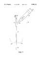
- FIG. 7is a plan view of another deflectable optical feeding tube.
- FIG. 8Ais a plan view of an optical feeding tube.
- FIG. 8Bis a cross section view of the optical feeding tube of FIG. 8A taken along line 8B'-8B".
- FIG. 9Ais a plan view of a stylet deflectable in two directions.
- FIG. 9Bis a cross section view of the stylet of FIG. 9A taken along line 9B'--9B'.
- FIG. 10Ais a plan view of the optical feeding tube of FIG. 8A having the stylet of FIG. 9A inserted through the feeding lumen.
- FIG. 10Bis a cross section view of the optical feeding tube of FIG. 10A taken along line 10B'-10B".
- an optical feeding tube 10includes an elongated sheath 14 having several lumens 18, 22, 26 and ports 50, 52, 46 corresponding to each lumen 18, 22, 26 respectively.
- the lumens 18, 22, 26extend from a distal end 16 of the elongated sheath 14 to a proximal end 15 of the elongated sheath 14, and each lumen 18, 22, 26 meets with its corresponding port 50, 52, 46 at the proximal end 15 of the elongated sheath 14.
- the elongated sheath 14is constructed from a standard catheter material which renders it flexible enough to be inserted through a gastro-intestinal tract.
- the elongated sheath 14may be constructed from silicone, a family of urethanes including polyurethane, Tecoflex® manufactured by Thermedics (Woburn, Mass.), PercuflexTM manufactured by Boston Scientific Corporation (Natick, Mass.), and FleximaTM manufactured by Boston Scientific Corporation (Natick, Mass.).
- the elongated sheath 14 and the ports 50, 52, 46may be constructed as a single piece by a single material.
- the first lumenis a feeding lumen 18, which has a larger cross section area than the other lumens 22, 26. The feeding lumen receives nutrients at the feeding port 50 and delivers the nutrients to a gastro-intestinal tract of a patient.
- the second lumenis an imaging lumen 22 which houses an imaging device 23.
- the imaging device 23provides visualization of a passageway ahead of the feeding tube 10 while the feeding tube 10 is being placed in a gastro-intestinal tract of a patient. More specifically, the imaging device 23 provides visualization of an area adjacent the distal end 16 of the elongated sheath 14.
- the imaging device 23,for example, may comprise a bundle of optical fibers, extending from the proximal end 15 to the distal end 16 of the elongated sheath 14 and a lens in communication with the optical fibers disposed at the distal end 16.
- the optical fibersfor example, may be coextruded in the second lumen.
- the imaging device port 52includes a coupler (not shown) to which a handle 42 connects.
- the handle 42includes an eye piece 41 and a light source connector 43 to which an external light source (not shown) connects.
- the external light sourceprovide visualization by transmitting light through the imaging device and illuminating an area near the distal end 16 of the elongated sheath 14.
- a person placing the feeding tube 10can look into the eye piece 41 and observe the illuminated gastro-intestinal tract of a patient, while placing the feeding tube 10 inside the stomach cavity or the jejunum of the patient.
- the optical feeding tube 10further includes a retention device (not shown) disposed at the distal end 16 of the elongated sheath 14.
- the retention deviceis a balloon.
- the elongated sheath 14includes a third lumen or a fluid lumen 26 through which fluid travels to and from the balloon to inflate and deflate the balloon.
- the balloonfunctions as a retention device when inflated.
- the balloonremains deflated to facilitate insertion of the feeding tube 10 through the abdominal wall.
- the balloonis inflated to prevent movement of the feeding tube 10. More specifically the balloon prevents the feeding tube 10 from sliding out of the stomach.
- a fluid port 46 in communication with the fluid lumen 26 at the distal end 15 of the elongated sheath 14receives fluid from an external fluid source (not shown).
- an optical feeding tube 54includes an elongated sheath 56 having a feeding lumen 18 and an imaging lumen 22.
- a feeding port 50communicates with a proximal end of the feeding lumen 18.
- An imaging device port 52communicates with a proximal end of the imaging lumen 22.
- a coupler(not shown) connects a handle 42 with the imaging device port 52.
- the handle 42includes an eye piece 41 and a light source connector 43.
- the elongated sheath 56has numbers from 1 to 9 displayed on its outer surface. These number assist in monitoring the feeding tube 54 movement, while the feeding tube 54 is placed inside a patient.
- the elongated sheath 54further includes a bolster 58 which functions as an internal retention device.
- bolstersare available for use with a feeding tube.
- the optical feeding tube 54may include a deformable bolster, a hollow sleeve surrounding and restricting the bolster and a rip-cord (not shown). While the optical feeding tube 54 is being inserted through the abdominal wall, the sleeve surrounding the bolster 58 restrains its figure. Once the distal end 59 of the feeding tube 54 is positioned inside the gastro-intestinal tract, the rip-cord is pulled and the sleeve is ripped, thereby exposing the full figure of the bolster 58.
- the extended bolster 58has a cross section area larger than a cross section area of the elongated sheath 56 and the opening through which the feeding tube 54 was inserted. The sleeve is removed and the bolster 58 keeps the feeding tube 54 from movement. Any bolster known to those skilled in this art may be used with the optical feeding tube 54.
- the optical feeding tube 54further includes an external retention device 45.
- the external retention device 45is slidably mounted on the body of the elongated sheath 56 prior to the feeding tube 54 placement. Once the optical feeding tube 54 is positioned, the external retention device 45 is placed against the abdomen, to further prevent movement of the feeding tube 54.
- an optical feeding tube 62is deflectable.
- a deflectable feeding tubefacilitates its insertion through a gastro-intestinal tract.
- the optical feeding tube 62includes an elongated sheath 64 having a deflectable tip 66.
- the tip 66deflects by deflecting a stylet 72 inserted in a feeding lumen of the feeding tube 62.
- FIGS. 4A and 4Bshow an optical feeding tube 62 of FIG. 3.
- the optical feeding tube 62includes an elongated sheath 64 having a feeding lumen 18 and an imaging lumen 22.
- An imaging device port 52has a coupler (not shown) at the proximal end for connecting a handle 42 to the port 52.
- FIG. 5A and 5Bshow a deflectable stylet 72 insertable in the feeding lumen 18 of the optical feeding tube 62 of FIG. 4A.
- the stylet 72includes a deflecting wire 74 and a deflecting handle 68.
- the deflecting handle 68operates in a push pull mode.
- the deflecting handle 68includes a piston member 67 and a receptacle member 69. As the piston member 67 pushes against the receptacle member 69, the deflecting wire 74 deflects. Conversely, as the piston member 67 pulls away from the receptacle member 69, the deflecting wire 74 straightens.
- FIGS. 6A and 6Bthe stylet of FIG. 5A is inserted and secured into the feeding lumen 18 of the optical feeding tube 62 of FIG. 4A.
- the optical feeding tube 62is then inserted into a patient's gastro-intestinal tract.
- the tip 66may be deflected by adjusting relative positions between the piston member 67 and the receptacle member 69 of the deflecting handle 68.
- FIG. 6Ashows the feeding tube 62 in a straight position and FIG. 3 shows the feeding tube 62 with the tip 66 deflected.
- the optical feeding tube 78includes an elongated sheath 74 and a deflectable tip 82.
- the tip 82is deflectable in two directions as shown by phantom lines 83, 84.
- the tip 82is deflected by deflecting a stylet inserted in a feeding lumen of the feeding tube 78.
- FIGS. 8A and 8Bshow the optical feeding tube 78 of FIG. 7 before a stylet is inserted into its feeding lumen 18.
- the optical feeding tube 78is identical to the optical feeding tube 62 of FIGS. 4A and 4B.
- FIGS. 9A and 9Bshow a stylet 84.
- the stylet 84includes two deflecting wires 85 and a deflecting handle 86.
- the stylet 84deflects in one direction when the handle 86 rotates clockwise and in the opposite direction when the handle 86 rotates counter-clockwise.
- This type of steering mechanismis well known to those skilled in the art and does not constitute an inventive aspect.
- the stylet 84is positioned inside the feeding lumen 18 of the optical feeding tube 78 prior to positioning the optical feeding tube 78 inside a gastro-intestinal tract of a patient.
- an operatorcontrols deflection of the tip 82 by rotating the handle 86.
- Rotation of the handle 86causes distal ends of the deflecting wires 85 to deflect, which in turn causes the deflectable tip 82 of the feeding tube 78 to deflect.
- the stylet 84is removed. The feeding tube 78 is then ready to receive nutrients.
- the optical feeding tubes of the present inventionmay be used as both gastrostomy tubes and jejunostomy tubes.
- a difference between the two types of feeding tubeis that a jejunostomy tube is typically longer and has a smaller cross-section area than a gastrostomy tube.
- a jejunostomy tubefor example, may have a cross-section diameter ranging from about 8 fr. to about 24 fr.
- a gastrostomy tubefor example, may have a cross-section diameter ranging from about 12 fr. to about 30 fr.
- an optical feeding tube of the present inventionprovides easy replacement of a gastrostomy tube, eliminating the need for an endoscopy or any other radiology procedures necessary to confirm proper tube placement.
- an optical feeding tube of the present inventionis used as an initial gastrostomy tube.
- an opening which provides access to the stomach through the abdominal wallis first created.
- the openingfor example, may be created by inserting a needle through a patient's abdomen into his or her stomach cavity.
- a guidewireis advanced through the opening and an introducer with a peel-away sheath is passed over the guide wire.
- the introduceris then removed, and the optical feeding tube is inserted into the stomach through the sheath under observation.
- the sheathis then peeled away leaving behind the feeding tube.
- this methoddoes not require an endoscope to confirm proper placement of the feeding tube.
- the optical feeding tubeis used as a jejunostomy tube.
- the jejunostomy tubefor example, is placed inside a gastro-intestinal tract through a regular gastrostomy tube already placed inside a patient.
- a regular gastrostomy tubedoes not have an imaging device.
- the jejunostomy tubeis inserted into the feeding lumen of the regular gastrostomy tube.
- the jejunostomy tubeis then inserted through a duodenum into a jejunum under visual guidance provided by the imaging device inside the jejunostomy tube.
- an endoscopeis not required during the placement of the jejunostomy tube.
- the jejunostomy tubemay be inserted into a jejunum with an assistance of a guide wire.
- the jejunostomy tube having its own retention devicemay be placed inside a gastro-intestinal tract without the assistance of the gastrostromy tube.
Landscapes
- Health & Medical Sciences (AREA)
- Life Sciences & Earth Sciences (AREA)
- Surgery (AREA)
- Biomedical Technology (AREA)
- Medical Informatics (AREA)
- Optics & Photonics (AREA)
- Pathology (AREA)
- Radiology & Medical Imaging (AREA)
- Biophysics (AREA)
- Engineering & Computer Science (AREA)
- Physics & Mathematics (AREA)
- Heart & Thoracic Surgery (AREA)
- Nuclear Medicine, Radiotherapy & Molecular Imaging (AREA)
- Molecular Biology (AREA)
- Animal Behavior & Ethology (AREA)
- General Health & Medical Sciences (AREA)
- Public Health (AREA)
- Veterinary Medicine (AREA)
- Gastroenterology & Hepatology (AREA)
- Endoscopes (AREA)
- Medical Preparation Storing Or Oral Administration Devices (AREA)
Abstract
Description
Claims (13)
Priority Applications (2)
| Application Number | Priority Date | Filing Date | Title |
|---|---|---|---|
| US09/007,500US5989231A (en) | 1998-01-15 | 1998-01-15 | Optical gastrostomy and jejunostomy |
| US09/419,398US6322495B1 (en) | 1998-01-15 | 1999-10-15 | Method for placing a feeding tube inside a gastro-intestinal tract |
Applications Claiming Priority (1)
| Application Number | Priority Date | Filing Date | Title |
|---|---|---|---|
| US09/007,500US5989231A (en) | 1998-01-15 | 1998-01-15 | Optical gastrostomy and jejunostomy |
Related Child Applications (1)
| Application Number | Title | Priority Date | Filing Date |
|---|---|---|---|
| US09/419,398DivisionUS6322495B1 (en) | 1998-01-15 | 1999-10-15 | Method for placing a feeding tube inside a gastro-intestinal tract |
Publications (1)
| Publication Number | Publication Date |
|---|---|
| US5989231Atrue US5989231A (en) | 1999-11-23 |
Family
ID=21726561
Family Applications (2)
| Application Number | Title | Priority Date | Filing Date |
|---|---|---|---|
| US09/007,500Expired - LifetimeUS5989231A (en) | 1998-01-15 | 1998-01-15 | Optical gastrostomy and jejunostomy |
| US09/419,398Expired - LifetimeUS6322495B1 (en) | 1998-01-15 | 1999-10-15 | Method for placing a feeding tube inside a gastro-intestinal tract |
Family Applications After (1)
| Application Number | Title | Priority Date | Filing Date |
|---|---|---|---|
| US09/419,398Expired - LifetimeUS6322495B1 (en) | 1998-01-15 | 1999-10-15 | Method for placing a feeding tube inside a gastro-intestinal tract |
Country Status (1)
| Country | Link |
|---|---|
| US (2) | US5989231A (en) |
Cited By (29)
| Publication number | Priority date | Publication date | Assignee | Title |
|---|---|---|---|---|
| US20030130562A1 (en)* | 2002-01-09 | 2003-07-10 | Scimed Life Systems, Inc. | Imaging device and related methods |
| US20030225369A1 (en)* | 2002-05-31 | 2003-12-04 | Kimberly-Clark Worldwide, Inc. | Low profile transpyloric jejunostomy system |
| US20040220516A1 (en)* | 2002-11-04 | 2004-11-04 | Stephen Solomon | Food extraction apparatus and method |
| US6864242B2 (en) | 2001-03-05 | 2005-03-08 | Stephen P. Ernest | Enteral formulation |
| US6882875B1 (en) | 1997-09-29 | 2005-04-19 | Boston Scientific Corporation | Visible display for an interventional device |
| US20050171468A1 (en)* | 2004-02-04 | 2005-08-04 | Wood Scott D. | Gastric aspirate intestinal feeding tube |
| US20050283130A1 (en)* | 2002-11-04 | 2005-12-22 | Samuel Klein | Method for treating obesity by extracting food |
| US20060030818A1 (en)* | 2004-08-09 | 2006-02-09 | Mcvey Robert D | System and method for securing a medical access device |
| US7066914B2 (en) | 2000-07-12 | 2006-06-27 | Bird Products Corporation | Catheter having a tip with an elongated collar |
| USD561329S1 (en) | 2006-10-04 | 2008-02-05 | Kimberly-Clark Worldwide, Inc. | Low profile transpyloric jejunostomy catheter |
| US20080039809A1 (en)* | 2006-08-03 | 2008-02-14 | Deka Products Limited Partnership | Systems and methods for removing ingested material from a stomach |
| US20100113880A1 (en)* | 2008-11-05 | 2010-05-06 | Page Charles W | Gastrostomy-jejunostomy tube apparatus and method for endoscopically placing same within a patient |
| US20100305503A1 (en)* | 2009-04-09 | 2010-12-02 | John Fang | Optically guided feeding tube, catheters and associated methods |
| US7976518B2 (en) | 2005-01-13 | 2011-07-12 | Corpak Medsystems, Inc. | Tubing assembly and signal generator placement control device and method for use with catheter guidance systems |
| US20120116160A1 (en)* | 2009-04-09 | 2012-05-10 | Nieman Timothy R | Optically guided medical tube and control unit assembly and methods of use |
| US20130231533A1 (en)* | 2011-05-23 | 2013-09-05 | Stephanos Papademetriou | Medical applications of a miniature videoscope |
| US8632513B2 (en) | 2006-08-03 | 2014-01-21 | Aspire Bariatrics, Inc. | Systems and methods for removing ingested material from a stomach |
| USD716841S1 (en) | 2012-09-07 | 2014-11-04 | Covidien Lp | Display screen with annotate file icon |
| USD717340S1 (en) | 2012-09-07 | 2014-11-11 | Covidien Lp | Display screen with enteral feeding icon |
| US9028441B2 (en) | 2011-09-08 | 2015-05-12 | Corpak Medsystems, Inc. | Apparatus and method used with guidance system for feeding and suctioning |
| US9039677B2 (en) | 2002-11-04 | 2015-05-26 | Aspire Bariatrics, Inc. | Apparatus for treating obesity by extracting food |
| US9055995B2 (en) | 2002-11-04 | 2015-06-16 | Aspire Bariatrics, Inc. | Method for treating obesity by extracting food |
| USD735343S1 (en) | 2012-09-07 | 2015-07-28 | Covidien Lp | Console |
| US9198835B2 (en) | 2012-09-07 | 2015-12-01 | Covidien Lp | Catheter with imaging assembly with placement aid and related methods therefor |
| US9433339B2 (en) | 2010-09-08 | 2016-09-06 | Covidien Lp | Catheter with imaging assembly and console with reference library and related methods therefor |
| AU2011349234B2 (en)* | 2010-12-21 | 2016-09-15 | University Of Utah Research Foundation | Optically guided medical tube and control unit assembly and methods of use |
| US9517184B2 (en) | 2012-09-07 | 2016-12-13 | Covidien Lp | Feeding tube with insufflation device and related methods therefor |
| US20170367870A1 (en)* | 2014-12-18 | 2017-12-28 | Evoluzione S.R.L. | Medical device for performing ileostomies and/or jejunostomies |
| US10085866B2 (en) | 2013-02-23 | 2018-10-02 | Aspire Bariatrics, Inc. | Apparatus and method for draining material from a stomach |
Families Citing this family (75)
| Publication number | Priority date | Publication date | Assignee | Title |
|---|---|---|---|---|
| US6289229B1 (en)* | 1998-01-20 | 2001-09-11 | Scimed Life Systems, Inc. | Readable probe array for in vivo use |
| DE10105592A1 (en) | 2001-02-06 | 2002-08-08 | Achim Goepferich | Placeholder for drug release in the frontal sinus |
| US8317816B2 (en) | 2002-09-30 | 2012-11-27 | Acclarent, Inc. | Balloon catheters and methods for treating paranasal sinuses |
| US7431694B2 (en) | 2003-05-16 | 2008-10-07 | Ethicon Endo-Surgery, Inc. | Method of guiding medical devices |
| US7615003B2 (en) | 2005-05-13 | 2009-11-10 | Ethicon Endo-Surgery, Inc. | Track for medical devices |
| US6939293B2 (en) | 2003-08-07 | 2005-09-06 | Chris N. Conteas | Gastrointestinal lavage system |
| US8425539B2 (en) | 2004-04-12 | 2013-04-23 | Xlumena, Inc. | Luminal structure anchoring devices and methods |
| US12303105B2 (en) | 2004-04-12 | 2025-05-20 | Boston Scientific Scimed, Inc. | Luminal structure anchoring devices and methods |
| US7654997B2 (en) | 2004-04-21 | 2010-02-02 | Acclarent, Inc. | Devices, systems and methods for diagnosing and treating sinusitus and other disorders of the ears, nose and/or throat |
| US7410480B2 (en) | 2004-04-21 | 2008-08-12 | Acclarent, Inc. | Devices and methods for delivering therapeutic substances for the treatment of sinusitis and other disorders |
| US7803150B2 (en) | 2004-04-21 | 2010-09-28 | Acclarent, Inc. | Devices, systems and methods useable for treating sinusitis |
| US7361168B2 (en) | 2004-04-21 | 2008-04-22 | Acclarent, Inc. | Implantable device and methods for delivering drugs and other substances to treat sinusitis and other disorders |
| US8764729B2 (en) | 2004-04-21 | 2014-07-01 | Acclarent, Inc. | Frontal sinus spacer |
| US10188413B1 (en) | 2004-04-21 | 2019-01-29 | Acclarent, Inc. | Deflectable guide catheters and related methods |
| US20060004323A1 (en) | 2004-04-21 | 2006-01-05 | Exploramed Nc1, Inc. | Apparatus and methods for dilating and modifying ostia of paranasal sinuses and other intranasal or paranasal structures |
| US9554691B2 (en) | 2004-04-21 | 2017-01-31 | Acclarent, Inc. | Endoscopic methods and devices for transnasal procedures |
| US7559925B2 (en) | 2006-09-15 | 2009-07-14 | Acclarent Inc. | Methods and devices for facilitating visualization in a surgical environment |
| US8747389B2 (en) | 2004-04-21 | 2014-06-10 | Acclarent, Inc. | Systems for treating disorders of the ear, nose and throat |
| US7462175B2 (en) | 2004-04-21 | 2008-12-09 | Acclarent, Inc. | Devices, systems and methods for treating disorders of the ear, nose and throat |
| US9089258B2 (en) | 2004-04-21 | 2015-07-28 | Acclarent, Inc. | Endoscopic methods and devices for transnasal procedures |
| US8932276B1 (en) | 2004-04-21 | 2015-01-13 | Acclarent, Inc. | Shapeable guide catheters and related methods |
| US8864787B2 (en) | 2004-04-21 | 2014-10-21 | Acclarent, Inc. | Ethmoidotomy system and implantable spacer devices having therapeutic substance delivery capability for treatment of paranasal sinusitis |
| US8702626B1 (en) | 2004-04-21 | 2014-04-22 | Acclarent, Inc. | Guidewires for performing image guided procedures |
| US20190314620A1 (en) | 2004-04-21 | 2019-10-17 | Acclarent, Inc. | Apparatus and methods for dilating and modifying ostia of paranasal sinuses and other intranasal or paranasal structures |
| US20070167682A1 (en) | 2004-04-21 | 2007-07-19 | Acclarent, Inc. | Endoscopic methods and devices for transnasal procedures |
| US8894614B2 (en) | 2004-04-21 | 2014-11-25 | Acclarent, Inc. | Devices, systems and methods useable for treating frontal sinusitis |
| US9101384B2 (en) | 2004-04-21 | 2015-08-11 | Acclarent, Inc. | Devices, systems and methods for diagnosing and treating sinusitis and other disorders of the ears, Nose and/or throat |
| US8146400B2 (en) | 2004-04-21 | 2012-04-03 | Acclarent, Inc. | Endoscopic methods and devices for transnasal procedures |
| US20060063973A1 (en) | 2004-04-21 | 2006-03-23 | Acclarent, Inc. | Methods and apparatus for treating disorders of the ear, nose and throat |
| US7419497B2 (en) | 2004-04-21 | 2008-09-02 | Acclarent, Inc. | Methods for treating ethmoid disease |
| US9351750B2 (en) | 2004-04-21 | 2016-05-31 | Acclarent, Inc. | Devices and methods for treating maxillary sinus disease |
| US20070208252A1 (en) | 2004-04-21 | 2007-09-06 | Acclarent, Inc. | Systems and methods for performing image guided procedures within the ear, nose, throat and paranasal sinuses |
| US9399121B2 (en) | 2004-04-21 | 2016-07-26 | Acclarent, Inc. | Systems and methods for transnasal dilation of passageways in the ear, nose or throat |
| JP5111112B2 (en) | 2004-12-08 | 2012-12-26 | エックスルミナ, インコーポレイテッド | Device for performing needle-guided therapy |
| US7857750B2 (en)* | 2005-01-18 | 2010-12-28 | The Regents Of The University Of California | Endoscopic tube delivery system |
| US20060258904A1 (en)* | 2005-05-13 | 2006-11-16 | David Stefanchik | Feeding tube and track |
| US20060258903A1 (en)* | 2005-05-13 | 2006-11-16 | David Stefanchik | Method of inserting a feeding tube |
| US7905830B2 (en) | 2005-05-13 | 2011-03-15 | Ethicon Endo-Surgery, Inc. | Sheath for use with an endoscope |
| US7857754B2 (en) | 2005-05-13 | 2010-12-28 | Ethicon Endo-Surgery, Inc. | Apparatus useful for positioning a device on an endoscope |
| US7648457B2 (en) | 2005-05-13 | 2010-01-19 | Ethicon Endo-Surgery, Inc. | Method of positioning a device on an endoscope |
| US8784437B2 (en) | 2005-06-09 | 2014-07-22 | Xlumena, Inc. | Methods and devices for endosonography-guided fundoplexy |
| US8777967B2 (en) | 2005-06-09 | 2014-07-15 | Xlumena, Inc. | Methods and devices for anchoring to tissue |
| US8951225B2 (en) | 2005-06-10 | 2015-02-10 | Acclarent, Inc. | Catheters with non-removable guide members useable for treatment of sinusitis |
| US8114113B2 (en) | 2005-09-23 | 2012-02-14 | Acclarent, Inc. | Multi-conduit balloon catheter |
| US8190389B2 (en) | 2006-05-17 | 2012-05-29 | Acclarent, Inc. | Adapter for attaching electromagnetic image guidance components to a medical device |
| US9820688B2 (en) | 2006-09-15 | 2017-11-21 | Acclarent, Inc. | Sinus illumination lightwire device |
| US8157765B2 (en) | 2006-10-20 | 2012-04-17 | Boston Scientific Scimed, Inc. | Medical catheter assembly including a balloon bolster |
| US8439687B1 (en) | 2006-12-29 | 2013-05-14 | Acclarent, Inc. | Apparatus and method for simulated insertion and positioning of guidewares and other interventional devices |
| US20130046172A1 (en)* | 2007-03-14 | 2013-02-21 | Kathryn A. McKenzie Waitzman | Methods and systems for locating a feeding tube inside of a person |
| US8118757B2 (en) | 2007-04-30 | 2012-02-21 | Acclarent, Inc. | Methods and devices for ostium measurement |
| US8485199B2 (en) | 2007-05-08 | 2013-07-16 | Acclarent, Inc. | Methods and devices for protecting nasal turbinate during surgery |
| US10206821B2 (en) | 2007-12-20 | 2019-02-19 | Acclarent, Inc. | Eustachian tube dilation balloon with ventilation path |
| US8057429B2 (en)* | 2008-02-26 | 2011-11-15 | Nath Iyunni Venkata Sesha Sayi | Feeding tube |
| JP2011512958A (en)* | 2008-02-27 | 2011-04-28 | ザ リージェンツ オブ ザ ユニバーシティ オブ カリフォルニア | Nutrition tube system |
| US8182432B2 (en) | 2008-03-10 | 2012-05-22 | Acclarent, Inc. | Corewire design and construction for medical devices |
| US8454632B2 (en) | 2008-05-12 | 2013-06-04 | Xlumena, Inc. | Tissue anchor for securing tissue layers |
| US20090318757A1 (en)* | 2008-06-23 | 2009-12-24 | Percuvision, Llc | Flexible visually directed medical intubation instrument and method |
| RU2500337C2 (en) | 2008-07-30 | 2013-12-10 | Аккларент, Инк. | Device and methods of identifying orifice of paranasal sinus |
| BRPI0919195A2 (en) | 2008-09-18 | 2019-09-24 | Acclarent Inc | Methods and Apparatus for the Treatment of Ear, Nose, and Throat Disorders |
| US20100241155A1 (en) | 2009-03-20 | 2010-09-23 | Acclarent, Inc. | Guide system with suction |
| US7978742B1 (en) | 2010-03-24 | 2011-07-12 | Corning Incorporated | Methods for operating diode lasers |
| US8435290B2 (en) | 2009-03-31 | 2013-05-07 | Acclarent, Inc. | System and method for treatment of non-ventilating middle ear by providing a gas pathway through the nasopharynx |
| US20100268029A1 (en)* | 2009-04-21 | 2010-10-21 | Xlumena, Inc. | Methods and apparatus for advancing a device from one body lumen to another |
| US9364259B2 (en) | 2009-04-21 | 2016-06-14 | Xlumena, Inc. | System and method for delivering expanding trocar through a sheath |
| JP5535313B2 (en) | 2009-05-29 | 2014-07-02 | エックスルミナ, インコーポレイテッド | Device and method for deploying a stent across adjacent tissue layers |
| WO2010141024A1 (en)* | 2009-06-04 | 2010-12-09 | John Isham | Prostate immobilizer apparatus |
| US9155492B2 (en) | 2010-09-24 | 2015-10-13 | Acclarent, Inc. | Sinus illumination lightwire device |
| US8473034B2 (en) | 2011-03-04 | 2013-06-25 | Cook Medical Technologies Llc | System and method for feeding tube placement |
| EP3111984B1 (en)* | 2011-08-29 | 2018-04-11 | ART MEDICAL Ltd. | Postpyloric feeding device |
| JP6360042B2 (en) | 2012-05-17 | 2018-07-18 | ボストン サイエンティフィック サイムド,インコーポレイテッドBoston Scientific Scimed,Inc. | Method and device for access across adjacent tissue layers |
| ES2980140T3 (en) | 2013-02-21 | 2024-09-30 | Boston Scient Scimed Inc | Devices for forming an anastomosis |
| US9629684B2 (en) | 2013-03-15 | 2017-04-25 | Acclarent, Inc. | Apparatus and method for treatment of ethmoid sinusitis |
| US9433437B2 (en) | 2013-03-15 | 2016-09-06 | Acclarent, Inc. | Apparatus and method for treatment of ethmoid sinusitis |
| WO2017085724A1 (en) | 2015-11-18 | 2017-05-26 | Art Healthcare Ltd. | Sheath and hub for imaging endoscope |
| WO2023229904A1 (en)* | 2022-05-27 | 2023-11-30 | Inmed, Inc. | Catheter exchange strategy and catheter configured for same |
Citations (18)
| Publication number | Priority date | Publication date | Assignee | Title |
|---|---|---|---|---|
| US4624243A (en)* | 1985-04-08 | 1986-11-25 | American Hospital Supply Corp. | Endoscope having a reusable eyepiece and a disposable distal section |
| US4769014A (en)* | 1987-06-02 | 1988-09-06 | Superior Biosystems Inc. | Gastroenteric feeding tube for endoscopic placement |
| US5007900A (en)* | 1989-10-31 | 1991-04-16 | Applied Medical Technology, Inc. | Percutaneous endoscopic gastrostomy device |
| USD323887S (en) | 1989-05-16 | 1992-02-11 | Applied Medical Technology, Inc. | Percutaneous endoscopic gastrostomy tube |
| USD323886S (en) | 1989-05-16 | 1992-02-11 | Applied Medical Technology, Inc. | Percutaneous endoscopil gastrostomy tube |
| US5112310A (en)* | 1991-02-06 | 1992-05-12 | Grobe James L | Apparatus and methods for percutaneous endoscopic gastrostomy |
| US5152277A (en)* | 1987-07-23 | 1992-10-06 | Terumo Kabushiki Kaisha | Catheter tube |
| US5188596A (en)* | 1990-09-27 | 1993-02-23 | Mentor Corporation | Transparent prostate dilation balloon and scope |
| US5279553A (en)* | 1992-04-02 | 1994-01-18 | Martin J. Winkler | Transpyloric jejunostomy cannulating system |
| US5327881A (en)* | 1993-02-26 | 1994-07-12 | Beth Israel Hospital Association | Fiberoptic intubating stylet |
| US5356391A (en)* | 1992-06-22 | 1994-10-18 | Medical Innovations Corp. | Flexible retainer flange for gastrostomy tube and the method of installing it |
| US5518406A (en)* | 1993-11-24 | 1996-05-21 | Waters; Tammie C. | Percutaneous endoscopic gastrostomy teaching device |
| US5527280A (en)* | 1995-03-29 | 1996-06-18 | The Children's Seashore House | Multi-lumen enteral feeding and medicating device |
| US5676635A (en)* | 1995-08-30 | 1997-10-14 | Levin; Bruce | Instrument for insertion of an endotracheal tube |
| US5733241A (en)* | 1996-02-01 | 1998-03-31 | King; George Hwa Kou | Fiberoptic intubation stylet |
| US5733242A (en)* | 1996-02-07 | 1998-03-31 | Rayburn; Robert L. | Intubation system having an axially moveable memory cylinder |
| US5803898A (en)* | 1997-05-05 | 1998-09-08 | Bashour; Charles Allen | Intubation system |
| US5823940A (en)* | 1993-08-18 | 1998-10-20 | Vista Medical Technologies, Inc. | Optical surgical device for examining genitourinary tissue |
- 1998
- 1998-01-15USUS09/007,500patent/US5989231A/ennot_activeExpired - Lifetime
- 1999
- 1999-10-15USUS09/419,398patent/US6322495B1/ennot_activeExpired - Lifetime
Patent Citations (19)
| Publication number | Priority date | Publication date | Assignee | Title |
|---|---|---|---|---|
| US4624243A (en)* | 1985-04-08 | 1986-11-25 | American Hospital Supply Corp. | Endoscope having a reusable eyepiece and a disposable distal section |
| US4769014A (en)* | 1987-06-02 | 1988-09-06 | Superior Biosystems Inc. | Gastroenteric feeding tube for endoscopic placement |
| US4769014B1 (en)* | 1987-06-02 | 1990-02-13 | Superior Biosystems Inc | |
| US5152277A (en)* | 1987-07-23 | 1992-10-06 | Terumo Kabushiki Kaisha | Catheter tube |
| USD323887S (en) | 1989-05-16 | 1992-02-11 | Applied Medical Technology, Inc. | Percutaneous endoscopic gastrostomy tube |
| USD323886S (en) | 1989-05-16 | 1992-02-11 | Applied Medical Technology, Inc. | Percutaneous endoscopil gastrostomy tube |
| US5007900A (en)* | 1989-10-31 | 1991-04-16 | Applied Medical Technology, Inc. | Percutaneous endoscopic gastrostomy device |
| US5188596A (en)* | 1990-09-27 | 1993-02-23 | Mentor Corporation | Transparent prostate dilation balloon and scope |
| US5112310A (en)* | 1991-02-06 | 1992-05-12 | Grobe James L | Apparatus and methods for percutaneous endoscopic gastrostomy |
| US5279553A (en)* | 1992-04-02 | 1994-01-18 | Martin J. Winkler | Transpyloric jejunostomy cannulating system |
| US5356391A (en)* | 1992-06-22 | 1994-10-18 | Medical Innovations Corp. | Flexible retainer flange for gastrostomy tube and the method of installing it |
| US5327881A (en)* | 1993-02-26 | 1994-07-12 | Beth Israel Hospital Association | Fiberoptic intubating stylet |
| US5823940A (en)* | 1993-08-18 | 1998-10-20 | Vista Medical Technologies, Inc. | Optical surgical device for examining genitourinary tissue |
| US5518406A (en)* | 1993-11-24 | 1996-05-21 | Waters; Tammie C. | Percutaneous endoscopic gastrostomy teaching device |
| US5527280A (en)* | 1995-03-29 | 1996-06-18 | The Children's Seashore House | Multi-lumen enteral feeding and medicating device |
| US5676635A (en)* | 1995-08-30 | 1997-10-14 | Levin; Bruce | Instrument for insertion of an endotracheal tube |
| US5733241A (en)* | 1996-02-01 | 1998-03-31 | King; George Hwa Kou | Fiberoptic intubation stylet |
| US5733242A (en)* | 1996-02-07 | 1998-03-31 | Rayburn; Robert L. | Intubation system having an axially moveable memory cylinder |
| US5803898A (en)* | 1997-05-05 | 1998-09-08 | Bashour; Charles Allen | Intubation system |
Non-Patent Citations (4)
| Title |
|---|
| Chaurasia et al., "A Novel Technique for Percutaneous Endoscopic Gastrojejunostomy Tube Placement", Gastrointestinal Endoscopy, vol. 42, No. 2, 1995, pp. 165 to 168. |
| Chaurasia et al., A Novel Technique for Percutaneous Endoscopic Gastrojejunostomy Tube Placement , Gastrointestinal Endoscopy, vol. 42, No. 2, 1995, pp. 165 to 168.* |
| Marks et al, "Access Routes for Enteral Nutrition", The Gastroenterologist, vol. 3, No. 2, Jun. 1995, pp. 130-139. |
| Marks et al, Access Routes for Enteral Nutrition , The Gastroenterologist, vol. 3, No. 2, Jun. 1995, pp. 130 139.* |
Cited By (54)
| Publication number | Priority date | Publication date | Assignee | Title |
|---|---|---|---|---|
| US6882875B1 (en) | 1997-09-29 | 2005-04-19 | Boston Scientific Corporation | Visible display for an interventional device |
| US7066914B2 (en) | 2000-07-12 | 2006-06-27 | Bird Products Corporation | Catheter having a tip with an elongated collar |
| US7196065B2 (en) | 2001-03-05 | 2007-03-27 | Ernest Stephen P | Enteral formulation |
| US6864242B2 (en) | 2001-03-05 | 2005-03-08 | Stephen P. Ernest | Enteral formulation |
| US20030130562A1 (en)* | 2002-01-09 | 2003-07-10 | Scimed Life Systems, Inc. | Imaging device and related methods |
| US8423110B2 (en) | 2002-01-09 | 2013-04-16 | Boston Scientific Scimed, Inc. | Imaging device and related methods |
| US20030225369A1 (en)* | 2002-05-31 | 2003-12-04 | Kimberly-Clark Worldwide, Inc. | Low profile transpyloric jejunostomy system |
| US20040220516A1 (en)* | 2002-11-04 | 2004-11-04 | Stephen Solomon | Food extraction apparatus and method |
| US20050283130A1 (en)* | 2002-11-04 | 2005-12-22 | Samuel Klein | Method for treating obesity by extracting food |
| US9039677B2 (en) | 2002-11-04 | 2015-05-26 | Aspire Bariatrics, Inc. | Apparatus for treating obesity by extracting food |
| US9055995B2 (en) | 2002-11-04 | 2015-06-16 | Aspire Bariatrics, Inc. | Method for treating obesity by extracting food |
| US7740624B2 (en)* | 2002-11-04 | 2010-06-22 | Aspiration Medical Technology, Llc | Method for treating obesity by extracting food |
| US8282623B2 (en) | 2002-11-04 | 2012-10-09 | Aspire Bariatrics Llc | Method for treating obesity by extracting food |
| US20050171468A1 (en)* | 2004-02-04 | 2005-08-04 | Wood Scott D. | Gastric aspirate intestinal feeding tube |
| US20060030818A1 (en)* | 2004-08-09 | 2006-02-09 | Mcvey Robert D | System and method for securing a medical access device |
| US9579488B2 (en) | 2005-01-13 | 2017-02-28 | Corpak Medsystems, Inc. | Tubing assembly and signal generator placement control device and method for use with catheter guidance systems |
| US9889277B2 (en) | 2005-01-13 | 2018-02-13 | Avent, Inc. | Tubing assembly and signal generator placement control device and method for use with catheter guidance systems |
| US7976518B2 (en) | 2005-01-13 | 2011-07-12 | Corpak Medsystems, Inc. | Tubing assembly and signal generator placement control device and method for use with catheter guidance systems |
| US9131956B2 (en) | 2005-01-13 | 2015-09-15 | Corpak Medsystems, Inc. | Tubing assembly and signal generator placement control device and method for use with catheter guidance systems |
| US10549074B2 (en) | 2005-01-13 | 2020-02-04 | Avent, Inc. | Tubing assembly and signal generation placement device and method for use with catheter guidance systems |
| US8002758B2 (en) | 2006-08-03 | 2011-08-23 | Aspire Bariatrics, Llc | Systems and methods for removing ingested material from a stomach |
| US8414561B2 (en) | 2006-08-03 | 2013-04-09 | Aspire Bariatrics, Llc | Systems and methods for removing ingested material from a stomach |
| US20080039809A1 (en)* | 2006-08-03 | 2008-02-14 | Deka Products Limited Partnership | Systems and methods for removing ingested material from a stomach |
| US8808221B2 (en) | 2006-08-03 | 2014-08-19 | Aspire Bariatrics, Inc. | Systems and methods for removing ingested material from a stomach |
| US8632513B2 (en) | 2006-08-03 | 2014-01-21 | Aspire Bariatrics, Inc. | Systems and methods for removing ingested material from a stomach |
| USD561329S1 (en) | 2006-10-04 | 2008-02-05 | Kimberly-Clark Worldwide, Inc. | Low profile transpyloric jejunostomy catheter |
| US20100113880A1 (en)* | 2008-11-05 | 2010-05-06 | Page Charles W | Gastrostomy-jejunostomy tube apparatus and method for endoscopically placing same within a patient |
| US20140039253A1 (en)* | 2009-04-09 | 2014-02-06 | University Of Utah Research Foundation | Optically Guided Feeding Tube, Catheters and Associated Methods |
| US9060922B2 (en)* | 2009-04-09 | 2015-06-23 | The University Of Utah | Optically guided medical tube and control unit assembly and methods of use |
| US20100305503A1 (en)* | 2009-04-09 | 2010-12-02 | John Fang | Optically guided feeding tube, catheters and associated methods |
| US9254245B2 (en) | 2009-04-09 | 2016-02-09 | University Of Utah | Optically guided medical tube and control unit assembly and methods of use |
| CN102448535A (en)* | 2009-04-09 | 2012-05-09 | 犹他大学研究基金会 | Optically guided feeding tube, catheters and associated methods |
| EP2416833A4 (en)* | 2009-04-09 | 2013-07-17 | Univ Utah Res Found | OPTICALLY GUIDED FOOD TUBE, CATHETERS AND ASSOCIATED METHODS |
| US8361041B2 (en)* | 2009-04-09 | 2013-01-29 | University Of Utah Research Foundation | Optically guided feeding tube, catheters and associated methods |
| US9532704B2 (en)* | 2009-04-09 | 2017-01-03 | University Of Utah | Optically guided feeding tube, catheters and associated methods |
| US20120116160A1 (en)* | 2009-04-09 | 2012-05-10 | Nieman Timothy R | Optically guided medical tube and control unit assembly and methods of use |
| JP2012523288A (en)* | 2009-04-09 | 2012-10-04 | ユニバーシティ・オブ・ユタ・リサーチ・ファウンデイション | Optically guided feeding tube, catheter and related methods |
| US9585813B2 (en) | 2010-09-08 | 2017-03-07 | Covidien Lp | Feeding tube system with imaging assembly and console |
| US10272016B2 (en) | 2010-09-08 | 2019-04-30 | Kpr U.S., Llc | Catheter with imaging assembly |
| US9433339B2 (en) | 2010-09-08 | 2016-09-06 | Covidien Lp | Catheter with imaging assembly and console with reference library and related methods therefor |
| US9538908B2 (en) | 2010-09-08 | 2017-01-10 | Covidien Lp | Catheter with imaging assembly |
| AU2011349234B2 (en)* | 2010-12-21 | 2016-09-15 | University Of Utah Research Foundation | Optically guided medical tube and control unit assembly and methods of use |
| EP3205325A3 (en)* | 2010-12-21 | 2017-12-20 | University of Utah Research Foundation | Optically guided medical tube and control unit assembly and methods of use |
| US20130231533A1 (en)* | 2011-05-23 | 2013-09-05 | Stephanos Papademetriou | Medical applications of a miniature videoscope |
| US9028441B2 (en) | 2011-09-08 | 2015-05-12 | Corpak Medsystems, Inc. | Apparatus and method used with guidance system for feeding and suctioning |
| US9918907B2 (en) | 2011-09-08 | 2018-03-20 | Avent, Inc. | Method for electromagnetic guidance of feeding and suctioning tube assembly |
| US9517184B2 (en) | 2012-09-07 | 2016-12-13 | Covidien Lp | Feeding tube with insufflation device and related methods therefor |
| US9198835B2 (en) | 2012-09-07 | 2015-12-01 | Covidien Lp | Catheter with imaging assembly with placement aid and related methods therefor |
| USD735343S1 (en) | 2012-09-07 | 2015-07-28 | Covidien Lp | Console |
| USD717340S1 (en) | 2012-09-07 | 2014-11-11 | Covidien Lp | Display screen with enteral feeding icon |
| USD716841S1 (en) | 2012-09-07 | 2014-11-04 | Covidien Lp | Display screen with annotate file icon |
| US10085866B2 (en) | 2013-02-23 | 2018-10-02 | Aspire Bariatrics, Inc. | Apparatus and method for draining material from a stomach |
| US20170367870A1 (en)* | 2014-12-18 | 2017-12-28 | Evoluzione S.R.L. | Medical device for performing ileostomies and/or jejunostomies |
| US11033419B2 (en)* | 2014-12-18 | 2021-06-15 | Evoluzione S.R.L. | Medical device for performing ileostomies and/or jejunostomies |
Also Published As
| Publication number | Publication date |
|---|---|
| US6322495B1 (en) | 2001-11-27 |
Similar Documents
| Publication | Publication Date | Title |
|---|---|---|
| US5989231A (en) | Optical gastrostomy and jejunostomy | |
| US6090073A (en) | Direct percutaneous endoscopic jejunostomy method and apparatus | |
| US8475430B2 (en) | Catheter assembly and method for internally anchoring a catheter in a patient | |
| EP1836980B1 (en) | Intubation device for enteral feeding | |
| US5807314A (en) | Feeding tube and method for placing same | |
| JP5834081B2 (en) | Multi-balloon expansion device for placement of catheter tubes | |
| US5151086A (en) | Laparoscopic tube placement method | |
| JP4163513B2 (en) | Apparatus and method for inserting medical instruments | |
| US7803137B2 (en) | Intubation system for use with an endoscope | |
| EP1954339B1 (en) | Short wire peg and peg-j tube | |
| JPH09511934A (en) | Surgical access device and access method | |
| JP2011512955A (en) | Nutrition tube | |
| US20070239171A1 (en) | Medical snaring device | |
| US20090024091A1 (en) | Catheter assembly including coiled internal bolster | |
| US20240415741A1 (en) | Systems, apparatus, and methods for placing a guidewire for a jejunostomy tube | |
| CA2582818C (en) | Intubation device for colonic decompression | |
| US6015400A (en) | Method for placing a feeding tube | |
| JP2025532900A (en) | Beacon Devices, Systems, and Methods for Medical Procedures | |
| TR2021021692A1 (en) | Percutaneous endoscopic gastrostomy set with balloon. | |
| CN120018807A (en) | Beacon device, system and method for medical surgery | |
| WO2023129065A1 (en) | Percutaneous endoscopic gastrostomy set with balloon |
Legal Events
| Date | Code | Title | Description |
|---|---|---|---|
| AS | Assignment | Owner name:BOSTON SCIENTIFIC CORPORATION, MASSACHUSETTS Free format text:ASSIGNMENT OF ASSIGNORS INTEREST;ASSIGNORS:SNOW, TODD H.;PHALEN, MICHAEL P.;REEL/FRAME:008962/0057 Effective date:19980115 | |
| AS | Assignment | Owner name:SCIMED LIFE SYSTEMS, INC., MINNESOTA Free format text:RERECORDED ON REEL 9372, FRAME 0645, TO CORRECT SERIAL NUMBER FROM 6006860 TO 60060830.;ASSIGNOR:BOSTON SCIENTIFIC CORPORATION;REEL/FRAME:009192/0467 Effective date:19980505 | |
| STCF | Information on status: patent grant | Free format text:PATENTED CASE | |
| FPAY | Fee payment | Year of fee payment:4 | |
| REMI | Maintenance fee reminder mailed | ||
| AS | Assignment | Owner name:BOSTON SCIENTIFIC SCIMED, INC., MINNESOTA Free format text:CHANGE OF NAME;ASSIGNOR:SCIMED LIFE SYSTEMS, INC.;REEL/FRAME:018505/0868 Effective date:20050101 Owner name:BOSTON SCIENTIFIC SCIMED, INC.,MINNESOTA Free format text:CHANGE OF NAME;ASSIGNOR:SCIMED LIFE SYSTEMS, INC.;REEL/FRAME:018505/0868 Effective date:20050101 | |
| FPAY | Fee payment | Year of fee payment:8 | |
| FPAY | Fee payment | Year of fee payment:12 |