US5987346A - Device and method for classification of tissue - Google Patents
Device and method for classification of tissueDownload PDFInfo
- Publication number
- US5987346A US5987346AUS08/771,952US77195296AUS5987346AUS 5987346 AUS5987346 AUS 5987346AUS 77195296 AUS77195296 AUS 77195296AUS 5987346 AUS5987346 AUS 5987346A
- Authority
- US
- United States
- Prior art keywords
- tissue
- classification
- monitor
- detector
- emitter
- Prior art date
- Legal status (The legal status is an assumption and is not a legal conclusion. Google has not performed a legal analysis and makes no representation as to the accuracy of the status listed.)
- Expired - Lifetime
Links
Images


Classifications
- A—HUMAN NECESSITIES
- A61—MEDICAL OR VETERINARY SCIENCE; HYGIENE
- A61B—DIAGNOSIS; SURGERY; IDENTIFICATION
- A61B5/00—Measuring for diagnostic purposes; Identification of persons
- A61B5/145—Measuring characteristics of blood in vivo, e.g. gas concentration or pH-value ; Measuring characteristics of body fluids or tissues, e.g. interstitial fluid or cerebral tissue
- A61B5/1455—Measuring characteristics of blood in vivo, e.g. gas concentration or pH-value ; Measuring characteristics of body fluids or tissues, e.g. interstitial fluid or cerebral tissue using optical sensors, e.g. spectral photometrical oximeters
- A61B5/1459—Measuring characteristics of blood in vivo, e.g. gas concentration or pH-value ; Measuring characteristics of body fluids or tissues, e.g. interstitial fluid or cerebral tissue using optical sensors, e.g. spectral photometrical oximeters invasive, e.g. introduced into the body by a catheter
- A—HUMAN NECESSITIES
- A61—MEDICAL OR VETERINARY SCIENCE; HYGIENE
- A61B—DIAGNOSIS; SURGERY; IDENTIFICATION
- A61B18/00—Surgical instruments, devices or methods for transferring non-mechanical forms of energy to or from the body
- A—HUMAN NECESSITIES
- A61—MEDICAL OR VETERINARY SCIENCE; HYGIENE
- A61B—DIAGNOSIS; SURGERY; IDENTIFICATION
- A61B5/00—Measuring for diagnostic purposes; Identification of persons
- A61B5/0059—Measuring for diagnostic purposes; Identification of persons using light, e.g. diagnosis by transillumination, diascopy, fluorescence
- A—HUMAN NECESSITIES
- A61—MEDICAL OR VETERINARY SCIENCE; HYGIENE
- A61B—DIAGNOSIS; SURGERY; IDENTIFICATION
- A61B5/00—Measuring for diagnostic purposes; Identification of persons
- A61B5/145—Measuring characteristics of blood in vivo, e.g. gas concentration or pH-value ; Measuring characteristics of body fluids or tissues, e.g. interstitial fluid or cerebral tissue
- A61B5/1455—Measuring characteristics of blood in vivo, e.g. gas concentration or pH-value ; Measuring characteristics of body fluids or tissues, e.g. interstitial fluid or cerebral tissue using optical sensors, e.g. spectral photometrical oximeters
- A61B5/14551—Measuring characteristics of blood in vivo, e.g. gas concentration or pH-value ; Measuring characteristics of body fluids or tissues, e.g. interstitial fluid or cerebral tissue using optical sensors, e.g. spectral photometrical oximeters for measuring blood gases
- A61B5/14553—Measuring characteristics of blood in vivo, e.g. gas concentration or pH-value ; Measuring characteristics of body fluids or tissues, e.g. interstitial fluid or cerebral tissue using optical sensors, e.g. spectral photometrical oximeters for measuring blood gases specially adapted for cerebral tissue
- A—HUMAN NECESSITIES
- A61—MEDICAL OR VETERINARY SCIENCE; HYGIENE
- A61B—DIAGNOSIS; SURGERY; IDENTIFICATION
- A61B5/00—Measuring for diagnostic purposes; Identification of persons
- A61B5/40—Detecting, measuring or recording for evaluating the nervous system
- A61B5/4076—Diagnosing or monitoring particular conditions of the nervous system
- A—HUMAN NECESSITIES
- A61—MEDICAL OR VETERINARY SCIENCE; HYGIENE
- A61B—DIAGNOSIS; SURGERY; IDENTIFICATION
- A61B5/00—Measuring for diagnostic purposes; Identification of persons
- A61B5/41—Detecting, measuring or recording for evaluating the immune or lymphatic systems
- A61B5/414—Evaluating particular organs or parts of the immune or lymphatic systems
- A61B5/415—Evaluating particular organs or parts of the immune or lymphatic systems the glands, e.g. tonsils, adenoids or thymus
- A—HUMAN NECESSITIES
- A61—MEDICAL OR VETERINARY SCIENCE; HYGIENE
- A61B—DIAGNOSIS; SURGERY; IDENTIFICATION
- A61B5/00—Measuring for diagnostic purposes; Identification of persons
- A61B5/41—Detecting, measuring or recording for evaluating the immune or lymphatic systems
- A61B5/414—Evaluating particular organs or parts of the immune or lymphatic systems
- A61B5/418—Evaluating particular organs or parts of the immune or lymphatic systems lymph vessels, ducts or nodes
- A—HUMAN NECESSITIES
- A61—MEDICAL OR VETERINARY SCIENCE; HYGIENE
- A61B—DIAGNOSIS; SURGERY; IDENTIFICATION
- A61B5/00—Measuring for diagnostic purposes; Identification of persons
- A61B5/68—Arrangements of detecting, measuring or recording means, e.g. sensors, in relation to patient
- A61B5/6846—Arrangements of detecting, measuring or recording means, e.g. sensors, in relation to patient specially adapted to be brought in contact with an internal body part, i.e. invasive
- A61B5/6847—Arrangements of detecting, measuring or recording means, e.g. sensors, in relation to patient specially adapted to be brought in contact with an internal body part, i.e. invasive mounted on an invasive device
- A61B5/6848—Needles
- A—HUMAN NECESSITIES
- A61—MEDICAL OR VETERINARY SCIENCE; HYGIENE
- A61B—DIAGNOSIS; SURGERY; IDENTIFICATION
- A61B5/00—Measuring for diagnostic purposes; Identification of persons
- A61B5/72—Signal processing specially adapted for physiological signals or for diagnostic purposes
- A61B5/7235—Details of waveform analysis
- A61B5/7264—Classification of physiological signals or data, e.g. using neural networks, statistical classifiers, expert systems or fuzzy systems
- G—PHYSICS
- G01—MEASURING; TESTING
- G01N—INVESTIGATING OR ANALYSING MATERIALS BY DETERMINING THEIR CHEMICAL OR PHYSICAL PROPERTIES
- G01N21/00—Investigating or analysing materials by the use of optical means, i.e. using sub-millimetre waves, infrared, visible or ultraviolet light
- G01N21/17—Systems in which incident light is modified in accordance with the properties of the material investigated
- G01N21/47—Scattering, i.e. diffuse reflection
- G01N21/4795—Scattering, i.e. diffuse reflection spatially resolved investigating of object in scattering medium
- A—HUMAN NECESSITIES
- A61—MEDICAL OR VETERINARY SCIENCE; HYGIENE
- A61B—DIAGNOSIS; SURGERY; IDENTIFICATION
- A61B17/00—Surgical instruments, devices or methods
- A61B17/068—Surgical staplers, e.g. containing multiple staples or clamps
- A—HUMAN NECESSITIES
- A61—MEDICAL OR VETERINARY SCIENCE; HYGIENE
- A61B—DIAGNOSIS; SURGERY; IDENTIFICATION
- A61B18/00—Surgical instruments, devices or methods for transferring non-mechanical forms of energy to or from the body
- A61B18/04—Surgical instruments, devices or methods for transferring non-mechanical forms of energy to or from the body by heating
- A61B18/12—Surgical instruments, devices or methods for transferring non-mechanical forms of energy to or from the body by heating by passing a current through the tissue to be heated, e.g. high-frequency current
- A61B18/14—Probes or electrodes therefor
- A—HUMAN NECESSITIES
- A61—MEDICAL OR VETERINARY SCIENCE; HYGIENE
- A61B—DIAGNOSIS; SURGERY; IDENTIFICATION
- A61B17/00—Surgical instruments, devices or methods
- A61B2017/00017—Electrical control of surgical instruments
- A61B2017/00022—Sensing or detecting at the treatment site
- A—HUMAN NECESSITIES
- A61—MEDICAL OR VETERINARY SCIENCE; HYGIENE
- A61B—DIAGNOSIS; SURGERY; IDENTIFICATION
- A61B17/00—Surgical instruments, devices or methods
- A61B2017/00017—Electrical control of surgical instruments
- A61B2017/00115—Electrical control of surgical instruments with audible or visual output
- A61B2017/00119—Electrical control of surgical instruments with audible or visual output alarm; indicating an abnormal situation
- A61B2017/00123—Electrical control of surgical instruments with audible or visual output alarm; indicating an abnormal situation and automatic shutdown
- A—HUMAN NECESSITIES
- A61—MEDICAL OR VETERINARY SCIENCE; HYGIENE
- A61B—DIAGNOSIS; SURGERY; IDENTIFICATION
- A61B17/00—Surgical instruments, devices or methods
- A61B17/00491—Surgical glue applicators
- A61B2017/00504—Tissue welding
- A61B2017/00508—Tissue welding using laser
- A—HUMAN NECESSITIES
- A61—MEDICAL OR VETERINARY SCIENCE; HYGIENE
- A61B—DIAGNOSIS; SURGERY; IDENTIFICATION
- A61B17/00—Surgical instruments, devices or methods
- A61B2017/00535—Surgical instruments, devices or methods pneumatically or hydraulically operated
- A61B2017/00557—Surgical instruments, devices or methods pneumatically or hydraulically operated inflatable
- A—HUMAN NECESSITIES
- A61—MEDICAL OR VETERINARY SCIENCE; HYGIENE
- A61B—DIAGNOSIS; SURGERY; IDENTIFICATION
- A61B2560/00—Constructional details of operational features of apparatus; Accessories for medical measuring apparatus
- A61B2560/02—Operational features
- A61B2560/0266—Operational features for monitoring or limiting apparatus function
- A61B2560/0276—Determining malfunction
- A—HUMAN NECESSITIES
- A61—MEDICAL OR VETERINARY SCIENCE; HYGIENE
- A61B—DIAGNOSIS; SURGERY; IDENTIFICATION
- A61B2562/00—Details of sensors; Constructional details of sensor housings or probes; Accessories for sensors
- A61B2562/02—Details of sensors specially adapted for in-vivo measurements
- A61B2562/0233—Special features of optical sensors or probes classified in A61B5/00
- A61B2562/0242—Special features of optical sensors or probes classified in A61B5/00 for varying or adjusting the optical path length in the tissue
- A—HUMAN NECESSITIES
- A61—MEDICAL OR VETERINARY SCIENCE; HYGIENE
- A61B—DIAGNOSIS; SURGERY; IDENTIFICATION
- A61B2562/00—Details of sensors; Constructional details of sensor housings or probes; Accessories for sensors
- A61B2562/08—Sensors provided with means for identification, e.g. barcodes or memory chips
- A—HUMAN NECESSITIES
- A61—MEDICAL OR VETERINARY SCIENCE; HYGIENE
- A61B—DIAGNOSIS; SURGERY; IDENTIFICATION
- A61B5/00—Measuring for diagnostic purposes; Identification of persons
- A61B5/0059—Measuring for diagnostic purposes; Identification of persons using light, e.g. diagnosis by transillumination, diascopy, fluorescence
- A61B5/0073—Measuring for diagnostic purposes; Identification of persons using light, e.g. diagnosis by transillumination, diascopy, fluorescence by tomography, i.e. reconstruction of 3D images from 2D projections
- A—HUMAN NECESSITIES
- A61—MEDICAL OR VETERINARY SCIENCE; HYGIENE
- A61B—DIAGNOSIS; SURGERY; IDENTIFICATION
- A61B5/00—Measuring for diagnostic purposes; Identification of persons
- A61B5/0059—Measuring for diagnostic purposes; Identification of persons using light, e.g. diagnosis by transillumination, diascopy, fluorescence
- A61B5/0075—Measuring for diagnostic purposes; Identification of persons using light, e.g. diagnosis by transillumination, diascopy, fluorescence by spectroscopy, i.e. measuring spectra, e.g. Raman spectroscopy, infrared absorption spectroscopy
- A—HUMAN NECESSITIES
- A61—MEDICAL OR VETERINARY SCIENCE; HYGIENE
- A61B—DIAGNOSIS; SURGERY; IDENTIFICATION
- A61B5/00—Measuring for diagnostic purposes; Identification of persons
- A61B5/0059—Measuring for diagnostic purposes; Identification of persons using light, e.g. diagnosis by transillumination, diascopy, fluorescence
- A61B5/0082—Measuring for diagnostic purposes; Identification of persons using light, e.g. diagnosis by transillumination, diascopy, fluorescence adapted for particular medical purposes
- A61B5/0084—Measuring for diagnostic purposes; Identification of persons using light, e.g. diagnosis by transillumination, diascopy, fluorescence adapted for particular medical purposes for introduction into the body, e.g. by catheters
- A—HUMAN NECESSITIES
- A61—MEDICAL OR VETERINARY SCIENCE; HYGIENE
- A61B—DIAGNOSIS; SURGERY; IDENTIFICATION
- A61B6/00—Apparatus or devices for radiation diagnosis; Apparatus or devices for radiation diagnosis combined with radiation therapy equipment
- A61B6/50—Apparatus or devices for radiation diagnosis; Apparatus or devices for radiation diagnosis combined with radiation therapy equipment specially adapted for specific body parts; specially adapted for specific clinical applications
- A61B6/506—Apparatus or devices for radiation diagnosis; Apparatus or devices for radiation diagnosis combined with radiation therapy equipment specially adapted for specific body parts; specially adapted for specific clinical applications for diagnosis of nerves
- G—PHYSICS
- G16—INFORMATION AND COMMUNICATION TECHNOLOGY [ICT] SPECIALLY ADAPTED FOR SPECIFIC APPLICATION FIELDS
- G16H—HEALTHCARE INFORMATICS, i.e. INFORMATION AND COMMUNICATION TECHNOLOGY [ICT] SPECIALLY ADAPTED FOR THE HANDLING OR PROCESSING OF MEDICAL OR HEALTHCARE DATA
- G16H50/00—ICT specially adapted for medical diagnosis, medical simulation or medical data mining; ICT specially adapted for detecting, monitoring or modelling epidemics or pandemics
- G16H50/20—ICT specially adapted for medical diagnosis, medical simulation or medical data mining; ICT specially adapted for detecting, monitoring or modelling epidemics or pandemics for computer-aided diagnosis, e.g. based on medical expert systems
Definitions
- the present inventionrelates to a device and method for detecting, localizing, and imaging in a radiation-scattering medium, and more particularly relates to an optical device and method for measuring information regarding the interaction of emitted light with biological tissue during passage of light through the tissue, and using said information to classify the tissue by type or state, either for detection, localization, or imaging.
- a major portion of time spent in medicineis directed toward the problem of diagnosis, and a large proportion of the errors in medicine are made here.
- a delayed diagnosisraises the level of pain and suffering, and may allow progression to the point of irreversibility; an incorrect diagnosis can be even worse, leading to treatment that is at best unnecessary and at worst harmful or fatal.
- tissue-type diagnosisusually requires surgical tissue removal (such as biopsy) and subsequent analysis by a pathologist, but still this decision is based upon subjective classification by eye, touch, chemical analysis, or even upon the absorption of exogenous dyes.
- tissue removalsuch as biopsy
- pathologistbut still this decision is based upon subjective classification by eye, touch, chemical analysis, or even upon the absorption of exogenous dyes.
- tissue removalsuch as biopsy
- pathologistbut still this decision is based upon subjective classification by eye, touch, chemical analysis, or even upon the absorption of exogenous dyes.
- tissue classification or identificationdoes not perform a tissue analysis, requires fluid or tissue removal or sampling, utilizes fluorescence or other emission-based techniques which measure light other than that used to perform the illumination, is restricted to external or penetrating use, or does not teach tissue classification or identification. Automated classification of tissues for general clinical use via light in vivo has not been taught, nor has such a tool been successfully commercialized.
- the present inventionuses optical methods to allow for a rapid tissue diagnosis via characterization of tissue in an automated manner.
- the present inventionrelies upon the optical characteristics of tissue, either by variations in absorbance or scattering by wavelength or over space, in order to make a medical diagnosis, namely an optical classification of the tissue by tissue type or state, either as a present/absent decision, as a localization, or as an image.
- a salient feature of the present inventionis an incorporation of the observation that light, while both being scattered and absorbed by scattering media, can be made to penetrate human tissue, then be detected upon reemergence in order to allow quantitation of characteristics of the interior of the tissue, such as tissue types or biochemical composition, imaging and localization of tissue types, and that such information is medically useful.
- an object of the present inventionis to provide a method for detecting the presence of tissue types using light, whether to merely detect, classify, localize, or image the tissue.
- a second objectis that classification of the tissue can be made, wherein the classification can be selected from normal tissue types (such as artery, vein, nerve, lymph, liver, muscle, brain, gray matter, white matter, colon, blood), from tissue components (water, fat, hemoglobin), from tissue states (frozen, thawed, coagulated), from tissue functional status (alive, dead, at risk for dying), and that such classifications can even be used to determine tissue pathology (normal or abnormal).
- normal tissue typessuch as artery, vein, nerve, lymph, liver, muscle, brain, gray matter, white matter, colon, blood
- tissue componentswater, fat, hemoglobin
- tissue statesfrozen, thawed, coagulated
- tissue functional statusas live, dead, at risk for dying
- a third objectis that localization of tissue by type can be made, such that the tissue may be classified as present or absent, distances from one tissue to a reference point can be measured, or the tissue can be localized in space.
- a measurement that characterizes a tissue at a defined point in spaceis considered imaging. This spatial distribution can be key in medical diagnosis.
- Another objectis to provide a noninvasive method for optically detecting, quantifying, or imaging a change in the tissue state, whether to merely detect, classify, localize or image the change in the tissue.
- This change in statecan be in response to a medical intervention, such as a change in the blood volume of the motor cortex of the brain during muscle activity, or the tool itself can initiate the change, such as by squeezing the tissue to assess vascular responsiveness, or freezing, thawing, welding, denaturing, or otherwise affecting the tissue.
- this techniqueis not limited to monitoring the tissue from the outside (e.g., such as is commonly done in computed x-ray tomography), but also may be used to allow a probe to measure its surrounding medium, such as if an optical fiber is inserted into a cyst, to allow sizing and diagnosis of the cyst from the inside, or if an underwater probe is to take note of objects nearby, such as rocks, when the water is cloudy, to allow better guidance.
- a probecan measure its surrounding medium, such as if an optical fiber is inserted into a cyst, to allow sizing and diagnosis of the cyst from the inside, or if an underwater probe is to take note of objects nearby, such as rocks, when the water is cloudy, to allow better guidance.
- This methodhas the advantage of being noninvasive, should this be desired, or invasive, should measurement inside the tissue be useful. For example, the characterization of tissue as a probe is advanced through the tissue can be important in diagnosis and localization.
- any medical probecan be modified to perform this classification function, such that measurements may be made using existing medical equipment, modified to hold emitter and detector elements, such as modified hand-held medical probes, tips of surgical tools, stethoscopes, EKG leads, or other devices.
- the ability to classifycan also be designed into new or unforeseen medical probes or devices. This function can be incorporated into replaceable device tips.
- the classificationcan be enhanced by a priori knowledge, such as the spectral characteristics of target tissues (which can be stored for reference in the device or in the probe), the area of the body the physician is working (such that far away tissues need not be considered in the analysis), or other medical scans (such as a CT or MRI scan).
- a priori knowledgesuch as the spectral characteristics of target tissues (which can be stored for reference in the device or in the probe), the area of the body the physician is working (such that far away tissues need not be considered in the analysis), or other medical scans (such as a CT or MRI scan).
- Another objectis that this data can be enhanced by collection over time.
- the value of a measurementis enhanced by determination of temporal characteristics. For example, the detection of an enlarging bleed in head tissue holds a different significance than the detection of a stable, but otherwise similar, bleed.
- the ability to detect moving nearby objectsmay also be important. Subtraction of the data at one point in time from data collected at a second point in time allows elimination of many types of individual tissue variations, and can yield improved data.
- this classificationrepresents a decision point upon which a human response may be initiated, such as with an alarm bell, or an interlock decision may be initiated, such as via an output signal attached to a medical device.
- a final objectis that the detection, localization, or imaging information can be presented to the user in a number of ways, such as an image of object location or even an image of characteristics of the medium such as absorbance, in such a manner as to allow the user to gain an incremental understanding of the presence or location of inhomogeneities in the medium, or even an understanding of characteristics of the medium itself.
- a diagnostic monitor for classifying biological tissuein which a light emitter is optically coupled to the tissue to be diagnosed and a light detector is optically coupled to the tissue to detect a portion of the light which passes through the tissue.
- the tissue classifierreceives a signal from the detector and provides an optical classification output signal.
- a method of classifying tissueis also described.
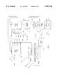
- FIG. 1is a schematic diagram of a monitor for classifying biological tissue in accordance with the invention.
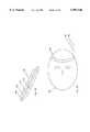
- FIGS. 2A-2Eare examples of probes which can be used in the monitor as shown in FIG. 1.
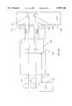
- FIGS. 3A-3Bshow a probe which can be used for minimally invasive diagnosis.
- FIG. 4schematically shows typical photon paths through the tissue.
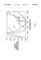
- FIG. 5shows the optical spectrum of two sample tissues.
- FIG. 6shows an imaging headband mounted on an infant's head.
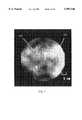
- FIG. 7is a photograph of a classified optical image of brain hemorrhage obtained with a monitor constructed in accordance with the present invention.
- FIGS. 8A-8Billustrate the optical detection and classification of freezing in tissue.
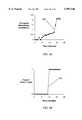
- FIGS. 9A-9Bgraphically show data used to construct a classified optical image of tissue freezing.
- FIG. 10A-10Dschematically show an a nearby object classified in a tissue model as an optical image, a numerical distance-to-object, and a graph of object presence versus depth.
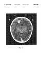
- FIG. 11is a photograph of a classified optical image of brain stroke.
- FIG. 12is a photograph of a classified optical image showing brain functional activity.
- a tissue classificationimplies an automated processing of the raw information contained in the usual medical image or measurement (such as shadows from bones) into a quantitative parameter or decision about the tissue, such as a classification (e.g., "is this a hemorrhage?") or a localization or a classification (e.g., "how far is the frozen tissue from my probe").
- a classification of tissuecan be into a tissue category by type, such as nerve, artery, vein, lymph node, hemorrhage, or by tissue state, such as frozen, denatured, coagulated.
- a localization of the classificationcan be as a distance, as an image (e.g., "where is the stroke"), or even as a characterization of a tissue at a point in space (e.g., "what is the type of tissue located exactly 4 cm below this probe?").
- the electromagnetic radiation usedis intended to be between 10 nm and 100 microns in wavelength, but includes any radiative wave in theory.
- Living tissue or tissue-like radiation-scattering mediasuch as skin, brain, bone, or even cloudy water.
- a probe that emits lightmay be composed of a simple light bulb, a laser, a flash lamp, or another light source or combination of sources, or it may be a complex form including a light source, a transmission element such as an optical fiber, a guidance element such as a reflective prism, and other elements intended to enhance the optical coupling of the light from the emitter to the skin or tissue under study.
- the light sourcemay be continuous, pulsed, or even analyzed as time-, frequency-, or spatially-resolved.
- the emittermay consist of a single or multiple light emitting elements.
- a probe that detects lightmay be single or multiple, simple or complex.
- the detectionmay be performed in reflectance or in transmission.
- a light emitterin such a way that light from the emitter (or detector) is transmitted to (or detected from) the tissue.
- Thismay include the use of optical elements such as lenses, collimators, concentrators, collectors, optical fibers, prisms, filters, mirrors, or mirrored surfaces.
- Optical fibershave two ends, which are generally interchangeable, and are referred here as the entrance end if the light is generally entering the fiber, and as the exit end if the light is generally leaving the fiber.
- a blood vessel in a muscleis a regional inhomogeneity, as is a stroke in a normal brain.
- Such changes in pathcan be induced by changes in scattering or absorbance at one or more wavelengths, and can be monitored in part by measuring reflectance, scattering, or absorbance, or any feature of the detected light that is affected by changes in these quantities.
- a region of spacein at least zero dimensions.
- An example of a zero dimension scanis the use of more than one point measurements on the surface of the scalp in order to determine the oxygenation of a specific, deeper portion of the brain, such as the gray matter, at one point in space or over one region in space.
- a one-dimensional scancould be the display of the presence of a certain tissue type, such as glandular tissue in the uterine wall, as a function of depth, as shown in Example 8, below.
- Two-D and 3-D scansare standard radiological views, and are well-known, as shown in Examples 2, 4, 6, and 7.
- a 4-D scancould include the three spatial dimensions x, y, and z, as well as time t.
- a measurement that can be quantitatively measuredsuch as a classification of tissue by type, or the distance of a type of tissue from the measuring probe.
- emitter 102Mini-MagliteTM Krypton miniature bulbs, Mag Instrument, Torrance, Calif.
- emitter switch 125Model GP-700, DiCon FiberOptics, Berkeley, Calif.
- N fibers 131A to 131N200 ⁇ m core glass fibers with cladding and buffer, Purdy Electronics Corp., Sunnyvale, Calif.
- the light sourcesuch as a surface mount LED, could be placed directly on the probe and electronically switched.
- Reference fiber 131Zconnected to switch 125, bypasses the tissue for use in monitoring the optical characteristics of source 102.
- Illumination fibers 131A to 131Nconnect to fiber bundle 132 which passes into first needle 133 that extends into tissue 145.
- Light from bundle 132passes through first needle ports 137A to 137N, containing fibers 131A to 131N respectively, and into tissue 145.
- Light traveling through tissue 145is collected through second needle ports 147A to 147M by collection fibers 151A to 151M, respectively, passing as fiber bundle 152 from second needle 153, offset a small distance from first needle 133.
- Light from one of collection fibers 151A to 151M, or from reference loop fiber 131Z,is chosen for monitoring by detector switch 165.
- Output fiber 167 from detector switch 165is connected to spectrum analyzer 174 (Ocean Optics Spectrophotometer, Model PS1000, Dunedin, Fla.), which records the light, and transmits an electronic signal to be stored in multichannel memory 181 (A/D-converter board Model PCM-DAS16/330-1, Computer Boards Inc., Mansfield, Mass.) via cable 183. Multiple spectra can be stored in Memory 181, allowing for collection of standardization spectra for correction of the spectra for instrument response, and also allowing for multiple regions of the tissue to be sampled and later compared.
- spectrum analyzer 174Ocean Optics Spectrophotometer, Model PS1000, Dunedin, Fla.
- multichannel memory 181A/D-converter board Model PCM-DAS16/330-1, Computer Boards Inc., Mansfield, Mass.
- Multiple spectracan be stored in Memory 181, allowing for collection of standardization spectra for correction of the spectra for instrument response, and also allowing
- tissue classifier 184in this case, a computer configured so as to perform tissue classification, AMS Laptop PentiumTM 120 MHz computer, Model AMS SY19-T40177 Travel Pro 1900, available through Ocean Optics, Dunedin, Fla.
- tissue classifier 184a computer configured so as to perform tissue classification, AMS Laptop PentiumTM 120 MHz computer, Model AMS SY19-T40177 Travel Pro 1900, available through Ocean Optics, Dunedin, Fla.
- processing of the identified tissue types by computer 187may consist of the computation of a graph or image, or the calculation of a number, such as a distance. The result of this calculation is output 195.
- emitter switch 125 and detector switch 165are under the control of computer 187 via cables 205 and 207, respectively, to allow for control of the data collection.
- Computer 187may be a different computer than that used in classifier 184, or the same computer may be used for both functions.
- reference fiber 131Zallows calibration of light emitter 102, and that such calibration information may
- a reference databasemay be stored as an internal database within memory 181 or contained within programmable probe memory 211 and transmitted to classifier 184 via probe cable 213 for use in classification.
- the reference databasecontains various information needed to make classifications, such as key features used to discriminate known tissues or a library of characteristic signals from previously identified tissues. Information in this database may then be used by classifier 184 in making tissue classification decisions using standard methods (least squares fits, partial components regression, neural networks, etc.).
- the instrument responseis determined, in order to produce an instrument response baseline.
- the probeis submerged in a vial containing 1 L of 20% fat-emulsion (Liposyn-IITM 20%, Abbott Labs, Chicago, Ill.), which scatters light, but does not absorb significantly save for the water spectrum absorbance.
- Emitter switch 125directs light to fiber 131A, while detector switch 165 collects light from selected collection fiber 151A.
- sample illumination spectracollected between the two needles and across a scattering sample using particular emitter-detector fiber pair, are called sample illumination spectra.
- Emitter switch 125then directs light to fiber 131Z, while detector switch 165 collects light from fiber 131Z.
- Such spectracollected from the light source without intervening tissue, are called source illumination spectra.
- the light sourceis turned off, and the measurements from fiber pair 131A and 151A, and the measurements across fiber 131Z, are repeated without emitter fiber 114 illuminated.
- These non-illuminated spectrarepresent the background detector signal in the absence of illuminating light, and are called sample and source background spectra, respectively.
- the sample and source background spectraare subtracted from the sample and source illumination spectra, respectively, thus removing the background light counts and producing background-corrected spectra.
- each intensity point in the background-corrected source spectraare divided by the corresponding intensity point in the background-corrected sample spectra, to produce a series of raw sample spectra.
- the raw sample spectrarepresent the instrument response, and correspond to the spectra seen by the each emitter-detector pair in the probe in the absence of any real non-water absorbance features.
- a scattering sample without any water presentcan be used as the standardizing fluid if the detection of water absorption in the sample is important.
- These instrument response spectraare saved in memory 181. All future spectra in this experiment will now automatically be divided by the corresponding instrument response spectrum to produce a set of final sample spectra corrected for instrument response. After all measurements have been completed from emitter fiber 131A, this process is then repeated for the same or other pairs of selected emitter fibers 131A to 131N and detector fibers 151A to 151M.
- the lipidis now remeasured using the same steps listed above, to produce a second set of raw sample spectra.
- each intensity point in these second raw sample spectraare divided by the corresponding intensity points in the saved instrument response spectra, to produce a set of final sample spectra.
- the raw sample spectra set and the instrument response spectra setshould be similar, and thus the division of one by the other should produce an intensity of one, or nearly one, in all channels measured.
- Other types of spectra analysisincluding differential spectra, normalization, and other corrections can be made within the spirit of this invention.
- a sample tissuecan be measured.
- penetrating needles 133 and 153are placed into the tissue, as described earlier, and pairs of fibers, in this example 131A/151A, 131B/151B, . . . 131N/151M are scanned, though other scanning arrangements may be desirable for other applications.
- a source spectrumis also collected through fiber 131Z to correct for changes in source intensity and spectrum, and then each sample spectrum is corrected for instrument response as described above, to generate a series of final sample tissue spectra. The result is a set of spectra at different depths or locations in the tissue, and are stored in memory 181.
- each corrected spectrumis passed to classifier 184, where it is analyzed by tissue type.
- the result of this analysis and classificationis passed to computer 187, producing output 195 as a result.
- This resultmay be a diagnostic classification (such as the presence or absence of a specific tissue type as shown in Example 1), a table (such percentage of a type of tissue by depth as shown in Example 8), a graph (such as the presence or absence of a tissue type over time as shown in Example 3 or a distance as shown in Example 5), a number (such as the distance to an object as shown in Example 5), an image (such as the location of a stroke as shown in Examples 2, 4, 5, 6, and 7), or a localization (such as a measurement of distance as shown in Example 4).
- a diagnostic classificationsuch as the presence or absence of a specific tissue type as shown in Example 1
- a tablesuch percentage of a type of tissue by depth as shown in Example 8
- a graphsuch as the presence or absence of a tissue type over time as shown in Example 3 or a distance
- classification by classifier 184is performed by a computer, constructed with analysis routines, and arranged so as to provide a classification of tissue.
- the tissue classifiercan be a calculator or other device configured so as to provide tissue classification output.
- computer 187may be a different computer than that used in classifier 184, or the same computer may be used for both functions.
- Analysis methods used by the classifiermay involve spectral features, such as peak wavelength, slope of a spectral region, or the first, second, or higher order differentials of the spectrum.
- spectral featuressuch as peak wavelength, slope of a spectral region, or the first, second, or higher order differentials of the spectrum.
- Such methods of analyzing spectraare known, and methods exist for removing background signal or scattering effect, or in emphasizing low-concentration substances such as glucose or cytochrome.
- Methods of analysisinclude principal components regression (e.g., Pirouette, Infometrix, Seattle, Wash.), least squares multivariate fits (SigmaPlot, Jandel Scientific, San Rafael, Calif.), neural networks (e.g., BrainMaker, California Scientific Software, Nevada City, Calif.), and the like, all of which are well known to those skilled in the art. For example, one method of such classification would be to use a neural network.
- the networkis "trained” using a series of spectra from known tissues, and then the network is “queried” by giving the network the unknown spectrum and asking the network to classify the tissue.
- Optical path effectscan be measured, such as mean photon distance traveled, or the like, as taught in time-resolved or frequency-resolved methods. Identification may be improved by using a computational comparison to set of reference criteria (spectra or features of the spectra such as the first differential of the spectrum), rather than a simple ratio, in order to arrive at a determination.
- Such reference valuesmay be updated over time as better understanding of the meaning of the spectra is reached, and may even be built into the sensor itself, such that each sensor comes calibrated for a certain tissue set or for a certain diagnostic procedure.
- identificationcould be improved by background correction and correction for the instrument response function, as is well known in the art.
- the known approaches for spectral analysisfall within the scope of the present invention whenever they are used to classify tissues by type within a scattering medium such as human tissue. Such analysis and classification may allow for a chemical analysis of the tissue, allowing resolution of the optical data into concentrations of hemoglobin, water, fat, etc.
- identificationsmay be used to identify tissues in the body, such as nerve, artery, vein, lymph node, and muscle.
- Emitter bundle 132 and detector bundle 152containing fibers 131A to 131N, and 151A to 151M, respectively, can be held in place by incorporation into the body of medical probe 303 (FIG. 2A), into surgical tools such as knife 307 (FIG. 2B) or grasper 314 (FIG. 2C), or into another structure which holds the fibers in a desired optical contact with the tissue to be measured.
- the probemay be designed to act upon the tissue in a defined way, such as cryoprobe 325 (FIG. 2D) that monitors tissue as it freezes the tissue with a cold liquid nitrogen source flowing into input pipe 327 and out through output pipe 329.
- a probecan be noninvasive or invasive.
- a probemay be constructed to image from the surface of the tissue, rather than penetrating the surface of the tissue.
- emitter fibers 131A to 131N and detector fibers 151A to 151Mmay be woven into headband 352 and wrapped around a tissue, such as head 362 (FIG. 2E). From such a surface probe, an image can be reconstructed using imaging algorithms that are known. This image can then be further processed by tissue type, using the present method.
- a probecan be automated to invasively sample at different depths as it is pushed into the tissue.
- This simplified proberequires only one emitter and one detector, and depth is estimated by the fractional time passing between entry and full insertion, with the speed of the probe assumed to be constant during insertion and sampling.
- the probecan be motorized and move into the tissue in defined amounts, such that the depth of the probe at each sample is precisely known and under device control.
- emitter fiber 412is connected to prism 414 (1 mm ⁇ 1 mm ⁇ 1.4 mm hypotenuse-mirrored prisms, Reynard Corporation, San Clemente, Calif.) inside emitter needle 417
- detector fiber 422is connected to prism 424 inside detector needle 429.
- Needles 417 and 429are mounted in sliding base 432, contained within tubular sleeve 435.
- Base 432is moved back and forth within sleeve 435 whenever sliding cabled wire 437 is pulled back and forth by motor 447 (Super Vexta Model PH264-01, Oriental Motor Co., Tokyo, Japan), much as a remote cable release for a camera operates a distant camera shutter when the cable release is pushed or released.
- motor 447is controlled by computer 187 over electrical cable 452. Extending base 432 moves needles 417 and 429 into tissue 455, as shown with the needles extended deep into tissue 455 in FIG.
- This range of pathsis due to the scattering of light by tissue, in which an emitted ray of photons turns into a diffuse glow as the original directionality of the photon beam is lost, which destroys standard optical imaging clarity, similar to photons becoming randomized in a fog leading to the images of far-away objects becoming obscured.
- the present devicetakes advantage of this effect as the scattering provides an averaging and volume sampling function.
- the detected light in the present inventionis comprised of multiple regional component signals, each regional component signal comprised of radiation having propagated through a different region of the tissue.
- Tissue classificationcan be used to recognize different tissue types.
- different tissueswere measured using the device similar to that shown in FIG. 1, and light was collected from one emitter and detector pair.
- Optical spectra from muscle and fatare shown in FIG. 5.
- tissueis fat
- the tissueis not fat.
- This methodrequires use of the entire collected spectrum in order to identity a peak wavelength.
- the classificationis performed by a computer-based classifier, such as classifier 184 in FIG. 1.
- a more complex algorithmcould use the ratio of absorbance at two wavelengths, for example at 675 nm and 800 nm, where the ratio of A 675 /A 800 is used as follows:
- This latter methodrequires only two wavelengths, allowing for simple light sources such as two wavelengths of surface-mounted LEDs, rather than a broad spectrum source, and a simple light detector, rather than a more complex spectrophotometer.
- a classifiersuch as classifier 184 in FIG. 1.
- Optical methodscan be used to perform imaging (Benaron, U.S. Pat. No. 5,413,098). Tissue classification criteria, taught in the present invention, can then be applied to such images.
- image classificationhas been used to process an optical image of tissue, and then to classify for the presence of a bleed in the brain, or hemorrhage, in the brain of an infant.
- the datawas classified using the approach of the present invention by identifying areas with an absorbance more than 1 event per centimeter ( ⁇ a >1 cm -1 ), consistent with an area having a high concentration of blood, thus localizing brain hemorrhage 528 (yellow) in optical image 526 (gray).
- the optical classificationwas based upon an automated classification analysis. This optical approach may be medically important, as bleeding in the brain in premature infants can lead to brain injury or excess fluid accumulation and pressure build-up, and is a major cause of morbidity and mortality in those infants. Other identifications could be made, allowing localization of gray matter, white matter, spinal fluid, and the like.
- a change in the state of the tissueis monitored.
- Freezinga change of tissue state, can be detected using changes in the optical characteristics of the tissue.
- the detection of freezing in a turbid liquidmay be important in the monitoring of materials which must be frozen, such as with biologic samples. It may also be important to be able to detect when freezing has been completed, such as use of an optical device to verify that poultry has been fully frozen, in order to minimize time of freezing before removal from a freezing bath, or that human tissue has been adequately frozen during a procedure known as cryosurgery.
- cryosurgerytreatment of a cancer or other lesion is achieved by freezing the tumor using a liquid nitrogen filled needle stuck into the tumor. This allows killing of the tumor without having to cut up tissue in order to remove it.
- chicken breastinitially at room temperature, was frozen using a liquid-nitrogen cooled probe, and the changes during freezing were monitored using device similar to that shown in FIG. 1 and a probe similar to that shown in FIG. 2A.
- the initial average absorbance at all wavelengths measured400 nm to 1100 nm was recorded, and used as the baseline value of absorbance. Changes in average absorbance were recorded, producing absorbance graph 528 in FIG. 8A. As tissue freezes, the scattering of light increases greatly, and therefore the amount of light reaching the detector falls. This fall in detected light is recorded as an increase in absorbance.
- a classification algorithmwas developed, in which "frozen tissue” was defined as tissue with an increase in absorbance of greater than 0.55 events/cm, and an automated classification was used to produce the classified output of frozen versus not frozen graph 529 in FIG. 8B.
- Other, more sophisticated algorithmscould be developed, if needed, but in this case a simple algorithm for classification suffices.
- Such changes in path or spectrumcan be used to follow the welding of tissue using lasers, or the treatment of tumors using cryosurgery.
- Such an approachcan be used to monitor the heating of tissues. Warming of tissue is used to weld tissue and to kill tissue, such as during laser welding or electrocautery Feedback as to when the tissue is correctly denatured would be of use in these approaches.
- Example 3Classification of "frozen” versus “not frozen” was computed as in Example 3, above. These classifications were used as input into an imaging algorithm, producing a sequence of images, two of which are shown in FIG. 9A and 9B. In these images, an area of freezing can be classified and localized. Initially, as shown in FIG. 9A, the area of freezing measured a few minutes after the start of freezing is at point 536, at a depth of 5 cm and a position offset of zero cm, which is near freezing probe location 537. Later, as shown in FIG. 9B, the freezing front advances to point 538, at a depth of 1 cm and a position offset of zero cm, which is much farther from the probe and approaching tissue edge 539.
- a long series of emitter and detector fiberscan be placed into needles, or similarly into catheters, to perform such imaging of the advancing freezing front during cryosurgery on living subjects.
- the datamay be further processed to yield a number, such as millimeters from the freezing front to the urethra, as will be shown.
- the outputwould be a number (a distance) rather than an image. This simplification would allow for a simple device that could warn the cryosurgeon when the advancing front is within a critical distance from the urethra. This is important, as freezing of such structures as the urethra or the colon are major causes of morbidity associated with these procedures.
- a simple proximity detectorcan be constructed from such a monitor.
- Proximity detectionsuch as the detection of nearby objects in turbid media, can be expensive and complicated.
- the present approachcan be used to form an imaging probe located on the surface of skin, yet able to visualize the structure and character of the tissue below it.
- resin cylinder 556 containing a light-scattering Titanium dioxide suspensionsimilar in scattering properties to tissue, has embedded within it object 558, made of poorly absorbing solid PlexiglasTM, which could represent a fluid-filled cyst (FIG. 10A).
- object 558made of poorly absorbing solid PlexiglasTM, which could represent a fluid-filled cyst (FIG. 10A).
- optical headband 352as shown in FIG. 2E, cylinder 556 was imaged and object 558 is classified as fluid (diagonal lines), as shown in the resulting image of FIG. 10B.
- the output of a proximity detectorneed not be an image, an may be a number such as distance from the surface to the object 562 (FIG. 10C), or as a graph of percent fluid versus depth 564 (FIG. 10D).
- This numeric approachhas the advantage of being easily interpreted, which may be useful, for example, in the detection of blood vessels under the surface of the skin.
- This approachcould be used as a noninvasive optical biopsy, characterizing tissue based upon optical properties to distinguish nerves, blood vessels, plaques on arteries, fat deposits, bleeding, air in tissues, bony growths, swelling, foreign objects, type of fluid in tissues or joints, normal tissue, or other inhomogeneities in tissue from one another.
- a needle fitted with classification fibers and hardwarecould warn if it is placed too close to a fragile structure, for example an aspiration needle placed near the spinal column to aspirate a herniated disk could warn if fragile nerve roots were about to be aspirated and damaged.
- an aspiration needle placed near the spinal column to aspirate a herniated diskcould warn if fragile nerve roots were about to be aspirated and damaged.
- a surgical knife, studded with light emitting and detecting fibersSuch a knife would be able to optically image tissue directly under the knife while the knife is cutting, allowing the surgeon to visualize the tissue and structures about to be cut.
- a probecould be used to warn a physician that a structure has been picked up the forceps that might easily be unintentionally damaged, such as a ureter unintentionally grasped during fallopian tube surgery.
- the present approachcan be combined with optical brain images to image oxygenation of brain or other tissue to allow classification of diseases such as stroke (FIG. 11).
- optical scanningwas performed using a soft, flexible fiber optic headband though which brief, low power (100 ⁇ W average, 60 ps FWHM) pulses of laser light are emitted and measured using time-resolved detection from multiple distributed locations.
- Pathologic changes in brain oxygenationwere studied in ill infants with and without suspected hypoxic brain injury using optical imaging, with and without CT.
- CT and optical imagingwere sequentially performed, optical hemoglobin saturation was calculated, tomographically reconstructed, and an area of stroke 632 (yellow) was identified as the region having oxygenation more than 2 standard deviations below average. This image was overlaid on CT scan 636 (gray). Area of stroke 642 (red) on the CT scan was identified by a physician.
- optical stroke 632was identified automatically using a classification analysis, while CT localization of the stroke 642 was performed manually by a physician.
- the classificationis for suspected stroke, but similar analysis allows imaging of tissue at risk for death or stroke in the future, based upon degree of blood flow, oxygenation, dye uptake, or other optical feature.
- exogenous dyescan help such images.
- a dyecan be infused to mark the location of a stroke, as a lack of blood flow may show up as a delay in the dye reaching the area with low blood flow, or as a delay in clearance from this region of the brain.
- the ability to monitor and localize stroke noninvasivelymay allow for identification of existing or impending brain injury, providing opportunity for intervention.
- the imaging of brain activation that occurs with movement of the handis imaged (FIG. 12).
- a resting statebaseline
- an activated statemotor task
- Optical imagingwas performed, with or without functional MRI during a sequential thumb-to-finger apposition task known to result in localized increases in brain blood volume and oxygenation. Increases in oxygenation during cortical activation were calculated from the optical data, tomographically reconstructed, and compared to functional MRI activation maps generated from the same subject.
- an area of the brain automatically identified as having an increase in oxygenation more than two standard deviations above baseline during right hand movement 642R and left hand movement 642L (yellow)are shown overlaid on a standard MRI scan 645 (gray). Localization of brain activity using functional MRI is shown during similar right hand movement 647R (blue) and left hand movement 647L (red). Using this approach, brain changes with hearing, thinking, and muscle movement can also be imaged.
- the distance into the tissue at which the glandular tissue is foundis diagnostic of the disease of adenomyosis.
- the presence of glandular tissue beyond the glandular layer (myometrium) and into the muscular layer (myometrium)confirms the disease.
- Classification of the tissue typesis performed by a computer, or by some calculating device specifically arranged to provide a classification function, and may be based upon stored reference spectra and diagnostic criteria (a reference library or database).
- the probeitself may contain some calibration and reference information that is transmitted to the diagnostic device during operation, allowing for the construction of smart probes programmed for identification of a specific tissue type or group of tissue types.
Landscapes
- Health & Medical Sciences (AREA)
- Life Sciences & Earth Sciences (AREA)
- Physics & Mathematics (AREA)
- Engineering & Computer Science (AREA)
- General Health & Medical Sciences (AREA)
- Surgery (AREA)
- Animal Behavior & Ethology (AREA)
- Veterinary Medicine (AREA)
- Public Health (AREA)
- Molecular Biology (AREA)
- Heart & Thoracic Surgery (AREA)
- Medical Informatics (AREA)
- Pathology (AREA)
- Biomedical Technology (AREA)
- Biophysics (AREA)
- Neurology (AREA)
- Immunology (AREA)
- Optics & Photonics (AREA)
- Physiology (AREA)
- Vascular Medicine (AREA)
- Spectroscopy & Molecular Physics (AREA)
- Artificial Intelligence (AREA)
- Psychiatry (AREA)
- Endocrinology (AREA)
- Signal Processing (AREA)
- Computer Vision & Pattern Recognition (AREA)
- Mathematical Physics (AREA)
- Neurosurgery (AREA)
- Fuzzy Systems (AREA)
- Evolutionary Computation (AREA)
- Chemical & Material Sciences (AREA)
- Analytical Chemistry (AREA)
- Biochemistry (AREA)
- General Physics & Mathematics (AREA)
- Nuclear Medicine, Radiotherapy & Molecular Imaging (AREA)
- Otolaryngology (AREA)
- Investigating Or Analysing Materials By Optical Means (AREA)
- Investigating Or Analysing Biological Materials (AREA)
Abstract
Description
______________________________________ Ratio Classification ______________________________________ 0.00-0.02 Unknown 0.02-0.10 Muscle 0.01-5.0 Unknown 5.0-7.0 Fat 7.0 and up Unknown ______________________________________
______________________________________ Depth % Glandular Tissue ______________________________________ 0mm 100% 5mm 100% 10mm 5% 15mm 0% 20mm 0% ______________________________________
______________________________________ Depth: % Glandular Tissue: ______________________________________ 0mm 100% 5mm 100% 10 mm 75% 15 mm 40% 20 mm 40% ______________________________________
Claims (66)
Priority Applications (7)
| Application Number | Priority Date | Filing Date | Title |
|---|---|---|---|
| US08/771,952US5987346A (en) | 1993-02-26 | 1996-12-23 | Device and method for classification of tissue |
| AU28085/97AAU2808597A (en) | 1996-12-23 | 1997-04-21 | Device and method for classification of tissue |
| EP97922406AEP1006875B1 (en) | 1996-12-23 | 1997-04-21 | Device for classification of tissue |
| PCT/US1997/006774WO1998027865A1 (en) | 1996-12-23 | 1997-04-21 | Device and method for classification of tissue |
| AT97922406TATE387884T1 (en) | 1996-12-23 | 1997-04-21 | DEVICE FOR CLASSIFYING TISSUE |
| DE69738550TDE69738550T2 (en) | 1996-12-23 | 1997-04-21 | DEVICE FOR CLASSIFYING TISSUE |
| US09/012,602US6594518B1 (en) | 1993-02-26 | 1998-01-23 | Device and method for classification of tissue |
Applications Claiming Priority (2)
| Application Number | Priority Date | Filing Date | Title |
|---|---|---|---|
| US08/024,278US5746210A (en) | 1993-02-26 | 1993-02-26 | Device and method for detection, localization, and characterization of inhomogeneities in turbid media |
| US08/771,952US5987346A (en) | 1993-02-26 | 1996-12-23 | Device and method for classification of tissue |
Related Parent Applications (1)
| Application Number | Title | Priority Date | Filing Date |
|---|---|---|---|
| US08/024,278Continuation-In-PartUS5746210A (en) | 1992-09-14 | 1993-02-26 | Device and method for detection, localization, and characterization of inhomogeneities in turbid media |
Related Child Applications (1)
| Application Number | Title | Priority Date | Filing Date |
|---|---|---|---|
| US09/012,602DivisionUS6594518B1 (en) | 1993-02-26 | 1998-01-23 | Device and method for classification of tissue |
Publications (1)
| Publication Number | Publication Date |
|---|---|
| US5987346Atrue US5987346A (en) | 1999-11-16 |
Family
ID=25093435
Family Applications (2)
| Application Number | Title | Priority Date | Filing Date |
|---|---|---|---|
| US08/771,952Expired - LifetimeUS5987346A (en) | 1993-02-26 | 1996-12-23 | Device and method for classification of tissue |
| US09/012,602Expired - LifetimeUS6594518B1 (en) | 1993-02-26 | 1998-01-23 | Device and method for classification of tissue |
Family Applications After (1)
| Application Number | Title | Priority Date | Filing Date |
|---|---|---|---|
| US09/012,602Expired - LifetimeUS6594518B1 (en) | 1993-02-26 | 1998-01-23 | Device and method for classification of tissue |
Country Status (6)
| Country | Link |
|---|---|
| US (2) | US5987346A (en) |
| EP (1) | EP1006875B1 (en) |
| AT (1) | ATE387884T1 (en) |
| AU (1) | AU2808597A (en) |
| DE (1) | DE69738550T2 (en) |
| WO (1) | WO1998027865A1 (en) |
Cited By (312)
| Publication number | Priority date | Publication date | Assignee | Title |
|---|---|---|---|---|
| US20020116022A1 (en)* | 2000-07-25 | 2002-08-22 | Lebouitz Kyle S. | Method of making a cutting instrument having integrated sensors |
| US20020169379A1 (en)* | 2001-05-10 | 2002-11-14 | Hospital For Special Surgery | Utilization of an infrared probe to discriminate between materials |
| WO2002012854A3 (en)* | 2000-08-04 | 2002-12-19 | Photonify Technologies Inc | Systems and methods for providing information concerning chromophores in physiological media |
| US20030004432A1 (en)* | 2001-05-24 | 2003-01-02 | Michel Assenheimer | Anomaly detection based on signal variations |
| US6516214B1 (en)* | 2000-01-24 | 2003-02-04 | The General Hospital Corporation | Detection of stroke events using diffuse optical tomography |
| US6516209B2 (en) | 2000-08-04 | 2003-02-04 | Photonify Technologies, Inc. | Self-calibrating optical imaging system |
| US20030045798A1 (en)* | 2001-09-04 | 2003-03-06 | Richard Hular | Multisensor probe for tissue identification |
| US20030055360A1 (en)* | 2001-09-05 | 2003-03-20 | Zeleznik Matthew A. | Minimally invasive sensing system for measuring rigidity of anatomical matter |
| US6553356B1 (en)* | 1999-12-23 | 2003-04-22 | University Of Pittsburgh - Of The Commonwealth System Of Higher Education | Multi-view computer-assisted diagnosis |
| US6577884B1 (en) | 2000-06-19 | 2003-06-10 | The General Hospital Corporation | Detection of stroke events using diffuse optical tomagraphy |
| US6587703B2 (en) | 2000-09-18 | 2003-07-01 | Photonify Technologies, Inc. | System and method for measuring absolute oxygen saturation |
| US6597931B1 (en) | 2000-09-18 | 2003-07-22 | Photonify Technologies, Inc. | System and method for absolute oxygen saturation |
| US6611833B1 (en)* | 1999-06-23 | 2003-08-26 | Tissueinformatics, Inc. | Methods for profiling and classifying tissue using a database that includes indices representative of a tissue population |
| US6628809B1 (en) | 1999-10-08 | 2003-09-30 | Lumidigm, Inc. | Apparatus and method for identification of individuals by near-infrared spectrum |
| US20040077951A1 (en)* | 2002-07-05 | 2004-04-22 | Wei-Chiang Lin | Apparatus and methods of detection of radiation injury using optical spectroscopy |
| WO2004026363A3 (en)* | 2002-09-17 | 2004-05-21 | Us Gov Sec Army | Needle with fiberoptic capability |
| WO2003050506A3 (en)* | 2001-12-12 | 2004-08-19 | Reliant Technologies Inc | Multiple laser diagnostics |
| US6816605B2 (en) | 1999-10-08 | 2004-11-09 | Lumidigm, Inc. | Methods and systems for biometric identification of individuals using linear optical spectroscopy |
| US20040230179A1 (en)* | 2003-02-07 | 2004-11-18 | Alfred E. Mann Institute For Biomedical Engineering | Surgical drain with sensors for monitoring fluid lumen |
| US20040236192A1 (en)* | 2003-02-07 | 2004-11-25 | Alfred E. Mann Inst. For Biomedical Engineering At The Univ. Of Southern California | Implantable device with sensors for differential monitoring of internal condition |
| WO2004066824A3 (en)* | 2003-01-24 | 2004-12-29 | Gen Hospital Corp | System and method for identifying tissue using low-coherence interferometry |
| US20040267243A1 (en)* | 2003-06-30 | 2004-12-30 | Klotz Conrad Lee | Surgical scalpel and system particularly for use in a transverse carpal ligament surgical procedure |
| US20040264751A1 (en)* | 2003-06-27 | 2004-12-30 | Avinash Gopal B. | Systems and methods for correcting inhomogeneity in images |
| US20050010130A1 (en)* | 2003-07-01 | 2005-01-13 | The Regents Of The University Of Michigan | Method and apparatus for diagnosing bone tissue conditions |
| US20050119587A1 (en)* | 2003-07-01 | 2005-06-02 | University Of Michigan | Method and apparatus for evaluating connective tissue conditions |
| US20050182328A1 (en)* | 2002-08-09 | 2005-08-18 | Hamamatsu Photonics K.K. | System enabling chromaticity measurement in the visible and invisible ranges |
| US6934576B2 (en) | 2000-05-12 | 2005-08-23 | Hospital For Special Surgery | Determination of the ultrastructure of connective tissue by an infrared fiber-optic spectroscopic probe |
| US20050190982A1 (en)* | 2003-11-28 | 2005-09-01 | Matsushita Electric Industrial Co., Ltd. | Image reducing device and image reducing method |
| US6975899B2 (en) | 1998-09-11 | 2005-12-13 | Spectrx, Inc. | Multi-modal optical tissue diagnostic system |
| US6980299B1 (en) | 2001-10-16 | 2005-12-27 | General Hospital Corporation | Systems and methods for imaging a sample |
| US7000321B1 (en)* | 2002-09-17 | 2006-02-21 | Rodgers Sandra J | Optical source and sensor for detecting living tissue within an animal nail |
| US20060093216A1 (en)* | 2004-10-28 | 2006-05-04 | Odry Benjamin L | System and method for detection of ground glass objects and nodules |
| US20060200012A1 (en)* | 2005-02-14 | 2006-09-07 | Mansour Hebah N | Methods and sensors for monitoring internal tissue conditions |
| US7147153B2 (en) | 2003-04-04 | 2006-12-12 | Lumidigm, Inc. | Multispectral biometric sensor |
| US20070015981A1 (en)* | 2003-08-29 | 2007-01-18 | Benaron David A | Device and methods for the detection of locally-weighted tissue ischemia |
| US20070027371A1 (en)* | 2005-07-29 | 2007-02-01 | Spectros Corporation | Implantable tissue ischemia sensor |
| US20070049808A1 (en)* | 2005-09-01 | 2007-03-01 | The Regents Of The University Of Michigan | Method and apparatus for evaluating connective tissue conditions |
| US20070123773A1 (en)* | 2005-07-15 | 2007-05-31 | Siemens Corporate Research Inc | Method and Apparatus for Classifying Tissue Using Image Data |
| US7231243B2 (en) | 2000-10-30 | 2007-06-12 | The General Hospital Corporation | Optical methods for tissue analysis |
| US7263213B2 (en) | 2003-12-11 | 2007-08-28 | Lumidigm, Inc. | Methods and systems for estimation of personal characteristics from biometric measurements |
| US20070263227A1 (en)* | 2006-05-12 | 2007-11-15 | The General Hospital Corporation | Processes, arrangements and systems for providing a fiber layer thickness map based on optical coherence tomography images |
| US7310150B2 (en) | 2002-01-11 | 2007-12-18 | The General Hospital Corporation | Apparatus and method for low coherence ranging |
| US7347365B2 (en) | 2003-04-04 | 2008-03-25 | Lumidigm, Inc. | Combined total-internal-reflectance and tissue imaging systems and methods |
| US7355716B2 (en) | 2002-01-24 | 2008-04-08 | The General Hospital Corporation | Apparatus and method for ranging and noise reduction of low coherence interferometry LCI and optical coherence tomography OCT signals by parallel detection of spectral bands |
| US7366376B2 (en) | 2004-09-29 | 2008-04-29 | The General Hospital Corporation | System and method for optical coherence imaging |
| US7365859B2 (en) | 2004-09-10 | 2008-04-29 | The General Hospital Corporation | System and method for optical coherence imaging |
| US7382949B2 (en) | 2004-11-02 | 2008-06-03 | The General Hospital Corporation | Fiber-optic rotational device, optical system and method for imaging a sample |
| US7394919B2 (en) | 2004-06-01 | 2008-07-01 | Lumidigm, Inc. | Multispectral biometric imaging |
| US20080188727A1 (en)* | 2002-04-09 | 2008-08-07 | Benaron David A | Broadband solid-state spectroscopy illuminator and method |
| US7418169B2 (en) | 2006-02-01 | 2008-08-26 | The General Hospital Corporation | Apparatus for controlling at least one of at least two sections of at least one fiber |
| US20080221447A1 (en)* | 2007-03-08 | 2008-09-11 | Olympus Medical Systems Corp. | Medical apparatus obtaining information indicative of internal state of an object based on interaction between sound waves and light |
| US20080226137A1 (en)* | 2007-03-14 | 2008-09-18 | Benaron David A | Metabolism- or Biochemical-Based Anti-Spoofing Biometrics Devices, Systems, and Methods |
| US20080269735A1 (en)* | 2007-04-26 | 2008-10-30 | Agustina Vila Echague | Optical array for treating biological tissue |
| US7447408B2 (en) | 2004-07-02 | 2008-11-04 | The General Hospital Corproation | Imaging system and related techniques |
| US7460696B2 (en) | 2004-06-01 | 2008-12-02 | Lumidigm, Inc. | Multispectral imaging biometrics |
| US20090054908A1 (en)* | 2005-04-15 | 2009-02-26 | Jason Matthew Zand | Surgical instruments with sensors for detecting tissue properties, and system using such instruments |
| US20090076396A1 (en)* | 2007-09-17 | 2009-03-19 | The General Hospital Corporation | Optical wavelength range for high contrast imaging of cancer |
| US7508965B2 (en) | 2004-06-01 | 2009-03-24 | Lumidigm, Inc. | System and method for robust fingerprint acquisition |
| US20090080757A1 (en)* | 2007-09-24 | 2009-03-26 | Baxter International Inc. | Detecting access disconnect by pattern recognition |
| US7519096B2 (en) | 2003-06-06 | 2009-04-14 | The General Hospital Corporation | Process and apparatus for a wavelength tuning source |
| US7539330B2 (en) | 2004-06-01 | 2009-05-26 | Lumidigm, Inc. | Multispectral liveness determination |
| US7538859B2 (en) | 2006-02-01 | 2009-05-26 | The General Hospital Corporation | Methods and systems for monitoring and obtaining information of at least one portion of a sample using conformal laser therapy procedures, and providing electromagnetic radiation thereto |
| US7545963B2 (en) | 2003-04-04 | 2009-06-09 | Lumidigm, Inc. | Texture-biometrics sensor |
| US7551293B2 (en) | 2003-11-28 | 2009-06-23 | The General Hospital Corporation | Method and apparatus for three-dimensional spectrally encoded imaging |
| US20090187086A1 (en)* | 2002-04-09 | 2009-07-23 | Benaron David A | Integrated White LED Illuminator and Color Sensor Detector System and Method |
| US7567349B2 (en) | 2003-03-31 | 2009-07-28 | The General Hospital Corporation | Speckle reduction in optical coherence tomography by path length encoded angular compounding |
| US20090219523A1 (en)* | 2005-09-16 | 2009-09-03 | The Regents Of The University Of Michigan | Method and System for Measuring Sub-Surface Composition of a Sample |
| US20090234248A1 (en)* | 2008-03-15 | 2009-09-17 | Surgisense Corporation | Sensing Adjunct for Surgical Staplers |
| US7613504B2 (en) | 2001-06-05 | 2009-11-03 | Lumidigm, Inc. | Spectroscopic cross-channel method and apparatus for improved optical measurements of tissue |
| US7620212B1 (en) | 2002-08-13 | 2009-11-17 | Lumidigm, Inc. | Electro-optical sensor |
| US20090292211A1 (en)* | 2002-07-05 | 2009-11-26 | Vanderbilt University | Methods and Apparatus for Optical Spectroscopic Detection of Cell and Tissue Death |
| US7627151B2 (en) | 2003-04-04 | 2009-12-01 | Lumidigm, Inc. | Systems and methods for improved biometric feature definition |
| US7643153B2 (en) | 2003-01-24 | 2010-01-05 | The General Hospital Corporation | Apparatus and method for ranging and noise reduction of low coherence interferometry LCI and optical coherence tomography OCT signals by parallel detection of spectral bands |
| US20100022861A1 (en)* | 2008-07-28 | 2010-01-28 | Medtronic, Inc. | Implantable optical hemodynamic sensor including an extension member |
| US7668350B2 (en) | 2003-04-04 | 2010-02-23 | Lumidigm, Inc. | Comparative texture analysis of tissue for biometric spoof detection |
| US7733497B2 (en) | 2003-10-27 | 2010-06-08 | The General Hospital Corporation | Method and apparatus for performing optical imaging using frequency-domain interferometry |
| US7742173B2 (en) | 2006-04-05 | 2010-06-22 | The General Hospital Corporation | Methods, arrangements and systems for polarization-sensitive optical frequency domain imaging of a sample |
| US7751594B2 (en) | 2003-04-04 | 2010-07-06 | Lumidigm, Inc. | White-light spectral biometric sensors |
| US20100191141A1 (en)* | 2009-01-27 | 2010-07-29 | Peter Aberg | Method and apparatus for diagnosing a diseased condition in tissue of a subject |
| US7796270B2 (en) | 2006-01-10 | 2010-09-14 | The General Hospital Corporation | Systems and methods for generating data based on one or more spectrally-encoded endoscopy techniques |
| US7801339B2 (en) | 2006-07-31 | 2010-09-21 | Lumidigm, Inc. | Biometrics with spatiospectral spoof detection |
| US7801338B2 (en) | 2005-04-27 | 2010-09-21 | Lumidigm, Inc. | Multispectral biometric sensors |
| US7804984B2 (en) | 2006-07-31 | 2010-09-28 | Lumidigm, Inc. | Spatial-spectral fingerprint spoof detection |
| WO2010080611A3 (en)* | 2008-12-19 | 2010-11-18 | The Trustees Of Dartmouth College | Apparatus and method for surgical instrument with integral automated tissue classifier |
| US7843572B2 (en) | 2005-09-29 | 2010-11-30 | The General Hospital Corporation | Method and apparatus for optical imaging via spectral encoding |
| US7859679B2 (en) | 2005-05-31 | 2010-12-28 | The General Hospital Corporation | System, method and arrangement which can use spectral encoding heterodyne interferometry techniques for imaging |
| US7865230B1 (en)* | 1997-02-07 | 2011-01-04 | Texas A&M University System | Method and system for detecting sentinel lymph nodes |
| US7865231B2 (en) | 2001-05-01 | 2011-01-04 | The General Hospital Corporation | Method and apparatus for determination of atherosclerotic plaque type by measurement of tissue optical properties |
| US7889348B2 (en) | 2005-10-14 | 2011-02-15 | The General Hospital Corporation | Arrangements and methods for facilitating photoluminescence imaging |
| US7899217B2 (en) | 2006-07-19 | 2011-03-01 | Lumidign, Inc. | Multibiometric multispectral imager |
| US7911621B2 (en) | 2007-01-19 | 2011-03-22 | The General Hospital Corporation | Apparatus and method for controlling ranging depth in optical frequency domain imaging |
| US7920271B2 (en) | 2006-08-25 | 2011-04-05 | The General Hospital Corporation | Apparatus and methods for enhancing optical coherence tomography imaging using volumetric filtering techniques |
| US7933021B2 (en) | 2007-10-30 | 2011-04-26 | The General Hospital Corporation | System and method for cladding mode detection |
| US7949019B2 (en) | 2007-01-19 | 2011-05-24 | The General Hospital | Wavelength tuning source based on a rotatable reflector |
| US7982879B2 (en) | 2006-02-24 | 2011-07-19 | The General Hospital Corporation | Methods and systems for performing angle-resolved fourier-domain optical coherence tomography |
| US7995210B2 (en) | 2004-11-24 | 2011-08-09 | The General Hospital Corporation | Devices and arrangements for performing coherence range imaging using a common path interferometer |
| US7995808B2 (en) | 2006-07-19 | 2011-08-09 | Lumidigm, Inc. | Contactless multispectral biometric capture |
| US8018598B2 (en) | 2004-05-29 | 2011-09-13 | The General Hospital Corporation | Process, system and software arrangement for a chromatic dispersion compensation using reflective layers in optical coherence tomography (OCT) imaging |
| US8040608B2 (en) | 2007-08-31 | 2011-10-18 | The General Hospital Corporation | System and method for self-interference fluorescence microscopy, and computer-accessible medium associated therewith |
| US8045177B2 (en) | 2007-04-17 | 2011-10-25 | The General Hospital Corporation | Apparatus and methods for measuring vibrations using spectrally-encoded endoscopy |
| US8081316B2 (en) | 2004-08-06 | 2011-12-20 | The General Hospital Corporation | Process, system and software arrangement for determining at least one location in a sample using an optical coherence tomography |
| US8097864B2 (en) | 2009-01-26 | 2012-01-17 | The General Hospital Corporation | System, method and computer-accessible medium for providing wide-field superresolution microscopy |
| US8115919B2 (en) | 2007-05-04 | 2012-02-14 | The General Hospital Corporation | Methods, arrangements and systems for obtaining information associated with a sample using optical microscopy |
| US8145018B2 (en) | 2006-01-19 | 2012-03-27 | The General Hospital Corporation | Apparatus for obtaining information for a structure using spectrally-encoded endoscopy techniques and methods for producing one or more optical arrangements |
| US8175685B2 (en) | 2006-05-10 | 2012-05-08 | The General Hospital Corporation | Process, arrangements and systems for providing frequency domain imaging of a sample |
| US8175346B2 (en) | 2006-07-19 | 2012-05-08 | Lumidigm, Inc. | Whole-hand multispectral biometric imaging |
| US8208995B2 (en) | 2004-08-24 | 2012-06-26 | The General Hospital Corporation | Method and apparatus for imaging of vessel segments |
| US8229185B2 (en) | 2004-06-01 | 2012-07-24 | Lumidigm, Inc. | Hygienic biometric sensors |
| US8249696B2 (en) | 2007-12-19 | 2012-08-21 | Depuy Spine, Inc. | Smart pedicle tool |
| US8285010B2 (en) | 2007-03-21 | 2012-10-09 | Lumidigm, Inc. | Biometrics based on locally consistent features |
| US8351665B2 (en) | 2005-04-28 | 2013-01-08 | The General Hospital Corporation | Systems, processes and software arrangements for evaluating information associated with an anatomical structure by an optical coherence ranging technique |
| US8355545B2 (en) | 2007-04-10 | 2013-01-15 | Lumidigm, Inc. | Biometric detection using spatial, temporal, and/or spectral techniques |
| US8551088B2 (en) | 2008-03-31 | 2013-10-08 | Applied Medical Resources Corporation | Electrosurgical system |
| US8570149B2 (en) | 2010-03-16 | 2013-10-29 | Lumidigm, Inc. | Biometric imaging using an optical adaptive interface |
| US8593619B2 (en) | 2008-05-07 | 2013-11-26 | The General Hospital Corporation | System, method and computer-accessible medium for tracking vessel motion during three-dimensional coronary artery microscopy |
| WO2013185087A1 (en)* | 2012-06-07 | 2013-12-12 | The Trustees Of Dartmouth College | Methods and systems for intraoperative tumor margin assessment in surgical cavities and resected tissue specimens |
| US8721077B2 (en) | 2011-04-29 | 2014-05-13 | The General Hospital Corporation | Systems, methods and computer-readable medium for determining depth-resolved physical and/or optical properties of scattering media by analyzing measured data over a range of depths |
| US8731250B2 (en) | 2009-08-26 | 2014-05-20 | Lumidigm, Inc. | Multiplexed biometric imaging |
| US8787630B2 (en) | 2004-08-11 | 2014-07-22 | Lumidigm, Inc. | Multispectral barcode imaging |
| US8804126B2 (en) | 2010-03-05 | 2014-08-12 | The General Hospital Corporation | Systems, methods and computer-accessible medium which provide microscopic images of at least one anatomical structure at a particular resolution |
| US8821397B2 (en) | 2010-09-28 | 2014-09-02 | Masimo Corporation | Depth of consciousness monitor including oximeter |
| US8838213B2 (en) | 2006-10-19 | 2014-09-16 | The General Hospital Corporation | Apparatus and method for obtaining and providing imaging information associated with at least one portion of a sample, and effecting such portion(s) |
| US8861910B2 (en) | 2008-06-20 | 2014-10-14 | The General Hospital Corporation | Fused fiber optic coupler arrangement and method for use thereof |
| US8922781B2 (en) | 2004-11-29 | 2014-12-30 | The General Hospital Corporation | Arrangements, devices, endoscopes, catheters and methods for performing optical imaging by simultaneously illuminating and detecting multiple points on a sample |
| US8937724B2 (en) | 2008-12-10 | 2015-01-20 | The General Hospital Corporation | Systems and methods for extending imaging depth range of optical coherence tomography through optical sub-sampling |
| US8948838B2 (en) | 2009-01-27 | 2015-02-03 | Scibase Ab | Switch probe for multiple electrode measurement of impedance |
| US8965487B2 (en) | 2004-08-24 | 2015-02-24 | The General Hospital Corporation | Process, system and software arrangement for measuring a mechanical strain and elastic properties of a sample |
| US9060689B2 (en) | 2005-06-01 | 2015-06-23 | The General Hospital Corporation | Apparatus, method and system for performing phase-resolved optical frequency domain imaging |
| US9069130B2 (en) | 2010-05-03 | 2015-06-30 | The General Hospital Corporation | Apparatus, method and system for generating optical radiation from biological gain media |
| US9087368B2 (en) | 2006-01-19 | 2015-07-21 | The General Hospital Corporation | Methods and systems for optical imaging or epithelial luminal organs by beam scanning thereof |
| US9176319B2 (en) | 2007-03-23 | 2015-11-03 | The General Hospital Corporation | Methods, arrangements and apparatus for utilizing a wavelength-swept laser using angular scanning and dispersion procedures |
| US9178330B2 (en) | 2009-02-04 | 2015-11-03 | The General Hospital Corporation | Apparatus and method for utilization of a high-speed optical wavelength tuning source |
| USD748259S1 (en) | 2014-12-29 | 2016-01-26 | Applied Medical Resources Corporation | Electrosurgical instrument |
| US9254089B2 (en) | 2008-07-14 | 2016-02-09 | The General Hospital Corporation | Apparatus and methods for facilitating at least partial overlap of dispersed ration on at least one sample |
| US9295391B1 (en) | 2000-11-10 | 2016-03-29 | The General Hospital Corporation | Spectrally encoded miniature endoscopic imaging probe |
| US9320563B2 (en) | 2010-10-01 | 2016-04-26 | Applied Medical Resources Corporation | Electrosurgical instruments and connections thereto |
| US9330092B2 (en) | 2011-07-19 | 2016-05-03 | The General Hospital Corporation | Systems, methods, apparatus and computer-accessible-medium for providing polarization-mode dispersion compensation in optical coherence tomography |
| US9332942B2 (en) | 2008-01-28 | 2016-05-10 | The General Hospital Corporation | Systems, processes and computer-accessible medium for providing hybrid flourescence and optical coherence tomography imaging |
| US9341783B2 (en) | 2011-10-18 | 2016-05-17 | The General Hospital Corporation | Apparatus and methods for producing and/or providing recirculating optical delay(s) |
| US9351642B2 (en) | 2009-03-12 | 2016-05-31 | The General Hospital Corporation | Non-contact optical system, computer-accessible medium and method for measurement at least one mechanical property of tissue using coherent speckle technique(s) |
| US9375158B2 (en) | 2007-07-31 | 2016-06-28 | The General Hospital Corporation | Systems and methods for providing beam scan patterns for high speed doppler optical frequency domain imaging |
| US9415550B2 (en) | 2012-08-22 | 2016-08-16 | The General Hospital Corporation | System, method, and computer-accessible medium for fabrication miniature endoscope using soft lithography |
| US9420967B2 (en) | 2013-03-19 | 2016-08-23 | Surgisense Corporation | Apparatus, systems and methods for determining tissue oxygenation |
| US9441948B2 (en) | 2005-08-09 | 2016-09-13 | The General Hospital Corporation | Apparatus, methods and storage medium for performing polarization-based quadrature demodulation in optical coherence tomography |
| US9510758B2 (en) | 2010-10-27 | 2016-12-06 | The General Hospital Corporation | Apparatus, systems and methods for measuring blood pressure within at least one vessel |
| US9557154B2 (en) | 2010-05-25 | 2017-01-31 | The General Hospital Corporation | Systems, devices, methods, apparatus and computer-accessible media for providing optical imaging of structures and compositions |
| US9629528B2 (en) | 2012-03-30 | 2017-04-25 | The General Hospital Corporation | Imaging system, method and distal attachment for multidirectional field of view endoscopy |
| US9668652B2 (en) | 2013-07-26 | 2017-06-06 | The General Hospital Corporation | System, apparatus and method for utilizing optical dispersion for fourier-domain optical coherence tomography |
| US9733460B2 (en) | 2014-01-08 | 2017-08-15 | The General Hospital Corporation | Method and apparatus for microscopic imaging |
| US9777053B2 (en) | 2006-02-08 | 2017-10-03 | The General Hospital Corporation | Methods, arrangements and systems for obtaining information associated with an anatomical sample using optical microscopy |
| US9775545B2 (en) | 2010-09-28 | 2017-10-03 | Masimo Corporation | Magnetic electrical connector for patient monitors |
| US9784681B2 (en) | 2013-05-13 | 2017-10-10 | The General Hospital Corporation | System and method for efficient detection of the phase and amplitude of a periodic modulation associated with self-interfering fluorescence |
| US9795301B2 (en) | 2010-05-25 | 2017-10-24 | The General Hospital Corporation | Apparatus, systems, methods and computer-accessible medium for spectral analysis of optical coherence tomography images |
| US9861264B2 (en) | 2010-07-28 | 2018-01-09 | Olympus Corporation | Rigid endoscope |
| US9897538B2 (en) | 2001-04-30 | 2018-02-20 | The General Hospital Corporation | Method and apparatus for improving image clarity and sensitivity in optical coherence tomography using dynamic feedback to control focal properties and coherence gating |
| US10117576B2 (en) | 2013-07-19 | 2018-11-06 | The General Hospital Corporation | System, method and computer accessible medium for determining eye motion by imaging retina and providing feedback for acquisition of signals from the retina |
| US10149713B2 (en) | 2014-05-16 | 2018-12-11 | Applied Medical Resources Corporation | Electrosurgical system |
| US10154815B2 (en) | 2014-10-07 | 2018-12-18 | Masimo Corporation | Modular physiological sensors |
| US10228556B2 (en) | 2014-04-04 | 2019-03-12 | The General Hospital Corporation | Apparatus and method for controlling propagation and/or transmission of electromagnetic radiation in flexible waveguide(s) |
| US10228326B2 (en)* | 2012-10-03 | 2019-03-12 | Konica Minolta, Inc. | Immunoassay method utilizing surface plasmon |
| US10241028B2 (en) | 2011-08-25 | 2019-03-26 | The General Hospital Corporation | Methods, systems, arrangements and computer-accessible medium for providing micro-optical coherence tomography procedures |
| US10251600B2 (en) | 2014-03-25 | 2019-04-09 | Briteseed, Llc | Vessel detector and method of detection |
| US10285568B2 (en) | 2010-06-03 | 2019-05-14 | The General Hospital Corporation | Apparatus and method for devices for imaging structures in or at one or more luminal organs |
| US10346981B2 (en)* | 2016-11-04 | 2019-07-09 | Eric Kenneth Anderson | System and method for non-invasive tissue characterization and classification |
| US10420603B2 (en) | 2014-12-23 | 2019-09-24 | Applied Medical Resources Corporation | Bipolar electrosurgical sealer and divider |
| US10426548B2 (en) | 2006-02-01 | 2019-10-01 | The General Hosppital Corporation | Methods and systems for providing electromagnetic radiation to at least one portion of a sample using conformal laser therapy procedures |
| US10478072B2 (en) | 2013-03-15 | 2019-11-19 | The General Hospital Corporation | Methods and system for characterizing an object |
| US10485631B2 (en)* | 2015-03-31 | 2019-11-26 | Fujifilm Corporation | Insertion device and photoacoustic measurement apparatus |
| US10534129B2 (en) | 2007-03-30 | 2020-01-14 | The General Hospital Corporation | System and method providing intracoronary laser speckle imaging for the detection of vulnerable plaque |
| US10716508B2 (en) | 2015-10-08 | 2020-07-21 | Briteseed, Llc | System and method for determining vessel size |
| US10736494B2 (en) | 2014-01-31 | 2020-08-11 | The General Hospital Corporation | System and method for facilitating manual and/or automatic volumetric imaging with real-time tension or force feedback using a tethered imaging device |
| US10792092B2 (en) | 2014-05-30 | 2020-10-06 | Applied Medical Resources Corporation | Electrosurgical seal and dissection systems |
| US20200337601A1 (en)* | 2016-06-28 | 2020-10-29 | Kevin Hazen | Tissue state classifier for noninvasive glucose concentration determination analyzer apparatus and method of use thereof |
| US10820838B2 (en) | 2015-02-19 | 2020-11-03 | Briteseed, Llc | System for determining vessel size using light absorption |
| US10893806B2 (en) | 2013-01-29 | 2021-01-19 | The General Hospital Corporation | Apparatus, systems and methods for providing information regarding the aortic valve |
| US10912462B2 (en) | 2014-07-25 | 2021-02-09 | The General Hospital Corporation | Apparatus, devices and methods for in vivo imaging and diagnosis |
| US11096693B2 (en) | 2017-12-28 | 2021-08-24 | Cilag Gmbh International | Adjustment of staple height of at least one row of staples based on the sensed tissue thickness or force in closing |
| US11114195B2 (en) | 2017-12-28 | 2021-09-07 | Cilag Gmbh International | Surgical instrument with a tissue marking assembly |
| US11123047B2 (en) | 2008-01-28 | 2021-09-21 | The General Hospital Corporation | Hybrid systems and methods for multi-modal acquisition of intravascular imaging data and counteracting the effects of signal absorption in blood |
| US11129611B2 (en) | 2018-03-28 | 2021-09-28 | Cilag Gmbh International | Surgical staplers with arrangements for maintaining a firing member thereof in a locked configuration unless a compatible cartridge has been installed therein |
| US11132462B2 (en) | 2017-12-28 | 2021-09-28 | Cilag Gmbh International | Data stripping method to interrogate patient records and create anonymized record |
| US11129636B2 (en) | 2017-10-30 | 2021-09-28 | Cilag Gmbh International | Surgical instruments comprising an articulation drive that provides for high articulation angles |
| US20210322020A1 (en)* | 2017-12-28 | 2021-10-21 | Cilag Gmbh International | Method of hub communication |
| US11160605B2 (en) | 2017-12-28 | 2021-11-02 | Cilag Gmbh International | Surgical evacuation sensing and motor control |
| US11166772B2 (en) | 2017-12-28 | 2021-11-09 | Cilag Gmbh International | Surgical hub coordination of control and communication of operating room devices |
| US11179028B2 (en) | 2013-02-01 | 2021-11-23 | The General Hospital Corporation | Objective lens arrangement for confocal endomicroscopy |
| US11179208B2 (en) | 2017-12-28 | 2021-11-23 | Cilag Gmbh International | Cloud-based medical analytics for security and authentication trends and reactive measures |
| US11179204B2 (en) | 2017-12-28 | 2021-11-23 | Cilag Gmbh International | Wireless pairing of a surgical device with another device within a sterile surgical field based on the usage and situational awareness of devices |
| US11202606B2 (en) | 2015-04-17 | 2021-12-21 | Koninklijke Philips N.V. | Detection of anisotropic biological tissue |
| US11202570B2 (en) | 2017-12-28 | 2021-12-21 | Cilag Gmbh International | Communication hub and storage device for storing parameters and status of a surgical device to be shared with cloud based analytics systems |
| US11207067B2 (en) | 2018-03-28 | 2021-12-28 | Cilag Gmbh International | Surgical stapling device with separate rotary driven closure and firing systems and firing member that engages both jaws while firing |
| US11213359B2 (en) | 2017-12-28 | 2022-01-04 | Cilag Gmbh International | Controllers for robot-assisted surgical platforms |
| US11219453B2 (en) | 2018-03-28 | 2022-01-11 | Cilag Gmbh International | Surgical stapling devices with cartridge compatible closure and firing lockout arrangements |
| US11229436B2 (en) | 2017-10-30 | 2022-01-25 | Cilag Gmbh International | Surgical system comprising a surgical tool and a surgical hub |
| US11234756B2 (en) | 2017-12-28 | 2022-02-01 | Cilag Gmbh International | Powered surgical tool with predefined adjustable control algorithm for controlling end effector parameter |
| US11253315B2 (en) | 2017-12-28 | 2022-02-22 | Cilag Gmbh International | Increasing radio frequency to create pad-less monopolar loop |
| US11257589B2 (en) | 2017-12-28 | 2022-02-22 | Cilag Gmbh International | Real-time analysis of comprehensive cost of all instrumentation used in surgery utilizing data fluidity to track instruments through stocking and in-house processes |
| US11259806B2 (en) | 2018-03-28 | 2022-03-01 | Cilag Gmbh International | Surgical stapling devices with features for blocking advancement of a camming assembly of an incompatible cartridge installed therein |
| US11259830B2 (en) | 2018-03-08 | 2022-03-01 | Cilag Gmbh International | Methods for controlling temperature in ultrasonic device |
| US11259807B2 (en) | 2019-02-19 | 2022-03-01 | Cilag Gmbh International | Staple cartridges with cam surfaces configured to engage primary and secondary portions of a lockout of a surgical stapling device |
| US11266468B2 (en) | 2017-12-28 | 2022-03-08 | Cilag Gmbh International | Cooperative utilization of data derived from secondary sources by intelligent surgical hubs |
| US11273001B2 (en) | 2017-12-28 | 2022-03-15 | Cilag Gmbh International | Surgical hub and modular device response adjustment based on situational awareness |
| US11278281B2 (en) | 2017-12-28 | 2022-03-22 | Cilag Gmbh International | Interactive surgical system |
| US11278280B2 (en) | 2018-03-28 | 2022-03-22 | Cilag Gmbh International | Surgical instrument comprising a jaw closure lockout |
| US11284936B2 (en) | 2017-12-28 | 2022-03-29 | Cilag Gmbh International | Surgical instrument having a flexible electrode |
| US11291495B2 (en) | 2017-12-28 | 2022-04-05 | Cilag Gmbh International | Interruption of energy due to inadvertent capacitive coupling |
| US11291510B2 (en) | 2017-10-30 | 2022-04-05 | Cilag Gmbh International | Method of hub communication with surgical instrument systems |
| US11298148B2 (en)* | 2018-03-08 | 2022-04-12 | Cilag Gmbh International | Live time tissue classification using electrical parameters |
| US11304745B2 (en) | 2017-12-28 | 2022-04-19 | Cilag Gmbh International | Surgical evacuation sensing and display |
| US11304720B2 (en) | 2017-12-28 | 2022-04-19 | Cilag Gmbh International | Activation of energy devices |
| US11304699B2 (en) | 2017-12-28 | 2022-04-19 | Cilag Gmbh International | Method for adaptive control schemes for surgical network control and interaction |
| US11304763B2 (en) | 2017-12-28 | 2022-04-19 | Cilag Gmbh International | Image capturing of the areas outside the abdomen to improve placement and control of a surgical device in use |
| US11308075B2 (en) | 2017-12-28 | 2022-04-19 | Cilag Gmbh International | Surgical network, instrument, and cloud responses based on validation of received dataset and authentication of its source and integrity |
| US11311342B2 (en) | 2017-10-30 | 2022-04-26 | Cilag Gmbh International | Method for communicating with surgical instrument systems |
| US11311306B2 (en) | 2017-12-28 | 2022-04-26 | Cilag Gmbh International | Surgical systems for detecting end effector tissue distribution irregularities |
| US11317937B2 (en) | 2018-03-08 | 2022-05-03 | Cilag Gmbh International | Determining the state of an ultrasonic end effector |
| US11317915B2 (en) | 2019-02-19 | 2022-05-03 | Cilag Gmbh International | Universal cartridge based key feature that unlocks multiple lockout arrangements in different surgical staplers |
| USD950728S1 (en) | 2019-06-25 | 2022-05-03 | Cilag Gmbh International | Surgical staple cartridge |
| US11317919B2 (en) | 2017-10-30 | 2022-05-03 | Cilag Gmbh International | Clip applier comprising a clip crimping system |
| US11324557B2 (en) | 2017-12-28 | 2022-05-10 | Cilag Gmbh International | Surgical instrument with a sensing array |
| USD952144S1 (en) | 2019-06-25 | 2022-05-17 | Cilag Gmbh International | Surgical staple cartridge retainer with firing system authentication key |
| US11337746B2 (en) | 2018-03-08 | 2022-05-24 | Cilag Gmbh International | Smart blade and power pulsing |
| US11357503B2 (en) | 2019-02-19 | 2022-06-14 | Cilag Gmbh International | Staple cartridge retainers with frangible retention features and methods of using same |
| US11364075B2 (en) | 2017-12-28 | 2022-06-21 | Cilag Gmbh International | Radio frequency energy device for delivering combined electrical signals |
| US11369377B2 (en) | 2019-02-19 | 2022-06-28 | Cilag Gmbh International | Surgical stapling assembly with cartridge based retainer configured to unlock a firing lockout |
| US11376002B2 (en) | 2017-12-28 | 2022-07-05 | Cilag Gmbh International | Surgical instrument cartridge sensor assemblies |
| US11382697B2 (en) | 2017-12-28 | 2022-07-12 | Cilag Gmbh International | Surgical instruments comprising button circuits |
| US11389164B2 (en) | 2017-12-28 | 2022-07-19 | Cilag Gmbh International | Method of using reinforced flexible circuits with multiple sensors to optimize performance of radio frequency devices |
| US11399898B2 (en) | 2012-03-06 | 2022-08-02 | Briteseed, Llc | User interface for a system used to determine tissue or artifact characteristics |
| US11410259B2 (en) | 2017-12-28 | 2022-08-09 | Cilag Gmbh International | Adaptive control program updates for surgical devices |
| US11406390B2 (en) | 2017-10-30 | 2022-08-09 | Cilag Gmbh International | Clip applier comprising interchangeable clip reloads |
| US11419630B2 (en) | 2017-12-28 | 2022-08-23 | Cilag Gmbh International | Surgical system distributed processing |
| US11419667B2 (en) | 2017-12-28 | 2022-08-23 | Cilag Gmbh International | Ultrasonic energy device which varies pressure applied by clamp arm to provide threshold control pressure at a cut progression location |
| US11423007B2 (en) | 2017-12-28 | 2022-08-23 | Cilag Gmbh International | Adjustment of device control programs based on stratified contextual data in addition to the data |
| US11424027B2 (en) | 2017-12-28 | 2022-08-23 | Cilag Gmbh International | Method for operating surgical instrument systems |
| US11426180B2 (en)* | 2017-08-04 | 2022-08-30 | University College Cork—National University Of Ireland Cork | Tissue penetrating surgical systems and methods |
| US11432885B2 (en) | 2017-12-28 | 2022-09-06 | Cilag Gmbh International | Sensing arrangements for robot-assisted surgical platforms |
| US11446052B2 (en) | 2017-12-28 | 2022-09-20 | Cilag Gmbh International | Variation of radio frequency and ultrasonic power level in cooperation with varying clamp arm pressure to achieve predefined heat flux or power applied to tissue |
| USD964564S1 (en) | 2019-06-25 | 2022-09-20 | Cilag Gmbh International | Surgical staple cartridge retainer with a closure system authentication key |
| US11452433B2 (en) | 2013-07-19 | 2022-09-27 | The General Hospital Corporation | Imaging apparatus and method which utilizes multidirectional field of view endoscopy |
| US11464511B2 (en) | 2019-02-19 | 2022-10-11 | Cilag Gmbh International | Surgical staple cartridges with movable authentication key arrangements |
| US11464535B2 (en) | 2017-12-28 | 2022-10-11 | Cilag Gmbh International | Detection of end effector emersion in liquid |
| US11464559B2 (en) | 2017-12-28 | 2022-10-11 | Cilag Gmbh International | Estimating state of ultrasonic end effector and control system therefor |
| US11471156B2 (en) | 2018-03-28 | 2022-10-18 | Cilag Gmbh International | Surgical stapling devices with improved rotary driven closure systems |
| US11490820B2 (en) | 2015-02-19 | 2022-11-08 | Briteseed, Llc | System and method for determining vessel size and/or edge |
| US11490826B2 (en) | 2009-07-14 | 2022-11-08 | The General Hospital Corporation | Apparatus, systems and methods for measuring flow and pressure within a vessel |
| US11490797B2 (en) | 2012-05-21 | 2022-11-08 | The General Hospital Corporation | Apparatus, device and method for capsule microscopy |
| US11504192B2 (en) | 2014-10-30 | 2022-11-22 | Cilag Gmbh International | Method of hub communication with surgical instrument systems |
| US11510741B2 (en) | 2017-10-30 | 2022-11-29 | Cilag Gmbh International | Method for producing a surgical instrument comprising a smart electrical system |
| US11529187B2 (en) | 2017-12-28 | 2022-12-20 | Cilag Gmbh International | Surgical evacuation sensor arrangements |
| US11540855B2 (en) | 2017-12-28 | 2023-01-03 | Cilag Gmbh International | Controlling activation of an ultrasonic surgical instrument according to the presence of tissue |
| US11559308B2 (en) | 2017-12-28 | 2023-01-24 | Cilag Gmbh International | Method for smart energy device infrastructure |
| US11559307B2 (en) | 2017-12-28 | 2023-01-24 | Cilag Gmbh International | Method of robotic hub communication, detection, and control |
| US11564756B2 (en) | 2017-10-30 | 2023-01-31 | Cilag Gmbh International | Method of hub communication with surgical instrument systems |
| US11571234B2 (en) | 2017-12-28 | 2023-02-07 | Cilag Gmbh International | Temperature control of ultrasonic end effector and control system therefor |
| US11576677B2 (en) | 2017-12-28 | 2023-02-14 | Cilag Gmbh International | Method of hub communication, processing, display, and cloud analytics |
| US11589852B2 (en) | 2016-08-30 | 2023-02-28 | Briteseed, Llc | Optical surgical system having light sensor on its jaw and method for determining vessel size with angular distortion compensation |
| US11589932B2 (en) | 2017-12-28 | 2023-02-28 | Cilag Gmbh International | Usage and technique analysis of surgeon / staff performance against a baseline to optimize device utilization and performance for both current and future procedures |
| US11589888B2 (en) | 2017-12-28 | 2023-02-28 | Cilag Gmbh International | Method for controlling smart energy devices |
| US11596291B2 (en) | 2017-12-28 | 2023-03-07 | Cilag Gmbh International | Method of compressing tissue within a stapling device and simultaneously displaying of the location of the tissue within the jaws |
| US11601371B2 (en) | 2017-12-28 | 2023-03-07 | Cilag Gmbh International | Surgical network determination of prioritization of communication, interaction, or processing based on system or device needs |
| US11602393B2 (en) | 2017-12-28 | 2023-03-14 | Cilag Gmbh International | Surgical evacuation sensing and generator control |
| US11612444B2 (en) | 2017-12-28 | 2023-03-28 | Cilag Gmbh International | Adjustment of a surgical device function based on situational awareness |
| US11612408B2 (en) | 2017-12-28 | 2023-03-28 | Cilag Gmbh International | Determining tissue composition via an ultrasonic system |
| US11633149B2 (en)* | 2017-04-28 | 2023-04-25 | Enspectra Health, Inc. | Systems and methods for imaging and measurement of sarcomeres |
| US11666331B2 (en) | 2017-12-28 | 2023-06-06 | Cilag Gmbh International | Systems for detecting proximity of surgical end effector to cancerous tissue |
| US11696777B2 (en) | 2017-12-22 | 2023-07-11 | Briteseed, Llc | Compact system used to determine tissue or artifact characteristics |
| US11696760B2 (en) | 2017-12-28 | 2023-07-11 | Cilag Gmbh International | Safety systems for smart powered surgical stapling |
| US11696796B2 (en) | 2018-11-16 | 2023-07-11 | Applied Medical Resources Corporation | Electrosurgical system |
| US11723600B2 (en) | 2017-09-05 | 2023-08-15 | Briteseed, Llc | System and method used to determine tissue and/or artifact characteristics |
| US11744604B2 (en) | 2017-12-28 | 2023-09-05 | Cilag Gmbh International | Surgical instrument with a hardware-only control circuit |
| US11771487B2 (en) | 2017-12-28 | 2023-10-03 | Cilag Gmbh International | Mechanisms for controlling different electromechanical systems of an electrosurgical instrument |
| US11786251B2 (en) | 2017-12-28 | 2023-10-17 | Cilag Gmbh International | Method for adaptive control schemes for surgical network control and interaction |
| US11786245B2 (en) | 2017-12-28 | 2023-10-17 | Cilag Gmbh International | Surgical systems with prioritized data transmission capabilities |
| US11801098B2 (en) | 2017-10-30 | 2023-10-31 | Cilag Gmbh International | Method of hub communication with surgical instrument systems |
| US11818052B2 (en) | 2017-12-28 | 2023-11-14 | Cilag Gmbh International | Surgical network determination of prioritization of communication, interaction, or processing based on system or device needs |
| US11832899B2 (en) | 2017-12-28 | 2023-12-05 | Cilag Gmbh International | Surgical systems with autonomously adjustable control programs |
| US11832840B2 (en) | 2017-12-28 | 2023-12-05 | Cilag Gmbh International | Surgical instrument having a flexible circuit |
| US11857152B2 (en) | 2017-12-28 | 2024-01-02 | Cilag Gmbh International | Surgical hub spatial awareness to determine devices in operating theater |
| US11864812B2 (en) | 2018-09-05 | 2024-01-09 | Applied Medical Resources Corporation | Electrosurgical generator control system |
| US11864728B2 (en) | 2017-12-28 | 2024-01-09 | Cilag Gmbh International | Characterization of tissue irregularities through the use of mono-chromatic light refractivity |
| US11871901B2 (en) | 2012-05-20 | 2024-01-16 | Cilag Gmbh International | Method for situational awareness for surgical network or surgical network connected device capable of adjusting function based on a sensed situation or usage |
| US11877826B2 (en) | 2016-03-08 | 2024-01-23 | Enspectra Health, Inc. | Non-invasive detection of skin disease |
| US11890065B2 (en) | 2017-12-28 | 2024-02-06 | Cilag Gmbh International | Surgical system to limit displacement |
| US11896322B2 (en) | 2017-12-28 | 2024-02-13 | Cilag Gmbh International | Sensing the patient position and contact utilizing the mono-polar return pad electrode to provide situational awareness to the hub |
| US11896443B2 (en) | 2017-12-28 | 2024-02-13 | Cilag Gmbh International | Control of a surgical system through a surgical barrier |
| US11903601B2 (en) | 2017-12-28 | 2024-02-20 | Cilag Gmbh International | Surgical instrument comprising a plurality of drive systems |
| US11903587B2 (en) | 2017-12-28 | 2024-02-20 | Cilag Gmbh International | Adjustment to the surgical stapling control based on situational awareness |
| US11911045B2 (en) | 2017-10-30 | 2024-02-27 | Cllag GmbH International | Method for operating a powered articulating multi-clip applier |
| US11931027B2 (en) | 2018-03-28 | 2024-03-19 | Cilag Gmbh Interntional | Surgical instrument comprising an adaptive control system |
| US11937769B2 (en) | 2017-12-28 | 2024-03-26 | Cilag Gmbh International | Method of hub communication, processing, storage and display |
| US11969216B2 (en) | 2017-12-28 | 2024-04-30 | Cilag Gmbh International | Surgical network recommendations from real time analysis of procedure variables against a baseline highlighting differences from the optimal solution |
| US11992235B2 (en) | 2016-02-12 | 2024-05-28 | Briteseed, Llc | System to differentiate and identify types of tissue within a region proximate to a working end of a surgical instrument |
| US11992338B2 (en) | 2018-12-30 | 2024-05-28 | Briteseed, Llc | System and method used to detect or differentiate tissue or an artifact |
| US11998193B2 (en) | 2017-12-28 | 2024-06-04 | Cilag Gmbh International | Method for usage of the shroud as an aspect of sensing or controlling a powered surgical device, and a control algorithm to adjust its default operation |
| US12029506B2 (en) | 2017-12-28 | 2024-07-09 | Cilag Gmbh International | Method of cloud based data analytics for use with the hub |
| US12035890B2 (en) | 2017-12-28 | 2024-07-16 | Cilag Gmbh International | Method of sensing particulate from smoke evacuated from a patient, adjusting the pump speed based on the sensed information, and communicating the functional parameters of the system to the hub |
| US12048496B2 (en) | 2017-12-28 | 2024-07-30 | Cilag Gmbh International | Adaptive control program updates for surgical hubs |
| US12062442B2 (en) | 2017-12-28 | 2024-08-13 | Cilag Gmbh International | Method for operating surgical instrument systems |
| US12127729B2 (en) | 2017-12-28 | 2024-10-29 | Cilag Gmbh International | Method for smoke evacuation for surgical hub |
| US12133773B2 (en) | 2017-12-28 | 2024-11-05 | Cilag Gmbh International | Surgical hub and modular device response adjustment based on situational awareness |
| US12226151B2 (en) | 2017-12-28 | 2025-02-18 | Cilag Gmbh International | Capacitive coupled return path pad with separable array elements |
| WO2025041017A1 (en)* | 2023-08-23 | 2025-02-27 | Aboerjaib Sarah Y Y | System and method for detecting brain abnormalities using backscattered radiation |
| US20250064376A1 (en)* | 2023-08-23 | 2025-02-27 | Sarah Y.Y. Aboerjaib | System and method for detecting brain abnormalities using backscattered radiation |
| US12285236B2 (en) | 2018-11-13 | 2025-04-29 | Enspectra Health, Inc. | Methods and systems for generating depth profiles with improved optical resolution |
| US12303159B2 (en) | 2018-03-08 | 2025-05-20 | Cilag Gmbh International | Methods for estimating and controlling state of ultrasonic end effector |
| US12318152B2 (en) | 2017-12-28 | 2025-06-03 | Cilag Gmbh International | Computer implemented interactive surgical systems |
| US12376855B2 (en) | 2017-12-28 | 2025-08-05 | Cilag Gmbh International | Safety systems for smart powered surgical stapling |
| US20250248639A1 (en)* | 2023-08-23 | 2025-08-07 | Sarah Y.Y. Aboerjaib | System and method for detecting brain abnormalities using backscattered radiation |
| US12396806B2 (en) | 2017-12-28 | 2025-08-26 | Cilag Gmbh International | Adjustment of a surgical device function based on situational awareness |
| US12419550B2 (en) | 2014-04-05 | 2025-09-23 | Surgisense Corporation | Apparatus, systems, and methods for mapping of tissue oxygenation |
| US12433508B2 (en) | 2017-12-28 | 2025-10-07 | Cilag Gmbh International | Surgical system having a surgical instrument controlled based on comparison of sensor and database data |
Families Citing this family (53)
| Publication number | Priority date | Publication date | Assignee | Title |
|---|---|---|---|---|
| US6564087B1 (en)* | 1991-04-29 | 2003-05-13 | Massachusetts Institute Of Technology | Fiber optic needle probes for optical coherence tomography imaging |
| US5803909A (en)* | 1994-10-06 | 1998-09-08 | Hitachi, Ltd. | Optical system for measuring metabolism in a body and imaging method |
| US6544193B2 (en)* | 1996-09-04 | 2003-04-08 | Marcio Marc Abreu | Noninvasive measurement of chemical substances |
| US20080146965A1 (en)* | 2003-08-11 | 2008-06-19 | Salvatore Privitera | Surgical Device for The Collection of Soft Tissue |
| US6587702B1 (en) | 1999-01-22 | 2003-07-01 | Instrumentation Metrics, Inc | Classification and characterization of tissue through features related to adipose tissue |
| US6839581B1 (en)* | 2000-04-10 | 2005-01-04 | The Research Foundation Of State University Of New York | Method for detecting Cheyne-Stokes respiration in patients with congestive heart failure |
| AU2001268459A1 (en)* | 2000-06-14 | 2001-12-24 | Sleep Solutions, Inc. | Secure test and test result delivery system |
| US20040006274A1 (en)* | 2000-10-16 | 2004-01-08 | Cole Giller | Method and apparatus for probe localization in brain matter |
| GB0103030D0 (en) | 2001-02-07 | 2001-03-21 | Univ London | Spectrum processing and processor |
| US7013173B2 (en)* | 2001-11-29 | 2006-03-14 | The Regents Of The University Of California | Optical probe with reference fiber |
| US9717840B2 (en)* | 2002-01-04 | 2017-08-01 | Nxstage Medical, Inc. | Method and apparatus for machine error detection by combining multiple sensor inputs |
| US20040010204A1 (en)* | 2002-03-28 | 2004-01-15 | Pearl Technology Holdings, Llc | Electronic/fiberoptic tissue differentiation instrumentation |
| US20050107709A1 (en)* | 2002-04-02 | 2005-05-19 | Technische Universitat Dresden | Method and arrangement for optically measuring swelling of the nose |
| JP4871121B2 (en)* | 2003-03-07 | 2012-02-08 | コーニンクレッカ フィリップス エレクトロニクス エヌ ヴィ | Apparatus and method for locating an instrument in a body |
| JP3635332B2 (en)* | 2003-03-20 | 2005-04-06 | 独立行政法人情報通信研究機構 | Head wearing equipment |
| WO2005030048A1 (en)* | 2003-09-23 | 2005-04-07 | The Research Foundation Of State University Of New York | Method for predicting apnea-hypopnea index from overnight pulse oximetry readings |
| US7647083B2 (en) | 2005-03-01 | 2010-01-12 | Masimo Laboratories, Inc. | Multiple wavelength sensor equalization |
| US20070232871A1 (en)* | 2006-04-03 | 2007-10-04 | Edward Sinofsky | Method and system for determining tissue properties |
| EP1892001A1 (en)* | 2006-08-23 | 2008-02-27 | B. Braun Medizintechnologie GmbH | Medical device for extracorporeal blood treatment |
| US8265723B1 (en) | 2006-10-12 | 2012-09-11 | Cercacor Laboratories, Inc. | Oximeter probe off indicator defining probe off space |
| US20090326385A1 (en)* | 2006-12-06 | 2009-12-31 | Koninklijke Philips Electronics N.V. | Obtaining optical tissue properties |
| US8690864B2 (en)* | 2007-03-09 | 2014-04-08 | Covidien Lp | System and method for controlling tissue treatment |
| US8374665B2 (en) | 2007-04-21 | 2013-02-12 | Cercacor Laboratories, Inc. | Tissue profile wellness monitor |
| US20090137893A1 (en)* | 2007-11-27 | 2009-05-28 | University Of Washington | Adding imaging capability to distal tips of medical tools, catheters, and conduits |
| US20090287120A1 (en) | 2007-12-18 | 2009-11-19 | Searete Llc, A Limited Liability Corporation Of The State Of Delaware | Circulatory monitoring systems and methods |
| US8280484B2 (en) | 2007-12-18 | 2012-10-02 | The Invention Science Fund I, Llc | System, devices, and methods for detecting occlusions in a biological subject |
| US9672471B2 (en)* | 2007-12-18 | 2017-06-06 | Gearbox Llc | Systems, devices, and methods for detecting occlusions in a biological subject including spectral learning |
| US8636670B2 (en) | 2008-05-13 | 2014-01-28 | The Invention Science Fund I, Llc | Circulatory monitoring systems and methods |
| US9717896B2 (en) | 2007-12-18 | 2017-08-01 | Gearbox, Llc | Treatment indications informed by a priori implant information |
| US9339221B1 (en) | 2009-03-24 | 2016-05-17 | Vioptix, Inc. | Diagnosing intestinal ischemia based on oxygen saturation measurements |
| US8781548B2 (en) | 2009-03-31 | 2014-07-15 | Covidien Lp | Medical sensor with flexible components and technique for using the same |
| US9445766B1 (en) | 2009-07-08 | 2016-09-20 | Vioptix, Inc. | Methods for locating a blood vessel |
| US9839381B1 (en) | 2009-11-24 | 2017-12-12 | Cercacor Laboratories, Inc. | Physiological measurement system with automatic wavelength adjustment |
| WO2011069122A1 (en) | 2009-12-04 | 2011-06-09 | Masimo Corporation | Calibration for multi-stage physiological monitors |
| US8792951B1 (en) | 2010-02-23 | 2014-07-29 | Vioptix, Inc. | Bone oxygenation measurement |
| US7884933B1 (en) | 2010-05-05 | 2011-02-08 | Revolutionary Business Concepts, Inc. | Apparatus and method for determining analyte concentrations |
| JP6008700B2 (en)* | 2012-11-08 | 2016-10-19 | 富士フイルム株式会社 | Endoscope system |
| JP6034668B2 (en)* | 2012-11-08 | 2016-11-30 | 富士フイルム株式会社 | Endoscope system |
| US11020182B1 (en)* | 2013-09-30 | 2021-06-01 | Michael Feloney | Tactile feedback for surgical robots |
| CN106572878B (en)* | 2014-07-22 | 2020-04-10 | 皇家飞利浦有限公司 | Tissue sealing device with optical feedback |
| WO2016054079A1 (en) | 2014-09-29 | 2016-04-07 | Zyomed Corp. | Systems and methods for blood glucose and other analyte detection and measurement using collision computing |
| US9486128B1 (en) | 2014-10-03 | 2016-11-08 | Verily Life Sciences Llc | Sensing and avoiding surgical equipment |
| US10695128B2 (en)* | 2015-04-16 | 2020-06-30 | Boston Scientific Scimed, Inc. | Methods and devices for targeted ablation of tissue |
| US12268433B2 (en) | 2015-05-12 | 2025-04-08 | National University Of Ireland, Galway | Devices for therapeutic nasal neuromodulation and associated methods and systems |
| US9554738B1 (en) | 2016-03-30 | 2017-01-31 | Zyomed Corp. | Spectroscopic tomography systems and methods for noninvasive detection and measurement of analytes using collision computing |
| RU2676050C1 (en)* | 2018-06-29 | 2018-12-25 | Федеральное государственное бюджетное образовательное учреждение дополнительного профессионального образования "Российская медицинская академия непрерывного профессионального образования" Министерства здравоохранения Российской Федерации (ФГБОУ ДПО РМАНПО Минздрава России) | Method for predicting the probability of developing adenomyosis in women with uterine myoma |
| US11547473B2 (en) | 2018-12-11 | 2023-01-10 | Neurent Medical Limited | Systems and methods for therapeutic nasal neuromodulation |
| WO2020144581A1 (en)* | 2019-01-07 | 2020-07-16 | Translational Research Institute Pty Ltd As Trustee For Translational Research Institute Trust | Systems architecture for analysis of spectroscopy and fmri data using multiple integrated classifiers |
| JP7381590B2 (en)* | 2019-02-04 | 2023-11-15 | マサチューセッツ インスティテュート オブ テクノロジー | Systems and methods for lymph node and lymph vessel imaging |
| WO2021168721A1 (en) | 2020-02-27 | 2021-09-02 | Shenzhen Xpectvision Technology Co., Ltd. | Apparatus for blood sugar level detection |
| JP2023521987A (en)* | 2020-04-09 | 2023-05-26 | ニューレント メディカル リミテッド | Systems and methods for identifying and characterizing tissue and providing targeted therapy thereof |
| WO2021205229A1 (en) | 2020-04-09 | 2021-10-14 | Neurent Medical Limited | Systems and methods for improving sleep with therapeutic nasal treatment |
| US12408815B2 (en) | 2021-03-31 | 2025-09-09 | Riken | Tomographic image processing device, tomographic image processing method, program, information recording medium, and puncture member |
Citations (31)
| Publication number | Priority date | Publication date | Assignee | Title |
|---|---|---|---|---|
| US3638640A (en)* | 1967-11-01 | 1972-02-01 | Robert F Shaw | Oximeter and method for in vivo determination of oxygen saturation in blood using three or more different wavelengths |
| US3674008A (en)* | 1970-07-13 | 1972-07-04 | Battelle Development Corp | Quantitative pulsed transilluminator and method of operation |
| US4555179A (en)* | 1982-11-08 | 1985-11-26 | John Langerholc | Detection and imaging of objects in scattering media by light irradiation |
| US4718417A (en)* | 1985-03-22 | 1988-01-12 | Massachusetts Institute Of Technology | Visible fluorescence spectral diagnostic for laser angiosurgery |
| US4773097A (en)* | 1984-05-31 | 1988-09-20 | Omron Tateisi Electronics Co. | Image analyzing apparatus |
| US4805623A (en)* | 1987-09-04 | 1989-02-21 | Vander Corporation | Spectrophotometric method for quantitatively determining the concentration of a dilute component in a light- or other radiation-scattering environment |
| US4810875A (en)* | 1987-02-02 | 1989-03-07 | Wyatt Technology Corporation | Method and apparatus for examining the interior of semi-opaque objects |
| US4910404A (en)* | 1988-02-17 | 1990-03-20 | Sumitomo Electric Industries, Ltd. | CT computed tomograph |
| US4948974A (en)* | 1984-06-25 | 1990-08-14 | Nelson Robert S | High resolution imaging apparatus and method for approximating scattering effects |
| US4972331A (en)* | 1989-02-06 | 1990-11-20 | Nim, Inc. | Phase modulated spectrophotometry |
| US4975581A (en)* | 1989-06-21 | 1990-12-04 | University Of New Mexico | Method of and apparatus for determining the similarity of a biological analyte from a model constructed from known biological fluids |
| US5070874A (en)* | 1990-01-30 | 1991-12-10 | Biocontrol Technology, Inc. | Non-invasive determination of glucose concentration in body of patients |
| US5088493A (en)* | 1984-08-07 | 1992-02-18 | Sclavo, S.P.A. | Multiple wavelength light photometer for non-invasive monitoring |
| US5099123A (en)* | 1989-05-23 | 1992-03-24 | Biosensors Technology, Inc. | Method for determining by absorption of radiations the concentration of substances in absorbing and turbid matrices |
| US5119815A (en)* | 1988-12-21 | 1992-06-09 | Nim, Incorporated | Apparatus for determining the concentration of a tissue pigment of known absorbance, in vivo, using the decay characteristics of scintered electromagnetic radiation |
| US5137355A (en)* | 1988-06-08 | 1992-08-11 | The Research Foundation Of State University Of New York | Method of imaging a random medium |
| US5148022A (en)* | 1989-02-15 | 1992-09-15 | Hitachi, Ltd. | Method for optically inspecting human body and apparatus for the same |
| US5203339A (en)* | 1991-06-28 | 1993-04-20 | The Government Of The United States Of America As Represented By The Secretary Of The Department Health And Human Services | Method and apparatus for imaging a physical parameter in turbid media using diffuse waves |
| US5213105A (en)* | 1990-12-04 | 1993-05-25 | Research Corporation Technologies, Inc. | Frequency domain optical imaging using diffusion of intensity modulated radiation |
| US5257087A (en)* | 1990-05-21 | 1993-10-26 | Kowa Company Ltd. | Method and apparatus for measuring particles in a fluid |
| US5275168A (en)* | 1992-03-31 | 1994-01-04 | The United States Of America As Represented By The Secretary Of The Navy | Time-gated imaging through dense-scattering materials using stimulated Raman amplification |
| US5280788A (en)* | 1991-02-26 | 1994-01-25 | Massachusetts Institute Of Technology | Devices and methods for optical diagnosis of tissue |
| US5303026A (en)* | 1991-02-26 | 1994-04-12 | The Regents Of The University Of California Los Alamos National Laboratory | Apparatus and method for spectroscopic analysis of scattering media |
| US5333610A (en)* | 1991-03-27 | 1994-08-02 | Otsuka Electronics Co., Ltd. | Absorption spectrum determining method and spectrometric measuring apparatus for light-diffusive object using the method |
| US5348018A (en)* | 1991-11-25 | 1994-09-20 | Alfano Robert R | Method for determining if tissue is malignant as opposed to non-malignant using time-resolved fluorescence spectroscopy |
| US5371368A (en)* | 1992-07-23 | 1994-12-06 | Alfano; Robert R. | Ultrafast optical imaging of objects in a scattering medium |
| US5385143A (en)* | 1992-02-06 | 1995-01-31 | Nihon Kohden Corporation | Apparatus for measuring predetermined data of living tissue |
| US5447159A (en)* | 1993-02-03 | 1995-09-05 | Massachusetts Institute Of Technology | Optical imaging for specimens having dispersive properties |
| US5596992A (en)* | 1993-06-30 | 1997-01-28 | Sandia Corporation | Multivariate classification of infrared spectra of cell and tissue samples |
| US5792053A (en)* | 1997-03-17 | 1998-08-11 | Polartechnics, Limited | Hybrid probe for tissue type recognition |
| US5800350A (en)* | 1993-11-01 | 1998-09-01 | Polartechnics, Limited | Apparatus for tissue type recognition |
Family Cites Families (47)
| Publication number | Priority date | Publication date | Assignee | Title |
|---|---|---|---|---|
| JPS5493890A (en) | 1977-12-30 | 1979-07-25 | Minolta Camera Kk | Eyeeground oximeter |
| JPS5524004A (en) | 1978-06-22 | 1980-02-20 | Minolta Camera Kk | Oxymeter |
| DE2908854C2 (en) | 1979-03-07 | 1986-04-17 | Endress U. Hauser Gmbh U. Co, 7867 Maulburg | Distance measuring device based on the pulse time-of-flight method |
| US4290433A (en) | 1979-08-20 | 1981-09-22 | Alfano Robert R | Method and apparatus for detecting the presence of caries in teeth using visible luminescence |
| US4515165A (en)* | 1980-02-04 | 1985-05-07 | Energy Conversion Devices, Inc. | Apparatus and method for detecting tumors |
| US4509368A (en) | 1981-06-22 | 1985-04-09 | The Commonwealth Of Australia | Ultrasound tomography |
| DE3215847C2 (en) | 1982-04-28 | 1985-10-31 | MTC, Meßtechnik und Optoelektronik AG, Neuenburg/Neuchâtel | Timing method and apparatus for its implementation |
| CH644243B (en) | 1982-05-06 | Wild Heerbrugg Ag | DEVICE FOR MEASURING THE RUN TIME OF ELECTRIC PULSE SIGNALS. | |
| FR2527339A1 (en) | 1982-05-21 | 1983-11-25 | Schlumberger Etienne | METHOD AND INSTALLATION FOR ANALYZING DISCONTINUITIES LOCATED IN A SUBSTANTIALLY HOMOGENEOUS ENVIRONMENT |
| CH641308B (en) | 1982-07-13 | Wild Heerbrugg Ag | DEVICE FOR MEASURING THE RUN TIME OF PULSE SIGNALS. | |
| US4653498A (en) | 1982-09-13 | 1987-03-31 | Nellcor Incorporated | Pulse oximeter monitor |
| DE3340646A1 (en) | 1983-11-10 | 1985-05-23 | Ernst Leitz Wetzlar Gmbh, 6330 Wetzlar | METHOD AND ARRANGEMENT FOR DETECTING DISTANCE BETWEEN AN OBJECT AND AN ULTRASONIC LENS |
| US4622974A (en) | 1984-03-07 | 1986-11-18 | University Of Tennessee Research Corporation | Apparatus and method for in-vivo measurements of chemical concentrations |
| JPS60201276A (en) | 1984-03-27 | 1985-10-11 | Nissan Motor Co Ltd | Distance measuring device |
| US5199431A (en)* | 1985-03-22 | 1993-04-06 | Massachusetts Institute Of Technology | Optical needle for spectroscopic diagnosis |
| US5104392A (en)* | 1985-03-22 | 1992-04-14 | Massachusetts Institute Of Technology | Laser spectro-optic imaging for diagnosis and treatment of diseased tissue |
| US4655225A (en)* | 1985-04-18 | 1987-04-07 | Kurabo Industries Ltd. | Spectrophotometric method and apparatus for the non-invasive |
| FR2582825B1 (en) | 1985-05-29 | 1988-08-05 | Crouzet Sa | METHOD AND DEVICE FOR MEASURING THE WAVE PROPAGATION TIME |
| US5078140A (en)* | 1986-05-08 | 1992-01-07 | Kwoh Yik S | Imaging device - aided robotic stereotaxis system |
| US4859056A (en) | 1986-08-18 | 1989-08-22 | Physio-Control Corporation | Multiple-pulse method and apparatus for use in oximetry |
| US4819752A (en) | 1987-10-02 | 1989-04-11 | Datascope Corp. | Blood constituent measuring device and method |
| US4859057A (en) | 1987-10-13 | 1989-08-22 | Lawrence Medical Systems, Inc. | Oximeter apparatus |
| US4781195A (en) | 1987-12-02 | 1988-11-01 | The Boc Group, Inc. | Blood monitoring apparatus and methods with amplifier input dark current correction |
| US4981138A (en)* | 1988-06-30 | 1991-01-01 | Yale University | Endoscopic fiberoptic fluorescence spectrometer |
| US4945895A (en) | 1989-03-20 | 1990-08-07 | Vance Products Incorporated | Remote fiber optic medical procedure and device |
| US5421337A (en)* | 1989-04-14 | 1995-06-06 | Massachusetts Institute Of Technology | Spectral diagnosis of diseased tissue |
| US5201318A (en)* | 1989-04-24 | 1993-04-13 | Rava Richard P | Contour mapping of spectral diagnostics |
| US5240011A (en)* | 1991-11-27 | 1993-08-31 | Fischer Imaging Corporation | Motorized biopsy needle positioner |
| US5131398A (en) | 1990-01-22 | 1992-07-21 | Mediscience Technology Corp. | Method and apparatus for distinguishing cancerous tissue from benign tumor tissue, benign tissue or normal tissue using native fluorescence |
| US5197470A (en)* | 1990-07-16 | 1993-03-30 | Eastman Kodak Company | Near infrared diagnostic method and instrument |
| US5030207A (en) | 1990-11-02 | 1991-07-09 | Becton, Dickinson And Company | Instantaneous vein entry indicator for intravenous needle |
| DE4035146A1 (en) | 1990-11-06 | 1992-05-07 | Riek Siegfried | INSTRUMENT FOR PENETRATING BODY TISSUE |
| US5318023A (en) | 1991-04-03 | 1994-06-07 | Cedars-Sinai Medical Center | Apparatus and method of use for a photosensitizer enhanced fluorescence based biopsy needle |
| US5413098A (en)* | 1991-12-24 | 1995-05-09 | Sextant Medical Corporation | Path constrained spectrophotometer and method for determination of spatial distribution of light or other radiation scattering and absorbing substances in a radiation scattering medium |
| JP3142079B2 (en)* | 1992-03-19 | 2001-03-07 | 株式会社日立製作所 | Optical CT device |
| US5293210A (en) | 1992-04-24 | 1994-03-08 | Becton, Dickinson And Company | Detection of bacteria in blood culture bottles by time-resolved light scattering and absorption measurement |
| US5460182A (en)* | 1992-09-14 | 1995-10-24 | Sextant Medical Corporation | Tissue penetrating apparatus and methods |
| JPH06245938A (en)* | 1993-02-23 | 1994-09-06 | Hitachi Ltd | Optical measuring instrument |
| US5421339A (en)* | 1993-05-12 | 1995-06-06 | Board Of Regents, The University Of Texas System | Diagnosis of dysplasia using laser induced fluoroescence |
| US5820558A (en)* | 1994-12-02 | 1998-10-13 | Non-Invasive Technology, Inc. | Optical techniques for examination of biological tissue |
| US6058324A (en)* | 1993-06-17 | 2000-05-02 | Non-Invasive Technology, Inc. | Examination and imaging of biological tissue |
| IL107523A (en)* | 1993-11-07 | 2000-01-31 | Ultraguide Ltd | Articulated needle guide for ultrasound imaging and method of using same |
| US5983125A (en)* | 1993-12-13 | 1999-11-09 | The Research Foundation Of City College Of New York | Method and apparatus for in vivo examination of subcutaneous tissues inside an organ of a body using optical spectroscopy |
| US5492118A (en)* | 1993-12-16 | 1996-02-20 | Board Of Trustees Of The University Of Illinois | Determining material concentrations in tissues |
| JP3310782B2 (en)* | 1994-07-14 | 2002-08-05 | 株式会社日立製作所 | Imaging device for spatial distribution of absorption substance concentration |
| US5579773A (en)* | 1994-09-30 | 1996-12-03 | Martin Marietta Energy Systems, Inc. | Laser-induced differential normalized fluorescence method for cancer diagnosis |
| CA2209240C (en)* | 1995-01-03 | 2009-07-21 | Non-Invasive Technology, Inc. | Optical coupler for in vivo examination of biological tissue |
- 1996
- 1996-12-23USUS08/771,952patent/US5987346A/ennot_activeExpired - Lifetime
- 1997
- 1997-04-21DEDE69738550Tpatent/DE69738550T2/ennot_activeExpired - Lifetime
- 1997-04-21AUAU28085/97Apatent/AU2808597A/ennot_activeAbandoned
- 1997-04-21ATAT97922406Tpatent/ATE387884T1/ennot_activeIP Right Cessation
- 1997-04-21WOPCT/US1997/006774patent/WO1998027865A1/enactiveApplication Filing
- 1997-04-21EPEP97922406Apatent/EP1006875B1/ennot_activeExpired - Lifetime
- 1998
- 1998-01-23USUS09/012,602patent/US6594518B1/ennot_activeExpired - Lifetime
Patent Citations (31)
| Publication number | Priority date | Publication date | Assignee | Title |
|---|---|---|---|---|
| US3638640A (en)* | 1967-11-01 | 1972-02-01 | Robert F Shaw | Oximeter and method for in vivo determination of oxygen saturation in blood using three or more different wavelengths |
| US3674008A (en)* | 1970-07-13 | 1972-07-04 | Battelle Development Corp | Quantitative pulsed transilluminator and method of operation |
| US4555179A (en)* | 1982-11-08 | 1985-11-26 | John Langerholc | Detection and imaging of objects in scattering media by light irradiation |
| US4773097A (en)* | 1984-05-31 | 1988-09-20 | Omron Tateisi Electronics Co. | Image analyzing apparatus |
| US4948974A (en)* | 1984-06-25 | 1990-08-14 | Nelson Robert S | High resolution imaging apparatus and method for approximating scattering effects |
| US5088493A (en)* | 1984-08-07 | 1992-02-18 | Sclavo, S.P.A. | Multiple wavelength light photometer for non-invasive monitoring |
| US4718417A (en)* | 1985-03-22 | 1988-01-12 | Massachusetts Institute Of Technology | Visible fluorescence spectral diagnostic for laser angiosurgery |
| US4810875A (en)* | 1987-02-02 | 1989-03-07 | Wyatt Technology Corporation | Method and apparatus for examining the interior of semi-opaque objects |
| US4805623A (en)* | 1987-09-04 | 1989-02-21 | Vander Corporation | Spectrophotometric method for quantitatively determining the concentration of a dilute component in a light- or other radiation-scattering environment |
| US4910404A (en)* | 1988-02-17 | 1990-03-20 | Sumitomo Electric Industries, Ltd. | CT computed tomograph |
| US5137355A (en)* | 1988-06-08 | 1992-08-11 | The Research Foundation Of State University Of New York | Method of imaging a random medium |
| US5119815A (en)* | 1988-12-21 | 1992-06-09 | Nim, Incorporated | Apparatus for determining the concentration of a tissue pigment of known absorbance, in vivo, using the decay characteristics of scintered electromagnetic radiation |
| US4972331A (en)* | 1989-02-06 | 1990-11-20 | Nim, Inc. | Phase modulated spectrophotometry |
| US5148022A (en)* | 1989-02-15 | 1992-09-15 | Hitachi, Ltd. | Method for optically inspecting human body and apparatus for the same |
| US5099123A (en)* | 1989-05-23 | 1992-03-24 | Biosensors Technology, Inc. | Method for determining by absorption of radiations the concentration of substances in absorbing and turbid matrices |
| US4975581A (en)* | 1989-06-21 | 1990-12-04 | University Of New Mexico | Method of and apparatus for determining the similarity of a biological analyte from a model constructed from known biological fluids |
| US5070874A (en)* | 1990-01-30 | 1991-12-10 | Biocontrol Technology, Inc. | Non-invasive determination of glucose concentration in body of patients |
| US5257087A (en)* | 1990-05-21 | 1993-10-26 | Kowa Company Ltd. | Method and apparatus for measuring particles in a fluid |
| US5213105A (en)* | 1990-12-04 | 1993-05-25 | Research Corporation Technologies, Inc. | Frequency domain optical imaging using diffusion of intensity modulated radiation |
| US5280788A (en)* | 1991-02-26 | 1994-01-25 | Massachusetts Institute Of Technology | Devices and methods for optical diagnosis of tissue |
| US5303026A (en)* | 1991-02-26 | 1994-04-12 | The Regents Of The University Of California Los Alamos National Laboratory | Apparatus and method for spectroscopic analysis of scattering media |
| US5333610A (en)* | 1991-03-27 | 1994-08-02 | Otsuka Electronics Co., Ltd. | Absorption spectrum determining method and spectrometric measuring apparatus for light-diffusive object using the method |
| US5203339A (en)* | 1991-06-28 | 1993-04-20 | The Government Of The United States Of America As Represented By The Secretary Of The Department Health And Human Services | Method and apparatus for imaging a physical parameter in turbid media using diffuse waves |
| US5348018A (en)* | 1991-11-25 | 1994-09-20 | Alfano Robert R | Method for determining if tissue is malignant as opposed to non-malignant using time-resolved fluorescence spectroscopy |
| US5385143A (en)* | 1992-02-06 | 1995-01-31 | Nihon Kohden Corporation | Apparatus for measuring predetermined data of living tissue |
| US5275168A (en)* | 1992-03-31 | 1994-01-04 | The United States Of America As Represented By The Secretary Of The Navy | Time-gated imaging through dense-scattering materials using stimulated Raman amplification |
| US5371368A (en)* | 1992-07-23 | 1994-12-06 | Alfano; Robert R. | Ultrafast optical imaging of objects in a scattering medium |
| US5447159A (en)* | 1993-02-03 | 1995-09-05 | Massachusetts Institute Of Technology | Optical imaging for specimens having dispersive properties |
| US5596992A (en)* | 1993-06-30 | 1997-01-28 | Sandia Corporation | Multivariate classification of infrared spectra of cell and tissue samples |
| US5800350A (en)* | 1993-11-01 | 1998-09-01 | Polartechnics, Limited | Apparatus for tissue type recognition |
| US5792053A (en)* | 1997-03-17 | 1998-08-11 | Polartechnics, Limited | Hybrid probe for tissue type recognition |
Cited By (564)
| Publication number | Priority date | Publication date | Assignee | Title |
|---|---|---|---|---|
| US7865230B1 (en)* | 1997-02-07 | 2011-01-04 | Texas A&M University System | Method and system for detecting sentinel lymph nodes |
| US9487398B2 (en) | 1997-06-09 | 2016-11-08 | Hid Global Corporation | Apparatus and method of biometric determination using specialized optical spectroscopy systems |
| US6975899B2 (en) | 1998-09-11 | 2005-12-13 | Spectrx, Inc. | Multi-modal optical tissue diagnostic system |
| US6611833B1 (en)* | 1999-06-23 | 2003-08-26 | Tissueinformatics, Inc. | Methods for profiling and classifying tissue using a database that includes indices representative of a tissue population |
| US20040117343A1 (en)* | 1999-06-23 | 2004-06-17 | Johnson Peter C. | Methods for profiling and classifying tissue using a database that includes indices representative of a tissue population |
| US6816605B2 (en) | 1999-10-08 | 2004-11-09 | Lumidigm, Inc. | Methods and systems for biometric identification of individuals using linear optical spectroscopy |
| US7203345B2 (en) | 1999-10-08 | 2007-04-10 | Lumidigm, Inc. | Apparatus and method for identification of individuals by near-infrared spectrum |
| US6628809B1 (en) | 1999-10-08 | 2003-09-30 | Lumidigm, Inc. | Apparatus and method for identification of individuals by near-infrared spectrum |
| US6553356B1 (en)* | 1999-12-23 | 2003-04-22 | University Of Pittsburgh - Of The Commonwealth System Of Higher Education | Multi-view computer-assisted diagnosis |
| US6516214B1 (en)* | 2000-01-24 | 2003-02-04 | The General Hospital Corporation | Detection of stroke events using diffuse optical tomography |
| US6934576B2 (en) | 2000-05-12 | 2005-08-23 | Hospital For Special Surgery | Determination of the ultrastructure of connective tissue by an infrared fiber-optic spectroscopic probe |
| US6577884B1 (en) | 2000-06-19 | 2003-06-10 | The General Hospital Corporation | Detection of stroke events using diffuse optical tomagraphy |
| US20020116022A1 (en)* | 2000-07-25 | 2002-08-22 | Lebouitz Kyle S. | Method of making a cutting instrument having integrated sensors |
| US6494882B1 (en) | 2000-07-25 | 2002-12-17 | Verimetra, Inc. | Cutting instrument having integrated sensors |
| US6972199B2 (en) | 2000-07-25 | 2005-12-06 | Verimetra, Inc. | Method of making a cutting instrument having integrated sensors |
| US6516209B2 (en) | 2000-08-04 | 2003-02-04 | Photonify Technologies, Inc. | Self-calibrating optical imaging system |
| WO2002012854A3 (en)* | 2000-08-04 | 2002-12-19 | Photonify Technologies Inc | Systems and methods for providing information concerning chromophores in physiological media |
| US6597931B1 (en) | 2000-09-18 | 2003-07-22 | Photonify Technologies, Inc. | System and method for absolute oxygen saturation |
| US6587703B2 (en) | 2000-09-18 | 2003-07-01 | Photonify Technologies, Inc. | System and method for measuring absolute oxygen saturation |
| US8032200B2 (en) | 2000-10-30 | 2011-10-04 | The General Hospital Corporation | Methods and systems for tissue analysis |
| US7231243B2 (en) | 2000-10-30 | 2007-06-12 | The General Hospital Corporation | Optical methods for tissue analysis |
| US9282931B2 (en) | 2000-10-30 | 2016-03-15 | The General Hospital Corporation | Methods for tissue analysis |
| US9295391B1 (en) | 2000-11-10 | 2016-03-29 | The General Hospital Corporation | Spectrally encoded miniature endoscopic imaging probe |
| US9897538B2 (en) | 2001-04-30 | 2018-02-20 | The General Hospital Corporation | Method and apparatus for improving image clarity and sensitivity in optical coherence tomography using dynamic feedback to control focal properties and coherence gating |
| US8150496B2 (en) | 2001-05-01 | 2012-04-03 | The General Hospital Corporation | Method and apparatus for determination of atherosclerotic plaque type by measurement of tissue optical properties |
| US7865231B2 (en) | 2001-05-01 | 2011-01-04 | The General Hospital Corporation | Method and apparatus for determination of atherosclerotic plaque type by measurement of tissue optical properties |
| US8050747B2 (en) | 2001-05-01 | 2011-11-01 | The General Hospital Corporation | Method and apparatus for determination of atherosclerotic plaque type by measurement of tissue optical properties |
| US7167742B2 (en) | 2001-05-10 | 2007-01-23 | Hospital For Special Surgery | Utilization of an infrared probe to discriminate between materials |
| US20020169379A1 (en)* | 2001-05-10 | 2002-11-14 | Hospital For Special Surgery | Utilization of an infrared probe to discriminate between materials |
| US6993383B2 (en) | 2001-05-24 | 2006-01-31 | Mirabel Medical Systems Ltd. | Anomaly detection based on signal variations |
| US20030004432A1 (en)* | 2001-05-24 | 2003-01-02 | Michel Assenheimer | Anomaly detection based on signal variations |
| US7890158B2 (en) | 2001-06-05 | 2011-02-15 | Lumidigm, Inc. | Apparatus and method of biometric determination using specialized optical spectroscopy systems |
| US7613504B2 (en) | 2001-06-05 | 2009-11-03 | Lumidigm, Inc. | Spectroscopic cross-channel method and apparatus for improved optical measurements of tissue |
| US20030045798A1 (en)* | 2001-09-04 | 2003-03-06 | Richard Hular | Multisensor probe for tissue identification |
| US20050261568A1 (en)* | 2001-09-04 | 2005-11-24 | Bioluminate, Inc. | Multisensor probe for tissue identification |
| US20030055360A1 (en)* | 2001-09-05 | 2003-03-20 | Zeleznik Matthew A. | Minimally invasive sensing system for measuring rigidity of anatomical matter |
| US7148970B2 (en) | 2001-10-16 | 2006-12-12 | The General Hospital Corporation | Systems and methods for imaging a sample |
| US6980299B1 (en) | 2001-10-16 | 2005-12-27 | General Hospital Corporation | Systems and methods for imaging a sample |
| WO2003050506A3 (en)* | 2001-12-12 | 2004-08-19 | Reliant Technologies Inc | Multiple laser diagnostics |
| US7310150B2 (en) | 2002-01-11 | 2007-12-18 | The General Hospital Corporation | Apparatus and method for low coherence ranging |
| US7903257B2 (en) | 2002-01-24 | 2011-03-08 | The General Hospital Corporation | Apparatus and method for ranging and noise reduction of low coherence interferometry (LCI) and optical coherence tomography (OCT) signals by parallel detection of spectral bands |
| US7643152B2 (en) | 2002-01-24 | 2010-01-05 | The General Hospital Corporation | Apparatus and method for ranging and noise reduction of low coherence interferometry LCI and optical coherence tomography OCT signals by parallel detection of spectral bands |
| US7355716B2 (en) | 2002-01-24 | 2008-04-08 | The General Hospital Corporation | Apparatus and method for ranging and noise reduction of low coherence interferometry LCI and optical coherence tomography OCT signals by parallel detection of spectral bands |
| US7797119B2 (en) | 2002-01-24 | 2010-09-14 | The General Hospital Corporation | Apparatus and method for rangings and noise reduction of low coherence interferometry LCI and optical coherence tomography OCT signals by parallel detection of spectral bands |
| US7630083B2 (en) | 2002-01-24 | 2009-12-08 | The General Hospital Corporation | Apparatus and method for ranging and noise reduction of low coherence interferometry LCI and optical coherence tomography OCT signals by parallel detection of spectral bands |
| US7872757B2 (en) | 2002-01-24 | 2011-01-18 | The General Hospital Corporation | Apparatus and method for ranging and noise reduction of low coherence interferometry LCI and optical coherence tomography OCT signals by parallel detection of spectral bands |
| US20090187086A1 (en)* | 2002-04-09 | 2009-07-23 | Benaron David A | Integrated White LED Illuminator and Color Sensor Detector System and Method |
| US20100198030A1 (en)* | 2002-04-09 | 2010-08-05 | Spectros Corporation | Solid-State General Illumination With Broadband White LED And Integrated Heat Sink |
| US20080188727A1 (en)* | 2002-04-09 | 2008-08-07 | Benaron David A | Broadband solid-state spectroscopy illuminator and method |
| US20080287758A1 (en)* | 2002-04-09 | 2008-11-20 | Benaron David A | Illuminator probe with memory device and method |
| US20090292211A1 (en)* | 2002-07-05 | 2009-11-26 | Vanderbilt University | Methods and Apparatus for Optical Spectroscopic Detection of Cell and Tissue Death |
| US20040077951A1 (en)* | 2002-07-05 | 2004-04-22 | Wei-Chiang Lin | Apparatus and methods of detection of radiation injury using optical spectroscopy |
| US7979107B2 (en) | 2002-07-05 | 2011-07-12 | Vanderbilt University | System and method for differentiation of normal and malignant in vivo liver tissues |
| US20050182328A1 (en)* | 2002-08-09 | 2005-08-18 | Hamamatsu Photonics K.K. | System enabling chromaticity measurement in the visible and invisible ranges |
| US7620212B1 (en) | 2002-08-13 | 2009-11-17 | Lumidigm, Inc. | Electro-optical sensor |
| US7124669B1 (en) | 2002-09-17 | 2006-10-24 | Rodgers Sandra J | Methods employing optical source and optical sensor for detecting living tissue within an animal nail |
| US7149562B2 (en) | 2002-09-17 | 2006-12-12 | The United States Of America As Represented By The Secretary Of The Army | Needle with fiberoptic capability |
| US20040127776A1 (en)* | 2002-09-17 | 2004-07-01 | Walker Steven C. | Needle with fiberoptic capability |
| WO2004026363A3 (en)* | 2002-09-17 | 2004-05-21 | Us Gov Sec Army | Needle with fiberoptic capability |
| US7000321B1 (en)* | 2002-09-17 | 2006-02-21 | Rodgers Sandra J | Optical source and sensor for detecting living tissue within an animal nail |
| US8559012B2 (en) | 2003-01-24 | 2013-10-15 | The General Hospital Corporation | Speckle reduction in optical coherence tomography by path length encoded angular compounding |
| US9226665B2 (en) | 2003-01-24 | 2016-01-05 | The General Hospital Corporation | Speckle reduction in optical coherence tomography by path length encoded angular compounding |
| AU2004206998B2 (en)* | 2003-01-24 | 2009-12-17 | The General Hospital Corporation | System and method for identifying tissue using low-coherence interferometry |
| US7643153B2 (en) | 2003-01-24 | 2010-01-05 | The General Hospital Corporation | Apparatus and method for ranging and noise reduction of low coherence interferometry LCI and optical coherence tomography OCT signals by parallel detection of spectral bands |
| WO2004066824A3 (en)* | 2003-01-24 | 2004-12-29 | Gen Hospital Corp | System and method for identifying tissue using low-coherence interferometry |
| US7761139B2 (en) | 2003-01-24 | 2010-07-20 | The General Hospital Corporation | System and method for identifying tissue using low-coherence interferometry |
| US8174702B2 (en) | 2003-01-24 | 2012-05-08 | The General Hospital Corporation | Speckle reduction in optical coherence tomography by path length encoded angular compounding |
| US8054468B2 (en) | 2003-01-24 | 2011-11-08 | The General Hospital Corporation | Apparatus and method for ranging and noise reduction of low coherence interferometry LCI and optical coherence tomography OCT signals by parallel detection of spectral bands |
| US7252659B2 (en) | 2003-02-07 | 2007-08-07 | Alfred E. Mann Institute For Biomedical Engineering At The University Of Southern California | Implanted surgical drain with sensing and transmitting elements for monitoring internal tissue condition |
| US20040254431A1 (en)* | 2003-02-07 | 2004-12-16 | Alfred E. Mann Institute For Biomedical Engineering At The University Of Southern Ca | Surgical drain with sensors for monitoring internal tissue condition by transmittance |
| US20060217684A1 (en)* | 2003-02-07 | 2006-09-28 | Alfred E. Mann Institute For Biomedical Engineering | Surgical Drain with Sensors for Monitoring Internal Tissue Condition |
| US7534207B2 (en)* | 2003-02-07 | 2009-05-19 | Alfred E. Mann Institute For Biomedical Engineering At The University Of Southern California | Implantable device with sensors for differential monitoring of internal condition |
| US7244251B2 (en) | 2003-02-07 | 2007-07-17 | Alfred E. Mann Institute For Biomedical Engineering | Implanted surgical drain with multiple sensing elements for monitoring internal tissue condition |
| US7267671B2 (en) | 2003-02-07 | 2007-09-11 | Alfred E. Mann Institute For Biomedical Research At The University Of Southern California | Surgical drain with sensors for monitoring fluid lumen |
| US7322971B2 (en) | 2003-02-07 | 2008-01-29 | Alfred E. Mann Institute For Biomedical Engineering At The University Of Southern California | Surgical drain with sensors for monitoring internal tissue condition by transmittance |
| US7419483B2 (en) | 2003-02-07 | 2008-09-02 | Alfred E. Mann Institute For Biomedical Engineering At The University Of Southern California | Surgical drain with positioning and protective features |
| US20060217685A1 (en)* | 2003-02-07 | 2006-09-28 | Alfred E. Mann Institute For Biomedical Engineering | Surgical Drain with Sensors for Monitoring Internal Tissue Condition |
| US7241287B2 (en) | 2003-02-07 | 2007-07-10 | Alfred E. Mann Institute For Biomedical Engineering At The University Of Southern California | Implanted surgical drain with drain holes for monitoring internal tissue condition |
| US7264616B2 (en) | 2003-02-07 | 2007-09-04 | Alfred E. Mann Institute For Biomedical Engineering At The University Of Southern California | Method of utilizing a surgical drain with sensors for monitoring internal tissue condition |
| US20040254432A1 (en)* | 2003-02-07 | 2004-12-16 | Alfred E. Mann Institute For Biomedical Engineering At The Univ. Of S. California | Surgical drain with sensors for differential monitoring of internal condition |
| US20040236192A1 (en)* | 2003-02-07 | 2004-11-25 | Alfred E. Mann Inst. For Biomedical Engineering At The Univ. Of Southern California | Implantable device with sensors for differential monitoring of internal condition |
| US20040230118A1 (en)* | 2003-02-07 | 2004-11-18 | Alfred E. Mann Institute For Biomedical Engineering At The University Of Southern Ca | Surgical drain with sensors for monitoring internal tissue condition |
| US20040230132A1 (en)* | 2003-02-07 | 2004-11-18 | Alfred E. Mann Institute For Biomedical Engineering At The | Surgical drain with positioning and protective features |
| US20040230179A1 (en)* | 2003-02-07 | 2004-11-18 | Alfred E. Mann Institute For Biomedical Engineering | Surgical drain with sensors for monitoring fluid lumen |
| US20090163782A1 (en)* | 2003-02-26 | 2009-06-25 | Alfred E. Mann Institute For Biomedical Engineering At The University Of Southern Californ | Implantable device with sensors for differential monitoring of internal condition |
| US7567349B2 (en) | 2003-03-31 | 2009-07-28 | The General Hospital Corporation | Speckle reduction in optical coherence tomography by path length encoded angular compounding |
| US7386152B2 (en) | 2003-04-04 | 2008-06-10 | Lumidigm, Inc. | Noninvasive alcohol sensor |
| US7347365B2 (en) | 2003-04-04 | 2008-03-25 | Lumidigm, Inc. | Combined total-internal-reflectance and tissue imaging systems and methods |
| US7735729B2 (en) | 2003-04-04 | 2010-06-15 | Lumidigm, Inc. | Biometric sensor |
| US8184873B2 (en) | 2003-04-04 | 2012-05-22 | Lumidigm, Inc. | White-light spectral biometric sensors |
| US7147153B2 (en) | 2003-04-04 | 2006-12-12 | Lumidigm, Inc. | Multispectral biometric sensor |
| US7668350B2 (en) | 2003-04-04 | 2010-02-23 | Lumidigm, Inc. | Comparative texture analysis of tissue for biometric spoof detection |
| US7751594B2 (en) | 2003-04-04 | 2010-07-06 | Lumidigm, Inc. | White-light spectral biometric sensors |
| US7545963B2 (en) | 2003-04-04 | 2009-06-09 | Lumidigm, Inc. | Texture-biometrics sensor |
| US7440597B2 (en) | 2003-04-04 | 2008-10-21 | Rowe Robert K | Liveness sensor |
| US7627151B2 (en) | 2003-04-04 | 2009-12-01 | Lumidigm, Inc. | Systems and methods for improved biometric feature definition |
| US7819311B2 (en) | 2003-04-04 | 2010-10-26 | Lumidigm, Inc. | Multispectral biometric sensor |
| US8416818B2 (en) | 2003-06-06 | 2013-04-09 | The General Hospital Corporation | Process and apparatus for a wavelength tuning source |
| US7995627B2 (en) | 2003-06-06 | 2011-08-09 | The General Hospital Corporation | Process and apparatus for a wavelength tuning source |
| US7724786B2 (en) | 2003-06-06 | 2010-05-25 | The General Hospital Corporation | Process and apparatus for a wavelength tuning source |
| US7519096B2 (en) | 2003-06-06 | 2009-04-14 | The General Hospital Corporation | Process and apparatus for a wavelength tuning source |
| USRE47675E1 (en) | 2003-06-06 | 2019-10-29 | The General Hospital Corporation | Process and apparatus for a wavelength tuning source |
| US7359540B2 (en) | 2003-06-27 | 2008-04-15 | Ge Medical Systems Global Technology Company, Llc | Systems and methods for correcting inhomogeneity in images |
| US20040264751A1 (en)* | 2003-06-27 | 2004-12-30 | Avinash Gopal B. | Systems and methods for correcting inhomogeneity in images |
| US9636140B2 (en)* | 2003-06-30 | 2017-05-02 | DePuy Synthes Products, Inc. | Surgical scalpel and system particularly for use in a transverse carpal ligament surgical procedure |
| US10398467B2 (en) | 2003-06-30 | 2019-09-03 | DePuy Synthes Products, Inc. | Surgical scalpel and system particularly for use in a transverse carpal ligament surgical procedure |
| US20040267243A1 (en)* | 2003-06-30 | 2004-12-30 | Klotz Conrad Lee | Surgical scalpel and system particularly for use in a transverse carpal ligament surgical procedure |
| US20130158575A1 (en)* | 2003-06-30 | 2013-06-20 | Depuy Products, Inc. | Surgical scalpel and system particularly for use in a transverse carpal ligament surgical procedure |
| US8419728B2 (en)* | 2003-06-30 | 2013-04-16 | Depuy Products, Inc. | Surgical scalpel and system particularly for use in a transverse carpal ligament surgical procedure |
| US8417322B2 (en)* | 2003-07-01 | 2013-04-09 | Regents Of The University Of Michigan | Method and apparatus for diagnosing bone tissue conditions |
| US20050010130A1 (en)* | 2003-07-01 | 2005-01-13 | The Regents Of The University Of Michigan | Method and apparatus for diagnosing bone tissue conditions |
| US20050119587A1 (en)* | 2003-07-01 | 2005-06-02 | University Of Michigan | Method and apparatus for evaluating connective tissue conditions |
| US20070015981A1 (en)* | 2003-08-29 | 2007-01-18 | Benaron David A | Device and methods for the detection of locally-weighted tissue ischemia |
| US8705046B2 (en) | 2003-10-27 | 2014-04-22 | The General Hospital Corporation | Method and apparatus for performing optical imaging using frequency-domain interferometry |
| US8384909B2 (en) | 2003-10-27 | 2013-02-26 | The General Hospital Corporation | Method and apparatus for performing optical imaging using frequency-domain interferometry |
| US7733497B2 (en) | 2003-10-27 | 2010-06-08 | The General Hospital Corporation | Method and apparatus for performing optical imaging using frequency-domain interferometry |
| US8355138B2 (en) | 2003-10-27 | 2013-01-15 | The General Hospital Corporation | Method and apparatus for performing optical imaging using frequency-domain interferometry |
| US9812846B2 (en) | 2003-10-27 | 2017-11-07 | The General Hospital Corporation | Method and apparatus for performing optical imaging using frequency-domain interferometry |
| US9377290B2 (en) | 2003-10-27 | 2016-06-28 | The General Hospital Corporation | Method and apparatus for performing optical imaging using frequency-domain interferometry |
| US7969578B2 (en) | 2003-10-27 | 2011-06-28 | The General Hospital Corporation | Method and apparatus for performing optical imaging using frequency-domain interferometry |
| US7551293B2 (en) | 2003-11-28 | 2009-06-23 | The General Hospital Corporation | Method and apparatus for three-dimensional spectrally encoded imaging |
| US20050190982A1 (en)* | 2003-11-28 | 2005-09-01 | Matsushita Electric Industrial Co., Ltd. | Image reducing device and image reducing method |
| US7263213B2 (en) | 2003-12-11 | 2007-08-28 | Lumidigm, Inc. | Methods and systems for estimation of personal characteristics from biometric measurements |
| US8018598B2 (en) | 2004-05-29 | 2011-09-13 | The General Hospital Corporation | Process, system and software arrangement for a chromatic dispersion compensation using reflective layers in optical coherence tomography (OCT) imaging |
| US8229185B2 (en) | 2004-06-01 | 2012-07-24 | Lumidigm, Inc. | Hygienic biometric sensors |
| US7539330B2 (en) | 2004-06-01 | 2009-05-26 | Lumidigm, Inc. | Multispectral liveness determination |
| US8913800B2 (en) | 2004-06-01 | 2014-12-16 | Lumidigm, Inc. | Optical biometrics imaging with films |
| US8165357B2 (en) | 2004-06-01 | 2012-04-24 | Lumidigm, Inc. | Two camera biometric imaging |
| US7508965B2 (en) | 2004-06-01 | 2009-03-24 | Lumidigm, Inc. | System and method for robust fingerprint acquisition |
| US7460696B2 (en) | 2004-06-01 | 2008-12-02 | Lumidigm, Inc. | Multispectral imaging biometrics |
| US7394919B2 (en) | 2004-06-01 | 2008-07-01 | Lumidigm, Inc. | Multispectral biometric imaging |
| US7835554B2 (en) | 2004-06-01 | 2010-11-16 | Lumidigm, Inc. | Multispectral imaging biometrics |
| US7831072B2 (en) | 2004-06-01 | 2010-11-09 | Lumidigm, Inc. | Multispectral imaging biometrics |
| US8676013B2 (en) | 2004-07-02 | 2014-03-18 | The General Hospital Corporation | Imaging system using and related techniques |
| US8369669B2 (en) | 2004-07-02 | 2013-02-05 | The General Hospital Corporation | Imaging system and related techniques |
| US7809225B2 (en) | 2004-07-02 | 2010-10-05 | The General Hospital Corporation | Imaging system and related techniques |
| US7809226B2 (en) | 2004-07-02 | 2010-10-05 | The General Hospital Corporation | Imaging system and related techniques |
| US7447408B2 (en) | 2004-07-02 | 2008-11-04 | The General Hospital Corproation | Imaging system and related techniques |
| US7925133B2 (en) | 2004-07-02 | 2011-04-12 | The General Hospital Corporation | Imaging system and related techniques |
| US9664615B2 (en) | 2004-07-02 | 2017-05-30 | The General Hospital Corporation | Imaging system and related techniques |
| US8081316B2 (en) | 2004-08-06 | 2011-12-20 | The General Hospital Corporation | Process, system and software arrangement for determining at least one location in a sample using an optical coherence tomography |
| US9226660B2 (en) | 2004-08-06 | 2016-01-05 | The General Hospital Corporation | Process, system and software arrangement for determining at least one location in a sample using an optical coherence tomography |
| US8787630B2 (en) | 2004-08-11 | 2014-07-22 | Lumidigm, Inc. | Multispectral barcode imaging |
| US9254102B2 (en) | 2004-08-24 | 2016-02-09 | The General Hospital Corporation | Method and apparatus for imaging of vessel segments |
| US8208995B2 (en) | 2004-08-24 | 2012-06-26 | The General Hospital Corporation | Method and apparatus for imaging of vessel segments |
| US9763623B2 (en) | 2004-08-24 | 2017-09-19 | The General Hospital Corporation | Method and apparatus for imaging of vessel segments |
| US8965487B2 (en) | 2004-08-24 | 2015-02-24 | The General Hospital Corporation | Process, system and software arrangement for measuring a mechanical strain and elastic properties of a sample |
| US7365859B2 (en) | 2004-09-10 | 2008-04-29 | The General Hospital Corporation | System and method for optical coherence imaging |
| USRE44042E1 (en) | 2004-09-10 | 2013-03-05 | The General Hospital Corporation | System and method for optical coherence imaging |
| USRE45512E1 (en) | 2004-09-29 | 2015-05-12 | The General Hospital Corporation | System and method for optical coherence imaging |
| US7366376B2 (en) | 2004-09-29 | 2008-04-29 | The General Hospital Corporation | System and method for optical coherence imaging |
| USRE43875E1 (en) | 2004-09-29 | 2012-12-25 | The General Hospital Corporation | System and method for optical coherence imaging |
| US7548642B2 (en)* | 2004-10-28 | 2009-06-16 | Siemens Medical Solutions Usa, Inc. | System and method for detection of ground glass objects and nodules |
| US20060093216A1 (en)* | 2004-10-28 | 2006-05-04 | Odry Benjamin L | System and method for detection of ground glass objects and nodules |
| US7382949B2 (en) | 2004-11-02 | 2008-06-03 | The General Hospital Corporation | Fiber-optic rotational device, optical system and method for imaging a sample |
| US7995210B2 (en) | 2004-11-24 | 2011-08-09 | The General Hospital Corporation | Devices and arrangements for performing coherence range imaging using a common path interferometer |
| US8922781B2 (en) | 2004-11-29 | 2014-12-30 | The General Hospital Corporation | Arrangements, devices, endoscopes, catheters and methods for performing optical imaging by simultaneously illuminating and detecting multiple points on a sample |
| US7930015B2 (en) | 2005-02-14 | 2011-04-19 | Hebah Noshy Mansour | Methods and sensors for monitoring internal tissue conditions |
| US20060200012A1 (en)* | 2005-02-14 | 2006-09-07 | Mansour Hebah N | Methods and sensors for monitoring internal tissue conditions |
| US11517213B2 (en) | 2005-04-15 | 2022-12-06 | Surgisense Corporation | Surgical instruments with sensors for detecting tissue properties, and system using such instruments |
| CN101495025B (en)* | 2005-04-15 | 2013-09-04 | 塞基森斯公司 | Surgical instrument with sensor for detecting tissue properties, and system using said instrument |
| CN101495025A (en)* | 2005-04-15 | 2009-07-29 | 塞基森斯公司 | Surgical instrument with sensor for detecting tissue properties, and system using said instrument |
| US11324412B2 (en) | 2005-04-15 | 2022-05-10 | Surgisense Coporation | Surgical instruments with sensors for detecting tissue properties, and system using such instruments |
| CN103622725A (en)* | 2005-04-15 | 2014-03-12 | 塞基森斯公司 | Surgical instruments with sensors for detecting tissue properties, and systems using such instruments |
| US20230363658A1 (en)* | 2005-04-15 | 2023-11-16 | Surgisense Corporation | Surgical instruments with sensors for detecting tissue properties, and system using such instruments |
| WO2006113394A3 (en)* | 2005-04-15 | 2009-04-16 | Surgisense Corp | Surgical instruments with sensors for detecting tissue properties, and systems using such instruments |
| US10231634B2 (en) | 2005-04-15 | 2019-03-19 | Surgisense Corporation | Surgical instruments with sensors for detecting tissue properties, and system using such instruments |
| US9204830B2 (en) | 2005-04-15 | 2015-12-08 | Surgisense Corporation | Surgical instruments with sensors for detecting tissue properties, and system using such instruments |
| US20090054908A1 (en)* | 2005-04-15 | 2009-02-26 | Jason Matthew Zand | Surgical instruments with sensors for detecting tissue properties, and system using such instruments |
| US7801338B2 (en) | 2005-04-27 | 2010-09-21 | Lumidigm, Inc. | Multispectral biometric sensors |
| US9326682B2 (en) | 2005-04-28 | 2016-05-03 | The General Hospital Corporation | Systems, processes and software arrangements for evaluating information associated with an anatomical structure by an optical coherence ranging technique |
| US8351665B2 (en) | 2005-04-28 | 2013-01-08 | The General Hospital Corporation | Systems, processes and software arrangements for evaluating information associated with an anatomical structure by an optical coherence ranging technique |
| US7859679B2 (en) | 2005-05-31 | 2010-12-28 | The General Hospital Corporation | System, method and arrangement which can use spectral encoding heterodyne interferometry techniques for imaging |
| US9060689B2 (en) | 2005-06-01 | 2015-06-23 | The General Hospital Corporation | Apparatus, method and system for performing phase-resolved optical frequency domain imaging |
| US7720267B2 (en)* | 2005-07-15 | 2010-05-18 | Siemens Medical Solutions Usa, Inc. | Method and apparatus for classifying tissue using image data |
| US20070123773A1 (en)* | 2005-07-15 | 2007-05-31 | Siemens Corporate Research Inc | Method and Apparatus for Classifying Tissue Using Image Data |
| US7813778B2 (en) | 2005-07-29 | 2010-10-12 | Spectros Corporation | Implantable tissue ischemia sensor |
| US20100312081A1 (en)* | 2005-07-29 | 2010-12-09 | Spectros Corporation | Implantable Tissue Ischemia Sensor |
| US20070027371A1 (en)* | 2005-07-29 | 2007-02-01 | Spectros Corporation | Implantable tissue ischemia sensor |
| US9441948B2 (en) | 2005-08-09 | 2016-09-13 | The General Hospital Corporation | Apparatus, methods and storage medium for performing polarization-based quadrature demodulation in optical coherence tomography |
| US20070049808A1 (en)* | 2005-09-01 | 2007-03-01 | The Regents Of The University Of Michigan | Method and apparatus for evaluating connective tissue conditions |
| US20100234740A1 (en)* | 2005-09-01 | 2010-09-16 | The Regents Of The University Of Michigan | Method and Apparatus for Evaluating Ocular Tissue Conditions |
| US7729749B2 (en) | 2005-09-01 | 2010-06-01 | The Regents Of The University Of Michigan | Method and apparatus for evaluating connective tissue conditions |
| US20090219523A1 (en)* | 2005-09-16 | 2009-09-03 | The Regents Of The University Of Michigan | Method and System for Measuring Sub-Surface Composition of a Sample |
| US8054463B2 (en) | 2005-09-16 | 2011-11-08 | The Regents Of The University Of Michigan | Method and system for measuring sub-surface composition of a sample |
| US7843572B2 (en) | 2005-09-29 | 2010-11-30 | The General Hospital Corporation | Method and apparatus for optical imaging via spectral encoding |
| US9513276B2 (en) | 2005-09-29 | 2016-12-06 | The General Hospital Corporation | Method and apparatus for optical imaging via spectral encoding |
| US8289522B2 (en) | 2005-09-29 | 2012-10-16 | The General Hospital Corporation | Arrangements and methods for providing multimodality microscopic imaging of one or more biological structures |
| US8760663B2 (en) | 2005-09-29 | 2014-06-24 | The General Hospital Corporation | Method and apparatus for optical imaging via spectral encoding |
| US9304121B2 (en) | 2005-09-29 | 2016-04-05 | The General Hospital Corporation | Method and apparatus for optical imaging via spectral encoding |
| US7847949B2 (en) | 2005-09-29 | 2010-12-07 | The General Hospital Corporation | Method and apparatus for optical imaging via spectral encoding |
| US8928889B2 (en) | 2005-09-29 | 2015-01-06 | The General Hospital Corporation | Arrangements and methods for providing multimodality microscopic imaging of one or more biological structures |
| US8384907B2 (en) | 2005-09-29 | 2013-02-26 | The General Hospital Corporation | Method and apparatus for optical imaging via spectral encoding |
| US8149418B2 (en) | 2005-09-29 | 2012-04-03 | The General Hospital Corporation | Method and apparatus for optical imaging via spectral encoding |
| US7872759B2 (en) | 2005-09-29 | 2011-01-18 | The General Hospital Corporation | Arrangements and methods for providing multimodality microscopic imaging of one or more biological structures |
| US7889348B2 (en) | 2005-10-14 | 2011-02-15 | The General Hospital Corporation | Arrangements and methods for facilitating photoluminescence imaging |
| US7796270B2 (en) | 2006-01-10 | 2010-09-14 | The General Hospital Corporation | Systems and methods for generating data based on one or more spectrally-encoded endoscopy techniques |
| US9516997B2 (en) | 2006-01-19 | 2016-12-13 | The General Hospital Corporation | Spectrally-encoded endoscopy techniques, apparatus and methods |
| US10987000B2 (en) | 2006-01-19 | 2021-04-27 | The General Hospital Corporation | Methods and systems for optical imaging or epithelial luminal organs by beam scanning thereof |
| US8145018B2 (en) | 2006-01-19 | 2012-03-27 | The General Hospital Corporation | Apparatus for obtaining information for a structure using spectrally-encoded endoscopy techniques and methods for producing one or more optical arrangements |
| US9791317B2 (en) | 2006-01-19 | 2017-10-17 | The General Hospital Corporation | Spectrally-encoded endoscopy techniques and methods |
| US9646377B2 (en) | 2006-01-19 | 2017-05-09 | The General Hospital Corporation | Methods and systems for optical imaging or epithelial luminal organs by beam scanning thereof |
| US8818149B2 (en) | 2006-01-19 | 2014-08-26 | The General Hospital Corporation | Spectrally-encoded endoscopy techniques, apparatus and methods |
| US9087368B2 (en) | 2006-01-19 | 2015-07-21 | The General Hospital Corporation | Methods and systems for optical imaging or epithelial luminal organs by beam scanning thereof |
| US9186066B2 (en) | 2006-02-01 | 2015-11-17 | The General Hospital Corporation | Apparatus for applying a plurality of electro-magnetic radiations to a sample |
| US10426548B2 (en) | 2006-02-01 | 2019-10-01 | The General Hosppital Corporation | Methods and systems for providing electromagnetic radiation to at least one portion of a sample using conformal laser therapy procedures |
| US9186067B2 (en) | 2006-02-01 | 2015-11-17 | The General Hospital Corporation | Apparatus for applying a plurality of electro-magnetic radiations to a sample |
| US7418169B2 (en) | 2006-02-01 | 2008-08-26 | The General Hospital Corporation | Apparatus for controlling at least one of at least two sections of at least one fiber |
| US7538859B2 (en) | 2006-02-01 | 2009-05-26 | The General Hospital Corporation | Methods and systems for monitoring and obtaining information of at least one portion of a sample using conformal laser therapy procedures, and providing electromagnetic radiation thereto |
| US9777053B2 (en) | 2006-02-08 | 2017-10-03 | The General Hospital Corporation | Methods, arrangements and systems for obtaining information associated with an anatomical sample using optical microscopy |
| US7982879B2 (en) | 2006-02-24 | 2011-07-19 | The General Hospital Corporation | Methods and systems for performing angle-resolved fourier-domain optical coherence tomography |
| USRE46412E1 (en) | 2006-02-24 | 2017-05-23 | The General Hospital Corporation | Methods and systems for performing angle-resolved Fourier-domain optical coherence tomography |
| US7742173B2 (en) | 2006-04-05 | 2010-06-22 | The General Hospital Corporation | Methods, arrangements and systems for polarization-sensitive optical frequency domain imaging of a sample |
| US9364143B2 (en) | 2006-05-10 | 2016-06-14 | The General Hospital Corporation | Process, arrangements and systems for providing frequency domain imaging of a sample |
| US8175685B2 (en) | 2006-05-10 | 2012-05-08 | The General Hospital Corporation | Process, arrangements and systems for providing frequency domain imaging of a sample |
| US10413175B2 (en) | 2006-05-10 | 2019-09-17 | The General Hospital Corporation | Process, arrangements and systems for providing frequency domain imaging of a sample |
| US7782464B2 (en) | 2006-05-12 | 2010-08-24 | The General Hospital Corporation | Processes, arrangements and systems for providing a fiber layer thickness map based on optical coherence tomography images |
| US20070263227A1 (en)* | 2006-05-12 | 2007-11-15 | The General Hospital Corporation | Processes, arrangements and systems for providing a fiber layer thickness map based on optical coherence tomography images |
| US8781181B2 (en) | 2006-07-19 | 2014-07-15 | Lumidigm, Inc. | Contactless multispectral biometric capture |
| US7899217B2 (en) | 2006-07-19 | 2011-03-01 | Lumidign, Inc. | Multibiometric multispectral imager |
| US7995808B2 (en) | 2006-07-19 | 2011-08-09 | Lumidigm, Inc. | Contactless multispectral biometric capture |
| US8831297B2 (en) | 2006-07-19 | 2014-09-09 | Lumidigm, Inc. | Contactless multispectral biometric capture |
| US8175346B2 (en) | 2006-07-19 | 2012-05-08 | Lumidigm, Inc. | Whole-hand multispectral biometric imaging |
| US7801339B2 (en) | 2006-07-31 | 2010-09-21 | Lumidigm, Inc. | Biometrics with spatiospectral spoof detection |
| US7804984B2 (en) | 2006-07-31 | 2010-09-28 | Lumidigm, Inc. | Spatial-spectral fingerprint spoof detection |
| US7920271B2 (en) | 2006-08-25 | 2011-04-05 | The General Hospital Corporation | Apparatus and methods for enhancing optical coherence tomography imaging using volumetric filtering techniques |
| US9968245B2 (en) | 2006-10-19 | 2018-05-15 | The General Hospital Corporation | Apparatus and method for obtaining and providing imaging information associated with at least one portion of a sample, and effecting such portion(s) |
| US8838213B2 (en) | 2006-10-19 | 2014-09-16 | The General Hospital Corporation | Apparatus and method for obtaining and providing imaging information associated with at least one portion of a sample, and effecting such portion(s) |
| US7911621B2 (en) | 2007-01-19 | 2011-03-22 | The General Hospital Corporation | Apparatus and method for controlling ranging depth in optical frequency domain imaging |
| US7949019B2 (en) | 2007-01-19 | 2011-05-24 | The General Hospital | Wavelength tuning source based on a rotatable reflector |
| US20080221447A1 (en)* | 2007-03-08 | 2008-09-11 | Olympus Medical Systems Corp. | Medical apparatus obtaining information indicative of internal state of an object based on interaction between sound waves and light |
| US20080226137A1 (en)* | 2007-03-14 | 2008-09-18 | Benaron David A | Metabolism- or Biochemical-Based Anti-Spoofing Biometrics Devices, Systems, and Methods |
| US8285010B2 (en) | 2007-03-21 | 2012-10-09 | Lumidigm, Inc. | Biometrics based on locally consistent features |
| US9176319B2 (en) | 2007-03-23 | 2015-11-03 | The General Hospital Corporation | Methods, arrangements and apparatus for utilizing a wavelength-swept laser using angular scanning and dispersion procedures |
| US10534129B2 (en) | 2007-03-30 | 2020-01-14 | The General Hospital Corporation | System and method providing intracoronary laser speckle imaging for the detection of vulnerable plaque |
| US8355545B2 (en) | 2007-04-10 | 2013-01-15 | Lumidigm, Inc. | Biometric detection using spatial, temporal, and/or spectral techniques |
| US8045177B2 (en) | 2007-04-17 | 2011-10-25 | The General Hospital Corporation | Apparatus and methods for measuring vibrations using spectrally-encoded endoscopy |
| US20080269734A1 (en)* | 2007-04-26 | 2008-10-30 | Agustina Vila Echague | Optical Array for Treating Biological Tissue |
| US20080269735A1 (en)* | 2007-04-26 | 2008-10-30 | Agustina Vila Echague | Optical array for treating biological tissue |
| US8115919B2 (en) | 2007-05-04 | 2012-02-14 | The General Hospital Corporation | Methods, arrangements and systems for obtaining information associated with a sample using optical microscopy |
| US9375158B2 (en) | 2007-07-31 | 2016-06-28 | The General Hospital Corporation | Systems and methods for providing beam scan patterns for high speed doppler optical frequency domain imaging |
| US8040608B2 (en) | 2007-08-31 | 2011-10-18 | The General Hospital Corporation | System and method for self-interference fluorescence microscopy, and computer-accessible medium associated therewith |
| US20090076396A1 (en)* | 2007-09-17 | 2009-03-19 | The General Hospital Corporation | Optical wavelength range for high contrast imaging of cancer |
| US20090080757A1 (en)* | 2007-09-24 | 2009-03-26 | Baxter International Inc. | Detecting access disconnect by pattern recognition |
| US7995816B2 (en)* | 2007-09-24 | 2011-08-09 | Baxter International Inc. | Detecting access disconnect by pattern recognition |
| US7933021B2 (en) | 2007-10-30 | 2011-04-26 | The General Hospital Corporation | System and method for cladding mode detection |
| US8249696B2 (en) | 2007-12-19 | 2012-08-21 | Depuy Spine, Inc. | Smart pedicle tool |
| US9332942B2 (en) | 2008-01-28 | 2016-05-10 | The General Hospital Corporation | Systems, processes and computer-accessible medium for providing hybrid flourescence and optical coherence tomography imaging |
| US11123047B2 (en) | 2008-01-28 | 2021-09-21 | The General Hospital Corporation | Hybrid systems and methods for multi-modal acquisition of intravascular imaging data and counteracting the effects of signal absorption in blood |
| US8118206B2 (en)* | 2008-03-15 | 2012-02-21 | Surgisense Corporation | Sensing adjunct for surgical staplers |
| US20090234248A1 (en)* | 2008-03-15 | 2009-09-17 | Surgisense Corporation | Sensing Adjunct for Surgical Staplers |
| US8562598B2 (en) | 2008-03-31 | 2013-10-22 | Applied Medical Resources Corporation | Electrosurgical system |
| US10888371B2 (en) | 2008-03-31 | 2021-01-12 | Applied Medical Resources Corporation | Electrosurgical system |
| US8579894B2 (en) | 2008-03-31 | 2013-11-12 | Applied Medical Resources Corporation | Electrosurgical system |
| US10342604B2 (en) | 2008-03-31 | 2019-07-09 | Applied Medical Resources Corporation | Electrosurgical system |
| US8915910B2 (en) | 2008-03-31 | 2014-12-23 | Applied Medical Resources Corporation | Electrosurgical system |
| US8551088B2 (en) | 2008-03-31 | 2013-10-08 | Applied Medical Resources Corporation | Electrosurgical system |
| US11660136B2 (en) | 2008-03-31 | 2023-05-30 | Applied Medical Resources Corporation | Electrosurgical system |
| US8568411B2 (en) | 2008-03-31 | 2013-10-29 | Applied Medical Resources Corporation | Electrosurgical system |
| US9566108B2 (en) | 2008-03-31 | 2017-02-14 | Applied Medical Resources Corporation | Electrosurgical system |
| US12295642B2 (en) | 2008-03-31 | 2025-05-13 | Applied Medical Resources Corporation | Electrosurgical system |
| US8593619B2 (en) | 2008-05-07 | 2013-11-26 | The General Hospital Corporation | System, method and computer-accessible medium for tracking vessel motion during three-dimensional coronary artery microscopy |
| US9173572B2 (en) | 2008-05-07 | 2015-11-03 | The General Hospital Corporation | System, method and computer-accessible medium for tracking vessel motion during three-dimensional coronary artery microscopy |
| US8861910B2 (en) | 2008-06-20 | 2014-10-14 | The General Hospital Corporation | Fused fiber optic coupler arrangement and method for use thereof |
| US9254089B2 (en) | 2008-07-14 | 2016-02-09 | The General Hospital Corporation | Apparatus and methods for facilitating at least partial overlap of dispersed ration on at least one sample |
| US10835110B2 (en) | 2008-07-14 | 2020-11-17 | The General Hospital Corporation | Apparatus and method for facilitating at least partial overlap of dispersed ration on at least one sample |
| US20100022861A1 (en)* | 2008-07-28 | 2010-01-28 | Medtronic, Inc. | Implantable optical hemodynamic sensor including an extension member |
| US8937724B2 (en) | 2008-12-10 | 2015-01-20 | The General Hospital Corporation | Systems and methods for extending imaging depth range of optical coherence tomography through optical sub-sampling |
| WO2010080611A3 (en)* | 2008-12-19 | 2010-11-18 | The Trustees Of Dartmouth College | Apparatus and method for surgical instrument with integral automated tissue classifier |
| US8097864B2 (en) | 2009-01-26 | 2012-01-17 | The General Hospital Corporation | System, method and computer-accessible medium for providing wide-field superresolution microscopy |
| CN102341039A (en)* | 2009-01-27 | 2012-02-01 | 科学基础有限公司 | Method and apparatus for diagnosing a diseased condition in tissue of a subject |
| AU2010209801B2 (en)* | 2009-01-27 | 2014-07-10 | Scibase Ab | Method and apparatus for diagnosing a diseased condition in tissue of a subject |
| US8948838B2 (en) | 2009-01-27 | 2015-02-03 | Scibase Ab | Switch probe for multiple electrode measurement of impedance |
| US20100191141A1 (en)* | 2009-01-27 | 2010-07-29 | Peter Aberg | Method and apparatus for diagnosing a diseased condition in tissue of a subject |
| US9178330B2 (en) | 2009-02-04 | 2015-11-03 | The General Hospital Corporation | Apparatus and method for utilization of a high-speed optical wavelength tuning source |
| US9351642B2 (en) | 2009-03-12 | 2016-05-31 | The General Hospital Corporation | Non-contact optical system, computer-accessible medium and method for measurement at least one mechanical property of tissue using coherent speckle technique(s) |
| US11490826B2 (en) | 2009-07-14 | 2022-11-08 | The General Hospital Corporation | Apparatus, systems and methods for measuring flow and pressure within a vessel |
| US8731250B2 (en) | 2009-08-26 | 2014-05-20 | Lumidigm, Inc. | Multiplexed biometric imaging |
| US8872908B2 (en) | 2009-08-26 | 2014-10-28 | Lumidigm, Inc | Dual-imager biometric sensor |
| US8804126B2 (en) | 2010-03-05 | 2014-08-12 | The General Hospital Corporation | Systems, methods and computer-accessible medium which provide microscopic images of at least one anatomical structure at a particular resolution |
| US8896838B2 (en) | 2010-03-05 | 2014-11-25 | The General Hospital Corporation | Systems, methods and computer-accessible medium which provide microscopic images of at least one anatomical structure at a particular resolution |
| US10463254B2 (en) | 2010-03-05 | 2019-11-05 | The General Hospital Corporation | Light tunnel and lens which provide extended focal depth of at least one anatomical structure at a particular resolution |
| US9408539B2 (en) | 2010-03-05 | 2016-08-09 | The General Hospital Corporation | Systems, methods and computer-accessible medium which provide microscopic images of at least one anatomical structure at a particular resolution |
| US9081148B2 (en) | 2010-03-05 | 2015-07-14 | The General Hospital Corporation | Systems, methods and computer-accessible medium which provide microscopic images of at least one anatomical structure at a particular resolution |
| US9642531B2 (en) | 2010-03-05 | 2017-05-09 | The General Hospital Corporation | Systems, methods and computer-accessible medium which provide microscopic images of at least one anatomical structure at a particular resolution |
| US8570149B2 (en) | 2010-03-16 | 2013-10-29 | Lumidigm, Inc. | Biometric imaging using an optical adaptive interface |
| US9069130B2 (en) | 2010-05-03 | 2015-06-30 | The General Hospital Corporation | Apparatus, method and system for generating optical radiation from biological gain media |
| US9951269B2 (en) | 2010-05-03 | 2018-04-24 | The General Hospital Corporation | Apparatus, method and system for generating optical radiation from biological gain media |
| US9795301B2 (en) | 2010-05-25 | 2017-10-24 | The General Hospital Corporation | Apparatus, systems, methods and computer-accessible medium for spectral analysis of optical coherence tomography images |
| US10939825B2 (en) | 2010-05-25 | 2021-03-09 | The General Hospital Corporation | Systems, devices, methods, apparatus and computer-accessible media for providing optical imaging of structures and compositions |
| US9557154B2 (en) | 2010-05-25 | 2017-01-31 | The General Hospital Corporation | Systems, devices, methods, apparatus and computer-accessible media for providing optical imaging of structures and compositions |
| US10285568B2 (en) | 2010-06-03 | 2019-05-14 | The General Hospital Corporation | Apparatus and method for devices for imaging structures in or at one or more luminal organs |
| US9861264B2 (en) | 2010-07-28 | 2018-01-09 | Olympus Corporation | Rigid endoscope |
| US9538949B2 (en) | 2010-09-28 | 2017-01-10 | Masimo Corporation | Depth of consciousness monitor including oximeter |
| US8821397B2 (en) | 2010-09-28 | 2014-09-02 | Masimo Corporation | Depth of consciousness monitor including oximeter |
| US11717210B2 (en) | 2010-09-28 | 2023-08-08 | Masimo Corporation | Depth of consciousness monitor including oximeter |
| US10531811B2 (en) | 2010-09-28 | 2020-01-14 | Masimo Corporation | Depth of consciousness monitor including oximeter |
| US9775545B2 (en) | 2010-09-28 | 2017-10-03 | Masimo Corporation | Magnetic electrical connector for patient monitors |
| US9320563B2 (en) | 2010-10-01 | 2016-04-26 | Applied Medical Resources Corporation | Electrosurgical instruments and connections thereto |
| US9962222B2 (en) | 2010-10-01 | 2018-05-08 | Applied Medical Resources Corporation | Electrosurgical instruments and connections thereto |
| US10874452B2 (en) | 2010-10-01 | 2020-12-29 | Applied Medical Resources Corporation | Electrosurgical instruments and connections thereto |
| US12357374B2 (en) | 2010-10-01 | 2025-07-15 | Applied Medical Resources Corporation | Electrosurgical instruments and connections thereto |
| US11864823B2 (en) | 2010-10-01 | 2024-01-09 | Applied Medical Resources Corporation | Electrosurgical instruments and connections thereto |
| US9510758B2 (en) | 2010-10-27 | 2016-12-06 | The General Hospital Corporation | Apparatus, systems and methods for measuring blood pressure within at least one vessel |
| US8721077B2 (en) | 2011-04-29 | 2014-05-13 | The General Hospital Corporation | Systems, methods and computer-readable medium for determining depth-resolved physical and/or optical properties of scattering media by analyzing measured data over a range of depths |
| US9330092B2 (en) | 2011-07-19 | 2016-05-03 | The General Hospital Corporation | Systems, methods, apparatus and computer-accessible-medium for providing polarization-mode dispersion compensation in optical coherence tomography |
| US10241028B2 (en) | 2011-08-25 | 2019-03-26 | The General Hospital Corporation | Methods, systems, arrangements and computer-accessible medium for providing micro-optical coherence tomography procedures |
| US9341783B2 (en) | 2011-10-18 | 2016-05-17 | The General Hospital Corporation | Apparatus and methods for producing and/or providing recirculating optical delay(s) |
| US12201385B2 (en) | 2012-03-06 | 2025-01-21 | Briteseed, Llc | User interface for a system used to determine tissue or artifact characteristics |
| US11399898B2 (en) | 2012-03-06 | 2022-08-02 | Briteseed, Llc | User interface for a system used to determine tissue or artifact characteristics |
| US9629528B2 (en) | 2012-03-30 | 2017-04-25 | The General Hospital Corporation | Imaging system, method and distal attachment for multidirectional field of view endoscopy |
| US11871901B2 (en) | 2012-05-20 | 2024-01-16 | Cilag Gmbh International | Method for situational awareness for surgical network or surgical network connected device capable of adjusting function based on a sensed situation or usage |
| US11490797B2 (en) | 2012-05-21 | 2022-11-08 | The General Hospital Corporation | Apparatus, device and method for capsule microscopy |
| WO2013185087A1 (en)* | 2012-06-07 | 2013-12-12 | The Trustees Of Dartmouth College | Methods and systems for intraoperative tumor margin assessment in surgical cavities and resected tissue specimens |
| US9415550B2 (en) | 2012-08-22 | 2016-08-16 | The General Hospital Corporation | System, method, and computer-accessible medium for fabrication miniature endoscope using soft lithography |
| US10228326B2 (en)* | 2012-10-03 | 2019-03-12 | Konica Minolta, Inc. | Immunoassay method utilizing surface plasmon |
| US10893806B2 (en) | 2013-01-29 | 2021-01-19 | The General Hospital Corporation | Apparatus, systems and methods for providing information regarding the aortic valve |
| US11179028B2 (en) | 2013-02-01 | 2021-11-23 | The General Hospital Corporation | Objective lens arrangement for confocal endomicroscopy |
| US10478072B2 (en) | 2013-03-15 | 2019-11-19 | The General Hospital Corporation | Methods and system for characterizing an object |
| US9420967B2 (en) | 2013-03-19 | 2016-08-23 | Surgisense Corporation | Apparatus, systems and methods for determining tissue oxygenation |
| US9784681B2 (en) | 2013-05-13 | 2017-10-10 | The General Hospital Corporation | System and method for efficient detection of the phase and amplitude of a periodic modulation associated with self-interfering fluorescence |
| US10117576B2 (en) | 2013-07-19 | 2018-11-06 | The General Hospital Corporation | System, method and computer accessible medium for determining eye motion by imaging retina and providing feedback for acquisition of signals from the retina |
| US11452433B2 (en) | 2013-07-19 | 2022-09-27 | The General Hospital Corporation | Imaging apparatus and method which utilizes multidirectional field of view endoscopy |
| US9668652B2 (en) | 2013-07-26 | 2017-06-06 | The General Hospital Corporation | System, apparatus and method for utilizing optical dispersion for fourier-domain optical coherence tomography |
| US10058250B2 (en) | 2013-07-26 | 2018-08-28 | The General Hospital Corporation | System, apparatus and method for utilizing optical dispersion for fourier-domain optical coherence tomography |
| US9733460B2 (en) | 2014-01-08 | 2017-08-15 | The General Hospital Corporation | Method and apparatus for microscopic imaging |
| US10736494B2 (en) | 2014-01-31 | 2020-08-11 | The General Hospital Corporation | System and method for facilitating manual and/or automatic volumetric imaging with real-time tension or force feedback using a tethered imaging device |
| US10251600B2 (en) | 2014-03-25 | 2019-04-09 | Briteseed, Llc | Vessel detector and method of detection |
| US10228556B2 (en) | 2014-04-04 | 2019-03-12 | The General Hospital Corporation | Apparatus and method for controlling propagation and/or transmission of electromagnetic radiation in flexible waveguide(s) |
| US12419550B2 (en) | 2014-04-05 | 2025-09-23 | Surgisense Corporation | Apparatus, systems, and methods for mapping of tissue oxygenation |
| US11672589B2 (en) | 2014-05-16 | 2023-06-13 | Applied Medical Resources Corporation | Electrosurgical system |
| US10149713B2 (en) | 2014-05-16 | 2018-12-11 | Applied Medical Resources Corporation | Electrosurgical system |
| US10792092B2 (en) | 2014-05-30 | 2020-10-06 | Applied Medical Resources Corporation | Electrosurgical seal and dissection systems |
| US12239359B2 (en) | 2014-05-30 | 2025-03-04 | Applied Medical Resources Corporation | Electrosurgical seal and dissection systems |
| US10912462B2 (en) | 2014-07-25 | 2021-02-09 | The General Hospital Corporation | Apparatus, devices and methods for in vivo imaging and diagnosis |
| US11717218B2 (en) | 2014-10-07 | 2023-08-08 | Masimo Corporation | Modular physiological sensor |
| US10154815B2 (en) | 2014-10-07 | 2018-12-18 | Masimo Corporation | Modular physiological sensors |
| US10765367B2 (en) | 2014-10-07 | 2020-09-08 | Masimo Corporation | Modular physiological sensors |
| US11504192B2 (en) | 2014-10-30 | 2022-11-22 | Cilag Gmbh International | Method of hub communication with surgical instrument systems |
| US12029472B2 (en) | 2014-12-23 | 2024-07-09 | Applied Medical Resources Corporation | Bipolar electrosurgical sealer and divider |
| US11540871B2 (en) | 2014-12-23 | 2023-01-03 | Applied Medical Resources Corporation | Bipolar electrosurgical sealer and divider |
| US10420603B2 (en) | 2014-12-23 | 2019-09-24 | Applied Medical Resources Corporation | Bipolar electrosurgical sealer and divider |
| USD748259S1 (en) | 2014-12-29 | 2016-01-26 | Applied Medical Resources Corporation | Electrosurgical instrument |
| US10820838B2 (en) | 2015-02-19 | 2020-11-03 | Briteseed, Llc | System for determining vessel size using light absorption |
| US11490820B2 (en) | 2015-02-19 | 2022-11-08 | Briteseed, Llc | System and method for determining vessel size and/or edge |
| US10485631B2 (en)* | 2015-03-31 | 2019-11-26 | Fujifilm Corporation | Insertion device and photoacoustic measurement apparatus |
| US11202606B2 (en) | 2015-04-17 | 2021-12-21 | Koninklijke Philips N.V. | Detection of anisotropic biological tissue |
| US11969258B2 (en) | 2015-10-08 | 2024-04-30 | Briteseed, Llc | System and method for determining vessel size |
| US10716508B2 (en) | 2015-10-08 | 2020-07-21 | Briteseed, Llc | System and method for determining vessel size |
| US11992235B2 (en) | 2016-02-12 | 2024-05-28 | Briteseed, Llc | System to differentiate and identify types of tissue within a region proximate to a working end of a surgical instrument |
| US11877826B2 (en) | 2016-03-08 | 2024-01-23 | Enspectra Health, Inc. | Non-invasive detection of skin disease |
| US11690541B2 (en)* | 2016-06-28 | 2023-07-04 | Kevin Hazen | Tissue state classifier for noninvasive glucose concentration determination analyzer apparatus and method of use thereof |
| US20200337601A1 (en)* | 2016-06-28 | 2020-10-29 | Kevin Hazen | Tissue state classifier for noninvasive glucose concentration determination analyzer apparatus and method of use thereof |
| US12108944B2 (en) | 2016-08-30 | 2024-10-08 | Briteseed, Llc | Optical surgical system having light emitters and light sensors coupled to a controller configured to remove angular distortion via comparison of illumination pattern |
| US11589852B2 (en) | 2016-08-30 | 2023-02-28 | Briteseed, Llc | Optical surgical system having light sensor on its jaw and method for determining vessel size with angular distortion compensation |
| US10346981B2 (en)* | 2016-11-04 | 2019-07-09 | Eric Kenneth Anderson | System and method for non-invasive tissue characterization and classification |
| US11633149B2 (en)* | 2017-04-28 | 2023-04-25 | Enspectra Health, Inc. | Systems and methods for imaging and measurement of sarcomeres |
| US11426180B2 (en)* | 2017-08-04 | 2022-08-30 | University College Cork—National University Of Ireland Cork | Tissue penetrating surgical systems and methods |
| US11723600B2 (en) | 2017-09-05 | 2023-08-15 | Briteseed, Llc | System and method used to determine tissue and/or artifact characteristics |
| US11564703B2 (en) | 2017-10-30 | 2023-01-31 | Cilag Gmbh International | Surgical suturing instrument comprising a capture width which is larger than trocar diameter |
| US12059218B2 (en) | 2017-10-30 | 2024-08-13 | Cilag Gmbh International | Method of hub communication with surgical instrument systems |
| US11291510B2 (en) | 2017-10-30 | 2022-04-05 | Cilag Gmbh International | Method of hub communication with surgical instrument systems |
| US11291465B2 (en) | 2017-10-30 | 2022-04-05 | Cilag Gmbh International | Surgical instruments comprising a lockable end effector socket |
| US11925373B2 (en) | 2017-10-30 | 2024-03-12 | Cilag Gmbh International | Surgical suturing instrument comprising a non-circular needle |
| US11648022B2 (en) | 2017-10-30 | 2023-05-16 | Cilag Gmbh International | Surgical instrument systems comprising battery arrangements |
| US11229436B2 (en) | 2017-10-30 | 2022-01-25 | Cilag Gmbh International | Surgical system comprising a surgical tool and a surgical hub |
| US11602366B2 (en) | 2017-10-30 | 2023-03-14 | Cilag Gmbh International | Surgical suturing instrument configured to manipulate tissue using mechanical and electrical power |
| US11819231B2 (en) | 2017-10-30 | 2023-11-21 | Cilag Gmbh International | Adaptive control programs for a surgical system comprising more than one type of cartridge |
| US11759224B2 (en) | 2017-10-30 | 2023-09-19 | Cilag Gmbh International | Surgical instrument systems comprising handle arrangements |
| US11564756B2 (en) | 2017-10-30 | 2023-01-31 | Cilag Gmbh International | Method of hub communication with surgical instrument systems |
| US12035983B2 (en) | 2017-10-30 | 2024-07-16 | Cilag Gmbh International | Method for producing a surgical instrument comprising a smart electrical system |
| US11311342B2 (en) | 2017-10-30 | 2022-04-26 | Cilag Gmbh International | Method for communicating with surgical instrument systems |
| US11911045B2 (en) | 2017-10-30 | 2024-02-27 | Cllag GmbH International | Method for operating a powered articulating multi-clip applier |
| US11510741B2 (en) | 2017-10-30 | 2022-11-29 | Cilag Gmbh International | Method for producing a surgical instrument comprising a smart electrical system |
| US11406390B2 (en) | 2017-10-30 | 2022-08-09 | Cilag Gmbh International | Clip applier comprising interchangeable clip reloads |
| US12121255B2 (en) | 2017-10-30 | 2024-10-22 | Cilag Gmbh International | Electrical power output control based on mechanical forces |
| US11317919B2 (en) | 2017-10-30 | 2022-05-03 | Cilag Gmbh International | Clip applier comprising a clip crimping system |
| US11129636B2 (en) | 2017-10-30 | 2021-09-28 | Cilag Gmbh International | Surgical instruments comprising an articulation drive that provides for high articulation angles |
| US12329467B2 (en) | 2017-10-30 | 2025-06-17 | Cilag Gmbh International | Method of hub communication with surgical instrument systems |
| US11696778B2 (en) | 2017-10-30 | 2023-07-11 | Cilag Gmbh International | Surgical dissectors configured to apply mechanical and electrical energy |
| US11801098B2 (en) | 2017-10-30 | 2023-10-31 | Cilag Gmbh International | Method of hub communication with surgical instrument systems |
| US11793537B2 (en) | 2017-10-30 | 2023-10-24 | Cilag Gmbh International | Surgical instrument comprising an adaptive electrical system |
| US11413042B2 (en) | 2017-10-30 | 2022-08-16 | Cilag Gmbh International | Clip applier comprising a reciprocating clip advancing member |
| US11696777B2 (en) | 2017-12-22 | 2023-07-11 | Briteseed, Llc | Compact system used to determine tissue or artifact characteristics |
| US11291495B2 (en) | 2017-12-28 | 2022-04-05 | Cilag Gmbh International | Interruption of energy due to inadvertent capacitive coupling |
| US11786251B2 (en) | 2017-12-28 | 2023-10-17 | Cilag Gmbh International | Method for adaptive control schemes for surgical network control and interaction |
| US12433508B2 (en) | 2017-12-28 | 2025-10-07 | Cilag Gmbh International | Surgical system having a surgical instrument controlled based on comparison of sensor and database data |
| US11376002B2 (en) | 2017-12-28 | 2022-07-05 | Cilag Gmbh International | Surgical instrument cartridge sensor assemblies |
| US11382697B2 (en) | 2017-12-28 | 2022-07-12 | Cilag Gmbh International | Surgical instruments comprising button circuits |
| US11096693B2 (en) | 2017-12-28 | 2021-08-24 | Cilag Gmbh International | Adjustment of staple height of at least one row of staples based on the sensed tissue thickness or force in closing |
| US11389164B2 (en) | 2017-12-28 | 2022-07-19 | Cilag Gmbh International | Method of using reinforced flexible circuits with multiple sensors to optimize performance of radio frequency devices |
| US12396806B2 (en) | 2017-12-28 | 2025-08-26 | Cilag Gmbh International | Adjustment of a surgical device function based on situational awareness |
| US12383115B2 (en) | 2017-12-28 | 2025-08-12 | Cilag Gmbh International | Method for smart energy device infrastructure |
| US11410259B2 (en) | 2017-12-28 | 2022-08-09 | Cilag Gmbh International | Adaptive control program updates for surgical devices |
| US12376855B2 (en) | 2017-12-28 | 2025-08-05 | Cilag Gmbh International | Safety systems for smart powered surgical stapling |
| US11114195B2 (en) | 2017-12-28 | 2021-09-07 | Cilag Gmbh International | Surgical instrument with a tissue marking assembly |
| US12318152B2 (en) | 2017-12-28 | 2025-06-03 | Cilag Gmbh International | Computer implemented interactive surgical systems |
| US11419630B2 (en) | 2017-12-28 | 2022-08-23 | Cilag Gmbh International | Surgical system distributed processing |
| US11419667B2 (en) | 2017-12-28 | 2022-08-23 | Cilag Gmbh International | Ultrasonic energy device which varies pressure applied by clamp arm to provide threshold control pressure at a cut progression location |
| US11423007B2 (en) | 2017-12-28 | 2022-08-23 | Cilag Gmbh International | Adjustment of device control programs based on stratified contextual data in addition to the data |
| US11424027B2 (en) | 2017-12-28 | 2022-08-23 | Cilag Gmbh International | Method for operating surgical instrument systems |
| US12310586B2 (en) | 2017-12-28 | 2025-05-27 | Cilag Gmbh International | Method for adaptive control schemes for surgical network control and interaction |
| US11432885B2 (en) | 2017-12-28 | 2022-09-06 | Cilag Gmbh International | Sensing arrangements for robot-assisted surgical platforms |
| US11446052B2 (en) | 2017-12-28 | 2022-09-20 | Cilag Gmbh International | Variation of radio frequency and ultrasonic power level in cooperation with varying clamp arm pressure to achieve predefined heat flux or power applied to tissue |
| US11132462B2 (en) | 2017-12-28 | 2021-09-28 | Cilag Gmbh International | Data stripping method to interrogate patient records and create anonymized record |
| US12295674B2 (en) | 2017-12-28 | 2025-05-13 | Cilag Gmbh International | Usage and technique analysis of surgeon / staff performance against a baseline to optimize device utilization and performance for both current and future procedures |
| US12256995B2 (en) | 2017-12-28 | 2025-03-25 | Cilag Gmbh International | Surgical network recommendations from real time analysis of procedure variables against a baseline highlighting differences from the optimal solution |
| US12239320B2 (en) | 2017-12-28 | 2025-03-04 | Cilag Gmbh International | Method of using reinforced flexible circuits with multiple sensors to optimize performance of radio frequency devices |
| US11464535B2 (en) | 2017-12-28 | 2022-10-11 | Cilag Gmbh International | Detection of end effector emersion in liquid |
| US11464559B2 (en) | 2017-12-28 | 2022-10-11 | Cilag Gmbh International | Estimating state of ultrasonic end effector and control system therefor |
| US20210322020A1 (en)* | 2017-12-28 | 2021-10-21 | Cilag Gmbh International | Method of hub communication |
| US12232729B2 (en) | 2017-12-28 | 2025-02-25 | Cilag Gmbh International | Systems for detecting proximity of surgical end effector to cancerous tissue |
| US12226166B2 (en) | 2017-12-28 | 2025-02-18 | Cilag Gmbh International | Surgical instrument with a sensing array |
| US11324557B2 (en) | 2017-12-28 | 2022-05-10 | Cilag Gmbh International | Surgical instrument with a sensing array |
| US12226151B2 (en) | 2017-12-28 | 2025-02-18 | Cilag Gmbh International | Capacitive coupled return path pad with separable array elements |
| US12207817B2 (en) | 2017-12-28 | 2025-01-28 | Cilag Gmbh International | Safety systems for smart powered surgical stapling |
| US11160605B2 (en) | 2017-12-28 | 2021-11-02 | Cilag Gmbh International | Surgical evacuation sensing and motor control |
| US12193636B2 (en) | 2017-12-28 | 2025-01-14 | Cilag Gmbh International | Characterization of tissue irregularities through the use of mono-chromatic light refractivity |
| US11311306B2 (en) | 2017-12-28 | 2022-04-26 | Cilag Gmbh International | Surgical systems for detecting end effector tissue distribution irregularities |
| US11529187B2 (en) | 2017-12-28 | 2022-12-20 | Cilag Gmbh International | Surgical evacuation sensor arrangements |
| US12193766B2 (en) | 2017-12-28 | 2025-01-14 | Cilag Gmbh International | Situationally aware surgical system configured for use during a surgical procedure |
| US11540855B2 (en) | 2017-12-28 | 2023-01-03 | Cilag Gmbh International | Controlling activation of an ultrasonic surgical instrument according to the presence of tissue |
| US11308075B2 (en) | 2017-12-28 | 2022-04-19 | Cilag Gmbh International | Surgical network, instrument, and cloud responses based on validation of received dataset and authentication of its source and integrity |
| US11559308B2 (en) | 2017-12-28 | 2023-01-24 | Cilag Gmbh International | Method for smart energy device infrastructure |
| US11559307B2 (en) | 2017-12-28 | 2023-01-24 | Cilag Gmbh International | Method of robotic hub communication, detection, and control |
| US11304763B2 (en) | 2017-12-28 | 2022-04-19 | Cilag Gmbh International | Image capturing of the areas outside the abdomen to improve placement and control of a surgical device in use |
| US11304699B2 (en) | 2017-12-28 | 2022-04-19 | Cilag Gmbh International | Method for adaptive control schemes for surgical network control and interaction |
| US11571234B2 (en) | 2017-12-28 | 2023-02-07 | Cilag Gmbh International | Temperature control of ultrasonic end effector and control system therefor |
| US11576677B2 (en) | 2017-12-28 | 2023-02-14 | Cilag Gmbh International | Method of hub communication, processing, display, and cloud analytics |
| US12144518B2 (en) | 2017-12-28 | 2024-11-19 | Cilag Gmbh International | Surgical systems for detecting end effector tissue distribution irregularities |
| US11304720B2 (en) | 2017-12-28 | 2022-04-19 | Cilag Gmbh International | Activation of energy devices |
| US11589932B2 (en) | 2017-12-28 | 2023-02-28 | Cilag Gmbh International | Usage and technique analysis of surgeon / staff performance against a baseline to optimize device utilization and performance for both current and future procedures |
| US12137991B2 (en) | 2017-12-28 | 2024-11-12 | Cilag Gmbh International | Display arrangements for robot-assisted surgical platforms |
| US11589888B2 (en) | 2017-12-28 | 2023-02-28 | Cilag Gmbh International | Method for controlling smart energy devices |
| US11596291B2 (en) | 2017-12-28 | 2023-03-07 | Cilag Gmbh International | Method of compressing tissue within a stapling device and simultaneously displaying of the location of the tissue within the jaws |
| US11601371B2 (en) | 2017-12-28 | 2023-03-07 | Cilag Gmbh International | Surgical network determination of prioritization of communication, interaction, or processing based on system or device needs |
| US11602393B2 (en) | 2017-12-28 | 2023-03-14 | Cilag Gmbh International | Surgical evacuation sensing and generator control |
| US11304745B2 (en) | 2017-12-28 | 2022-04-19 | Cilag Gmbh International | Surgical evacuation sensing and display |
| US11612444B2 (en) | 2017-12-28 | 2023-03-28 | Cilag Gmbh International | Adjustment of a surgical device function based on situational awareness |
| US11612408B2 (en) | 2017-12-28 | 2023-03-28 | Cilag Gmbh International | Determining tissue composition via an ultrasonic system |
| US12133773B2 (en) | 2017-12-28 | 2024-11-05 | Cilag Gmbh International | Surgical hub and modular device response adjustment based on situational awareness |
| US12133709B2 (en) | 2017-12-28 | 2024-11-05 | Cilag Gmbh International | Communication hub and storage device for storing parameters and status of a surgical device to be shared with cloud based analytics systems |
| US11633237B2 (en) | 2017-12-28 | 2023-04-25 | Cilag Gmbh International | Usage and technique analysis of surgeon / staff performance against a baseline to optimize device utilization and performance for both current and future procedures |
| US12133660B2 (en) | 2017-12-28 | 2024-11-05 | Cilag Gmbh International | Controlling a temperature of an ultrasonic electromechanical blade according to frequency |
| US11659023B2 (en) | 2017-12-28 | 2023-05-23 | Cilag Gmbh International | Method of hub communication |
| US12127729B2 (en) | 2017-12-28 | 2024-10-29 | Cilag Gmbh International | Method for smoke evacuation for surgical hub |
| US11666331B2 (en) | 2017-12-28 | 2023-06-06 | Cilag Gmbh International | Systems for detecting proximity of surgical end effector to cancerous tissue |
| US11672605B2 (en) | 2017-12-28 | 2023-06-13 | Cilag Gmbh International | Sterile field interactive control displays |
| US11166772B2 (en) | 2017-12-28 | 2021-11-09 | Cilag Gmbh International | Surgical hub coordination of control and communication of operating room devices |
| US12096916B2 (en) | 2017-12-28 | 2024-09-24 | Cilag Gmbh International | Method of sensing particulate from smoke evacuated from a patient, adjusting the pump speed based on the sensed information, and communicating the functional parameters of the system to the hub |
| US11678881B2 (en) | 2017-12-28 | 2023-06-20 | Cilag Gmbh International | Spatial awareness of surgical hubs in operating rooms |
| US12096985B2 (en) | 2017-12-28 | 2024-09-24 | Cilag Gmbh International | Surgical network recommendations from real time analysis of procedure variables against a baseline highlighting differences from the optimal solution |
| US12076010B2 (en) | 2017-12-28 | 2024-09-03 | Cilag Gmbh International | Surgical instrument cartridge sensor assemblies |
| US11284936B2 (en) | 2017-12-28 | 2022-03-29 | Cilag Gmbh International | Surgical instrument having a flexible electrode |
| US11696760B2 (en) | 2017-12-28 | 2023-07-11 | Cilag Gmbh International | Safety systems for smart powered surgical stapling |
| US11179208B2 (en) | 2017-12-28 | 2021-11-23 | Cilag Gmbh International | Cloud-based medical analytics for security and authentication trends and reactive measures |
| US12062442B2 (en) | 2017-12-28 | 2024-08-13 | Cilag Gmbh International | Method for operating surgical instrument systems |
| US12059169B2 (en) | 2017-12-28 | 2024-08-13 | Cilag Gmbh International | Controlling an ultrasonic surgical instrument according to tissue location |
| US11701185B2 (en) | 2017-12-28 | 2023-07-18 | Cilag Gmbh International | Wireless pairing of a surgical device with another device within a sterile surgical field based on the usage and situational awareness of devices |
| US12059124B2 (en) | 2017-12-28 | 2024-08-13 | Cilag Gmbh International | Surgical hub spatial awareness to determine devices in operating theater |
| US12053159B2 (en) | 2017-12-28 | 2024-08-06 | Cilag Gmbh International | Method of sensing particulate from smoke evacuated from a patient, adjusting the pump speed based on the sensed information, and communicating the functional parameters of the system to the hub |
| US11712303B2 (en) | 2017-12-28 | 2023-08-01 | Cilag Gmbh International | Surgical instrument comprising a control circuit |
| US11278281B2 (en) | 2017-12-28 | 2022-03-22 | Cilag Gmbh International | Interactive surgical system |
| US11273001B2 (en) | 2017-12-28 | 2022-03-15 | Cilag Gmbh International | Surgical hub and modular device response adjustment based on situational awareness |
| US12048496B2 (en) | 2017-12-28 | 2024-07-30 | Cilag Gmbh International | Adaptive control program updates for surgical hubs |
| US11737668B2 (en) | 2017-12-28 | 2023-08-29 | Cilag Gmbh International | Communication hub and storage device for storing parameters and status of a surgical device to be shared with cloud based analytics systems |
| US11744604B2 (en) | 2017-12-28 | 2023-09-05 | Cilag Gmbh International | Surgical instrument with a hardware-only control circuit |
| US11751958B2 (en) | 2017-12-28 | 2023-09-12 | Cilag Gmbh International | Surgical hub coordination of control and communication of operating room devices |
| US12042207B2 (en) | 2017-12-28 | 2024-07-23 | Cilag Gmbh International | Estimating state of ultrasonic end effector and control system therefor |
| US11266468B2 (en) | 2017-12-28 | 2022-03-08 | Cilag Gmbh International | Cooperative utilization of data derived from secondary sources by intelligent surgical hubs |
| US11771487B2 (en) | 2017-12-28 | 2023-10-03 | Cilag Gmbh International | Mechanisms for controlling different electromechanical systems of an electrosurgical instrument |
| US11775682B2 (en) | 2017-12-28 | 2023-10-03 | Cilag Gmbh International | Data stripping method to interrogate patient records and create anonymized record |
| US11779337B2 (en) | 2017-12-28 | 2023-10-10 | Cilag Gmbh International | Method of using reinforced flexible circuits with multiple sensors to optimize performance of radio frequency devices |
| US11364075B2 (en) | 2017-12-28 | 2022-06-21 | Cilag Gmbh International | Radio frequency energy device for delivering combined electrical signals |
| US11786245B2 (en) | 2017-12-28 | 2023-10-17 | Cilag Gmbh International | Surgical systems with prioritized data transmission capabilities |
| US12035890B2 (en) | 2017-12-28 | 2024-07-16 | Cilag Gmbh International | Method of sensing particulate from smoke evacuated from a patient, adjusting the pump speed based on the sensed information, and communicating the functional parameters of the system to the hub |
| US11179204B2 (en) | 2017-12-28 | 2021-11-23 | Cilag Gmbh International | Wireless pairing of a surgical device with another device within a sterile surgical field based on the usage and situational awareness of devices |
| US11818052B2 (en) | 2017-12-28 | 2023-11-14 | Cilag Gmbh International | Surgical network determination of prioritization of communication, interaction, or processing based on system or device needs |
| US12029506B2 (en) | 2017-12-28 | 2024-07-09 | Cilag Gmbh International | Method of cloud based data analytics for use with the hub |
| US11257589B2 (en) | 2017-12-28 | 2022-02-22 | Cilag Gmbh International | Real-time analysis of comprehensive cost of all instrumentation used in surgery utilizing data fluidity to track instruments through stocking and in-house processes |
| US11832899B2 (en) | 2017-12-28 | 2023-12-05 | Cilag Gmbh International | Surgical systems with autonomously adjustable control programs |
| US11832840B2 (en) | 2017-12-28 | 2023-12-05 | Cilag Gmbh International | Surgical instrument having a flexible circuit |
| US12009095B2 (en) | 2017-12-28 | 2024-06-11 | Cilag Gmbh International | Real-time analysis of comprehensive cost of all instrumentation used in surgery utilizing data fluidity to track instruments through stocking and in-house processes |
| US11998193B2 (en) | 2017-12-28 | 2024-06-04 | Cilag Gmbh International | Method for usage of the shroud as an aspect of sensing or controlling a powered surgical device, and a control algorithm to adjust its default operation |
| US11844579B2 (en) | 2017-12-28 | 2023-12-19 | Cilag Gmbh International | Adjustments based on airborne particle properties |
| US11857152B2 (en) | 2017-12-28 | 2024-01-02 | Cilag Gmbh International | Surgical hub spatial awareness to determine devices in operating theater |
| US11202570B2 (en) | 2017-12-28 | 2021-12-21 | Cilag Gmbh International | Communication hub and storage device for storing parameters and status of a surgical device to be shared with cloud based analytics systems |
| US11864845B2 (en) | 2017-12-28 | 2024-01-09 | Cilag Gmbh International | Sterile field interactive control displays |
| US11864728B2 (en) | 2017-12-28 | 2024-01-09 | Cilag Gmbh International | Characterization of tissue irregularities through the use of mono-chromatic light refractivity |
| US11253315B2 (en) | 2017-12-28 | 2022-02-22 | Cilag Gmbh International | Increasing radio frequency to create pad-less monopolar loop |
| US11234756B2 (en) | 2017-12-28 | 2022-02-01 | Cilag Gmbh International | Powered surgical tool with predefined adjustable control algorithm for controlling end effector parameter |
| US11969142B2 (en) | 2017-12-28 | 2024-04-30 | Cilag Gmbh International | Method of compressing tissue within a stapling device and simultaneously displaying the location of the tissue within the jaws |
| US11890065B2 (en) | 2017-12-28 | 2024-02-06 | Cilag Gmbh International | Surgical system to limit displacement |
| US11896322B2 (en) | 2017-12-28 | 2024-02-13 | Cilag Gmbh International | Sensing the patient position and contact utilizing the mono-polar return pad electrode to provide situational awareness to the hub |
| US11896443B2 (en) | 2017-12-28 | 2024-02-13 | Cilag Gmbh International | Control of a surgical system through a surgical barrier |
| US11903601B2 (en) | 2017-12-28 | 2024-02-20 | Cilag Gmbh International | Surgical instrument comprising a plurality of drive systems |
| US11903587B2 (en) | 2017-12-28 | 2024-02-20 | Cilag Gmbh International | Adjustment to the surgical stapling control based on situational awareness |
| US11213359B2 (en) | 2017-12-28 | 2022-01-04 | Cilag Gmbh International | Controllers for robot-assisted surgical platforms |
| US11918302B2 (en) | 2017-12-28 | 2024-03-05 | Cilag Gmbh International | Sterile field interactive control displays |
| US11969216B2 (en) | 2017-12-28 | 2024-04-30 | Cilag Gmbh International | Surgical network recommendations from real time analysis of procedure variables against a baseline highlighting differences from the optimal solution |
| US11937769B2 (en) | 2017-12-28 | 2024-03-26 | Cilag Gmbh International | Method of hub communication, processing, storage and display |
| US11931110B2 (en) | 2017-12-28 | 2024-03-19 | Cilag Gmbh International | Surgical instrument comprising a control system that uses input from a strain gage circuit |
| US11701162B2 (en) | 2018-03-08 | 2023-07-18 | Cilag Gmbh International | Smart blade application for reusable and disposable devices |
| US11259830B2 (en) | 2018-03-08 | 2022-03-01 | Cilag Gmbh International | Methods for controlling temperature in ultrasonic device |
| US11389188B2 (en) | 2018-03-08 | 2022-07-19 | Cilag Gmbh International | Start temperature of blade |
| US11464532B2 (en) | 2018-03-08 | 2022-10-11 | Cilag Gmbh International | Methods for estimating and controlling state of ultrasonic end effector |
| US11457944B2 (en) | 2018-03-08 | 2022-10-04 | Cilag Gmbh International | Adaptive advanced tissue treatment pad saver mode |
| US11317937B2 (en) | 2018-03-08 | 2022-05-03 | Cilag Gmbh International | Determining the state of an ultrasonic end effector |
| US11986233B2 (en) | 2018-03-08 | 2024-05-21 | Cilag Gmbh International | Adjustment of complex impedance to compensate for lost power in an articulating ultrasonic device |
| US11534196B2 (en) | 2018-03-08 | 2022-12-27 | Cilag Gmbh International | Using spectroscopy to determine device use state in combo instrument |
| US11589915B2 (en) | 2018-03-08 | 2023-02-28 | Cilag Gmbh International | In-the-jaw classifier based on a model |
| US11617597B2 (en) | 2018-03-08 | 2023-04-04 | Cilag Gmbh International | Application of smart ultrasonic blade technology |
| US11844545B2 (en) | 2018-03-08 | 2023-12-19 | Cilag Gmbh International | Calcified vessel identification |
| US11839396B2 (en) | 2018-03-08 | 2023-12-12 | Cilag Gmbh International | Fine dissection mode for tissue classification |
| US11298148B2 (en)* | 2018-03-08 | 2022-04-12 | Cilag Gmbh International | Live time tissue classification using electrical parameters |
| US12303159B2 (en) | 2018-03-08 | 2025-05-20 | Cilag Gmbh International | Methods for estimating and controlling state of ultrasonic end effector |
| US11701139B2 (en) | 2018-03-08 | 2023-07-18 | Cilag Gmbh International | Methods for controlling temperature in ultrasonic device |
| US11337746B2 (en) | 2018-03-08 | 2022-05-24 | Cilag Gmbh International | Smart blade and power pulsing |
| US11399858B2 (en) | 2018-03-08 | 2022-08-02 | Cilag Gmbh International | Application of smart blade technology |
| US11344326B2 (en) | 2018-03-08 | 2022-05-31 | Cilag Gmbh International | Smart blade technology to control blade instability |
| US12121256B2 (en) | 2018-03-08 | 2024-10-22 | Cilag Gmbh International | Methods for controlling temperature in ultrasonic device |
| US11707293B2 (en) | 2018-03-08 | 2023-07-25 | Cilag Gmbh International | Ultrasonic sealing algorithm with temperature control |
| US11678927B2 (en) | 2018-03-08 | 2023-06-20 | Cilag Gmbh International | Detection of large vessels during parenchymal dissection using a smart blade |
| US11678901B2 (en) | 2018-03-08 | 2023-06-20 | Cilag Gmbh International | Vessel sensing for adaptive advanced hemostasis |
| US11197668B2 (en) | 2018-03-28 | 2021-12-14 | Cilag Gmbh International | Surgical stapling assembly comprising a lockout and an exterior access orifice to permit artificial unlocking of the lockout |
| US11986185B2 (en) | 2018-03-28 | 2024-05-21 | Cilag Gmbh International | Methods for controlling a surgical stapler |
| US11259806B2 (en) | 2018-03-28 | 2022-03-01 | Cilag Gmbh International | Surgical stapling devices with features for blocking advancement of a camming assembly of an incompatible cartridge installed therein |
| US11471156B2 (en) | 2018-03-28 | 2022-10-18 | Cilag Gmbh International | Surgical stapling devices with improved rotary driven closure systems |
| US11129611B2 (en) | 2018-03-28 | 2021-09-28 | Cilag Gmbh International | Surgical staplers with arrangements for maintaining a firing member thereof in a locked configuration unless a compatible cartridge has been installed therein |
| US11937817B2 (en) | 2018-03-28 | 2024-03-26 | Cilag Gmbh International | Surgical instruments with asymmetric jaw arrangements and separate closure and firing systems |
| US11166716B2 (en) | 2018-03-28 | 2021-11-09 | Cilag Gmbh International | Stapling instrument comprising a deactivatable lockout |
| US11278280B2 (en) | 2018-03-28 | 2022-03-22 | Cilag Gmbh International | Surgical instrument comprising a jaw closure lockout |
| US11406382B2 (en) | 2018-03-28 | 2022-08-09 | Cilag Gmbh International | Staple cartridge comprising a lockout key configured to lift a firing member |
| US11213294B2 (en) | 2018-03-28 | 2022-01-04 | Cilag Gmbh International | Surgical instrument comprising co-operating lockout features |
| US11207067B2 (en) | 2018-03-28 | 2021-12-28 | Cilag Gmbh International | Surgical stapling device with separate rotary driven closure and firing systems and firing member that engages both jaws while firing |
| US11589865B2 (en) | 2018-03-28 | 2023-02-28 | Cilag Gmbh International | Methods for controlling a powered surgical stapler that has separate rotary closure and firing systems |
| US11219453B2 (en) | 2018-03-28 | 2022-01-11 | Cilag Gmbh International | Surgical stapling devices with cartridge compatible closure and firing lockout arrangements |
| US11931027B2 (en) | 2018-03-28 | 2024-03-19 | Cilag Gmbh Interntional | Surgical instrument comprising an adaptive control system |
| US11864812B2 (en) | 2018-09-05 | 2024-01-09 | Applied Medical Resources Corporation | Electrosurgical generator control system |
| US12285236B2 (en) | 2018-11-13 | 2025-04-29 | Enspectra Health, Inc. | Methods and systems for generating depth profiles with improved optical resolution |
| US11696796B2 (en) | 2018-11-16 | 2023-07-11 | Applied Medical Resources Corporation | Electrosurgical system |
| US11992338B2 (en) | 2018-12-30 | 2024-05-28 | Briteseed, Llc | System and method used to detect or differentiate tissue or an artifact |
| US11259807B2 (en) | 2019-02-19 | 2022-03-01 | Cilag Gmbh International | Staple cartridges with cam surfaces configured to engage primary and secondary portions of a lockout of a surgical stapling device |
| US11272931B2 (en) | 2019-02-19 | 2022-03-15 | Cilag Gmbh International | Dual cam cartridge based feature for unlocking a surgical stapler lockout |
| US11369377B2 (en) | 2019-02-19 | 2022-06-28 | Cilag Gmbh International | Surgical stapling assembly with cartridge based retainer configured to unlock a firing lockout |
| US11925350B2 (en) | 2019-02-19 | 2024-03-12 | Cilag Gmbh International | Method for providing an authentication lockout in a surgical stapler with a replaceable cartridge |
| US11751872B2 (en) | 2019-02-19 | 2023-09-12 | Cilag Gmbh International | Insertable deactivator element for surgical stapler lockouts |
| US11464511B2 (en) | 2019-02-19 | 2022-10-11 | Cilag Gmbh International | Surgical staple cartridges with movable authentication key arrangements |
| US11317915B2 (en) | 2019-02-19 | 2022-05-03 | Cilag Gmbh International | Universal cartridge based key feature that unlocks multiple lockout arrangements in different surgical staplers |
| US11517309B2 (en) | 2019-02-19 | 2022-12-06 | Cilag Gmbh International | Staple cartridge retainer with retractable authentication key |
| US11357503B2 (en) | 2019-02-19 | 2022-06-14 | Cilag Gmbh International | Staple cartridge retainers with frangible retention features and methods of using same |
| US11331101B2 (en) | 2019-02-19 | 2022-05-17 | Cilag Gmbh International | Deactivator element for defeating surgical stapling device lockouts |
| US11298130B2 (en) | 2019-02-19 | 2022-04-12 | Cilag Gmbh International | Staple cartridge retainer with frangible authentication key |
| US11291445B2 (en) | 2019-02-19 | 2022-04-05 | Cilag Gmbh International | Surgical staple cartridges with integral authentication keys |
| US11331100B2 (en) | 2019-02-19 | 2022-05-17 | Cilag Gmbh International | Staple cartridge retainer system with authentication keys |
| US11298129B2 (en) | 2019-02-19 | 2022-04-12 | Cilag Gmbh International | Method for providing an authentication lockout in a surgical stapler with a replaceable cartridge |
| US11291444B2 (en) | 2019-02-19 | 2022-04-05 | Cilag Gmbh International | Surgical stapling assembly with cartridge based retainer configured to unlock a closure lockout |
| USD964564S1 (en) | 2019-06-25 | 2022-09-20 | Cilag Gmbh International | Surgical staple cartridge retainer with a closure system authentication key |
| USD952144S1 (en) | 2019-06-25 | 2022-05-17 | Cilag Gmbh International | Surgical staple cartridge retainer with firing system authentication key |
| USD950728S1 (en) | 2019-06-25 | 2022-05-03 | Cilag Gmbh International | Surgical staple cartridge |
| US12303284B2 (en)* | 2023-08-23 | 2025-05-20 | Giftedness And Creativity Company | System and method for detecting brain abnormalities using backscattered radiation |
| US20250248639A1 (en)* | 2023-08-23 | 2025-08-07 | Sarah Y.Y. Aboerjaib | System and method for detecting brain abnormalities using backscattered radiation |
| US20250064376A1 (en)* | 2023-08-23 | 2025-02-27 | Sarah Y.Y. Aboerjaib | System and method for detecting brain abnormalities using backscattered radiation |
| WO2025041017A1 (en)* | 2023-08-23 | 2025-02-27 | Aboerjaib Sarah Y Y | System and method for detecting brain abnormalities using backscattered radiation |
Also Published As
| Publication number | Publication date |
|---|---|
| EP1006875A4 (en) | 2002-09-11 |
| WO1998027865A1 (en) | 1998-07-02 |
| EP1006875A1 (en) | 2000-06-14 |
| ATE387884T1 (en) | 2008-03-15 |
| DE69738550T2 (en) | 2009-04-02 |
| EP1006875B1 (en) | 2008-03-05 |
| US6594518B1 (en) | 2003-07-15 |
| AU2808597A (en) | 1998-07-17 |
| DE69738550D1 (en) | 2008-04-17 |
Similar Documents
| Publication | Publication Date | Title |
|---|---|---|
| US5987346A (en) | Device and method for classification of tissue | |
| JP4340392B2 (en) | Method and apparatus for detecting, locating and targeting an in-vivo interior field using an optical contrast factor | |
| JP3725156B2 (en) | Optical coupler for in vivo examination of biological tissue | |
| US11457812B1 (en) | Using an oximeter probe to detect intestinal ischemia | |
| JP5749164B2 (en) | Quantitative multispectral photoacoustic tomography of tissue biomarkers | |
| US10674947B1 (en) | Diagnosing intestinal ischemia based on oxygen saturation measurements | |
| US20060058595A1 (en) | Blood optode | |
| JP2011528923A5 (en) | ||
| JP2011512979A (en) | Image-based X-ray guidance system and biopsy guidance with a light needle | |
| RU2639037C2 (en) | Biopsy needle with large interfiber distance at tip | |
| Basij et al. | Dual-illumination ultrasound/photoacoustic system for cervical cancer imaging | |
| WO2014001961A1 (en) | Real-time tumor perfusion imaging during radiation therapy delivery | |
| JP2018524030A (en) | Optical tissue feedback device for electrosurgical devices | |
| US11883236B2 (en) | Methods and systems for detecting sub-tissue anomalies | |
| US9757201B2 (en) | Energy application planning apparatus | |
| WO2012127378A1 (en) | An apparatus for optical analysis of an associated tissue sample | |
| US20250169708A1 (en) | Enhanced thermal imaging for the intraoperative detection of microvasculature and the intraoperative and postoperative monitoring of blood perfusion | |
| Bearden et al. | Deep penetration of light into biotissue | |
| Benaron et al. | Automated quantitation of tissue components using real-time spectroscopy |
Legal Events
| Date | Code | Title | Description |
|---|---|---|---|
| STCF | Information on status: patent grant | Free format text:PATENTED CASE | |
| FPAY | Fee payment | Year of fee payment:4 | |
| FPAY | Fee payment | Year of fee payment:8 | |
| FPAY | Fee payment | Year of fee payment:12 | |
| AS | Assignment | Owner name:ALIPHCOM, CALIFORNIA Free format text:ASSIGNMENT OF ASSIGNORS INTEREST;ASSIGNOR:SPECTROS CORPORATION;REEL/FRAME:036630/0699 Effective date:20150917 | |
| AS | Assignment | Owner name:BLACKROCK ADVISORS, LLC, NEW JERSEY Free format text:SECURITY INTEREST;ASSIGNOR:ALIPHCOM;REEL/FRAME:037196/0229 Effective date:20150917 | |
| AS | Assignment | Owner name:JB IP ACQUISITION LLC, NEW YORK Free format text:ASSIGNMENT OF ASSIGNORS INTEREST;ASSIGNORS:ALIPHCOM, LLC;BODYMEDIA, INC.;REEL/FRAME:049805/0582 Effective date:20180205 | |
| AS | Assignment | Owner name:J FITNESS LLC, NEW YORK Free format text:UCC FINANCING STATEMENT;ASSIGNOR:JAWBONE HEALTH HUB, INC.;REEL/FRAME:049825/0659 Effective date:20180205 Owner name:J FITNESS LLC, NEW YORK Free format text:UCC FINANCING STATEMENT;ASSIGNOR:JB IP ACQUISITION, LLC;REEL/FRAME:049825/0718 Effective date:20180205 Owner name:J FITNESS LLC, NEW YORK Free format text:SECURITY INTEREST;ASSIGNOR:JB IP ACQUISITION, LLC;REEL/FRAME:049825/0907 Effective date:20180205 | |
| AS | Assignment | Owner name:ALIPHCOM LLC, NEW YORK Free format text:RELEASE BY SECURED PARTY;ASSIGNOR:BLACKROCK ADVISORS, LLC;REEL/FRAME:050005/0095 Effective date:20190529 | |
| AS | Assignment | Owner name:J FITNESS LLC, NEW YORK Free format text:RELEASE BY SECURED PARTY;ASSIGNORS:JAWBONE HEALTH HUB, INC.;JB IP ACQUISITION, LLC;REEL/FRAME:050067/0286 Effective date:20190808 |