US5964804A - Close vascularization implant material - Google Patents
Close vascularization implant materialDownload PDFInfo
- Publication number
- US5964804A US5964804AUS08/484,011US48401195AUS5964804AUS 5964804 AUS5964804 AUS 5964804AUS 48401195 AUS48401195 AUS 48401195AUS 5964804 AUS5964804 AUS 5964804A
- Authority
- US
- United States
- Prior art keywords
- host
- cells
- membrane
- cavities
- foreign body
- Prior art date
- Legal status (The legal status is an assumption and is not a legal conclusion. Google has not performed a legal analysis and makes no representation as to the accuracy of the status listed.)
- Expired - Lifetime
Links
- 239000000463materialSubstances0.000titleclaimsabstractdescription90
- 239000007943implantSubstances0.000titledescription50
- 238000002513implantationMethods0.000claimsabstractdescription10
- 210000004027cellAnatomy0.000claimsdescription65
- 239000011148porous materialSubstances0.000claimsdescription41
- 210000004969inflammatory cellAnatomy0.000claimsdescription40
- 229920002301cellulose acetatePolymers0.000claimsdescription15
- 229920000728polyesterPolymers0.000claimsdescription13
- 150000002148estersChemical class0.000claimsdescription11
- 229920001343polytetrafluoroethylenePolymers0.000claimsdescription11
- 239000004810polytetrafluoroethyleneSubstances0.000claimsdescription11
- 229920002678cellulosePolymers0.000claimsdescription10
- 239000001913celluloseSubstances0.000claimsdescription10
- 235000015097nutrientsNutrition0.000claimsdescription8
- 210000002865immune cellAnatomy0.000claimsdescription3
- 229940088592immunologic factorDrugs0.000claims1
- 239000000367immunologic factorSubstances0.000claims1
- 230000002792vascularEffects0.000abstractdescription57
- 239000012528membraneSubstances0.000description103
- 210000001519tissueAnatomy0.000description46
- 230000004044responseEffects0.000description35
- 239000002775capsuleSubstances0.000description32
- 210000002540macrophageAnatomy0.000description30
- 210000002950fibroblastAnatomy0.000description15
- 239000004809TeflonSubstances0.000description12
- 229920006362Teflon®Polymers0.000description12
- 230000015572biosynthetic processEffects0.000description12
- -1for exampleSubstances0.000description12
- 210000002808connective tissueAnatomy0.000description10
- 238000009792diffusion processMethods0.000description9
- WQZGKKKJIJFFOK-GASJEMHNSA-NGlucoseNatural productsOC[C@H]1OC(O)[C@H](O)[C@@H](O)[C@@H]1OWQZGKKKJIJFFOK-GASJEMHNSA-N0.000description6
- 239000004677NylonSubstances0.000description6
- 239000004743PolypropyleneSubstances0.000description6
- 239000008103glucoseSubstances0.000description6
- 230000009545invasionEffects0.000description6
- 229920001778nylonPolymers0.000description6
- 229920000515polycarbonatePolymers0.000description6
- 239000004417polycarbonateSubstances0.000description6
- 229920001155polypropylenePolymers0.000description6
- 239000000835fiberSubstances0.000description5
- 230000001788irregularEffects0.000description5
- 210000004153islets of langerhanAnatomy0.000description5
- 238000000034methodMethods0.000description5
- 238000001000micrographMethods0.000description5
- 229920002492poly(sulfone)Polymers0.000description5
- 238000007920subcutaneous administrationMethods0.000description5
- 239000004698PolyethyleneSubstances0.000description4
- 241000700159RattusSpecies0.000description4
- NOESYZHRGYRDHS-UHFFFAOYSA-NinsulinChemical compoundN1C(=O)C(NC(=O)C(CCC(N)=O)NC(=O)C(CCC(O)=O)NC(=O)C(C(C)C)NC(=O)C(NC(=O)CN)C(C)CC)CSSCC(C(NC(CO)C(=O)NC(CC(C)C)C(=O)NC(CC=2C=CC(O)=CC=2)C(=O)NC(CCC(N)=O)C(=O)NC(CC(C)C)C(=O)NC(CCC(O)=O)C(=O)NC(CC(N)=O)C(=O)NC(CC=2C=CC(O)=CC=2)C(=O)NC(CSSCC(NC(=O)C(C(C)C)NC(=O)C(CC(C)C)NC(=O)C(CC=2C=CC(O)=CC=2)NC(=O)C(CC(C)C)NC(=O)C(C)NC(=O)C(CCC(O)=O)NC(=O)C(C(C)C)NC(=O)C(CC(C)C)NC(=O)C(CC=2NC=NC=2)NC(=O)C(CO)NC(=O)CNC2=O)C(=O)NCC(=O)NC(CCC(O)=O)C(=O)NC(CCCNC(N)=N)C(=O)NCC(=O)NC(CC=3C=CC=CC=3)C(=O)NC(CC=3C=CC=CC=3)C(=O)NC(CC=3C=CC(O)=CC=3)C(=O)NC(C(C)O)C(=O)N3C(CCC3)C(=O)NC(CCCCN)C(=O)NC(C)C(O)=O)C(=O)NC(CC(N)=O)C(O)=O)=O)NC(=O)C(C(C)CC)NC(=O)C(CO)NC(=O)C(C(C)O)NC(=O)C1CSSCC2NC(=O)C(CC(C)C)NC(=O)C(NC(=O)C(CCC(N)=O)NC(=O)C(CC(N)=O)NC(=O)C(NC(=O)C(N)CC=1C=CC=CC=1)C(C)C)CC1=CN=CN1NOESYZHRGYRDHS-UHFFFAOYSA-N0.000description4
- 229920000573polyethylenePolymers0.000description4
- 102000008186CollagenHuman genes0.000description3
- 108010035532CollagenProteins0.000description3
- 230000004913activationEffects0.000description3
- 238000004458analytical methodMethods0.000description3
- 230000001413cellular effectEffects0.000description3
- 239000003795chemical substances by applicationSubstances0.000description3
- 230000001684chronic effectEffects0.000description3
- 229920001436collagenPolymers0.000description3
- 238000013461designMethods0.000description3
- 238000002955isolationMethods0.000description3
- 238000012986modificationMethods0.000description3
- 230000004048modificationEffects0.000description3
- 210000005087mononuclear cellAnatomy0.000description3
- 229920000642polymerPolymers0.000description3
- 229920005597polymer membranePolymers0.000description3
- 230000001177retroviral effectEffects0.000description3
- 241000894007speciesSpecies0.000description3
- 238000011282treatmentMethods0.000description3
- 239000013598vectorSubstances0.000description3
- AOJJSUZBOXZQNB-TZSSRYMLSA-NDoxorubicinChemical compoundO([C@H]1C[C@@](O)(CC=2C(O)=C3C(=O)C=4C=CC=C(C=4C(=O)C3=C(O)C=21)OC)C(=O)CO)[C@H]1C[C@H](N)[C@H](O)[C@H](C)O1AOJJSUZBOXZQNB-TZSSRYMLSA-N0.000description2
- 102000004877InsulinHuman genes0.000description2
- 108090001061InsulinProteins0.000description2
- 239000000020NitrocelluloseSubstances0.000description2
- 208000012868OvergrowthDiseases0.000description2
- FJWGYAHXMCUOOM-QHOUIDNNSA-N[(2s,3r,4s,5r,6r)-2-[(2r,3r,4s,5r,6s)-4,5-dinitrooxy-2-(nitrooxymethyl)-6-[(2r,3r,4s,5r,6s)-4,5,6-trinitrooxy-2-(nitrooxymethyl)oxan-3-yl]oxyoxan-3-yl]oxy-3,5-dinitrooxy-6-(nitrooxymethyl)oxan-4-yl] nitrateChemical compoundO([C@@H]1O[C@@H]([C@H]([C@H](O[N+]([O-])=O)[C@H]1O[N+]([O-])=O)O[C@H]1[C@@H]([C@@H](O[N+]([O-])=O)[C@H](O[N+]([O-])=O)[C@@H](CO[N+]([O-])=O)O1)O[N+]([O-])=O)CO[N+](=O)[O-])[C@@H]1[C@@H](CO[N+]([O-])=O)O[C@@H](O[N+]([O-])=O)[C@H](O[N+]([O-])=O)[C@H]1O[N+]([O-])=OFJWGYAHXMCUOOM-QHOUIDNNSA-N0.000description2
- 210000000481breastAnatomy0.000description2
- 230000000694effectsEffects0.000description2
- 230000002124endocrineEffects0.000description2
- 210000002889endothelial cellAnatomy0.000description2
- 230000003328fibroblastic effectEffects0.000description2
- 230000028993immune responseEffects0.000description2
- 210000000987immune systemAnatomy0.000description2
- 230000002163immunogenEffects0.000description2
- 208000015181infectious diseaseDiseases0.000description2
- 230000002757inflammatory effectEffects0.000description2
- 230000028709inflammatory responseEffects0.000description2
- 229940125396insulinDrugs0.000description2
- 230000007774longtermEffects0.000description2
- 239000011159matrix materialSubstances0.000description2
- 238000001471micro-filtrationMethods0.000description2
- 238000012544monitoring processMethods0.000description2
- 229920001220nitrocellulosPolymers0.000description2
- 230000000149penetrating effectEffects0.000description2
- 230000035515penetrationEffects0.000description2
- 238000001878scanning electron micrographMethods0.000description2
- 210000004872soft tissueAnatomy0.000description2
- 239000000758substrateSubstances0.000description2
- 238000012360testing methodMethods0.000description2
- 238000002054transplantationMethods0.000description2
- 230000004865vascular responseEffects0.000description2
- 230000035899viabilityEffects0.000description2
- RZVAJINKPMORJF-UHFFFAOYSA-NAcetaminophenChemical compoundCC(=O)NC1=CC=C(O)C=C1RZVAJINKPMORJF-UHFFFAOYSA-N0.000description1
- 241000894006BacteriaSpecies0.000description1
- 208000027219Deficiency diseaseDiseases0.000description1
- 206010028980NeoplasmDiseases0.000description1
- 206010029113NeovascularisationDiseases0.000description1
- 229920002302Nylon 6,6Polymers0.000description1
- 239000004695Polyether sulfoneSubstances0.000description1
- 241000700157Rattus norvegicusSpecies0.000description1
- XUIMIQQOPSSXEZ-UHFFFAOYSA-NSiliconChemical compound[Si]XUIMIQQOPSSXEZ-UHFFFAOYSA-N0.000description1
- 230000001154acute effectEffects0.000description1
- 239000003470adrenal cortex hormoneSubstances0.000description1
- 238000013459approachMethods0.000description1
- 239000003124biologic agentSubstances0.000description1
- 239000012620biological materialSubstances0.000description1
- 230000008512biological responseEffects0.000description1
- 229920001222biopolymerPolymers0.000description1
- 230000000903blocking effectEffects0.000description1
- 239000008280bloodSubstances0.000description1
- 210000004369bloodAnatomy0.000description1
- 230000036755cellular responseEffects0.000description1
- 239000000919ceramicSubstances0.000description1
- 239000011248coating agentSubstances0.000description1
- 238000000576coating methodMethods0.000description1
- 239000000501collagen implantSubstances0.000description1
- 238000010276constructionMethods0.000description1
- 230000008094contradictory effectEffects0.000description1
- 238000007796conventional methodMethods0.000description1
- RKHQGWMMUURILY-UHRZLXHJSA-NcortivazolChemical compoundC([C@H]1[C@@H]2C[C@H]([C@]([C@@]2(C)C[C@H](O)[C@@H]1[C@@]1(C)C2)(O)C(=O)COC(C)=O)C)=C(C)C1=CC1=C2C=NN1C1=CC=CC=C1RKHQGWMMUURILY-UHRZLXHJSA-N0.000description1
- 210000000805cytoplasmAnatomy0.000description1
- 230000001419dependent effectEffects0.000description1
- 230000006866deteriorationEffects0.000description1
- 230000001627detrimental effectEffects0.000description1
- 206010012601diabetes mellitusDiseases0.000description1
- 238000000502dialysisMethods0.000description1
- 230000003467diminishing effectEffects0.000description1
- 229960004679doxorubicinDrugs0.000description1
- 239000003814drugSubstances0.000description1
- 229940079593drugDrugs0.000description1
- 238000001493electron microscopyMethods0.000description1
- 238000005538encapsulationMethods0.000description1
- 238000005516engineering processMethods0.000description1
- 238000002474experimental methodMethods0.000description1
- 230000003176fibrotic effectEffects0.000description1
- 239000002657fibrous materialSubstances0.000description1
- 239000012530fluidSubstances0.000description1
- 230000002068genetic effectEffects0.000description1
- 125000001475halogen functional groupChemical group0.000description1
- 230000002209hydrophobic effectEffects0.000description1
- 238000001727in vivoMethods0.000description1
- 230000001939inductive effectEffects0.000description1
- 238000007912intraperitoneal administrationMethods0.000description1
- 230000033001locomotionEffects0.000description1
- 230000004904long-term responseEffects0.000description1
- 238000012423maintenanceMethods0.000description1
- 238000004519manufacturing processMethods0.000description1
- 238000005259measurementMethods0.000description1
- 239000002184metalSubstances0.000description1
- 229910052751metalInorganic materials0.000description1
- 150000002739metalsChemical class0.000description1
- 238000000386microscopyMethods0.000description1
- 239000000203mixtureSubstances0.000description1
- 230000017074necrotic cell deathEffects0.000description1
- 210000004940nucleusAnatomy0.000description1
- 239000002245particleSubstances0.000description1
- 230000000737periodic effectEffects0.000description1
- 229920002239polyacrylonitrilePolymers0.000description1
- 229920006393polyether sulfonePolymers0.000description1
- 229920001296polysiloxanePolymers0.000description1
- 229920002981polyvinylidene fluoridePolymers0.000description1
- 230000009467reductionEffects0.000description1
- 238000011160researchMethods0.000description1
- 238000012552reviewMethods0.000description1
- 230000003248secreting effectEffects0.000description1
- 238000000926separation methodMethods0.000description1
- 229910052710siliconInorganic materials0.000description1
- 239000010703siliconSubstances0.000description1
- 238000013222sprague-dawley male ratMethods0.000description1
- 230000007480spreadingEffects0.000description1
- 230000001629suppressionEffects0.000description1
- 238000001356surgical procedureMethods0.000description1
- 230000001225therapeutic effectEffects0.000description1
- 238000012876topographyMethods0.000description1
- 238000012546transferMethods0.000description1
- 230000006444vascular growthEffects0.000description1
- 239000002699waste materialSubstances0.000description1
Images


Classifications
- A—HUMAN NECESSITIES
- A61—MEDICAL OR VETERINARY SCIENCE; HYGIENE
- A61F—FILTERS IMPLANTABLE INTO BLOOD VESSELS; PROSTHESES; DEVICES PROVIDING PATENCY TO, OR PREVENTING COLLAPSING OF, TUBULAR STRUCTURES OF THE BODY, e.g. STENTS; ORTHOPAEDIC, NURSING OR CONTRACEPTIVE DEVICES; FOMENTATION; TREATMENT OR PROTECTION OF EYES OR EARS; BANDAGES, DRESSINGS OR ABSORBENT PADS; FIRST-AID KITS
- A61F2/00—Filters implantable into blood vessels; Prostheses, i.e. artificial substitutes or replacements for parts of the body; Appliances for connecting them with the body; Devices providing patency to, or preventing collapsing of, tubular structures of the body, e.g. stents
- A—HUMAN NECESSITIES
- A61—MEDICAL OR VETERINARY SCIENCE; HYGIENE
- A61L—METHODS OR APPARATUS FOR STERILISING MATERIALS OR OBJECTS IN GENERAL; DISINFECTION, STERILISATION OR DEODORISATION OF AIR; CHEMICAL ASPECTS OF BANDAGES, DRESSINGS, ABSORBENT PADS OR SURGICAL ARTICLES; MATERIALS FOR BANDAGES, DRESSINGS, ABSORBENT PADS OR SURGICAL ARTICLES
- A61L29/00—Materials for catheters, medical tubing, cannulae, or endoscopes or for coating catheters
- A61L29/08—Materials for coatings
- A61L29/085—Macromolecular materials
- A—HUMAN NECESSITIES
- A61—MEDICAL OR VETERINARY SCIENCE; HYGIENE
- A61F—FILTERS IMPLANTABLE INTO BLOOD VESSELS; PROSTHESES; DEVICES PROVIDING PATENCY TO, OR PREVENTING COLLAPSING OF, TUBULAR STRUCTURES OF THE BODY, e.g. STENTS; ORTHOPAEDIC, NURSING OR CONTRACEPTIVE DEVICES; FOMENTATION; TREATMENT OR PROTECTION OF EYES OR EARS; BANDAGES, DRESSINGS OR ABSORBENT PADS; FIRST-AID KITS
- A61F2/00—Filters implantable into blood vessels; Prostheses, i.e. artificial substitutes or replacements for parts of the body; Appliances for connecting them with the body; Devices providing patency to, or preventing collapsing of, tubular structures of the body, e.g. stents
- A61F2/02—Prostheses implantable into the body
- A61F2/022—Artificial gland structures using bioreactors
- A—HUMAN NECESSITIES
- A61—MEDICAL OR VETERINARY SCIENCE; HYGIENE
- A61L—METHODS OR APPARATUS FOR STERILISING MATERIALS OR OBJECTS IN GENERAL; DISINFECTION, STERILISATION OR DEODORISATION OF AIR; CHEMICAL ASPECTS OF BANDAGES, DRESSINGS, ABSORBENT PADS OR SURGICAL ARTICLES; MATERIALS FOR BANDAGES, DRESSINGS, ABSORBENT PADS OR SURGICAL ARTICLES
- A61L27/00—Materials for grafts or prostheses or for coating grafts or prostheses
- A61L27/36—Materials for grafts or prostheses or for coating grafts or prostheses containing ingredients of undetermined constitution or reaction products thereof, e.g. transplant tissue, natural bone, extracellular matrix
- A61L27/38—Materials for grafts or prostheses or for coating grafts or prostheses containing ingredients of undetermined constitution or reaction products thereof, e.g. transplant tissue, natural bone, extracellular matrix containing added animal cells
- A61L27/3839—Materials for grafts or prostheses or for coating grafts or prostheses containing ingredients of undetermined constitution or reaction products thereof, e.g. transplant tissue, natural bone, extracellular matrix containing added animal cells characterised by the site of application in the body
- A—HUMAN NECESSITIES
- A61—MEDICAL OR VETERINARY SCIENCE; HYGIENE
- A61L—METHODS OR APPARATUS FOR STERILISING MATERIALS OR OBJECTS IN GENERAL; DISINFECTION, STERILISATION OR DEODORISATION OF AIR; CHEMICAL ASPECTS OF BANDAGES, DRESSINGS, ABSORBENT PADS OR SURGICAL ARTICLES; MATERIALS FOR BANDAGES, DRESSINGS, ABSORBENT PADS OR SURGICAL ARTICLES
- A61L27/00—Materials for grafts or prostheses or for coating grafts or prostheses
- A61L27/50—Materials characterised by their function or physical properties, e.g. injectable or lubricating compositions, shape-memory materials, surface modified materials
- A61L27/56—Porous materials, e.g. foams or sponges
- A—HUMAN NECESSITIES
- A61—MEDICAL OR VETERINARY SCIENCE; HYGIENE
- A61L—METHODS OR APPARATUS FOR STERILISING MATERIALS OR OBJECTS IN GENERAL; DISINFECTION, STERILISATION OR DEODORISATION OF AIR; CHEMICAL ASPECTS OF BANDAGES, DRESSINGS, ABSORBENT PADS OR SURGICAL ARTICLES; MATERIALS FOR BANDAGES, DRESSINGS, ABSORBENT PADS OR SURGICAL ARTICLES
- A61L29/00—Materials for catheters, medical tubing, cannulae, or endoscopes or for coating catheters
- A61L29/005—Ingredients of undetermined constitution or reaction products thereof
- A—HUMAN NECESSITIES
- A61—MEDICAL OR VETERINARY SCIENCE; HYGIENE
- A61L—METHODS OR APPARATUS FOR STERILISING MATERIALS OR OBJECTS IN GENERAL; DISINFECTION, STERILISATION OR DEODORISATION OF AIR; CHEMICAL ASPECTS OF BANDAGES, DRESSINGS, ABSORBENT PADS OR SURGICAL ARTICLES; MATERIALS FOR BANDAGES, DRESSINGS, ABSORBENT PADS OR SURGICAL ARTICLES
- A61L29/00—Materials for catheters, medical tubing, cannulae, or endoscopes or for coating catheters
- A61L29/14—Materials characterised by their function or physical properties, e.g. lubricating compositions
- A61L29/146—Porous materials, e.g. foams or sponges
- A—HUMAN NECESSITIES
- A61—MEDICAL OR VETERINARY SCIENCE; HYGIENE
- A61B—DIAGNOSIS; SURGERY; IDENTIFICATION
- A61B5/00—Measuring for diagnostic purposes; Identification of persons
- A61B5/145—Measuring characteristics of blood in vivo, e.g. gas concentration or pH-value ; Measuring characteristics of body fluids or tissues, e.g. interstitial fluid or cerebral tissue
- A61B5/14532—Measuring characteristics of blood in vivo, e.g. gas concentration or pH-value ; Measuring characteristics of body fluids or tissues, e.g. interstitial fluid or cerebral tissue for measuring glucose, e.g. by tissue impedance measurement
- A—HUMAN NECESSITIES
- A61—MEDICAL OR VETERINARY SCIENCE; HYGIENE
- A61B—DIAGNOSIS; SURGERY; IDENTIFICATION
- A61B5/00—Measuring for diagnostic purposes; Identification of persons
- A61B5/41—Detecting, measuring or recording for evaluating the immune or lymphatic systems
- A61B5/413—Monitoring transplanted tissue or organ, e.g. for possible rejection reactions after a transplant
- Y—GENERAL TAGGING OF NEW TECHNOLOGICAL DEVELOPMENTS; GENERAL TAGGING OF CROSS-SECTIONAL TECHNOLOGIES SPANNING OVER SEVERAL SECTIONS OF THE IPC; TECHNICAL SUBJECTS COVERED BY FORMER USPC CROSS-REFERENCE ART COLLECTIONS [XRACs] AND DIGESTS
- Y10—TECHNICAL SUBJECTS COVERED BY FORMER USPC
- Y10S—TECHNICAL SUBJECTS COVERED BY FORMER USPC CROSS-REFERENCE ART COLLECTIONS [XRACs] AND DIGESTS
- Y10S623/00—Prosthesis, i.e. artificial body members, parts thereof, or aids and accessories therefor
- Y10S623/92—Method or apparatus for preparing or treating prosthetic
- Y—GENERAL TAGGING OF NEW TECHNOLOGICAL DEVELOPMENTS; GENERAL TAGGING OF CROSS-SECTIONAL TECHNOLOGIES SPANNING OVER SEVERAL SECTIONS OF THE IPC; TECHNICAL SUBJECTS COVERED BY FORMER USPC CROSS-REFERENCE ART COLLECTIONS [XRACs] AND DIGESTS
- Y10—TECHNICAL SUBJECTS COVERED BY FORMER USPC
- Y10S—TECHNICAL SUBJECTS COVERED BY FORMER USPC CROSS-REFERENCE ART COLLECTIONS [XRACs] AND DIGESTS
- Y10S623/00—Prosthesis, i.e. artificial body members, parts thereof, or aids and accessories therefor
- Y10S623/924—Material characteristic
- Y10S623/925—Natural
Definitions
- the present inventionrelates to material implanted in a host. More particularly, the present invention relates to material that promotes the formation of vascular structures at the interface between at least a portion of the implanted material and the host.
- Such implantscan include indwelling catheters, indwelling sensors, and devices for holding tissue that are implanted in vivo.
- the implanted deviceis utilized to hold tissue, in a variety of such applications it is necessary to isolate the implanted tissue from the immune response of the host (immunoisolation). For example, this is critical when the implanted tissues are xenografts, i.e., graft cells from donors of another species, or allografts, i.e., cells from the same species but having a different genetic make-up. A failure to properly isolate such tissue will result in an invasion from host cells or host immunogenic factors rejecting the implant cells.
- autograftsi.e., cells previously isolated from the tissue of the patient to be implanted
- the host inflammatory cells(macrophages, giant calls, and fibroblasts) produce an inflammatory response called a foreign body response.
- This responseinvariably results in a zone of nonvascular tissue that surrounds the implanted material.
- the foreign body responseis the body's attempt to remove or isolate the foreign entity (Anderson, J. M., "Inflammatory Response to Impants", Trans. Am. Soc. Artif. Interm. Ograns, Vol. XXXIV:101-107 (1988)).
- the implantmay lead to the formation of fibroblast layers of increased thickness and density as the host attempts to isolate the foreign body. This creates a fibrous capsule of cells and collagen.
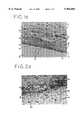
- FIG. 1a micrograph (1(a)) and a drawing (1(b)) are provided to illustrate a classical tissue response to an implanted foreign body.
- FIG. 1represents a typical histological section taken through a tissue block removed after approximately three weeks from a dorsal subcutaneous implant in a Sprague-Dawley rat. As illustrated, the implant 10 is surrounded by a foreign body capsule 12 that forms adjacent to the implant.
- the foreign body capsule 12typically consists of three-layers.
- the first layer 13 of the foreign body capsule 12includes macrophages 14 and foreign body giant cells 16 at an interface 18 between the implant 10 and the tissue.
- This first layer 13, consisting of the macrophages 14,is generally approximately 5 to about 15 microns thick.
- the next, or second layer 15, of the foreign body capsule 12includes fibroblasts 20.
- the fibroblasts 20are oriented parallel to the surface of the implant 10 and embedded in a collagenous matrix including collagen fibers that are also oriented parallel with the surface of the implant.
- the second layer 15 consisting of the fibroblasts 20 and collagen fibersis generally approximately 30 to about 200 microns thick.
- the first and second layers 13 and 15 of the foreign body capsule 12are usually completely avascular throughout.
- vascular structures 24begin to appear in the outer regions of the fibroblast second zone 15.
- a third layer 17lying approximately 30 to about 200 microns away from the surface of the implant 10 is loose connective tissue that is highly vascular. This layer 17 is amorphous and widely varies in thickness depending on the tissue location and time after the implant.
- the classical foreign body responseresults in the implant 10 being surrounded by a foreign body capsule 12 that does not include vascular structures near the surface of the implant.
- the foreign body capsule generated from the foreign body responseis desirable, or at least not detrimental, for certain types of implants, such as, for example, silicon breast implants and collagen implants
- the foreign body capsuleprevents certain applications and treatments utilizing such implants.
- indwelling sensors for applicationssuch as glucose analysis in diabetics, become occluded after only a few days due to the foreign body capsule. Indeed, the foreign body capsule becomes so thick that it inhibits the diffusion of glucose to the membrane surface preventing the sensor from functioning.
- pancreatic isletswhen pancreatic islets are implanted within a semipermeable membrane for treatment of diabetes, they usually die within a few days or weeks. The loss of function of the pancreatic islets is attributed to the poor diffusion of nutrients to the islets due to the thickness of the foreign body capsule. Likewise, other tissues that are implanted within the host do not remain viable due to the foreign body capsule that effectively prevents the transport of nutrients from the capillaries to cells enclosed within the implanted membrane.
- indwelling cathetersthat have a variety of applications, typically have a high drop-out rate because the site of the catheter entry becomes infected. It is generally believed that this infection is caused by poor adhesion of the tissues to the catheter surface and poor vascularization of the region around the catheter because of the thick foreign body capsule that forms. Implants have been proposed having surfaces designed to increase the adhesion or anchorage of the implant in the host tissue (e.g. European Patent Application No. 0359575 of Von Recum and Campbell). This patent application describes materials with surface topography designed to provide "improved soft tissue implant having a surface texture that optimizes anchorage of the implant to the tissue without causing inflammatory tissue at the implantation site.”
- the uses of the material of the present inventioninclude: as a coating for indwelling catheters; means for transport of physiological factors to indwelling sensors; means for transport of drugs from a chamber or catheter to the tissues of the host; and means for encapsulation of grafted cells for treatment of cell and molecular deficiency diseases (immunoisolation).
- the present inventionprovides an asymmetric material having a first zone that induces close vascularization at the material host interface and a second adjacent zone that prevents passage of cells through the zone.
- the vascularizing zoneallows the material to be vascularized while the second zone maintains immunoisolation of the interior of an implanted device incorporating the invention on its exterior.
- the materialmay consist of a bilayer of zones as described or it may be a gradient of zones. The gradient consists of an outer zone with a conformation that results in close vascularization. The structure of the material becomes gradually tighter until the material is impermeable to calls.
- the second adjacent zoneis molecular permeable for selective diffusion. In yet another embodiment the second zone is non-permeable for use in non-transport functions in devices such as indwelling catheters.
- the rounded macrophageis observed to have substantially conformed to the contours of the material. Although there is a correlation with macrophage shape, it is not clear that macrophages control the observed response. However, it is clear that invasion of the structure by host cells is required. Although the bulk of the cells appear to be macrophages, it is possible that other inflammatory cells control the response, therefore we will refer to the invading cells as "inflammatory cells,” which include but are not limited to macrophages.
- the material that results in formation of close vascular structuresis a polymer membrane having an average nominal pore size of approximately 0.6 to about 20 ⁇ m, using conventional methods for determination of pore size in the trade.
- at least approximately 50% of the pores of the membranehave an average size of approximately 0.6 to about 20 ⁇ m.
- the structural elements which provide the three dimensional conformationmay include fibers, strands, globules, cones or rods of amorphous or uniform geometry which are smooth or rough. These elements, hereafter referred to as "strands,” have in general one dimension larger than the other two and the smaller dimensions do not exceed five microns.
- an asymmetric materialhaving a gradient or layer of varying porosity. At least some of the apertures at the surface of the material that contacts the host tissue, allow inflammatory cells to enter the cavities. But, due to size restrictions, the apertures do not allow the inflammatory calls to transverse the material to the interior of the implant.
- an immunoisolation containerin an embodiment of the present invention, includes a first membrane having cavities and situated proximal to the host tissue. At least some of the apertures of the first membrane have a sufficient size to allow inflammatory cells to enter the cavities and cause at least some vascular structures to contact the membrane.
- the containerincludes a second porous membrane, the apertures of the second membrane being sufficiently small to prevent immune cells and/or immunogenic factors from entering an interior of the container.
- the second membraneis situated proximal to graft tissues.
- an indwelling catheterincluding a porous membrane and a catheter body, the porous membrane surrounding at least a portion of the catheter body. At least some apertures of the porous membrane have a sufficient size to allow inflammatory cells to enter the cavities and cause at least some vascular structures to form that contact the porous membrane.
- the present inventionprovides an indwelling sensor.
- the indwelling sensorcomprising a sensor for monitoring a condition or agent in the body and a porous membrane that surrounds at least a portion of the sensor body. At least some of the apertures of the membrane have a sufficient size to allow inflammatory cells to enter the cavities and cause at least some vascular structures to form that contact the porous membrane.
- the present inventionalso provides a method for the vascularization of a surface of an implanted device.
- the methodcomprises the steps of allowing inflammatory cells to enter a first layer of a membrane structure and cause vascular structures to form that contact a surface of the first layer of the membrane and preventing the inflammatory cells from entering a second layer of the membrane structure.
- This embodimentwould be applicable in, for example, a breast prosthesis.
- FIG. 1(a)is a micrograph that illustrates a classical foreign body response to an implanted device.
- FIG. 1(b)is a drawing illustrating a classical foreign body response to an implanted device.
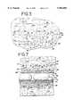
- FIG. 2(a)is a micrograph of an embodiment of the present invention.
- FIG. 2(b)is a cross-sectional view of an embodiment of the present invention with vascular structures growing at the host-material interface.
- FIG. 3illustrates a cross-sectional view of a foreign body capsule in a pore of a membrane.
- FIGS. 4(a) and (b)are scanning electron micrographs of, respectively, a mixed ester of cellulose membrane with a 5 ⁇ m pore size and a teflon membrane with 3 ⁇ m pore size.
- FIGS. 5(a) and (b)are scanning electron micrographs of, respectively, a teflon membrane with a 5 ⁇ m pore size and a polycarbonate with 12 ⁇ m pore size.
- FIG. 6illustrates a light micrograph showing the teflon membrane of FIG. 5(a) implanted for 3 weeks in a subcutaneous dorsal pocket in a rat.
- FIG. 8illustrates a cross-sectional view of a further embodiment of the present invention.
- FIG. 9illustrates an indwelling catheter incorporating the present invention.
- FIG. 10illustrates an indwelling sensor incorporating the present invention.
- the present inventionprovides a material for inducing close vascularization at the interface between the material and host into which the material is implanted such that a standard foreign body capsule consisting of flattened macrophages, foreign body giant cells, and fibroblasts does not intervene between the vascular structures and the material.
- the materialcan be utilized for various applications including the creation of a container for implanting tissues to be isolated from the immune system of a host, for surrounding a portion of a catheter, or surrounding a portion of an indwelling sensor device.
- the material utilizedresults in the growth of vascular structures close to or immediately adjacent to the material.
- close vascular structures or vascular structures that contactare those capillaries whose surface lies within about one cell layer of the surface of the material.
- a polymer membrane 30at least partially surrounds an implant and includes three dimensional cavities 32. At least some of the cavities 32 of the membrane 30 have a sufficient size and structure to allow inflammatory cells 34 to completely enter therein through the apertures that define the cavities, and are defined by frames composed of strands that are less than five microns in all but the longest dimension.
- growth of vascular structures 36occurs within about one cell layer from the interface 35 of the membrane 30 and host.
- vascular structuresmay be formed within the irregularities 32 of the membrane.
- a foreign body-like capsule of fibroblastsstill forms that surrounds the membrane 30, the entire foreign body-like capsule, including fibroblast layers, is well vascularized.
- the formation of close vascular structuresis dependent on entry of the inflammatory cells into the cavities of the membrane so that the cells are surrounded by the strands that define the apertures and cavities.
- the topographic features at the implant surfacedo not effect the morphology of the inflammatory cells. Indeed, inflammatory cells at the implant surface often maintain a flat morphology.
- the size and shape of the strands and cavities 32 for the material 30 of the present inventionit must first be appreciated that not all of the cavities must have a sufficient size to allow inflammatory cells 34 to enter therein. What is required is that a sufficient number of cavities 32 have a size that allows a sufficient number of inflammatory cells 34 to enter therein. Nor is it necessary that all of the strands be less than five microns in all but the longest dimension. Some strands may be longer, as long as a sufficient number of the strands are within the prescribed size limits. The presence of a sufficient number of strands and cavities of the prescribed size creates a sufficient number of vascular structures at the host-material interface. These vascular structures will provide sufficient nutrients to an immunoisolated container and/or allow components and agents produced by cells within the interior of the chamber to enter the host.
- the cavities 32must have a sufficient size and shape to allow inflammatory cells 34 to enter therein, it is also important that extensive ingrowth of vascular and connective tissues within the cavities 32 does not occur. As illustrated in FIG. 3, in the case where the apertures and cavities are too large, an extensive growth of vascular tissue 36 and connective tissue 39 occurs within a large cavity 32a; this causes the vascular tissue to be isolated within the large cavity. The isolation of the vascular tissue 36 within the large cavity 32a by fibroblasts and connective tissues 39 is similar to the standard foreign body response previously discussed. By selecting cavities 32 of appropriate size, one can prevent the formation of fibroblasts and connective tissue 39 therein.
- a porous polymer membrane having an average nominal pore size of approximately 0.6 to about 20 microns and average strand sizes of less than about five microns in all but the longest dimensionfunctions satisfactorily in creating a vascular bed at the tissue-membrane interface.
- nominal pore sizeis derived from methods of analysis common to the membrane trade, such as the ability of the membrane to filter particles of a particular size, or the resistance of the membrane to the flow of fluids. Because of the amorphous, random and irregular nature of most of these commercially available membranes, the "pore" size designation does not actually indicate the size or shape of the apertures and cavities, which in reality have a high degree of variability.
- the cavitiesare not really "pores" in that they typically are not uniform regular holes or channels through the material.
- these commercial membranescan be composed of, for example, extruded filaments which act as sieves as shown, for example, in FIG. 4b.
- pore sizeis a manufacturer's convention used to identify a particular membrane of a particular commercial source which has a certain bubble point.
- the term "pore”does not describe the size of the cavities of the material used in the instant invention. The bubble point measurement is described in Pharmaceutical Technology May 1983 pp. 36 to 42.
- the apertures 32(FIG. 2) of the material 30 allow inflammatory cells 34 to penetrate the material or, conversely prevent connective tissues from forming within the cavities. What is required is that a sufficient number of the cavities 32 have a size that allows inflammatory cells 34 to enter therein and yet prevent connective tissue from forming therein. In the materials tested by Applicants the desired result is obtained where the strands that define the apertures of the cavities have a size of less than about five microns in all but the longest dimension.
- membranes created from mixed esters of cellulose and having nominal pore sizes of 0.1, 0.22, and 0.45 micronsdid not induce close vascular structures when subcutaneously implanted into rats.
- mixed esters of cellulose membranes with nominal pore sizes of 1.2 and 8 micronsdid induce close vascular structures.
- cellulose acetate membranes having a nominal pore size of 0.2, 0.45, and 0.65 microns and teflon membranes having a nominal pore size of 0.02 and 0.2 micronsdid not induce close vascular structures.
- cellulose acetate membranes having a nominal pore size of 0.8, 1.2, 3, 5, and 8 microns, and teflon membranes having a nominal pore size of 1.0 and 3.0 micronsdid induce close vascular structures.
- SEMScanning Electron Microscope analysis of the membranes revealed three dimensional structural or architectural properties that distinguish membranes that do have close vascular structures (positive response) from those that do not (negative response).
- Membranes with a positive responsehad high porosities and were composed of strands (fibers, filaments, microglobules, cone-like or rod-like structures with a small diameter ( ⁇ 5 microns)).
- Millipore brand mixed esters of cellulose membranes with nominal pore size of 5 ⁇ mare composed of irregular, amorphous globular structures and strands with diameters from about 1 to 3 ⁇ m, and irregular cavities from 0.5 to 5 microns in diameter, and having a percent porosity of 75% (FIG. 4a).
- Gore® teflon membranes with a nominal pore size of 3 ⁇ mare composed of strands with diameters of less than about 1 micron that interconnect with teflon clusters less than about 3 microns in diameter (FIG. 4b).
- the cavitiesare very elongated being generally about 1 to 2 microns wide by 10 to 15 ⁇ m long. After implantation, both of these membranes were invaded by inflammatory cells which had a round morphology under the light microscope (see invading cells in FIG. 2), and both consistently had close vascular structures.
- membranes with a negative responsehad apertures and cavities defined by strands with a relatively high surface area, large enough for inflammatory cells to use as a substrate to flatten against.
- Millipore brand teflon membranes with a nominal pore size of 5 micronsare composed of globular or plate-like structures about 5 to 10 microns in diameter, and have irregular amorphous cavities about 5 to 10 microns in diameter.
- Nuclepore brand membranes with a nominal pore size of 12 microns(FIG. 5b), have uniform circular holes that are 9 microns in diameter that are scattered within a membrane sheet, with from 5 to 25 microns between the edges of the holes. After implantation, both of these membranes were invaded by cells but the cells maintained a flattened morphology (see invading cells in FIG. 6).
- materials with a positive responsehad structural features that caused penetrating cells to assume a round morphology. Whereas materials with a negative response had structural features that caused penetrating cells to assume a relatively flattened morphology.
- FIG. 6is a light micrograph illustrating a teflon membrane (the same membrane illustrated in FIG. 5a) implanted for 3 weeks in a subcutaneous dorsal pocket in a rat. Note the extensive cytoplasm of the cells invading the polytetrafluoroethylene (“PTFE”) membrane shown in FIG. 6. The cells appear to have flattened against the plate-like PTFE structure and have the appearance of cells of a standard foreign body response (FIG. 1) in contrast to the rounded cells invading the membrane in FIG. 2.
- PTFEpolytetrafluoroethylene
- the maintenance of a long-term foreign body responseis characterized by inflammatory cells which spread upon and cover the foreign material. Applicants have discovered that this response appears to require a surface-like area capable of acting as a substrate for flattening and spreading of the cells.
- the implanted materialhas an architecture of strands that have a diameter ( ⁇ 5 ⁇ m) too small or configuration too irregular to allow a surface for flattening of cells, as do the membranes that give a positive vascular response (FIG. 2 and Table 2)
- the efforts of the inflammatory cells to cover and wall off the materialare thwarted, and the cells do not obtain a flattened morphology. Instead, they remain rounded and Applicants hypothesize that the inflammatory cells induce the formation of close vascular structures at the material-host interface.
- the implanted materialis never completely walled off, and therefore a chronic response is never obtained.
- inflammatory cellswhich leads to foreign body capsule formation
- the implant materialprovides a structure onto which the inflammatory cells can flatten and spread.
- An inflammatory celldoes not require a smooth area for flattening. For example, an area composed of closely adjacent pillars of equal height and diameter might be recognized by the inflammatory cell as essentially "smooth" and the cells would then spread on the surface.
- Applicantsfurther hypothesize that if the inflammatory cell nucleus cannot enter a cavity or irregularity then the cell will "see” the material as flat and will flatten onto the material at that location. Conversely, cells in contact with a cavity or irregularity from more than one direction or plane will not "see” a flat area and will retain a rounded conformation or even conform to the shape of the cavity or irregularity. Accordingly, material having a surface-like area greater than about 5 microns would not be likely to result in close vascularization. For example, the material shown in FIG. 5a which gave a negative response has many cavities and irregularities which are smaller than about 6 um, but it also has leafy-appearing somewhat flat structures onto which macrophage may flatten.
- materialmust be selected so that it has sufficient irregularities and cavities to prevent substantial numbers of inflammatory cells from flattening.
- the rounded cellmay conform to the cavities and irregularities but will not flatten. Formation of some flattened cells, especially at the "surface" of the implant is often seen and is within the scope of the invention provided that there are not so many flattened cells that the material is walled off by nonvascularized fibroblasts.
- Macrophage behavioris not yet fully understood. It is believed that macrophages are activated when they become flat. Upon activation they are believed to secret factors which signal fibroblasts to form and proliferate. Accordingly, Applicants hypothesize that by utilizing a material whose three dimensional cavities and irregularities prevent the macrophage from flattening, this invention will avoid macrophage activation and consequent formation of the typical foreign body capsule. On the other hand, it may be that rounded macrophages are secreting factors that either stimulate neovascularization directly or interrupt an existing suppression of new vascularization.
- the host inflammatory cell response described above for the various materialsis generally observed for up to about 12 weeks following implantation. Thereafter, in both the standard foreign body capsule response and in the use of the instant invention, the inflammatory cells gradually diminish leaving either a stable foreign body capsule or, in the instant invention, a stable vascularized bed.
- the Applicantshave observed a stable vascular bed for 1.5 years in subcutaneous implants of 3 ⁇ m GoreTM teflon in rats.
- a vascularized membraneis achieved.
- the endothelial cells that make up the capillary wallsare immediately adjacent to or very close to the material-host interface. There are no, or few, intervening macrophages or fibroblasts. Accordingly, molecules coming through the material will be at the surface of an endothelial cell for transportation into the capillaries. For example, molecules secreted by pancreatic islet cells on one side of the material will be available for uptake by capillaries on the other side of the material. Likewise, molecules such as glucose coming from the capillary, will be sensed by islet cells contained within an implanted chamber made of the material. The resistance to diffusion of such molecules will be related to the distance necessary to traverse the material.
- the capillariesare not in contact with or adjacent to the material but rather typically lie at some distance from the material due to a halo of macrophages and fibroblasts in a connective tissue matrix that surrounds the capillaries as illustrated in FIG. 3.
- the capillariesare separated from the polymer surface by several layers of cells producing the same kind of diffusive resistance encountered in a classical foreign body response.
- the implantin contrast to the present invention, in a typical implant, the implant is encapsulated by the foreign body capsule and is typically at the edges of a large cellular vascular space, see FIG. 1.
- the close vascularization of the present inventionimproves on previous biopolymer implants because the vascular bed is formed immediately adjacent to the material-host interface.
- this method of vascularizationhas a variety of applications.
- the materialcan be used in conjunction with an indwelling sensor, an indwelling catheter, and for an immunoisolation container.
- an immunoisolation membrane 42is illustrated. As illustrated, the membrane 42 is selected such that it allows macrophages 34 to enter at least some of the cavities 44 of the membrane causing vascular structures 46 to be formed at the host-membrane interface 47. Again, it should be noted that although some vascular structures can be formed within the cavities 44 of the membrane 42, this is not critical to the success of the material or the creation of a vascular bed.
- the membrane 42surrounds at least a portion of a second membrane or layer 50 that defines an immunoisolated interior 52.
- This interior 52can include tissue 54 that must be protected from contact by host cells that would reject the implanted cells.
- tissue 54that must be protected from contact by host cells that would reject the implanted cells.
- allografts or xenografts or in the case of isografts, such as autologous implants of genetically engineered cellsthe membrane would need only to prevent passage of cells to prevent movement of the genetically engineered cells, which often contain retroviral vectors, out of the membrane enclosures and into the host tissues.
- This isolation of graft tissues from host tissuerepresents a significant advance over previous methods used for autologous transplantation of genetically engineered cells, because it prevents the genetically engineered cells from potentially invading host tissues in an unregulated manner and causing tumors in the host via the retroviral vector.
- the second membrane 50allow for the diffusion of components generated by the tissues 54, for example, insulin from pancreatic islets. Likewise, it is desirable that the second membrane 50 allow nutrients from the host to enter the interior 52 of the implant and nourish the tissue 54. To this end, the second membrane 50 preferably includes pores 56 that allow glucose or other components to diffuse into the first membrane 42 but prevents macrophages 34 and/or humoral factors from entering the second membrane.
- the device illustrated in FIG. 7includes two membrane layers, it should be noted that other constructions can be utilized.
- the deviceincludes a single membrane 61 that includes cavities 62 having a gradient of size.
- the larger outer cavities 62allow macrophages to enter at least an outer portion 64 of the cavity 62, causing vascularization at the host-membrane interface 65.
- the smaller inner cavities 66prevent macrophages from entering an inner portion of the membrane and thereby isolating an interior 68 defined by the membrane.
- an indwelling catheter 70 including an embodiment of the material 72 of the present inventionis illustrated.
- a catheter 70can be, for example, a catheter for continuous ambulatory peritoneal dialysis.
- the material 72covers the wall 74 of the catheter 70 and allows the creation of a vascular bed around the catheter 70.
- the wall 74 of the catheter 70is preferably impenetrable to both cells and molecules.
- a thick foreign body of nonvascularized collagenous materialis produced around the catheter that acts as a conduit for bacteria.
- vascularization around the catheterprevents tunnel site infections because necrosis of the tissue is prevented and the vascular bed bathes the area with the entire repertoire of blood borne immune cells.
- a flange on a catheterwould be covered with a vascularizing material, or would be made entirely from the material.
- a sensor 80 including an embodiment of the material 82 of the present inventionis illustrated.
- a sensor 80can include, for example, a glucose sensor for monitoring glucose levels in diabetics.
- the material 82covers a body 84 which contains an electrode 85 of the sensor 80 and causes a vascular bed 86 to be created around the sensor 80.
- the creation of the vascular bedcircumvents the problem of foreign body occlusion typically encountered with indwelling sensors.
- the vascular responseis believed to be unrelated to the composition of the material. This is illustrated by the above examples wherein similar responses of the tissue were found with respect to hydrophilic (cellulose) and hydrophobic (teflon) materials. Therefore, the inventors believe that the material can be constructed from a variety of polymers including, inter alia, polyethylene, polypropylene, teflon, cellulose acetate, cellulose nitrate, polycarbonate, polyester, nylon, polyester, polysulfone, mixed esters of cellulose polyvinylidene difluoride, silicone, and polyacrylonitrile.
- Known biocompatible medical implantsare composed of ceramics and metals. Assuming these materials could be manipulated to provide the three dimensional structures described herein, they would also be useful in the present invention.
Landscapes
- Health & Medical Sciences (AREA)
- Life Sciences & Earth Sciences (AREA)
- Veterinary Medicine (AREA)
- Public Health (AREA)
- Chemical & Material Sciences (AREA)
- General Health & Medical Sciences (AREA)
- Animal Behavior & Ethology (AREA)
- Transplantation (AREA)
- Engineering & Computer Science (AREA)
- Biomedical Technology (AREA)
- Epidemiology (AREA)
- Oral & Maxillofacial Surgery (AREA)
- Chemical Kinetics & Catalysis (AREA)
- Medicinal Chemistry (AREA)
- Dermatology (AREA)
- Vascular Medicine (AREA)
- Cell Biology (AREA)
- Zoology (AREA)
- Dispersion Chemistry (AREA)
- Cardiology (AREA)
- Heart & Thoracic Surgery (AREA)
- Botany (AREA)
- Materials For Medical Uses (AREA)
- Prostheses (AREA)
- Medicines Containing Material From Animals Or Micro-Organisms (AREA)
- Magnetic Resonance Imaging Apparatus (AREA)
- Chair Legs, Seat Parts, And Backrests (AREA)
- Electrotherapy Devices (AREA)
- Photoreceptors In Electrophotography (AREA)
- Acyclic And Carbocyclic Compounds In Medicinal Compositions (AREA)
- Design And Manufacture Of Integrated Circuits (AREA)
- Medicinal Preparation (AREA)
Abstract
Description
TABLE I ______________________________________ MEMBRANES THAT ARE NOT INVADED BY CELLS AND DO NOT HAVE CLOSE VASCULAR STRUCTURES Nominal Company Membrane Pore Size ______________________________________ Millipore Mixed Esters Cellulose 0.1 Millipore Mixed Esters Celluose 0.22 Millipore Mixed Esters Celluose 0.45 Celenase polypropylene 0.05 Celenase polypropylene 0.075 Gore PTFE/Polyester 0.02 Gore PTFE/Polyester 0.2 Akzo polypropylene 0.01-0.29 Akzo polypropylene 0.02-0.58 Akzo polyethylene 0.1 Akzo polyethylene 0.08 Akzo polyethylene 0.6 Supor polysulfone 0.1 Amicon YC, YM, PM, XM 10-300 kD Omega polyethersulfone 100-30okD Millipore Durapore ® 0.22 Millipore Immobilon-n ® 0.22 Gelman Versapore ® 0.22 Gelman Supor ® 0.22 Gelman Supor ® 0.8 Gelman Polysulfone HT-200 0.22 Gelman Polysulfone HT-200 0.6 Gelman Polyester 0.22 Gelman Polysulfone/polyester 0.8 Sartorius Cellulose Acetate 0.22 Sartorius Cellulose Acetate 0.22 Sartorius Cellulose Acetate 0.45 Sartorius Cellulose Acetate 0.65 Sartorius Cellulose Nitrate 0.22 Sartorius Reinforced Cell. Acet. 0.22 Nucleopore Polyester 0.8 Pall Uncharged Nylon 0.22 AMF Cumo Charged Nylon 0.22Micron Separation Nylon 66 0.22 Inc. Micro Filtration Cellulose Acetate 0.22 Sys. Micro Filtration Cellulose Acetate 0.22 Sys. Akzo Polypropylene-HF 0.2-0.8 ______________________________________
TABLE 2 ______________________________________ MEMBRANES THAT ARE INVADED BY CELLS AND HAVE CLOSE VASCULAR STRUCTURES Nominal Company Membrane Pore Size ______________________________________ Millipore Mixed Esters Cellulose 1.2 Millipore Mixed Esters Cellulose 8.0 Sartorius Cellulose Acetate 0.8 Sartorius Cellulose Acetate 1.2 Sartorius Cellulose Acetate 3.0 Sartorius Cellulose Acetate 5.0 Sartorius Cellulose Acetate 8.0 Gore PTFE/Polyester 1.0 Gore PTFE/Polypropylene 3.0 Gore PTFE/Polyester 3.0 Gelman Versapore ® 0.8 Gelman Versapore ® 1.2 Gelman Versapore ® 3.0 Gelman Versapore ® 5.0 ______________________________________
TABLE 3 ______________________________________ MEMBRANES THAT ARE INVADED BY CELLS BUT DO NOT HAVE CLOSE VASCULAR STRUCTURES Nominal Company Membrane Pore Size ______________________________________ Tetco Polyester 3 Tetco Polyester 5 Tetco Polyester 8Tetco Nylon 10Tetco Nylon 10Tetco Nylon 10 Millipore PTFE 5Millipore PTFE 10 Nucleopore Polycarbonate 1 Nucleopore Polycarbonate 3 Nucleopore Polycarbonate 8Nucleopore Polycarbonate 12 ______________________________________
Claims (4)
Priority Applications (1)
| Application Number | Priority Date | Filing Date | Title |
|---|---|---|---|
| US08/484,011US5964804A (en) | 1990-10-31 | 1995-06-07 | Close vascularization implant material |
Applications Claiming Priority (5)
| Application Number | Priority Date | Filing Date | Title |
|---|---|---|---|
| US60679190A | 1990-10-31 | 1990-10-31 | |
| US73540191A | 1991-07-24 | 1991-07-24 | |
| US93387192A | 1992-08-21 | 1992-08-21 | |
| US08/210,068US5782912A (en) | 1990-10-31 | 1994-03-17 | Close vascularization implant material |
| US08/484,011US5964804A (en) | 1990-10-31 | 1995-06-07 | Close vascularization implant material |
Related Parent Applications (1)
| Application Number | Title | Priority Date | Filing Date |
|---|---|---|---|
| US08/210,068ContinuationUS5782912A (en) | 1990-10-31 | 1994-03-17 | Close vascularization implant material |
Publications (1)
| Publication Number | Publication Date |
|---|---|
| US5964804Atrue US5964804A (en) | 1999-10-12 |
Family
ID=27085331
Family Applications (5)
| Application Number | Title | Priority Date | Filing Date |
|---|---|---|---|
| US08/210,068Expired - LifetimeUS5782912A (en) | 1990-10-31 | 1994-03-17 | Close vascularization implant material |
| US08/481,886Expired - LifetimeUS5800529A (en) | 1990-10-31 | 1995-06-07 | Close vascularization implant material |
| US08/484,011Expired - LifetimeUS5964804A (en) | 1990-10-31 | 1995-06-07 | Close vascularization implant material |
| US08/485,632Expired - LifetimeUS5741330A (en) | 1990-10-31 | 1995-06-07 | Close vascularization implant material |
| US08/480,198Expired - LifetimeUS5882354A (en) | 1990-10-31 | 1995-06-07 | Close vascularization implant material |
Family Applications Before (2)
| Application Number | Title | Priority Date | Filing Date |
|---|---|---|---|
| US08/210,068Expired - LifetimeUS5782912A (en) | 1990-10-31 | 1994-03-17 | Close vascularization implant material |
| US08/481,886Expired - LifetimeUS5800529A (en) | 1990-10-31 | 1995-06-07 | Close vascularization implant material |
Family Applications After (2)
| Application Number | Title | Priority Date | Filing Date |
|---|---|---|---|
| US08/485,632Expired - LifetimeUS5741330A (en) | 1990-10-31 | 1995-06-07 | Close vascularization implant material |
| US08/480,198Expired - LifetimeUS5882354A (en) | 1990-10-31 | 1995-06-07 | Close vascularization implant material |
Country Status (20)
| Country | Link |
|---|---|
| US (5) | US5782912A (en) |
| EP (1) | EP0507933B1 (en) |
| JP (1) | JP3508023B2 (en) |
| KR (1) | KR0169495B1 (en) |
| CN (1) | CN1063046A (en) |
| AT (1) | ATE138256T1 (en) |
| AU (1) | AU645155B2 (en) |
| BR (1) | BR9106205A (en) |
| CA (1) | CA2070816A1 (en) |
| DE (1) | DE69119748T2 (en) |
| DK (1) | DK0507933T3 (en) |
| ES (1) | ES2090364T3 (en) |
| FI (1) | FI923023L (en) |
| GR (1) | GR3020673T3 (en) |
| IE (1) | IE75706B1 (en) |
| IL (1) | IL99732A (en) |
| MX (1) | MX9101734A (en) |
| NO (1) | NO300993B1 (en) |
| TW (1) | TW393322B (en) |
| WO (1) | WO1992007525A1 (en) |
Cited By (77)
| Publication number | Priority date | Publication date | Assignee | Title |
|---|---|---|---|---|
| US6459917B1 (en)* | 2000-05-22 | 2002-10-01 | Ashok Gowda | Apparatus for access to interstitial fluid, blood, or blood plasma components |
| US20040014704A1 (en)* | 2002-07-18 | 2004-01-22 | Gonzalo Hortelano | Oral administration of therapeutic agent coupled to transporting agent induces tolerance |
| US20040014698A1 (en)* | 2002-07-18 | 2004-01-22 | Gonzalo Hortelano | Oral administration of therapeutic agent coupled to transporting agent |
| US20040016013A1 (en)* | 2002-07-18 | 2004-01-22 | Gonzalo Hortelano | Transgenic animals produced using oral administration of a genetic agent coupled to a transporting agent |
| US20040126404A1 (en)* | 1998-08-21 | 2004-07-01 | University Of Queensland Of St. Lucia | Implant material |
| US20050208032A1 (en)* | 2004-01-16 | 2005-09-22 | Gonzalo Hortelano | Oral administration of therapeutic agent coupled to transporting agent |
| US20050251083A1 (en)* | 2004-02-12 | 2005-11-10 | Victoria Carr-Brendel | Biointerface with macro-and micro-architecture |
| US20050260178A1 (en)* | 1999-06-29 | 2005-11-24 | Cell Based Delivery | Delivery of an organized tissue to an organism |
| US20060078847A1 (en)* | 2000-09-29 | 2006-04-13 | Kwan Norman H | Dental implant system and additional methods of attachment |
| US20060178697A1 (en)* | 2005-02-04 | 2006-08-10 | Carr-Brendel Victoria E | Vaso-occlusive devices including non-biodegradable biomaterials |
| US7110803B2 (en) | 1997-03-04 | 2006-09-19 | Dexcom, Inc. | Device and method for determining analyte levels |
| US7134999B2 (en) | 2003-04-04 | 2006-11-14 | Dexcom, Inc. | Optimized sensor geometry for an implantable glucose sensor |
| US7192450B2 (en) | 2003-05-21 | 2007-03-20 | Dexcom, Inc. | Porous membranes for use with implantable devices |
| US7226978B2 (en) | 2002-05-22 | 2007-06-05 | Dexcom, Inc. | Techniques to improve polyurethane membranes for implantable glucose sensors |
| US7310544B2 (en) | 2004-07-13 | 2007-12-18 | Dexcom, Inc. | Methods and systems for inserting a transcutaneous analyte sensor |
| US20080268017A1 (en)* | 1998-08-21 | 2008-10-30 | The University Of Queensland Of St. Lucia | Method of producing tissue by placing a molding support within a body cavity |
| US7471972B2 (en) | 2001-07-27 | 2008-12-30 | Dexcom, Inc. | Sensor head for use with implantable devices |
| US7494465B2 (en) | 2004-07-13 | 2009-02-24 | Dexcom, Inc. | Transcutaneous analyte sensor |
| US7613491B2 (en) | 2002-05-22 | 2009-11-03 | Dexcom, Inc. | Silicone based membranes for use in implantable glucose sensors |
| US7632228B2 (en) | 2001-07-27 | 2009-12-15 | Dexcom, Inc. | Membrane for use with implantable devices |
| US7640048B2 (en) | 2004-07-13 | 2009-12-29 | Dexcom, Inc. | Analyte sensor |
| US7711402B2 (en) | 1997-03-04 | 2010-05-04 | Dexcom, Inc. | Device and method for determining analyte levels |
| US20100124564A1 (en)* | 2008-11-14 | 2010-05-20 | Laura Martinson | Encapsulation of pancreatic cells derived from human pluripotent stem cells |
| US7774145B2 (en) | 2003-08-01 | 2010-08-10 | Dexcom, Inc. | Transcutaneous analyte sensor |
| US7783333B2 (en) | 2004-07-13 | 2010-08-24 | Dexcom, Inc. | Transcutaneous medical device with variable stiffness |
| US20100272772A1 (en)* | 2009-02-28 | 2010-10-28 | Charles Knezevich | Apparatus, system, and method for creating immunologically enhanced spaces in-vivo |
| US7860544B2 (en) | 1998-04-30 | 2010-12-28 | Abbott Diabetes Care Inc. | Analyte monitoring device and methods of use |
| US7860545B2 (en) | 1997-03-04 | 2010-12-28 | Dexcom, Inc. | Analyte measuring device |
| US7875293B2 (en) | 2003-05-21 | 2011-01-25 | Dexcom, Inc. | Biointerface membranes incorporating bioactive agents |
| US7896809B2 (en) | 2003-07-25 | 2011-03-01 | Dexcom, Inc. | Dual electrode system for a continuous analyte sensor |
| US7920907B2 (en) | 2006-06-07 | 2011-04-05 | Abbott Diabetes Care Inc. | Analyte monitoring system and method |
| US7976778B2 (en) | 2001-04-02 | 2011-07-12 | Abbott Diabetes Care Inc. | Blood glucose tracking apparatus |
| US8060174B2 (en) | 2005-04-15 | 2011-11-15 | Dexcom, Inc. | Analyte sensing biointerface |
| US8160669B2 (en) | 2003-08-01 | 2012-04-17 | Dexcom, Inc. | Transcutaneous analyte sensor |
| US8233959B2 (en) | 2003-08-22 | 2012-07-31 | Dexcom, Inc. | Systems and methods for processing analyte sensor data |
| US8260393B2 (en) | 2003-07-25 | 2012-09-04 | Dexcom, Inc. | Systems and methods for replacing signal data artifacts in a glucose sensor data stream |
| US8275437B2 (en) | 2003-08-01 | 2012-09-25 | Dexcom, Inc. | Transcutaneous analyte sensor |
| US8280475B2 (en) | 2004-07-13 | 2012-10-02 | Dexcom, Inc. | Transcutaneous analyte sensor |
| US8287454B2 (en) | 1998-04-30 | 2012-10-16 | Abbott Diabetes Care Inc. | Analyte monitoring device and methods of use |
| US8290559B2 (en) | 2007-12-17 | 2012-10-16 | Dexcom, Inc. | Systems and methods for processing sensor data |
| US8346337B2 (en) | 1998-04-30 | 2013-01-01 | Abbott Diabetes Care Inc. | Analyte monitoring device and methods of use |
| US8364229B2 (en) | 2003-07-25 | 2013-01-29 | Dexcom, Inc. | Analyte sensors having a signal-to-noise ratio substantially unaffected by non-constant noise |
| US8417312B2 (en) | 2007-10-25 | 2013-04-09 | Dexcom, Inc. | Systems and methods for processing sensor data |
| US8423113B2 (en) | 2003-07-25 | 2013-04-16 | Dexcom, Inc. | Systems and methods for processing sensor data |
| US8465425B2 (en) | 1998-04-30 | 2013-06-18 | Abbott Diabetes Care Inc. | Analyte monitoring device and methods of use |
| US8483793B2 (en) | 2003-12-05 | 2013-07-09 | Dexcom, Inc. | Dual electrode system for a continuous analyte sensor |
| US8565848B2 (en) | 2004-07-13 | 2013-10-22 | Dexcom, Inc. | Transcutaneous analyte sensor |
| US8612159B2 (en) | 1998-04-30 | 2013-12-17 | Abbott Diabetes Care Inc. | Analyte monitoring device and methods of use |
| US8632489B1 (en) | 2011-12-22 | 2014-01-21 | A. Mateen Ahmed | Implantable medical assembly and methods |
| US8652043B2 (en) | 2001-01-02 | 2014-02-18 | Abbott Diabetes Care Inc. | Analyte monitoring device and methods of use |
| US8688188B2 (en) | 1998-04-30 | 2014-04-01 | Abbott Diabetes Care Inc. | Analyte monitoring device and methods of use |
| WO2014138691A1 (en) | 2013-03-07 | 2014-09-12 | Viacyte, Inc. | 3-dimensional large capacity cell encapsulation device assembly |
| USD720469S1 (en) | 2013-03-07 | 2014-12-30 | Viacyte, Inc. | Cell encapsulation device |
| US8974386B2 (en) | 1998-04-30 | 2015-03-10 | Abbott Diabetes Care Inc. | Analyte monitoring device and methods of use |
| US9066695B2 (en) | 1998-04-30 | 2015-06-30 | Abbott Diabetes Care Inc. | Analyte monitoring device and methods of use |
| US9135402B2 (en) | 2007-12-17 | 2015-09-15 | Dexcom, Inc. | Systems and methods for processing sensor data |
| US9247901B2 (en) | 2003-08-22 | 2016-02-02 | Dexcom, Inc. | Systems and methods for replacing signal artifacts in a glucose sensor data stream |
| US9247900B2 (en) | 2004-07-13 | 2016-02-02 | Dexcom, Inc. | Analyte sensor |
| US9381112B1 (en) | 2011-10-06 | 2016-07-05 | William Eric Sponsell | Bleb drainage device, ophthalmological product and methods |
| US9763609B2 (en) | 2003-07-25 | 2017-09-19 | Dexcom, Inc. | Analyte sensors having a signal-to-noise ratio substantially unaffected by non-constant noise |
| WO2018089011A1 (en) | 2016-11-10 | 2018-05-17 | Viacyte, Inc | Pdx1 pancreatic endoderm cells in cell delivery devices and methods thereof |
| US9986942B2 (en) | 2004-07-13 | 2018-06-05 | Dexcom, Inc. | Analyte sensor |
| USD856517S1 (en) | 2016-06-03 | 2019-08-13 | Musculoskeletal Transplant Foundation | Asymmetric tissue graft |
| US10391156B2 (en) | 2017-07-12 | 2019-08-27 | Viacyte, Inc. | University donor cells and related methods |
| US10610136B2 (en) | 2005-03-10 | 2020-04-07 | Dexcom, Inc. | System and methods for processing analyte sensor data for sensor calibration |
| USD895812S1 (en) | 2018-09-07 | 2020-09-08 | Musculoskeletal Transplant Foundation | Soft tissue repair graft |
| US10791928B2 (en) | 2007-05-18 | 2020-10-06 | Dexcom, Inc. | Analyte sensors having a signal-to-noise ratio substantially unaffected by non-constant noise |
| US10813743B2 (en) | 2018-09-07 | 2020-10-27 | Musculoskeletal Transplant Foundation | Soft tissue repair grafts and processes for preparing and using same |
| US10945831B2 (en) | 2016-06-03 | 2021-03-16 | Musculoskeletal Transplant Foundation | Asymmetric tissue graft |
| US11399745B2 (en) | 2006-10-04 | 2022-08-02 | Dexcom, Inc. | Dual electrode system for a continuous analyte sensor |
| US11559260B2 (en) | 2003-08-22 | 2023-01-24 | Dexcom, Inc. | Systems and methods for processing analyte sensor data |
| US11589823B2 (en) | 2003-08-22 | 2023-02-28 | Dexcom, Inc. | Systems and methods for replacing signal artifacts in a glucose sensor data stream |
| US11633133B2 (en) | 2003-12-05 | 2023-04-25 | Dexcom, Inc. | Dual electrode system for a continuous analyte sensor |
| WO2023164171A2 (en) | 2022-02-25 | 2023-08-31 | Viacyte, Inc. | Multilayer implantable cell encapsulation devices and methods thereof |
| US11951136B2 (en) | 2017-12-12 | 2024-04-09 | The Regents Of The University Of California | Preservation of pancreatic islet grafts in the extrahepatic space |
| US11963862B2 (en) | 2018-08-22 | 2024-04-23 | Boston Scientific Scimed, Inc. | Cell encapsulation device including a porous tube |
| US12016327B2 (en) | 2017-03-20 | 2024-06-25 | Boston Scientific Medical Device Limited | Cell encapsulation device |
Families Citing this family (166)
| Publication number | Priority date | Publication date | Assignee | Title |
|---|---|---|---|---|
| US5733336A (en)* | 1990-10-31 | 1998-03-31 | Baxter International, Inc. | Ported tissue implant systems and methods of using same |
| ES2107537T3 (en)* | 1991-04-25 | 1997-12-01 | Univ Brown Res Found | IMMUNO INSULATED BIOCOMPATIBLE VEHICLE IMPLANTABLE TO SUPPLY SELECTED THERAPEUTIC PRODUCTS. |
| US5800829A (en)* | 1991-04-25 | 1998-09-01 | Brown University Research Foundation | Methods for coextruding immunoisolatory implantable vehicles with a biocompatible jacket and a biocompatible matrix core |
| US5387237A (en)* | 1992-07-30 | 1995-02-07 | The University Of Toledo | Bioartificial pancreas |
| CA2147626C (en) | 1993-08-10 | 1998-04-21 | Mark D. Butler | Cell encapsulating device |
| US5540718A (en)* | 1993-09-20 | 1996-07-30 | Bartlett; Edwin C. | Apparatus and method for anchoring sutures |
| EP0670738A1 (en)* | 1993-09-24 | 1995-09-13 | Baxter International Inc. | Methods for enhancing vascularization of implant devices |
| US5549675A (en)* | 1994-01-11 | 1996-08-27 | Baxter International, Inc. | Method for implanting tissue in a host |
| US5716404A (en)* | 1994-12-16 | 1998-02-10 | Massachusetts Institute Of Technology | Breast tissue engineering |
| US6123727A (en) | 1995-05-01 | 2000-09-26 | Massachusetts Institute Of Technology | Tissue engineered tendons and ligaments |
| US5855610A (en) | 1995-05-19 | 1999-01-05 | Children's Medical Center Corporation | Engineering of strong, pliable tissues |
| US6060640A (en)* | 1995-05-19 | 2000-05-09 | Baxter International Inc. | Multiple-layer, formed-in-place immunoisolation membrane structures for implantation of cells in host tissue |
| US5681740A (en)* | 1995-06-05 | 1997-10-28 | Cytotherapeutics, Inc. | Apparatus and method for storage and transporation of bioartificial organs |
| EP0773753B1 (en)* | 1995-06-07 | 2003-10-08 | Gore Hybrid Technologies, Inc. | An implantable containment apparatus for a therapeutical device |
| US6149688A (en)* | 1995-06-07 | 2000-11-21 | Surgical Dynamics, Inc. | Artificial bone graft implant |
| US5837234A (en)* | 1995-06-07 | 1998-11-17 | Cytotherapeutics, Inc. | Bioartificial organ containing cells encapsulated in a permselective polyether suflfone membrane |
| AU6854696A (en)* | 1995-09-22 | 1997-04-09 | Gore Hybrid Technologies, Inc. | Improved cell encapsulation device |
| SE9700384D0 (en)* | 1997-02-04 | 1997-02-04 | Biacore Ab | Analytical method and apparatus |
| US8527026B2 (en) | 1997-03-04 | 2013-09-03 | Dexcom, Inc. | Device and method for determining analyte levels |
| US6862465B2 (en) | 1997-03-04 | 2005-03-01 | Dexcom, Inc. | Device and method for determining analyte levels |
| US7899511B2 (en) | 2004-07-13 | 2011-03-01 | Dexcom, Inc. | Low oxygen in vivo analyte sensor |
| US6558321B1 (en) | 1997-03-04 | 2003-05-06 | Dexcom, Inc. | Systems and methods for remote monitoring and modulation of medical devices |
| US9155496B2 (en) | 1997-03-04 | 2015-10-13 | Dexcom, Inc. | Low oxygen in vivo analyte sensor |
| US7657297B2 (en) | 2004-05-03 | 2010-02-02 | Dexcom, Inc. | Implantable analyte sensor |
| US6042543A (en)* | 1997-03-11 | 2000-03-28 | Regents Of The University Of Minnesota | Test device and method for quantitative measurement of an analyte in a liquid |
| US5954715A (en) | 1997-06-05 | 1999-09-21 | Adiana, Inc. | Method and apparatus for tubal occlusion |
| DE19728489A1 (en)* | 1997-07-03 | 1999-01-07 | Huels Chemische Werke Ag | Medical device for improving the skin fixation of indwelling catheters and other transcutaneous implants with a reduced risk of infection |
| US6117166A (en)* | 1997-10-27 | 2000-09-12 | Winston; Thomas R. | Apparatus and methods for grafting blood vessel tissue |
| US6251418B1 (en)* | 1997-12-18 | 2001-06-26 | C.R. Bard, Inc. | Systems and methods for local delivery of an agent |
| US6197324B1 (en) | 1997-12-18 | 2001-03-06 | C. R. Bard, Inc. | System and methods for local delivery of an agent |
| US6689121B1 (en) | 1998-09-24 | 2004-02-10 | C. R. Bard, Inc. | Systems and methods for treating ischemia |
| US6432126B1 (en)* | 1998-09-30 | 2002-08-13 | C.R. Bard, Inc. | Flexible vascular inducing implants |
| US6458092B1 (en) | 1998-09-30 | 2002-10-01 | C. R. Bard, Inc. | Vascular inducing implants |
| US6248112B1 (en) | 1998-09-30 | 2001-06-19 | C. R. Bard, Inc. | Implant delivery system |
| BE1012536A3 (en)* | 1998-11-04 | 2000-12-05 | Baxter Int | Element with a layer fibrin its preparation and use. |
| US6692520B1 (en) | 1998-12-15 | 2004-02-17 | C. R. Bard, Inc. | Systems and methods for imbedded intramuscular implants |
| US6102946A (en)* | 1998-12-23 | 2000-08-15 | Anamed, Inc. | Corneal implant and method of manufacture |
| US6361560B1 (en)* | 1998-12-23 | 2002-03-26 | Anamed, Inc. | Corneal implant and method of manufacture |
| US6626941B2 (en) | 1998-12-23 | 2003-09-30 | Anamed, Inc. | Corneal implant and method of manufacture |
| US6517571B1 (en) | 1999-01-22 | 2003-02-11 | Gore Enterprise Holdings, Inc. | Vascular graft with improved flow surfaces |
| US6309384B1 (en) | 1999-02-01 | 2001-10-30 | Adiana, Inc. | Method and apparatus for tubal occlusion |
| US8702727B1 (en) | 1999-02-01 | 2014-04-22 | Hologic, Inc. | Delivery catheter with implant ejection mechanism |
| US6303355B1 (en) | 1999-03-22 | 2001-10-16 | Duke University | Method of culturing, cryopreserving and encapsulating pancreatic islet cells |
| US6365385B1 (en) | 1999-03-22 | 2002-04-02 | Duke University | Methods of culturing and encapsulating pancreatic islet cells |
| US6986784B1 (en) | 1999-05-14 | 2006-01-17 | C. R. Bard, Inc. | Implant anchor systems |
| US6368274B1 (en)* | 1999-07-01 | 2002-04-09 | Medtronic Minimed, Inc. | Reusable analyte sensor site and method of using the same |
| US7247138B2 (en) | 1999-07-01 | 2007-07-24 | Medtronic Minimed, Inc. | Reusable analyte sensor site and method of using the same |
| US6855160B1 (en) | 1999-08-04 | 2005-02-15 | C. R. Bard, Inc. | Implant and agent delivery device |
| US6342294B1 (en)* | 1999-08-12 | 2002-01-29 | Bruce G. Ruefer | Composite PTFE article and method of manufacture |
| US7947069B2 (en)* | 1999-11-24 | 2011-05-24 | University Of Washington | Medical devices comprising small fiber biomaterials, and methods of use |
| US6479066B1 (en) | 1999-12-16 | 2002-11-12 | Rst Implanted Cell Technology, Llc | Device having a microporous membrane lined deformable wall for implanting cell cultures |
| US7232421B1 (en) | 2000-05-12 | 2007-06-19 | C. R. Bard, Inc. | Agent delivery systems |
| US7204847B1 (en) | 2000-07-28 | 2007-04-17 | C. R. Bard, Inc. | Implant anchor systems |
| CA2421948C (en) | 2000-09-12 | 2009-12-22 | Anamed, Inc. | System for packaging and handling an implant and method of use |
| US8668735B2 (en) | 2000-09-12 | 2014-03-11 | Revision Optics, Inc. | Corneal implant storage and delivery devices |
| AU2002306436A1 (en)* | 2001-02-12 | 2002-10-15 | Asm America, Inc. | Improved process for deposition of semiconductor films |
| US8465466B2 (en)* | 2001-10-23 | 2013-06-18 | Medtronic Minimed, Inc | Method and system for non-vascular sensor implantation |
| US7379765B2 (en) | 2003-07-25 | 2008-05-27 | Dexcom, Inc. | Oxygen enhancing membrane systems for implantable devices |
| US20060258761A1 (en)* | 2002-05-22 | 2006-11-16 | Robert Boock | Silicone based membranes for use in implantable glucose sensors |
| US6780182B2 (en)* | 2002-05-23 | 2004-08-24 | Adiana, Inc. | Catheter placement detection system and operator interface |
| US7074307B2 (en) | 2003-07-25 | 2006-07-11 | Dexcom, Inc. | Electrode systems for electrochemical sensors |
| WO2005012871A2 (en)* | 2003-07-25 | 2005-02-10 | Dexcom, Inc. | Increasing bias for oxygen production in an electrode system |
| US8060173B2 (en) | 2003-08-01 | 2011-11-15 | Dexcom, Inc. | System and methods for processing analyte sensor data |
| US7519408B2 (en) | 2003-11-19 | 2009-04-14 | Dexcom, Inc. | Integrated receiver for continuous analyte sensor |
| US8845536B2 (en) | 2003-08-01 | 2014-09-30 | Dexcom, Inc. | Transcutaneous analyte sensor |
| US20190357827A1 (en) | 2003-08-01 | 2019-11-28 | Dexcom, Inc. | Analyte sensor |
| US20080119703A1 (en) | 2006-10-04 | 2008-05-22 | Mark Brister | Analyte sensor |
| US20100168657A1 (en) | 2003-08-01 | 2010-07-01 | Dexcom, Inc. | System and methods for processing analyte sensor data |
| US7591801B2 (en) | 2004-02-26 | 2009-09-22 | Dexcom, Inc. | Integrated delivery device for continuous glucose sensor |
| US7433727B2 (en)* | 2003-09-24 | 2008-10-07 | Legacy Good Samaritan Hospital And Medical Center | Implantable biosensor |
| SE526959C2 (en)* | 2003-10-02 | 2005-11-29 | Tikomed Ab | Bioartificial implant comprising a semipermeable barrier and a method for reducing the risk of connective tissue formation in the implant after implantation by providing the barrier with a permeable coating of bioactive metal |
| US20050090607A1 (en)* | 2003-10-28 | 2005-04-28 | Dexcom, Inc. | Silicone composition for biocompatible membrane |
| EP2301428B1 (en) | 2003-12-09 | 2016-11-30 | Dexcom, Inc. | Signal processing for continuous analyte sensor |
| US20050182451A1 (en)* | 2004-01-12 | 2005-08-18 | Adam Griffin | Implantable device with improved radio frequency capabilities |
| US7637868B2 (en) | 2004-01-12 | 2009-12-29 | Dexcom, Inc. | Composite material for implantable device |
| US8808228B2 (en) | 2004-02-26 | 2014-08-19 | Dexcom, Inc. | Integrated medicament delivery device for use with continuous analyte sensor |
| WO2009048462A1 (en) | 2007-10-09 | 2009-04-16 | Dexcom, Inc. | Integrated insulin delivery system with continuous glucose sensor |
| US10835371B2 (en) | 2004-04-30 | 2020-11-17 | Rvo 2.0, Inc. | Small diameter corneal inlay methods |
| US8057541B2 (en) | 2006-02-24 | 2011-11-15 | Revision Optics, Inc. | Method of using small diameter intracorneal inlays to treat visual impairment |
| US7776086B2 (en) | 2004-04-30 | 2010-08-17 | Revision Optics, Inc. | Aspherical corneal implant |
| US20050245799A1 (en)* | 2004-05-03 | 2005-11-03 | Dexcom, Inc. | Implantable analyte sensor |
| US8277713B2 (en) | 2004-05-03 | 2012-10-02 | Dexcom, Inc. | Implantable analyte sensor |
| US8792955B2 (en) | 2004-05-03 | 2014-07-29 | Dexcom, Inc. | Transcutaneous analyte sensor |
| US7966969B2 (en)* | 2004-09-22 | 2011-06-28 | Asm International N.V. | Deposition of TiN films in a batch reactor |
| US7629267B2 (en)* | 2005-03-07 | 2009-12-08 | Asm International N.V. | High stress nitride film and method for formation thereof |
| US8744546B2 (en) | 2005-05-05 | 2014-06-03 | Dexcom, Inc. | Cellulosic-based resistance domain for an analyte sensor |
| US7718518B2 (en)* | 2005-12-16 | 2010-05-18 | Asm International N.V. | Low temperature doped silicon layer formation |
| US9757061B2 (en) | 2006-01-17 | 2017-09-12 | Dexcom, Inc. | Low oxygen in vivo analyte sensor |
| US10555805B2 (en) | 2006-02-24 | 2020-02-11 | Rvo 2.0, Inc. | Anterior corneal shapes and methods of providing the shapes |
| WO2007120381A2 (en) | 2006-04-14 | 2007-10-25 | Dexcom, Inc. | Analyte sensor |
| EP1862188B1 (en)* | 2006-06-02 | 2018-02-28 | Eidgenössische Technische Hochschule Zürich | Porous membrane comprising a biocompatible block-copolymer |
| US7691757B2 (en) | 2006-06-22 | 2010-04-06 | Asm International N.V. | Deposition of complex nitride films |
| US20110054391A1 (en)* | 2006-07-28 | 2011-03-03 | Ward W Kenneth | Analyte sensing and response system |
| KR100770440B1 (en)* | 2006-08-29 | 2007-10-26 | 삼성전기주식회사 | Nitride Semiconductor Light Emitting Device |
| US20090036840A1 (en)* | 2006-11-22 | 2009-02-05 | Cytyc Corporation | Atraumatic ball tip and side wall opening |
| EP2125056B1 (en)* | 2007-02-19 | 2015-11-18 | Ticapex AB | Implant assembly |
| US8162953B2 (en) | 2007-03-28 | 2012-04-24 | Revision Optics, Inc. | Insertion system for corneal implants |
| US9549848B2 (en) | 2007-03-28 | 2017-01-24 | Revision Optics, Inc. | Corneal implant inserters and methods of use |
| US9271828B2 (en) | 2007-03-28 | 2016-03-01 | Revision Optics, Inc. | Corneal implant retaining devices and methods of use |
| US7629256B2 (en)* | 2007-05-14 | 2009-12-08 | Asm International N.V. | In situ silicon and titanium nitride deposition |
| WO2008154312A1 (en) | 2007-06-08 | 2008-12-18 | Dexcom, Inc. | Integrated medicament delivery device for use with continuous analyte sensor |
| US20090125023A1 (en)* | 2007-11-13 | 2009-05-14 | Cytyc Corporation | Electrosurgical Instrument |
| EP2252196A4 (en) | 2008-02-21 | 2013-05-15 | Dexcom Inc | Systems and methods for processing, transmitting and displaying sensor data |
| WO2009121026A1 (en)* | 2008-03-28 | 2009-10-01 | Dexcom, Inc. | Polymer membranes for continuous analyte sensors |
| US8583204B2 (en) | 2008-03-28 | 2013-11-12 | Dexcom, Inc. | Polymer membranes for continuous analyte sensors |
| US8682408B2 (en) | 2008-03-28 | 2014-03-25 | Dexcom, Inc. | Polymer membranes for continuous analyte sensors |
| US11730407B2 (en) | 2008-03-28 | 2023-08-22 | Dexcom, Inc. | Polymer membranes for continuous analyte sensors |
| US8241616B2 (en)* | 2008-04-03 | 2012-08-14 | Rohm And Haas Company | Hair styling composition |
| JP2011516180A (en) | 2008-04-04 | 2011-05-26 | レヴィジオン・オプティックス・インコーポレーテッド | Corneal inlay design and method for correcting vision |
| US9539143B2 (en) | 2008-04-04 | 2017-01-10 | Revision Optics, Inc. | Methods of correcting vision |
| EP4227675B1 (en) | 2008-09-19 | 2024-12-11 | DexCom, Inc. | Particle-containing membrane and particulate electrode for analyte sensors |
| US7833906B2 (en) | 2008-12-11 | 2010-11-16 | Asm International N.V. | Titanium silicon nitride deposition |
| US8231619B2 (en) | 2010-01-22 | 2012-07-31 | Cytyc Corporation | Sterilization device and method |
| US8550086B2 (en) | 2010-05-04 | 2013-10-08 | Hologic, Inc. | Radiopaque implant |
| US8636711B2 (en) | 2010-06-14 | 2014-01-28 | Legacy Emanuel Hospital & Health Center | Stabilized glucagon solutions and uses therefor |
| US8469948B2 (en) | 2010-08-23 | 2013-06-25 | Revision Optics, Inc. | Methods and devices for forming corneal channels |
| EP2697650B1 (en) | 2011-04-15 | 2020-09-30 | Dexcom, Inc. | Advanced analyte sensor calibration and error detection |
| KR20140082642A (en) | 2011-07-26 | 2014-07-02 | 글리젠스 인코포레이티드 | Tissue implantable sensor with hermetically sealed housing |
| WO2013059813A1 (en) | 2011-10-21 | 2013-04-25 | Revision Optics, Inc. | Corneal implant storage and delivery devices |
| US9211185B2 (en) | 2012-06-13 | 2015-12-15 | Elwha Llc | Breast implant with analyte sensors and internal power source |
| US8808373B2 (en) | 2012-06-13 | 2014-08-19 | Elwha Llc | Breast implant with regionalized analyte sensors responsive to external power source |
| US8795359B2 (en) | 2012-06-13 | 2014-08-05 | Elwha Llc | Breast implant with regionalized analyte sensors and internal power source |
| US9144488B2 (en) | 2012-06-13 | 2015-09-29 | Elwha Llc | Breast implant with analyte sensors responsive to external power source |
| US9144489B2 (en) | 2012-06-13 | 2015-09-29 | Elwha Llc | Breast implant with covering, analyte sensors and internal power source |
| US8790400B2 (en) | 2012-06-13 | 2014-07-29 | Elwha Llc | Breast implant with covering and analyte sensors responsive to external power source |
| US10561353B2 (en) | 2016-06-01 | 2020-02-18 | Glysens Incorporated | Biocompatible implantable sensor apparatus and methods |
| US10660550B2 (en) | 2015-12-29 | 2020-05-26 | Glysens Incorporated | Implantable sensor apparatus and methods |
| US9950149B2 (en) | 2013-04-24 | 2018-04-24 | Nestec S.A. | Biomolecular cell encapsulation device and methods of implantation |
| RU2525737C1 (en)* | 2013-05-22 | 2014-08-20 | Федеральное государственное бюджетное образовательное учреждение высшего профессионального образования "Саратовский государственный технический университет имени Гагарина Ю.А." (СГТУ имени Гагарина Ю.А.) | Method of manufacturing intraosseous dental implant |
| RU2526252C1 (en)* | 2013-05-30 | 2014-08-20 | Федеральное государственное бюджетное образовательное учреждение высшего профессионального образования "Саратовский государственный технический университет имени Гагарина Ю.А." (СГТУ имени Гагарина Ю.А.) | Method of manufacturing intraosseous implants with multi-layered coating |
| JP6749239B2 (en) | 2013-09-24 | 2020-09-02 | ガイナー ライフ サイエンシズ,インク. | Cell implant gas treatment system |
| DK3148699T3 (en)* | 2014-05-23 | 2018-10-08 | 4I S R L | STERILE CLOSED DEVICE AND PROCEDURE FOR COLLECTION, TRANSPORT AND TREATMENT IN A COMPLETE STERILE CHAIN |
| AU2015385773A1 (en) | 2015-03-12 | 2017-10-05 | Revision Optics, Inc. | Methods of correcting vision |
| US10638962B2 (en)* | 2016-06-29 | 2020-05-05 | Glysens Incorporated | Bio-adaptable implantable sensor apparatus and methods |
| US12016973B2 (en) | 2016-10-05 | 2024-06-25 | Arizona Board Of Regents On Behalf Of The University Of Arizona | Methods and systems for augmenting immune system responses |
| CA3042868A1 (en)* | 2016-11-03 | 2018-06-07 | Arizona Board Of Regents On Behalf Of The University Of Arizona | Methods, systems, and implantable devices for enhancing blood glucose regulation |
| KR102460406B1 (en) | 2016-11-03 | 2022-10-31 | 아리조나 보드 오브 리전츠 온 비해프 오브 더 유니버시티 오브 아리조나 | Layered Tissue Encapsulation Device Systems with or Without Oxygen Delivery |
| CA3042712A1 (en) | 2016-11-03 | 2018-08-09 | Arizona Board Of Regents On Behalf Of The University Of Arizona | Methods and systems for real-time assessment of cells in encapsulation devices pre- and post-transplantation |
| EP3534834A4 (en) | 2016-11-03 | 2020-05-13 | Arizona Board of Regents on behalf of the University of Arizona | ENCAPSULATION DEVICE SYSTEMS WITH OXYGEN SENSORS WITH OR WITHOUT EXOGENOUS OXYGEN DELIVERY |
| WO2018088451A1 (en)* | 2016-11-11 | 2018-05-17 | 富士フイルム株式会社 | Immunoisolation membrane, transplantation chamber, and transplantation device |
| JP6818042B2 (en)* | 2016-11-11 | 2021-01-20 | 富士フイルム株式会社 | Immune isolation membranes, transplant chambers, and transplant devices |
| US11033666B2 (en) | 2016-11-15 | 2021-06-15 | Giner Life Sciences, Inc. | Percutaneous gas diffusion device suitable for use with a subcutaneous implant |
| JP6870087B2 (en)* | 2017-06-29 | 2021-05-12 | 富士フイルム株式会社 | Implantation chamber, method of manufacturing the implantation chamber, implantation device, and method of fusing the porous membrane. |
| JP6790267B2 (en)* | 2017-06-29 | 2020-11-25 | 富士フイルム株式会社 | Transplant chambers and devices |
| JP6790266B2 (en)* | 2017-06-29 | 2020-11-25 | 富士フイルム株式会社 | Transplant chambers and devices |
| US10638979B2 (en) | 2017-07-10 | 2020-05-05 | Glysens Incorporated | Analyte sensor data evaluation and error reduction apparatus and methods |
| WO2019044991A1 (en)* | 2017-08-30 | 2019-03-07 | 富士フイルム株式会社 | Angiogenic agent and method for producing same |
| WO2019044990A1 (en)* | 2017-08-30 | 2019-03-07 | 富士フイルム株式会社 | Cell transplantation device and method for manufacturing same |
| US11331022B2 (en) | 2017-10-24 | 2022-05-17 | Dexcom, Inc. | Pre-connected analyte sensors |
| US20190120785A1 (en) | 2017-10-24 | 2019-04-25 | Dexcom, Inc. | Pre-connected analyte sensors |
| US11278668B2 (en) | 2017-12-22 | 2022-03-22 | Glysens Incorporated | Analyte sensor and medicant delivery data evaluation and error reduction apparatus and methods |
| US11255839B2 (en) | 2018-01-04 | 2022-02-22 | Glysens Incorporated | Apparatus and methods for analyte sensor mismatch correction |
| SG11202102910VA (en)* | 2018-09-24 | 2021-04-29 | Procyon Tech Llc | Methods and systems for implantable medical devices and vascularization membranes |
| CA3140483A1 (en)* | 2019-06-27 | 2020-12-30 | W. L. Gore & Associates, Inc. | Biointerfaces for growing seaweed |
| WO2020264391A1 (en)* | 2019-06-27 | 2020-12-30 | W.L. Gore & Associates, Inc. | Cultivation systems for seaweeds |
| CN116761652A (en) | 2020-10-30 | 2023-09-15 | 亚利桑那大学董事会 | Methods and systems for packaging devices containing cells and reagents |
| WO2022197982A1 (en) | 2021-03-19 | 2022-09-22 | Dexcom, Inc. | Drug releasing membrane for analyte sensor |
| WO2022212867A1 (en) | 2021-04-02 | 2022-10-06 | Dexcom, Inc. | Personalized modeling of blood glucose concentration impacted by individualized sensor characteristics and individualized physiological characteristics |
| WO2023014869A1 (en) | 2021-08-05 | 2023-02-09 | W. L. Gore & Associates, Inc. | Composite materials promoting the catchment and attachment of seaweed holdfasts |
| EP4401634A1 (en) | 2021-09-15 | 2024-07-24 | Dexcom, Inc. | Bioactive releasing membrane for analyte sensor |
| EP4493060A1 (en) | 2022-03-18 | 2025-01-22 | DexCom, Inc. | Continuous analyte monitoring sensor systems |
| JP2025510659A (en) | 2022-03-18 | 2025-04-15 | デックスコム・インコーポレーテッド | Continuous multianalyte sensor system. |
| JP2025530666A (en) | 2022-09-02 | 2025-09-17 | デックスコム・インコーポレーテッド | Continuous analyte sensor devices and methods |
| AU2023415722A1 (en) | 2022-12-30 | 2025-07-31 | Dexcom, Inc. | Sensing systems and methods for hybrid glucose and ketone monitoring |
| US20240306956A1 (en) | 2023-03-16 | 2024-09-19 | Dexcom, Inc. | Continuous multi-analyte sensor systems |
| US20250221642A1 (en) | 2024-01-05 | 2025-07-10 | Dexcom, Inc. | Polymers with dual functionality |
Citations (98)
| Publication number | Priority date | Publication date | Assignee | Title |
|---|---|---|---|---|
| US3093831A (en)* | 1959-10-22 | 1963-06-18 | Jordan Gerhard Paul Wilhelm | Artificial gland |
| US3313289A (en)* | 1964-06-23 | 1967-04-11 | Frank A Kapral | Implant chamber |
| US3646616A (en)* | 1969-07-23 | 1972-03-07 | Jesse G Keshin | Prosthesis for implanting around a body duct such as the urethra and method of treating urinary incontinence |
| US3699956A (en)* | 1970-10-01 | 1972-10-24 | Tecna Corp | Percutaneous lead device |
| US3967618A (en)* | 1969-04-01 | 1976-07-06 | Alza Corporation | Drug delivery device |
| US3993072A (en)* | 1974-08-28 | 1976-11-23 | Alza Corporation | Microporous drug delivery device |
| US4011861A (en)* | 1974-04-03 | 1977-03-15 | Case Western Reserve University | Implantable electric terminal for organic tissue |
| US4069307A (en)* | 1970-10-01 | 1978-01-17 | Alza Corporation | Drug-delivery device comprising certain polymeric materials for controlled release of drug |
| US4180560A (en)* | 1976-10-26 | 1979-12-25 | Syntex Corporation | Inert core implant pellet |
| US4192308A (en)* | 1977-10-25 | 1980-03-11 | Alza Corporation | Device using prestretched polymer for dispensing medication |
| US4207390A (en)* | 1978-11-02 | 1980-06-10 | Gould Inc. | One piece battery side terminal connector |
| US4217664A (en)* | 1979-02-02 | 1980-08-19 | Faso Joseph M | Prosthesis and method for creating a stoma |
| US4229838A (en)* | 1977-07-01 | 1980-10-28 | Sumitomo Electric Industries, Ltd. | Vascular prosthesis having a composite structure |
| US4266999A (en)* | 1979-07-30 | 1981-05-12 | Calspan Corporation | Catheter for long-term emplacement |
| US4298002A (en)* | 1979-09-10 | 1981-11-03 | National Patent Development Corporation | Porous hydrophilic materials, chambers therefrom, and devices comprising such chambers and biologically active tissue and methods of preparation |
| US4306318A (en)* | 1978-10-12 | 1981-12-22 | Sumitomo Electric Industries, Ltd. | Tubular organic prosthesis |
| US4309776A (en)* | 1980-05-13 | 1982-01-12 | Ramon Berguer | Intravascular implantation device and method of using the same |
| US4309996A (en)* | 1980-04-28 | 1982-01-12 | Alza Corporation | System with microporous releasing diffusor |
| US4321711A (en)* | 1978-10-18 | 1982-03-30 | Sumitomo Electric Industries, Ltd. | Vascular prosthesis |
| GB2095257A (en) | 1981-01-30 | 1982-09-29 | Junkosha Co Ltd | Napper porous fluororesin materials and their manufacture |
| US4352883A (en)* | 1979-03-28 | 1982-10-05 | Damon Corporation | Encapsulation of biological material |
| US4353888A (en)* | 1980-12-23 | 1982-10-12 | Sefton Michael V | Encapsulation of live animal cells |
| US4368737A (en)* | 1980-07-07 | 1983-01-18 | Purdue Research Foundation | Implantable catheter |
| US4374669A (en)* | 1975-05-09 | 1983-02-22 | Mac Gregor David C | Cardiovascular prosthetic devices and implants with porous systems |
| US4378016A (en)* | 1981-07-15 | 1983-03-29 | Biotek, Inc. | Artificial endocrine gland containing hormone-producing cells |
| US4391909A (en)* | 1979-03-28 | 1983-07-05 | Damon Corporation | Microcapsules containing viable tissue cells |
| US4413359A (en)* | 1981-03-04 | 1983-11-08 | Koken Co., Ltd. | Impermeable laminate membrane |
| US4475916A (en)* | 1982-03-18 | 1984-10-09 | Merck & Co., Inc. | Osmotic drug delivery system |
| EP0127989A2 (en)* | 1983-06-01 | 1984-12-12 | Connaught Laboratories Limited | Microencapsulation of living tissue and cells |
| US4505277A (en)* | 1982-08-11 | 1985-03-19 | The United States Of America As Represented By The Secretary Of Agriculture | Implantation device for use in vivo stimulation and collection of monocytes from peritoneum of vertebrate |
| US4508113A (en)* | 1984-03-09 | 1985-04-02 | Chicopee | Microfine fiber laminate |
| US4542539A (en)* | 1982-03-12 | 1985-09-24 | Artech Corp. | Surgical implant having a graded porous coating |
| US4553272A (en)* | 1981-02-26 | 1985-11-19 | University Of Pittsburgh | Regeneration of living tissues by growth of isolated cells in porous implant and product thereof |
| CA1196862A (en)* | 1983-06-01 | 1985-11-19 | Anthony M.F. Sun | Microencapsulation of living tissue and cells |
| US4557724A (en)* | 1981-02-17 | 1985-12-10 | University Of Utah Research Foundation | Apparatus and methods for minimizing cellular adhesion on peritoneal injection catheters |
| US4576608A (en)* | 1980-11-06 | 1986-03-18 | Homsy Charles A | Porous body-implantable polytetrafluoroethylene |
| US4597765A (en)* | 1984-12-27 | 1986-07-01 | American Medical Systems, Inc. | Method and apparatus for packaging a fluid containing prosthesis |
| US4601893A (en)* | 1984-02-08 | 1986-07-22 | Pfizer Inc. | Laminate device for controlled and prolonged release of substances to an ambient environment and method of use |
| EP0188309A2 (en)* | 1985-01-03 | 1986-07-23 | Connaught Laboratories Limited | Microencapsulation of living cells |
| US4624847A (en)* | 1985-04-22 | 1986-11-25 | Alza Corporation | Drug delivery device for programmed delivery of beneficial drug |
| EP0213908A2 (en)* | 1985-08-26 | 1987-03-11 | Hana Biologics, Inc. | Transplantable artificial tissue and process |
| EP0216149A2 (en)* | 1985-08-23 | 1987-04-01 | Kanegafuchi Kagaku Kogyo Kabushiki Kaisha | Artificial vessel having excellent patency |
| US4657544A (en)* | 1984-04-18 | 1987-04-14 | Cordis Corporation | Cardiovascular graft and method of forming same |
| US4664669A (en)* | 1983-02-28 | 1987-05-12 | Kuraray Co. Ltd. | Composite hollow fiber-type separation membranes, processes for the preparation thereof and their use |
| US4670286A (en)* | 1983-09-20 | 1987-06-02 | Allied Corporation | Method of forming prosthetic devices |
| US4681582A (en)* | 1984-06-13 | 1987-07-21 | Terumo Kabushiki Kaisha | Drug administering apparatus |
| US4684524A (en)* | 1984-03-19 | 1987-08-04 | Alza Corporation | Rate controlled dispenser for administering beneficial agent |
| US4685447A (en)* | 1985-03-25 | 1987-08-11 | Pmt Corporation | Tissue expander system |
| US4686098A (en)* | 1984-05-14 | 1987-08-11 | Merck & Co., Inc. | Encapsulated mouse cells transformed with avian retrovirus-bovine growth hormone DNA, and a method of administering BGH in vivo |
| US4687481A (en)* | 1984-10-01 | 1987-08-18 | Biotek, Inc. | Transdermal drug delivery system |
| EP0232543A2 (en)* | 1985-12-24 | 1987-08-19 | Sumitomo Electric Industries Limited | Tubular internal organ prosthetic material |
| US4689293A (en)* | 1983-06-06 | 1987-08-25 | Connaught Laboratories Limited | Microencapsulation of living tissue and cells |
| US4712553A (en)* | 1985-05-30 | 1987-12-15 | Cordis Corporation | Sutures having a porous surface |
| US4723947A (en)* | 1986-04-09 | 1988-02-09 | Pacesetter Infusion, Ltd. | Insulin compatible infusion set |
| EP0259536A2 (en)* | 1986-09-11 | 1988-03-16 | BAXTER INTERNATIONAL INC. (a Delaware corporation) | Biological implant with textured surface |
| US4743252A (en)* | 1986-01-13 | 1988-05-10 | Corvita Corporation | Composite grafts |
| EP0277678A1 (en)* | 1987-01-19 | 1988-08-10 | Stichting Science Park Groningen | A graft suitable for treatment by reconstructive surgery and having tissue-specific porosity, and a process for making such graft |
| US4795459A (en)* | 1987-05-18 | 1989-01-03 | Rhode Island Hospital | Implantable prosthetic device with lectin linked endothelial cells |
| US4798585A (en)* | 1986-06-06 | 1989-01-17 | Asahi Kogaku Kogyo Kabushiki Kaisha | Support for biomedical implant device |
| US4803168A (en)* | 1983-09-01 | 1989-02-07 | Damon Biotech, Inc. | Microencapsulation with polymers |
| US4804381A (en)* | 1986-06-02 | 1989-02-14 | Sulzer Brothers Limited | Artificial vessel |
| US4806355A (en)* | 1983-06-06 | 1989-02-21 | Connaught Laboratories Limited | Microencapsulation of living tissue and cells |
| DE8813531U1 (en)* | 1988-10-18 | 1989-03-23 | Medior S.A., Neuchatel | Packaging box for medicines |
| US4816339A (en)* | 1987-04-28 | 1989-03-28 | Baxter International Inc. | Multi-layered poly(tetrafluoroethylene)/elastomer materials useful for in vivo implantation |
| US4822361A (en)* | 1985-12-24 | 1989-04-18 | Sumitomo Electric Industries, Ltd. | Tubular prosthesis having a composite structure |
| US4826480A (en)* | 1987-04-29 | 1989-05-02 | Pacesetter Infusion, Ltd. | Omentum diffusion catheter |
| US4855141A (en)* | 1988-03-25 | 1989-08-08 | Alza Corporation | Device comprising means for protecting and dispensing fluid sensitive medicament |
| US4868098A (en)* | 1987-01-06 | 1989-09-19 | Ciba-Geigy Ag | Method of processing exposed photographic silver dye bleach materials comprising heating after processing liquid application |
| US4871366A (en)* | 1986-05-27 | 1989-10-03 | Clemson University | Soft tissue implants for promoting tissue adhesion to same |
| US4871542A (en)* | 1987-04-30 | 1989-10-03 | Ferring Service Center, N.V. | Method and apparatus useful for delivering medicinal compositions into the bladder and urinary tract |
| US4877029A (en)* | 1987-03-30 | 1989-10-31 | Brown University Research Foundation | Semipermeable nerve guidance channels |
| US4878913A (en)* | 1987-09-04 | 1989-11-07 | Pfizer Hospital Products Group, Inc. | Devices for neural signal transmission |
| US4878895A (en)* | 1988-09-26 | 1989-11-07 | The United States Of America As Represented By The Secretary Of Agriculture | In-vivo stimulation, collection, and modification of peritoneal macrophage |
| US4880006A (en)* | 1986-11-21 | 1989-11-14 | Nobelpharma Ab | Bone ingrowth chamber |
| US4892538A (en)* | 1987-11-17 | 1990-01-09 | Brown University Research Foundation | In vivo delivery of neurotransmitters by implanted, encapsulated cells |
| GB2185408B (en) | 1986-01-16 | 1990-02-14 | Rhode Island Hospital | Neovascularization |
| EP0359575A2 (en)* | 1988-09-16 | 1990-03-21 | Clemson University | Soft tissue implant with micron-scale surface texture to optimize anchorage |
| US4911717A (en)* | 1987-06-18 | 1990-03-27 | Gaskill Iii Harold V | Intravasular artificial organ |
| US4922926A (en)* | 1987-10-16 | 1990-05-08 | Siemens Aktiengesellschaft | Arrangement for delivering medications in an implantable medical device |
| EP0370292A1 (en)* | 1988-11-22 | 1990-05-30 | Baxter International Inc. | Biological implant with textured surface |
| US4937196A (en)* | 1989-08-18 | 1990-06-26 | Brunswick Corporation | Membrane bioreactor system |
| US4950483A (en)* | 1988-06-30 | 1990-08-21 | Collagen Corporation | Collagen wound healing matrices and process for their production |
| US4990138A (en)* | 1989-07-18 | 1991-02-05 | Baxter International Inc. | Catheter apparatus, and compositions useful for producing same |
| US5002661A (en)* | 1989-08-25 | 1991-03-26 | W. R. Grace & Co.-Conn. | Artificial pancreatic perfusion device |
| US5011472A (en)* | 1988-09-06 | 1991-04-30 | Brown University Research Foundation | Implantable delivery system for biological factors |
| US5015476A (en)* | 1989-08-11 | 1991-05-14 | Paravax, Inc. | Immunization implant and method |
| US5017490A (en)* | 1989-03-10 | 1991-05-21 | Baxter International Inc. | Method for in vitro reproduction and growth of cells in culture medium |
| US5024670A (en)* | 1989-10-02 | 1991-06-18 | Depuy, Division Of Boehringer Mannheim Corporation | Polymeric bearing component |
| US5047050A (en)* | 1987-02-17 | 1991-09-10 | Alberto Arpesani | Internal prosthesis with radiopaque annular portions |
| US5077215A (en)* | 1987-09-17 | 1991-12-31 | Telectronics Pty. Limited | Neutralized perfluoro-3,6-dioxa-4-methyl-7-octene sulphonyl fluoride copolymer surface for attachment and growth of animal cells |
| US5100392A (en)* | 1989-12-08 | 1992-03-31 | Biosynthesis, Inc. | Implantable device for administration of drugs or other liquid solutions |
| US5112614A (en)* | 1989-09-14 | 1992-05-12 | Alza Corporation | Implantable delivery dispenser |
| US5156623A (en)* | 1990-04-16 | 1992-10-20 | Olympus Optical Co., Ltd. | Sustained release material and method of manufacturing the same |
| US5182111A (en)* | 1987-11-17 | 1993-01-26 | Boston University Research Foundation | In vivo delivery of active factors by co-cultured cell implants |
| US5201728A (en)* | 1991-05-03 | 1993-04-13 | Giampapa Vincent C | Subcutaneous implantable multiple-agent delivery system |
| US5213574A (en)* | 1991-09-06 | 1993-05-25 | Device Labs, Inc. | Composite implantable biocompatible vascular access port device |
| US5219361A (en)* | 1988-09-16 | 1993-06-15 | Clemson University | Soft tissue implant with micron-scale surface texture to optimize anchorage |
| US5262055A (en)* | 1992-10-19 | 1993-11-16 | The University Of Utah | Implantable and refillable biohybrid artificial pancreas |
Family Cites Families (21)
| Publication number | Priority date | Publication date | Assignee | Title |
|---|---|---|---|---|
| US3400016A (en)* | 1965-11-15 | 1968-09-03 | Rca Corp | Method of coating superconducting niobium tin with lattice defects |
| US3967818A (en)* | 1974-12-16 | 1976-07-06 | Xerox Corporation | Duplicating system |
| US4229836A (en)* | 1979-03-07 | 1980-10-28 | William Stinger | Face shield/helmet airflow noise reducer |
| US4810868A (en)* | 1985-08-06 | 1989-03-07 | Drexler Technology Corporation | Frasable optical wallet-size data card |
| EP0107711A4 (en)* | 1982-04-19 | 1985-10-24 | Massachusetts Inst Technology | A multilayer bioreplaceable blood vessel prosthesis. |
| FR2531333B1 (en)* | 1982-08-09 | 1986-04-04 | Centre Nat Rech Scient | BIO-ARTIFICIAL PANCREAS WITH ULTRAFILTRATION |
| JPS59501747A (en)* | 1982-09-29 | 1984-10-18 | スピ−ルバ−グ セオドア− イ− | Encapsulated genetically engineered organisms capable of producing therapeutic substances |
| US4487768A (en)* | 1982-12-22 | 1984-12-11 | Fujisawa Pharmaceutical Co., Ltd. | Cephem compounds |
| EP0147939A2 (en)* | 1983-11-15 | 1985-07-10 | JOHNSON & JOHNSON | Implantable module for gylcemia regulation |
| US4553262A (en)* | 1983-11-25 | 1985-11-12 | Motorola, Inc. | Communications system enabling radio link access for non-trunked radio units to a multifrequency trunked two-way communications systems |
| US4620286A (en)* | 1984-01-16 | 1986-10-28 | Itt Corporation | Probabilistic learning element |
| CA1340581C (en)* | 1986-11-20 | 1999-06-08 | Joseph P. Vacanti | Chimeric neomorphogenesis of organs by controlled cellular implantation using artificial matrices |
| US5026365A (en)* | 1987-04-29 | 1991-06-25 | The University Of Massachusetts | Method and apparatus for therapeutically treating immunological disorders and disease states |
| US5035891A (en)* | 1987-10-05 | 1991-07-30 | Syntex (U.S.A.) Inc. | Controlled release subcutaneous implant |
| US4827934A (en)* | 1987-10-27 | 1989-05-09 | Siemens-Pacesetter, Inc. | Sensing margin detectors for implantable electromedical devices |
| AU4072189A (en)* | 1988-02-24 | 1989-09-22 | American National Red Cross, The | Device for site directed neovascularization and method for same |
| WO1990000888A1 (en)* | 1988-07-28 | 1990-02-08 | The Cooper Companies, Inc. | Implants with a cover which resists formation of firm spherical encapsulation |
| JPH02255139A (en)* | 1989-02-28 | 1990-10-15 | David C Macgregor | Device for orthopedic operation |
| WO1991000119A1 (en)* | 1989-06-30 | 1991-01-10 | Baxter International Inc. | Implantable device |
| IT1230047B (en)* | 1989-07-04 | 1991-09-27 | Giovanni Brotzu | VASCULAR PROSTHESIS CONTAINING IN THE WALL INGLOBAN MICROCAPS HORMONE-PRODUCING CELLS. |
| WO1991019783A1 (en)* | 1990-06-15 | 1991-12-26 | E.I. Du Pont De Nemours And Company | Elastomeric polymer surfaces that support mammalian cells and processes for the preparation thereof |
- 1991
- 1991-10-10ATAT91920717Tpatent/ATE138256T1/ennot_activeIP Right Cessation
- 1991-10-10WOPCT/US1991/007486patent/WO1992007525A1/enactiveIP Right Grant
- 1991-10-10KRKR1019920701549Apatent/KR0169495B1/ennot_activeExpired - Fee Related
- 1991-10-10ESES91920717Tpatent/ES2090364T3/ennot_activeExpired - Lifetime
- 1991-10-10AUAU89514/91Apatent/AU645155B2/ennot_activeCeased
- 1991-10-10EPEP91920717Apatent/EP0507933B1/ennot_activeExpired - Lifetime
- 1991-10-10FIFI923023Apatent/FI923023L/enunknown
- 1991-10-10DEDE69119748Tpatent/DE69119748T2/ennot_activeExpired - Fee Related
- 1991-10-10DKDK91920717.5Tpatent/DK0507933T3/enactive
- 1991-10-10CACA002070816Apatent/CA2070816A1/ennot_activeAbandoned
- 1991-10-10BRBR919106205Apatent/BR9106205A/ennot_activeApplication Discontinuation
- 1991-10-10JPJP51856991Apatent/JP3508023B2/ennot_activeExpired - Fee Related
- 1991-10-14TWTW080108076Apatent/TW393322B/enactive
- 1991-10-14ILIL9973291Apatent/IL99732A/ennot_activeIP Right Cessation
- 1991-10-24MXMX9101734Apatent/MX9101734A/ennot_activeIP Right Cessation
- 1991-10-30IEIE380291Apatent/IE75706B1/ennot_activeIP Right Cessation
- 1991-10-31CNCN91111141Apatent/CN1063046A/enactivePending
- 1992
- 1992-06-29NONO922566Apatent/NO300993B1/enunknown
- 1994
- 1994-03-17USUS08/210,068patent/US5782912A/ennot_activeExpired - Lifetime
- 1995
- 1995-06-07USUS08/481,886patent/US5800529A/ennot_activeExpired - Lifetime
- 1995-06-07USUS08/484,011patent/US5964804A/ennot_activeExpired - Lifetime
- 1995-06-07USUS08/485,632patent/US5741330A/ennot_activeExpired - Lifetime
- 1995-06-07USUS08/480,198patent/US5882354A/ennot_activeExpired - Lifetime
- 1996
- 1996-07-31GRGR960402031Tpatent/GR3020673T3/enunknown
Patent Citations (101)
| Publication number | Priority date | Publication date | Assignee | Title |
|---|---|---|---|---|
| US3093831A (en)* | 1959-10-22 | 1963-06-18 | Jordan Gerhard Paul Wilhelm | Artificial gland |
| US3313289A (en)* | 1964-06-23 | 1967-04-11 | Frank A Kapral | Implant chamber |
| US3967618A (en)* | 1969-04-01 | 1976-07-06 | Alza Corporation | Drug delivery device |
| US3646616A (en)* | 1969-07-23 | 1972-03-07 | Jesse G Keshin | Prosthesis for implanting around a body duct such as the urethra and method of treating urinary incontinence |
| US3699956A (en)* | 1970-10-01 | 1972-10-24 | Tecna Corp | Percutaneous lead device |
| US4069307A (en)* | 1970-10-01 | 1978-01-17 | Alza Corporation | Drug-delivery device comprising certain polymeric materials for controlled release of drug |
| US4011861A (en)* | 1974-04-03 | 1977-03-15 | Case Western Reserve University | Implantable electric terminal for organic tissue |
| US3993072A (en)* | 1974-08-28 | 1976-11-23 | Alza Corporation | Microporous drug delivery device |
| US4374669A (en)* | 1975-05-09 | 1983-02-22 | Mac Gregor David C | Cardiovascular prosthetic devices and implants with porous systems |
| US4936317A (en)* | 1975-05-09 | 1990-06-26 | Macgregor David C | Cardiovascular prosthetic devices and implants with porous systems |
| US4180560A (en)* | 1976-10-26 | 1979-12-25 | Syntex Corporation | Inert core implant pellet |
| US4229838A (en)* | 1977-07-01 | 1980-10-28 | Sumitomo Electric Industries, Ltd. | Vascular prosthesis having a composite structure |
| US4192308A (en)* | 1977-10-25 | 1980-03-11 | Alza Corporation | Device using prestretched polymer for dispensing medication |
| US4306318A (en)* | 1978-10-12 | 1981-12-22 | Sumitomo Electric Industries, Ltd. | Tubular organic prosthesis |
| US4321711A (en)* | 1978-10-18 | 1982-03-30 | Sumitomo Electric Industries, Ltd. | Vascular prosthesis |
| US4207390A (en)* | 1978-11-02 | 1980-06-10 | Gould Inc. | One piece battery side terminal connector |
| US4217664A (en)* | 1979-02-02 | 1980-08-19 | Faso Joseph M | Prosthesis and method for creating a stoma |
| US4352883A (en)* | 1979-03-28 | 1982-10-05 | Damon Corporation | Encapsulation of biological material |
| US4391909A (en)* | 1979-03-28 | 1983-07-05 | Damon Corporation | Microcapsules containing viable tissue cells |
| US4266999A (en)* | 1979-07-30 | 1981-05-12 | Calspan Corporation | Catheter for long-term emplacement |
| US4298002A (en)* | 1979-09-10 | 1981-11-03 | National Patent Development Corporation | Porous hydrophilic materials, chambers therefrom, and devices comprising such chambers and biologically active tissue and methods of preparation |
| US4309996A (en)* | 1980-04-28 | 1982-01-12 | Alza Corporation | System with microporous releasing diffusor |
| US4309776A (en)* | 1980-05-13 | 1982-01-12 | Ramon Berguer | Intravascular implantation device and method of using the same |
| US4368737A (en)* | 1980-07-07 | 1983-01-18 | Purdue Research Foundation | Implantable catheter |
| US4576608A (en)* | 1980-11-06 | 1986-03-18 | Homsy Charles A | Porous body-implantable polytetrafluoroethylene |
| US4353888A (en)* | 1980-12-23 | 1982-10-12 | Sefton Michael V | Encapsulation of live animal cells |
| GB2095257A (en) | 1981-01-30 | 1982-09-29 | Junkosha Co Ltd | Napper porous fluororesin materials and their manufacture |
| US4557724A (en)* | 1981-02-17 | 1985-12-10 | University Of Utah Research Foundation | Apparatus and methods for minimizing cellular adhesion on peritoneal injection catheters |
| US4553272A (en)* | 1981-02-26 | 1985-11-19 | University Of Pittsburgh | Regeneration of living tissues by growth of isolated cells in porous implant and product thereof |
| US4413359A (en)* | 1981-03-04 | 1983-11-08 | Koken Co., Ltd. | Impermeable laminate membrane |
| US4378016A (en)* | 1981-07-15 | 1983-03-29 | Biotek, Inc. | Artificial endocrine gland containing hormone-producing cells |
| US4542539A (en)* | 1982-03-12 | 1985-09-24 | Artech Corp. | Surgical implant having a graded porous coating |
| US4475916A (en)* | 1982-03-18 | 1984-10-09 | Merck & Co., Inc. | Osmotic drug delivery system |
| US4505277A (en)* | 1982-08-11 | 1985-03-19 | The United States Of America As Represented By The Secretary Of Agriculture | Implantation device for use in vivo stimulation and collection of monocytes from peritoneum of vertebrate |
| US4664669A (en)* | 1983-02-28 | 1987-05-12 | Kuraray Co. Ltd. | Composite hollow fiber-type separation membranes, processes for the preparation thereof and their use |
| US4673566A (en)* | 1983-06-01 | 1987-06-16 | Connaught Laboratories Limited | Microencapsulation of living tissue and cells |
| CA1196862A (en)* | 1983-06-01 | 1985-11-19 | Anthony M.F. Sun | Microencapsulation of living tissue and cells |
| EP0127989A2 (en)* | 1983-06-01 | 1984-12-12 | Connaught Laboratories Limited | Microencapsulation of living tissue and cells |
| US4689293A (en)* | 1983-06-06 | 1987-08-25 | Connaught Laboratories Limited | Microencapsulation of living tissue and cells |
| US4806355A (en)* | 1983-06-06 | 1989-02-21 | Connaught Laboratories Limited | Microencapsulation of living tissue and cells |
| US4803168A (en)* | 1983-09-01 | 1989-02-07 | Damon Biotech, Inc. | Microencapsulation with polymers |
| US4670286A (en)* | 1983-09-20 | 1987-06-02 | Allied Corporation | Method of forming prosthetic devices |
| US4601893A (en)* | 1984-02-08 | 1986-07-22 | Pfizer Inc. | Laminate device for controlled and prolonged release of substances to an ambient environment and method of use |
| US4508113A (en)* | 1984-03-09 | 1985-04-02 | Chicopee | Microfine fiber laminate |
| US4684524A (en)* | 1984-03-19 | 1987-08-04 | Alza Corporation | Rate controlled dispenser for administering beneficial agent |
| US4657544A (en)* | 1984-04-18 | 1987-04-14 | Cordis Corporation | Cardiovascular graft and method of forming same |
| US4686098A (en)* | 1984-05-14 | 1987-08-11 | Merck & Co., Inc. | Encapsulated mouse cells transformed with avian retrovirus-bovine growth hormone DNA, and a method of administering BGH in vivo |
| US4681582A (en)* | 1984-06-13 | 1987-07-21 | Terumo Kabushiki Kaisha | Drug administering apparatus |
| US4687481A (en)* | 1984-10-01 | 1987-08-18 | Biotek, Inc. | Transdermal drug delivery system |
| US4597765A (en)* | 1984-12-27 | 1986-07-01 | American Medical Systems, Inc. | Method and apparatus for packaging a fluid containing prosthesis |
| EP0188309A2 (en)* | 1985-01-03 | 1986-07-23 | Connaught Laboratories Limited | Microencapsulation of living cells |
| US4685447A (en)* | 1985-03-25 | 1987-08-11 | Pmt Corporation | Tissue expander system |
| US4624847A (en)* | 1985-04-22 | 1986-11-25 | Alza Corporation | Drug delivery device for programmed delivery of beneficial drug |
| US4712553A (en)* | 1985-05-30 | 1987-12-15 | Cordis Corporation | Sutures having a porous surface |
| EP0216149A2 (en)* | 1985-08-23 | 1987-04-01 | Kanegafuchi Kagaku Kogyo Kabushiki Kaisha | Artificial vessel having excellent patency |
| EP0213908A2 (en)* | 1985-08-26 | 1987-03-11 | Hana Biologics, Inc. | Transplantable artificial tissue and process |
| US4822361A (en)* | 1985-12-24 | 1989-04-18 | Sumitomo Electric Industries, Ltd. | Tubular prosthesis having a composite structure |
| EP0232543A2 (en)* | 1985-12-24 | 1987-08-19 | Sumitomo Electric Industries Limited | Tubular internal organ prosthetic material |
| US4743252A (en)* | 1986-01-13 | 1988-05-10 | Corvita Corporation | Composite grafts |
| GB2185408B (en) | 1986-01-16 | 1990-02-14 | Rhode Island Hospital | Neovascularization |
| US4723947A (en)* | 1986-04-09 | 1988-02-09 | Pacesetter Infusion, Ltd. | Insulin compatible infusion set |
| US4871366A (en)* | 1986-05-27 | 1989-10-03 | Clemson University | Soft tissue implants for promoting tissue adhesion to same |
| US4804381A (en)* | 1986-06-02 | 1989-02-14 | Sulzer Brothers Limited | Artificial vessel |
| US4798585A (en)* | 1986-06-06 | 1989-01-17 | Asahi Kogaku Kogyo Kabushiki Kaisha | Support for biomedical implant device |
| EP0259536A2 (en)* | 1986-09-11 | 1988-03-16 | BAXTER INTERNATIONAL INC. (a Delaware corporation) | Biological implant with textured surface |
| US4880006A (en)* | 1986-11-21 | 1989-11-14 | Nobelpharma Ab | Bone ingrowth chamber |
| US4868098A (en)* | 1987-01-06 | 1989-09-19 | Ciba-Geigy Ag | Method of processing exposed photographic silver dye bleach materials comprising heating after processing liquid application |
| EP0277678A1 (en)* | 1987-01-19 | 1988-08-10 | Stichting Science Park Groningen | A graft suitable for treatment by reconstructive surgery and having tissue-specific porosity, and a process for making such graft |
| US5047050A (en)* | 1987-02-17 | 1991-09-10 | Alberto Arpesani | Internal prosthesis with radiopaque annular portions |
| US4877029A (en)* | 1987-03-30 | 1989-10-31 | Brown University Research Foundation | Semipermeable nerve guidance channels |
| US4816339A (en)* | 1987-04-28 | 1989-03-28 | Baxter International Inc. | Multi-layered poly(tetrafluoroethylene)/elastomer materials useful for in vivo implantation |
| US4826480A (en)* | 1987-04-29 | 1989-05-02 | Pacesetter Infusion, Ltd. | Omentum diffusion catheter |
| US4871542A (en)* | 1987-04-30 | 1989-10-03 | Ferring Service Center, N.V. | Method and apparatus useful for delivering medicinal compositions into the bladder and urinary tract |
| US4795459A (en)* | 1987-05-18 | 1989-01-03 | Rhode Island Hospital | Implantable prosthetic device with lectin linked endothelial cells |
| US4911717A (en)* | 1987-06-18 | 1990-03-27 | Gaskill Iii Harold V | Intravasular artificial organ |
| US4878913A (en)* | 1987-09-04 | 1989-11-07 | Pfizer Hospital Products Group, Inc. | Devices for neural signal transmission |
| US5077215A (en)* | 1987-09-17 | 1991-12-31 | Telectronics Pty. Limited | Neutralized perfluoro-3,6-dioxa-4-methyl-7-octene sulphonyl fluoride copolymer surface for attachment and growth of animal cells |
| US4922926A (en)* | 1987-10-16 | 1990-05-08 | Siemens Aktiengesellschaft | Arrangement for delivering medications in an implantable medical device |
| US5182111A (en)* | 1987-11-17 | 1993-01-26 | Boston University Research Foundation | In vivo delivery of active factors by co-cultured cell implants |
| US4892538A (en)* | 1987-11-17 | 1990-01-09 | Brown University Research Foundation | In vivo delivery of neurotransmitters by implanted, encapsulated cells |
| US4855141A (en)* | 1988-03-25 | 1989-08-08 | Alza Corporation | Device comprising means for protecting and dispensing fluid sensitive medicament |
| US4950483A (en)* | 1988-06-30 | 1990-08-21 | Collagen Corporation | Collagen wound healing matrices and process for their production |
| US5011472A (en)* | 1988-09-06 | 1991-04-30 | Brown University Research Foundation | Implantable delivery system for biological factors |
| EP0359575A2 (en)* | 1988-09-16 | 1990-03-21 | Clemson University | Soft tissue implant with micron-scale surface texture to optimize anchorage |
| US5219361A (en)* | 1988-09-16 | 1993-06-15 | Clemson University | Soft tissue implant with micron-scale surface texture to optimize anchorage |
| US4878895A (en)* | 1988-09-26 | 1989-11-07 | The United States Of America As Represented By The Secretary Of Agriculture | In-vivo stimulation, collection, and modification of peritoneal macrophage |
| DE8813531U1 (en)* | 1988-10-18 | 1989-03-23 | Medior S.A., Neuchatel | Packaging box for medicines |
| EP0370292A1 (en)* | 1988-11-22 | 1990-05-30 | Baxter International Inc. | Biological implant with textured surface |
| US5017490A (en)* | 1989-03-10 | 1991-05-21 | Baxter International Inc. | Method for in vitro reproduction and growth of cells in culture medium |
| US4990138A (en)* | 1989-07-18 | 1991-02-05 | Baxter International Inc. | Catheter apparatus, and compositions useful for producing same |
| US5015476A (en)* | 1989-08-11 | 1991-05-14 | Paravax, Inc. | Immunization implant and method |
| US4937196A (en)* | 1989-08-18 | 1990-06-26 | Brunswick Corporation | Membrane bioreactor system |
| US5002661A (en)* | 1989-08-25 | 1991-03-26 | W. R. Grace & Co.-Conn. | Artificial pancreatic perfusion device |
| US5112614A (en)* | 1989-09-14 | 1992-05-12 | Alza Corporation | Implantable delivery dispenser |
| US5024670A (en)* | 1989-10-02 | 1991-06-18 | Depuy, Division Of Boehringer Mannheim Corporation | Polymeric bearing component |
| US5100392A (en)* | 1989-12-08 | 1992-03-31 | Biosynthesis, Inc. | Implantable device for administration of drugs or other liquid solutions |
| US5324518A (en)* | 1989-12-08 | 1994-06-28 | Biosynthesis, Inc. | Implantable structure for containing substances for delivery to a body |
| US5156623A (en)* | 1990-04-16 | 1992-10-20 | Olympus Optical Co., Ltd. | Sustained release material and method of manufacturing the same |
| US5201728A (en)* | 1991-05-03 | 1993-04-13 | Giampapa Vincent C | Subcutaneous implantable multiple-agent delivery system |
| US5213574A (en)* | 1991-09-06 | 1993-05-25 | Device Labs, Inc. | Composite implantable biocompatible vascular access port device |
| US5262055A (en)* | 1992-10-19 | 1993-11-16 | The University Of Utah | Implantable and refillable biohybrid artificial pancreas |
Non-Patent Citations (66)
| Title |
|---|
| Anderson et al. (1988), "Inflammatory Response to Implants," Trans. Am. Soc. Artif. Intern. Organs, vol. XXXIV, pp. 101-107. |
| Anderson et al. (1988), Inflammatory Response to Implants, Trans. Am. Soc. Artif. Intern. Organs , vol. XXXIV, pp. 101 107.* |
| Boyce, B. (1982), "Physical Characteristics of Expanded Polytetrafluoroethylene Grafts," Biologic and Synthetic Vascular Prostheses, pp. 553-559. |
| Boyce, B. (1982), Physical Characteristics of Expanded Polytetrafluoroethylene Grafts, Biologic and Synthetic Vascular Prostheses , pp. 553 559.* |
| Brauker et al. (1992), "Neovascularization at a Membrane-Tissue Interface is Dependent on Microarchitecture," Fourth world Biomaterials Congress. |
| Brauker et al. (1992), Neovascularization at a Membrane Tissue Interface is Dependent on Microarchitecture, Fourth world Biomaterials Congress.* |
| Campbell and von Recum (1989), "Microtopography and Soft Tissue Response," Journal of Investigative Surgery, vol. 2, pp. 51-74. |
| Campbell and von Recum (1989), Microtopography and Soft Tissue Response, Journal of Investigative Surgery , vol. 2, pp. 51 74.* |
| Campbell et al. (1979), "A Small Arterial Substitute: Expanded Microporous Polytetrafluoroethylene: Patency Versus Porosity," Annals of Surgery, pp. 138-143. |
| Campbell et al. (1979), "Expanded Microporous Polytetrafluoroethylene as a Vascular Substitute: A Two Year Follow-Up," Surgery, pp. 177-183. |
| Campbell et al. (1979), A Small Arterial Substitute: Expanded Microporous Polytetrafluoroethylene: Patency Versus Porosity, Annals of Surgery , pp. 138 143.* |
| Campbell et al. (1979), Expanded Microporous Polytetrafluoroethylene as a Vascular Substitute: A Two Year Follow Up, Surgery , pp. 177 183.* |
| Chehroudi et al. (1988), "Effects of a Grooved Epoxy Substratum on Epithelial Cell Behavior In Vitro and In Vivo," J. Biomedical Materials Research, vol. 22, pp. 459-473. |
| Chehroudi et al. (1988), Effects of a Grooved Epoxy Substratum on Epithelial Cell Behavior In Vitro and In Vivo, J. Biomedical Materials Research , vol. 22, pp. 459 473.* |
| Christenson et al. (1989), "Tissue Reaction to Intraperitoneal Polymer Implants: Species Difference and Effects of Corticaid and Doxorubicin," Journal of Biomedical Materials Research, vol. 23, pp. 705-718. |
| Christenson et al. (1989), Tissue Reaction to Intraperitoneal Polymer Implants: Species Difference and Effects of Corticaid and Doxorubicin, Journal of Biomedical Materials Research , vol. 23, pp. 705 718.* |
| Colton et al. (1991), "Bioengineering in Development of the Hybrid Artificial Pancreas," Transactions of the ASME, vol. 113, pp. 152-170. |
| Colton et al. (1991), Bioengineering in Development of the Hybrid Artificial Pancreas, Transactions of the ASME , vol. 113, pp. 152 170.* |
| Eskin et al. (1978), "Endothelial Cell Culture on Dacron Fabrics of Different Configurations," J. Biomedical Materials Research, vol. 12, pp. 517-524. |
| Eskin et al. (1978), Endothelial Cell Culture on Dacron Fabrics of Different Configurations, J. Biomedical Materials Research , vol. 12, pp. 517 524.* |
| Freed et al. (1985), "Long-Term Percutaneous Access Device," Trans. Am. Soc. Artif. Intern. Organs, vol. XXXI, pp. 230-232. |
| Freed et al. (1985), Long Term Percutaneous Access Device, Trans. Am. Soc. Artif. Intern. Organs , vol. XXXI, pp. 230 232.* |
| Green et al. (1994), "Fibroblast Response to Microtextured Silicone Surfaces: Texture Orientation into or Out of the Surface," J. Biomedical Materials Research, vol. 28, pp. 647-653. |
| Green et al. (1994), Fibroblast Response to Microtextured Silicone Surfaces: Texture Orientation into or Out of the Surface, J. Biomedical Materials Research, vol. 28, pp. 647 653.* |
| Klomp et al. (1983), "Macroporous Hydrogel Membranes for a Hybrid Artificial Pancreas," Journal of Biomedical Materials Research, vol. 17, pp. 865-871. |
| Klomp et al. (1983), Macroporous Hydrogel Membranes for a Hybrid Artificial Pancreas, Journal of Biomedical Materials Research , vol. 17, pp. 865 871.* |
| Knighton and Fiegel (1989), "Macrophage-derived Growth Factors in Wound Healing," Am. Rev. Respir. Dis., vol. 140, pp. 1108-1111. |
| Knighton and Fiegel (1989), Macrophage derived Growth Factors in Wound Healing, Am. Rev. Respir. Dis. , vol. 140, pp. 1108 1111.* |
| Knighton et al. (1981), "Oxygen Tension Regulates the Expression of Angiogenesis Factor by Macrophages," Science, vol. 221, pp. 1283-1285. |
| Knighton et al. (1981), Oxygen Tension Regulates the Expression of Angiogenesis Factor by Macrophages, Science , vol. 221, pp. 1283 1285.* |
| Lanza et al. (1991), "Xenotransplantation of canine, bovine, and porcine islets in diabetic rats without immunosuppression," Proc. Natl. Acad. Sci. USA, vol. 88, pp. 11100-11104. |
| Lanza et al. (1991), Xenotransplantation of canine, bovine, and porcine islets in diabetic rats without immunosuppression, Proc. Natl. Acad. Sci. USA , vol. 88, pp. 11100 11104.* |
| Lanza, Robert P. (1992), "Islet Transplantation With Immunoisolation," Diabetes, vol. 41, pp. 1503-1510. |
| Lanza, Robert P. (1992), Islet Transplantation With Immunoisolation, Diabetes , vol. 41, pp. 1503 1510.* |
| Menger et al. (1991), "The Influence of Cyclosporine on the Microvasculature of Xenogenic Pancreatic Islet Grafts," Transplantation Proceedings, vol. 23, pp. 2217-2218. |
| Menger et al. (1991), The Influence of Cyclosporine on the Microvasculature of Xenogenic Pancreatic Islet Grafts, Transplantation Proceedings , vol. 23, pp. 2217 2218.* |
| Meyle et al. (1991), "Fibroblast Shape Conformation to Surface Micromorphology," J. Applied Biomaterials, vol. 2, pp. 273-276. |
| Meyle et al. (1991), Fibroblast Shape Conformation to Surface Micromorphology, J. Applied Biomaterials, vol. 2, pp. 273 276.* |
| Meyle et al. (1994), Influence of Surface Microgeometry on Orientation and Anchorage of Fibroblasts, Fourth World Biomedical Congress.* |
| Miller et al. (1989), "Characterization of Biomedical Polymer Adherent Macrophages: Interleukin 1 Generation and Scanning Electron Microscopy Studies," Biomaterials, vol. 10, pp. 187-196. |
| Miller et al. (1989), Characterization of Biomedical Polymer Adherent Macrophages: Interleukin 1 Generation and Scanning Electron Microscopy Studies, Biomaterials , vol. 10, pp. 187 196.* |
| Polverini et al. (1977), "Activated macrophages induce vascluar proliferation," Nature, vol. 269, pp. 804-806. |
| Polverini et al. (1977), Activated macrophages induce vascluar proliferation, Nature , vol. 269, pp. 804 806.* |
| Ratner, Buddy D. (1993), "New Ideas in Biomaterials Science--A Path to Engineered Biomaterials," J. Biomedical Materials Research, vol. 27, pp. 837-850. |
| Ratner, Buddy D. (1993), New Ideas in Biomaterials Science A Path to Engineered Biomaterials, J. Biomedical Materials Research , vol. 27, pp. 837 850.* |
| Rooth et al. (1989), "Prevention of Detrimental Effect of Cyclosporin A on Vascular Ingrowth of Transplanted Pancreatic Islets With Verapamil," Diabetes, vol. 38(1), pp. 202-205. |
| Rooth et al. (1989), Prevention of Detrimental Effect of Cyclosporin A on Vascular Ingrowth of Transplanted Pancreatic Islets With Verapamil, Diabetes , vol. 38(1), pp. 202 205.* |
| Scharp et al. (1984), "Islet Immuno-Isolation: The Use of Hybrid Artificial Organs to Prevent Islet Tissue Rejection," World Journal of Surgery, vol. 8, pp. 221-229. |
| Scharp et al. (1984), Islet Immuno Isolation: The Use of Hybrid Artificial Organs to Prevent Islet Tissue Rejection, World Journal of Surgery , vol. 8, pp. 221 229.* |
| Scharp. David W. (1984), "Isolation and Transplantation of Islet Tissue," World J. Surg., vol. 8, pp. 143-151. |
| Scharp. David W. (1984), Isolation and Transplantation of Islet Tissue, World J. Surg. , vol. 8, pp. 143 151.* |
| Schmidt and von Recum (1991), "Texturing of Polymer Surfaces at the Cellular Level," Biomaterials, vol. 12, pp. 385-389. |
| Schmidt and von Recum (1991), Texturing of Polymer Surfaces at the Cellular Level, Biomaterials , vol. 12, pp. 385 389.* |
| Schmidt and von Recum (1992), Cellular Metabolic Activity on Microtextured Silicone, Fourth World Biomaterials Congress, p. 138.* |
| Schmidt and von Recum (1992), Surface Characterization of Microtextured Silicone, Fourth World Biomaterials Congress, p. 351.* |
| Schreuders et al. (1988), "Normal Wound Healing Compared to Healing Within Porous Dacron Implants," J. Biomedical Materials Research, vol. 22, pp. 121-135. |
| Schreuders et al. (1988), Normal Wound Healing Compared to Healing Within Porous Dacron Implants, J. Biomedical Materials Research , vol. 22, pp. 121 135.* |
| Squier and Collins (1981), "The Relationship Between Soft Tissue Attachment, Epithelial Downgrowth and Surface Porosity," J. Peridontal Research, vol. 16, pp. 434-440. |
| Squier and Collins (1981), The Relationship Between Soft Tissue Attachment, Epithelial Downgrowth and Surface Porosity, J. Peridontal Research, vol. 16, pp. 434 440.* |
| Thompson et al. (1989), "Heparin-binding growth factor 1 induces the formation of organoid neovascular structures in vivo," Proc. Natl. Acad. Sci. USA, vol. 86, pp. 7928-7932. |
| Thompson et al. (1989), Heparin binding growth factor 1 induces the formation of organoid neovascular structures in vivo, Proc. Natl. Acad. Sci. USA , vol. 86, pp. 7928 7932.* |
| Wasfie et al. (1984), "Inhibition of Epithelial Downgrowth on Percutaneous Access Devices in Swine: II," Trans. Am. Soc. Artif. Intern. Organs, vol. XXX, pp. 556-560. |
| Wasfie et al. (1984), Inhibition of Epithelial Downgrowth on Percutaneous Access Devices in Swine: II, Trans. Am. Soc. Artif. Intern. Organs , vol. XXX, pp. 556 560.* |
| Woodward and Salthouse, "The Tissue Response to Implants and Its Evaluation by Light Microscopy," Handbook of Biomaterials Evaluation, pp. 364-378. |
| Woodward and Salthouse, The Tissue Response to Implants and Its Evaluation by Light Microscopy, Handbook of Biomaterials Evaluation , pp. 364 378.* |
| Wu and von Recum (1992), Bulk Chemistry Versus Surface Texture and In Vivo Study of Titanium, Hydroxyapatite, and Silicone, Fourth World Biomedical Congress, p. 159.* |
Cited By (296)
| Publication number | Priority date | Publication date | Assignee | Title |
|---|---|---|---|---|
| US7110803B2 (en) | 1997-03-04 | 2006-09-19 | Dexcom, Inc. | Device and method for determining analyte levels |
| US7860545B2 (en) | 1997-03-04 | 2010-12-28 | Dexcom, Inc. | Analyte measuring device |
| US7711402B2 (en) | 1997-03-04 | 2010-05-04 | Dexcom, Inc. | Device and method for determining analyte levels |
| US8366614B2 (en) | 1998-04-30 | 2013-02-05 | Abbott Diabetes Care Inc. | Analyte monitoring device and methods of use |
| US8175673B2 (en) | 1998-04-30 | 2012-05-08 | Abbott Diabetes Care Inc. | Analyte monitoring device and methods of use |
| US8273022B2 (en) | 1998-04-30 | 2012-09-25 | Abbott Diabetes Care Inc. | Analyte monitoring device and methods of use |
| US8306598B2 (en) | 1998-04-30 | 2012-11-06 | Abbott Diabetes Care Inc. | Analyte monitoring device and methods of use |
| US9326714B2 (en) | 1998-04-30 | 2016-05-03 | Abbott Diabetes Care Inc. | Analyte monitoring device and methods of use |
| US8275439B2 (en) | 1998-04-30 | 2012-09-25 | Abbott Diabetes Care Inc. | Analyte monitoring device and methods of use |
| US9072477B2 (en) | 1998-04-30 | 2015-07-07 | Abbott Diabetes Care Inc. | Analyte monitoring device and methods of use |
| US8265726B2 (en) | 1998-04-30 | 2012-09-11 | Abbott Diabetes Care Inc. | Analyte monitoring device and methods of use |
| US9066697B2 (en) | 1998-04-30 | 2015-06-30 | Abbott Diabetes Care Inc. | Analyte monitoring device and methods of use |
| US9066694B2 (en) | 1998-04-30 | 2015-06-30 | Abbott Diabetes Care Inc. | Analyte monitoring device and methods of use |
| US8346337B2 (en) | 1998-04-30 | 2013-01-01 | Abbott Diabetes Care Inc. | Analyte monitoring device and methods of use |
| US9066695B2 (en) | 1998-04-30 | 2015-06-30 | Abbott Diabetes Care Inc. | Analyte monitoring device and methods of use |
| US9042953B2 (en) | 1998-04-30 | 2015-05-26 | Abbott Diabetes Care Inc. | Analyte monitoring device and methods of use |
| US9011331B2 (en) | 1998-04-30 | 2015-04-21 | Abbott Diabetes Care Inc. | Analyte monitoring device and methods of use |
| US9014773B2 (en) | 1998-04-30 | 2015-04-21 | Abbott Diabetes Care Inc. | Analyte monitoring device and methods of use |
| US8260392B2 (en) | 1998-04-30 | 2012-09-04 | Abbott Diabetes Care Inc. | Analyte monitoring device and methods of use |
| US8255031B2 (en) | 1998-04-30 | 2012-08-28 | Abbott Diabetes Care Inc. | Analyte monitoring device and methods of use |
| US8974386B2 (en) | 1998-04-30 | 2015-03-10 | Abbott Diabetes Care Inc. | Analyte monitoring device and methods of use |
| US8880137B2 (en) | 1998-04-30 | 2014-11-04 | Abbott Diabetes Care Inc. | Analyte monitoring device and methods of use |
| US8235896B2 (en) | 1998-04-30 | 2012-08-07 | Abbott Diabetes Care Inc. | Analyte monitoring device and methods of use |
| US8840553B2 (en) | 1998-04-30 | 2014-09-23 | Abbott Diabetes Care Inc. | Analyte monitoring device and methods of use |
| US8774887B2 (en) | 1998-04-30 | 2014-07-08 | Abbott Diabetes Care Inc. | Analyte monitoring device and methods of use |
| US8346336B2 (en) | 1998-04-30 | 2013-01-01 | Abbott Diabetes Care Inc. | Analyte monitoring device and methods of use |
| US8744545B2 (en) | 1998-04-30 | 2014-06-03 | Abbott Diabetes Care Inc. | Analyte monitoring device and methods of use |
| US10478108B2 (en) | 1998-04-30 | 2019-11-19 | Abbott Diabetes Care Inc. | Analyte monitoring device and methods of use |
| US8738109B2 (en) | 1998-04-30 | 2014-05-27 | Abbott Diabetes Care Inc. | Analyte monitoring device and methods of use |
| US8734348B2 (en) | 1998-04-30 | 2014-05-27 | Abbott Diabetes Care Inc. | Analyte monitoring device and methods of use |
| US7885699B2 (en) | 1998-04-30 | 2011-02-08 | Abbott Diabetes Care Inc. | Analyte monitoring device and methods of use |
| US8688188B2 (en) | 1998-04-30 | 2014-04-01 | Abbott Diabetes Care Inc. | Analyte monitoring device and methods of use |
| US8672844B2 (en) | 1998-04-30 | 2014-03-18 | Abbott Diabetes Care Inc. | Analyte monitoring device and methods of use |
| US8231532B2 (en) | 1998-04-30 | 2012-07-31 | Abbott Diabetes Care Inc. | Analyte monitoring device and methods of use |
| US7860544B2 (en) | 1998-04-30 | 2010-12-28 | Abbott Diabetes Care Inc. | Analyte monitoring device and methods of use |
| US8353829B2 (en) | 1998-04-30 | 2013-01-15 | Abbott Diabetes Care Inc. | Analyte monitoring device and methods of use |
| US8670815B2 (en) | 1998-04-30 | 2014-03-11 | Abbott Diabetes Care Inc. | Analyte monitoring device and methods of use |
| US7869853B1 (en) | 1998-04-30 | 2011-01-11 | Abbott Diabetes Care Inc. | Analyte monitoring device and methods of use |
| US8666469B2 (en) | 1998-04-30 | 2014-03-04 | Abbott Diabetes Care Inc. | Analyte monitoring device and methods of use |
| US8226558B2 (en) | 1998-04-30 | 2012-07-24 | Abbott Diabetes Care Inc. | Analyte monitoring device and methods of use |
| US8226555B2 (en) | 1998-04-30 | 2012-07-24 | Abbott Diabetes Care Inc. | Analyte monitoring device and methods of use |
| US8734346B2 (en) | 1998-04-30 | 2014-05-27 | Abbott Diabetes Care Inc. | Analyte monitoring device and methods of use |
| US8649841B2 (en) | 1998-04-30 | 2014-02-11 | Abbott Diabetes Care Inc. | Analyte monitoring device and methods of use |
| US8641619B2 (en) | 1998-04-30 | 2014-02-04 | Abbott Diabetes Care Inc. | Analyte monitoring device and methods of use |
| US8622906B2 (en) | 1998-04-30 | 2014-01-07 | Abbott Diabetes Care Inc. | Analyte monitoring device and methods of use |
| US8617071B2 (en) | 1998-04-30 | 2013-12-31 | Abbott Diabetes Care Inc. | Analyte monitoring device and methods of use |
| US8612159B2 (en) | 1998-04-30 | 2013-12-17 | Abbott Diabetes Care Inc. | Analyte monitoring device and methods of use |
| US8226557B2 (en) | 1998-04-30 | 2012-07-24 | Abbott Diabetes Care Inc. | Analyte monitoring device and methods of use |
| US8597189B2 (en) | 1998-04-30 | 2013-12-03 | Abbott Diabetes Care Inc. | Analyte monitoring device and methods of use |
| US8480580B2 (en) | 1998-04-30 | 2013-07-09 | Abbott Diabetes Care Inc. | Analyte monitoring device and methods of use |
| US8473021B2 (en) | 1998-04-30 | 2013-06-25 | Abbott Diabetes Care Inc. | Analyte monitoring device and methods of use |
| US8465425B2 (en) | 1998-04-30 | 2013-06-18 | Abbott Diabetes Care Inc. | Analyte monitoring device and methods of use |
| US8409131B2 (en) | 1998-04-30 | 2013-04-02 | Abbott Diabetes Care Inc. | Analyte monitoring device and methods of use |
| US8391945B2 (en) | 1998-04-30 | 2013-03-05 | Abbott Diabetes Care Inc. | Analyte monitoring device and methods of use |
| US8380273B2 (en) | 1998-04-30 | 2013-02-19 | Abbott Diabetes Care Inc. | Analyte monitoring device and methods of use |
| US8372005B2 (en) | 1998-04-30 | 2013-02-12 | Abbott Diabetes Care Inc. | Analyte monitoring device and methods of use |
| US8162829B2 (en) | 1998-04-30 | 2012-04-24 | Abbott Diabetes Care Inc. | Analyte monitoring device and methods of use |
| US8287454B2 (en) | 1998-04-30 | 2012-10-16 | Abbott Diabetes Care Inc. | Analyte monitoring device and methods of use |
| US8660627B2 (en) | 1998-04-30 | 2014-02-25 | Abbott Diabetes Care Inc. | Analyte monitoring device and methods of use |
| US8177716B2 (en) | 1998-04-30 | 2012-05-15 | Abbott Diabetes Care Inc. | Analyte monitoring device and methods of use |
| US8224413B2 (en) | 1998-04-30 | 2012-07-17 | Abbott Diabetes Care Inc. | Analyte monitoring device and methods of use |
| US8357091B2 (en) | 1998-04-30 | 2013-01-22 | Abbott Diabetes Care Inc. | Analyte monitoring device and methods of use |
| US20040126404A1 (en)* | 1998-08-21 | 2004-07-01 | University Of Queensland Of St. Lucia | Implant material |
| US20080268017A1 (en)* | 1998-08-21 | 2008-10-30 | The University Of Queensland Of St. Lucia | Method of producing tissue by placing a molding support within a body cavity |
| US20050260178A1 (en)* | 1999-06-29 | 2005-11-24 | Cell Based Delivery | Delivery of an organized tissue to an organism |
| US6459917B1 (en)* | 2000-05-22 | 2002-10-01 | Ashok Gowda | Apparatus for access to interstitial fluid, blood, or blood plasma components |
| US20020183604A1 (en)* | 2000-05-22 | 2002-12-05 | Ashok Gowda | Apparatus for access to interstitial fluid, blood, or blood plasma components |
| US20060078847A1 (en)* | 2000-09-29 | 2006-04-13 | Kwan Norman H | Dental implant system and additional methods of attachment |
| US8668645B2 (en) | 2001-01-02 | 2014-03-11 | Abbott Diabetes Care Inc. | Analyte monitoring device and methods of use |
| US9610034B2 (en) | 2001-01-02 | 2017-04-04 | Abbott Diabetes Care Inc. | Analyte monitoring device and methods of use |
| US9011332B2 (en) | 2001-01-02 | 2015-04-21 | Abbott Diabetes Care Inc. | Analyte monitoring device and methods of use |
| US9498159B2 (en) | 2001-01-02 | 2016-11-22 | Abbott Diabetes Care Inc. | Analyte monitoring device and methods of use |
| US8652043B2 (en) | 2001-01-02 | 2014-02-18 | Abbott Diabetes Care Inc. | Analyte monitoring device and methods of use |
| US7976778B2 (en) | 2001-04-02 | 2011-07-12 | Abbott Diabetes Care Inc. | Blood glucose tracking apparatus |
| US8765059B2 (en) | 2001-04-02 | 2014-07-01 | Abbott Diabetes Care Inc. | Blood glucose tracking apparatus |
| US8236242B2 (en) | 2001-04-02 | 2012-08-07 | Abbott Diabetes Care Inc. | Blood glucose tracking apparatus and methods |
| US8268243B2 (en) | 2001-04-02 | 2012-09-18 | Abbott Diabetes Care Inc. | Blood glucose tracking apparatus and methods |
| US9477811B2 (en) | 2001-04-02 | 2016-10-25 | Abbott Diabetes Care Inc. | Blood glucose tracking apparatus and methods |
| US8840552B2 (en) | 2001-07-27 | 2014-09-23 | Dexcom, Inc. | Membrane for use with implantable devices |
| US10039480B2 (en) | 2001-07-27 | 2018-08-07 | Dexcom, Inc. | Membrane for use with implantable devices |
| US7632228B2 (en) | 2001-07-27 | 2009-12-15 | Dexcom, Inc. | Membrane for use with implantable devices |
| US8509871B2 (en) | 2001-07-27 | 2013-08-13 | Dexcom, Inc. | Sensor head for use with implantable devices |
| US9532741B2 (en) | 2001-07-27 | 2017-01-03 | Dexcom, Inc. | Membrane for use with implantable devices |
| US9328371B2 (en) | 2001-07-27 | 2016-05-03 | Dexcom, Inc. | Sensor head for use with implantable devices |
| US9804114B2 (en) | 2001-07-27 | 2017-10-31 | Dexcom, Inc. | Sensor head for use with implantable devices |
| US7471972B2 (en) | 2001-07-27 | 2008-12-30 | Dexcom, Inc. | Sensor head for use with implantable devices |
| US9801574B2 (en) | 2002-05-22 | 2017-10-31 | Dexcom, Inc. | Techniques to improve polyurethane membranes for implantable glucose sensors |
| US9549693B2 (en) | 2002-05-22 | 2017-01-24 | Dexcom, Inc. | Silicone based membranes for use in implantable glucose sensors |
| US7613491B2 (en) | 2002-05-22 | 2009-11-03 | Dexcom, Inc. | Silicone based membranes for use in implantable glucose sensors |
| US8865249B2 (en) | 2002-05-22 | 2014-10-21 | Dexcom, Inc. | Techniques to improve polyurethane membranes for implantable glucose sensors |
| US10154807B2 (en) | 2002-05-22 | 2018-12-18 | Dexcom, Inc. | Techniques to improve polyurethane membranes for implantable glucose sensors |
| US8053018B2 (en) | 2002-05-22 | 2011-11-08 | Dexcom, Inc. | Techniques to improve polyurethane membranes for implantable glucose sensors |
| US9179869B2 (en) | 2002-05-22 | 2015-11-10 | Dexcom, Inc. | Techniques to improve polyurethane membranes for implantable glucose sensors |
| US10052051B2 (en) | 2002-05-22 | 2018-08-21 | Dexcom, Inc. | Silicone based membranes for use in implantable glucose sensors |
| US8064977B2 (en) | 2002-05-22 | 2011-11-22 | Dexcom, Inc. | Silicone based membranes for use in implantable glucose sensors |
| US7226978B2 (en) | 2002-05-22 | 2007-06-05 | Dexcom, Inc. | Techniques to improve polyurethane membranes for implantable glucose sensors |
| US8543184B2 (en) | 2002-05-22 | 2013-09-24 | Dexcom, Inc. | Silicone based membranes for use in implantable glucose sensors |
| US8050731B2 (en) | 2002-05-22 | 2011-11-01 | Dexcom, Inc. | Techniques to improve polyurethane membranes for implantable glucose sensors |
| US11020026B2 (en) | 2002-05-22 | 2021-06-01 | Dexcom, Inc. | Silicone based membranes for use in implantable glucose sensors |
| US20040014704A1 (en)* | 2002-07-18 | 2004-01-22 | Gonzalo Hortelano | Oral administration of therapeutic agent coupled to transporting agent induces tolerance |
| US20040014698A1 (en)* | 2002-07-18 | 2004-01-22 | Gonzalo Hortelano | Oral administration of therapeutic agent coupled to transporting agent |
| US20060030540A1 (en)* | 2002-07-18 | 2006-02-09 | Neox, Inc. | Transgenic animals produced using oral administration of a genetic agent coupled to a transporting agent |
| US20060178336A1 (en)* | 2002-07-18 | 2006-08-10 | Gonzalo Hortelano | Oral administration of therapeutic agent coupled to transporting agent induces tolerance |
| US20040016013A1 (en)* | 2002-07-18 | 2004-01-22 | Gonzalo Hortelano | Transgenic animals produced using oral administration of a genetic agent coupled to a transporting agent |
| US7881763B2 (en) | 2003-04-04 | 2011-02-01 | Dexcom, Inc. | Optimized sensor geometry for an implantable glucose sensor |
| US7134999B2 (en) | 2003-04-04 | 2006-11-14 | Dexcom, Inc. | Optimized sensor geometry for an implantable glucose sensor |
| US8118877B2 (en) | 2003-05-21 | 2012-02-21 | Dexcom, Inc. | Porous membranes for use with implantable devices |
| US7875293B2 (en) | 2003-05-21 | 2011-01-25 | Dexcom, Inc. | Biointerface membranes incorporating bioactive agents |
| US7192450B2 (en) | 2003-05-21 | 2007-03-20 | Dexcom, Inc. | Porous membranes for use with implantable devices |
| US8364229B2 (en) | 2003-07-25 | 2013-01-29 | Dexcom, Inc. | Analyte sensors having a signal-to-noise ratio substantially unaffected by non-constant noise |
| US9763609B2 (en) | 2003-07-25 | 2017-09-19 | Dexcom, Inc. | Analyte sensors having a signal-to-noise ratio substantially unaffected by non-constant noise |
| US7896809B2 (en) | 2003-07-25 | 2011-03-01 | Dexcom, Inc. | Dual electrode system for a continuous analyte sensor |
| US8423113B2 (en) | 2003-07-25 | 2013-04-16 | Dexcom, Inc. | Systems and methods for processing sensor data |
| US8260393B2 (en) | 2003-07-25 | 2012-09-04 | Dexcom, Inc. | Systems and methods for replacing signal data artifacts in a glucose sensor data stream |
| US10376143B2 (en) | 2003-07-25 | 2019-08-13 | Dexcom, Inc. | Analyte sensors having a signal-to-noise ratio substantially unaffected by non-constant noise |
| US8915849B2 (en) | 2003-08-01 | 2014-12-23 | Dexcom, Inc. | Transcutaneous analyte sensor |
| US8986209B2 (en) | 2003-08-01 | 2015-03-24 | Dexcom, Inc. | Transcutaneous analyte sensor |
| US8160669B2 (en) | 2003-08-01 | 2012-04-17 | Dexcom, Inc. | Transcutaneous analyte sensor |
| US7774145B2 (en) | 2003-08-01 | 2010-08-10 | Dexcom, Inc. | Transcutaneous analyte sensor |
| US8000901B2 (en) | 2003-08-01 | 2011-08-16 | Dexcom, Inc. | Transcutaneous analyte sensor |
| US8275437B2 (en) | 2003-08-01 | 2012-09-25 | Dexcom, Inc. | Transcutaneous analyte sensor |
| US8788007B2 (en) | 2003-08-01 | 2014-07-22 | Dexcom, Inc. | Transcutaneous analyte sensor |
| US8233959B2 (en) | 2003-08-22 | 2012-07-31 | Dexcom, Inc. | Systems and methods for processing analyte sensor data |
| US9247901B2 (en) | 2003-08-22 | 2016-02-02 | Dexcom, Inc. | Systems and methods for replacing signal artifacts in a glucose sensor data stream |
| US11559260B2 (en) | 2003-08-22 | 2023-01-24 | Dexcom, Inc. | Systems and methods for processing analyte sensor data |
| US11589823B2 (en) | 2003-08-22 | 2023-02-28 | Dexcom, Inc. | Systems and methods for replacing signal artifacts in a glucose sensor data stream |
| US11633133B2 (en) | 2003-12-05 | 2023-04-25 | Dexcom, Inc. | Dual electrode system for a continuous analyte sensor |
| US8483793B2 (en) | 2003-12-05 | 2013-07-09 | Dexcom, Inc. | Dual electrode system for a continuous analyte sensor |
| USRE44695E1 (en) | 2003-12-05 | 2014-01-07 | Dexcom, Inc. | Dual electrode system for a continuous analyte sensor |
| US20050208032A1 (en)* | 2004-01-16 | 2005-09-22 | Gonzalo Hortelano | Oral administration of therapeutic agent coupled to transporting agent |
| US20050251083A1 (en)* | 2004-02-12 | 2005-11-10 | Victoria Carr-Brendel | Biointerface with macro-and micro-architecture |
| US7364592B2 (en) | 2004-02-12 | 2008-04-29 | Dexcom, Inc. | Biointerface membrane with macro-and micro-architecture |
| US8690775B2 (en) | 2004-07-13 | 2014-04-08 | Dexcom, Inc. | Transcutaneous analyte sensor |
| US8515516B2 (en) | 2004-07-13 | 2013-08-20 | Dexcom, Inc. | Transcutaneous analyte sensor |
| US11883164B2 (en) | 2004-07-13 | 2024-01-30 | Dexcom, Inc. | System and methods for processing analyte sensor data for sensor calibration |
| US8731630B2 (en) | 2004-07-13 | 2014-05-20 | Dexcom, Inc. | Transcutaneous analyte sensor |
| US8290560B2 (en) | 2004-07-13 | 2012-10-16 | Dexcom, Inc. | Transcutaneous analyte sensor |
| US11064917B2 (en) | 2004-07-13 | 2021-07-20 | Dexcom, Inc. | Analyte sensor |
| US7713574B2 (en) | 2004-07-13 | 2010-05-11 | Dexcom, Inc. | Transcutaneous analyte sensor |
| US7654956B2 (en) | 2004-07-13 | 2010-02-02 | Dexcom, Inc. | Transcutaneous analyte sensor |
| US8750955B2 (en) | 2004-07-13 | 2014-06-10 | Dexcom, Inc. | Analyte sensor |
| US7640048B2 (en) | 2004-07-13 | 2009-12-29 | Dexcom, Inc. | Analyte sensor |
| US7783333B2 (en) | 2004-07-13 | 2010-08-24 | Dexcom, Inc. | Transcutaneous medical device with variable stiffness |
| US10524703B2 (en) | 2004-07-13 | 2020-01-07 | Dexcom, Inc. | Transcutaneous analyte sensor |
| US8792953B2 (en) | 2004-07-13 | 2014-07-29 | Dexcom, Inc. | Transcutaneous analyte sensor |
| US8812072B2 (en) | 2004-07-13 | 2014-08-19 | Dexcom, Inc. | Transcutaneous medical device with variable stiffness |
| US8825127B2 (en) | 2004-07-13 | 2014-09-02 | Dexcom, Inc. | Transcutaneous analyte sensor |
| US11045120B2 (en) | 2004-07-13 | 2021-06-29 | Dexcom, Inc. | Analyte sensor |
| US7857760B2 (en) | 2004-07-13 | 2010-12-28 | Dexcom, Inc. | Analyte sensor |
| US7497827B2 (en) | 2004-07-13 | 2009-03-03 | Dexcom, Inc. | Transcutaneous analyte sensor |
| US8858434B2 (en) | 2004-07-13 | 2014-10-14 | Dexcom, Inc. | Transcutaneous analyte sensor |
| US8663109B2 (en) | 2004-07-13 | 2014-03-04 | Dexcom, Inc. | Transcutaneous analyte sensor |
| US7494465B2 (en) | 2004-07-13 | 2009-02-24 | Dexcom, Inc. | Transcutaneous analyte sensor |
| US8886272B2 (en) | 2004-07-13 | 2014-11-11 | Dexcom, Inc. | Analyte sensor |
| US11026605B1 (en) | 2004-07-13 | 2021-06-08 | Dexcom, Inc. | Analyte sensor |
| US7885697B2 (en) | 2004-07-13 | 2011-02-08 | Dexcom, Inc. | Transcutaneous analyte sensor |
| US8313434B2 (en) | 2004-07-13 | 2012-11-20 | Dexcom, Inc. | Analyte sensor inserter system |
| US8231531B2 (en) | 2004-07-13 | 2012-07-31 | Dexcom, Inc. | Analyte sensor |
| US7905833B2 (en) | 2004-07-13 | 2011-03-15 | Dexcom, Inc. | Transcutaneous analyte sensor |
| US10993642B2 (en) | 2004-07-13 | 2021-05-04 | Dexcom, Inc. | Analyte sensor |
| US8989833B2 (en) | 2004-07-13 | 2015-03-24 | Dexcom, Inc. | Transcutaneous analyte sensor |
| US10993641B2 (en) | 2004-07-13 | 2021-05-04 | Dexcom, Inc. | Analyte sensor |
| US7310544B2 (en) | 2004-07-13 | 2007-12-18 | Dexcom, Inc. | Methods and systems for inserting a transcutaneous analyte sensor |
| US7946984B2 (en) | 2004-07-13 | 2011-05-24 | Dexcom, Inc. | Transcutaneous analyte sensor |
| US8615282B2 (en) | 2004-07-13 | 2013-12-24 | Dexcom, Inc. | Analyte sensor |
| US9044199B2 (en) | 2004-07-13 | 2015-06-02 | Dexcom, Inc. | Transcutaneous analyte sensor |
| US9055901B2 (en) | 2004-07-13 | 2015-06-16 | Dexcom, Inc. | Transcutaneous analyte sensor |
| US7949381B2 (en) | 2004-07-13 | 2011-05-24 | Dexcom, Inc. | Transcutaneous analyte sensor |
| US8229534B2 (en) | 2004-07-13 | 2012-07-24 | Dexcom, Inc. | Transcutaneous analyte sensor |
| US8571625B2 (en) | 2004-07-13 | 2013-10-29 | Dexcom, Inc. | Transcutaneous analyte sensor |
| US8565849B2 (en) | 2004-07-13 | 2013-10-22 | Dexcom, Inc. | Transcutaneous analyte sensor |
| US10980452B2 (en) | 2004-07-13 | 2021-04-20 | Dexcom, Inc. | Analyte sensor |
| US10932700B2 (en) | 2004-07-13 | 2021-03-02 | Dexcom, Inc. | Analyte sensor |
| US10314525B2 (en) | 2004-07-13 | 2019-06-11 | Dexcom, Inc. | Analyte sensor |
| US10918315B2 (en) | 2004-07-13 | 2021-02-16 | Dexcom, Inc. | Analyte sensor |
| US10918314B2 (en) | 2004-07-13 | 2021-02-16 | Dexcom, Inc. | Analyte sensor |
| US8565848B2 (en) | 2004-07-13 | 2013-10-22 | Dexcom, Inc. | Transcutaneous analyte sensor |
| US8548551B2 (en) | 2004-07-13 | 2013-10-01 | Dexcom, Inc. | Transcutaneous analyte sensor |
| US9247900B2 (en) | 2004-07-13 | 2016-02-02 | Dexcom, Inc. | Analyte sensor |
| US10709363B2 (en) | 2004-07-13 | 2020-07-14 | Dexcom, Inc. | Analyte sensor |
| US8515519B2 (en) | 2004-07-13 | 2013-08-20 | Dexcom, Inc. | Transcutaneous analyte sensor |
| US8280475B2 (en) | 2004-07-13 | 2012-10-02 | Dexcom, Inc. | Transcutaneous analyte sensor |
| US10918313B2 (en) | 2004-07-13 | 2021-02-16 | Dexcom, Inc. | Analyte sensor |
| US10709362B2 (en) | 2004-07-13 | 2020-07-14 | Dexcom, Inc. | Analyte sensor |
| US8170803B2 (en) | 2004-07-13 | 2012-05-01 | Dexcom, Inc. | Transcutaneous analyte sensor |
| US9414777B2 (en) | 2004-07-13 | 2016-08-16 | Dexcom, Inc. | Transcutaneous analyte sensor |
| US8483791B2 (en) | 2004-07-13 | 2013-07-09 | Dexcom, Inc. | Transcutaneous analyte sensor |
| US8474397B2 (en) | 2004-07-13 | 2013-07-02 | Dexcom, Inc. | Transcutaneous analyte sensor |
| US10722152B2 (en) | 2004-07-13 | 2020-07-28 | Dexcom, Inc. | Analyte sensor |
| US8475373B2 (en) | 2004-07-13 | 2013-07-02 | Dexcom, Inc. | Transcutaneous analyte sensor |
| US10022078B2 (en) | 2004-07-13 | 2018-07-17 | Dexcom, Inc. | Analyte sensor |
| US9603557B2 (en) | 2004-07-13 | 2017-03-28 | Dexcom, Inc. | Transcutaneous analyte sensor |
| US8463350B2 (en) | 2004-07-13 | 2013-06-11 | Dexcom, Inc. | Transcutaneous analyte sensor |
| US9668677B2 (en) | 2004-07-13 | 2017-06-06 | Dexcom, Inc. | Analyte sensor |
| US9986942B2 (en) | 2004-07-13 | 2018-06-05 | Dexcom, Inc. | Analyte sensor |
| US8457708B2 (en) | 2004-07-13 | 2013-06-04 | Dexcom, Inc. | Transcutaneous analyte sensor |
| US10827956B2 (en) | 2004-07-13 | 2020-11-10 | Dexcom, Inc. | Analyte sensor |
| US9775543B2 (en) | 2004-07-13 | 2017-10-03 | Dexcom, Inc. | Transcutaneous analyte sensor |
| US10799159B2 (en) | 2004-07-13 | 2020-10-13 | Dexcom, Inc. | Analyte sensor |
| US8452368B2 (en) | 2004-07-13 | 2013-05-28 | Dexcom, Inc. | Transcutaneous analyte sensor |
| US10813576B2 (en) | 2004-07-13 | 2020-10-27 | Dexcom, Inc. | Analyte sensor |
| US9833176B2 (en) | 2004-07-13 | 2017-12-05 | Dexcom, Inc. | Transcutaneous analyte sensor |
| US10799158B2 (en) | 2004-07-13 | 2020-10-13 | Dexcom, Inc. | Analyte sensor |
| EP2404559A1 (en) | 2005-02-04 | 2012-01-11 | Stryker Corporation | Vaso-occlusive devices including non-biodegradable biomaterials |
| US20060178697A1 (en)* | 2005-02-04 | 2006-08-10 | Carr-Brendel Victoria E | Vaso-occlusive devices including non-biodegradable biomaterials |
| US10617336B2 (en) | 2005-03-10 | 2020-04-14 | Dexcom, Inc. | System and methods for processing analyte sensor data for sensor calibration |
| US10925524B2 (en) | 2005-03-10 | 2021-02-23 | Dexcom, Inc. | System and methods for processing analyte sensor data for sensor calibration |
| US10610135B2 (en) | 2005-03-10 | 2020-04-07 | Dexcom, Inc. | System and methods for processing analyte sensor data for sensor calibration |
| US10898114B2 (en) | 2005-03-10 | 2021-01-26 | Dexcom, Inc. | System and methods for processing analyte sensor data for sensor calibration |
| US10743801B2 (en) | 2005-03-10 | 2020-08-18 | Dexcom, Inc. | System and methods for processing analyte sensor data for sensor calibration |
| US10716498B2 (en) | 2005-03-10 | 2020-07-21 | Dexcom, Inc. | System and methods for processing analyte sensor data for sensor calibration |
| US10918316B2 (en) | 2005-03-10 | 2021-02-16 | Dexcom, Inc. | System and methods for processing analyte sensor data for sensor calibration |
| US10709364B2 (en) | 2005-03-10 | 2020-07-14 | Dexcom, Inc. | System and methods for processing analyte sensor data for sensor calibration |
| US10918317B2 (en) | 2005-03-10 | 2021-02-16 | Dexcom, Inc. | System and methods for processing analyte sensor data for sensor calibration |
| US10610137B2 (en) | 2005-03-10 | 2020-04-07 | Dexcom, Inc. | System and methods for processing analyte sensor data for sensor calibration |
| US10856787B2 (en) | 2005-03-10 | 2020-12-08 | Dexcom, Inc. | System and methods for processing analyte sensor data for sensor calibration |
| US10610136B2 (en) | 2005-03-10 | 2020-04-07 | Dexcom, Inc. | System and methods for processing analyte sensor data for sensor calibration |
| US10918318B2 (en) | 2005-03-10 | 2021-02-16 | Dexcom, Inc. | System and methods for processing analyte sensor data for sensor calibration |
| US11051726B2 (en) | 2005-03-10 | 2021-07-06 | Dexcom, Inc. | System and methods for processing analyte sensor data for sensor calibration |
| US11000213B2 (en) | 2005-03-10 | 2021-05-11 | Dexcom, Inc. | System and methods for processing analyte sensor data for sensor calibration |
| US10667729B2 (en) | 2005-04-15 | 2020-06-02 | Dexcom, Inc. | Analyte sensing biointerface |
| US9788766B2 (en) | 2005-04-15 | 2017-10-17 | Dexcom, Inc. | Analyte sensing biointerface |
| US10667730B2 (en) | 2005-04-15 | 2020-06-02 | Dexcom, Inc. | Analyte sensing biointerface |
| US10702193B2 (en) | 2005-04-15 | 2020-07-07 | Dexcom, Inc. | Analyte sensing biointerface |
| US10376188B2 (en) | 2005-04-15 | 2019-08-13 | Dexcom, Inc. | Analyte sensing biointerface |
| US8060174B2 (en) | 2005-04-15 | 2011-11-15 | Dexcom, Inc. | Analyte sensing biointerface |
| US10813577B2 (en) | 2005-06-21 | 2020-10-27 | Dexcom, Inc. | Analyte sensor |
| US10231654B2 (en) | 2005-11-01 | 2019-03-19 | Abbott Diabetes Care Inc. | Analyte monitoring device and methods of use |
| US11363975B2 (en) | 2005-11-01 | 2022-06-21 | Abbott Diabetes Care Inc. | Analyte monitoring device and methods of use |
| US10952652B2 (en) | 2005-11-01 | 2021-03-23 | Abbott Diabetes Care Inc. | Analyte monitoring device and methods of use |
| US11911151B1 (en) | 2005-11-01 | 2024-02-27 | Abbott Diabetes Care Inc. | Analyte monitoring device and methods of use |
| US11103165B2 (en) | 2005-11-01 | 2021-08-31 | Abbott Diabetes Care Inc. | Analyte monitoring device and methods of use |
| US10201301B2 (en) | 2005-11-01 | 2019-02-12 | Abbott Diabetes Care Inc. | Analyte monitoring device and methods of use |
| US9078607B2 (en) | 2005-11-01 | 2015-07-14 | Abbott Diabetes Care Inc. | Analyte monitoring device and methods of use |
| US11399748B2 (en) | 2005-11-01 | 2022-08-02 | Abbott Diabetes Care Inc. | Analyte monitoring device and methods of use |
| US8920319B2 (en) | 2005-11-01 | 2014-12-30 | Abbott Diabetes Care Inc. | Analyte monitoring device and methods of use |
| US11272867B2 (en) | 2005-11-01 | 2022-03-15 | Abbott Diabetes Care Inc. | Analyte monitoring device and methods of use |
| US8915850B2 (en) | 2005-11-01 | 2014-12-23 | Abbott Diabetes Care Inc. | Analyte monitoring device and methods of use |
| US9326716B2 (en) | 2005-11-01 | 2016-05-03 | Abbott Diabetes Care Inc. | Analyte monitoring device and methods of use |
| US7920907B2 (en) | 2006-06-07 | 2011-04-05 | Abbott Diabetes Care Inc. | Analyte monitoring system and method |
| US11432772B2 (en) | 2006-08-02 | 2022-09-06 | Dexcom, Inc. | Systems and methods for replacing signal artifacts in a glucose sensor data stream |
| US11399745B2 (en) | 2006-10-04 | 2022-08-02 | Dexcom, Inc. | Dual electrode system for a continuous analyte sensor |
| US10791928B2 (en) | 2007-05-18 | 2020-10-06 | Dexcom, Inc. | Analyte sensors having a signal-to-noise ratio substantially unaffected by non-constant noise |
| US12433485B2 (en) | 2007-05-18 | 2025-10-07 | Dexcom, Inc. | Analyte sensors having a signal-to-noise ratio substantially unaffected by non-constant noise |
| US11272869B2 (en) | 2007-10-25 | 2022-03-15 | Dexcom, Inc. | Systems and methods for processing sensor data |
| US8417312B2 (en) | 2007-10-25 | 2013-04-09 | Dexcom, Inc. | Systems and methods for processing sensor data |
| US10182751B2 (en) | 2007-10-25 | 2019-01-22 | Dexcom, Inc. | Systems and methods for processing sensor data |
| US9717449B2 (en) | 2007-10-25 | 2017-08-01 | Dexcom, Inc. | Systems and methods for processing sensor data |
| US10827980B2 (en) | 2007-12-17 | 2020-11-10 | Dexcom, Inc. | Systems and methods for processing sensor data |
| US9839395B2 (en) | 2007-12-17 | 2017-12-12 | Dexcom, Inc. | Systems and methods for processing sensor data |
| US12165757B2 (en) | 2007-12-17 | 2024-12-10 | Dexcom, Inc. | Systems and methods for processing sensor data |
| US9339238B2 (en) | 2007-12-17 | 2016-05-17 | Dexcom, Inc. | Systems and methods for processing sensor data |
| US9149233B2 (en) | 2007-12-17 | 2015-10-06 | Dexcom, Inc. | Systems and methods for processing sensor data |
| US9149234B2 (en) | 2007-12-17 | 2015-10-06 | Dexcom, Inc. | Systems and methods for processing sensor data |
| US8290559B2 (en) | 2007-12-17 | 2012-10-16 | Dexcom, Inc. | Systems and methods for processing sensor data |
| US9135402B2 (en) | 2007-12-17 | 2015-09-15 | Dexcom, Inc. | Systems and methods for processing sensor data |
| US11342058B2 (en) | 2007-12-17 | 2022-05-24 | Dexcom, Inc. | Systems and methods for processing sensor data |
| US9901307B2 (en) | 2007-12-17 | 2018-02-27 | Dexcom, Inc. | Systems and methods for processing sensor data |
| US10506982B2 (en) | 2007-12-17 | 2019-12-17 | Dexcom, Inc. | Systems and methods for processing sensor data |
| EP4176888A1 (en) | 2008-11-14 | 2023-05-10 | ViaCyte, Inc. | Encapsulation of pancreatic cells derived from human pluripotent stem cells |
| US20100124564A1 (en)* | 2008-11-14 | 2010-05-20 | Laura Martinson | Encapsulation of pancreatic cells derived from human pluripotent stem cells |
| US8425928B2 (en) | 2008-11-14 | 2013-04-23 | Viacyte, Inc. | Encapsulation of pancreatic cells derived from human pluripotent stem cells |
| US8278106B2 (en) | 2008-11-14 | 2012-10-02 | Viacyte, Inc. | Encapsulation of pancreatic cells derived from human pluripotent stem cells |
| US9764062B2 (en) | 2008-11-14 | 2017-09-19 | Viacyte, Inc. | Encapsulation of pancreatic cells derived from human pluripotent stem cells |
| US11660377B2 (en) | 2008-11-14 | 2023-05-30 | Viacyte, Inc. | Cryopreserved in vitro cell culture of human pancreatic progenitor cells |
| US10272179B2 (en) | 2008-11-14 | 2019-04-30 | Viacyte, Inc. | Encapsulation of pancreatic cells derived from human pluripotent stem cells |
| US9913930B2 (en) | 2008-11-14 | 2018-03-13 | Viacyte, Inc. | Encapsulation of pancreatic cells derived from human pluripotent stem cells |
| US9132226B2 (en) | 2008-11-14 | 2015-09-15 | Viacyte, Inc. | Encapsulation of pancreatic cells derived from human pluripotent stem cells |
| WO2010057039A2 (en) | 2008-11-14 | 2010-05-20 | Cythera, Inc. | Encapsulation of pancreatic cells derived from human pluripotent stem cells |
| US8697104B2 (en) | 2009-02-28 | 2014-04-15 | Charles Knezevich | Apparatus, system, and method for creating immunologically enhanced spaces in-vivo |
| US20100272772A1 (en)* | 2009-02-28 | 2010-10-28 | Charles Knezevich | Apparatus, system, and method for creating immunologically enhanced spaces in-vivo |
| US11065154B1 (en) | 2011-10-06 | 2021-07-20 | William Eric Sponsel | Bleb drainage device, ophthalmic product and methods |
| US9381112B1 (en) | 2011-10-06 | 2016-07-05 | William Eric Sponsell | Bleb drainage device, ophthalmological product and methods |
| US8632489B1 (en) | 2011-12-22 | 2014-01-21 | A. Mateen Ahmed | Implantable medical assembly and methods |
| US9526880B2 (en) | 2013-03-07 | 2016-12-27 | Viacyte, Inc. | 3-dimensional large capacity cell encapsulation device assembly |
| EP3398559A1 (en) | 2013-03-07 | 2018-11-07 | ViaCyte, Inc. | 3-dimensional large capacity cell encapsulation device assembly |
| USD755986S1 (en) | 2013-03-07 | 2016-05-10 | Viacyte, Inc. | Cell encapsulation device |
| US12296125B2 (en) | 2013-03-07 | 2025-05-13 | Viacyte, Inc. | 3-dimensional large capacity cell encapsulation device assembly |
| US11077289B2 (en) | 2013-03-07 | 2021-08-03 | Viacyte, Inc. | 3-dimensional large capacity cell encapsulation device assembly |
| WO2014138691A1 (en) | 2013-03-07 | 2014-09-12 | Viacyte, Inc. | 3-dimensional large capacity cell encapsulation device assembly |
| USD720469S1 (en) | 2013-03-07 | 2014-12-30 | Viacyte, Inc. | Cell encapsulation device |
| USD856517S1 (en) | 2016-06-03 | 2019-08-13 | Musculoskeletal Transplant Foundation | Asymmetric tissue graft |
| US10945831B2 (en) | 2016-06-03 | 2021-03-16 | Musculoskeletal Transplant Foundation | Asymmetric tissue graft |
| US12144908B2 (en) | 2016-11-10 | 2024-11-19 | Viacyte, Inc. | PDX1 pancreatic endoderm cells in cell delivery devices and methods thereof |
| WO2018089011A1 (en) | 2016-11-10 | 2018-05-17 | Viacyte, Inc | Pdx1 pancreatic endoderm cells in cell delivery devices and methods thereof |
| US11623023B2 (en) | 2016-11-10 | 2023-04-11 | Viacyte, Inc. | PDX1 pancreatic endoderm cells in cell delivery devices and methods thereof |
| EP4595951A2 (en) | 2016-11-10 | 2025-08-06 | ViaCyte, Inc. | Pdx1 pancreatic endoderm cells in cell delivery devices |
| US12016327B2 (en) | 2017-03-20 | 2024-06-25 | Boston Scientific Medical Device Limited | Cell encapsulation device |
| US11904001B2 (en) | 2017-07-12 | 2024-02-20 | Viacyte, Inc. | Universal donor cells and related methods |
| US10391156B2 (en) | 2017-07-12 | 2019-08-27 | Viacyte, Inc. | University donor cells and related methods |
| US11951136B2 (en) | 2017-12-12 | 2024-04-09 | The Regents Of The University Of California | Preservation of pancreatic islet grafts in the extrahepatic space |
| US11963862B2 (en) | 2018-08-22 | 2024-04-23 | Boston Scientific Scimed, Inc. | Cell encapsulation device including a porous tube |
| USD895812S1 (en) | 2018-09-07 | 2020-09-08 | Musculoskeletal Transplant Foundation | Soft tissue repair graft |
| US11642216B2 (en) | 2018-09-07 | 2023-05-09 | Musculoskeletal Transplant Foundation | Soft tissue repair grafts and processes for preparing and using same |
| US10813743B2 (en) | 2018-09-07 | 2020-10-27 | Musculoskeletal Transplant Foundation | Soft tissue repair grafts and processes for preparing and using same |
| WO2023164171A2 (en) | 2022-02-25 | 2023-08-31 | Viacyte, Inc. | Multilayer implantable cell encapsulation devices and methods thereof |
Also Published As
| Publication number | Publication date |
|---|---|
| KR920702974A (en) | 1992-12-17 |
| KR0169495B1 (en) | 1999-01-15 |
| DK0507933T3 (en) | 1996-06-17 |
| IL99732A0 (en) | 1992-08-18 |
| MX9101734A (en) | 1992-06-05 |
| US5782912A (en) | 1998-07-21 |
| DE69119748D1 (en) | 1996-06-27 |
| TW393322B (en) | 2000-06-11 |
| JPH05504704A (en) | 1993-07-22 |
| NO922566L (en) | 1992-06-29 |
| ATE138256T1 (en) | 1996-06-15 |
| FI923023A7 (en) | 1992-06-29 |
| NO300993B1 (en) | 1997-09-01 |
| EP0507933A1 (en) | 1992-10-14 |
| IE913802A1 (en) | 1992-05-22 |
| IL99732A (en) | 1996-03-31 |
| FI923023A0 (en) | 1992-06-29 |
| JP3508023B2 (en) | 2004-03-22 |
| CA2070816A1 (en) | 1992-05-01 |
| BR9106205A (en) | 1993-03-30 |
| IE75706B1 (en) | 1997-09-10 |
| NO922566D0 (en) | 1992-06-29 |
| FI923023L (en) | 1992-06-29 |
| ES2090364T3 (en) | 1996-10-16 |
| CN1063046A (en) | 1992-07-29 |
| AU645155B2 (en) | 1994-01-06 |
| US5800529A (en) | 1998-09-01 |
| GR3020673T3 (en) | 1996-10-31 |
| AU8951491A (en) | 1992-05-26 |
| US5741330A (en) | 1998-04-21 |
| WO1992007525A1 (en) | 1992-05-14 |
| US5882354A (en) | 1999-03-16 |
| DE69119748T2 (en) | 1996-12-05 |
| EP0507933B1 (en) | 1996-05-22 |
Similar Documents
| Publication | Publication Date | Title |
|---|---|---|
| US5964804A (en) | Close vascularization implant material | |
| Padera et al. | Time course of membrane microarchitecture-driven neovascularization | |
| US5713888A (en) | Tissue implant systems | |
| US6372244B1 (en) | Retrievable bioartificial implants having dimensions allowing rapid diffusion of oxygen and rapid biological response to physiological change, processes for their manufacture, and methods for their use | |
| EP0633755B1 (en) | Systems for cells implants | |
| EP0676935B1 (en) | Angiogenic tissue implant systems | |
| US5545223A (en) | Ported tissue implant systems and methods of using same | |
| US5344454A (en) | Closed porous chambers for implanting tissue in a host | |
| DE69017820T2 (en) | IMPLANTS FOR LARGE QUANTITIES OF CELLS ON POLYMERIC MATRICES. | |
| JPH10503965A (en) | Implantable containment device for a therapeutic device and method for loading and reloading the device therein | |
| WO1996032076A1 (en) | Tissue implant systems | |
| Gentile et al. | Polymer science for macroencapsulation of cells for central nervous system transplantation | |
| EP0688196B1 (en) | Ported tissue implant systems |
Legal Events
| Date | Code | Title | Description |
|---|---|---|---|
| STCF | Information on status: patent grant | Free format text:PATENTED CASE | |
| FEPP | Fee payment procedure | Free format text:PAYOR NUMBER ASSIGNED (ORIGINAL EVENT CODE: ASPN); ENTITY STATUS OF PATENT OWNER: LARGE ENTITY | |
| FPAY | Fee payment | Year of fee payment:4 | |
| FPAY | Fee payment | Year of fee payment:8 | |
| FEPP | Fee payment procedure | Free format text:PAYOR NUMBER ASSIGNED (ORIGINAL EVENT CODE: ASPN); ENTITY STATUS OF PATENT OWNER: LARGE ENTITY Free format text:PAYER NUMBER DE-ASSIGNED (ORIGINAL EVENT CODE: RMPN); ENTITY STATUS OF PATENT OWNER: LARGE ENTITY | |
| REMI | Maintenance fee reminder mailed | ||
| FPAY | Fee payment | Year of fee payment:12 | |
| SULP | Surcharge for late payment | Year of fee payment:11 | |
| AS | Assignment | Owner name:BAXALTA GMBH, SWITZERLAND Free format text:ASSIGNMENT OF ASSIGNORS INTEREST;ASSIGNOR:BAXTER INTERNATIONAL INC.;REEL/FRAME:036366/0517 Effective date:20150811 Owner name:BAXALTA INCORPORATED, ILLINOIS Free format text:ASSIGNMENT OF ASSIGNORS INTEREST;ASSIGNOR:BAXTER INTERNATIONAL INC.;REEL/FRAME:036366/0517 Effective date:20150811 |