US5957920A - Medical instruments and techniques for treatment of urinary incontinence - Google Patents
Medical instruments and techniques for treatment of urinary incontinenceDownload PDFInfo
- Publication number
- US5957920A US5957920AUS08/920,291US92029197AUS5957920AUS 5957920 AUS5957920 AUS 5957920AUS 92029197 AUS92029197 AUS 92029197AUS 5957920 AUS5957920 AUS 5957920A
- Authority
- US
- United States
- Prior art keywords
- tissue
- electrodes
- proximal
- laterally
- volume
- Prior art date
- Legal status (The legal status is an assumption and is not a legal conclusion. Google has not performed a legal analysis and makes no representation as to the accuracy of the status listed.)
- Expired - Lifetime
Links
- 238000000034methodMethods0.000titleclaimsabstractdescription47
- 206010046543Urinary incontinenceDiseases0.000titleclaimsdescription10
- 238000011282treatmentMethods0.000titledescription28
- 210000001519tissueAnatomy0.000claimsabstractdescription182
- 108010035532CollagenProteins0.000claimsabstractdescription65
- 102000008186CollagenHuman genes0.000claimsabstractdescription65
- 229920001436collagenPolymers0.000claimsabstractdescription65
- 210000003722extracellular fluidAnatomy0.000claimsabstractdescription29
- 239000000835fiberSubstances0.000claimsabstractdescription28
- 210000003708urethraAnatomy0.000claimsabstractdescription24
- 238000007906compressionMethods0.000claimsabstractdescription20
- 230000006835compressionEffects0.000claimsabstractdescription11
- 230000007246mechanismEffects0.000claimsdescription10
- 238000012544monitoring processMethods0.000claimsdescription6
- 230000003444anaesthetic effectEffects0.000claimsdescription2
- 230000003685thermal hair damageEffects0.000claims3
- 230000006378damageEffects0.000abstractdescription34
- 208000014674injuryDiseases0.000abstractdescription32
- 208000027418Wounds and injuryDiseases0.000abstractdescription27
- 210000005070sphincterAnatomy0.000abstractdescription22
- 239000011159matrix materialSubstances0.000abstractdescription20
- 210000004027cellAnatomy0.000abstractdescription18
- 230000005779cell damageEffects0.000abstractdescription13
- 208000037887cell injuryDiseases0.000abstractdescription12
- 230000001225therapeutic effectEffects0.000abstractdescription8
- 230000035876healingEffects0.000description14
- 230000001413cellular effectEffects0.000description13
- 230000004044responseEffects0.000description12
- 210000002808connective tissueAnatomy0.000description8
- 239000000463materialSubstances0.000description8
- 238000010438heat treatmentMethods0.000description7
- 230000001965increasing effectEffects0.000description7
- 239000010410layerSubstances0.000description7
- 230000008602contractionEffects0.000description6
- 230000000694effectsEffects0.000description6
- 230000008569processEffects0.000description6
- 230000008439repair processEffects0.000description6
- 239000002344surface layerSubstances0.000description6
- 206010021639IncontinenceDiseases0.000description5
- 238000004925denaturationMethods0.000description5
- 230000036425denaturationEffects0.000description5
- 230000006870functionEffects0.000description5
- 230000008733traumaEffects0.000description5
- 208000000921Urge Urinary IncontinenceDiseases0.000description4
- 238000013459approachMethods0.000description4
- 238000001816coolingMethods0.000description4
- 238000003780insertionMethods0.000description4
- 230000037431insertionEffects0.000description4
- 210000000056organAnatomy0.000description4
- 108090000765processed proteins & peptidesProteins0.000description4
- 238000002604ultrasonographyMethods0.000description4
- 102000009123FibrinHuman genes0.000description3
- 108010073385FibrinProteins0.000description3
- BWGVNKXGVNDBDI-UHFFFAOYSA-NFibrin monomerChemical compoundCNC(=O)CNC(=O)CNBWGVNKXGVNDBDI-UHFFFAOYSA-N0.000description3
- 206010066218Stress Urinary IncontinenceDiseases0.000description3
- 238000002679ablationMethods0.000description3
- 230000015572biosynthetic processEffects0.000description3
- 230000003028elevating effectEffects0.000description3
- 210000001723extracellular spaceAnatomy0.000description3
- 229950003499fibrinDrugs0.000description3
- 210000002950fibroblastAnatomy0.000description3
- 230000001976improved effectEffects0.000description3
- 230000001939inductive effectEffects0.000description3
- 239000003589local anesthetic agentSubstances0.000description3
- 230000002459sustained effectEffects0.000description3
- 102000016942ElastinHuman genes0.000description2
- 108010014258ElastinProteins0.000description2
- 206010061218InflammationDiseases0.000description2
- 206010071051Soft tissue massDiseases0.000description2
- 238000013019agitationMethods0.000description2
- 230000008512biological responseEffects0.000description2
- 239000004067bulking agentSubstances0.000description2
- 150000001875compoundsChemical class0.000description2
- 230000001687destabilizationEffects0.000description2
- 238000010586diagramMethods0.000description2
- 229920002549elastinPolymers0.000description2
- 108010020199glutaraldehyde-cross-linked collagenProteins0.000description2
- 229910052739hydrogenInorganic materials0.000description2
- 239000001257hydrogenSubstances0.000description2
- 238000003384imaging methodMethods0.000description2
- 230000006698inductionEffects0.000description2
- 230000004054inflammatory processEffects0.000description2
- 238000002347injectionMethods0.000description2
- 239000007924injectionSubstances0.000description2
- 208000024449overflow incontinenceDiseases0.000description2
- 230000036407painEffects0.000description2
- 230000000737periodic effectEffects0.000description2
- 239000004033plasticSubstances0.000description2
- 229920003023plasticPolymers0.000description2
- BASFCYQUMIYNBI-UHFFFAOYSA-NplatinumChemical compound[Pt]BASFCYQUMIYNBI-UHFFFAOYSA-N0.000description2
- 230000035755proliferationEffects0.000description2
- 230000009467reductionEffects0.000description2
- 238000012163sequencing techniqueMethods0.000description2
- 230000035882stressEffects0.000description2
- 208000024891symptomDiseases0.000description2
- 238000007669thermal treatmentMethods0.000description2
- 230000009772tissue formationEffects0.000description2
- 230000000699topical effectEffects0.000description2
- 210000002700urineAnatomy0.000description2
- 239000013598vectorSubstances0.000description2
- OFNXOACBUMGOPC-HZYVHMACSA-N5'-hydroxystreptomycinChemical compoundCN[C@H]1[C@H](O)[C@@H](O)[C@H](CO)O[C@H]1O[C@@H]1[C@](C=O)(O)[C@H](CO)O[C@H]1O[C@@H]1[C@@H](NC(N)=N)[C@H](O)[C@@H](NC(N)=N)[C@H](O)[C@H]1OOFNXOACBUMGOPC-HZYVHMACSA-N0.000description1
- 206010004446Benign prostatic hyperplasiaDiseases0.000description1
- 201000009030CarcinomaDiseases0.000description1
- 102000012422Collagen Type IHuman genes0.000description1
- 108010022452Collagen Type IProteins0.000description1
- 102000001187Collagen Type IIIHuman genes0.000description1
- 108010069502Collagen Type IIIProteins0.000description1
- RYGMFSIKBFXOCR-UHFFFAOYSA-NCopperChemical compound[Cu]RYGMFSIKBFXOCR-UHFFFAOYSA-N0.000description1
- 206010011224CoughDiseases0.000description1
- 229920001605DextranomerPolymers0.000description1
- 102000004190EnzymesHuman genes0.000description1
- 108090000790EnzymesProteins0.000description1
- 102000010834Extracellular Matrix ProteinsHuman genes0.000description1
- 108010037362Extracellular Matrix ProteinsProteins0.000description1
- 108050001049Extracellular proteinsProteins0.000description1
- 108010049003FibrinogenProteins0.000description1
- 102000008946FibrinogenHuman genes0.000description1
- 206010016654FibrosisDiseases0.000description1
- UFHFLCQGNIYNRP-UHFFFAOYSA-NHydrogenChemical compound[H][H]UFHFLCQGNIYNRP-UHFFFAOYSA-N0.000description1
- NNJVILVZKWQKPM-UHFFFAOYSA-NLidocaineChemical compoundCCN(CC)CC(=O)NC1=C(C)C=CC=C1CNNJVILVZKWQKPM-UHFFFAOYSA-N0.000description1
- 206010062575Muscle contractureDiseases0.000description1
- 208000004403Prostatic HyperplasiaDiseases0.000description1
- 108010081750ReticulinProteins0.000description1
- 229920002385Sodium hyaluronatePolymers0.000description1
- HZEWFHLRYVTOIW-UHFFFAOYSA-N[Ti].[Ni]Chemical compound[Ti].[Ni]HZEWFHLRYVTOIW-UHFFFAOYSA-N0.000description1
- 230000002159abnormal effectEffects0.000description1
- 230000002745absorbentEffects0.000description1
- 239000002250absorbentSubstances0.000description1
- 238000010521absorption reactionMethods0.000description1
- 230000009471actionEffects0.000description1
- 210000000577adipose tissueAnatomy0.000description1
- 230000032683agingEffects0.000description1
- 230000004075alterationEffects0.000description1
- 229910052782aluminiumInorganic materials0.000description1
- XAGFODPZIPBFFR-UHFFFAOYSA-NaluminiumChemical compound[Al]XAGFODPZIPBFFR-UHFFFAOYSA-N0.000description1
- 150000001413amino acidsChemical class0.000description1
- 210000003484anatomyAnatomy0.000description1
- 238000003491arrayMethods0.000description1
- 230000003190augmentative effectEffects0.000description1
- 230000004888barrier functionEffects0.000description1
- 210000004369bloodAnatomy0.000description1
- 239000008280bloodSubstances0.000description1
- 230000036760body temperatureEffects0.000description1
- 210000000988bone and boneAnatomy0.000description1
- 230000035606childbirthEffects0.000description1
- 230000013228contact guidanceEffects0.000description1
- 208000006111contractureDiseases0.000description1
- 239000002826coolantSubstances0.000description1
- 229910052802copperInorganic materials0.000description1
- 239000010949copperSubstances0.000description1
- 230000003247decreasing effectEffects0.000description1
- 229960002864dextranomerDrugs0.000description1
- 201000010099diseaseDiseases0.000description1
- 208000037265diseases, disorders, signs and symptomsDiseases0.000description1
- 210000004177elastic tissueAnatomy0.000description1
- 229920001971elastomerPolymers0.000description1
- 239000000806elastomerSubstances0.000description1
- 239000007772electrode materialSubstances0.000description1
- 210000000981epitheliumAnatomy0.000description1
- 210000002744extracellular matrixAnatomy0.000description1
- 210000000416exudates and transudateAnatomy0.000description1
- 230000008713feedback mechanismEffects0.000description1
- 230000035557fibrillogenesisEffects0.000description1
- 229940012952fibrinogenDrugs0.000description1
- 230000002344fibroplastic effectEffects0.000description1
- 230000004761fibrosisEffects0.000description1
- 102000034240fibrous proteinsHuman genes0.000description1
- 108091005899fibrous proteinsProteins0.000description1
- 229920002457flexible plasticPolymers0.000description1
- 239000012530fluidSubstances0.000description1
- 238000002695general anesthesiaMethods0.000description1
- PCHJSUWPFVWCPO-UHFFFAOYSA-NgoldChemical compound[Au]PCHJSUWPFVWCPO-UHFFFAOYSA-N0.000description1
- 229910052737goldInorganic materials0.000description1
- 239000010931goldSubstances0.000description1
- 210000003714granulocyteAnatomy0.000description1
- 230000017525heat dissipationEffects0.000description1
- 230000002439hemostatic effectEffects0.000description1
- OFNXOACBUMGOPC-UHFFFAOYSA-NhydroxystreptomycinNatural productsCNC1C(O)C(O)C(CO)OC1OC1C(C=O)(O)C(CO)OC1OC1C(N=C(N)N)C(O)C(N=C(N)N)C(O)C1OOFNXOACBUMGOPC-UHFFFAOYSA-N0.000description1
- 230000028993immune responseEffects0.000description1
- 238000002847impedance measurementMethods0.000description1
- 239000007943implantSubstances0.000description1
- 230000002757inflammatory effectEffects0.000description1
- 230000028709inflammatory responseEffects0.000description1
- 239000012212insulatorSubstances0.000description1
- 230000003834intracellular effectEffects0.000description1
- 230000037041intracellular levelEffects0.000description1
- 150000002500ionsChemical class0.000description1
- OKPOKMCPHKVCPP-UHFFFAOYSA-NisoorientalineNatural productsC1=C(O)C(OC)=CC(CC2C3=CC(OC)=C(O)C=C3CCN2C)=C1OKPOKMCPHKVCPP-UHFFFAOYSA-N0.000description1
- 235000015110jelliesNutrition0.000description1
- 239000008274jellySubstances0.000description1
- 239000004816latexSubstances0.000description1
- 229920000126latexPolymers0.000description1
- 229960004194lidocaineDrugs0.000description1
- 210000003041ligamentAnatomy0.000description1
- 238000002690local anesthesiaMethods0.000description1
- 230000007774longtermEffects0.000description1
- 239000000314lubricantSubstances0.000description1
- 210000002540macrophageAnatomy0.000description1
- 230000003211malignant effectEffects0.000description1
- 238000005259measurementMethods0.000description1
- 239000004005microsphereSubstances0.000description1
- 230000009525mild injuryEffects0.000description1
- 230000004048modificationEffects0.000description1
- 238000012986modificationMethods0.000description1
- 210000003205muscleAnatomy0.000description1
- 210000002346musculoskeletal systemAnatomy0.000description1
- 230000017074necrotic cell deathEffects0.000description1
- 229910001000nickel titaniumInorganic materials0.000description1
- 239000013307optical fiberSubstances0.000description1
- 230000033667organ regenerationEffects0.000description1
- 230000008058pain sensationEffects0.000description1
- 238000002638palliative careMethods0.000description1
- 230000000149penetrating effectEffects0.000description1
- 230000035515penetrationEffects0.000description1
- 230000000704physical effectEffects0.000description1
- 229910052697platinumInorganic materials0.000description1
- 229920001184polypeptidePolymers0.000description1
- 229920001296polysiloxanePolymers0.000description1
- 238000003825pressingMethods0.000description1
- 102000004196processed proteins & peptidesHuman genes0.000description1
- 210000002307prostateAnatomy0.000description1
- 102000004169proteins and genesHuman genes0.000description1
- 108090000623proteins and genesProteins0.000description1
- 210000003689pubic boneAnatomy0.000description1
- 230000011514reflexEffects0.000description1
- 238000007634remodelingMethods0.000description1
- JTQHYPFKHZLTSH-UHFFFAOYSA-NreticulinNatural productsCOC1CC(OC2C(CO)OC(OC3C(O)CC(OC4C(C)OC(CC4OC)OC5CCC6(C)C7CCC8(C)C(CCC8(O)C7CC=C6C5)C(C)O)OC3C)C(O)C2OC)OC(C)C1OJTQHYPFKHZLTSH-UHFFFAOYSA-N0.000description1
- 238000011268retreatmentMethods0.000description1
- 231100000241scarToxicity0.000description1
- 210000003491skinAnatomy0.000description1
- 229940010747sodium hyaluronateDrugs0.000description1
- YWIVKILSMZOHHF-QJZPQSOGSA-Nsodium;(2s,3s,4s,5r,6r)-6-[(2s,3r,4r,5s,6r)-3-acetamido-2-[(2s,3s,4r,5r,6r)-6-[(2r,3r,4r,5s,6r)-3-acetamido-2,5-dihydroxy-6-(hydroxymethyl)oxan-4-yl]oxy-2-carboxy-4,5-dihydroxyoxan-3-yl]oxy-5-hydroxy-6-(hydroxymethyl)oxan-4-yl]oxy-3,4,5-trihydroxyoxane-2-Chemical compound[Na+].CC(=O)N[C@H]1[C@H](O)O[C@H](CO)[C@@H](O)[C@@H]1O[C@H]1[C@H](O)[C@@H](O)[C@H](O[C@H]2[C@@H]([C@@H](O[C@H]3[C@@H]([C@@H](O)[C@H](O)[C@H](O3)C(O)=O)O)[C@H](O)[C@@H](CO)O2)NC(C)=O)[C@@H](C(O)=O)O1YWIVKILSMZOHHF-QJZPQSOGSA-N0.000description1
- 210000004872soft tissueAnatomy0.000description1
- 238000002693spinal anesthesiaMethods0.000description1
- 210000000278spinal cordAnatomy0.000description1
- 229910001220stainless steelInorganic materials0.000description1
- 239000010935stainless steelSubstances0.000description1
- 230000000638stimulationEffects0.000description1
- 239000000126substanceSubstances0.000description1
- 239000000758substrateSubstances0.000description1
- 230000008093supporting effectEffects0.000description1
- 238000001356surgical procedureMethods0.000description1
- 210000002435tendonAnatomy0.000description1
- 238000002560therapeutic procedureMethods0.000description1
- 238000013334tissue modelMethods0.000description1
- 230000017423tissue regenerationEffects0.000description1
- 230000009466transformationEffects0.000description1
- 238000000844transformationMethods0.000description1
- 238000012285ultrasound imagingMethods0.000description1
- 206010046494urge incontinenceDiseases0.000description1
- 210000001215vaginaAnatomy0.000description1
- 238000012800visualizationMethods0.000description1
Images


Classifications
- A—HUMAN NECESSITIES
- A61—MEDICAL OR VETERINARY SCIENCE; HYGIENE
- A61B—DIAGNOSIS; SURGERY; IDENTIFICATION
- A61B18/00—Surgical instruments, devices or methods for transferring non-mechanical forms of energy to or from the body
- A61B18/04—Surgical instruments, devices or methods for transferring non-mechanical forms of energy to or from the body by heating
- A61B18/12—Surgical instruments, devices or methods for transferring non-mechanical forms of energy to or from the body by heating by passing a current through the tissue to be heated, e.g. high-frequency current
- A61B18/14—Probes or electrodes therefor
- A61B18/1485—Probes or electrodes therefor having a short rigid shaft for accessing the inner body through natural openings
- A—HUMAN NECESSITIES
- A61—MEDICAL OR VETERINARY SCIENCE; HYGIENE
- A61B—DIAGNOSIS; SURGERY; IDENTIFICATION
- A61B18/00—Surgical instruments, devices or methods for transferring non-mechanical forms of energy to or from the body
- A61B18/04—Surgical instruments, devices or methods for transferring non-mechanical forms of energy to or from the body by heating
- A61B18/12—Surgical instruments, devices or methods for transferring non-mechanical forms of energy to or from the body by heating by passing a current through the tissue to be heated, e.g. high-frequency current
- A61B18/14—Probes or electrodes therefor
- A61B18/1492—Probes or electrodes therefor having a flexible, catheter-like structure, e.g. for heart ablation
- A—HUMAN NECESSITIES
- A61—MEDICAL OR VETERINARY SCIENCE; HYGIENE
- A61B—DIAGNOSIS; SURGERY; IDENTIFICATION
- A61B18/00—Surgical instruments, devices or methods for transferring non-mechanical forms of energy to or from the body
- A61B2018/00053—Mechanical features of the instrument of device
- A61B2018/00214—Expandable means emitting energy, e.g. by elements carried thereon
- A—HUMAN NECESSITIES
- A61—MEDICAL OR VETERINARY SCIENCE; HYGIENE
- A61B—DIAGNOSIS; SURGERY; IDENTIFICATION
- A61B18/00—Surgical instruments, devices or methods for transferring non-mechanical forms of energy to or from the body
- A61B2018/00315—Surgical instruments, devices or methods for transferring non-mechanical forms of energy to or from the body for treatment of particular body parts
- A61B2018/00505—Urinary tract
- A—HUMAN NECESSITIES
- A61—MEDICAL OR VETERINARY SCIENCE; HYGIENE
- A61B—DIAGNOSIS; SURGERY; IDENTIFICATION
- A61B18/00—Surgical instruments, devices or methods for transferring non-mechanical forms of energy to or from the body
- A61B2018/00315—Surgical instruments, devices or methods for transferring non-mechanical forms of energy to or from the body for treatment of particular body parts
- A61B2018/00553—Sphincter
- A—HUMAN NECESSITIES
- A61—MEDICAL OR VETERINARY SCIENCE; HYGIENE
- A61B—DIAGNOSIS; SURGERY; IDENTIFICATION
- A61B18/00—Surgical instruments, devices or methods for transferring non-mechanical forms of energy to or from the body
- A61B2018/00636—Sensing and controlling the application of energy
- A61B2018/00666—Sensing and controlling the application of energy using a threshold value
- A61B2018/00678—Sensing and controlling the application of energy using a threshold value upper
- A—HUMAN NECESSITIES
- A61—MEDICAL OR VETERINARY SCIENCE; HYGIENE
- A61B—DIAGNOSIS; SURGERY; IDENTIFICATION
- A61B18/00—Surgical instruments, devices or methods for transferring non-mechanical forms of energy to or from the body
- A61B2018/00636—Sensing and controlling the application of energy
- A61B2018/00696—Controlled or regulated parameters
- A61B2018/00702—Power or energy
- A61B2018/00708—Power or energy switching the power on or off
- A—HUMAN NECESSITIES
- A61—MEDICAL OR VETERINARY SCIENCE; HYGIENE
- A61B—DIAGNOSIS; SURGERY; IDENTIFICATION
- A61B18/00—Surgical instruments, devices or methods for transferring non-mechanical forms of energy to or from the body
- A61B2018/00636—Sensing and controlling the application of energy
- A61B2018/00696—Controlled or regulated parameters
- A61B2018/00714—Temperature
- A—HUMAN NECESSITIES
- A61—MEDICAL OR VETERINARY SCIENCE; HYGIENE
- A61B—DIAGNOSIS; SURGERY; IDENTIFICATION
- A61B18/00—Surgical instruments, devices or methods for transferring non-mechanical forms of energy to or from the body
- A61B2018/00636—Sensing and controlling the application of energy
- A61B2018/00773—Sensed parameters
- A61B2018/00791—Temperature
- A—HUMAN NECESSITIES
- A61—MEDICAL OR VETERINARY SCIENCE; HYGIENE
- A61B—DIAGNOSIS; SURGERY; IDENTIFICATION
- A61B18/00—Surgical instruments, devices or methods for transferring non-mechanical forms of energy to or from the body
- A61B2018/00636—Sensing and controlling the application of energy
- A61B2018/00773—Sensed parameters
- A61B2018/00875—Resistance or impedance
- A—HUMAN NECESSITIES
- A61—MEDICAL OR VETERINARY SCIENCE; HYGIENE
- A61B—DIAGNOSIS; SURGERY; IDENTIFICATION
- A61B18/00—Surgical instruments, devices or methods for transferring non-mechanical forms of energy to or from the body
- A61B2018/00636—Sensing and controlling the application of energy
- A61B2018/00898—Alarms or notifications created in response to an abnormal condition
- A—HUMAN NECESSITIES
- A61—MEDICAL OR VETERINARY SCIENCE; HYGIENE
- A61B—DIAGNOSIS; SURGERY; IDENTIFICATION
- A61B18/00—Surgical instruments, devices or methods for transferring non-mechanical forms of energy to or from the body
- A61B18/04—Surgical instruments, devices or methods for transferring non-mechanical forms of energy to or from the body by heating
- A61B18/12—Surgical instruments, devices or methods for transferring non-mechanical forms of energy to or from the body by heating by passing a current through the tissue to be heated, e.g. high-frequency current
- A61B18/1206—Generators therefor
- A61B2018/1246—Generators therefor characterised by the output polarity
- A61B2018/126—Generators therefor characterised by the output polarity bipolar
Definitions
- This inventionrelates to unique therapeutic instruments and techniques for delivering thermal energy to a target tissue volume or site in an interior of a patient's body in a "non-invasive" manner for medical purposes, such as selective cell damage, cell necrosis, molecular contraction or tissue stimulation.
- An exemplary embodiment of the inventionis a catheter-like device with a working portion that can be introduced in a patient's urethra in a treatment for urinary incontinence.
- the devicedelivers thermal energy to "subsurface” or extraluminal tissues at a precise pre-selected "target” site, at the same time minimizing trauma to the wall around the lumen as well as tissues outward from the "target” site.
- the principal use of the exemplary embodimentis to selectively damage cells around a patient's sphincter between the bladder and urethra which thereafter causes population of the extracellular compartment of the injury site with a collagen fiber matrix.
- the collagen matrixserves as a means of altering cellular architecture and thus the bio-mechanical characteristics of the sphincter.
- the instrument of the inventionalso may be used to hydrothermally shrink such collagen fiber matrices in a periodic treatment cycle to further "model" target tissue flexibility to further alter the bio-mechanics of the sphincter.
- the novel treatmentis adapted to replace "invasive" surgical methods for treating urinary incontinence, such as a Burch suprapubic urethropexy or injections of bulking agents.
- Urinary incontinenceis one of the most common medical problems experienced by postmenopausal women. Incontinence is a symptom-not a disease. The symptoms result typically from aging and childbirth which causes the re-orientation of urethra relative to the bladder neck resulting in reduced sphincter function.
- incontinencestress urinary incontinence (SUI) is caused by stress or coughing; urge urinary incontinence (UUI) is associated with a sudden uncontrollable urge to urinate; overflow incontinence (OI) occurs when the bladder is distended; reflex incontinence (RI) is caused by abnormal spinal cord signals. Over 75% of patients suffering from incontinence fall into the stress (SUI) category; about 20% of the affected population suffer from urge incontinence UUI. Absorbent devices are widely used as a palliative treatment for all categories of UI.
- Contigen®manufactured by Collagen Corporation, Palo Alto, Calif. is a cytoscopically insertable implant material made largely of collagen. Collagen is a fiber found naturally in the body and nonantigenic and biocompatible. Contigen treatments typically are short-lived with as many as 20% of the patients needing retreatment after 9 months due to absorption of the collagen.
- injectable materialssuch as a combination of 50% dextranomer microspheres and 50% sodium hyaluronate. For such injectable materials to succeed in augmenting sphincter function in the long-term, studies will have to confirm that the materials are not autobiodegradable from either enzymic or immune responses.
- the subjects and objects of this disclosurerelate to novel techniques and instruments for the controlled modeling or remodeling of cellular architectures in the interior of a patient's body to alter the structural support of tissue layers, the support within anatomic structures such as organs or body conduits, or to alter the biomechanical characteristics of tissue masses or volumes in the interior of the body, such tissues hereafter referred to as a "target" tissue volume or mass.
- a medical devicesuch as an thermal electrode
- various microwave, radiofrequency and light energy (laser) deviceshave been developed for intraluminal use to thermally treat intraluminal tissues as well as extraluminal tissue volumes to destroy malignant, benign and other types of cells and tissues in a wide variety of anatomic sites.
- Tissues treatedinclude isolated carcinoma masses, and more specifically, organs such as the prostate.
- Such prior art devicestypically include a catheter or cannula which is used to carry a radiofrequency electrode or microwave antenna through an anatomic duct or conduit to the region of treatment to apply energy directly through the conduit wall into the surrounding tissue in all directions. Severe trauma often is sustained by the duct wall during the thermal energy delivery to extraluminal target tissues. Some prior art devices combine cooling systems to reduce trauma to the conduit wall. Such cooling mechanisms complicate the device and require that that the device be sufficiently large to accommodate this cooling system. Other prior art devices use catheters with penetrating elements that are extendable through the duct wall to access the target tissue mass, such as a device for treating benign prostatic hyperplasia.
- non-invasive techniques and instrumentsthat utilize thermal energy to selectively damage or injure certain cells in a site-specific volume in the interior of a body.
- non-invasiveit is meant that the working end of the device does not penetrate the interior of the body through any incision in tissue.
- the non-invasive working end of the devicestill may be disposed in the interior of the body by passing through an orifice into a lumen or duct in a body--however, the device will not penetrate a wall of the orifice.
- the non-invasive selective damage to cells in target tissuesinduces a biological response to the injury.
- a biological responseincludes cell reproduction or repopulation along with the proliferation of a fiber matrix of collagen in the extracellular space.
- the controlled modeling of the structural or mechanical characteristics of targeted tissue volumeis possible by creation of such a collagen fiber matrix therein.
- Such selective injury to a particular cell volumeis accomplished by modifying the extracellular fluid content (ECF) so as to increase its resistance (R) to RF energy when compared to the surrounding tissue volume, thus causing site-specific thermal energy delivery to selectively injure a certain cell population.
- ECFextracellular fluid content
- Rresistance
- Various termsmay be suitable for describing either elements of the process of thermal modeling of tissue by altering the bio-mechanical characteristics of the targeted tissue volume with the creation of a collagen matrix in the extracellular space.
- Termssuch as inducing connective tissue formation, aggregrating fibrous tissue, inducing the formation of scar tissue, tissue massing or tissue bulking, fibrosis, fibrogenesis, fibrillogenesis, etc. have been used.
- Various other termshave been used to describe the thermal effects on collagen molecules or fibers in the interior of the body and deal with dimensional changes--such as tissue shrinkage, molecular (both intra- and intermolecular) shrinkage, cellular (both intra- and extracellular) shrinkage or contraction, contracture, etc.
- modelingFor clarity of presentation, this disclosure will use the terms “modeling” to describe an object of a treatment. Other various terms relating to the formation of a extracellular "collagen matrix” or “matrices” having “fiber” characteristics will be used for the purpose of describing more specific objects of the invention. When referring to reducing dimensional changes in a tissue volume, whether at the cellular or intracellular level, the terms “shrinkage” or “contraction” will be used. These terms are thus inclusive of the aforementioned words, and all other phrases and similar terms that relate to biophysical phenomena of collagen matrix formation and tissue modeling described in more detail below.
- the above-described objects or the inventionare accomplished by controlled manipulation of bio-physical actions or phenomena relating to (i) induction of the injury healing response within a tissue volume in the interior of a body to populate the volume with a collagen fiber matrix of in the extracellular space.
- the objects of the inventionfurther include (ii) the selective hydrothermal shrinkage of collagen fibers in the target tissue volume of surrounding tissue volumes subsequent to, or during, the injury healing response.
- the injury healing response in a human bodyis complex and first involves an inflammatory response.
- a very mild injurywill produce only the inflammatory reaction.
- This disclosurerelates principally to induction of the injury healing process by thermal energy delivery; the temperature required to induce the process ranging from about 45° to 65° C. depending on the target tissue and the duration of exposure.
- T cdtemperature level that causes "cell damage” to induce the injury healing response. It is important to note that the temperature necessary to cause cell damage may be substantially lower than the temperature (T sc ) necessary to shrink collagen fibers described below.
- a thermal energy sourceis provided to selectively induce the injury healing response, and more particularly an RF source.
- RF sourcee.g., a laser with or without a diffuser mechanism, or shortwave, microwave or ultrasound.
- a high frequency alternating currente.g., from 100,000 Hz to 500,000 Hz
- the alternating currentcauses ionic agitation and friction in the target tissue mass as the ions follow the changes in direction of the alternating current.
- IE/R
- Ithe intensity of the current in amperes
- Ethe energy potential measured in volts
- Rthe tissue resistance measured in ohms.
- current densityor level of current intensity is an important gauge of energy delivery which relates to the impedance of the tissue mass (I tc is impedance of target cells).
- the level of heat generated within the target tissue massthus is influenced by several factors, such as (i) RF current intensity, (ii) RF current frequency, (iii) cellular impedance (I tc ) levels within the target cells, (v) heat dissipation from the target tissue mass; duration of RF delivery, and (vi) distance of the tissue mass from the electrodes.
- an object of the present inventionis the delivery of "controlled" thermal energy to a target tissue volume by utilizing a computer-controlled system to vary the duration of current intensity and frequency based on sensor feedback mechanisms.
- the novel techniques disclosed hereinalso delivery thermal energy in (i) a site-specific manner to a target tissue volume, and (ii) in a manner that does not injure surface tissue while at the same time delivering sufficient energy to damage subsurface cells.
- the novel techniquesare adapted to manipulate (compress or decompress) the target tissue volume to alter regional cell or tissue impedance (I tc ). More particularly, in soft tissues which are the subject of this disclosure, there is a varying amount of extracellular fluid (ECF) that has a measurable ECF level. By altering the ECF level, and/or the ionic character of the fluid, the thermal energy that is delivered to a target site will generate differing levels of extracellular temperature resulting in altered levels of cell damage (from different current density).
- ECFextracellular fluid
- a target tissue volumeFor example, mechanical compression of a target tissue volume will lower the volume's ECF level in a subsurface site-specific region, the tissue volume thus increasing in impedance (I tc ) and becoming more of a resistor.
- the surface tissuesare less susceptible to ECF alteration by such mechanical compression which allows the temperature in the subsurface target volume to reach the T cd (cell damage temperature) without ablation of the surface layer.
- the inflammatory exudatecontains fibrinogen which together with enzymes released from blood and tissue cells, cause fibrin to be formed and laid down in the area of the injury.
- the fibrinserves as a hemostatic barrier and acts as a scaffold for repair of the injury site.
- fibroblastsmigrate and either utilize the fibrin as scaffolding or for contact guidance thus further developing a fiber-like scaffold in the injury area.
- the fibroblastsnot only migrate to the injury site but also proliferate.
- an extracellular repair matrixis laid down that is largely comprised of collagen. Depending on the extent of the injury to tissue, it is the fibroblasts that synthesize collagen within the extracellular compartment as a connective tissue matrix including collagen (hereafter nascent collagen), typically commencing about 36 to 72 hours after the injury.
- tissue repairoccurs principally by fibrous tissue proliferation rather than by organ regeneration.
- Most compound tissues or organse.g., epithelium which is a tissue
- connective tissue matricesare the single most prevalent tissue in the body and give structural rigidity or support to tissue masses or layers.
- the principal components of such connective tissuesare three fiber-like proteins-principally collagen, along with reticulin, elastin and a ground substrate.
- the bio-mechanical properties of fibrous connective tissue and the repair matrixare related primarily to the fibrous proteins of collagen and elastin. As much as 25% of total body protein is native collagen. In repair matrix tissue, it is believed that nascent collagen is well in excess of 50%.
- Collagenis an extracellular protein found in connective tissues throughout the body and thus contributes to the strength of the musculo-skeletal system as well as the structural support of organs.
- Five types of collagenhave been identified that seem to be specific to certain tissues, each differing in the sequencing of amino acids in the collagen molecule.
- Type I collagenis most commonly found in skin, tendons, bones and other connective tissues of the integument.
- Type III collagenis most common in muscles and other more elastic tissues.
- T sctemperature to shrink collagen
- Extracellular collagenconsists of a continuous helical molecule made up of three polypeptide coil chains. Each of the three chains is approximate equal length with the molecule being about 1.4 nanometers in diameter and 300 nm. in length along its longitudinal axis in its helical domain (medial portion of the molecule). The spatial arrangement of the three peptide chains is unique to collagen with each chain existing as a right-handed helical coil. The superstructure of the molecule is represented by the three chains being twisted into a left-handed superhelix.
- each collagen moleculeis bonded together by heat labile intermolecular cross-links (or hydrogen cross-links) between the three peptide chains providing the molecule with unique physical properties, including high tensile strength along with moderate elasticity. Additionally, there exists at one heat stabile or covalent cross-link between the individual coils.
- the heat labile cross-linksmay be broken by mild thermal effects thus causing the helical structure of the molecule to be destroyed (or denatured) with the peptide chains separating into individual randomly coiled structures. Such thermal destruction of the cross-links results in the shrinkage of the collagen molecule along its longitudinal axis to approximately one-third of its original dimension.
- the contraction of collagen fibers at from 60° C. to 70° C.is alternatively referred to as denaturing, cleaving or partially denaturing the intermolecular cross-links or hydrogen bonds.
- a plurality of collagen moleculesaggregate naturally to form collagen fibers that collectively make up the fibrous repair matrix.
- the collagen fibrilspolymerize into chains in a head-to-tail arrangement generally with each adjacent chain overlapping another by one-forth the length of the helical domain in a quarter stagger fashion.
- the chainsoverlap in three dimensions and each collagen fiber reaches a natural maximum diameter, it is believed because the entire fiber is twisted resulting in an increased surface area such that succeeding layers of collagen molecules cannot bond with contact points on underlying layers in the quarter-stagger arrangement.
- pre-denaturational changes in collagen fibrils and fibers due to elevation of heatwhich include (i) initial destabilization of the intramolecular cross-links, (ii) destabilization of the intermolecular cross-links, (iii) partial helix-to-coil transformations associated with denaturation of some or both intramolecular and intermolecular cross-links, and (iii) complete denaturation of some, but not all, molecules making up a collagen fibrils.
- Such pre-denaturational changesall result in partial contraction or shrinkage of collagen fibers in a collagen-containing tissue volume.
- partial denaturationor “at least partial denaturation” as used herein which are associated with a method of the invention, it is meant that at least some (but probably not all) of the heat labile cross-links of the collagen molecules making up a collagen fiber are destabilized or denatured thus causing substantial contraction of collagen fibers in a tissue mass. It is believed that such at least partial denaturation of the collagen fibers will result in shrinkage of the collagen and "tightening" of a collagen-containing tissue volume up to about 50 to 60 percent of its original dimensions (or volume).
- the present inventionis directed to non-invasive techniques and instruments for controlled thermal energy delivery to a selected tissue volume in the interior of a body to: (i) selectively injure certain cells in the target tissue volume to induce the biological injury healing response to populate the extracellular compartment with a fiber matrix thereby altering the structural support or flexibility characteristics of the target tissue volume; and optionally (ii) to cause the shrinkage of either "native" collagen or "nascent” collagen in the tissue volume to further alter bio-mechanical characteristics of the tissue volume.
- the thermal energy delivery (TED) device of the present inventionhas a catheter-like form with a proximal control end and a distal working portion dimensioned for transurethral introduction.
- the working portionhas radiused laterally-extending elements that are deployable to engage target tissues around the patient's sphincter from both within the bladder and within the urethra.
- RF electrodesare carried on the working faces of the opposing laterally-extending elements for delivering thermal energy to the target tissues.
- the working portion of deviceis capable of site-specific compression of the target tissue to decrease the level of extracellular fluid (ECF) of the tissue to increase its resistance to RF energy.
- ECFextracellular fluid
- the resistanceis increased only locally within the target tissue volume by lowering of the ECF level while contemporaneously increasing the ECF level in the surrounding tissue volume.
- the interior of the target tissuemay be thermally elevated to a T cd (temperature for cell damage) while at the same time the wall surface around the urethra should not be ablated due by the thermal energy delivery.
- the therapeutic phasecommences and is accomplished under various monitoring mechanisms, including but not limited to (i) direct visualization, (ii) measurement of tissue impedance of the target tissue volume, and (iii) utilization of ultrasound imaging before and during treatment.
- the physicianactuates the pre-programmed therapeutic cycle for a period of time necessary to elevate the target tissue volume to T cd (temperature of cellular damage) which is from 45° to 65° depending on duration.
- the delivery of thermal energyis conducted under full-process feedback control.
- the delivery of thermal energyinduces the injury healing response which populates the volume with an extracellular collagen matrix which after a period of from 3 days to two weeks increases pressure on the sphincter.
- the physicianmay thereafter repeat the treatment to further model the cellular architecture around the sphincter.
- the delivery of thermal energymay be elevated to at least partially denature collagen fibers in the extracellular matrix without damage or substantial modification of surrounding tissue masses at a range between 60° to 80° C.
- the effect of collagen shrinkagewill further stiffen the treated tissue volume to further increase extraluminal pressures on the sphincter.
- the present inventionadvantageously provides technique and devices for creating preferential injury to a cellular volume in a subsurface target tissue.
- the present inventionprovides techniques and instruments for altering the flexibility or bio-mechanical characteristics of subsurface target tissues.
- the present inventionprovides a novel non-invasive devices and techniques for thermally inducing the injury healing process in the interior of the body without penetration of a tissue wall with an instrument.
- the present inventionprovides an instrument and technique for modifying extracellular fluid content (ECF) of a target tissue volume to alter the tissue's resistance to electrical energy.
- ECFextracellular fluid content
- the present inventionadvantageously provides an electrode array for elevating current density from an electromagnetic (thermal) energy source in "surface” tissues to a lesser level while simultaneously elevating current density in "subsurface” tissues to a higher level.
- the present inventionadvantageously provides a thermal energy delivery device which gives the operator information about the temperature and other conditions created in both the tissue targeted for treatment and the surrounding tissue.
- the present inventionprovides a device that is both inexpensive and disposable.
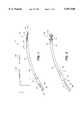
- FIG. 1is an elevational view of a the present invention with the working portion in an insertion configuration.
- FIG. 2is an elevational view of device of FIG. 1 with the working portion in a deployed configuration.
- FIG. 3is an enlarged perspective view of components of the working portion of the device of FIG. 1 de-mated from one another.
- FIGS. 4A-4Care enlarged elevational views of the components of the working portion of FIG. 1 in various positions.
- FIG. 5is a transverse sectional view of the device of FIG. 1 taken along line 5--5 of FIG. 4A.
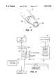
- FIG. 6is a block diagram of a control portion of the invention that includes a computer controller and energy source.
- FIGS. 7A-7Dare a sequence of sectional views of a patient's bladder and urethra showing the manner in which the instrument of FIG. 1 is utilized to perform a method of the invention in thermally treating tissue around the patient's sphincter;
- FIG. 7Abeing a sectional views of the initial step of introducing the device into the urethra;
- FIG. 7Bbeing a view of actuating certain laterally-extending elements of the working portion in the patient's bladder;
- FIG. 7Cbeing a view of actuating certain laterally-extending elements of the working portion in the patient's urethra;
- FIG. 7Dbeing a view of approximating the laterally-extending elements to compress tissue therebetween to alter extracellular fluid content therein to facilitate an electrosurgical treatment.
- FIG. 8is an enlarged sectional view of the compression of target tissue and the delivery thermal energy taken along line 8--8 of FIG. 7D.
- FIG. 9is another enlarged sectional view of the compression of target tissues similar to FIGS. 7D and 9 showing optional current vectors.
- FIGS. 10A-10Care plan views of a working portion of a Type "B" embodiment of thermal energy delivery device.
- FIGS. 11A-11Bare plan views of an alternative embodiment of the working portion of the Type "B" embodiment of FIG. 10A.
- FIGS. 12A-12Bare perspective and sectional views of a working portion of a Type "C" embodiment of thermal energy delivery device.
- thermal energy delivery (TED) system 5comprises elongate flexible outer catheter sleeve 10 dimensioned for transurethral passage with proximal end 11 and distal end 12 and extending along axis 15.
- First distal handle portion 16is coupled to proximal end 11 of the outer sleeve.
- Catheter sleeve 10has axial lumen 18 extending therethrough for accommodating the reciprocation of co-axial inner sleeve 20 with working portion 25 coupled to both inner and outer sleeves as described below.
- Second proximal handle portion 26is coupled to proximal end 21 of the inner sleeve 20.
- Inner sleeve 20 with proximal end 21 and distal end 22has (optional) lumen 28 therein dimensioned to slidably receive a fiberscope or so that system 5 may be introduced over a guide catheter or scope (not shown).
- FIG. 3shows tissue-compression members 30 and 32 de-mated from one another and de-mated from outer and inner catheter sleeves, 10 and 20.
- Proximal member 30is coupled to distal end 12 of outer sleeve 10.
- Distal tissue-compression member 32is coupled to distal end 22 of inner sleeve 10.
- Tissue-compression member 30has proximal end 40a and distal end 40b and tissue-compression member 32 has proximal end 42a and distal end 42b.
- FIG. 3also shows a plurality of cooperating longitudinal keys 43a (collectively) in members 30 and 32 maintain angular registration between the tissue-compression members and allows for their axial reciprocation relative to one another as described below.
- each tissue-compression member 30 and 32has laterally-extending elements in a (first) repose position.
- the tissue-compression members 30 and 32have laterally-extending elements in a (second) articulated position.
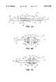
- member 30has three laterally-extending elements or arm portions 45a-45c that flex outwardly away from axis 15 of working portion 25.
- Each armis shown with living-type hinges wherein the entire member 30 is of a resilient plastic.
- proximal sliding movement of inner sleeve 20 within bore 18 of outer sleeve 10 by means of moving handle portion 26 in the proximal direction relative to handle 16causes proximal end 40a and distal end 40b of member 30 to be compressed axially toward one another resulting in arms 45a-45c flexing at living hinge points 46, 47 and 48 (collectively).
- Tissue-compression member 32has three cooperating arm portions 47a-47c that flex outwardly (similar to counterpart member 30) at equivalent hinge points (not numbered). It should be appreciated that the number of arms 45a-45c and 47a-47c may be from one to four or more and for convenience are shown as numbering three.
- 3 and 4Bshow that distal end 22 of inner sleeve 20 is coupled to shaft portion 50 and shaft 50 is fixed to distal end 42b of member 32 thus allowing proximal end 42a (and bore portion 52 therein) of member 32 to slide over shaft 50.
- FIG. 4Bshows that each laterally-extending elements or arms 45a-45c of member 30 have radiused working faces 55 (collectively) that are somewhat rounded for engaging tissue such that the tissue will not be penetrated. Similarly, arms 47a-47c of member 32 have radiused working faces 56 (collectively).
- Meansare thus provided for altering the extracellular fluid (ECF) content of tissue engaged by the laterally-extending elements of working end 25 by tissue compression.
- ECFextracellular fluid
- arms 45a-45c and 47a-47cdefine gap 58 therebetween for engaging target tissue and thereafter compressing the targeted tissue sites.
- Additional axial reciprocating meansare provided for reducing the axial dimension of gap 58 after the arms are deployed as shown in FIG. 4B.
- gap 58is capable of moving from initial dimension A to reduced dimension A' when inner sleeve 20 is moved axially relative to outer sleeve 10 and overcomes the spring constant of helically wound extension spring 59 that is disposed between opposing annular faces (60a and 60b) of members 30 and 32, respectively (see FIG. 3).
- the spring constant of spring 59is stronger than the collective spring constants of the living hinges (e.g., 47-49) of the arm elements described above.
- initial proximal axial movement of inner sleeve 20 relative to outer sleeve 10causes arms 45a-45c and 47a-47 to deploy (FIG. 4B).
- Additional proximal axial movement of inner sleeve 20 relative to outer sleeve 10causes working faces 55 and 56 of the arms to move closer axially (FIG. 4C).
- the first proximal axial movement of inner sleeve 20 relative to outer sleeve 10will causes arms 47a-47c of member 32 to deploy.
- the second or next proximal axial movement of inner sleeve 20 relative to outer sleeve 10will cause arms 45a-45c of member 30 to deploy.
- two stepsmay be required to move working end 25 to the configuration of FIG. 4B from the configuration of FIG. 4A.
- the next or third proximal axial movement of inner sleeve 20 relative to outer sleeve 10will cause gap 58 to be reduced from A to A' (see FIG.
- the control end (handles 16 and 26) of the devicepreferably may be locked (not shown) by any suitable means to maintain members 30 and 32 in the articulated position. Further, the control end may comprise any suitable mechanism for actuating the working end, e.g., a lever arm, trigger, etc. and is shown as cooperating slidable handles 16 and 26 for convenience only.
- Thermal energy delivery meansare provided for thermally treating target tissue engaged or compressed between working faces 55 and 56.
- Conductive electrodes or electrode arrays 70 and 72(collectively) for delivering RF energy are shown carried in respective working faces 55 and 56. Each electrode preferably is individually controlled as described further below.
- FIG. 5shows that the walls of outer sleeve 10 and inner sleeve 20 have embedded therein individual current-carrying wires 75a and 75b that supply RF energy to each conductive electrode.
- Both groups of the electrodes 70 and 72are shown in FIGS. 4A-4C as being bipolar but the electrodes may be operated in a mono-polar fashion with a groundplate (not shown).
- Electrode materialmay include gold, nickel titanium, platinum, stainless steel, aluminum and copper.
- electrical cables 77(collectively) are connected to an RF energy source through a controller described below which is adapted to deliver energy to electrodes 70 and 72.
- a sensor array of individual sensors 80(collectively) is provided in a spaced relationship around working end 25 and arms 45a-45c and 47a-47c.
- the sensor arraytypically will include temperature sensors, thermisters (temperature sensors that have resistances that vary with the temperature level) and/or impedance sensing elements that measure tissue impedance in various conventional manners, although impedance measurement may be obtained through electrodes 70 and 72 without resort to dedicated electrodes and circuits for impedance measuring purposes.
- the electomagnetic energy delivery source 88may be assumed to be an RF generator delivering energy to electrode 70 and 72.
- a multiplexer 90is depicted in FIG. 6 which is operatively connected to each electrode for measuring current, voltage and temperature at thermal sensors 80 (collectively) spaced around working end 25 or individually associated with each electrode.
- Multiplexer 90is driven by a controller 100 which typically is a digital computer with appropriate software.
- the controllertypically would include a CPU coupled to the multiplexer through a bus.
- On the controller systemthere may be a keyboard, disk drive or other non-volatile memory system, displays as are well known in the art for operating the system.
- Such an operator interfacemay include various types of imaging systems for observing the treatment such as thermal or infrared sensed displays, ultrasonic imaging displays or impedance monitoring displays.
- thermal sensors 80 carried in a position proximate to electrodes 70 and 72 together with thermal sensors 102 positioned within RF generatorare adapted to measure energy delivery (current and voltage) to each electrode at a treatment site during a treatment cycle.
- the output measured by thermal sensors 80 and 102are fed to controller 100 to control the delivery of power to each electrode site.
- the controller 100thus can be programmed to control temperature and power such that a certain particular temperature is never exceeded at the treatment site.
- the operatorfurther can set the desired temperature which can be maintained.
- the controllerhas a timing feature further providing the operator with the capability of maintaining a particular temperature at an electrode site for a particular length of time.
- a power delivery profilemay be incorporated into controller 100 as well as a pre-set for delivering a particular amount of energy.
- a feedback system or feedback circuitrycan be operatively connected to impedance measuring system, the temperature sensors and other indicators at the controller 100 or within the power source 88.
- the controllercan determine when the treatment is completed based on time, temperature or impedance or any combination thereof.
- the above-listed process variablescan be controlled and varied in response to tissue temperatures measured at multiple sites on tissue surfaces in contact with the device as well as by impedance to current flow at measured at each electrode which indicates the current carrying capability of the tissue during the treatment process.
- controller 100can provide multiplexing, can monitor circuit continuity for each electrode and determine which electrode is delivering energy.
- FIG. 6shows a block diagram of a particular embodiment of control circuitry.
- thermal sensorscan be thermisters which provide differing resistance levels depending on temperature.
- Amplifier 105can be a conventional analog differential amplifier for use with thermisters and transducers.
- the output of amplifier 105is sequentially connected by analog multiplexer 90 to the input of analog digital converter 110.
- the output of amplifier 105is a particular voltage that represents the respective sensed temperatures.
- the digitized amplifier output voltagesare supplied to microprocessor 115.
- Microprocessor 115thereafter calculates the temperature and/or impedance of the tissue site in question.
- Microprocessor 115sequentially receives and stores digital data representing impedance and temperature values. Each digital value received by microprocessor corresponds to a different temperature or impedance at a particular site.
- the temperature and impedance valuesmay be displayed on operator interface as numerical values.
- the temperature and impedance valuesalso are compared by microprocessor 115 with pre-programmed temperature and impedance limits. When the measured temperature value or impedance value at a particular site exceeds a pre-determined limit, a warning or other indication is given on operator interface and delivery of electromagnetic to a particular electrode site or area can be decreased or multiplexed to another electrode.
- a control signal from the microprocessormay reduce the power level at the generator or power source, or de-energize the power delivery to any particular electrode site.
- Controllerreceives and stores digital values which represent temperatures and impedance sent from the electrode and sensor sites. Calculated wall surface temperatures within the urethra and the bladder may be forwarded by controller 100 to the display and compared to a predetermined limit to activate a warning indicator on the display.
- FIG. 7Ais a schematic cross-sectional drawing of the lower female anatomy during use of the instrument and method of the invention.
- the urethra 102extends from the bladder 104 within fat pad 106.
- Urinary incontinenceis a condition characterized by a malfunctioning sphincter 108 often caused by movement or slippage of the bladder relative to pubic bone 110 within pad 106 and other regional anatomic structures.
- the catheter system 5is passed upwardly through the urethra 102 into the bladder 104 in the insertion configuration (see FIG. 1).
- the position of working portion 25is precisely controlled using an ultrasound image, for example, obtained from signals received from the conventional ultrasound transducer 125 inserted into vagina 130 adjacent to the bladder or with an ultrasound transducer positioned outside the body (not shown).
- the catheter systemalternatively may be introduced over a fiberscope previously inserted into the patient's urethra (not shown).
- the physicianmay angularly rotate the entire catheter about its axis to orient one or more of the arm elements toward tissues to be treated.
- the physicianthen moves handle portion 26 (FIG. 1) and inner sleeve 20 proximally a second distance relative to outer sleeve 10.
- handle portion 26FIG. 1
- inner sleeve 20proximally a second distance relative to outer sleeve 10.
- FIG. 7Csuch further actuation moves arm elements 45a-45c to a second deployed position being within the urethra 102 such that working faces 55 of the these arms along with electrodes 70 are laterally extended somewhat deep into the target tissues indicated at S.
- the curvature and radiusing of working faces 50insure that the arms do not penetrate the walls 135 of the urethra.
- the physicianmoves handle portion 26 and inner sleeve 20 proximally a third distance relative to outer sleeve 10 thereby moving arm elements 45a-45c and 47a-47c (a third deployed position) closer together to compress target tissue S therebetween (cf. FIG. 4C).
- the compression of target tissue S between working faces 55 and 56reduces the extracellular fluid (ECF) content in tissue S thus increasing tissue S's resistance to RF current.
- ECFextracellular fluid
- delivery of RF current through either electrodes 70 or 72will allow tissue S to be elevated to T cd (temperature of cell damage) to induce the injury healing response while surface layer L remains at a lower temperature such that the surface layers will not be ablated, principally due to the fact that surface layer L has a lower resistance (R) due to its higher ECF level as well as the fact that evaporative and convective forces further reduce the temperature of surface layer L indicated by arrows 138.
- Electrodes 70 and 72 on the arm elementsare energized from RF energy source 88 by actuation of a switch in the control end (handle 16 or 26) of the catheter system 5 or from a foot pedal or other suitable means.
- the time and/or power levelsare preset by the controller 100.
- the RF energy from energy source 88is delivered to the target tissue S for a pre-selected time. Impedance also is monitored, and when or if it exceeds a preset value, the energy source can be reduced or terminated automatically by controller 100.
- the temperature of surfaces of working portion 25 adjacent the urethral wall 135 and adjacent to the bladder wall 122are also monitored using temperature sensors attached to these components to precisely control the treatment parameters and prevent excessive heating of surface tissue layers L.
- the physicianmay collapse the arms 45a-45c and 47a-47c back onto working portion and either rotate the catheter slightly and repeat the treatment or remove the device from the patient's body.
- arm elementsalthough shown as angularly symmetric, may be asymmetric thus delivering energy to a target sites in a pre-determined asymmetric pattern.
- One or more temperature sensors 80which can be conventional thermistors, thermocouples or even optical fibers communicating with external sensors, are positioned along the catheter or arm elements to provide a temperature profile of the urethra adjacent to and preferably on both sides the electrodes 70 and 72.
- This temperature profilecan be used by the operator to prevent the temperature of the urethral wall or bladder wall from reaching level which would cause surface ablation.
- the RF energythus exposes the target tissue S to controlled heating to T cd (temperature of cell damage) of approximately 45° C. to 65° C.
- T cdtemperature of cell damage
- the temperature rangeis from 45° C. to 55° C.
- the temperature rangeis from 45° C. to 50° C.
- the RF currenttypically is delivered between opposing electrodes 70 and 72 in a bi-polar manner as shown by broken arrows in FIG. 8.
- more directed cell damagecan be obtained by alternating the bipolar flow of current in various vectors indicated by broken arrows in FIG. 9.
- one or more of the electrodesmay act as a mono-polar electrode and a delivery energy to a grounding plate (not shown).
- the RF treatmentis continued until the cells in the target tissue S have been damaged as indicated by dotted lines in FIGS. 8 and 9.
- the cell damageinduces the body's injury healing response which thereafter populates the extracellular compartment with a collagen fiber matrix having the effect bulking tissue and reducing the flexibility of tissues as described above.
- tissue bulking or tissue stiffeningcauses extraluminal pressures around the sphincter and helps restore the sphincter's ability to pinch off urine flow.
- This procedureis unique in that it is the first transurethral procedure which selectively provides the ability to limit the treatment to the extraluminal target tissues and spares the normal tissue of urethra wall 122 from excessive temperatures.
- This procedurealso minimizes the trauma sustained by tissues surrounding urethra 102, especially when compared to previously known procedures for relieving urinary incontinence.
- the proceduremay be carried out under local anesthesia only, depending upon the rate of energy delivery and degree of pain sensation experienced by the patient. When local anesthetic is adequate, the procedure can be performed in the physician's office.
- Local anestheticmay be delivered in the form of a lubricant containing a topical anesthetic such as lidocaine mixed with K-Y jelly and is applied to the catheter.
- the patientmay be sedated in addition to application of topical local anesthetic.
- topical local anestheticSuch a procedure still could be provided on an outpatient basis and would require a short term (1-3 hour) observation. If the procedure and patient require greater pain control, then spinal anesthesia or a general anesthesia may be used which would mandate that the procedure be carried out in the operating room.
- the patientmay return to normal activities with careful monitoring of the sphincter function. Thereafter, perhaps on a bi-weekly or monthly basis, the identical treatment cycle may be repeated in a one or more subsequent cycles until the desired reduction in tissue flexibility and pressure on the sphincter is achieved. It is believed that such periodic treatments (e.g., from 1 to 3 treatments over a period of a few weeks) may be best suited to stiffen target tissue S and to correct sphincter function.
- the temperature profilemay be programmed to attain the slightly higher level T sc necessary to shrink collagen fibers in the extracellular collagen matrix induced by the original treatment.
- the controller 100 and pre-programmed therapy cyclestill will allow the temperature of the urethral wall 135 and bladder wall 122 to be low enough so as to prevent surface ablation by making the energy delivery intermittent.
- the RF energythus exposes the target tissue S to controlled heating to T sc (temperature necessary to shrink collagen) of approximately 50° C. to 80° C. More preferably, the RF energy exposes tissue S to controlled heating of approximately 60° C. to 70° C. Still more preferably, the RF energy exposes tissue S to controlled heating of approximately 65° C. to 70° C.
- Type "B" embodiment of the present inventionis shown that is adapted for transurethral introduction and is similar in most respects to the first-described embodiment.
- Like reference numeralsrefer to like components of the Type "A” and Type “B” devices.
- the Type "B” devicediffers principally in that the distal tissue compression member 32 that in coupled to the distal end 22 of inner sleeve 20 carries an inflatable structure 150 rather than laterally extendable elements.
- the inflatable structure 150communicates with any conventional pressure source (e.g., a syringe) through lumen 152 in the wall of inner sleeve 20 (FIG. 10).
- a pressure sourcee.g., a syringe
- inflatable structure 150is of a non-compliant material such as PET but also may be an elastomer such as latex or silicone.
- the Type "B" embodimentis used in a fashion similar to that described above.
- the catheteris introduced into the urethra and then inflatable structure 150 is expanded.
- the proximal arms 45a-45care extended laterally to compress tissue between the arms 45a-45c and inflatable structure 150.
- RF energymay be delivered in a mono-polar fashion.
- the surface of the inflatable structure 150may have a plurality of opposing electrodes 152 and the RF energy may be delivered in a bi-polar fashion as described previously.
- Another alternative embodiment of inflatable structure 150could include a metallic mesh 155 as a return electrode covering a substantial portion of the surface of the inflatable structure facing electrodes 60 (see FIGS. 11A-11B).
- Type "C” embodiment of the present inventionis shown that is very similar to the first-described Type “A” embodiment.
- Like reference numeralsrefer to like components of the Type “A” device.
- This Type “C” working end 25has the spring mechanism for sequencing the articulation of laterally extending elements 45a-45c and 47a-47c eliminated from the working end. The spring mechanism may be moved to the handle end or control end of the instrument (not shown).
- FIGS. 12A and 12Bshow more in particular how each laterally-extending element may be configured with four living hinges points 200a, 200b, 200c and 200d to allow electrodes 70 to assume a face angle at about 90° to axis 15.
- Each living hinge pointcomprises a reduced sectional dimension of the resilient plastic of the member.
- four living hinges points 202a, 202b, 202c and 202dallow electrodes 72 to assume a face angle at about 90° relative to axis 15.
- FIG. 12Bshows that by varying the lengths of the certain segments of the laterally extending elements 45a and 47a, electrodes 70 and 72 may be aligned and opposed at a similar distance D from axis 15.
- the Type "C" embodimentis used in a fashion similar as described above.
Landscapes
- Health & Medical Sciences (AREA)
- Surgery (AREA)
- Engineering & Computer Science (AREA)
- Life Sciences & Earth Sciences (AREA)
- Biomedical Technology (AREA)
- Otolaryngology (AREA)
- Nuclear Medicine, Radiotherapy & Molecular Imaging (AREA)
- Plasma & Fusion (AREA)
- Physics & Mathematics (AREA)
- Heart & Thoracic Surgery (AREA)
- Medical Informatics (AREA)
- Molecular Biology (AREA)
- Animal Behavior & Ethology (AREA)
- General Health & Medical Sciences (AREA)
- Public Health (AREA)
- Veterinary Medicine (AREA)
- Surgical Instruments (AREA)
Abstract
Description
Claims (23)
Priority Applications (7)
| Application Number | Priority Date | Filing Date | Title |
|---|---|---|---|
| US08/920,291US5957920A (en) | 1997-08-28 | 1997-08-28 | Medical instruments and techniques for treatment of urinary incontinence |
| US09/191,413US6083223A (en) | 1997-08-28 | 1998-11-12 | Methods and apparatus for welding blood vessels |
| US09/258,006US6197022B1 (en) | 1996-07-30 | 1999-02-25 | Medical instruments and techniques for treatment of gastro-esophageal reflux disease |
| US09/648,345US6535768B1 (en) | 1996-08-30 | 2000-08-25 | Medical instruments and techniques for treatment of gastro-esophageal reflux disease |
| US10/375,505US7167758B2 (en) | 1996-08-30 | 2003-02-27 | Medical instruments and techniques for treatment of gastro-esophageal reflux disease |
| US11/656,062US8095222B2 (en) | 1997-08-28 | 2007-01-22 | Medical instruments and techniques for treatment of gastro-esophageal reflux disease |
| US13/347,267US20120109268A1 (en) | 1997-08-28 | 2012-01-10 | Medical instruments and techniques for treatment of gastro-esophageal reflux disease |
Applications Claiming Priority (1)
| Application Number | Priority Date | Filing Date | Title |
|---|---|---|---|
| US08/920,291US5957920A (en) | 1997-08-28 | 1997-08-28 | Medical instruments and techniques for treatment of urinary incontinence |
Related Child Applications (2)
| Application Number | Title | Priority Date | Filing Date |
|---|---|---|---|
| US09/191,413Continuation-In-PartUS6083223A (en) | 1997-08-28 | 1998-11-12 | Methods and apparatus for welding blood vessels |
| US09/258,006ContinuationUS6197022B1 (en) | 1996-07-30 | 1999-02-25 | Medical instruments and techniques for treatment of gastro-esophageal reflux disease |
Publications (1)
| Publication Number | Publication Date |
|---|---|
| US5957920Atrue US5957920A (en) | 1999-09-28 |
Family
ID=25443520
Family Applications (6)
| Application Number | Title | Priority Date | Filing Date |
|---|---|---|---|
| US08/920,291Expired - LifetimeUS5957920A (en) | 1996-07-30 | 1997-08-28 | Medical instruments and techniques for treatment of urinary incontinence |
| US09/258,006Expired - LifetimeUS6197022B1 (en) | 1996-07-30 | 1999-02-25 | Medical instruments and techniques for treatment of gastro-esophageal reflux disease |
| US09/648,345Expired - Fee RelatedUS6535768B1 (en) | 1996-08-30 | 2000-08-25 | Medical instruments and techniques for treatment of gastro-esophageal reflux disease |
| US10/375,505Expired - Fee RelatedUS7167758B2 (en) | 1996-08-30 | 2003-02-27 | Medical instruments and techniques for treatment of gastro-esophageal reflux disease |
| US11/656,062Expired - Fee RelatedUS8095222B2 (en) | 1997-08-28 | 2007-01-22 | Medical instruments and techniques for treatment of gastro-esophageal reflux disease |
| US13/347,267AbandonedUS20120109268A1 (en) | 1997-08-28 | 2012-01-10 | Medical instruments and techniques for treatment of gastro-esophageal reflux disease |
Family Applications After (5)
| Application Number | Title | Priority Date | Filing Date |
|---|---|---|---|
| US09/258,006Expired - LifetimeUS6197022B1 (en) | 1996-07-30 | 1999-02-25 | Medical instruments and techniques for treatment of gastro-esophageal reflux disease |
| US09/648,345Expired - Fee RelatedUS6535768B1 (en) | 1996-08-30 | 2000-08-25 | Medical instruments and techniques for treatment of gastro-esophageal reflux disease |
| US10/375,505Expired - Fee RelatedUS7167758B2 (en) | 1996-08-30 | 2003-02-27 | Medical instruments and techniques for treatment of gastro-esophageal reflux disease |
| US11/656,062Expired - Fee RelatedUS8095222B2 (en) | 1997-08-28 | 2007-01-22 | Medical instruments and techniques for treatment of gastro-esophageal reflux disease |
| US13/347,267AbandonedUS20120109268A1 (en) | 1997-08-28 | 2012-01-10 | Medical instruments and techniques for treatment of gastro-esophageal reflux disease |
Country Status (1)
| Country | Link |
|---|---|
| US (6) | US5957920A (en) |
Cited By (81)
| Publication number | Priority date | Publication date | Assignee | Title |
|---|---|---|---|---|
| US6110111A (en)* | 1999-05-26 | 2000-08-29 | Diagnostic Ultrasound Corporation | System for quantizing bladder distension due to pressure using normalized surface area of the bladder |
| WO2000069515A1 (en)* | 1999-05-17 | 2000-11-23 | Marchitto Kevin S | Remote and local controlled delivery of pharmaceutical compounds using electromagnetic energy |
| US6156060A (en)* | 1998-07-31 | 2000-12-05 | Surx, Inc. | Static devices and methods to shrink tissues for incontinence |
| WO2001006942A1 (en)* | 1998-02-19 | 2001-02-01 | Curon Medical Inc. | Method for treating a sphincter |
| US6216704B1 (en)* | 1997-08-13 | 2001-04-17 | Surx, Inc. | Noninvasive devices, methods, and systems for shrinking of tissues |
| US6236891B1 (en)* | 1998-07-31 | 2001-05-22 | Surx, Inc. | Limited heat transfer devices and methods to shrink tissues |
| US6283987B1 (en) | 1998-01-14 | 2001-09-04 | Surx, Inc. | Ribbed electrodes and methods for their use |
| US6292700B1 (en)* | 1999-09-10 | 2001-09-18 | Surx, Inc. | Endopelvic fascia treatment for incontinence |
| US6470219B1 (en) | 2000-10-02 | 2002-10-22 | Novasys Medical, Inc. | Apparatus and method for treating female urinary incontinence |
| US6480746B1 (en)* | 1997-08-13 | 2002-11-12 | Surx, Inc. | Noninvasive devices, methods, and systems for shrinking of tissues |
| US20030036804A1 (en)* | 2000-10-02 | 2003-02-20 | Thomas Simon W. H. | Apparatus and methods for treating female urinary incontinence |
| US20030130710A1 (en)* | 1996-08-30 | 2003-07-10 | Baker James A. | Medical instruments and techniques for treatment of gastro-esophageal reflux disease |
| US20030144655A1 (en)* | 2002-01-31 | 2003-07-31 | Scimed Life Systems, Inc. | Compensation for power variation along patient cables |
| US20030181897A1 (en)* | 2000-10-02 | 2003-09-25 | Thomas Simon W.H. | Apparatus and methods for treating female urinary incontinence |
| WO2003079931A1 (en)* | 2002-03-19 | 2003-10-02 | Surx, Inc. | Heating method for tissue contraction |
| US20030195604A1 (en)* | 1996-11-08 | 2003-10-16 | Surx, Inc. | Devices, methods, and sytems for shrinking tissues |
| US6673070B2 (en) | 1994-06-24 | 2004-01-06 | Curon Medical, Inc. | Sphincter treatment apparatus |
| US20040044337A1 (en)* | 2002-08-27 | 2004-03-04 | Gal Shafirstein | Conductive interstitial thermal therapy device |
| US20040089313A1 (en)* | 1998-02-19 | 2004-05-13 | Curon Medical, Inc. | Systems and methods for treating obesity and other gastrointestinal conditions |
| US6740082B2 (en) | 1998-12-29 | 2004-05-25 | John H. Shadduck | Surgical instruments for treating gastro-esophageal reflux |
| US20040193238A1 (en)* | 2003-01-16 | 2004-09-30 | Surx, Inc., A Corporation Of Delaware | Non-surgical incontinence treatment system and method |
| US20040236177A1 (en)* | 2001-11-20 | 2004-11-25 | Solarant Medical, Inc. | Incontinence treatment with urethral guide |
| US20040267336A1 (en)* | 1996-11-08 | 2004-12-30 | Solarant Medical, Inc. | Energy induced bulking and buttressing of tissues for incontinence |
| US20050171583A1 (en)* | 2004-01-30 | 2005-08-04 | Solarant Medical, Inc. | Heating method for tissue contraction |
| US20050171582A1 (en)* | 2004-01-30 | 2005-08-04 | Solarant Medical, Inc. | Electrically heated/phase change probe temperature control |
| US20050183732A1 (en)* | 1999-05-18 | 2005-08-25 | Edwards Stuart D. | Surgical weight control device |
| US6971395B2 (en)* | 1998-01-14 | 2005-12-06 | Curon Medical, Inc. | Sphincter treatment method |
| US20060095103A1 (en)* | 2003-12-11 | 2006-05-04 | Apsara Medical Corporation | Aesthetic thermal sculpting of skin |
| US20060167445A1 (en)* | 2002-08-27 | 2006-07-27 | Gal Shafirstein | Selective conductive interstitial thermal therapy device |
| US20060205996A1 (en)* | 2001-11-20 | 2006-09-14 | Solorant Medical, Inc. | Incontinence treatment with urethral guide |
| US20060264831A1 (en)* | 2005-05-20 | 2006-11-23 | Medtronic, Inc. | Portable therapy delivery device with fluid delivery |
| US7160294B2 (en) | 2003-09-02 | 2007-01-09 | Curon Medical, Inc. | Systems and methods for treating hemorrhoids |
| US20070010761A1 (en)* | 2003-04-07 | 2007-01-11 | Seung-Kee Mo | Method and apparatus for verifying data measured by several means in real-time |
| US20070027511A1 (en)* | 2005-08-01 | 2007-02-01 | Ty Fairneny | Collagen injection combined with focused ultrasound for the treatment of incontinence |
| US20070050001A1 (en)* | 2005-08-26 | 2007-03-01 | Solarant Medical, Inc. | Adjustable open loop control devices and methods |
| US20070077827A1 (en)* | 2005-09-30 | 2007-04-05 | Medtronic, Inc. | Sliding lock device |
| US20070260288A1 (en)* | 2006-03-03 | 2007-11-08 | Yossi Gross | Apparatus for treating stress and urge incontinence |
| US20080125771A1 (en)* | 2006-11-27 | 2008-05-29 | Michael Lau | Methods and apparatuses for contouring tissue by selective application of energy |
| US7536225B2 (en) | 2005-01-21 | 2009-05-19 | Ams Research Corporation | Endo-pelvic fascia penetrating heating systems and methods for incontinence treatment |
| US20090157091A1 (en)* | 2006-04-04 | 2009-06-18 | Ams Research Corporation | Apparatus for Implanting Neural Stimulation Leads |
| US20090312755A1 (en)* | 2008-06-13 | 2009-12-17 | Vytronus, Inc. | System and method for positioning an elongate member with respect to an anatomical structure |
| US20100004648A1 (en)* | 1998-02-19 | 2010-01-07 | Mederi Therapeutics, Inc. | Method to treat gastric reflux via the detection and ablation of gastro-esophageal nerves and receptors |
| US20100004644A1 (en)* | 2008-07-01 | 2010-01-07 | Ralph Zipper | Method for Decreasing the Size and/or Changing the Shape of Pelvic Tissues |
| US7648500B2 (en) | 1998-02-19 | 2010-01-19 | Mederi Therapeutics, Inc. | Sphincter treatment apparatus |
| US20110060389A1 (en)* | 2009-04-02 | 2011-03-10 | Julie Reil | Method for correction of female incontinence and reconstruction of tissue |
| US20110077550A1 (en)* | 2001-09-19 | 2011-03-31 | Mederi Therapeutics, Inc. | Systems and methods for treating tissue regions of the body |
| US20110152861A1 (en)* | 2009-12-17 | 2011-06-23 | Aesculap Ag | Surgical System For Connecting Body Tissue |
| US20110172655A1 (en)* | 2000-10-17 | 2011-07-14 | Asthmatx, Inc. | Control system and process for application of energy to airway walls and other mediums |
| US20110184500A1 (en)* | 2009-04-02 | 2011-07-28 | Genityte, Inc. | Correction of female urinary incontinence and skin reduction |
| US8160710B2 (en) | 2006-07-10 | 2012-04-17 | Ams Research Corporation | Systems and methods for implanting tissue stimulation electrodes in the pelvic region |
| US20120241417A1 (en)* | 2011-03-25 | 2012-09-27 | Illinois Tool Works Inc. | Welding power supply with controlled auxiliary power |
| US8380312B2 (en) | 2009-12-31 | 2013-02-19 | Ams Research Corporation | Multi-zone stimulation implant system and method |
| US8403927B1 (en) | 2012-04-05 | 2013-03-26 | William Bruce Shingleton | Vasectomy devices and methods |
| US20130197505A1 (en)* | 1996-09-20 | 2013-08-01 | Novasys Medical, Inc. | Treatment of tissue in sphincters, sinuses, and orifices |
| US8758337B2 (en) | 2006-09-29 | 2014-06-24 | Medtronic, Inc. | User interface for ablation therapy |
| US8774942B2 (en) | 2007-07-10 | 2014-07-08 | Ams Research Corporation | Tissue anchor |
| US8888774B2 (en)* | 1999-05-04 | 2014-11-18 | Mederi Therapeutics, Inc. | Methods for treating the cardia of the stomach |
| US8894646B2 (en) | 1998-02-19 | 2014-11-25 | Mederi Therapeutics, Inc. | Sphincter treatment apparatus |
| US20150099011A1 (en)* | 2001-09-25 | 2015-04-09 | Jenny Colleen McCloskey | Inactivation of papillomavirus |
| US9023031B2 (en) | 1997-08-13 | 2015-05-05 | Verathon Inc. | Noninvasive devices, methods, and systems for modifying tissues |
| US9061121B2 (en) | 2009-08-03 | 2015-06-23 | Emory University | Targeting therapeutic agents |
| US9204921B2 (en) | 2012-12-13 | 2015-12-08 | Cook Medical Technologies Llc | RF energy controller and method for electrosurgical medical devices |
| US9220887B2 (en) | 2011-06-09 | 2015-12-29 | Astora Women's Health LLC | Electrode lead including a deployable tissue anchor |
| US9364277B2 (en) | 2012-12-13 | 2016-06-14 | Cook Medical Technologies Llc | RF energy controller and method for electrosurgical medical devices |
| US9427573B2 (en) | 2007-07-10 | 2016-08-30 | Astora Women's Health, Llc | Deployable electrode lead anchor |
| US9474565B2 (en) | 2009-09-22 | 2016-10-25 | Mederi Therapeutics, Inc. | Systems and methods for treating tissue with radiofrequency energy |
| US9539433B1 (en) | 2009-03-18 | 2017-01-10 | Astora Women's Health, Llc | Electrode implantation in a pelvic floor muscular structure |
| EP2582314A4 (en)* | 2010-06-15 | 2017-08-02 | Avenu Medical, Inc. | Intravascular arterial to venous anastomosis and tissue welding catheter |
| US9731112B2 (en) | 2011-09-08 | 2017-08-15 | Paul J. Gindele | Implantable electrode assembly |
| US9750563B2 (en) | 2009-09-22 | 2017-09-05 | Mederi Therapeutics, Inc. | Systems and methods for treating tissue with radiofrequency energy |
| US9770293B2 (en) | 2012-06-04 | 2017-09-26 | Boston Scientific Scimed, Inc. | Systems and methods for treating tissue of a passageway within a body |
| US9775664B2 (en) | 2009-09-22 | 2017-10-03 | Mederi Therapeutics, Inc. | Systems and methods for treating tissue with radiofrequency energy |
| US10004458B2 (en) | 2013-03-27 | 2018-06-26 | Autonomix Medical, Inc. | Systems and methods for neurological traffic and/or receptor functional evaluation and/or modification |
| US10206616B2 (en) | 2012-06-14 | 2019-02-19 | Autonomix Medical, Inc. | Devices, systems, and methods for diagnosis and treatment of overactive bladder |
| US10386990B2 (en) | 2009-09-22 | 2019-08-20 | Mederi Rf, Llc | Systems and methods for treating tissue with radiofrequency energy |
| US20200054385A1 (en)* | 2017-06-12 | 2020-02-20 | Kardium Inc. | Medical device systems and methods for activating transducers based on delivery shaft member temperature |
| US20210007791A1 (en)* | 2018-03-29 | 2021-01-14 | Terumo Kabushiki Kaisha | Medical device |
| US11154349B2 (en) | 2009-09-18 | 2021-10-26 | Viveve, Inc. | Vaginal remodeling device and methods |
| US11439456B2 (en)* | 2017-03-15 | 2022-09-13 | Olympus Corporation | Energy source apparatus |
| US11648380B2 (en) | 2017-12-05 | 2023-05-16 | Jenny Colleen McCloskey | Device for treatment of a body canal and adjacent surfaces |
| US11896823B2 (en) | 2017-04-04 | 2024-02-13 | Btl Healthcare Technologies A.S. | Method and device for pelvic floor tissue treatment |
Families Citing this family (171)
| Publication number | Priority date | Publication date | Assignee | Title |
|---|---|---|---|---|
| US7229436B2 (en) | 1996-01-05 | 2007-06-12 | Thermage, Inc. | Method and kit for treatment of tissue |
| US7141049B2 (en) | 1999-03-09 | 2006-11-28 | Thermage, Inc. | Handpiece for treatment of tissue |
| US7006874B2 (en) | 1996-01-05 | 2006-02-28 | Thermage, Inc. | Treatment apparatus with electromagnetic energy delivery device and non-volatile memory |
| US7473251B2 (en) | 1996-01-05 | 2009-01-06 | Thermage, Inc. | Methods for creating tissue effect utilizing electromagnetic energy and a reverse thermal gradient |
| US7452358B2 (en) | 1996-01-05 | 2008-11-18 | Thermage, Inc. | RF electrode assembly for handpiece |
| US7189230B2 (en) | 1996-01-05 | 2007-03-13 | Thermage, Inc. | Method for treating skin and underlying tissue |
| US7267675B2 (en) | 1996-01-05 | 2007-09-11 | Thermage, Inc. | RF device with thermo-electric cooler |
| US7115123B2 (en) | 1996-01-05 | 2006-10-03 | Thermage, Inc. | Handpiece with electrode and non-volatile memory |
| US7022121B2 (en) | 1999-03-09 | 2006-04-04 | Thermage, Inc. | Handpiece for treatment of tissue |
| US6073052A (en)* | 1996-11-15 | 2000-06-06 | Zelickson; Brian D. | Device and method for treatment of gastroesophageal reflux disease |
| WO1999035986A1 (en)* | 1998-01-14 | 1999-07-22 | Conway-Stuart Medical, Inc. | Electrosurgical apparatus for treating gastroesophageal reflux disease (gerd) and method |
| US6358245B1 (en) | 1998-02-19 | 2002-03-19 | Curon Medical, Inc. | Graphical user interface for association with an electrode structure deployed in contact with a tissue region |
| US7165551B2 (en)* | 1998-02-19 | 2007-01-23 | Curon Medical, Inc. | Apparatus to detect and treat aberrant myoelectric activity |
| US8906010B2 (en) | 1998-02-19 | 2014-12-09 | Mederi Therapeutics, Inc. | Graphical user interface for association with an electrode structure deployed in contact with a tissue region |
| US6591838B2 (en)* | 1998-07-06 | 2003-07-15 | Scimed Life Systems, Inc. | Implant system and method for bulking tissue |
| US6447508B1 (en)* | 1999-06-16 | 2002-09-10 | Hugh R. Sharkey | Stent inductive heating catheter |
| US7618426B2 (en)* | 2002-12-11 | 2009-11-17 | Usgi Medical, Inc. | Apparatus and methods for forming gastrointestinal tissue approximations |
| US7637905B2 (en) | 2003-01-15 | 2009-12-29 | Usgi Medical, Inc. | Endoluminal tool deployment system |
| US7744613B2 (en)* | 1999-06-25 | 2010-06-29 | Usgi Medical, Inc. | Apparatus and methods for forming and securing gastrointestinal tissue folds |
| US7416554B2 (en) | 2002-12-11 | 2008-08-26 | Usgi Medical Inc | Apparatus and methods for forming and securing gastrointestinal tissue folds |
| WO2003053266A2 (en)* | 1999-06-30 | 2003-07-03 | Thermage, Inc. | Liquid cooled rf handpiece |
| US7229469B1 (en)* | 1999-10-02 | 2007-06-12 | Quantumcor, Inc. | Methods for treating and repairing mitral valve annulus |
| US20060095032A1 (en)* | 1999-11-16 | 2006-05-04 | Jerome Jackson | Methods and systems for determining physiologic characteristics for treatment of the esophagus |
| US20040215235A1 (en) | 1999-11-16 | 2004-10-28 | Barrx, Inc. | Methods and systems for determining physiologic characteristics for treatment of the esophagus |
| WO2001035846A1 (en)* | 1999-11-16 | 2001-05-25 | Ganz Robert A | System and method of treating abnormal tissue in the human esophagus |
| US7033373B2 (en) | 2000-11-03 | 2006-04-25 | Satiety, Inc. | Method and device for use in minimally invasive placement of space-occupying intragastric devices |
| US6475179B1 (en)* | 2000-11-10 | 2002-11-05 | New England Medical Center | Tissue folding device for tissue ablation, and method thereof |
| US20040087936A1 (en)* | 2000-11-16 | 2004-05-06 | Barrx, Inc. | System and method for treating abnormal tissue in an organ having a layered tissue structure |
| US6558400B2 (en) | 2001-05-30 | 2003-05-06 | Satiety, Inc. | Obesity treatment tools and methods |
| US7083629B2 (en)* | 2001-05-30 | 2006-08-01 | Satiety, Inc. | Overtube apparatus for insertion into a body |
| US6675809B2 (en) | 2001-08-27 | 2004-01-13 | Richard S. Stack | Satiation devices and methods |
| CN101810521B (en) | 2001-08-27 | 2015-05-13 | 辛尼科有限责任公司 | Satiation devices and methods |
| US6845776B2 (en)* | 2001-08-27 | 2005-01-25 | Richard S. Stack | Satiation devices and methods |
| US7097665B2 (en)* | 2003-01-16 | 2006-08-29 | Synecor, Llc | Positioning tools and methods for implanting medical devices |
| US6669693B2 (en)* | 2001-11-13 | 2003-12-30 | Mayo Foundation For Medical Education And Research | Tissue ablation device and methods of using |
| US8709488B2 (en) | 2002-02-14 | 2014-04-29 | Gholam A. Peyman | Method and composition for hyperthermally treating cells |
| US8795251B2 (en) | 2002-02-14 | 2014-08-05 | Gholam A. Peyman | Method and composition for hyperthermally treating cells |
| US8137698B2 (en)* | 2002-02-14 | 2012-03-20 | Peyman Gholam A | Method and composition for hyperthermally treating cells |
| US9017729B2 (en) | 2002-02-14 | 2015-04-28 | Gholam A. Peyman | Method and composition for hyperthermally treating cells |
| US8932636B2 (en) | 2002-02-14 | 2015-01-13 | Gholam A. Peyman | Method and composition for hyperthermally treating cells |
| US9233157B2 (en) | 2002-02-14 | 2016-01-12 | Gholam A. Peyman | Method and composition for hyperthermally treating cells |
| US9320813B2 (en) | 2002-02-14 | 2016-04-26 | Gholam A. Peyman | Method and composition for hyperthermally treating cells |
| US8481082B2 (en) | 2002-02-14 | 2013-07-09 | Gholam A. Peyman | Method and composition for hyperthermally treating cells |
| US8119165B2 (en) | 2002-02-14 | 2012-02-21 | Peyman Gholam A | Method and composition for hyperthermally treating cells |
| US8801690B2 (en) | 2002-02-14 | 2014-08-12 | Gholam A. Peyman | Method and composition for hyperthermally treating cells |
| US8808268B2 (en) | 2002-02-14 | 2014-08-19 | Gholam A. Peyman | Method and composition for hyperthermally treating cells |
| US9289491B2 (en) | 2002-02-14 | 2016-03-22 | Gholam A. Peyman | Method and composition for hyperthermally treating cells |
| US8668935B2 (en) | 2002-02-14 | 2014-03-11 | Gholam A. Peyman | Method and composition for hyperthermally treating cells |
| US9393396B2 (en) | 2002-02-14 | 2016-07-19 | Gholam A. Peyman | Method and composition for hyperthermally treating cells |
| US9302087B2 (en) | 2002-02-14 | 2016-04-05 | Gholam A. Peyman | Method and composition for hyperthermally treating cells |
| US7146984B2 (en) | 2002-04-08 | 2006-12-12 | Synecor, Llc | Method and apparatus for modifying the exit orifice of a satiation pouch |
| US7274876B2 (en)* | 2002-06-06 | 2007-09-25 | At&T Corp. | Integrated electrical/optical hybrid communication system with revertive hitless switch |
| US20040002702A1 (en)* | 2002-06-28 | 2004-01-01 | Ethicon, Inc. | RF device for treating the uterus |
| US6929642B2 (en)* | 2002-06-28 | 2005-08-16 | Ethicon, Inc. | RF device for treating the uterus |
| US6773440B2 (en)* | 2002-07-02 | 2004-08-10 | Satiety, Inc. | Method and device for use in tissue approximation and fixation |
| US6746460B2 (en) | 2002-08-07 | 2004-06-08 | Satiety, Inc. | Intra-gastric fastening devices |
| US7211114B2 (en)* | 2002-08-26 | 2007-05-01 | The Trustees Of Columbia University In The City Of New York | Endoscopic gastric bypass |
| US7214233B2 (en)* | 2002-08-30 | 2007-05-08 | Satiety, Inc. | Methods and devices for maintaining a space occupying device in a relatively fixed location within a stomach |
| US7033384B2 (en) | 2002-08-30 | 2006-04-25 | Satiety, Inc. | Stented anchoring of gastric space-occupying devices |
| US7101367B2 (en) | 2002-09-30 | 2006-09-05 | Ethicon, Inc. | Deployable cryosurgical catheter |
| US7229428B2 (en) | 2002-10-23 | 2007-06-12 | Satiety, Inc. | Method and device for use in endoscopic organ procedures |
| US7220237B2 (en)* | 2002-10-23 | 2007-05-22 | Satiety, Inc. | Method and device for use in endoscopic organ procedures |
| US6656194B1 (en)* | 2002-11-05 | 2003-12-02 | Satiety, Inc. | Magnetic anchoring devices |
| CA2507803C (en) | 2002-11-29 | 2012-12-04 | John R. Liddicoat | Apparatus and method for manipulating tissue |
| US7942898B2 (en) | 2002-12-11 | 2011-05-17 | Usgi Medical, Inc. | Delivery systems and methods for gastric reduction |
| US7942884B2 (en) | 2002-12-11 | 2011-05-17 | Usgi Medical, Inc. | Methods for reduction of a gastric lumen |
| US20040143342A1 (en) | 2003-01-16 | 2004-07-22 | Stack Richard S. | Satiation pouches and methods of use |
| US7783358B2 (en)* | 2003-03-14 | 2010-08-24 | Endovx, Inc. | Methods and apparatus for treatment of obesity with an ultrasound device movable in two or three axes |
| US7684865B2 (en)* | 2003-03-14 | 2010-03-23 | Endovx, Inc. | Methods and apparatus for treatment of obesity |
| US7430449B2 (en)* | 2003-03-14 | 2008-09-30 | Endovx, Inc. | Methods and apparatus for testing disruption of a vagal nerve |
| US7175638B2 (en)* | 2003-04-16 | 2007-02-13 | Satiety, Inc. | Method and devices for modifying the function of a body organ |
| US8216252B2 (en) | 2004-05-07 | 2012-07-10 | Usgi Medical, Inc. | Tissue manipulation and securement system |
| US20050247320A1 (en)* | 2003-10-10 | 2005-11-10 | Stack Richard S | Devices and methods for retaining a gastro-esophageal implant |
| US8206456B2 (en) | 2003-10-10 | 2012-06-26 | Barosense, Inc. | Restrictive and/or obstructive implant system for inducing weight loss |
| US7914543B2 (en)* | 2003-10-14 | 2011-03-29 | Satiety, Inc. | Single fold device for tissue fixation |
| US7097650B2 (en)* | 2003-10-14 | 2006-08-29 | Satiety, Inc. | System for tissue approximation and fixation |
| US20050251189A1 (en)* | 2004-05-07 | 2005-11-10 | Usgi Medical Inc. | Multi-position tissue manipulation assembly |
| US7347863B2 (en)* | 2004-05-07 | 2008-03-25 | Usgi Medical, Inc. | Apparatus and methods for manipulating and securing tissue |
| US7361180B2 (en) | 2004-05-07 | 2008-04-22 | Usgi Medical, Inc. | Apparatus for manipulating and securing tissue |
| US7150745B2 (en) | 2004-01-09 | 2006-12-19 | Barrx Medical, Inc. | Devices and methods for treatment of luminal tissue |
| US20050177176A1 (en)* | 2004-02-05 | 2005-08-11 | Craig Gerbi | Single-fold system for tissue approximation and fixation |
| CA2556228C (en)* | 2004-02-13 | 2014-05-13 | Satiety, Inc. | Methods for reducing hollow organ volume |
| MXPA06009971A (en)* | 2004-02-27 | 2007-08-08 | Satiety Inc | Methods and devices for reducing hollow organ volume. |
| US9028511B2 (en) | 2004-03-09 | 2015-05-12 | Ethicon Endo-Surgery, Inc. | Devices and methods for placement of partitions within a hollow body organ |
| US20050203488A1 (en)* | 2004-03-09 | 2005-09-15 | Usgi Medical Inc. | Apparatus and methods for mapping out endoluminal gastrointestinal surgery |
| US8449560B2 (en) | 2004-03-09 | 2013-05-28 | Satiety, Inc. | Devices and methods for placement of partitions within a hollow body organ |
| US8628547B2 (en) | 2004-03-09 | 2014-01-14 | Ethicon Endo-Surgery, Inc. | Devices and methods for placement of partitions within a hollow body organ |
| US8252009B2 (en)* | 2004-03-09 | 2012-08-28 | Ethicon Endo-Surgery, Inc. | Devices and methods for placement of partitions within a hollow body organ |
| US7703459B2 (en)* | 2004-03-09 | 2010-04-27 | Usgi Medical, Inc. | Apparatus and methods for mapping out endoluminal gastrointestinal surgery |
| AU2005231323B2 (en) | 2004-03-26 | 2011-03-31 | Ethicon Endo-Surgery, Inc | Systems and methods for treating obesity |
| EP2301471A1 (en) | 2004-04-01 | 2011-03-30 | The General Hospital Corporation | Method and apparatus for dermatological treatment and tissue reshaping |
| WO2005105003A1 (en) | 2004-04-26 | 2005-11-10 | Synecor, Llc | Restrictive and/or obstructive implant for inducing weight loss |
| US7520884B2 (en)* | 2004-05-07 | 2009-04-21 | Usgi Medical Inc. | Methods for performing gastroplasty |
| US8257394B2 (en) | 2004-05-07 | 2012-09-04 | Usgi Medical, Inc. | Apparatus and methods for positioning and securing anchors |
| US8444657B2 (en) | 2004-05-07 | 2013-05-21 | Usgi Medical, Inc. | Apparatus and methods for rapid deployment of tissue anchors |
| US7918869B2 (en)* | 2004-05-07 | 2011-04-05 | Usgi Medical, Inc. | Methods and apparatus for performing endoluminal gastroplasty |
| US20050250984A1 (en)* | 2004-05-07 | 2005-11-10 | Usgi Medical Inc. | Multiple removable apparatus and methods for manipulating and securing tissue |
| US7736374B2 (en)* | 2004-05-07 | 2010-06-15 | Usgi Medical, Inc. | Tissue manipulation and securement system |
| US7931661B2 (en)* | 2004-06-14 | 2011-04-26 | Usgi Medical, Inc. | Apparatus and methods for performing transluminal gastrointestinal procedures |
| US20060020276A1 (en)* | 2004-07-23 | 2006-01-26 | Usgi Medical Inc. | Apparatus and methods for achieving prolonged maintenance of gastrointestinal tissue folds |
| US20090326578A1 (en)* | 2004-09-30 | 2009-12-31 | Usgi Medical, Inc. | Interlocking tissue anchor apparatus and methods |
| US20060106288A1 (en)* | 2004-11-17 | 2006-05-18 | Roth Alex T | Remote tissue retraction device |
| US7218969B2 (en)* | 2005-01-19 | 2007-05-15 | Cardiac Pacemakers, Inc. | Dynamic channel selection for RF telemetry with implantable device |
| US9585651B2 (en)* | 2005-05-26 | 2017-03-07 | Usgi Medical, Inc. | Methods and apparatus for securing and deploying tissue anchors |
| US8298291B2 (en)* | 2005-05-26 | 2012-10-30 | Usgi Medical, Inc. | Methods and apparatus for securing and deploying tissue anchors |
| US8034051B2 (en)* | 2005-07-15 | 2011-10-11 | Atricure, Inc. | Ablation device with sensor |
| DE602006016692D1 (en) | 2005-08-02 | 2010-10-21 | Neurotherm Inc | Device to diagnose and treat nerve dysfunction |
| EP1754512A3 (en)* | 2005-08-18 | 2008-03-05 | Neurotherm, Inc. | Method and apparatus for diagnosing and treating neural dysfunction |
| US7643862B2 (en)* | 2005-09-15 | 2010-01-05 | Biomet Manufacturing Corporation | Virtual mouse for use in surgical navigation |
| US7799023B2 (en)* | 2005-09-26 | 2010-09-21 | Coherex Medical, Inc. | Compliant electrode for patent foramen ovale closure device |
| US9055942B2 (en) | 2005-10-03 | 2015-06-16 | Boston Scienctific Scimed, Inc. | Endoscopic plication devices and methods |
| US20070083192A1 (en)* | 2005-10-07 | 2007-04-12 | Eric Welch | Apparatus and method for ablation of targeted tissue |
| US8830945B2 (en)* | 2005-10-31 | 2014-09-09 | Lg Electronics Inc. | Method for processing control information in a wireless mobile communication system |
| US7997278B2 (en) | 2005-11-23 | 2011-08-16 | Barrx Medical, Inc. | Precision ablating method |
| US7959627B2 (en) | 2005-11-23 | 2011-06-14 | Barrx Medical, Inc. | Precision ablating device |
| US8702694B2 (en) | 2005-11-23 | 2014-04-22 | Covidien Lp | Auto-aligning ablating device and method of use |
| US8726909B2 (en) | 2006-01-27 | 2014-05-20 | Usgi Medical, Inc. | Methods and apparatus for revision of obesity procedures |
| US8221405B2 (en)* | 2006-02-06 | 2012-07-17 | Coherex Medical, Inc. | Patent foramen ovale closure device and methods for determining RF dose for patent foramen ovale closure |
| US20080228126A1 (en)* | 2006-03-23 | 2008-09-18 | The Trustees Of Columbia University In The City Of New York | Method of inhibiting disruption of the healing process in a physically modified stomach |
| US7938826B2 (en)* | 2006-05-30 | 2011-05-10 | Coherex Medical, Inc. | Methods, systems, and devices for closing a patent foramen ovale using mechanical structures |
| US8402974B2 (en)* | 2006-05-30 | 2013-03-26 | Coherex Medical, Inc. | Methods, systems, and devices for sensing, measuring, and controlling closure of a patent foramen ovale |
| US8870916B2 (en) | 2006-07-07 | 2014-10-28 | USGI Medical, Inc | Low profile tissue anchors, tissue anchor systems, and methods for their delivery and use |
| JP5238702B2 (en) | 2006-09-02 | 2013-07-17 | シネコー・エルエルシー | Intestinal sleeve, installation system thereof, and method thereof |
| FR2905591B1 (en)* | 2006-09-11 | 2009-04-17 | Raouf Ayman Abdul | PROBE FOR ELECTROCOAGULATION OCCLUSION OF BLOOD VESSEL AND OCCLUSION DEVICE INCORPORATING SUCH PROBE |
| US20090125040A1 (en)* | 2006-09-13 | 2009-05-14 | Hambly Pablo R | Tissue acquisition devices and methods |
| US9314361B2 (en)* | 2006-09-15 | 2016-04-19 | Boston Scientific Scimed, Inc. | System and method for anchoring stomach implant |
| WO2008091655A2 (en)* | 2007-01-23 | 2008-07-31 | The Regents Of The University Of California | Methods, compositions and device for directed and controlled heating and release of agents |
| US8641711B2 (en) | 2007-05-04 | 2014-02-04 | Covidien Lp | Method and apparatus for gastrointestinal tract ablation for treatment of obesity |
| CA2691269C (en)* | 2007-05-12 | 2016-04-12 | Barosense, Inc. | Devices and methods for stomach partitioning |
| US8784338B2 (en) | 2007-06-22 | 2014-07-22 | Covidien Lp | Electrical means to normalize ablational energy transmission to a luminal tissue surface of varying size |
| CN102688092B (en)* | 2007-07-06 | 2015-04-22 | 柯惠有限合伙公司 | Ablation in the gastrointestinal tract to achieve hemostasis and eradicate lesions with a propensity for bleeding |
| US20090012518A1 (en)* | 2007-07-06 | 2009-01-08 | Utley David S | Method and Apparatus for Ablation of Benign, Pre-Cancerous and Early Cancerous Lesions That Originate Within the Epithelium and are Limited to the Mucosal Layer of the Gastrointestinal Tract |
| WO2009009443A1 (en) | 2007-07-06 | 2009-01-15 | Barrx Medical, Inc. | Method and apparatus for gastrointestinal tract ablation to achieve loss of persistent and/or recurrent excess body weight following a weight-loss operation |
| WO2009011881A1 (en)* | 2007-07-18 | 2009-01-22 | Barosense, Inc. | Overtube introducer for use in endoscopic bariatric surgery |
| JP5581209B2 (en)* | 2007-07-18 | 2014-08-27 | ボストン サイエンティフィック サイムド,インコーポレイテッド | Endoscopic implant system |
| US8646460B2 (en) | 2007-07-30 | 2014-02-11 | Covidien Lp | Cleaning device and methods |
| US8273012B2 (en) | 2007-07-30 | 2012-09-25 | Tyco Healthcare Group, Lp | Cleaning device and methods |
| US20090171383A1 (en) | 2007-12-31 | 2009-07-02 | David Cole | Gastric space occupier systems and methods of use |
| US8020741B2 (en) | 2008-03-18 | 2011-09-20 | Barosense, Inc. | Endoscopic stapling devices and methods |
| US8971997B2 (en)* | 2008-03-24 | 2015-03-03 | The Regents Of The University Of Michigan | Non-contact infrared fiber-optic device for measuring temperature in a vessel |
| US20090287140A1 (en)* | 2008-05-16 | 2009-11-19 | Rittman Iii William J | Electrical stimulation and infusion introducer assembly |
| US20090318914A1 (en)* | 2008-06-18 | 2009-12-24 | Utley David S | System and method for ablational treatment of uterine cervical neoplasia |
| US7934631B2 (en) | 2008-11-10 | 2011-05-03 | Barosense, Inc. | Multi-fire stapling systems and methods for delivering arrays of staples |
| US9353733B2 (en)* | 2008-12-04 | 2016-05-31 | Deep Science, Llc | Device and system for generation of power from intraluminal pressure changes |
| US20100276469A1 (en)* | 2009-05-01 | 2010-11-04 | Barosense, Inc. | Plication tagging device and method |
| US8961539B2 (en) | 2009-05-04 | 2015-02-24 | Boston Scientific Scimed, Inc. | Endoscopic implant system and method |
| JP5764564B2 (en) | 2009-09-22 | 2015-08-19 | メデリ セラピューティクス インコーポレイテッド | Systems and methods for controlling the use and operation of various therapeutic device groups |
| CN102596082B (en) | 2009-11-10 | 2015-02-18 | 卡尔迪雅(天津)医疗器械有限公司司 | Hollow Body Ablation Device |
| US10660697B2 (en) | 2009-11-10 | 2020-05-26 | Cardea Medsystems (Tianjin) Co., Ltd. | Hollow body cavity ablation apparatus |
| DE102009059192A1 (en)* | 2009-12-17 | 2011-06-22 | Aesculap AG, 78532 | Surgical instrument surgical system and method and method for connecting body tissue parts |
| EP2571427B1 (en) | 2010-05-21 | 2017-07-19 | Boston Scientific Scimed, Inc. | Tissue-acquisition and fastening devices |
| US10070793B2 (en) | 2010-11-27 | 2018-09-11 | Securus Medical Group, Inc. | Ablation and temperature measurement devices |
| US10278774B2 (en) | 2011-03-18 | 2019-05-07 | Covidien Lp | Selectively expandable operative element support structure and methods of use |
| EP2744386B1 (en) | 2011-08-19 | 2018-12-19 | Cook Medical Technologies LLC | Cap for attachment to an endoscope |
| CA2845528C (en) | 2011-08-19 | 2016-09-27 | Cook Medical Technologies Llc | Ablation cap |
| US9265514B2 (en) | 2012-04-17 | 2016-02-23 | Miteas Ltd. | Manipulator for grasping tissue |
| US9526570B2 (en) | 2012-10-04 | 2016-12-27 | Cook Medical Technologies Llc | Tissue cutting cap |
| EP2967567B1 (en) | 2013-03-15 | 2022-08-24 | Boston Scientific Scimed, Inc. | System for controlling a tissue-stapling operation with pressurized fluid |
| US10548664B2 (en) | 2015-03-16 | 2020-02-04 | Hermes Innovations, LLC | Systems and methods for permanent female contraception |
| US11660229B2 (en) | 2015-12-21 | 2023-05-30 | Gholam A. Peyman | Cancer treatment methods using thermotherapy and/or enhanced immunotherapy |
| US11433260B2 (en) | 2015-12-21 | 2022-09-06 | Gholam A. Peyman | Cancer treatment methods using thermotherapy and/or enhanced immunotherapy |
| US10136820B2 (en) | 2015-12-21 | 2018-11-27 | Gholam A. Peyman | Method to visualize very early stage neoplasm or other lesions |
| US10300121B2 (en) | 2015-12-21 | 2019-05-28 | Gholam A. Peyman | Early cancer detection and enhanced immunotherapy |
| US9849092B2 (en) | 2015-12-21 | 2017-12-26 | Gholam A. Peyman | Early cancer detection and enhanced immunotherapy |
| US11090385B2 (en) | 2015-12-21 | 2021-08-17 | Gholam A. Peyman | Early cancer detection and enhanced immunotherapy |
| US10376600B2 (en) | 2016-03-03 | 2019-08-13 | Gholam A. Peyman | Early disease detection and therapy |
| US11419543B1 (en) | 2016-03-03 | 2022-08-23 | Gholam A. Peyman | Early disease detection and therapy |
| CN105919667A (en)* | 2016-04-01 | 2016-09-07 | 孔德兴 | Radio frequency needle made of ceramic material and of threaded structure |
| US11246644B2 (en) | 2018-04-05 | 2022-02-15 | Covidien Lp | Surface ablation using bipolar RF electrode |
| HUE062411T2 (en) | 2019-06-13 | 2023-10-28 | Hollister Inc | Reusable urinary catheter products |
| US11998765B2 (en) | 2020-09-29 | 2024-06-04 | Cancer Rx, LLC | Cancer imaging methods and cancer treatment methods using thermotherapy and drug delivery |
Citations (4)
| Publication number | Priority date | Publication date | Assignee | Title |
|---|---|---|---|---|
| US5425364A (en)* | 1991-02-15 | 1995-06-20 | Cardiac Pathways Corporation | Flexible strip assembly without feedthrough holes and device utilizing the same |
| US5443470A (en)* | 1992-05-01 | 1995-08-22 | Vesta Medical, Inc. | Method and apparatus for endometrial ablation |
| US5716340A (en)* | 1991-08-02 | 1998-02-10 | Scimed Life Systems, Inc. | Blood perfusion catheter |
| US5757755A (en)* | 1995-08-24 | 1998-05-26 | Nec Corporation | Data reading/writing optical head with holographic coupler |
Family Cites Families (49)
| Publication number | Priority date | Publication date | Assignee | Title |
|---|---|---|---|---|
| US600755A (en)* | 1898-03-15 | Island | ||
| US4396019A (en) | 1978-03-06 | 1983-08-02 | Perry Jr John D | Vaginal myograph method and apparatus |
| US5385544A (en) | 1992-08-12 | 1995-01-31 | Vidamed, Inc. | BPH ablation method and apparatus |
| US5249585A (en) | 1988-07-28 | 1993-10-05 | Bsd Medical Corporation | Urethral inserted applicator for prostate hyperthermia |
| US5191883A (en)* | 1988-10-28 | 1993-03-09 | Prutech Research And Development Partnership Ii | Device for heating tissue in a patient's body |
| US5151100A (en)* | 1988-10-28 | 1992-09-29 | Boston Scientific Corporation | Heating catheters |
| US5103804A (en) | 1990-07-03 | 1992-04-14 | Boston Scientific Corporation | Expandable tip hemostatic probes and the like |
| JPH05506174A (en)* | 1990-09-14 | 1993-09-16 | アメリカン・メディカル・システムズ・インコーポレーテッド | Combined hyperthermia and dilatation catheter |
| US5571153A (en)* | 1991-09-20 | 1996-11-05 | Wallst+E,Acu E+Ee N; Hans I. | Device for hyperthermia treatment |
| US5662680A (en) | 1991-10-18 | 1997-09-02 | Desai; Ashvin H. | Endoscopic surgical instrument |
| US5413588A (en)* | 1992-03-06 | 1995-05-09 | Urologix, Inc. | Device and method for asymmetrical thermal therapy with helical dipole microwave antenna |
| US5393907A (en)* | 1992-03-24 | 1995-02-28 | Tokyo Ohka Kogyo Co., Ltd. | Coating solution for forming composite metal oxide film and process for making same |
| US5341807A (en)* | 1992-06-30 | 1994-08-30 | American Cardiac Ablation Co., Inc. | Ablation catheter positioning system |
| US5561785A (en)* | 1992-10-29 | 1996-10-01 | International Business Machines Corporation | System for allocating and returning storage and collecting garbage using subpool of available blocks |
| US5348554A (en) | 1992-12-01 | 1994-09-20 | Cardiac Pathways Corporation | Catheter for RF ablation with cooled electrode |
| US5657755A (en) | 1993-03-11 | 1997-08-19 | Desai; Jawahar M. | Apparatus and method for cardiac ablation |
| US5395363A (en) | 1993-06-29 | 1995-03-07 | Utah Medical Products | Diathermy coagulation and ablation apparatus and method |
| US5571154A (en)* | 1993-07-19 | 1996-11-05 | Hunan University | Microwave deep-diathermy apparatus |
| US5397321A (en) | 1993-07-30 | 1995-03-14 | Ep Technologies, Inc. | Variable curve electrophysiology catheter |
| US5464404A (en) | 1993-09-20 | 1995-11-07 | Abela Laser Systems, Inc. | Cardiac ablation catheters and method |
| US5651785A (en) | 1993-09-20 | 1997-07-29 | Abela Laser Systems, Inc. | Optical fiber catheter and method |
| US6009877A (en) | 1994-06-24 | 2000-01-04 | Edwards; Stuart D. | Method for treating a sphincter |
| US5575788A (en)* | 1994-06-24 | 1996-11-19 | Stuart D. Edwards | Thin layer ablation apparatus |
| US6092528A (en) | 1994-06-24 | 2000-07-25 | Edwards; Stuart D. | Method to treat esophageal sphincters |
| US6006755A (en) | 1994-06-24 | 1999-12-28 | Edwards; Stuart D. | Method to detect and treat aberrant myoelectric activity |
| US6056744A (en) | 1994-06-24 | 2000-05-02 | Conway Stuart Medical, Inc. | Sphincter treatment apparatus |
| US6405732B1 (en)* | 1994-06-24 | 2002-06-18 | Curon Medical, Inc. | Method to treat gastric reflux via the detection and ablation of gastro-esophageal nerves and receptors |
| US6044846A (en) | 1994-06-24 | 2000-04-04 | Edwards; Stuart D. | Method to treat esophageal sphincters |
| US5746224A (en) | 1994-06-24 | 1998-05-05 | Somnus Medical Technologies, Inc. | Method for ablating turbinates |
| US5454782A (en) | 1994-08-11 | 1995-10-03 | Perkins; Rodney C. | Translumenal circumferential energy delivery device |
| US5785705A (en) | 1994-10-11 | 1998-07-28 | Oratec Interventions, Inc. | RF method for controlled depth ablation of soft tissue |
| US5897553A (en) | 1995-11-02 | 1999-04-27 | Medtronic, Inc. | Ball point fluid-assisted electrocautery device |
| US5947964A (en) | 1995-03-03 | 1999-09-07 | Neothermia Corporation | Methods and apparatus for therapeutic cauterization of predetermined volumes of biological tissue |
| US5688267A (en) | 1995-05-01 | 1997-11-18 | Ep Technologies, Inc. | Systems and methods for sensing multiple temperature conditions during tissue ablation |
| US5681280A (en)* | 1995-05-02 | 1997-10-28 | Heart Rhythm Technologies, Inc. | Catheter control system |
| US6363937B1 (en) | 1995-06-07 | 2002-04-02 | Arthrocare Corporation | System and methods for electrosurgical treatment of the digestive system |
| US5718666A (en) | 1996-02-29 | 1998-02-17 | Bioenterics Corporation | Transilluminating bougie |
| US6033397A (en)* | 1996-03-05 | 2000-03-07 | Vnus Medical Technologies, Inc. | Method and apparatus for treating esophageal varices |
| US6015407A (en) | 1996-03-06 | 2000-01-18 | Cardiac Pathways Corporation | Combination linear ablation and cooled tip RF catheters |
| US5830179A (en)* | 1996-04-09 | 1998-11-03 | Endocare, Inc. | Urological stent therapy system and method |
| US6066139A (en)* | 1996-05-14 | 2000-05-23 | Sherwood Services Ag | Apparatus and method for sterilization and embolization |
| US5957920A (en) | 1997-08-28 | 1999-09-28 | Isothermix, Inc. | Medical instruments and techniques for treatment of urinary incontinence |
| US5891134A (en) | 1996-09-24 | 1999-04-06 | Goble; Colin | System and method for applying thermal energy to tissue |
| US5891027A (en) | 1996-10-21 | 1999-04-06 | Irvine Biomedical, Inc. | Cardiovascular catheter system with an inflatable soft tip |
| US6091995A (en) | 1996-11-08 | 2000-07-18 | Surx, Inc. | Devices, methods, and systems for shrinking tissues |
| US6073052A (en)* | 1996-11-15 | 2000-06-06 | Zelickson; Brian D. | Device and method for treatment of gastroesophageal reflux disease |
| US6411852B1 (en) | 1997-04-07 | 2002-06-25 | Broncus Technologies, Inc. | Modification of airways by application of energy |
| US6401719B1 (en) | 1997-09-11 | 2002-06-11 | Vnus Medical Technologies, Inc. | Method of ligating hollow anatomical structures |
| US6066138A (en)* | 1998-05-27 | 2000-05-23 | Sheffer; Yehiel | Medical instrument and method of utilizing same for eye capsulotomy |
- 1997
- 1997-08-28USUS08/920,291patent/US5957920A/ennot_activeExpired - Lifetime
- 1999
- 1999-02-25USUS09/258,006patent/US6197022B1/ennot_activeExpired - Lifetime
- 2000
- 2000-08-25USUS09/648,345patent/US6535768B1/ennot_activeExpired - Fee Related
- 2003
- 2003-02-27USUS10/375,505patent/US7167758B2/ennot_activeExpired - Fee Related
- 2007
- 2007-01-22USUS11/656,062patent/US8095222B2/ennot_activeExpired - Fee Related
- 2012
- 2012-01-10USUS13/347,267patent/US20120109268A1/ennot_activeAbandoned
Patent Citations (4)
| Publication number | Priority date | Publication date | Assignee | Title |
|---|---|---|---|---|
| US5425364A (en)* | 1991-02-15 | 1995-06-20 | Cardiac Pathways Corporation | Flexible strip assembly without feedthrough holes and device utilizing the same |
| US5716340A (en)* | 1991-08-02 | 1998-02-10 | Scimed Life Systems, Inc. | Blood perfusion catheter |
| US5443470A (en)* | 1992-05-01 | 1995-08-22 | Vesta Medical, Inc. | Method and apparatus for endometrial ablation |
| US5757755A (en)* | 1995-08-24 | 1998-05-26 | Nec Corporation | Data reading/writing optical head with holographic coupler |
Cited By (185)
| Publication number | Priority date | Publication date | Assignee | Title |
|---|---|---|---|---|
| US7125407B2 (en) | 1994-06-24 | 2006-10-24 | Curon Medical, Inc. | Sphincter treatment apparatus |
| US6673070B2 (en) | 1994-06-24 | 2004-01-06 | Curon Medical, Inc. | Sphincter treatment apparatus |
| US20030130710A1 (en)* | 1996-08-30 | 2003-07-10 | Baker James A. | Medical instruments and techniques for treatment of gastro-esophageal reflux disease |
| US7167758B2 (en) | 1996-08-30 | 2007-01-23 | Baker James A | Medical instruments and techniques for treatment of gastro-esophageal reflux disease |
| US20130197505A1 (en)* | 1996-09-20 | 2013-08-01 | Novasys Medical, Inc. | Treatment of tissue in sphincters, sinuses, and orifices |
| US8740846B2 (en)* | 1996-09-20 | 2014-06-03 | Verathon, Inc. | Treatment of tissue in sphincters, sinuses, and orifices |
| US7483755B2 (en) | 1996-11-08 | 2009-01-27 | Ams Res Corp | Devices, methods, and systems for shrinking tissues |
| US7167757B2 (en) | 1996-11-08 | 2007-01-23 | Ams Research Corporation | Energy induced bulking and buttressing of tissue for incontinence |
| US20030195604A1 (en)* | 1996-11-08 | 2003-10-16 | Surx, Inc. | Devices, methods, and sytems for shrinking tissues |
| US6836688B2 (en) | 1996-11-08 | 2004-12-28 | Solarant Medical, Inc. | Devices, methods, and systems for shrinking tissues |
| US20040267336A1 (en)* | 1996-11-08 | 2004-12-30 | Solarant Medical, Inc. | Energy induced bulking and buttressing of tissues for incontinence |
| US7689290B2 (en) | 1996-11-08 | 2010-03-30 | Ams Research Corporation | Devices, methods, and systems for shrinking tissues |
| US7317949B2 (en) | 1996-11-08 | 2008-01-08 | Ams Research Corporation | Energy induced bulking and buttressing of tissues for incontinence |
| US6546934B1 (en)* | 1996-11-08 | 2003-04-15 | Surx, Inc. | Noninvasive devices and methods for shrinking of tissues |
| US20040034400A1 (en)* | 1996-11-08 | 2004-02-19 | Surx, Inc. | Devices, methods, and systems for shrinking tissues |
| US20040260368A1 (en)* | 1996-11-08 | 2004-12-23 | Solarant Medical, Inc. | Devices, methods, and systems for shrinking tissues |
| US20040236393A1 (en)* | 1996-11-08 | 2004-11-25 | Solarant Medical, Inc. | Energy induced bulking and buttressing of tissue for incontinence |
| US20030195593A1 (en)* | 1996-11-08 | 2003-10-16 | Surx, Inc. | Devices, methods, and systems for shrinking tissues |
| US6976492B2 (en) | 1997-08-13 | 2005-12-20 | Solarant Medical, Inc. | Noninvasive devices, methods, and systems for shrinking of tissues |
| US6216704B1 (en)* | 1997-08-13 | 2001-04-17 | Surx, Inc. | Noninvasive devices, methods, and systems for shrinking of tissues |
| US6629535B2 (en) | 1997-08-13 | 2003-10-07 | Surx, Inc. | Noninvasive devices, methods, and systems for shrinking of tissues |
| US9023031B2 (en) | 1997-08-13 | 2015-05-05 | Verathon Inc. | Noninvasive devices, methods, and systems for modifying tissues |
| US6480746B1 (en)* | 1997-08-13 | 2002-11-12 | Surx, Inc. | Noninvasive devices, methods, and systems for shrinking of tissues |
| US20030139790A1 (en)* | 1997-08-13 | 2003-07-24 | Surx, Inc. | Noninvasive devices, methods, and systems for shrinking of tissues |
| US6558381B2 (en) | 1997-08-13 | 2003-05-06 | Surx, Inc. | Noninvasive devices, methods, and systems for shrinking of tissues |
| US6533780B1 (en) | 1997-08-13 | 2003-03-18 | Surx, Inc. | Ribbed electrodes and methods for their use |
| US20070118109A1 (en)* | 1997-08-28 | 2007-05-24 | Baker James A | Medical instruments and techniques for treatment of gastro-esophageal reflux disease |
| US8095222B2 (en) | 1997-08-28 | 2012-01-10 | Baker James A | Medical instruments and techniques for treatment of gastro-esophageal reflux disease |
| US6971395B2 (en)* | 1998-01-14 | 2005-12-06 | Curon Medical, Inc. | Sphincter treatment method |
| US6283987B1 (en) | 1998-01-14 | 2001-09-04 | Surx, Inc. | Ribbed electrodes and methods for their use |
| US7004942B2 (en) | 1998-01-14 | 2006-02-28 | Solarant Medical, Inc. | Ribbed electrodes and methods for their use |
| US8313484B2 (en)* | 1998-01-14 | 2012-11-20 | Mederi Therapeutics Inc. | GERD treatment apparatus and method |
| US20040089313A1 (en)* | 1998-02-19 | 2004-05-13 | Curon Medical, Inc. | Systems and methods for treating obesity and other gastrointestinal conditions |
| US8894646B2 (en) | 1998-02-19 | 2014-11-25 | Mederi Therapeutics, Inc. | Sphincter treatment apparatus |
| US9539052B2 (en) | 1998-02-19 | 2017-01-10 | Mederi Therapeutics, Inc. | Sphincter treatment apparatus |
| WO2001006942A1 (en)* | 1998-02-19 | 2001-02-01 | Curon Medical Inc. | Method for treating a sphincter |
| US8454595B2 (en) | 1998-02-19 | 2013-06-04 | Mederi Therapeutics, Inc | Sphincter treatment apparatus |
| US9351787B2 (en) | 1998-02-19 | 2016-05-31 | Mederi Therapeutics, Inc. | Sphincter treatment apparatus |
| US20090118699A1 (en)* | 1998-02-19 | 2009-05-07 | Respiratory Diagnostic, Inc. | Systems and methods for treating obesity and other gastrointestinal conditions |
| US6712814B2 (en)* | 1998-02-19 | 2004-03-30 | Curon Medical, Inc. | Method for treating a sphincter |
| US7648500B2 (en) | 1998-02-19 | 2010-01-19 | Mederi Therapeutics, Inc. | Sphincter treatment apparatus |
| US7468060B2 (en)* | 1998-02-19 | 2008-12-23 | Respiratory Diagnostic, Inc. | Systems and methods for treating obesity and other gastrointestinal conditions |
| US20100004648A1 (en)* | 1998-02-19 | 2010-01-07 | Mederi Therapeutics, Inc. | Method to treat gastric reflux via the detection and ablation of gastro-esophageal nerves and receptors |
| US8870862B2 (en)* | 1998-02-19 | 2014-10-28 | Mederi Therapeutics, Inc. | Method to treat gastric reflux via the detection and ablation of gastro-esophageal nerves and receptors |
| US20070142831A1 (en)* | 1998-05-20 | 2007-06-21 | Shadduck John H | Surgical Instruments And Techniques For Treating Gastro-Esophageal Reflux Disease |
| US8556952B2 (en) | 1998-05-20 | 2013-10-15 | Mederi Therapeutics Inc. | Surgical instruments and techniques for treating gastro-esophageal reflux disease |
| US20100042091A1 (en)* | 1998-05-20 | 2010-02-18 | Mederi Therapeutics, Inc. | Surgical instruments and techniques for treating gastro-esophageal reflux disease |
| US20040254622A1 (en)* | 1998-05-20 | 2004-12-16 | Shadduck John H. | Surgical instruments and techniques for treating gastro-esophageal reflux disease |
| US7008419B2 (en) | 1998-05-20 | 2006-03-07 | Shadduck John H | Surgical instruments and techniques for treating gastro-esophageal reflux disease |
| US20060149224A1 (en)* | 1998-05-20 | 2006-07-06 | Shadduck John H | Surgical instruments and techniques for treating gastro-esophageal reflux disease |
| US7507239B2 (en) | 1998-05-20 | 2009-03-24 | Shadduck John H | Surgical instruments and techniques for treating gastro-esophageal reflux disease |
| US7470271B2 (en) | 1998-07-31 | 2008-12-30 | Ams Research Corporation | Static devices and methods to shrink tissues for incontinence |
| US6776779B1 (en) | 1998-07-31 | 2004-08-17 | Solarant Medical, Inc. | Static devices and methods to shrink tissues for incontinence |
| US6236891B1 (en)* | 1998-07-31 | 2001-05-22 | Surx, Inc. | Limited heat transfer devices and methods to shrink tissues |
| US20040249425A1 (en)* | 1998-07-31 | 2004-12-09 | Solarant Medical, Inc. | Static devices and methods to shrink tissues for incontinence |
| US6156060A (en)* | 1998-07-31 | 2000-12-05 | Surx, Inc. | Static devices and methods to shrink tissues for incontinence |
| US6740082B2 (en) | 1998-12-29 | 2004-05-25 | John H. Shadduck | Surgical instruments for treating gastro-esophageal reflux |
| US8888774B2 (en)* | 1999-05-04 | 2014-11-18 | Mederi Therapeutics, Inc. | Methods for treating the cardia of the stomach |
| WO2000069515A1 (en)* | 1999-05-17 | 2000-11-23 | Marchitto Kevin S | Remote and local controlled delivery of pharmaceutical compounds using electromagnetic energy |
| US20080108988A1 (en)* | 1999-05-18 | 2008-05-08 | Edwards Stuart D | Surgical weight control systems and methods |
| US20110224768A1 (en)* | 1999-05-18 | 2011-09-15 | Mederi Therapeutics Inc. | Surgical weight control systems and methods |
| US7947038B2 (en) | 1999-05-18 | 2011-05-24 | Mederi Therapeutics Inc. | Obesity treatment system including inflatable balloon structures with micropores for transport of liquid |
| US20050183732A1 (en)* | 1999-05-18 | 2005-08-25 | Edwards Stuart D. | Surgical weight control device |
| US8740894B2 (en) | 1999-05-18 | 2014-06-03 | Mederi Therapeutics Inc. | Surgical weight control systems and methods |
| US7326207B2 (en) | 1999-05-18 | 2008-02-05 | Curon Medical, Inc. | Surgical weight control device |
| US6110111A (en)* | 1999-05-26 | 2000-08-29 | Diagnostic Ultrasound Corporation | System for quantizing bladder distension due to pressure using normalized surface area of the bladder |
| WO2000071030A1 (en)* | 1999-05-26 | 2000-11-30 | Diagnostic Ultrasound Corporation | System for quantizing bladder distension due to pressure using normalized surface area of the bladder |
| US6292700B1 (en)* | 1999-09-10 | 2001-09-18 | Surx, Inc. | Endopelvic fascia treatment for incontinence |
| US20070112340A1 (en)* | 2000-10-02 | 2007-05-17 | Novasys Medical, Inc. | Apparatus and methods for treating female urinary incontinence |
| US8177781B2 (en)* | 2000-10-02 | 2012-05-15 | Novasys Medical, Inc. | Apparatus and methods for treating female urinary incontinence |
| US20030181897A1 (en)* | 2000-10-02 | 2003-09-25 | Thomas Simon W.H. | Apparatus and methods for treating female urinary incontinence |
| US6470219B1 (en) | 2000-10-02 | 2002-10-22 | Novasys Medical, Inc. | Apparatus and method for treating female urinary incontinence |
| US8968284B2 (en)* | 2000-10-02 | 2015-03-03 | Verathon Inc. | Apparatus and methods for treating female urinary incontinence |
| US7291129B2 (en) | 2000-10-02 | 2007-11-06 | Novasys Medical Inc. | Apparatus and methods for treating female urinary incontinence |
| US8465482B2 (en) | 2000-10-02 | 2013-06-18 | Verathon, Inc. | Apparatus and methods for treating female urinary incontinence |
| US7306591B2 (en) | 2000-10-02 | 2007-12-11 | Novasys Medical, Inc. | Apparatus and methods for treating female urinary incontinence |
| US20030036804A1 (en)* | 2000-10-02 | 2003-02-20 | Thomas Simon W. H. | Apparatus and methods for treating female urinary incontinence |
| US8888769B2 (en)* | 2000-10-17 | 2014-11-18 | Asthmatx, Inc. | Control system and process for application of energy to airway walls and other mediums |
| US20110172655A1 (en)* | 2000-10-17 | 2011-07-14 | Asthmatx, Inc. | Control system and process for application of energy to airway walls and other mediums |
| US20110077550A1 (en)* | 2001-09-19 | 2011-03-31 | Mederi Therapeutics, Inc. | Systems and methods for treating tissue regions of the body |
| US20150099011A1 (en)* | 2001-09-25 | 2015-04-09 | Jenny Colleen McCloskey | Inactivation of papillomavirus |
| US10328101B2 (en)* | 2001-09-25 | 2019-06-25 | Jenny Colleen McCloskey | Inactivation of papillomavirus |
| US10016592B2 (en) | 2001-10-17 | 2018-07-10 | Boston Scientific Scimed, Inc. | Control system and process for application of energy to airway walls and other mediums |
| US7179219B2 (en) | 2001-11-20 | 2007-02-20 | Ams Research Corporation | Incontinence treatment with urethral guide |
| US20060205996A1 (en)* | 2001-11-20 | 2006-09-14 | Solorant Medical, Inc. | Incontinence treatment with urethral guide |
| US20050288544A9 (en)* | 2001-11-20 | 2005-12-29 | Solarant Medical, Inc. | Incontinence treatment with urethral guide |
| US20040236177A1 (en)* | 2001-11-20 | 2004-11-25 | Solarant Medical, Inc. | Incontinence treatment with urethral guide |
| US20030144655A1 (en)* | 2002-01-31 | 2003-07-31 | Scimed Life Systems, Inc. | Compensation for power variation along patient cables |
| US6790206B2 (en)* | 2002-01-31 | 2004-09-14 | Scimed Life Systems, Inc. | Compensation for power variation along patient cables |
| US6882885B2 (en)* | 2002-03-19 | 2005-04-19 | Solarant Medical, Inc. | Heating method for tissue contraction |
| WO2003079931A1 (en)* | 2002-03-19 | 2003-10-02 | Surx, Inc. | Heating method for tissue contraction |
| US20050154433A1 (en)* | 2002-03-19 | 2005-07-14 | Solarant Medical, Inc. | Heating method for tissue contraction |
| US7792589B2 (en) | 2002-03-19 | 2010-09-07 | Ams Research Corporation | Heating method for tissue contraction |
| EP1585562A4 (en)* | 2002-07-25 | 2010-08-04 | Novasys Medical Inc | Apparatus and methods for treating female urinary incontinence |
| JP2009297527A (en)* | 2002-07-25 | 2009-12-24 | Novasys Medical Inc | Apparatus and method for treating female urinary incontinence |
| WO2004010843A3 (en)* | 2002-07-25 | 2004-07-01 | Novasys Medical Inc | Apparatus and methods for treating female urinary incontinence |
| US20040044337A1 (en)* | 2002-08-27 | 2004-03-04 | Gal Shafirstein | Conductive interstitial thermal therapy device |
| US20050119645A1 (en)* | 2002-08-27 | 2005-06-02 | Gal Shafirstein | Conductive interstitial thermal therapy device |
| US20060167445A1 (en)* | 2002-08-27 | 2006-07-27 | Gal Shafirstein | Selective conductive interstitial thermal therapy device |
| US6872203B2 (en) | 2002-08-27 | 2005-03-29 | Board Of Trustees Of The University Of Arkansas | Conductive interstitial thermal therapy device |
| US8858545B2 (en) | 2002-08-27 | 2014-10-14 | Board Of Trustees Of The University Of Arkansas | Selective conductive interstitial thermal therapy device |
| US7361173B2 (en) | 2002-08-27 | 2008-04-22 | Board Of Trustees Of The University Of Arkansas | Conductive interstitial thermal therapy device |
| US7315762B2 (en) | 2003-01-16 | 2008-01-01 | American Medical Systems, Inc. | Non-surgical incontinence treatment system and method |
| WO2004064599A3 (en)* | 2003-01-16 | 2005-06-23 | Solarant Medical Inc | Non-surgical incontinence treatment system andmethod |
| AU2004206871B2 (en)* | 2003-01-16 | 2009-10-29 | Ams Research Corporation | Non-surgical incontinence treatment system and method |
| US20040193238A1 (en)* | 2003-01-16 | 2004-09-30 | Surx, Inc., A Corporation Of Delaware | Non-surgical incontinence treatment system and method |
| US20070010761A1 (en)* | 2003-04-07 | 2007-01-11 | Seung-Kee Mo | Method and apparatus for verifying data measured by several means in real-time |
| US8211101B2 (en) | 2003-09-02 | 2012-07-03 | Mederi Therapeutics Inc. | Systems and methods for treating hemorrhoids |
| US20070118108A1 (en)* | 2003-09-02 | 2007-05-24 | Curon Medical, Inc. | Systems and methods for treating hemorrhoids |
| US7160294B2 (en) | 2003-09-02 | 2007-01-09 | Curon Medical, Inc. | Systems and methods for treating hemorrhoids |
| US7613523B2 (en)* | 2003-12-11 | 2009-11-03 | Apsara Medical Corporation | Aesthetic thermal sculpting of skin |
| US20060095103A1 (en)* | 2003-12-11 | 2006-05-04 | Apsara Medical Corporation | Aesthetic thermal sculpting of skin |
| US20050171583A1 (en)* | 2004-01-30 | 2005-08-04 | Solarant Medical, Inc. | Heating method for tissue contraction |
| US20050171582A1 (en)* | 2004-01-30 | 2005-08-04 | Solarant Medical, Inc. | Electrically heated/phase change probe temperature control |
| US7251531B2 (en) | 2004-01-30 | 2007-07-31 | Ams Research Corporation | Heating method for tissue contraction |
| US7476242B2 (en) | 2004-01-30 | 2009-01-13 | Ams Research Corporation | Electrically heated/phase change probe temperature control |
| EP1722703A4 (en)* | 2004-03-10 | 2008-10-08 | Ams Res Corp | Incontinence treatment with urethral guide |
| US7536225B2 (en) | 2005-01-21 | 2009-05-19 | Ams Research Corporation | Endo-pelvic fascia penetrating heating systems and methods for incontinence treatment |
| US20060270916A1 (en)* | 2005-05-20 | 2006-11-30 | Medtronic, Inc. | Portable therapy delivery device with a removable connector board |
| US20060264831A1 (en)* | 2005-05-20 | 2006-11-23 | Medtronic, Inc. | Portable therapy delivery device with fluid delivery |
| US20070027511A1 (en)* | 2005-08-01 | 2007-02-01 | Ty Fairneny | Collagen injection combined with focused ultrasound for the treatment of incontinence |
| US20070050001A1 (en)* | 2005-08-26 | 2007-03-01 | Solarant Medical, Inc. | Adjustable open loop control devices and methods |
| US20070077827A1 (en)* | 2005-09-30 | 2007-04-05 | Medtronic, Inc. | Sliding lock device |
| US8550743B2 (en) | 2005-09-30 | 2013-10-08 | Medtronic, Inc. | Sliding lock device |
| US20070260288A1 (en)* | 2006-03-03 | 2007-11-08 | Yossi Gross | Apparatus for treating stress and urge incontinence |
| US8195296B2 (en)* | 2006-03-03 | 2012-06-05 | Ams Research Corporation | Apparatus for treating stress and urge incontinence |
| US9889298B2 (en) | 2006-03-03 | 2018-02-13 | Astora Women's Health, Llc | Electrode sling for treating stress and urge incontinence |
| US20090157091A1 (en)* | 2006-04-04 | 2009-06-18 | Ams Research Corporation | Apparatus for Implanting Neural Stimulation Leads |
| US8160710B2 (en) | 2006-07-10 | 2012-04-17 | Ams Research Corporation | Systems and methods for implanting tissue stimulation electrodes in the pelvic region |
| US8758337B2 (en) | 2006-09-29 | 2014-06-24 | Medtronic, Inc. | User interface for ablation therapy |
| US20080125771A1 (en)* | 2006-11-27 | 2008-05-29 | Michael Lau | Methods and apparatuses for contouring tissue by selective application of energy |
| US8774942B2 (en) | 2007-07-10 | 2014-07-08 | Ams Research Corporation | Tissue anchor |
| US9427573B2 (en) | 2007-07-10 | 2016-08-30 | Astora Women's Health, Llc | Deployable electrode lead anchor |
| US20090312755A1 (en)* | 2008-06-13 | 2009-12-17 | Vytronus, Inc. | System and method for positioning an elongate member with respect to an anatomical structure |
| US9155588B2 (en)* | 2008-06-13 | 2015-10-13 | Vytronus, Inc. | System and method for positioning an elongate member with respect to an anatomical structure |
| US8795264B2 (en) | 2008-07-01 | 2014-08-05 | Ralph Zipper | Method for decreasing the size and/or changing the shape of pelvic tissues |
| US20100004644A1 (en)* | 2008-07-01 | 2010-01-07 | Ralph Zipper | Method for Decreasing the Size and/or Changing the Shape of Pelvic Tissues |
| US9539433B1 (en) | 2009-03-18 | 2017-01-10 | Astora Women's Health, Llc | Electrode implantation in a pelvic floor muscular structure |
| US20110184500A1 (en)* | 2009-04-02 | 2011-07-28 | Genityte, Inc. | Correction of female urinary incontinence and skin reduction |
| US8961577B2 (en) | 2009-04-02 | 2015-02-24 | Julie Ann Reil | Correction of female urinary incontinence and skin reduction |
| US10493295B2 (en) | 2009-04-02 | 2019-12-03 | Julie Ann Reil | Correction of female urinary incontinence and skin reduction |
| US20110060389A1 (en)* | 2009-04-02 | 2011-03-10 | Julie Reil | Method for correction of female incontinence and reconstruction of tissue |
| US11116991B2 (en) | 2009-04-02 | 2021-09-14 | Julie Ann Reil | Correction of female urinary incontinence and skin reduction |
| US10188805B2 (en) | 2009-08-03 | 2019-01-29 | Emory University | Targeting therapeutic agents |
| US9061121B2 (en) | 2009-08-03 | 2015-06-23 | Emory University | Targeting therapeutic agents |
| US11154349B2 (en) | 2009-09-18 | 2021-10-26 | Viveve, Inc. | Vaginal remodeling device and methods |
| US9775664B2 (en) | 2009-09-22 | 2017-10-03 | Mederi Therapeutics, Inc. | Systems and methods for treating tissue with radiofrequency energy |
| US11507247B2 (en) | 2009-09-22 | 2022-11-22 | Mederi Rf, Llc | Systems and methods for treating tissue with radiofrequency energy |
| US9474565B2 (en) | 2009-09-22 | 2016-10-25 | Mederi Therapeutics, Inc. | Systems and methods for treating tissue with radiofrequency energy |
| US10363089B2 (en) | 2009-09-22 | 2019-07-30 | Mederi Rf, Llc | Systems and methods for treating tissue with radiofrequency energy |
| US9675404B2 (en) | 2009-09-22 | 2017-06-13 | Mederi Therapeutics, Inc. | Systems and methods for treating tissue with radiofrequency energy |
| US10292756B2 (en) | 2009-09-22 | 2019-05-21 | Mederi Rf, Llc | Systems and methods for treating tissue with radiofrequency energy |
| US10386990B2 (en) | 2009-09-22 | 2019-08-20 | Mederi Rf, Llc | Systems and methods for treating tissue with radiofrequency energy |
| US9750563B2 (en) | 2009-09-22 | 2017-09-05 | Mederi Therapeutics, Inc. | Systems and methods for treating tissue with radiofrequency energy |
| US11471214B2 (en) | 2009-09-22 | 2022-10-18 | Mederi Rf, Llc | Systems and methods for treating tissue with radiofrequency energy |
| US20110152861A1 (en)* | 2009-12-17 | 2011-06-23 | Aesculap Ag | Surgical System For Connecting Body Tissue |
| US10772679B2 (en) | 2009-12-17 | 2020-09-15 | Aesculap Ag | Surgical system for connecting body tissue |
| US9492172B2 (en) | 2009-12-17 | 2016-11-15 | Aesculap Ag | Surgical system for connecting body tissue |
| EP2335608A3 (en)* | 2009-12-17 | 2012-09-26 | Aesculap Ag | Surgical system for connecting bodily tissues |
| EP3097870A1 (en) | 2009-12-17 | 2016-11-30 | Aesculap AG | Surgical system for connecting bodily tissues |
| US8380312B2 (en) | 2009-12-31 | 2013-02-19 | Ams Research Corporation | Multi-zone stimulation implant system and method |
| US11083518B2 (en) | 2010-06-15 | 2021-08-10 | Avenu Medical, Inc. | Intravascular arterial to venous anastomosis and tissue welding catheter and methods |
| US9931164B2 (en) | 2010-06-15 | 2018-04-03 | Avenu Medical, Inc. | Intravascular arterial to venous anastomosis and tissue welding catheter |
| EP2582314A4 (en)* | 2010-06-15 | 2017-08-02 | Avenu Medical, Inc. | Intravascular arterial to venous anastomosis and tissue welding catheter |
| US20120241417A1 (en)* | 2011-03-25 | 2012-09-27 | Illinois Tool Works Inc. | Welding power supply with controlled auxiliary power |
| US11167367B2 (en)* | 2011-03-25 | 2021-11-09 | Illinois Tool Works Inc. | Welding power supply with controlled auxiliary power |
| US9220887B2 (en) | 2011-06-09 | 2015-12-29 | Astora Women's Health LLC | Electrode lead including a deployable tissue anchor |
| US9731112B2 (en) | 2011-09-08 | 2017-08-15 | Paul J. Gindele | Implantable electrode assembly |
| US8403927B1 (en) | 2012-04-05 | 2013-03-26 | William Bruce Shingleton | Vasectomy devices and methods |
| US9770293B2 (en) | 2012-06-04 | 2017-09-26 | Boston Scientific Scimed, Inc. | Systems and methods for treating tissue of a passageway within a body |
| US11564616B2 (en) | 2012-06-14 | 2023-01-31 | Autonomix Medical, Inc. | Devices, systems, and methods for diagnosis and treatment of overactive bladder |
| US10206616B2 (en) | 2012-06-14 | 2019-02-19 | Autonomix Medical, Inc. | Devices, systems, and methods for diagnosis and treatment of overactive bladder |
| US9204921B2 (en) | 2012-12-13 | 2015-12-08 | Cook Medical Technologies Llc | RF energy controller and method for electrosurgical medical devices |
| US9364277B2 (en) | 2012-12-13 | 2016-06-14 | Cook Medical Technologies Llc | RF energy controller and method for electrosurgical medical devices |
| US10765370B2 (en) | 2013-03-27 | 2020-09-08 | Autonomix Medical, Inc. | Systems and methods for neurological traffic and/or receptor functional evaluation and/or modification |
| US10004458B2 (en) | 2013-03-27 | 2018-06-26 | Autonomix Medical, Inc. | Systems and methods for neurological traffic and/or receptor functional evaluation and/or modification |
| US11589820B2 (en) | 2013-03-27 | 2023-02-28 | Autonomix Medical, Inc. | Systems and methods for neurological traffic and/or receptor functional evaluation and/or modification |
| US12279889B2 (en) | 2013-03-27 | 2025-04-22 | Autonomix Medical, Inc. | Systems and methods for neurological traffic and/or receptor functional evaluation and/or modification |
| US11439456B2 (en)* | 2017-03-15 | 2022-09-13 | Olympus Corporation | Energy source apparatus |
| US11896823B2 (en) | 2017-04-04 | 2024-02-13 | Btl Healthcare Technologies A.S. | Method and device for pelvic floor tissue treatment |
| US20200054385A1 (en)* | 2017-06-12 | 2020-02-20 | Kardium Inc. | Medical device systems and methods for activating transducers based on delivery shaft member temperature |
| US11911091B2 (en)* | 2017-06-12 | 2024-02-27 | Kardium Inc. | Medical device systems and methods for activating transducers based on delivery shaft member temperature |
| US11648380B2 (en) | 2017-12-05 | 2023-05-16 | Jenny Colleen McCloskey | Device for treatment of a body canal and adjacent surfaces |
| US20210007791A1 (en)* | 2018-03-29 | 2021-01-14 | Terumo Kabushiki Kaisha | Medical device |
| US12274487B2 (en)* | 2018-03-29 | 2025-04-15 | Terumo Kabushiki Kaisha | Medical device |
Also Published As
| Publication number | Publication date |
|---|---|
| US20120109268A1 (en) | 2012-05-03 |
| US6197022B1 (en) | 2001-03-06 |
| US20030130710A1 (en) | 2003-07-10 |
| US8095222B2 (en) | 2012-01-10 |
| US20070118109A1 (en) | 2007-05-24 |
| US6535768B1 (en) | 2003-03-18 |
| US7167758B2 (en) | 2007-01-23 |
Similar Documents
| Publication | Publication Date | Title |
|---|---|---|
| US5957920A (en) | Medical instruments and techniques for treatment of urinary incontinence | |
| US11865336B2 (en) | Apparatus and method for cosmetic treatment of human mucosal tissue | |
| US7008419B2 (en) | Surgical instruments and techniques for treating gastro-esophageal reflux disease | |
| JP4012247B2 (en) | Skin surface regeneration device | |
| US6080149A (en) | Method and apparatus for monitoring solid tissue heating | |
| CN108366828B (en) | Apparatus and method for cosmetic treatment of human mucosal tissue | |
| US10278766B2 (en) | Methods for treating airways | |
| US6241753B1 (en) | Method for scar collagen formation and contraction | |
| US20060089688A1 (en) | Method and apparatus to reduce wrinkles through application of radio frequency energy to nerves | |
| JP2007268297A6 (en) | Skin surface regeneration device | |
| JP2001501505A (en) | Method and apparatus for controlled contraction of soft tissue | |
| JP2002521153A (en) | Static device and method for contracting tissue for incontinence |
Legal Events
| Date | Code | Title | Description |
|---|---|---|---|
| STCF | Information on status: patent grant | Free format text:PATENTED CASE | |
| AS | Assignment | Owner name:ISOTHERMIX, INC., CALIFORNIA Free format text:"DOCUMENT PREVIOUSLY RECORDED AT REEL 9560 FRAME 0156 CONTAINED AN ERROR IN PROPERTY NUMBER 09/920,291. DOCUMENT RECORDED TO CORRECT ERROR IN STATED REEL".;ASSIGNOR:BAKER, JAMES A.;REEL/FRAME:010678/0154 Effective date:19980824 | |
| AS | Assignment | Owner name:BAKER, JAMES A., CALIFORNIA Free format text:ASSIGNMENT OF ASSIGNORS INTEREST;ASSIGNOR:ISOTHERMIX, INC.;REEL/FRAME:010736/0269 Effective date:20000331 | |
| FPAY | Fee payment | Year of fee payment:4 | |
| FEPP | Fee payment procedure | Free format text:PAT HOLDER NO LONGER CLAIMS SMALL ENTITY STATUS, ENTITY STATUS SET TO UNDISCOUNTED (ORIGINAL EVENT CODE: STOL); ENTITY STATUS OF PATENT OWNER: LARGE ENTITY | |
| REFU | Refund | Free format text:REFUND - 7.5 YR SURCHARGE - LATE PMT W/IN 6 MO, SMALL ENTITY (ORIGINAL EVENT CODE: R2555); ENTITY STATUS OF PATENT OWNER: LARGE ENTITY Free format text:REFUND - PAYMENT OF MAINTENANCE FEE, 8TH YR, SMALL ENTITY (ORIGINAL EVENT CODE: R2552); ENTITY STATUS OF PATENT OWNER: LARGE ENTITY | |
| REMI | Maintenance fee reminder mailed | ||
| FPAY | Fee payment | Year of fee payment:8 | |
| SULP | Surcharge for late payment | Year of fee payment:7 | |
| AS | Assignment | Owner name:RESPIRATORY DIAGNOSTIC, INC., WASHINGTON Free format text:ASSIGNMENT OF ASSIGNORS INTEREST;ASSIGNOR:CURON MEDICAL, INC. BY JOHN T. KENDALL, TRUSTEE;REEL/FRAME:022034/0702 Effective date:20070413 Owner name:RESPIRATORY DIAGNOSTIC, INC.,WASHINGTON Free format text:ASSIGNMENT OF ASSIGNORS INTEREST;ASSIGNOR:CURON MEDICAL, INC. BY JOHN T. KENDALL, TRUSTEE;REEL/FRAME:022034/0702 Effective date:20070413 | |
| AS | Assignment | Owner name:MEDERI THERAPEUTICS, INC., CONNECTICUT Free format text:ASSIGNMENT OF ASSIGNORS INTEREST;ASSIGNOR:RESPIRATORY DIAGNOSTIC, INC.;REEL/FRAME:022597/0080 Effective date:20080912 | |
| FPAY | Fee payment | Year of fee payment:12 | |
| AS | Assignment | Owner name:HORIZON CREDIT II LLC, CONNECTICUT Free format text:SECURITY INTEREST;ASSIGNOR:MEDERI THERAPEUTICS INC.;REEL/FRAME:036252/0728 Effective date:20150730 | |
| AS | Assignment | Owner name:MEDERI RF, LLC, TEXAS Free format text:ASSIGNMENT OF ASSIGNORS INTEREST;ASSIGNOR:HORIZON CREDIT II LLC;REEL/FRAME:046069/0567 Effective date:20180424 Owner name:HORIZON CREDIT II LLC, CONNECTICUT Free format text:ASSIGNMENT OF ASSIGNORS INTEREST;ASSIGNOR:MEDERI THERAPEUTICS INC.;REEL/FRAME:046076/0317 Effective date:20180424 |