US5944019A - Closed chest coronary bypass - Google Patents
Closed chest coronary bypassDownload PDFInfo
- Publication number
- US5944019A US5944019AUS08/882,397US88239797AUS5944019AUS 5944019 AUS5944019 AUS 5944019AUS 88239797 AUS88239797 AUS 88239797AUS 5944019 AUS5944019 AUS 5944019A
- Authority
- US
- United States
- Prior art keywords
- vessel
- conduit
- heart
- blood
- coronary artery
- Prior art date
- Legal status (The legal status is an assumption and is not a legal conclusion. Google has not performed a legal analysis and makes no representation as to the accuracy of the status listed.)
- Expired - Fee Related
Links
Images


Classifications
- A—HUMAN NECESSITIES
- A61—MEDICAL OR VETERINARY SCIENCE; HYGIENE
- A61B—DIAGNOSIS; SURGERY; IDENTIFICATION
- A61B17/00—Surgical instruments, devices or methods
- A61B17/11—Surgical instruments, devices or methods for performing anastomosis; Buttons for anastomosis
- A—HUMAN NECESSITIES
- A61—MEDICAL OR VETERINARY SCIENCE; HYGIENE
- A61B—DIAGNOSIS; SURGERY; IDENTIFICATION
- A61B17/00—Surgical instruments, devices or methods
- A61B17/12—Surgical instruments, devices or methods for ligaturing or otherwise compressing tubular parts of the body, e.g. blood vessels or umbilical cord
- A61B17/12022—Occluding by internal devices, e.g. balloons or releasable wires
- A61B17/12027—Type of occlusion
- A61B17/1204—Type of occlusion temporary occlusion
- A61B17/12045—Type of occlusion temporary occlusion double occlusion, e.g. during anastomosis
- A—HUMAN NECESSITIES
- A61—MEDICAL OR VETERINARY SCIENCE; HYGIENE
- A61B—DIAGNOSIS; SURGERY; IDENTIFICATION
- A61B17/00—Surgical instruments, devices or methods
- A61B17/12—Surgical instruments, devices or methods for ligaturing or otherwise compressing tubular parts of the body, e.g. blood vessels or umbilical cord
- A61B17/12022—Occluding by internal devices, e.g. balloons or releasable wires
- A61B17/12131—Occluding by internal devices, e.g. balloons or releasable wires characterised by the type of occluding device
- A—HUMAN NECESSITIES
- A61—MEDICAL OR VETERINARY SCIENCE; HYGIENE
- A61F—FILTERS IMPLANTABLE INTO BLOOD VESSELS; PROSTHESES; DEVICES PROVIDING PATENCY TO, OR PREVENTING COLLAPSING OF, TUBULAR STRUCTURES OF THE BODY, e.g. STENTS; ORTHOPAEDIC, NURSING OR CONTRACEPTIVE DEVICES; FOMENTATION; TREATMENT OR PROTECTION OF EYES OR EARS; BANDAGES, DRESSINGS OR ABSORBENT PADS; FIRST-AID KITS
- A61F2/00—Filters implantable into blood vessels; Prostheses, i.e. artificial substitutes or replacements for parts of the body; Appliances for connecting them with the body; Devices providing patency to, or preventing collapsing of, tubular structures of the body, e.g. stents
- A61F2/02—Prostheses implantable into the body
- A61F2/04—Hollow or tubular parts of organs, e.g. bladders, tracheae, bronchi or bile ducts
- A61F2/06—Blood vessels
- A61F2/064—Blood vessels with special features to facilitate anastomotic coupling
- A—HUMAN NECESSITIES
- A61—MEDICAL OR VETERINARY SCIENCE; HYGIENE
- A61F—FILTERS IMPLANTABLE INTO BLOOD VESSELS; PROSTHESES; DEVICES PROVIDING PATENCY TO, OR PREVENTING COLLAPSING OF, TUBULAR STRUCTURES OF THE BODY, e.g. STENTS; ORTHOPAEDIC, NURSING OR CONTRACEPTIVE DEVICES; FOMENTATION; TREATMENT OR PROTECTION OF EYES OR EARS; BANDAGES, DRESSINGS OR ABSORBENT PADS; FIRST-AID KITS
- A61F2/00—Filters implantable into blood vessels; Prostheses, i.e. artificial substitutes or replacements for parts of the body; Appliances for connecting them with the body; Devices providing patency to, or preventing collapsing of, tubular structures of the body, e.g. stents
- A61F2/02—Prostheses implantable into the body
- A61F2/04—Hollow or tubular parts of organs, e.g. bladders, tracheae, bronchi or bile ducts
- A61F2/06—Blood vessels
- A61F2/07—Stent-grafts
- A—HUMAN NECESSITIES
- A61—MEDICAL OR VETERINARY SCIENCE; HYGIENE
- A61F—FILTERS IMPLANTABLE INTO BLOOD VESSELS; PROSTHESES; DEVICES PROVIDING PATENCY TO, OR PREVENTING COLLAPSING OF, TUBULAR STRUCTURES OF THE BODY, e.g. STENTS; ORTHOPAEDIC, NURSING OR CONTRACEPTIVE DEVICES; FOMENTATION; TREATMENT OR PROTECTION OF EYES OR EARS; BANDAGES, DRESSINGS OR ABSORBENT PADS; FIRST-AID KITS
- A61F2/00—Filters implantable into blood vessels; Prostheses, i.e. artificial substitutes or replacements for parts of the body; Appliances for connecting them with the body; Devices providing patency to, or preventing collapsing of, tubular structures of the body, e.g. stents
- A61F2/02—Prostheses implantable into the body
- A61F2/24—Heart valves ; Vascular valves, e.g. venous valves; Heart implants, e.g. passive devices for improving the function of the native valve or the heart muscle; Transmyocardial revascularisation [TMR] devices; Valves implantable in the body
- A61F2/2493—Transmyocardial revascularisation [TMR] devices
- A—HUMAN NECESSITIES
- A61—MEDICAL OR VETERINARY SCIENCE; HYGIENE
- A61F—FILTERS IMPLANTABLE INTO BLOOD VESSELS; PROSTHESES; DEVICES PROVIDING PATENCY TO, OR PREVENTING COLLAPSING OF, TUBULAR STRUCTURES OF THE BODY, e.g. STENTS; ORTHOPAEDIC, NURSING OR CONTRACEPTIVE DEVICES; FOMENTATION; TREATMENT OR PROTECTION OF EYES OR EARS; BANDAGES, DRESSINGS OR ABSORBENT PADS; FIRST-AID KITS
- A61F2/00—Filters implantable into blood vessels; Prostheses, i.e. artificial substitutes or replacements for parts of the body; Appliances for connecting them with the body; Devices providing patency to, or preventing collapsing of, tubular structures of the body, e.g. stents
- A61F2/82—Devices providing patency to, or preventing collapsing of, tubular structures of the body, e.g. stents
- A61F2/856—Single tubular stent with a side portal passage
- A—HUMAN NECESSITIES
- A61—MEDICAL OR VETERINARY SCIENCE; HYGIENE
- A61F—FILTERS IMPLANTABLE INTO BLOOD VESSELS; PROSTHESES; DEVICES PROVIDING PATENCY TO, OR PREVENTING COLLAPSING OF, TUBULAR STRUCTURES OF THE BODY, e.g. STENTS; ORTHOPAEDIC, NURSING OR CONTRACEPTIVE DEVICES; FOMENTATION; TREATMENT OR PROTECTION OF EYES OR EARS; BANDAGES, DRESSINGS OR ABSORBENT PADS; FIRST-AID KITS
- A61F2/00—Filters implantable into blood vessels; Prostheses, i.e. artificial substitutes or replacements for parts of the body; Appliances for connecting them with the body; Devices providing patency to, or preventing collapsing of, tubular structures of the body, e.g. stents
- A61F2/82—Devices providing patency to, or preventing collapsing of, tubular structures of the body, e.g. stents
- A61F2/86—Stents in a form characterised by the wire-like elements; Stents in the form characterised by a net-like or mesh-like structure
- A61F2/90—Stents in a form characterised by the wire-like elements; Stents in the form characterised by a net-like or mesh-like structure characterised by a net-like or mesh-like structure
- A—HUMAN NECESSITIES
- A61—MEDICAL OR VETERINARY SCIENCE; HYGIENE
- A61F—FILTERS IMPLANTABLE INTO BLOOD VESSELS; PROSTHESES; DEVICES PROVIDING PATENCY TO, OR PREVENTING COLLAPSING OF, TUBULAR STRUCTURES OF THE BODY, e.g. STENTS; ORTHOPAEDIC, NURSING OR CONTRACEPTIVE DEVICES; FOMENTATION; TREATMENT OR PROTECTION OF EYES OR EARS; BANDAGES, DRESSINGS OR ABSORBENT PADS; FIRST-AID KITS
- A61F2/00—Filters implantable into blood vessels; Prostheses, i.e. artificial substitutes or replacements for parts of the body; Appliances for connecting them with the body; Devices providing patency to, or preventing collapsing of, tubular structures of the body, e.g. stents
- A61F2/95—Instruments specially adapted for placement or removal of stents or stent-grafts
- A61F2/954—Instruments specially adapted for placement or removal of stents or stent-grafts for placing stents or stent-grafts in a bifurcation
- A—HUMAN NECESSITIES
- A61—MEDICAL OR VETERINARY SCIENCE; HYGIENE
- A61F—FILTERS IMPLANTABLE INTO BLOOD VESSELS; PROSTHESES; DEVICES PROVIDING PATENCY TO, OR PREVENTING COLLAPSING OF, TUBULAR STRUCTURES OF THE BODY, e.g. STENTS; ORTHOPAEDIC, NURSING OR CONTRACEPTIVE DEVICES; FOMENTATION; TREATMENT OR PROTECTION OF EYES OR EARS; BANDAGES, DRESSINGS OR ABSORBENT PADS; FIRST-AID KITS
- A61F2/00—Filters implantable into blood vessels; Prostheses, i.e. artificial substitutes or replacements for parts of the body; Appliances for connecting them with the body; Devices providing patency to, or preventing collapsing of, tubular structures of the body, e.g. stents
- A61F2/95—Instruments specially adapted for placement or removal of stents or stent-grafts
- A61F2/958—Inflatable balloons for placing stents or stent-grafts
- A—HUMAN NECESSITIES
- A61—MEDICAL OR VETERINARY SCIENCE; HYGIENE
- A61B—DIAGNOSIS; SURGERY; IDENTIFICATION
- A61B17/00—Surgical instruments, devices or methods
- A61B17/12—Surgical instruments, devices or methods for ligaturing or otherwise compressing tubular parts of the body, e.g. blood vessels or umbilical cord
- A61B17/12022—Occluding by internal devices, e.g. balloons or releasable wires
- A—HUMAN NECESSITIES
- A61—MEDICAL OR VETERINARY SCIENCE; HYGIENE
- A61B—DIAGNOSIS; SURGERY; IDENTIFICATION
- A61B17/00—Surgical instruments, devices or methods
- A61B17/34—Trocars; Puncturing needles
- A61B17/3415—Trocars; Puncturing needles for introducing tubes or catheters, e.g. gastrostomy tubes, drain catheters
- A—HUMAN NECESSITIES
- A61—MEDICAL OR VETERINARY SCIENCE; HYGIENE
- A61B—DIAGNOSIS; SURGERY; IDENTIFICATION
- A61B18/00—Surgical instruments, devices or methods for transferring non-mechanical forms of energy to or from the body
- A61B18/18—Surgical instruments, devices or methods for transferring non-mechanical forms of energy to or from the body by applying electromagnetic radiation, e.g. microwaves
- A61B18/20—Surgical instruments, devices or methods for transferring non-mechanical forms of energy to or from the body by applying electromagnetic radiation, e.g. microwaves using laser
- A61B18/22—Surgical instruments, devices or methods for transferring non-mechanical forms of energy to or from the body by applying electromagnetic radiation, e.g. microwaves using laser the beam being directed along or through a flexible conduit, e.g. an optical fibre; Couplings or hand-pieces therefor
- A61B18/24—Surgical instruments, devices or methods for transferring non-mechanical forms of energy to or from the body by applying electromagnetic radiation, e.g. microwaves using laser the beam being directed along or through a flexible conduit, e.g. an optical fibre; Couplings or hand-pieces therefor with a catheter
- A—HUMAN NECESSITIES
- A61—MEDICAL OR VETERINARY SCIENCE; HYGIENE
- A61B—DIAGNOSIS; SURGERY; IDENTIFICATION
- A61B17/00—Surgical instruments, devices or methods
- A61B17/00234—Surgical instruments, devices or methods for minimally invasive surgery
- A61B2017/00238—Type of minimally invasive operation
- A61B2017/00243—Type of minimally invasive operation cardiac
- A61B2017/00247—Making holes in the wall of the heart, e.g. laser Myocardial revascularization
- A—HUMAN NECESSITIES
- A61—MEDICAL OR VETERINARY SCIENCE; HYGIENE
- A61B—DIAGNOSIS; SURGERY; IDENTIFICATION
- A61B17/00—Surgical instruments, devices or methods
- A61B17/11—Surgical instruments, devices or methods for performing anastomosis; Buttons for anastomosis
- A61B2017/1107—Surgical instruments, devices or methods for performing anastomosis; Buttons for anastomosis for blood vessels
- A—HUMAN NECESSITIES
- A61—MEDICAL OR VETERINARY SCIENCE; HYGIENE
- A61B—DIAGNOSIS; SURGERY; IDENTIFICATION
- A61B17/00—Surgical instruments, devices or methods
- A61B17/11—Surgical instruments, devices or methods for performing anastomosis; Buttons for anastomosis
- A61B2017/1135—End-to-side connections, e.g. T- or Y-connections
- A—HUMAN NECESSITIES
- A61—MEDICAL OR VETERINARY SCIENCE; HYGIENE
- A61B—DIAGNOSIS; SURGERY; IDENTIFICATION
- A61B17/00—Surgical instruments, devices or methods
- A61B17/12—Surgical instruments, devices or methods for ligaturing or otherwise compressing tubular parts of the body, e.g. blood vessels or umbilical cord
- A61B17/12022—Occluding by internal devices, e.g. balloons or releasable wires
- A61B2017/12127—Double occlusion, e.g. for creating blood-free anastomosis site
- A—HUMAN NECESSITIES
- A61—MEDICAL OR VETERINARY SCIENCE; HYGIENE
- A61B—DIAGNOSIS; SURGERY; IDENTIFICATION
- A61B18/00—Surgical instruments, devices or methods for transferring non-mechanical forms of energy to or from the body
- A61B2018/00315—Surgical instruments, devices or methods for transferring non-mechanical forms of energy to or from the body for treatment of particular body parts
- A61B2018/00345—Vascular system
- A61B2018/00351—Heart
- A61B2018/00392—Transmyocardial revascularisation
- A—HUMAN NECESSITIES
- A61—MEDICAL OR VETERINARY SCIENCE; HYGIENE
- A61F—FILTERS IMPLANTABLE INTO BLOOD VESSELS; PROSTHESES; DEVICES PROVIDING PATENCY TO, OR PREVENTING COLLAPSING OF, TUBULAR STRUCTURES OF THE BODY, e.g. STENTS; ORTHOPAEDIC, NURSING OR CONTRACEPTIVE DEVICES; FOMENTATION; TREATMENT OR PROTECTION OF EYES OR EARS; BANDAGES, DRESSINGS OR ABSORBENT PADS; FIRST-AID KITS
- A61F2/00—Filters implantable into blood vessels; Prostheses, i.e. artificial substitutes or replacements for parts of the body; Appliances for connecting them with the body; Devices providing patency to, or preventing collapsing of, tubular structures of the body, e.g. stents
- A61F2/02—Prostheses implantable into the body
- A61F2/04—Hollow or tubular parts of organs, e.g. bladders, tracheae, bronchi or bile ducts
- A61F2/06—Blood vessels
- A61F2002/061—Blood vessels provided with means for allowing access to secondary lumens
- A—HUMAN NECESSITIES
- A61—MEDICAL OR VETERINARY SCIENCE; HYGIENE
- A61F—FILTERS IMPLANTABLE INTO BLOOD VESSELS; PROSTHESES; DEVICES PROVIDING PATENCY TO, OR PREVENTING COLLAPSING OF, TUBULAR STRUCTURES OF THE BODY, e.g. STENTS; ORTHOPAEDIC, NURSING OR CONTRACEPTIVE DEVICES; FOMENTATION; TREATMENT OR PROTECTION OF EYES OR EARS; BANDAGES, DRESSINGS OR ABSORBENT PADS; FIRST-AID KITS
- A61F2/00—Filters implantable into blood vessels; Prostheses, i.e. artificial substitutes or replacements for parts of the body; Appliances for connecting them with the body; Devices providing patency to, or preventing collapsing of, tubular structures of the body, e.g. stents
- A61F2/02—Prostheses implantable into the body
- A61F2/04—Hollow or tubular parts of organs, e.g. bladders, tracheae, bronchi or bile ducts
- A61F2/06—Blood vessels
- A61F2002/065—Y-shaped blood vessels
- A—HUMAN NECESSITIES
- A61—MEDICAL OR VETERINARY SCIENCE; HYGIENE
- A61F—FILTERS IMPLANTABLE INTO BLOOD VESSELS; PROSTHESES; DEVICES PROVIDING PATENCY TO, OR PREVENTING COLLAPSING OF, TUBULAR STRUCTURES OF THE BODY, e.g. STENTS; ORTHOPAEDIC, NURSING OR CONTRACEPTIVE DEVICES; FOMENTATION; TREATMENT OR PROTECTION OF EYES OR EARS; BANDAGES, DRESSINGS OR ABSORBENT PADS; FIRST-AID KITS
- A61F2/00—Filters implantable into blood vessels; Prostheses, i.e. artificial substitutes or replacements for parts of the body; Appliances for connecting them with the body; Devices providing patency to, or preventing collapsing of, tubular structures of the body, e.g. stents
- A61F2/02—Prostheses implantable into the body
- A61F2/04—Hollow or tubular parts of organs, e.g. bladders, tracheae, bronchi or bile ducts
- A61F2/06—Blood vessels
- A61F2002/068—Modifying the blood flow model, e.g. by diffuser or deflector
- A—HUMAN NECESSITIES
- A61—MEDICAL OR VETERINARY SCIENCE; HYGIENE
- A61F—FILTERS IMPLANTABLE INTO BLOOD VESSELS; PROSTHESES; DEVICES PROVIDING PATENCY TO, OR PREVENTING COLLAPSING OF, TUBULAR STRUCTURES OF THE BODY, e.g. STENTS; ORTHOPAEDIC, NURSING OR CONTRACEPTIVE DEVICES; FOMENTATION; TREATMENT OR PROTECTION OF EYES OR EARS; BANDAGES, DRESSINGS OR ABSORBENT PADS; FIRST-AID KITS
- A61F2/00—Filters implantable into blood vessels; Prostheses, i.e. artificial substitutes or replacements for parts of the body; Appliances for connecting them with the body; Devices providing patency to, or preventing collapsing of, tubular structures of the body, e.g. stents
- A61F2/82—Devices providing patency to, or preventing collapsing of, tubular structures of the body, e.g. stents
- A61F2002/821—Ostial stents
- A—HUMAN NECESSITIES
- A61—MEDICAL OR VETERINARY SCIENCE; HYGIENE
- A61F—FILTERS IMPLANTABLE INTO BLOOD VESSELS; PROSTHESES; DEVICES PROVIDING PATENCY TO, OR PREVENTING COLLAPSING OF, TUBULAR STRUCTURES OF THE BODY, e.g. STENTS; ORTHOPAEDIC, NURSING OR CONTRACEPTIVE DEVICES; FOMENTATION; TREATMENT OR PROTECTION OF EYES OR EARS; BANDAGES, DRESSINGS OR ABSORBENT PADS; FIRST-AID KITS
- A61F2240/00—Manufacturing or designing of prostheses classified in groups A61F2/00 - A61F2/26 or A61F2/82 or A61F9/00 or A61F11/00 or subgroups thereof
- A61F2240/001—Designing or manufacturing processes
- A61F2240/008—Means for testing implantable prostheses
- Y—GENERAL TAGGING OF NEW TECHNOLOGICAL DEVELOPMENTS; GENERAL TAGGING OF CROSS-SECTIONAL TECHNOLOGIES SPANNING OVER SEVERAL SECTIONS OF THE IPC; TECHNICAL SUBJECTS COVERED BY FORMER USPC CROSS-REFERENCE ART COLLECTIONS [XRACs] AND DIGESTS
- Y10—TECHNICAL SUBJECTS COVERED BY FORMER USPC
- Y10S—TECHNICAL SUBJECTS COVERED BY FORMER USPC CROSS-REFERENCE ART COLLECTIONS [XRACs] AND DIGESTS
- Y10S623/00—Prosthesis, i.e. artificial body members, parts thereof, or aids and accessories therefor
- Y10S623/902—Method of implanting
- Y10S623/903—Blood vessel
Definitions
- the present inventionrelates generally to a method and apparatus for performing a coronary artery bypass procedure. More particularly, the present invention performs a coronary artery bypass by providing a direct flow path from a heart chamber to the coronary artery.
- the present inventionis suitable for a number of approaches including an open-chest approach (with and without cardiopulmonary bypass), a closed-chest approach under direct viewing and/or indirect thoracoscopic viewing (with and without cardiopulmonary bypass), and an internal approach through catheterization of the heart and a coronary arterial vasculature without direct or indirect viewing (with and without cardiopulmonary bypass).
- Coronary artery diseaseis the leading cause of premature death in industrialized societies. The mortality statistics tell only a portion of the story. Many who survive face prolonged suffering and disability.
- Arteriosclerosisis "a group of diseases characterized by thickening and loss of elasticity of arterial walls.” DORLAND'S ILLUSTRATED MEDICAL DICTIONARY 137 (27th ed. 1988). Arteriosclerosis "comprises three distinct forms: atherosclerosis, Monckeberg's arteriosclerosis, and arteriolosclerosis.” Id.
- Coronary artery diseasehas been treated by a number of means. Early in this century, the treatment for arteriosclerotic heart disease was largely limited to medical measures of symptomatic control. Evolving methods of diagnosis, coupled with improving techniques of post-operative support, now allow the precise localization of the blocked site or sites and either their surgical re-opening or bypass.
- Angioplastythe expansion of areas of narrowing of a blood vessel, is most often accomplished by the intravascular introduction of a balloon-equipped catheter. Inflation of the balloon causes mechanical compression of the arteriosclerotic plaque against the vessel wall.
- Atherectomywhich results in the physical desolution of plaque by a catheter equipped with a removal tool (e.g., a cutting blade or high-speed rotating tip). Any of these techniques may or may not be followed by the placement of a mechanical support (i.e., a stent) which physically holds open the artery.
- a mechanical supporti.e., a stent
- Angioplastyand the other above-described techniques (although less invasive than coronary artery bypass grafting) are fraught with a correspondingly greater failure rate due to intimal proliferation.
- Contemporary reportssuggest re-stenosis is realized in as many as 25 to 55 percent of cases within 6 months of successful angioplasty. See Bojan Cercek et al., 68 AM. J. CADIOL. 24C-33C (Nov. 4, 1991). It is presently believed stenting can reduce the re-stenosis rate.
- the traditional open-chest procedure for coronary artery bypass graftingrequires an incision of the skin anteriorly from nearly the neck to the navel, the sawing of the sternum in half longitudinally, and the spreading of the ribcage with a mechanical device to afford prolonged exposure of the heart cavity. If the heart chamber or a vessel is opened, a heart-lung, or cardiopulmonary bypass, procedure is usually necessary.
- each bypassis accomplished by the surgical formation of a separate conduit from the aorta to the stenosed or obstructed coronary artery at a location distal to the diseased site.
- the major obstacles to coronary artery bypass graftinginclude both the limited number of vessels that are available to serve as conduits and the skill required to effect complicated multiple vessel repair.
- Potential conduitsinclude the two saphenous veins of the lower extremities, the two internal thoracic (mammary) arteries under the sternum, and the single gastroepiploic artery in the upper abdomen.
- Stoppage of the heartenhances visualization of the coronary vessels and eliminates movement of the heart while removing the need for blood flow through the coronary arteries during the procedure. This provides the surgeon with a "dry field" in which to operate and create a functional anastomosis.
- cardioplegiais reversed, and the heart electrically stimulated if necessary.
- the cardiopulmonary bypassis gradually withdrawn.
- the separated sternal sectionsare then re-joined, and the overlying skin and saphenous donor site or sites (if opened) are sutured closed.
- bypass vessel re-occlusionThis has been a particular problem with bypass grafting of the left anterior descending coronary artery when the saphenous vein is employed.
- grafting with the internal thoracic (internal mammary) arteryresults in a long-term patency rate superior to saphenous vein grafts. This is particularly the case when the left anterior descending coronary artery is bypassed.
- some cardiothoracic surgeonscontinue to utilize the saphenous vein because the internal thoracic artery is smaller in diameter and more fragile to manipulation. This makes the bypass more complex, time-consuming, and technically difficult.
- physiological characteristics of an arterysuch as a tendency to constrict
- the stentis closed during either systole or diastole to block return flow of blood from the coronary artery during the heart's cycle.
- the '861 patentteaches a stent which collapses to a closed state in response to heart muscle contraction during systole.
- the '019 patent(particularly FIGS. 7A and 7B) teaches a rigid stent (i.e., open during systole) with a one-way valve which closes during diastole to block return flow of blood from the coronary artery.
- the interruption of blood flow during either diastole or systoleis undesirable since such interruption can result in areas of stagnant or turbulent blood flow. Such areas of stagnation can result in clot formation which can result in occlusion or thrombi breaking lose. Such thrombi can be carried to the coronary arteries causing one or more areas of cardiac muscle ischemia (myocardial infarction) which can be fatal. Further, the teachings of the aforementioned patents direct blood flow with a substantial velocity vector orthogonal to the axis of the coronary artery. Such flow can damage the wall of the coronary artery.
- the present inventionis directed to an apparatus and method for providing a direct blood flow path from a heart chamber to a coronary artery downstream of an obstruction.
- the present inventionprovides substantial net blood flow to the coronary artery.
- a method and apparatus for surgically bypassing an obstructed coronary arteryestablishes a channel leading directly from a chamber of the heart into the obstructed coronary artery at a site distal to the obstruction and holding the channel open during both systole and diastole. Additionally, the apparatus of the invention avoids impingement of high velocity blood flow directly against the coronary artery wall.
- the present inventionis particularly useful for coronary artery bypass procedures in a patient suffering from obstructive coronary artery disease.
- the present inventionpermits an array of procedures of varying invasiveness.
- the present inventionavoids the previous limitations on the number of performable bypass procedures. Due to the limited number of arteries and/or veins available, standard procedures become increasingly risky to repeat. Rather than relying on harvested veins and arteries as bypass conduits, the present invention forms a channel (or conduit) which leads directly from a chamber of a patient's heart into a coronary artery at a site distal to the obstruction or narrowing.
- the left ventricleis the chamber of the heart utilized.
- the left ventriclenormally provides blood to the coronary arteries, because it pumps blood into the aorta from which the coronary arteries branch. Therefore, the magnitude of the blood pressure peak generated by the left ventricle is most similar to the blood pressure peak the proximal coronary artery would normally experience.
- the blood which flows into the left ventricleis returning from the lungs. In the lungs, the blood acquires oxygen and loses carbon dioxide.
- the blood available by shunting from the chambers of the left side of the heartwill have a higher oxygen and lower carbon dioxide content then blood within the right-side heart chambers.
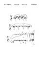
- FIG. 1Ais a right, front and top perspective view of an L-shaped conduit for use in the present invention
- FIG. 1Bis a side elevation view of the apparatus of FIG. 1A shown partially in section to reveal an optional bi-directional flow regulator located in a lumen of an anchor arm of the conduit;
- FIG. 1Cis a side elevation view of a conduit similar to that of FIG. 1A showing the addition of a capacitance pressure reservoir as an alternative embodiment
- FIG. 2Ais a right, front and top perspective view of a T-shaped conduit according to the present invention.
- FIG. 2Bis a side elevation view of the conduit of FIG. 2A shown partially in section to reveal an optional bi-directional flow regulator located in a lumen of an anchor arm of the conduit;
- FIG. 2Cis a side elevation view of the conduit of FIG. 2A shown partially in section to reveal one optional bi-directional flow regulator located in the lumen of the anchor arm of the conduit, and another optional bi-directional flow regulator located in an intracoronary arm of the conduit;
- FIG. 2Dis a side elevation view of a conduit similar to that of FIG. 2A showing the addition of a capacitance pressure reservoir as an alternative embodiment
- FIG. 3Ais a partial side elevation view of a conduit similar to that of FIGS. 1A and 2A shown partially in section to reveal a flexible anchor arm with rigid rings ensheathed in a flexible covering as an alternative embodiment;
- FIG. 3Bis a partial side elevation view of a conduit similar to that of FIG. 3A shown in section in an extended form;
- FIG. 3Cis a partial side elevation view of a conduit similar to that of FIG. 3A shown in section in a compressed form;
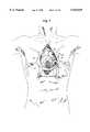
- FIG. 4is an anterior view of a human chest which is incised longitudinally to reveal a dissected pericardium and mediastinal contents;
- FIG. 5is a magnified view of an area circled 200 in FIG. 4 illustrating a longitudinally incised coronary artery;
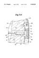
- FIG. 6is a partial external perspective view of a transversely sectioned coronary artery and heart wall illustrating a channel leading from a lumen of a coronary artery and into a chamber of the heart according to the method of the present invention
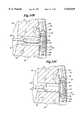
- FIG. 7is a partial external perspective view of a transversely sectioned coronary artery and heart wall illustrating the partial placement of one embodiment of the conduit of the present invention into the incised coronary artery and formed channel illustrated in FIG. 6;
- FIG. 8is a partial external perspective view of a transversely sectioned coronary artery and heart wall illustrating the completed placement of one embodiment of the conduit of the present invention into the incised coronary artery and formed channel illustrated in FIG. 6;
- FIG. 9is a partial external perspective view of a sutured coronary artery and phantom view of the conduit of the present invention.
- FIG. 10is a schematic illustration of the use of an endovascular catheter to catheterize the patient's coronary artery
- FIG. 11Ais a cutaway side elevation view of the coronary artery of the bypass procedure illustrating an intravascular catheter with distally-located stent prior to inflation of a catheter balloon underlying the stent;
- FIG 11Bis a cutaway side elevation view of the coronary artery of the bypass procedure illustrating the intravascular catheter with distally-located stent following inflation of the catheter balloon underlying the stent;
- FIG. 11Cis a cutaway side elevation view of a coronary artery illustrating the stent seated to the walls of the coronary artery and the catheter partially withdrawn following deflation of the catheter balloon;
- FIG. 12is a schematic illustration with the heart in partial cutaway of the use of an endovascular catheter to catheterize the patient's left ventricle.
- FIG. 13Ais a cutaway view of the left ventricle and a partial cutaway view of the coronary artery with seated stent illustrating the formation of a channel into the wall of the left ventricle;
- FIG. 13Bis a cutaway view of the left ventricle and a partial cutaway view of the coronary artery with seated stent illustrating a completed channel through the wall of the left ventricle and deep wall of the coronary artery at the chosen bypass site;
- FIG. 14Ais a cross-sectioned view of the left ventricle and a partial cutaway view of the coronary artery with seated stent illustrating the placement of the second intraventricular catheter within the formed channel;
- FIG. 14Bis a cross-sectioned view of the left ventricle and a partial cutaway view of the coronary artery with seated stent illustrating a blockage of the formed channel by the re-inflated balloon of the intracoronary catheter;
- FIG. 14Cis a cross-sectioned view of the left ventricle and a partial cutaway view of the coronary artery with seated stent illustrating an inflation of the balloon located on the distal end of the intraventricular catheter and the seating of an overlying spiral-shaped device against the walls of the formed channel;
- FIG. 14Dis a cross-sectioned view of the left ventricle and a partial cutaway view of the coronary artery with seated stent illustrating the device in its locked cylindrical shape seated against the channel walls and the partially withdrawn second intraventricular catheter;
- FIG. 15Ais a right anterior superior perspective view of the device placed within the formed channel in its spiral shape
- FIG. 15Bis a right anterior superior perspective view of the device placed within the formed channel in its cylindrical form
- FIG. 16is a cross-sectional view of an interlocking mechanism of the device of FIGS. 15A and 15B in its locked position;
- FIG. 17Ais a cross-sectioned view of the left ventricle and a partial cutaway view of the coronary artery, with the device shown in FIGS. 15A and 15B seated within the formed channel, illustrating the introduction of a third intraventricular catheter into the formed channel;
- FIG. 17Bis a cross-sectioned view of the left ventricle and a partial cutaway view of the coronary artery, with the device shown in FIGS. 15A and 15B seated within the formed channel, illustrating a tongue and groove interlocking of the bi-directional flow regulator equipped device to the device seated within the formed channel;
- FIG. 18Ais a schematic longitudinal cross-sectional view of a bi-directional flow regulator shown in a full flow position.
- FIG. 18Bis the view of FIG. 18A with the bi-directional flow regulator shown in a reduced flow position
- FIG. 18Cis a transverse cross-sectional view of the bi-directional flow regulator of FIG. 18B;
- FIG. 19Ais a schematic cross-section longitudinal view of an alternative embodiment of a bi-directional flow regulator shown in a full flow position
- FIG. 19Bis the view of FIG. 19A showing the bi-directional flow regulator in a reduced flow position
- FIG. 19Cis a transverse cross-sectional view of the bi-directional flow regulator of FIG. 19B;
- FIG. 20is a schematic longitudinal cross-sectional view of a channel defining conduit with an alternative embodiment tapered anchor arm
- FIG. 21is a schematic longitudinal cross-sectional view of the conduit of FIG. 1A in place in a coronary artery;
- FIG. 22is a schematic longitudinal cross-sectional view of a test conduit for animal testing of the invention.
- FIG. 23is a schematic longitudinal cross-sectional view of a conduit in place in a coronary artery illustrating a deflecting shield to protect the coronary artery.
- the inventiondeparts from the traditional bypass approach. Rather then providing an alternative pathway for blood to flow from an aorta to a coronary artery, the invention provides a blood flow path leading directly from a chamber of a heart to a coronary artery at a site downstream from the stenosis or occlusion. Unlike U.S. Pat. Nos. 5,429,144; 5,287,861 and 5,409,019 and contrary to the teachings of these patents, the ventricular-to-coronary artery blood flow path remains open during both diastole and systole. The surgical placement of the apparatus of the present invention establishes this alternative pathway. Also, and as will be more fully described, the invention includes means for protecting the coronary artery from direct impingement of high velocity blood flow.
- FIG. 1AThe presently preferred embodiment is illustrated in FIG. 1A as an L-shaped conduit 10' with an intracoronary arm 14' to reside in the coronary artery (and opening downstream of an occlusion).
- the conduit 10'has an anchor arm 12' extending through the heart wall with an opening 12a' in communication with the interior of the left ventricle.
- While various minimally invasive surgical proceduresare described with respect to alternative embodiments, the presently preferred embodiment places the conduit 10' into a coronary artery through an open-chest approach to be described in greater detail with reference to FIGS. 4-9. While minimally invasive procedures are desirable, an open chest procedure is presently preferred due to the already large number of physicians trained and skilled in such procedures thus making the benefits of the present invention more rapidly available to patients who currently lack effective treatment.
- FIG. 21is a schematic cross-sectional view of a conduit 10' of FIG. 1A placed within a coronary artery 30.
- Coronary artery 30has a lower surface 40 residing against an external surface of a heart wall 42 surrounding the left ventricle 44.
- the wall 36 of the artery 30defines an artery lumen 48 through which blood flows in the direction of arrow A.
- an obstruction 34is shown within the lumen 48. The obstruction 34 acts to reduce the volume of blood flow along the direction of arrow A.
- the conduit 10'is a rigid, L-shaped tube having an anchor arm 12' with a longitudinal axis X--X and an opening 12a' at an axial end.
- the conduit 10'may be any suitable device (e.g., rigid tube, lattice stent, etc.) for defining and maintaining a fluid pathway during contraction of the heart.
- the conduit 10'has an intracoronary arm 14' with a longitudinal axis Y--Y and an opening 14a' at an axial end. Both of arms 12', 14' are cylindrical in shape and define a continuous blood flow pathway 11' from opening 12a' to opening 14a'.
- the axes X--X and Y--Yare perpendicular in a preferred embodiment.
- the axes X--X, Y--Ycould define an angle greater than 90° to provide a less turbulent blood flow from arm 12' to arm 14'.
- the conduit 10'is positioned for the anchor arm 12' to pass through a preformed opening 50 in the heart wall 42 and extending from the lower surface 40 of the coronary artery 30 into the left ventricle 44.
- the opening 12a'is in blood flow communication with the interior of the left ventricle 44 so that blood may flow from the left ventricle 44 directly into path 11'.
- the arm 14'is coaxially aligned with the coronary artery 30 and with the opening 14a' facing downstream (i.e., in a direction facing away from obstruction 34).
- Blood flow from opening 12a'passes through the pathway 11' and is discharged through opening 14a' into the lumen 48 of the coronary artery 30 downstream of the obstruction 34.
- the outer diameter of arm 14a'is approximate to or slightly less than the diameter of the lumen 48.
- the axial length of the anchor arm 12'is preferably greater than the thickness of the heart wall 42 such that a length L protrudes beyond the interior surface of the heart wall 42 into the left ventricle 44.
- the length L of penetration into the left ventricle 44is about 1-3 millimeters in order to prevent tissue growth and occlusions over the opening 12a'.
- the arm 14'holds the conduit 10' within the coronary artery 30 to prevent the conduit 10' from otherwise migrating through the preformed opening 50 and into the left ventricle 44.
- an upper wall 14b' of arm 14'defines a region 15' against which blood flow may impinge. Stated differently, in the absence of an arm 14' or region 15', blood flow would pass through the anchor arm 12' and impinge directly against the upper wall 36 of the coronary artery 30. High velocity blood flow could damage the wall 36, as will be more fully described, resulting in risk to the patient.
- the region 15'acts as a shield to protect the coronary artery 30 from such blood flow and to redirect the blood flow axially out of opening 14a' into the coronary artery 30.
- Thisis schematically illustrated in FIG. 23.
- the axis X--X of the anchor arm 12'is shown at a non-orthogonal angle with respect to the direction A of blood flow in the coronary artery 30 (axis X--X may be either orthogonal or non-orthogonal to direction A).
- the vector B of blood flow from the anchor arm 12'has a vector component B' parallel to blood flow A and a vector component B" perpendicular to direction A.
- the region 15'is positioned between the wall 36 and anchor arm 12' to prevent the blood flow B with high vector component B" from impinging upon wall 36.
- the blood flow deflected off region 15'has a reduced vector component perpendicular to flow direction A and reduced likelihood of damage to the coronary artery 30.
- the region 15'may be a portion of an intracoronary arm 14' or the arm 14' may be eliminated with the region 15' being an axially spaced extension from arm 12' or a separate shield surgically positioned within the coronary artery.
- a portion 17' of the anchor arm 12'extends from the lower surface 40 of the coronary artery 30 and through the lumen 48 to the upper surface 36 to block the cross-section of the coronary artery upstream from opening 14a'.
- the region 17'acts as a barrier to impede or prevent any dislodged portions of the obstruction 34 from passing the conduit 10' and flowing downstream through the coronary artery 30.
- the present inventionmaintains blood flow through the conduit 10' during both diastole and systole. Therefore, while the net blood flow is in the direction of arrow A, during diastole, blood will flow in a direction opposite of that of arrow A.
- FIG. 22schematically illustrates the tests as the placement of a test conduit 10* in the coronary artery 30' of a pig.
- a stainless steel T-shaped conduit 10*is used having aligned openings 14a*, 16a* positioned within the coronary artery 30' and with a third opening 12a* protruding 90° out of the coronary artery 30'.
- the conduit 10*has a uniform interior diameter of 3 millimeters to correspond in sizing with a 3 millimeter lumen of coronary artery 30'.
- the third opening 12a*is connected by a 3 millimeter conduit 13 to a 3 millimeter rigid Teflon (PTFE) sleeve 13a which was passed through the heart wall 42' into the left ventricle 44'.
- PTFETeflon
- the conduit 13 and sleeve 13ado not pass through the coronary artery 30'.
- a first closure device in the form of a suture loop 300surrounds the artery 30' adjacent the upstream opening 14a* of the conduit 10*.
- the loop 300provides a means for closing the upstream opening 14a* by selectively constricting or opening the loop 300 to selectively open or block blood flow through the coronary artery 30'.
- the first loop 300permits the test to simulate blockage of the coronary artery 30' upstream of the conduit 10*.
- a flow meter 304 to measure volumetric flow of blood downstream of the conduit 10*is placed adjacent downstream opening 16a*.
- a second closure device 302 functioning the same as loop 300is placed on conduit 13 to selectively open or close blood flow through conduit 13.
- the conduit 10*simulates normal blood flow through a healthy coronary artery 30' and the normal blood flow can be measured by the flow measuring device 304.
- the test conduit 10*can simulate the placement of a conduit such as that in FIG. 21 with an obstruction located on the upstream side of the conduit.
- the flow meter 304can then measure flow of blood through the conduit 10* during both diastole and systole.
- the results of the testsindicate there is a substantial net forward blood flow (i.e., volumetric forward flow less volumetric retro-flow) with the second device 302 remaining open during both diastole and systole and with the first device 300 closed to simulate an obstruction.
- net blood flows in excess of 80 percent of normal net forward blood flowwere measured. It was also noted that with the second device 302 closed and first device 300 open to simulate normal blood flow, the peak blood flow through the coronary artery 30' occurred during systole. With the first device 300 closed to simulate an obstruction and with the second device 302 open, the peak blood flow occurred at diastole.
- a volumetric forward flow greater than a volumetric rearward flowcan be manipulated through a variety of means including sizing of the interior diameter of the conduit, geometry of the conduit (e.g., taper, cross-sectional geometry and angle) and, as will be more fully discussed, structure to restrict rear flow relevant to forward flow.
- the sizing of the interior diameter of the flow pathway 11'can be selected to minimize back flow.
- the net flowincreases with a reduction in the diameter as suggested by simulation modeling of flow through a conduit.
- One method in which shear rate and flow bias can be controlledis by providing a tapered diameter for a narrower diameter at opening 14a' than at opening 12a'.
- the selection of the conduit geometrye.g., an angled anchor arm as shown in FIG. 23 or a tapered geometry as will be discussed with reference to FIG. 20) can be selected to modify the degree to which the conduit is biased to net forward flow (i.e., the conduit offers less resistance to forward flow than to retro-flow) without stopping or blocking retro-flow.
- the substantial net blood flow measured in animal testing through the inventionis extraordinarily high when compared to minimum acceptable levels of net blood flow following traditional bypass techniques (i.e., about 25 percent of normal net blood flow). Further, the results are counter-intuitive and contradictory to the prior teachings of the art of U.S. Pat. Nos. 5,429,144; 5,287,861 and 5,409,919 and the afore-mentioned Munro et al. article.
- the present inventionprovides a conduit with a shielding area to prevent damaging impingement of blood flow directly onto the coronary artery wall as well as providing a blocking area to prevent the migration of debris from an obstruction to a location downstream of the conduit.
- the present inventionplaces an apparatus for defining a blood flow conduit directly from a chamber of a heart to a coronary artery downstream of an occluded site.
- an apparatus of the present inventionwill be described.
- the apparatus of the present inventioncan be a variety of shapes or sizes, and is not meant to be limited as to size, shape, construction, material, or in any other way by the following examples in which a preferred embodiment is illustrated.
- FIGS. 2A, 2B, 2C, 2D and 2Erelated embodiments of an apparatus according to the present invention are shown as a rigid T-shaped conduit 10 (a preferred L-shaped conduit 10' having already been summarized and to be later described in detail).
- the conduit 10is hollow and includes two axially-aligned intracoronary arms 14, 16 terminating at open ends 14a, 16a.
- An anchor arm 12(having an open end 12a) extends perpendicularly to arms 14, 16.
- the entire conduit 10is hollow to define a blood flow conduit 11 providing blood flow communication between open ends 12a, 14a and 16a.
- arms 14 and 16are adapted to be placed and retained within a lumen of a coronary artery on a downstream side of an occlusion with open ends 14a, 16a in blood flow communication with the lumen.
- the anchor arm 12is adapted to extend through and be retained in a heart wall (e.g., a wall of the left ventricle) with the open end 12a in blood flow communication with blood within the chamber.
- the conduit 10defines a surgically-placed conduit establishing direct blood flow from the heart chamber to the artery.
- directit is meant that the blood flow does not pass through the aorta as occurs in traditional bypass procedures.
- the conduit 10is sufficiently rigid such that it defines an open blood flow path during both diastole and systole.
- back flowcan be controlled by the geometry of the conduit. The following describes a presently less preferred alternative embodiment for controlling back flow.
- FIG. 2Billustrates use of an optional bi-directional flow regulator 22 within the conduit 10 and positioned in anchor arm 12.
- the bi-directional flow regulator 22permits unimpeded flow in the direction of arrow A (i.e., from open end 12a to open ends 14a, 16a) while permitting a reduced (but not blocked) reverse flow.
- FIG. 2Cillustrates the use of a first bi-directional flow regulator 22 as well as a second bi-directional flow regulator 26 in arm 16 near the open end 16a of the apparatus.
- the second bi-directional flow regulator 26permits unimpeded blood flow in the direction of arrow B.
- the second bi-directional flow regulator 26is used to permit a reduced (but not zero) back flow of blood in an upstream direction within the coronary artery.
- the coronary arterymay not be completely obstructed and may have a reduced flow past an obstruction.
- the use of the T-conduit 10 with axially aligned arms 14, 16takes advantage of such reduced flow and supplements such flow with blood through anchor arm 12.
- the conduit 10is placed with the arms 14, 16 in the lumen of the artery with opening 16a positioned on the upstream side (i.e., nearest to, but still downstream of, the obstruction).
- the flow regulator 22is a bi-directional flow regulator. By this it is meant that the flow regulator 22 does not block flow of blood in any direction. Instead, the flow regulator 22 permits a first or maximum flow rate in one direction and a second or reduced flow rate in a second direction.
- the flow regulatoris schematically illustrated in FIGS. 18A through 19C. In each of these embodiments, the arrow A indicates the direction of blood flow from the left ventricle to the coronary artery.
- FIGS. 18A through 18Cillustrate one embodiment of a bi-directional flow regulator 22.
- FIGS. 19A through 19Cillustrate an alternative embodiment of a bi-directional flow regulator 22.
- the regulator 22 of FIGS. 18A through 18Cshows a butterfly valve 222 mounted in the anchor arm 12 of a rigid conduit 10.
- Valve 222may be pivoted (in response to blood flow in the direction of arrow A) between a position with the plate 222 generally parallel to the walls 12 of the conduit 10 as illustrated in FIG. 18A.
- the plate 222can be rotated (in response to blood flow reverse to arrow A) to a position angled relative to the walls 12 of the conduit 10 as illustrated in FIG. 18B.
- FIG. 18Amay be conveniently referred to as a full flow position.
- FIG. 18Bmay be conveniently referred to as a reduced flow position.
- FIG. 18Cis a cross-section of the conduit 10 when the plate 222 is in the reduced flow position.
- the plate 222is sized relative to the conduit 10 such that the cross-sectional area of the conduit 10 which remains open is sufficient to permit about 20% of the blood flow (measured volumetrically) to flow back through the conduit 10 in a direction opposite to that of arrow A during diastole.
- blood flow from the heart to the coronary arteryurges the plate 222 to the full flow position of FIG. 18A such blood may flow unobstructed through the device to the coronary artery.
- the blooddue to pressure differentials between the coronary artery and the left ventricle
- the plate 222will flow in a direction opposite of that of arrow A causing the plate 222 to rotate to the position of FIG. 18B and 18C.
- the plate 222is prevented from moving to a full closed position such that flow through the device is never blocked and instead may proceed with a back flow of about 20% (volumetrically measured) of the normal flow in the direction of A.
- FIGS. 19A through 19Cshow an alternative design of the conduit 10 with the flow regulator 22a in the form of three leafs 222a, 222b, 222c which, in response to blood flow from the left ventricle to the coronary artery, open to a full open position shown in FIG. 19B and move to a restricted flow position in FIGS. 19A and 19C in response to back flow.
- the leaves 222a, 222b, 222care provided with openings 223 to permit flow through the leaves 222a, 222b, 222c at all times.
- Back flow necessary to wash the componentscan be achieved through either a conduit 10 which has a constant opening through both systole and diastole (i.e., conduit 10 of FIG. 2A without the use of a bi-directional flow regulator 22) or with a device coupled with a bi-directional flow regulator 22 (FIGS. 2B-2C) which permits a 20% flow rate back flow during diastole.
- an L-shaped conduit 10'(FIGS. 1A, 1B, 1C) is used to completely bypass the coronary obstruction.
- An L-shaped conduit 10'has an anchor arm 12' with an open end 12a'. Unlike conduit 10, conduit 10' has only one intracoronary arm 14' perpendicular to arm 12'. Arm 14' has an open end 14a' and conduit 10' is hollow to define a continuous fluid pathway 11' from end 12a' to end 14a'.
- arm 14'is placed within the lumen of an artery. End 14a' faces downstream from an obstruction.
- Arm 12'is placed through the heart wall with end 12a' in fluid communication with blood within the heart chamber.
- the anchor arm 12'can include a bi-directional flow regulator 22' similar to bi-directional flow regulator 22 of conduit 10.
- Conduit 10, 10'may be rigid, or have varying flexibilities. Regardless of such flexibility, the conduit 10, 10' should be sufficiently rigid for pathway 11, 11' to remain open during both diastole and systole.
- FIGS. 3A, 3B and 3Cdemonstrate one embodiment where the anchor arm (i.e., elements 12, 12' of FIGS. 1A and 2A) is comprised of a number of rings 17 surrounded by a membrane 18. In FIGS. 3A-3C, only anchor arm 12 is shown. It will be appreciated that anchor arm 12' may be identically constructed.
- the rings 17can be constructed of Teflon, and the surrounding membrane 18 can be constructed of a double-walled Dacron sheath into which the rigid supporting rings 17 are sewn.
- the rings 17provide structural strength. The structural strength maintains an open lumen or conduit 11 leading into the coronary artery by preventing the conduit 11 from collapsing by reason of contraction of the heart muscle surrounding the anchor arm 12.
- the series of rings 17provide a degree of flexibility which allows a channel formed through the heart chamber muscular wall (receiving anchor arm 12) to be angled or curved.
- the flexibility of the surrounding sheath 18 in concert with the rigid rings 17will allow the anchor arm 12 to expand, FIG. 3B, and contract, FIG. 3C, with the contractions and relaxations of the surrounding cardiac musculature.
- this attaching mechanism 19is a rigid flange 12a. It will be appreciated that other mechanisms of attachment, such as suturing, biologically gluing, etc. are alternative options.
- the apparatus of the present inventionprovides a path 11 through which blood flows from a chamber of a heart and into a coronary artery. Additionally, such a device can store blood under pressure for a period of time prior to its introduction into a coronary artery.
- this aspect of the conduit 10, 10' of the present inventionis referred to as a capacitance pressure reservoir (CPR) 24, 24'.
- CPRcapacitance pressure reservoir
- Blood flow through the normal coronary arteryis cyclical. Blood flow is increased during diastole (when the heart muscle is in a relaxing state), and decreases or reverses during systole (when the heart muscle is in a contracting state). See, e.g., F. Kajiya et al., Velocity Profiles and Phasic Flow Patterns in the Non-Stenotic Human Left Anterior Descending Coronary Artery during Cardiac Surgery, 27 CARDIOVASCULAR RES. 845-50 (1993).
- the pressure gradient across the lumens 12a, 12a', 14a', 16a of the apparatus 10, 10' of the present inventionwill vary over the cardiac cycle. For example, during systole, the contraction of the heart muscles will generate high relative pressures within the left ventricle.
- the pressures within the coronary arterioles and capillaries distal to the bypass sitecan also be high during this time, due to the external compression of the contracting cardiac musculature surrounding these vessels. This is particularly true for the vessels of the microcirculation deep within the heart which serve the endocardium.
- the optional CPR 24, 24'stores the pressurized blood during systole for delivery to the heart muscles via the coronary circulation during diastole when pressures are reduced.
- the CPR 24, 24'serves a function similar to the elastic connective tissue of the thick-walled aorta.
- the necessary function of the CPR 24, 24'is to store blood under higher pressure, and to later provide that stored blood to the microcirculation when the external pressures on that microcirculation are reduced.
- the bi-directional flow regulators 22, 22'provide full blood flow in the direction of A, which is from a chamber of a heart into the conduit 10, 10' via the lumen 11, 11'.
- Awhich is from a chamber of a heart into the conduit 10, 10' via the lumen 11, 11'.
- the pressure on the blood within the chamber of a heartwill be greatest when the surrounding cardiac musculature is in the contracting phase of the cardiac cycle. Because it is during this phase of the cardiac cycle that the external pressure on the coronary artery microcirculation is also highest, blood flow through the lumen 11, 11' of the conduit 10, 10' could be limited.
- the conduit 10, 10'is equipped with a reservoir 24, 24' which stores this pressurized blood flowing from a chamber of the heart during the cardiac contraction.
- the reservoir, or CPR 24, 24'is schematically illustrated in FIGS. 1C, 2D.
- the conduit 10, 10'is provided with a fluid passage 28, 28' in communication with pathway 11, 11'.
- the passage 28, 28'communicates with an expandable volume (or storage chamber) 27, 27' defined by a movable wall 31, 31' contained within a fixed housing 33, 33'.
- Springs 29, 29' between wall 31, 31' and housing 33, 33'urge the wall 31, 31' to move to reduce the size of volume 27, 27'.
- the springs 29, 29'are pre-loaded to exert a force on wall 31, 31' less than a force exerted by blood within volume 27, 27' during the contraction phase of the cardiac cycle, but greater than the force exerted by blood within volume 27, 27' during the relaxation phase of the cardiac cycle.
- the conduit 10, 10'is constructed in a manner which allows blood to flow into the storage chamber 27, 27' of the conduit 10, 10' through the lumen 11, 11' of arm 28, 28' of the conduit when the cardiac musculature is contracting.
- the kinetic energy of the flowing bloodis converted to potential energy, and stored in 29, 29'.
- the potential energy stored in 29, 29' of the CPR 24, 24'is then reconverted to kinetic energy in the form of blood flow out of the storage chamber 27, 27' of the conduit 10, 10' via the lumen 11, 11' of arm 28, 28' of the conduit.
- the CPR 24, 24'is illustrated with a movable wall 31, 31' and springs 29, 29' to define a variable volume, other designs can be used.
- the CPR 24, 24'can be a balloon-like structure. As it fills with blood, the pressure on that blood increases through the stretching of an elastic component of a balloon.
- the CPR, 24, 24'can be a hollow bag, made of a material which is elastic, but impermeable to liquids, and pliable similar to a plastic bag. When the heart contracts, blood is forced through lumen 11, 11' of arm 28, 28' of the apparatus 10, 10' of the invention into the collection bag.
- bi-directional flow regulators 22, 22' within the anchoring arm 12, 12' of the conduit 10, 10'provide most (about 80%) of the flow of blood out of the device during diastole to the coronary artery via the lumen 11' 11' of arms 14a, 14a', 16a of the device, of the conduit 10, 10'.
- the incorporation of the bi-directional flow regulator 26 within the intracoronary arm 16 of the T-shaped conduit 10when employed with the bi-directional flow regulator 22 within the anchor arm 20 of the conduit 10, would provide most of the flow of blood out of the device during diastole to the portion of the coronary artery distal to the bypass site via the downstream lumen 11 of arm 14a.
- the inner and outer cross-sectional diameters of a coronary arterydecreases with the distance from the arterial origin. Eventually, the artery branches into a number of arterioles, which feed the capillary bed of the coronary arterial microcirculation.
- the typical diameter of a lumen of a coronary arteryis, in general, species specific; increasing with heart size. In humans, this lumen diameter is dependent upon which artery is being evaluated, but usually ranges from 1.0 to 4 mm in diameter, and decreases with distance from the aortic origin.
- the cross-sectional outer diameter of the intracoronary arms 14, 14', 16 of the device of the present inventionshould effectively approximate the diameter of the lumen of the coronary artery being bypassed, at the bypass site. This allows the complete re-approximation of the previously opened superficial wall of the coronary artery during surgical closure, without high suture or staple tension resulting.
- the outer diameter of the intracoronary arms 14, 14', 16 of the conduit 10, 10' of the present inventionis equal to the diameter of the lumen of the coronary artery which is being bypassed, at the bypass location.
- the artery wallmay need to be expanded by the addition of a patch, such as Dacron, well known in the art.
- the cross-sectional diameter of a lumen of a coronary arterywill increase with the oxygen demand of cardiac muscle during times of stress.
- the cross-sectional inner diameter of the intracoronary arms 14, 14', 16 of the conduit 10, 10' of the present inventionshould effectively approximate that diameter necessary to provide adequate blood flow through the downstream lumen of the conduit to effectively oxygenate the cardiac musculature normally supplied by the microcirculation of the coronary artery.
- the cross-sectional inner diameter of the intracoronary arms 14, 14', 16 of the conduit 10, 10' of the present inventionshould effectively approximate that diameter necessary to provide adequate blood flow through the lumen of the device to effectively oxygenate the cardiac musculature normally supplied by the microcirculation of the coronary artery during both times of cardiovascular resting and stress.
- the operating pressure of the bi-directional flow regulator 22, 22'i.e., the pressure at which the flow regulator moves from a reduced back-flow to a full forward flow position
- the dynamic measurements of coronary artery pressure, blood flow, and heart chamber pressurescan be determined by the dynamic measurements of coronary artery pressure, blood flow, and heart chamber pressures through selective catheterization with standard techniques. See Minoru Hongo et al., 127 (3) AM. HEART J. 545-51 (March 1994).
- the most appropriate sizing of the intracoronary arms 14, 14', 16 of the conduit 10, 10' of the present inventioncan be re-assessed. This can be accomplished by probing the distal and, if needed, the proximal aspects of the coronary artery at the chosen bypass site with blunt instruments of known outer diameters. Such sizing by probes is well-known in the literature.
- an assortment of conduits of the present invention of various diameterscan be available for the surgeon to select from.
- the anchor arm 12, 12'is sized to maximize net blood flow from the left ventricle to the coronary artery.
- maximizing the diameter of anchor arm 12, 12'is not desirable. For example, such simulation assuming diameters of 3.00 mm, 2.25 mm and 1.50 mm for an unrestricted fistula (i.e., without a flow regulator 22) suggests that the smaller diameter of 1.50 mm most closely approximates normal coronary blood flow and minimizes back flow thus maximizing net forward flow.
- anchor arm 12, 12'protrudes into the heart chamber such that end 12a is spaced from the heart wall. This prevents tissue growth over end 12a.
- the anchor arm 12defines a longitudinal axis (e.g., axis X--X in FIG. 18A).
- the region 15 of arms 14, 14intersects axis X--X.
- the region 15acts as a deflection surface to prevent high velocity blood flow from arm 12 impinging directly upon the coronary artery wall. Instead, the high velocity blood flow impinges upon region 15 and is directed axially into the coronary artery.
- the coronary artery wall covered by region 15is protected from damage which would otherwise be caused by the high velocity blood flow and the blood components are transitioned to axial flow with a minimum of cell damaging shear.
- FIG. 20shows a still further embodiment 10" where the anchor arm 12" has a longitudinal axis X'--X' at a non-orthogonal angle relative to the axis Y'--Y' of the coronary arms 14", 16". Further, the anchor arm 12" has a taper. In other words, the arm 12" is widest at opening 12a". The taper and angle act to reduce blood flow velocity and to restrict back flow (arrows B) while facilitating forward flow (arrow A'). Also, the blood in the forward flow A' impacts against the deflection region 15" at an angle to reduce impact of blood cells.
- the method of the present inventionis suitable for performing a variety of surgical cardiac procedures.
- the proceduresmay be performed utilizing an open-chest approach, or through minimally invasive approaches by the creation of access means into the chest, or through percutaneous access utilizing intracoronary and intraventricular catheterization.
- the heartcan be allowed to pulse normally, be slowed by varying amounts, or stopped completely. A significant period of complete heart stoppage can necessitate the use of supportive cardiopulmonary bypass.
- the method of the present invention for performing a coronary artery bypass procedurewill now be described in detail.
- the patient who is to undergo the procedurecan be prepared in a conventional manner for cardiac bypass surgery.
- the patient preparation, anesthesia utilized, and access route to the coronary circulation,will vary depending upon the invasiveness of the specific procedure chosen.
- the patientcan be prepared for surgery as outlined by Von Segesser.
- Preparationscan include the sterile scrubbing and draping of at least one groin to permit access to a femoral artery for catheterization of the coronary vasculature and the sterile scrubbing and draping of the right superior anterior chest wall to permit access to the innominate artery for catheterization of the left ventricle.
- Further suggested preparationscan include those outlined by Sterman and Acuff for thoracoscopic surgery with and without cardiopulmonary bypass, respectively.
- the patientwill be placed under general anesthesia prior to the procedure.
- standard cardiac operative anesthetic techniquessuch as premedication with diazepam, induction with propofol and sufentanil, and maintenance with desflurane can be employed.
- less than general anesthesiacan be utilized.
- Less than general anesthesiais well known in the literature.
- Selective ventilation of the lungscan be achieved through the placement of a double-lumen endobronchial tube which independently provides for the intubation of the left and right main stem bronchi.
- An intraesophageal probecan be placed to facilitate cardiac monitoring and the synchronization of power to the laser, when deemed useful.
- access to the patient's coronary arterial vasculaturecan be attained through a variety of techniques, dependent upon the route of access chosen.
- Von Segesserhas reported a method of access to the coronary arterial vasculature when utilizing an open-chest approach and cardiopulmonary bypass. In one embodiment, utilizing an open-chest approach with cardiopulmonary bypass, access to the coronary vasculature can be obtained as reported by Von Segesser.
- Buffolo et al.has reported an open-chest approach to the coronary arterial vasculature when performed without cardiopulmonary bypass. See Buffolo et al., 61 ANN. THORAC. SURG. 63-66 (1996). In one embodiment utilizing an open-chest approach without cardiopulmonary bypass, access to the coronary vasculature can be obtained as reported by Buffolo.
- Sterman et al.has reported a method of access to the coronary arterial vasculature when a closed-chest approach with cardiopulmonary bypass is utilized. See Sterman et al., U.S. Pat. No. 5,452,733 (1995). Sterman positions a plurality of access trocar sheaths along the patient's left and right anterolateral chest wall. These trocar sheaths provide access to the coronary vasculature, and allow the temporary repositioning of the heart to facilitate the performance of the procedure. The repositioning is accomplished utilizing grasping tools introduced through the appropriate trocar sheaths.
- Visualization during this procedurecan be either indirectly via thoracoscopy, or directly via a ⁇ window ⁇ placed in the left middle anterior chest wall by the surgical removal of the fourth rib. Access to the bypass site can therefore be obtained by following the techniques outlined by Sterman.
- the instruments to be used in the procedurecan also be similar to those described by Sterman.
- Acuff et al.has described a method of access to the coronary arterial vasculature when a closed-chest approach without cardiopulmonary bypass is utilized. See Acuff et al., 61 ANN. THORAC. SURG. 135-37 (1996). Similar to the techniques of Sterman, Acuff positions a plurality of access trocar sheaths along the patient's left and right anterolateral chest wall. Also similar to Sterman, Acuff surgically establishes an access space, or window in the left anterior chest wall through the removal of the left fourth rib cartilage. The trocar sheaths, in concert with this window, allow the temporary repositioning of the heart, and access to the coronary arterial vasculature. Visualization during this procedure can be either indirectly via thoracoscopy, or directly via the window. Access to the bypass site can therefore be obtained by following the techniques outlined by Acuff. The instruments to be used in the procedure can also be similar to those described by Acuff.
- Access to a chamber of a heart and a coronary artery when the bypass is performed through the percutaneous approach of intracoronary and intraventricular catheterizationcan be obtained as follows.
- Access to a coronary arterycan be obtained by the introduction of a catheter into the left or right femoral artery through an arterial cut down procedure.
- the cathetercan then be fed retrograde past the descending aorta, through the ascending aorta, and into the coronary artery by standard catheterization techniques.
- access to a chamber of the left side of a heartcan be obtained by the introduction of a catheter into the innominate artery, also through an arterial cut down procedure.
- access to the left ventricleis obtained by the introduction of a catheter into the innominate artery and the advancement of this catheter into the left ventricle.
- the catheteris advanced through the ascending aorta, past the aortic valve. and into the left ventricle. Techniques by which the left ventricle is catheterized are well known in the literature.
- a chamber of a heartprovides blood to a coronary artery.
- the method of the present inventioncan accomplish this by establishing one or more channels through the wall of a chamber of a heart which lead directly from a chamber of a heart into a coronary artery at a site distal to the narrowing or blockage.
- the methods of the inventionin various embodiments can achieve the establishment of such a channel or channels through a variety of techniques.
- FIGS. 4, 5, 6, 7, 8, and 9an exemplary open-chest procedure, which may or may not include cardiopulmonary bypass, by which a coronary artery bypass procedure may be accomplished will be described.
- the open-chest approachaffords maximal access to, and visualization of, the coronary vasculature; although at the expense of injury to normal tissue.
- conduit 10, 10' of the present inventionwhich provides blood from a chamber of a heart 43 directly into a coronary artery 30, is placed.
- conduit 10'is discussed. It will be appreciated that conduit 10 can be similarly placed.
- exampleswill be limited to the embodiment of the conduit of the invention as illustrated in FIG. 1A.
- the chest cavityis entered, and pericardium 52 incised anteriorly, to expose a coronary artery 30 (having an obstruction 34) to be bypassed. This is illustrated in FIG. 4.
- cardiopulmonary bypassmay be initiated by a variety of standard techniques as outlined by George Silvay et al., Cardiopulmonary Bypass for Adult patients: A Survey of Equipment and Techniques, 9 (4) J. CARDIOTHORAC. VASC. ANESTH. 420-24 (August 1995).
- the heartis slowed and/or stopped by a variety of standard techniques.
- One standard techniqueis to electrically induce ventricular fibrillation.
- Another standard techniqueis warm or cold blood cardioplegia, delivered antegrade or retrograde, and intermittent or continuous, as outlined by Gerald D. Buckberg, Update on Current Techniques of Myocardial Protection, 60 ANN. THORAC. SURG. 805-14 (1995).
- the heartis inspected and coronary arteries identified.
- the narrowed or occluded coronary artery 30can be visually identified, and an appropriate site distal or downstream from the occlusion 34 chosen.
- blood flow through the target coronary artery 30is halted by standard techniques.
- standard techniquesinclude clamping the aorta above the coronary ostia with an arterial clamp.
- the flow of blood within the coronary artery 30can be halted by forming a loop around the artery 30 with suture either proximally, or both proximally and distally, and applying appropriate tension on the suture or sutures, or tying the suture or sutures.
- the epicardium overlying the coronary artery at the selected bypass siteis incised. This exposure can facilitate locating the lumen of the coronary artery 30 via palpation.
- the superficial wall 36 of the coronary artery 30is longitudinally incised by standard techniques, such as incision with a scalpel, electrosurgical cutting device, or similar tool; taking care not to damage the deep wall of the artery.
- This initial incisioncan be lengthened, if necessary, to accommodate the intracoronary arms 14' using standard tools such as fine angled scissors.
- a channel 50is initiated into the deep coronary arterial wall 40 and through the musculature 42 of a chamber of a heart.
- the chamber of a heartis the left ventricular chamber of the heart.
- the channel 50can be initiated by standard techniques such as awl punching, incising, use of a laser, or the like.
- the channel 50is then extended into the chamber of a heart, in this case the left ventricle 44, by standard techniques (such as punching with a trocar 46, incising with a scalpel blade, electrosurgical cutting with an electrosurgical cutting tool, laser or radio frequency ablation, blunt dissection, etc.).
- conduit 10' of appropriate dimensionsis selected, as outlined above.
- the anchor arm 12'is inserted into the formed channel 50.
- the intracoronary arm 14'is then seated within the lumen 48 of the coronary artery 30.
- the longitudinal incision 38 previously incised in the anterior wall 36 of the coronary artery 30is surgically re-approximated.
- the re-approximationcan be performed by a number of conventional techniques, including suturing 52, laser welding, microstapling, and the like.
- a closed chest approach according to the method of the present inventionmay use the conduit 10, 10' as described above. Such a procedure will now be described. Following this description, a closed chest approach using alternative embodiments of the apparatus of the invention will be described.
- the closed-chest approachis less invasive than the open-chest approach, although providing the surgeon with somewhat poorer visualization and limited direct access to both the chambers of the heart and coronary artery bypass site.
- a plurality of access trocar sheathsis positioned anterior and laterally along the left and right chest walls as outlined by Acuff et al.
- a space in the left low anterior chest wallmay be formed by removal of the fourth rib cartilage, as outlined by Acuff et al.
- the heart and coronary arterycan be both directly viewed via this space or window, as well as indirectly visualized via a thoracoscope.
- a standard pericardiotomyis performed using a scalpel or electrosurgical cutting tool introduced through the left lateral chest trocar sheaths while viewing under thoroacoscopy.
- the pericardiumcan be excised and either spread open, or removed from the thoracic cavity as outlined by Acuff et al.
- the heartcan be rotated within the mediastinum. Direct access and visualization through the formed chest wall space can require rotation of the heart. Rotation of the heart can be accomplished by the grasping of the heart by tools inserted through access trocar sheaths located along the left and right chest wall as described by Sterman et al. Alternatively, traction on sutures placed in the pericardium can distract the heart allowing appropriate direct visualization of the area to be bypassed as described by Acuff et al. In another alternative procedure, the heart can be accessed from the patient's back with an endoscope for implantation of the stent in the posterior vascular beds which are not currently accessible by minimally invasive techniques.
- the heart ratecan be pharmacologically slowed to approximately 40 beats/minute to minimize motion within the operative field as described by Acuff et. al.
- Nitroglycerin and heparincan also be administered to reduce cardiac ischemia and prevent clotting respectively as outlined by Acuff et al.
- cardiopulmonary bypassis omitted in this embodiment, intermittent coronary artery occlusion to induce ischemic preconditioning, as well as transesophageal echocardiography to review cardiac wall motion changes, can be utilized as described by Acuff et al.
- the epicardiumcan be incised over the area selected for bypass and the anterior surface of the artery cleared under direct visualization through the space or window, or via remote instruments inserted through the trocar sheaths under thoracoscopic guidance.
- the lumen 48 of the coronary arteryis identified by palpation. Either under direct visualization, or under thoracoscopic guidance and using instruments manipulated through the trocar sheaths, the superficial wall 36 of the coronary artery is then longitudinally opened. As above, care is taken to leave the deep wall 40 of the artery undamaged.
- the incision 38can be enlarged, as necessary, to accommodate the intracoronary arms 14, 14', 16 of the conduit 10, 10' using fine angled scissors. This enlargement can be performed with standard surgical scissors under direct viewing through the window, or via other surgical instruments remotely manipulated following their insertion through the trocar sheaths.
- a channel 50 through the heart wallis initiated by incising or laser ablating into the deep wall 40 of the coronary artery. This also can be performed by standard surgical tools under direct viewing, or by the remote manipulation of specialized instruments introduced through the trocar sheaths and viewed thoracoscopically.
- the channel 50is then extended through the deep coronary arterial wall 40, through underlying cardiac musculature 42, and into the underlying chamber of the heart 44 by incising with a scalpel or electrosurgical cutting blade, laser ablation, blunt dissection, or the like.
- a chamber of a heart 44is one of the two chambers of the left side of the heart. In the most preferred embodiment, a chamber of a heart 44 is the left ventricle.
- the channel 50 extending through the entire thickness of a muscular wall 42can be systematically sized by the passage of standard measuring probes.
- These standard measuring probes, with fixed and known tip diameters,can be similarly used to size and determine the proximal and distal patency of the coronary artery being bypassed.
- conduit 10, 10' of the present inventionis selected.
- an array of conduits 10, 10' of various sizescan be available for the operation.
- the anchoring arm 12, 12' of the conduit 10, 10' of the inventionis inserted into the formed channel 50.
- the conduit 10, 10'can be introduced into the cardiac cavity through the space or window previously formed within the anterior inferior aspect of the left chest wall.
- the conduit 10, 10'can be grasped, once introduced into the chest cavity, by surgical instruments inserted through the trocar sheaths and remotely manipulated into position. In this manner the anchor arm 12, 12' of the conduit 10, 10' is then inserted into the channel formed 50 via the remote manipulation of these instruments.
- the incision present in the superficial wall 38 of the coronary artery 30is closed by conventional surgical techniques such as suturing, laser welding, microstapling, and the like.
- closureis by indirect thoracoscopic versus direct viewing, suturing, laser welding, microstapling and the like can be accomplished by utilizing surgical instruments remotely manipulated following their introduction through the trocar sheaths.
- the heartif rotated, can be returned to its normal orientation.
- contractions of the heartcan be allowed to return to their normal resting rate by the discontinuation of intravenous esmolol and diltiazem, if utilized.
- the pericardium 52is partially or completely re-approximated.
- An external draincan be placed inside the pericardium, as needed, as described by Acuff et al.
- FIGS. 10, 11, 12, 13, 14, 15, and 16an exemplary coronary artery bypass procedure performed through catheterization will be described.
- This approachallows no direct visualization of the coronary vasculature, although the chamber of the heart could be indirectly visualized during the procedure by equipping the intraventricular catheter with a standard fiber-optic device, if desired. Because the procedure is performed through catheters introduced remotely, normal tissue injury is minimized.
- cardiopulmonary bypassis unnecessary. However, the procedure would be in no way limited if cardiopulmonary bypass were performed.
- an intracoronary catheter 120(FIG. 10) is inserted via an incision in the groin 126 and advanced within the femoral artery 124. Through continued advancement within the descending aorta 128, and the ascending aorta 122, the coronary artery 30 is entered.
- the heartmay be slowed while catheterizing the coronary vasculature, during the construction of a channel or channels 50 leading from a chamber of a heart 44 into a lumen of a coronary artery 30 itself, or both.
- Such slowingcan improve visualization of the catheters as facilitated by fluoroscopy or the alternative radiologic techniques by which the procedure can be performed.
- Standard pharmacologic methods, as described above, to slow the heartare well known in the literature.
- the intracoronary catheter 120is advanced within the coronary arterial vasculature tree to the target location through standard catheter manipulation techniques.
- the proper location of the intracoronary catheter tip 136 in relation to the targeted bypass sitecan be determined through standard radiographic techniques.
- a balloon 130 located on the distal end of the intracoronary catheter 120is inflated (FIG. 11B).
- Inflation of the balloon 130causes a stent 134 located circumferentially surrounding the balloon 130 to be seated against the coronary arterial walls 36, 40.
- the stent 134is a hollow expandable stent having a cut-out area 135 along the cylindrical wall of the stent 134, for reasons that will become apparent.
- the stent 134is positioned at placement within the coronary artery in a manner that the cut-out 135 is juxtaposed against the deep wall 40 of the coronary artery 30 upon inflation of the intracoronary catheter balloon 130.
- the balloon 130is deflated (FIG. 11C) and the catheter 120 withdrawn into the ascending aorta 122 leaving the expanded stent 134 in place.
- an intraventricular catheter 140is inserted into the innominate artery 144 via an incision in the anterior superior right chest wall 142 as shown in FIG. 12.
- the intraventricular catheter 140is advanced in a retrograde fashion through the ascending aorta 22, and into the chambers of the left side of the heart.
- the intraventricular catheter 140is extended past the semilunar valves 148 and into the left ventricle 44.
- the location of the intraventricular catheter 140 within a chamber of a heart 44can be ascertained by either indirect visualization employing standard fiber-optic instrumentation inherent to the intraventricular catheter, or and/or by standard radiographic techniques.
- a channel 50can be ablated (FIGS. 13A-13B) through both a wall of a chamber of a heart 42 and the deep wall of a coronary artery 40 utilizing an ablating tip 132.
- ablating devicesare well known in the literature and can include a laser, a radio frequency device, or the like.
- Power to the ablating tip 132can be synchronized via the intraesophageal probe such that ablation occurs at a recurring aspect of the cardiac cycle. Such synchronization of devices to physiological function is well-known in the literature.
- the ablationcan be indirectly observed via fiber optics associated with the intraventricular catheter 140.
- the location of the ablating tip 132can be determined by standard radiographic techniques.
- the intracoronary catheter 120is re-advanced into the coronary artery 30.
- the balloon 130 on the distal end of the intracoronary catheter 120is re-inflated upon reaching the target bypass site, as illustrated in FIGS. 14A and 14B.
- Inflation of the intracoronary catheter balloon 130seals the formed channel 50 so that blood is prevented from flowing from the coronary artery lumen 48, through the formed channel 50, and into a chamber of the heart 44.
- the inflation of the intracoronary catheter balloon 130still allows blood to perfuse the downstream portion of the coronary artery 30. This is because the intracoronary catheter 120 is equipped with channels 138 which allow blood to pass internally within the intracoronary catheter 120 from the upstream portion of the coronary artery 30, and to exit the catheter into the downstream portion of the coronary artery 30.
- the ablating catheter 140is removed from the body completely.
- a second intraventricular catheter 160is inserted into the innominate artery 144 at the arterial cut-down site 142, as shown in FIG. 12.
- the intraventricular catheter 160is next advanced in a retrograde fashion into the ascending aorta 22.
- the intraventricular catheter 160is finally extended past the semilunar valves 148 and into the left ventricle 44.
- This intraventricular catheteris equipped with a inflatable balloon 60 on the catheter's distal end, and a stent-forming device 61 circumferentially surrounding the balloon 60 on the catheter's distal end (FIGS. 14A-14D).
- the stent forming device 61is a spiral sheet shown separately in FIGS. 15A and 15B.
- the device 61is a sheet formed in a spiral shape as shown in FIG. 15A to present a reduced diameter smaller than the diameter of the formed channel 50.
- device 61expands to a cylinder as shown in FIG. 15B.
- Interlocking tabs 61a and recesses 61b on opposing edges of the device 61define a locking mechanism 62 to retain the device 61 in a cylindrical shape.
- the cylindrical shape of device 61 after expansion of the balloon 60, as shown in FIG. 15B,is larger in diameter than the spiral shape of device 61 prior to expansion of the balloon 60, as shown in FIG. 15A.
- the device 61 as expandedis sized to be retained within the formed channel 50 upon expansion.
- this second intraventricular catheter 160within a chamber of a heart 44 can be ascertained by either indirect visualization employing standard fiber-optic instrumentation inherent to the second intraventricular catheter, or and/or by standard radiographic techniques.
- the tip 180 (FIG. 14A) of the second intraventricular catheter 160is introduced into and advanced within the formed channel 50.
- a balloon 60 surrounding circumferentially the tip of the second intraventricular catheter 160is inflated.
- inflation of the balloon 60causes the device 61 located circumferentially around the balloon 60 located on the end of the second intraventricular catheter 160 to become seated against the walls of the formed channel 50.
- the device 61is locked into the cylindrical position when the underlying balloon 60 is inflated by an interlocking mechanism 62 constructed as part of the device 61.
- the balloon 60 on the intraventricular catheter tipis deflated, and the catheter removed from the body, as shown in FIG. 14D.
- a third intraventricular catheter 70is inserted at the innominate artery access site 142. This third intraventricular catheter 70 is then advanced in a retrograde fashion into a chamber of the left side of a heart, as outlined above.
- This third intraventricular catheter 70is equipped with a hollow tube 71 on its distal tip which can interlock to the device 61 previously placed within the formed channel 50, as shown in FIGS. 17A and 17B.
- the hollow tube 71is forwarded within the formed channel 50, and interlocked to the device 61.
- the hollow tube 71can partially insert into the device 61 previously seated within the formed channel 50.
- the hollow tube 71can, but may not necessarily, be equipped with a bi-directional flow regulator 74 to provide full blood flow in the direction of arrow C with reduced (but not blocked) blood flow opposite the direction of arrow C.
- a bi-directional flow regulator 74to provide full blood flow in the direction of arrow C with reduced (but not blocked) blood flow opposite the direction of arrow C.
- the balloon 130 on the end of the intracoronary catheter 120is deflated.
- angiographic dyecan be introduced into a chamber of the heart through a port internal to the third intraventricular catheter 71.
- the introduction of angiographic dyecan allow the blood flow to be visualized under fluoroscopy, digital subtraction angiography, or similar standard techniques.
- radiographic examinationblood flow directly from a chamber of a heart into a coronary artery can be ascertained.
- a bi-directional flow regulator 74is utilized, the bi-directional flow from a chamber of a heart and into a coronary artery and the flow rates can be verified.
- the third intraventricular catheter 70is withdrawn from the body through the innominate incision site 142.
- the intracoronary catheter 120is withdrawn from the body through the femoral incision site 126.
- the sites of the innominate incision 142 and femoral incision 126are surgically re-approximated through standard closure techniques.
Landscapes
- Health & Medical Sciences (AREA)
- Engineering & Computer Science (AREA)
- Biomedical Technology (AREA)
- Life Sciences & Earth Sciences (AREA)
- Veterinary Medicine (AREA)
- Heart & Thoracic Surgery (AREA)
- Animal Behavior & Ethology (AREA)
- General Health & Medical Sciences (AREA)
- Public Health (AREA)
- Vascular Medicine (AREA)
- Cardiology (AREA)
- Transplantation (AREA)
- Oral & Maxillofacial Surgery (AREA)
- Surgery (AREA)
- Nuclear Medicine, Radiotherapy & Molecular Imaging (AREA)
- Medical Informatics (AREA)
- Molecular Biology (AREA)
- Gastroenterology & Hepatology (AREA)
- Pulmonology (AREA)
- Reproductive Health (AREA)
- Prostheses (AREA)
- Surgical Instruments (AREA)
- Laser Surgery Devices (AREA)
- External Artificial Organs (AREA)
- Measuring Pulse, Heart Rate, Blood Pressure Or Blood Flow (AREA)
- Electrotherapy Devices (AREA)
- Transplanting Machines (AREA)
Abstract
Description
Claims (17)
Priority Applications (21)
| Application Number | Priority Date | Filing Date | Title |
|---|---|---|---|
| US08/882,397US5944019A (en) | 1996-08-13 | 1997-06-25 | Closed chest coronary bypass |
| JP9216550AJP2886847B2 (en) | 1996-08-13 | 1997-08-11 | Coronary artery bypass device |
| AU40573/97AAU716771B2 (en) | 1996-08-13 | 1997-08-12 | Method and apparatus for performing coronary artery bypass surgery |
| NZ334541ANZ334541A (en) | 1996-08-13 | 1997-08-12 | "L" or "T" shaped apparatus for surgically bypassing an obstructed coronary artery and additionally avoids impingement of high velocity blood flow directly against the coronary artery wall |
| AT97938185TATE229306T1 (en) | 1996-08-13 | 1997-08-12 | DEVICE FOR PERFORMING CORONARY ARTERY BYPASS OPERATION |
| EP02014488AEP1312320A3 (en) | 1996-08-13 | 1997-08-12 | Method and apparatus for performing coronory artery bypass surgery |
| PCT/US1997/013980WO1998006356A1 (en) | 1996-08-13 | 1997-08-12 | Method and apparatus for performing coronary artery bypass surgery |
| CA002262623ACA2262623A1 (en) | 1996-08-13 | 1997-08-12 | Method and apparatus for performing coronary artery bypass surgery |
| EP97938185AEP0959815B1 (en) | 1996-08-13 | 1997-08-12 | Apparatus for performing coronary artery bypass surgery |
| PT97938185TPT959815E (en) | 1996-08-13 | 1997-08-12 | APPARATUS FOR THE PERFORMANCE OF BYPASS SURGERY OF THE CORONARY ARTERY |
| GB9717116AGB2316322B (en) | 1996-08-13 | 1997-08-12 | Apparatus for use in performing coronary artery bypass surgery |
| DE69717859TDE69717859T2 (en) | 1996-08-13 | 1997-08-12 | DEVICE FOR CARRYING OUT A CORONARY ARTERY BYPASS OPERATION |
| DE19735141ADE19735141A1 (en) | 1996-08-13 | 1997-08-13 | Device and method for performing coronary artery bypass surgery |
| NO19990688ANO313655B1 (en) | 1996-08-13 | 1999-02-12 | Apparatus for performing bypass surgery in the coronary artery |
| US09/326,819US6454794B1 (en) | 1996-08-13 | 1999-06-07 | Coronary bypass implant |
| US10/043,684US6701932B2 (en) | 1996-08-13 | 2002-01-09 | Method and apparatus for revascularizing a coronary vessel with an implant having a tapered myocardial leg |
| US10/076,735US20020072699A1 (en) | 1996-08-13 | 2002-02-15 | Transmyocardial implant with improved flow |
| US10/245,556US20030018379A1 (en) | 1996-08-13 | 2002-09-17 | Method and apparatus for performing coronary artery bypass surgery |
| US10/682,067US20040073157A1 (en) | 1996-08-13 | 2003-10-09 | Method and apparatus for performing coronary artery bypass surgery |
| US10/684,198US6929011B2 (en) | 1996-08-13 | 2003-10-10 | Method to deliver blood from a heart chamber to a vessel |
| US10/732,378US6913021B2 (en) | 1996-08-13 | 2003-12-11 | Method for revascularizing a coronary vessel |
Applications Claiming Priority (2)
| Application Number | Priority Date | Filing Date | Title |
|---|---|---|---|
| US08/689,773US5755682A (en) | 1996-08-13 | 1996-08-13 | Method and apparatus for performing coronary artery bypass surgery |
| US08/882,397US5944019A (en) | 1996-08-13 | 1997-06-25 | Closed chest coronary bypass |
Related Parent Applications (1)
| Application Number | Title | Priority Date | Filing Date |
|---|---|---|---|
| US08/689,773Continuation-In-PartUS5755682A (en) | 1996-08-13 | 1996-08-13 | Method and apparatus for performing coronary artery bypass surgery |
Related Child Applications (1)
| Application Number | Title | Priority Date | Filing Date |
|---|---|---|---|
| US09/326,819DivisionUS6454794B1 (en) | 1996-08-13 | 1999-06-07 | Coronary bypass implant |
Publications (1)
| Publication Number | Publication Date |
|---|---|
| US5944019Atrue US5944019A (en) | 1999-08-31 |
Family
ID=24769845
Family Applications (16)
| Application Number | Title | Priority Date | Filing Date |
|---|---|---|---|
| US08/689,773Expired - Fee RelatedUS5755682A (en) | 1996-08-13 | 1996-08-13 | Method and apparatus for performing coronary artery bypass surgery |
| US08/882,397Expired - Fee RelatedUS5944019A (en) | 1996-08-13 | 1997-06-25 | Closed chest coronary bypass |
| US09/054,815Expired - Fee RelatedUS6123682A (en) | 1996-08-13 | 1998-04-03 | Closed chest coronary bypass |
| US09/055,488Expired - Fee RelatedUS6093166A (en) | 1996-08-13 | 1998-04-03 | Coronary bypass implant |
| US09/326,819Expired - Fee RelatedUS6454794B1 (en) | 1996-08-13 | 1999-06-07 | Coronary bypass implant |
| US09/548,175Expired - Fee RelatedUS6361519B1 (en) | 1996-08-13 | 2000-04-13 | Mesh tip myocardial implant |
| US09/548,173Expired - Fee RelatedUS6350248B1 (en) | 1996-08-13 | 2000-04-13 | Expandable myocardial implant |
| US09/845,527AbandonedUS20020049486A1 (en) | 1996-08-13 | 2001-04-30 | Method and apparatus for performing coronary artery bypass surgery |
| US10/043,684Expired - Fee RelatedUS6701932B2 (en) | 1996-08-13 | 2002-01-09 | Method and apparatus for revascularizing a coronary vessel with an implant having a tapered myocardial leg |
| US10/076,735AbandonedUS20020072699A1 (en) | 1996-08-13 | 2002-02-15 | Transmyocardial implant with improved flow |
| US10/245,556AbandonedUS20030018379A1 (en) | 1996-08-13 | 2002-09-17 | Method and apparatus for performing coronary artery bypass surgery |
| US10/682,067AbandonedUS20040073157A1 (en) | 1996-08-13 | 2003-10-09 | Method and apparatus for performing coronary artery bypass surgery |
| US10/684,198Expired - Fee RelatedUS6929011B2 (en) | 1996-08-13 | 2003-10-10 | Method to deliver blood from a heart chamber to a vessel |
| US10/732,378Expired - Fee RelatedUS6913021B2 (en) | 1996-08-13 | 2003-12-11 | Method for revascularizing a coronary vessel |
| US11/139,674AbandonedUS20050228334A1 (en) | 1996-08-13 | 2005-05-31 | Method to deliver blood from a heart chamber to vessel |
| US11/360,669AbandonedUS20060155239A1 (en) | 1996-08-13 | 2006-02-24 | Method to deliver blood from a heart chamber to vessel |
Family Applications Before (1)
| Application Number | Title | Priority Date | Filing Date |
|---|---|---|---|
| US08/689,773Expired - Fee RelatedUS5755682A (en) | 1996-08-13 | 1996-08-13 | Method and apparatus for performing coronary artery bypass surgery |
Family Applications After (14)
| Application Number | Title | Priority Date | Filing Date |
|---|---|---|---|
| US09/054,815Expired - Fee RelatedUS6123682A (en) | 1996-08-13 | 1998-04-03 | Closed chest coronary bypass |
| US09/055,488Expired - Fee RelatedUS6093166A (en) | 1996-08-13 | 1998-04-03 | Coronary bypass implant |
| US09/326,819Expired - Fee RelatedUS6454794B1 (en) | 1996-08-13 | 1999-06-07 | Coronary bypass implant |
| US09/548,175Expired - Fee RelatedUS6361519B1 (en) | 1996-08-13 | 2000-04-13 | Mesh tip myocardial implant |
| US09/548,173Expired - Fee RelatedUS6350248B1 (en) | 1996-08-13 | 2000-04-13 | Expandable myocardial implant |
| US09/845,527AbandonedUS20020049486A1 (en) | 1996-08-13 | 2001-04-30 | Method and apparatus for performing coronary artery bypass surgery |
| US10/043,684Expired - Fee RelatedUS6701932B2 (en) | 1996-08-13 | 2002-01-09 | Method and apparatus for revascularizing a coronary vessel with an implant having a tapered myocardial leg |
| US10/076,735AbandonedUS20020072699A1 (en) | 1996-08-13 | 2002-02-15 | Transmyocardial implant with improved flow |
| US10/245,556AbandonedUS20030018379A1 (en) | 1996-08-13 | 2002-09-17 | Method and apparatus for performing coronary artery bypass surgery |
| US10/682,067AbandonedUS20040073157A1 (en) | 1996-08-13 | 2003-10-09 | Method and apparatus for performing coronary artery bypass surgery |
| US10/684,198Expired - Fee RelatedUS6929011B2 (en) | 1996-08-13 | 2003-10-10 | Method to deliver blood from a heart chamber to a vessel |
| US10/732,378Expired - Fee RelatedUS6913021B2 (en) | 1996-08-13 | 2003-12-11 | Method for revascularizing a coronary vessel |
| US11/139,674AbandonedUS20050228334A1 (en) | 1996-08-13 | 2005-05-31 | Method to deliver blood from a heart chamber to vessel |
| US11/360,669AbandonedUS20060155239A1 (en) | 1996-08-13 | 2006-02-24 | Method to deliver blood from a heart chamber to vessel |
Country Status (12)
| Country | Link |
|---|---|
| US (16) | US5755682A (en) |
| EP (2) | EP0959815B1 (en) |
| JP (1) | JP2886847B2 (en) |
| AT (1) | ATE229306T1 (en) |
| AU (1) | AU716771B2 (en) |
| CA (1) | CA2262623A1 (en) |
| DE (2) | DE69717859T2 (en) |
| GB (1) | GB2316322B (en) |
| NO (1) | NO313655B1 (en) |
| NZ (1) | NZ334541A (en) |
| PT (1) | PT959815E (en) |
| WO (1) | WO1998006356A1 (en) |
Cited By (100)
| Publication number | Priority date | Publication date | Assignee | Title |
|---|---|---|---|---|
| US6102941A (en)* | 1997-10-06 | 2000-08-15 | Heartstent Corporation | Transmyocardial implant with coronary ingrowth |
| US6117156A (en)* | 1996-05-03 | 2000-09-12 | Medinol Ltd. | Bifurcated stent and method of making same |
| US6149681A (en)* | 1996-09-20 | 2000-11-21 | Converge Medical, Inc. | Radially expanding prostheses and systems for their deployment |
| US6196230B1 (en) | 1998-09-10 | 2001-03-06 | Percardia, Inc. | Stent delivery system and method of use |
| US6217585B1 (en) | 1996-08-16 | 2001-04-17 | Converge Medical, Inc. | Mechanical stent and graft delivery system |
| US6251133B1 (en) | 1996-05-03 | 2001-06-26 | Medinol Ltd. | Bifurcated stent with improved side branch aperture and method of making same |
| US6250305B1 (en) | 1998-01-20 | 2001-06-26 | Heartstent Corporation | Method for using a flexible transmyocardial implant |
| US6253769B1 (en) | 1997-03-06 | 2001-07-03 | Scimed Life Systems, Inc. | Method for percutaneous coronary artery bypass |
| US6254564B1 (en) | 1998-09-10 | 2001-07-03 | Percardia, Inc. | Left ventricular conduit with blood vessel graft |
| US6253768B1 (en)* | 1999-08-04 | 2001-07-03 | Percardia, Inc. | Vascular graft bypass |
| US6261304B1 (en) | 1998-09-10 | 2001-07-17 | Percardia, Inc. | Delivery methods for left ventricular conduit |
| US6290728B1 (en) | 1998-09-10 | 2001-09-18 | Percardia, Inc. | Designs for left ventricular conduit |
| US6293955B1 (en) | 1996-09-20 | 2001-09-25 | Converge Medical, Inc. | Percutaneous bypass graft and securing system |
| US6302892B1 (en)* | 1999-08-04 | 2001-10-16 | Percardia, Inc. | Blood flow conduit delivery system and method of use |
| US6361559B1 (en) | 1998-06-10 | 2002-03-26 | Converge Medical, Inc. | Thermal securing anastomosis systems |
| WO2002030325A2 (en) | 2000-10-11 | 2002-04-18 | Heartstent Corporation | Flexible transmyocardial implant |
| WO2002030326A2 (en) | 2000-10-11 | 2002-04-18 | Heartstent Corporation | Transmyocardial implant with anchor element |
| WO2002030330A2 (en) | 2000-10-11 | 2002-04-18 | Heartstent Corporation | Autoanastomosis |
| US6390098B1 (en) | 1997-03-06 | 2002-05-21 | Scimed Life Systems, Inc. | Percutaneous bypass with branching vessel |
| WO2002002163A3 (en)* | 2000-07-05 | 2002-06-20 | Richard R Heuser | Catheter apparatus and method for arterializing a vein |
| US6409697B2 (en)* | 1999-05-04 | 2002-06-25 | Heartstent Corporation | Transmyocardial implant with forward flow bias |
| WO2002058567A2 (en) | 2001-01-24 | 2002-08-01 | Heartstent Corporation | Autoanastomosis device and connection technique |
| US20020123786A1 (en)* | 2001-03-02 | 2002-09-05 | Ventrica, Inc. | Methods and devices for bypassing an obstructed target vessel by placing the vessel in communication with a heart chamber containing blood |
| US6454760B2 (en) | 1999-01-15 | 2002-09-24 | Heartstent Corporation | Insertion tool for transmyocardial implant |
| US6454794B1 (en) | 1996-08-13 | 2002-09-24 | Heartstent Corporation | Coronary bypass implant |
| US6475226B1 (en) | 1999-02-03 | 2002-11-05 | Scimed Life Systems, Inc. | Percutaneous bypass apparatus and method |
| US20020173809A1 (en)* | 1999-09-01 | 2002-11-21 | Fleischman Sidney D. | Sutureless anastomosis system deployment concepts |
| US6494889B1 (en) | 1999-09-01 | 2002-12-17 | Converge Medical, Inc. | Additional sutureless anastomosis embodiments |
| US20030014063A1 (en)* | 1996-11-08 | 2003-01-16 | Houser Russell A. | Percutaneous bypass graft and securing system |
| US20030023252A1 (en)* | 2001-07-05 | 2003-01-30 | Whayne James G. | Distal anastomosis system |
| WO2003015638A2 (en) | 2001-08-16 | 2003-02-27 | Percardia, Inc. | Interventional and diagnostic catheter and method for use |
| WO2003030780A2 (en) | 2001-10-12 | 2003-04-17 | Rosengart Todd K | Method and apparatus for performing an anastamosis |
| US20030074007A1 (en)* | 2001-10-12 | 2003-04-17 | Rosengart Todd K. | Method and apparatus for performing an anastamosis |
| US20030093095A1 (en)* | 2001-07-05 | 2003-05-15 | Whayne James G. | Distal anastomosis system |
| US6605053B1 (en) | 1999-09-10 | 2003-08-12 | Percardia, Inc. | Conduit designs and related methods for optimal flow control |
| US20030167064A1 (en)* | 1999-09-01 | 2003-09-04 | Whayne James G. | Advanced anastomosis systems (II) |
| US6638237B1 (en) | 1999-08-04 | 2003-10-28 | Percardia, Inc. | Left ventricular conduits and methods for delivery |
| US6641610B2 (en) | 1998-09-10 | 2003-11-04 | Percardia, Inc. | Valve designs for left ventricular conduits |
| US20030216801A1 (en)* | 2002-05-17 | 2003-11-20 | Heartstent Corporation | Transmyocardial implant with natural vessel graft and method |
| US6651670B2 (en)* | 1998-02-13 | 2003-11-25 | Ventrica, Inc. | Delivering a conduit into a heart wall to place a coronary vessel in communication with a heart chamber and removing tissue from the vessel or heart wall to facilitate such communication |
| US20030229365A1 (en)* | 2002-06-10 | 2003-12-11 | Whayne James G. | Angled vascular anastomosis system |
| US6689062B1 (en) | 1999-11-23 | 2004-02-10 | Microaccess Medical Systems, Inc. | Method and apparatus for transesophageal cardiovascular procedures |
| US20040068278A1 (en)* | 1999-12-06 | 2004-04-08 | Converge Medical Inc. | Anastomosis systems |
| US6808498B2 (en) | 1998-02-13 | 2004-10-26 | Ventrica, Inc. | Placing a guide member into a heart chamber through a coronary vessel and delivering devices for placing the coronary vessel in communication with the heart chamber |
| US6808504B2 (en) | 2001-10-04 | 2004-10-26 | Percardia, Inc. | Multi-lumen implant |
| US20050004584A1 (en)* | 2000-09-25 | 2005-01-06 | Cohesion Technologies, Inc. | Resorbable anastomosis stents and plugs and their use in patients |
| US20050021124A1 (en)* | 2003-07-22 | 2005-01-27 | Brendan Cunniffe | Stents and stent delivery system |
| US20050033401A1 (en)* | 2003-07-17 | 2005-02-10 | Brendan Cunniffe | Methods and devices for placing a fistula device in fluid communication with a target vessel |
| US6854467B2 (en) | 2000-05-04 | 2005-02-15 | Percardia, Inc. | Methods and devices for delivering a ventricular stent |
| US6858035B2 (en) | 2001-07-05 | 2005-02-22 | Converge Medical, Inc. | Distal anastomosis system |
| US20050165428A1 (en)* | 2000-09-25 | 2005-07-28 | Franco Kenneth L. | Absorable surgical structure |
| US6945949B2 (en) | 1998-01-30 | 2005-09-20 | Percardia, Inc. | Left ventricular conduits to coronary arteries and methods for coronary bypass |
| US6949118B2 (en) | 2002-01-16 | 2005-09-27 | Percardia, Inc. | Encased implant and methods |
| US20050251163A1 (en)* | 2001-07-05 | 2005-11-10 | Converge Medical, Inc. | Vascular anastomosis systems |
| US20050251180A1 (en)* | 2004-04-12 | 2005-11-10 | Vanderbilt University | Intravascular vessel anastomosis device |
| US6976990B2 (en) | 2001-01-25 | 2005-12-20 | Percardia, Inc. | Intravascular ventriculocoronary bypass via a septal passageway |
| US20050288618A1 (en)* | 2004-06-24 | 2005-12-29 | Scimed Life Systems, Inc. | Myocardial treatment apparatus and method |
| US7008397B2 (en) | 2002-02-13 | 2006-03-07 | Percardia, Inc. | Cardiac implant and methods |
| US7025773B2 (en) | 1999-01-15 | 2006-04-11 | Medtronic, Inc. | Methods and devices for placing a conduit in fluid communication with a target vessel |
| US7033372B1 (en) | 1999-08-04 | 2006-04-25 | Percardia, Inc. | Corkscrew reinforced left ventricle to coronary artery channel |
| US20060168791A1 (en)* | 1996-05-03 | 2006-08-03 | Jacob Richter | Method of making a bifurcated stent with improved side branch aperture |
| US20060247756A1 (en)* | 1996-05-03 | 2006-11-02 | Jacob Richter | System and method for delivering a bifurcated stent |
| US20070173878A1 (en)* | 2006-01-25 | 2007-07-26 | Heuser Richard R | Catheter system for connecting adjacent blood vessels |
| US7300459B2 (en) | 2002-10-17 | 2007-11-27 | Heuser Richard R | Stent with covering and differential dilation |
| US7326219B2 (en) | 2002-09-09 | 2008-02-05 | Wilk Patent Development | Device for placing transmyocardial implant |
| US7402141B2 (en) | 2003-08-27 | 2008-07-22 | Heuser Richard R | Catheter guidewire system using concentric wires |
| US7578828B2 (en) | 1999-01-15 | 2009-08-25 | Medtronic, Inc. | Methods and devices for placing a conduit in fluid communication with a target vessel |
| US20090299281A1 (en)* | 2007-12-06 | 2009-12-03 | Crisco L Van Thomas | Minimally Invasive Method For Coronary Bypass And Other Treatments |
| US20100036197A1 (en)* | 2008-08-01 | 2010-02-11 | Sameh Mesallum | Methods and apparatus for transesophageal microaccess surgery |
| US7727228B2 (en) | 2004-03-23 | 2010-06-01 | Medtronic Cryocath Lp | Method and apparatus for inflating and deflating balloon catheters |
| US7914492B2 (en) | 2003-01-27 | 2011-03-29 | Heuser Richard R | Catheter introducer system |
| US8062321B2 (en) | 2006-01-25 | 2011-11-22 | Pq Bypass, Inc. | Catheter system for connecting adjacent blood vessels |
| US8491636B2 (en) | 2004-03-23 | 2013-07-23 | Medtronic Cryopath LP | Method and apparatus for inflating and deflating balloon catheters |
| US8512360B2 (en) | 1998-02-13 | 2013-08-20 | Medtronic, Inc. | Conduits for use in placing a target vessel in fluid communication with source of blood |
| US8545418B2 (en) | 2004-08-25 | 2013-10-01 | Richard R. Heuser | Systems and methods for ablation of occlusions within blood vessels |
| US9555223B2 (en) | 2004-03-23 | 2017-01-31 | Medtronic Cryocath Lp | Method and apparatus for inflating and deflating balloon catheters |
| US20180242971A1 (en)* | 2015-08-28 | 2018-08-30 | University Of Cincinnati | Arteriovenous Fistula Implant Effective for Inducing Laminar Blood Flow |
| JP2019022740A (en)* | 2017-04-28 | 2019-02-14 | 株式会社Screenホールディングス | Body part of anastomosis aid and anastomosis aid |
| US10322275B2 (en) | 2015-10-30 | 2019-06-18 | ECMOtek, LLC | Devices for endovascular access through extracorporeal life support circuits |
| US10993805B2 (en) | 2008-02-26 | 2021-05-04 | Jenavalve Technology, Inc. | Stent for the positioning and anchoring of a valvular prosthesis in an implantation site in the heart of a patient |
| US11065138B2 (en) | 2016-05-13 | 2021-07-20 | Jenavalve Technology, Inc. | Heart valve prosthesis delivery system and method for delivery of heart valve prosthesis with introducer sheath and loading system |
| US11185405B2 (en) | 2013-08-30 | 2021-11-30 | Jenavalve Technology, Inc. | Radially collapsible frame for a prosthetic valve and method for manufacturing such a frame |
| US11197754B2 (en) | 2017-01-27 | 2021-12-14 | Jenavalve Technology, Inc. | Heart valve mimicry |
| US11337800B2 (en) | 2015-05-01 | 2022-05-24 | Jenavalve Technology, Inc. | Device and method with reduced pacemaker rate in heart valve replacement |
| US11357624B2 (en) | 2007-04-13 | 2022-06-14 | Jenavalve Technology, Inc. | Medical device for treating a heart valve insufficiency |
| US11517431B2 (en) | 2005-01-20 | 2022-12-06 | Jenavalve Technology, Inc. | Catheter system for implantation of prosthetic heart valves |
| US11564794B2 (en) | 2008-02-26 | 2023-01-31 | Jenavalve Technology, Inc. | Stent for the positioning and anchoring of a valvular prosthesis in an implantation site in the heart of a patient |
| US20230056924A1 (en)* | 2020-02-05 | 2023-02-23 | Shifamed Holdings, Llc | Intracardiac pressure sensor with clip structure |
| US11589981B2 (en) | 2010-05-25 | 2023-02-28 | Jenavalve Technology, Inc. | Prosthetic heart valve and transcatheter delivered endoprosthesis comprising a prosthetic heart valve and a stent |
| US11771431B2 (en) | 2017-04-28 | 2023-10-03 | SCREEN Holdings Co., Ltd. | Anastomosis assist tool and vascular anastomosis method |
| US11801369B2 (en) | 2020-08-25 | 2023-10-31 | Shifamed Holdings, Llc | Adjustable interatrial shunts and associated systems and methods |
| US11857197B2 (en) | 2020-11-12 | 2024-01-02 | Shifamed Holdings, Llc | Adjustable implantable devices and associated methods |
| US12090290B2 (en) | 2021-03-09 | 2024-09-17 | Shifamed Holdings, Llc | Shape memory actuators for adjustable shunting systems, and associated systems and methods |
| US12121461B2 (en) | 2015-03-20 | 2024-10-22 | Jenavalve Technology, Inc. | Heart valve prosthesis delivery system and method for delivery of heart valve prosthesis with introducer sheath |
| US12151071B2 (en) | 2019-09-09 | 2024-11-26 | Shifamed Holdings, Llc | Adjustable shunts and associated systems and methods |
| US12171658B2 (en) | 2022-11-09 | 2024-12-24 | Jenavalve Technology, Inc. | Catheter system for sequential deployment of an expandable implant |
| USRE50347E1 (en) | 2015-10-30 | 2025-03-25 | ECMOtek, LLC | Devices for endovascular access through extracorporeal life support circuits |
| US12357792B2 (en) | 2019-01-04 | 2025-07-15 | Shifamed Holdings, Llc | Internal recharging systems and methods of use |
| US12414854B2 (en) | 2010-05-20 | 2025-09-16 | Jenavalve Technology, Inc. | Catheter system for introducing an expandable stent into the body of a patient |
| US12440656B2 (en) | 2021-04-23 | 2025-10-14 | Shifamed Holdings, Llc | Power management for interatrial shunts and associated systems and methods |
Families Citing this family (358)
| Publication number | Priority date | Publication date | Assignee | Title |
|---|---|---|---|---|
| US5888247A (en) | 1995-04-10 | 1999-03-30 | Cardiothoracic Systems, Inc | Method for coronary artery bypass |
| ATE440559T1 (en)* | 1995-10-13 | 2009-09-15 | Medtronic Vascular Inc | DEVICE FOR INTERSTITIAL TRANSVASCULAR PROCEDURES |
| DE69633411T2 (en)* | 1995-10-13 | 2005-10-20 | Transvascular, Inc., Menlo Park | METHOD AND DEVICE FOR PREVENTING ARTERIAL ATTRACTIONS AND / OR FOR CARRYING OUT OTHER TRANSVASCULAR INTERVENTIONS |
| AU733341B2 (en) | 1996-02-02 | 2001-05-10 | Transvascular, Inc. | A device, system and method for interstitial transvascular intervention |
| US6852075B1 (en) | 1996-02-20 | 2005-02-08 | Cardiothoracic Systems, Inc. | Surgical devices for imposing a negative pressure to stabilize cardiac tissue during surgery |
| US5913876A (en) | 1996-02-20 | 1999-06-22 | Cardiothoracic Systems, Inc. | Method and apparatus for using vagus nerve stimulation in surgery |
| US7269457B2 (en)* | 1996-04-30 | 2007-09-11 | Medtronic, Inc. | Method and system for vagal nerve stimulation with multi-site cardiac pacing |
| US7225019B2 (en) | 1996-04-30 | 2007-05-29 | Medtronic, Inc. | Method and system for nerve stimulation and cardiac sensing prior to and during a medical procedure |
| US8036741B2 (en) | 1996-04-30 | 2011-10-11 | Medtronic, Inc. | Method and system for nerve stimulation and cardiac sensing prior to and during a medical procedure |
| US20040199209A1 (en)* | 2003-04-07 | 2004-10-07 | Hill Michael R.S. | Method and system for delivery of vasoactive drugs to the heart prior to and during a medical procedure |
| US6904318B2 (en)* | 2000-09-26 | 2005-06-07 | Medtronic, Inc. | Method and system for monitoring and controlling systemic and pulmonary circulation during a medical procedure |
| US6449507B1 (en)* | 1996-04-30 | 2002-09-10 | Medtronic, Inc. | Method and system for nerve stimulation prior to and during a medical procedure |
| US6628987B1 (en)* | 2000-09-26 | 2003-09-30 | Medtronic, Inc. | Method and system for sensing cardiac contractions during vagal stimulation-induced cardiopalegia |
| IL118352A0 (en) | 1996-05-21 | 1996-09-12 | Sudai Amnon | Apparatus and methods for revascularization |
| US5662124A (en)* | 1996-06-19 | 1997-09-02 | Wilk Patent Development Corp. | Coronary artery by-pass method |
| US6447539B1 (en) | 1996-09-16 | 2002-09-10 | Transvascular, Inc. | Method and apparatus for treating ischemic heart disease by providing transvenous myocardial perfusion |
| US6599316B2 (en)* | 1996-11-04 | 2003-07-29 | Advanced Stent Technologies, Inc. | Extendible stent apparatus |
| US6155264A (en) | 1997-03-06 | 2000-12-05 | Scimed Life Systems, Inc. | Percutaneous bypass by tunneling through vessel wall |
| WO1998046115A2 (en)* | 1997-04-11 | 1998-10-22 | Transvascular, Inc. | Methods and apparatus for transmyocardial direct coronary revascularization |
| US6443158B1 (en) | 1997-06-19 | 2002-09-03 | Scimed Life Systems, Inc. | Percutaneous coronary artery bypass through a venous vessel |
| US6213126B1 (en) | 1997-06-19 | 2001-04-10 | Scimed Life Systems, Inc. | Percutaneous artery to artery bypass using heart tissue as a portion of a bypass conduit |
| US6092526A (en) | 1997-06-19 | 2000-07-25 | Scimed Life Systems, Inc. | Percutaneous chamber-to-artery bypass |
| US6479523B1 (en)* | 1997-08-26 | 2002-11-12 | Emory University | Pharmacologic drug combination in vagal-induced asystole |
| US5984956A (en)* | 1997-10-06 | 1999-11-16 | Heartstent Corporation | Transmyocardial implant |
| US5928181A (en)* | 1997-11-21 | 1999-07-27 | Advanced International Technologies, Inc. | Cardiac bypass catheter system and method of use |
| US6251418B1 (en)* | 1997-12-18 | 2001-06-26 | C.R. Bard, Inc. | Systems and methods for local delivery of an agent |
| US6197324B1 (en) | 1997-12-18 | 2001-03-06 | C. R. Bard, Inc. | System and methods for local delivery of an agent |
| US6214041B1 (en)* | 1998-01-20 | 2001-04-10 | Heartstent Corporation | Transmyocardial implant with septal perfusion |
| AU757647B2 (en)* | 1998-01-30 | 2003-02-27 | Wilk Patent Development Corporation | Transmyocardial coronary artery bypass and revascularization |
| US7027398B2 (en)* | 2001-04-12 | 2006-04-11 | General Instrument Corporation | Method and apparatus for monitoring voice conversations from customer premises equipment |
| WO1999040868A1 (en)* | 1998-02-13 | 1999-08-19 | Ventrica, Inc. | Methods and devices providing transmyocardial blood flow to the arterial vascular system of the heart |
| US6129756A (en) | 1998-03-16 | 2000-10-10 | Teramed, Inc. | Biluminal endovascular graft system |
| US6029672A (en) | 1998-04-20 | 2000-02-29 | Heartstent Corporation | Transmyocardial implant procedure and tools |
| US6076529A (en)* | 1998-04-20 | 2000-06-20 | Heartstent Corporation | Transmyocardial implant with inserted vessel |
| US6287314B1 (en)* | 1998-04-21 | 2001-09-11 | Advanced Cardiovascular Systems, Inc. | Stent deploying catheter system |
| US6113823A (en)* | 1998-06-09 | 2000-09-05 | Heartstent Corporation | Pyrolytic carbon transmyocardial implant |
| US6290689B1 (en)* | 1999-10-22 | 2001-09-18 | Corazón Technologies, Inc. | Catheter devices and methods for their use in the treatment of calcified vascular occlusions |
| US6325813B1 (en) | 1998-08-18 | 2001-12-04 | Scimed Life Systems, Inc. | Method and apparatus for stabilizing vascular wall |
| US6053942A (en)* | 1998-08-18 | 2000-04-25 | Heartstent Corporation | Transmyocardial implant with coronary stent |
| US6406488B1 (en)* | 1998-08-27 | 2002-06-18 | Heartstent Corporation | Healing transmyocardial implant |
| US6139541A (en)* | 1998-09-02 | 2000-10-31 | Heartstent Corporation | Guide for transmyocardial implant |
| JP2002524196A (en) | 1998-09-10 | 2002-08-06 | パーカーディア,インコーポレイティド | Transmyocardial shunt for left ventricular revascularization and its mounting mechanism |
| US6197050B1 (en) | 1998-09-14 | 2001-03-06 | Heartstent Corporation | Transmyocardial implant with compliance collar |
| ATE347333T1 (en)* | 1998-09-21 | 2006-12-15 | Metz Katharina | PROSTHESIS HOSE CONNECTION |
| US6689121B1 (en) | 1998-09-24 | 2004-02-10 | C. R. Bard, Inc. | Systems and methods for treating ischemia |
| US6251079B1 (en) | 1998-09-30 | 2001-06-26 | C. R. Bard, Inc. | Transthoracic drug delivery device |
| US6248112B1 (en)* | 1998-09-30 | 2001-06-19 | C. R. Bard, Inc. | Implant delivery system |
| US6458092B1 (en) | 1998-09-30 | 2002-10-01 | C. R. Bard, Inc. | Vascular inducing implants |
| US6432126B1 (en)* | 1998-09-30 | 2002-08-13 | C.R. Bard, Inc. | Flexible vascular inducing implants |
| US6692520B1 (en) | 1998-12-15 | 2004-02-17 | C. R. Bard, Inc. | Systems and methods for imbedded intramuscular implants |
| CA2360587A1 (en) | 1999-01-15 | 2000-07-20 | Darin C. Gittings | Methods and devices for forming vascular anastomoses |
| DE19904975A1 (en)* | 1999-02-06 | 2000-09-14 | Impella Cardiotech Ag | Device for intravascular heart valve surgery |
| USD429334S (en)* | 1999-03-03 | 2000-08-08 | Jan Otto Solem | Graft connector |
| US6315752B1 (en)* | 1999-03-26 | 2001-11-13 | Scimed Life Systems, Inc. | Implantable bypass device |
| US6146370A (en)* | 1999-04-07 | 2000-11-14 | Coaxia, Inc. | Devices and methods for preventing distal embolization from the internal carotid artery using flow reversal by partial occlusion of the external carotid artery |
| US6551334B2 (en) | 1999-04-16 | 2003-04-22 | Integrated Vascular Interventional Technologies, Lc | Externally directed anastomosis systems and externally positioned anastomosis fenestra cutting apparatus |
| US6623494B1 (en) | 1999-04-16 | 2003-09-23 | Integrated Vascular Interventional Technologies, L.C. (Ivit, Lc) | Methods and systems for intraluminally directed vascular anastomosis |
| US7160311B2 (en) | 1999-04-16 | 2007-01-09 | Integrated Vascular Interventional Technologies, L.C. (Ivit Lc) | Locking compression plate anastomosis apparatus |
| US6248117B1 (en) | 1999-04-16 | 2001-06-19 | Vital Access Corp | Anastomosis apparatus for use in intraluminally directed vascular anastomosis |
| KR20020035476A (en)* | 1999-04-26 | 2002-05-11 | 지엠피 비젼 솔루션즈 인코포레이티드 | Shunt device and method for treating glaucoma |
| US6406491B1 (en) | 1999-05-04 | 2002-06-18 | Heartstent Corporation | Compliant transmyocardial implant |
| US6182668B1 (en) | 1999-05-13 | 2001-02-06 | Heartstent Corporation | Transmyocardial implant with induced tissue flap |
| US6986784B1 (en) | 1999-05-14 | 2006-01-17 | C. R. Bard, Inc. | Implant anchor systems |
| US7285235B2 (en)* | 1999-05-19 | 2007-10-23 | Medtronic, Inc. | Manufacturing conduits for use in placing a target vessel in fluid communication with a source of blood |
| US6719805B1 (en) | 1999-06-09 | 2004-04-13 | C. R. Bard, Inc. | Devices and methods for treating tissue |
| JP2003503119A (en) | 1999-06-25 | 2003-01-28 | エモリ ユニバーシティ | Vagal nerve stimulation device and method |
| GB2352401B (en)* | 1999-07-20 | 2001-06-06 | Ajoy Inder Singh | Atheroma ablation |
| US6277082B1 (en) | 1999-07-22 | 2001-08-21 | C. R. Bard, Inc. | Ischemia detection system |
| US6629987B1 (en) | 1999-07-30 | 2003-10-07 | C. R. Bard, Inc. | Catheter positioning systems |
| US6855160B1 (en) | 1999-08-04 | 2005-02-15 | C. R. Bard, Inc. | Implant and agent delivery device |
| US6709427B1 (en)* | 1999-08-05 | 2004-03-23 | Kensey Nash Corporation | Systems and methods for delivering agents into targeted tissue of a living being |
| US6113630A (en)* | 1999-08-13 | 2000-09-05 | Heartstent Corporation | Transmyocardial implant with minimized coronary insertion |
| USD438618S1 (en) | 1999-08-27 | 2001-03-06 | Jan Otto Solem | Graft connector |
| WO2001015618A2 (en)* | 1999-08-31 | 2001-03-08 | The Cleveland Clinic Foundation | Non-occlusive vascular bypass surgical methods and instruments |
| US20080018016A1 (en)* | 1999-09-10 | 2008-01-24 | Rapacki Alan R | Manufacturing conduits for use in placing a target vessel in fluid communication with a source of blood |
| US6635214B2 (en)* | 1999-09-10 | 2003-10-21 | Ventrica, Inc. | Manufacturing conduits for use in placing a target vessel in fluid communication with a source of blood |
| US6926689B2 (en)* | 2002-03-13 | 2005-08-09 | Albertus Scheule | Aortic balloon occlusion cannula |
| US20060089588A1 (en)* | 1999-09-13 | 2006-04-27 | Albertus Scheule | Aortic balloon occlusion cannula |
| ATE262918T1 (en)* | 1999-09-30 | 2004-04-15 | Kaken Pharma Co Ltd | PRODUCTION OF A MEDICATION FOR IMPROVED HEALING OF THE STERNUM AFTER A STERNOTOMY |
| US6361555B1 (en) | 1999-12-15 | 2002-03-26 | Advanced Cardiovascular Systems, Inc. | Stent and stent delivery assembly and method of use |
| US6641571B2 (en)* | 2000-01-05 | 2003-11-04 | Ed. Geistlich Soehne Ag Fuer Chemische Industrie | Reduction of postoperative complications of cardiopulmonary bypass (CPB) surgery |
| US6436118B1 (en) | 2000-02-25 | 2002-08-20 | General Surgical Innovations, Inc. | IMA dissection device |
| JP3782297B2 (en)* | 2000-03-28 | 2006-06-07 | 株式会社東芝 | Solid-state imaging device and manufacturing method thereof |
| US6485461B1 (en) | 2000-04-04 | 2002-11-26 | Insulet, Inc. | Disposable infusion device |
| US7867186B2 (en) | 2002-04-08 | 2011-01-11 | Glaukos Corporation | Devices and methods for treatment of ocular disorders |
| US6638239B1 (en) | 2000-04-14 | 2003-10-28 | Glaukos Corporation | Apparatus and method for treating glaucoma |
| US7232421B1 (en) | 2000-05-12 | 2007-06-19 | C. R. Bard, Inc. | Agent delivery systems |
| AU2000260531A1 (en)* | 2000-06-20 | 2002-01-02 | Chf Solutions, Inc. | Implantable flow diversion device |
| US7204847B1 (en) | 2000-07-28 | 2007-04-17 | C. R. Bard, Inc. | Implant anchor systems |
| US6669669B2 (en)* | 2001-10-12 | 2003-12-30 | Insulet Corporation | Laminated patient infusion device |
| CA2421133C (en) | 2000-09-08 | 2012-06-26 | Insulet Corporation | Devices, systems and methods for patient infusion |
| US6487446B1 (en)* | 2000-09-26 | 2002-11-26 | Medtronic, Inc. | Method and system for spinal cord stimulation prior to and during a medical procedure |
| US6695833B1 (en)* | 2000-09-27 | 2004-02-24 | Nellix, Inc. | Vascular stent-graft apparatus and forming method |
| WO2002026310A1 (en)* | 2000-09-28 | 2002-04-04 | Ventrica, Inc. | Conduits for use in placing a target vessel in fluid communication with a source of blood |
| ES2281457T3 (en) | 2000-11-09 | 2007-10-01 | Insulet Corporation | TRANSCUTANEOUS SUPPLY MEDIA. |
| EP1341487B1 (en)* | 2000-12-15 | 2005-11-23 | Angiomed GmbH & Co. Medizintechnik KG | Stent with valve |
| AU2002239709B2 (en) | 2000-12-21 | 2007-02-15 | Insulet Corporation | Medical apparatus remote control and method |
| US20020169497A1 (en)* | 2001-01-02 | 2002-11-14 | Petra Wholey | Endovascular stent system and method of providing aneurysm embolization |
| EP1381408A4 (en) | 2001-02-22 | 2007-06-13 | Insulet Corp | Modular infusion device and method |
| US6733525B2 (en)* | 2001-03-23 | 2004-05-11 | Edwards Lifesciences Corporation | Rolled minimally-invasive heart valves and methods of use |
| AU2002258754B2 (en) | 2001-04-07 | 2006-08-17 | Glaukos Corporation | Glaucoma stent and methods thereof for glaucoma treatment |
| US20020173838A1 (en)* | 2001-05-18 | 2002-11-21 | Frazier O. Howard | Method and apparatus for surgically restoring coronary blood vessels |
| US20030181927A1 (en)* | 2001-06-21 | 2003-09-25 | Wallace Michael P. | Aneurysm neck obstruction device |
| US20030036698A1 (en)* | 2001-08-16 | 2003-02-20 | Robert Kohler | Interventional diagnostic catheter and a method for using a catheter to access artificial cardiac shunts |
| US7331984B2 (en) | 2001-08-28 | 2008-02-19 | Glaukos Corporation | Glaucoma stent for treating glaucoma and methods of use |
| US6776784B2 (en) | 2001-09-06 | 2004-08-17 | Core Medical, Inc. | Clip apparatus for closing septal defects and methods of use |
| US6702835B2 (en) | 2001-09-07 | 2004-03-09 | Core Medical, Inc. | Needle apparatus for closing septal defects and methods for using such apparatus |
| US20060052821A1 (en) | 2001-09-06 | 2006-03-09 | Ovalis, Inc. | Systems and methods for treating septal defects |
| US7762261B1 (en)* | 2001-11-08 | 2010-07-27 | Fortuna Anibal De Oliveira | Combination artificial airway device and esophageal obturator |
| US20040078028A1 (en)* | 2001-11-09 | 2004-04-22 | Flaherty J. Christopher | Plunger assembly for patient infusion device |
| US7147661B2 (en) | 2001-12-20 | 2006-12-12 | Boston Scientific Santa Rosa Corp. | Radially expandable stent |
| US7037329B2 (en)* | 2002-01-07 | 2006-05-02 | Eric C. Martin | Bifurcated stent for percutaneous arterialization of the coronary sinus and retrograde perfusion of the myocardium |
| US6893413B2 (en)* | 2002-01-07 | 2005-05-17 | Eric C. Martin | Two-piece stent combination for percutaneous arterialization of the coronary sinus and retrograde perfusion of the myocardium |
| US7311705B2 (en) | 2002-02-05 | 2007-12-25 | Medtronic, Inc. | Catheter apparatus for treatment of heart arrhythmia |
| US6830558B2 (en) | 2002-03-01 | 2004-12-14 | Insulet Corporation | Flow condition sensor assembly for patient infusion device |
| US6692457B2 (en) | 2002-03-01 | 2004-02-17 | Insulet Corporation | Flow condition sensor assembly for patient infusion device |
| US7270669B1 (en) | 2002-03-14 | 2007-09-18 | Medtronic, Inc. | Epicardial lead placement for bi-ventricular pacing using thoracoscopic approach |
| US7756583B2 (en) | 2002-04-08 | 2010-07-13 | Ardian, Inc. | Methods and apparatus for intravascularly-induced neuromodulation |
| US8347891B2 (en) | 2002-04-08 | 2013-01-08 | Medtronic Ardian Luxembourg S.A.R.L. | Methods and apparatus for performing a non-continuous circumferential treatment of a body lumen |
| US20030199917A1 (en)* | 2002-04-22 | 2003-10-23 | Knudson Mark B. | Thrombus treatment with emboli management |
| US20040153032A1 (en)* | 2002-04-23 | 2004-08-05 | Garribotto John T. | Dispenser for patient infusion device |
| US6656158B2 (en) | 2002-04-23 | 2003-12-02 | Insulet Corporation | Dispenser for patient infusion device |
| US6656159B2 (en) | 2002-04-23 | 2003-12-02 | Insulet Corporation | Dispenser for patient infusion device |
| US6960192B1 (en) | 2002-04-23 | 2005-11-01 | Insulet Corporation | Transcutaneous fluid delivery system |
| US20050238507A1 (en)* | 2002-04-23 | 2005-10-27 | Insulet Corporation | Fluid delivery device |
| US6676699B2 (en)* | 2002-04-26 | 2004-01-13 | Medtronic Ave, Inc | Stent graft with integrated valve device and method |
| JP3854190B2 (en)* | 2002-04-26 | 2006-12-06 | 株式会社ジェイテクト | Motor control device |
| US20030220661A1 (en)* | 2002-05-21 | 2003-11-27 | Heartstent Corporation | Transmyocardial implant delivery system |
| CA2487733C (en)* | 2002-05-29 | 2011-07-05 | University Of Saskatchewan Technologies Inc. | A shunt and method treatment of glaucoma |
| US6723072B2 (en) | 2002-06-06 | 2004-04-20 | Insulet Corporation | Plunger assembly for patient infusion device |
| US7018360B2 (en)* | 2002-07-16 | 2006-03-28 | Insulet Corporation | Flow restriction system and method for patient infusion device |
| US7144384B2 (en)* | 2002-09-30 | 2006-12-05 | Insulet Corporation | Dispenser components and methods for patient infusion device |
| US7128727B2 (en) | 2002-09-30 | 2006-10-31 | Flaherty J Christopher | Components and methods for patient infusion device |
| FR2846520B1 (en)* | 2002-11-06 | 2006-09-29 | Roquette Freres | USE OF MALTODEXTRINS BRANCHED AS BLEACHES OF GRANULATION |
| US20040116866A1 (en)* | 2002-12-17 | 2004-06-17 | William Gorman | Skin attachment apparatus and method for patient infusion device |
| US6928669B2 (en)* | 2003-01-10 | 2005-08-16 | Tyler Pipe Company | Closet carrier system and method of assembly |
| US20040147868A1 (en)* | 2003-01-27 | 2004-07-29 | Earl Bardsley | Myocardial implant with collar |
| US20050182366A1 (en)* | 2003-04-18 | 2005-08-18 | Insulet Corporation | Method For Visual Output Verification |
| CA2529481C (en)* | 2003-06-18 | 2012-03-13 | Azcona Gamboa, Martin | Intracardiac device with sealable fenestration for total cavopulmonary anastomosis by catherisation |
| US7344543B2 (en)* | 2003-07-01 | 2008-03-18 | Medtronic, Inc. | Method and apparatus for epicardial left atrial appendage isolation in patients with atrial fibrillation |
| DE202004021953U1 (en) | 2003-09-12 | 2013-06-19 | Vessix Vascular, Inc. | Selectable eccentric remodeling and / or ablation of atherosclerotic material |
| US20050065760A1 (en)* | 2003-09-23 | 2005-03-24 | Robert Murtfeldt | Method for advising patients concerning doses of insulin |
| US20050070847A1 (en)* | 2003-09-29 | 2005-03-31 | Van Erp Wilhelmus Petrus Martinus Maria | Rapid-exchange balloon catheter with hypotube shaft |
| US7668594B2 (en)* | 2005-08-19 | 2010-02-23 | Cardiac Pacemakers, Inc. | Method and apparatus for delivering chronic and post-ischemia cardiac therapies |
| US7799041B2 (en)* | 2004-03-23 | 2010-09-21 | Correx, Inc. | Apparatus and method for forming a hole in a hollow organ |
| WO2005094525A2 (en)* | 2004-03-23 | 2005-10-13 | Correx, Inc. | Apparatus and method for connecting a conduit to a hollow organ |
| RU2258478C1 (en)* | 2004-04-06 | 2005-08-20 | Крыжановский Сергей Александрович | Method for applying vascular anastomosis upon artery and device for its implementation named "anchor 15" |
| US11207457B2 (en)* | 2004-08-27 | 2021-12-28 | Edwards Lifesciences Corporation | Device and method for establishing an artificial arterio-venous fistula |
| US9713730B2 (en) | 2004-09-10 | 2017-07-25 | Boston Scientific Scimed, Inc. | Apparatus and method for treatment of in-stent restenosis |
| US8396548B2 (en) | 2008-11-14 | 2013-03-12 | Vessix Vascular, Inc. | Selective drug delivery in a lumen |
| CA2583938A1 (en)* | 2004-10-06 | 2006-04-20 | Cook Incorporated | Medical device with bioactive agent |
| GB0427145D0 (en)* | 2004-12-10 | 2005-01-12 | Ketocytonyx Inc | Compositions for use in surgery |
| US8226592B2 (en)* | 2004-12-15 | 2012-07-24 | Rox Medical, Inc. | Method of treating COPD with artificial arterio-venous fistula and flow mediating systems |
| US8252016B2 (en) | 2005-01-13 | 2012-08-28 | Azam Anwar | System and method for providing embolic protection |
| US20060178633A1 (en)* | 2005-02-03 | 2006-08-10 | Insulet Corporation | Chassis for fluid delivery device |
| US7320665B2 (en) | 2005-03-02 | 2008-01-22 | Venkataramana Vijay | Cardiac Ventricular Geometry Restoration Device and Treatment for Heart Failure |
| US20060199995A1 (en)* | 2005-03-02 | 2006-09-07 | Venkataramana Vijay | Percutaneous cardiac ventricular geometry restoration device and treatment for heart failure |
| US20060200168A1 (en)* | 2005-03-03 | 2006-09-07 | Azam Anwar | System and method for providing access in divergent directions in a vascular environment |
| US20100010610A1 (en)* | 2005-03-18 | 2010-01-14 | The Board Of Trustees Of The University Of Illinois | Anastomosis stent and graft apparatus and method |
| US20060259128A1 (en)* | 2005-04-18 | 2006-11-16 | Cook Incorporated | Method for implanting prosthetic valves |
| US8002730B2 (en) | 2005-04-29 | 2011-08-23 | Medtronic, Inc. | Anti-thrombogenic venous shunt system and method |
| US8083664B2 (en) | 2005-05-25 | 2011-12-27 | Maquet Cardiovascular Llc | Surgical stabilizers and methods for use in reduced-access surgical sites |
| US20070042326A1 (en)* | 2005-06-01 | 2007-02-22 | Osseous Technologies Of America | Collagen antral membrane expander |
| CA2612130C (en)* | 2005-06-30 | 2014-05-06 | Rox Medical, Inc. | Devices, systems, and methods for creation of a peripherally located fistula |
| US8579936B2 (en) | 2005-07-05 | 2013-11-12 | ProMed, Inc. | Centering of delivery devices with respect to a septal defect |
| JP4809894B2 (en)* | 2005-07-29 | 2011-11-09 | シーブイデバイシズ リミテッド ライアビリティ カンパニー | Apparatus and method for controlling blood pressure using a reverse perfusion cannula |
| US7846179B2 (en) | 2005-09-01 | 2010-12-07 | Ovalis, Inc. | Suture-based systems and methods for treating septal defects |
| US7678141B2 (en)* | 2006-04-18 | 2010-03-16 | Medtronic Vascular, Inc. | Stent graft having a flexible, articulable, and axially compressible branch graft |
| US20080082036A1 (en)* | 2006-04-25 | 2008-04-03 | Medtronic, Inc. | Cerebrospinal fluid shunt having long term anti-occlusion agent delivery |
| US8019435B2 (en) | 2006-05-02 | 2011-09-13 | Boston Scientific Scimed, Inc. | Control of arterial smooth muscle tone |
| US8517933B2 (en)* | 2006-06-13 | 2013-08-27 | Intuitive Surgical Operations, Inc. | Retraction of tissue for single port entry, robotically assisted medical procedures |
| US7568753B2 (en)* | 2006-06-15 | 2009-08-04 | Mattel, Inc. | Children's ride-on vehicles with reconfigured bodies and methods for forming the same |
| US10004584B2 (en) | 2006-07-10 | 2018-06-26 | First Quality Hygienic, Inc. | Resilient intravaginal device |
| WO2008008794A2 (en)* | 2006-07-10 | 2008-01-17 | Mc Neil-Ppc, Inc. | Resilient device |
| US10219884B2 (en) | 2006-07-10 | 2019-03-05 | First Quality Hygienic, Inc. | Resilient device |
| US7717892B2 (en)* | 2006-07-10 | 2010-05-18 | Mcneil-Ppc, Inc. | Method of treating urinary incontinence |
| US8613698B2 (en)* | 2006-07-10 | 2013-12-24 | Mcneil-Ppc, Inc. | Resilient device |
| WO2008040076A1 (en)* | 2006-10-05 | 2008-04-10 | Waratek Pty Limited | Contention resolution with echo cancellation |
| EP2076198A4 (en) | 2006-10-18 | 2009-12-09 | Minnow Medical Inc | Inducing desirable temperature effects on body tissue |
| JP5559539B2 (en) | 2006-10-18 | 2014-07-23 | べシックス・バスキュラー・インコーポレイテッド | System that induces desirable temperature effects on body tissue |
| EP2455036B1 (en) | 2006-10-18 | 2015-07-15 | Vessix Vascular, Inc. | Tuned RF energy and electrical tissue characterization for selective treatment of target tissues |
| US20110257723A1 (en)* | 2006-11-07 | 2011-10-20 | Dc Devices, Inc. | Devices and methods for coronary sinus pressure relief |
| US10413284B2 (en) | 2006-11-07 | 2019-09-17 | Corvia Medical, Inc. | Atrial pressure regulation with control, sensing, monitoring and therapy delivery |
| US9232997B2 (en) | 2006-11-07 | 2016-01-12 | Corvia Medical, Inc. | Devices and methods for retrievable intra-atrial implants |
| EP2097012A4 (en) | 2006-11-07 | 2012-08-15 | David Stephen Celermajer | Devices and methods for the treatment of heart failure |
| EP2088976B1 (en) | 2006-11-10 | 2019-07-03 | Glaukos Corporation | Uveoscleral shunt |
| US20080195125A1 (en)* | 2007-02-12 | 2008-08-14 | Hoffman Grant T | Device for heart bypass surgery and anastomosis |
| US7678092B2 (en)* | 2007-03-09 | 2010-03-16 | Haitham Matloub | Collapsible fluid containment device with semi-rigid support members |
| US8734466B2 (en)* | 2007-04-25 | 2014-05-27 | Medtronic, Inc. | Method and apparatus for controlled insertion and withdrawal of electrodes |
| SE531374C2 (en)* | 2007-05-14 | 2009-03-17 | Graftcraft I Goeteborg Ab | New vascular prosthesis |
| US20090076531A1 (en)* | 2007-09-18 | 2009-03-19 | Richardson Charles L | Method and apparatus for bypass graft |
| WO2009082718A1 (en)* | 2007-12-21 | 2009-07-02 | Feinstein Ara J | Devices, systems, and methods for repair of vascular defects |
| EP2237742A4 (en)* | 2007-12-31 | 2015-02-25 | Bard Inc C R | Vascular graft prosthesis with selective flow reduction |
| US20100286705A1 (en)* | 2008-01-31 | 2010-11-11 | Emory University | Vascular access to extra-vascular space |
| US8092515B2 (en) | 2008-03-27 | 2012-01-10 | Tyco Healthcare Group Lp | Energized stents and methods of using the same |
| US8713026B2 (en)* | 2008-06-13 | 2014-04-29 | Sandisk Technologies Inc. | Method for playing digital media files with a digital media player using a plurality of playlists |
| US8328828B2 (en)* | 2008-08-11 | 2012-12-11 | Board Of Trustees Of The University Of Arkansas | Device for performing an anastomosis |
| US20100036400A1 (en)* | 2008-08-11 | 2010-02-11 | Aboud Emad T | Vascular anastomosis device |
| US7959598B2 (en) | 2008-08-20 | 2011-06-14 | Asante Solutions, Inc. | Infusion pump systems and methods |
| US20100106255A1 (en)* | 2008-10-24 | 2010-04-29 | Dubin Marc G | Self-expanding frontal sinus stent and insertion tool |
| EP2355737B1 (en) | 2008-11-17 | 2021-08-11 | Boston Scientific Scimed, Inc. | Selective accumulation of energy without knowledge of tissue topography |
| EP2421468B1 (en)* | 2009-04-20 | 2016-12-07 | Rox Medical, Inc. | Device for establishing an artificial arterio-venous fistula |
| WO2012071476A2 (en) | 2010-11-24 | 2012-05-31 | David Haffner | Drug eluting ocular implant |
| US9468515B2 (en)* | 2009-07-01 | 2016-10-18 | Correx, Inc. | Method and apparatus for effecting a percutaneous aortic valve bypass |
| WO2011003019A1 (en)* | 2009-07-01 | 2011-01-06 | Correx, Inc. | Method and apparatus for effecting an aortic valve bypass, including the provision and use of a t-stent for effecting a distal anastomosis for the same |
| US9757107B2 (en) | 2009-09-04 | 2017-09-12 | Corvia Medical, Inc. | Methods and devices for intra-atrial shunts having adjustable sizes |
| US20110152604A1 (en)* | 2009-12-23 | 2011-06-23 | Hull Jr Raymond J | Intravaginal incontinence device |
| EP2528646A4 (en) | 2010-01-29 | 2017-06-28 | DC Devices, Inc. | Devices and systems for treating heart failure |
| ES2766450T3 (en)* | 2010-03-04 | 2020-06-12 | Terumo Corp | Artificial blood vessel |
| WO2011126580A2 (en) | 2010-04-09 | 2011-10-13 | Minnow Medical, Inc. | Power generating and control apparatus for the treatment of tissue |
| US9192790B2 (en) | 2010-04-14 | 2015-11-24 | Boston Scientific Scimed, Inc. | Focused ultrasonic renal denervation |
| US8620425B2 (en) | 2010-04-29 | 2013-12-31 | Medtronic, Inc. | Nerve signal differentiation in cardiac therapy |
| US8888699B2 (en) | 2010-04-29 | 2014-11-18 | Medtronic, Inc. | Therapy using perturbation and effect of physiological systems |
| US8639327B2 (en) | 2010-04-29 | 2014-01-28 | Medtronic, Inc. | Nerve signal differentiation in cardiac therapy |
| US8473067B2 (en) | 2010-06-11 | 2013-06-25 | Boston Scientific Scimed, Inc. | Renal denervation and stimulation employing wireless vascular energy transfer arrangement |
| US9084609B2 (en) | 2010-07-30 | 2015-07-21 | Boston Scientific Scime, Inc. | Spiral balloon catheter for renal nerve ablation |
| US9155589B2 (en) | 2010-07-30 | 2015-10-13 | Boston Scientific Scimed, Inc. | Sequential activation RF electrode set for renal nerve ablation |
| US9408661B2 (en) | 2010-07-30 | 2016-08-09 | Patrick A. Haverkost | RF electrodes on multiple flexible wires for renal nerve ablation |
| US9463062B2 (en) | 2010-07-30 | 2016-10-11 | Boston Scientific Scimed, Inc. | Cooled conductive balloon RF catheter for renal nerve ablation |
| US9358365B2 (en) | 2010-07-30 | 2016-06-07 | Boston Scientific Scimed, Inc. | Precision electrode movement control for renal nerve ablation |
| US8974451B2 (en) | 2010-10-25 | 2015-03-10 | Boston Scientific Scimed, Inc. | Renal nerve ablation using conductive fluid jet and RF energy |
| US9220558B2 (en) | 2010-10-27 | 2015-12-29 | Boston Scientific Scimed, Inc. | RF renal denervation catheter with multiple independent electrodes |
| US9028485B2 (en) | 2010-11-15 | 2015-05-12 | Boston Scientific Scimed, Inc. | Self-expanding cooling electrode for renal nerve ablation |
| US9089350B2 (en) | 2010-11-16 | 2015-07-28 | Boston Scientific Scimed, Inc. | Renal denervation catheter with RF electrode and integral contrast dye injection arrangement |
| US9668811B2 (en) | 2010-11-16 | 2017-06-06 | Boston Scientific Scimed, Inc. | Minimally invasive access for renal nerve ablation |
| US9326751B2 (en) | 2010-11-17 | 2016-05-03 | Boston Scientific Scimed, Inc. | Catheter guidance of external energy for renal denervation |
| US9060761B2 (en) | 2010-11-18 | 2015-06-23 | Boston Scientific Scime, Inc. | Catheter-focused magnetic field induced renal nerve ablation |
| US9023034B2 (en) | 2010-11-22 | 2015-05-05 | Boston Scientific Scimed, Inc. | Renal ablation electrode with force-activatable conduction apparatus |
| US9192435B2 (en) | 2010-11-22 | 2015-11-24 | Boston Scientific Scimed, Inc. | Renal denervation catheter with cooled RF electrode |
| US20120157993A1 (en) | 2010-12-15 | 2012-06-21 | Jenson Mark L | Bipolar Off-Wall Electrode Device for Renal Nerve Ablation |
| US8725259B2 (en) | 2011-01-19 | 2014-05-13 | Medtronic, Inc. | Vagal stimulation |
| US8781582B2 (en) | 2011-01-19 | 2014-07-15 | Medtronic, Inc. | Vagal stimulation |
| US9220561B2 (en) | 2011-01-19 | 2015-12-29 | Boston Scientific Scimed, Inc. | Guide-compatible large-electrode catheter for renal nerve ablation with reduced arterial injury |
| US8706223B2 (en) | 2011-01-19 | 2014-04-22 | Medtronic, Inc. | Preventative vagal stimulation |
| US8718763B2 (en) | 2011-01-19 | 2014-05-06 | Medtronic, Inc. | Vagal stimulation |
| US8781583B2 (en) | 2011-01-19 | 2014-07-15 | Medtronic, Inc. | Vagal stimulation |
| US12303119B2 (en) | 2011-02-10 | 2025-05-20 | Corvia Medical, Inc. | Apparatus and methods to create and maintain an intra-atrial pressure relief opening |
| WO2012109557A2 (en) | 2011-02-10 | 2012-08-16 | Dc Devices, Inc. | Apparatus and methods to create and maintain an intra-atrial pressure relief opening |
| JP5759615B2 (en) | 2011-04-08 | 2015-08-05 | コヴィディエン リミテッド パートナーシップ | Iontophoretic catheter system and method for renal sympathetic denervation and iontophoretic drug delivery |
| WO2012148969A2 (en) | 2011-04-25 | 2012-11-01 | Brian Kelly | Apparatus and methods related to constrained deployment of cryogenic balloons for limited cryogenic ablation of vessel walls |
| CN103813745B (en) | 2011-07-20 | 2016-06-29 | 波士顿科学西美德公司 | In order to visualize, be directed at and to melt transcutaneous device and the method for nerve |
| EP2734264B1 (en) | 2011-07-22 | 2018-11-21 | Boston Scientific Scimed, Inc. | Nerve modulation system with a nerve modulation element positionable in a helical guide |
| WO2013055826A1 (en) | 2011-10-10 | 2013-04-18 | Boston Scientific Scimed, Inc. | Medical devices including ablation electrodes |
| EP2765940B1 (en) | 2011-10-11 | 2015-08-26 | Boston Scientific Scimed, Inc. | Off-wall electrode device for nerve modulation |
| US9420955B2 (en) | 2011-10-11 | 2016-08-23 | Boston Scientific Scimed, Inc. | Intravascular temperature monitoring system and method |
| US9364284B2 (en) | 2011-10-12 | 2016-06-14 | Boston Scientific Scimed, Inc. | Method of making an off-wall spacer cage |
| EP2768568B1 (en) | 2011-10-18 | 2020-05-06 | Boston Scientific Scimed, Inc. | Integrated crossing balloon catheter |
| US9162046B2 (en) | 2011-10-18 | 2015-10-20 | Boston Scientific Scimed, Inc. | Deflectable medical devices |
| US8951251B2 (en) | 2011-11-08 | 2015-02-10 | Boston Scientific Scimed, Inc. | Ostial renal nerve ablation |
| WO2013074813A1 (en) | 2011-11-15 | 2013-05-23 | Boston Scientific Scimed, Inc. | Device and methods for renal nerve modulation monitoring |
| US9119632B2 (en) | 2011-11-21 | 2015-09-01 | Boston Scientific Scimed, Inc. | Deflectable renal nerve ablation catheter |
| US9265969B2 (en) | 2011-12-21 | 2016-02-23 | Cardiac Pacemakers, Inc. | Methods for modulating cell function |
| US8951223B2 (en) | 2011-12-22 | 2015-02-10 | Dc Devices, Inc. | Methods and devices for intra-atrial shunts having adjustable sizes |
| US9028472B2 (en) | 2011-12-23 | 2015-05-12 | Vessix Vascular, Inc. | Methods and apparatuses for remodeling tissue of or adjacent to a body passage |
| EP2797534A1 (en) | 2011-12-28 | 2014-11-05 | Boston Scientific Scimed, Inc. | Device and methods for nerve modulation using a novel ablation catheter with polymeric ablative elements |
| US9050106B2 (en) | 2011-12-29 | 2015-06-09 | Boston Scientific Scimed, Inc. | Off-wall electrode device and methods for nerve modulation |
| CA2868341C (en) | 2012-03-26 | 2021-01-12 | Glaukos Corporation | System and method for delivering multiple ocular implants |
| EP4406568A3 (en) | 2012-03-30 | 2024-10-16 | Insulet Corporation | Fluid delivery device with transcutaneous access tool, insertion mechanism and blood glucose monitoring for use therewith |
| US10660703B2 (en) | 2012-05-08 | 2020-05-26 | Boston Scientific Scimed, Inc. | Renal nerve modulation devices |
| US10321946B2 (en) | 2012-08-24 | 2019-06-18 | Boston Scientific Scimed, Inc. | Renal nerve modulation devices with weeping RF ablation balloons |
| CN104780859B (en) | 2012-09-17 | 2017-07-25 | 波士顿科学西美德公司 | Self-positioning electrode systems and methods for renal neuromodulation |
| US10398464B2 (en) | 2012-09-21 | 2019-09-03 | Boston Scientific Scimed, Inc. | System for nerve modulation and innocuous thermal gradient nerve block |
| US10549127B2 (en) | 2012-09-21 | 2020-02-04 | Boston Scientific Scimed, Inc. | Self-cooling ultrasound ablation catheter |
| CN104869930B (en) | 2012-10-10 | 2020-12-25 | 波士顿科学国际有限公司 | Renal neuromodulation apparatus and methods |
| US9414752B2 (en) | 2012-11-09 | 2016-08-16 | Elwha Llc | Embolism deflector |
| WO2014163987A1 (en) | 2013-03-11 | 2014-10-09 | Boston Scientific Scimed, Inc. | Medical devices for modulating nerves |
| WO2014143571A1 (en) | 2013-03-11 | 2014-09-18 | Boston Scientific Scimed, Inc. | Medical devices for modulating nerves |
| US9808311B2 (en) | 2013-03-13 | 2017-11-07 | Boston Scientific Scimed, Inc. | Deflectable medical devices |
| US10265122B2 (en) | 2013-03-15 | 2019-04-23 | Boston Scientific Scimed, Inc. | Nerve ablation devices and related methods of use |
| CN105228546B (en) | 2013-03-15 | 2017-11-14 | 波士顿科学国际有限公司 | Medical devices and methods for treating hypertension utilizing impedance compensation |
| EP2967734B1 (en) | 2013-03-15 | 2019-05-15 | Boston Scientific Scimed, Inc. | Methods and apparatuses for remodeling tissue of or adjacent to a body passage |
| US9592151B2 (en) | 2013-03-15 | 2017-03-14 | Glaukos Corporation | Systems and methods for delivering an ocular implant to the suprachoroidal space within an eye |
| CN105473091B (en) | 2013-06-21 | 2020-01-21 | 波士顿科学国际有限公司 | Renal denervation balloon catheter with co-movable electrode supports |
| CN105473092B (en) | 2013-06-21 | 2019-05-17 | 波士顿科学国际有限公司 | The medical instrument for renal nerve ablation with rotatable shaft |
| US9707036B2 (en) | 2013-06-25 | 2017-07-18 | Boston Scientific Scimed, Inc. | Devices and methods for nerve modulation using localized indifferent electrodes |
| CN105358084B (en) | 2013-07-01 | 2018-11-09 | 波士顿科学国际有限公司 | Medical instrument for renal nerve ablation |
| CN105377169B (en) | 2013-07-11 | 2019-04-19 | 波士顿科学国际有限公司 | Devices and methods for neuromodulation |
| US10413357B2 (en) | 2013-07-11 | 2019-09-17 | Boston Scientific Scimed, Inc. | Medical device with stretchable electrode assemblies |
| US9925001B2 (en) | 2013-07-19 | 2018-03-27 | Boston Scientific Scimed, Inc. | Spiral bipolar electrode renal denervation balloon |
| US9561324B2 (en) | 2013-07-19 | 2017-02-07 | Bigfoot Biomedical, Inc. | Infusion pump system and method |
| US10342609B2 (en) | 2013-07-22 | 2019-07-09 | Boston Scientific Scimed, Inc. | Medical devices for renal nerve ablation |
| US10695124B2 (en) | 2013-07-22 | 2020-06-30 | Boston Scientific Scimed, Inc. | Renal nerve ablation catheter having twist balloon |
| WO2015017714A2 (en)* | 2013-07-31 | 2015-02-05 | Cvdevices, Llc | Unitary body systems and devices and methods to use the same for retroperfusion |
| CN105473093B (en) | 2013-08-22 | 2019-02-05 | 波士顿科学国际有限公司 | Flexible circuit with improved adhesion to renal neuromodulation balloon |
| US9895194B2 (en) | 2013-09-04 | 2018-02-20 | Boston Scientific Scimed, Inc. | Radio frequency (RF) balloon catheter having flushing and cooling capability |
| EP3043733A1 (en) | 2013-09-13 | 2016-07-20 | Boston Scientific Scimed, Inc. | Ablation balloon with vapor deposited cover layer |
| EP3057488B1 (en) | 2013-10-14 | 2018-05-16 | Boston Scientific Scimed, Inc. | High resolution cardiac mapping electrode array catheter |
| US11246654B2 (en) | 2013-10-14 | 2022-02-15 | Boston Scientific Scimed, Inc. | Flexible renal nerve ablation devices and related methods of use and manufacture |
| US9962223B2 (en) | 2013-10-15 | 2018-05-08 | Boston Scientific Scimed, Inc. | Medical device balloon |
| US9770606B2 (en) | 2013-10-15 | 2017-09-26 | Boston Scientific Scimed, Inc. | Ultrasound ablation catheter with cooling infusion and centering basket |
| EP3057521B1 (en) | 2013-10-18 | 2020-03-25 | Boston Scientific Scimed, Inc. | Balloon catheters with flexible conducting wires |
| CN105658163B (en) | 2013-10-25 | 2020-08-18 | 波士顿科学国际有限公司 | Embedded thermocouple in denervation flexible circuit |
| EP3091922B1 (en) | 2014-01-06 | 2018-10-17 | Boston Scientific Scimed, Inc. | Tear resistant flex circuit assembly |
| GB2523989B (en) | 2014-01-30 | 2020-07-29 | Insulet Netherlands B V | Therapeutic product delivery system and method of pairing |
| US11000679B2 (en) | 2014-02-04 | 2021-05-11 | Boston Scientific Scimed, Inc. | Balloon protection and rewrapping devices and related methods of use |
| CN106572881B (en) | 2014-02-04 | 2019-07-26 | 波士顿科学国际有限公司 | Alternative placement of thermal sensors on bipolar electrodes |
| US10675450B2 (en) | 2014-03-12 | 2020-06-09 | Corvia Medical, Inc. | Devices and methods for treating heart failure |
| US9687239B2 (en)* | 2014-04-15 | 2017-06-27 | Abbott Cardiovascular Systems Inc. | Intravascular devices supporting an arteriovenous fistula |
| US10709490B2 (en) | 2014-05-07 | 2020-07-14 | Medtronic Ardian Luxembourg S.A.R.L. | Catheter assemblies comprising a direct heating element for renal neuromodulation and associated systems and methods |
| JP6799526B2 (en) | 2014-07-23 | 2020-12-16 | コルヴィア メディカル インコーポレイテッド | Equipment and methods for the treatment of heart failure |
| CN104208798A (en)* | 2014-10-02 | 2014-12-17 | 黄晓 | Axillary artery cannula for achieving no perfusion ischemic areas during extracorporeal circulation period and application thereof |
| WO2016128983A1 (en)* | 2015-02-12 | 2016-08-18 | Hemodynamx-Technologies Ltd. | Aortic implant |
| JP2018505756A (en) | 2015-02-18 | 2018-03-01 | インシュレット コーポレイション | Fluid delivery and infusion device and method of use thereof |
| US10716896B2 (en) | 2015-11-24 | 2020-07-21 | Insulet Corporation | Wearable automated medication delivery system |
| US10413665B2 (en) | 2015-11-25 | 2019-09-17 | Insulet Corporation | Wearable medication delivery device |
| EP3374905A1 (en) | 2016-01-13 | 2018-09-19 | Bigfoot Biomedical, Inc. | User interface for diabetes management system |
| HK1256995A1 (en) | 2016-01-14 | 2019-10-11 | Bigfoot Biomedical, Inc. | Occlusion resolution in medication delivery devices, systems, and methods |
| US10806859B2 (en) | 2016-01-14 | 2020-10-20 | Bigfoot Biomedical, Inc. | Adjusting insulin delivery rates |
| US10363342B2 (en) | 2016-02-04 | 2019-07-30 | Insulet Corporation | Anti-inflammatory cannula |
| CN105597216B (en)* | 2016-02-18 | 2019-02-19 | 吴强 | Liquid diverter with reversible flow direction |
| US12383166B2 (en) | 2016-05-23 | 2025-08-12 | Insulet Corporation | Insulin delivery system and methods with risk-based set points |
| US10765807B2 (en) | 2016-09-23 | 2020-09-08 | Insulet Corporation | Fluid delivery device with sensor |
| EP3500161A4 (en) | 2016-12-12 | 2020-01-08 | Bigfoot Biomedical, Inc. | ALARMS AND WARNINGS FOR MEDICINE DELIVERY DEVICES AND RELATED SYSTEMS AND METHODS |
| US10500334B2 (en) | 2017-01-13 | 2019-12-10 | Bigfoot Biomedical, Inc. | System and method for adjusting insulin delivery |
| EP3568859A1 (en) | 2017-01-13 | 2019-11-20 | Bigfoot Biomedical, Inc. | Insulin delivery methods, systems and devices |
| US10758675B2 (en) | 2017-01-13 | 2020-09-01 | Bigfoot Biomedical, Inc. | System and method for adjusting insulin delivery |
| US10881792B2 (en) | 2017-01-13 | 2021-01-05 | Bigfoot Biomedical, Inc. | System and method for adjusting insulin delivery |
| WO2018156548A1 (en) | 2017-02-22 | 2018-08-30 | Insulet Corporation | Needle insertion mechanisms for drug containers |
| EP3687600B1 (en) | 2017-09-26 | 2022-04-27 | Insulet Corporation | Needle mechanism module for drug delivery device |
| GB201715658D0 (en)* | 2017-09-27 | 2017-11-08 | Vascutek Ltd | An endoluminal device |
| US11116625B2 (en) | 2017-09-28 | 2021-09-14 | Glaukos Corporation | Apparatus and method for controlling placement of intraocular implants |
| CN110573117B (en) | 2017-10-06 | 2021-10-26 | 格劳科斯公司 | Systems and methods for delivering multiple ocular implants |
| USD846738S1 (en) | 2017-10-27 | 2019-04-23 | Glaukos Corporation | Implant delivery apparatus |
| US11147931B2 (en) | 2017-11-17 | 2021-10-19 | Insulet Corporation | Drug delivery device with air and backflow elimination |
| WO2019108217A1 (en) | 2017-12-01 | 2019-06-06 | C.R. Bard, Inc. | Adjustable vascular graft for custom inner diameter reduction and related methods |
| USD928199S1 (en) | 2018-04-02 | 2021-08-17 | Bigfoot Biomedical, Inc. | Medication delivery device with icons |
| EP4492399A3 (en) | 2018-05-04 | 2025-03-26 | Insulet Corporation | Safety constraints for a control algorithm-based drug delivery system |
| WO2020069406A1 (en) | 2018-09-28 | 2020-04-02 | Insulet Corporation | Activity mode for artificial pancreas system |
| US11565039B2 (en) | 2018-10-11 | 2023-01-31 | Insulet Corporation | Event detection for drug delivery system |
| USD920343S1 (en) | 2019-01-09 | 2021-05-25 | Bigfoot Biomedical, Inc. | Display screen or portion thereof with graphical user interface associated with insulin delivery |
| FI3941392T3 (en) | 2019-03-20 | 2025-07-28 | Inqb8 Medical Tech Llc | Aortic dissection implant |
| EP4017384A1 (en) | 2019-08-22 | 2022-06-29 | Edwards Lifesciences Corporation | Puncture needles |
| US11801344B2 (en) | 2019-09-13 | 2023-10-31 | Insulet Corporation | Blood glucose rate of change modulation of meal and correction insulin bolus quantity |
| US11935637B2 (en) | 2019-09-27 | 2024-03-19 | Insulet Corporation | Onboarding and total daily insulin adaptivity |
| CR20220218A (en) | 2019-11-14 | 2022-08-22 | Edwards Lifesciences Corp | Transcatheter medical implant delivery |
| EP4354455A3 (en) | 2019-12-06 | 2024-07-10 | Insulet Corporation | Techniques and devices providing adaptivity and personalization in diabetes treatment |
| US11833329B2 (en) | 2019-12-20 | 2023-12-05 | Insulet Corporation | Techniques for improved automatic drug delivery performance using delivery tendencies from past delivery history and use patterns |
| AU2021206190A1 (en) | 2020-01-06 | 2022-08-18 | Insulet Corporation | Prediction of meal and/or exercise events based on persistent residuals |
| WO2021158580A1 (en) | 2020-02-03 | 2021-08-12 | Insulet Corporation | Use of fuzzy logic in predicting user behavior affecting blood glucose concentration |
| US11551802B2 (en) | 2020-02-11 | 2023-01-10 | Insulet Corporation | Early meal detection and calorie intake detection |
| US11547800B2 (en) | 2020-02-12 | 2023-01-10 | Insulet Corporation | User parameter dependent cost function for personalized reduction of hypoglycemia and/or hyperglycemia in a closed loop artificial pancreas system |
| US11986630B2 (en) | 2020-02-12 | 2024-05-21 | Insulet Corporation | Dual hormone delivery system for reducing impending hypoglycemia and/or hyperglycemia risk |
| US11324889B2 (en) | 2020-02-14 | 2022-05-10 | Insulet Corporation | Compensation for missing readings from a glucose monitor in an automated insulin delivery system |
| US11607493B2 (en) | 2020-04-06 | 2023-03-21 | Insulet Corporation | Initial total daily insulin setting for user onboarding |
| WO2022020197A1 (en) | 2020-07-22 | 2022-01-27 | Insulet Corporation | Open-loop insulin delivery basal parameters based on insulin delivery records |
| US11684716B2 (en) | 2020-07-31 | 2023-06-27 | Insulet Corporation | Techniques to reduce risk of occlusions in drug delivery systems |
| WO2022072618A1 (en) | 2020-09-30 | 2022-04-07 | Insulet Corporation | Secure wireless communications between a glucose monitor and other devices |
| EP4221785A1 (en) | 2020-09-30 | 2023-08-09 | Insulet Corporation | Drug delivery device with integrated optical-based glucose monitor |
| US12318528B2 (en) | 2020-10-30 | 2025-06-03 | Mozarc Medical Us Llc | Variable orifice fistula graft |
| WO2022133218A1 (en) | 2020-12-18 | 2022-06-23 | Insulet Corporation | Adhesive pad with a metallic coil for securing an on-body medical device |
| US11160925B1 (en) | 2021-01-29 | 2021-11-02 | Insulet Corporation | Automatic drug delivery system for delivery of a GLP-1 therapeutic |
| US11904140B2 (en) | 2021-03-10 | 2024-02-20 | Insulet Corporation | Adaptable asymmetric medicament cost component in a control system for medicament delivery |
| EP4305636A1 (en) | 2021-03-10 | 2024-01-17 | Insulet Corporation | A medicament delivery device with an adjustable and piecewise analyte level cost component to address persistent positive analyte level excursions |
| EP4101482A1 (en) | 2021-06-07 | 2022-12-14 | Insulet Corporation | Exercise safety prediction based on physiological conditions |
| US11738144B2 (en) | 2021-09-27 | 2023-08-29 | Insulet Corporation | Techniques enabling adaptation of parameters in aid systems by user input |
| US11439754B1 (en) | 2021-12-01 | 2022-09-13 | Insulet Corporation | Optimizing embedded formulations for drug delivery |
| KR20250123163A (en)* | 2022-12-13 | 2025-08-14 | 보스톤 싸이엔티픽 싸이메드 인코포레이티드 | Devices, systems and methods for implanting medical devices |
| US12097355B2 (en) | 2023-01-06 | 2024-09-24 | Insulet Corporation | Automatically or manually initiated meal bolus delivery with subsequent automatic safety constraint relaxation |
Citations (45)
| Publication number | Priority date | Publication date | Assignee | Title |
|---|---|---|---|---|
| US2127903A (en)* | 1936-05-05 | 1938-08-23 | Davis & Geck Inc | Tube for surgical purposes and method of preparing and using the same |
| US3042021A (en)* | 1958-11-25 | 1962-07-03 | Read Thane | Bypass type insert plug for body passageway |
| US4546499A (en)* | 1982-12-13 | 1985-10-15 | Possis Medical, Inc. | Method of supplying blood to blood receiving vessels |
| US4581017A (en)* | 1983-03-07 | 1986-04-08 | Harvinder Sahota | Catheter systems |
| US4712551A (en)* | 1986-10-14 | 1987-12-15 | Rayhanabad Simon B | Vascular shunt |
| US4769031A (en)* | 1986-06-25 | 1988-09-06 | Mcgough Edwin C | Ventricular access device and method |
| US4862886A (en)* | 1985-05-08 | 1989-09-05 | Summit Technology Inc. | Laser angioplasty |
| US4902289A (en)* | 1982-04-19 | 1990-02-20 | Massachusetts Institute Of Technology | Multilayer bioreplaceable blood vessel prosthesis |
| US4995857A (en)* | 1989-04-07 | 1991-02-26 | Arnold John R | Left ventricular assist device and method for temporary and permanent procedures |
| US5054484A (en)* | 1990-11-21 | 1991-10-08 | Hebeler Jr Robert F | Tracheostomy device |
| US5071406A (en)* | 1987-05-06 | 1991-12-10 | Jang G David | Limacon geometry balloon angioplasty catheter systems |
| SU1754128A1 (en)* | 1989-03-06 | 1992-08-15 | Харьковский научно-исследовательский институт общей и неотложной хирургии | Method for treating the myocardial ischemia |
| EP0515867A2 (en)* | 1991-05-01 | 1992-12-02 | The Trustees Of Columbia University In The City Of New York | Myocardial revascularization through the endocardial surface using a laser |
| WO1993000868A1 (en)* | 1991-07-04 | 1993-01-21 | Earl Ronald Owen | Tubular surgical implant |
| US5209731A (en)* | 1991-12-13 | 1993-05-11 | Endovascular Technologies, Inc. | Hand-held gun for inflating and aspirating large volume balloons |
| US5254097A (en)* | 1992-01-06 | 1993-10-19 | Datascope Investment Corp. | Combined percutaneous cardiopulmonary bypass (PBY) and intra-aortic balloon (IAB) access cannula |
| US5256150A (en)* | 1991-12-13 | 1993-10-26 | Endovascular Technologies, Inc. | Large-diameter expandable sheath and method |
| US5275622A (en)* | 1983-12-09 | 1994-01-04 | Harrison Medical Technologies, Inc. | Endovascular grafting apparatus, system and method and devices for use therewith |
| US5287861A (en)* | 1992-10-30 | 1994-02-22 | Wilk Peter J | Coronary artery by-pass method and associated catheter |
| US5370685A (en)* | 1991-07-16 | 1994-12-06 | Stanford Surgical Technologies, Inc. | Endovascular aortic valve replacement |
| RU2026640C1 (en)* | 1984-09-13 | 1995-01-20 | Адолий Яковлевич Кононов | Method for surgical treatment of ischemia |
| US5383925A (en)* | 1992-09-14 | 1995-01-24 | Meadox Medicals, Inc. | Three-dimensional braided soft tissue prosthesis |
| US5395349A (en)* | 1991-12-13 | 1995-03-07 | Endovascular Technologies, Inc. | Dual valve reinforced sheath and method |
| US5409019A (en)* | 1992-10-30 | 1995-04-25 | Wilk; Peter J. | Coronary artery by-pass method |
| US5425705A (en)* | 1993-02-22 | 1995-06-20 | Stanford Surgical Technologies, Inc. | Thoracoscopic devices and methods for arresting the heart |
| US5429144A (en)* | 1992-10-30 | 1995-07-04 | Wilk; Peter J. | Coronary artery by-pass method |
| US5443497A (en)* | 1993-11-22 | 1995-08-22 | The Johns Hopkins University | Percutaneous prosthetic by-pass graft and method of use |
| US5452733A (en)* | 1993-02-22 | 1995-09-26 | Stanford Surgical Technologies, Inc. | Methods for performing thoracoscopic coronary artery bypass |
| US5458574A (en)* | 1994-03-16 | 1995-10-17 | Heartport, Inc. | System for performing a cardiac procedure |
| WO1995035065A1 (en)* | 1994-06-17 | 1995-12-28 | Heartport, Inc. | Surgical stapling instrument and method thereof |
| WO1996000033A1 (en)* | 1994-06-24 | 1996-01-04 | Heartport, Inc. | Endoscopic vascular clamping system and method |
| US5489295A (en)* | 1991-04-11 | 1996-02-06 | Endovascular Technologies, Inc. | Endovascular graft having bifurcation and apparatus and method for deploying the same |
| WO1996004854A1 (en)* | 1994-08-10 | 1996-02-22 | Heartport, Inc. | Surgical knot pusher and method of use |
| WO1996005773A1 (en)* | 1994-08-23 | 1996-02-29 | Heartport, Inc. | Endoscopic retraction system and method |
| US5501698A (en)* | 1994-02-14 | 1996-03-26 | Heartport, Inc. | Endoscopic microsurgical instruments and methods |
| US5505725A (en)* | 1990-10-30 | 1996-04-09 | Cardiogenesis Corporation | Shapeable optical fiber apparatus |
| WO1997013471A1 (en)* | 1995-10-13 | 1997-04-17 | Transvascular, Inc. | A device, system and method for interstitial transvascular intervention |
| WO1997013463A1 (en)* | 1995-10-13 | 1997-04-17 | Transvascular, Inc. | Methods and apparatus for bypassing arterial obstructions and/or performing other transvascular procedures |
| WO1997027893A1 (en)* | 1996-02-02 | 1997-08-07 | Transvascular, Inc. | Methods and apparatus for blocking flow through blood vessels |
| WO1997027897A1 (en)* | 1996-02-02 | 1997-08-07 | Transvascular, Inc. | A device, system and method for interstitial transvascular intervention |
| WO1997027898A1 (en)* | 1996-02-02 | 1997-08-07 | Transvascular, Inc. | Methods and apparatus for connecting openings formed in adjacent blood vessels or other anatomical structures |
| US5655548A (en)* | 1996-09-16 | 1997-08-12 | Circulation, Inc. | Method for treatment of ischemic heart disease by providing transvenous myocardial perfusion |
| US5662124A (en)* | 1996-06-19 | 1997-09-02 | Wilk Patent Development Corp. | Coronary artery by-pass method |
| US5676696A (en)* | 1995-02-24 | 1997-10-14 | Intervascular, Inc. | Modular bifurcated intraluminal grafts and methods for delivering and assembling same |
| WO1998008456A1 (en)* | 1996-08-26 | 1998-03-05 | Transvascular, Inc. | Methods and apparatus for transmyocardial direct coronary revascularization |
Family Cites Families (128)
| Publication number | Priority date | Publication date | Assignee | Title |
|---|---|---|---|---|
| US15192A (en)* | 1856-06-24 | Tubular | ||
| US1678642A (en)* | 1925-03-20 | 1928-07-31 | Swanberg Mfg Co | Container for leads |
| US2127983A (en)* | 1936-10-01 | 1938-08-23 | Eclipse Aviat Corp | Combustion control for internal combustion engines |
| US2189556A (en)* | 1938-09-27 | 1940-02-06 | James L Younghusband | Pipe cleaner |
| US3995617A (en) | 1972-05-31 | 1976-12-07 | Watkins David H | Heart assist method and catheter |
| US4118806A (en) | 1976-02-04 | 1978-10-10 | Thermo Electron Corporation | Prosthetic blood vessel |
| US4086665A (en) | 1976-12-16 | 1978-05-02 | Thermo Electron Corporation | Artificial blood conduit |
| US4142528A (en)* | 1977-01-28 | 1979-03-06 | Whelan Jr Joseph G | Surgical tubular member |
| US4188959A (en)* | 1977-09-30 | 1980-02-19 | Karalius Edward J | Pipe cleaner |
| US4441215A (en)* | 1980-11-17 | 1984-04-10 | Kaster Robert L | Vascular graft |
| US4604762A (en)* | 1981-02-13 | 1986-08-12 | Thoratec Laboratories Corporation | Arterial graft prosthesis |
| US4787899A (en)* | 1983-12-09 | 1988-11-29 | Lazarus Harrison M | Intraluminal graft device, system and method |
| US4861330A (en) | 1987-03-12 | 1989-08-29 | Gene Voss | Cardiac assist device and method |
| US4953553A (en) | 1989-05-11 | 1990-09-04 | Advanced Cardiovascular Systems, Inc. | Pressure monitoring guidewire with a flexible distal portion |
| US4788975B1 (en) | 1987-11-05 | 1999-03-02 | Trimedyne Inc | Control system and method for improved laser angioplasty |
| FR2627982B1 (en)* | 1988-03-02 | 1995-01-27 | Artemis | TUBULAR ENDOPROSTHESIS FOR ANATOMICAL CONDUITS, AND INSTRUMENT AND METHOD FOR ITS PLACEMENT |
| US6004261A (en) | 1989-04-28 | 1999-12-21 | C. R. Bard, Inc. | Formed-in-place endovascular stent and delivery system |
| JP3127378B2 (en)* | 1989-05-31 | 2001-01-22 | バクスター インターナショナル インコーポレーテッド | Biological valve prosthesis |
| US5609626A (en)* | 1989-05-31 | 1997-03-11 | Baxter International Inc. | Stent devices and support/restrictor assemblies for use in conjunction with prosthetic vascular grafts |
| US4955856A (en) | 1989-06-30 | 1990-09-11 | Phillips Steven J | Method and apparatus for installing a ventricular assist device cannulae |
| US4985014A (en) | 1989-07-11 | 1991-01-15 | Orejola Wilmo C | Ventricular venting loop |
| US5485418A (en)* | 1990-01-16 | 1996-01-16 | Mitsubishi Denki Kabushiki Kaisha | Associative memory |
| US5344426A (en) | 1990-04-25 | 1994-09-06 | Advanced Cardiovascular Systems, Inc. | Method and system for stent delivery |
| US5123917A (en)* | 1990-04-27 | 1992-06-23 | Lee Peter Y | Expandable intraluminal vascular graft |
| US5411552A (en)* | 1990-05-18 | 1995-05-02 | Andersen; Henning R. | Valve prothesis for implantation in the body and a catheter for implanting such valve prothesis |
| DK124690D0 (en)* | 1990-05-18 | 1990-05-18 | Henning Rud Andersen | FAT PROTECTION FOR IMPLEMENTATION IN THE BODY FOR REPLACEMENT OF NATURAL FLEET AND CATS FOR USE IN IMPLEMENTING A SUCH FAT PROTECTION |
| US5143093A (en) | 1990-10-05 | 1992-09-01 | Harvinder Sahota | Methods of angioplasty treatment of stenotic regions |
| US5389096A (en) | 1990-12-18 | 1995-02-14 | Advanced Cardiovascular Systems | System and method for percutaneous myocardial revascularization |
| US5353744A (en) | 1991-05-14 | 1994-10-11 | Dogwatch, Inc. | Animal control apparatus |
| US5193546A (en) | 1991-05-15 | 1993-03-16 | Alexander Shaknovich | Coronary intravascular ultrasound imaging method and apparatus |
| US5190058A (en) | 1991-05-22 | 1993-03-02 | Medtronic, Inc. | Method of using a temporary stent catheter |
| CA2074304C (en) | 1991-08-02 | 1996-11-26 | Cyril J. Schweich, Jr. | Drug delivery catheter |
| CA2087132A1 (en) | 1992-01-31 | 1993-08-01 | Michael S. Williams | Stent capable of attachment within a body lumen |
| US5758663A (en) | 1992-04-10 | 1998-06-02 | Wilk; Peter J. | Coronary artery by-pass method |
| US5470320A (en) | 1992-04-10 | 1995-11-28 | Tiefenbrun; Jonathan | Method and related device for obtaining access to a hollow organ |
| US5330486A (en) | 1992-07-29 | 1994-07-19 | Wilk Peter J | Laparoscopic or endoscopic anastomosis technique and associated instruments |
| US5258008A (en) | 1992-07-29 | 1993-11-02 | Wilk Peter J | Surgical stapling device and associated method |
| US5797960A (en) | 1993-02-22 | 1998-08-25 | Stevens; John H. | Method and apparatus for thoracoscopic intracardiac procedures |
| AU689094B2 (en) | 1993-04-22 | 1998-03-26 | C.R. Bard Inc. | Non-migrating vascular prosthesis and minimally invasive placement system therefor |
| US5441515A (en) | 1993-04-23 | 1995-08-15 | Advanced Cardiovascular Systems, Inc. | Ratcheting stent |
| SE504294C2 (en) | 1993-10-01 | 1996-12-23 | Lucocer Ab | Implants intended to be fixed by contact with newly formed bone tissue |
| ES2135520T3 (en) | 1993-11-04 | 1999-11-01 | Bard Inc C R | NON-MIGRANT VASCULAR PROSTHESIS. |
| US5695504A (en)* | 1995-02-24 | 1997-12-09 | Heartport, Inc. | Devices and methods for performing a vascular anastomosis |
| US5591197A (en) | 1995-03-14 | 1997-01-07 | Advanced Cardiovascular Systems, Inc. | Expandable stent forming projecting barbs and method for deploying |
| US5571168A (en) | 1995-04-05 | 1996-11-05 | Scimed Lifesystems Inc | Pull back stent delivery system |
| DE19514638C2 (en) | 1995-04-20 | 1998-06-04 | Peter Dr Med Boekstegers | Device for the selective suction and retroinfusion of a fluid from or into body veins controlled by venous pressure |
| US5653744A (en)* | 1995-04-27 | 1997-08-05 | Khouri Biomedical Research, Inc. | Device and method for vascular anastomosis |
| US6251104B1 (en) | 1995-05-10 | 2001-06-26 | Eclipse Surgical Technologies, Inc. | Guiding catheter system for ablating heart tissue |
| WO1996035469A1 (en) | 1995-05-10 | 1996-11-14 | Cardiogenesis Corporation | System for treating or diagnosing heart tissue |
| US6322548B1 (en) | 1995-05-10 | 2001-11-27 | Eclipse Surgical Technologies | Delivery catheter system for heart chamber |
| US6224584B1 (en) | 1997-01-14 | 2001-05-01 | Eclipse Surgical Technologies, Inc. | Therapeutic and diagnostic agent delivery |
| US5728091A (en) | 1995-06-07 | 1998-03-17 | Cardiogenesis Corporation | Optical fiber for myocardial channel formation |
| US5840059A (en) | 1995-06-07 | 1998-11-24 | Cardiogenesis Corporation | Therapeutic and diagnostic agent delivery |
| US6132451A (en) | 1995-06-07 | 2000-10-17 | Eclipse Surgical Technologies, Inc. | Optical fiber for myocardial channel formation |
| WO1996039965A1 (en) | 1995-06-07 | 1996-12-19 | Cardiogenesis Corporation | Surgical channel forming device with penetration limiter |
| US6156031A (en) | 1995-08-09 | 2000-12-05 | Eclipse Surgical Technologies | Transmyocardial revascularization using radiofrequency energy |
| US6375615B1 (en) | 1995-10-13 | 2002-04-23 | Transvascular, Inc. | Tissue penetrating catheters having integral imaging transducers and their methods of use |
| US6283951B1 (en) | 1996-10-11 | 2001-09-04 | Transvascular, Inc. | Systems and methods for delivering drugs to selected locations within the body |
| US6302875B1 (en) | 1996-10-11 | 2001-10-16 | Transvascular, Inc. | Catheters and related devices for forming passageways between blood vessels or other anatomical structures |
| US6283983B1 (en) | 1995-10-13 | 2001-09-04 | Transvascular, Inc. | Percutaneous in-situ coronary bypass method and apparatus |
| US6726677B1 (en) | 1995-10-13 | 2004-04-27 | Transvascular, Inc. | Stabilized tissue penetrating catheters |
| ATE248621T1 (en) | 1996-01-19 | 2003-09-15 | Scimed Life Systems Inc | CATHETER WITH AN INCREASING RADIUS CURVE |
| US20030229366A1 (en) | 1996-02-02 | 2003-12-11 | Transvascular, Inc. | Implantable lumen occluding devices and methods |
| US6709444B1 (en) | 1996-02-02 | 2004-03-23 | Transvascular, Inc. | Methods for bypassing total or near-total obstructions in arteries or other anatomical conduits |
| US5713894A (en) | 1996-02-27 | 1998-02-03 | Murphy-Chutorian; Douglas | Combined mechanical/optical system for transmyocardial revascularization |
| US5810836A (en) | 1996-03-04 | 1998-09-22 | Myocardial Stents, Inc. | Device and method for trans myocardial revascularization (TMR) |
| US5830224A (en) | 1996-03-15 | 1998-11-03 | Beth Israel Deaconess Medical Center | Catheter apparatus and methodology for generating a fistula on-demand between closely associated blood vessels at a pre-chosen anatomic site in-vivo |
| US5725523A (en) | 1996-03-29 | 1998-03-10 | Mueller; Richard L. | Lateral-and posterior-aspect method and apparatus for laser-assisted transmyocardial revascularization and other surgical applications |
| US6027497A (en) | 1996-03-29 | 2000-02-22 | Eclipse Surgical Technologies, Inc. | TMR energy delivery system |
| US5725521A (en) | 1996-03-29 | 1998-03-10 | Eclipse Surgical Technologies, Inc. | Depth stop apparatus and method for laser-assisted transmyocardial revascularization and other surgical applications |
| US5782823A (en) | 1996-04-05 | 1998-07-21 | Eclipse Surgical Technologies, Inc. | Laser device for transmyocardial revascularization procedures including means for enabling a formation of a pilot hole in the epicardium |
| US5766164A (en) | 1996-07-03 | 1998-06-16 | Eclipse Surgical Technologies, Inc. | Contiguous, branched transmyocardial revascularization (TMR) channel, method and device |
| US5738680A (en) | 1996-04-05 | 1998-04-14 | Eclipse Surgical Technologies, Inc. | Laser device with piercing tip for transmyocardial revascularization procedures |
| US5688245A (en)* | 1996-05-02 | 1997-11-18 | Runge; Thomas M. | Cannula system for a biventricular cardiac support system or a cardiopulmonary bypass system |
| SE509388C2 (en) | 1996-05-17 | 1999-01-18 | Jan Otto Solem | Branching device for a blood vessel |
| SE509389C2 (en) | 1996-07-24 | 1999-01-18 | Solem Jan Otto | Device for connecting the end of a first blood vessel to the side of a second blood vessel |
| US6569147B1 (en) | 1996-07-26 | 2003-05-27 | Kensey Nash Corporation | Systems and methods of use for delivering beneficial agents for revascularizing stenotic bypass grafts and other occluded blood vessels and for other purposes |
| US6080170A (en) | 1996-07-26 | 2000-06-27 | Kensey Nash Corporation | System and method of use for revascularizing stenotic bypass grafts and other occluded blood vessels |
| US6652546B1 (en) | 1996-07-26 | 2003-11-25 | Kensey Nash Corporation | System and method of use for revascularizing stenotic bypass grafts and other occluded blood vessels |
| US5755682A (en)* | 1996-08-13 | 1998-05-26 | Heartstent Corporation | Method and apparatus for performing coronary artery bypass surgery |
| US6007543A (en) | 1996-08-23 | 1999-12-28 | Scimed Life Systems, Inc. | Stent delivery system with stent securement means |
| US5871495A (en) | 1996-09-13 | 1999-02-16 | Eclipse Surgical Technologies, Inc. | Method and apparatus for mechanical transmyocardial revascularization of the heart |
| US6186972B1 (en) | 1996-09-16 | 2001-02-13 | James A. Nelson | Methods and apparatus for treating ischemic heart disease by providing transvenous myocardial perfusion |
| US6447539B1 (en)* | 1996-09-16 | 2002-09-10 | Transvascular, Inc. | Method and apparatus for treating ischemic heart disease by providing transvenous myocardial perfusion |
| US6379319B1 (en) | 1996-10-11 | 2002-04-30 | Transvascular, Inc. | Systems and methods for directing and snaring guidewires |
| WO1998016161A1 (en) | 1996-10-11 | 1998-04-23 | Transvascular, Inc. | Methods and apparatus for bypassing arterial obstructions and/or performing other transvascular procedures |
| US6432127B1 (en)* | 1996-10-11 | 2002-08-13 | Transvascular, Inc. | Devices for forming and/or maintaining connections between adjacent anatomical conduits |
| US20020029079A1 (en) | 1996-10-11 | 2002-03-07 | Transvascular, Inc. | Devices for forming and/or maintaining connections between adjacent anatomical conduits |
| US5971993A (en) | 1996-11-07 | 1999-10-26 | Myocardial Stents, Inc. | System for delivery of a trans myocardial device to a heart wall |
| US6053924A (en) | 1996-11-07 | 2000-04-25 | Hussein; Hany | Device and method for trans myocardial revascularization |
| US6258119B1 (en) | 1996-11-07 | 2001-07-10 | Myocardial Stents, Inc. | Implant device for trans myocardial revascularization |
| US5899915A (en) | 1996-12-02 | 1999-05-04 | Angiotrax, Inc. | Apparatus and method for intraoperatively performing surgery |
| US5910150A (en) | 1996-12-02 | 1999-06-08 | Angiotrax, Inc. | Apparatus for performing surgery |
| US6165188A (en) | 1996-12-02 | 2000-12-26 | Angiotrax, Inc. | Apparatus for percutaneously performing myocardial revascularization having controlled cutting depth and methods of use |
| US5925074A (en) | 1996-12-03 | 1999-07-20 | Atrium Medical Corporation | Vascular endoprosthesis and method |
| US5944716A (en) | 1996-12-09 | 1999-08-31 | Scimed Life Systems, Inc. | Radio frequency transmyocardial revascularization corer |
| US5807384A (en) | 1996-12-20 | 1998-09-15 | Eclipse Surgical Technologies, Inc. | Transmyocardial revascularization (TMR) enhanced treatment for coronary artery disease |
| US6067988A (en) | 1996-12-26 | 2000-05-30 | Eclipse Surgical Technologies, Inc. | Method for creation of drug delivery and/or stimulation pockets in myocardium |
| CA2225521C (en) | 1996-12-27 | 2004-04-06 | Eclipse Surgical Technologies, Inc. | Laser assisted drug delivery apparatus |
| US5999678A (en) | 1996-12-27 | 1999-12-07 | Eclipse Surgical Technologies, Inc. | Laser delivery means adapted for drug delivery |
| US5925012A (en) | 1996-12-27 | 1999-07-20 | Eclipse Surgical Technologies, Inc. | Laser assisted drug delivery |
| US20040088042A1 (en) | 1997-01-31 | 2004-05-06 | Transvascular, Inc. | Devices for forming and/or maintaining connections between adjacent anatomical conduits |
| US6508825B1 (en) | 1997-02-28 | 2003-01-21 | Lumend, Inc. | Apparatus for treating vascular occlusions |
| US6217549B1 (en) | 1997-02-28 | 2001-04-17 | Lumend, Inc. | Methods and apparatus for treating vascular occlusions |
| US5968064A (en) | 1997-02-28 | 1999-10-19 | Lumend, Inc. | Catheter system for treating a vascular occlusion |
| US6010449A (en) | 1997-02-28 | 2000-01-04 | Lumend, Inc. | Intravascular catheter system for treating a vascular occlusion |
| US6045565A (en) | 1997-11-04 | 2000-04-04 | Scimed Life Systems, Inc. | Percutaneous myocardial revascularization growth factor mediums and method |
| US6026814A (en) | 1997-03-06 | 2000-02-22 | Scimed Life Systems, Inc. | System and method for percutaneous coronary artery bypass |
| US5968059A (en) | 1997-03-06 | 1999-10-19 | Scimed Life Systems, Inc. | Transmyocardial revascularization catheter and method |
| US5938632A (en) | 1997-03-06 | 1999-08-17 | Scimed Life Systems, Inc. | Radiofrequency transmyocardial revascularization apparatus and method |
| US6035856A (en) | 1997-03-06 | 2000-03-14 | Scimed Life Systems | Percutaneous bypass with branching vessel |
| US6155264A (en) | 1997-03-06 | 2000-12-05 | Scimed Life Systems, Inc. | Percutaneous bypass by tunneling through vessel wall |
| EP1905392B1 (en) | 1997-03-07 | 2011-05-18 | Kyphon SÀRL | System for percutaneous bone and spinal stabilization, fixation and repair |
| US6093177A (en) | 1997-03-07 | 2000-07-25 | Cardiogenesis Corporation | Catheter with flexible intermediate section |
| US6086534A (en) | 1997-03-07 | 2000-07-11 | Cardiogenesis Corporation | Apparatus and method of myocardial revascularization using ultrasonic pulse-echo distance ranging |
| US5876373A (en) | 1997-04-04 | 1999-03-02 | Eclipse Surgical Technologies, Inc. | Steerable catheter |
| WO1998046115A2 (en) | 1997-04-11 | 1998-10-22 | Transvascular, Inc. | Methods and apparatus for transmyocardial direct coronary revascularization |
| US6024703A (en) | 1997-05-07 | 2000-02-15 | Eclipse Surgical Technologies, Inc. | Ultrasound device for axial ranging |
| WO1998049964A1 (en) | 1997-05-08 | 1998-11-12 | C. R. Bard, Inc. | Tmr stent and delivery system |
| EP0884029B1 (en) | 1997-06-13 | 2004-12-22 | Gary J. Becker | Expandable intraluminal endoprosthesis |
| US7329277B2 (en) | 1997-06-13 | 2008-02-12 | Orbusneich Medical, Inc. | Stent having helical elements |
| US6213126B1 (en) | 1997-06-19 | 2001-04-10 | Scimed Life Systems, Inc. | Percutaneous artery to artery bypass using heart tissue as a portion of a bypass conduit |
| US6092526A (en) | 1997-06-19 | 2000-07-25 | Scimed Life Systems, Inc. | Percutaneous chamber-to-artery bypass |
| US6443158B1 (en) | 1997-06-19 | 2002-09-03 | Scimed Life Systems, Inc. | Percutaneous coronary artery bypass through a venous vessel |
| US6102941A (en)* | 1997-10-06 | 2000-08-15 | Heartstent Corporation | Transmyocardial implant with coronary ingrowth |
| US6010614A (en) | 1998-06-03 | 2000-01-04 | Praxair Technology, Inc. | Temperature control in a ceramic membrane reactor |
| US6290728B1 (en)* | 1998-09-10 | 2001-09-18 | Percardia, Inc. | Designs for left ventricular conduit |
| US6062382A (en)* | 1999-08-02 | 2000-05-16 | Czerkie; David S. | Rod holder |
- 1996
- 1996-08-13USUS08/689,773patent/US5755682A/ennot_activeExpired - Fee Related
- 1997
- 1997-06-25USUS08/882,397patent/US5944019A/ennot_activeExpired - Fee Related
- 1997-08-11JPJP9216550Apatent/JP2886847B2/ennot_activeExpired - Fee Related
- 1997-08-12PTPT97938185Tpatent/PT959815E/enunknown
- 1997-08-12DEDE69717859Tpatent/DE69717859T2/ennot_activeExpired - Fee Related
- 1997-08-12EPEP97938185Apatent/EP0959815B1/ennot_activeExpired - Lifetime
- 1997-08-12AUAU40573/97Apatent/AU716771B2/ennot_activeCeased
- 1997-08-12NZNZ334541Apatent/NZ334541A/enunknown
- 1997-08-12WOPCT/US1997/013980patent/WO1998006356A1/enactiveIP Right Grant
- 1997-08-12ATAT97938185Tpatent/ATE229306T1/ennot_activeIP Right Cessation
- 1997-08-12EPEP02014488Apatent/EP1312320A3/ennot_activeCeased
- 1997-08-12CACA002262623Apatent/CA2262623A1/ennot_activeAbandoned
- 1997-08-12GBGB9717116Apatent/GB2316322B/ennot_activeExpired - Fee Related
- 1997-08-13DEDE19735141Apatent/DE19735141A1/ennot_activeWithdrawn
- 1998
- 1998-04-03USUS09/054,815patent/US6123682A/ennot_activeExpired - Fee Related
- 1998-04-03USUS09/055,488patent/US6093166A/ennot_activeExpired - Fee Related
- 1999
- 1999-02-12NONO19990688Apatent/NO313655B1/enunknown
- 1999-06-07USUS09/326,819patent/US6454794B1/ennot_activeExpired - Fee Related
- 2000
- 2000-04-13USUS09/548,175patent/US6361519B1/ennot_activeExpired - Fee Related
- 2000-04-13USUS09/548,173patent/US6350248B1/ennot_activeExpired - Fee Related
- 2001
- 2001-04-30USUS09/845,527patent/US20020049486A1/ennot_activeAbandoned
- 2002
- 2002-01-09USUS10/043,684patent/US6701932B2/ennot_activeExpired - Fee Related
- 2002-02-15USUS10/076,735patent/US20020072699A1/ennot_activeAbandoned
- 2002-09-17USUS10/245,556patent/US20030018379A1/ennot_activeAbandoned
- 2003
- 2003-10-09USUS10/682,067patent/US20040073157A1/ennot_activeAbandoned
- 2003-10-10USUS10/684,198patent/US6929011B2/ennot_activeExpired - Fee Related
- 2003-12-11USUS10/732,378patent/US6913021B2/ennot_activeExpired - Fee Related
- 2005
- 2005-05-31USUS11/139,674patent/US20050228334A1/ennot_activeAbandoned
- 2006
- 2006-02-24USUS11/360,669patent/US20060155239A1/ennot_activeAbandoned
Patent Citations (49)
| Publication number | Priority date | Publication date | Assignee | Title |
|---|---|---|---|---|
| US2127903A (en)* | 1936-05-05 | 1938-08-23 | Davis & Geck Inc | Tube for surgical purposes and method of preparing and using the same |
| US3042021A (en)* | 1958-11-25 | 1962-07-03 | Read Thane | Bypass type insert plug for body passageway |
| US4902289A (en)* | 1982-04-19 | 1990-02-20 | Massachusetts Institute Of Technology | Multilayer bioreplaceable blood vessel prosthesis |
| US4546499A (en)* | 1982-12-13 | 1985-10-15 | Possis Medical, Inc. | Method of supplying blood to blood receiving vessels |
| US4562597A (en)* | 1982-12-13 | 1986-01-07 | Possis Medical, Inc. | Method of supplying blood to blood receiving vessels |
| US4581017B1 (en)* | 1983-03-07 | 1994-05-17 | Bard Inc C R | Catheter systems |
| US4581017A (en)* | 1983-03-07 | 1986-04-08 | Harvinder Sahota | Catheter systems |
| US5275622A (en)* | 1983-12-09 | 1994-01-04 | Harrison Medical Technologies, Inc. | Endovascular grafting apparatus, system and method and devices for use therewith |
| RU2026640C1 (en)* | 1984-09-13 | 1995-01-20 | Адолий Яковлевич Кононов | Method for surgical treatment of ischemia |
| US4862886A (en)* | 1985-05-08 | 1989-09-05 | Summit Technology Inc. | Laser angioplasty |
| US4769031A (en)* | 1986-06-25 | 1988-09-06 | Mcgough Edwin C | Ventricular access device and method |
| US4712551A (en)* | 1986-10-14 | 1987-12-15 | Rayhanabad Simon B | Vascular shunt |
| US5071406A (en)* | 1987-05-06 | 1991-12-10 | Jang G David | Limacon geometry balloon angioplasty catheter systems |
| SU1754128A1 (en)* | 1989-03-06 | 1992-08-15 | Харьковский научно-исследовательский институт общей и неотложной хирургии | Method for treating the myocardial ischemia |
| US4995857A (en)* | 1989-04-07 | 1991-02-26 | Arnold John R | Left ventricular assist device and method for temporary and permanent procedures |
| US5505725A (en)* | 1990-10-30 | 1996-04-09 | Cardiogenesis Corporation | Shapeable optical fiber apparatus |
| US5054484A (en)* | 1990-11-21 | 1991-10-08 | Hebeler Jr Robert F | Tracheostomy device |
| US5489295A (en)* | 1991-04-11 | 1996-02-06 | Endovascular Technologies, Inc. | Endovascular graft having bifurcation and apparatus and method for deploying the same |
| EP0515867A2 (en)* | 1991-05-01 | 1992-12-02 | The Trustees Of Columbia University In The City Of New York | Myocardial revascularization through the endocardial surface using a laser |
| WO1993000868A1 (en)* | 1991-07-04 | 1993-01-21 | Earl Ronald Owen | Tubular surgical implant |
| US5370685A (en)* | 1991-07-16 | 1994-12-06 | Stanford Surgical Technologies, Inc. | Endovascular aortic valve replacement |
| US5395349A (en)* | 1991-12-13 | 1995-03-07 | Endovascular Technologies, Inc. | Dual valve reinforced sheath and method |
| US5484418A (en)* | 1991-12-13 | 1996-01-16 | Endovascular Technologies, Inc. | Dual valve reinforced sheath and method |
| US5256150A (en)* | 1991-12-13 | 1993-10-26 | Endovascular Technologies, Inc. | Large-diameter expandable sheath and method |
| US5209731A (en)* | 1991-12-13 | 1993-05-11 | Endovascular Technologies, Inc. | Hand-held gun for inflating and aspirating large volume balloons |
| US5254097A (en)* | 1992-01-06 | 1993-10-19 | Datascope Investment Corp. | Combined percutaneous cardiopulmonary bypass (PBY) and intra-aortic balloon (IAB) access cannula |
| US5383925A (en)* | 1992-09-14 | 1995-01-24 | Meadox Medicals, Inc. | Three-dimensional braided soft tissue prosthesis |
| US5409019A (en)* | 1992-10-30 | 1995-04-25 | Wilk; Peter J. | Coronary artery by-pass method |
| US5429144A (en)* | 1992-10-30 | 1995-07-04 | Wilk; Peter J. | Coronary artery by-pass method |
| US5287861A (en)* | 1992-10-30 | 1994-02-22 | Wilk Peter J | Coronary artery by-pass method and associated catheter |
| US5425705A (en)* | 1993-02-22 | 1995-06-20 | Stanford Surgical Technologies, Inc. | Thoracoscopic devices and methods for arresting the heart |
| US5452733A (en)* | 1993-02-22 | 1995-09-26 | Stanford Surgical Technologies, Inc. | Methods for performing thoracoscopic coronary artery bypass |
| US5443497A (en)* | 1993-11-22 | 1995-08-22 | The Johns Hopkins University | Percutaneous prosthetic by-pass graft and method of use |
| US5501698A (en)* | 1994-02-14 | 1996-03-26 | Heartport, Inc. | Endoscopic microsurgical instruments and methods |
| US5458574A (en)* | 1994-03-16 | 1995-10-17 | Heartport, Inc. | System for performing a cardiac procedure |
| WO1995035065A1 (en)* | 1994-06-17 | 1995-12-28 | Heartport, Inc. | Surgical stapling instrument and method thereof |
| WO1996000033A1 (en)* | 1994-06-24 | 1996-01-04 | Heartport, Inc. | Endoscopic vascular clamping system and method |
| WO1996004854A1 (en)* | 1994-08-10 | 1996-02-22 | Heartport, Inc. | Surgical knot pusher and method of use |
| WO1996005773A1 (en)* | 1994-08-23 | 1996-02-29 | Heartport, Inc. | Endoscopic retraction system and method |
| US5676696A (en)* | 1995-02-24 | 1997-10-14 | Intervascular, Inc. | Modular bifurcated intraluminal grafts and methods for delivering and assembling same |
| WO1997013471A1 (en)* | 1995-10-13 | 1997-04-17 | Transvascular, Inc. | A device, system and method for interstitial transvascular intervention |
| WO1997013463A1 (en)* | 1995-10-13 | 1997-04-17 | Transvascular, Inc. | Methods and apparatus for bypassing arterial obstructions and/or performing other transvascular procedures |
| US5830222A (en)* | 1995-10-13 | 1998-11-03 | Transvascular, Inc. | Device, system and method for intersititial transvascular intervention |
| WO1997027893A1 (en)* | 1996-02-02 | 1997-08-07 | Transvascular, Inc. | Methods and apparatus for blocking flow through blood vessels |
| WO1997027898A1 (en)* | 1996-02-02 | 1997-08-07 | Transvascular, Inc. | Methods and apparatus for connecting openings formed in adjacent blood vessels or other anatomical structures |
| WO1997027897A1 (en)* | 1996-02-02 | 1997-08-07 | Transvascular, Inc. | A device, system and method for interstitial transvascular intervention |
| US5662124A (en)* | 1996-06-19 | 1997-09-02 | Wilk Patent Development Corp. | Coronary artery by-pass method |
| WO1998008456A1 (en)* | 1996-08-26 | 1998-03-05 | Transvascular, Inc. | Methods and apparatus for transmyocardial direct coronary revascularization |
| US5655548A (en)* | 1996-09-16 | 1997-08-12 | Circulation, Inc. | Method for treatment of ischemic heart disease by providing transvenous myocardial perfusion |
Non-Patent Citations (98)
| Title |
|---|
| Alfred Goldman, M.D., et al., Experimental Methods for Producing a Collateral Circulation to the Heart Directly from the Left Ventricle, 31 J. Thoracic Surg. 364 374 (Mar. 1956).* |
| Alfred Goldman, M.D., et al., Experimental Methods for Producing a Collateral Circulation to the Heart Directly from the Left Ventricle, 31 J. Thoracic Surg. 364-374 (Mar. 1956). |
| Andrews et al., Assessment of Feasibility for Endovascular Prosthetic Tube Correction of Aortic Aneurysm, 82 Brit. J. of Surgery 917 919 (1995).* |
| Andrews et al., Assessment of Feasibility for Endovascular Prosthetic Tube Correction of Aortic Aneurysm, 82 Brit. J. of Surgery 917-919 (1995). |
| Black, Martin M. et al., Design and Flow Characteristics, p. 4, Replacement Cardiac Valves, Bodner, Endre et al., Editors, Pergamon Press (1991) (title page, page v and page 4 reproduced).* |
| Bojan Cercek, M.D. et al., Growth Factors in Pathogenesis of Coronary Arterial Restenosis, 68 Am. J. Cardiology 24C 33C (Nov. 4, 1991).* |
| Bojan Cercek, M.D. et al., Growth Factors in Pathogenesis of Coronary Arterial Restenosis, 68 Am. J. Cardiology 24C-33C (Nov. 4, 1991). |
| Bruce F. Waller & Cass A. Pinkerton, The Pathology of Interventional Coronary Artery Techniques and Devices, in 1 Topol s Textbook Of Interventional Cardiology 449 476 (Eric J. Topol ed., 2nd ed. 1994).* |
| Bruce F. Waller & Cass A. Pinkerton, The Pathology of Interventional Coronary Artery Techniques and Devices, in 1 Topol's Textbook Of Interventional Cardiology 449-476 (Eric J. Topol ed., 2nd ed. 1994). |
| Carmelo A. Milano, M.D. et al., Mediastinitis After Coronary Artery Bypass Graft Surgery, 92 Circulation 2245 2251 (Oct. 15, 1995).* |
| Carmelo A. Milano, M.D. et al., Mediastinitis After Coronary Artery Bypass Graft Surgery, 92 Circulation 2245-2251 (Oct. 15, 1995). |
| Combined Search and Examination Report Under Sections 17 & 18(3) dated Nov. 10, 1997 on UK Patent Application No. GB 9717116.9.* |
| Currently pending claims in U.S. patent application Serial No. 08/689,773, filed Aug. 13, 1996.* |
| Daniel S. Schwartz, M.D. et al., Minimally Invasive Cardiopulmonary Bypass with Cardioplegic Arrest: A Closed Chest Technique with Equivalent Myocardial Protection, 111 J. Thoracic & Cardiovascular Surgery 556 566 (Mar. 1996).* |
| Daniel S. Schwartz, M.D. et al., Minimally Invasive Cardiopulmonary Bypass with Cardioplegic Arrest: A Closed Chest Technique with Equivalent Myocardial Protection, 111 J. Thoracic & Cardiovascular Surgery 556-566 (Mar. 1996). |
| Enio Buffolo, M.D. et al., Coronary Artery Bypass Grafting Without Cardiopulmonary Bypass, 61 Annals Of Thoracic Surgery 63 66 (1996).* |
| Enio Buffolo, M.D. et al., Coronary Artery Bypass Grafting Without Cardiopulmonary Bypass, 61 Annals Of Thoracic Surgery 63-66 (1996). |
| Frank M. Galioto, Jr., M.D., et al., Right Coronary Artery to Left Ventricle Fistula, 82 American Heart Journal 93 97 (Jul. 1971).* |
| Frank M. Galioto, Jr., M.D., et al., Right Coronary Artery to Left Ventricle Fistula, 82 American Heart Journal 93-97 (Jul. 1971). |
| Fumihiko Kajiya et al., Velocity Profiles and Phasic Flow Patterns Patterns in the Non Stenotic Human Left Anterior Descending Coronary Artery During Cardiac Surgery, 27 Cardiovascular Res. 845 850 (1993).* |
| Fumihiko Kajiya et al., Velocity Profiles and Phasic Flow Patterns Patterns in the Non-Stenotic Human Left Anterior Descending Coronary Artery During Cardiac Surgery, 27 Cardiovascular Res. 845-850 (1993). |
| Fumihiko Kajiya M.D., Ph.D. et al., Mechanical Control of Coronary Artery Inflow and Vein Outflow, 53 Japanese Circulation J. 431 439 (May 1989).* |
| Fumihiko Kajiya M.D., Ph.D. et al., Mechanical Control of Coronary Artery Inflow and Vein Outflow, 53 Japanese Circulation J. 431-439 (May 1989). |
| Fumihiko Kajiya, et al., Endocardial Coronary Microcirculation of the Beating Heart, in Interactive Phenomena In The Cardiac System, 173 180 (S. Sideman and R. Beyar eds. 1993).* |
| Fumihiko Kajiya, et al., Endocardial Coronary Microcirculation of the Beating Heart, in Interactive Phenomena In The Cardiac System, 173-180 (S. Sideman and R. Beyar eds. 1993). |
| George Silvay, M.D., Ph.D. et al., Cardiopulmonary Bypass for Adult Patients: A Survey of Equipment and Techniques, 9 J. Of Cardiothoracic & Vascular Anesthesia 420 424 (Aug. 1995).* |
| George Silvay, M.D., Ph.D. et al., Cardiopulmonary Bypass for Adult Patients: A Survey of Equipment and Techniques, 9 J. Of Cardiothoracic & Vascular Anesthesia 420-424 (Aug. 1995). |
| Gerald D. Buckberg, MD, Update on Current Techniques of Myocardial Protection, 60 Annals of Thoracic Surgery 805 814 (1995).* |
| Gerald D. Buckberg, MD, Update on Current Techniques of Myocardial Protection, 60 Annals of Thoracic Surgery 805-814 (1995). |
| Hausdorf et al., Radiofrequency Assisted Reconstruction of the Right Ventricular Outflow Tract in Muscular Pulmonary Alresia with Ventricular Septal Defect, 69 Br Heart J. 343 346 (1993).* |
| Hausdorf et al., Radiofrequency-Assisted "Reconstruction" of the Right Ventricular Outflow Tract in Muscular Pulmonary Alresia with Ventricular Septal Defect, 69 Br Heart J. 343-346 (1993). |
| Ian Munro, et al., The Possibility of Myocardial Revascularization by Creation of a Left Ventriculocoronary Artery Fistula, 58 J. Thoracic & Cardiovascular Surgery 25 32 (Jul. 1969).* |
| Ian Munro, et al., The Possibility of Myocardial Revascularization by Creation of a Left Ventriculocoronary Artery Fistula, 58 J. Thoracic & Cardiovascular Surgery 25-32 (Jul. 1969). |
| International Search Report dated Dec. 11, 1997 on PCT/US9713980.* |
| Jerome Segal, M.D. et al., Alterations of Phasic Coronary Artery Flow Velocity in Humans During Percutaneous Coronary Angioplasty 20 J. Am. College Of Cardiology 276 286 (Aug. 1992).* |
| Jerome Segal, M.D. et al., Alterations of Phasic Coronary Artery Flow Velocity in Humans During Percutaneous Coronary Angioplasty 20 J. Am. College Of Cardiology 276-286 (Aug. 1992). |
| John H. Stevens, M.D. et al., Port Access Coronary Artery Bypass Grafting: A Proposed Surgical Method, 111 J. Thoracic & Cardiovascular Surgery (Mar. 1996).* |
| John H. Stevens, M.D. et al., Port-Access Coronary Artery Bypass Grafting: A Proposed Surgical Method, 111 J. Thoracic & Cardiovascular Surgery (Mar. 1996). |
| Kit V. Arom, M.D, Ph.D. et al., Patient Characteristics, Safety, and Benefits of Same Day Admission for Coronary Artery Bypass Grafting, 61 Annals Of Thoracic Surgery 1136 1140 (1996).* |
| Kit V. Arom, M.D, Ph.D. et al., Patient Characteristics, Safety, and Benefits of Same-Day Admission for Coronary Artery Bypass Grafting, 61 Annals Of Thoracic Surgery 1136-1140 (1996). |
| Kohmoto, et al., Does Blood Flow Through Holmium: YAG Transmyocardial Laser Channels , 61 Ann. Thorac. Surg. 861 868 (1996).* |
| Kohmoto, et al., Does Blood Flow Through Holmium: YAG Transmyocardial Laser Channels?, 61 Ann. Thorac. Surg. 861-868 (1996). |
| Larry R. Kaiser et al., Video Assisted Thoracic Surgery: The Current State of the Art, 165 Am. J. Roentgenology 1111 1117 (Nov. 1995).* |
| Larry R. Kaiser et al., Video-Assisted Thoracic Surgery: The Current State of the Art, 165 Am. J. Roentgenology 1111-1117 (Nov. 1995). |
| Louagie et al., Operative Risk Assessment in Coronary Artery Bypass Surgery, 1990 1993: Evaluation of Perioperative Variables, 43 Thoracic Cardiovascular Surgeon 134 141 (1995).* |
| Louagie et al., Operative Risk Assessment in Coronary Artery Bypass Surgery, 1990-1993: Evaluation of Perioperative Variables, 43 Thoracic Cardiovascular Surgeon 134-141 (1995). |
| Ludwig K. Von Segesser, Arterial Grafting For Myocardial Revascularization: Indications, Surgical Techniques And Results 4 5, 38 39, 48 80 (1990).* |
| Ludwig K. Von Segesser, Arterial Grafting For Myocardial Revascularization: Indications, Surgical Techniques And Results 4-5, 38-39, 48-80 (1990). |
| Mahmood Mirhoseini, M.D., et al., New Concepts in Revascularization of the Myocardium, 45 Annals of Thoracic Surgery 415 420 (Apr. 1988).* |
| Mahmood Mirhoseini, M.D., et al., New Concepts in Revascularization of the Myocardium, 45 Annals of Thoracic Surgery 415-420 (Apr. 1988). |
| Mark Vierra, M.D., Minimally Invasive Surgery, 46 Ann. Rev. Med. 147 158 (1995).* |
| Mark Vierra, M.D., Minimally Invasive Surgery, 46 Ann. Rev. Med. 147-158 (1995). |
| Mark W. Connolly & Robert A. Guyton, Cardiopulmonary Bypass and Intraoperative Protection, in Hurst s The Heart 2443 450 (Robert C. Schlant & R. Wayne Alexander eds., 8th ed. 1994).* |
| Mark W. Connolly & Robert A. Guyton, Cardiopulmonary Bypass and Intraoperative Protection, in Hurst's The Heart 2443-450 (Robert C. Schlant & R. Wayne Alexander eds., 8th ed. 1994). |
| Martin Schneider, M.D. et al., Transcatheter Radiofrequency Perforation and Stent Implantation for Palliation of Pulmonary Atresia in a 3060 g Infant, 34 Catheterization And Cardiovascular Diagnosis 42 45 (1995).* |
| Martin Schneider, M.D. et al., Transcatheter Radiofrequency Perforation and Stent Implantation for Palliation of Pulmonary Atresia in a 3060-g Infant, 34 Catheterization And Cardiovascular Diagnosis 42-45 (1995). |
| Massimo, M.D., et al., Myocardial Revascularization by a New Method of Carrying Blood Directly From the Left Ventricular Cavity into the Coronary Circulation, 34 J. Thoracic Surg. 257 264 (Aug. 1957).* |
| Massimo, M.D., et al., Myocardial Revascularization by a New Method of Carrying Blood Directly From the Left Ventricular Cavity into the Coronary Circulation, 34 J. Thoracic Surg. 257-264 (Aug. 1957). |
| Michael D. Dake, M.D. et al., Transluminal Placement of Endovascular Stent Grafts for the Treatment of Descending Thoracic Aortic Aneurysms, 331 N.E.J.M. 1729 1734 (Dec. 29, 1994).* |
| Michael D. Dake, M.D. et al., Transluminal Placement of Endovascular Stent--Grafts for the Treatment of Descending Thoracic Aortic Aneurysms, 331 N.E.J.M. 1729-1734 (Dec. 29, 1994). |
| Michael L. Marin, M.D. et al., Initial Experience with Transluminally Placed Endovascular Grafts for the Treatment of Complex Vascular Lesions, 222 Annals Of Surgery 449 469 (Oct. 1995).* |
| Michael L. Marin, M.D. et al., Initial Experience with Transluminally Placed Endovascular Grafts for the Treatment of Complex Vascular Lesions, 222 Annals Of Surgery 449-469 (Oct. 1995). |
| Minoru Hongo, M.D. et al., Effects of Heart Rate on Phasic Coronary Blood Flow Pattern and Flow Reserve in Patients with Normal Coronary Arteries: A Study with an intravascular Doppler Catheter and Spectral Analysis, 127 Am. Heart J. 545 551 (Mar. 1994).* |
| Minoru Hongo, M.D. et al., Effects of Heart Rate on Phasic Coronary Blood Flow Pattern and Flow Reserve in Patients with Normal Coronary Arteries: A Study with an intravascular Doppler Catheter and Spectral Analysis, 127 Am. Heart J. 545-551 (Mar. 1994). |
| Mirhoseini, M.D., et al., Myocardial Revascularization by Laser: A Clinical Report, 3 Lasers in Surgery and Medicine 241 245 (1983).* |
| Mirhoseini, M.D., et al., Myocardial Revascularization by Laser: A Clinical Report, 3 Lasers in Surgery and Medicine 241-245 (1983). |
| Nishida, Flow Study of Surgical Coronary Artery Fistula as an Alternative to Sequential Bypass, 3 Cardiovascular Surgery 375 380 (Aug. 1995).* |
| Nishida, Flow Study of Surgical Coronary Artery Fistula as an Alternative to Sequential Bypass, 3 Cardiovascular Surgery 375-380 (Aug. 1995). |
| Nollert et al., Use of the Internal Mammary Artery as a Graft in Emergency Coronary Artery Bypass Grafting after Failed PTCA, 43 Thoracic Cardiovascular Surgeon 142 147 (1995).* |
| Nollert et al., Use of the Internal Mammary Artery as a Graft in Emergency Coronary Artery Bypass Grafting after Failed PTCA, 43 Thoracic Cardiovascular Surgeon 142-147 (1995). |
| Noritake Houki et al., A Simulation Study of Coronary Circulatory System A Theoretical Analysis of Intramyocardial Flow Distribution Mechanism, 41 Japanese Circulation J. (Nov. 1977).* |
| Noritake Houki et al., A Simulation Study of Coronary Circulatory System--A Theoretical Analysis of Intramyocardial Flow Distribution Mechanism, 41 Japanese Circulation J. (Nov. 1977). |
| Peter Whittaker, Ph.D. et al., Transmural Channels Can Protect Ischemic Tissue, 93 Circulation 143 1S2 (Jan. 1, 1996).* |
| Peter Whittaker, Ph.D. et al., Transmural Channels Can Protect Ischemic Tissue, 93 Circulation 143-1S2 (Jan. 1, 1996). |
| Prospectus of CardioGenesis Corporation, May 21, 1996, pp. 1 59.* |
| Prospectus of CardioGenesis Corporation, May 21, 1996, pp. 1-59. |
| Prospectus of CardioThoracic Systems, Apr. 18, 1996, pp. 1 61, F1 F20.* |
| Prospectus of CardioThoracic Systems, Apr. 18, 1996, pp. 1-61, F1-F20. |
| Prospectus of CardioThoracic Systems, Inc., May 22, 1996, pp. 1 7.* |
| Prospectus of CardioThoracic Systems, Inc., May 22, 1996, pp. 1-7. |
| Prospectus of Heartport, Apr. 25, 1996, pp. 1 64, F1 F15.* |
| Prospectus of Heartport, Apr. 25, 1996, pp. 1-64, F1-F15. |
| Roque Pifarre, M.D., et al., Myocardial Revascularization from the Left Ventricle: A Physiologic Impossibility, 19 Surgical Forum 157 159 (1968).* |
| Roque Pifarre, M.D., et al., Myocardial Revascularization from the Left Ventricle: A Physiologic Impossibility, 19 Surgical Forum 157-159 (1968). |
| Stuart W. Jamieson, Aortocoronary Saphenous Vein Bypass Grafting, in Rob & Smith s Operative Surgery: Cardiac Surgery 454 470 (Stuart W. Jamieson & Norman E. Shumway, eds., 4th ed. 1986).* |
| Stuart W. Jamieson, Aortocoronary Saphenous Vein Bypass Grafting, in Rob & Smith's Operative Surgery: Cardiac Surgery 454-470 (Stuart W. Jamieson & Norman E. Shumway, eds., 4th ed. 1986). |
| Tea E. Acuff, M.D. et al., Minimally Invasive Coronary Artery Bypass Grafting, 61 Annals Of Thoracic Surgery 135 137 (1996).* |
| Tea E. Acuff, M.D. et al., Minimally Invasive Coronary Artery Bypass Grafting, 61 Annals Of Thoracic Surgery 135-137 (1996). |
| Toshiyuki Beppu, ME et al., A Computerized Control System for Cardiopulmonary Bypass, 109 J. Thoracic & Cardiovascular Surgery 428 438 (Mar. 1995).* |
| Toshiyuki Beppu, ME et al., A Computerized Control System for Cardiopulmonary Bypass, 109 J. Thoracic & Cardiovascular Surgery 428-438 (Mar. 1995). |
| U.S. application No. 60/005,164, Mackower, filed Oct. 13, 1995.* |
| U.S. application No. 60/010,614, Makower, filed Feb. 2, 1996.* |
| Ulrich Sigwart, An Overview of Intravascular Stents: Old and New, in 2 Topol s Textbook of Interventional Cardiology 803 815 (Eric J. Topol ed., 2nd ed. 1994).* |
| Ulrich Sigwart, An Overview of Intravascular Stents: Old and New, in 2 Topol's Textbook of Interventional Cardiology 803-815 (Eric J. Topol ed., 2nd ed. 1994). |
| Vineberg, M.D., et al., Treatment of Acute Myocardial Infarction by Endocardial Resection, 57 Surgery 832 835 (Jun. 1965).* |
| Vineberg, M.D., et al., Treatment of Acute Myocardial Infarction by Endocardial Resection, 57 Surgery 832-835 (Jun. 1965). |
| Wanpen Vongpatanasin, M.D. et al., Prosthetic Heart Valves, 335 N.E.J.M. 407 416 (Aug. 8, 1996).* |
| Wanpen Vongpatanasin, M.D. et al., Prosthetic Heart Valves, 335 N.E.J.M. 407-416 (Aug. 8, 1996). |
Cited By (184)
| Publication number | Priority date | Publication date | Assignee | Title |
|---|---|---|---|---|
| US20060168791A1 (en)* | 1996-05-03 | 2006-08-03 | Jacob Richter | Method of making a bifurcated stent with improved side branch aperture |
| US20030036793A1 (en)* | 1996-05-03 | 2003-02-20 | Jacob Richter | Bifurcated stent with improved side branch aperture and method of making same |
| US7371255B2 (en) | 1996-05-03 | 2008-05-13 | Medinol Ltd. | Method of making a bifurcated stent with improved side branch aperture |
| US20030125794A1 (en)* | 1996-05-03 | 2003-07-03 | Gregory Pinchasik | Bifurcated stent with improved side branch aperture and method of making same |
| US20100076540A1 (en)* | 1996-05-03 | 2010-03-25 | Medinol, Ltd. | System and method for delivering a bifurcated stent |
| US6251133B1 (en) | 1996-05-03 | 2001-06-26 | Medinol Ltd. | Bifurcated stent with improved side branch aperture and method of making same |
| US7875071B2 (en) | 1996-05-03 | 2011-01-25 | Medinol, Ltd. | System and method for delivering a bifurcated stent |
| US6540779B2 (en) | 1996-05-03 | 2003-04-01 | Medinol Ltd. | Bifurcated stent with improved side branch aperture and method of making same |
| US20060247756A1 (en)* | 1996-05-03 | 2006-11-02 | Jacob Richter | System and method for delivering a bifurcated stent |
| US6436134B2 (en) | 1996-05-03 | 2002-08-20 | Medinol Ltd. | Bifurcated stent with improved side branch aperture and method of making same |
| US6117156A (en)* | 1996-05-03 | 2000-09-12 | Medinol Ltd. | Bifurcated stent and method of making same |
| US7641685B2 (en) | 1996-05-03 | 2010-01-05 | Medinol Ltd. | System and method for delivering a bifurcated stent |
| US6701932B2 (en)* | 1996-08-13 | 2004-03-09 | Percardia, Inc. | Method and apparatus for revascularizing a coronary vessel with an implant having a tapered myocardial leg |
| US20040073157A1 (en)* | 1996-08-13 | 2004-04-15 | Heartstent Corporation | Method and apparatus for performing coronary artery bypass surgery |
| US20040077990A1 (en)* | 1996-08-13 | 2004-04-22 | Heartstent Corporation | Method and apparatus for revascularizing a coronary vessel with an implant having a tapered myocardial leg |
| US6454794B1 (en) | 1996-08-13 | 2002-09-24 | Heartstent Corporation | Coronary bypass implant |
| US20040122347A1 (en)* | 1996-08-13 | 2004-06-24 | Percardia, Inc. | Method and apparatus for revascularizing a coronary vessel with an implant having a tapered myocardial leg |
| US6929011B2 (en) | 1996-08-13 | 2005-08-16 | Percardia, Inc. | Method to deliver blood from a heart chamber to a vessel |
| US6913021B2 (en) | 1996-08-13 | 2005-07-05 | Percardia, Inc. | Method for revascularizing a coronary vessel |
| US6217585B1 (en) | 1996-08-16 | 2001-04-17 | Converge Medical, Inc. | Mechanical stent and graft delivery system |
| US6293955B1 (en) | 1996-09-20 | 2001-09-25 | Converge Medical, Inc. | Percutaneous bypass graft and securing system |
| US6149681A (en)* | 1996-09-20 | 2000-11-21 | Converge Medical, Inc. | Radially expanding prostheses and systems for their deployment |
| US20030014062A1 (en)* | 1996-11-08 | 2003-01-16 | Houser Russell A. | Percutaneous bypass graft and securing system |
| US7083631B2 (en) | 1996-11-08 | 2006-08-01 | Converge Medical, Inc. | Percutaneous bypass graft and securing system |
| US6652544B2 (en) | 1996-11-08 | 2003-11-25 | Converge Medical, Inc. | Percutaneous bypass graft and securing system |
| US20030014063A1 (en)* | 1996-11-08 | 2003-01-16 | Houser Russell A. | Percutaneous bypass graft and securing system |
| US6390098B1 (en) | 1997-03-06 | 2002-05-21 | Scimed Life Systems, Inc. | Percutaneous bypass with branching vessel |
| US6575168B2 (en) | 1997-03-06 | 2003-06-10 | Scimed Life Systems, Inc. | System and method for percutaneous coronary artery bypass |
| US6253769B1 (en) | 1997-03-06 | 2001-07-03 | Scimed Life Systems, Inc. | Method for percutaneous coronary artery bypass |
| US7004175B2 (en) | 1997-03-06 | 2006-02-28 | Scimed Life Systems, Inc. | System and method for percutaneous coronary artery bypass |
| US6102941A (en)* | 1997-10-06 | 2000-08-15 | Heartstent Corporation | Transmyocardial implant with coronary ingrowth |
| US6250305B1 (en) | 1998-01-20 | 2001-06-26 | Heartstent Corporation | Method for using a flexible transmyocardial implant |
| US6945949B2 (en) | 1998-01-30 | 2005-09-20 | Percardia, Inc. | Left ventricular conduits to coronary arteries and methods for coronary bypass |
| US6949080B2 (en) | 1998-01-30 | 2005-09-27 | Percardia, Inc. | Left ventricular conduits to coronary arteries and methods for coronary bypass |
| US7294115B1 (en)* | 1998-01-30 | 2007-11-13 | Percardia, Inc. | Methods of providing direct blood flow between a heart chamber and a coronary vessel |
| US6808498B2 (en) | 1998-02-13 | 2004-10-26 | Ventrica, Inc. | Placing a guide member into a heart chamber through a coronary vessel and delivering devices for placing the coronary vessel in communication with the heart chamber |
| US8512360B2 (en) | 1998-02-13 | 2013-08-20 | Medtronic, Inc. | Conduits for use in placing a target vessel in fluid communication with source of blood |
| US6651670B2 (en)* | 1998-02-13 | 2003-11-25 | Ventrica, Inc. | Delivering a conduit into a heart wall to place a coronary vessel in communication with a heart chamber and removing tissue from the vessel or heart wall to facilitate such communication |
| US7993356B2 (en) | 1998-02-13 | 2011-08-09 | Medtronic, Inc. | Delivering a conduit into a heart wall to place a coronary vessel in communication with a heart chamber and removing tissue from the vessel or heart wall to facilitate such communication |
| US7214234B2 (en) | 1998-02-13 | 2007-05-08 | Medtronic, Inc. | Delivering a conduit into a heart wall to place a coronary vessel in communication with a heart chamber and removing tissue from the vessel or heart wall to facilitate such communication |
| US20040077987A1 (en)* | 1998-02-13 | 2004-04-22 | Ventrica, Inc., Corporation Of Delaware | Delivering a conduit into a heart wall to place a coronary vessel in communication with a heart chamber and removing tissue from the vessel or heart wall to facilitate such communication |
| US6740101B2 (en) | 1998-06-10 | 2004-05-25 | Converge Medical, Inc. | Sutureless anastomosis systems |
| US6843795B1 (en) | 1998-06-10 | 2005-01-18 | Converge Medical, Inc. | Anastomotic connector for sutureless anastomosis systems |
| US6648901B2 (en) | 1998-06-10 | 2003-11-18 | Converge Medical, Inc. | Anastomosis systems |
| US6887249B1 (en) | 1998-06-10 | 2005-05-03 | Converge Medical Inc. | Positioning systems for sutureless anastomosis systems |
| US6648900B2 (en) | 1998-06-10 | 2003-11-18 | Converge Medical, Inc. | Anastomosis systems |
| US6361559B1 (en) | 1998-06-10 | 2002-03-26 | Converge Medical, Inc. | Thermal securing anastomosis systems |
| US20020099394A1 (en)* | 1998-06-10 | 2002-07-25 | Houser Russell A. | Sutureless anastomosis systems |
| US6599302B2 (en) | 1998-06-10 | 2003-07-29 | Converge Medical, Inc. | Aortic aneurysm treatment systems |
| US6610100B2 (en) | 1998-09-10 | 2003-08-26 | Percardia, Inc. | Designs for left ventricular conduit |
| US7704222B2 (en) | 1998-09-10 | 2010-04-27 | Jenavalve Technology, Inc. | Methods and conduits for flowing blood from a heart chamber to a blood vessel |
| US6196230B1 (en) | 1998-09-10 | 2001-03-06 | Percardia, Inc. | Stent delivery system and method of use |
| US7101402B2 (en) | 1998-09-10 | 2006-09-05 | Percardia, Inc. | Designs for left ventricular conduit |
| US7011095B2 (en) | 1998-09-10 | 2006-03-14 | Percardia, Inc. | Valve designs for left ventricular conduits |
| US6254564B1 (en) | 1998-09-10 | 2001-07-03 | Percardia, Inc. | Left ventricular conduit with blood vessel graft |
| US6261304B1 (en) | 1998-09-10 | 2001-07-17 | Percardia, Inc. | Delivery methods for left ventricular conduit |
| US6641610B2 (en) | 1998-09-10 | 2003-11-04 | Percardia, Inc. | Valve designs for left ventricular conduits |
| US6290728B1 (en) | 1998-09-10 | 2001-09-18 | Percardia, Inc. | Designs for left ventricular conduit |
| US8216174B2 (en) | 1998-09-10 | 2012-07-10 | Jenavalve Technology, Inc. | Methods and conduits for flowing blood from a heart chamber to a blood vessel |
| US8597226B2 (en) | 1998-09-10 | 2013-12-03 | Jenavalve Technology, Inc. | Methods and conduits for flowing blood from a heart chamber to a blood vessel |
| US6953481B2 (en) | 1998-09-10 | 2005-10-11 | Percardia, Inc. | Designs for left ventricular conduit |
| US7347867B2 (en) | 1998-09-10 | 2008-03-25 | Wilk Patent And Development Corporation | Designs for left ventricular conduit |
| US7736327B2 (en) | 1998-09-10 | 2010-06-15 | Jenavalve Technology, Inc. | Methods and conduits for flowing blood from a heart chamber to a blood vessel |
| US20020007138A1 (en)* | 1998-09-10 | 2002-01-17 | Percardia, Inc. | Left ventricular conduit with blood vessel graft |
| US6694983B2 (en) | 1998-09-10 | 2004-02-24 | Percardia, Inc. | Delivery methods for left ventricular conduit |
| US6387119B2 (en) | 1998-09-10 | 2002-05-14 | Percardia, Inc. | Delivery methods for left ventricular conduit |
| US6881199B2 (en)* | 1998-09-10 | 2005-04-19 | Percardia, Inc. | Left ventricular conduit with blood vessel graft |
| US6409751B1 (en) | 1998-09-10 | 2002-06-25 | Percardia, Inc. | Stent delivery system and method of use |
| US7578828B2 (en) | 1999-01-15 | 2009-08-25 | Medtronic, Inc. | Methods and devices for placing a conduit in fluid communication with a target vessel |
| US6454760B2 (en) | 1999-01-15 | 2002-09-24 | Heartstent Corporation | Insertion tool for transmyocardial implant |
| US7025773B2 (en) | 1999-01-15 | 2006-04-11 | Medtronic, Inc. | Methods and devices for placing a conduit in fluid communication with a target vessel |
| US6475226B1 (en) | 1999-02-03 | 2002-11-05 | Scimed Life Systems, Inc. | Percutaneous bypass apparatus and method |
| US6409697B2 (en)* | 1999-05-04 | 2002-06-25 | Heartstent Corporation | Transmyocardial implant with forward flow bias |
| US6916304B2 (en) | 1999-05-04 | 2005-07-12 | Percardia, Inc. | Transmyocardial implant with flow reduction |
| US20020143285A1 (en)* | 1999-05-04 | 2002-10-03 | Heartstent Corporation | Transmyocardial implant with flow reduction |
| US6582444B2 (en) | 1999-08-04 | 2003-06-24 | Percardia, Inc. | Blood flow conduit delivery system and method of use |
| US7033372B1 (en) | 1999-08-04 | 2006-04-25 | Percardia, Inc. | Corkscrew reinforced left ventricle to coronary artery channel |
| US6605113B2 (en) | 1999-08-04 | 2003-08-12 | Percardia Inc. | Vascular graft bypass |
| US6964652B2 (en) | 1999-08-04 | 2005-11-15 | Percardia, Inc. | Left ventricular conduits and methods for delivery |
| US6302892B1 (en)* | 1999-08-04 | 2001-10-16 | Percardia, Inc. | Blood flow conduit delivery system and method of use |
| US6638237B1 (en) | 1999-08-04 | 2003-10-28 | Percardia, Inc. | Left ventricular conduits and methods for delivery |
| US6253768B1 (en)* | 1999-08-04 | 2001-07-03 | Percardia, Inc. | Vascular graft bypass |
| US20020173809A1 (en)* | 1999-09-01 | 2002-11-21 | Fleischman Sidney D. | Sutureless anastomosis system deployment concepts |
| US20030167064A1 (en)* | 1999-09-01 | 2003-09-04 | Whayne James G. | Advanced anastomosis systems (II) |
| US6494889B1 (en) | 1999-09-01 | 2002-12-17 | Converge Medical, Inc. | Additional sutureless anastomosis embodiments |
| US20030204160A1 (en)* | 1999-09-10 | 2003-10-30 | Percardia, Inc. | Conduit designs and related methods for optimal flow control |
| US6605053B1 (en) | 1999-09-10 | 2003-08-12 | Percardia, Inc. | Conduit designs and related methods for optimal flow control |
| US8951201B2 (en) | 1999-11-23 | 2015-02-10 | Microaccess Inc. | Method and apparatus for performing transesophageal cardiovascular procedures |
| US6689062B1 (en) | 1999-11-23 | 2004-02-10 | Microaccess Medical Systems, Inc. | Method and apparatus for transesophageal cardiovascular procedures |
| US8714160B2 (en) | 1999-11-23 | 2014-05-06 | Microaccess Inc. | Method and apparatus for performing transesophageal cardiovascular procedures |
| US20040068278A1 (en)* | 1999-12-06 | 2004-04-08 | Converge Medical Inc. | Anastomosis systems |
| US6854467B2 (en) | 2000-05-04 | 2005-02-15 | Percardia, Inc. | Methods and devices for delivering a ventricular stent |
| US6464665B1 (en)* | 2000-07-05 | 2002-10-15 | Richard R. Heuser | Catheter apparatus and method for arterializing a vein |
| WO2002002163A3 (en)* | 2000-07-05 | 2002-06-20 | Richard R Heuser | Catheter apparatus and method for arterializing a vein |
| EP1299145A4 (en)* | 2000-07-05 | 2006-08-23 | Richard R Heuser | Catheter apparatus and method for arterializing a vein |
| CN1311879C (en)* | 2000-07-05 | 2007-04-25 | 理查德·R·休瑟 | Catheter device and method for converting vein into artery |
| US20050165428A1 (en)* | 2000-09-25 | 2005-07-28 | Franco Kenneth L. | Absorable surgical structure |
| US20050004584A1 (en)* | 2000-09-25 | 2005-01-06 | Cohesion Technologies, Inc. | Resorbable anastomosis stents and plugs and their use in patients |
| WO2002030326A2 (en) | 2000-10-11 | 2002-04-18 | Heartstent Corporation | Transmyocardial implant with anchor element |
| US6582463B1 (en) | 2000-10-11 | 2003-06-24 | Heartstent Corporation | Autoanastomosis |
| WO2002030325A2 (en) | 2000-10-11 | 2002-04-18 | Heartstent Corporation | Flexible transmyocardial implant |
| WO2002030330A2 (en) | 2000-10-11 | 2002-04-18 | Heartstent Corporation | Autoanastomosis |
| US20040092976A1 (en)* | 2000-10-11 | 2004-05-13 | Mowry David H. | Autoanastomosis |
| WO2002058567A2 (en) | 2001-01-24 | 2002-08-01 | Heartstent Corporation | Autoanastomosis device and connection technique |
| US6976990B2 (en) | 2001-01-25 | 2005-12-20 | Percardia, Inc. | Intravascular ventriculocoronary bypass via a septal passageway |
| US20020123786A1 (en)* | 2001-03-02 | 2002-09-05 | Ventrica, Inc. | Methods and devices for bypassing an obstructed target vessel by placing the vessel in communication with a heart chamber containing blood |
| US20050251163A1 (en)* | 2001-07-05 | 2005-11-10 | Converge Medical, Inc. | Vascular anastomosis systems |
| US6858035B2 (en) | 2001-07-05 | 2005-02-22 | Converge Medical, Inc. | Distal anastomosis system |
| US20060064119A9 (en)* | 2001-07-05 | 2006-03-23 | Converge Medical, Inc. | Vascular anastomosis systems |
| US6626920B2 (en) | 2001-07-05 | 2003-09-30 | Converge Medical, Inc. | Distal anastomosis system |
| US20030023252A1 (en)* | 2001-07-05 | 2003-01-30 | Whayne James G. | Distal anastomosis system |
| US6972023B2 (en) | 2001-07-05 | 2005-12-06 | Converge Medical, Inc. | Distal anastomosis system |
| US20030093095A1 (en)* | 2001-07-05 | 2003-05-15 | Whayne James G. | Distal anastomosis system |
| WO2003015638A2 (en) | 2001-08-16 | 2003-02-27 | Percardia, Inc. | Interventional and diagnostic catheter and method for use |
| US6808504B2 (en) | 2001-10-04 | 2004-10-26 | Percardia, Inc. | Multi-lumen implant |
| US20030074007A1 (en)* | 2001-10-12 | 2003-04-17 | Rosengart Todd K. | Method and apparatus for performing an anastamosis |
| WO2003030780A2 (en) | 2001-10-12 | 2003-04-17 | Rosengart Todd K | Method and apparatus for performing an anastamosis |
| US7066953B2 (en) | 2001-10-12 | 2006-06-27 | Rosengart Todd K | Method and apparatus for performing an anastamosis |
| US6814751B2 (en) | 2001-10-12 | 2004-11-09 | Rosengart Todd K | Method and apparatus for performing an anastamosis |
| US6949118B2 (en) | 2002-01-16 | 2005-09-27 | Percardia, Inc. | Encased implant and methods |
| US7008397B2 (en) | 2002-02-13 | 2006-03-07 | Percardia, Inc. | Cardiac implant and methods |
| US20030216801A1 (en)* | 2002-05-17 | 2003-11-20 | Heartstent Corporation | Transmyocardial implant with natural vessel graft and method |
| US20030229365A1 (en)* | 2002-06-10 | 2003-12-11 | Whayne James G. | Angled vascular anastomosis system |
| US7326219B2 (en) | 2002-09-09 | 2008-02-05 | Wilk Patent Development | Device for placing transmyocardial implant |
| US7300459B2 (en) | 2002-10-17 | 2007-11-27 | Heuser Richard R | Stent with covering and differential dilation |
| US7914492B2 (en) | 2003-01-27 | 2011-03-29 | Heuser Richard R | Catheter introducer system |
| US20050033401A1 (en)* | 2003-07-17 | 2005-02-10 | Brendan Cunniffe | Methods and devices for placing a fistula device in fluid communication with a target vessel |
| US7727268B2 (en) | 2003-07-17 | 2010-06-01 | Medtronic Vascular, Inc. | Methods and devices for placing a fistula device in fluid communication with a target vessel |
| US8156942B2 (en) | 2003-07-22 | 2012-04-17 | Medtronic Vascular, Inc. | Method of implanting a transmyocardial stent |
| US20050021124A1 (en)* | 2003-07-22 | 2005-01-27 | Brendan Cunniffe | Stents and stent delivery system |
| US7402141B2 (en) | 2003-08-27 | 2008-07-22 | Heuser Richard R | Catheter guidewire system using concentric wires |
| US8900222B2 (en) | 2004-03-23 | 2014-12-02 | Medtronic Cryocath Lp | Method and apparatus for inflating and deflating balloon catheters |
| US7727228B2 (en) | 2004-03-23 | 2010-06-01 | Medtronic Cryocath Lp | Method and apparatus for inflating and deflating balloon catheters |
| US11357563B2 (en) | 2004-03-23 | 2022-06-14 | Medtronic Cryocath Lp | Method and apparatus for inflating and deflating balloon catheters |
| US8382747B2 (en) | 2004-03-23 | 2013-02-26 | Medtronic Cryocath Lp | Method and apparatus for inflating and deflating balloon catheters |
| US8491636B2 (en) | 2004-03-23 | 2013-07-23 | Medtronic Cryopath LP | Method and apparatus for inflating and deflating balloon catheters |
| US8545491B2 (en) | 2004-03-23 | 2013-10-01 | Medtronic Cryocath Lp | Method and apparatus for inflating and deflating balloon catheters |
| US9808301B2 (en) | 2004-03-23 | 2017-11-07 | Medtronic Cryocath Lp | Method and apparatus for inflating and deflating balloon catheters |
| US9555223B2 (en) | 2004-03-23 | 2017-01-31 | Medtronic Cryocath Lp | Method and apparatus for inflating and deflating balloon catheters |
| US20050251180A1 (en)* | 2004-04-12 | 2005-11-10 | Vanderbilt University | Intravascular vessel anastomosis device |
| US20050288618A1 (en)* | 2004-06-24 | 2005-12-29 | Scimed Life Systems, Inc. | Myocardial treatment apparatus and method |
| US8545418B2 (en) | 2004-08-25 | 2013-10-01 | Richard R. Heuser | Systems and methods for ablation of occlusions within blood vessels |
| US11517431B2 (en) | 2005-01-20 | 2022-12-06 | Jenavalve Technology, Inc. | Catheter system for implantation of prosthetic heart valves |
| US20070173878A1 (en)* | 2006-01-25 | 2007-07-26 | Heuser Richard R | Catheter system for connecting adjacent blood vessels |
| US7374567B2 (en) | 2006-01-25 | 2008-05-20 | Heuser Richard R | Catheter system for connecting adjacent blood vessels |
| US8062321B2 (en) | 2006-01-25 | 2011-11-22 | Pq Bypass, Inc. | Catheter system for connecting adjacent blood vessels |
| US11357624B2 (en) | 2007-04-13 | 2022-06-14 | Jenavalve Technology, Inc. | Medical device for treating a heart valve insufficiency |
| US20090299281A1 (en)* | 2007-12-06 | 2009-12-03 | Crisco L Van Thomas | Minimally Invasive Method For Coronary Bypass And Other Treatments |
| US8663321B2 (en) | 2007-12-06 | 2014-03-04 | L. Van Thomas Crisco | Minimally invasive method for coronary bypass and other treatments |
| US9301776B2 (en) | 2007-12-06 | 2016-04-05 | Access Flow Systems, Llc | Minimally invasive method for coronary bypass and other treatments |
| US10960188B2 (en) | 2007-12-06 | 2021-03-30 | Access Flow Systems, Llc | Devices and methods for minimally invasive coronary bypass and other treatments |
| US12232957B2 (en) | 2008-02-26 | 2025-02-25 | Jenavalve Technology, Inc. | Stent for the positioning and anchoring of a valvular prosthesis in an implantation site in the heart of a patient |
| US11564794B2 (en) | 2008-02-26 | 2023-01-31 | Jenavalve Technology, Inc. | Stent for the positioning and anchoring of a valvular prosthesis in an implantation site in the heart of a patient |
| US10993805B2 (en) | 2008-02-26 | 2021-05-04 | Jenavalve Technology, Inc. | Stent for the positioning and anchoring of a valvular prosthesis in an implantation site in the heart of a patient |
| US11154398B2 (en) | 2008-02-26 | 2021-10-26 | JenaValve Technology. Inc. | Stent for the positioning and anchoring of a valvular prosthesis in an implantation site in the heart of a patient |
| US20100036197A1 (en)* | 2008-08-01 | 2010-02-11 | Sameh Mesallum | Methods and apparatus for transesophageal microaccess surgery |
| US8771170B2 (en) | 2008-08-01 | 2014-07-08 | Microaccess, Inc. | Methods and apparatus for transesophageal microaccess surgery |
| US12414854B2 (en) | 2010-05-20 | 2025-09-16 | Jenavalve Technology, Inc. | Catheter system for introducing an expandable stent into the body of a patient |
| US11589981B2 (en) | 2010-05-25 | 2023-02-28 | Jenavalve Technology, Inc. | Prosthetic heart valve and transcatheter delivered endoprosthesis comprising a prosthetic heart valve and a stent |
| US11185405B2 (en) | 2013-08-30 | 2021-11-30 | Jenavalve Technology, Inc. | Radially collapsible frame for a prosthetic valve and method for manufacturing such a frame |
| US12318281B2 (en) | 2013-08-30 | 2025-06-03 | Jenavalve Technology, Inc. | Radially collapsible frame for a prosthetic valve and method for manufacturing such a frame |
| US12121461B2 (en) | 2015-03-20 | 2024-10-22 | Jenavalve Technology, Inc. | Heart valve prosthesis delivery system and method for delivery of heart valve prosthesis with introducer sheath |
| US12343255B2 (en) | 2015-05-01 | 2025-07-01 | Jenavalve Technology, Inc. | Device and method with reduced pacemaker rate in heart valve replacement |
| US11337800B2 (en) | 2015-05-01 | 2022-05-24 | Jenavalve Technology, Inc. | Device and method with reduced pacemaker rate in heart valve replacement |
| US20180242971A1 (en)* | 2015-08-28 | 2018-08-30 | University Of Cincinnati | Arteriovenous Fistula Implant Effective for Inducing Laminar Blood Flow |
| US10849627B2 (en)* | 2015-08-28 | 2020-12-01 | University Of Cincinnati | Arteriovenous fistula implant effective for inducing laminar blood flow |
| USRE50347E1 (en) | 2015-10-30 | 2025-03-25 | ECMOtek, LLC | Devices for endovascular access through extracorporeal life support circuits |
| US10576260B2 (en) | 2015-10-30 | 2020-03-03 | ECMOtek, LLC | Devices for endovascular access through extracorporeal life support circuits |
| US10441774B2 (en) | 2015-10-30 | 2019-10-15 | ECMOtek, LLC | Devices for endovascular access through extracorporeal life support circuits |
| US10322275B2 (en) | 2015-10-30 | 2019-06-18 | ECMOtek, LLC | Devices for endovascular access through extracorporeal life support circuits |
| US11065138B2 (en) | 2016-05-13 | 2021-07-20 | Jenavalve Technology, Inc. | Heart valve prosthesis delivery system and method for delivery of heart valve prosthesis with introducer sheath and loading system |
| US12433745B2 (en) | 2017-01-27 | 2025-10-07 | Jenavalve Technology, Inc. | Heart valve mimicry |
| US11197754B2 (en) | 2017-01-27 | 2021-12-14 | Jenavalve Technology, Inc. | Heart valve mimicry |
| US11771431B2 (en) | 2017-04-28 | 2023-10-03 | SCREEN Holdings Co., Ltd. | Anastomosis assist tool and vascular anastomosis method |
| JP2019022740A (en)* | 2017-04-28 | 2019-02-14 | 株式会社Screenホールディングス | Body part of anastomosis aid and anastomosis aid |
| US12357792B2 (en) | 2019-01-04 | 2025-07-15 | Shifamed Holdings, Llc | Internal recharging systems and methods of use |
| US12151071B2 (en) | 2019-09-09 | 2024-11-26 | Shifamed Holdings, Llc | Adjustable shunts and associated systems and methods |
| US12343487B2 (en) | 2019-09-09 | 2025-07-01 | Shifamed Holdings, Llc | Adjustable shunts and associated systems and methods |
| US20230056924A1 (en)* | 2020-02-05 | 2023-02-23 | Shifamed Holdings, Llc | Intracardiac pressure sensor with clip structure |
| US11801369B2 (en) | 2020-08-25 | 2023-10-31 | Shifamed Holdings, Llc | Adjustable interatrial shunts and associated systems and methods |
| US11857197B2 (en) | 2020-11-12 | 2024-01-02 | Shifamed Holdings, Llc | Adjustable implantable devices and associated methods |
| US12090290B2 (en) | 2021-03-09 | 2024-09-17 | Shifamed Holdings, Llc | Shape memory actuators for adjustable shunting systems, and associated systems and methods |
| US12440656B2 (en) | 2021-04-23 | 2025-10-14 | Shifamed Holdings, Llc | Power management for interatrial shunts and associated systems and methods |
| US12171658B2 (en) | 2022-11-09 | 2024-12-24 | Jenavalve Technology, Inc. | Catheter system for sequential deployment of an expandable implant |
Also Published As
Similar Documents
| Publication | Publication Date | Title |
|---|---|---|
| US5944019A (en) | Closed chest coronary bypass | |
| US6283983B1 (en) | Percutaneous in-situ coronary bypass method and apparatus | |
| US8512360B2 (en) | Conduits for use in placing a target vessel in fluid communication with source of blood | |
| US6929009B2 (en) | Method and apparatus for transmyocardial direct coronary revascularization | |
| EP1024753B1 (en) | Perfusion-occlusion catheter | |
| US6808498B2 (en) | Placing a guide member into a heart chamber through a coronary vessel and delivering devices for placing the coronary vessel in communication with the heart chamber | |
| US20020033180A1 (en) | Device, an introducer and a method for providing a supplemental flow of blood | |
| EP1281357A2 (en) | Perfusion-occlusion catheter and methods |
Legal Events
| Date | Code | Title | Description |
|---|---|---|---|
| AS | Assignment | Owner name:HEARTSTENT, LLC, MINNESOTA Free format text:ASSIGNMENT OF ASSIGNORS INTEREST;ASSIGNORS:KNUDSON, MARK B.;GIESE, WILLIAM L.;REEL/FRAME:008632/0153;SIGNING DATES FROM 19970623 TO 19970624 | |
| AS | Assignment | Owner name:HEARTSTENT CORPORATION, MINNESOTA Free format text:ASSIGNMENT OF ASSIGNORS INTEREST;ASSIGNOR:HEARTSTENT LLC;REEL/FRAME:008752/0342 Effective date:19971001 | |
| FPAY | Fee payment | Year of fee payment:4 | |
| AS | Assignment | Owner name:PERCARDIA, INC., NEW HAMPSHIRE Free format text:ASSIGNMENT OF ASSIGNORS INTEREST;ASSIGNOR:HEARTSTENT CORPORATION;REEL/FRAME:014926/0380 Effective date:20031024 | |
| AS | Assignment | Owner name:HORIZON TECHNOLOGY FUNDING COMPANY LLC, CONNECTICU Free format text:ASSIGNMENT OF ASSIGNORS INTEREST;ASSIGNOR:PERCARDIA, INC.;REEL/FRAME:018375/0912 Effective date:20060701 | |
| REMI | Maintenance fee reminder mailed | ||
| FPAY | Fee payment | Year of fee payment:8 | |
| SULP | Surcharge for late payment | Year of fee payment:7 | |
| AS | Assignment | Owner name:WILK PATENT DEVELOPMENT CORPORATION, NEW YORK Free format text:ASSET PURCHASE AGREEMENT;ASSIGNOR:PERCARDIA, INC.;REEL/FRAME:020876/0587 Effective date:20070116 | |
| REMI | Maintenance fee reminder mailed | ||
| LAPS | Lapse for failure to pay maintenance fees | ||
| STCH | Information on status: patent discontinuation | Free format text:PATENT EXPIRED DUE TO NONPAYMENT OF MAINTENANCE FEES UNDER 37 CFR 1.362 | |
| FP | Lapsed due to failure to pay maintenance fee | Effective date:20110831 |