US5921917A - Hand-held viewing system with removable sheath - Google Patents
Hand-held viewing system with removable sheathDownload PDFInfo
- Publication number
- US5921917A US5921917AUS08/954,145US95414597AUS5921917AUS 5921917 AUS5921917 AUS 5921917AUS 95414597 AUS95414597 AUS 95414597AUS 5921917 AUS5921917 AUS 5921917A
- Authority
- US
- United States
- Prior art keywords
- sheath
- mounting element
- section
- mounting
- endoscope assembly
- Prior art date
- Legal status (The legal status is an assumption and is not a legal conclusion. Google has not performed a legal analysis and makes no representation as to the accuracy of the status listed.)
- Expired - Lifetime
Links
- 239000000835fiberSubstances0.000claimsabstractdescription82
- 230000029058respiratory gaseous exchangeEffects0.000claimsabstractdescription58
- 230000013011matingEffects0.000claimsdescription47
- 238000005286illuminationMethods0.000claimsdescription35
- 239000013305flexible fiberSubstances0.000claimsdescription11
- 239000012530fluidSubstances0.000claims3
- 238000002347injectionMethods0.000claims3
- 239000007924injectionSubstances0.000claims3
- 238000002627tracheal intubationMethods0.000abstractdescription22
- 238000000034methodMethods0.000abstractdescription20
- 210000003437tracheaAnatomy0.000abstractdescription16
- QVGXLLKOCUKJST-UHFFFAOYSA-Natomic oxygenChemical compound[O]QVGXLLKOCUKJST-UHFFFAOYSA-N0.000description10
- 239000001301oxygenSubstances0.000description10
- 229910052760oxygenInorganic materials0.000description10
- 230000008901benefitEffects0.000description8
- 239000007789gasSubstances0.000description6
- 239000003994anesthetic gasSubstances0.000description5
- 210000004072lungAnatomy0.000description5
- 229920003023plasticPolymers0.000description5
- 238000010276constructionMethods0.000description4
- XAGFODPZIPBFFR-UHFFFAOYSA-NaluminiumChemical compound[Al]XAGFODPZIPBFFR-UHFFFAOYSA-N0.000description3
- 229910052782aluminiumInorganic materials0.000description3
- 229920002457flexible plasticPolymers0.000description3
- 230000002262irrigationEffects0.000description3
- 238000003973irrigationMethods0.000description3
- 239000000463materialSubstances0.000description3
- 230000007246mechanismEffects0.000description3
- 210000001260vocal cordAnatomy0.000description3
- 210000002409epiglottisAnatomy0.000description2
- 230000006872improvementEffects0.000description2
- 208000014674injuryDiseases0.000description2
- 229910001220stainless steelInorganic materials0.000description2
- 239000010935stainless steelSubstances0.000description2
- 210000001562sternumAnatomy0.000description2
- 230000008733traumaEffects0.000description2
- 206010063560Excessive granulation tissueDiseases0.000description1
- 208000031481Pathologic ConstrictionDiseases0.000description1
- 208000003443UnconsciousnessDiseases0.000description1
- 210000003484anatomyAnatomy0.000description1
- 230000009286beneficial effectEffects0.000description1
- 230000005540biological transmissionEffects0.000description1
- 210000000621bronchiAnatomy0.000description1
- 230000008859changeEffects0.000description1
- 229940060799clarusDrugs0.000description1
- 238000011161developmentMethods0.000description1
- 230000018109developmental processEffects0.000description1
- 210000003238esophagusAnatomy0.000description1
- 210000001126granulation tissueAnatomy0.000description1
- 208000015181infectious diseaseDiseases0.000description1
- 238000001746injection mouldingMethods0.000description1
- 238000009413insulationMethods0.000description1
- 210000000867larynxAnatomy0.000description1
- 230000007774longtermEffects0.000description1
- 238000003754machiningMethods0.000description1
- 238000004519manufacturing processMethods0.000description1
- 238000012986modificationMethods0.000description1
- 230000004048modificationEffects0.000description1
- 230000036262stenosisEffects0.000description1
- 208000037804stenosisDiseases0.000description1
- 210000005177subglottisAnatomy0.000description1
- 238000001356surgical procedureMethods0.000description1
- 210000001519tissueAnatomy0.000description1
- 210000002105tongueAnatomy0.000description1
- 210000000515toothAnatomy0.000description1
- 230000003442weekly effectEffects0.000description1
- 230000036642wellbeingEffects0.000description1
Images


Classifications
- A—HUMAN NECESSITIES
- A61—MEDICAL OR VETERINARY SCIENCE; HYGIENE
- A61B—DIAGNOSIS; SURGERY; IDENTIFICATION
- A61B1/00—Instruments for performing medical examinations of the interior of cavities or tubes of the body by visual or photographical inspection, e.g. endoscopes; Illuminating arrangements therefor
- A61B1/267—Instruments for performing medical examinations of the interior of cavities or tubes of the body by visual or photographical inspection, e.g. endoscopes; Illuminating arrangements therefor for the respiratory tract, e.g. laryngoscopes, bronchoscopes
- A—HUMAN NECESSITIES
- A61—MEDICAL OR VETERINARY SCIENCE; HYGIENE
- A61B—DIAGNOSIS; SURGERY; IDENTIFICATION
- A61B1/00—Instruments for performing medical examinations of the interior of cavities or tubes of the body by visual or photographical inspection, e.g. endoscopes; Illuminating arrangements therefor
- A61B1/06—Instruments for performing medical examinations of the interior of cavities or tubes of the body by visual or photographical inspection, e.g. endoscopes; Illuminating arrangements therefor with illuminating arrangements
- A61B1/07—Instruments for performing medical examinations of the interior of cavities or tubes of the body by visual or photographical inspection, e.g. endoscopes; Illuminating arrangements therefor with illuminating arrangements using light-conductive means, e.g. optical fibres
- A—HUMAN NECESSITIES
- A61—MEDICAL OR VETERINARY SCIENCE; HYGIENE
- A61B—DIAGNOSIS; SURGERY; IDENTIFICATION
- A61B90/00—Instruments, implements or accessories specially adapted for surgery or diagnosis and not covered by any of the groups A61B1/00 - A61B50/00, e.g. for luxation treatment or for protecting wound edges
- A61B90/05—Splash shields for protection of the surgeon, e.g. splash guards connected to the apparatus
- A—HUMAN NECESSITIES
- A61—MEDICAL OR VETERINARY SCIENCE; HYGIENE
- A61M—DEVICES FOR INTRODUCING MEDIA INTO, OR ONTO, THE BODY; DEVICES FOR TRANSDUCING BODY MEDIA OR FOR TAKING MEDIA FROM THE BODY; DEVICES FOR PRODUCING OR ENDING SLEEP OR STUPOR
- A61M16/00—Devices for influencing the respiratory system of patients by gas treatment, e.g. ventilators; Tracheal tubes
- A61M16/04—Tracheal tubes
- A61M16/0488—Mouthpieces; Means for guiding, securing or introducing the tubes
Definitions
- the present inventionrelates generally to a hand-held viewing system for assisting in endotracheal intubation or tracheostomy procedures or other endoscopic procedures.
- Endoscopeshave been used in medical procedures for many years. Relatively recent developments in the field of fiber optics have allowed endoscopic devices to be developed for a wide range of medical applications. Also, advances in materials have resulted in devices that can be disposable and more economical.
- endoscopesIn addition to procedures such as examinations of a patient, endoscopes have been employed to assist with procedures such as endotracheal intubation.
- a flexible plastic endotracheal breathing tubeis inserted into a patient's trachea for providing oxygen or anesthetic gases to the lungs.
- the endotracheal tubeis introduced into the patient's trachea after the patient has been sedated or has become unconscious.
- the patientis placed on his or her back, and the patient's chin is lifted in order to place the patient in the so-called "sniffing" position.
- the patient's tonguetypically falls downward toward the roof of the patient's mouth.
- the endotracheal tubemust be inserted past the patient's teeth and tongue and further past the epiglottis and vocal cords into the trachea. After the endotracheal tube is advanced past the vocal cords and into the patient's trachea, the distal end of the tube should be approximately 2 to 4 centimeters (about 1 to 2 inches) in front of the bifurcation of the trachea in order to ventilate both of the patient's lungs equally.
- a tracheostomyAnother procedure for providing oxygen or anesthetic gases to the lungs by placing a breathing tube into a patient's trachea is called a tracheostomy. Instead of inserting the breathing tube through the patient's mouth, an incision is made in the base of the patient's neck above the sternum so that a tracheostomy tube can be inserted into the patient's trachea.
- a breathing tubeProper initial placement of a breathing tube is vital to the well-being of the patient. While breathing tubes are most often used for a relatively short period of time such as for surgery or under emergency conditions, sometimes a breathing tube is required by a patient for an extended period. In these cases it is desirable to change the patient's endotracheal or tracheostomy tube approximately weekly to prevent harmful reaction from long-term intubation such as granulation tissue reaction, infection, or stenosis of the trachea, larynx, or subglottis.
- the placement of the tubeis made difficult due to trauma or physical differences in the tracheal areas of different patients. Also, patients differ in size, age, and sex. Serious complications may result if the tube is placed incorrectly, such as into the esophagus or into only one bronchus. With an endoscopic intubation assist device, the practitioner can view the patient's tracheal area and is able to more accurately place the tube.
- Existing devicesare not adjustable for different size patients that require various sizes of endotracheal or tracheostomy tubes.
- an elongated wire or stylet made of malleable material which can be bent or shaped to accommodate a particular patientis used to assist intubation.
- the malleable styletis inserted into the endotracheal tube and then used to guide the tube into place within the patient's tracheal passage.
- the styletis then removed, and the tube is connected to a supply conduit which then supplies the oxygen or other gas to the lungs of the patient.
- the medical practitionerpre-shapes a 3 to 4 mm outside diameter aluminum stylet over which the endotracheal tube is placed and then follows a blind approach to accomplish intubation.
- Fiber opticsmay be incorporated into the stylet which is used to guide the tube into place.
- intubation assist deviceswhich incorporate fiber optics are disclosed in Adair, U.S. Pat. No. 5,329,940 entitled “Endotracheal Tube Intubation Assist Device;” Salerno, U.S. Pat. No. 5,337,735 entitled “Fiber-Lighted Stylet;” Berci, U.S. Pat. No. 4,846,153 entitled “Intubating Video Endoscope;” and Zukowski, U.S. Pat. No. 3,677,262 entitled “Surgical Instrument Illuminating Endotracheal Tube Inserter.”
- a hand-held viewing system with a construction that is easily assembled and disassembledis preferable. It would also be beneficial to be able to use one size of endoscopic viewing system with several sizes of endotracheal tubes such as from pediatric to adult sizes.
- the devicealso should be simple, inexpensive, and easy to use. The present invention meets these desires.
- the present inventionrelates to a viewing system which can be used for several medical procedures including endotracheal intubation, tracheostomy, or other endoscopic procedures.
- the inventionprovides an image of the endotracheal area for use as a guide during an intubation procedure, for example.
- the viewing systemis comprised of an endoscope assembly that includes a mounting portion having a forward section with a first mating surface.
- the mounting portionalso includes middle and rearward sections.
- the endoscope assemblyalso includes a flexible fiber optic bundle extending from the forward section and having a distal end.
- a first connectoris preferably located on the rearward section of the mounting portion, and a second connector is preferably on the middle section.
- a malleable sheathis mounted onto the endoscope assembly.
- the malleable sheathhas a distal tip and a proximal end.
- the proximal end of the sheathpreferably has a fitting for mounting the sheath onto the forward section of the endoscope assembly.
- the fittinghas a second mating surface for frictionally engaging the first mating surface.
- a viewing meansis releasably connected to one of the first connector and the second connector of the endoscope assembly, while an illumination source is releasably connected to the other of the first connector and the second connector.
- the viewing meansis releasably connected to the first connector and the illumination source is connected to the second connector.
- a handlethat includes the illumination source and a support section for receiving and supporting the viewing means.
- the illumination sourcemay include a light bulb and a battery, both enclosed within the handle.
- a receptacleis provided on the handle for connection to the second connector.
- the endoscope assembly and the viewing meansWhen the endoscope assembly and the viewing means are assembled, they can be further assembled to the handle.
- the endoscope assemblyis removably mounted on the handle by the connection between the second connector and the receptacle, while the viewing means is removably mounted to the handle at the support section.
- the handlethus simultaneously cooperates with the endoscope assembly and the viewing means in a complimentary interengaging relationship.
- the receptacle and the support section of the handleare aligned and spaced apart so as to provide a stable structure supporting the endoscope assembly and viewing means at two places.
- An advantage of an assembly supported at the two placesis that during use, twisting and rotation of the endoscope assembly in relation to the handle can be avoided.
- Another advantage of the viewing system of the present inventionis the ease of assembly and disassembly.
- the malleable sheath of the present inventionis removably mounted to the mounting portion of the endoscope assembly.
- the preferred embodimentalso may include an adapter stop for precisely locating the distal tip of the sheath as desired with respect to the distal end of the breathing tube so that the image of the tracheal area in front of the breathing tube can be provided.
- the advantage provided by the adapter stop of the present inventionis that the sheath mounted on the endoscope assembly can be of a standard length and still be used with a breathing tube of any length.
- the adapter stopis disengaged from the breathing tube, and the endoscope assembly can be removed while the tube is held in place. Oxygen or other gases can then be supplied to the patient.
- an advantage of the present inventionis that the adapter stop, the malleable sheath, and the endoscope assembly can be reusable.
- Other advantages of the present inventionare the relatively inexpensive and simple constructions of the adapter stop, the malleable sheath and the endoscope assembly compared to prior art devices.
- the viewing systemis assembled by inserting the fiber optic bundle of the endoscope assembly into the malleable sheath.
- the sheathis then inserted through the adapter stop.
- the sheathis then inserted into the breathing tube.
- the practitionerthen aligns the distal tip of the sheath of with respect to the distal end of the breathing tube.
- the adapter stop with the endoscope assemblyis mounted to the tube by mating the first and second mounting elements.
- the second mounting element of the adapter stopis inserted into the first mounting element of the breathing tube.
- the adjustability provided by the adapter stopis advantageous, because the end of the fiber optic bundle can be placed substantially coterminously with the end of the breathing tube or at any other position desired. This is desirable, because the system will provide an image of the tracheal area from the point of view of the end of the breathing tube.
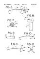
- FIG. 1is a plan view showing a viewing system of the present invention with an adapter stop, a malleable sheath, an endoscope assembly, and a viewing means mounted onto a handle, the viewing system being used with an endotracheal breathing tube;
- FIG. 2is a side elevational view of the endotracheal tube
- FIG. 3is a front elevational view of the first mounting element
- FIG. 4is a cross-sectional view of the first mounting element taken along plane 4--4 of FIG. 3;
- FIG. 5is a cross-sectional view of an alternate embodiment of the first mounting element taken along plane 4--4 of FIG. 3;
- FIG. 6is a side elevational view of the endoscope assembly showing the fiber optic bundle and the mounting portion;
- FIG. 7is an end view of the fiber optic bundle of the endoscope assembly of FIG. 6;
- FIG. 8is a side elevational view of the handle
- FIG. 9is a side elevational view of the viewing means
- FIG. 10is a side elevational view of the sheath
- FIG. 11is a side elevational view of an alternate embodiment of the sheath of FIG. 10;
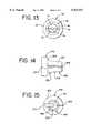
- FIG. 12is a side elevational view of the adapter stop
- FIG. 13is a front elevational view of the adapter stop of FIG. 12;
- FIG. 14is a side elevational view of an alternate embodiment of the adapter stop of FIG. 13;
- FIG. 15is a front elevational view of the alternate embodiment of the adapter stop of FIG. 14;
- FIG. 16is a side elevational view of an alternate embodiment of the adapter stop of FIG. 12.
- FIG. 17is a plan view showing the viewing system and adapter stop mounted onto a tracheostomy tube.
- FIG. 1 of the drawingsone embodiment of the present invention, a viewing system 20 with an endoscope assembly 30, a malleable sheath 40, a viewing means 50, a handle 60, and an adapter stop 70 is shown.
- the viewing system 20can be used with a breathing tube such as an endotracheal tube 80, as shown in FIGS. 1 and 2.
- the viewing system 20,comprising the endoscope assembly 30 and handle 60, when used for endotracheal intubation procedures has the advantage of providing an image of the tracheal area while inserting the endotracheal tube 80. This is a vast improvement over blind intubation. It is also a great improvement over the simple laryngoscope devices of the prior art which merely illuminate but do not provide an image of the tracheal area. Also, the adapter stop 70 provides an advantage over prior art devices which incorporate fiber-optics but do not allow the use of a standard length shaft 40 with endotracheal tubes 80 of varied lengths.
- FIG. 1shows the viewing system of the present invention as embodiment in an easily assembled and disassembled system wherein all the parts have a complimentary interengaging relationship.
- the present inventionmay be used with any endotracheal tube 80 that is commonly available.
- the common endotracheal tube 80as shown in FIG. 2, has a proximal end 82, a distal end 83, and a first mounting element 84 on the proximal end 82, and is made of flexible plastic.
- the first mounting element 84is commonly known as a "port” or “fitting.”
- the first mounting element 84is connected to a supply of oxygen or anesthetic gas during use.
- the first mounting element 84is a standard size on all endotracheal and tracheostomy tubes, but the length of the tube may vary for different size patients such as pediatric or adult.
- the breathing tube 80is made of flexible plastic
- the first mounting element 84is typically made of a rigid plastic material and may be of the type commonly known as a "Luer fitting.”
- the tube including the first mounting element 84is sterile.
- the endotracheal tube 80also defines a lumen 86 between the ends 82 and 83. It is the distal end 83 of the endotracheal tube 80 that is inserted into the trachea of a patient.
- An inflatable cuff 88is provided near the distal end 83 of the endotracheal tube 80. The cuff 88 is inflated after the tube 80 is in place within the trachea. Once inflated, the cuff 88 provides a seal between the tracheal wall and the endotracheal tube 80.
- the endotracheal tube 80is open at both ends 82 and 83 to allow oxygen or anesthetic gas to flow through the tube 80 and into the patient's lungs once the tube 80 is inserted into the trachea and connected to an oxygen or gas supply.
- the first mounting element 84is comprised of a hollow cylinder 90 that defines a cavity 92 having a bottom surface 94 and a hole 96 that axially cooperates with the lumen 86 of the endotracheal tube 80 to join the cavity 92 and the lumen 86.
- the cavity 92 of the first mounting element 84is usually defined by a substantially cylindrical inner surface 95.
- An alternate embodiment, shown in FIG. 5,provides the cavity 192 with a tapered inner surface 195 such that the taper becomes narrower toward the bottom surface 194 of the cavity 192.
- the first mounting element 84may be attached to the endotracheal tube 80 in any manner that provides a seal between the endotracheal tube 80 and the first mounting element 84.
- the endotracheal tube 80is slipped tightly over a tubular protrusion 98 that is opposite the cavity 92.
- the tubular protrusion 98has an outer diameter dimensioned to be larger than the inner diameter of the endotracheal tube 80 so that the endotracheal tube 80 fits tightly over the protrusion 98.
- first mounting element 84may include a threaded inner surface, a twist lock connector, or any type of connector that provides a positive locking mechanism for connecting the endotracheal tube 80 to an oxygen or anesthetic gas supply means.
- the adapter stop 70is configured to cooperate with whatever configuration is used for the first mounting element 84.
- the endoscope assembly 30 of the preferred embodimentincludes a mounting portion 31 and a fiber optic bundle 32.
- the mounting portion of the endoscope assemblyincludes a first connector that can be connected to a viewing means such as an image viewing apparatus.
- the image viewing apparatusis preferably a video monitor, but it may be a mechanical lens or other device that allows the practitioner to view the image provided by the endoscope assembly.
- the fiber-optic bundlepreferably includes optic fibers that carry illumination to the distal end of the bundle to illuminate the endotracheal area of the patient so that the practitioner can more easily see the area into which the breathing tube is being guided.
- the mounting portioncan be molded around the proximal end 82 of the fiber optic bundle to secure the optic fibers in relation to the first and second connectors. Illumination may be provided by a light source within the handle attached to the endoscope assembly by the second connector.
- the mounting portion 31preferably includes a forward section 34, a middle section 35, and a rearward section 36.
- the forward section 34includes a first mating surface 37
- the rearward section 36includes a first connector 38
- the middle sectionincludes a second connector 39.
- the fiber optic bundle 32extends from the forward section 34.
- the fiber optic bundle 32preferably contains at least one image-carrying optic fiber 56 for transmission of the image to the viewing means 50.
- the preferred embodiment of the fiber optic bundle 32also includes at least one illumination-carrying optic fibers 57.
- One example of a configuration of the fiber of the fiber optic bundle 32is the image-carrying fiber 56 disposed substantially in the center of the bundle with the illumination fibers 59 surrounding it as illustrated in FIG. 7.
- the bundle 32 of fibersis preferably flexible and encased in a flexible jacket 58 of thin-wall plastic.
- the first mating surface 37is preferably a substantially cylindrical protrusion extending from the forward section 34.
- the first mating surface 37can alternatively be tapered slightly such that the taper is narrowest toward the fiber optic bundle 32 and widest toward the forward section 34.
- first connector 38 of the rearward section 36is exemplified in FIG. 6 as a threaded locking ring type that can preferably be connected to the viewing means 50, shown in FIG. 9, via a mating connector.
- the viewing means 50can be integral with the mounting portion 31.
- the image-carrying fiber 56is preferably located through the mounting portion 31 after it exits the fiber optic bundle 32, and is terminated adjacent to the first connector 38.
- the second connector 39is also illustrated in FIG. 6.
- the second connector 39is a push-in type of connector which allows the endoscope assembly 30 to be mounted onto the handle 60 quickly and easily by snapping into a receptacle 62 on the handle 60.
- the illumination-carrying fibers 57are preferably located through the mounting portion 31 after exiting the fiber optic bundle 32. In the preferred embodiment, the illumination carrying fibers 57 are disposed within the mounting portion 31 at approximately a right angle so as to terminate adjacent to the second connector 39.
- first and second connectorsgenerally form a right angle.
- first and second connectorscan be at any angle that allows the endoscope assembly to be assembled to a viewing means and mounted on a handle.
- the endoscope assembly 30 and the viewing means 50are mounted to the handle 60, the endoscope assembly 30 and the viewing means 50 are electrically insulated from the power source within the handle 60 (which can be a battery or ac). This insulation feature is advantageous because of increased safety to the patient.
- Both the image-carrying fiber 56 and the illumination-carrying fibers 57are located adjacent the respective connectors so as to be adjacent to the viewing means 50 and the illumination source 64.
- the image-carrying fibers 56can be located to be adjacent to the second connector 39, and the illumination-carrying fibers 57 can be located to be within the first connector 38.
- the fiber optic bundle 32 of the preferred embodimentis at least one optic fiber and preferably is a bundle of 30,000 optic fibers. Multiple optic fibers may be used to provide better image resolution or to carry illumination in addition to carrying an image.
- the fiber-optic bundlemay be comprised of 30,000 fibers that are about 0.88 mm in diameter.
- an image focusing devicesuch as a lens may be associated with the fiber-optic bundle 32 at the distal end 83 of the bundle 32.
- the fiber optic bundle 32can be any other suitable system which is flexible or bendable.
- the endoscope assembly 30is in a cooperating interengaging relationship with the malleable sheath 40, as shown in FIG. 1.
- FIG. 9illustrates a preferred embodiment of the malleable sheath 40 of the present invention.
- the sheath 40 of the viewing system 20is preferably malleable so that the practitioner can bend the sheath 40 into a desired shape for a particular patient.
- the malleable sheath 40 of this embodimentsubstantially retains the shape into which it is bent while the breathing tube is being inserted into the patient.
- This featureallows the viewing system 20 to be used with endotracheal tubes 80 of any length for different size patients when the viewing system is used for intubation. Since the sheath 40 may be shaped as desired, a single standard length sheath 40 may be used for any patient. Preferably, the length of the sheath 40 is approximately as long as the longest available endotracheal tube 80.
- Yet another embodiment of the sheath 40has an atraumatic tip of the type disclosed in U.S. Pat. No. 5,512,034.
- the bulbous tipprevents trauma to the patient should the sheath 40 of the viewing system 20 contact any internal tissue of the patient during the intubation procedure.
- the sheath 40is tubular and malleable and the fiber optic bundle 32 is carried within the sheath 40.
- the distal tip 41is placed into the breathing tube and is generally aligned with the distal end of the tube.
- the sheathis at least as long as the longest available endotracheal or tracheostomy tube.
- the sheathis malleable and is made of aluminum or preferably stainless steel tubing so that it can be bent or shaped to accommodate a particular patient's anatomy. The malleable sheath will substantially retain its shape in typical use once it is bent.
- the sheathis tubular so that the fiber optic bundle may extend through the sheath. This configuration results in a generally annular space between the endoscope and the sheath. The space allows irrigation or aspiration to be accomplished.
- the distal tip of the sheathis preferably open so that the distal end of the fiber optic bundle can provide illumination to the area into which the breathing tube is being inserted, and so that an image of this area can be transmitted back to the viewing means.
- the proximal end 42 of sheath 40has a fitting 43 for mounting the sheath onto the forward section 34 of the endoscope assembly 30.
- the fitting 43is substantially cylindrical and defines a second mating surface 44 for frictionally engaging the first mating surface 37.
- the fitting 43may define a locking or threaded mechanism for mounting onto the mounting portion 31.
- FIG. 11Another alternative embodiment of the sheath 140 is illustrated in FIG. 11.
- This embodimentincludes a port or opening 145 defined by the fitting 143 for aspiration or irrigation.
- An irrigation or aspiration tube 146can be attached to the fitting 143 at the opening 145.
- the sheath 40is approximately 35 cm in length, about 2.5 to about 3.5 mm in diameter, and made of aluminum tubing about 0.5 mm thick or annealed stainless steel tubing with a wall thickness of about 0.3 mm.
- the viewing system 20can be used for an intubation procedure, for example.
- the adapter stop 70is used for intubation procedures.
- the adapter stop 70comprises a second mounting element 74 which is adapted to cooperate with the first mounting element 84 such that the adapter stop 70 receives and holds the sheath 40 in place within the lumen 86 of the endotracheal tube 80 when the first and second mounting elements are operably associated.
- the first and second mounting elementsbecome operably associated when the second mounting element 74 is inserted into the cavity 92 of the first mounting element 84.
- the first mounting element 84slips tightly over the second mounting element 74 while compressing the second mounting element 74 to hold the sheath.
- the adapter stopis preferably a one-piece plastic construction that allows the practitioner to securely mount the breathing tube to the sheath which is mounted on the endoscope assembly. When mounted to the tube, the adapter stop compresses on the sheath to hold it in the desired position within the breathing tube. Once the distal tip of the sheath is located with respect to the distal end of the tube, the sheath is locked into place by inserting the second mounting element of the adapter stop into the first mounting element of the tube.
- the second mounting element 74comprises a locking section.
- the locking section 76is dimensioned to mate within the cavity 92 of the first mounting element 84.
- the locking section 76 of the preferred embodimenthas the shape of a tapered cylinder.
- the diameter of the tapered locking section 76is dimensioned so that as the locking section 76 is pushed into the cavity 92 of the first mounting element 84, an interference fit is eventually produced between the first and second mounting elements when the locking section 76 is mounted within the cavity 92.
- the interference fitproduces friction which holds the locking section 76 within the cavity 92 and also causes the locking section 76 to compress on the sheath 40 to hold it in place.
- the viewing system 20can be used as a handle to physically maneuver the endotracheal tube 80 past the patient's teeth, tongue, epiglottis, and vocal cords into its proper position about 2 to 4 cm (about 1 to 2 inches) in front of the bifurcation of the trachea.
- the locking section 176is substantially cylindrical, as shown in FIG. 16, while the inner surface 195 defining the cavity 192 is tapered, as shown in FIG. 5.
- Other possible embodiments of the locking section 76may include non-cylindrically shaped locking sections and cavities, i.e., rectangular or triangular, or threaded or snap-fit locking sections which provide a positive mechanical connection between the first and second mounting elements.
- the adapter stop 70comprises the locking section 76 having two ends and defining a channel between the ends for movably receiving the sheath 40 when the adapter stop 70 is unmounted.
- the adapter stop 70holds the sheath 40 in place inside of the endotracheal tube 80 when the first and second mounting elements are operably associated.
- the channelis dimensioned to be larger in diameter than the sheath 40 so that the sheath 40 moves freely within the adapter stop 70 when the adapter stop 70 is not mounted within the cavity 92 of the first mounting element 84.
- the sheath 40 gripping feature of the second mounting element 74may be accomplished in the preferred embodiment by a longitudinal slot 79 defined by the locking section 76.
- the slot 79extends radially from the channel 78 of the locking section and axially through the second mounting element 74.
- the slotseparates the second mounting element 74 into a first half 71 and a second half 72.
- FIGS. 14-15show an alternate preferred embodiment of the adapter stop 170.
- This embodimentis similar to the embodiment shown in FIGS. 12-14 except that the locking section 276 and the second mounting element 274 define a substantially longitudinal shaft receiving slot for receiving the sheath 40.
- the slot 277 of this alternate embodimentextends radially from the axis of the elongated hollow body as is shown in FIGS. 14-15.
- the slot 277is for laterally receiving the sheath 40 so that the adapter handle may be assembled to the endoscope assembly by aligning sheath 40 with the slot and inserting the sheath 40 into the adapter handle.
- FIGS. 14-15shows a clamping slot 279 defined by the second mounting element 74.
- the clamping slot 279extends radially from the axis of the second mounting element 74 and extends axially through the second mounting element 74.
- the clamping slot and the shaft receiving slotare operably associated such that the second mounting element 74 grips and holds the sheath 40 in place within the lumen 86 of the endotracheal tube 80 via the same mechanism as the previously described preferred embodiment.
- FIGS. 12-13show another feature of the adapter stop which is a collar 73 extending radially from the second mounting element 74.
- the collaris disc shaped.
- the alternate preferred embodiments of the adapter stop 270shown in FIGS. 14-15 accomplish the same result of gripping and holding the sheath 40 within the lumen 86 of the endotracheal tube 80 by receiving the sheath 40 laterally through the shaft receiving slots.
- the adapter stop 70preferably is made of sterilizable plastic and is a simple, inexpensive, one-piece construction.
- the adapter stop 70can be made by any method of manufacture suitable for making plastic pieces including injection molding or machining.
- the practitionerassembles the viewing system 20 by inserting the sheath 40 into the adapter stop 70 and through the endotracheal tube 80. If the sheath 40 is malleable, it may first be shaped as desired or required for a particular patient. Alternatively, the sheath 40 may by shaped after the tube 80 is mounted onto the adapter stop 70.
- endotracheal tubes 80vary in length
- the location of the endoscope assembly 30 with respect to the end of the endotracheal tube 80should preferably be adjustable.
- the present inventionallows the user to align the distal tip of the sheath 40 with respect to the distal end 83 of the tube 80, regardless of the length of the tube 80. Once the endoscope 30 assembly is aligned, the adapter stop 70 with the endoscope assembly 30 is mounted to the endotracheal tube 80 by mating the first and second mounting elements.
- the adapter stop 70When mounted to the tube 80, the adapter stop 70 preferably compresses on the sheath 40 of the endoscope assembly 30 to hold it in the desired position within the endotracheal tube 80.
- the adjustability provided by the adapter stop 70is advantageous, because the end of the sheath 40 of the endoscope assembly 30 can be placed substantially conterminously with the end of the endotracheal tube 80. This is desirable, because the viewing system 20 can provide an image of the tracheal area from the point of view of the end of the endotracheal tube 80.
- the practitionerAfter assembling the viewing system 20 and the endotracheal tube 80 and preparing the patient for intubation, the practitioner uses the image provided by the viewing system 20 as a guide to insert the endotracheal tube 80 into the trachea of the patient.
- the handle 60may be used together with the sheath 40 to maneuver the endotracheal tube 80 into position.
- the adapter stop 70is disengaged from the endotracheal tube 80, and the endoscope assembly is removed while the tube 80 is held in place.
- the cuff 88is then inflated and oxygen or other gases can be supplied to the patient.
- the adapter stop 70may be used with the endoscope assembly 30 and a tracheostomy tube 280 in the same manner as with the endotracheal tube 80 as described herein.
- the tracheostomy tubeis inserted into the patient via an incision at the base of the patient's neck above the sternum.
- the tracheostomy tubemay have the same type of first mounting element 84 as the endotracheal tube 80.
- the tracheostomy tubealso has a proximal end 282, a distal end 283, a lumen 286, and an inflatable cuff 288.
Landscapes
- Health & Medical Sciences (AREA)
- Life Sciences & Earth Sciences (AREA)
- Surgery (AREA)
- Medical Informatics (AREA)
- Molecular Biology (AREA)
- Veterinary Medicine (AREA)
- Public Health (AREA)
- Nuclear Medicine, Radiotherapy & Molecular Imaging (AREA)
- General Health & Medical Sciences (AREA)
- Pathology (AREA)
- Animal Behavior & Ethology (AREA)
- Heart & Thoracic Surgery (AREA)
- Engineering & Computer Science (AREA)
- Biomedical Technology (AREA)
- Physics & Mathematics (AREA)
- Radiology & Medical Imaging (AREA)
- Optics & Photonics (AREA)
- Biophysics (AREA)
- Physiology (AREA)
- Otolaryngology (AREA)
- Pulmonology (AREA)
- Oral & Maxillofacial Surgery (AREA)
- Endoscopes (AREA)
Abstract
Description
Claims (56)
Priority Applications (2)
| Application Number | Priority Date | Filing Date | Title |
|---|---|---|---|
| US08/954,145US5921917A (en) | 1997-10-20 | 1997-10-20 | Hand-held viewing system with removable sheath |
| PCT/US1998/022156WO1999020170A1 (en) | 1997-10-20 | 1998-10-20 | Hand-held viewing system with removable sheath |
Applications Claiming Priority (1)
| Application Number | Priority Date | Filing Date | Title |
|---|---|---|---|
| US08/954,145US5921917A (en) | 1997-10-20 | 1997-10-20 | Hand-held viewing system with removable sheath |
Publications (1)
| Publication Number | Publication Date |
|---|---|
| US5921917Atrue US5921917A (en) | 1999-07-13 |
Family
ID=25494996
Family Applications (1)
| Application Number | Title | Priority Date | Filing Date |
|---|---|---|---|
| US08/954,145Expired - LifetimeUS5921917A (en) | 1997-10-20 | 1997-10-20 | Hand-held viewing system with removable sheath |
Country Status (2)
| Country | Link |
|---|---|
| US (1) | US5921917A (en) |
| WO (1) | WO1999020170A1 (en) |
Cited By (56)
| Publication number | Priority date | Publication date | Assignee | Title |
|---|---|---|---|---|
| US6258024B1 (en)* | 1996-11-01 | 2001-07-10 | S.S.H. Medical Limited | Speculum device |
| US20020108610A1 (en)* | 1996-02-26 | 2002-08-15 | Christopher Kent L. | Method and apparatus for endotracheal intubation using a light wand and curved guide |
| WO2001093742A3 (en)* | 2000-06-05 | 2002-09-12 | Boris Sudakov | Multifunctional medical tool |
| US6508757B1 (en)* | 1999-10-25 | 2003-01-21 | Paul Huan Song | Attachment to flexible bronchoscope, slotted tubular stylet for endotracheal intubation |
| US6543446B1 (en) | 1996-02-26 | 2003-04-08 | Evergreen Medical Incorporated | Method and apparatus for ventilation/oxygenation during guided insertion of an endotracheal tube |
| US20030078476A1 (en)* | 2001-07-24 | 2003-04-24 | Hill Stephen D. | Apparatus for intubation |
| US6568388B2 (en) | 1996-02-26 | 2003-05-27 | Evergreen Medical Incorporated | Method and apparatus for ventilation / oxygenation during guided insertion of an endotracheal tube |
| US6679833B2 (en)* | 1996-03-22 | 2004-01-20 | Sdgi Holdings, Inc. | Devices and methods for percutaneous surgery |
| US20040176763A1 (en)* | 1996-03-22 | 2004-09-09 | Foley Kevin T. | Methods for percutaneous surgery |
| US20040186346A1 (en)* | 1996-03-22 | 2004-09-23 | Smith Maurice M. | Devices and methods for percutaneous surgery |
| US20040220451A1 (en)* | 1996-10-04 | 2004-11-04 | Dietrich Gravenstein | Imaging scope |
| US6814698B2 (en) | 2001-10-05 | 2004-11-09 | Clarus Medical, Llc | Endoscope with flexible light guide having offset distal end |
| US6832986B2 (en)* | 2001-03-22 | 2004-12-21 | Karl Storz Gmbh & Co. Kg | Endoscopic intubation system |
| US6840903B2 (en) | 2002-03-21 | 2005-01-11 | Nuvista Technology Corporation | Laryngoscope with image sensor |
| US20050139220A1 (en)* | 1996-02-26 | 2005-06-30 | Evergreen Medical Incorporated | Method and apparatus for ventilation / oxygenation during guided insertion of an endotracheal tube |
| US20050182297A1 (en)* | 1996-10-04 | 2005-08-18 | Dietrich Gravenstein | Imaging scope |
| US7056321B2 (en) | 2000-08-01 | 2006-06-06 | Endius, Incorporated | Method of securing vertebrae |
| US20060159409A1 (en)* | 2004-12-21 | 2006-07-20 | Cml Innovative Technologies, Inc. | Boroscope with selectable stiffness light shaft |
| US20070129605A1 (en)* | 2004-02-05 | 2007-06-07 | Polydiagnost Gmbh | Endoscope comprising a flexible probe |
| US20070179342A1 (en)* | 2006-01-12 | 2007-08-02 | Kb Port Llc | Wireless Laryngoscope with Internal Antennae and One Piece Construction Adapted for Laryngoscopy Training |
| US20090192350A1 (en)* | 2008-01-28 | 2009-07-30 | Mauricio Mejia | Wireless video stylet with display mounted to laryngoscope blade and method for using the same |
| US20090192355A1 (en)* | 2008-01-28 | 2009-07-30 | Mauricio Mejia | Scope for managing difficult pathways and method to improve visibility of the same |
| US20100094090A1 (en)* | 2008-01-28 | 2010-04-15 | Mauricio Mejia | Self-cleaning wireless video stylet with display mounted to laryngoscope blade and method for using the same |
| US20110023887A1 (en)* | 2009-02-06 | 2011-02-03 | Endoclear, Llc | Methods for tracheostomy visualization |
| US7985247B2 (en) | 2000-08-01 | 2011-07-26 | Zimmer Spine, Inc. | Methods and apparatuses for treating the spine through an access device |
| US20110265797A1 (en)* | 2010-04-30 | 2011-11-03 | Nellcor Puritan Bennett Llc | Extendable tracheal tube |
| US20130014750A1 (en)* | 2011-07-14 | 2013-01-17 | Soheil Etesham | Intubation Apparatus |
| US8382665B1 (en) | 2009-02-12 | 2013-02-26 | Alfred Fam | Endotracheal tube placement system and method |
| US8540746B2 (en) | 1998-08-20 | 2013-09-24 | Zimmer Spine, Inc. | Cannula for receiving surgical instruments |
| US8601633B2 (en) | 2009-02-06 | 2013-12-10 | Endoclear Llc | Cleaning of body-inserted medical tubes |
| WO2010091440A3 (en)* | 2009-01-20 | 2014-02-27 | Samir Bhatt | Airway management devices, endoscopic conduits, surgical kits, and methods using the same |
| US20140135583A1 (en)* | 2012-11-14 | 2014-05-15 | Manuel V. Moreno | Intubation Tube |
| US8887730B2 (en) | 2011-05-26 | 2014-11-18 | Covidien Lp | Dual-lumen tracheal tube with assembly portion |
| US8978657B2 (en) | 2010-07-29 | 2015-03-17 | Covidien Lp | Dual-lumen tracheal tube with shaped lumen divider |
| US8998798B2 (en) | 2010-12-29 | 2015-04-07 | Covidien Lp | Multi-lumen tracheal tube with visualization device |
| US20150126808A1 (en)* | 2011-05-18 | 2015-05-07 | Centre Hospitalier Universitaire De Bordeaux (C.H.U. De Bordeaux) | Method for positioning a disposable sterile endotracheal tube, and corresponding system for intubation |
| US9155854B2 (en) | 2011-08-31 | 2015-10-13 | Covidien Lp | Tracheal tube with visualization device and integrated flushing system |
| US9211060B2 (en) | 2011-04-05 | 2015-12-15 | Covidien Lp | Visualization device and holder for use with a tracheal tube |
| US20160022131A1 (en)* | 2014-07-22 | 2016-01-28 | Iei Integration Corp. | Waterproof laryngoscope and waterproof structure |
| US9421341B2 (en) | 2010-02-27 | 2016-08-23 | King Systems Corporation | Laryngeal tube |
| US9427543B2 (en) | 2014-05-05 | 2016-08-30 | Deye Wei | Intubation device and method |
| US9445714B2 (en) | 2010-03-29 | 2016-09-20 | Endoclear Llc | Endotracheal tube coupling adapters |
| US20170086664A1 (en)* | 2011-05-18 | 2017-03-30 | Centre Hospitalier Universitaire De Bordeaux (C.H.U De Bordeaux) | Method for positioning a disposable sterile endotracheal tube, and corresponding system for intubation |
| US9622651B2 (en) | 2012-01-27 | 2017-04-18 | Kbport Llc | Wireless laryngoscope simulator with onboard event recording adapted for laryngoscopy training |
| US9788755B2 (en) | 2011-05-26 | 2017-10-17 | Covidien Lp | Illumination systems and devices for tracheal tubes |
| US9840266B2 (en) | 2013-10-09 | 2017-12-12 | Glidemachines Llc | Apparatus and method for towing a load by a person |
| US10004863B2 (en) | 2012-12-04 | 2018-06-26 | Endoclear Llc | Closed suction cleaning devices, systems and methods |
| US10016575B2 (en) | 2014-06-03 | 2018-07-10 | Endoclear Llc | Cleaning devices, systems and methods |
| US10595710B2 (en)* | 2001-10-19 | 2020-03-24 | Visionscope Technologies Llc | Portable imaging system employing a miniature endoscope |
| US10722322B2 (en) | 2010-03-29 | 2020-07-28 | Endoclear Llc | Distal airway cleaning devices |
| US10912622B2 (en)* | 2019-06-01 | 2021-02-09 | Nizam M. Meah | Disposable endoscope shield |
| US11291351B2 (en)* | 2011-08-19 | 2022-04-05 | Harold I. Daily | Hysteroscopes with curved tips |
| US20220184359A1 (en)* | 2017-04-20 | 2022-06-16 | Resnent, Llc | Endoscopic balloon dilator systems |
| US11484189B2 (en) | 2001-10-19 | 2022-11-01 | Visionscope Technologies Llc | Portable imaging system employing a miniature endoscope |
| US11559646B1 (en)* | 2019-02-11 | 2023-01-24 | Ali Osman | System and method for video assisted percutaneous needle cricothyrotomy and tracheostomy |
| WO2024192137A1 (en)* | 2023-03-13 | 2024-09-19 | University Of Southern California | Airway exchange bronchoscope construction |
Citations (22)
| Publication number | Priority date | Publication date | Assignee | Title |
|---|---|---|---|---|
| US3677262A (en)* | 1970-07-23 | 1972-07-18 | Henry J Zukowski | Surgical instrument illuminating endotracheal tube inserter |
| US3776222A (en)* | 1971-12-23 | 1973-12-04 | Lurosso A | Fiber optic entubator and method of entubation of the trachea through the nasopharynx |
| US4126127A (en)* | 1976-09-27 | 1978-11-21 | May Laurence M | Suctioning/oxygenating laryngoscope blade |
| US4319563A (en)* | 1977-12-02 | 1982-03-16 | Olympus Optical Co., Ltd. | Endoscope with a smoothly curved distal end face |
| US4561446A (en)* | 1981-10-15 | 1985-12-31 | Siemens Aktiengesellschaft | Ultrasonic probe which can be introduced into a body |
| US4782819A (en)* | 1987-02-25 | 1988-11-08 | Adair Edwin Lloyd | Optical catheter |
| US4798193A (en)* | 1987-05-18 | 1989-01-17 | Thomas J. Fogarty | Protective sheath instrument carrier |
| US4800870A (en)* | 1988-03-11 | 1989-01-31 | Reid Jr Ben A | Method and apparatus for bile duct exploration |
| US4846153A (en)* | 1988-06-10 | 1989-07-11 | George Berci | Intubating video endoscope |
| US4869238A (en)* | 1988-04-22 | 1989-09-26 | Opielab, Inc. | Endoscope for use with a disposable sheath |
| US4946442A (en)* | 1985-06-28 | 1990-08-07 | Olympus Optical Co., Ltd. | Endoscope treatment device |
| US4947896A (en)* | 1988-11-04 | 1990-08-14 | Bartlett Robert L | Laryngoscope |
| US4982729A (en)* | 1989-02-10 | 1991-01-08 | Wu Tzu Lang | Rigid fiberoptic intubating laryngoscope |
| US5127393A (en)* | 1991-05-28 | 1992-07-07 | Medilase, Inc. | Flexible endoscope with rigid introducer |
| US5183031A (en)* | 1991-05-13 | 1993-02-02 | Rossoff Leonard J | Fiberoptic intubating laryngoscope |
| US5329940A (en)* | 1990-02-14 | 1994-07-19 | Adair Edwin Lloyd | Endotracheal tube intubation assist device |
| US5337735A (en)* | 1992-12-28 | 1994-08-16 | Albert Salerno | Fiber-lighted stylet |
| US5512034A (en)* | 1992-11-25 | 1996-04-30 | Finn; Miles A. | Surgical instrument including viewing optics and a ball probe |
| US5569159A (en)* | 1994-12-16 | 1996-10-29 | Anderson; Keven C. | Endoscopic sleeve |
| US5607386A (en)* | 1993-09-21 | 1997-03-04 | Flam; Gary H. | Malleable fiberoptic intubating stylet and method |
| US5636625A (en)* | 1994-08-30 | 1997-06-10 | Machida Endoscope Co., Ltd. | Tracheal airway apparatus |
| US5645519A (en)* | 1994-03-18 | 1997-07-08 | Jai S. Lee | Endoscopic instrument for controlled introduction of tubular members in the body and methods therefor |
- 1997
- 1997-10-20USUS08/954,145patent/US5921917A/ennot_activeExpired - Lifetime
- 1998
- 1998-10-20WOPCT/US1998/022156patent/WO1999020170A1/enactiveApplication Filing
Patent Citations (22)
| Publication number | Priority date | Publication date | Assignee | Title |
|---|---|---|---|---|
| US3677262A (en)* | 1970-07-23 | 1972-07-18 | Henry J Zukowski | Surgical instrument illuminating endotracheal tube inserter |
| US3776222A (en)* | 1971-12-23 | 1973-12-04 | Lurosso A | Fiber optic entubator and method of entubation of the trachea through the nasopharynx |
| US4126127A (en)* | 1976-09-27 | 1978-11-21 | May Laurence M | Suctioning/oxygenating laryngoscope blade |
| US4319563A (en)* | 1977-12-02 | 1982-03-16 | Olympus Optical Co., Ltd. | Endoscope with a smoothly curved distal end face |
| US4561446A (en)* | 1981-10-15 | 1985-12-31 | Siemens Aktiengesellschaft | Ultrasonic probe which can be introduced into a body |
| US4946442A (en)* | 1985-06-28 | 1990-08-07 | Olympus Optical Co., Ltd. | Endoscope treatment device |
| US4782819A (en)* | 1987-02-25 | 1988-11-08 | Adair Edwin Lloyd | Optical catheter |
| US4798193A (en)* | 1987-05-18 | 1989-01-17 | Thomas J. Fogarty | Protective sheath instrument carrier |
| US4800870A (en)* | 1988-03-11 | 1989-01-31 | Reid Jr Ben A | Method and apparatus for bile duct exploration |
| US4869238A (en)* | 1988-04-22 | 1989-09-26 | Opielab, Inc. | Endoscope for use with a disposable sheath |
| US4846153A (en)* | 1988-06-10 | 1989-07-11 | George Berci | Intubating video endoscope |
| US4947896A (en)* | 1988-11-04 | 1990-08-14 | Bartlett Robert L | Laryngoscope |
| US4982729A (en)* | 1989-02-10 | 1991-01-08 | Wu Tzu Lang | Rigid fiberoptic intubating laryngoscope |
| US5329940A (en)* | 1990-02-14 | 1994-07-19 | Adair Edwin Lloyd | Endotracheal tube intubation assist device |
| US5183031A (en)* | 1991-05-13 | 1993-02-02 | Rossoff Leonard J | Fiberoptic intubating laryngoscope |
| US5127393A (en)* | 1991-05-28 | 1992-07-07 | Medilase, Inc. | Flexible endoscope with rigid introducer |
| US5512034A (en)* | 1992-11-25 | 1996-04-30 | Finn; Miles A. | Surgical instrument including viewing optics and a ball probe |
| US5337735A (en)* | 1992-12-28 | 1994-08-16 | Albert Salerno | Fiber-lighted stylet |
| US5607386A (en)* | 1993-09-21 | 1997-03-04 | Flam; Gary H. | Malleable fiberoptic intubating stylet and method |
| US5645519A (en)* | 1994-03-18 | 1997-07-08 | Jai S. Lee | Endoscopic instrument for controlled introduction of tubular members in the body and methods therefor |
| US5636625A (en)* | 1994-08-30 | 1997-06-10 | Machida Endoscope Co., Ltd. | Tracheal airway apparatus |
| US5569159A (en)* | 1994-12-16 | 1996-10-29 | Anderson; Keven C. | Endoscopic sleeve |
Cited By (89)
| Publication number | Priority date | Publication date | Assignee | Title |
|---|---|---|---|---|
| US6568388B2 (en) | 1996-02-26 | 2003-05-27 | Evergreen Medical Incorporated | Method and apparatus for ventilation / oxygenation during guided insertion of an endotracheal tube |
| US20020108610A1 (en)* | 1996-02-26 | 2002-08-15 | Christopher Kent L. | Method and apparatus for endotracheal intubation using a light wand and curved guide |
| US20050139220A1 (en)* | 1996-02-26 | 2005-06-30 | Evergreen Medical Incorporated | Method and apparatus for ventilation / oxygenation during guided insertion of an endotracheal tube |
| US6860264B2 (en)* | 1996-02-26 | 2005-03-01 | Evergreen Medical Incorporated | Method and apparatus for endotracheal intubation using a light wand and curved guide |
| US6543446B1 (en) | 1996-02-26 | 2003-04-08 | Evergreen Medical Incorporated | Method and apparatus for ventilation/oxygenation during guided insertion of an endotracheal tube |
| US20040176763A1 (en)* | 1996-03-22 | 2004-09-09 | Foley Kevin T. | Methods for percutaneous surgery |
| US6679833B2 (en)* | 1996-03-22 | 2004-01-20 | Sdgi Holdings, Inc. | Devices and methods for percutaneous surgery |
| US20040186346A1 (en)* | 1996-03-22 | 2004-09-23 | Smith Maurice M. | Devices and methods for percutaneous surgery |
| US7198598B2 (en) | 1996-03-22 | 2007-04-03 | Warsaw Orthopedic, Inc. | Devices and methods for percutaneous surgery |
| US20040220451A1 (en)* | 1996-10-04 | 2004-11-04 | Dietrich Gravenstein | Imaging scope |
| US20050182297A1 (en)* | 1996-10-04 | 2005-08-18 | Dietrich Gravenstein | Imaging scope |
| US6258024B1 (en)* | 1996-11-01 | 2001-07-10 | S.S.H. Medical Limited | Speculum device |
| US8540746B2 (en) | 1998-08-20 | 2013-09-24 | Zimmer Spine, Inc. | Cannula for receiving surgical instruments |
| US6508757B1 (en)* | 1999-10-25 | 2003-01-21 | Paul Huan Song | Attachment to flexible bronchoscope, slotted tubular stylet for endotracheal intubation |
| WO2001093742A3 (en)* | 2000-06-05 | 2002-09-12 | Boris Sudakov | Multifunctional medical tool |
| US6923756B2 (en) | 2000-06-05 | 2005-08-02 | Boris Sudakov | Multifunctional medical tool |
| US8864785B2 (en) | 2000-08-01 | 2014-10-21 | Zimmer Spine, Inc. | Method for securing vertebrae |
| US7722530B2 (en) | 2000-08-01 | 2010-05-25 | Zimmer Spine, Inc. | Method of securing vertebrae |
| US8277486B2 (en) | 2000-08-01 | 2012-10-02 | Zimmer Spine, Inc. | System for performing a procedure at a spinal location |
| US7985247B2 (en) | 2000-08-01 | 2011-07-26 | Zimmer Spine, Inc. | Methods and apparatuses for treating the spine through an access device |
| US7056321B2 (en) | 2000-08-01 | 2006-06-06 | Endius, Incorporated | Method of securing vertebrae |
| US8777997B2 (en) | 2000-08-01 | 2014-07-15 | Zimmer Spine, Inc. | Method for securing vertebrae |
| US7850695B2 (en) | 2000-08-01 | 2010-12-14 | Zimmer Spine, Inc. | Method of securing vertebrae |
| US7699877B2 (en) | 2000-08-01 | 2010-04-20 | Zimmer Spine, Inc. | Method of securing vertebrae |
| US9101353B2 (en) | 2000-08-01 | 2015-08-11 | Zimmer Spine, Inc. | Method of securing vertebrae |
| US9622735B2 (en) | 2000-08-01 | 2017-04-18 | Zimmer Spine, Inc. | Method for securing vertebrae |
| US6832986B2 (en)* | 2001-03-22 | 2004-12-21 | Karl Storz Gmbh & Co. Kg | Endoscopic intubation system |
| US20030078476A1 (en)* | 2001-07-24 | 2003-04-24 | Hill Stephen D. | Apparatus for intubation |
| US6929600B2 (en) | 2001-07-24 | 2005-08-16 | Stephen D. Hill | Apparatus for intubation |
| US6814698B2 (en) | 2001-10-05 | 2004-11-09 | Clarus Medical, Llc | Endoscope with flexible light guide having offset distal end |
| US10595710B2 (en)* | 2001-10-19 | 2020-03-24 | Visionscope Technologies Llc | Portable imaging system employing a miniature endoscope |
| US11484189B2 (en) | 2001-10-19 | 2022-11-01 | Visionscope Technologies Llc | Portable imaging system employing a miniature endoscope |
| US20050043590A1 (en)* | 2002-03-21 | 2005-02-24 | Mazzei William J. | Laryngoscope with image sensor |
| US6840903B2 (en) | 2002-03-21 | 2005-01-11 | Nuvista Technology Corporation | Laryngoscope with image sensor |
| US20070129605A1 (en)* | 2004-02-05 | 2007-06-07 | Polydiagnost Gmbh | Endoscope comprising a flexible probe |
| US8333691B2 (en)* | 2004-02-05 | 2012-12-18 | Polydiagnost Gmbh | Endoscope comprising a flexible probe |
| US20060159409A1 (en)* | 2004-12-21 | 2006-07-20 | Cml Innovative Technologies, Inc. | Boroscope with selectable stiffness light shaft |
| US20070179342A1 (en)* | 2006-01-12 | 2007-08-02 | Kb Port Llc | Wireless Laryngoscope with Internal Antennae and One Piece Construction Adapted for Laryngoscopy Training |
| US20100094090A1 (en)* | 2008-01-28 | 2010-04-15 | Mauricio Mejia | Self-cleaning wireless video stylet with display mounted to laryngoscope blade and method for using the same |
| US20090192350A1 (en)* | 2008-01-28 | 2009-07-30 | Mauricio Mejia | Wireless video stylet with display mounted to laryngoscope blade and method for using the same |
| US20090192355A1 (en)* | 2008-01-28 | 2009-07-30 | Mauricio Mejia | Scope for managing difficult pathways and method to improve visibility of the same |
| US8888683B2 (en) | 2008-01-28 | 2014-11-18 | Mauricio Mejia | Modifications in endoscope apparatus, using fluid and gas dynamics, and methods for improving visibility during endoscopy |
| WO2010091440A3 (en)* | 2009-01-20 | 2014-02-27 | Samir Bhatt | Airway management devices, endoscopic conduits, surgical kits, and methods using the same |
| US10441380B2 (en) | 2009-02-06 | 2019-10-15 | Endoclear Llc | Body-inserted tube cleaning |
| US9095286B2 (en) | 2009-02-06 | 2015-08-04 | Endoclear Llc | Body-inserted tube cleaning |
| US9398837B2 (en) | 2009-02-06 | 2016-07-26 | Endoclear Llc | Methods for confirming placement of endotracheal tubes |
| US8601633B2 (en) | 2009-02-06 | 2013-12-10 | Endoclear Llc | Cleaning of body-inserted medical tubes |
| US9962233B2 (en) | 2009-02-06 | 2018-05-08 | Endoclear Llc | Body-inserted tube cleaning |
| US9907624B2 (en) | 2009-02-06 | 2018-03-06 | Endoclear Llc | Body-inserted tube cleaning with suction |
| US9855111B2 (en) | 2009-02-06 | 2018-01-02 | Endoclear Llc | Methods of removing biofilm from endotracheal tubes |
| US20110023887A1 (en)* | 2009-02-06 | 2011-02-03 | Endoclear, Llc | Methods for tracheostomy visualization |
| US10682203B2 (en) | 2009-02-06 | 2020-06-16 | Endoclear Llc | Methods of cleaning endotracheal tubes including light treatment |
| US8534287B2 (en)* | 2009-02-06 | 2013-09-17 | Endoclear, Llc | Methods for tracheostomy visualization |
| US9579012B2 (en) | 2009-02-06 | 2017-02-28 | Endoclear Llc | Visualized endotracheal tube placement systems |
| US9386907B2 (en) | 2009-02-06 | 2016-07-12 | Endoclear Llc | Visualization systems and methods |
| US9332891B2 (en) | 2009-02-06 | 2016-05-10 | Endoclear Llc | Tracheostomy visualization |
| US8382665B1 (en) | 2009-02-12 | 2013-02-26 | Alfred Fam | Endotracheal tube placement system and method |
| US9421341B2 (en) | 2010-02-27 | 2016-08-23 | King Systems Corporation | Laryngeal tube |
| US9445714B2 (en) | 2010-03-29 | 2016-09-20 | Endoclear Llc | Endotracheal tube coupling adapters |
| US10722322B2 (en) | 2010-03-29 | 2020-07-28 | Endoclear Llc | Distal airway cleaning devices |
| US20110265797A1 (en)* | 2010-04-30 | 2011-11-03 | Nellcor Puritan Bennett Llc | Extendable tracheal tube |
| US8978657B2 (en) | 2010-07-29 | 2015-03-17 | Covidien Lp | Dual-lumen tracheal tube with shaped lumen divider |
| US8998798B2 (en) | 2010-12-29 | 2015-04-07 | Covidien Lp | Multi-lumen tracheal tube with visualization device |
| US9211060B2 (en) | 2011-04-05 | 2015-12-15 | Covidien Lp | Visualization device and holder for use with a tracheal tube |
| US20170086664A1 (en)* | 2011-05-18 | 2017-03-30 | Centre Hospitalier Universitaire De Bordeaux (C.H.U De Bordeaux) | Method for positioning a disposable sterile endotracheal tube, and corresponding system for intubation |
| US20150126808A1 (en)* | 2011-05-18 | 2015-05-07 | Centre Hospitalier Universitaire De Bordeaux (C.H.U. De Bordeaux) | Method for positioning a disposable sterile endotracheal tube, and corresponding system for intubation |
| US9788755B2 (en) | 2011-05-26 | 2017-10-17 | Covidien Lp | Illumination systems and devices for tracheal tubes |
| US10806371B2 (en) | 2011-05-26 | 2020-10-20 | Covidien Lp | Illumination systems and devices for tracheal tubes |
| US8887730B2 (en) | 2011-05-26 | 2014-11-18 | Covidien Lp | Dual-lumen tracheal tube with assembly portion |
| US20130014750A1 (en)* | 2011-07-14 | 2013-01-17 | Soheil Etesham | Intubation Apparatus |
| US11291351B2 (en)* | 2011-08-19 | 2022-04-05 | Harold I. Daily | Hysteroscopes with curved tips |
| US9155854B2 (en) | 2011-08-31 | 2015-10-13 | Covidien Lp | Tracheal tube with visualization device and integrated flushing system |
| US9622651B2 (en) | 2012-01-27 | 2017-04-18 | Kbport Llc | Wireless laryngoscope simulator with onboard event recording adapted for laryngoscopy training |
| US20140135583A1 (en)* | 2012-11-14 | 2014-05-15 | Manuel V. Moreno | Intubation Tube |
| US11173266B2 (en) | 2012-12-04 | 2021-11-16 | Endoclear Llc | Closed suction cleaning devices, systems and methods |
| US10004863B2 (en) | 2012-12-04 | 2018-06-26 | Endoclear Llc | Closed suction cleaning devices, systems and methods |
| US10821249B2 (en) | 2012-12-04 | 2020-11-03 | Endoclear Llc | Closed suction cleaning devices, systems and methods |
| US9840266B2 (en) | 2013-10-09 | 2017-12-12 | Glidemachines Llc | Apparatus and method for towing a load by a person |
| US9427543B2 (en) | 2014-05-05 | 2016-08-30 | Deye Wei | Intubation device and method |
| US10850062B2 (en) | 2014-06-03 | 2020-12-01 | Endoclear Llc | Cleaning devices, systems and methods |
| US10016575B2 (en) | 2014-06-03 | 2018-07-10 | Endoclear Llc | Cleaning devices, systems and methods |
| US20160022131A1 (en)* | 2014-07-22 | 2016-01-28 | Iei Integration Corp. | Waterproof laryngoscope and waterproof structure |
| US20220184359A1 (en)* | 2017-04-20 | 2022-06-16 | Resnent, Llc | Endoscopic balloon dilator systems |
| US12083300B2 (en) | 2017-04-20 | 2024-09-10 | Resnent, Llc | Endoscopic balloon dilator systems |
| US12194261B2 (en)* | 2017-04-20 | 2025-01-14 | Resnent, Llc | Endoscopic balloon dilator systems |
| US11559646B1 (en)* | 2019-02-11 | 2023-01-24 | Ali Osman | System and method for video assisted percutaneous needle cricothyrotomy and tracheostomy |
| US12109361B1 (en)* | 2019-02-11 | 2024-10-08 | Ali Osman | System and method for video assisted percutaneous needle cricothyrotomy and tracheostomy |
| US10912622B2 (en)* | 2019-06-01 | 2021-02-09 | Nizam M. Meah | Disposable endoscope shield |
| WO2024192137A1 (en)* | 2023-03-13 | 2024-09-19 | University Of Southern California | Airway exchange bronchoscope construction |
Also Published As
| Publication number | Publication date |
|---|---|
| WO1999020170A1 (en) | 1999-04-29 |
Similar Documents
| Publication | Publication Date | Title |
|---|---|---|
| US5921917A (en) | Hand-held viewing system with removable sheath | |
| US5941816A (en) | Viewing system with adapter handle for medical breathing tubes | |
| US5607386A (en) | Malleable fiberoptic intubating stylet and method | |
| US6832986B2 (en) | Endoscopic intubation system | |
| US5551946A (en) | Multifunctional intubating guide stylet and laryngoscope | |
| US10335023B2 (en) | Endotracheal tube insertion device | |
| US6539942B2 (en) | Endotracheal intubation device | |
| US5733242A (en) | Intubation system having an axially moveable memory cylinder | |
| FI104467B (en) | Medical device for insertion of a tube or tube-like instrument | |
| US5443058A (en) | Blade for telescopic laryngoscope | |
| US5327881A (en) | Fiberoptic intubating stylet | |
| US6322498B1 (en) | Imaging scope | |
| US5329940A (en) | Endotracheal tube intubation assist device | |
| US4947829A (en) | Modular blade laryngoscope | |
| EP2756795A1 (en) | Intubation device | |
| US10286171B2 (en) | Endotracheal tube insertion device | |
| US20110004065A2 (en) | Intubation tube | |
| US9283342B1 (en) | Endotracheal tube insertion device | |
| US5279281A (en) | Single-handed fibre-optic flexible laryngoscope | |
| US20060100483A1 (en) | Integrated viewing assembly for an endoscope | |
| US20140128681A1 (en) | Intubation guide assembly | |
| US5425356A (en) | Telescopic laryngoscope blade | |
| US20200171256A1 (en) | Intubation Systems and Methods | |
| GB2454018A (en) | Introducer device for bougie tube | |
| CN113520290B (en) | Disposable diameter-adjustable tracheal or bronchial rigid mirror operating system and operating method |
Legal Events
| Date | Code | Title | Description |
|---|---|---|---|
| AS | Assignment | Owner name:CLARUS MEDICAL SYSTEMS, MINNESOTA Free format text:ASSIGNMENT OF ASSIGNORS INTEREST;ASSIGNORS:BARTHEL, THOMAS C.;BROWN, MARK F.;SHIKANI, ALAN H.;REEL/FRAME:009085/0850;SIGNING DATES FROM 19980311 TO 19980312 | |
| AS | Assignment | Owner name:TRANSAMERICA BUSINESS CREDIT CORPORATION, ILLINOIS Free format text:SECURITY INTEREST;ASSIGNOR:CLARUS MEDICAL SYSTEMS, INC.;REEL/FRAME:009648/0416 Effective date:19981123 Owner name:TRANSAMERICA BUSINESS CREDIT CORPORATION, CALIFORN Free format text:SECURITY INTEREST;ASSIGNOR:CLARUS MEDICAL SYSTEMS, INC.;REEL/FRAME:009648/0416 Effective date:19981123 | |
| STCF | Information on status: patent grant | Free format text:PATENTED CASE | |
| AS | Assignment | Owner name:TRANSAMERICA BUSINESS CREDIT CORPORATION, ILLINOIS Free format text:ASSIGNMENT OF ASSIGNORS INTEREST;ASSIGNOR:CLARUS MEDICAL SYSTEMS, INC.;REEL/FRAME:011783/0628 Effective date:20000310 Owner name:CLARUS MEDICAL, LLC, MINNESOTA Free format text:CHANGE OF NAME;ASSIGNOR:LASE MEDICAL, LLC.;REEL/FRAME:011783/0878 Effective date:20000323 Owner name:LASE MEDICAL LLC, MINNESOTA Free format text:ASSIGNMENT OF ASSIGNORS INTEREST;ASSIGNOR:TRANSAMERICA BUSINESS CREDIT CORPORATION;REEL/FRAME:011783/0890 Effective date:20000310 | |
| FPAY | Fee payment | Year of fee payment:4 | |
| FPAY | Fee payment | Year of fee payment:8 | |
| FPAY | Fee payment | Year of fee payment:12 |