US5908446A - Catheter assembly, catheter and multi-port introducer for use therewith - Google Patents
Catheter assembly, catheter and multi-port introducer for use therewithDownload PDFInfo
- Publication number
- US5908446A US5908446AUS08/812,354US81235497AUS5908446AUS 5908446 AUS5908446 AUS 5908446AUS 81235497 AUS81235497 AUS 81235497AUS 5908446 AUS5908446 AUS 5908446A
- Authority
- US
- United States
- Prior art keywords
- flexible elongate
- catheters
- distal
- lumens
- proximal
- Prior art date
- Legal status (The legal status is an assumption and is not a legal conclusion. Google has not performed a legal analysis and makes no representation as to the accuracy of the status listed.)
- Expired - Fee Related
Links
- 210000002216heartAnatomy0.000claimsdescription55
- 238000002679ablationMethods0.000claimsdescription20
- 238000013507mappingMethods0.000claimsdescription19
- 239000008280bloodSubstances0.000claimsdescription4
- 210000004369bloodAnatomy0.000claimsdescription4
- 230000015654memoryEffects0.000claimsdescription3
- 238000000034methodMethods0.000abstractdescription18
- 238000004891communicationMethods0.000abstractdescription4
- 238000002001electrophysiologyMethods0.000description13
- 230000007831electrophysiologyEffects0.000description13
- 239000000463materialSubstances0.000description11
- 210000005240left ventricleAnatomy0.000description10
- KDLHZDBZIXYQEI-UHFFFAOYSA-NPalladiumChemical compound[Pd]KDLHZDBZIXYQEI-UHFFFAOYSA-N0.000description6
- 210000003191femoral veinAnatomy0.000description6
- 210000005241right ventricleAnatomy0.000description5
- 230000004872arterial blood pressureEffects0.000description4
- 238000010276constructionMethods0.000description4
- 210000001105femoral arteryAnatomy0.000description4
- 230000007246mechanismEffects0.000description4
- 210000005245right atriumAnatomy0.000description4
- 238000005452bendingMethods0.000description3
- 239000004020conductorSubstances0.000description3
- 230000023597hemostasisEffects0.000description3
- HLXZNVUGXRDIFK-UHFFFAOYSA-Nnickel titaniumChemical compound[Ti].[Ti].[Ti].[Ti].[Ti].[Ti].[Ti].[Ti].[Ti].[Ti].[Ti].[Ni].[Ni].[Ni].[Ni].[Ni].[Ni].[Ni].[Ni].[Ni].[Ni].[Ni].[Ni].[Ni].[Ni]HLXZNVUGXRDIFK-UHFFFAOYSA-N0.000description3
- 229910001000nickel titaniumInorganic materials0.000description3
- 229910052763palladiumInorganic materials0.000description3
- ZONODCCBXBRQEZ-UHFFFAOYSA-Nplatinum tungstenChemical compound[W].[Pt]ZONODCCBXBRQEZ-UHFFFAOYSA-N0.000description3
- 206010018852HaematomaDiseases0.000description2
- 208000032843HemorrhageDiseases0.000description2
- 229910001260Pt alloyInorganic materials0.000description2
- 208000001871TachycardiaDiseases0.000description2
- 238000013459approachMethods0.000description2
- 210000001367arteryAnatomy0.000description2
- 230000000740bleeding effectEffects0.000description2
- 238000005259measurementMethods0.000description2
- BASFCYQUMIYNBI-UHFFFAOYSA-NplatinumChemical compound[Pt]BASFCYQUMIYNBI-UHFFFAOYSA-N0.000description2
- 230000006794tachycardiaEffects0.000description2
- 210000003462veinAnatomy0.000description2
- 206010053567CoagulopathiesDiseases0.000description1
- 206010019280Heart failuresDiseases0.000description1
- HTTJABKRGRZYRN-UHFFFAOYSA-NHeparinChemical compoundOC1C(NC(=O)C)C(O)OC(COS(O)(=O)=O)C1OC1C(OS(O)(=O)=O)C(O)C(OC2C(C(OS(O)(=O)=O)C(OC3C(C(O)C(O)C(O3)C(O)=O)OS(O)(=O)=O)C(CO)O2)NS(O)(=O)=O)C(C(O)=O)O1HTTJABKRGRZYRN-UHFFFAOYSA-N0.000description1
- AFCARXCZXQIEQB-UHFFFAOYSA-NN-[3-oxo-3-(2,4,6,7-tetrahydrotriazolo[4,5-c]pyridin-5-yl)propyl]-2-[[3-(trifluoromethoxy)phenyl]methylamino]pyrimidine-5-carboxamideChemical compoundO=C(CCNC(=O)C=1C=NC(=NC=1)NCC1=CC(=CC=C1)OC(F)(F)F)N1CC2=C(CC1)NN=N2AFCARXCZXQIEQB-UHFFFAOYSA-N0.000description1
- FAPWRFPIFSIZLT-UHFFFAOYSA-MSodium chlorideChemical compound[Na+].[Cl-]FAPWRFPIFSIZLT-UHFFFAOYSA-M0.000description1
- 229910001080W alloyInorganic materials0.000description1
- 210000000709aortaAnatomy0.000description1
- 206010003119arrhythmiaDiseases0.000description1
- 230000006793arrhythmiaEffects0.000description1
- 230000000712assemblyEffects0.000description1
- 238000000429assemblyMethods0.000description1
- 238000010009beatingMethods0.000description1
- 230000036772blood pressureEffects0.000description1
- 210000005242cardiac chamberAnatomy0.000description1
- 230000035602clottingEffects0.000description1
- 230000007423decreaseEffects0.000description1
- 238000002405diagnostic procedureMethods0.000description1
- 210000001174endocardiumAnatomy0.000description1
- 238000002594fluoroscopyMethods0.000description1
- 230000002439hemostatic effectEffects0.000description1
- 229960002897heparinDrugs0.000description1
- 229920000669heparinPolymers0.000description1
- 239000011810insulating materialSubstances0.000description1
- 210000004731jugular veinAnatomy0.000description1
- 239000003550markerSubstances0.000description1
- 230000013011matingEffects0.000description1
- 229910052751metalInorganic materials0.000description1
- 239000002184metalSubstances0.000description1
- 230000002085persistent effectEffects0.000description1
- 229910052697platinumInorganic materials0.000description1
- 239000012781shape memory materialSubstances0.000description1
- 239000011780sodium chlorideSubstances0.000description1
- 239000010935stainless steelSubstances0.000description1
- 229910001220stainless steelInorganic materials0.000description1
- 238000012546transferMethods0.000description1
- 210000001631vena cava inferiorAnatomy0.000description1
- 210000002620vena cava superiorAnatomy0.000description1
Images


Classifications
- A—HUMAN NECESSITIES
- A61—MEDICAL OR VETERINARY SCIENCE; HYGIENE
- A61B—DIAGNOSIS; SURGERY; IDENTIFICATION
- A61B17/00—Surgical instruments, devices or methods
- A61B17/22—Implements for squeezing-off ulcers or the like on inner organs of the body; Implements for scraping-out cavities of body organs, e.g. bones; for invasive removal or destruction of calculus using mechanical vibrations; for removing obstructions in blood vessels, not otherwise provided for
- A61B17/221—Gripping devices in the form of loops or baskets for gripping calculi or similar types of obstructions
- A—HUMAN NECESSITIES
- A61—MEDICAL OR VETERINARY SCIENCE; HYGIENE
- A61B—DIAGNOSIS; SURGERY; IDENTIFICATION
- A61B18/00—Surgical instruments, devices or methods for transferring non-mechanical forms of energy to or from the body
- A61B18/04—Surgical instruments, devices or methods for transferring non-mechanical forms of energy to or from the body by heating
- A61B18/12—Surgical instruments, devices or methods for transferring non-mechanical forms of energy to or from the body by heating by passing a current through the tissue to be heated, e.g. high-frequency current
- A61B18/14—Probes or electrodes therefor
- A61B18/1492—Probes or electrodes therefor having a flexible, catheter-like structure, e.g. for heart ablation
- A—HUMAN NECESSITIES
- A61—MEDICAL OR VETERINARY SCIENCE; HYGIENE
- A61B—DIAGNOSIS; SURGERY; IDENTIFICATION
- A61B5/00—Measuring for diagnostic purposes; Identification of persons
- A61B5/24—Detecting, measuring or recording bioelectric or biomagnetic signals of the body or parts thereof
- A61B5/25—Bioelectric electrodes therefor
- A61B5/279—Bioelectric electrodes therefor specially adapted for particular uses
- A61B5/28—Bioelectric electrodes therefor specially adapted for particular uses for electrocardiography [ECG]
- A61B5/283—Invasive
- A61B5/287—Holders for multiple electrodes, e.g. electrode catheters for electrophysiological study [EPS]
- A—HUMAN NECESSITIES
- A61—MEDICAL OR VETERINARY SCIENCE; HYGIENE
- A61B—DIAGNOSIS; SURGERY; IDENTIFICATION
- A61B5/00—Measuring for diagnostic purposes; Identification of persons
- A61B5/68—Arrangements of detecting, measuring or recording means, e.g. sensors, in relation to patient
- A61B5/6846—Arrangements of detecting, measuring or recording means, e.g. sensors, in relation to patient specially adapted to be brought in contact with an internal body part, i.e. invasive
- A61B5/6847—Arrangements of detecting, measuring or recording means, e.g. sensors, in relation to patient specially adapted to be brought in contact with an internal body part, i.e. invasive mounted on an invasive device
- A61B5/6852—Catheters
- A61B5/6858—Catheters with a distal basket, e.g. expandable basket
- A—HUMAN NECESSITIES
- A61—MEDICAL OR VETERINARY SCIENCE; HYGIENE
- A61B—DIAGNOSIS; SURGERY; IDENTIFICATION
- A61B5/00—Measuring for diagnostic purposes; Identification of persons
- A61B5/68—Arrangements of detecting, measuring or recording means, e.g. sensors, in relation to patient
- A61B5/6846—Arrangements of detecting, measuring or recording means, e.g. sensors, in relation to patient specially adapted to be brought in contact with an internal body part, i.e. invasive
- A61B5/6847—Arrangements of detecting, measuring or recording means, e.g. sensors, in relation to patient specially adapted to be brought in contact with an internal body part, i.e. invasive mounted on an invasive device
- A61B5/6852—Catheters
- A61B5/6859—Catheters with multiple distal splines
- A—HUMAN NECESSITIES
- A61—MEDICAL OR VETERINARY SCIENCE; HYGIENE
- A61B—DIAGNOSIS; SURGERY; IDENTIFICATION
- A61B17/00—Surgical instruments, devices or methods
- A61B2017/00017—Electrical control of surgical instruments
- A61B2017/00022—Sensing or detecting at the treatment site
- A61B2017/00039—Electric or electromagnetic phenomena other than conductivity, e.g. capacity, inductivity, Hall effect
- A61B2017/00044—Sensing electrocardiography, i.e. ECG
- A61B2017/00048—Spectral analysis
- A61B2017/00053—Mapping
- A—HUMAN NECESSITIES
- A61—MEDICAL OR VETERINARY SCIENCE; HYGIENE
- A61B—DIAGNOSIS; SURGERY; IDENTIFICATION
- A61B17/00—Surgical instruments, devices or methods
- A61B17/00234—Surgical instruments, devices or methods for minimally invasive surgery
- A61B2017/00238—Type of minimally invasive operation
- A61B2017/00243—Type of minimally invasive operation cardiac
- A—HUMAN NECESSITIES
- A61—MEDICAL OR VETERINARY SCIENCE; HYGIENE
- A61B—DIAGNOSIS; SURGERY; IDENTIFICATION
- A61B17/00—Surgical instruments, devices or methods
- A61B17/22—Implements for squeezing-off ulcers or the like on inner organs of the body; Implements for scraping-out cavities of body organs, e.g. bones; for invasive removal or destruction of calculus using mechanical vibrations; for removing obstructions in blood vessels, not otherwise provided for
- A61B17/221—Gripping devices in the form of loops or baskets for gripping calculi or similar types of obstructions
- A61B2017/2212—Gripping devices in the form of loops or baskets for gripping calculi or similar types of obstructions having a closed distal end, e.g. a loop
- A—HUMAN NECESSITIES
- A61—MEDICAL OR VETERINARY SCIENCE; HYGIENE
- A61B—DIAGNOSIS; SURGERY; IDENTIFICATION
- A61B18/00—Surgical instruments, devices or methods for transferring non-mechanical forms of energy to or from the body
- A61B2018/00053—Mechanical features of the instrument of device
- A61B2018/00107—Coatings on the energy applicator
- A61B2018/00148—Coatings on the energy applicator with metal
- A—HUMAN NECESSITIES
- A61—MEDICAL OR VETERINARY SCIENCE; HYGIENE
- A61B—DIAGNOSIS; SURGERY; IDENTIFICATION
- A61B18/00—Surgical instruments, devices or methods for transferring non-mechanical forms of energy to or from the body
- A61B2018/00053—Mechanical features of the instrument of device
- A61B2018/00214—Expandable means emitting energy, e.g. by elements carried thereon
- A61B2018/00267—Expandable means emitting energy, e.g. by elements carried thereon having a basket shaped structure
- A—HUMAN NECESSITIES
- A61—MEDICAL OR VETERINARY SCIENCE; HYGIENE
- A61B—DIAGNOSIS; SURGERY; IDENTIFICATION
- A61B18/00—Surgical instruments, devices or methods for transferring non-mechanical forms of energy to or from the body
- A61B2018/0091—Handpieces of the surgical instrument or device
- A61B2018/00916—Handpieces of the surgical instrument or device with means for switching or controlling the main function of the instrument or device
- A—HUMAN NECESSITIES
- A61—MEDICAL OR VETERINARY SCIENCE; HYGIENE
- A61B—DIAGNOSIS; SURGERY; IDENTIFICATION
- A61B18/00—Surgical instruments, devices or methods for transferring non-mechanical forms of energy to or from the body
- A61B18/04—Surgical instruments, devices or methods for transferring non-mechanical forms of energy to or from the body by heating
- A61B18/12—Surgical instruments, devices or methods for transferring non-mechanical forms of energy to or from the body by heating by passing a current through the tissue to be heated, e.g. high-frequency current
- A61B18/14—Probes or electrodes therefor
- A61B2018/1405—Electrodes having a specific shape
- A61B2018/1435—Spiral
- A—HUMAN NECESSITIES
- A61—MEDICAL OR VETERINARY SCIENCE; HYGIENE
- A61B—DIAGNOSIS; SURGERY; IDENTIFICATION
- A61B2562/00—Details of sensors; Constructional details of sensor housings or probes; Accessories for sensors
- A61B2562/04—Arrangements of multiple sensors of the same type
- A61B2562/043—Arrangements of multiple sensors of the same type in a linear array
- A—HUMAN NECESSITIES
- A61—MEDICAL OR VETERINARY SCIENCE; HYGIENE
- A61M—DEVICES FOR INTRODUCING MEDIA INTO, OR ONTO, THE BODY; DEVICES FOR TRANSDUCING BODY MEDIA OR FOR TAKING MEDIA FROM THE BODY; DEVICES FOR PRODUCING OR ENDING SLEEP OR STUPOR
- A61M25/00—Catheters; Hollow probes
- A61M25/0021—Catheters; Hollow probes characterised by the form of the tubing
- A61M25/0023—Catheters; Hollow probes characterised by the form of the tubing by the form of the lumen, e.g. cross-section, variable diameter
- A61M25/0026—Multi-lumen catheters with stationary elements
- A61M2025/0036—Multi-lumen catheters with stationary elements with more than four lumina
- A—HUMAN NECESSITIES
- A61—MEDICAL OR VETERINARY SCIENCE; HYGIENE
- A61M—DEVICES FOR INTRODUCING MEDIA INTO, OR ONTO, THE BODY; DEVICES FOR TRANSDUCING BODY MEDIA OR FOR TAKING MEDIA FROM THE BODY; DEVICES FOR PRODUCING OR ENDING SLEEP OR STUPOR
- A61M25/00—Catheters; Hollow probes
- A61M25/0021—Catheters; Hollow probes characterised by the form of the tubing
- A61M25/0023—Catheters; Hollow probes characterised by the form of the tubing by the form of the lumen, e.g. cross-section, variable diameter
- A61M25/0026—Multi-lumen catheters with stationary elements
- A61M2025/004—Multi-lumen catheters with stationary elements characterized by lumina being arranged circumferentially
- A—HUMAN NECESSITIES
- A61—MEDICAL OR VETERINARY SCIENCE; HYGIENE
- A61M—DEVICES FOR INTRODUCING MEDIA INTO, OR ONTO, THE BODY; DEVICES FOR TRANSDUCING BODY MEDIA OR FOR TAKING MEDIA FROM THE BODY; DEVICES FOR PRODUCING OR ENDING SLEEP OR STUPOR
- A61M25/00—Catheters; Hollow probes
- A61M25/01—Introducing, guiding, advancing, emplacing or holding catheters
Definitions
- This inventionrelates to a catheter assembly, catheter and multi-port introducer for use therewith and more particularly to a diagnostic catheter assembly for electrophysiology studies and diagnostic catheter and multi-port introducer for use therewith.
- Persistent bleedingis a problem because typically such patients have been heparinized. Thus it is often necessary to hold down physically the puncture sites for periods as great as 20 minutes before clotting occurs. There is therefore need for new and improved catheter and introducer for the same and a method which will greatly reduce the number of puncture sites to only one.
- Another object of the inventionis to provide a catheter assembly, catheter, multi-port introducer and method which is particularly useful for making diagnostic electrophysiology studies.
- Another object of the inventionis to provide a catheter assembly of the above character which utilizes a plurality of small diameter catheters which are introduced through a single multi-port introducer.
- Another object of the inventionis to provide catheters of the above character which are small in size and which can be provided in different electrode configurations.
- Another object of the inventionis to provide a catheter of the above character in which the distal extremity can be steered.
- Another object of the inventionis to provide a multi-lumen introducer of the above character which can receive a plurality of catheters through its multi-ports.
- Another object of the inventionis to provide a catheter of the above character which has a distal extremity which is very flexible to ensure that it will not penetrate the endocardium of the heart.
- Another object of the inventionis to provide a catheter of the above character in which the distal extremity and proximal extremities of the catheters are marked so that one catheter can be distinguished from another.
- Another object of the inventionis to provide a multi-port introducer of the above character which can be relatively short and which is provided with an atraumatic distal extremity.
- Another object of the inventionis to provide a multi-port introducer of the above character which is relatively long and which has an atraumatic distal extremity so that it can be introduced into a vessel of the patient adjacent the heart.
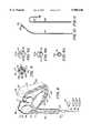
- FIG. 1is a schematic illustration showing the manner in which a catheter assembly and multi-port introducer of the present invention are introduced into the femoral vein and into the heart of a patient.
- FIG. 2is a side elevational view partially in cross section of the catheter assembly and the multi-port introducer shown in FIG. 1.
- FIG. 3is an end elevational view taken along the line 3--3 of FIG. 2.
- FIG. 4is a side elevational view of a catheter incorporating the present invention.
- FIG. 5is a side elevational view partially in cross section of the catheter shown in FIG. 4.
- FIG. 6is a schematic illustration showing the use of another catheter assembly and a long multi-port introducer incorporating the present invention.
- FIG. 7is a side elevational view of the catheter assembly and the long multi-port introducer shown in FIG. 6.
- FIG. 8is another schematic illustration showing the use of another embodiment of a catheter assembly and a multi-port introducer incorporating the present invention.
- FIG. 9is a cross-sectional view taken along the line 9--9 of FIG. 8.
- FIGS. 9A, 9B and 9Care cross-sectional views similar to FIG. 9 showing additional embodiments of multi-port introducers incorporating the present invention.
- FIG. 10is a partial side-elevational view of the distal extremity of a catheter incorporating the present invention having one type of bend formed in the distal extremity thereof.
- FIG. 11is another partial side-elevational view of a catheter incorporating the present invention having a different bend therein.
- FIG. 12there is shown another schematic illustration showing the use of another embodiment of a catheter assembly and multi-port introducer incorporating the present invention.
- FIG. 13is a cross-sectional view taken along the line 13--13 of FIG. 12.
- FIG. 13Ais a cross-sectional view similar to FIG. 13 showing a smaller multi-port introducer.
- FIG. 14is another schematic illustration showing the use of another embodiment of a catheter assembly and introducer incorporating another embodiment of the present invention.
- FIG. 15is a partial end-elevational view of the distal extremity of the catheter assembly and introducer with the catheter assembly retracted into the introducer.
- FIG. 16is an isometric view showing the manner in which the catheter assembly shown in FIG. 14 can be deployed.
- FIG. 17is a schematic illustration of another embodiment of a catheter assembly and multi-port introducer incorporating the present invention.
- FIG. 18is an end elevational view showing another arrangement for the catheters as they are deployed from the distal extremity of the multi-port introducer.
- FIG. 19is a side elevational view of an apparatus utilizing a multi-port introducer and a catheter assembly of the present invention.
- the catheter assembly of the present invention for performing a medical procedure on a bodyconsists of an introducer adapted to be inserted into a vessel of the patient.
- the introduceris provided with a plurality of lumens extending therethrough and opening into an entrance port.
- a plurality of catheters having proximal and distal extremitiesare removably positioned within the plurality of lumens in the introducer and extend distally of the introducer and are adapted to have their distal extremities positioned in different locations within the patient.
- the catheter assembly 11comprises a multi-port or multi-catheter introducer 12 and a plurality of catheters 13 which are introduced through the multi-port introducer 12.
- the catheter assembly 11is shown being utilized in FIG. 1 in connection with diagnostic procedures, as for example electrophysiology studies of the heart 16 of a patient 17 introduced through a femoral vein 18 of the patient.
- the multi-port introducer 12 as shown in FIG. 2consists of an introducer body 21 having proximal and distal extremities 22 and 23.
- the body 21is formed of a suitable material such as a medical grade plastic and can be relatively flexible.
- the introducer 12can be of a suitable size, as for example 6 or 7 French ranging from 0.080 to 0.093 inches in diameter.
- the distal extremity 23is provided with a rounded slightly tapered end 24 to provide an atraumatic tip for the introducer 12.
- the proximal extremity 22is enlarged and is provided with a plurality of ports 26 spaced circumferentially around an end wall 27, as for example four or more ports.
- the ports 26open into lumens 28 which extend longitudinally of the body 21 and which open through openings 29 in the rounded tapered end 24. Because of the tapering of the end 24, the openings 29 have generally an oval-shaped configuration even though the lumens 28 are circular in cross section.
- the lumens 28can have a suitable size which can accommodate the catheters 13 which are to be inserted therethrough. Thus, by way of example if the catheters are of a 2 French size, the lumens 28 should have a slightly greater size so that they can be introduced through the lumens with relatively little friction.
- a plurality of tubular members 31are bonded to the proximal extremity 22 of the body 21 and are in registration with the ports 26 so that the ports 26 are in communication with the tubular members 31.
- Hemostases valves 32 of a conventional typeare mounted on and carried by the tubular members 31. They are sized so that they can accommodate the 2 French catheters 13 and form a seal therewith when the 2 French catheters are introduced through the introducer 12 as hereinafter described. It should be appreciated that if desired, hemostases valves 32 can be incorporated in the proximal extremity 22 of the introducer body 21 and the tubular members 31 eliminated.
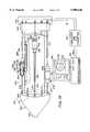
- a catheter 13 utilized in the multi-port introducer 12is shown in FIG. 4 and as shown therein consists of a flexible elongate member 36 which is provided with proximal and distal extremities 37 and 38.
- a handle 39is secured to the proximal extremity 37 and includes a slider 41 which is slidably mounted in a slot 42 provided in the handle.
- the slider 41controls means utilized for causing bending of the distal extremity 38.
- the handleis connected to first and second electrical cords 46 and 47 which are connected to plugs 48 and 49 that are utilized for a purpose hereinafter described.
- the distal extremity 38can be bent in a conventional manner by the use of steering wires actuated by the slider 41 or alternatively by electrically energized Nitinol elements as described in U.S. Pat. No. 5,238,005, issued Aug. 24, 1993.
- the flexible elongate member 36is comprised of a tubular member 61 formed of a suitable material such as stainless steel and having a suitable diameter, as for example 2 French of approximately 0.026 inches in diameter.
- the distal extremity 38be very flexible and for that reason it is formed of a helical coil 62 which is flexible and hollow.
- the coil 62can be formed of a suitable material such as a flat metal ribbon. Also, if desired, it can be formed of a radiopaque material such as a tungsten platinum alloy or palladium.
- the coil 62can have a suitable length, as for example 20-30 centimeters and can be formed of flattened round wire as shown. At the very distal extremity of the coil, the coil 62 is provided with an initial portion 62a having approximately the same diameter of the tubular member 61 for a length from 10-20 centimeters which is followed distally by step down portion 62b of a smaller diameter which can be accomplished by utilizing a mandrel having the two different diameters with the portion 62b having suitable length, as for example 1-2 centimeters.
- a tube 64 formed of a suitable insulating material, as for example shrink tubing formed of plasticis placed over the portion 62b.
- a plurality of electrodes 66 formed of a suitable material such as platinumare provided in longitudinally spaced-apart positions on the tube 64.
- the electrodes 66are connected by insulated conductors 67 that extend interiorly of the coil 62 to the proximal extremity of the flexible elongate member 36 and are connected through the handle 39 to the cord 47 and the connector 49.
- the last 5-10 millimeter portion 62c of the distal extremity of the coil 62is of a larger diameter, as for example the same diameter as the portion 62a.
- the coilsare spaced-apart with each coil having a diameter ranging from 0.016 to 0.018 inches a thickness of 0.002 to 0.003 inches and a spacing therebetween ranging from 0.004 to 0.006 inches.
- a rounded tip 71is provided in the distal extremity of the portion 62c and is formed of a suitable material such as a platinum tungsten alloy or palladium.
- a marker 73is carried by the distal extremity of the catheter 13 to identify the catheter. Such identifying markers 73 can be formed as a bond as shown of a suitable radiopaque material such as a tungsten platinum alloy or palladium.
- the catheterscan be coded or marked in such a manner that each is provided with the same marking on the proximal end distal extremities.
- two bonds 73can be provided on the distal extremity and similarly two markers 74 visible to the human eye such as colored markers can be provided on the proximal extremity.
- the two French catheters 13can be designed to have the same mechanical characteristics, as for example a conventional 6 or 7 French size catheter.
- the tubular member 61can be formed in such a manner so as to provide good torquability characteristics whereby when the proximal extremity 37 is rotated, the distal extremity will rotate in a one-for-one relationship.
- the tubular member 61can be formed in a conventional manner to achieve the desired stiffness and torque transfer capabilities desired.
- Preformed bendscan be placed in the distal extremity 38 of the flexible elongate member 36 by forming the catheter 13 in a suitable manner.
- the catheter 13can be provided in a desired preformed shape which is assumed as soon as there is space for the distal extremity 38 to curve, as for example, as shown in broken lines in FIG. 4.
- a controller and power supply 76is provided for each of the catheters 13.
- This controller and power supplycan be provided in the handle 39 and be controlled by the slider 41.
- the signal outputs which are being measured by the electrodes 66 carried by the distal extremity of the catheters 13can be viewed on instrumentation 78 connected to the connectors 48 connected to the handle 39 of each of the catheters 13.
- the physiciancan view the distal extremities of the catheters 13 by viewing the same under fluoroscopy and then can move the proximal extremities by movement of the handles.
- the physiciancan associate a marking with a marking provided on the proximal extremity so that he can move the catheter he desires to move.
- the physiciancan advance or retract the distal extremity of the catheter by grasping on the handle 39.
- the physiciancan rotate the catheter by rotation of the handle 39. Bending can be accomplished by moving of the slider 41 on the handle 39.
- the catheters 13can be removed one by one from the multi-port introducer 12 after which the multi-port introducer can be removed.
- the multi-port introducer 12 and the catheters 13 carried therebycan be removed as a unit.
- the incision through which the introducer has been introducedcan be closed in a conventional manner. It can be seen that with the catheter assembly 11 of the present invention and the method herein disclosed it is only necessary to make a single incision at a single site, into the patient while permitting multiple catheters to be introduced into the patient from this same site. This greatly decreases bleeding which may occur and minimizes the possibilities of the occurrence of hematomas.
- a long multi-port introducer 81which as shown schematically in FIG. 6 can again be introduced through the femoral vein 18 with the introducer having a length so that its distal extremity can be positioned near the opening to the right atrium of the heart at the junction of the superior and inferior vena cavas.
- a side elevational view of the long multi-port introducer 81is shown in FIG. 7 with a plurality of catheters 13 of the type hereinbefore described disposed therein.
- the introducer 81is provided with a body 82 generally of the same diameter as the body 21 of the multi-port introducer 12 and can have a suitable size, as for example 6-7 French.
- proximal and distal extremities 83 and 84can have a suitable length, as for example 30-70 centimeters.
- the body 82can be formed of a suitable material such as plastic. It is provided within a large head 86 formed integral with the proximal extremity 83 and is provided with a wall 87 having a plurality of spaced-apart ports 88 therein which open into lumens 89 that are in communication with staggered openings 91 provided in the distal extremity 84.
- the ports 88, the lumens 89 and the openings 91are sized so that they can accommodate the catheters 13.
- the ports 88, the lumens 89 and the openings 91should be of a size which is slightly greater than that to permit easy slidable movement of the catheters 13 within the introducer 81.
- Tubular members 96are connected into the ports 88 and are provided with hemostatic valves 97 on their proximal extremities which are adapted to receive the catheters 13.
- the openings 91have been staggered on the distal extremity 84 to reduce the possibility of stasis in the blood which could potentially cause emboli.
- the distal extremity 84has been provided with an atraumatic tip 98 similar to that hereinbefore described for the introducer 12.
- the openings 91are positioned proximally of the tip 98 rather than having the openings be in the tip 24 as with the introducer 12 hereinbefore described.
- the tip 93can be positioned so that the catheters 13 can be readily positioned.
- a radiopaque material 99can be provided on the tip 98 so that the tip can be visualized fluoroscopically.
- the long introducer 81 in connection with the catheters 13is similar to that hereinbefore described with the principal difference being that the long multi-port introducer 81 is positioned so that its tip is near the opening of the right atrium of the heart as hereinbefore described. Thereafter, the catheters 13 can be deployed and manipulated in the same manner as hereinbefore described to accomplish the desired medical procedures. After the desired medical procedure, as for example, an electrophysiology study has been completed, the catheters 13 can be removed after which the introducer 81 can be removed and the puncture site closed in a conventional manner.
- FIGS. 8-11Another embodiment of a catheter assembly 101 incorporating the present invention is shown in FIGS. 8-11 which utilizes a very short introducer that is adapted to receive the catheter assembly 101 and consists of a flexible elongate member 102 having proximal and distal extremities 103 and 104.
- the flexible elongate member 102is formed of a suitable material such as plastic and is provided with a plurality of lumens, as for example four lumens 106, 107, 108 and 109 (see FIG. 9) extending from the proximal extremity 103 to the distal extremity 104.
- the lumens 107, 108 and 109exit through the side parts 111, 112 and 113 of the flexible elongate member 102 adjacent the distal extremity in spaced-apart positions for a purpose hereinafter described.
- the distal extremity 104is provided with a tip electrode 116 and a ring electrode 117 spaced proximally of the tip electrode and are insulated from each other.
- the tip electrode and the ring electrode 117are connected by leads 118 and 119 which extend through the lumen 106 to the proximal extremity and are connected to an electrical connector 121.
- the flexible elongate member 102 as describedcan comprise a catheter of a suitable size, as for example 7 French which can be utilized for sensing a signal in the wall 126 a chamber, forming the right ventricle 127 in a heart 128.
- a flexible elongate member 102 of this sizeit can serve as the catheter which can be also utilized for introducing smaller size catheters into the right ventricle.
- three additional catheters 131, 132 and 133can be provided of a suitable size, as for example 2 French which can be introduced into the lumens 107, 108 and 109.
- Each of the catheters 131, 132 and 133can be constructed in the manner hereinbefore described and can be provided with tip electrodes 136 and a plurality of longitudinally spaced-apart ring electrodes 137.
- the 2 French catheters 131, 132 and 133can be advanced through their respective side ports 111, 112 and 113 and moved into engagement with different portions of the wall of the heart as shown in FIG. 8. They can be utilized to make a conventional electrophysiology study using the signals from the right atrium and the right ventricle of the heart 128.
- the 7 French catheter formed by the flexible elongate member 102 as well as the catheters 131, 132 and 133need not necessarily be steerable, however, it should be appreciated that if steerability is desired, that feature can be readily provided in the catheters.
- the 2 French catheters which are utilized in the larger 7 French catheter 102can have preformed bends, as for example those shown in FIGS. 10 and 11.
- the large 7 French catheter 102can either be considered to be a multi-port introducer which has electrodes carried by the distal extremity and which can be positioned in the right ventricle of the heart for mapping signals in one area of the heart and that the other smaller 2 French catheters 131, 132 and 133 can be used for mapping other areas of the heart.
- catheters 131, 132 and 133which have been provided with steering capabilities in the manner hereinbefore described or which have been provided with preformed bends, it is still possible to perform a conventional electrophysiology study from a single puncture site by with as few as three additional catheters in addition to the flexible elongate member 102 which also serves as a multi-port introducer with its own mapping capabilities.
- FIGS. 9A, 9B and 9Ccan be utilized to provide catheter assemblies of smaller diameter.
- the outside diameteris approximately 0.092".
- the diametercan be reduced to approximately 0.080.
- the outside diameter of the flexible elongate member 102bcan be still further reduced by only providing two lumens 146 and 147 which can slidably receive 2 French catheters of a type hereinbefore described, the diameter can be reduced to 0.070". With only a single lumen 151 in a flexible elongate member 102c, the diameter can be reduced to 0.040". In these embodiments shown in FIGS. 9A, 9B and 9C all of the lumens have an inside diameter of 0.030".
- FIGS. 12 and 13there is shown another embodiment of a catheter assembly 161 and a multi-port introducer 162.
- the multi-port introducer 162is introduced through the femoral artery 163 into the aorta 164 and into the left ventricle 166 of the heart 128.
- the multi-port introducer 162is comprised of a flexible elongate member 168 having proximal and distal extremities 169 and 171.
- the flexible elongate member 168can be provided with lumens 172, 173 and 174 extending between the proximal and distal extremities 169 and 171.
- the lumens 172, 173 and 174can be sized in the manner hereinbefore described and can accommodate mapping and/or ablation catheters 176 and 177 which are introduced therethrough into the lumens 172, 173 and which exit through end ports 178 and 179 provided in the distal extremity 171 of the flexible elongate member 168.
- Another port 180can be utilized for carrying a pressure transducer 181 connected by wires 182 to a connector assembly 183 which is connected into a arterial pressure measuring device 186 of a conventional type.
- the proximal extremities of the catheters 176 and 171are connected to connectors 188 and 189.
- FIG. 13AWhen it is desired to provide a smaller multi-port introducer 162, an arrangement such as shown in FIG. 13A can be utilized in which the flexible elongate member 168a is provided with only two lumens 191 and 192 in which lumen 191 can be utilized for receiving an ablation catheter 193 and the lumen 192 can be utilized for measuring arterial pressure.
- small cathetershave been discussed as being of the 2 French size it should be appreciated if desired even smaller catheters can be provided, as for example 11/2 or 1 French or larger catheters, as for example 21/2 or 3 French size ablation catheters.
- the present inventionhas also been described principally in connection with making electrophysiology measurements in the heart, it should be appreciated that the present concept of utilizing a multi-port introducer to reduce the number of incisions in a patient is not limited to mapping and ablation procedures but also can be utilized for introducing ablation catheters, catheters for measuring blood pressure and the like.
- FIGS. 14, 15 and 16show a catheter assembly 201 used with a multi-port introducer 202 of the present invention.
- the multi-port introducer 202consists of a flexible elongate member 203 having proximal and distal extremities 204 and 206.
- the flexible elongate member 203is formed of a suitable medical grade plastic and can have a size ranging from 7 to 81/2 French. It is provided with a plurality of circumferentially-spaced lumens 207 surrounding a central lumen 208 all which extend from the proximal extremity 204 to the distal extremity 206.
- the flexible elongate member 203has a length so that it can extend from outside the body through the femoral artery into the left ventricle of the heart 212 or from the femoral vein to the right atrium or ventricle.
- a plurality of small size catheters 216can be provided in the lumens 207 and can be of the type and size hereinbefore described and can range from 1 to 4 French, and preferably 2 French, for example the small size catheter 13 as shown in FIG. 4.
- the distal extremities of the catheters 216are provided with a plurality of electrodes 217 which are spaced apart axially of the catheters shown in FIG. 1.
- the electrodes 217are connected by conductors or wires (not shown) to the proximal extremity of the catheters and are connected to the electrical connectors 48 and 49 hereinbefore described in conjunction with FIG. 4.
- the ends of the distal extremities of the catheter 216are connected to an end cap 218 which is provided with a rounded outer surface 219.
- the distal extremities of the catheter 16are interconnected so that they can be formed into a basket-like construction as shown in FIG. 14.
- FIGS. 14-16Operation and use of the catheter assembly 201 with the multi-port introducer 202 can now be briefly described in conjunction with FIGS. 14-16.
- the small size catheters 216have been withdrawn so that the cap 218 is at the distal extremity of the multi-port introducer 202 as shown in FIG. 15 with the rounded surface 219 protruding therefrom.
- the introducer 202can be introduced into a femoral artery of a patient through a guiding catheter if that is deemed appropriate until the distal extremity has been advanced into the left ventricle 211 of the heart 212 having a wall 213.
- the small catheters 216can be pushed in the lumens 207 since they are slidably mounted therein so that the distal extremities thereof advance out of the distal extremity 206 of the multi-port introducer 202.
- the catheters 216can be pushed until the rounded surface 219 is in engagement with the apex of the left ventricle and thereafter they can be further pushed to cause them to flow outwardly to engage the side wall of the heart forming the left ventricle to move the electrodes 217 into engagement with the wall of the heart.
- left ventriclesas for example diseased hearts which may be misshapen or of greatly different sizes can be accommodated by this construction merely by pushing inwardly on individual catheters 216 until they are in engagement with the wall 213 of the heart 212.
- a pull wire 221can be provided which extends through the central lumen 208 provided in the multi-port introducer 202.
- pull wiresit is possible to provide small size catheter 216 which have distal ends which are pre-shaped into a desired bow or can have a straight shape.
- the pull wire 221can be utilized to form the basket without providing undue pressure on the wall 212 forming the apex of the left ventricle.
- a second steerable ablation catheter 226of the type described in co-pending application Ser. No. 07/894,529 filed Jun. 15, 1992, U.S. Pat. No. 5,324,284 can be introduced through the central lumen 208 alongside the guide wire 221 to perform an ablation in a manner hereinbefore described.
- the ablation catheter 226is provided with an ablation electrode 227 on its distal extremity which is adapted to be moved into engagement with the wall of the heart to perform an ablation in a manner hereinbefore described.
- FIG. 17which consists of a catheter assembly 231 and a multi-port introducer 232.
- the multi-port introducer 232is of the type hereinbefore described and is formed of flexible elongate member 233 having proximal and distal extremities 234 and 236. It is provided with a plurality of circumferentially spaced-apart lumens 237 extending from the proximal extremity to the distal extremity and a central lumen 238 extending in the same manner. It is also provided with another small lumen 239 positioned between the lumens 237 and 238.
- the lumens 237are adapted to receive small size catheters 241 of the type hereinbefore described which are provided with longitudinally spaced-apart electrodes 242 on their distal extremities.
- the distal extremities of the catheters 241are also provided with preformed memories so that when the distal extremities extend beyond distal extremity of the flexible elongate member 233 they will bend at a substantially right angle as shown in FIG. 17 to form what can be termed a "plaque" or in other words an arrangement in which the electrodes 242 carried by the distal extremities of the catheters 241 lie in a plane.
- the right angle bend of the distal extremity of the catheters 241can be readily obtained by utilizing shape memory materials, as for example one which has the right angle memory therein but which can be straightened out when the catheter is retracted into the lumens 237 of the multi-port introducer 232.
- shape memory materialsas for example one which has the right angle memory therein but which can be straightened out when the catheter is retracted into the lumens 237 of the multi-port introducer 232.
- the catheters 241can be torqued so that the distal extremities are arranged in a desired pattern, as for example a circular pattern in which the distal extremities are spaced apart at substantially equal angles.
- theycan be positioned to extend from two sides of the distal extremity of the flexible elongate member 233 to form essentially a rectangle in plan.
- Such geometric arrangementsare particularly desirable in mapping certain portions of the heart.
- the distal extremitiescan be torqued until they are brought in close proximity to each other to provide the high resolution mapping desired of that portion of the wall of the heart.
- a steerable ablation catheter of the type hereinbefore describedcan be introduced through the central lumen 238 or, alternatively, through a separate lumen, to accomplish the desired ablation. It should be appreciated that if desired, the ablation catheter can be in place at the time that the multi-port introducer 232 is introduced into the heart, as for example into the left ventricle of the heart.
- meanscan be provided for fixing or securing the distal extremity of the multi-port introducer 232 to the wall of the heart so that the electrodes can remain stationary with respect to the wall of the heart even though the wall of the heart is moving because of beating of the heart.
- Thiscan be accomplished by the use of a fixation pin 246 mounted on a flexible elongate actuation member 247 extending through the lumen 239 extending out of the distal extremity 236 of the flexible elongate member 233 to move the pin 246 into and out of engagement with the wall of the heart.
- the pin 246when advanced can be utilized to retain the distal extremity 236 of the multi-port introducer 232 in the desired position on the wall of the heart during a mapping procedure or during an ablation procedure.
- FIG. 19there is shown an apparatus which incorporates the multi-port introducer and catheter assembly of the present invention for performing high resolution mapping.
- this apparatus 261is comprised of a multi-port introducer 262 which is generally of the type hereinbefore described which is provided with an enlarged hub 263 which remains outside of the body and which is coupled to a large catheter-like portion 264 of a suitable size, such as 7 or 8 French, which can be introduced into a vessel of a patient in the manner hereinbefore described.
- a handle assembly 266is adapted to be engaged by the hand of a human being and is secured to the hub 263 as shown and extends proximally therefrom.
- the handle assembly 266is provided with a cylindrical housing 267 formed of a suitable material, such as plastic, and thus by way of example can have a suitable diameter as, for example, approximately one and one-half inches and can have a suitable length as for example, five to six inches.
- a plurality of circumferentially spaced apart lumens 271are provided in the hub 263 and open through the proximal extremity thereof and are spaced radially from the cylindrical housing 267 so that they are readily accessible permitting catheters 276 of the type hereinbefore described to be introduced through the lumens 271 and to extend through the large catheter-like portion 264 to enter into a chamber of the heart as hereinbefore described.
- Each of the catheters 276is adapted to be rotated and is also adapted to be advanced and retracted in the manner hereinbefore described.
- Such a meanshas been provided as a part of the handle assembly 266 and consists of a generally cylindrical slider mechanism 281 which is provided with a housing 282 depending member 283 which extends through a longitudinally extending slot 284 provided on the cylindrical housing 267.
- the depending member 283carries a slider 286 which is adapted to engage the longitudinally extending resistor 287 to form a linear potentiometer 288.
- a rotary potentiometer 291is mounted in the cylindrical housing 292 of the slider mechanism 281.
- the rotary potentiometer 281includes a shaft 293 rotatably mounted in the housing 282 and which is secured to the catheter 276.
- the shaft 293carries a contact member 294 which engages a cylindrical resistive element 296 secured to the interior of the cylindrical housing 282.
- the shaft 293is adapted to be rotated by a knob 298 mounted on the shaft 293.
- the knob 298is adapted to be engaged by the fingers of the hand carrying the handle assembly 266.
- Conductors (not shown) extending from the catheter 276 and from the rotary potentiometerare connected through a strain relief cable 301 and connected to an electrical connector 302 mounted on the proximal extremity of the handle of the cylindrical housing 267.
- the linear potentiometer 288is connected by a cable 306 extending interiorly of the cylindrical housing 267 and connected to the electrical connector 302.
- the connector 302is adapted to be mated with a mating connector 309 which is connected by a cable 311 to an interface box 312 containing certain conventional electronic circuitry, including amplifiers and A/D converters.
- the interface box 312is connected by a cable 313 to a conventional computer 314 which has mounted thereon a video monitor 316 which by way of example can carry a pair of screens 317 and 318, one of which can be utilized to display the electrical signals received from the heart and the other of which can be utilized for displaying an isochronal map displaying the position of the catheters 276 in the chamber of the heart with their electrodes.
- a scale(not shown) can be provided alongside each of the linear potentiometers 288 visible from the exterior of the cylindrical housing 267 and similarly a scale (not shown) can be provided on the cylindrical housing 282 or alternatively on the knob 298.
- rotations associated with the knob 298can be observed to ascertain the rotational positions of the distal extremities of the catheters 276 in a chamber of the heart.
- the rotational positions given by the rotary potentiometers 291 and the linear positions given by the linear potentiometers 282can be tracked by the computer 314 so that an approximate isochronal or isopotential map can be displayed on the screen 318.
- each catheter 276has the capability of extending a variable mount and being rotated by a variable mount it is possible for the computer 314 by ascertaining the extensions of the catheters 276 into the heart to ascertain how many electrodes on the various catheters are in engagement with the wall of the heart.
- the computer 314then can make appropriate adjustments on the isochronal or isopotential images created on the screen 318. For example, for a small heart chamber it is possible that a number of the electrodes carried by the catheter might still be within the lumen of the multi-lumen catheter 264.
- By providing the rotary information to the computer 314 from the rotary potentiometersit possible to ascertain the spacing between catheters in the chamber of the heart.
- the apparatus shown in FIG. 19is particularly suitable for deployment in irregularly shaped chambers of hearts. It is possible to display an appropriate isochronal map even though some of the arms of the catheters may be much shorter than others in the chamber of the heart and have different numbers of electrodes in contact with the wall forming the chamber of the heart.
Landscapes
- Health & Medical Sciences (AREA)
- Life Sciences & Earth Sciences (AREA)
- Surgery (AREA)
- Engineering & Computer Science (AREA)
- General Health & Medical Sciences (AREA)
- Veterinary Medicine (AREA)
- Biomedical Technology (AREA)
- Public Health (AREA)
- Medical Informatics (AREA)
- Molecular Biology (AREA)
- Heart & Thoracic Surgery (AREA)
- Animal Behavior & Ethology (AREA)
- Physics & Mathematics (AREA)
- Pathology (AREA)
- Biophysics (AREA)
- Cardiology (AREA)
- Nuclear Medicine, Radiotherapy & Molecular Imaging (AREA)
- Physiology (AREA)
- Plasma & Fusion (AREA)
- Otolaryngology (AREA)
- Orthopedic Medicine & Surgery (AREA)
- Vascular Medicine (AREA)
- Media Introduction/Drainage Providing Device (AREA)
Abstract
Description
Claims (9)
Priority Applications (2)
| Application Number | Priority Date | Filing Date | Title |
|---|---|---|---|
| US08/812,354US5908446A (en) | 1994-07-07 | 1997-03-04 | Catheter assembly, catheter and multi-port introducer for use therewith |
| US09/140,194US5964796A (en) | 1993-09-24 | 1998-08-25 | Catheter assembly, catheter and multi-port introducer for use therewith |
Applications Claiming Priority (2)
| Application Number | Priority Date | Filing Date | Title |
|---|---|---|---|
| US08/271,867US5607462A (en) | 1993-09-24 | 1994-07-07 | Catheter assembly, catheter and multi-catheter introducer for use therewith |
| US08/812,354US5908446A (en) | 1994-07-07 | 1997-03-04 | Catheter assembly, catheter and multi-port introducer for use therewith |
Related Parent Applications (1)
| Application Number | Title | Priority Date | Filing Date |
|---|---|---|---|
| US08/271,867ContinuationUS5607462A (en) | 1993-09-24 | 1994-07-07 | Catheter assembly, catheter and multi-catheter introducer for use therewith |
Related Child Applications (1)
| Application Number | Title | Priority Date | Filing Date |
|---|---|---|---|
| US09/140,194DivisionUS5964796A (en) | 1993-09-24 | 1998-08-25 | Catheter assembly, catheter and multi-port introducer for use therewith |
Publications (1)
| Publication Number | Publication Date |
|---|---|
| US5908446Atrue US5908446A (en) | 1999-06-01 |
Family
ID=23037427
Family Applications (2)
| Application Number | Title | Priority Date | Filing Date |
|---|---|---|---|
| US08/812,354Expired - Fee RelatedUS5908446A (en) | 1993-09-24 | 1997-03-04 | Catheter assembly, catheter and multi-port introducer for use therewith |
| US09/140,194Expired - Fee RelatedUS5964796A (en) | 1993-09-24 | 1998-08-25 | Catheter assembly, catheter and multi-port introducer for use therewith |
Family Applications After (1)
| Application Number | Title | Priority Date | Filing Date |
|---|---|---|---|
| US09/140,194Expired - Fee RelatedUS5964796A (en) | 1993-09-24 | 1998-08-25 | Catheter assembly, catheter and multi-port introducer for use therewith |
Country Status (1)
| Country | Link |
|---|---|
| US (2) | US5908446A (en) |
Cited By (85)
| Publication number | Priority date | Publication date | Assignee | Title |
|---|---|---|---|---|
| US6156003A (en)* | 1998-05-12 | 2000-12-05 | Chase Medical, Inc. | Surgical visualization and moisturizing device |
| US6178355B1 (en)* | 1997-04-29 | 2001-01-23 | Medtronic, Inc. | Intracardiac defibrillation leads |
| US6200333B1 (en) | 1997-04-07 | 2001-03-13 | Broncus Technologies, Inc. | Bronchial stenter |
| US6273907B1 (en) | 1997-04-07 | 2001-08-14 | Broncus Technologies, Inc. | Bronchial stenter |
| US6283989B1 (en) | 1997-04-07 | 2001-09-04 | Broncus Technolgies, Inc. | Method of treating a bronchial tube with a bronchial stenter having diametrically adjustable electrodes |
| US6283988B1 (en) | 1997-04-07 | 2001-09-04 | Broncus Technologies, Inc. | Bronchial stenter having expandable electrodes |
| US6293282B1 (en)* | 1996-11-05 | 2001-09-25 | Jerome Lemelson | System and method for treating select tissue in living being |
| US6375654B1 (en)* | 2000-05-19 | 2002-04-23 | Cardiofocus, Inc. | Catheter system with working portion radially expandable upon rotation |
| US6398266B1 (en) | 1999-09-22 | 2002-06-04 | Ballard Medical Products | Collapse resistant popoid connector |
| US6411852B1 (en) | 1997-04-07 | 2002-06-25 | Broncus Technologies, Inc. | Modification of airways by application of energy |
| USD466607S1 (en) | 2001-08-27 | 2002-12-03 | Kimberly-Clark Worldwide, Inc. | Flexible connector |
| US6488673B1 (en) | 1997-04-07 | 2002-12-03 | Broncus Technologies, Inc. | Method of increasing gas exchange of a lung |
| USD473941S1 (en) | 2001-08-27 | 2003-04-29 | Kimberly-Clark Worldwide, Inc. | Flexible connecting device |
| WO2002072186A3 (en)* | 2001-03-14 | 2003-05-08 | E V R Endo Vascular Res Es Sa | Vascular catheter guide wire carrier |
| USD476731S1 (en) | 2001-08-27 | 2003-07-01 | Kimberly-Clark Worldwide, Inc. | Bendable connector |
| US6634363B1 (en) | 1997-04-07 | 2003-10-21 | Broncus Technologies, Inc. | Methods of treating lungs having reversible obstructive pulmonary disease |
| US6647617B1 (en)* | 1992-09-23 | 2003-11-18 | Graydon Ernest Beatty | Method of construction an endocardial mapping catheter |
| US6673023B2 (en) | 2001-03-23 | 2004-01-06 | Stryker Puerto Rico Limited | Micro-invasive breast biopsy device |
| USD486909S1 (en) | 2001-08-27 | 2004-02-17 | Kimberly-Clark Worldwide, Inc. | Bendable connecting device |
| US6746446B1 (en) | 2000-08-04 | 2004-06-08 | Cardima, Inc. | Electrophysiological device for the isthmus |
| US20040242984A1 (en)* | 2003-06-02 | 2004-12-02 | Plaza Claudio P. | Catheter and method for mapping a pulmonary vein |
| US20050043604A1 (en)* | 1992-09-23 | 2005-02-24 | Beatty Graydon Ernest | Chamber location method |
| US20050101874A1 (en)* | 1992-09-23 | 2005-05-12 | Beatty Graydon E. | Software for mapping potential distribution of a heart chamber |
| US20050113660A1 (en)* | 2002-08-30 | 2005-05-26 | Biosense Webster, Inc. | Catheter and method for mapping purkinje fibers |
| US20050148837A1 (en)* | 2001-12-31 | 2005-07-07 | Fuimaono Kristine B. | Catheter having multiple spines each having electrical mapping and location sensing capabilities |
| US20050187467A1 (en)* | 2004-01-21 | 2005-08-25 | Martin Kleen | Catheter |
| US6944490B1 (en) | 2002-09-25 | 2005-09-13 | Advanced Cardiovascular Systems, Inc. | Apparatus and method for positioning and delivering a therapeutic tool to the inside of a heart |
| US20050209530A1 (en)* | 2001-03-23 | 2005-09-22 | Stryker Puerto Rico Limited | Micro-invasive tissue removal device |
| US7027869B2 (en) | 1998-01-07 | 2006-04-11 | Asthmatx, Inc. | Method for treating an asthma attack |
| US7101362B2 (en) | 2003-07-02 | 2006-09-05 | St. Jude Medical, Atrial Fibrillation Division, Inc. | Steerable and shapable catheter employing fluid force |
| US7189208B1 (en) | 1992-09-23 | 2007-03-13 | Endocardial Solutions, Inc. | Method for measuring heart electrophysiology |
| US20070118184A1 (en)* | 1998-06-10 | 2007-05-24 | Asthmatx, Inc. | Devices for modification of airways by transfer of energy |
| US7235070B2 (en) | 2003-07-02 | 2007-06-26 | St. Jude Medical, Atrial Fibrillation Division, Inc. | Ablation fluid manifold for ablation catheter |
| US7263397B2 (en) | 1998-06-30 | 2007-08-28 | St. Jude Medical, Atrial Fibrillation Division, Inc. | Method and apparatus for catheter navigation and location and mapping in the heart |
| US20070225558A1 (en)* | 2004-05-28 | 2007-09-27 | Hauck John A | Robotic surgical system and method for surface modeling |
| US20070255172A1 (en)* | 2001-03-23 | 2007-11-01 | Stryker Puerto Rico Limited | Micro-invasive nucleotomy device and method |
| US20070299435A1 (en)* | 2006-06-23 | 2007-12-27 | Crowe John E | Apparatus and method for ablating tissue |
| US20080009758A1 (en)* | 2006-05-17 | 2008-01-10 | Voth Eric J | System and method for mapping electrophysiology information onto complex geometry |
| US20080033284A1 (en)* | 2005-05-27 | 2008-02-07 | Hauck John A | Robotically controlled catheter and method of its calibration |
| US7425212B1 (en) | 1998-06-10 | 2008-09-16 | Asthmatx, Inc. | Devices for modification of airways by transfer of energy |
| WO2008002654A3 (en)* | 2006-06-28 | 2008-12-18 | Bard Inc C R | Methods and apparatus for assessing and improving electrode contact with cardiac tissue |
| US7632265B2 (en) | 2004-05-28 | 2009-12-15 | St. Jude Medical, Atrial Fibrillation Division, Inc. | Radio frequency ablation servo catheter and method |
| US7670297B1 (en) | 1998-06-30 | 2010-03-02 | St. Jude Medical, Atrial Fibrillation Division, Inc. | Chamber mapping system |
| USRE41334E1 (en) | 1992-09-23 | 2010-05-11 | St. Jude Medical, Atrial Fibrillation Division, Inc. | Endocardial mapping system |
| US7806829B2 (en) | 1998-06-30 | 2010-10-05 | St. Jude Medical, Atrial Fibrillation Division, Inc. | System and method for navigating an ultrasound catheter to image a beating heart |
| US7818048B2 (en) | 2003-06-02 | 2010-10-19 | Biosense Webster, Inc. | Catheter and method for mapping a pulmonary vein |
| US7819868B2 (en) | 2005-06-21 | 2010-10-26 | St. Jude Medical, Atrial Fibrilation Division, Inc. | Ablation catheter with fluid distribution structures |
| US7819866B2 (en) | 2003-01-21 | 2010-10-26 | St. Jude Medical, Atrial Fibrillation Division, Inc. | Ablation catheter and electrode |
| US7837679B2 (en) | 2000-10-17 | 2010-11-23 | Asthmatx, Inc. | Control system and process for application of energy to airway walls and other mediums |
| US7853331B2 (en) | 2004-11-05 | 2010-12-14 | Asthmatx, Inc. | Medical device with procedure improvement features |
| US7921855B2 (en) | 1998-01-07 | 2011-04-12 | Asthmatx, Inc. | Method for treating an asthma attack |
| US7931647B2 (en) | 2006-10-20 | 2011-04-26 | Asthmatx, Inc. | Method of delivering energy to a lung airway using markers |
| US7949407B2 (en) | 2004-11-05 | 2011-05-24 | Asthmatx, Inc. | Energy delivery devices and methods |
| US7992572B2 (en) | 1998-06-10 | 2011-08-09 | Asthmatx, Inc. | Methods of evaluating individuals having reversible obstructive pulmonary disease |
| US8181656B2 (en) | 1998-06-10 | 2012-05-22 | Asthmatx, Inc. | Methods for treating airways |
| US8235983B2 (en) | 2007-07-12 | 2012-08-07 | Asthmatx, Inc. | Systems and methods for delivering energy to passageways in a patient |
| US8251070B2 (en) | 2000-03-27 | 2012-08-28 | Asthmatx, Inc. | Methods for treating airways |
| US8483831B1 (en) | 2008-02-15 | 2013-07-09 | Holaira, Inc. | System and method for bronchial dilation |
| US8528565B2 (en) | 2004-05-28 | 2013-09-10 | St. Jude Medical, Atrial Fibrillation Division, Inc. | Robotic surgical system and method for automated therapy delivery |
| US8740895B2 (en) | 2009-10-27 | 2014-06-03 | Holaira, Inc. | Delivery devices with coolable energy emitting assemblies |
| US8755864B2 (en) | 2004-05-28 | 2014-06-17 | St. Jude Medical, Atrial Fibrillation Division, Inc. | Robotic surgical system and method for diagnostic data mapping |
| US8808280B2 (en) | 2008-05-09 | 2014-08-19 | Holaira, Inc. | Systems, assemblies, and methods for treating a bronchial tree |
| US8911439B2 (en) | 2009-11-11 | 2014-12-16 | Holaira, Inc. | Non-invasive and minimally invasive denervation methods and systems for performing the same |
| US8920413B2 (en) | 2004-11-12 | 2014-12-30 | Asthmatx, Inc. | Energy delivery devices and methods |
| US20150157344A1 (en)* | 2013-12-06 | 2015-06-11 | Boston Scientific Scimed, Inc. | Medical retrieval devices and related methods of use |
| WO2015114120A1 (en)* | 2014-02-03 | 2015-08-06 | Olympus Winter & Ibe Gmbh | Electrosurgical instrument |
| US9149328B2 (en) | 2009-11-11 | 2015-10-06 | Holaira, Inc. | Systems, apparatuses, and methods for treating tissue and controlling stenosis |
| US20150351835A1 (en)* | 2013-02-05 | 2015-12-10 | Handok Inc. | Catheter for denervation |
| US20160051831A1 (en)* | 2012-11-21 | 2016-02-25 | Circuit Therapeutics, Inc. | System and method for optogenetic therapy |
| US9272132B2 (en) | 2012-11-02 | 2016-03-01 | Boston Scientific Scimed, Inc. | Medical device for treating airways and related methods of use |
| US9283374B2 (en) | 2012-11-05 | 2016-03-15 | Boston Scientific Scimed, Inc. | Devices and methods for delivering energy to body lumens |
| WO2016057096A1 (en)* | 2014-10-07 | 2016-04-14 | Qxmedical, Llc | Segmented catheter structure and improved catheter tip and related systems, methods, and devices |
| US9314299B2 (en) | 2012-03-21 | 2016-04-19 | Biosense Webster (Israel) Ltd. | Flower catheter for mapping and ablating veinous and other tubular locations |
| US9339618B2 (en) | 2003-05-13 | 2016-05-17 | Holaira, Inc. | Method and apparatus for controlling narrowing of at least one airway |
| US9398933B2 (en) | 2012-12-27 | 2016-07-26 | Holaira, Inc. | Methods for improving drug efficacy including a combination of drug administration and nerve modulation |
| US9592086B2 (en) | 2012-07-24 | 2017-03-14 | Boston Scientific Scimed, Inc. | Electrodes for tissue treatment |
| EP3141183A1 (en)* | 2015-09-14 | 2017-03-15 | Biosense Webster (Israel) Ltd. | Basket catheter with individual spine control |
| EP3181082A1 (en)* | 2015-12-17 | 2017-06-21 | Pythagoras Medical Ltd. | Transluminal electrode catheters |
| US9770293B2 (en) | 2012-06-04 | 2017-09-26 | Boston Scientific Scimed, Inc. | Systems and methods for treating tissue of a passageway within a body |
| US9782130B2 (en) | 2004-05-28 | 2017-10-10 | St. Jude Medical, Atrial Fibrillation Division, Inc. | Robotic surgical system |
| US9814618B2 (en) | 2013-06-06 | 2017-11-14 | Boston Scientific Scimed, Inc. | Devices for delivering energy and related methods of use |
| WO2018011632A1 (en)* | 2016-07-15 | 2018-01-18 | Dragon Medical Development Limited | Multi-spline, multi-electrode catheter and method of use for mapping of internal organs |
| US10258285B2 (en) | 2004-05-28 | 2019-04-16 | St. Jude Medical, Atrial Fibrillation Division, Inc. | Robotic surgical system and method for automated creation of ablation lesions |
| US10478247B2 (en) | 2013-08-09 | 2019-11-19 | Boston Scientific Scimed, Inc. | Expandable catheter and related methods of manufacture and use |
| US10863945B2 (en) | 2004-05-28 | 2020-12-15 | St. Jude Medical, Atrial Fibrillation Division, Inc. | Robotic surgical system with contact sensing feature |
Families Citing this family (84)
| Publication number | Priority date | Publication date | Assignee | Title |
|---|---|---|---|---|
| FR2652928B1 (en) | 1989-10-05 | 1994-07-29 | Diadix Sa | INTERACTIVE LOCAL INTERVENTION SYSTEM WITHIN A AREA OF A NON-HOMOGENEOUS STRUCTURE. |
| AU675077B2 (en) | 1992-08-14 | 1997-01-23 | British Telecommunications Public Limited Company | Position location system |
| US5592939A (en) | 1995-06-14 | 1997-01-14 | Martinelli; Michael A. | Method and system for navigating a catheter probe |
| US6226548B1 (en) | 1997-09-24 | 2001-05-01 | Surgical Navigation Technologies, Inc. | Percutaneous registration apparatus and method for use in computer-assisted surgical navigation |
| US6021343A (en) | 1997-11-20 | 2000-02-01 | Surgical Navigation Technologies | Image guided awl/tap/screwdriver |
| US6348058B1 (en) | 1997-12-12 | 2002-02-19 | Surgical Navigation Technologies, Inc. | Image guided spinal surgery guide, system, and method for use thereof |
| US6477400B1 (en) | 1998-08-20 | 2002-11-05 | Sofamor Danek Holdings, Inc. | Fluoroscopic image guided orthopaedic surgery system with intraoperative registration |
| ES2228165T3 (en)* | 1998-12-09 | 2005-04-01 | Cook Incorporated | HOLLOW NEEDLE, CURVED, SUPERELASTIC, FOR MEDICAL USE. |
| US6470207B1 (en) | 1999-03-23 | 2002-10-22 | Surgical Navigation Technologies, Inc. | Navigational guidance via computer-assisted fluoroscopic imaging |
| US6491699B1 (en) | 1999-04-20 | 2002-12-10 | Surgical Navigation Technologies, Inc. | Instrument guidance method and system for image guided surgery |
| US6353762B1 (en)* | 1999-04-30 | 2002-03-05 | Medtronic, Inc. | Techniques for selective activation of neurons in the brain, spinal cord parenchyma or peripheral nerve |
| US8239001B2 (en) | 2003-10-17 | 2012-08-07 | Medtronic Navigation, Inc. | Method and apparatus for surgical navigation |
| US6379302B1 (en) | 1999-10-28 | 2002-04-30 | Surgical Navigation Technologies Inc. | Navigation information overlay onto ultrasound imagery |
| US11331150B2 (en) | 1999-10-28 | 2022-05-17 | Medtronic Navigation, Inc. | Method and apparatus for surgical navigation |
| US6474341B1 (en) | 1999-10-28 | 2002-11-05 | Surgical Navigation Technologies, Inc. | Surgical communication and power system |
| US6381485B1 (en) | 1999-10-28 | 2002-04-30 | Surgical Navigation Technologies, Inc. | Registration of human anatomy integrated for electromagnetic localization |
| US6499488B1 (en)* | 1999-10-28 | 2002-12-31 | Winchester Development Associates | Surgical sensor |
| US8644907B2 (en) | 1999-10-28 | 2014-02-04 | Medtronic Navigaton, Inc. | Method and apparatus for surgical navigation |
| US7366562B2 (en) | 2003-10-17 | 2008-04-29 | Medtronic Navigation, Inc. | Method and apparatus for surgical navigation |
| US6493573B1 (en) | 1999-10-28 | 2002-12-10 | Winchester Development Associates | Method and system for navigating a catheter probe in the presence of field-influencing objects |
| US6497676B1 (en) | 2000-02-10 | 2002-12-24 | Baxter International | Method and apparatus for monitoring and controlling peritoneal dialysis therapy |
| US6725080B2 (en) | 2000-03-01 | 2004-04-20 | Surgical Navigation Technologies, Inc. | Multiple cannula image guided tool for image guided procedures |
| US6535756B1 (en) | 2000-04-07 | 2003-03-18 | Surgical Navigation Technologies, Inc. | Trajectory storage apparatus and method for surgical navigation system |
| US7085400B1 (en) | 2000-06-14 | 2006-08-01 | Surgical Navigation Technologies, Inc. | System and method for image based sensor calibration |
| US6976973B1 (en) | 2000-10-12 | 2005-12-20 | Baxter International Inc. | Peritoneal dialysis catheters |
| US20020188287A1 (en)* | 2001-05-21 | 2002-12-12 | Roni Zvuloni | Apparatus and method for cryosurgery within a body cavity |
| US6706037B2 (en)* | 2000-10-24 | 2004-03-16 | Galil Medical Ltd. | Multiple cryoprobe apparatus and method |
| US20020068929A1 (en)* | 2000-10-24 | 2002-06-06 | Roni Zvuloni | Apparatus and method for compressing a gas, and cryosurgery system and method utilizing same |
| US20080045934A1 (en)* | 2000-10-24 | 2008-02-21 | Galil Medical Ltd. | Device and method for coordinated insertion of a plurality of cryoprobes |
| US6579300B2 (en)* | 2001-01-18 | 2003-06-17 | Scimed Life Systems, Inc. | Steerable sphincterotome and methods for cannulation, papillotomy and sphincterotomy |
| US7087040B2 (en) | 2001-02-28 | 2006-08-08 | Rex Medical, L.P. | Apparatus for delivering ablation fluid to treat lesions |
| US6989004B2 (en) | 2001-02-28 | 2006-01-24 | Rex Medical, L.P. | Apparatus for delivering ablation fluid to treat lesions |
| ES2257539T3 (en)* | 2001-02-28 | 2006-08-01 | Rex Medical, L.P. | APPLIANCE TO SUPPLY ABLATIVE FLUID FOR THE TREATMENT OF INJURIES. |
| US20080051774A1 (en)* | 2001-05-21 | 2008-02-28 | Galil Medical Ltd. | Device and method for coordinated insertion of a plurality of cryoprobes |
| US20080051776A1 (en)* | 2001-05-21 | 2008-02-28 | Galil Medical Ltd. | Thin uninsulated cryoprobe and insulating probe introducer |
| US6520183B2 (en)* | 2001-06-11 | 2003-02-18 | Memorial Sloan-Kettering Cancer Center | Double endobronchial catheter for one lung isolation anesthesia and surgery |
| DE10153842A1 (en)* | 2001-10-24 | 2003-05-08 | Biotronik Mess & Therapieg | electrode assembly |
| US6993384B2 (en)* | 2001-12-04 | 2006-01-31 | Advanced Bionics Corporation | Apparatus and method for determining the relative position and orientation of neurostimulation leads |
| US7853330B2 (en)* | 2001-12-04 | 2010-12-14 | Boston Scientific Neuromodulation Corporation | Apparatus and method for determining the relative position and orientation of neurostimulation leads |
| US6758836B2 (en) | 2002-02-07 | 2004-07-06 | C. R. Bard, Inc. | Split tip dialysis catheter |
| US6947786B2 (en) | 2002-02-28 | 2005-09-20 | Surgical Navigation Technologies, Inc. | Method and apparatus for perspective inversion |
| US7588581B2 (en)* | 2002-03-26 | 2009-09-15 | Medtronic, Inc. | Placement of chronic micro-catheter device and method |
| US6990368B2 (en) | 2002-04-04 | 2006-01-24 | Surgical Navigation Technologies, Inc. | Method and apparatus for virtual digital subtraction angiography |
| US6892090B2 (en) | 2002-08-19 | 2005-05-10 | Surgical Navigation Technologies, Inc. | Method and apparatus for virtual endoscopy |
| US7697972B2 (en) | 2002-11-19 | 2010-04-13 | Medtronic Navigation, Inc. | Navigation system for cardiac therapies |
| US7599730B2 (en) | 2002-11-19 | 2009-10-06 | Medtronic Navigation, Inc. | Navigation system for cardiac therapies |
| US7660623B2 (en) | 2003-01-30 | 2010-02-09 | Medtronic Navigation, Inc. | Six degree of freedom alignment display for medical procedures |
| US7542791B2 (en) | 2003-01-30 | 2009-06-02 | Medtronic Navigation, Inc. | Method and apparatus for preplanning a surgical procedure |
| US7229450B1 (en) | 2003-02-11 | 2007-06-12 | Pacesetter, Inc. | Kink resistant introducer with mapping capabilities |
| US7393339B2 (en)* | 2003-02-21 | 2008-07-01 | C. R. Bard, Inc. | Multi-lumen catheter with separate distal tips |
| US8435249B2 (en)* | 2003-04-01 | 2013-05-07 | Medron, Inc. | Flexible connection catheter tunneler and methods for using the same |
| US7570791B2 (en) | 2003-04-25 | 2009-08-04 | Medtronic Navigation, Inc. | Method and apparatus for performing 2D to 3D registration |
| US20040243095A1 (en) | 2003-05-27 | 2004-12-02 | Shekhar Nimkar | Methods and apparatus for inserting multi-lumen spit-tip catheters into a blood vessel |
| US20040267256A1 (en)* | 2003-06-24 | 2004-12-30 | Garabedian Robert J. | Compound lesion alignment device |
| US7313430B2 (en) | 2003-08-28 | 2007-12-25 | Medtronic Navigation, Inc. | Method and apparatus for performing stereotactic surgery |
| US7835778B2 (en) | 2003-10-16 | 2010-11-16 | Medtronic Navigation, Inc. | Method and apparatus for surgical navigation of a multiple piece construct for implantation |
| US7840253B2 (en) | 2003-10-17 | 2010-11-23 | Medtronic Navigation, Inc. | Method and apparatus for surgical navigation |
| US8007847B2 (en) | 2004-01-13 | 2011-08-30 | Eytan Biderman | Feeding formula appliance |
| US8992454B2 (en) | 2004-06-09 | 2015-03-31 | Bard Access Systems, Inc. | Splitable tip catheter with bioresorbable adhesive |
| US7717875B2 (en)* | 2004-07-20 | 2010-05-18 | St. Jude Medical, Atrial Fibrillation Division, Inc. | Steerable catheter with hydraulic or pneumatic actuator |
| US7835784B2 (en) | 2005-09-21 | 2010-11-16 | Medtronic Navigation, Inc. | Method and apparatus for positioning a reference frame |
| WO2007069248A2 (en)* | 2005-12-16 | 2007-06-21 | Galil Medical Ltd. | Apparatus and method for thermal ablation of uterine fibroids |
| US9168102B2 (en) | 2006-01-18 | 2015-10-27 | Medtronic Navigation, Inc. | Method and apparatus for providing a container to a sterile environment |
| US20090292279A1 (en)* | 2006-01-26 | 2009-11-26 | Galil Medical Ltd. | Device and Method for Coordinated Insertion of a Plurality of Cryoprobes |
| US8112292B2 (en) | 2006-04-21 | 2012-02-07 | Medtronic Navigation, Inc. | Method and apparatus for optimizing a therapy |
| US20070299438A1 (en)* | 2006-06-23 | 2007-12-27 | Holzbaur Michael C | Torque transfer agent for introducer and method |
| WO2009051967A1 (en) | 2007-10-17 | 2009-04-23 | Spire Corporation | Manufacture of split tip catheters |
| US8292841B2 (en) | 2007-10-26 | 2012-10-23 | C. R. Bard, Inc. | Solid-body catheter including lateral distal openings |
| US8066660B2 (en) | 2007-10-26 | 2011-11-29 | C. R. Bard, Inc. | Split-tip catheter including lateral distal openings |
| CN101918067B (en) | 2007-11-01 | 2013-04-10 | C·R·巴德股份有限公司 | Catheter assembly including three lumen tips |
| US9579485B2 (en) | 2007-11-01 | 2017-02-28 | C. R. Bard, Inc. | Catheter assembly including a multi-lumen configuration |
| WO2010014568A1 (en)* | 2008-07-28 | 2010-02-04 | William Beaumont Hospital | Multiple port introducer for thrombolysis |
| US8175681B2 (en) | 2008-12-16 | 2012-05-08 | Medtronic Navigation Inc. | Combination of electromagnetic and electropotential localization |
| US8394218B2 (en) | 2009-07-20 | 2013-03-12 | Covidien Lp | Method for making a multi-lumen catheter having a separated tip section |
| US8494614B2 (en) | 2009-08-31 | 2013-07-23 | Regents Of The University Of Minnesota | Combination localization system |
| US8494613B2 (en) | 2009-08-31 | 2013-07-23 | Medtronic, Inc. | Combination localization system |
| US9492113B2 (en) | 2011-07-15 | 2016-11-15 | Boston Scientific Scimed, Inc. | Systems and methods for monitoring organ activity |
| WO2013052852A1 (en)* | 2011-10-07 | 2013-04-11 | Boston Scientific Scimed, Inc. | Methods and systems for detection and thermal treatment of lower urinary tract conditions |
| USD748252S1 (en) | 2013-02-08 | 2016-01-26 | C. R. Bard, Inc. | Multi-lumen catheter tip |
| US9855402B2 (en) | 2014-02-15 | 2018-01-02 | Rex Medical, L.P. | Apparatus for delivering fluid to treat renal hypertension |
| US9849229B2 (en) | 2014-04-25 | 2017-12-26 | Covidien Lp | Split-tip catheter |
| WO2016011091A1 (en) | 2014-07-14 | 2016-01-21 | C. R. Bard, Inc. | Apparatuses, systems, and methods for inserting split tip catheters having enhanced stiffening and guiding features |
| JP6785301B2 (en) | 2015-11-04 | 2020-11-18 | ボストン サイエンティフィック サイムド,インコーポレイテッドBoston Scientific Scimed,Inc. | Medical devices and related methods |
| US11179516B2 (en) | 2017-06-22 | 2021-11-23 | Baxter International Inc. | Systems and methods for incorporating patient pressure into medical fluid delivery |
Citations (35)
| Publication number | Priority date | Publication date | Assignee | Title |
|---|---|---|---|---|
| FR2565491A1 (en)* | 1984-06-08 | 1985-12-13 | Monties Jean Raoul | Multi-path catheter |
| US4602645A (en)* | 1982-12-16 | 1986-07-29 | C. R. Bard, Inc. | Atrio-ventricular pacing catheter |
| US4637388A (en)* | 1984-10-15 | 1987-01-20 | Portex, Inc. | Tracheal tube obturator with reversible tip |
| US4670009A (en)* | 1985-04-16 | 1987-06-02 | American Hospital Supply Corp. | Backform inserts for catheter |
| US4671291A (en)* | 1986-03-31 | 1987-06-09 | Siemens Medical Systems, Inc. | Angle encoding catheter |
| US4699147A (en)* | 1985-09-25 | 1987-10-13 | Cordis Corporation | Intraventricular multielectrode cardial mapping probe and method for using same |
| US4769005A (en)* | 1987-08-06 | 1988-09-06 | Robert Ginsburg | Selective catheter guide |
| US4807626A (en)* | 1985-02-14 | 1989-02-28 | Mcgirr Douglas B | Stone extractor and method |
| US4808157A (en)* | 1987-07-13 | 1989-02-28 | Neuro Delivery Technology, Inc. | Multi-lumen epidural-spinal needle |
| US4813930A (en)* | 1987-10-13 | 1989-03-21 | Dimed, Inc. | Angioplasty guiding catheters and methods for performing angioplasty |
| US4838879A (en)* | 1986-05-08 | 1989-06-13 | Terumo Kabushiki Kaisha | Catheter |
| US4905667A (en)* | 1987-05-12 | 1990-03-06 | Ernst Foerster | Apparatus for endoscopic-transpapillary exploration of biliary tract |
| US4920980A (en)* | 1987-09-14 | 1990-05-01 | Cordis Corporation | Catheter with controllable tip |
| US4940064A (en)* | 1986-11-14 | 1990-07-10 | Desai Jawahar M | Catheter for mapping and ablation and method therefor |
| US4963128A (en)* | 1989-03-21 | 1990-10-16 | University Of Virginia Alumni Patents Foundation | Chest tube and catheter grid for intrathoracic afterload radiotherapy |
| US5059177A (en)* | 1990-04-19 | 1991-10-22 | Cordis Corporation | Triple lumen balloon catheter |
| US5078714A (en)* | 1990-03-02 | 1992-01-07 | Jefferson Katims | Method and apparatus for placement of a probe in the body and the medical procedure for guiding and locating a catheter or probe in the body |
| US5135599A (en)* | 1987-12-22 | 1992-08-04 | Vas-Cath Incorporated | Method of making a triple lumen catheter |
| US5149330A (en)* | 1991-01-10 | 1992-09-22 | The Kendall Company | Catheter convertible from single to multilumen |
| US5199950A (en)* | 1990-12-07 | 1993-04-06 | Willy Rusch Ag | Medical instrument |
| US5203338A (en)* | 1990-12-17 | 1993-04-20 | Cardiovascular Imaging Systems, Inc. | Vascular catheter having low-profile distal end |
| US5211176A (en)* | 1990-11-30 | 1993-05-18 | Fuji Photo Optical Co., Ltd. | Ultrasound examination system |
| US5215527A (en)* | 1991-12-12 | 1993-06-01 | Becton, Dickinson And Company | Catheter introducer assembly |
| US5236417A (en)* | 1992-09-22 | 1993-08-17 | Utah Pioneer Medical, Inc. | Cholangiography catheter apparatus and method |
| US5237996A (en)* | 1992-02-11 | 1993-08-24 | Waldman Lewis K | Endocardial electrical mapping catheter |
| US5255679A (en)* | 1992-06-02 | 1993-10-26 | Cardiac Pathways Corporation | Endocardial catheter for mapping and/or ablation with an expandable basket structure having means for providing selective reinforcement and pressure sensing mechanism for use therewith, and method |
| US5292305A (en)* | 1991-09-10 | 1994-03-08 | Cordis Corporation | Double-lumen angioscopy catheter |
| US5324284A (en)* | 1992-06-05 | 1994-06-28 | Cardiac Pathways, Inc. | Endocardial mapping and ablation system utilizing a separately controlled ablation catheter and method |
| US5327889A (en)* | 1992-12-01 | 1994-07-12 | Cardiac Pathways Corporation | Mapping and ablation catheter with individually deployable arms and method |
| US5342299A (en)* | 1992-07-06 | 1994-08-30 | Catheter Imaging Systems | Steerable catheter |
| US5345936A (en)* | 1991-02-15 | 1994-09-13 | Cardiac Pathways Corporation | Apparatus with basket assembly for endocardial mapping |
| US5354266A (en)* | 1992-07-06 | 1994-10-11 | Catheter Imaging Systems | Method of epidural surgery |
| US5400783A (en)* | 1993-10-12 | 1995-03-28 | Cardiac Pathways Corporation | Endocardial mapping apparatus with rotatable arm and method |
| US5471982A (en)* | 1992-09-29 | 1995-12-05 | Ep Technologies, Inc. | Cardiac mapping and ablation systems |
| US5628313A (en)* | 1992-06-30 | 1997-05-13 | Cordis Webster, Inc. | Cardiovascular catheter with laterally stable basket-shaped electrode array |
- 1997
- 1997-03-04USUS08/812,354patent/US5908446A/ennot_activeExpired - Fee Related
- 1998
- 1998-08-25USUS09/140,194patent/US5964796A/ennot_activeExpired - Fee Related
Patent Citations (36)
| Publication number | Priority date | Publication date | Assignee | Title |
|---|---|---|---|---|
| US4602645A (en)* | 1982-12-16 | 1986-07-29 | C. R. Bard, Inc. | Atrio-ventricular pacing catheter |
| FR2565491A1 (en)* | 1984-06-08 | 1985-12-13 | Monties Jean Raoul | Multi-path catheter |
| US4637388A (en)* | 1984-10-15 | 1987-01-20 | Portex, Inc. | Tracheal tube obturator with reversible tip |
| US4807626A (en)* | 1985-02-14 | 1989-02-28 | Mcgirr Douglas B | Stone extractor and method |
| US4670009A (en)* | 1985-04-16 | 1987-06-02 | American Hospital Supply Corp. | Backform inserts for catheter |
| US4699147A (en)* | 1985-09-25 | 1987-10-13 | Cordis Corporation | Intraventricular multielectrode cardial mapping probe and method for using same |
| US4671291A (en)* | 1986-03-31 | 1987-06-09 | Siemens Medical Systems, Inc. | Angle encoding catheter |
| US4838879A (en)* | 1986-05-08 | 1989-06-13 | Terumo Kabushiki Kaisha | Catheter |
| US4940064A (en)* | 1986-11-14 | 1990-07-10 | Desai Jawahar M | Catheter for mapping and ablation and method therefor |
| US4905667A (en)* | 1987-05-12 | 1990-03-06 | Ernst Foerster | Apparatus for endoscopic-transpapillary exploration of biliary tract |
| US4808157A (en)* | 1987-07-13 | 1989-02-28 | Neuro Delivery Technology, Inc. | Multi-lumen epidural-spinal needle |
| US4769005A (en)* | 1987-08-06 | 1988-09-06 | Robert Ginsburg | Selective catheter guide |
| US4920980A (en)* | 1987-09-14 | 1990-05-01 | Cordis Corporation | Catheter with controllable tip |
| US4813930A (en)* | 1987-10-13 | 1989-03-21 | Dimed, Inc. | Angioplasty guiding catheters and methods for performing angioplasty |
| US5135599A (en)* | 1987-12-22 | 1992-08-04 | Vas-Cath Incorporated | Method of making a triple lumen catheter |
| US4963128A (en)* | 1989-03-21 | 1990-10-16 | University Of Virginia Alumni Patents Foundation | Chest tube and catheter grid for intrathoracic afterload radiotherapy |
| US5078714A (en)* | 1990-03-02 | 1992-01-07 | Jefferson Katims | Method and apparatus for placement of a probe in the body and the medical procedure for guiding and locating a catheter or probe in the body |
| US5059177A (en)* | 1990-04-19 | 1991-10-22 | Cordis Corporation | Triple lumen balloon catheter |
| US5211176A (en)* | 1990-11-30 | 1993-05-18 | Fuji Photo Optical Co., Ltd. | Ultrasound examination system |
| US5199950A (en)* | 1990-12-07 | 1993-04-06 | Willy Rusch Ag | Medical instrument |
| US5203338A (en)* | 1990-12-17 | 1993-04-20 | Cardiovascular Imaging Systems, Inc. | Vascular catheter having low-profile distal end |
| US5149330A (en)* | 1991-01-10 | 1992-09-22 | The Kendall Company | Catheter convertible from single to multilumen |
| US5345936A (en)* | 1991-02-15 | 1994-09-13 | Cardiac Pathways Corporation | Apparatus with basket assembly for endocardial mapping |
| US5292305A (en)* | 1991-09-10 | 1994-03-08 | Cordis Corporation | Double-lumen angioscopy catheter |
| US5215527A (en)* | 1991-12-12 | 1993-06-01 | Becton, Dickinson And Company | Catheter introducer assembly |
| US5237996A (en)* | 1992-02-11 | 1993-08-24 | Waldman Lewis K | Endocardial electrical mapping catheter |
| US5255679A (en)* | 1992-06-02 | 1993-10-26 | Cardiac Pathways Corporation | Endocardial catheter for mapping and/or ablation with an expandable basket structure having means for providing selective reinforcement and pressure sensing mechanism for use therewith, and method |
| US5324284A (en)* | 1992-06-05 | 1994-06-28 | Cardiac Pathways, Inc. | Endocardial mapping and ablation system utilizing a separately controlled ablation catheter and method |
| US5628313A (en)* | 1992-06-30 | 1997-05-13 | Cordis Webster, Inc. | Cardiovascular catheter with laterally stable basket-shaped electrode array |
| US5354266A (en)* | 1992-07-06 | 1994-10-11 | Catheter Imaging Systems | Method of epidural surgery |
| US5342299A (en)* | 1992-07-06 | 1994-08-30 | Catheter Imaging Systems | Steerable catheter |
| US5236417A (en)* | 1992-09-22 | 1993-08-17 | Utah Pioneer Medical, Inc. | Cholangiography catheter apparatus and method |
| US5471982A (en)* | 1992-09-29 | 1995-12-05 | Ep Technologies, Inc. | Cardiac mapping and ablation systems |
| US5327889A (en)* | 1992-12-01 | 1994-07-12 | Cardiac Pathways Corporation | Mapping and ablation catheter with individually deployable arms and method |
| US5400783A (en)* | 1993-10-12 | 1995-03-28 | Cardiac Pathways Corporation | Endocardial mapping apparatus with rotatable arm and method |
| US5558073A (en)* | 1993-10-12 | 1996-09-24 | Cardiac Pathways Corporation | Endocardial mapping apparatus with rotatable arm and method |
Non-Patent Citations (3)
| Title |
|---|
| The American Heritage Dictionary (second edition), p. 248, 1987.* |
| Webster s New World Dictionary (third edition), p. 222, 1988.* |
| Webster's New World Dictionary (third edition), p. 222, 1988. |
Cited By (209)
| Publication number | Priority date | Publication date | Assignee | Title |
|---|---|---|---|---|
| US8208998B2 (en) | 1992-09-23 | 2012-06-26 | St. Jude Medical, Atrial Fibrillation Division, Inc. | Representing the geometry of a heart chamber |
| US7289843B2 (en) | 1992-09-23 | 2007-10-30 | St. Jude Medical, Atrial Fibrillation Division, Inc. | Software for mapping potential distribution of a heart chamber |
| US20070244479A1 (en)* | 1992-09-23 | 2007-10-18 | St. Jude Medical, Atrial Fibrillation Division, Inc. | Electrophysiology Therapy Catheter |
| US7826881B1 (en) | 1992-09-23 | 2010-11-02 | St. Jude Medical, Atrial Fibrillation Division, Inc. | Endocardial chamber mapping system |
| US20060084972A1 (en)* | 1992-09-23 | 2006-04-20 | Endocardial Solutions, Inc. | Delivering ablation therapy in a heart chamber |
| US20060058693A1 (en)* | 1992-09-23 | 2006-03-16 | Endocardial Solutions, Inc. | Mapping electrophysiological data in a heart chamber |
| US7930012B2 (en) | 1992-09-23 | 2011-04-19 | St. Jude Medical, Atrial Fibrillation Division, Inc. | Chamber location method |
| US6647617B1 (en)* | 1992-09-23 | 2003-11-18 | Graydon Ernest Beatty | Method of construction an endocardial mapping catheter |
| US7831288B1 (en) | 1992-09-23 | 2010-11-09 | St. Jude Medical, Atrial Fibrillation Division, Inc. | Method for mapping potential distribution of a heart chamber |
| US20100312086A9 (en)* | 1992-09-23 | 2010-12-09 | Beatty Graydon Ernest | Chamber location method |
| US7189208B1 (en) | 1992-09-23 | 2007-03-13 | Endocardial Solutions, Inc. | Method for measuring heart electrophysiology |
| US20050101874A1 (en)* | 1992-09-23 | 2005-05-12 | Beatty Graydon E. | Software for mapping potential distribution of a heart chamber |
| US20050043604A1 (en)* | 1992-09-23 | 2005-02-24 | Beatty Graydon Ernest | Chamber location method |
| USRE41334E1 (en) | 1992-09-23 | 2010-05-11 | St. Jude Medical, Atrial Fibrillation Division, Inc. | Endocardial mapping system |
| US6293282B1 (en)* | 1996-11-05 | 2001-09-25 | Jerome Lemelson | System and method for treating select tissue in living being |
| US11033317B2 (en) | 1997-04-07 | 2021-06-15 | Boston Scientific Scimed, Inc. | Methods for treating a lung |
| US9027564B2 (en) | 1997-04-07 | 2015-05-12 | Asthmatx, Inc. | Method for treating a lung |
| US8267094B2 (en) | 1997-04-07 | 2012-09-18 | Asthmatx, Inc. | Modification of airways by application of ultrasound energy |
| US10058370B2 (en) | 1997-04-07 | 2018-08-28 | Boston Scientific Scimed, Inc. | Method for treating a lung |
| US9956023B2 (en) | 1997-04-07 | 2018-05-01 | Boston Scientific Scimed, Inc. | System for treating a lung |
| US6200333B1 (en) | 1997-04-07 | 2001-03-13 | Broncus Technologies, Inc. | Bronchial stenter |
| US7740017B2 (en) | 1997-04-07 | 2010-06-22 | Asthmatx, Inc. | Method for treating an asthma attack |
| US7770584B2 (en) | 1997-04-07 | 2010-08-10 | Asthmatx, Inc. | Modification of airways by application of microwave energy |
| US6273907B1 (en) | 1997-04-07 | 2001-08-14 | Broncus Technologies, Inc. | Bronchial stenter |
| US6488673B1 (en) | 1997-04-07 | 2002-12-03 | Broncus Technologies, Inc. | Method of increasing gas exchange of a lung |
| US7938123B2 (en) | 1997-04-07 | 2011-05-10 | Asthmatx, Inc. | Modification of airways by application of cryo energy |
| US7556624B2 (en) | 1997-04-07 | 2009-07-07 | Asthmatx, Inc. | Method of increasing gas exchange of a lung |
| US8161978B2 (en) | 1997-04-07 | 2012-04-24 | Asthmatx, Inc. | Methods for treating asthma by damaging nerve tissue |
| US6634363B1 (en) | 1997-04-07 | 2003-10-21 | Broncus Technologies, Inc. | Methods of treating lungs having reversible obstructive pulmonary disease |
| US8944071B2 (en) | 1997-04-07 | 2015-02-03 | Asthmatx, Inc. | Method for treating an asthma attack |
| US6411852B1 (en) | 1997-04-07 | 2002-06-25 | Broncus Technologies, Inc. | Modification of airways by application of energy |
| US8640711B2 (en) | 1997-04-07 | 2014-02-04 | Asthmatx, Inc. | Method for treating an asthma attack |
| US6299633B1 (en) | 1997-04-07 | 2001-10-09 | Broncus Technologies, Inc. | Bronchial stenter |
| US6283988B1 (en) | 1997-04-07 | 2001-09-04 | Broncus Technologies, Inc. | Bronchial stenter having expandable electrodes |
| US6283989B1 (en) | 1997-04-07 | 2001-09-04 | Broncus Technolgies, Inc. | Method of treating a bronchial tube with a bronchial stenter having diametrically adjustable electrodes |
| US6178355B1 (en)* | 1997-04-29 | 2001-01-23 | Medtronic, Inc. | Intracardiac defibrillation leads |
| US7027869B2 (en) | 1998-01-07 | 2006-04-11 | Asthmatx, Inc. | Method for treating an asthma attack |
| US8584681B2 (en) | 1998-01-07 | 2013-11-19 | Asthmatx, Inc. | Method for treating an asthma attack |
| US9789331B2 (en) | 1998-01-07 | 2017-10-17 | Boston Scientific Scimed, Inc. | Methods of treating a lung |
| US7921855B2 (en) | 1998-01-07 | 2011-04-12 | Asthmatx, Inc. | Method for treating an asthma attack |
| US6156003A (en)* | 1998-05-12 | 2000-12-05 | Chase Medical, Inc. | Surgical visualization and moisturizing device |
| US8534291B2 (en) | 1998-06-10 | 2013-09-17 | Asthmatx, Inc. | Methods of treating inflammation in airways |
| US7425212B1 (en) | 1998-06-10 | 2008-09-16 | Asthmatx, Inc. | Devices for modification of airways by transfer of energy |
| US7542802B2 (en) | 1998-06-10 | 2009-06-02 | Asthmatx, Inc. | Methods of regenerating tissue in airways |
| US8443810B2 (en) | 1998-06-10 | 2013-05-21 | Asthmatx, Inc. | Methods of reducing mucus in airways |
| US7992572B2 (en) | 1998-06-10 | 2011-08-09 | Asthmatx, Inc. | Methods of evaluating individuals having reversible obstructive pulmonary disease |
| US20070118184A1 (en)* | 1998-06-10 | 2007-05-24 | Asthmatx, Inc. | Devices for modification of airways by transfer of energy |
| US7264002B2 (en) | 1998-06-10 | 2007-09-04 | Asthmatx, Inc. | Methods of treating reversible obstructive pulmonary disease |
| US7273055B2 (en) | 1998-06-10 | 2007-09-25 | Asthmatx, Inc. | Methods of treating asthma |
| US8181656B2 (en) | 1998-06-10 | 2012-05-22 | Asthmatx, Inc. | Methods for treating airways |
| US8464723B2 (en) | 1998-06-10 | 2013-06-18 | Asthmatx, Inc. | Methods of evaluating individuals having reversible obstructive pulmonary disease |
| US8733367B2 (en) | 1998-06-10 | 2014-05-27 | Asthmatx, Inc. | Methods of treating inflammation in airways |
| US7263397B2 (en) | 1998-06-30 | 2007-08-28 | St. Jude Medical, Atrial Fibrillation Division, Inc. | Method and apparatus for catheter navigation and location and mapping in the heart |
| US7670297B1 (en) | 1998-06-30 | 2010-03-02 | St. Jude Medical, Atrial Fibrillation Division, Inc. | Chamber mapping system |
| US20110009740A1 (en)* | 1998-06-30 | 2011-01-13 | Hauck John A | System and method for navigating an ultrasound catheter to image a beating heart |
| US8876723B2 (en) | 1998-06-30 | 2014-11-04 | St. Jude Medical, Atrial Fibrillation Division, Inc. | System and method for navigating an ultrasound catheter to image a beating heart |
| US7806829B2 (en) | 1998-06-30 | 2010-10-05 | St. Jude Medical, Atrial Fibrillation Division, Inc. | System and method for navigating an ultrasound catheter to image a beating heart |
| US8333705B2 (en) | 1998-06-30 | 2012-12-18 | St. Jude Medical, Atrial Fibrillation Division, Inc. | System and method for navigating an ultrasound catheter to image a beating heart |
| US6398266B1 (en) | 1999-09-22 | 2002-06-04 | Ballard Medical Products | Collapse resistant popoid connector |
| US10561458B2 (en) | 2000-03-27 | 2020-02-18 | Boston Scientific Scimed, Inc. | Methods for treating airways |
| US8459268B2 (en) | 2000-03-27 | 2013-06-11 | Asthmatx, Inc. | Methods for treating airways |
| US9358024B2 (en) | 2000-03-27 | 2016-06-07 | Asthmatx, Inc. | Methods for treating airways |
| US10278766B2 (en) | 2000-03-27 | 2019-05-07 | Boston Scientific Scimed, Inc. | Methods for treating airways |
| US8251070B2 (en) | 2000-03-27 | 2012-08-28 | Asthmatx, Inc. | Methods for treating airways |
| US6375654B1 (en)* | 2000-05-19 | 2002-04-23 | Cardiofocus, Inc. | Catheter system with working portion radially expandable upon rotation |
| US6746446B1 (en) | 2000-08-04 | 2004-06-08 | Cardima, Inc. | Electrophysiological device for the isthmus |
| US7854734B2 (en) | 2000-10-17 | 2010-12-21 | Asthmatx, Inc. | Control system and process for application of energy to airway walls and other mediums |
| US9931163B2 (en) | 2000-10-17 | 2018-04-03 | Boston Scientific Scimed, Inc. | Energy delivery devices |
| US9033976B2 (en) | 2000-10-17 | 2015-05-19 | Asthmatx, Inc. | Modification of airways by application of energy |
| US8465486B2 (en) | 2000-10-17 | 2013-06-18 | Asthmatx, Inc. | Modification of airways by application of energy |
| US8888769B2 (en) | 2000-10-17 | 2014-11-18 | Asthmatx, Inc. | Control system and process for application of energy to airway walls and other mediums |
| US7837679B2 (en) | 2000-10-17 | 2010-11-23 | Asthmatx, Inc. | Control system and process for application of energy to airway walls and other mediums |
| US8257413B2 (en) | 2000-10-17 | 2012-09-04 | Asthmatx, Inc. | Modification of airways by application of energy |
| WO2002072186A3 (en)* | 2001-03-14 | 2003-05-08 | E V R Endo Vascular Res Es Sa | Vascular catheter guide wire carrier |
| US7645273B2 (en) | 2001-03-14 | 2010-01-12 | Evr Medical S.A.R.L. | Vascular catheter guide wire carrier |
| US20040220473A1 (en)* | 2001-03-14 | 2004-11-04 | Allessandro Lualdi | Vascular catheter guide wire carrier |
| US20070255172A1 (en)* | 2001-03-23 | 2007-11-01 | Stryker Puerto Rico Limited | Micro-invasive nucleotomy device and method |
| US20040059254A1 (en)* | 2001-03-23 | 2004-03-25 | Stryker Puerto Rico Limited | Micro-invasive breast biopsy device |
| US20050209530A1 (en)* | 2001-03-23 | 2005-09-22 | Stryker Puerto Rico Limited | Micro-invasive tissue removal device |
| US6673023B2 (en) | 2001-03-23 | 2004-01-06 | Stryker Puerto Rico Limited | Micro-invasive breast biopsy device |
| US7591790B2 (en) | 2001-03-23 | 2009-09-22 | Stryker Puerto Rico Limited | Micro-invasive device |
| USD473941S1 (en) | 2001-08-27 | 2003-04-29 | Kimberly-Clark Worldwide, Inc. | Flexible connecting device |
| USD486909S1 (en) | 2001-08-27 | 2004-02-17 | Kimberly-Clark Worldwide, Inc. | Bendable connecting device |
| USD466607S1 (en) | 2001-08-27 | 2002-12-03 | Kimberly-Clark Worldwide, Inc. | Flexible connector |
| USD476731S1 (en) | 2001-08-27 | 2003-07-01 | Kimberly-Clark Worldwide, Inc. | Bendable connector |
| US10016592B2 (en) | 2001-10-17 | 2018-07-10 | Boston Scientific Scimed, Inc. | Control system and process for application of energy to airway walls and other mediums |
| US6961602B2 (en) | 2001-12-31 | 2005-11-01 | Biosense Webster, Inc. | Catheter having multiple spines each having electrical mapping and location sensing capabilities |
| US20050148837A1 (en)* | 2001-12-31 | 2005-07-07 | Fuimaono Kristine B. | Catheter having multiple spines each having electrical mapping and location sensing capabilities |
| US7099712B2 (en) | 2001-12-31 | 2006-08-29 | Biosense Webster, Inc. | Catheter having multiple spines each having electrical mapping and location sensing capabilities |
| US20060111627A1 (en)* | 2002-08-30 | 2006-05-25 | Fuimaono Kristine B | Catheter and method for mapping purkinje fibers |
| US7089045B2 (en) | 2002-08-30 | 2006-08-08 | Biosense Webster, Inc. | Catheter and method for mapping Purkinje fibers |
| US7228164B2 (en) | 2002-08-30 | 2007-06-05 | Biosense Webster Inc. | Catheter and method for mapping Purkinje fibers |
| US20050113660A1 (en)* | 2002-08-30 | 2005-05-26 | Biosense Webster, Inc. | Catheter and method for mapping purkinje fibers |
| US7302285B2 (en) | 2002-08-30 | 2007-11-27 | Biosense Webster, Inc. | Catheter and method for mapping purkinje fibers |
| US6944490B1 (en) | 2002-09-25 | 2005-09-13 | Advanced Cardiovascular Systems, Inc. | Apparatus and method for positioning and delivering a therapeutic tool to the inside of a heart |
| US7819866B2 (en) | 2003-01-21 | 2010-10-26 | St. Jude Medical, Atrial Fibrillation Division, Inc. | Ablation catheter and electrode |
| US9339618B2 (en) | 2003-05-13 | 2016-05-17 | Holaira, Inc. | Method and apparatus for controlling narrowing of at least one airway |
| US10953170B2 (en) | 2003-05-13 | 2021-03-23 | Nuvaira, Inc. | Apparatus for treating asthma using neurotoxin |
| US7738938B2 (en) | 2003-06-02 | 2010-06-15 | Biosense Webster, Inc. | Catheter and method for mapping a pulmonary vein |
| US7257435B2 (en) | 2003-06-02 | 2007-08-14 | Biosense Webster, Inc. | Catheter and method for mapping a pulmonary vein |
| US7003342B2 (en) | 2003-06-02 | 2006-02-21 | Biosense Webster, Inc. | Catheter and method for mapping a pulmonary vein |
| US7818048B2 (en) | 2003-06-02 | 2010-10-19 | Biosense Webster, Inc. | Catheter and method for mapping a pulmonary vein |
| US20080027303A1 (en)* | 2003-06-02 | 2008-01-31 | Biosense Webster, Inc. | Catheter and method for maping a pulmonary vein |
| US20040242984A1 (en)* | 2003-06-02 | 2004-12-02 | Plaza Claudio P. | Catheter and method for mapping a pulmonary vein |
| US20060142653A1 (en)* | 2003-06-02 | 2006-06-29 | Biosene, Inc | Catheter and method for mapping a pulmonary vein |
| US7235070B2 (en) | 2003-07-02 | 2007-06-26 | St. Jude Medical, Atrial Fibrillation Division, Inc. | Ablation fluid manifold for ablation catheter |
| US7101362B2 (en) | 2003-07-02 | 2006-09-05 | St. Jude Medical, Atrial Fibrillation Division, Inc. | Steerable and shapable catheter employing fluid force |
| US20050187467A1 (en)* | 2004-01-21 | 2005-08-25 | Martin Kleen | Catheter |
| US7957790B2 (en)* | 2004-01-21 | 2011-06-07 | Siemens Aktiengesellschaft | Catheter |
| US7974674B2 (en) | 2004-05-28 | 2011-07-05 | St. Jude Medical, Atrial Fibrillation Division, Inc. | Robotic surgical system and method for surface modeling |
| US9204935B2 (en) | 2004-05-28 | 2015-12-08 | St. Jude Medical, Atrial Fibrillation Division, Inc. | Robotic surgical system and method for diagnostic data mapping |
| US9566119B2 (en) | 2004-05-28 | 2017-02-14 | St. Jude Medical, Atrial Fibrillation Division, Inc. | Robotic surgical system and method for automated therapy delivery |
| US8551084B2 (en) | 2004-05-28 | 2013-10-08 | St. Jude Medical, Atrial Fibrillation Division, Inc. | Radio frequency ablation servo catheter and method |
| US10258285B2 (en) | 2004-05-28 | 2019-04-16 | St. Jude Medical, Atrial Fibrillation Division, Inc. | Robotic surgical system and method for automated creation of ablation lesions |
| US8528565B2 (en) | 2004-05-28 | 2013-09-10 | St. Jude Medical, Atrial Fibrillation Division, Inc. | Robotic surgical system and method for automated therapy delivery |
| US7632265B2 (en) | 2004-05-28 | 2009-12-15 | St. Jude Medical, Atrial Fibrillation Division, Inc. | Radio frequency ablation servo catheter and method |
| US10863945B2 (en) | 2004-05-28 | 2020-12-15 | St. Jude Medical, Atrial Fibrillation Division, Inc. | Robotic surgical system with contact sensing feature |
| US9782130B2 (en) | 2004-05-28 | 2017-10-10 | St. Jude Medical, Atrial Fibrillation Division, Inc. | Robotic surgical system |
| US8755864B2 (en) | 2004-05-28 | 2014-06-17 | St. Jude Medical, Atrial Fibrillation Division, Inc. | Robotic surgical system and method for diagnostic data mapping |
| US20070225558A1 (en)* | 2004-05-28 | 2007-09-27 | Hauck John A | Robotic surgical system and method for surface modeling |
| US7853331B2 (en) | 2004-11-05 | 2010-12-14 | Asthmatx, Inc. | Medical device with procedure improvement features |
| US10076380B2 (en) | 2004-11-05 | 2018-09-18 | Boston Scientific Scimed, Inc. | Energy delivery devices and methods |
| US7949407B2 (en) | 2004-11-05 | 2011-05-24 | Asthmatx, Inc. | Energy delivery devices and methods |
| US10398502B2 (en) | 2004-11-05 | 2019-09-03 | Boston Scientific Scimed, Inc. | Energy delivery devices and methods |
| US8480667B2 (en) | 2004-11-05 | 2013-07-09 | Asthmatx, Inc. | Medical device with procedure improvement features |
| US8920413B2 (en) | 2004-11-12 | 2014-12-30 | Asthmatx, Inc. | Energy delivery devices and methods |
| US20080033284A1 (en)* | 2005-05-27 | 2008-02-07 | Hauck John A | Robotically controlled catheter and method of its calibration |
| US8407023B2 (en) | 2005-05-27 | 2013-03-26 | St. Jude Medical, Atrial Fibrillation Division, Inc. | Robotically controlled catheter and method of its calibration |
| US8155910B2 (en) | 2005-05-27 | 2012-04-10 | St. Jude Medical, Atrial Fibrillation Divison, Inc. | Robotically controlled catheter and method of its calibration |
| US9237930B2 (en) | 2005-05-27 | 2016-01-19 | St. Jude Medical, Atrial Fibrillation Division, Inc. | Robotically controlled catheter and method of its calibration |
| US7819868B2 (en) | 2005-06-21 | 2010-10-26 | St. Jude Medical, Atrial Fibrilation Division, Inc. | Ablation catheter with fluid distribution structures |
| US8364253B2 (en) | 2006-05-17 | 2013-01-29 | St. Jude Medical, Atrial Fibrillation Division, Inc. | System and method for mapping electrophysiology information onto complex geometry |
| US20080009758A1 (en)* | 2006-05-17 | 2008-01-10 | Voth Eric J | System and method for mapping electrophysiology information onto complex geometry |
| US7774051B2 (en) | 2006-05-17 | 2010-08-10 | St. Jude Medical, Atrial Fibrillation Division, Inc. | System and method for mapping electrophysiology information onto complex geometry |
| US20070299435A1 (en)* | 2006-06-23 | 2007-12-27 | Crowe John E | Apparatus and method for ablating tissue |
| WO2008002654A3 (en)* | 2006-06-28 | 2008-12-18 | Bard Inc C R | Methods and apparatus for assessing and improving electrode contact with cardiac tissue |
| US8504132B2 (en) | 2006-06-28 | 2013-08-06 | Paul Friedman | Methods and apparatus for assessing and improving electrode contact with cardiac tissue |
| US20100198040A1 (en)* | 2006-06-28 | 2010-08-05 | C. R. Bard, Inc. | Methods and apparatus for assessing and improving electrode contact with cardiac tissue |
| US7931647B2 (en) | 2006-10-20 | 2011-04-26 | Asthmatx, Inc. | Method of delivering energy to a lung airway using markers |
| US11478299B2 (en) | 2007-07-12 | 2022-10-25 | Boston Scientific Scimed, Inc. | Systems and methods for delivering energy to passageways in a patient |
| US12029476B2 (en) | 2007-07-12 | 2024-07-09 | Boston Scientific Scimed, Inc. | Systems and methods for delivering energy to passageways in a patient |
| US8235983B2 (en) | 2007-07-12 | 2012-08-07 | Asthmatx, Inc. | Systems and methods for delivering energy to passageways in a patient |
| US10368941B2 (en) | 2007-07-12 | 2019-08-06 | Boston Scientific Scimed, Inc. | Systems and methods for delivering energy to passageways in a patient |
| US9125643B2 (en) | 2008-02-15 | 2015-09-08 | Holaira, Inc. | System and method for bronchial dilation |
| US8731672B2 (en) | 2008-02-15 | 2014-05-20 | Holaira, Inc. | System and method for bronchial dilation |
| US11058879B2 (en) | 2008-02-15 | 2021-07-13 | Nuvaira, Inc. | System and method for bronchial dilation |
| US8489192B1 (en) | 2008-02-15 | 2013-07-16 | Holaira, Inc. | System and method for bronchial dilation |
| US12357827B2 (en) | 2008-02-15 | 2025-07-15 | Nuvaira, Inc. | System and method for bronchial dilation |
| US8483831B1 (en) | 2008-02-15 | 2013-07-09 | Holaira, Inc. | System and method for bronchial dilation |
| US8961508B2 (en) | 2008-05-09 | 2015-02-24 | Holaira, Inc. | Systems, assemblies, and methods for treating a bronchial tree |
| US9668809B2 (en) | 2008-05-09 | 2017-06-06 | Holaira, Inc. | Systems, assemblies, and methods for treating a bronchial tree |
| US10149714B2 (en) | 2008-05-09 | 2018-12-11 | Nuvaira, Inc. | Systems, assemblies, and methods for treating a bronchial tree |
| US8808280B2 (en) | 2008-05-09 | 2014-08-19 | Holaira, Inc. | Systems, assemblies, and methods for treating a bronchial tree |
| US8821489B2 (en) | 2008-05-09 | 2014-09-02 | Holaira, Inc. | Systems, assemblies, and methods for treating a bronchial tree |
| US8961507B2 (en) | 2008-05-09 | 2015-02-24 | Holaira, Inc. | Systems, assemblies, and methods for treating a bronchial tree |
| US11937868B2 (en) | 2008-05-09 | 2024-03-26 | Nuvaira, Inc. | Systems, assemblies, and methods for treating a bronchial tree |
| US9649153B2 (en) | 2009-10-27 | 2017-05-16 | Holaira, Inc. | Delivery devices with coolable energy emitting assemblies |
| US9931162B2 (en) | 2009-10-27 | 2018-04-03 | Nuvaira, Inc. | Delivery devices with coolable energy emitting assemblies |
| US8740895B2 (en) | 2009-10-27 | 2014-06-03 | Holaira, Inc. | Delivery devices with coolable energy emitting assemblies |
| US8777943B2 (en) | 2009-10-27 | 2014-07-15 | Holaira, Inc. | Delivery devices with coolable energy emitting assemblies |
| US8932289B2 (en) | 2009-10-27 | 2015-01-13 | Holaira, Inc. | Delivery devices with coolable energy emitting assemblies |
| US9675412B2 (en) | 2009-10-27 | 2017-06-13 | Holaira, Inc. | Delivery devices with coolable energy emitting assemblies |
| US9005195B2 (en) | 2009-10-27 | 2015-04-14 | Holaira, Inc. | Delivery devices with coolable energy emitting assemblies |
| US9017324B2 (en) | 2009-10-27 | 2015-04-28 | Holaira, Inc. | Delivery devices with coolable energy emitting assemblies |
| US8911439B2 (en) | 2009-11-11 | 2014-12-16 | Holaira, Inc. | Non-invasive and minimally invasive denervation methods and systems for performing the same |
| US12343060B2 (en) | 2009-11-11 | 2025-07-01 | Nuvaira, Inc. | Non-invasive and minimally invasive denervation methods and systems for performing the same |
| US9149328B2 (en) | 2009-11-11 | 2015-10-06 | Holaira, Inc. | Systems, apparatuses, and methods for treating tissue and controlling stenosis |
| US10610283B2 (en) | 2009-11-11 | 2020-04-07 | Nuvaira, Inc. | Non-invasive and minimally invasive denervation methods and systems for performing the same |
| US11712283B2 (en) | 2009-11-11 | 2023-08-01 | Nuvaira, Inc. | Non-invasive and minimally invasive denervation methods and systems for performing the same |
| US11389233B2 (en) | 2009-11-11 | 2022-07-19 | Nuvaira, Inc. | Systems, apparatuses, and methods for treating tissue and controlling stenosis |
| US12290309B2 (en) | 2009-11-11 | 2025-05-06 | Nuvaira, Inc. | Systems, apparatuses, and methods for treating tissue and controlling stenosis |
| US9649154B2 (en) | 2009-11-11 | 2017-05-16 | Holaira, Inc. | Non-invasive and minimally invasive denervation methods and systems for performing the same |
| US9314299B2 (en) | 2012-03-21 | 2016-04-19 | Biosense Webster (Israel) Ltd. | Flower catheter for mapping and ablating veinous and other tubular locations |
| US9770293B2 (en) | 2012-06-04 | 2017-09-26 | Boston Scientific Scimed, Inc. | Systems and methods for treating tissue of a passageway within a body |
| US9592086B2 (en) | 2012-07-24 | 2017-03-14 | Boston Scientific Scimed, Inc. | Electrodes for tissue treatment |
| US9272132B2 (en) | 2012-11-02 | 2016-03-01 | Boston Scientific Scimed, Inc. | Medical device for treating airways and related methods of use |
| US9572619B2 (en) | 2012-11-02 | 2017-02-21 | Boston Scientific Scimed, Inc. | Medical device for treating airways and related methods of use |
| US9283374B2 (en) | 2012-11-05 | 2016-03-15 | Boston Scientific Scimed, Inc. | Devices and methods for delivering energy to body lumens |
| US9974609B2 (en) | 2012-11-05 | 2018-05-22 | Boston Scientific Scimed, Inc. | Devices and methods for delivering energy to body lumens |
| US10492859B2 (en) | 2012-11-05 | 2019-12-03 | Boston Scientific Scimed, Inc. | Devices and methods for delivering energy to body lumens |
| US10022553B2 (en) | 2012-11-21 | 2018-07-17 | Circuit Therapeutics, Inc. | System and method for optogenetic therapy |
| US10105550B2 (en) | 2012-11-21 | 2018-10-23 | Circuit Therapeutics, Inc. | System and method for optogenetic therapy |
| US10188870B2 (en)* | 2012-11-21 | 2019-01-29 | Circuit Therapeutics, Inc. | System and method for optogenetic therapy |
| US10213617B2 (en) | 2012-11-21 | 2019-02-26 | Circuit Therapeutics, Inc. | System and method for optogenetic therapy |
| US9662508B2 (en) | 2012-11-21 | 2017-05-30 | Circuit Therapeutics, Inc. | System and method for optogenetic therapy |
| US9821170B2 (en) | 2012-11-21 | 2017-11-21 | Circuit Therapeutics, Inc. | System and method for optogenetic therapy |
| US9814900B2 (en) | 2012-11-21 | 2017-11-14 | Circuit Therapeutics, Inc. | System and method for optogenetic therapy |
| US20160051831A1 (en)* | 2012-11-21 | 2016-02-25 | Circuit Therapeutics, Inc. | System and method for optogenetic therapy |
| US9649503B2 (en) | 2012-11-21 | 2017-05-16 | Circuit Therapeutic, Inc. | System and method for optogenetic therapy |
| US10022552B2 (en) | 2012-11-21 | 2018-07-17 | Circuit Therapeutics, Inc. | System and method for optogenetic therapy |
| US10512787B2 (en) | 2012-11-21 | 2019-12-24 | Circuit Therapeutics, Inc. | System and method for optogenetic therapy |
| US9398933B2 (en) | 2012-12-27 | 2016-07-26 | Holaira, Inc. | Methods for improving drug efficacy including a combination of drug administration and nerve modulation |
| US10499982B2 (en)* | 2013-02-05 | 2019-12-10 | Handok Kalos Medical Inc. | Catheter for denervation |
| US20150351835A1 (en)* | 2013-02-05 | 2015-12-10 | Handok Inc. | Catheter for denervation |
| USRE49433E1 (en)* | 2013-02-05 | 2023-02-28 | Handock Inc. | Catheter for denervation |
| US9814618B2 (en) | 2013-06-06 | 2017-11-14 | Boston Scientific Scimed, Inc. | Devices for delivering energy and related methods of use |
| US10478247B2 (en) | 2013-08-09 | 2019-11-19 | Boston Scientific Scimed, Inc. | Expandable catheter and related methods of manufacture and use |
| US11801090B2 (en) | 2013-08-09 | 2023-10-31 | Boston Scientific Scimed, Inc. | Expandable catheter and related methods of manufacture and use |
| US20150157344A1 (en)* | 2013-12-06 | 2015-06-11 | Boston Scientific Scimed, Inc. | Medical retrieval devices and related methods of use |
| WO2015114120A1 (en)* | 2014-02-03 | 2015-08-06 | Olympus Winter & Ibe Gmbh | Electrosurgical instrument |
| CN105960214A (en)* | 2014-02-03 | 2016-09-21 | 奥林匹斯冬季和Ibe有限公司 | Electrosurgical instrument |
| WO2016057096A1 (en)* | 2014-10-07 | 2016-04-14 | Qxmedical, Llc | Segmented catheter structure and improved catheter tip and related systems, methods, and devices |
| EP3141183A1 (en)* | 2015-09-14 | 2017-03-15 | Biosense Webster (Israel) Ltd. | Basket catheter with individual spine control |
| US20170071544A1 (en)* | 2015-09-14 | 2017-03-16 | Biosense Webster (Israel) Ltd. | Basket catheter with individual spine control |
| CN106510697A (en)* | 2015-09-14 | 2017-03-22 | 韦伯斯特生物官能(以色列)有限公司 | Basket catheter with individual spine control |
| US10987045B2 (en) | 2015-09-14 | 2021-04-27 | Biosense Webster (Israel) Ltd. | Basket catheter with individual spine control |
| EP3181082A1 (en)* | 2015-12-17 | 2017-06-21 | Pythagoras Medical Ltd. | Transluminal electrode catheters |
| CN107049481A (en)* | 2015-12-17 | 2017-08-18 | 毕达哥拉斯医疗有限公司 | Electrode catheter through chamber |
| WO2018011632A1 (en)* | 2016-07-15 | 2018-01-18 | Dragon Medical Development Limited | Multi-spline, multi-electrode catheter and method of use for mapping of internal organs |
Also Published As
| Publication number | Publication date |
|---|---|
| US5964796A (en) | 1999-10-12 |
Similar Documents
| Publication | Publication Date | Title |
|---|---|---|
| US5908446A (en) | Catheter assembly, catheter and multi-port introducer for use therewith | |
| US5607462A (en) | Catheter assembly, catheter and multi-catheter introducer for use therewith | |
| US5342295A (en) | Catheter assembly, catheter and multi-port introducer for use therewith | |
| EP3153124B1 (en) | Catheter with membraned spines for pulmonary vein isolation | |
| US5509411A (en) | Intravascular sensing device | |
| US5857997A (en) | Catheter for electrophysiological procedures | |
| US5364352A (en) | Catheter for electrophysiological procedures | |
| US7846157B2 (en) | Method and apparatus for control of ablation energy and electrogram acquisition through multiple common electrodes in an electrophysiology catheter | |
| EP1383426B1 (en) | Catheter for three dimensional mapping of electrical activity in blood vessels | |
| US5643255A (en) | Steerable catheter with rotatable tip electrode and method of use | |
| JP3785190B2 (en) | High resolution intravascular signal detection | |
| US6088610A (en) | Method and system for using multiple intravascular sensing devices to detect electrical activity | |
| US5921923A (en) | Method for locating accessory pathways in the heart using multiple pairs of sensing electrodes | |
| EP1006905A1 (en) | Steerable catheter with rotatable tip electrode and method of use | |
| US10143518B2 (en) | Catheter with distal section having side-by-side loops | |
| US20230210433A1 (en) | Reconfigurable electrode apparatus for diagnosis of arrhythmias |
Legal Events
| Date | Code | Title | Description |
|---|---|---|---|
| AS | Assignment | Owner name:BANK AMERICA VENTURES, AS COLLATERAL AGENT, CALIFO Free format text:SECURITY INTEREST;ASSIGNOR:CARDIAC PATHWAYS CORPORATION;REEL/FRAME:010206/0834 Effective date:19990520 | |
| FEPP | Fee payment procedure | Free format text:PAYOR NUMBER ASSIGNED (ORIGINAL EVENT CODE: ASPN); ENTITY STATUS OF PATENT OWNER: LARGE ENTITY | |
| FEPP | Fee payment procedure | Free format text:PAT HOLDER NO LONGER CLAIMS SMALL ENTITY STATUS, ENTITY STATUS SET TO UNDISCOUNTED (ORIGINAL EVENT CODE: STOL); ENTITY STATUS OF PATENT OWNER: LARGE ENTITY | |
| REFU | Refund | Free format text:REFUND - SURCHARGE, PETITION TO ACCEPT PYMT AFTER EXP, UNINTENTIONAL (ORIGINAL EVENT CODE: R2551); ENTITY STATUS OF PATENT OWNER: LARGE ENTITY | |
| FPAY | Fee payment | Year of fee payment:4 | |
| AS | Assignment | Owner name:BOSTON SCIENTIFIC SCIMED, INC., MINNESOTA Free format text:CHANGE OF NAME;ASSIGNOR:SCIMED LIFE SYSTEMS, INC.;REEL/FRAME:018552/0026 Effective date:20050101 Owner name:SCIMED LIFE SYSTEMS, INC., MINNESOTA Free format text:ASSIGNMENT OF ASSIGNORS INTEREST;ASSIGNOR:BOSTON SCIENTIFIC CORPORATION;REEL/FRAME:018463/0621 Effective date:20020101 Owner name:BOSTON SCIENTIFIC CORPORATION, MASSACHUSETTS Free format text:ASSIGNMENT OF ASSIGNORS INTEREST;ASSIGNOR:CARDIAC PATHWAYS CORPORATION;REEL/FRAME:018515/0565 Effective date:20020101 | |
| REMI | Maintenance fee reminder mailed | ||
| LAPS | Lapse for failure to pay maintenance fees | ||
| STCH | Information on status: patent discontinuation | Free format text:PATENT EXPIRED DUE TO NONPAYMENT OF MAINTENANCE FEES UNDER 37 CFR 1.362 | |
| FP | Lapsed due to failure to pay maintenance fee | Effective date:20070601 |