US5902310A - Apparatus and method for marking tissue - Google Patents
Apparatus and method for marking tissueDownload PDFInfo
- Publication number
- US5902310A US5902310AUS08/802,958US80295897AUS5902310AUS 5902310 AUS5902310 AUS 5902310AUS 80295897 AUS80295897 AUS 80295897AUS 5902310 AUS5902310 AUS 5902310A
- Authority
- US
- United States
- Prior art keywords
- marker element
- particular tissue
- marking
- tissue area
- pull wire
- Prior art date
- Legal status (The legal status is an assumption and is not a legal conclusion. Google has not performed a legal analysis and makes no representation as to the accuracy of the status listed.)
- Expired - Lifetime
Links
Images


Classifications
- A—HUMAN NECESSITIES
- A61—MEDICAL OR VETERINARY SCIENCE; HYGIENE
- A61K—PREPARATIONS FOR MEDICAL, DENTAL OR TOILETRY PURPOSES
- A61K49/00—Preparations for testing in vivo
- A61K49/001—Preparation for luminescence or biological staining
- A61K49/006—Biological staining of tissues in vivo, e.g. methylene blue or toluidine blue O administered in the buccal area to detect epithelial cancer cells, dyes used for delineating tissues during surgery
- A—HUMAN NECESSITIES
- A61—MEDICAL OR VETERINARY SCIENCE; HYGIENE
- A61B—DIAGNOSIS; SURGERY; IDENTIFICATION
- A61B17/00—Surgical instruments, devices or methods
- A61B17/064—Surgical staples, i.e. penetrating the tissue
- A61B17/0644—Surgical staples, i.e. penetrating the tissue penetrating the tissue, deformable to closed position
- A—HUMAN NECESSITIES
- A61—MEDICAL OR VETERINARY SCIENCE; HYGIENE
- A61B—DIAGNOSIS; SURGERY; IDENTIFICATION
- A61B17/00—Surgical instruments, devices or methods
- A61B17/068—Surgical staplers, e.g. containing multiple staples or clamps
- A61B17/0682—Surgical staplers, e.g. containing multiple staples or clamps for applying U-shaped staples or clamps, e.g. without a forming anvil
- A—HUMAN NECESSITIES
- A61—MEDICAL OR VETERINARY SCIENCE; HYGIENE
- A61B—DIAGNOSIS; SURGERY; IDENTIFICATION
- A61B90/00—Instruments, implements or accessories specially adapted for surgery or diagnosis and not covered by any of the groups A61B1/00 - A61B50/00, e.g. for luxation treatment or for protecting wound edges
- A61B90/39—Markers, e.g. radio-opaque or breast lesions markers
- A—HUMAN NECESSITIES
- A61—MEDICAL OR VETERINARY SCIENCE; HYGIENE
- A61B—DIAGNOSIS; SURGERY; IDENTIFICATION
- A61B10/00—Instruments for taking body samples for diagnostic purposes; Other methods or instruments for diagnosis, e.g. for vaccination diagnosis, sex determination or ovulation-period determination; Throat striking implements
- A61B10/02—Instruments for taking cell samples or for biopsy
- A61B10/0233—Pointed or sharp biopsy instruments
- A61B10/0266—Pointed or sharp biopsy instruments means for severing sample
- A61B10/0275—Pointed or sharp biopsy instruments means for severing sample with sample notch, e.g. on the side of inner stylet
- A—HUMAN NECESSITIES
- A61—MEDICAL OR VETERINARY SCIENCE; HYGIENE
- A61B—DIAGNOSIS; SURGERY; IDENTIFICATION
- A61B10/00—Instruments for taking body samples for diagnostic purposes; Other methods or instruments for diagnosis, e.g. for vaccination diagnosis, sex determination or ovulation-period determination; Throat striking implements
- A61B10/02—Instruments for taking cell samples or for biopsy
- A61B10/0233—Pointed or sharp biopsy instruments
- A61B10/0283—Pointed or sharp biopsy instruments with vacuum aspiration, e.g. caused by retractable plunger or by connected syringe
- A—HUMAN NECESSITIES
- A61—MEDICAL OR VETERINARY SCIENCE; HYGIENE
- A61B—DIAGNOSIS; SURGERY; IDENTIFICATION
- A61B17/00—Surgical instruments, devices or methods
- A61B17/064—Surgical staples, i.e. penetrating the tissue
- A—HUMAN NECESSITIES
- A61—MEDICAL OR VETERINARY SCIENCE; HYGIENE
- A61B—DIAGNOSIS; SURGERY; IDENTIFICATION
- A61B17/00—Surgical instruments, devices or methods
- A61B17/10—Surgical instruments, devices or methods for applying or removing wound clamps, e.g. containing only one clamp or staple; Wound clamp magazines
- A—HUMAN NECESSITIES
- A61—MEDICAL OR VETERINARY SCIENCE; HYGIENE
- A61B—DIAGNOSIS; SURGERY; IDENTIFICATION
- A61B17/00—Surgical instruments, devices or methods
- A61B17/28—Surgical forceps
- A61B17/29—Forceps for use in minimally invasive surgery
- A61B2017/2901—Details of shaft
- A61B2017/2905—Details of shaft flexible
- A—HUMAN NECESSITIES
- A61—MEDICAL OR VETERINARY SCIENCE; HYGIENE
- A61B—DIAGNOSIS; SURGERY; IDENTIFICATION
- A61B90/00—Instruments, implements or accessories specially adapted for surgery or diagnosis and not covered by any of the groups A61B1/00 - A61B50/00, e.g. for luxation treatment or for protecting wound edges
- A61B90/03—Automatic limiting or abutting means, e.g. for safety
- A61B2090/037—Automatic limiting or abutting means, e.g. for safety with a frangible part, e.g. by reduced diameter
- A—HUMAN NECESSITIES
- A61—MEDICAL OR VETERINARY SCIENCE; HYGIENE
- A61B—DIAGNOSIS; SURGERY; IDENTIFICATION
- A61B90/00—Instruments, implements or accessories specially adapted for surgery or diagnosis and not covered by any of the groups A61B1/00 - A61B50/00, e.g. for luxation treatment or for protecting wound edges
- A61B90/39—Markers, e.g. radio-opaque or breast lesions markers
- A61B2090/3904—Markers, e.g. radio-opaque or breast lesions markers specially adapted for marking specified tissue
- A61B2090/3908—Soft tissue, e.g. breast tissue
- A—HUMAN NECESSITIES
- A61—MEDICAL OR VETERINARY SCIENCE; HYGIENE
- A61B—DIAGNOSIS; SURGERY; IDENTIFICATION
- A61B90/00—Instruments, implements or accessories specially adapted for surgery or diagnosis and not covered by any of the groups A61B1/00 - A61B50/00, e.g. for luxation treatment or for protecting wound edges
- A61B90/39—Markers, e.g. radio-opaque or breast lesions markers
- A61B2090/3987—Applicators for implanting markers
Definitions
- This inventionrelates to methods and devices for marking and defining particular locations in body tissue, particularly human tissue, and more particularly relates to methods and devices for permanently defining the location and margins of lesions detected in biopsy cavity walls.
- Biopsymay be an open or percutaneous technique. Open biopsy removes the entire mass (excisional biopsy) or a part of the mass (incisional biopsy).
- Percutaneous biopsyon the other hand is usually done with a needle-like instrument and may be either a fine needle aspiration (FNA) or a core biopsy.
- FNA biopsyvery small needles are used to obtain individual cells or clusters of cells for cytologic examination. The cells may be prepared such as in a Papanicolaou (Pap) smear.
- core biopsyas the term suggests, a core or fragment of tissue is obtained for histologic examination which may be done via a frozen section or paraffin section. The chief difference between FNA and core biopsy is the size of the tissue sample taken.
- 5,240,011is employed to guide the extraction instrument to the lesion.
- Advantageous methods and devices for performing core biopsiesare described in the assignee's U.S. Pat. No. 5,526,822 and co-pending patent applications Ser. No. 08/386,941, filed on Feb. 10, 1995; Ser. No. 08/568,143, filed on Dec. 6, 1995; and Ser. No. 08/645,225, filed on May 13, 1996. All of these patents and applications are herein expressly incorporated by reference.
- the lesionis merely a calcification derived from dead abnormal tissue, which may be cancerous or pre-cancerous, and it is desirable to remove only a sample of the lesion, rather than the entire lesion, to evaluate it. This is because such a lesion actually serves to mark or define the location of adjacent abnormal tissue, so the physician does not wish to remove the entire lesion and thereby lose a critical means for later re-locating the affected tissue.
- One of the benefits to the patient from core biopsyis that the mass of the tissue taken is relatively small. However, oftentimes, either inadvertently or because the lesion is too small, the entire lesion is removed for evaluation, even though it is desired to remove only a portion.
- tissue to be malignanttissue requires removal, days or weeks later, of tissue around the immediate site of the original biopsy
- the lesionis found to be benign, there will be no evidence of its location during future examinations to mark the location of the previously removed calcification so that the affected tissue may be carefully monitored for future reoccurrences.
- location wire guidessuch as that described in U.S. Pat. No. 5,221,269 to Miller et al, are well known for locating lesions, particularly in the breast.
- the device described by Millercomprises a tubular introducer needle and an attached wire guide, which has at its distal end a helical coil configuration for locking into position about the targeted lesion.
- the needleis introduced into the breast and guided to the lesion site by an imaging system of a known type, for example, x-ray, ultrasound, or magnetic resonance imaging (MRI), at which time the helical coil at the distal end is deployed about the lesion.
- MRImagnetic resonance imaging
- the needlemay be removed from the wire guide, which remains in a locked position distally about the lesion for guiding a surgeon down the wire to the lesion site during subsequent surgery. While such a location system is effective, it is obviously intended and designed to be only temporary, and is removed once the surgery or other procedure has been completed.
- U.S. Pat. No. 5,192,270 to Carswell, Jr.discloses a syringe which dispenses a colorant to give a visual indication on the surface of the skin of the point at which an injection has or will be given.
- U.S. Pat. No. 5,147,307 to Gluckdiscloses a device which has patterning elements for impressing a temporary mark in a patient's skin, for guiding the location of an injection or the like. It is also known to tape or otherwise adhere a small metallic marker, e.g.
- a method of identifying and treating abnormal neoplastic tissue or pathogens within the bodyis described in U.S. Pat. No. 4,649,151 to Dougherty et al.
- a tumor-selective photosensitizing drugis introduced into a patient's body, where it is cleared from normal tissue faster than it is cleared from abnormal tissue.
- the abnormal neoplastic tissuemay be located by the luminescence of the drug within the abnormal tissue. The fluorescence may be observed with low intensity light, some of which is within the drug's absorbance spectrum, or higher intensity light, a portion of which is not in the drug's absorbance spectrum.
- the tissuemay be destroyed by further application of higher intensity light having a frequency within the absorbance spectrum of the drug.
- this methodalso is only a temporary means for marking the abnormal tissue, since eventually the drug will clear from even the abnormal tissue. Additionally, once the abnormal tissue has been destroyed during treatment, the marker is destroyed as well.
- CABGcoronary artery bypass graft
- CABGcoronary artery bypass graft
- markersshould be easy to deploy and easily detected using state of the art imaging techniques.
- This inventionsolves the problems noted above by providing an implantable marking device which is designed to percutaneously deliver permanent markers to desired tissue locations within a patient's body, even if the desired locations are laterally disposed relative to the distal end of the delivery device, as is the case for conduit or cavity walls.
- the deviceallows the physician to accurately position and deploy a radiographic clip at the site of a biopsy.
- tissue abnormalitiessuch as a means of localization of a tissue abnormality for follow-up surgical treatment, and a means of tissue abnormality site identification for purposes of ongoing diagnostic follow-up. It may also prevent inadvertent repeat biopsy of a lesion if the patient were to move or if adequate records did not follow the patient.
- the inventive systemalso represents a less traumatic means for tissue marking and a reduced procedural duration relative to the standard open surgical method.
- a second aspect of the inventive systemcomprises a unique tissue marker delivery assembly, available from the present assignee, Biopsys Medical, Inc.
- This assemblyincludes a radiographic clip that is configured in the form of a surgical staple.
- a disposable applierAlso incorporated in the tissue marker assembly is a disposable applier.
- the applierprovides a flexible tube, deployment mechanism, and squeeze handle as a means to advance and deploy the clip to a desired tissue location.
- a flexible tissue marker introduceris employed.
- the flexible tissue marker introducerincorporates a flexible tube that allows the physician to access and deliver the tissue marker through a cassette housing on the biopsy probe.
- the introduceremploys a distal tip ramp feature which enables the tissue marker to be advanced laterally out of a laterally facing sample notch at a distal end of the biopsy probe, so that the tissue marker can be fixed to the side wall of the tissue cavity.
- One important inventive featureis the inclusion of an orientation mark on the hub of the introducer to allow the physician to obtain the desired placement position at the biopsy site.
- a rigid introduceris utilized rather than a flexible introducer, so that the biopsy power driver and probe is not necessary to provide a rigid fixed position access channel for the marker delivery system.
- This embodimentis particularly useful when the biopsy power driver and probe being used is too small to accommodate the aforementioned flexible introducer, and an alternate access and delivery means is required.
- the rigid introducer of the inventionmay be utilized with or without a distal end ramp feature, depending upon whether lateral deployment of the marker is required.
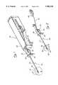
- FIG. 1is a perspective view of a first embodiment of the invention, illustrating an arrangement for delivering and deploying a tissue marker through a flexible introducer, utilizing a motor-driven biopsy probe of known construction as an access conduit;
- FIG. 2is a perspective view similar to FIG. 1, wherein the driver portion of the motor-driven biopsy probe has been deleted in order to better isolate the flexible introducer and tissue marker applier;
- FIG. 3is a side elevational view of the flexible introducer and tissue marker applier illustrated in FIGS. 1 and 2;
- FIG. 4is a cross-sectional view of the distal end portion 4--4 of the flexible introducer illustrated in FIG. 3;
- FIG. 5is a perspective view of a second embodiment of the present invention, illustrating an arrangement for delivering and deploying a tissue marker through a rigid introducer;
- FIG. 6is a cross-sectional view of a one-piece marking device constructed in accordance with the principles of the present invention.
- FIG. 7is a cross-sectional view similar to FIG. 6, illustrating the one-piece marking device as the marker thereof is being pulled back against the forming die for partially closing the marker;
- FIG. 8is a cross-sectional view similar to FIG. 7, illustrating the marker as it is separated from the remainder of the marking device and deployed to mark a desired tissue site;
- FIG. 9is a schematic view illustrating a portion of the length of a pull wire which is constructed of twisted rectangular stock, in accordance with one of the principles of the present invention.
- tissue markers 12are preferably comprised of a non-magnetic, radiographic material, and are preferably constructed in the form of a clip, or surgical staple, to facilitate attachment to the tissue they are intended to identify and to provide an easily recognized shape which would not be mistaken for another lesion.
- the maximum width of a tissue marker 12is within a range of approximately 0.030 inches -0.050 inches, and preferably about 0.039 inches (1 mm).
- a biopsy power driver and probe 18is preferably used, such as the MAMMOTOME® power driver and probe manufactured and sold by Biopsys Medical, Inc., of Irvine, Calif. the assignee of the present application.
- the biopsy power driver and probe 18comprises a driver housing 20, a hollow outer piercing needle 22 having a distal piercing end 24, and a tissue cassette housing 26.
- the hollow outer piercing needle 22includes a laterally facing tissue receiving port 28 near its distal end.
- the biopsy power driver and probe 18is operated to obtain a tissue sample by first moving the distal piercing end 24 of the needle 22 into position to pierce the lesion or selected tissue which is to be sampled, using a known imaging device, such as a stereotactic imaging unit. Then, a vacuum may be drawn through a vacuum port 30 (FIG. 1) of the tissue cassette housing 26, and through the hollow needle 22 to create a negative pressure condition at the tissue receiving port 28, thereby drawing tissue into the port, where it is severed by an inner cutting cannula to capture a tissue sample.
- the tissue that is captured within the inner cutting cannulais transported proximally in an intact fashion by retracting the cutting cannula (not shown) rearwardly, preferably to a slot 32 (FIG. 2) in the tissue cassette housing 26.
- a plurality of tissue samples, from different orientations in the vicinity of the tissue receiving port 28,may be obtained without withdrawing the needle 22.
- tissue abnormality site identificationfor the purpose of ongoing diagnostic follow-up.
- the present inventionis particularly advantageous in that it utilizes the lumen of the hollow outer piercing needle 22 as the marker delivery conduit.
- the probe 34(FIG. 2) is still inserted within the patient's body at the biopsy site, it may be utilized as a fixed position, rigid, annular conduit for delivery and deployment of the tissue marker 12.
- the fact that the probe 34 never leaves the biopsy siteensures accurate delivery of the marker to the cavity 16, while also providing a less traumatic and quicker tissue marking process than the standard open surgical methods.
- the introducer 10comprises a flexible tube 36 having an opening 38 adjacent to its distal end and a hub 40 at its proximal end.
- a plug 42is disposed at the distal end of the flexible tube 36, which plug includes an angled, sloping ramp 44 on a proximal end face thereof.
- the flexible tube 36 of the flexible introducer 10is adapted to receive a flexible tube or deployment shaft 46 of a disposable tissue marker applier 48.
- the applier 48comprises a squeeze handle 50 on its proximal end, which has a ring 52 to which is attached a pull wire 54.
- the pull wire 54extends through the lumen of the deployment shaft 46, and is attached at its distal end to the marker 12 (FIG. 4).
- the flexible tube 36 of the introducer 10is inserted into the hollow needle 22 of the probe 34 through the tissue cassette housing 26, until the hub 40 abuts the tissue cassette housing 26, as illustrated in FIGS. 1 and 2.
- the hub 40is rotated by the physician until an indexing mark or notch 56 (FIG. 3) is properly oriented, thereby ensuring that the introducer 10 is circumferentially aligned within the probe 34.
- the tissue marker applier 48may be advanced into the lumen of the introducer 10, as illustrated in FIGS. 1 and 2, so that the distal end thereof exits from the notch 38 and tissue receiving port 28, extending into the cavity 16 (alternatively, the applier 48 may be first inserted into the introducer, and then the introducer may be inserted into the probe 34, if desired).
- An important aspect of the inventionis the use of the ramp 44 to direct the flexible deployment shaft 46 radially outwardly from the notch 38 so that the marker 12 disposed at the distal end of the shaft 46 may be laterally transported to the cavity wall 14 for placement.
- the squeeze handle 50is squeezed by the physician so that the pull wire 54 is retracted by the squeezing motion sufficiently to break the pull wire, thus releasing the marker 12 for implantation into the target tissue 14.
- the flexible deployment shaft 46may be withdrawn from the introducer 10 and discarded, while a new applier 48 is inserted into the introducer to implant a second marker.
- the hub 40may be counter-rotated 90-270 degrees and the entire probe 34 withdrawn from the patient. If it is desired to mark various locations about the cavity wall 14, the probe needle 22 may be rotated between marker implantations to change the orientation of the tissue receiving port 28, using the thumbwheel 58. Additionally, the axial position of the port 28 may be adjusted, if desired.
- FIG. 5a second embodiment of the inventive introducer mechanism is illustrated.
- like elements to those of the first embodimentare designated by like reference numerals, followed by the letter a.
- the introducer 10ais rigid, rather than flexible.
- the flexible introducer 10 of the first embodimentis adapted for use with a biopsy power driver and probe 18, which functions as the access mechanism. Therefore, the flexible characteristic of the tube 36 is necessary in order to facilitate threading of the tube 36 through the lumen of the needle 22, via the tissue cassette housing 26.
- This embodimentworks very well in connection with larger sized probes, such as 11 gauge MAMMOTOME probes manufactured by Biopsys Medical, Inc., the present assignee, for example.
- the flexible tube 36is too large to be threaded through smaller probes, such as the 14 gauge MAMMOTOME probe manufactured by the present assignee. Therefore, the second embodiment has been developed to provide a stand alone access device for introducing the tissue marker applier 48a.
- the rigid introducer 10a illustrated in FIG. 5comprises a rigid tube 36a having a piercing distal end 60, a distal laterally facing opening 38a, and a ramp 62. Since, in this embodiment, the introducer is not delivered through another access device, but rather is itself an access device, it is preferably loaded onto an introducer needle mount 64, so that the shaft 36a is disposed in a shaft channel 66 of the mount 64, and held in position by means of cover portion 68.
- the biopsy probe and driver 18are removed from the imaging system (not shown), typically a stereotactic table available from Fischer Imaging, Inc. or from Lorad, Inc.
- the probe guide holder(not shown) is replaced by the loaded introducer needle mount 64.
- the introduceris then advanced to the desired tissue sampling site, following which the tissue marker applier is inserted through the introducer cannula to an appropriate depth mark to allow the distal tip clip to extend over the ramp 62 and to extend laterally sufficiently far to pierce tissue.
- the handle 50ais then squeezed in the manner discussed supra to deploy the distal tip clip 12. Then, the disposable applier is removed.
- the introducer 10amay be utilized without the ramp 62, in the case where lateral placement of the marker with respect to the introducer 10a is not required.
- a particularly advantageous embodiment of the present inventionis the employment of a one-piece marking element 70 (FIGS. 6-8), comprising a marker 12b and a marker closing ribbon or pull wire 54b which are comprised of a single piece of wire.
- the single piece marking element 70is preferably fabricated of a single piece of sheet material, ideally using a photochemical etching process to eliminate any fabrication and thermal stresses from being introduced into the part.
- the single-piece elementis fabricated such that a weak spot or failure point 72 (FIGS. 6-7) is disposed at a location on the marking element which will break at a predetermined load after the legs 73, 74 of the marker have closed down and gripped the tissue to which the marker 12b is to be attached.
- a forming die 75is provided which is disposed proximally of the marker portion 12b of the single-piece marking element 70.
- the failure point 72is disposed between the forming die 75 and the marker 12b, at the distal end of the pull wire 54b.
- a pulling forceis applied proximally to the pull wire 54b, in the direction shown by arrow 76.
- This pulling forcemay be applied, for example, by a squeeze handle 50 like that shown in FIGS. 1-3 and 5, or by some other means.
- This proximal pulling forcecauses the marker portion 12b to travel proxally to a point where it impacts the distal end of the forming die 75, as illustrated in FIG. 7.
- proximal pulling forces on the pull wire 54bresults in closure forces being applied against the legs 73, 74 of the marker portion 12b.
- closure forcesbeing applied against the legs 73, 74 of the marker portion 12b.
- continued application of a proximal pulling force on the pull wire 54bwill result in breakage of the pull wire 54b at the failure point 72, so that the marker 12b becomes separated therefrom, with the legs 73, 74 of the marker being closed upon the tissue desired to be marked.
- the inventive marking elementmay be round in cross-section
- the marking element 70is fabricated of rectangular stock, which has been clamped at each end and twisted along its length (FIG. 9).
- the inventorshave found that, absent the twisting step, the sharp edges of the rectangular stock tend to snag against the sides of the tube 46 (FIG. 3) as it is being pulled therethrough. Twisting, on the other hand, has been found to soften the edges of the stock sufficiently to ease passage of the pull wire 54b through the tube.
Landscapes
- Health & Medical Sciences (AREA)
- Life Sciences & Earth Sciences (AREA)
- Surgery (AREA)
- Biomedical Technology (AREA)
- Engineering & Computer Science (AREA)
- General Health & Medical Sciences (AREA)
- Veterinary Medicine (AREA)
- Public Health (AREA)
- Nuclear Medicine, Radiotherapy & Molecular Imaging (AREA)
- Animal Behavior & Ethology (AREA)
- Molecular Biology (AREA)
- Medical Informatics (AREA)
- Heart & Thoracic Surgery (AREA)
- Physics & Mathematics (AREA)
- Biodiversity & Conservation Biology (AREA)
- Oncology (AREA)
- Optics & Photonics (AREA)
- Epidemiology (AREA)
- Oral & Maxillofacial Surgery (AREA)
- Pathology (AREA)
- Surgical Instruments (AREA)
Abstract
Description
Claims (45)
Priority Applications (1)
| Application Number | Priority Date | Filing Date | Title |
|---|---|---|---|
| US08/802,958US5902310A (en) | 1996-08-12 | 1997-02-21 | Apparatus and method for marking tissue |
Applications Claiming Priority (2)
| Application Number | Priority Date | Filing Date | Title |
|---|---|---|---|
| US2388396P | 1996-08-12 | 1996-08-12 | |
| US08/802,958US5902310A (en) | 1996-08-12 | 1997-02-21 | Apparatus and method for marking tissue |
Publications (1)
| Publication Number | Publication Date |
|---|---|
| US5902310Atrue US5902310A (en) | 1999-05-11 |
Family
ID=21817730
Family Applications (1)
| Application Number | Title | Priority Date | Filing Date |
|---|---|---|---|
| US08/802,958Expired - LifetimeUS5902310A (en) | 1996-08-12 | 1997-02-21 | Apparatus and method for marking tissue |
Country Status (7)
| Country | Link |
|---|---|
| US (1) | US5902310A (en) |
| EP (2) | EP0873091B1 (en) |
| JP (1) | JP2000513610A (en) |
| CA (1) | CA2262550C (en) |
| DE (3) | DE69739397D1 (en) |
| ES (3) | ES2323966T3 (en) |
| WO (1) | WO1998006346A1 (en) |
Cited By (233)
| Publication number | Priority date | Publication date | Assignee | Title |
|---|---|---|---|---|
| US6056700A (en)* | 1998-10-13 | 2000-05-02 | Emx, Inc. | Biopsy marker assembly and method of use |
| US6071301A (en)* | 1998-05-01 | 2000-06-06 | Sub Q., Inc. | Device and method for facilitating hemostasis of a biopsy tract |
| US6162192A (en)* | 1998-05-01 | 2000-12-19 | Sub Q, Inc. | System and method for facilitating hemostasis of blood vessel punctures with absorbable sponge |
| US6173715B1 (en)* | 1999-03-01 | 2001-01-16 | Lucent Medical Systems, Inc. | Magnetic anatomical marker and method of use |
| US6183497B1 (en) | 1998-05-01 | 2001-02-06 | Sub-Q, Inc. | Absorbable sponge with contrasting agent |
| US6200328B1 (en) | 1998-05-01 | 2001-03-13 | Sub Q, Incorporated | Device and method for facilitating hemostasis of a biopsy tract |
| US6220248B1 (en)* | 1998-10-21 | 2001-04-24 | Ethicon Endo-Surgery, Inc. | Method for implanting a biopsy marker |
| US20010034528A1 (en)* | 1994-09-16 | 2001-10-25 | Foerster Seth A. | Methods and devices for defining and marking tissue |
| US6315753B1 (en) | 1998-05-01 | 2001-11-13 | Sub-Q, Inc. | System and method for facilitating hemostasis of blood vessel punctures with absorbable sponge |
| US6336904B1 (en)* | 1998-04-07 | 2002-01-08 | Pro Duct Health, Inc. | Methods and devices for the localization of lesions in solid tissue |
| US6350244B1 (en)* | 2000-02-21 | 2002-02-26 | Biopsy Sciences, Llc | Bioabsorable markers for use in biopsy procedures |
| US6355275B1 (en) | 2000-06-23 | 2002-03-12 | Carbon Medical Technologies, Inc. | Embolization using carbon coated microparticles |
| US6363940B1 (en) | 1998-05-14 | 2002-04-02 | Calypso Medical Technologies, Inc. | System and method for bracketing and removing tissue |
| US20020058882A1 (en)* | 1998-06-22 | 2002-05-16 | Artemis Medical, Incorporated | Biopsy localization method and device |
| WO2002039918A1 (en)* | 1998-05-14 | 2002-05-23 | Calypso Medical, Inc. | Systems and methods for stabilizing a targert location within a human body |
| US6394965B1 (en) | 2000-08-15 | 2002-05-28 | Carbon Medical Technologies, Inc. | Tissue marking using biocompatible microparticles |
| US6405733B1 (en) | 2000-02-18 | 2002-06-18 | Thomas J. Fogarty | Device for accurately marking tissue |
| US6427081B1 (en)* | 1999-02-02 | 2002-07-30 | Senorx, Inc. | Methods and chemical preparations for time-limited marking of biopsy sites |
| WO2002060327A1 (en)* | 2001-01-31 | 2002-08-08 | Eberhard-Karls-Universität Tübingen | Device for the introduction and fashioning of skin clips |
| US6432064B1 (en)* | 2001-04-09 | 2002-08-13 | Ethicon Endo-Surgery, Inc. | Biopsy instrument with tissue marking element |
| US6450938B1 (en) | 2000-03-21 | 2002-09-17 | Promex, Llc | Brachytherapy device |
| US20020133092A1 (en)* | 2001-03-16 | 2002-09-19 | Microvena Corporation | Wire convertible from over-the-wire length to rapid exchange length |
| US20020156495A1 (en)* | 1995-09-15 | 2002-10-24 | Rodney Brenneman | Apparatus and method for percutaneous sealing of blood vessel punctures |
| US20020190226A1 (en)* | 2001-03-12 | 2002-12-19 | Mark Ashby | Methods for sterilizing cross-linked gelatin compositions |
| US20030028140A1 (en)* | 2001-03-12 | 2003-02-06 | Greff Richard J. | Cross-linked gelatin composition comprising a wetting agent |
| US20030052785A1 (en)* | 2001-09-14 | 2003-03-20 | Margo Gisselberg | Miniature resonating marker assembly |
| US6540735B1 (en) | 2000-05-12 | 2003-04-01 | Sub-Q, Inc. | System and method for facilitating hemostasis of blood vessel punctures with absorbable sponge |
| US6544236B1 (en) | 1999-02-10 | 2003-04-08 | Sub-Q, Incorporated | Device, system and method for improving delivery of hemostatic material |
| US6544185B2 (en) | 2000-10-23 | 2003-04-08 | Valentino Montegrande | Ultrasound imaging marker and method of use |
| US20030088269A1 (en)* | 2001-11-08 | 2003-05-08 | Sub-Q, Inc. | System and method for delivering hemostasis promoting material to a blood vessel puncture site by fluid pressure |
| US20030101604A1 (en)* | 2001-12-04 | 2003-06-05 | Mcgivern Joseph F. | Programmable sighting system for a hunting bow |
| US20030120141A1 (en)* | 2001-12-20 | 2003-06-26 | Adler John R. | Anchored fiducial apparatus and method |
| US6605047B2 (en) | 2001-09-10 | 2003-08-12 | Vivant Medical, Inc. | Biopsy marker delivery system |
| US6610026B2 (en) | 1998-05-01 | 2003-08-26 | Sub-Q, Inc. | Method of hydrating a sponge material for delivery to a body |
| US20030195500A1 (en)* | 1999-06-17 | 2003-10-16 | Moorman Jack W. | Needle kit and method for microwave ablation, track coagulation, and biopsy |
| US20030192557A1 (en)* | 1998-05-14 | 2003-10-16 | David Krag | Systems and methods for locating and defining a target location within a human body |
| US20030195499A1 (en)* | 2002-04-16 | 2003-10-16 | Mani Prakash | Microwave antenna having a curved configuration |
| US6638234B2 (en) | 1998-03-03 | 2003-10-28 | Senorx, Inc. | Sentinel node location and biopsy |
| US20030204137A1 (en)* | 1999-06-17 | 2003-10-30 | Inrad, Inc. | Apparatus for the percutaneous marking of a lesion |
| US6654629B2 (en) | 2002-01-23 | 2003-11-25 | Valentino Montegrande | Implantable biomarker and method of use |
| US20030225420A1 (en)* | 2002-03-11 | 2003-12-04 | Wardle John L. | Surgical coils and methods of deploying |
| US6662041B2 (en) | 1999-02-02 | 2003-12-09 | Senorx, Inc. | Imageable biopsy site marker |
| WO2004004578A1 (en)* | 2002-07-03 | 2004-01-15 | Christy Cummins | Surgical stapling device |
| US6679851B2 (en) | 1998-09-01 | 2004-01-20 | Senorx, Inc. | Tissue accessing and anchoring device and method |
| US20040019330A1 (en)* | 2001-11-08 | 2004-01-29 | Sub-Q, Inc., A California Corporation | Sheath based blood vessel puncture locator and depth indicator |
| US20040044311A1 (en)* | 1998-10-23 | 2004-03-04 | Felix Espositio | Site marker device |
| US20040052333A1 (en)* | 2002-09-13 | 2004-03-18 | James Sayre | Device and method for margin marking of radiography specimens |
| US6722371B1 (en) | 2000-02-18 | 2004-04-20 | Thomas J. Fogarty | Device for accurately marking tissue |
| US20040102730A1 (en)* | 2002-10-22 | 2004-05-27 | Davis Thomas P. | System and method for facilitating hemostasis of blood vessel punctures with absorbable sponge |
| US6752767B2 (en) | 2002-04-16 | 2004-06-22 | Vivant Medical, Inc. | Localization element with energized tip |
| US6752154B2 (en) | 2000-02-18 | 2004-06-22 | Thomas J. Fogarty | Device for accurately marking tissue |
| US20040122312A1 (en)* | 2002-11-18 | 2004-06-24 | Inrad, Inc. | Apparatus and method for implanting a preloaded localization wire |
| US20040124105A1 (en)* | 2002-12-30 | 2004-07-01 | Keith Seiler | Packaged systems for implanting markers in a patient and methods for manufacturing and using such systems |
| US20040127787A1 (en)* | 2002-12-30 | 2004-07-01 | Dimmer Steven C. | Implantable marker with a leadless signal transmitter compatible for use in magnetic resonance devices |
| US20040133101A1 (en)* | 2001-06-08 | 2004-07-08 | Mate Timothy P. | Guided radiation therapy system |
| US6780179B2 (en) | 2002-05-22 | 2004-08-24 | Rubicor Medical, Inc. | Methods and systems for in situ tissue marking and orientation stabilization |
| US20040176723A1 (en)* | 2001-11-08 | 2004-09-09 | Sing Eduardo Chi | Pledget-handling system and method for delivering hemostasis promoting material to a blood vessel puncture site by fluid pressure |
| WO2004084738A1 (en)* | 2003-03-19 | 2004-10-07 | Suros Surgical Systems, Inc. | Adapter assembly for stereotactic biopsy |
| US20040204660A1 (en)* | 1998-06-22 | 2004-10-14 | Artemis Medical, Inc. | Biopsy localization method and device |
| US6812842B2 (en) | 2001-12-20 | 2004-11-02 | Calypso Medical Technologies, Inc. | System for excitation of a leadless miniature marker |
| US6822570B2 (en) | 2001-12-20 | 2004-11-23 | Calypso Medical Technologies, Inc. | System for spatially adjustable excitation of leadless miniature marker |
| US20040236211A1 (en)* | 2003-05-23 | 2004-11-25 | Senorx, Inc. | Marker or filler forming fluid |
| US20040236212A1 (en)* | 2003-05-23 | 2004-11-25 | Senorx, Inc. | Fibrous marker and intracorporeal delivery thereof |
| US6838990B2 (en) | 2001-12-20 | 2005-01-04 | Calypso Medical Technologies, Inc. | System for excitation leadless miniature marker |
| US20050015081A1 (en)* | 2003-07-18 | 2005-01-20 | Roman Turovskiy | Devices and methods for cooling microwave antennas |
| US6846320B2 (en) | 1998-05-01 | 2005-01-25 | Sub-Q, Inc. | Device and method for facilitating hemostasis of a biopsy tract |
| US20050033360A1 (en)* | 2001-11-08 | 2005-02-10 | Sing Eduardo Chi | Pledget-handling system and method for delivering hemostasis promoting material to a blood vessel puncture site by fluid pressure |
| JP2005505337A (en)* | 2001-10-05 | 2005-02-24 | ボストン サイエンティフィック リミテッド | Scope Endoscopic hemostatic clip device |
| US20050063908A1 (en)* | 1999-02-02 | 2005-03-24 | Senorx, Inc. | Tissue site markers for in vivo imaging |
| USD503980S1 (en) | 2002-09-13 | 2005-04-12 | Beekley Corporation | Marker for radiography specimens |
| US20050080454A1 (en)* | 2003-10-08 | 2005-04-14 | Drews Michael J. | Attachment device and methods of using the same |
| US20050119562A1 (en)* | 2003-05-23 | 2005-06-02 | Senorx, Inc. | Fibrous marker formed of synthetic polymer strands |
| US20050119652A1 (en)* | 1998-09-03 | 2005-06-02 | Rubicor Medical, Inc. | Devices and methods for performing procedures on a breast |
| US20050119695A1 (en)* | 2000-12-07 | 2005-06-02 | Carley Michael T. | Closure device and methods for making and using them |
| WO2005053560A1 (en) | 2003-11-26 | 2005-06-16 | Invivo Germany Gmbh | Tissue marker and method and apparatus for deploying the marker |
| US20050143656A1 (en)* | 1999-02-02 | 2005-06-30 | Senorx, Inc. | Cavity-filling biopsy site markers |
| US20050154293A1 (en)* | 2003-12-24 | 2005-07-14 | Margo Gisselberg | Implantable marker with wireless signal transmitter |
| EP1582168A1 (en)* | 2004-03-31 | 2005-10-05 | Ethicon Endo-Surgery, Inc. | Marker device and method of deploying a cavity marker using a surgical biopsy device |
| US20050234336A1 (en)* | 2004-03-26 | 2005-10-20 | Beckman Andrew T | Apparatus and method for marking tissue |
| US20050255045A1 (en)* | 2004-05-13 | 2005-11-17 | Woltering Eugene A | Surgical marking composition and method |
| US20050288707A1 (en)* | 2004-06-28 | 2005-12-29 | Cardio Life Research S.A | Fluidtight puncturing and occlusion device for anatomical structure |
| US6984219B2 (en) | 1999-09-23 | 2006-01-10 | Mark Ashby | Depth and puncture control for blood vessel hemostasis system |
| US7008440B2 (en) | 2001-11-08 | 2006-03-07 | Sub-Q, Inc. | System and method for delivering hemostasis promoting material to a blood vessel puncture site by fluid pressure |
| US20060058648A1 (en)* | 2004-07-23 | 2006-03-16 | Eric Meier | Integrated radiation therapy systems and methods for treating a target in a patient |
| US20060079805A1 (en)* | 2004-10-13 | 2006-04-13 | Miller Michael E | Site marker visable under multiple modalities |
| US7029489B1 (en) | 2001-05-18 | 2006-04-18 | Sub-Q, Inc. | System and method for delivering hemostasis promoting material to a blood vessel puncture site |
| US7037322B1 (en) | 2001-11-08 | 2006-05-02 | Sub-Q, Inc. | System and method for delivering hemostasis promoting material to a blood vessel puncture with a staging tube |
| US20060111646A1 (en)* | 2003-08-13 | 2006-05-25 | Gellman Barry N | Marking biopsy sites |
| US20060116573A1 (en)* | 2003-11-17 | 2006-06-01 | Inrad, Inc. | Self Contained, Self Piercing, Side-Expelling Marking Apparatus |
| US20060151460A1 (en)* | 2005-01-10 | 2006-07-13 | Wardle John L | Eluting coils and methods of deploying and retrieving |
| US20060190014A1 (en)* | 2000-01-05 | 2006-08-24 | Ginn Richard S | Integrated vascular device with puncture site closure component and sealant and methods of use |
| US20060217635A1 (en)* | 2005-03-24 | 2006-09-28 | Mccombs Elizabeth S | Biopsy device marker deployment |
| US20070021763A1 (en)* | 2004-11-22 | 2007-01-25 | Inrad, Inc. | Removable Localizing Wire |
| US20070038145A1 (en)* | 2004-11-22 | 2007-02-15 | Inrad, Inc. | Post Decompression Marker Introducer System |
| US7201725B1 (en) | 2000-09-25 | 2007-04-10 | Sub-Q, Inc. | Device and method for determining a depth of an incision |
| US20070093726A1 (en)* | 2004-10-13 | 2007-04-26 | Leopold Phillip M | Site marker visible under multiple modalities |
| WO2007060576A3 (en)* | 2005-11-22 | 2007-10-18 | Suros Surgical Systems Inc | Surgical site marker delivery system |
| US20070270681A1 (en)* | 2006-05-16 | 2007-11-22 | Vector Surgical, Inc. | Tissue marking system and method |
| US20080009886A1 (en)* | 2006-07-05 | 2008-01-10 | Wilson-Cook Medical Inc. | Suction cup |
| USD559985S1 (en) | 2005-04-08 | 2008-01-15 | Beekley Corporation | Mammogram marker |
| US7318824B2 (en) | 2001-11-02 | 2008-01-15 | Vivant Medical, Inc. | High-strength microwave antenna assemblies |
| US20080017691A1 (en)* | 2005-03-28 | 2008-01-24 | Cardica, Inc. | System for Closing a Tissue Structure from Inside |
| US20080039819A1 (en)* | 2006-08-04 | 2008-02-14 | Senorx, Inc. | Marker formed of starch or other suitable polysaccharide |
| US20080045922A1 (en)* | 1997-10-17 | 2008-02-21 | Micro Therapeutics, Inc. | Device and method for controlling injection of liquid embolic composition |
| US7335219B1 (en) | 2002-11-04 | 2008-02-26 | Sub-Q, Inc. | Hemostatic device including a capsule |
| US20080082093A1 (en)* | 2006-09-29 | 2008-04-03 | Prakash Mani N | Microwave antenna assembly and method of using the same |
| US20080254298A1 (en)* | 2006-02-23 | 2008-10-16 | Meadwestvaco Corporation | Method for treating a substrate |
| US20080269603A1 (en)* | 2004-10-13 | 2008-10-30 | Nicoson Zachary R | Site marker visible under multiple modalities |
| US20080287965A1 (en)* | 2007-05-17 | 2008-11-20 | Richard Ducharme | Radiopaque band ligator |
| US20090030309A1 (en)* | 2007-07-26 | 2009-01-29 | Senorx, Inc. | Deployment of polysaccharide markers |
| US20090039138A1 (en)* | 2005-03-28 | 2009-02-12 | Cardica, Inc. | Vascular Closure System With Splayable Staple |
| US20090131815A1 (en)* | 2007-11-20 | 2009-05-21 | Brian Zimmer | Marker deployment device |
| US20090209854A1 (en)* | 2008-02-19 | 2009-08-20 | Parihar Shailendra K | Biopsy method |
| US20090216115A1 (en)* | 2004-07-23 | 2009-08-27 | Calypso Medical Technologies, Inc. | Anchoring wirless markers within a human body |
| US7621936B2 (en) | 2000-07-14 | 2009-11-24 | Boston Scientific Scimed, Inc. | Sheath-mounted arterial plug delivery device |
| US7625352B1 (en) | 1998-05-01 | 2009-12-01 | Sub-Q, Inc. | Depth and puncture control for system for hemostasis of blood vessel |
| US20100030149A1 (en)* | 2006-10-23 | 2010-02-04 | C.R. Bard, Inc. | Breast marker |
| US20100049165A1 (en)* | 2008-08-19 | 2010-02-25 | Micro Therapeutics, Inc | Detachable tip microcatheter |
| USD611144S1 (en) | 2006-06-28 | 2010-03-02 | Abbott Laboratories | Apparatus for delivering a closure element |
| US7695492B1 (en) | 1999-09-23 | 2010-04-13 | Boston Scientific Scimed, Inc. | Enhanced bleed back system |
| US20100114287A1 (en)* | 2008-10-30 | 2010-05-06 | Salvatore Privitera | Implantable tissue marker electrode |
| US20100155452A1 (en)* | 2003-09-01 | 2010-06-24 | Laszlo Csiky | Circular stapler for hemorrhoid operations |
| US7806910B2 (en) | 2002-11-26 | 2010-10-05 | Abbott Laboratories | Multi-element biased suture clip |
| US7806904B2 (en) | 2000-12-07 | 2010-10-05 | Integrated Vascular Systems, Inc. | Closure device |
| US7819895B2 (en) | 2000-01-05 | 2010-10-26 | Integrated Vascular Systems, Inc. | Vascular sheath with bioabsorbable puncture site closure apparatus and methods of use |
| US7828817B2 (en) | 2000-01-05 | 2010-11-09 | Integrated Vascular Systems, Inc. | Apparatus and methods for delivering a closure device |
| US7842068B2 (en) | 2000-12-07 | 2010-11-30 | Integrated Vascular Systems, Inc. | Apparatus and methods for providing tactile feedback while delivering a closure device |
| US7841502B2 (en) | 2007-12-18 | 2010-11-30 | Abbott Laboratories | Modular clip applier |
| US7850709B2 (en) | 2002-06-04 | 2010-12-14 | Abbott Vascular Inc. | Blood vessel closure clip and delivery device |
| US7850797B2 (en) | 2002-12-31 | 2010-12-14 | Integrated Vascular Systems, Inc. | Methods for manufacturing a clip and clip |
| US7867249B2 (en) | 2003-01-30 | 2011-01-11 | Integrated Vascular Systems, Inc. | Clip applier and methods of use |
| US7875043B1 (en) | 2003-12-09 | 2011-01-25 | Sub-Q, Inc. | Cinching loop |
| US7879071B2 (en) | 2000-12-07 | 2011-02-01 | Integrated Vascular Systems, Inc. | Closure device and methods for making and using them |
| US7887563B2 (en) | 2001-06-07 | 2011-02-15 | Abbott Vascular Inc. | Surgical staple |
| US20110071391A1 (en)* | 2009-09-24 | 2011-03-24 | Speeg Trevor W V | Biopsy marker delivery device with positioning component |
| US7931669B2 (en) | 2000-01-05 | 2011-04-26 | Integrated Vascular Systems, Inc. | Integrated vascular device with puncture site closure component and sealant and methods of use |
| US7955353B1 (en) | 2002-11-04 | 2011-06-07 | Sub-Q, Inc. | Dissolvable closure device |
| US20110190662A1 (en)* | 2008-10-01 | 2011-08-04 | Beacon Endoscopic Corporation | Rapid exchange fna biopsy device with diagnostic and therapeutic capabilities |
| US8007512B2 (en) | 2002-02-21 | 2011-08-30 | Integrated Vascular Systems, Inc. | Plunger apparatus and methods for delivering a closure device |
| US8048108B2 (en) | 2005-08-24 | 2011-11-01 | Abbott Vascular Inc. | Vascular closure methods and apparatuses |
| US8157862B2 (en) | 1997-10-10 | 2012-04-17 | Senorx, Inc. | Tissue marking implant |
| US8177792B2 (en) | 2002-06-17 | 2012-05-15 | Senorx, Inc. | Plugged tip delivery tube for marker placement |
| US8202294B2 (en) | 2003-01-30 | 2012-06-19 | Integrated Vascular Systems, Inc. | Clip applier and methods of use |
| US8202293B2 (en) | 2003-01-30 | 2012-06-19 | Integrated Vascular Systems, Inc. | Clip applier and methods of use |
| US8226681B2 (en) | 2007-06-25 | 2012-07-24 | Abbott Laboratories | Methods, devices, and apparatus for managing access through tissue |
| US20120228355A1 (en)* | 2011-01-07 | 2012-09-13 | Z-Medical Gmbh & Co.Kg | Surgical instrument |
| US8280486B2 (en) | 2004-10-13 | 2012-10-02 | Suros Surgical Systems, Inc. | Site marker visable under multiple modalities |
| US8292880B2 (en) | 2007-11-27 | 2012-10-23 | Vivant Medical, Inc. | Targeted cooling of deployable microwave antenna |
| US8303624B2 (en) | 2010-03-15 | 2012-11-06 | Abbott Cardiovascular Systems, Inc. | Bioabsorbable plug |
| US8311610B2 (en) | 2008-01-31 | 2012-11-13 | C. R. Bard, Inc. | Biopsy tissue marker |
| US8313497B2 (en) | 2005-07-01 | 2012-11-20 | Abbott Laboratories | Clip applier and methods of use |
| US8317821B1 (en) | 2002-11-04 | 2012-11-27 | Boston Scientific Scimed, Inc. | Release mechanism |
| US8323312B2 (en) | 2008-12-22 | 2012-12-04 | Abbott Laboratories | Closure device |
| WO2012171029A1 (en)* | 2011-06-09 | 2012-12-13 | The Regents Of The University Of California | Excised specimen imaging using a combined pet and micro ct scanner |
| WO2013008204A2 (en) | 2011-07-12 | 2013-01-17 | Maestroheart Sa | System for tissue marking and treatment |
| US8361082B2 (en) | 1999-02-02 | 2013-01-29 | Senorx, Inc. | Marker delivery device with releasable plug |
| US8398676B2 (en) | 2008-10-30 | 2013-03-19 | Abbott Vascular Inc. | Closure device |
| US8398656B2 (en) | 2003-01-30 | 2013-03-19 | Integrated Vascular Systems, Inc. | Clip applier and methods of use |
| US8401622B2 (en) | 2006-12-18 | 2013-03-19 | C. R. Bard, Inc. | Biopsy marker with in situ-generated imaging properties |
| US8486028B2 (en) | 2005-10-07 | 2013-07-16 | Bard Peripheral Vascular, Inc. | Tissue marking apparatus having drug-eluting tissue marker |
| US8498693B2 (en) | 1999-02-02 | 2013-07-30 | Senorx, Inc. | Intracorporeal marker and marker delivery device |
| US8556930B2 (en) | 2006-06-28 | 2013-10-15 | Abbott Laboratories | Vessel closure device |
| US8556932B2 (en) | 2011-05-19 | 2013-10-15 | Abbott Cardiovascular Systems, Inc. | Collapsible plug for tissue closure |
| US8594768B2 (en) | 2004-11-01 | 2013-11-26 | Michael J. Phillips | Surgical system with clips for identifying the orientation of a tissue sample |
| US8590760B2 (en) | 2004-05-25 | 2013-11-26 | Abbott Vascular Inc. | Surgical stapler |
| US8603116B2 (en) | 2010-08-04 | 2013-12-10 | Abbott Cardiovascular Systems, Inc. | Closure device with long tines |
| US8617184B2 (en) | 2011-02-15 | 2013-12-31 | Abbott Cardiovascular Systems, Inc. | Vessel closure system |
| US8634899B2 (en) | 2003-11-17 | 2014-01-21 | Bard Peripheral Vascular, Inc. | Multi mode imaging marker |
| US8670818B2 (en) | 2008-12-30 | 2014-03-11 | C. R. Bard, Inc. | Marker delivery device for tissue marker placement |
| US8668737B2 (en) | 1997-10-10 | 2014-03-11 | Senorx, Inc. | Tissue marking implant |
| US8672953B2 (en) | 2007-12-17 | 2014-03-18 | Abbott Laboratories | Tissue closure system and methods of use |
| US8690910B2 (en) | 2000-12-07 | 2014-04-08 | Integrated Vascular Systems, Inc. | Closure device and methods for making and using them |
| US8718745B2 (en) | 2000-11-20 | 2014-05-06 | Senorx, Inc. | Tissue site markers for in vivo imaging |
| US8758398B2 (en) | 2006-09-08 | 2014-06-24 | Integrated Vascular Systems, Inc. | Apparatus and method for delivering a closure element |
| US8758399B2 (en) | 2010-08-02 | 2014-06-24 | Abbott Cardiovascular Systems, Inc. | Expandable bioabsorbable plug apparatus and method |
| US8758400B2 (en) | 2000-01-05 | 2014-06-24 | Integrated Vascular Systems, Inc. | Closure system and methods of use |
| US8784447B2 (en) | 2000-09-08 | 2014-07-22 | Abbott Vascular Inc. | Surgical stapler |
| US8808310B2 (en) | 2006-04-20 | 2014-08-19 | Integrated Vascular Systems, Inc. | Resettable clip applier and reset tools |
| US8821534B2 (en) | 2010-12-06 | 2014-09-02 | Integrated Vascular Systems, Inc. | Clip applier having improved hemostasis and methods of use |
| USD715442S1 (en) | 2013-09-24 | 2014-10-14 | C. R. Bard, Inc. | Tissue marker for intracorporeal site identification |
| US8858594B2 (en) | 2008-12-22 | 2014-10-14 | Abbott Laboratories | Curved closure device |
| USD715942S1 (en) | 2013-09-24 | 2014-10-21 | C. R. Bard, Inc. | Tissue marker for intracorporeal site identification |
| USD716450S1 (en) | 2013-09-24 | 2014-10-28 | C. R. Bard, Inc. | Tissue marker for intracorporeal site identification |
| USD716451S1 (en) | 2013-09-24 | 2014-10-28 | C. R. Bard, Inc. | Tissue marker for intracorporeal site identification |
| US8893947B2 (en) | 2007-12-17 | 2014-11-25 | Abbott Laboratories | Clip applier and methods of use |
| US8905937B2 (en) | 2009-02-26 | 2014-12-09 | Integrated Vascular Systems, Inc. | Methods and apparatus for locating a surface of a body lumen |
| US8920442B2 (en) | 2005-08-24 | 2014-12-30 | Abbott Vascular Inc. | Vascular opening edge eversion methods and apparatuses |
| US8926633B2 (en) | 2005-06-24 | 2015-01-06 | Abbott Laboratories | Apparatus and method for delivering a closure element |
| US20150010470A1 (en)* | 2000-11-16 | 2015-01-08 | Microspherix, LLC | Flexible and/or Elastic Brachytherapy Seed or Strand |
| US8968210B2 (en) | 2008-10-01 | 2015-03-03 | Covidien LLP | Device for needle biopsy with integrated needle protection |
| US9089311B2 (en) | 2009-01-09 | 2015-07-28 | Abbott Vascular Inc. | Vessel closure devices and methods |
| US9089674B2 (en) | 2000-10-06 | 2015-07-28 | Integrated Vascular Systems, Inc. | Apparatus and methods for positioning a vascular sheath |
| US20150265273A1 (en)* | 1996-08-22 | 2015-09-24 | The Trustees Of Columbia University In The City Of New York | Endovascular Flexible Stapling Device |
| US9149276B2 (en) | 2011-03-21 | 2015-10-06 | Abbott Cardiovascular Systems, Inc. | Clip and deployment apparatus for tissue closure |
| US9173644B2 (en) | 2009-01-09 | 2015-11-03 | Abbott Vascular Inc. | Closure devices, systems, and methods |
| US9237860B2 (en) | 2008-06-05 | 2016-01-19 | Varian Medical Systems, Inc. | Motion compensation for medical imaging and associated systems and methods |
| US9282965B2 (en) | 2008-05-16 | 2016-03-15 | Abbott Laboratories | Apparatus and methods for engaging tissue |
| US9314230B2 (en) | 2009-01-09 | 2016-04-19 | Abbott Vascular Inc. | Closure device with rapidly eroding anchor |
| US9327061B2 (en) | 2008-09-23 | 2016-05-03 | Senorx, Inc. | Porous bioabsorbable implant |
| US9332973B2 (en) | 2008-10-01 | 2016-05-10 | Covidien Lp | Needle biopsy device with exchangeable needle and integrated needle protection |
| US9332976B2 (en) | 2011-11-30 | 2016-05-10 | Abbott Cardiovascular Systems, Inc. | Tissue closure device |
| US9364209B2 (en) | 2012-12-21 | 2016-06-14 | Abbott Cardiovascular Systems, Inc. | Articulating suturing device |
| US9375189B1 (en)* | 2015-08-18 | 2016-06-28 | Abdulmuhsen A. Alsahhaf | X-ray stick marker |
| US9375219B2 (en) | 2009-12-22 | 2016-06-28 | Cook Medical Technologies Llc | Medical devices with detachable pivotable jaws |
| US9414820B2 (en) | 2009-01-09 | 2016-08-16 | Abbott Vascular Inc. | Closure devices, systems, and methods |
| US9414824B2 (en) | 2009-01-16 | 2016-08-16 | Abbott Vascular Inc. | Closure devices, systems, and methods |
| US9456811B2 (en) | 2005-08-24 | 2016-10-04 | Abbott Vascular Inc. | Vascular closure methods and apparatuses |
| US9486191B2 (en) | 2009-01-09 | 2016-11-08 | Abbott Vascular, Inc. | Closure devices |
| US9579077B2 (en) | 2006-12-12 | 2017-02-28 | C.R. Bard, Inc. | Multiple imaging mode tissue marker |
| US9579091B2 (en) | 2000-01-05 | 2017-02-28 | Integrated Vascular Systems, Inc. | Closure system and methods of use |
| US9585647B2 (en) | 2009-08-26 | 2017-03-07 | Abbott Laboratories | Medical device for repairing a fistula |
| US9782565B2 (en) | 2008-10-01 | 2017-10-10 | Covidien Lp | Endoscopic ultrasound-guided biliary access system |
| US9820824B2 (en) | 1999-02-02 | 2017-11-21 | Senorx, Inc. | Deployment of polysaccharide markers for treating a site within a patent |
| US9936892B1 (en) | 2009-05-04 | 2018-04-10 | Cortex Manufacturing Inc. | Systems and methods for providing a fiducial marker |
| US9943704B1 (en) | 2009-01-21 | 2018-04-17 | Varian Medical Systems, Inc. | Method and system for fiducials contained in removable device for radiation therapy |
| US9955977B2 (en) | 2009-12-22 | 2018-05-01 | Cook Medical Technologies Llc | Medical devices with detachable pivotable jaws |
| US20180117294A1 (en)* | 2013-06-12 | 2018-05-03 | Boston Scientific Scimed, Inc. | Fiducial deployment mechanisms, and related methods of use |
| US10010336B2 (en) | 2009-12-22 | 2018-07-03 | Cook Medical Technologies, Inc. | Medical devices with detachable pivotable jaws |
| US10076316B2 (en) | 2008-10-01 | 2018-09-18 | Covidien Lp | Needle biopsy device |
| US10124087B2 (en) | 2012-06-19 | 2018-11-13 | Covidien Lp | Detachable coupling for catheter |
| US10182868B2 (en) | 2005-11-17 | 2019-01-22 | Varian Medical Systems, Inc. | Apparatus and methods for using an electromagnetic transponder in orthopedic procedures |
| US10342635B2 (en) | 2005-04-20 | 2019-07-09 | Bard Peripheral Vascular, Inc. | Marking device with retractable cannula |
| US10548612B2 (en) | 2009-12-22 | 2020-02-04 | Cook Medical Technologies Llc | Medical devices with detachable pivotable jaws |
| US10683119B2 (en) | 2014-05-23 | 2020-06-16 | Merit Medical Systems, Inc. | Marker element, device for making a marker element, and method for making a marker element |
| US10888316B1 (en)* | 2009-06-02 | 2021-01-12 | Aesculap Ag | Work hardening of staples within surgical stapler |
| US10925510B2 (en)* | 2015-05-08 | 2021-02-23 | Cedars-Sinai Medical Center | Characterization of respiratory motion in the abdomen using a 4D MRI technique with 3D radial sampling and respiratory self-gating |
| US20220071608A1 (en)* | 2019-05-30 | 2022-03-10 | Devicor Medical Products, Inc. | Shape memory marker deployment device |
| US11298113B2 (en) | 2008-10-01 | 2022-04-12 | Covidien Lp | Device for needle biopsy with integrated needle protection |
| US20220240931A1 (en)* | 2019-06-28 | 2022-08-04 | Medartis Holding Ag | Surgical tool and surgical set |
| US11464599B1 (en) | 2015-05-15 | 2022-10-11 | Marginview, Llc | Specimen marking mechanism |
| USD990679S1 (en) | 2021-06-21 | 2023-06-27 | Marginview, Llc | Tissue specimen marking clip |
| US11779397B2 (en) | 2019-05-08 | 2023-10-10 | Atricure, Inc. | Biological tissue position location and marking |
| US12376939B2 (en) | 2015-05-15 | 2025-08-05 | Marginview, Llc | Method of marking a specimen |
Families Citing this family (25)
| Publication number | Priority date | Publication date | Assignee | Title |
|---|---|---|---|---|
| US6659105B2 (en) | 1998-02-26 | 2003-12-09 | Senorx, Inc. | Tissue specimen isolating and damaging device and method |
| US6540693B2 (en) | 1998-03-03 | 2003-04-01 | Senorx, Inc. | Methods and apparatus for securing medical instruments to desired locations in a patients body |
| US6344026B1 (en) | 1998-04-08 | 2002-02-05 | Senorx, Inc. | Tissue specimen encapsulation device and method thereof |
| US6758848B2 (en) | 1998-03-03 | 2004-07-06 | Senorx, Inc. | Apparatus and method for accessing a body site |
| US6875182B2 (en) | 1998-03-03 | 2005-04-05 | Senorx, Inc. | Electrosurgical specimen-collection system |
| US6471700B1 (en) | 1998-04-08 | 2002-10-29 | Senorx, Inc. | Apparatus and method for accessing biopsy site |
| US6997885B2 (en) | 1998-04-08 | 2006-02-14 | Senorx, Inc. | Dilation devices and methods for removing tissue specimens |
| US5941890A (en)* | 1998-06-26 | 1999-08-24 | Ethicon Endo-Surgery, Inc. | Implantable surgical marker |
| US6261302B1 (en)* | 1998-06-26 | 2001-07-17 | Ethicon Endo-Surgery, Inc. | Applier for implantable surgical marker |
| JP4559630B2 (en)* | 1998-11-25 | 2010-10-13 | ユナイテッド ステイツ サージカル コーポレイション | Biopsy system |
| US6692447B1 (en) | 1999-02-16 | 2004-02-17 | Frederic Picard | Optimizing alignment of an appendicular |
| US6494844B1 (en) | 2000-06-21 | 2002-12-17 | Sanarus Medical, Inc. | Device for biopsy and treatment of breast tumors |
| AU2002211568B2 (en) | 2000-10-16 | 2005-11-17 | Sanarus Medical, Inc. | Device for biopsy of tumors |
| US6540694B1 (en) | 2000-10-16 | 2003-04-01 | Sanarus Medical, Inc. | Device for biopsy tumors |
| EP1479354A1 (en)* | 2003-05-21 | 2004-11-24 | Prohealth AG | Marker for position determination by MRI |
| US9408592B2 (en) | 2003-12-23 | 2016-08-09 | Senorx, Inc. | Biopsy device with aperture orientation and improved tip |
| US7402140B2 (en) | 2004-02-12 | 2008-07-22 | Sanarus Medical, Inc. | Rotational core biopsy device with liquid cryogen adhesion probe |
| DE102004030391A1 (en)* | 2004-06-23 | 2006-01-26 | Somatex Medical Technologies Gmbh | marker |
| US8343071B2 (en) | 2004-12-16 | 2013-01-01 | Senorx, Inc. | Biopsy device with aperture orientation and improved tip |
| US9095325B2 (en) | 2005-05-23 | 2015-08-04 | Senorx, Inc. | Tissue cutting member for a biopsy device |
| US7572236B2 (en) | 2005-08-05 | 2009-08-11 | Senorx, Inc. | Biopsy device with fluid delivery to tissue specimens |
| US8317725B2 (en) | 2005-08-05 | 2012-11-27 | Senorx, Inc. | Biopsy device with fluid delivery to tissue specimens |
| US8684962B2 (en)* | 2008-03-27 | 2014-04-01 | St. Jude Medical, Atrial Fibrillation Division, Inc. | Robotic catheter device cartridge |
| US8529465B2 (en)* | 2009-09-24 | 2013-09-10 | Devicor Medical Products, Inc. | Biopsy marker delivery devices and methods |
| JP2013215586A (en)* | 2013-05-29 | 2013-10-24 | C R Bard Inc | Marker transmission device for arrangement of tissue marker |
Citations (47)
| Publication number | Priority date | Publication date | Assignee | Title |
|---|---|---|---|---|
| US3120230A (en)* | 1960-10-24 | 1964-02-04 | Jack H Sanders | Surgical clamp |
| US3915162A (en)* | 1974-02-13 | 1975-10-28 | Peter S Miller | Orthopedic pin identification means |
| US3958576A (en)* | 1973-11-14 | 1976-05-25 | Olympus Optical Co., Ltd. | Surgical instrument for clipping any affected portion of a body cavity |
| US4080959A (en)* | 1976-06-18 | 1978-03-28 | Leveen Robert F | Method for detection of tumors of the breast |
| US4103690A (en)* | 1977-03-21 | 1978-08-01 | Cordis Corporation | Self-suturing cardiac pacer lead |
| GB2132091A (en)* | 1982-12-07 | 1984-07-04 | Bard Inc C R | Ureteral stent |
| EP0146699A1 (en)* | 1983-12-22 | 1985-07-03 | GebràDer Sulzer Aktiengesellschaft | Implanted marker |
| US4583538A (en)* | 1984-05-04 | 1986-04-22 | Onik Gary M | Method and apparatus for stereotaxic placement of probes in the body utilizing CT scanner localization |
| US4649151A (en)* | 1982-09-27 | 1987-03-10 | Health Research, Inc. | Drugs comprising porphyrins |
| US4682606A (en)* | 1986-02-03 | 1987-07-28 | Decaprio Vincent H | Localizing biopsy apparatus |
| US4693237A (en)* | 1986-01-21 | 1987-09-15 | Hoffman Richard B | Radiopaque coded ring markers for use in identifying surgical grafts |
| US4733664A (en)* | 1983-12-01 | 1988-03-29 | University Of New Mexico | Surgical clip, applier, and method |
| EP0293605A1 (en)* | 1987-05-22 | 1988-12-07 | Boston Scientific Corporation | Implantable filter |
| US4853210A (en)* | 1984-04-27 | 1989-08-01 | Cytocolor, Inc. | Method of staining cells with a diazo dye and compositions thereof |
| US4881551A (en)* | 1988-02-01 | 1989-11-21 | Hart Enterprises, Inc. | Soft tissue core biopsy instrument |
| US4907599A (en)* | 1988-02-01 | 1990-03-13 | Hart Enterprises, Inc. | Soft tissue core biopsy instrument |
| US4909250A (en)* | 1988-11-14 | 1990-03-20 | Smith Joseph R | Implant system for animal identification |
| US4929240A (en)* | 1983-12-01 | 1990-05-29 | University Of New Mexico | Surgical clip and applier |
| WO1990005491A2 (en)* | 1988-11-18 | 1990-05-31 | Hillway Surgical Limited | Device for applying an aneurysm clip |
| WO1990015576A1 (en)* | 1989-06-15 | 1990-12-27 | Research Corporation Technologies, Inc. | Lesion localization device and method of using |
| US4994069A (en)* | 1988-11-02 | 1991-02-19 | Target Therapeutics | Vaso-occlusion coil and method |
| US5025797A (en)* | 1989-03-29 | 1991-06-25 | Baran Gregory W | Automated biopsy instrument |
| EP0481685A1 (en)* | 1990-10-15 | 1992-04-22 | Cook Incorporated | Medical device for localizing a lesion |
| US5108407A (en)* | 1990-06-08 | 1992-04-28 | Rush-Presbyterian St. Luke's Medical Center | Method and apparatus for placement of an embolic coil |
| US5127916A (en)* | 1991-01-22 | 1992-07-07 | Medical Device Technologies, Inc. | Localization needle assembly |
| EP0350043B1 (en)* | 1988-07-08 | 1992-08-19 | Aubrey M. Palestrant | Mechanically locking blood clot filter |
| US5147307A (en)* | 1991-06-17 | 1992-09-15 | Gluck Seymour M | Anatomical marker device and method |
| US5156609A (en)* | 1989-12-26 | 1992-10-20 | Nakao Naomi L | Endoscopic stapling device and method |
| US5188111A (en)* | 1991-01-18 | 1993-02-23 | Catheter Research, Inc. | Device for seeking an area of interest within a body |
| US5192270A (en)* | 1990-11-19 | 1993-03-09 | Carswell Jr Donald D | Hypodermic syringe and a method for marking injections |
| US5195540A (en)* | 1991-08-12 | 1993-03-23 | Samuel Shiber | Lesion marking process |
| US5197482A (en)* | 1989-06-15 | 1993-03-30 | Research Corporation Technologies, Inc. | Helical-tipped lesion localization needle device and method of using the same |
| US5201314A (en)* | 1989-03-09 | 1993-04-13 | Vance Products Incorporated | Echogenic devices, material and method |
| US5209232A (en)* | 1990-01-30 | 1993-05-11 | Elscint Ltd. | Biopsy needle positioning |
| US5226911A (en)* | 1991-10-02 | 1993-07-13 | Target Therapeutics | Vasoocclusion coil with attached fibrous element(s) |
| US5234426A (en)* | 1989-06-15 | 1993-08-10 | Research Corporation Technologies, Inc. | Helical-tipped lesion localization needle device and method of using the same |
| US5242457A (en)* | 1992-05-08 | 1993-09-07 | Ethicon, Inc. | Surgical instrument and staples for applying purse string sutures |
| US5242456A (en)* | 1991-11-21 | 1993-09-07 | Kensey Nash Corporation | Apparatus and methods for clamping tissue and reflecting the same |
| WO1993019803A1 (en)* | 1992-03-31 | 1993-10-14 | Boston Scientific Corporation | Medical wire |
| US5280457A (en)* | 1992-07-31 | 1994-01-18 | The Administrators Of The Tulane Educational Fund | Position detecting system and method |
| US5342283A (en)* | 1990-08-13 | 1994-08-30 | Good Roger R | Endocurietherapy |
| US5364406A (en)* | 1991-07-22 | 1994-11-15 | Sewell Jr Frank | Laparoscopic surgical staple |
| US5400798A (en)* | 1989-03-29 | 1995-03-28 | Baran; Gregory W. | Automated biopsy instrument |
| US5411522A (en)* | 1993-08-25 | 1995-05-02 | Linvatec Corporation | Unitary anchor for soft tissue fixation |
| US5413584A (en)* | 1992-05-11 | 1995-05-09 | Ethicon, Inc. | "Omega"-shaped staple for surgical, especially endoscopic, purposes |
| USRE34936E (en)* | 1986-10-06 | 1995-05-09 | Bio Medic Data Systems, Inc. | Animal marker implanting system |
| US5445167A (en)* | 1987-05-14 | 1995-08-29 | Yoon; Inbae | Methods of applying surgical chips and suture tie devices to bodily tissue during endoscopic procedures |
Family Cites Families (4)
| Publication number | Priority date | Publication date | Assignee | Title |
|---|---|---|---|---|
| US5240011A (en) | 1991-11-27 | 1993-08-31 | Fischer Imaging Corporation | Motorized biopsy needle positioner |
| JPH05212043A (en) | 1992-02-07 | 1993-08-24 | Olympus Optical Co Ltd | Clipping device |
| US5526822A (en) | 1994-03-24 | 1996-06-18 | Biopsys Medical, Inc. | Method and apparatus for automated biopsy and collection of soft tissue |
| JPH10508504A (en)* | 1994-09-16 | 1998-08-25 | バイオプシス メディカル インコーポレイテッド | Method and apparatus for identifying and marking tissue |
- 1997
- 1997-02-21USUS08/802,958patent/US5902310A/ennot_activeExpired - Lifetime
- 1997-07-31EPEP97936328Apatent/EP0873091B1/ennot_activeExpired - Lifetime
- 1997-07-31WOPCT/US1997/013496patent/WO1998006346A1/enactiveIP Right Grant
- 1997-07-31CACA002262550Apatent/CA2262550C/ennot_activeExpired - Fee Related
- 1997-07-31ESES05075861Tpatent/ES2323966T3/ennot_activeExpired - Lifetime
- 1997-07-31DEDE69739397Tpatent/DE69739397D1/ennot_activeExpired - Lifetime
- 1997-07-31ESES02079867Tpatent/ES2245393T3/ennot_activeExpired - Lifetime
- 1997-07-31EPEP09075187Apatent/EP2080489A3/ennot_activeWithdrawn
- 1997-07-31ESES97936328Tpatent/ES2200188T3/ennot_activeExpired - Lifetime
- 1997-07-31DEDE69722171Tpatent/DE69722171T2/ennot_activeExpired - Lifetime
- 1997-07-31DEDE69733782Tpatent/DE69733782T2/ennot_activeExpired - Lifetime
- 1997-07-31JPJP10509788Apatent/JP2000513610A/ennot_activeCeased
Patent Citations (50)
| Publication number | Priority date | Publication date | Assignee | Title |
|---|---|---|---|---|
| US3120230A (en)* | 1960-10-24 | 1964-02-04 | Jack H Sanders | Surgical clamp |
| US3958576A (en)* | 1973-11-14 | 1976-05-25 | Olympus Optical Co., Ltd. | Surgical instrument for clipping any affected portion of a body cavity |
| US3915162A (en)* | 1974-02-13 | 1975-10-28 | Peter S Miller | Orthopedic pin identification means |
| US4080959A (en)* | 1976-06-18 | 1978-03-28 | Leveen Robert F | Method for detection of tumors of the breast |
| US4103690A (en)* | 1977-03-21 | 1978-08-01 | Cordis Corporation | Self-suturing cardiac pacer lead |
| US4649151A (en)* | 1982-09-27 | 1987-03-10 | Health Research, Inc. | Drugs comprising porphyrins |
| GB2132091A (en)* | 1982-12-07 | 1984-07-04 | Bard Inc C R | Ureteral stent |
| US4929240A (en)* | 1983-12-01 | 1990-05-29 | University Of New Mexico | Surgical clip and applier |
| US4733664A (en)* | 1983-12-01 | 1988-03-29 | University Of New Mexico | Surgical clip, applier, and method |
| EP0146699A1 (en)* | 1983-12-22 | 1985-07-03 | GebràDer Sulzer Aktiengesellschaft | Implanted marker |
| US4853210A (en)* | 1984-04-27 | 1989-08-01 | Cytocolor, Inc. | Method of staining cells with a diazo dye and compositions thereof |
| US4583538A (en)* | 1984-05-04 | 1986-04-22 | Onik Gary M | Method and apparatus for stereotaxic placement of probes in the body utilizing CT scanner localization |
| US4693237A (en)* | 1986-01-21 | 1987-09-15 | Hoffman Richard B | Radiopaque coded ring markers for use in identifying surgical grafts |
| US4682606A (en)* | 1986-02-03 | 1987-07-28 | Decaprio Vincent H | Localizing biopsy apparatus |
| USRE34936E (en)* | 1986-10-06 | 1995-05-09 | Bio Medic Data Systems, Inc. | Animal marker implanting system |
| US5445167A (en)* | 1987-05-14 | 1995-08-29 | Yoon; Inbae | Methods of applying surgical chips and suture tie devices to bodily tissue during endoscopic procedures |
| EP0293605A1 (en)* | 1987-05-22 | 1988-12-07 | Boston Scientific Corporation | Implantable filter |
| US4907599A (en)* | 1988-02-01 | 1990-03-13 | Hart Enterprises, Inc. | Soft tissue core biopsy instrument |
| US4881551A (en)* | 1988-02-01 | 1989-11-21 | Hart Enterprises, Inc. | Soft tissue core biopsy instrument |
| EP0350043B1 (en)* | 1988-07-08 | 1992-08-19 | Aubrey M. Palestrant | Mechanically locking blood clot filter |
| US4994069A (en)* | 1988-11-02 | 1991-02-19 | Target Therapeutics | Vaso-occlusion coil and method |
| US4909250A (en)* | 1988-11-14 | 1990-03-20 | Smith Joseph R | Implant system for animal identification |
| WO1990005491A2 (en)* | 1988-11-18 | 1990-05-31 | Hillway Surgical Limited | Device for applying an aneurysm clip |
| US5201314A (en)* | 1989-03-09 | 1993-04-13 | Vance Products Incorporated | Echogenic devices, material and method |
| US5025797A (en)* | 1989-03-29 | 1991-06-25 | Baran Gregory W | Automated biopsy instrument |
| US5125413A (en)* | 1989-03-29 | 1992-06-30 | Baran Gregory W | Automated biopsy instrument |
| US5400798A (en)* | 1989-03-29 | 1995-03-28 | Baran; Gregory W. | Automated biopsy instrument |
| US5018530A (en)* | 1989-06-15 | 1991-05-28 | Research Corporation Technologies, Inc. | Helical-tipped lesion localization needle device and method of using the same |
| US5197482A (en)* | 1989-06-15 | 1993-03-30 | Research Corporation Technologies, Inc. | Helical-tipped lesion localization needle device and method of using the same |
| US5234426A (en)* | 1989-06-15 | 1993-08-10 | Research Corporation Technologies, Inc. | Helical-tipped lesion localization needle device and method of using the same |
| WO1990015576A1 (en)* | 1989-06-15 | 1990-12-27 | Research Corporation Technologies, Inc. | Lesion localization device and method of using |
| US5156609A (en)* | 1989-12-26 | 1992-10-20 | Nakao Naomi L | Endoscopic stapling device and method |
| US5209232A (en)* | 1990-01-30 | 1993-05-11 | Elscint Ltd. | Biopsy needle positioning |
| US5108407A (en)* | 1990-06-08 | 1992-04-28 | Rush-Presbyterian St. Luke's Medical Center | Method and apparatus for placement of an embolic coil |
| US5342283A (en)* | 1990-08-13 | 1994-08-30 | Good Roger R | Endocurietherapy |
| US5221269A (en)* | 1990-10-15 | 1993-06-22 | Cook Incorporated | Guide for localizing a nonpalpable breast lesion |
| EP0481685A1 (en)* | 1990-10-15 | 1992-04-22 | Cook Incorporated | Medical device for localizing a lesion |
| US5192270A (en)* | 1990-11-19 | 1993-03-09 | Carswell Jr Donald D | Hypodermic syringe and a method for marking injections |
| US5188111A (en)* | 1991-01-18 | 1993-02-23 | Catheter Research, Inc. | Device for seeking an area of interest within a body |
| US5127916A (en)* | 1991-01-22 | 1992-07-07 | Medical Device Technologies, Inc. | Localization needle assembly |
| US5147307A (en)* | 1991-06-17 | 1992-09-15 | Gluck Seymour M | Anatomical marker device and method |
| US5364406A (en)* | 1991-07-22 | 1994-11-15 | Sewell Jr Frank | Laparoscopic surgical staple |
| US5195540A (en)* | 1991-08-12 | 1993-03-23 | Samuel Shiber | Lesion marking process |
| US5226911A (en)* | 1991-10-02 | 1993-07-13 | Target Therapeutics | Vasoocclusion coil with attached fibrous element(s) |
| US5242456A (en)* | 1991-11-21 | 1993-09-07 | Kensey Nash Corporation | Apparatus and methods for clamping tissue and reflecting the same |
| WO1993019803A1 (en)* | 1992-03-31 | 1993-10-14 | Boston Scientific Corporation | Medical wire |
| US5242457A (en)* | 1992-05-08 | 1993-09-07 | Ethicon, Inc. | Surgical instrument and staples for applying purse string sutures |
| US5413584A (en)* | 1992-05-11 | 1995-05-09 | Ethicon, Inc. | "Omega"-shaped staple for surgical, especially endoscopic, purposes |
| US5280457A (en)* | 1992-07-31 | 1994-01-18 | The Administrators Of The Tulane Educational Fund | Position detecting system and method |
| US5411522A (en)* | 1993-08-25 | 1995-05-02 | Linvatec Corporation | Unitary anchor for soft tissue fixation |
Non-Patent Citations (6)
| Title |
|---|
| Finan et al; "Interstitial Radiotherapy for Early Stage Vaginal Cancer"; The Journal of Reproductive Medicine; vol. 38, No.3/Mar. 1993. |
| Finan et al; Interstitial Radiotherapy for Early Stage Vaginal Cancer ; The Journal of Reproductive Medicine; vol. 38, No.3/Mar. 1993.* |
| Homer et al; "The Geographic Cluster of Microcalcifications of the Breast",Surgery, Gynecology & Obstetrics, Dec. (1985). |
| Homer et al; The Geographic Cluster of Microcalcifications of the Breast , Surgery, Gynecology & Obstetrics, Dec. (1985).* |
| S.S. Kramer et al; A Permanent Radiopaque Marker Technique for the Study of Phayngeal Swallowing in Dogs ; Dysphagia vol., pp. 163 167 (1987).* |
| S.S. Kramer et al;"A Permanent Radiopaque Marker Technique for the Study of Phayngeal Swallowing in Dogs"; Dysphagia vol., pp. 163-167 (1987). |
Cited By (546)
| Publication number | Priority date | Publication date | Assignee | Title |
|---|---|---|---|---|
| US7229417B2 (en) | 1994-09-16 | 2007-06-12 | Ethicon Endo-Surgery, Inc. | Methods for marking a biopsy site |
| US7625397B2 (en)* | 1994-09-16 | 2009-12-01 | Ethicon Endo-Surgery, Inc. | Methods for defining and marking tissue |
| US20020026201A1 (en)* | 1994-09-16 | 2002-02-28 | Foerster Seth A. | Methods for defining and marking tissue |
| US7044957B2 (en) | 1994-09-16 | 2006-05-16 | Ethicon Endo-Surgery, Inc. | Devices for defining and marking tissue |
| US20020193815A1 (en)* | 1994-09-16 | 2002-12-19 | Foerster Seth A. | Methods and devices for defining and marking tissue |
| US8277391B2 (en) | 1994-09-16 | 2012-10-02 | Devicor Medical Products, Inc. | Methods and devices for defining and marking tissue |
| US20010034528A1 (en)* | 1994-09-16 | 2001-10-25 | Foerster Seth A. | Methods and devices for defining and marking tissue |
| US7175646B2 (en) | 1995-09-15 | 2007-02-13 | Boston Scientific Scimed, Inc. | Apparatus and method for percutaneous sealing of blood vessel punctures |
| US20020156495A1 (en)* | 1995-09-15 | 2002-10-24 | Rodney Brenneman | Apparatus and method for percutaneous sealing of blood vessel punctures |
| US20150265273A1 (en)* | 1996-08-22 | 2015-09-24 | The Trustees Of Columbia University In The City Of New York | Endovascular Flexible Stapling Device |
| US9610078B2 (en)* | 1996-08-22 | 2017-04-04 | The Trustees Of Columbia University In The City Of New York | Endovascular flexible stapling device |
| US8668737B2 (en) | 1997-10-10 | 2014-03-11 | Senorx, Inc. | Tissue marking implant |
| US9039763B2 (en) | 1997-10-10 | 2015-05-26 | Senorx, Inc. | Tissue marking implant |
| US8157862B2 (en) | 1997-10-10 | 2012-04-17 | Senorx, Inc. | Tissue marking implant |
| US9358014B2 (en) | 1997-10-17 | 2016-06-07 | Covidien Lp | Device and method for controlling injection of liquid embolic composition |
| US8454649B2 (en) | 1997-10-17 | 2013-06-04 | Covidien Lp | Device and method for controlling injection of liquid embolic composition |
| US20080045922A1 (en)* | 1997-10-17 | 2008-02-21 | Micro Therapeutics, Inc. | Device and method for controlling injection of liquid embolic composition |
| US7976527B2 (en) | 1997-10-17 | 2011-07-12 | Micro Therapeutics, Inc. | Device and method for controlling injection of liquid embolic composition |
| US6716179B2 (en) | 1998-03-03 | 2004-04-06 | Senorx, Inc. | Sentinel node location and biopsy |
| US6638234B2 (en) | 1998-03-03 | 2003-10-28 | Senorx, Inc. | Sentinel node location and biopsy |
| US6336904B1 (en)* | 1998-04-07 | 2002-01-08 | Pro Duct Health, Inc. | Methods and devices for the localization of lesions in solid tissue |
| US6440151B1 (en) | 1998-05-01 | 2002-08-27 | Sub-Q, Inc. | Device and method for facilitating hemostasis of a biopsy tract |
| US6527734B2 (en) | 1998-05-01 | 2003-03-04 | Sub-Q, Inc. | System and method for facilitating hemostasis of blood vessel punctures with absorbable sponge |
| US6440153B2 (en) | 1998-05-01 | 2002-08-27 | Sub-Q, Inc. | Device and method for facilitating hemostasis of a biopsy tract |
| US6447534B2 (en) | 1998-05-01 | 2002-09-10 | Sub-Q, Inc. | Device and method for facilitating hemostasis of a biopsy tract |
| US7048710B1 (en) | 1998-05-01 | 2006-05-23 | Sub-Q, Inc. | System and method for facilitating hemostasis of blood vessel punctures with absorbable sponge |
| US8050741B2 (en) | 1998-05-01 | 2011-11-01 | Boston Scientific Scimed, Inc. | Device and method for facilitating hemostasis of a biopsy tract |
| US7753872B2 (en) | 1998-05-01 | 2010-07-13 | Boston Scientific Scimed, Inc. | Device, system and method for improving delivery of hemostatic material |
| US20100036414A1 (en)* | 1998-05-01 | 2010-02-11 | Sub-Q, Inc. | System and method for facilitating hemostasis of blood vessel punctures with absorbable sponge |
| US20100029908A1 (en)* | 1998-05-01 | 2010-02-04 | Boston Scientific Scimed, Inc. | Absorbable sponge with contrasting agent |
| US7625352B1 (en) | 1998-05-01 | 2009-12-01 | Sub-Q, Inc. | Depth and puncture control for system for hemostasis of blood vessel |
| US6315753B1 (en) | 1998-05-01 | 2001-11-13 | Sub-Q, Inc. | System and method for facilitating hemostasis of blood vessel punctures with absorbable sponge |
| US6610026B2 (en) | 1998-05-01 | 2003-08-26 | Sub-Q, Inc. | Method of hydrating a sponge material for delivery to a body |
| US20050113737A1 (en)* | 1998-05-01 | 2005-05-26 | Mark Ashby | Device and method for facilitating hemostasis of a biopsy tract |
| US20050059080A1 (en)* | 1998-05-01 | 2005-03-17 | Sing Eduardo Chi | Absorbable sponge with contrasting agent |
| US6846320B2 (en) | 1998-05-01 | 2005-01-25 | Sub-Q, Inc. | Device and method for facilitating hemostasis of a biopsy tract |
| US7618567B2 (en) | 1998-05-01 | 2009-11-17 | Boston Scientific Scimed, Inc. | Absorbable sponge with contrasting agent |
| US7611479B2 (en) | 1998-05-01 | 2009-11-03 | Sub-Q, Inc. | System and method for facilitating hemostasis of blood vessel punctures with absorbable sponge |
| US20030088271A1 (en)* | 1998-05-01 | 2003-05-08 | Cragg Andrew M. | System and method for facilitating hemostasis of blood vessel punctures with absorbable sponge |
| US6200328B1 (en) | 1998-05-01 | 2001-03-13 | Sub Q, Incorporated | Device and method for facilitating hemostasis of a biopsy tract |
| US6183497B1 (en) | 1998-05-01 | 2001-02-06 | Sub-Q, Inc. | Absorbable sponge with contrasting agent |
| US6162192A (en)* | 1998-05-01 | 2000-12-19 | Sub Q, Inc. | System and method for facilitating hemostasis of blood vessel punctures with absorbable sponge |
| US6071301A (en)* | 1998-05-01 | 2000-06-06 | Sub Q., Inc. | Device and method for facilitating hemostasis of a biopsy tract |
| US20030135237A1 (en)* | 1998-05-01 | 2003-07-17 | Cragg Andrew H. | Device, system and method for improving delivery of hemostatic material |
| US20030192557A1 (en)* | 1998-05-14 | 2003-10-16 | David Krag | Systems and methods for locating and defining a target location within a human body |
| WO2002039918A1 (en)* | 1998-05-14 | 2002-05-23 | Calypso Medical, Inc. | Systems and methods for stabilizing a targert location within a human body |
| US8452375B2 (en) | 1998-05-14 | 2013-05-28 | Varian Medical Systems, Inc. | Systems and methods for locating and defining a target location within a human body |
| US6363940B1 (en) | 1998-05-14 | 2002-04-02 | Calypso Medical Technologies, Inc. | System and method for bracketing and removing tissue |
| US20040138555A1 (en)* | 1998-05-14 | 2004-07-15 | David Krag | Systems and methods for locating and defining a target location within a human body |
| US6918919B2 (en) | 1998-05-14 | 2005-07-19 | Calypso Medical Technologies, Inc. | System and method for bracketing and removing tissue |
| US20050059884A1 (en)* | 1998-05-14 | 2005-03-17 | Calypso Medical Technologies, Inc. | System and method for bracketing and removing tissue |
| US20020058882A1 (en)* | 1998-06-22 | 2002-05-16 | Artemis Medical, Incorporated | Biopsy localization method and device |
| US8292822B2 (en) | 1998-06-22 | 2012-10-23 | Devicor Medical Products, Inc. | Biopsy localization method and device |
| US20050045192A1 (en)* | 1998-06-22 | 2005-03-03 | Artemis Medical, Inc. | Biopsy localization method and device |
| US20050033195A1 (en)* | 1998-06-22 | 2005-02-10 | Fulton Richard E. | Biopsy localization method and device |
| US20040267155A1 (en)* | 1998-06-22 | 2004-12-30 | Fulton Richard Eustis | Biopsy localization method and device |
| US20040210160A1 (en)* | 1998-06-22 | 2004-10-21 | Fulton Richard E. | Biopsy localization method and device |
| US20040204660A1 (en)* | 1998-06-22 | 2004-10-14 | Artemis Medical, Inc. | Biopsy localization method and device |
| US20060079829A1 (en)* | 1998-06-22 | 2006-04-13 | Fulton Richard E | Biopsy localization method and device |
| US10010380B2 (en) | 1998-06-22 | 2018-07-03 | Devicor Medical Products, Inc. | Biopsy localization method and device |
| US6679851B2 (en) | 1998-09-01 | 2004-01-20 | Senorx, Inc. | Tissue accessing and anchoring device and method |
| US20040117652A1 (en)* | 1998-09-01 | 2004-06-17 | Senorx, Inc. | Tissue accessing and anchoring device and method |
| US20050197594A1 (en)* | 1998-09-01 | 2005-09-08 | Senorx, Inc. | Tissue accessing and anchoring device and method |
| US7282034B2 (en) | 1998-09-01 | 2007-10-16 | Senorx, Inc. | Tissue accessing and anchoring device and method |
| US20070197934A1 (en)* | 1998-09-03 | 2007-08-23 | Rubicor Medical, Inc. | Devices and methods for performing procedures on a breast |
| US20070203428A1 (en)* | 1998-09-03 | 2007-08-30 | Rubicor Medical, Inc. | Devices and methods for performing procedures on a breast |
| US20050119652A1 (en)* | 1998-09-03 | 2005-06-02 | Rubicor Medical, Inc. | Devices and methods for performing procedures on a breast |
| US20070203427A1 (en)* | 1998-09-03 | 2007-08-30 | Rubicor Medical, Inc. | Devices and methods for performing procedures on a breast |
| US7517348B2 (en) | 1998-09-03 | 2009-04-14 | Rubicor Medical, Inc. | Devices and methods for performing procedures on a breast |
| US6261243B1 (en) | 1998-10-13 | 2001-07-17 | Emx, Inc. | Biopsy marker assembly and method of use |
| US6056700A (en)* | 1998-10-13 | 2000-05-02 | Emx, Inc. | Biopsy marker assembly and method of use |
| US6220248B1 (en)* | 1998-10-21 | 2001-04-24 | Ethicon Endo-Surgery, Inc. | Method for implanting a biopsy marker |
| US20040044311A1 (en)* | 1998-10-23 | 2004-03-04 | Felix Espositio | Site marker device |
| US6951564B2 (en)* | 1998-10-23 | 2005-10-04 | United States Surgical Corporation | Site marker device |
| US6427081B1 (en)* | 1999-02-02 | 2002-07-30 | Senorx, Inc. | Methods and chemical preparations for time-limited marking of biopsy sites |
| US8626270B2 (en) | 1999-02-02 | 2014-01-07 | Senorx, Inc. | Cavity-filling biopsy site markers |
| US6567689B2 (en)* | 1999-02-02 | 2003-05-20 | Senorx, Inc. | Methods and chemical preparations for time-limited marking of biopsy sites |
| US8498693B2 (en) | 1999-02-02 | 2013-07-30 | Senorx, Inc. | Intracorporeal marker and marker delivery device |
| US7565191B2 (en)* | 1999-02-02 | 2009-07-21 | Senorx, Inc. | Tissue site markers for in vivo imaging |
| US20090131825A1 (en)* | 1999-02-02 | 2009-05-21 | Senorx, Inc. | Imageable biopsy site marker |
| US9237937B2 (en) | 1999-02-02 | 2016-01-19 | Senorx, Inc. | Cavity-filling biopsy site markers |
| US6996433B2 (en) | 1999-02-02 | 2006-02-07 | Senorx, Inc. | Imageable biopsy site marker |
| US10172674B2 (en) | 1999-02-02 | 2019-01-08 | Senorx, Inc. | Intracorporeal marker and marker delivery device |
| US9044162B2 (en) | 1999-02-02 | 2015-06-02 | Senorx, Inc. | Marker delivery device with releasable plug |
| US20040101479A1 (en)* | 1999-02-02 | 2004-05-27 | Senorx, Inc. | Biopsy site marker and process and apparatus for applying it |
| US8965486B2 (en) | 1999-02-02 | 2015-02-24 | Senorx, Inc. | Cavity filling biopsy site markers |
| US8361082B2 (en) | 1999-02-02 | 2013-01-29 | Senorx, Inc. | Marker delivery device with releasable plug |
| US7792569B2 (en) | 1999-02-02 | 2010-09-07 | Senorx, Inc. | Cavity-filling biopsy site markers |
| US20050143656A1 (en)* | 1999-02-02 | 2005-06-30 | Senorx, Inc. | Cavity-filling biopsy site markers |
| US20060036165A1 (en)* | 1999-02-02 | 2006-02-16 | Senorx, Inc. | Tissue site markers for in vivo imaging |
| US20100010342A1 (en)* | 1999-02-02 | 2010-01-14 | Senorx, Inc. | Tissue site markers for in vivo imaging |
| US20040116806A1 (en)* | 1999-02-02 | 2004-06-17 | Senorx, Inc. | Biopsy site marker and process and apparatus for applying it |
| US8224424B2 (en) | 1999-02-02 | 2012-07-17 | Senorx, Inc. | Tissue site markers for in vivo imaging |
| US20060155190A1 (en)* | 1999-02-02 | 2006-07-13 | Senorx, Inc. | Imageable biopsy site marker |
| US20060122503A1 (en)* | 1999-02-02 | 2006-06-08 | Senorx, Inc. | Imageable biopsy site marker |
| US9149341B2 (en) | 1999-02-02 | 2015-10-06 | Senorx, Inc | Deployment of polysaccharide markers for treating a site within a patient |
| US9649093B2 (en) | 1999-02-02 | 2017-05-16 | Senorx, Inc. | Cavity-filling biopsy site markers |
| US7047063B2 (en) | 1999-02-02 | 2006-05-16 | Senorx, Inc. | Tissue site markers for in vivo imaging |
| US6662041B2 (en) | 1999-02-02 | 2003-12-09 | Senorx, Inc. | Imageable biopsy site marker |
| US20060084865A1 (en)* | 1999-02-02 | 2006-04-20 | Burbank Fred H | Imageable biopsy site marker |
| US9820824B2 (en) | 1999-02-02 | 2017-11-21 | Senorx, Inc. | Deployment of polysaccharide markers for treating a site within a patent |
| US9861294B2 (en) | 1999-02-02 | 2018-01-09 | Senorx, Inc. | Marker delivery device with releasable plug |
| US20050063908A1 (en)* | 1999-02-02 | 2005-03-24 | Senorx, Inc. | Tissue site markers for in vivo imaging |
| US8219182B2 (en) | 1999-02-02 | 2012-07-10 | Senorx, Inc. | Cavity-filling biopsy site markers |
| US6544236B1 (en) | 1999-02-10 | 2003-04-08 | Sub-Q, Incorporated | Device, system and method for improving delivery of hemostatic material |
| US6173715B1 (en)* | 1999-03-01 | 2001-01-16 | Lucent Medical Systems, Inc. | Magnetic anatomical marker and method of use |
| US20060025795A1 (en)* | 1999-06-17 | 2006-02-02 | Inrad, Inc. | Apparatus for the percutaneous marking of a lesion |
| US20070161977A1 (en)* | 1999-06-17 | 2007-07-12 | Moorman Jack W | Needle kit and method for microwave ablation, track coagulation, and biopsy |
| US8052708B2 (en) | 1999-06-17 | 2011-11-08 | Bard Peripheral Vascular, Inc. | Apparatus for the percutaneous marking of a lesion |
| US9579159B2 (en) | 1999-06-17 | 2017-02-28 | Bard Peripheral Vascular, Inc. | Apparatus for the percutaneous marking of a lesion |
| US10463446B2 (en) | 1999-06-17 | 2019-11-05 | Bard Peripheral Vascular, Inc. | Apparatus for the percutaneous marking of a lesion |
| US7160292B2 (en) | 1999-06-17 | 2007-01-09 | Vivant Medical, Inc. | Needle kit and method for microwave ablation, track coagulation, and biopsy |
| US20090093714A1 (en)* | 1999-06-17 | 2009-04-09 | Bard Peripheral Vascular, Inc | Apparatus for the percutaneous marking of a lesion |
| US20030204137A1 (en)* | 1999-06-17 | 2003-10-30 | Inrad, Inc. | Apparatus for the percutaneous marking of a lesion |
| US20030195500A1 (en)* | 1999-06-17 | 2003-10-16 | Moorman Jack W. | Needle kit and method for microwave ablation, track coagulation, and biopsy |
| US8579931B2 (en) | 1999-06-17 | 2013-11-12 | Bard Peripheral Vascular, Inc. | Apparatus for the percutaneous marking of a lesion |
| US8690868B2 (en) | 1999-06-17 | 2014-04-08 | Covidien Lp | Needle kit and method for microwave ablation, track coagulation, and biopsy |
| US7569065B2 (en) | 1999-06-17 | 2009-08-04 | Bard Peripheral Vascular, Inc. | Apparatus for the percutaneous marking of a lesion |
| US7695492B1 (en) | 1999-09-23 | 2010-04-13 | Boston Scientific Scimed, Inc. | Enhanced bleed back system |
| US6984219B2 (en) | 1999-09-23 | 2006-01-10 | Mark Ashby | Depth and puncture control for blood vessel hemostasis system |
| US9050087B2 (en) | 2000-01-05 | 2015-06-09 | Integrated Vascular Systems, Inc. | Integrated vascular device with puncture site closure component and sealant and methods of use |
| US7819895B2 (en) | 2000-01-05 | 2010-10-26 | Integrated Vascular Systems, Inc. | Vascular sheath with bioabsorbable puncture site closure apparatus and methods of use |
| US20140309686A1 (en)* | 2000-01-05 | 2014-10-16 | Integrated Vascular Systems, Inc. | Closure system and methods of use |
| US7901428B2 (en) | 2000-01-05 | 2011-03-08 | Integrated Vascular Systems, Inc. | Vascular sheath with bioabsorbable puncture site closure apparatus and methods of use |
| US7931669B2 (en) | 2000-01-05 | 2011-04-26 | Integrated Vascular Systems, Inc. | Integrated vascular device with puncture site closure component and sealant and methods of use |
| US10111664B2 (en)* | 2000-01-05 | 2018-10-30 | Integrated Vascular Systems, Inc. | Closure system and methods of use |
| US8758400B2 (en) | 2000-01-05 | 2014-06-24 | Integrated Vascular Systems, Inc. | Closure system and methods of use |
| US9579091B2 (en) | 2000-01-05 | 2017-02-28 | Integrated Vascular Systems, Inc. | Closure system and methods of use |
| US8956388B2 (en) | 2000-01-05 | 2015-02-17 | Integrated Vascular Systems, Inc. | Integrated vascular device with puncture site closure component and sealant |
| US20060190014A1 (en)* | 2000-01-05 | 2006-08-24 | Ginn Richard S | Integrated vascular device with puncture site closure component and sealant and methods of use |
| US8758396B2 (en) | 2000-01-05 | 2014-06-24 | Integrated Vascular Systems, Inc. | Vascular sheath with bioabsorbable puncture site closure apparatus and methods of use |
| US7828817B2 (en) | 2000-01-05 | 2010-11-09 | Integrated Vascular Systems, Inc. | Apparatus and methods for delivering a closure device |
| US7322360B2 (en) | 2000-02-18 | 2008-01-29 | Thomas J. Fogarty | Device for accurately marking tissue |
| US6564806B1 (en) | 2000-02-18 | 2003-05-20 | Thomas J. Fogarty | Device for accurately marking tissue |
| US20040168692A1 (en)* | 2000-02-18 | 2004-09-02 | Thomas Fogarty | Device for accurately marking tissue |
| US6405733B1 (en) | 2000-02-18 | 2002-06-18 | Thomas J. Fogarty | Device for accurately marking tissue |
| US6752154B2 (en) | 2000-02-18 | 2004-06-22 | Thomas J. Fogarty | Device for accurately marking tissue |
| US6722371B1 (en) | 2000-02-18 | 2004-04-20 | Thomas J. Fogarty | Device for accurately marking tissue |
| US6350244B1 (en)* | 2000-02-21 | 2002-02-26 | Biopsy Sciences, Llc | Bioabsorable markers for use in biopsy procedures |
| US7429240B2 (en) | 2000-03-21 | 2008-09-30 | Promex Technologies, Llc | Device for depositing items into tissue |
| US6824507B2 (en) | 2000-03-21 | 2004-11-30 | Promex Technologies, Llc | Device for depositing items into tissue |
| US20030018233A1 (en)* | 2000-03-21 | 2003-01-23 | Promex, Llc | Brachytherapy device |
| US6450938B1 (en) | 2000-03-21 | 2002-09-17 | Promex, Llc | Brachytherapy device |
| US20070021714A1 (en)* | 2000-03-21 | 2007-01-25 | Promex Technologies, Llc | Device for depositing items into tissue |
| US6540735B1 (en) | 2000-05-12 | 2003-04-01 | Sub-Q, Inc. | System and method for facilitating hemostasis of blood vessel punctures with absorbable sponge |
| US6964658B2 (en) | 2000-05-12 | 2005-11-15 | Sub-Q, Inc. | System and method for facilitating hemostasis of blood vessel punctures with absorbable sponge |
| US6355275B1 (en) | 2000-06-23 | 2002-03-12 | Carbon Medical Technologies, Inc. | Embolization using carbon coated microparticles |
| US8696702B2 (en) | 2000-07-14 | 2014-04-15 | Boston Scientific Scimed, Inc. | Sheath-mounted arterial plug delivery device |
| US20100049245A1 (en)* | 2000-07-14 | 2010-02-25 | Boston Scientific Scimed, Inc. | Sheath-mounted arterial plug delivery device |
| US7621936B2 (en) | 2000-07-14 | 2009-11-24 | Boston Scientific Scimed, Inc. | Sheath-mounted arterial plug delivery device |
| US6394965B1 (en) | 2000-08-15 | 2002-05-28 | Carbon Medical Technologies, Inc. | Tissue marking using biocompatible microparticles |
| US9060769B2 (en) | 2000-09-08 | 2015-06-23 | Abbott Vascular Inc. | Surgical stapler |
| US9402625B2 (en) | 2000-09-08 | 2016-08-02 | Abbott Vascular Inc. | Surgical stapler |
| US8784447B2 (en) | 2000-09-08 | 2014-07-22 | Abbott Vascular Inc. | Surgical stapler |
| US7201725B1 (en) | 2000-09-25 | 2007-04-10 | Sub-Q, Inc. | Device and method for determining a depth of an incision |
| US9089674B2 (en) | 2000-10-06 | 2015-07-28 | Integrated Vascular Systems, Inc. | Apparatus and methods for positioning a vascular sheath |
| US6544185B2 (en) | 2000-10-23 | 2003-04-08 | Valentino Montegrande | Ultrasound imaging marker and method of use |
| US20150010470A1 (en)* | 2000-11-16 | 2015-01-08 | Microspherix, LLC | Flexible and/or Elastic Brachytherapy Seed or Strand |
| US9636401B2 (en)* | 2000-11-16 | 2017-05-02 | Microspherix Llc | Flexible and/or elastic brachytherapy seed or strand |
| US10994058B2 (en) | 2000-11-16 | 2021-05-04 | Microspherix Llc | Method for administering a flexible hormone rod |
| US8718745B2 (en) | 2000-11-20 | 2014-05-06 | Senorx, Inc. | Tissue site markers for in vivo imaging |
| US8236026B2 (en) | 2000-12-07 | 2012-08-07 | Integrated Vascular Systems, Inc. | Closure device and methods for making and using them |
| US7842068B2 (en) | 2000-12-07 | 2010-11-30 | Integrated Vascular Systems, Inc. | Apparatus and methods for providing tactile feedback while delivering a closure device |
| US9554786B2 (en) | 2000-12-07 | 2017-01-31 | Integrated Vascular Systems, Inc. | Closure device and methods for making and using them |
| US8486108B2 (en) | 2000-12-07 | 2013-07-16 | Integrated Vascular Systems, Inc. | Closure device and methods for making and using them |
| US7806904B2 (en) | 2000-12-07 | 2010-10-05 | Integrated Vascular Systems, Inc. | Closure device |
| US8128644B2 (en) | 2000-12-07 | 2012-03-06 | Integrated Vascular Systems, Inc. | Closure device and methods for making and using them |
| US7887555B2 (en) | 2000-12-07 | 2011-02-15 | Integrated Vascular Systems, Inc. | Closure device and methods for making and using them |
| US10245013B2 (en) | 2000-12-07 | 2019-04-02 | Integrated Vascular Systems, Inc. | Closure device and methods for making and using them |
| US8257390B2 (en) | 2000-12-07 | 2012-09-04 | Integrated Vascular Systems, Inc. | Closure device and methods for making and using them |
| US7879071B2 (en) | 2000-12-07 | 2011-02-01 | Integrated Vascular Systems, Inc. | Closure device and methods for making and using them |
| US9585646B2 (en) | 2000-12-07 | 2017-03-07 | Integrated Vascular Systems, Inc. | Closure device and methods for making and using them |
| US20050119695A1 (en)* | 2000-12-07 | 2005-06-02 | Carley Michael T. | Closure device and methods for making and using them |
| US8690910B2 (en) | 2000-12-07 | 2014-04-08 | Integrated Vascular Systems, Inc. | Closure device and methods for making and using them |
| US8486092B2 (en) | 2000-12-07 | 2013-07-16 | Integrated Vascular Systems, Inc. | Closure device and methods for making and using them |
| US8597325B2 (en) | 2000-12-07 | 2013-12-03 | Integrated Vascular Systems, Inc. | Apparatus and methods for providing tactile feedback while delivering a closure device |
| US8603136B2 (en) | 2000-12-07 | 2013-12-10 | Integrated Vascular Systems, Inc. | Apparatus and methods for providing tactile feedback while delivering a closure device |
| US8182497B2 (en) | 2000-12-07 | 2012-05-22 | Integrated Vascular Systems, Inc. | Closure device |
| US9320522B2 (en) | 2000-12-07 | 2016-04-26 | Integrated Vascular Systems, Inc. | Closure device and methods for making and using them |
| US20040059376A1 (en)* | 2001-01-31 | 2004-03-25 | Helmut Breuniger | Device for the introduction and fashioning of skin clips |
| WO2002060327A1 (en)* | 2001-01-31 | 2002-08-08 | Eberhard-Karls-Universität Tübingen | Device for the introduction and fashioning of skin clips |
| US7264772B2 (en) | 2001-03-12 | 2007-09-04 | Boston Scientific Scimed, Inc. | Methods for sterilizing cross-linked gelatin compositions |
| US20030028140A1 (en)* | 2001-03-12 | 2003-02-06 | Greff Richard J. | Cross-linked gelatin composition comprising a wetting agent |
| US20050106063A1 (en)* | 2001-03-12 | 2005-05-19 | Mark Ashby | Methods for sterilizing cross-linked gelatin compositions |
| US20020190226A1 (en)* | 2001-03-12 | 2002-12-19 | Mark Ashby | Methods for sterilizing cross-linked gelatin compositions |
| US8821918B2 (en) | 2001-03-12 | 2014-09-02 | Boston Scientific Scimed Inc. | Cross-linked gelatin composition comprising a wetting agent |
| US8187625B2 (en) | 2001-03-12 | 2012-05-29 | Boston Scientific Scimed, Inc. | Cross-linked gelatin composition comprising a wetting agent |
| US8524270B2 (en) | 2001-03-12 | 2013-09-03 | Boston Scientific Scimed, Inc. | Cross-linked gelatin composition coated with a wetting agent |
| US6849232B2 (en) | 2001-03-12 | 2005-02-01 | Sub-Q, Inc. | Methods for sterilizing cross-linked gelatin compositions |
| US8298160B2 (en)* | 2001-03-16 | 2012-10-30 | Ev3 Inc. | Wire convertible from over-the-wire length to rapid exchange length |
| US20020133092A1 (en)* | 2001-03-16 | 2002-09-19 | Microvena Corporation | Wire convertible from over-the-wire length to rapid exchange length |
| US6432064B1 (en)* | 2001-04-09 | 2002-08-13 | Ethicon Endo-Surgery, Inc. | Biopsy instrument with tissue marking element |
| US7029489B1 (en) | 2001-05-18 | 2006-04-18 | Sub-Q, Inc. | System and method for delivering hemostasis promoting material to a blood vessel puncture site |
| US7887563B2 (en) | 2001-06-07 | 2011-02-15 | Abbott Vascular Inc. | Surgical staple |
| US8728119B2 (en) | 2001-06-07 | 2014-05-20 | Abbott Vascular Inc. | Surgical staple |
| US7918873B2 (en) | 2001-06-07 | 2011-04-05 | Abbott Vascular Inc. | Surgical staple |
| US20040133101A1 (en)* | 2001-06-08 | 2004-07-08 | Mate Timothy P. | Guided radiation therapy system |
| US7657302B2 (en) | 2001-06-08 | 2010-02-02 | Calypso Medical Technologies, Inc. | Guided radiation therapy system |
| US9072895B2 (en) | 2001-06-08 | 2015-07-07 | Varian Medical Systems, Inc. | Guided radiation therapy system |
| US20050261570A1 (en)* | 2001-06-08 | 2005-11-24 | Mate Timothy P | Guided radiation therapy system |
| US7657303B2 (en) | 2001-06-08 | 2010-02-02 | Calypso Medical Technologies, Inc. | Guided radiation therapy system |
| US7657301B2 (en) | 2001-06-08 | 2010-02-02 | Calypso Medical Technologies, Inc. | Guided radiation therapy system |
| US20080188768A1 (en)* | 2001-09-10 | 2008-08-07 | Sascha Zarins | Biopsy marker delivery system |
| EP1424930A4 (en)* | 2001-09-10 | 2007-03-07 | Vivant Medical Inc | Biopsy marker delivery system |
| US6605047B2 (en) | 2001-09-10 | 2003-08-12 | Vivant Medical, Inc. | Biopsy marker delivery system |
| US20040049126A1 (en)* | 2001-09-10 | 2004-03-11 | Vivant Medical, Inc. | Biopsy marker delivery system |
| US7083576B2 (en) | 2001-09-10 | 2006-08-01 | Sascha Zarins | Biopsy marker delivery system |
| US20070057794A1 (en)* | 2001-09-14 | 2007-03-15 | Calypso Medical Technologies, Inc. | Miniature resonating marker assembly |
| US20030052785A1 (en)* | 2001-09-14 | 2003-03-20 | Margo Gisselberg | Miniature resonating marker assembly |
| US7535363B2 (en) | 2001-09-14 | 2009-05-19 | Calypso Medical Technologies, Inc. | Miniature resonating marker assembly |
| US7135978B2 (en) | 2001-09-14 | 2006-11-14 | Calypso Medical Technologies, Inc. | Miniature resonating marker assembly |
| JP2005505337A (en)* | 2001-10-05 | 2005-02-24 | ボストン サイエンティフィック リミテッド | Scope Endoscopic hemostatic clip device |
| US10154880B2 (en) | 2001-11-02 | 2018-12-18 | Covidien Lp | High-strength microwave antenna assemblies |
| US7318824B2 (en) | 2001-11-02 | 2008-01-15 | Vivant Medical, Inc. | High-strength microwave antenna assemblies |
| US20040019330A1 (en)* | 2001-11-08 | 2004-01-29 | Sub-Q, Inc., A California Corporation | Sheath based blood vessel puncture locator and depth indicator |
| US6863680B2 (en) | 2001-11-08 | 2005-03-08 | Sub-Q, Inc. | System and method for delivering hemostasis promoting material to a blood vessel puncture site by fluid pressure |
| US7192436B2 (en) | 2001-11-08 | 2007-03-20 | Sub-Q, Inc. | Pledget-handling system and method for delivering hemostasis promoting material to a blood vessel puncture site by fluid pressure |
| US7008440B2 (en) | 2001-11-08 | 2006-03-07 | Sub-Q, Inc. | System and method for delivering hemostasis promoting material to a blood vessel puncture site by fluid pressure |
| US20040176723A1 (en)* | 2001-11-08 | 2004-09-09 | Sing Eduardo Chi | Pledget-handling system and method for delivering hemostasis promoting material to a blood vessel puncture site by fluid pressure |
| US7025748B2 (en) | 2001-11-08 | 2006-04-11 | Boston Scientific Scimed, Inc. | Sheath based blood vessel puncture locator and depth indicator |
| US7037322B1 (en) | 2001-11-08 | 2006-05-02 | Sub-Q, Inc. | System and method for delivering hemostasis promoting material to a blood vessel puncture with a staging tube |
| US7037323B2 (en) | 2001-11-08 | 2006-05-02 | Sub-Q, Inc. | Pledget-handling system and method for delivering hemostasis promoting material to a blood vessel puncture site by fluid pressure |
| US20050033360A1 (en)* | 2001-11-08 | 2005-02-10 | Sing Eduardo Chi | Pledget-handling system and method for delivering hemostasis promoting material to a blood vessel puncture site by fluid pressure |
| US20030088269A1 (en)* | 2001-11-08 | 2003-05-08 | Sub-Q, Inc. | System and method for delivering hemostasis promoting material to a blood vessel puncture site by fluid pressure |
| US20030101604A1 (en)* | 2001-12-04 | 2003-06-05 | Mcgivern Joseph F. | Programmable sighting system for a hunting bow |
| US7696876B2 (en) | 2001-12-20 | 2010-04-13 | Calypso Medical Technologies, Inc. | System for spatially adjustable excitation of leadless miniature marker |
| US7176798B2 (en) | 2001-12-20 | 2007-02-13 | Calypso Medical Technologies, Inc. | System for spatially adjustable excitation of leadless miniature marker |
| US6838990B2 (en) | 2001-12-20 | 2005-01-04 | Calypso Medical Technologies, Inc. | System for excitation leadless miniature marker |
| US6812842B2 (en) | 2001-12-20 | 2004-11-02 | Calypso Medical Technologies, Inc. | System for excitation of a leadless miniature marker |
| US20050195084A1 (en)* | 2001-12-20 | 2005-09-08 | Calypso Medical Technologies, Inc. | System for spatially adjustable excitation of leadless miniature marker |
| US6822570B2 (en) | 2001-12-20 | 2004-11-23 | Calypso Medical Technologies, Inc. | System for spatially adjustable excitation of leadless miniature marker |
| US7280865B2 (en) | 2001-12-20 | 2007-10-09 | Accuray Incorporated | Anchored fiducial apparatus and method |
| US20030120141A1 (en)* | 2001-12-20 | 2003-06-26 | Adler John R. | Anchored fiducial apparatus and method |
| US6654629B2 (en) | 2002-01-23 | 2003-11-25 | Valentino Montegrande | Implantable biomarker and method of use |
| US9498196B2 (en) | 2002-02-21 | 2016-11-22 | Integrated Vascular Systems, Inc. | Sheath apparatus and methods for delivering a closure device |
| US8007512B2 (en) | 2002-02-21 | 2011-08-30 | Integrated Vascular Systems, Inc. | Plunger apparatus and methods for delivering a closure device |
| US8579932B2 (en) | 2002-02-21 | 2013-11-12 | Integrated Vascular Systems, Inc. | Sheath apparatus and methods for delivering a closure device |
| US10201340B2 (en) | 2002-02-21 | 2019-02-12 | Integrated Vascular Systems, Inc. | Sheath apparatus and methods for delivering a closure device |
| US20030225420A1 (en)* | 2002-03-11 | 2003-12-04 | Wardle John L. | Surgical coils and methods of deploying |
| US9301756B2 (en) | 2002-03-11 | 2016-04-05 | John L. Wardle | Surgical coils and methods of deploying |
| US8128641B2 (en) | 2002-03-11 | 2012-03-06 | Wardle John L | Surgical coils and methods of deploying |
| US20070198006A1 (en)* | 2002-04-16 | 2007-08-23 | Mani Prakash | Microwave antenna having a curved configuration |
| US10039602B2 (en) | 2002-04-16 | 2018-08-07 | Covidien Lp | Electrosurgical energy channel splitters and systems for delivering electrosurgical energy |
| US11045253B2 (en) | 2002-04-16 | 2021-06-29 | Covidien Lp | Electrosurgical energy channel splitters and systems for delivering electrosurgical energy |
| US6752767B2 (en) | 2002-04-16 | 2004-06-22 | Vivant Medical, Inc. | Localization element with energized tip |
| US7846108B2 (en) | 2002-04-16 | 2010-12-07 | Vivant Medical, Inc. | Localization element with energized tip |
| US20030195499A1 (en)* | 2002-04-16 | 2003-10-16 | Mani Prakash | Microwave antenna having a curved configuration |
| US7468042B2 (en) | 2002-04-16 | 2008-12-23 | Vivant Medical, Inc. | Localization element with energized tip |
| US7197363B2 (en) | 2002-04-16 | 2007-03-27 | Vivant Medical, Inc. | Microwave antenna having a curved configuration |
| US8808282B2 (en) | 2002-04-16 | 2014-08-19 | Covidien Lp | Microwave antenna having a curved configuration |
| US10363097B2 (en) | 2002-04-16 | 2019-07-30 | Coviden Lp | Ablation system having multiple energy sources |
| US20040267156A1 (en)* | 2002-04-16 | 2004-12-30 | Vivant Medical, Inc. | Localization element with energized tip |
| US10143520B2 (en) | 2002-04-16 | 2018-12-04 | Covidien Lp | Microwave antenna guide assembly |
| US20050027291A1 (en)* | 2002-05-22 | 2005-02-03 | Rubicor Medical, Inc. | Methods and systems for in situ tissue marking and orientation stabilization |
| US20070198010A1 (en)* | 2002-05-22 | 2007-08-23 | Rubicor Medical, Inc. | Methods and systems for in situ tissue marking and orientation stabilization |
| US7553310B2 (en) | 2002-05-22 | 2009-06-30 | Rubicor Medical, Inc. | Methods and systems for in situ tissue marking and orientation stabilization |
| EP1505918A4 (en)* | 2002-05-22 | 2007-10-31 | Rubicor Medical Inc | Methods and devices for in situ tissue marking and orientation stabilization |
| US6780179B2 (en) | 2002-05-22 | 2004-08-24 | Rubicor Medical, Inc. | Methods and systems for in situ tissue marking and orientation stabilization |
| US7850709B2 (en) | 2002-06-04 | 2010-12-14 | Abbott Vascular Inc. | Blood vessel closure clip and delivery device |
| US9980728B2 (en) | 2002-06-04 | 2018-05-29 | Abbott Vascular Inc | Blood vessel closure clip and delivery device |
| US8192459B2 (en) | 2002-06-04 | 2012-06-05 | Abbott Vascular Inc. | Blood vessel closure clip and delivery device |
| US9295469B2 (en) | 2002-06-04 | 2016-03-29 | Abbott Vascular Inc. | Blood vessel closure clip and delivery device |
| US8469995B2 (en) | 2002-06-04 | 2013-06-25 | Abbott Vascular Inc. | Blood vessel closure clip and delivery device |
| US20060074301A1 (en)* | 2002-06-05 | 2006-04-06 | Eric Meier | Integrated radiation therapy systems and methods for treating a target in a patient |
| US9616248B2 (en) | 2002-06-05 | 2017-04-11 | Varian Medical Systems, Inc. | Integrated radiation therapy systems and methods for treating a target in a patient |
| US9682253B2 (en) | 2002-06-05 | 2017-06-20 | Varian Medical Systems, Inc. | Integrated radiation therapy systems and methods for treating a target in a patient |
| US8177792B2 (en) | 2002-06-17 | 2012-05-15 | Senorx, Inc. | Plugged tip delivery tube for marker placement |
| US8784433B2 (en) | 2002-06-17 | 2014-07-22 | Senorx, Inc. | Plugged tip delivery tube for marker placement |
| WO2004004578A1 (en)* | 2002-07-03 | 2004-01-15 | Christy Cummins | Surgical stapling device |
| US7111768B2 (en)* | 2002-07-03 | 2006-09-26 | Christy Cummins | Surgical stapling device |
| US20050256537A1 (en)* | 2002-07-03 | 2005-11-17 | Christy Cummins | Surgical stapling device |
| USD631547S1 (en) | 2002-09-13 | 2011-01-25 | Beekley Corporation | Marker for radiography specimens |
| US7127040B2 (en) | 2002-09-13 | 2006-10-24 | Beekley Corporation | Device and method for margin marking tissue to be radiographed |
| US20040052333A1 (en)* | 2002-09-13 | 2004-03-18 | James Sayre | Device and method for margin marking of radiography specimens |
| USD503980S1 (en) | 2002-09-13 | 2005-04-12 | Beekley Corporation | Marker for radiography specimens |
| US20050152841A1 (en)* | 2002-09-13 | 2005-07-14 | James Sayre | Device and method for margin marking of radiography specimens |
| US6826257B2 (en) | 2002-09-13 | 2004-11-30 | Beekley Corporation | Device and method for margin marking of radiography specimens |
| US20040102730A1 (en)* | 2002-10-22 | 2004-05-27 | Davis Thomas P. | System and method for facilitating hemostasis of blood vessel punctures with absorbable sponge |
| US7455680B1 (en) | 2002-11-04 | 2008-11-25 | Boston Scientific Scimed, Inc. | Apparatus and method for inhibiting blood loss |
| US7335219B1 (en) | 2002-11-04 | 2008-02-26 | Sub-Q, Inc. | Hemostatic device including a capsule |
| US7955353B1 (en) | 2002-11-04 | 2011-06-07 | Sub-Q, Inc. | Dissolvable closure device |
| US8317821B1 (en) | 2002-11-04 | 2012-11-27 | Boston Scientific Scimed, Inc. | Release mechanism |
| US10813716B2 (en) | 2002-11-18 | 2020-10-27 | Bard Peripheral Vascular, Inc. | Self-contained, self-piercing, side-expelling marking apparatus |
| US9848956B2 (en) | 2002-11-18 | 2017-12-26 | Bard Peripheral Vascular, Inc. | Self-contained, self-piercing, side-expelling marking apparatus |
| US8886292B2 (en) | 2002-11-18 | 2014-11-11 | Bard Peripheral Vascular, Inc. | Apparatus for implanting a preloaded localization wire |
| US20040122312A1 (en)* | 2002-11-18 | 2004-06-24 | Inrad, Inc. | Apparatus and method for implanting a preloaded localization wire |
| US8131346B2 (en) | 2002-11-18 | 2012-03-06 | Bard Peripheral Vascular, Inc. | Apparatus and method for implanting a preloaded localization wire |
| US11504206B2 (en) | 2002-11-18 | 2022-11-22 | Bard Peripheral Vascular, Inc. | Apparatus for implanting a preloaded localization wire |
| US9707042B2 (en) | 2002-11-18 | 2017-07-18 | Bard Peripheral Vascular, Inc. | Apparatus for implanting a preloaded localization wire |
| US7806910B2 (en) | 2002-11-26 | 2010-10-05 | Abbott Laboratories | Multi-element biased suture clip |
| US8857043B2 (en) | 2002-12-30 | 2014-10-14 | Varian Medical Systems, Inc. | Method of manufacturing an implantable marker with a leadless signal transmitter |
| US8297030B2 (en) | 2002-12-30 | 2012-10-30 | Varian Medical Systems, Inc. | Methods for manufacturing packaged systems for implanting markers in a patient |
| US20040127787A1 (en)* | 2002-12-30 | 2004-07-01 | Dimmer Steven C. | Implantable marker with a leadless signal transmitter compatible for use in magnetic resonance devices |
| US7778687B2 (en) | 2002-12-30 | 2010-08-17 | Calypso Medical Technologies, Inc. | Implantable marker with a leadless signal transmitter compatible for use in magnetic resonance devices |
| US20050205445A1 (en)* | 2002-12-30 | 2005-09-22 | Calypso Medical Technologies, Inc. | Packaged systems for implanting markers in a patient and methods for manufacturing and using such systems |
| US20080021308A1 (en)* | 2002-12-30 | 2008-01-24 | Calypso Medical Technologies, Inc. | Implantable Marker with a Leadless Signal Transmitter Compatible for Use in Magnetic Resonance Devices |
| US8011508B2 (en) | 2002-12-30 | 2011-09-06 | Calypso Medical Technologies, Inc. | Packaged systems for implanting markers in a patient and methods for manufacturing and using such systems |
| US7407054B2 (en) | 2002-12-30 | 2008-08-05 | Calypso Medical Technologies, Inc. | Packaged systems for implanting markers in a patient and methods for manufacturing and using such systems |
| US20040138554A1 (en)* | 2002-12-30 | 2004-07-15 | Dimmer Steven C. | Implantable marker with a leadless signal transmitter compatible for use in magnetic resonance devices |
| US20040124105A1 (en)* | 2002-12-30 | 2004-07-01 | Keith Seiler | Packaged systems for implanting markers in a patient and methods for manufacturing and using such systems |
| US6889833B2 (en) | 2002-12-30 | 2005-05-10 | Calypso Medical Technologies, Inc. | Packaged systems for implanting markers in a patient and methods for manufacturing and using such systems |
| US7289839B2 (en) | 2002-12-30 | 2007-10-30 | Calypso Medical Technologies, Inc. | Implantable marker with a leadless signal transmitter compatible for use in magnetic resonance devices |
| US20080287779A1 (en)* | 2002-12-30 | 2008-11-20 | Calypso Medical Technologies, Inc. | Packaged Systems for Implanting Markers in a Patient and Methods for Manufacturing and Using Such Systems |
| US7854810B2 (en) | 2002-12-31 | 2010-12-21 | Integrated Vascular Systems, Inc. | Methods for manufacturing a clip and clip |
| US8202283B2 (en) | 2002-12-31 | 2012-06-19 | Integrated Vascular Systems, Inc. | Methods for manufacturing a clip and clip |
| US8585836B2 (en) | 2002-12-31 | 2013-11-19 | Integrated Vascular Systems, Inc. | Methods for manufacturing a clip and clip |
| US7850797B2 (en) | 2002-12-31 | 2010-12-14 | Integrated Vascular Systems, Inc. | Methods for manufacturing a clip and clip |
| US7867249B2 (en) | 2003-01-30 | 2011-01-11 | Integrated Vascular Systems, Inc. | Clip applier and methods of use |
| US8398656B2 (en) | 2003-01-30 | 2013-03-19 | Integrated Vascular Systems, Inc. | Clip applier and methods of use |
| US8926656B2 (en) | 2003-01-30 | 2015-01-06 | Integated Vascular Systems, Inc. | Clip applier and methods of use |
| US9271707B2 (en) | 2003-01-30 | 2016-03-01 | Integrated Vascular Systems, Inc. | Clip applier and methods of use |
| US8529587B2 (en) | 2003-01-30 | 2013-09-10 | Integrated Vascular Systems, Inc. | Methods of use of a clip applier |
| US8202294B2 (en) | 2003-01-30 | 2012-06-19 | Integrated Vascular Systems, Inc. | Clip applier and methods of use |
| US10398418B2 (en) | 2003-01-30 | 2019-09-03 | Integrated Vascular Systems, Inc. | Clip applier and methods of use |
| US11589856B2 (en) | 2003-01-30 | 2023-02-28 | Integrated Vascular Systems, Inc. | Clip applier and methods of use |
| US7905900B2 (en) | 2003-01-30 | 2011-03-15 | Integrated Vascular Systems, Inc. | Clip applier and methods of use |
| US9398914B2 (en) | 2003-01-30 | 2016-07-26 | Integrated Vascular Systems, Inc. | Methods of use of a clip applier |
| US8202293B2 (en) | 2003-01-30 | 2012-06-19 | Integrated Vascular Systems, Inc. | Clip applier and methods of use |
| WO2004084738A1 (en)* | 2003-03-19 | 2004-10-07 | Suros Surgical Systems, Inc. | Adapter assembly for stereotactic biopsy |
| US20100069787A1 (en)* | 2003-03-19 | 2010-03-18 | Miller Michael E | Adapter assembly for stereotactic biopsy |
| US7628762B2 (en) | 2003-03-19 | 2009-12-08 | Suros Surgical Systems, Inc. | Adapter assembly for stereotactic biopsy |
| US8246551B2 (en) | 2003-03-19 | 2012-08-21 | Suros Surgical Systems, Inc. | Adapter assembly for stereotactic biopsy |
| US7983734B2 (en) | 2003-05-23 | 2011-07-19 | Senorx, Inc. | Fibrous marker and intracorporeal delivery thereof |
| US8626269B2 (en) | 2003-05-23 | 2014-01-07 | Senorx, Inc. | Fibrous marker and intracorporeal delivery thereof |
| US20110237943A1 (en)* | 2003-05-23 | 2011-09-29 | Senorx, Inc. | Fibrous marker and intracorporeal delivery thereof |
| US20050119562A1 (en)* | 2003-05-23 | 2005-06-02 | Senorx, Inc. | Fibrous marker formed of synthetic polymer strands |
| US8880154B2 (en) | 2003-05-23 | 2014-11-04 | Senorx, Inc. | Fibrous marker and intracorporeal delivery thereof |
| US10299881B2 (en) | 2003-05-23 | 2019-05-28 | Senorx, Inc. | Marker or filler forming fluid |
| US7970454B2 (en) | 2003-05-23 | 2011-06-28 | Senorx, Inc. | Marker delivery device with releasable plug |
| US8639315B2 (en) | 2003-05-23 | 2014-01-28 | Senorx, Inc. | Marker or filler forming fluid |
| US20040236213A1 (en)* | 2003-05-23 | 2004-11-25 | Senorx, Inc. | Marker delivery device with releasable plug |
| US20040236212A1 (en)* | 2003-05-23 | 2004-11-25 | Senorx, Inc. | Fibrous marker and intracorporeal delivery thereof |
| US20040236211A1 (en)* | 2003-05-23 | 2004-11-25 | Senorx, Inc. | Marker or filler forming fluid |
| US10045832B2 (en) | 2003-05-23 | 2018-08-14 | Senorx, Inc. | Marker or filler forming fluid |
| US7877133B2 (en) | 2003-05-23 | 2011-01-25 | Senorx, Inc. | Marker or filler forming fluid |
| US8447386B2 (en) | 2003-05-23 | 2013-05-21 | Senorx, Inc. | Marker or filler forming fluid |
| US20090287078A1 (en)* | 2003-05-23 | 2009-11-19 | Senorx, Inc. | Marker or filler forming fluid |
| US9801688B2 (en) | 2003-05-23 | 2017-10-31 | Senorx, Inc. | Fibrous marker and intracorporeal delivery thereof |
| US10405921B2 (en) | 2003-07-18 | 2019-09-10 | Covidien Lp | Devices and methods for cooling microwave antennas |
| US20050015081A1 (en)* | 2003-07-18 | 2005-01-20 | Roman Turovskiy | Devices and methods for cooling microwave antennas |
| US7311703B2 (en) | 2003-07-18 | 2007-12-25 | Vivant Medical, Inc. | Devices and methods for cooling microwave antennas |
| US20060111646A1 (en)* | 2003-08-13 | 2006-05-25 | Gellman Barry N | Marking biopsy sites |
| US8070037B2 (en)* | 2003-09-01 | 2011-12-06 | Tyco Healthcare Group, Lp | Circular stapler for hemorrhoid operations |
| US20100155452A1 (en)* | 2003-09-01 | 2010-06-24 | Laszlo Csiky | Circular stapler for hemorrhoid operations |
| US9179917B2 (en) | 2003-09-01 | 2015-11-10 | Covidien Lp | Circular stapler for hemorrhoid operations |
| US7556647B2 (en) | 2003-10-08 | 2009-07-07 | Arbor Surgical Technologies, Inc. | Attachment device and methods of using the same |
| US20050080454A1 (en)* | 2003-10-08 | 2005-04-14 | Drews Michael J. | Attachment device and methods of using the same |
| US20060116573A1 (en)* | 2003-11-17 | 2006-06-01 | Inrad, Inc. | Self Contained, Self Piercing, Side-Expelling Marking Apparatus |
| US7819820B2 (en)* | 2003-11-17 | 2010-10-26 | Bard Peripheral Vascular, Inc. | Self contained, self piercing, side-expelling marking apparatus |
| US8634899B2 (en) | 2003-11-17 | 2014-01-21 | Bard Peripheral Vascular, Inc. | Multi mode imaging marker |
| US20050143650A1 (en)* | 2003-11-26 | 2005-06-30 | Axel Winkel | Tissue marker and method and apparatus for deploying the marker |
| WO2005053560A1 (en) | 2003-11-26 | 2005-06-16 | Invivo Germany Gmbh | Tissue marker and method and apparatus for deploying the marker |
| US7875043B1 (en) | 2003-12-09 | 2011-01-25 | Sub-Q, Inc. | Cinching loop |
| US8196589B2 (en) | 2003-12-24 | 2012-06-12 | Calypso Medical Technologies, Inc. | Implantable marker with wireless signal transmitter |
| US20050154293A1 (en)* | 2003-12-24 | 2005-07-14 | Margo Gisselberg | Implantable marker with wireless signal transmitter |
| US20050234336A1 (en)* | 2004-03-26 | 2005-10-20 | Beckman Andrew T | Apparatus and method for marking tissue |
| US7465279B2 (en) | 2004-03-31 | 2008-12-16 | Ethicon Endo-Surgery, Inc. | Marker device and method of deploying a cavity marker using a surgical biopsy device |
| AU2005201304B2 (en)* | 2004-03-31 | 2010-07-29 | Ethicon Endo-Surgery, Inc. | Marker device and method of deploying a cavity marker using a surgical biopsy device |
| US20090088665A1 (en)* | 2004-03-31 | 2009-04-02 | Beckman Andrew T | Marker device and method of deploying a cavity marker using a surgical biopsy device |
| EP1582168A1 (en)* | 2004-03-31 | 2005-10-05 | Ethicon Endo-Surgery, Inc. | Marker device and method of deploying a cavity marker using a surgical biopsy device |
| US20050228311A1 (en)* | 2004-03-31 | 2005-10-13 | Beckman Andrew T | Marker device and method of deploying a cavity marker using a surgical biopsy device |
| US20050255045A1 (en)* | 2004-05-13 | 2005-11-17 | Woltering Eugene A | Surgical marking composition and method |
| US8590760B2 (en) | 2004-05-25 | 2013-11-26 | Abbott Vascular Inc. | Surgical stapler |
| US7951167B2 (en)* | 2004-06-28 | 2011-05-31 | St. Jude Medical Systems Ab | Fluidtight puncturing and occlusion device for anatomical structure |
| US20050288707A1 (en)* | 2004-06-28 | 2005-12-29 | Cardio Life Research S.A | Fluidtight puncturing and occlusion device for anatomical structure |
| US20060058648A1 (en)* | 2004-07-23 | 2006-03-16 | Eric Meier | Integrated radiation therapy systems and methods for treating a target in a patient |
| US20060074302A1 (en)* | 2004-07-23 | 2006-04-06 | Eric Meier | Integrated radiation therapy systems and methods for treating a target in a patient |
| US8244330B2 (en) | 2004-07-23 | 2012-08-14 | Varian Medical Systems, Inc. | Integrated radiation therapy systems and methods for treating a target in a patient |
| US8340742B2 (en) | 2004-07-23 | 2012-12-25 | Varian Medical Systems, Inc. | Integrated radiation therapy systems and methods for treating a target in a patient |
| US20090216115A1 (en)* | 2004-07-23 | 2009-08-27 | Calypso Medical Technologies, Inc. | Anchoring wirless markers within a human body |
| US20080269603A1 (en)* | 2004-10-13 | 2008-10-30 | Nicoson Zachary R | Site marker visible under multiple modalities |
| US20060079805A1 (en)* | 2004-10-13 | 2006-04-13 | Miller Michael E | Site marker visable under multiple modalities |
| US8352014B2 (en) | 2004-10-13 | 2013-01-08 | Suros Surgical Systems, Inc. | Site marker visible under multiple modalities |
| US8280486B2 (en) | 2004-10-13 | 2012-10-02 | Suros Surgical Systems, Inc. | Site marker visable under multiple modalities |
| US8442623B2 (en) | 2004-10-13 | 2013-05-14 | Suros Surgical Systems, Inc. | Site marker visible under multiple modalities |
| US8060183B2 (en) | 2004-10-13 | 2011-11-15 | Suros Surgical Systems, Inc. | Site marker visible under multiple modalities |
| US20070093726A1 (en)* | 2004-10-13 | 2007-04-26 | Leopold Phillip M | Site marker visible under multiple modalities |
| US8594768B2 (en) | 2004-11-01 | 2013-11-26 | Michael J. Phillips | Surgical system with clips for identifying the orientation of a tissue sample |
| US8750966B2 (en) | 2004-11-01 | 2014-06-10 | Michael J. Phillips | Method for marking a tissue sample |
| US20070038145A1 (en)* | 2004-11-22 | 2007-02-15 | Inrad, Inc. | Post Decompression Marker Introducer System |
| US8419656B2 (en) | 2004-11-22 | 2013-04-16 | Bard Peripheral Vascular, Inc. | Post decompression marker introducer system |
| US20070021763A1 (en)* | 2004-11-22 | 2007-01-25 | Inrad, Inc. | Removable Localizing Wire |
| US8744554B2 (en) | 2004-11-22 | 2014-06-03 | Bard Peripheral Vascular, Inc. | Removable localizing wire |
| US8170648B2 (en) | 2004-11-22 | 2012-05-01 | Bard Peripheral Vascular, Inc. | Removable localizing wire |
| US20060151460A1 (en)* | 2005-01-10 | 2006-07-13 | Wardle John L | Eluting coils and methods of deploying and retrieving |
| US8608725B2 (en) | 2005-01-10 | 2013-12-17 | John L. Wardle | Eluting coils and methods of deploying and retrieving |
| US20100204709A1 (en)* | 2005-01-10 | 2010-08-12 | Wardle John L | Eluting coils and methods of deploying and retrieving |
| US7731705B2 (en) | 2005-01-10 | 2010-06-08 | Wardle John L | Eluting coils and methods of deploying and retrieving |
| US8287553B2 (en) | 2005-01-10 | 2012-10-16 | Wardle John L | Eluting coils and methods of deploying and retrieving |
| US20060217635A1 (en)* | 2005-03-24 | 2006-09-28 | Mccombs Elizabeth S | Biopsy device marker deployment |
| US20080093414A1 (en)* | 2005-03-28 | 2008-04-24 | Cardica, Inc. | Heart defect closure apparatus |
| US7670348B2 (en) | 2005-03-28 | 2010-03-02 | Cardica, Inc. | Heart defect closure apparatus |
| US7744610B2 (en) | 2005-03-28 | 2010-06-29 | Cardica, Inc. | System for closing a tissue structure from inside |
| US20080017691A1 (en)* | 2005-03-28 | 2008-01-24 | Cardica, Inc. | System for Closing a Tissue Structure from Inside |
| US20090039138A1 (en)* | 2005-03-28 | 2009-02-12 | Cardica, Inc. | Vascular Closure System With Splayable Staple |
| USD559985S1 (en) | 2005-04-08 | 2008-01-15 | Beekley Corporation | Mammogram marker |
| USD702839S1 (en) | 2005-04-08 | 2014-04-15 | Beekley Corporation | Mammogram marker |
| USD688373S1 (en) | 2005-04-08 | 2013-08-20 | Beekley Corporation | Mammogram marker |
| USD602590S1 (en) | 2005-04-08 | 2009-10-20 | Beekley Corporation | Mammogram marker |
| USD643928S1 (en) | 2005-04-08 | 2011-08-23 | Beekley Corporation | Mammogram marker |
| USD627469S1 (en) | 2005-04-08 | 2010-11-16 | Beekley Corporation | Mammogram marker |
| USD767138S1 (en) | 2005-04-08 | 2016-09-20 | Beekley Corporation | Mammogram marker |
| US10357328B2 (en) | 2005-04-20 | 2019-07-23 | Bard Peripheral Vascular, Inc. and Bard Shannon Limited | Marking device with retractable cannula |
| US10342635B2 (en) | 2005-04-20 | 2019-07-09 | Bard Peripheral Vascular, Inc. | Marking device with retractable cannula |
| US11278370B2 (en) | 2005-04-20 | 2022-03-22 | Bard Peripheral Vascular, Inc. | Marking device with retractable cannula |
| US8926633B2 (en) | 2005-06-24 | 2015-01-06 | Abbott Laboratories | Apparatus and method for delivering a closure element |
| US8518057B2 (en) | 2005-07-01 | 2013-08-27 | Abbott Laboratories | Clip applier and methods of use |
| US11344304B2 (en) | 2005-07-01 | 2022-05-31 | Abbott Laboratories | Clip applier and methods of use |
| US10085753B2 (en) | 2005-07-01 | 2018-10-02 | Abbott Laboratories | Clip applier and methods of use |
| US12070214B2 (en) | 2005-07-01 | 2024-08-27 | Abbott Laboratories | Clip applier and methods of use |
| US8313497B2 (en) | 2005-07-01 | 2012-11-20 | Abbott Laboratories | Clip applier and methods of use |
| US9050068B2 (en) | 2005-07-01 | 2015-06-09 | Abbott Laboratories | Clip applier and methods of use |
| US8920442B2 (en) | 2005-08-24 | 2014-12-30 | Abbott Vascular Inc. | Vascular opening edge eversion methods and apparatuses |
| US8048108B2 (en) | 2005-08-24 | 2011-11-01 | Abbott Vascular Inc. | Vascular closure methods and apparatuses |
| US9456811B2 (en) | 2005-08-24 | 2016-10-04 | Abbott Vascular Inc. | Vascular closure methods and apparatuses |
| US8486028B2 (en) | 2005-10-07 | 2013-07-16 | Bard Peripheral Vascular, Inc. | Tissue marking apparatus having drug-eluting tissue marker |
| US10182868B2 (en) | 2005-11-17 | 2019-01-22 | Varian Medical Systems, Inc. | Apparatus and methods for using an electromagnetic transponder in orthopedic procedures |
| US8688198B2 (en) | 2005-11-22 | 2014-04-01 | Suros Surgical Sytems, Inc. | Surgical site marker delivery system |
| WO2007060576A3 (en)* | 2005-11-22 | 2007-10-18 | Suros Surgical Systems Inc | Surgical site marker delivery system |
| US20080254298A1 (en)* | 2006-02-23 | 2008-10-16 | Meadwestvaco Corporation | Method for treating a substrate |
| US8808310B2 (en) | 2006-04-20 | 2014-08-19 | Integrated Vascular Systems, Inc. | Resettable clip applier and reset tools |
| US8301227B2 (en)* | 2006-05-16 | 2012-10-30 | Vector Surgical, Llc | Tissue marking system and method |
| US20070270681A1 (en)* | 2006-05-16 | 2007-11-22 | Vector Surgical, Inc. | Tissue marking system and method |
| US9962144B2 (en) | 2006-06-28 | 2018-05-08 | Abbott Laboratories | Vessel closure device |
| USD611144S1 (en) | 2006-06-28 | 2010-03-02 | Abbott Laboratories | Apparatus for delivering a closure element |
| US8556930B2 (en) | 2006-06-28 | 2013-10-15 | Abbott Laboratories | Vessel closure device |
| US7905891B2 (en)* | 2006-07-05 | 2011-03-15 | Wilson-Cook Medical Inc. | Suction clip |
| US20080009886A1 (en)* | 2006-07-05 | 2008-01-10 | Wilson-Cook Medical Inc. | Suction cup |
| AU2007269829B2 (en)* | 2006-07-05 | 2012-04-05 | Cook Medical Technologies Llc | Suction clip |
| US20080058640A1 (en)* | 2006-08-04 | 2008-03-06 | Senoxrx, Inc. | Marker formed of starch or other suitable polysaccharide |
| US20080039819A1 (en)* | 2006-08-04 | 2008-02-14 | Senorx, Inc. | Marker formed of starch or other suitable polysaccharide |
| US8758398B2 (en) | 2006-09-08 | 2014-06-24 | Integrated Vascular Systems, Inc. | Apparatus and method for delivering a closure element |
| US9333032B2 (en) | 2006-09-29 | 2016-05-10 | Covidien Lp | Microwave antenna assembly and method of using the same |
| US20080082093A1 (en)* | 2006-09-29 | 2008-04-03 | Prakash Mani N | Microwave antenna assembly and method of using the same |
| US8068921B2 (en) | 2006-09-29 | 2011-11-29 | Vivant Medical, Inc. | Microwave antenna assembly and method of using the same |
| US20100030149A1 (en)* | 2006-10-23 | 2010-02-04 | C.R. Bard, Inc. | Breast marker |
| US8064987B2 (en) | 2006-10-23 | 2011-11-22 | C. R. Bard, Inc. | Breast marker |
| US8437834B2 (en) | 2006-10-23 | 2013-05-07 | C. R. Bard, Inc. | Breast marker |
| US11471244B2 (en) | 2006-12-12 | 2022-10-18 | C.R. Bard, Inc. | Multiple imaging mode tissue marker |
| US10682200B2 (en) | 2006-12-12 | 2020-06-16 | C. R. Bard, Inc. | Multiple imaging mode tissue marker |
| US9901415B2 (en) | 2006-12-12 | 2018-02-27 | C. R. Bard, Inc. | Multiple imaging mode tissue marker |
| US9579077B2 (en) | 2006-12-12 | 2017-02-28 | C.R. Bard, Inc. | Multiple imaging mode tissue marker |
| US9042965B2 (en) | 2006-12-18 | 2015-05-26 | C. R. Bard, Inc. | Biopsy marker with in situ-generated imaging properties |
| US8401622B2 (en) | 2006-12-18 | 2013-03-19 | C. R. Bard, Inc. | Biopsy marker with in situ-generated imaging properties |
| US20080287965A1 (en)* | 2007-05-17 | 2008-11-20 | Richard Ducharme | Radiopaque band ligator |
| US8226681B2 (en) | 2007-06-25 | 2012-07-24 | Abbott Laboratories | Methods, devices, and apparatus for managing access through tissue |
| US20090030309A1 (en)* | 2007-07-26 | 2009-01-29 | Senorx, Inc. | Deployment of polysaccharide markers |
| US20090131815A1 (en)* | 2007-11-20 | 2009-05-21 | Brian Zimmer | Marker deployment device |
| US8292880B2 (en) | 2007-11-27 | 2012-10-23 | Vivant Medical, Inc. | Targeted cooling of deployable microwave antenna |
| US8893947B2 (en) | 2007-12-17 | 2014-11-25 | Abbott Laboratories | Clip applier and methods of use |
| US8672953B2 (en) | 2007-12-17 | 2014-03-18 | Abbott Laboratories | Tissue closure system and methods of use |
| US8820602B2 (en) | 2007-12-18 | 2014-09-02 | Abbott Laboratories | Modular clip applier |
| US7841502B2 (en) | 2007-12-18 | 2010-11-30 | Abbott Laboratories | Modular clip applier |
| US8311610B2 (en) | 2008-01-31 | 2012-11-13 | C. R. Bard, Inc. | Biopsy tissue marker |
| US20090209854A1 (en)* | 2008-02-19 | 2009-08-20 | Parihar Shailendra K | Biopsy method |
| US20090209853A1 (en)* | 2008-02-19 | 2009-08-20 | Parihar Shailendra K | Biopsy site marker applier |
| US10413295B2 (en) | 2008-05-16 | 2019-09-17 | Abbott Laboratories | Engaging element for engaging tissue |
| US9282965B2 (en) | 2008-05-16 | 2016-03-15 | Abbott Laboratories | Apparatus and methods for engaging tissue |
| US9237860B2 (en) | 2008-06-05 | 2016-01-19 | Varian Medical Systems, Inc. | Motion compensation for medical imaging and associated systems and methods |
| US9468739B2 (en) | 2008-08-19 | 2016-10-18 | Covidien Lp | Detachable tip microcatheter |
| US20100049165A1 (en)* | 2008-08-19 | 2010-02-25 | Micro Therapeutics, Inc | Detachable tip microcatheter |
| US10512469B2 (en) | 2008-08-19 | 2019-12-24 | Covidien Lp | Detachable tip microcatheter |
| US11457927B2 (en) | 2008-08-19 | 2022-10-04 | Covidien Lp | Detachable tip microcatheter |
| US9486608B2 (en) | 2008-08-19 | 2016-11-08 | Covidien Lp | Detachable tip microcatheter |
| US11833275B2 (en) | 2008-09-23 | 2023-12-05 | Senorx, Inc. | Porous bioabsorbable implant |
| US10786604B2 (en) | 2008-09-23 | 2020-09-29 | Senorx, Inc. | Porous bioabsorbable implant |
| US9327061B2 (en) | 2008-09-23 | 2016-05-03 | Senorx, Inc. | Porous bioabsorbable implant |
| US11039816B2 (en) | 2008-10-01 | 2021-06-22 | Covidien Lp | Needle biopsy device with exchangeable needle and integrated needle protection |
| US9782565B2 (en) | 2008-10-01 | 2017-10-10 | Covidien Lp | Endoscopic ultrasound-guided biliary access system |
| US20110190662A1 (en)* | 2008-10-01 | 2011-08-04 | Beacon Endoscopic Corporation | Rapid exchange fna biopsy device with diagnostic and therapeutic capabilities |
| US11298113B2 (en) | 2008-10-01 | 2022-04-12 | Covidien Lp | Device for needle biopsy with integrated needle protection |
| US9913630B2 (en) | 2008-10-01 | 2018-03-13 | Covidien Lp | Device for needle biopsy with integrated needle protection |
| US10076316B2 (en) | 2008-10-01 | 2018-09-18 | Covidien Lp | Needle biopsy device |
| US10888689B2 (en) | 2008-10-01 | 2021-01-12 | Covidien Lp | Endoscopic ultrasound-guided biliary access system |
| US8968210B2 (en) | 2008-10-01 | 2015-03-03 | Covidien LLP | Device for needle biopsy with integrated needle protection |
| US9332973B2 (en) | 2008-10-01 | 2016-05-10 | Covidien Lp | Needle biopsy device with exchangeable needle and integrated needle protection |
| US8398676B2 (en) | 2008-10-30 | 2013-03-19 | Abbott Vascular Inc. | Closure device |
| US20100114287A1 (en)* | 2008-10-30 | 2010-05-06 | Salvatore Privitera | Implantable tissue marker electrode |
| US8657852B2 (en) | 2008-10-30 | 2014-02-25 | Abbott Vascular Inc. | Closure device |
| US9241696B2 (en) | 2008-10-30 | 2016-01-26 | Abbott Vascular Inc. | Closure device |
| US8323312B2 (en) | 2008-12-22 | 2012-12-04 | Abbott Laboratories | Closure device |
| US8858594B2 (en) | 2008-12-22 | 2014-10-14 | Abbott Laboratories | Curved closure device |
| US11779431B2 (en) | 2008-12-30 | 2023-10-10 | C. R. Bard, Inc. | Marker delivery device for tissue marker placement |
| US10258428B2 (en) | 2008-12-30 | 2019-04-16 | C. R. Bard, Inc. | Marker delivery device for tissue marker placement |
| US8670818B2 (en) | 2008-12-30 | 2014-03-11 | C. R. Bard, Inc. | Marker delivery device for tissue marker placement |
| US9414820B2 (en) | 2009-01-09 | 2016-08-16 | Abbott Vascular Inc. | Closure devices, systems, and methods |
| US12383247B2 (en) | 2009-01-09 | 2025-08-12 | Abbott Vascular, Inc. | Closure devices and methods |
| US10537313B2 (en) | 2009-01-09 | 2020-01-21 | Abbott Vascular, Inc. | Closure devices and methods |
| US9314230B2 (en) | 2009-01-09 | 2016-04-19 | Abbott Vascular Inc. | Closure device with rapidly eroding anchor |
| US9486191B2 (en) | 2009-01-09 | 2016-11-08 | Abbott Vascular, Inc. | Closure devices |
| US9089311B2 (en) | 2009-01-09 | 2015-07-28 | Abbott Vascular Inc. | Vessel closure devices and methods |
| US11439378B2 (en) | 2009-01-09 | 2022-09-13 | Abbott Cardiovascular Systems, Inc. | Closure devices and methods |
| US9173644B2 (en) | 2009-01-09 | 2015-11-03 | Abbott Vascular Inc. | Closure devices, systems, and methods |
| US9414824B2 (en) | 2009-01-16 | 2016-08-16 | Abbott Vascular Inc. | Closure devices, systems, and methods |
| US9943704B1 (en) | 2009-01-21 | 2018-04-17 | Varian Medical Systems, Inc. | Method and system for fiducials contained in removable device for radiation therapy |
| US8905937B2 (en) | 2009-02-26 | 2014-12-09 | Integrated Vascular Systems, Inc. | Methods and apparatus for locating a surface of a body lumen |
| US9936892B1 (en) | 2009-05-04 | 2018-04-10 | Cortex Manufacturing Inc. | Systems and methods for providing a fiducial marker |
| US10952632B2 (en) | 2009-05-04 | 2021-03-23 | Cortex Manufacturing Inc. | Imaging fiducial markers and methods |
| US10888316B1 (en)* | 2009-06-02 | 2021-01-12 | Aesculap Ag | Work hardening of staples within surgical stapler |
| US9585647B2 (en) | 2009-08-26 | 2017-03-07 | Abbott Laboratories | Medical device for repairing a fistula |
| US20110071391A1 (en)* | 2009-09-24 | 2011-03-24 | Speeg Trevor W V | Biopsy marker delivery device with positioning component |
| US10010336B2 (en) | 2009-12-22 | 2018-07-03 | Cook Medical Technologies, Inc. | Medical devices with detachable pivotable jaws |
| US12426892B2 (en) | 2009-12-22 | 2025-09-30 | Cook Medical Technologies Llc | Medical devices with detachable pivotable jaws |
| US9987018B2 (en) | 2009-12-22 | 2018-06-05 | Cook Medical Technologies Llc | Medical devices with detachable pivotable jaws |
| US9375219B2 (en) | 2009-12-22 | 2016-06-28 | Cook Medical Technologies Llc | Medical devices with detachable pivotable jaws |
| US11576682B2 (en) | 2009-12-22 | 2023-02-14 | Cook Medical Technologies Llc | Medical devices with detachable pivotable jaws |
| US10813650B2 (en) | 2009-12-22 | 2020-10-27 | Cook Medical Technologies Llc | Medical devices with detachable pivotable jaws |
| US10548612B2 (en) | 2009-12-22 | 2020-02-04 | Cook Medical Technologies Llc | Medical devices with detachable pivotable jaws |
| US10792046B2 (en) | 2009-12-22 | 2020-10-06 | Cook Medical Technologies Llc | Medical devices with detachable pivotable jaws |
| US9955977B2 (en) | 2009-12-22 | 2018-05-01 | Cook Medical Technologies Llc | Medical devices with detachable pivotable jaws |
| US8303624B2 (en) | 2010-03-15 | 2012-11-06 | Abbott Cardiovascular Systems, Inc. | Bioabsorbable plug |
| US8758399B2 (en) | 2010-08-02 | 2014-06-24 | Abbott Cardiovascular Systems, Inc. | Expandable bioabsorbable plug apparatus and method |
| US8603116B2 (en) | 2010-08-04 | 2013-12-10 | Abbott Cardiovascular Systems, Inc. | Closure device with long tines |
| US8821534B2 (en) | 2010-12-06 | 2014-09-02 | Integrated Vascular Systems, Inc. | Clip applier having improved hemostasis and methods of use |
| US9675344B2 (en)* | 2011-01-07 | 2017-06-13 | Z-Medical Gmbh & Co. Kg | Surgical instrument |
| US20120228355A1 (en)* | 2011-01-07 | 2012-09-13 | Z-Medical Gmbh & Co.Kg | Surgical instrument |
| US8617184B2 (en) | 2011-02-15 | 2013-12-31 | Abbott Cardiovascular Systems, Inc. | Vessel closure system |
| US9149276B2 (en) | 2011-03-21 | 2015-10-06 | Abbott Cardiovascular Systems, Inc. | Clip and deployment apparatus for tissue closure |
| US8556932B2 (en) | 2011-05-19 | 2013-10-15 | Abbott Cardiovascular Systems, Inc. | Collapsible plug for tissue closure |
| US9557281B2 (en)* | 2011-06-09 | 2017-01-31 | The Regents Of The University Of California | Excised specimen imaging using a combined PET and micro CT scanner |
| US20140198893A1 (en)* | 2011-06-09 | 2014-07-17 | The Regents Of The University Of California | Excised specimen imaging using a combined pet and micro ct scanner |
| WO2012171029A1 (en)* | 2011-06-09 | 2012-12-13 | The Regents Of The University Of California | Excised specimen imaging using a combined pet and micro ct scanner |
| WO2013008204A2 (en) | 2011-07-12 | 2013-01-17 | Maestroheart Sa | System for tissue marking and treatment |
| US9332976B2 (en) | 2011-11-30 | 2016-05-10 | Abbott Cardiovascular Systems, Inc. | Tissue closure device |
| US10124087B2 (en) | 2012-06-19 | 2018-11-13 | Covidien Lp | Detachable coupling for catheter |
| US9364209B2 (en) | 2012-12-21 | 2016-06-14 | Abbott Cardiovascular Systems, Inc. | Articulating suturing device |
| US11672518B2 (en) | 2012-12-21 | 2023-06-13 | Abbott Cardiovascular Systems, Inc. | Articulating suturing device |
| US10537312B2 (en) | 2012-12-21 | 2020-01-21 | Abbott Cardiovascular Systems, Inc. | Articulating suturing device |
| US20210178139A1 (en)* | 2013-06-12 | 2021-06-17 | Boston Scientific Scimed, Inc. | Fiducial deployment mechanisms, and related methods of use |
| US11957860B2 (en)* | 2013-06-12 | 2024-04-16 | Boston Scientific Scimed, Inc. | Fiducial deployment mechanisms, and related methods of use |
| US10953212B2 (en)* | 2013-06-12 | 2021-03-23 | Boston Scientific Scimed, Inc. | Fiducial deployment mechanisms, and related methods of use |
| US20180117294A1 (en)* | 2013-06-12 | 2018-05-03 | Boston Scientific Scimed, Inc. | Fiducial deployment mechanisms, and related methods of use |
| USD715442S1 (en) | 2013-09-24 | 2014-10-14 | C. R. Bard, Inc. | Tissue marker for intracorporeal site identification |
| USD716451S1 (en) | 2013-09-24 | 2014-10-28 | C. R. Bard, Inc. | Tissue marker for intracorporeal site identification |
| USD715942S1 (en) | 2013-09-24 | 2014-10-21 | C. R. Bard, Inc. | Tissue marker for intracorporeal site identification |
| USD716450S1 (en) | 2013-09-24 | 2014-10-28 | C. R. Bard, Inc. | Tissue marker for intracorporeal site identification |
| US10683119B2 (en) | 2014-05-23 | 2020-06-16 | Merit Medical Systems, Inc. | Marker element, device for making a marker element, and method for making a marker element |
| US10925510B2 (en)* | 2015-05-08 | 2021-02-23 | Cedars-Sinai Medical Center | Characterization of respiratory motion in the abdomen using a 4D MRI technique with 3D radial sampling and respiratory self-gating |
| US12376939B2 (en) | 2015-05-15 | 2025-08-05 | Marginview, Llc | Method of marking a specimen |
| US11464599B1 (en) | 2015-05-15 | 2022-10-11 | Marginview, Llc | Specimen marking mechanism |
| US9375189B1 (en)* | 2015-08-18 | 2016-06-28 | Abdulmuhsen A. Alsahhaf | X-ray stick marker |
| US11779397B2 (en) | 2019-05-08 | 2023-10-10 | Atricure, Inc. | Biological tissue position location and marking |
| US20220071608A1 (en)* | 2019-05-30 | 2022-03-10 | Devicor Medical Products, Inc. | Shape memory marker deployment device |
| US12295570B2 (en)* | 2019-06-28 | 2025-05-13 | Medartis Holding Ag | Surgical tool and surgical set |
| US20220240931A1 (en)* | 2019-06-28 | 2022-08-04 | Medartis Holding Ag | Surgical tool and surgical set |
| USD990679S1 (en) | 2021-06-21 | 2023-06-27 | Marginview, Llc | Tissue specimen marking clip |
Also Published As
| Publication number | Publication date |
|---|---|
| CA2262550C (en) | 2002-03-12 |
| DE69733782D1 (en) | 2005-08-25 |
| ES2323966T3 (en) | 2009-07-28 |
| EP0873091B1 (en) | 2003-05-21 |
| DE69739397D1 (en) | 2009-06-18 |
| DE69733782T2 (en) | 2006-04-20 |
| ES2200188T3 (en) | 2004-03-01 |
| EP2080489A3 (en) | 2011-07-06 |
| WO1998006346A1 (en) | 1998-02-19 |
| JP2000513610A (en) | 2000-10-17 |
| EP2080489A2 (en) | 2009-07-22 |
| DE69722171T2 (en) | 2004-04-08 |
| DE69722171D1 (en) | 2003-06-26 |
| EP0873091A1 (en) | 1998-10-28 |
| CA2262550A1 (en) | 1998-02-19 |
| ES2245393T3 (en) | 2006-01-01 |
Similar Documents
| Publication | Publication Date | Title |
|---|---|---|
| US5902310A (en) | Apparatus and method for marking tissue | |
| US7625397B2 (en) | Methods for defining and marking tissue | |
| EP0966925B1 (en) | Applier for implantable surgical marker | |
| US6220248B1 (en) | Method for implanting a biopsy marker | |
| EP0966924B1 (en) | Implantable surgical marker | |
| US6425903B1 (en) | Implantable surgical marker | |
| US20050143674A1 (en) | Methods and apparatus for securing medical instruments to desired locations in a patient's body | |
| EP1284123B1 (en) | Apparatus for marking tissue | |
| EP1552794B1 (en) | Apparatus for marking tissue |
Legal Events
| Date | Code | Title | Description |
|---|---|---|---|
| AS | Assignment | Owner name:BIOPSYS MEDICAL, CALIFORNIA Free format text:ASSIGNMENT OF ASSIGNORS INTEREST;ASSIGNORS:FOERSTER, SETH A.;COLE, MARK;REU, EUGENE B.;AND OTHERS;REEL/FRAME:008632/0348;SIGNING DATES FROM 19970616 TO 19970624 | |
| AS | Assignment | Owner name:ETHICON ENDO-SURGERY, OHIO Free format text:ASSIGNMENT OF ASSIGNORS INTEREST;ASSIGNORS:FOERSTER, SETH A.;COLE, MARK;REU, EUGENE B.;AND OTHERS;REEL/FRAME:008824/0128 Effective date:19971114 | |
| STCF | Information on status: patent grant | Free format text:PATENTED CASE | |
| FPAY | Fee payment | Year of fee payment:4 | |
| FPAY | Fee payment | Year of fee payment:8 | |
| FEPP | Fee payment procedure | Free format text:PAT HOLDER NO LONGER CLAIMS SMALL ENTITY STATUS, ENTITY STATUS SET TO UNDISCOUNTED (ORIGINAL EVENT CODE: STOL); ENTITY STATUS OF PATENT OWNER: LARGE ENTITY | |
| AS | Assignment | Owner name:DEVICOR MEDICAL PRODUCTS, INC., WISCONSIN Free format text:ASSIGNMENT OF ASSIGNORS INTEREST;ASSIGNOR:ETHICON ENDO-SURGERY, INC.;REEL/FRAME:024656/0606 Effective date:20100709 | |
| AS | Assignment | Owner name:GENERAL ELECTRIC CAPITAL CORPORATION, AS AGENT, MA Free format text:SECURITY AGREEMENT;ASSIGNOR:DEVICOR MEDICAL PRODUCTS, INC.;REEL/FRAME:024672/0088 Effective date:20100709 | |
| FPAY | Fee payment | Year of fee payment:12 |