US5891617A - Cryopreservation of harvested skin and cultured skin or cornea equivalents by slow freezing - Google Patents
Cryopreservation of harvested skin and cultured skin or cornea equivalents by slow freezingDownload PDFInfo
- Publication number
- US5891617A US5891617AUS08/380,099US38009995AUS5891617AUS 5891617 AUS5891617 AUS 5891617AUS 38009995 AUS38009995 AUS 38009995AUS 5891617 AUS5891617 AUS 5891617A
- Authority
- US
- United States
- Prior art keywords
- tissue
- temperature
- cryopreserved
- skin
- cryoprotectant solution
- Prior art date
- Legal status (The legal status is an assumption and is not a legal conclusion. Google has not performed a legal analysis and makes no representation as to the accuracy of the status listed.)
- Expired - Lifetime
Links
- 210000004087corneaAnatomy0.000titleclaimsabstractdescription28
- 238000005138cryopreservationMethods0.000titleabstractdescription34
- 238000007710freezingMethods0.000titleabstractdescription20
- 230000008014freezingEffects0.000titleabstractdescription20
- 239000002577cryoprotective agentSubstances0.000claimsabstractdescription84
- 238000010899nucleationMethods0.000claimsabstractdescription15
- 230000035515penetrationEffects0.000claimsabstractdescription8
- 238000000034methodMethods0.000claimsdescription82
- PEDCQBHIVMGVHV-UHFFFAOYSA-NGlycerineChemical compoundOCC(O)COPEDCQBHIVMGVHV-UHFFFAOYSA-N0.000claimsdescription71
- 239000000243solutionSubstances0.000claimsdescription53
- 239000006144Dulbecco’s modified Eagle's mediumSubstances0.000claimsdescription30
- IJGRMHOSHXDMSA-UHFFFAOYSA-NAtomic nitrogenChemical compoundN#NIJGRMHOSHXDMSA-UHFFFAOYSA-N0.000claimsdescription26
- 230000002500effect on skinEffects0.000claimsdescription26
- 239000000523sampleSubstances0.000claimsdescription25
- 238000001816coolingMethods0.000claimsdescription24
- 239000007788liquidSubstances0.000claimsdescription23
- 239000003795chemical substances by applicationSubstances0.000claimsdescription17
- IAZDPXIOMUYVGZ-UHFFFAOYSA-NDimethylsulphoxideChemical compoundCS(C)=OIAZDPXIOMUYVGZ-UHFFFAOYSA-N0.000claimsdescription16
- 238000007496glass formingMethods0.000claimsdescription14
- 239000002609mediumSubstances0.000claimsdescription13
- 229910052757nitrogenInorganic materials0.000claimsdescription13
- 238000010257thawingMethods0.000claimsdescription13
- 230000000149penetrating effectEffects0.000claimsdescription11
- 238000011067equilibrationMethods0.000claimsdescription10
- LYCAIKOWRPUZTN-UHFFFAOYSA-NEthylene glycolChemical compoundOCCOLYCAIKOWRPUZTN-UHFFFAOYSA-N0.000claimsdescription6
- DNIAPMSPPWPWGF-UHFFFAOYSA-NPropylene glycolChemical compoundCC(O)CODNIAPMSPPWPWGF-UHFFFAOYSA-N0.000claimsdescription6
- 229920001612Hydroxyethyl starchPolymers0.000claimsdescription5
- 238000007654immersionMethods0.000claimsdescription4
- 229940027278hetastarchDrugs0.000claimsdescription3
- SQDAZGGFXASXDW-UHFFFAOYSA-N5-bromo-2-(trifluoromethoxy)pyridineChemical compoundFC(F)(F)OC1=CC=C(Br)C=N1SQDAZGGFXASXDW-UHFFFAOYSA-N0.000claimsdescription2
- 229920001287Chondroitin sulfatePolymers0.000claimsdescription2
- 239000002202Polyethylene glycolSubstances0.000claimsdescription2
- 239000012980RPMI-1640 mediumSubstances0.000claimsdescription2
- 150000001720carbohydratesChemical class0.000claimsdescription2
- 235000014633carbohydratesNutrition0.000claimsdescription2
- 229940059329chondroitin sulfateDrugs0.000claimsdescription2
- LOKCTEFSRHRXRJ-UHFFFAOYSA-Idipotassium trisodium dihydrogen phosphate hydrogen phosphate dichlorideChemical compoundP(=O)(O)(O)[O-].[K+].P(=O)(O)([O-])[O-].[Na+].[Na+].[Cl-].[K+].[Cl-].[Na+]LOKCTEFSRHRXRJ-UHFFFAOYSA-I0.000claimsdescription2
- 229940050526hydroxyethylstarchDrugs0.000claimsdescription2
- 239000002953phosphate buffered salineSubstances0.000claimsdescription2
- 229920001223polyethylene glycolPolymers0.000claimsdescription2
- 229920000036polyvinylpyrrolidonePolymers0.000claimsdescription2
- 239000001267polyvinylpyrrolidoneSubstances0.000claimsdescription2
- 235000013855polyvinylpyrrolidoneNutrition0.000claimsdescription2
- 230000015572biosynthetic processEffects0.000abstractdescription19
- 238000000338in vitroMethods0.000abstractdescription18
- 238000003860storageMethods0.000abstractdescription10
- 238000001727in vivoMethods0.000abstractdescription8
- 150000001875compoundsChemical class0.000abstractdescription4
- 238000005516engineering processMethods0.000abstractdescription4
- 238000002513implantationMethods0.000abstractdescription3
- 238000012216screeningMethods0.000abstractdescription3
- 238000002054transplantationMethods0.000abstractdescription3
- 238000010874in vitro modelMethods0.000abstractdescription2
- 210000001519tissueAnatomy0.000description101
- XLYOFNOQVPJJNP-UHFFFAOYSA-NwaterChemical compoundOXLYOFNOQVPJJNP-UHFFFAOYSA-N0.000description77
- 210000003491skinAnatomy0.000description72
- 210000004027cellAnatomy0.000description60
- 239000010410layerSubstances0.000description28
- 241000699666Mus <mouse, genus>Species0.000description24
- 235000011187glycerolNutrition0.000description19
- 210000002615epidermisAnatomy0.000description17
- CURLTUGMZLYLDI-UHFFFAOYSA-NCarbon dioxideChemical compoundO=C=OCURLTUGMZLYLDI-UHFFFAOYSA-N0.000description16
- 230000010412perfusionEffects0.000description13
- 241001529936MurinaeSpecies0.000description11
- 239000013078crystalSubstances0.000description11
- 231100000002MTT assayToxicity0.000description9
- 238000000134MTT assayMethods0.000description9
- 241000699670Mus sp.Species0.000description8
- 230000003834intracellular effectEffects0.000description8
- 230000035899viabilityEffects0.000description8
- 238000004458analytical methodMethods0.000description7
- 230000003833cell viabilityEffects0.000description7
- 210000002950fibroblastAnatomy0.000description7
- 230000006870functionEffects0.000description7
- 238000012423maintenanceMethods0.000description7
- 238000003556assayMethods0.000description6
- 239000012595freezing mediumSubstances0.000description6
- 238000011534incubationMethods0.000description6
- 230000035699permeabilityEffects0.000description6
- 238000009827uniform distributionMethods0.000description6
- 238000010792warmingMethods0.000description6
- 102000008186CollagenHuman genes0.000description5
- 108010035532CollagenProteins0.000description5
- 241001465754MetazoaSpecies0.000description5
- 206010052428WoundDiseases0.000description5
- 208000027418Wounds and injuryDiseases0.000description5
- 238000013019agitationMethods0.000description5
- 235000011089carbon dioxideNutrition0.000description5
- 229920001436collagenPolymers0.000description5
- 238000011156evaluationMethods0.000description5
- 210000000434stratum corneumAnatomy0.000description5
- 230000004083survival effectEffects0.000description5
- 238000004017vitrificationMethods0.000description5
- KFZMGEQAYNKOFK-UHFFFAOYSA-NIsopropanolChemical compoundCC(C)OKFZMGEQAYNKOFK-UHFFFAOYSA-N0.000description4
- 238000013459approachMethods0.000description4
- 230000005779cell damageEffects0.000description4
- 208000037887cell injuryDiseases0.000description4
- 238000011161developmentMethods0.000description4
- YQGOJNYOYNNSMM-UHFFFAOYSA-NeosinChemical compound[Na+].OC(=O)C1=CC=CC=C1C1=C2C=C(Br)C(=O)C(Br)=C2OC2=C(Br)C(O)=C(Br)C=C21YQGOJNYOYNNSMM-UHFFFAOYSA-N0.000description4
- 229940105631nembutalDrugs0.000description4
- 210000000056organAnatomy0.000description4
- WEXRUCMBJFQVBZ-UHFFFAOYSA-NpentobarbitalChemical compoundCCCC(C)C1(CC)C(=O)NC(=O)NC1=OWEXRUCMBJFQVBZ-UHFFFAOYSA-N0.000description4
- 239000002356single layerSubstances0.000description4
- 231100000331toxicToxicity0.000description4
- 230000002588toxic effectEffects0.000description4
- 102000004190EnzymesHuman genes0.000description3
- 108090000790EnzymesProteins0.000description3
- 239000012911assay mediumSubstances0.000description3
- 239000012620biological materialSubstances0.000description3
- 239000007853buffer solutionSubstances0.000description3
- 239000003153chemical reaction reagentSubstances0.000description3
- 210000004207dermisAnatomy0.000description3
- 210000002510keratinocyteAnatomy0.000description3
- 238000004519manufacturing processMethods0.000description3
- 238000004806packaging method and processMethods0.000description3
- 239000000047productSubstances0.000description3
- -1pure waterChemical compound0.000description3
- 231100000416LDH assayToxicity0.000description2
- 206010062575Muscle contractureDiseases0.000description2
- 241000283973Oryctolagus cuniculusSpecies0.000description2
- 239000004264PetrolatumSubstances0.000description2
- UIIMBOGNXHQVGW-UHFFFAOYSA-MSodium bicarbonateChemical compound[Na+].OC([O-])=OUIIMBOGNXHQVGW-UHFFFAOYSA-M0.000description2
- 239000000853adhesiveSubstances0.000description2
- 230000001070adhesive effectEffects0.000description2
- 210000001142backAnatomy0.000description2
- 230000004888barrier functionEffects0.000description2
- 210000004204blood vesselAnatomy0.000description2
- 210000000170cell membraneAnatomy0.000description2
- 238000005119centrifugationMethods0.000description2
- 230000008859changeEffects0.000description2
- 238000006243chemical reactionMethods0.000description2
- 230000000052comparative effectEffects0.000description2
- 239000002131composite materialSubstances0.000description2
- 210000002808connective tissueAnatomy0.000description2
- 208000006111contractureDiseases0.000description2
- 238000002425crystallisationMethods0.000description2
- 230000008025crystallizationEffects0.000description2
- 230000006378damageEffects0.000description2
- 238000007872degassingMethods0.000description2
- 230000018044dehydrationEffects0.000description2
- 238000006297dehydration reactionMethods0.000description2
- 239000007933dermal patchSubstances0.000description2
- 230000001627detrimental effectEffects0.000description2
- 230000000694effectsEffects0.000description2
- 238000000605extractionMethods0.000description2
- 230000009477glass transitionEffects0.000description2
- 239000001963growth mediumSubstances0.000description2
- 238000010438heat treatmentMethods0.000description2
- 230000010354integrationEffects0.000description2
- 239000002085irritantSubstances0.000description2
- 231100000021irritantToxicity0.000description2
- 238000002843lactate dehydrogenase assayMethods0.000description2
- 239000000463materialSubstances0.000description2
- 239000011159matrix materialSubstances0.000description2
- 239000012092media componentSubstances0.000description2
- 239000012528membraneSubstances0.000description2
- 230000002503metabolic effectEffects0.000description2
- 238000001000micrographMethods0.000description2
- 230000004048modificationEffects0.000description2
- 238000012986modificationMethods0.000description2
- 235000015097nutrientsNutrition0.000description2
- 229940066842petrolatumDrugs0.000description2
- 235000019271petrolatumNutrition0.000description2
- 230000008569processEffects0.000description2
- 150000003839saltsChemical class0.000description2
- 238000007390skin biopsyMethods0.000description2
- RPACBEVZENYWOL-XFULWGLBSA-Msodium;(2r)-2-[6-(4-chlorophenoxy)hexyl]oxirane-2-carboxylateChemical compound[Na+].C=1C=C(Cl)C=CC=1OCCCCCC[C@]1(C(=O)[O-])CO1RPACBEVZENYWOL-XFULWGLBSA-M0.000description2
- 239000007787solidSubstances0.000description2
- 238000004781supercoolingMethods0.000description2
- 239000006228supernatantSubstances0.000description2
- 238000012360testing methodMethods0.000description2
- 108091003079Bovine Serum AlbuminProteins0.000description1
- 102000004266Collagen Type IVHuman genes0.000description1
- 108010042086Collagen Type IVProteins0.000description1
- 101710088194DehydrogenaseProteins0.000description1
- 229920002307DextranPolymers0.000description1
- 206010056340Diabetic ulcerDiseases0.000description1
- 102000010834Extracellular Matrix ProteinsHuman genes0.000description1
- 108010037362Extracellular Matrix ProteinsProteins0.000description1
- 241000282326Felis catusSpecies0.000description1
- WQZGKKKJIJFFOK-GASJEMHNSA-NGlucoseNatural productsOC[C@H]1OC(O)[C@H](O)[C@@H](O)[C@@H]1OWQZGKKKJIJFFOK-GASJEMHNSA-N0.000description1
- GUBGYTABKSRVRQ-QKKXKWKRSA-NLactoseNatural productsOC[C@H]1O[C@@H](O[C@H]2[C@H](O)[C@@H](O)C(O)O[C@@H]2CO)[C@H](O)[C@@H](O)[C@H]1OGUBGYTABKSRVRQ-QKKXKWKRSA-N0.000description1
- 208000004210Pressure UlcerDiseases0.000description1
- LCTONWCANYUPML-UHFFFAOYSA-MPyruvateChemical compoundCC(=O)C([O-])=OLCTONWCANYUPML-UHFFFAOYSA-M0.000description1
- 206010072170Skin woundDiseases0.000description1
- GSEJCLTVZPLZKY-UHFFFAOYSA-NTriethanolamineChemical compoundOCCN(CCO)CCOGSEJCLTVZPLZKY-UHFFFAOYSA-N0.000description1
- 208000025865UlcerDiseases0.000description1
- 208000000558Varicose UlcerDiseases0.000description1
- 238000002835absorbanceMethods0.000description1
- 239000000654additiveSubstances0.000description1
- 238000005280amorphizationMethods0.000description1
- 230000008901benefitEffects0.000description1
- WQZGKKKJIJFFOK-VFUOTHLCSA-Nbeta-D-glucoseChemical compoundOC[C@H]1O[C@@H](O)[C@H](O)[C@@H](O)[C@@H]1OWQZGKKKJIJFFOK-VFUOTHLCSA-N0.000description1
- 230000033228biological regulationEffects0.000description1
- 239000000872bufferSubstances0.000description1
- 239000008366buffered solutionSubstances0.000description1
- 230000010261cell growthEffects0.000description1
- 238000003570cell viability assayMethods0.000description1
- 230000001413cellular effectEffects0.000description1
- 239000013043chemical agentSubstances0.000description1
- 230000001684chronic effectEffects0.000description1
- 239000000512collagen gelSubstances0.000description1
- 230000002338cryopreservative effectEffects0.000description1
- 230000000959cryoprotective effectEffects0.000description1
- 238000012258culturingMethods0.000description1
- 231100000135cytotoxicityToxicity0.000description1
- 230000003013cytotoxicityEffects0.000description1
- 230000003247decreasing effectEffects0.000description1
- 230000003111delayed effectEffects0.000description1
- 230000001419dependent effectEffects0.000description1
- 230000000881depressing effectEffects0.000description1
- 238000007865dilutingMethods0.000description1
- 210000000871endothelium cornealAnatomy0.000description1
- 210000005175epidermal keratinocyteAnatomy0.000description1
- 210000002919epithelial cellAnatomy0.000description1
- 210000000981epitheliumAnatomy0.000description1
- 238000002474experimental methodMethods0.000description1
- 210000002744extracellular matrixAnatomy0.000description1
- 239000000284extractSubstances0.000description1
- 239000012894fetal calf serumSubstances0.000description1
- 239000000835fiberSubstances0.000description1
- 239000011521glassSubstances0.000description1
- 239000008103glucoseSubstances0.000description1
- 239000008187granular materialSubstances0.000description1
- 230000006910ice nucleationEffects0.000description1
- 238000003364immunohistochemistryMethods0.000description1
- 230000006698inductionEffects0.000description1
- 230000002757inflammatory effectEffects0.000description1
- 230000000977initiatory effectEffects0.000description1
- 230000003993interactionEffects0.000description1
- 238000011835investigationMethods0.000description1
- 108010075526keratohyalinProteins0.000description1
- 239000008101lactoseSubstances0.000description1
- 230000014759maintenance of locationEffects0.000description1
- 230000007246mechanismEffects0.000description1
- 230000002438mitochondrial effectEffects0.000description1
- 230000004898mitochondrial functionEffects0.000description1
- 230000000394mitotic effectEffects0.000description1
- 238000002156mixingMethods0.000description1
- 239000000203mixtureSubstances0.000description1
- VMGAPWLDMVPYIA-HIDZBRGKSA-Nn'-amino-n-iminomethanimidamideChemical compoundN\N=C\N=NVMGAPWLDMVPYIA-HIDZBRGKSA-N0.000description1
- BOPGDPNILDQYTO-NNYOXOHSSA-Nnicotinamide-adenine dinucleotideChemical compoundC1=CCC(C(=O)N)=CN1[C@H]1[C@H](O)[C@H](O)[C@@H](COP(O)(=O)OP(O)(=O)OC[C@@H]2[C@H]([C@@H](O)[C@@H](O2)N2C3=NC=NC(N)=C3N=C2)O)O1BOPGDPNILDQYTO-NNYOXOHSSA-N0.000description1
- 229930027945nicotinamide-adenine dinucleotideNatural products0.000description1
- 231100000252nontoxicToxicity0.000description1
- 230000003000nontoxic effectEffects0.000description1
- 230000006911nucleationEffects0.000description1
- 238000005457optimizationMethods0.000description1
- 230000008520organizationEffects0.000description1
- 239000008363phosphate bufferSubstances0.000description1
- 230000004962physiological conditionEffects0.000description1
- 239000004417polycarbonateSubstances0.000description1
- 229920000515polycarbonatePolymers0.000description1
- 229920000642polymerPolymers0.000description1
- 239000002244precipitateSubstances0.000description1
- 238000002360preparation methodMethods0.000description1
- 238000004321preservationMethods0.000description1
- 102000004169proteins and genesHuman genes0.000description1
- 108090000623proteins and genesProteins0.000description1
- 238000007388punch biopsyMethods0.000description1
- 238000003908quality control methodMethods0.000description1
- 239000002994raw materialSubstances0.000description1
- 230000009467reductionEffects0.000description1
- 230000001105regulatory effectEffects0.000description1
- BOLDJAUMGUJJKM-LSDHHAIUSA-Nrenifolin DNatural productsCC(=C)[C@@H]1Cc2c(O)c(O)ccc2[C@H]1CC(=O)c3ccc(O)cc3OBOLDJAUMGUJJKM-LSDHHAIUSA-N0.000description1
- 230000004044responseEffects0.000description1
- 230000008458response to injuryEffects0.000description1
- 230000000717retained effectEffects0.000description1
- 210000005123simple squamous epitheliumAnatomy0.000description1
- 230000036556skin irritationEffects0.000description1
- 235000017557sodium bicarbonateNutrition0.000description1
- 229910000030sodium bicarbonateInorganic materials0.000description1
- 210000002536stromal cellAnatomy0.000description1
- 239000000126substanceSubstances0.000description1
- 239000000758substrateSubstances0.000description1
- 238000001356surgical procedureMethods0.000description1
- 125000003831tetrazolyl groupChemical group0.000description1
- 231100000721toxic potentialToxicity0.000description1
- 238000002723toxicity assayMethods0.000description1
- 238000012546transferMethods0.000description1
- 230000007704transitionEffects0.000description1
- 231100000397ulcerToxicity0.000description1
- 239000002699waste materialSubstances0.000description1
- 230000037314wound repairEffects0.000description1
Images


Classifications
- A—HUMAN NECESSITIES
- A01—AGRICULTURE; FORESTRY; ANIMAL HUSBANDRY; HUNTING; TRAPPING; FISHING
- A01N—PRESERVATION OF BODIES OF HUMANS OR ANIMALS OR PLANTS OR PARTS THEREOF; BIOCIDES, e.g. AS DISINFECTANTS, AS PESTICIDES OR AS HERBICIDES; PEST REPELLANTS OR ATTRACTANTS; PLANT GROWTH REGULATORS
- A01N1/00—Preservation of bodies of humans or animals, or parts thereof
- A01N1/10—Preservation of living parts
- A01N1/12—Chemical aspects of preservation
- A01N1/122—Preservation or perfusion media
- A01N1/125—Freeze protecting agents, e.g. cryoprotectants or osmolarity regulators
- A—HUMAN NECESSITIES
- A01—AGRICULTURE; FORESTRY; ANIMAL HUSBANDRY; HUNTING; TRAPPING; FISHING
- A01N—PRESERVATION OF BODIES OF HUMANS OR ANIMALS OR PLANTS OR PARTS THEREOF; BIOCIDES, e.g. AS DISINFECTANTS, AS PESTICIDES OR AS HERBICIDES; PEST REPELLANTS OR ATTRACTANTS; PLANT GROWTH REGULATORS
- A01N1/00—Preservation of bodies of humans or animals, or parts thereof
- A01N1/10—Preservation of living parts
- A01N1/14—Mechanical aspects of preservation; Apparatus or containers therefor
- A01N1/146—Non-refrigerated containers specially adapted for transporting or storing living parts whilst preserving
- A—HUMAN NECESSITIES
- A01—AGRICULTURE; FORESTRY; ANIMAL HUSBANDRY; HUNTING; TRAPPING; FISHING
- A01N—PRESERVATION OF BODIES OF HUMANS OR ANIMALS OR PLANTS OR PARTS THEREOF; BIOCIDES, e.g. AS DISINFECTANTS, AS PESTICIDES OR AS HERBICIDES; PEST REPELLANTS OR ATTRACTANTS; PLANT GROWTH REGULATORS
- A01N1/00—Preservation of bodies of humans or animals, or parts thereof
- A01N1/10—Preservation of living parts
- A01N1/16—Physical preservation processes
- A01N1/162—Temperature processes, e.g. following predefined temperature changes over time
- B—PERFORMING OPERATIONS; TRANSPORTING
- B65—CONVEYING; PACKING; STORING; HANDLING THIN OR FILAMENTARY MATERIAL
- B65D—CONTAINERS FOR STORAGE OR TRANSPORT OF ARTICLES OR MATERIALS, e.g. BAGS, BARRELS, BOTTLES, BOXES, CANS, CARTONS, CRATES, DRUMS, JARS, TANKS, HOPPERS, FORWARDING CONTAINERS; ACCESSORIES, CLOSURES, OR FITTINGS THEREFOR; PACKAGING ELEMENTS; PACKAGES
- B65D55/00—Accessories for container closures not otherwise provided for
- B65D55/02—Locking devices; Means for discouraging or indicating unauthorised opening or removal of closure
- B65D55/06—Deformable or tearable wires, strings or strips; Use of seals
- B—PERFORMING OPERATIONS; TRANSPORTING
- B65—CONVEYING; PACKING; STORING; HANDLING THIN OR FILAMENTARY MATERIAL
- B65D—CONTAINERS FOR STORAGE OR TRANSPORT OF ARTICLES OR MATERIALS, e.g. BAGS, BARRELS, BOTTLES, BOXES, CANS, CARTONS, CRATES, DRUMS, JARS, TANKS, HOPPERS, FORWARDING CONTAINERS; ACCESSORIES, CLOSURES, OR FITTINGS THEREFOR; PACKAGING ELEMENTS; PACKAGES
- B65D77/00—Packages formed by enclosing articles or materials in preformed containers, e.g. boxes, cartons, sacks or bags
- B65D77/04—Articles or materials enclosed in two or more containers disposed one within another
- B65D77/048—Articles or materials enclosed in two or more containers disposed one within another the inner and outer containers being rigid and the outer container being of curved cross-section, e.g. cylindrical
- B65D77/0486—Articles or materials enclosed in two or more containers disposed one within another the inner and outer containers being rigid and the outer container being of curved cross-section, e.g. cylindrical the inner container being coaxially disposed within the outer container
- B—PERFORMING OPERATIONS; TRANSPORTING
- B65—CONVEYING; PACKING; STORING; HANDLING THIN OR FILAMENTARY MATERIAL
- B65D—CONTAINERS FOR STORAGE OR TRANSPORT OF ARTICLES OR MATERIALS, e.g. BAGS, BARRELS, BOTTLES, BOXES, CANS, CARTONS, CRATES, DRUMS, JARS, TANKS, HOPPERS, FORWARDING CONTAINERS; ACCESSORIES, CLOSURES, OR FITTINGS THEREFOR; PACKAGING ELEMENTS; PACKAGES
- B65D85/00—Containers, packaging elements or packages, specially adapted for particular articles or materials
- B65D85/50—Containers, packaging elements or packages, specially adapted for particular articles or materials for living organisms, articles or materials sensitive to changes of environment or atmospheric conditions, e.g. land animals, birds, fish, water plants, non-aquatic plants, flower bulbs, cut flowers or foliage
Definitions
- This inventionrelates to the cryopreservation of both harvested tissue and cultured tissue equivalents made using in vitro technology.
- cryopreservation technologyeither cryopreserved harvested tissue or cryopreserved cultured tissue may be stored for indefinite periods of time prior to use.
- the cultured tissueis an in vitro model of the equivalent human tissue, which, when retrieved from storage, can be used for transplantation or implantation, in vivo, or for screening compounds in vitro.
- tissue equivalentsfor the purposes of in vitro testing or in vivo grafting for wound repair.
- Methods of producing such tissue equivalentsare disclosed in U.S. Pat. Nos. 4,485,096, 4,604,346, 4,835,102 and 5,374,515 and are incorporated herein by reference.
- cryogenic temperaturesthe storage time of biological materials is extended by cooling to "cryogenic" temperatures.
- the transition from the liquid into the solid state by lowering the temperature of the systemcan take place either as crystallization (ice), involving an orderly arrangement of water molecules, or as vitrification or amorphization (glass formation), in the absence of such an orderly arrangement of crystalline phase.
- the challenge for a cryobiologistis to bring cells to cryogenic temperatures and then return them to physiological conditions without injuring them.
- freeze-thawtechniques
- the extracellular solutionis frozen (i.e., in crystalline form), but steps are taken to minimize the intracellular ice formation.
- vitrification proceduresthere is an attempt to prevent ice formation throughout the entire sample. The former approach is problematic in that if ice crystals are formed inside the cells, they are detrimental to cell viability upon thawing.
- cellscould survive a freeze-thaw cycle if they are cooled at controlled rates in the presence of non-toxic levels of cryoprotectants.
- vitrificationseeks to avoid potentially damaging affects of intra- and extracellular ice by depressing ice formation using very high concentrations of solutes and/or polymers.
- the cell damagemay occur to long exposure to toxic levels of these additives required for vitrification.
- Cryoprotectantsprotect living cells from the stresses involved in the freezing process.
- One way cryoprotectants protect cellsis by diluting the salt that becomes increasingly concentrated in the unfrozen solution as water is transformed to ice.
- the amount of iceis dictated by the temperature and initial composition of the solution; whereas the amount of unfrozen fraction is a function of temperature only.
- Cryoprotectantshave several other functions. An important one is that they usually reduce the intracellular ice formation temperatures. Another function is that they stabilize membranes and proteins.
- the cooling stepis one of the most critical steps in a freeze-thaw protocol. Due to the formation of ice, i.e., pure water, the partially frozen extracellular solution is more concentrated than the intracellular compartment. As a consequence, the cell will dehydrate by losing water in an attempt to restore thermodynamic equilibrium. As the system cools, more extracellular ice is generated and the concentration of solutes rises and forces the cells to dehydrate further. There are three characteristics of the cells that control their rate of dehydration. One is the cell membrane water permeability; the lower the water permeability, the longer it takes for the cells to dehydrate.
- the finalis cell size; larger cells take longer to dehydrate than smaller cells. Given that each cell type may have drastically different characteristics, the optimal cryopreservation conditions can vary by orders of magnitude for different cell types.
- cadaver skinwas used for the purposes of grafting. Cryopreservation protocols were developed so that burn centers and hospitals could maintain skin banks. A number of different protocols were developed utilizing different cryoprotectants, freeze rates, packaging formats and storage conditions. Most researchers agreed upon a fast thaw protocol. The success or failure of the protocol was measured either by graft take to a wound bed or by cell viability assay.
- U.S. Pat. No. 5,145,770 to Tubodiscloses a cryopreservation method for keratinocyte sheets that employs a cryoprotectant of a non-cell penetrating agent, such as dextran, and a cell penetrating reagent, such as glycerol, with a cooling rate of about -1° C./minute.
- a cryoprotectant of a non-cell penetrating agentsuch as dextran
- a cell penetrating reagentsuch as glycerol
- EP 0 364 306 to Chao et aldiscloses a method for cryopreserving a sheet of living, cultured epithelial cells but utilizing both DMSO and glycerol as a cryoprotectant and a freezing protocol of preferably -1° C./minute.
- U.S. Pat. No. 5,298,417 to Cancedda et aldiscloses a cryopreservation protocol developed for single layer constructs such as epithelial sheets prepared as described in U.S. Pat. Nos. 4,016,036, 4,304,866 and 4,456,687.
- Epidermal sheetswere incubated with a cryoprotectant of either 8-15% glycerol or DMSO and were cryopreserved by employing a controlled rate protocol where the cooling rate is slower at the start than at the end of the protocol and is characterized by an increase in temperature before the culmination of the freezing procedure.
- Nanchahal et al."Cultured composite skin grafts: Biological skin equivalents permitting massive expansion," The Lancet, 2 (8565), 191-193 (Jul. 22, 1989), discusses a technique for storage of composite cultured tissue grafts utilizing a cryoprotectant of 15% glycerol and 10% FCS in Medium 199.
- the grafts and the cryoprotectantwere incubated at 37° C. for two hours and were then frozen at -1° C. per minute to -70° C. and then stored in liquid nitrogen. After fast thawing of the grafts, their viability was determined by culturing for two weeks and by grafting to hairless mice. A final evaluation was made by grafting to three patients undergoing tattoo excision.
- cryopreservation of Rabbit and Cat Corneas at -18° to -24° C.is directed to a simple procedure for cryopreservation of rabbit and cat corneas which utilizes a domestic freezer rather than liquid nitrogen or very low temperature freezers. Perfusion of cryopreservative is obtained by placing corneas in successive solutions of 50% fetal calf serum and McCarey-Kaufman medium with increasing glycerol and glucose content.
- cryopreserve cultured tissue equivalentsin part because they are relatively thick and of heterogeneous cell layers.
- One of the functions of these tissues in vivoare to provide a permeability barrier. Tissue functions have to be considered in the development of a cryopreservation protocol.
- the present inventorshave discovered a method for cryopreservation that is applicable to a number of cultured tissue equivalents and to mammalian skin, one that is a surprisingly effective and commercially practical method of cryopreservation.
- the present inventionprovides a method for the successful preservation of cultured tissue equivalents at very low temperatures which avoids the formation of intracellular ice crystals, minimizes the effective concentration of potentially harmful chemicals, and permits the rapid introduction and removal of cryoprotectants at feasible temperatures using programmable freezing equipment.
- the inventorshave discovered a method for cryopreserving cultured tissue equivalents made from in vitro techniques so that the tissues maintain their viability and utility as equivalents of human tissues.
- the inventionincludes the use of agitation to enhance the penetration of an effective amount of cryoprotectant.
- the present methodprovides for the cryopreservation of both harvested tissue and cultured tissue equivalents in a manner which protects structural integrity and cellular viability.
- the method of this inventioninvolves the following steps:
- the harvested tissue or cultured tissue equivalentis immersed in a cryoprotectant solution and the cryoprotectant solution and the immersed tissue are agitated to achieve effective penetration of the cryoprotectant solution into the tissue (perfusion of the tissue); and,
- cryopreserved tissuecan be stored for indefinite time periods at a temperature of -196° C., the temperature of liquid nitrogen.
- Thawing the cryopreserved tissueis accomplished by warming the frozen tissue at a high rate, which is done in about 1 to 3 minutes.
- the frozen tissuemay be thawed by direct application of warmed culture media or physiologic buffered solution or by another rapid heating method.
- the thawed cultured tissue equivalentPrior to use as an equivalent for human tissue, for grafting or in vitro testing, the thawed cultured tissue equivalent is rinsed to remove the cryoprotectant solution.
- the cryoprotectant solutionmay be removed by rinsing with, for example, an isotonic buffer solution at physiological pH.
- the cultured tissue equivalentscan then be stored temporarily in such a buffer solution or recultured in an appropriate cell medium before use.
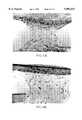
- FIGS. 1A-1Bshow photomicrographs of non-cryopreserved LSE (1A) and cryopreserved LSE (1B) of Example 1.
- the four basic features of LSEthe collagen lattice with fibroblasts, the basal layer of the epidermis, the suprabasal layers of the epidermis and the stratum corneum of the epidermis of the LSE in FIG. 1B are all preserved by this method. All layers are intact and the overall morphology of cryopreserved LSE is identical to that of non-cryopreserved LSE.
- FIGS. 2A-2Bshow photomicrographs of non-cryopreserved epidermal sheet (2A) and cryopreserved epidermal sheet (2B) of Example 2.
- the three basic features of the epidermisthe basal layer of the epidermis, the suprabasal layers of the epidermis and the stratum corneum of the epidermis are all preserved by this method. All layers are intact and the overall morphology of the cryopreserved epidermal sheet is identical to that of the non-cryopreserved epidermal sheet.
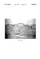
- FIGS. 3A-3Bshow photomicrographs of non-cryopreserved mouse skin (3A) and cryopreserved mouse skin (3B) of Example 5.
- the four basic features of mouse skin, the dermis with fibroblasts, the basal layer of the epidermis, the suprabasal layers of the epidermis and the stratum corneum of the epidermisare preserved by this method. All layers are intact and the overall morphology of cryopreserved mouse skin is identical to that of non-cryopreserved mouse skin.
- FIGS. 4A-4Bshow photomicrographs of non-cryopreserved SKIN2TM (4A) and cryopreserved SKIN2TM (4B) of Example 6.
- the four basic features of SKIN2TM, the collagen lattice with fibroblasts, the basal layer of the epidermis, the suprabasal layers of the epidermis and the stratum corneum of the epidermisare preserved by this method. All layers are intact and the overall morphology of cryopreserved SKIN2TM is similar to that of non-cryopreserved SKIN2TM.
- FIG. 5shows a graph of the viability of cryopreserved cultured tissues compared that of non-cryopreserved cultured tissues.
- the viability of cryopreserved cultured tissues as measured by MTTis shown.
- LSEwas additionally measured by LDH assay. All cultured tissues are compared to age-matched non-cryopreserved controls and are expressed as a percent of control.
- FIGS. 6A to 6Dshow comparative results.
- FIGS. 7A to 7Dshow comparative results.
- Tissue engineeringis an emerging area which utilizes cultured tissue cells to construct tissue equivalents which can be used to examine the response to injury by chemical agents or pharmaceutical compounds.
- the cultured tissuemay also be used to form graftable human tissue.
- LSELiving Skin Equivalent
- Parenteau, et al."Epidermis Generated In Vitro: Practical Considerations and Applications," J. of Cellular Biochemistry, 45:245-251 (1991); Parenteau, et al., "The organotypic culture of human skin keratinocytes and fibroblasts to achieve form and function," Cytotechnology, 9:163-171 (1992); and Bell et al., “The Living Skin Equivalent: Its Manufacture, Its Organotypic Properties and Its Responses to Irritants,” Toxic. in Vitro, 5:591-596 (1991).
- LSE for graftingis under investigation in clinical trials for indications relating to partial and full thickness skin wounds: excision surgery, burns, venous stasis ulcers, diabetic ulcers, decubitus ulcers, and chronic inflammatory ulcers.
- the LSEis a full-thickness, bilayered, in vitro engineered skin tissue.
- the cornea tissue equivalenthas three distinct cell layers, the external layer, a stratified squamous epithelium, the middle layer of collagen fibers and stromal cells, and an inner layer, simple squamous epithelium, also called the corneal endothelium.
- An in vitro cornea equivalentcan be used for in vitro toxicity assays to serve as accurate and inexpensive non-animal predictive models of in vivo ocular and dermal irritation potential for many types of products and raw materials.
- cryopreservationThe goal of cryopreservation is to preserve the structural integrity and viability of biological materials for an indefinite period of time so that these materials can be available and used as needed. Complex tissues of finite life span will require cryopreservation to expand product availability and utility.
- the history of cryopreservation of biological materialhas shown that the optimization of a cryopreservation protocol for a particular cell does not necessarily give good results when used with another cell type or with other cells in a tissue.
- the development of more specialized methods due to the differences in cell density, water content and level of structural organization of the full-thickness LSEwas required.
- the cryopreservation protocols of this inventionare surprisingly applicable to the single layer epidermal and dermal layers alone, trilayered cornea equivalent and mammalian skin.
- cultured tissue equivalentsmeans tissue equivalents of mammalian tissues, wherein the tissue equivalents are made by in vitro techniques and are meant to include monolayer skin equivalents, either a dermal equivalent or an epidermal sheet; bilayered skin equivalents, particularly LSE; and trilayered cornea equivalents and skin equivalents.
- the morphology of the cultured tissue equivalentsare similar to the in vivo mammalian organ, typically the human organ.
- the morphology of the LSEbears many similarities to human skin. Metabolically and mitotically active human dermal fibroblasts (HDF) are found throughout the dermal layer of the construct, and have been shown to secrete collagen and other matrix components into the lattice.
- HDFmetabolically and mitotically active human dermal fibroblasts
- the epidermisconsists of a basal layer shown to divide with a mitotic rate similar to that of human skin.
- the suprabasal epidermisshows the same strata as skin in vivo, with well defined spinous and granular layers containing keratohyalin and lamellar granules covered by a stratum corneum.
- Immunohistochemistrydemonstrates the presence of extracellular matrix components routinely found at the dermo-epidermal junction in normal human skin, such as laminin, Type IV collagen and kalanin (GB3).
- cell penetrating glass forming agentsor “non-cell penetrating glass forming agents” or both.
- the cell penetrating glass forming agentis preferably glycerol, but may include propylene glycol, ethylene glycol, dimethylsulfoxide, and other penetrating glass forming agents known in the art.
- Non-cell penetrating glass forming agentsinclude high molecular weight forms of complex carbohydrates, such as chondroitin sulfate, polyvinylpyrrolidone, polyethylene glycol or hetastarch, such as hydroxyethyl starch.
- the cell penetrating glass forming agents or non-cell penetrating glass forming agentsare diluted in a base of a physiological pH.
- the baseis preferably DMEM, but may be substituted with phosphate buffered saline, IDMEM, MEM, M199, RPMI 1640, Ham's F-12 Ham's F-10, NCTC 109, NCTC 135, or combinations thereof.
- the preferred cryoprotectant solutioncontains 1.5M to 2.5M glycerol, preferably 2M glycerol, in a base of Dulbecco's Modified Eagle's Medium (DMEM). These solutions can be modified and optimized by one of skill in the art using known cryoprotectants and freezing, storing, thawing, and rinsing procedures that are compatible with maintaining maximal viability, depending on the particular application.
- agitationany mechanical means of agitation to enhance perfusion of the cryoprotectant solution to achieve effective penetration of the solution into the tissue equivalent.
- the preferred means of agitationis shaking on an orbital shaker platform.
- Other means that may be employedare centrifugation, rocking platform, perfusion by means of a pump.
- Other methods of enhanced perfusionthat would achieve effective penetration of the cryoprotectant solution into the tissue equivalent can be contemplated and employed by the skilled artisan.
- a "gassed environment”is defined as an environment that prevents degassing of the sodium bicarbonate buffered base media component of the cryoprotectant solution. Over the period of time in which it takes to perfuse the tissue, these base media require the gassed environment to maintain proper pH of the cryoprotectant solution.
- CO 2 in airis used preferably at or about 5%, most preferably at or about 10%. The regulation of the pH of the cryoprotectant solution enables optimal cell viability.
- a slow freezing rateis defined as a rate of cooling that is about or less than -0.3° C. per minute, more preferably about or less than -0.2° C. per minute, and most preferably about -0.1° C. per minute.
- Ice seedingis defined as a method of initiating ice formation to the extracellular cryoprotectant.
- the preferred method of ice seedingis by contacting the tray containing the tissue with a chilled probe. Alternatively, the contact may be made directly to the cryoprotectant. Ice formation can be initiated by a chamber spike where the temperature of the chamber is lowered and raised within a range sufficient to form an ice crystal. Another method would be by introducing expanding gasses such as freon or CO 2 to either the outside of the package or in contact with the cryoprotectant solution. Other methods of ice seeding known in the art may be substituted.
- the present inventionrequires that the immersion of the harvested tissue or cultured tissue equivalent in cryoprotectant solution for a period of time under conditions sufficient to permit the perfusion of cryoprotectant in the cells of the tissue or cultured tissue equivalent.
- This method of cryopreservationallows for tissue to be slowly frozen and stored at or below -70° C. Tissues cryopreserved by this method are stable to fluctuations in temperature between -70° to -196° C. Short term storage is possible at -76° C., the temperature of dry ice, for transport and shipping. It is more preferable to freeze tissues to a temperature at or below -120° C., the glass transition temperature of water.
- the tissue that may used in the disclosed techniquescan include a full thickness skin equivalent such as those disclosed in U.S. Pat. Nos. 4,485,096; 4,604,346; 4,835,102; and 5,374,515 or those disclosed in U.S. Pat. Nos. 4,963,489; 5,032,508; and 5,266,480, all incorporated herein by reference, or any cultured epidermal sheet, any cultured dermal equivalent, a cultured cornea equivalent, or harvested mammalian skin.
- a full thickness skin equivalentsuch as those disclosed in U.S. Pat. Nos. 4,485,096; 4,604,346; 4,835,102; and 5,374,515 or those disclosed in U.S. Pat. Nos. 4,963,489; 5,032,508; and 5,266,480, all incorporated herein by reference, or any cultured epidermal sheet, any cultured dermal equivalent, a cultured cornea equivalent, or harvested mammalian skin.
- LSELiving Skin Equivalent
- the method of perfusing LSE with a cryoprotectant solutionis to submerge LSE and its attached transwell in a volume of cryoprotectant solution sufficient to submerge the sample and to have equal volume of cryoprotectant solution above and below the tissue equivalent. Equal volumes allow for the uniform conduction of heat away from the tissue equivalent during cooling.
- 25 mL of 2M Glycerol in DMEMis added to a 100 mm petri dish containing the LSE and the transwell for a period of time sufficient to completely perfuse the sample, preferably between one and two hours, but most preferably for about one hour.
- cryoprotectant solutionExtended periods of time in cryoprotectant solution result in reduced cell viability in the tissue, while too short of a time does not ensure complete permeation of cryoprotectant into the tissue. Monolayer constructs will typically require less time for perfusion as they have fewer cell layers and a reduced barrier function.
- penetration of cryoprotectant solutionis enhanced by agitating the sample and cryoprotectant solution, typically by shaking the petri dish on an orbital platform shaker (Bellco orbital shaker) at 70 rpm in a 10% CO 2 gassed chamber.
- the 10% CO 2 environmentthe same as the culture environment in which the LSE was fabricated, prevents the media from degassing, thus maintaining the pH of the base media component of the cryoprotectant.
- LSE unitsare then placed into a programmable freezer (Planer) set at a starting temperature of 20.0° C.
- LSE unitsare cooled at -10.0° C./minute to -6.0° C., and are allowed to hold for 20 minutes for the purpose of equilibrating the tissue equivalent to -6.0° C., the temperature necessary for seeding ice in the cryoprotectant. Any cooling rate may be used to obtain the seeding temperature.
- the hold time of 20 minutesis sufficient to ensure thermal equilibration, while at least 15 minutes is typically needed.
- extracellular iceis initiated by contact of the outside of the bag containing the unit with a liquid nitrogen chilled (-196° C.) probe.
- the contact sitemust be below the level of the cryoprotectant freeze media in the bag. After all LSE units are seeded with ice crystals, the units are held an additional 5 minutes to allow the chamber to readjust to -6.0° C., as the units were opened to manually seed the ice. Cooling is then resumed at a rate of -1.0° C./minute to -8.0° C. The temperature is held at -8.0° C., for 30 minutes to allow for physical and biological equilibration of the cryoprotectant in and around the LSE before it is cooled to -70° C.
- phase equilibrationis the extracellular ice formation that occurs in the cryoprotectant solution.
- the biological equilibrationis the equilibration that occurs between the concentration of cryoprotectant inside and outside the cells.
- the biological equilibrationis regulated by the permeability properties of the cells and such equilibration may not be complete. During the phase change from liquid to solid, heat is liberated that would affect the cooling rate of LSE if this hold were not allowed.
- LSE unitsare then cooled preferably at -0.3° C./minute; more preferably at -0.2° C./minute; and most preferably at -0.1° C./minute to a final temperature preferably at least or below -70.0° C.; more preferably at -120° C.; even more preferably at -140° C.; or most preferably at -196.0° C.
- a final temperaturepreferably at least or below -70.0° C.; more preferably at -120° C.; even more preferably at -140° C.; or most preferably at -196.0° C.
- the final freezing temperatureapproaches the glass transition temperature of water, -120.0° C., the less likely there will be detrimental temperature fluctuations during transfers to final storage locations.
- Cryopreserved LSEwas transferred from the freezer to storage preferably at -196° C. until use.
- the frozen LSE tissue equivalentis thawed by rapidly warming such that the tissue is thawed in about from 1 to 3 minutes.
- Suitable methods for warming frozen harvested tissue or tissue equivalents at a high thawing rateinclude warming using a water bath or warming using induction heating.
- the frozen tissue or tissue equivalentis thawed by direct addition of culture media, warmed to 37° C., to the surface of the cryopreserved tissue.
- the cryoprotectant solutionis removed from the thawed tissue or tissue equivalent within about 15 minutes after thawing, preferably as soon as possible after thawing, to avoid damaging the viability of the tissue or tissue equivalent.
- the cryoprotectant solutionis replaced with an isotonic buffer solution at physiological pH (about 6.8 to 7.4 pH).
- the cryopreserved LSEis stored in a sterile petri dish.
- the cryopreserved LSEis removed from storage and the outer bag is cut off the petri dish.
- the lid of the dishis removed and 40 mL of warmed (37° C.) DMEM, the amount that will fit into the petri dish, is aseptically poured into the dish.
- the temperatureshould not be any warmer than 37° C. as the viability of sample will be endangered. After 45 seconds all liquid is removed from the dish and an additional 40 mL aliquot is added to the dish for two minutes.
- the LSE unit and attached transwellis transferred to a new petri dish with 25 mL, or a volume sufficient to submerge the sample, of DMEM for 30 minutes.
- the mediais exchanged one time for an additional 30 minutes.
- a cultured tissue equivalent prepared as disclosed abovemay be used for transplantation or implantation in vivo or for screening compounds in vitro.
- LSE constructswere perfused with cryoprotectant by submerging the constructs and the transwell with 25 mL of cryoprotective media, 2M Glycerol in DMEM, in the 100 mm petri dish for one hour. This method was improved upon by shaking the petri dish for one hour on an orbital shaker (Bellco) at 70 rpm in a 10% CO 2 gassed chamber. Shaking allows for a more complete perfusion and better reproducibility when frozen.
- cryoprotectantby submerging the constructs and the transwell with 25 mL of cryoprotective media, 2M Glycerol in DMEM, in the 100 mm petri dish for one hour. This method was improved upon by shaking the petri dish for one hour on an orbital shaker (Bellco) at 70 rpm in a 10% CO 2 gassed chamber. Shaking allows for a more
- the petri dish containing LSE, transwell and extracellular freezing media (2M Glycerol and DMEM)are placed in a bag and vacuum sealed (Audiovox) programmed for a 5 second vacuum and a 3.9 second seal time.
- LSE unitswere placed into the programmable freezer (Planar) at a starting temperature of 20.0° C. LSE units were cooled at -10.0° C./minute to -6.0° C. and the chamber temperature was held at -6.0° C. for 20 minutes to equilibrate the constructs to the chamber temperature. After the 20 minute hold, extracellular ice was initiated by contact of the outside of the bag, below the level of the freeze media, with a liquid nitrogen chilled probe. After all LSE units were seeded with ice crystals, the chamber temperature was held for an additional 5 minutes at -6.0° C. The chamber temperature was then cooled at -1.0° C./minute to -8.0° C. The chamber temperature was held again for 30 minutes at -8.0° C. to allow for uniform distribution of ice throughout the sample. The freezer temperature was then cooled at -0.1° C./minute to a final temperature of -70.0° C.
- Cryopreserved LSEwere removed from the freezer and the bag was cut from the petri dish.
- the lid of the dishwas removed and 40 mL of warmed (37° C.) DMEM was aseptically poured into the petri dish. After 45 seconds, all liquid was removed from the dish and an additional 40 mL aliquot was added to the dish for two minutes. Once thawed, the LSE unit and attached transwell were transferred to a new petri dish, using sterile forceps.
- 25 mL of DMEMwas added to the petri dish containing the LSE for 30 minutes. The media was exchanged a second time for an additional 30 minutes.
- the LSE unitwas then transferred back to its original culture dish and was incubated in culture maintenance medium at 37° C./10% CO 2 for 24 hours prior to analysis.
- the incubation timewas allowed for the lag typically seen for frozen cells to reestablish steady state conditions.
- Epidermal sheetswere procured from mature LSE at 12 days post air-lift. Removal of the epidermal sheet was accomplished by peeling the dermal substrate layer from the epidermal layer with forceps and discarding the dermal layer. Each sheet was cut in to three equivalent pieces. One piece from each sheet was fixed as a control. The remaining two pieces form each sheet were placed on top of a 75 mm polycarbonate transwell membrane (Costar) in 100 mm culture dishes (Costar). Each piece was perfused with 25 mL of DMEM and 2 Molar Glycerol for one hour. The dishes containing constructs were placed on an orbital shaker (Bellco) at 70 rpm for one hour in a 10% CO 2 gassed chamber.
- Bellcoorbital shaker
- the petri dish containing epidermal sheet, transwell and extracellular freezing media(2M Glycerol and DMEM) were placed in a bag and vacuum sealed (Audiovox) programmed for a 5 second vacuum and a 3.9 second seal time.
- Epidermal sheetswere placed into the programmable freezer (Planer) at a starting temperature of 20.0° C. Epidermal sheets were cooled at -10.0° C./minute to -6.0° C. The temperature was allowed to hold for 20 minutes at -6.0° C. to equilibrate to chamber temperature. After the 20 minutes hold, extracellular ice was initiated by contact of the outside of the bag, below the level of the freeze media, with a liquid nitrogen chilled probe. After all epidermal sheets had been seeded with ice crystals, the temperature was held an additional 5 minutes at -6.0° C. The chamber was then cooled at -1.0° C./minute to -8.0° C. The temperature was then allowed to hold for 30 minutes at -8.0° C. to allow for uniform distribution of ice throughout the sample. The chamber was then cooled at -0.1° C./minute to a final temperature of -70.0° C.
- Cryopreserved epidermal sheetswere removed from the freezer and the bags were cut from the petri dishes and the lids removed.
- 40 mL of warmed (37° C.) DMEMwas aseptically poured into each petri dish. After 45 seconds, all liquid was removed from the dishes and an additional 40 mL aliquot was added to each dish for two minutes
- epidermal sheetswere rinsed with 25 mL of DMEM for 30 minutes. The media was exchanged a second time for an additional 30 minutes.
- the epidermal sheetswere then incubated in culture maintenance medium at 37° C./10% CO2 for 24 hours prior to analysis. The incubation time was allowed for the lag typically seen for frozen cells to reestablish steady state conditions. Again, the incubation time was allowed for the lag typically seen for frozen cells to reestablish steady state conditions.
- Dermal equivalents used in this studywere the non-epidermalized component of the LSE. Dermal equivalents were frozen at 8 days post cast. The dermal equivalents were perfused with cryoprotectant by submerging the dermal equivalents attached to 75 mm transwell (Costar) placed in 100 mm petri dishes (Costar) with 25 mL of DMEM and 2 Molar Glycerol for one hour. The petri dishes were placed on an orbital shaker (Bellco) at 70 rpm for one hour in a 10% CO2 gassed chamber. After dermal equivalents were perfused, the petri dishes, dermal equivalents, transwells and extracellular freezing media were placed into the programmable freezer (Planer) at a starting temperature of 20.0° C.
- programmable freezerPlant
- the chamberwas then cooled at -10.0° C./minute to -6.0° C. The temperature was held for 20 minutes to equilibrate to chamber temperature. After the 20 minutes hold, extracellular ice was initiated by contact of the outside of the dishes, below the level of the freeze media, with a liquid nitrogen chilled probe. After all dermal equivalents had been seeded with ice crystals, the temperature was held an additional 5 minutes at -6.0° C. The chamber was then cooled at -1.0° C./minute to -8.0° C. The chamber temperature was held again for 30 minutes at -8.0° C. to allow for uniform distribution of ice throughout the sample. Dermal equivalents units were then cooled at -0.1° C./min. to a final temperature of -70.0° C.
- the method of perfusing cornea equivalents with cryoprotectantwas to submerge each cornea equivalent and transwell with 4 mL extracellular freezing media (2M glycerol in DMEM) in the six well cluster dishes for one hour.
- the six well cluster dishes containing the cornea constructswere shaken for one hour on an orbital shaker (Bellco) at 70 rpm in a 10% CO 2 gassed chamber.
- the petri dishes containing cornea equivalents, transwells and extracellular freezing mediawere placed into the programmable freezer (Planer) at a starting temperature of 20.0° C.
- Cornea equivalentswere cooled at -10.0° C./min. to -6.0° C. The temperature was held for 20 minutes to equilibrate to chamber temperature. After the 20 minutes hold, extracellular ice was initiated by contact of the outside of each well in the cluster plate, below the level of the freeze media, with a liquid nitrogen chilled probe. After all cornea equivalents had been seeded with ice crystals the temperature was held an additional 5 minutes at -6.0° C. prior to cooling at -1.0° C./minute to -8.0° C. The temperature was held again for 30 minutes at -8.0° C. to allow for uniform distribution of ice throughout the sample. Cornea equivalent units were cooled at -0.1° C./minute to a final temperature of -70.0° C.
- Cryopreserved cornea equivalentswere removed from the freezer. The lid of the dish was removed. To thaw, 6 mL of warmed (37° C.) DMEM was aseptically poured into each well of the cluster plate. After 45 seconds, all liquid was removed from the dish and an additional 6 mL aliquot was added to the dish for two minutes Using sterile forceps the cornea equivalents and attached transwells was transferred to a new cluster dish. To rinse, 4 mL of DMEM was added to each well for 30 minutes. The media was exchanged a second time for an additional 30 minutes The cornea units were then transferred back to the/same culture dish and incubated in cornea maintenance medium at 37° C./10% CO 2 for 24 hours prior to analysis. The incubation time was allowed for the lag typically seen for frozen cells to reestablish steady state conditions. Samples were assayed using the MTT assay protocol.
- Wild type mice, strain B6CB6YF1were euthanized by Nembutal overdose.
- the skinwas harvested aseptically. Excess blood vessels, fat and connective tissue were removed from the dermis.
- Murine skinwas trimmed to a rectangular 1 cm ⁇ 2 cm piece. Murine skin pieces were then placed on a 75 mm transwell (Costar) in 100 mm petri dishes (Costar). Murine skin was perfused with cryoprotectant by submerging in 25 mL of 2M Glycerol in DMEM in the 100 mm petri dishes for one hour.
- the petri dishes each containing the murine skin and the transwellwere shaken for one hour on an orbital shaker (Bellco) at 70 rpm in a 10% CO2 gassed chamber.
- Control, non-cryopreserved mouse skinwas kept in nutrient media at 4° C. for the duration of the cryopreservation and thawing process (2 days) before skin grafting.
- the petri dishes containing murine skin, transwells and extracellular freezing mediaare placed into the Planer programmable freezer at a starting temperature of 20.0° C.
- Murine skinwas cooled at -10.0° C./minute to -6.0° C. and were allowed to hold at -6.0° C. for 20 minutes to equilibrate to chamber temperature.
- extracellular icewas initiated by contact of the outside of the dish with a liquid nitrogen chilled probe. The contact site must be below the level of the freeze media.
- the temperaturewas held an additional 5 minutes at -6.0° C. prior to cooling at -1.0° C./minute to -8.0° C. The temperature was held again for 30 minutes at -8.0° C. to allow for uniform distribution of ice throughout the sample.
- the unitswere then cooled at -0.1° C./min. to a final temperature of -70.0° C.
- Cryopreserved murine skinwas removed from the freezer and the lid of the dish was removed.
- 40 mL of warmed (37° C.) DMEMwas aseptically poured into the petri dishes. After 45 seconds, all liquid was removed from the dishes and an additional 40 mL aliquot was added to the dishes for two minutes
- murine skinwas rinsed in the dishes with 25 mL DMEM for 30 minutes The media was exchanged a second time for an additional 30 minutes.
- Thawed cryopreserved and control sampleswere processed for histology (FIG. 3) or were grafted to mice.
- SKIN2TMmodel ZK 1300 (Advanced Tissue Sciences, La Jolla, Calif.), was removed from the packaging according to shipping inserts upon arrival and were placed in culture dishes (Costar). Transwell inserts were placed above the SKIN2TM to keep the skin construct submerged. SKIN2TM was perfused with cryoprotectant by submerging SKIN2TM in 2M Glycerol in DMEM in the culture dishes for one hour. During perfusion, the culture dishes containing the construct and the transwell were shaken for one hour on an orbital shaker (Bellco) at 70 rpm in a 10% CO 2 gassed chamber.
- Bellcoorbital shaker
- SKIN2TMAfter SKIN2TM is perfused, the units were placed into the programmable freezer (Planar) at a starting temperature of 20.0° C. SKIN2TM units were cooled at -10.0° C./min. to -6.0° C. and were allowed to hold at -6.0° C. for 20 minutes to equilibrate to the chamber temperature. After the 20 minute hold, extracellular ice was initiated by contact of the outside of the dishes below the level of the freeze media with a liquid nitrogen chilled probe. After all SKIN2TM units were seeded with ice crystals, the temperature was held for an additional 5 minutes at -6.0° C. The chamber temperature was then cooled at -1.0° C./min. to -8.0° C.
- SKIN2TM unitswere allowed to hold for 30 minutes at -8.0° C. to allow for uniform distribution of ice throughout the sample. SKIN2TM units were then cooled at -0.1° C./min. to a final temperature of -70.0° C.
- SKIN2TMCryopreserved SKIN2TM were removed from the freezer, the lids of the dishes was removed and warmed (37° C.) DMEM was aseptically poured into each culture dish. After 45 seconds, all liquid was removed from the dishes and another addition of DMEM was added to each dish for two minutes. Once thawed, the SKIN2TM unit and attached transwell were transferred to a new culture dishes, using sterile forceps. To rinse the SKIN2TM of cryoprotectant, DMEM was added to each culture dish containing the SKIN2TM for 30 minutes. The media was exchanged a second time for an additional 30 minutes.
- the SKIN2TM unitwas then transferred back to the same type culture dish and was incubated in culture maintenance medium at 37° C./10% CO2 for 24 hours prior to analysis. Again, the incubation time was allowed for the lag typically seen for frozen cells to reestablish steady state conditions.
- the metabolic reduction of the soluble tetrazolium salt to a blue formazan precipitateis dependent on the presence of viable cells with intact mitochondrial function.
- This assayis used to quantitate cytotoxicity in a variety of cell types, including cultured human keratinocytes.
- the absorbencieswere read on a plate reader (Dynatech) at 570 nm with the isopropanol extraction medium as a blank. MTT values obtained from thawed cryopreserved samples were compared to corresponding control samples and expressed in terms of percent of control (FIG. 5).
- LDHis an enzyme typically found in a viable cell. Damage to the cell causes a release of the enzyme and a corresponding decrease in the enzyme activity detected by this assay.
- LSE cocktail reagentwas prepared by mixing together the following: 3.00 mL phosphate buffer (0.1 mol/l; pH 7.0), 0.1 mL pyruvate, Na salt (2.5 mg/mL), and 0.05 mL NADH, Na salt (10 mg/mL).
- LSE unitscryopreserved according to the method outlined in Example 1, were thawed one day before grafting and recovered in maintenance media for twenty-four hours.
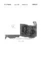
- FIGS. 6A, BThe gross photographs (FIGS. 6A, B) and micrographs (FIGS. 6C, D) show no difference in graft integration between control LSE or thawed, cryopreserved LSE. No significant difference was seen in rates of wound contracture between control or cryopreserved grafts.
- Wild type mice, strain B6CB6YF1were euthanized by Nembutal overdose.
- the skinwas harvested aseptically. Excess blood vessels, fat and connective tissue were removed from the dermis in preparation for skin grafting.
- Mouse skinwas cryopreserved and thawed following the method in Example 5. Control, non-cryopreserved mouse skin was kept in a nutrient media at 4° C. for the duration of the cryopreservation and thawing process, a total of two days, before skin grafting.
- mice of the same strainreceived skin grafts, two received control, non-cryopreserved mouse skin and four received thawed, cryopreserved mouse skin.
- the micewere anesthetized with Nembutal. A 2 ⁇ 2 cm full thickness skin section was excised from the dorsum of each mouse, sparing the panicculus carnosus.
- Mouse skin grafts, either control or cryopreserved,were placed on the wounds and trimmed to fit. All grafts were dressed with one layer of petrolatum impregnated gauze, covered by two adhesive bandages. These dressings were removed at 7 days post-graft.
- FIGS. 7A, BThe gross photographs (FIGS. 7A, B) and micrographs (FIGS. 7C, D) show that there is no difference in graft integration between control mouse skin or thawed, cryopreserved mouse skin. No significant difference was seen in rates of wound contracture between control or cryopreserved grafts.
Landscapes
- Life Sciences & Earth Sciences (AREA)
- Engineering & Computer Science (AREA)
- Health & Medical Sciences (AREA)
- Dentistry (AREA)
- General Health & Medical Sciences (AREA)
- Wood Science & Technology (AREA)
- Zoology (AREA)
- Environmental Sciences (AREA)
- Mechanical Engineering (AREA)
- Agricultural Chemicals And Associated Chemicals (AREA)
- Micro-Organisms Or Cultivation Processes Thereof (AREA)
- Apparatus Associated With Microorganisms And Enzymes (AREA)
- Packages (AREA)
- Packging For Living Organisms, Food Or Medicinal Products That Are Sensitive To Environmental Conditiond (AREA)
- Catching Or Destruction (AREA)
- Materials For Medical Uses (AREA)
- Breeding Of Plants And Reproduction By Means Of Culturing (AREA)
Abstract
Description
Claims (32)
Priority Applications (30)
| Application Number | Priority Date | Filing Date | Title |
|---|---|---|---|
| US08/380,099US5891617A (en) | 1993-09-15 | 1995-01-30 | Cryopreservation of harvested skin and cultured skin or cornea equivalents by slow freezing |
| HU9800411AHU223789B1 (en) | 1995-01-30 | 1996-01-30 | Method for the Cold Storage and Storage of Tissues and Cultured Tissue Equivalents from Mammals |
| CZ19972413ACZ294950B6 (en) | 1995-01-30 | 1996-01-30 | Method for cryopreserving harvested mammalian tissue or cultured tissue equivalents |
| KR1019970705324AKR100328742B1 (en) | 1995-01-30 | 1996-01-30 | Method and package design for cryopreservation and storage of cultured tisse equivalents |
| CNB031457983ACN1292651C (en) | 1995-01-30 | 1996-01-30 | Method and package design for cryopreservation and storage of cultured tissue equivalents |
| EP96905275AEP0807234B1 (en) | 1995-01-30 | 1996-01-30 | Method and package design for cryopreservation and storage of cultured tisse equivalents |
| CN96191673ACN1119599C (en) | 1995-01-30 | 1996-01-30 | Method and apparatus for refrigeration and storage of cultured skin tissue analogs |
| DE69637301TDE69637301T2 (en) | 1995-01-30 | 1996-01-30 | Packaging for cryopreservation and storage of cultured tissue equivalents |
| PT96905275TPT807234E (en) | 1995-01-30 | 1996-01-30 | METHOD AND MODEL OF PACKAGING FOR CRIOCONSERVATION AND STORAGE OF EQUIVALENTS OF FABRICS DEVELOPED IN CULTURE |
| SK1033-97ASK284801B6 (en) | 1995-01-30 | 1996-01-30 | A method for cryopreserving mammalian tissues |
| CA002210532ACA2210532C (en) | 1995-01-30 | 1996-01-30 | Method and package design for cryopreservation and storage of cultured tissue equivalents |
| JP8523665AJPH11501298A (en) | 1995-01-30 | 1996-01-30 | Method and package design for cryopreservation and storage of cultured tissue equivalents |
| ES96905275TES2231804T3 (en) | 1995-01-30 | 1996-01-30 | METHOD AND DESIGN OF ENCAPSULATE FOR CRIOCONSERVATION AND STORAGE OF EQUIVALENTS OF CULTIVATED FABRIC. |
| AU49082/96AAU716869B2 (en) | 1995-01-30 | 1996-01-30 | Method and package design for cryopreservation and storage of cultured tissue equivalents |
| SG9802806ASG106553A1 (en) | 1995-01-30 | 1996-01-30 | Method and package design for cryopreservation and storage of cultured tissue equivalents |
| DE69633854TDE69633854T2 (en) | 1995-01-30 | 1996-01-30 | METHOD AND PACKAGING FOR MAINTAINING AND STORING CULTURED TISSUE EQUIVALENTS FOR TEMPERATURE TEMPERATURES |
| NZ334152ANZ334152A (en) | 1995-01-30 | 1996-01-30 | Apparatus for cyropreserving biological tissue |
| AT96905275TATE282805T1 (en) | 1995-01-30 | 1996-01-30 | METHOD AND PACKAGING FOR PRESERVATION AND STORAGE OF CULTURED TISSUE EQUIVALENTS AT LOW TEMPERATURES |
| RU97116150/13ARU2178865C2 (en) | 1995-01-30 | 1996-01-30 | Method for cryopreservation of prepared tissue of mammalia or cultivated equivalent of tissue and device for its realization |
| PL96321089APL181762B1 (en) | 1995-01-30 | 1996-01-30 | Method of and design of a package for effecting cryogenic preservation and storage of artificially grown human tissue equivalents |
| AT04077072TATE376356T1 (en) | 1995-01-30 | 1996-01-30 | PACKAGING FOR CRYOPRESERVATION AND STORAGE OF CULTURED TISSUE EQUIVALENTS |
| PCT/US1996/001217WO1996024018A1 (en) | 1995-01-30 | 1996-01-30 | Method and package design for cryopreservation and storage of cultured tisse equivalents |
| NZ302913ANZ302913A (en) | 1995-01-30 | 1996-01-30 | Method and apparatus for cryopreservation of mammalian skin, cultured skin or cornea equivalent |
| BR9606864ABR9606864A (en) | 1995-01-30 | 1996-01-30 | Process and packaging model for cryopreservation and storage of cultured tissue equivalents |
| EP04077072AEP1516532B1 (en) | 1995-01-30 | 1996-01-30 | Package design for cryopreservation and storage of cultured tissue equivalents |
| FI972563AFI113694B (en) | 1995-01-30 | 1997-06-16 | Process and packaging design for cold storage and storage of cultured tissue equivalents |
| NO19973463ANO310491B1 (en) | 1995-01-30 | 1997-07-28 | Method and apparatus for cryopreservation |
| JP2006188993AJP4361550B2 (en) | 1995-01-30 | 2006-07-10 | Method and package design for cryopreservation and storage of cultured tissue equivalents |
| JP2007000107AJP2007161723A (en) | 1995-01-30 | 2007-01-04 | Method and package design for cryopreservation and storage of cultured tissue equivalents |
| JP2013272171AJP2014065736A (en) | 1995-01-30 | 2013-12-27 | Method and package design for cryopreservation and storage of cultured tissue equivalents |
Applications Claiming Priority (2)
| Application Number | Priority Date | Filing Date | Title |
|---|---|---|---|
| US08/121,377US5518878A (en) | 1993-09-15 | 1993-09-15 | Cryopreservation of cultured skin or cornea equivalents with agitation |
| US08/380,099US5891617A (en) | 1993-09-15 | 1995-01-30 | Cryopreservation of harvested skin and cultured skin or cornea equivalents by slow freezing |
Related Parent Applications (1)
| Application Number | Title | Priority Date | Filing Date |
|---|---|---|---|
| US08/121,377Continuation-In-PartUS5518878A (en) | 1993-09-15 | 1993-09-15 | Cryopreservation of cultured skin or cornea equivalents with agitation |
Publications (1)
| Publication Number | Publication Date |
|---|---|
| US5891617Atrue US5891617A (en) | 1999-04-06 |
Family
ID=23499896
Family Applications (1)
| Application Number | Title | Priority Date | Filing Date |
|---|---|---|---|
| US08/380,099Expired - LifetimeUS5891617A (en) | 1993-09-15 | 1995-01-30 | Cryopreservation of harvested skin and cultured skin or cornea equivalents by slow freezing |
Country Status (21)
| Country | Link |
|---|---|
| US (1) | US5891617A (en) |
| EP (2) | EP1516532B1 (en) |
| JP (4) | JPH11501298A (en) |
| KR (1) | KR100328742B1 (en) |
| CN (2) | CN1292651C (en) |
| AT (2) | ATE282805T1 (en) |
| AU (1) | AU716869B2 (en) |
| BR (1) | BR9606864A (en) |
| CZ (1) | CZ294950B6 (en) |
| DE (2) | DE69637301T2 (en) |
| ES (1) | ES2231804T3 (en) |
| FI (1) | FI113694B (en) |
| HU (1) | HU223789B1 (en) |
| NO (1) | NO310491B1 (en) |
| NZ (1) | NZ302913A (en) |
| PL (1) | PL181762B1 (en) |
| PT (1) | PT807234E (en) |
| RU (1) | RU2178865C2 (en) |
| SG (1) | SG106553A1 (en) |
| SK (1) | SK284801B6 (en) |
| WO (1) | WO1996024018A1 (en) |
Cited By (67)
| Publication number | Priority date | Publication date | Assignee | Title |
|---|---|---|---|---|
| US6291240B1 (en)* | 1998-01-29 | 2001-09-18 | Advanced Tissue Sciences, Inc. | Cells or tissues with increased protein factors and methods of making and using same |
| US6347525B2 (en) | 1996-01-30 | 2002-02-19 | Organogenesis Inc. | Ice seeding apparatus for cryopreservation systems |
| DE10061968A1 (en)* | 2000-12-13 | 2002-06-20 | Friedrich Hoffmann | Vitrification of cornea, employs tissues of rear cornea lamellae including the stroma, Descemet membrane and endothelium |
| US6521402B1 (en) | 2000-06-14 | 2003-02-18 | The Texas A&M University System | Cryopreservation of tissues for use in nuclear transfer |
| US20030044764A1 (en)* | 2000-03-14 | 2003-03-06 | Soane David S | Cryoprotective system |
| US20030054022A1 (en)* | 1999-12-22 | 2003-03-20 | Acell, Inc. | Tissue regenerative composition, method of making, and method of use thereof |
| US20030091542A1 (en)* | 2001-10-17 | 2003-05-15 | Boehringer Ingelheim Pharma Kg | Keratinocytes useful for the treatment of wounds |
| US6579538B1 (en) | 1999-12-22 | 2003-06-17 | Acell, Inc. | Tissue regenerative compositions for cardiac applications, method of making, and method of use thereof |
| US20030118982A1 (en)* | 2000-03-24 | 2003-06-26 | Naoka Yamamoto | Method of preserving tissue equivalent and tissue equivalent preserved in frozen state |
| US20030150002A1 (en)* | 2000-12-15 | 2003-08-07 | Westhusin Mark E. | Cloning bovines by nuclear transplantation |
| US6615592B2 (en)* | 2001-01-02 | 2003-09-09 | Supachill Technologies Pty. Ltd. | Method and system for preparing tissue samples for histological and pathological examination |
| US20030180270A1 (en)* | 2002-02-22 | 2003-09-25 | Bruce Simon | Methods and compositions for treating bone defects |
| US6638709B2 (en) | 2000-12-27 | 2003-10-28 | Ortec International, Inc. | Processes for making cryopreserved composite living constructs and products resulting therefrom |
| US6656380B2 (en) | 2001-10-16 | 2003-12-02 | Supachill Technologies Pty. Ltd. | Super-coolable composition having long-duration phase change capability, process for preparation of same, process for super-cooling same and articles comprising same |
| US6681581B2 (en) | 2001-11-20 | 2004-01-27 | Supachill Technologies Pty. Ltd. | Pre-conditioned solute for use in cryogenic processes |
| US20040043006A1 (en)* | 2002-08-27 | 2004-03-04 | Badylak Stephen F. | Tissue regenerative composition |
| US20040059430A1 (en)* | 2001-09-05 | 2004-03-25 | Tae-Woon Kim | Collagen-based biomaterial for tissue repair |
| WO2004010780A3 (en)* | 2002-07-30 | 2004-05-13 | Gen Biotechnology Llc | Innocuous intracellular ice |
| RU2233589C2 (en)* | 2002-01-14 | 2004-08-10 | Галина Степановна Лобынцева | Method for cryopreserving human hemopoietic cells |
| US20040176855A1 (en)* | 2003-03-07 | 2004-09-09 | Acell, Inc. | Decellularized liver for repair of tissue and treatment of organ deficiency |
| US20040175366A1 (en)* | 2003-03-07 | 2004-09-09 | Acell, Inc. | Scaffold for cell growth and differentiation |
| WO2004069156A3 (en)* | 2003-01-30 | 2004-09-23 | Univ California | Inactivated probiotic bacteria and methods of use thereof |
| US20050025838A1 (en)* | 2003-06-25 | 2005-02-03 | Badylak Stephen F. | Conditioned compositions for tissue restoration |
| US20050277107A1 (en)* | 2002-07-26 | 2005-12-15 | Mehmet Toner | Systems and methods for cell preservation |
| US20080077201A1 (en)* | 2006-09-26 | 2008-03-27 | Juniper Medical, Inc. | Cooling devices with flexible sensors |
| CN100386061C (en)* | 2005-05-17 | 2008-05-07 | 浙江大学医学院附属邵逸夫医院 | A frozen heterogeneous corneal stroma with low antigen content and its preparation method |
| EP2295540A1 (en) | 1998-11-19 | 2011-03-16 | Organogenesis, Inc. | Bioengineered tissue constructs and methods for producing and using them |
| US20110206776A1 (en)* | 2010-02-18 | 2011-08-25 | Samson Tom | Methods of manufacture of immunocompatible amniotic membrane products |
| WO2011110948A2 (en) | 2010-03-11 | 2011-09-15 | Antoine Turzi | Process,tube and device for the preparation of wound healant composition |
| US20110238051A1 (en)* | 2010-01-25 | 2011-09-29 | Zeltiq Aesthetics, Inc. | Home-use applicators for non-invasively removing heat from subcutaneous lipid-rich cells via phase change coolants, and associated devices, systems and methods |
| WO2012035403A1 (en)* | 2010-09-13 | 2012-03-22 | Singapore Health Services Pte Ltd | Method for performing a reversible refractive procedure |
| US20120329034A1 (en)* | 2010-02-26 | 2012-12-27 | Wook Chun | Method For Producing An Acellular Dermal Matrix, And Acellular Dermal Matrix Produced By Same |
| WO2013174769A1 (en) | 2012-05-25 | 2013-11-28 | Boehringer Ingelheim International Gmbh | Use of keratinocytes as a biologically active substance in the treatment of wounds, such as diabetic wounds, optionally in combination with a dpp-4 inhibitor |
| WO2014160000A1 (en)* | 2013-03-13 | 2014-10-02 | Stratatech Corporation | Cryopreservation of viable human skin substitutes |
| US9375345B2 (en) | 2006-09-26 | 2016-06-28 | Zeltiq Aesthetics, Inc. | Cooling device having a plurality of controllable cooling elements to provide a predetermined cooling profile |
| US9408745B2 (en) | 2007-08-21 | 2016-08-09 | Zeltiq Aesthetics, Inc. | Monitoring the cooling of subcutaneous lipid-rich cells, such as the cooling of adipose tissue |
| EP3111974A2 (en) | 2006-08-21 | 2017-01-04 | Antoine Turzi | Cell preparations for extemporaneous use, useful for healing and rejuvenation in vivo |
| US9545523B2 (en) | 2013-03-14 | 2017-01-17 | Zeltiq Aesthetics, Inc. | Multi-modality treatment systems, methods and apparatus for altering subcutaneous lipid-rich tissue |
| USD777338S1 (en) | 2014-03-20 | 2017-01-24 | Zeltiq Aesthetics, Inc. | Cryotherapy applicator for cooling tissue |
| US9655770B2 (en) | 2007-07-13 | 2017-05-23 | Zeltiq Aesthetics, Inc. | System for treating lipid-rich regions |
| US9737434B2 (en) | 2008-12-17 | 2017-08-22 | Zeltiq Aestehtics, Inc. | Systems and methods with interrupt/resume capabilities for treating subcutaneous lipid-rich cells |
| US9844460B2 (en) | 2013-03-14 | 2017-12-19 | Zeltiq Aesthetics, Inc. | Treatment systems with fluid mixing systems and fluid-cooled applicators and methods of using the same |
| US9861520B2 (en) | 2009-04-30 | 2018-01-09 | Zeltiq Aesthetics, Inc. | Device, system and method of removing heat from subcutaneous lipid-rich cells |
| US9861421B2 (en) | 2014-01-31 | 2018-01-09 | Zeltiq Aesthetics, Inc. | Compositions, treatment systems and methods for improved cooling of lipid-rich tissue |
| WO2018067747A1 (en)* | 2016-10-04 | 2018-04-12 | Smokey Tech, Llc | Systems and methods for natural cryoprotectants for preservation of cells |
| US10092346B2 (en) | 2010-07-20 | 2018-10-09 | Zeltiq Aesthetics, Inc. | Combined modality treatment systems, methods and apparatus for body contouring applications |
| US10383787B2 (en) | 2007-05-18 | 2019-08-20 | Zeltiq Aesthetics, Inc. | Treatment apparatus for removing heat from subcutaneous lipid-rich cells and massaging tissue |
| US10524956B2 (en) | 2016-01-07 | 2020-01-07 | Zeltiq Aesthetics, Inc. | Temperature-dependent adhesion between applicator and skin during cooling of tissue |
| US10555831B2 (en) | 2016-05-10 | 2020-02-11 | Zeltiq Aesthetics, Inc. | Hydrogel substances and methods of cryotherapy |
| US10568759B2 (en) | 2014-08-19 | 2020-02-25 | Zeltiq Aesthetics, Inc. | Treatment systems, small volume applicators, and methods for treating submental tissue |
| WO2020061429A1 (en) | 2018-09-20 | 2020-03-26 | Iovance Biotherapeutics, Inc. | Expansion of tils from cryopreserved tumor samples |
| US10675176B1 (en) | 2014-03-19 | 2020-06-09 | Zeltiq Aesthetics, Inc. | Treatment systems, devices, and methods for cooling targeted tissue |
| US10682297B2 (en) | 2016-05-10 | 2020-06-16 | Zeltiq Aesthetics, Inc. | Liposomes, emulsions, and methods for cryotherapy |
| US10765552B2 (en) | 2016-02-18 | 2020-09-08 | Zeltiq Aesthetics, Inc. | Cooling cup applicators with contoured heads and liner assemblies |
| US10935174B2 (en) | 2014-08-19 | 2021-03-02 | Zeltiq Aesthetics, Inc. | Stress relief couplings for cryotherapy apparatuses |
| US10952891B1 (en) | 2014-05-13 | 2021-03-23 | Zeltiq Aesthetics, Inc. | Treatment systems with adjustable gap applicators and methods for cooling tissue |
| US11076879B2 (en) | 2017-04-26 | 2021-08-03 | Zeltiq Aesthetics, Inc. | Shallow surface cryotherapy applicators and related technology |
| US11154418B2 (en) | 2015-10-19 | 2021-10-26 | Zeltiq Aesthetics, Inc. | Vascular treatment systems, cooling devices, and methods for cooling vascular structures |
| US11382790B2 (en) | 2016-05-10 | 2022-07-12 | Zeltiq Aesthetics, Inc. | Skin freezing systems for treating acne and skin conditions |
| US11395760B2 (en) | 2006-09-26 | 2022-07-26 | Zeltiq Aesthetics, Inc. | Tissue treatment methods |
| US11446175B2 (en) | 2018-07-31 | 2022-09-20 | Zeltiq Aesthetics, Inc. | Methods, devices, and systems for improving skin characteristics |
| US11654428B2 (en) | 2019-01-21 | 2023-05-23 | Vias Partners, Llc | Methods, systems and apparatus for separating components of a biological sample |
| WO2023196877A1 (en) | 2022-04-06 | 2023-10-12 | Iovance Biotherapeutics, Inc. | Treatment of nsclc patients with tumor infiltrating lymphocyte therapies |
| WO2023220608A1 (en) | 2022-05-10 | 2023-11-16 | Iovance Biotherapeutics, Inc. | Treatment of cancer patients with tumor infiltrating lymphocyte therapies in combination with an il-15r agonist |
| US12007382B2 (en) | 2019-10-31 | 2024-06-11 | Crown Laboratories, Inc. | Systems, methods and apparatus for separating components of a sample |
| US12070411B2 (en) | 2006-04-28 | 2024-08-27 | Zeltiq Aesthetics, Inc. | Cryoprotectant for use with a treatment device for improved cooling of subcutaneous lipid-rich cells |
| US12440835B2 (en) | 2020-01-21 | 2025-10-14 | Vias Partners, Llc | Methods, systems and apparatus for separating components of a biological sample |
Families Citing this family (30)
| Publication number | Priority date | Publication date | Assignee | Title |
|---|---|---|---|---|
| US5891617A (en)* | 1993-09-15 | 1999-04-06 | Organogenesis Inc. | Cryopreservation of harvested skin and cultured skin or cornea equivalents by slow freezing |
| GB2330516A (en)* | 1997-10-22 | 1999-04-28 | Elizabeth Acton | Cryopreservation of cell suspensions |
| EP1082006B1 (en) | 1998-05-26 | 2006-02-01 | Lifecell Corporation | Cryopreservation of human red blood cells |
| US20010036665A1 (en)* | 1999-03-18 | 2001-11-01 | Susan M. Young | Method of preparing cryogenically preserved adherent cell containing plate for tissue culture applications |
| WO2002045859A2 (en)* | 2000-12-08 | 2002-06-13 | Ardais Corporation | Container for cryopreserved material |
| WO2003024213A1 (en)* | 2001-09-17 | 2003-03-27 | Provincia Italiana Della Congregazione Dei Figli Dell'immacolata Concezione - Istituto Dermopatico Dell'immacolata | Case for the protection and the transportation of epidermis or skin grafts cultivated in vitro from a skin graft cut from a donor |
| ES2265057T3 (en)* | 2001-11-01 | 2007-02-01 | Integrated Biosystems Inc. | SYSTEMS AND PROCEDURES TO FREEZE, STORE AND DEFROST BIOPHARMACEUTICAL MATERIAL. |
| FR2858057B1 (en)* | 2003-07-21 | 2006-05-26 | Abx Sa | QUALITY CONTROL DEVICE FOR BLOOD ANALYZER OPERATING IN WHOLE BLOOD |
| EP1765986A4 (en) | 2004-06-02 | 2007-11-07 | Es Cell Int Pte Ltd | CELL PRESERVATION METHOD |
| RU2268590C1 (en)* | 2004-06-15 | 2006-01-27 | Государственное образовательное учреждение высшего профессионального образования Московская медицинская академия им. И.М. Сеченова | Method for organs and tissues cryoconservation in situ |
| AU2006294654B2 (en) | 2005-09-26 | 2012-05-24 | Lifecell Corporation | Dry platelet composition |
| CN101479377A (en)* | 2005-12-22 | 2009-07-08 | 简·恩尼斯 | Viable cells from frozen umbilical cord tissue |
| EP2221362A1 (en)* | 2009-02-19 | 2010-08-25 | Naturin GmbH & Co | Method for the cryopreservation of cells, artificial cell constructs or three-dimensional complex tissues assemblies |
| RU2420736C1 (en)* | 2010-03-15 | 2011-06-10 | Государственное образовательное учреждение высшего профессионального образования "Красноярский государственный медицинский университет имени профессора В.Ф. Войно - Ясенецкого Министерства здравоохранения и социального развития Российской Федерации" | Method of filling eye tissue |
| US9374995B2 (en)* | 2010-05-28 | 2016-06-28 | Genea Limited | Micromanipulation and storage apparatus and methods |
| JP5915977B2 (en)* | 2011-11-29 | 2016-05-11 | 学校法人明治大学 | Method for producing frozen cell sheet |
| DE102012013267A1 (en)* | 2012-07-04 | 2014-01-09 | Fraunhofer-Gesellschaft zur Förderung der angewandten Forschung e.V. | Substrate device, preservative device and method for cryopreserving a biological sample |
| TW201422155A (en) | 2012-07-06 | 2014-06-16 | Arigos Biomedical Inc | Method and apparatus for preventing thermal damage of vitrified tissue by gas perfusion rapid cooling and temperature rise |
| JP6130868B2 (en)* | 2015-02-19 | 2017-05-17 | テルモ株式会社 | Method for producing isolated sheet cell culture |
| AU2018211945B2 (en) | 2017-01-27 | 2022-12-15 | Stratatech Corporation | Tissue container systems |
| US11819020B2 (en) | 2017-02-01 | 2023-11-21 | The United States Of America, As Represented By The Secretary, Department Of Health And Human Services | Devices for tissue cryopreservation and recovery |
| RU2650694C1 (en)* | 2017-02-15 | 2018-04-17 | Федеральное государственное бюджетное учреждение "Государственный научный центр Российской Федерации - Федеральный медицинский биофизический центр имени А.И. Бурназяна" (ФГБУ ГНЦ ФМБЦ им. А.И. Бурназяна ФМБА России) | Method of effective and safe cryopreservation of donor vascular grafts, ensuring the optimization of their further processing - radiation sterilization and decellularization |
| SG11202005176UA (en)* | 2017-12-18 | 2020-07-29 | Terumo Corp | Fragile object holding device provided with protecting mechanism |
| WO2020047369A2 (en) | 2018-08-30 | 2020-03-05 | The Curators Of The University Of Missouri | An efficient cryopreservation device preventing the direct contact between samples and extracelluar ice |
| WO2020056392A1 (en) | 2018-09-14 | 2020-03-19 | University Of Miami | Dual-chamber vial for corneal graft preservation |
| US10610280B1 (en) | 2019-02-02 | 2020-04-07 | Ayad K. M. Agha | Surgical method and apparatus for destruction and removal of intraperitoneal, visceral, and subcutaneous fat |
| RU2731287C1 (en)* | 2019-11-21 | 2020-09-01 | Федеральное государственное бюджетное образовательное учреждение высшего образования "Санкт-Петербургский государственный педиатрический медицинский университет" Министерства здравоохранения Российской Федерации (ФГБОУ ВО СПбГПМУ Минздрава России) | Test tube for cryopreservation of ovary tissue |
| RU2731065C1 (en)* | 2019-12-24 | 2020-08-28 | Федеральное государственное бюджетное учреждение науки Институт биологии развития им. Н.К. Кольцова РАН (ИБР РАН) | Cryopreservant composition for long-term storage of primary keratinocytes |
| WO2022025240A1 (en)* | 2020-07-31 | 2022-02-03 | 学校法人同志社 | Method and vessel for preserving corneal endothelial cells |
| EP4375358A4 (en)* | 2021-07-21 | 2024-11-20 | Kitazato Corporation | WORK PLATE FOR PROCESSING BIOLOGICAL CELLS AND WORK KIT FOR PROCESSING FOR PREFRESHING BIOLOGICAL CELLS |
Citations (14)
| Publication number | Priority date | Publication date | Assignee | Title |
|---|---|---|---|---|
| US3842831A (en)* | 1972-10-27 | 1974-10-22 | Genetic Labor Inc | Cellular skin patch |
| US4485096A (en)* | 1982-02-26 | 1984-11-27 | Massachusetts Institute Of Technology | Tissue-equivalent and method for preparation thereof |
| US4485097A (en)* | 1982-05-26 | 1984-11-27 | Massachusetts Institute Of Technology | Bone-equivalent and method for preparation thereof |
| US4539716A (en)* | 1981-03-19 | 1985-09-10 | Massachusetts Institute Of Technology | Fabrication of living blood vessels and glandular tissues |
| US4546500A (en)* | 1981-05-08 | 1985-10-15 | Massachusetts Institute Of Technology | Fabrication of living blood vessels and glandular tissues |
| US4580409A (en)* | 1984-01-19 | 1986-04-08 | L'air Liquide, Societe Anonyme Pour L'etude Et L'exploitation Des Procedes Georges Claude | Device for freezing biological products contained in straws |
| US4604346A (en)* | 1984-10-09 | 1986-08-05 | Massachusetts Institute Of Technology | Skin-equivalent prepared by the use of punch biopsy |
| US4837379A (en)* | 1988-06-02 | 1989-06-06 | Organogenesis Inc. | Fibrin-collagen tissue equivalents and methods for preparation thereof |
| US4890457A (en)* | 1987-01-02 | 1990-01-02 | Cryolife, Inc. | Method for cryopreserving heart valves |
| EP0364306A2 (en)* | 1988-10-14 | 1990-04-18 | Tutortec Inc. | Process for producing cultured epidermal sheet, product and method of use |
| US5084377A (en)* | 1984-09-19 | 1992-01-28 | Larry Rowan | Cryogenic suspension method |
| US5145770A (en)* | 1990-06-04 | 1992-09-08 | Biosurface Technology, Inc. | Cryopreservation of cultured epithelial sheets |
| US5298417A (en)* | 1987-06-23 | 1994-03-29 | Istituto Nazionale Per La Ricerca Sul Cancro | Cryopreserved in vitro cultured epithelial tissue and method |
| US5518878A (en)* | 1993-09-15 | 1996-05-21 | Organogenesis Inc. | Cryopreservation of cultured skin or cornea equivalents with agitation |
Family Cites Families (18)
| Publication number | Priority date | Publication date | Assignee | Title |
|---|---|---|---|---|
| US2348448A (en)* | 1942-02-16 | 1944-05-09 | Kimble Glass Co | Apparatus for the cultivation of anaerobic and microaerophilic organisms |
| FR2486915A1 (en)* | 1980-07-21 | 1982-01-22 | Biomerieux Sa | SEALED BOX WITH JOINT CLOSURE |
| US5026649A (en)* | 1986-03-20 | 1991-06-25 | Costar Corporation | Apparatus for growing tissue cultures in vitro |
| US4978540A (en)* | 1988-01-20 | 1990-12-18 | Lee Tung Ching | Production of frozen foods and other products |
| US5194269A (en)* | 1988-01-20 | 1993-03-16 | Lee Tung Ching | Production of frozen foods and other products |
| CA1335723C (en)* | 1988-08-04 | 1995-05-30 | Thomas Glonek | Method and composition for cryopreservation of tissue |
| JPH0651601B2 (en)* | 1988-10-27 | 1994-07-06 | 伊藤忠飼料株式会社 | Frozen fertilized egg of pig and method for cryopreservation |
| JPH02300101A (en)* | 1989-05-12 | 1990-12-12 | Tabai Espec Corp | Ice inoculation and ice inoculator in freeze storage apparatus for biological sample |
| US4988302A (en)* | 1989-06-08 | 1991-01-29 | Difco Laboratories Incorporated | Culture media package |
| US5131850A (en)* | 1989-11-03 | 1992-07-21 | Cryolife, Inc. | Method for cryopreserving musculoskeletal tissues |
| JPH05504052A (en)* | 1989-11-20 | 1993-07-01 | セル・システムズ・リミテッド | Cooling process and equipment |
| US5040677A (en)* | 1990-06-04 | 1991-08-20 | Biosurface Technology, Inc. | Container for storage and distribution of a skin wound dressing |
| US5336616A (en)* | 1990-09-12 | 1994-08-09 | Lifecell Corporation | Method for processing and preserving collagen-based tissues for transplantation |
| JPH04365434A (en)* | 1991-06-12 | 1992-12-17 | Oji Paper Co Ltd | Cryopreservation method for adventitious roots and/or hairy roots |
| JP3672201B2 (en)* | 1991-12-26 | 2005-07-20 | 雪印乳業株式会社 | Mammal embryo transfer straw and method for producing the same |
| US5366893A (en)* | 1993-01-13 | 1994-11-22 | Becton, Dickinson And Company | Culture vessel |
| US5891617A (en)* | 1993-09-15 | 1999-04-06 | Organogenesis Inc. | Cryopreservation of harvested skin and cultured skin or cornea equivalents by slow freezing |
| US5689961A (en)* | 1996-01-30 | 1997-11-25 | Organogenesis Inc. | Ice seeding apparatus for cryopreservation systems |
- 1995
- 1995-01-30USUS08/380,099patent/US5891617A/ennot_activeExpired - Lifetime
- 1996
- 1996-01-30EPEP04077072Apatent/EP1516532B1/ennot_activeExpired - Lifetime
- 1996-01-30WOPCT/US1996/001217patent/WO1996024018A1/enactiveIP Right Grant
- 1996-01-30PTPT96905275Tpatent/PT807234E/enunknown
- 1996-01-30BRBR9606864Apatent/BR9606864A/ennot_activeIP Right Cessation
- 1996-01-30HUHU9800411Apatent/HU223789B1/ennot_activeIP Right Cessation
- 1996-01-30ATAT96905275Tpatent/ATE282805T1/ennot_activeIP Right Cessation
- 1996-01-30EPEP96905275Apatent/EP0807234B1/ennot_activeExpired - Lifetime
- 1996-01-30PLPL96321089Apatent/PL181762B1/ennot_activeIP Right Cessation
- 1996-01-30SGSG9802806Apatent/SG106553A1/enunknown
- 1996-01-30CNCNB031457983Apatent/CN1292651C/ennot_activeExpired - Fee Related
- 1996-01-30KRKR1019970705324Apatent/KR100328742B1/ennot_activeExpired - Fee Related
- 1996-01-30SKSK1033-97Apatent/SK284801B6/enunknown
- 1996-01-30ESES96905275Tpatent/ES2231804T3/ennot_activeExpired - Lifetime
- 1996-01-30ATAT04077072Tpatent/ATE376356T1/ennot_activeIP Right Cessation
- 1996-01-30CNCN96191673Apatent/CN1119599C/ennot_activeExpired - Fee Related
- 1996-01-30AUAU49082/96Apatent/AU716869B2/ennot_activeCeased
- 1996-01-30CZCZ19972413Apatent/CZ294950B6/ennot_activeIP Right Cessation
- 1996-01-30DEDE69637301Tpatent/DE69637301T2/ennot_activeExpired - Lifetime
- 1996-01-30NZNZ302913Apatent/NZ302913A/enunknown
- 1996-01-30RURU97116150/13Apatent/RU2178865C2/ennot_activeIP Right Cessation
- 1996-01-30DEDE69633854Tpatent/DE69633854T2/ennot_activeExpired - Lifetime
- 1996-01-30JPJP8523665Apatent/JPH11501298A/ennot_activeWithdrawn
- 1997
- 1997-06-16FIFI972563Apatent/FI113694B/enactive
- 1997-07-28NONO19973463Apatent/NO310491B1/enunknown
- 2006
- 2006-07-10JPJP2006188993Apatent/JP4361550B2/ennot_activeExpired - Fee Related
- 2007
- 2007-01-04JPJP2007000107Apatent/JP2007161723A/ennot_activeWithdrawn
- 2013
- 2013-12-27JPJP2013272171Apatent/JP2014065736A/enactivePending
Patent Citations (14)
| Publication number | Priority date | Publication date | Assignee | Title |
|---|---|---|---|---|
| US3842831A (en)* | 1972-10-27 | 1974-10-22 | Genetic Labor Inc | Cellular skin patch |
| US4539716A (en)* | 1981-03-19 | 1985-09-10 | Massachusetts Institute Of Technology | Fabrication of living blood vessels and glandular tissues |
| US4546500A (en)* | 1981-05-08 | 1985-10-15 | Massachusetts Institute Of Technology | Fabrication of living blood vessels and glandular tissues |
| US4485096A (en)* | 1982-02-26 | 1984-11-27 | Massachusetts Institute Of Technology | Tissue-equivalent and method for preparation thereof |
| US4485097A (en)* | 1982-05-26 | 1984-11-27 | Massachusetts Institute Of Technology | Bone-equivalent and method for preparation thereof |
| US4580409A (en)* | 1984-01-19 | 1986-04-08 | L'air Liquide, Societe Anonyme Pour L'etude Et L'exploitation Des Procedes Georges Claude | Device for freezing biological products contained in straws |
| US5084377A (en)* | 1984-09-19 | 1992-01-28 | Larry Rowan | Cryogenic suspension method |
| US4604346A (en)* | 1984-10-09 | 1986-08-05 | Massachusetts Institute Of Technology | Skin-equivalent prepared by the use of punch biopsy |
| US4890457A (en)* | 1987-01-02 | 1990-01-02 | Cryolife, Inc. | Method for cryopreserving heart valves |
| US5298417A (en)* | 1987-06-23 | 1994-03-29 | Istituto Nazionale Per La Ricerca Sul Cancro | Cryopreserved in vitro cultured epithelial tissue and method |
| US4837379A (en)* | 1988-06-02 | 1989-06-06 | Organogenesis Inc. | Fibrin-collagen tissue equivalents and methods for preparation thereof |
| EP0364306A2 (en)* | 1988-10-14 | 1990-04-18 | Tutortec Inc. | Process for producing cultured epidermal sheet, product and method of use |
| US5145770A (en)* | 1990-06-04 | 1992-09-08 | Biosurface Technology, Inc. | Cryopreservation of cultured epithelial sheets |
| US5518878A (en)* | 1993-09-15 | 1996-05-21 | Organogenesis Inc. | Cryopreservation of cultured skin or cornea equivalents with agitation |
Non-Patent Citations (34)
| Title |
|---|
| Armitage, et al., Cryobiology, vol. 27, issued 1990, Vitrification of Organized Tissues, pp. 483 491.* |
| Armitage, et al., Cryobiology, vol. 27, issued 1990, Vitrification of Organized Tissues, pp. 483-491. |
| Bell, et al., "The Living Skin Equivalent: Its Organotypic Properties and Its Responses to Irritants," Toxic in Vitro, 5:591-596 (1991). |
| Bell, et al., The Living Skin Equivalent: Its Organotypic Properties and Its Responses to Irritants, Toxic in Vitro, 5:591 596 (1991).* |
| Delbosc, et al., Journal of French Opthalmology, vol. 7, No. 4, issued 1984, "la cryoconservation corneenne chez l'homme: proposition technique originale", pp. 321-331. |
| Delbosc, et al., Journal of French Opthalmology, vol. 7, No. 4, issued 1984, la cryoconservation corneenne chez l homme: proposition technique originale , pp. 321 331.* |
| Fahy et al., "Vitrification as an Approach to Cryoprservation," Cryobiology 21:407-426 (1984). |
| Fahy et al., Vitrification as an Approach to Cryoprservation, Cryobiology 21:407 426 (1984).* |
| Gay, et al., "The Living Skin Equivalent as a Model In Vitro for Ranking the Toxic Potential of Dermal Irritants," Toxic in Vitro, 6:303-315 (1992). |
| Gay, et al., The Living Skin Equivalent as a Model In Vitro for Ranking the Toxic Potential of Dermal Irritants, Toxic in Vitro, 6:303 315 (1992).* |
| Hubel, et al., "Intercellular Ice Formation during the Freezing of Hepatocytes Cultured in a Double Collagen Gel," Biotechnology Program, 7:554-559 (1991). |
| Hubel, et al., Intercellular Ice Formation during the Freezing of Hepatocytes Cultured in a Double Collagen Gel, Biotechnology Program, 7:554 559 (1991).* |
| Johnstone, et al., Cornea, vol. 11, No. 3, issued 1992, Cryopreservation of Rabbit and Cat Corneas at 18 to 24 C pp. 211 220.* |
| Johnstone, et al., Cornea, vol. 11, No. 3, issued 1992, Cryopreservation of Rabbit and Cat Corneas at -18 to -24° C pp. 211-220. |
| Madden, et al., Cryobiology, vol. 30, issued 1993 "The Effect of Polyvinylpyrrolidone and the Cooling Rate during Corneal Cryopreservation", pp. 135-157. |
| Madden, et al., Cryobiology, vol. 30, issued 1993 The Effect of Polyvinylpyrrolidone and the Cooling Rate during Corneal Cryopreservation , pp. 135 157.* |
| May, S.R., et al., Cryobiology, vol. 17, issued 1980, "Skin Banking Methodology: An Evaluation of Package Format, Cooling and Warming Rates, and Storage Efficiency," pp. 33-45. |
| May, S.R., et al., Cryobiology, vol. 17, issued 1980, Skin Banking Methodology: An Evaluation of Package Format, Cooling and Warming Rates, and Storage Efficiency, pp. 33 45.* |
| Nanchahal, J., et al., Lancet, issued Jul. 22, 1989, "Cultured Composite Skin Grafts: Biological Skin Equivalents Permitting Massive Expansion," pp. 191-193. |
| Nanchahal, J., et al., Lancet, issued Jul. 22, 1989, Cultured Composite Skin Grafts: Biological Skin Equivalents Permitting Massive Expansion, pp. 191 193.* |
| Parenteau, et al., "Epidermis Generated in Vitro: Practical Considerations and Applications," J. of Cellular Biochemistry, 45:245-251 (1991). |
| Parenteau, et al., "The Organotypic Culture of Human Skin Keratinocytes and Fibroblasts to Achieve form and Function," Cytotechnology, 9:163-171 (1992). |
| Parenteau, et al., Epidermis Generated in Vitro: Practical Considerations and Applications, J. of Cellular Biochemistry, 45:245 251 (1991).* |
| Parenteau, et al., The Organotypic Culture of Human Skin Keratinocytes and Fibroblasts to Achieve form and Function, Cytotechnology, 9:163 171 (1992).* |
| Pegg, D.E., et al., "Ice Crystals in Tissues and Organs," The Biophysics of Organ Cryopreservation, pp. 117-140, edited by Pegg, D.E. and Karow, Jr., A.M., NATO ASI Series A: Life Sciences, vol. 4 (1987). |
| Pegg, D.E., et al., Ice Crystals in Tissues and Organs, The Biophysics of Organ Cryopreservation, pp. 117 140, edited by Pegg, D.E. and Karow, Jr., A.M., NATO ASI Series A: Life Sciences, vol. 4 (1987).* |
| Rall, W.F., Factors Affecting the Survival of Mouse Embryos Cryopreserved by Vitrification, Cryobiology, 24:387 402 (1987).* |
| Rall, W.F., Factors Affecting the Survival of Mouse Embryos Cryopreserved by Vitrification, Cryobiology, 24:387-402 (1987). |
| Rich SJ et al., "Corneal Tolerance of Vetrifiable Concentrations of Glyconol" CryoBiology 29: 153-64 (1992). |
| Rich SJ et al., Corneal Tolerance of Vetrifiable Concentrations of Glyconol CryoBiology 29: 153 64 (1992).* |
| Takahashi, et al., "Vitirification of Human Monocytes," Cryobiology, 23:103-115 (1986). |
| Takahashi, et al., Vitirification of Human Monocytes, Cryobiology, 23:103 115 (1986).* |
| Teasdale, B., et al., Burns, vol. 19 (5) , issued 1993, "Cryopreservation of cultured dermal fibroblast impregnated collagen gels", pp. 406-410. |
| Teasdale, B., et al., Burns, vol. 19 (5) , issued 1993, Cryopreservation of cultured dermal fibroblast impregnated collagen gels , pp. 406 410.* |
Cited By (148)
| Publication number | Priority date | Publication date | Assignee | Title |
|---|---|---|---|---|
| US6347525B2 (en) | 1996-01-30 | 2002-02-19 | Organogenesis Inc. | Ice seeding apparatus for cryopreservation systems |
| US6291240B1 (en)* | 1998-01-29 | 2001-09-18 | Advanced Tissue Sciences, Inc. | Cells or tissues with increased protein factors and methods of making and using same |
| EP2295540A1 (en) | 1998-11-19 | 2011-03-16 | Organogenesis, Inc. | Bioengineered tissue constructs and methods for producing and using them |
| US6893666B2 (en) | 1999-12-22 | 2005-05-17 | Acell, Inc. | Tissue regenerative composition, method of making, and method of use thereof |
| US6890564B2 (en) | 1999-12-22 | 2005-05-10 | Acell, Inc. | Tissue regenerative composition, method of making, and method of use thereof |
| US20030054022A1 (en)* | 1999-12-22 | 2003-03-20 | Acell, Inc. | Tissue regenerative composition, method of making, and method of use thereof |
| US20030059407A1 (en)* | 1999-12-22 | 2003-03-27 | Acell, Inc. | Tissue regenerative composition, method of making, and method of use thereof |
| US20030059411A1 (en)* | 1999-12-22 | 2003-03-27 | Acell, Inc. | Tissue regenerative composition, method of making, and method of use thereof |
| US20030059404A1 (en)* | 1999-12-22 | 2003-03-27 | Acell, Inc. | Tissue regenerative composition, method of making , and method of use thereof |
| US20030059405A1 (en)* | 1999-12-22 | 2003-03-27 | Acell, Inc. | Tissue regenerative composition, method of making, and method of use thereof |
| US20030059410A1 (en)* | 1999-12-22 | 2003-03-27 | Acell, Inc. | Tissue regenerative composition, method of making, and method of use thereof |
| US20030059409A1 (en)* | 1999-12-22 | 2003-03-27 | Acell, Inc. | Tissue regenerative composition, method of making, and method of use thereof |
| US20030064112A1 (en)* | 1999-12-22 | 2003-04-03 | Acell, Inc. | Tissue regenerative composition, method of making, and method of use thereof |
| US20030064111A1 (en)* | 1999-12-22 | 2003-04-03 | Acell, Inc. | Tissue regenerative composition, method of making, and method of use thereof |
| US10092678B2 (en) | 1999-12-22 | 2018-10-09 | Acell, Inc. | Extracellular matrix for the treatment of intestinal disease and methods thereof |
| US6576265B1 (en) | 1999-12-22 | 2003-06-10 | Acell, Inc. | Tissue regenerative composition, method of making, and method of use thereof |
| US6579538B1 (en) | 1999-12-22 | 2003-06-17 | Acell, Inc. | Tissue regenerative compositions for cardiac applications, method of making, and method of use thereof |
| US6861074B2 (en) | 1999-12-22 | 2005-03-01 | Acell, Inc. | Tissue regenerative composition, method of making, and method of use thereof |
| US6849273B2 (en) | 1999-12-22 | 2005-02-01 | Acell, Inc. | Tissue regenerative composition, method of making, and method of use thereof |
| US6869619B2 (en) | 1999-12-22 | 2005-03-22 | Acell, Inc. | Tissue regenerative composition, method of making, and method of use thereof |
| US6887495B2 (en) | 1999-12-22 | 2005-05-03 | Acell, Inc. | Tissue regenerative composition, method of making, and method of use thereof |
| US9433701B2 (en) | 1999-12-22 | 2016-09-06 | Acell, Inc. | Extracellular matrix for the treatment of intestinal disease and methods thereof |
| US9265860B2 (en) | 1999-12-22 | 2016-02-23 | Acell, Inc. | Tissue regenerative composition, method of making, and method of use thereof |
| US6890563B2 (en) | 1999-12-22 | 2005-05-10 | Acell, Inc. | Tissue regenerative composition, method of making, and method of use thereof |
| US6783776B2 (en) | 1999-12-22 | 2004-08-31 | Acell, Inc. | Tissue regenerative composition, method of making, and method of use thereof |
| US6890562B2 (en) | 1999-12-22 | 2005-05-10 | Acell, Inc. | Tissue regenerative composition, method of making, and method of use thereof |
| US6852339B2 (en) | 1999-12-22 | 2005-02-08 | Acell, Inc. | Tissue regenerative composition, method of making, and method of use thereof |
| US6951712B2 (en) | 2000-03-14 | 2005-10-04 | Alnis Biosciences, Inc. | Cryoprotective system comprising polymeric nano- or micro-particles |
| US20030044764A1 (en)* | 2000-03-14 | 2003-03-06 | Soane David S | Cryoprotective system |
| US20030118982A1 (en)* | 2000-03-24 | 2003-06-26 | Naoka Yamamoto | Method of preserving tissue equivalent and tissue equivalent preserved in frozen state |
| US6521402B1 (en) | 2000-06-14 | 2003-02-18 | The Texas A&M University System | Cryopreservation of tissues for use in nuclear transfer |
| DE10061968A1 (en)* | 2000-12-13 | 2002-06-20 | Friedrich Hoffmann | Vitrification of cornea, employs tissues of rear cornea lamellae including the stroma, Descemet membrane and endothelium |
| US20030150002A1 (en)* | 2000-12-15 | 2003-08-07 | Westhusin Mark E. | Cloning bovines by nuclear transplantation |
| US20040053409A1 (en)* | 2000-12-27 | 2004-03-18 | Ortec International, Inc. | Processes for making cryopreserved composite living constructs and products resulting therefrom |
| US6638709B2 (en) | 2000-12-27 | 2003-10-28 | Ortec International, Inc. | Processes for making cryopreserved composite living constructs and products resulting therefrom |
| WO2002058588A3 (en)* | 2000-12-27 | 2003-12-11 | Ortec International Inc | Processes for making cryopreserved composite living constructs and products resulting therefrom |
| US6615592B2 (en)* | 2001-01-02 | 2003-09-09 | Supachill Technologies Pty. Ltd. | Method and system for preparing tissue samples for histological and pathological examination |
| US20040059430A1 (en)* | 2001-09-05 | 2004-03-25 | Tae-Woon Kim | Collagen-based biomaterial for tissue repair |
| US6656380B2 (en) | 2001-10-16 | 2003-12-02 | Supachill Technologies Pty. Ltd. | Super-coolable composition having long-duration phase change capability, process for preparation of same, process for super-cooling same and articles comprising same |
| US7306943B2 (en) | 2001-10-17 | 2007-12-11 | Boehringer Ingelheim Pharma Gmbh & Co. Kg | Keratinocytes useful for the treatment of wounds |
| US7754481B2 (en) | 2001-10-17 | 2010-07-13 | Boehringer Ingelheim International Gmbh | Keratinocytes useful for the treatment of wounds |
| US20080131967A1 (en)* | 2001-10-17 | 2008-06-05 | Petra Eberhardt | Keratinocytes Useful for the Treatment of Wounds |
| US20070065419A1 (en)* | 2001-10-17 | 2007-03-22 | Petra Eberhardt | Keratinocytes useful for the treatment of wounds |
| US7247478B2 (en) | 2001-10-17 | 2007-07-24 | Boehringer Ingelheim Pharma Gmbh & Co. Kg | Keratinocytes useful for the treatment of wounds |
| US20030091542A1 (en)* | 2001-10-17 | 2003-05-15 | Boehringer Ingelheim Pharma Kg | Keratinocytes useful for the treatment of wounds |
| US6681581B2 (en) | 2001-11-20 | 2004-01-27 | Supachill Technologies Pty. Ltd. | Pre-conditioned solute for use in cryogenic processes |
| RU2233589C2 (en)* | 2002-01-14 | 2004-08-10 | Галина Степановна Лобынцева | Method for cryopreserving human hemopoietic cells |
| US20030180270A1 (en)* | 2002-02-22 | 2003-09-25 | Bruce Simon | Methods and compositions for treating bone defects |
| US20050277107A1 (en)* | 2002-07-26 | 2005-12-15 | Mehmet Toner | Systems and methods for cell preservation |
| WO2004010780A3 (en)* | 2002-07-30 | 2004-05-13 | Gen Biotechnology Llc | Innocuous intracellular ice |
| US20040043006A1 (en)* | 2002-08-27 | 2004-03-04 | Badylak Stephen F. | Tissue regenerative composition |
| US20050180962A1 (en)* | 2003-01-30 | 2005-08-18 | Eyal Raz | Inactivated probiotic bacteria and methods of use thereof |
| WO2004069156A3 (en)* | 2003-01-30 | 2004-09-23 | Univ California | Inactivated probiotic bacteria and methods of use thereof |
| US20040176855A1 (en)* | 2003-03-07 | 2004-09-09 | Acell, Inc. | Decellularized liver for repair of tissue and treatment of organ deficiency |
| US20100119579A1 (en)* | 2003-03-07 | 2010-05-13 | Badylak Stephen F | Decellularized liver for repair of tissue and treatment of organ deficiency |
| US20100297212A1 (en)* | 2003-03-07 | 2010-11-25 | Badylak Stephen F | Scaffold for cell growth and differentiation |
| US20110097378A1 (en)* | 2003-03-07 | 2011-04-28 | Badylak Stephen F | Decellularized liver for repair of tissue and treatment of organ deficiency |
| US20040175366A1 (en)* | 2003-03-07 | 2004-09-09 | Acell, Inc. | Scaffold for cell growth and differentiation |
| US20080058956A1 (en)* | 2003-03-07 | 2008-03-06 | Badylak Stephen F | Decellularized liver for repair of tissue and treatment of organ deficiency |
| US8409625B2 (en) | 2003-06-25 | 2013-04-02 | Acell, Inc. | Conditioned decellularized native tissues for tissue restoration |
| US20050025838A1 (en)* | 2003-06-25 | 2005-02-03 | Badylak Stephen F. | Conditioned compositions for tissue restoration |
| CN100386061C (en)* | 2005-05-17 | 2008-05-07 | 浙江大学医学院附属邵逸夫医院 | A frozen heterogeneous corneal stroma with low antigen content and its preparation method |
| US12070411B2 (en) | 2006-04-28 | 2024-08-27 | Zeltiq Aesthetics, Inc. | Cryoprotectant for use with a treatment device for improved cooling of subcutaneous lipid-rich cells |
| EP4137173A1 (en) | 2006-08-21 | 2023-02-22 | Regen Lab SA | New cell preparations for extemporaneous use, useful for healing and rejuvenation in vivo |
| EP3395383A1 (en) | 2006-08-21 | 2018-10-31 | Regen Lab SA | Cell preparations for extemporaneous use, useful for healing and rejuvenation in vivo |
| EP3111974A2 (en) | 2006-08-21 | 2017-01-04 | Antoine Turzi | Cell preparations for extemporaneous use, useful for healing and rejuvenation in vivo |
| US11179269B2 (en) | 2006-09-26 | 2021-11-23 | Zeltiq Aesthetics, Inc. | Cooling device having a plurality of controllable cooling elements to provide a predetermined cooling profile |
| US11395760B2 (en) | 2006-09-26 | 2022-07-26 | Zeltiq Aesthetics, Inc. | Tissue treatment methods |
| US20080077201A1 (en)* | 2006-09-26 | 2008-03-27 | Juniper Medical, Inc. | Cooling devices with flexible sensors |
| US11219549B2 (en) | 2006-09-26 | 2022-01-11 | Zeltiq Aesthetics, Inc. | Cooling device having a plurality of controllable cooling elements to provide a predetermined cooling profile |
| US9375345B2 (en) | 2006-09-26 | 2016-06-28 | Zeltiq Aesthetics, Inc. | Cooling device having a plurality of controllable cooling elements to provide a predetermined cooling profile |
| US11986421B2 (en) | 2006-09-26 | 2024-05-21 | Zeltiq Aesthetics, Inc. | Cooling devices with flexible sensors |
| US10292859B2 (en) | 2006-09-26 | 2019-05-21 | Zeltiq Aesthetics, Inc. | Cooling device having a plurality of controllable cooling elements to provide a predetermined cooling profile |
| US10383787B2 (en) | 2007-05-18 | 2019-08-20 | Zeltiq Aesthetics, Inc. | Treatment apparatus for removing heat from subcutaneous lipid-rich cells and massaging tissue |
| US11291606B2 (en) | 2007-05-18 | 2022-04-05 | Zeltiq Aesthetics, Inc. | Treatment apparatus for removing heat from subcutaneous lipid-rich cells and massaging tissue |
| US9655770B2 (en) | 2007-07-13 | 2017-05-23 | Zeltiq Aesthetics, Inc. | System for treating lipid-rich regions |
| US10675178B2 (en) | 2007-08-21 | 2020-06-09 | Zeltiq Aesthetics, Inc. | Monitoring the cooling of subcutaneous lipid-rich cells, such as the cooling of adipose tissue |
| US9408745B2 (en) | 2007-08-21 | 2016-08-09 | Zeltiq Aesthetics, Inc. | Monitoring the cooling of subcutaneous lipid-rich cells, such as the cooling of adipose tissue |
| US11583438B1 (en) | 2007-08-21 | 2023-02-21 | Zeltiq Aesthetics, Inc. | Monitoring the cooling of subcutaneous lipid-rich cells, such as the cooling of adipose tissue |
| US9737434B2 (en) | 2008-12-17 | 2017-08-22 | Zeltiq Aestehtics, Inc. | Systems and methods with interrupt/resume capabilities for treating subcutaneous lipid-rich cells |
| US11224536B2 (en) | 2009-04-30 | 2022-01-18 | Zeltiq Aesthetics, Inc. | Device, system and method of removing heat from subcutaneous lipid-rich cells |
| US11452634B2 (en) | 2009-04-30 | 2022-09-27 | Zeltiq Aesthetics, Inc. | Device, system and method of removing heat from subcutaneous lipid-rich cells |
| US9861520B2 (en) | 2009-04-30 | 2018-01-09 | Zeltiq Aesthetics, Inc. | Device, system and method of removing heat from subcutaneous lipid-rich cells |
| US9314368B2 (en) | 2010-01-25 | 2016-04-19 | Zeltiq Aesthetics, Inc. | Home-use applicators for non-invasively removing heat from subcutaneous lipid-rich cells via phase change coolants, and associates devices, systems and methods |
| US20110238051A1 (en)* | 2010-01-25 | 2011-09-29 | Zeltiq Aesthetics, Inc. | Home-use applicators for non-invasively removing heat from subcutaneous lipid-rich cells via phase change coolants, and associated devices, systems and methods |
| US9844461B2 (en) | 2010-01-25 | 2017-12-19 | Zeltiq Aesthetics, Inc. | Home-use applicators for non-invasively removing heat from subcutaneous lipid-rich cells via phase change coolants |
| US12109241B2 (en) | 2010-02-18 | 2024-10-08 | Osiris Therapeutics, Inc. | Methods of manufacture of immunocompatible chorionic membrane products |
| US10576104B2 (en) | 2010-02-18 | 2020-03-03 | Osiris Therapeutics, Inc. | Methods of manufacture of immunocompatible amniotic membrane products |
| US12318408B2 (en) | 2010-02-18 | 2025-06-03 | Osiris Therapeutics, Inc. | Methods of manufacture of therapeutic products comprising vitalized placental dispersions |
| US9956248B2 (en) | 2010-02-18 | 2018-05-01 | Osiris Therapeutics, Inc. | Methods of manufacture of therapeutic products comprising vitalized placental dispersions |
| US20110212158A1 (en)* | 2010-02-18 | 2011-09-01 | Samson Tom | Immunocompatible chorionic membrane products |
| US11207353B2 (en) | 2010-02-18 | 2021-12-28 | Osiris Therapeutics, Inc. | Immunocompatible amniotic membrane products |
| US11590172B2 (en) | 2010-02-18 | 2023-02-28 | Osiris Therapeutics, Inc. | Immunocompatible chorionic membrane products |
| US11510947B2 (en) | 2010-02-18 | 2022-11-29 | Osiris Therapeutics, Inc. | Methods of manufacture of immunocompatible amniotic membrane products |
| US11590173B2 (en) | 2010-02-18 | 2023-02-28 | Osiris Therapeutics, Inc. | Methods of manufacture of therapeutic products comprising vitalized placental dispersions |
| US20110212064A1 (en)* | 2010-02-18 | 2011-09-01 | Timothy Jansen | Therapeutic products comprising vitalized placental dispersions |
| US10258650B2 (en) | 2010-02-18 | 2019-04-16 | Osiris Therapeutics, Inc. | Methods of manufacture of immunocompatible chorionic membrane products |
| US10272116B2 (en) | 2010-02-18 | 2019-04-30 | Osiris Therapeutics, Inc. | Immunocompatible amniotic membrane products |
| WO2011103462A1 (en)* | 2010-02-18 | 2011-08-25 | Osiris Therapeutics, Inc. | Methods of manufacture of immunocompatible amniotic membrane products |
| US11638725B2 (en) | 2010-02-18 | 2023-05-02 | Osiris Therapeutics, Inc. | Methods of manufacture of immunocompatible chorionic membrane products |
| US20110212063A1 (en)* | 2010-02-18 | 2011-09-01 | Samson Tom | Methods of manufacture of immunocompatible chorionic membrane products |
| US10646519B2 (en) | 2010-02-18 | 2020-05-12 | Osiris Therapeutics, Inc. | Methods of manufacture of therapeutic products comprising vitalized placental dispersions |
| US11986498B2 (en) | 2010-02-18 | 2024-05-21 | Osiris Therapeutics, Inc. | Therapeutic products comprising vitalized placental dispersions |
| US20110206776A1 (en)* | 2010-02-18 | 2011-08-25 | Samson Tom | Methods of manufacture of immunocompatible amniotic membrane products |
| US20110212065A1 (en)* | 2010-02-18 | 2011-09-01 | Timothy Jansen | Methods of manufacture of therapeutic products comprising vitalized placental dispersions |
| US20120329034A1 (en)* | 2010-02-26 | 2012-12-27 | Wook Chun | Method For Producing An Acellular Dermal Matrix, And Acellular Dermal Matrix Produced By Same |
| US9623149B2 (en)* | 2010-02-26 | 2017-04-18 | CGBio Co., Ltd. | Method for producing an acellular dermal matrix, and acellular dermal matrix produced by same |
| EP3854406A1 (en) | 2010-03-11 | 2021-07-28 | Regen Lab SA | Process, tube and device for the preparation of wound healant composition |
| EP4241856A2 (en) | 2010-03-11 | 2023-09-13 | Regen Lab SA | Tube for the preparation of wound healant composition |
| EP3403659A1 (en) | 2010-03-11 | 2018-11-21 | Regen Lab SA | Process, tube and device for the preparation of wound healant composition |
| WO2011110948A2 (en) | 2010-03-11 | 2011-09-15 | Antoine Turzi | Process,tube and device for the preparation of wound healant composition |
| US10092346B2 (en) | 2010-07-20 | 2018-10-09 | Zeltiq Aesthetics, Inc. | Combined modality treatment systems, methods and apparatus for body contouring applications |
| AU2011303580B2 (en)* | 2010-09-13 | 2016-03-10 | Singapore Health Services Pte Ltd | Method for performing a reversible refractive procedure |
| WO2012035403A1 (en)* | 2010-09-13 | 2012-03-22 | Singapore Health Services Pte Ltd | Method for performing a reversible refractive procedure |
| WO2013174769A1 (en) | 2012-05-25 | 2013-11-28 | Boehringer Ingelheim International Gmbh | Use of keratinocytes as a biologically active substance in the treatment of wounds, such as diabetic wounds, optionally in combination with a dpp-4 inhibitor |
| WO2014160000A1 (en)* | 2013-03-13 | 2014-10-02 | Stratatech Corporation | Cryopreservation of viable human skin substitutes |
| US10091983B2 (en) | 2013-03-13 | 2018-10-09 | Stratatech Corporation | Cryopreservation of viable human skin substitutes |
| US11297829B2 (en) | 2013-03-13 | 2022-04-12 | Stratatech Corporation | Cryopreservation of viable human skin substitutes |
| US9844460B2 (en) | 2013-03-14 | 2017-12-19 | Zeltiq Aesthetics, Inc. | Treatment systems with fluid mixing systems and fluid-cooled applicators and methods of using the same |
| US9545523B2 (en) | 2013-03-14 | 2017-01-17 | Zeltiq Aesthetics, Inc. | Multi-modality treatment systems, methods and apparatus for altering subcutaneous lipid-rich tissue |
| US10912599B2 (en) | 2014-01-31 | 2021-02-09 | Zeltiq Aesthetics, Inc. | Compositions, treatment systems and methods for improved cooling of lipid-rich tissue |
| US10806500B2 (en) | 2014-01-31 | 2020-10-20 | Zeltiq Aesthetics, Inc. | Treatment systems, methods, and apparatuses for improving the appearance of skin and providing other treatments |
| US11819257B2 (en) | 2014-01-31 | 2023-11-21 | Zeltiq Aesthetics, Inc. | Compositions, treatment systems and methods for improved cooling of lipid-rich tissue |
| US10575890B2 (en) | 2014-01-31 | 2020-03-03 | Zeltiq Aesthetics, Inc. | Treatment systems and methods for affecting glands and other targeted structures |
| US10201380B2 (en) | 2014-01-31 | 2019-02-12 | Zeltiq Aesthetics, Inc. | Treatment systems, methods, and apparatuses for improving the appearance of skin and providing other treatments |
| US9861421B2 (en) | 2014-01-31 | 2018-01-09 | Zeltiq Aesthetics, Inc. | Compositions, treatment systems and methods for improved cooling of lipid-rich tissue |
| US10675176B1 (en) | 2014-03-19 | 2020-06-09 | Zeltiq Aesthetics, Inc. | Treatment systems, devices, and methods for cooling targeted tissue |
| USD777338S1 (en) | 2014-03-20 | 2017-01-24 | Zeltiq Aesthetics, Inc. | Cryotherapy applicator for cooling tissue |
| US10952891B1 (en) | 2014-05-13 | 2021-03-23 | Zeltiq Aesthetics, Inc. | Treatment systems with adjustable gap applicators and methods for cooling tissue |
| US10935174B2 (en) | 2014-08-19 | 2021-03-02 | Zeltiq Aesthetics, Inc. | Stress relief couplings for cryotherapy apparatuses |
| US10568759B2 (en) | 2014-08-19 | 2020-02-25 | Zeltiq Aesthetics, Inc. | Treatment systems, small volume applicators, and methods for treating submental tissue |
| US11154418B2 (en) | 2015-10-19 | 2021-10-26 | Zeltiq Aesthetics, Inc. | Vascular treatment systems, cooling devices, and methods for cooling vascular structures |
| US10524956B2 (en) | 2016-01-07 | 2020-01-07 | Zeltiq Aesthetics, Inc. | Temperature-dependent adhesion between applicator and skin during cooling of tissue |
| US10765552B2 (en) | 2016-02-18 | 2020-09-08 | Zeltiq Aesthetics, Inc. | Cooling cup applicators with contoured heads and liner assemblies |
| US11382790B2 (en) | 2016-05-10 | 2022-07-12 | Zeltiq Aesthetics, Inc. | Skin freezing systems for treating acne and skin conditions |
| US10555831B2 (en) | 2016-05-10 | 2020-02-11 | Zeltiq Aesthetics, Inc. | Hydrogel substances and methods of cryotherapy |
| US10682297B2 (en) | 2016-05-10 | 2020-06-16 | Zeltiq Aesthetics, Inc. | Liposomes, emulsions, and methods for cryotherapy |
| WO2018067747A1 (en)* | 2016-10-04 | 2018-04-12 | Smokey Tech, Llc | Systems and methods for natural cryoprotectants for preservation of cells |
| US11570982B2 (en) | 2016-10-04 | 2023-02-07 | Membrane Protective Technologies, Inc. | Systems and methods for natural cryoprotectants for preservation of cells |
| US11076879B2 (en) | 2017-04-26 | 2021-08-03 | Zeltiq Aesthetics, Inc. | Shallow surface cryotherapy applicators and related technology |
| US12102557B2 (en) | 2018-07-31 | 2024-10-01 | Zeltiq Aesthetics, Inc. | Methods, devices, and systems for improving skin characteristics |
| US11446175B2 (en) | 2018-07-31 | 2022-09-20 | Zeltiq Aesthetics, Inc. | Methods, devices, and systems for improving skin characteristics |
| WO2020061429A1 (en) | 2018-09-20 | 2020-03-26 | Iovance Biotherapeutics, Inc. | Expansion of tils from cryopreserved tumor samples |
| US11654428B2 (en) | 2019-01-21 | 2023-05-23 | Vias Partners, Llc | Methods, systems and apparatus for separating components of a biological sample |
| US12007382B2 (en) | 2019-10-31 | 2024-06-11 | Crown Laboratories, Inc. | Systems, methods and apparatus for separating components of a sample |
| US12440835B2 (en) | 2020-01-21 | 2025-10-14 | Vias Partners, Llc | Methods, systems and apparatus for separating components of a biological sample |
| WO2023196877A1 (en) | 2022-04-06 | 2023-10-12 | Iovance Biotherapeutics, Inc. | Treatment of nsclc patients with tumor infiltrating lymphocyte therapies |
| WO2023220608A1 (en) | 2022-05-10 | 2023-11-16 | Iovance Biotherapeutics, Inc. | Treatment of cancer patients with tumor infiltrating lymphocyte therapies in combination with an il-15r agonist |
Also Published As
Similar Documents
| Publication | Publication Date | Title |
|---|---|---|
| US5891617A (en) | Cryopreservation of harvested skin and cultured skin or cornea equivalents by slow freezing | |
| US5964096A (en) | Method and package design for cryopreservation and storage of cultured tissue equivalents | |
| EP0719084B1 (en) | Cryopreservation of cultured tissue equivalents | |
| CA2084648C (en) | Cryopreservation of cultured epithelial sheets | |
| CA2210532C (en) | Method and package design for cryopreservation and storage of cultured tissue equivalents | |
| JP5100078B2 (en) | Cryopreservation method of tissue-derived cells | |
| JP2009219503A (en) | Method for cryopreservation of cell derived from tissue | |
| Toner et al. | BIOPRESERVATION: CELLULAR ENGINEERING FOR SUSPENDED ANIMATION |
Legal Events
| Date | Code | Title | Description |
|---|---|---|---|
| AS | Assignment | Owner name:ORGANOGENESIS, INC., MASSACHUSETTS Free format text:ASSIGNMENT OF ASSIGNORS INTEREST;ASSIGNORS:WATSON, STEPHEN;TONER, MEHMET;REEL/FRAME:007474/0565;SIGNING DATES FROM 19950313 TO 19950421 | |
| STCF | Information on status: patent grant | Free format text:PATENTED CASE | |
| AS | Assignment | Owner name:FLEET NATIONAL BANK, MASSACHUSETTS Free format text:SECURITY INTEREST;ASSIGNOR:ORGANOGENESIS INC.;REEL/FRAME:011806/0407 Effective date:20010501 | |
| AS | Assignment | Owner name:BERKSHIRE BANK, THE, NEW YORK Free format text:SECURITY AGREEMENT;ASSIGNORS:ORGANOGENESIS INC.;DAN CAPITAL CORP.;ECM PHARMA, INC.;REEL/FRAME:012002/0266 Effective date:20010629 | |
| FEPP | Fee payment procedure | Free format text:PAYOR NUMBER ASSIGNED (ORIGINAL EVENT CODE: ASPN); ENTITY STATUS OF PATENT OWNER: SMALL ENTITY | |
| FPAY | Fee payment | Year of fee payment:4 | |
| REMI | Maintenance fee reminder mailed | ||
| REMI | Maintenance fee reminder mailed | ||
| FPAY | Fee payment | Year of fee payment:8 | |
| SULP | Surcharge for late payment | Year of fee payment:7 | |
| FEPP | Fee payment procedure | Free format text:PAYOR NUMBER ASSIGNED (ORIGINAL EVENT CODE: ASPN); ENTITY STATUS OF PATENT OWNER: SMALL ENTITY Free format text:PAYER NUMBER DE-ASSIGNED (ORIGINAL EVENT CODE: RMPN); ENTITY STATUS OF PATENT OWNER: SMALL ENTITY | |
| AS | Assignment | Owner name:ORGANOGENESIS INC., MASSACHUSETTS Free format text:RELEASE BY SECURED PARTY;ASSIGNOR:THE BERKSHIRE BANK;REEL/FRAME:022846/0100 Effective date:20021125 | |
| AS | Assignment | Owner name:ORGANOGENESIS, INC., MASSACHUSETTS Free format text:RELEASE BY SECURED PARTY;ASSIGNOR:FLEET NATIONAL BANK N/K/A BANK OF AMERICA N.A.;REEL/FRAME:022856/0363 Effective date:20090619 | |
| REMI | Maintenance fee reminder mailed | ||
| FPAY | Fee payment | Year of fee payment:12 | |
| SULP | Surcharge for late payment | Year of fee payment:11 | |
| AS | Assignment | Owner name:WELLS FARGO BANK, N.A., MASSACHUSETTS Free format text:SECURITY AGREEMENT;ASSIGNOR:ORGANOGENESIS, INC.;REEL/FRAME:026954/0666 Effective date:20110922 | |
| AS | Assignment | Owner name:LIFE INSURANCE COMMUNITY INVESTMENT INITIATIVE, LL Free format text:SECURITY AGREEMENT;ASSIGNOR:ORGANOGENESIS, INC.;REEL/FRAME:038601/0492 Effective date:20160412 Owner name:MASSACHUSETTS CAPITAL RESOURCE COMPANY, MASSACHUSE Free format text:SECURITY AGREEMENT;ASSIGNOR:ORGANOGENESIS, INC.;REEL/FRAME:038601/0492 Effective date:20160412 | |
| AS | Assignment | Owner name:ORGANOGENESIS INC., MASSACHUSETTS Free format text:RELEASE BY SECURED PARTY;ASSIGNOR:WELLS FARGO BANK, NATIONAL ASSOCIATION;REEL/FRAME:042072/0977 Effective date:20170321 | |
| AS | Assignment | Owner name:SILICON VALLEY BANK, MASSACHUSETTS Free format text:SECURITY INTEREST;ASSIGNOR:ORGANOGENESIS INC.;REEL/FRAME:046392/0242 Effective date:20180607 | |
| AS | Assignment | Owner name:SILICON VALLEY BANK, CALIFORNIA Free format text:TERMINATION AND RELEASE OF PATENT SECURITY AGREEMENT;ASSIGNOR:ORGANOGENESIS INC.;REEL/FRAME:057112/0677 Effective date:20210806 |