US5876400A - Electrocautery method and apparatus - Google Patents
Electrocautery method and apparatusDownload PDFInfo
- Publication number
- US5876400A US5876400AUS08/784,735US78473597AUS5876400AUS 5876400 AUS5876400 AUS 5876400AUS 78473597 AUS78473597 AUS 78473597AUS 5876400 AUS5876400 AUS 5876400A
- Authority
- US
- United States
- Prior art keywords
- wire
- sleeve
- electrode
- wound
- electrocautery
- Prior art date
- Legal status (The legal status is an assumption and is not a legal conclusion. Google has not performed a legal analysis and makes no representation as to the accuracy of the status listed.)
- Expired - Fee Related
Links
- 238000000034methodMethods0.000titleclaimsdescription20
- 239000000523sampleSubstances0.000claimsabstractdescription48
- 238000009413insulationMethods0.000claimsdescription6
- 230000000694effectsEffects0.000claimsdescription5
- 238000005452bendingMethods0.000claimsdescription3
- 230000000149penetrating effectEffects0.000abstractdescription4
- 206010052428WoundDiseases0.000description20
- 208000027418Wounds and injuryDiseases0.000description20
- 238000001356surgical procedureMethods0.000description6
- 229920001296polysiloxanePolymers0.000description5
- 229920002379silicone rubberPolymers0.000description4
- 239000004945silicone rubberSubstances0.000description4
- 208000002847Surgical WoundDiseases0.000description3
- 239000004020conductorSubstances0.000description3
- 239000004698PolyethyleneSubstances0.000description1
- 230000003187abdominal effectEffects0.000description1
- 239000000853adhesiveSubstances0.000description1
- 230000001070adhesive effectEffects0.000description1
- 230000015572biosynthetic processEffects0.000description1
- 210000000481breastAnatomy0.000description1
- 210000000038chestAnatomy0.000description1
- 230000015271coagulationEffects0.000description1
- 238000005345coagulationMethods0.000description1
- 238000004891communicationMethods0.000description1
- 230000006378damageEffects0.000description1
- 229920001971elastomerPolymers0.000description1
- 210000001624hipAnatomy0.000description1
- 208000014674injuryDiseases0.000description1
- 238000003780insertionMethods0.000description1
- 230000037431insertionEffects0.000description1
- 238000009434installationMethods0.000description1
- 210000004197pelvisAnatomy0.000description1
- 230000037368penetrate the skinEffects0.000description1
- 230000000704physical effectEffects0.000description1
- -1polyethylenePolymers0.000description1
- 229920000573polyethylenePolymers0.000description1
- 230000000717retained effectEffects0.000description1
- 238000009958sewingMethods0.000description1
- 244000005714skin microbiomeSpecies0.000description1
- 238000005476solderingMethods0.000description1
- 238000007460surgical drainageMethods0.000description1
Images
Classifications
- A—HUMAN NECESSITIES
- A61—MEDICAL OR VETERINARY SCIENCE; HYGIENE
- A61B—DIAGNOSIS; SURGERY; IDENTIFICATION
- A61B18/00—Surgical instruments, devices or methods for transferring non-mechanical forms of energy to or from the body
- A61B18/04—Surgical instruments, devices or methods for transferring non-mechanical forms of energy to or from the body by heating
- A61B18/12—Surgical instruments, devices or methods for transferring non-mechanical forms of energy to or from the body by heating by passing a current through the tissue to be heated, e.g. high-frequency current
- A61B18/14—Probes or electrodes therefor
- A61B18/1487—Trocar-like, i.e. devices producing an enlarged transcutaneous opening
- A—HUMAN NECESSITIES
- A61—MEDICAL OR VETERINARY SCIENCE; HYGIENE
- A61M—DEVICES FOR INTRODUCING MEDIA INTO, OR ONTO, THE BODY; DEVICES FOR TRANSDUCING BODY MEDIA OR FOR TAKING MEDIA FROM THE BODY; DEVICES FOR PRODUCING OR ENDING SLEEP OR STUPOR
- A61M27/00—Drainage appliance for wounds or the like, i.e. wound drains, implanted drains
Definitions
- Electrocautery systemsare used as a substitute for a mechanical, sharp trocar for the purpose of forming tunnels through tissue for surgery for the emplacement of drainage tubes and the like.
- the TroGard Electronic Trocar system of ConMed Surgical Systems of Utica, N.Y.comprises such a system.
- Such an electronic trocarhas a blunt tip, and can be advanced with electrocautery action to create a small tunnel through the tissue inwardly from the skin, to dilate the tissue to a desired size for instrument access to the interior.
- an obturatoris inserted into the tunnel.
- a significant advantage of such an electronic trocaris that, unlike a mechanical trocar, its tip cannot penetrate and injure the finger of the surgeon as it is being used.
- the rubber gloves that the surgeon is wearingprovide substantial protection against electronic burning of his fingers during the process if his finger inadvertently touches the electronic trocar tip.
- a surgeoncan be protected against accidental puncture wounds by an electronic trocar. The passing of sharp drain trocars are particularly dangerous risks for puncture injuries to the surgeon's hands.
- a drainage tubemay be simultaneously implanted as the electronic trocar is advanced, for a great savings of time and effort expended during surgical procedures.
- a wound drainmay be installed in a patient by the method which comprises the following steps.
- the woundcan be a surgical incision of the type which requires drainage for a few days after the surgery.
- the electrocautery probe which is usedcomprises a wire, a sleeve surrounding the wire, and an exposed electrode which is connected to the wire and positioned at a forward end of the sleeve.
- the wireis also connected at a rear end to a source of electrocautery energy, typically a conventional electrocautery power unit.
- the electrodeAfter insertion of the electrocautery probe to penetrate a desired distance, and typically forming a tunnel between the inside cavity of the wound and skin and also extending through the skin, the electrode is removed from the sleeve, and the wire is pulled out of the sleeve, typically disconnecting it first from the source of energy.
- the sleeveremains positioned in the tissue tunnel between the inside cavity of the wound and the skin of the patient to serve as a wound drain.
- the electrocautery probemay be inserted through the tissue from the wound area, being advanced toward and through the skin.
- the outward motion of the probe relative to the skinreduces the possibility that skin bacteria can be drawn into the tunnel or the wound.
- the wirewhich may be a single or multiple wire or cable, and the electrode, which may be a monopolar or bipolar electrode, may be surrounded by a conventional, tubular insulating layer for the wire within the sleeve.
- the insulating layeris in that circumstance slidably removable from the sleeve along with the wire.
- the probeprefferably has a blunt forward end, so that it cannot effectively penetrate the skin except through electrocautery action.
- the probepreferably is advanced through intact tissue which is free of prior cutting as by a trocar or the like in its path between the inside cavity of the wound and the skin of the patient, with the advancement being primarily by electrocautery action rather than mechanical cutting.
- an electrocautery probe in accordance with this invention for penetrating through tissuemay comprise a tissue-compatible, flexible sleeve made for example of silicone rubber, polyethylene, or the like.
- An exposed electrodeis carried at one end of the sleeve, with a wire connected at one end to the electrode, the wire extending through the sleeve, and an electrical connector being attached to the other end of the wire.
- the connectoris typically suitable for attachment to a conventional source of electrocautery power.
- the wireis separate from the sleeve, so that it is removable by pulling from the sleeve when the electrode is cut away or otherwise removed from the sleeve.
- the probecan penetrate tissue by electrocautery action to form a tissue tunnel, and the electrode and wire can then be removed, leaving the sleeve in the tissue tunnel to serve as a drainage catheter.
- the sleevemay then be cut to a desired length.
- the method and apparatus of this inventionmay be used in any desired surgical procedure, and are particularly useful in orthopaedic use, particularly in the areas that are difficult to pass a drain, for example in the area of the spinal, hip, or pelvic regions.
- the systemis also usable elsewhere, for example in abdominal use, chest or breast surgery, and the like.
- the sleeveis attached to the electrode by bonding or the like so that is cannot be displaced along the probe until the electrode has been transversely cut or otherwise removed away from the rest of the probe.
- the insulating layer surrounding the wiremay be of a desired stiffness to provide an appropriate, overall stiffness to the probe for purpose of advancing through tissue in a precise, controlled manner.
- the separable insulating sleevecan be soft and of the desired physical characteristics of conventional wound drain tubing.
- the wiremay also be thick near the electrode to provide a desired stiffness, while yet permitting bending.
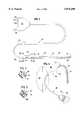
- FIG. 1is a plan view of the electrocautery probe of this invention
- FIG. 2is an enlarged, sectional view taken along line 2--2 of FIG. 1;
- FIG. 3is an enlarged, sectional view taken along line 3--3 of FIG. 1;
- FIG. 4is an enlarged, plan view of a surgical wound in a patient as shown during an intermediate stage of the surgery, and further showing how the electrocautery probe may be advanced through tissue to form a tunnel that extends between the wound and the skin, with the probe penetrating through the skin.
- electrocautery probe 10comprising a tissue compatible silicone rubber sleeve 12 which is proportioned to be of the diameter of a desirable surgical drainage tube, and being soft and of physical properties desirable for such drainage tubing.
- Electrode 14is connected to a wire 18, which is connected at its other end to wire 18a.
- Wire 18may be a single, thick wire, extending for the first few inches of the probe length.
- electrode 14is monopolar and conventionally suitable for use in electrocautery.
- Wire 18acomprises a group of thinner wires and connects to wire 18, being surrounded by a tubular insulating layer 20.
- Wire 18may or may not be surrounded by layer 20, and may be of greater wire diameter than wire 18a, as shown in FIGS. 2 and 3.
- Wire 18may comprise a thick, malleable wire, which not only provides electrical communication between wire 18a and electrode 14, but may be thick enough to be bent and to hold its shape, so that the surgeon may bend the forward portion 22 of probe 10 to a desired shape during use. As shown in FIG. 2, wire 18 fits loosely in sleeve 12 with extra space 27 instead of an insulating layer.
- the thick wire portion 18ends (at point 23), and connects by soldering or clamping with the group of smaller, flexible wires 18a, which extend proximally (rearwardly) within insulating sheath 20, providing an electrical conductor section of electrocautery probe 10 which is more flexible than portion 22.
- silicone rubber sleeve 12terminates at point 21, while two or three strand conductor 18a, surrounded by insulating layer 20, extends rearwardly further to terminate in a conventional electrical connector 24.
- Electrical connector 24carries three prongs, each of which could connect to a different one of three wires in multiple wire 18a, especially for use with a bipolar electrode.
- One of prongs 26may connect to ground; one of prongs 26 may connect to a first voltage source in a conventional electrocautery machine for coagulation; and the third of the prongs may connect to a second voltage source which is suitable for imposing a cutting voltage on electrode 14.
- a single prong 26may directly connect to a conventional electrocautery machine through an adapter without the conventional electrical connector 24.
- one or more of the wires of conductor 18amay connect to one single prong 26, which imposes a cutting voltage on monopolar electrode 14, and one or more wires 1a connects to ground through another prong 26.
- the third prongmay be a dummy prong, present to facilitate connection with a conventional connector receptacle.
- FIG. 4the use of the electrocautery probe of this invention is illustrated.
- a surgical wound 28 in the skin 30 of a patientis shown. Prior to sewing up the wound, surgical drains often need to be emplaced. To accomplish this, the electrocautery probe 10 of this invention is used. Probe 10 is advanced from the inside cavity of wound 28 through intact tissue of the patient under the skin 30, to exit the skin, as shown. The front probe portion may be manually curved as desired using thick wire 18. The probe is advanced until electrode 14 projects outwardly from the skin.
- Silicone sleeve 12advances through the entire length of the surgical tunnel 32 that is formed by this process.
- Power from power source 29provides the electrocautery action that permits the advancement of electrode 14 through the tissue and out of the skin.
- electrode 14may be transversely cut away or otherwise removed. Typically it may be transversely cut through somewhere along the length of silicone sleeve 12, preferably proximal of thick wire 18, such as at arrow 36 (FIGS. 1 and 4), and removed along with thick wire 18. Then, the remaining wire 18a, and its insulation 20 may be withdrawn retrograde out of sleeve 12 through wound 28, leaving behind a simple, silicone, tubular wound drain 12a which formerly surrounded the wire and insulation. Silicone tube 12a may be cut to its desired length, and wound 28 may be sutured, thus providing an installed wound drain, eliminating the need for several intermediate steps of the prior art installation of wound drains.
Landscapes
- Health & Medical Sciences (AREA)
- Life Sciences & Earth Sciences (AREA)
- Engineering & Computer Science (AREA)
- Animal Behavior & Ethology (AREA)
- Veterinary Medicine (AREA)
- Heart & Thoracic Surgery (AREA)
- Surgery (AREA)
- Biomedical Technology (AREA)
- Otolaryngology (AREA)
- General Health & Medical Sciences (AREA)
- Public Health (AREA)
- Anesthesiology (AREA)
- Physics & Mathematics (AREA)
- Plasma & Fusion (AREA)
- Nuclear Medicine, Radiotherapy & Molecular Imaging (AREA)
- Hematology (AREA)
- Medical Informatics (AREA)
- Molecular Biology (AREA)
- Surgical Instruments (AREA)
Abstract
Description
Claims (15)
Priority Applications (3)
| Application Number | Priority Date | Filing Date | Title |
|---|---|---|---|
| US08/784,735US5876400A (en) | 1997-01-13 | 1997-01-13 | Electrocautery method and apparatus |
| PCT/US1998/000528WO1998030158A1 (en) | 1997-01-13 | 1998-01-08 | Electrocautery method and apparatus |
| US09/192,787US6156035A (en) | 1997-01-13 | 1998-11-16 | Electrocautery method and apparatus |
Applications Claiming Priority (1)
| Application Number | Priority Date | Filing Date | Title |
|---|---|---|---|
| US08/784,735US5876400A (en) | 1997-01-13 | 1997-01-13 | Electrocautery method and apparatus |
Related Child Applications (1)
| Application Number | Title | Priority Date | Filing Date |
|---|---|---|---|
| US09/192,787DivisionUS6156035A (en) | 1997-01-13 | 1998-11-16 | Electrocautery method and apparatus |
Publications (1)
| Publication Number | Publication Date |
|---|---|
| US5876400Atrue US5876400A (en) | 1999-03-02 |
Family
ID=25133370
Family Applications (2)
| Application Number | Title | Priority Date | Filing Date |
|---|---|---|---|
| US08/784,735Expired - Fee RelatedUS5876400A (en) | 1997-01-13 | 1997-01-13 | Electrocautery method and apparatus |
| US09/192,787Expired - Fee RelatedUS6156035A (en) | 1997-01-13 | 1998-11-16 | Electrocautery method and apparatus |
Family Applications After (1)
| Application Number | Title | Priority Date | Filing Date |
|---|---|---|---|
| US09/192,787Expired - Fee RelatedUS6156035A (en) | 1997-01-13 | 1998-11-16 | Electrocautery method and apparatus |
Country Status (2)
| Country | Link |
|---|---|
| US (2) | US5876400A (en) |
| WO (1) | WO1998030158A1 (en) |
Cited By (45)
| Publication number | Priority date | Publication date | Assignee | Title |
|---|---|---|---|---|
| US6134467A (en)* | 1997-10-29 | 2000-10-17 | Asahi Kogaku Kogyo Kabushiki Kaisha | Drainage tube introducer for endoscope |
| US6395003B1 (en)* | 1998-09-08 | 2002-05-28 | Asahi Kogaku Kogyo Kabushiki Kaisha | Drainage tube indwelling device for endoscope |
| US20020107513A1 (en)* | 2000-04-27 | 2002-08-08 | Hooven Michael D. | Transmural ablation device with thin electrodes |
| US20020138109A1 (en)* | 2001-01-13 | 2002-09-26 | Medtronic, Inc. | Method and system for organ positioning and stabilization |
| US6517536B2 (en) | 2000-04-27 | 2003-02-11 | Atricure, Inc. | Transmural ablation device and method |
| US6605068B2 (en)* | 1999-07-13 | 2003-08-12 | Med Europe S.R.L. | Gravity drainage cord |
| EP1348392A1 (en)* | 2002-03-27 | 2003-10-01 | Integra Neurosciences Implants | Electrosurgical catheter-balloon |
| US20040092927A1 (en)* | 2002-11-05 | 2004-05-13 | Podhajsky Ronald J. | Electrosurgical pencil having a single button variable control |
| US20040260280A1 (en)* | 2003-05-01 | 2004-12-23 | Sartor Joe Don | Suction coagulator with dissecting probe |
| US20050107782A1 (en)* | 2003-11-19 | 2005-05-19 | Reschke Arlan J. | Pistol grip electrosurgical pencil with manual aspirator/irrigator and methods of using the same |
| US6905498B2 (en) | 2000-04-27 | 2005-06-14 | Atricure Inc. | Transmural ablation device with EKG sensor and pacing electrode |
| US6932811B2 (en) | 2000-04-27 | 2005-08-23 | Atricure, Inc. | Transmural ablation device with integral EKG sensor |
| US20050234444A1 (en)* | 2004-04-14 | 2005-10-20 | Hooven Michael D | Electrode and bipolar ablation method using same |
| US7156842B2 (en) | 2003-11-20 | 2007-01-02 | Sherwood Services Ag | Electrosurgical pencil with improved controls |
| US7156844B2 (en) | 2003-11-20 | 2007-01-02 | Sherwood Services Ag | Electrosurgical pencil with improved controls |
| US20070049926A1 (en)* | 2005-08-25 | 2007-03-01 | Sartor Joe D | Handheld electrosurgical apparatus for controlling operating room equipment |
| US20070049863A1 (en)* | 2003-01-14 | 2007-03-01 | Jahns Scott E | Devices and methods for interstitial injection of biologic agents into tissue |
| US7235072B2 (en) | 2003-02-20 | 2007-06-26 | Sherwood Services Ag | Motion detector for controlling electrosurgical output |
| US7288092B2 (en) | 2003-04-23 | 2007-10-30 | Atricure, Inc. | Method and apparatus for ablating cardiac tissue with guide facility |
| US7291161B2 (en) | 2002-10-02 | 2007-11-06 | Atricure, Inc. | Articulated clamping member |
| US20070260240A1 (en)* | 2006-05-05 | 2007-11-08 | Sherwood Services Ag | Soft tissue RF transection and resection device |
| US20080021446A1 (en)* | 2001-03-07 | 2008-01-24 | Swanson David K | Internal Indifferent Electrode Device For Use With Lesion Creation Apparatus And Method Of Forming Lesions Using The Same |
| US20080119846A1 (en)* | 2006-10-11 | 2008-05-22 | Rioux Robert F | Methods and apparatus for percutaneous patient access and subcutaneous tissue tunneling |
| US7393354B2 (en) | 2002-07-25 | 2008-07-01 | Sherwood Services Ag | Electrosurgical pencil with drag sensing capability |
| US7470272B2 (en) | 1997-07-18 | 2008-12-30 | Medtronic, Inc. | Device and method for ablating tissue |
| US20090054890A1 (en)* | 2007-08-23 | 2009-02-26 | Decarlo Arnold V | Electrosurgical device with LED adapter |
| US7503917B2 (en) | 2003-11-20 | 2009-03-17 | Covidien Ag | Electrosurgical pencil with improved controls |
| US20090149851A1 (en)* | 2007-12-05 | 2009-06-11 | Tyco Healthcare Group Lp | Thermal Penetration and Arc Length Controllable Electrosurgical Pencil |
| US7566334B2 (en) | 2004-06-02 | 2009-07-28 | Medtronic, Inc. | Ablation device with jaws |
| US20090248008A1 (en)* | 2008-03-31 | 2009-10-01 | Duane Kerr | Electrosurgical Pencil Including Improved Controls |
| US20090248018A1 (en)* | 2008-03-31 | 2009-10-01 | Tyco Healthcare Group Lp | Electrosurgical Pencil Including Improved Controls |
| US7628780B2 (en) | 2001-01-13 | 2009-12-08 | Medtronic, Inc. | Devices and methods for interstitial injection of biologic agents into tissue |
| US20090322034A1 (en)* | 2008-06-27 | 2009-12-31 | Cunningham James S | High Volume Fluid Seal for Electrosurgical Handpiece |
| US7740623B2 (en) | 2001-01-13 | 2010-06-22 | Medtronic, Inc. | Devices and methods for interstitial injection of biologic agents into tissue |
| US20100204696A1 (en)* | 2009-02-10 | 2010-08-12 | Tyco Healthcare Group Lp | Extension Cutting Blade |
| US7879033B2 (en) | 2003-11-20 | 2011-02-01 | Covidien Ag | Electrosurgical pencil with advanced ES controls |
| US20110112527A1 (en)* | 2009-11-06 | 2011-05-12 | Angiodynamics, Inc. | Flexible medical ablation device and method of use |
| US7967816B2 (en) | 2002-01-25 | 2011-06-28 | Medtronic, Inc. | Fluid-assisted electrosurgical instrument with shapeable electrode |
| US20120101339A1 (en)* | 2010-10-21 | 2012-04-26 | Brannon James K | Endoscopic surgery instrumentation |
| US8177760B2 (en) | 2004-05-12 | 2012-05-15 | C. R. Bard, Inc. | Valved connector |
| US8460289B2 (en) | 2005-06-28 | 2013-06-11 | Covidien Ag | Electrode with rotatably deployable sheath |
| US8636733B2 (en) | 2008-03-31 | 2014-01-28 | Covidien Lp | Electrosurgical pencil including improved controls |
| US9827140B2 (en) | 2013-07-17 | 2017-11-28 | William Thomas McClellan | Percutaneous blepharoplasty device and method |
| US11399888B2 (en) | 2019-08-14 | 2022-08-02 | Covidien Lp | Bipolar pencil |
| US11564732B2 (en) | 2019-12-05 | 2023-01-31 | Covidien Lp | Tensioning mechanism for bipolar pencil |
Families Citing this family (6)
| Publication number | Priority date | Publication date | Assignee | Title |
|---|---|---|---|---|
| US6471659B2 (en) | 1999-12-27 | 2002-10-29 | Neothermia Corporation | Minimally invasive intact recovery of tissue |
| US6705158B1 (en)* | 2000-12-22 | 2004-03-16 | Phil Louden | Hot wire anemometer with extendable probe |
| US20040093056A1 (en) | 2002-10-26 | 2004-05-13 | Johnson Lianw M. | Medical appliance delivery apparatus and method of use |
| US7637934B2 (en) | 2003-03-31 | 2009-12-29 | Merit Medical Systems, Inc. | Medical appliance optical delivery and deployment apparatus and method |
| US7604660B2 (en) | 2003-05-01 | 2009-10-20 | Merit Medical Systems, Inc. | Bifurcated medical appliance delivery apparatus and method |
| US20050215994A1 (en)* | 2003-08-21 | 2005-09-29 | Stephen Solomon | Tunneling device |
Citations (9)
| Publication number | Priority date | Publication date | Assignee | Title |
|---|---|---|---|---|
| US3595239A (en)* | 1969-04-04 | 1971-07-27 | Roy A Petersen | Catheter with electrical cutting means |
| US4976684A (en)* | 1988-11-21 | 1990-12-11 | Johnson & Johnson Orthopaedics, Inc. | Method of using a bendable trocar |
| US5197963A (en)* | 1991-12-02 | 1993-03-30 | Everest Medical Corporation | Electrosurgical instrument with extendable sheath for irrigation and aspiration |
| US5221281A (en)* | 1992-06-30 | 1993-06-22 | Valleylab Inc. | Electrosurgical tubular trocar |
| US5300070A (en)* | 1992-03-17 | 1994-04-05 | Conmed Corporation | Electrosurgical trocar assembly with bi-polar electrode |
| US5383876A (en)* | 1992-11-13 | 1995-01-24 | American Cardiac Ablation Co., Inc. | Fluid cooled electrosurgical probe for cutting and cauterizing tissue |
| US5417687A (en)* | 1993-04-30 | 1995-05-23 | Medical Scientific, Inc. | Bipolar electrosurgical trocar |
| US5429636A (en)* | 1993-10-08 | 1995-07-04 | United States Surgical Corporation | Conductive body tissue penetrating device |
| US5605539A (en)* | 1992-09-11 | 1997-02-25 | Urohealth Systems, Inc. | Self-introducing infusion catheter |
Family Cites Families (5)
| Publication number | Priority date | Publication date | Assignee | Title |
|---|---|---|---|---|
| US4592372A (en)* | 1984-05-22 | 1986-06-03 | Cordis Corporation | Pacing/sensing electrode sleeve and method of forming same |
| US4832048A (en)* | 1987-10-29 | 1989-05-23 | Cordis Corporation | Suction ablation catheter |
| US5242441A (en)* | 1992-02-24 | 1993-09-07 | Boaz Avitall | Deflectable catheter with rotatable tip electrode |
| US5348554A (en)* | 1992-12-01 | 1994-09-20 | Cardiac Pathways Corporation | Catheter for RF ablation with cooled electrode |
| US5437664A (en)* | 1994-01-18 | 1995-08-01 | Endovascular, Inc. | Apparatus and method for venous ligation |
- 1997
- 1997-01-13USUS08/784,735patent/US5876400A/ennot_activeExpired - Fee Related
- 1998
- 1998-01-08WOPCT/US1998/000528patent/WO1998030158A1/enactiveApplication Filing
- 1998-11-16USUS09/192,787patent/US6156035A/ennot_activeExpired - Fee Related
Patent Citations (10)
| Publication number | Priority date | Publication date | Assignee | Title |
|---|---|---|---|---|
| US3595239A (en)* | 1969-04-04 | 1971-07-27 | Roy A Petersen | Catheter with electrical cutting means |
| US4976684A (en)* | 1988-11-21 | 1990-12-11 | Johnson & Johnson Orthopaedics, Inc. | Method of using a bendable trocar |
| US5197963A (en)* | 1991-12-02 | 1993-03-30 | Everest Medical Corporation | Electrosurgical instrument with extendable sheath for irrigation and aspiration |
| US5300070A (en)* | 1992-03-17 | 1994-04-05 | Conmed Corporation | Electrosurgical trocar assembly with bi-polar electrode |
| US5599348A (en)* | 1992-03-17 | 1997-02-04 | Conmed Corporation | Electrosurgical trocar assembly |
| US5221281A (en)* | 1992-06-30 | 1993-06-22 | Valleylab Inc. | Electrosurgical tubular trocar |
| US5605539A (en)* | 1992-09-11 | 1997-02-25 | Urohealth Systems, Inc. | Self-introducing infusion catheter |
| US5383876A (en)* | 1992-11-13 | 1995-01-24 | American Cardiac Ablation Co., Inc. | Fluid cooled electrosurgical probe for cutting and cauterizing tissue |
| US5417687A (en)* | 1993-04-30 | 1995-05-23 | Medical Scientific, Inc. | Bipolar electrosurgical trocar |
| US5429636A (en)* | 1993-10-08 | 1995-07-04 | United States Surgical Corporation | Conductive body tissue penetrating device |
Non-Patent Citations (4)
| Title |
|---|
| Article by Michael F. Gleeson, MD, FACS entitled: "A Blunt-Tipped, Electronically-Controlled, Laparoscopic Trocar System" 5 pages, presented in Oct. 1994 at the annual meeting of the American College of Surgeons of Chicago, Illinois. |
| Article by Michael F. Gleeson, MD, FACS entitled: A Blunt Tipped, Electronically Controlled, Laparoscopic Trocar System 5 pages, presented in Oct. 1994 at the annual meeting of the American College of Surgeons of Chicago, Illinois.* |
| Brochure of ConMed Surgical Systems entitled: "Select One Minimal Access Surgery Systems", 6 pages. |
| Brochure of ConMed Surgical Systems entitled: Select One Minimal Access Surgery Systems , 6 pages.* |
Cited By (100)
| Publication number | Priority date | Publication date | Assignee | Title |
|---|---|---|---|---|
| US7470272B2 (en) | 1997-07-18 | 2008-12-30 | Medtronic, Inc. | Device and method for ablating tissue |
| US7678111B2 (en) | 1997-07-18 | 2010-03-16 | Medtronic, Inc. | Device and method for ablating tissue |
| US6134467A (en)* | 1997-10-29 | 2000-10-17 | Asahi Kogaku Kogyo Kabushiki Kaisha | Drainage tube introducer for endoscope |
| US6395003B1 (en)* | 1998-09-08 | 2002-05-28 | Asahi Kogaku Kogyo Kabushiki Kaisha | Drainage tube indwelling device for endoscope |
| US6605068B2 (en)* | 1999-07-13 | 2003-08-12 | Med Europe S.R.L. | Gravity drainage cord |
| US6932811B2 (en) | 2000-04-27 | 2005-08-23 | Atricure, Inc. | Transmural ablation device with integral EKG sensor |
| US20070135811A1 (en)* | 2000-04-27 | 2007-06-14 | Hooven Michael D | Method for ablating cardiac tissue |
| US7468061B2 (en) | 2000-04-27 | 2008-12-23 | Atricure, Inc. | Transmural ablation device with integral EKG sensor |
| US20020107513A1 (en)* | 2000-04-27 | 2002-08-08 | Hooven Michael D. | Transmural ablation device with thin electrodes |
| US7487780B2 (en) | 2000-04-27 | 2009-02-10 | Atricure, Inc. | Sub-xyphoid method for ablating cardiac tissue |
| US6517536B2 (en) | 2000-04-27 | 2003-02-11 | Atricure, Inc. | Transmural ablation device and method |
| US20050021024A1 (en)* | 2000-04-27 | 2005-01-27 | Hooven Michael D. | Transmural ablation device with temperature sensor |
| US20050033282A1 (en)* | 2000-04-27 | 2005-02-10 | Hooven Michael D. | Transmural ablation device with parallel electrodes |
| US6889694B2 (en) | 2000-04-27 | 2005-05-10 | Atricure Inc. | Transmural ablation device |
| US7543589B2 (en) | 2000-04-27 | 2009-06-09 | Atricure, Inc. | Method for ablating cardiac tissue |
| US6896673B2 (en) | 2000-04-27 | 2005-05-24 | Atricure, Inc. | Method for transmural ablation |
| US6899710B2 (en) | 2000-04-27 | 2005-05-31 | Atricure Inc. | Combination ablation and visualization apparatus for ablating cardiac tissue |
| US6905498B2 (en) | 2000-04-27 | 2005-06-14 | Atricure Inc. | Transmural ablation device with EKG sensor and pacing electrode |
| US6923806B2 (en) | 2000-04-27 | 2005-08-02 | Atricure Inc. | Transmural ablation device with spring loaded jaws |
| US20050171530A1 (en)* | 2000-04-27 | 2005-08-04 | Hooven Michael D. | Transmural ablation device |
| US7393353B2 (en) | 2000-04-27 | 2008-07-01 | Atricure, Inc. | Transmural ablation device with temperature sensor |
| US7241292B2 (en) | 2000-04-27 | 2007-07-10 | Atricure, Inc. | Cardiac ablation device with movable hinge |
| US6974454B2 (en) | 2000-04-27 | 2005-12-13 | Atricure, Inc. | Transmural ablation device with thermocouple for measuring tissue temperature |
| US6984233B2 (en) | 2000-04-27 | 2006-01-10 | Atricure, Inc. | Transmural ablation device with parallel electrodes |
| US7001415B2 (en) | 2000-04-27 | 2006-02-21 | Atricure, Inc. | Transmural ablation device |
| US7113831B2 (en) | 2000-04-27 | 2006-09-26 | Atricure, Inc. | Transmural ablation device |
| US7604634B2 (en) | 2000-04-27 | 2009-10-20 | Atricure, Inc. | Transmural ablation device |
| US6546935B2 (en) | 2000-04-27 | 2003-04-15 | Atricure, Inc. | Method for transmural ablation |
| US7507235B2 (en) | 2001-01-13 | 2009-03-24 | Medtronic, Inc. | Method and system for organ positioning and stabilization |
| US20020138109A1 (en)* | 2001-01-13 | 2002-09-26 | Medtronic, Inc. | Method and system for organ positioning and stabilization |
| US7740623B2 (en) | 2001-01-13 | 2010-06-22 | Medtronic, Inc. | Devices and methods for interstitial injection of biologic agents into tissue |
| US7628780B2 (en) | 2001-01-13 | 2009-12-08 | Medtronic, Inc. | Devices and methods for interstitial injection of biologic agents into tissue |
| US8758335B2 (en)* | 2001-03-07 | 2014-06-24 | Boston Scientific Scimed, Inc. | Internal indifferent electrode device for use with lesion creation apparatus and method of forming lesions using the same |
| US20080021446A1 (en)* | 2001-03-07 | 2008-01-24 | Swanson David K | Internal Indifferent Electrode Device For Use With Lesion Creation Apparatus And Method Of Forming Lesions Using The Same |
| US7967816B2 (en) | 2002-01-25 | 2011-06-28 | Medtronic, Inc. | Fluid-assisted electrosurgical instrument with shapeable electrode |
| FR2837693A1 (en)* | 2002-03-27 | 2003-10-03 | Nmt Neurosciences Implants Fra | BALLOON CATHETER |
| EP1348392A1 (en)* | 2002-03-27 | 2003-10-01 | Integra Neurosciences Implants | Electrosurgical catheter-balloon |
| US7621909B2 (en) | 2002-07-25 | 2009-11-24 | Covidien Ag | Electrosurgical pencil with drag sensing capability |
| US8016824B2 (en) | 2002-07-25 | 2011-09-13 | Covidien Ag | Electrosurgical pencil with drag sensing capability |
| US7393354B2 (en) | 2002-07-25 | 2008-07-01 | Sherwood Services Ag | Electrosurgical pencil with drag sensing capability |
| US7291161B2 (en) | 2002-10-02 | 2007-11-06 | Atricure, Inc. | Articulated clamping member |
| US8128622B2 (en) | 2002-11-05 | 2012-03-06 | Covidien Ag | Electrosurgical pencil having a single button variable control |
| US7244257B2 (en) | 2002-11-05 | 2007-07-17 | Sherwood Services Ag | Electrosurgical pencil having a single button variable control |
| US20070260239A1 (en)* | 2002-11-05 | 2007-11-08 | Podhajsky Ronald J | Electrosurgical pencil having a single button variable control |
| US20040092927A1 (en)* | 2002-11-05 | 2004-05-13 | Podhajsky Ronald J. | Electrosurgical pencil having a single button variable control |
| US20070049863A1 (en)* | 2003-01-14 | 2007-03-01 | Jahns Scott E | Devices and methods for interstitial injection of biologic agents into tissue |
| US8273072B2 (en) | 2003-01-14 | 2012-09-25 | Medtronic, Inc. | Devices and methods for interstitial injection of biologic agents into tissue |
| US7744562B2 (en) | 2003-01-14 | 2010-06-29 | Medtronics, Inc. | Devices and methods for interstitial injection of biologic agents into tissue |
| US7235072B2 (en) | 2003-02-20 | 2007-06-26 | Sherwood Services Ag | Motion detector for controlling electrosurgical output |
| US7955327B2 (en) | 2003-02-20 | 2011-06-07 | Covidien Ag | Motion detector for controlling electrosurgical output |
| US7288092B2 (en) | 2003-04-23 | 2007-10-30 | Atricure, Inc. | Method and apparatus for ablating cardiac tissue with guide facility |
| US7537594B2 (en)* | 2003-05-01 | 2009-05-26 | Covidien Ag | Suction coagulator with dissecting probe |
| US20040260280A1 (en)* | 2003-05-01 | 2004-12-23 | Sartor Joe Don | Suction coagulator with dissecting probe |
| US7241294B2 (en) | 2003-11-19 | 2007-07-10 | Sherwood Services Ag | Pistol grip electrosurgical pencil with manual aspirator/irrigator and methods of using the same |
| US20050107782A1 (en)* | 2003-11-19 | 2005-05-19 | Reschke Arlan J. | Pistol grip electrosurgical pencil with manual aspirator/irrigator and methods of using the same |
| US20090143778A1 (en)* | 2003-11-20 | 2009-06-04 | Sherwood Services Ag | Electrosurgical Pencil with Improved Controls |
| US7959633B2 (en) | 2003-11-20 | 2011-06-14 | Covidien Ag | Electrosurgical pencil with improved controls |
| US7879033B2 (en) | 2003-11-20 | 2011-02-01 | Covidien Ag | Electrosurgical pencil with advanced ES controls |
| US7156842B2 (en) | 2003-11-20 | 2007-01-02 | Sherwood Services Ag | Electrosurgical pencil with improved controls |
| US7503917B2 (en) | 2003-11-20 | 2009-03-17 | Covidien Ag | Electrosurgical pencil with improved controls |
| US7156844B2 (en) | 2003-11-20 | 2007-01-02 | Sherwood Services Ag | Electrosurgical pencil with improved controls |
| US8449540B2 (en) | 2003-11-20 | 2013-05-28 | Covidien Ag | Electrosurgical pencil with improved controls |
| US20050234444A1 (en)* | 2004-04-14 | 2005-10-20 | Hooven Michael D | Electrode and bipolar ablation method using same |
| US7530980B2 (en) | 2004-04-14 | 2009-05-12 | Atricure, Inc | Bipolar transmural ablation method and apparatus |
| US8177760B2 (en) | 2004-05-12 | 2012-05-15 | C. R. Bard, Inc. | Valved connector |
| US7875028B2 (en) | 2004-06-02 | 2011-01-25 | Medtronic, Inc. | Ablation device with jaws |
| US8162941B2 (en) | 2004-06-02 | 2012-04-24 | Medtronic, Inc. | Ablation device with jaws |
| US7566334B2 (en) | 2004-06-02 | 2009-07-28 | Medtronic, Inc. | Ablation device with jaws |
| US8460289B2 (en) | 2005-06-28 | 2013-06-11 | Covidien Ag | Electrode with rotatably deployable sheath |
| US20070049926A1 (en)* | 2005-08-25 | 2007-03-01 | Sartor Joe D | Handheld electrosurgical apparatus for controlling operating room equipment |
| US7828794B2 (en) | 2005-08-25 | 2010-11-09 | Covidien Ag | Handheld electrosurgical apparatus for controlling operating room equipment |
| US20070260240A1 (en)* | 2006-05-05 | 2007-11-08 | Sherwood Services Ag | Soft tissue RF transection and resection device |
| US8668688B2 (en) | 2006-05-05 | 2014-03-11 | Covidien Ag | Soft tissue RF transection and resection device |
| US20080119846A1 (en)* | 2006-10-11 | 2008-05-22 | Rioux Robert F | Methods and apparatus for percutaneous patient access and subcutaneous tissue tunneling |
| US8506565B2 (en) | 2007-08-23 | 2013-08-13 | Covidien Lp | Electrosurgical device with LED adapter |
| US20090054890A1 (en)* | 2007-08-23 | 2009-02-26 | Decarlo Arnold V | Electrosurgical device with LED adapter |
| US8235987B2 (en) | 2007-12-05 | 2012-08-07 | Tyco Healthcare Group Lp | Thermal penetration and arc length controllable electrosurgical pencil |
| US8945124B2 (en) | 2007-12-05 | 2015-02-03 | Covidien Lp | Thermal penetration and arc length controllable electrosurgical pencil |
| US20090149851A1 (en)* | 2007-12-05 | 2009-06-11 | Tyco Healthcare Group Lp | Thermal Penetration and Arc Length Controllable Electrosurgical Pencil |
| US20090248018A1 (en)* | 2008-03-31 | 2009-10-01 | Tyco Healthcare Group Lp | Electrosurgical Pencil Including Improved Controls |
| US8632536B2 (en) | 2008-03-31 | 2014-01-21 | Covidien Lp | Electrosurgical pencil including improved controls |
| US20090248008A1 (en)* | 2008-03-31 | 2009-10-01 | Duane Kerr | Electrosurgical Pencil Including Improved Controls |
| US9198720B2 (en) | 2008-03-31 | 2015-12-01 | Covidien Lp | Electrosurgical pencil including improved controls |
| US8663219B2 (en) | 2008-03-31 | 2014-03-04 | Covidien Lp | Electrosurgical pencil including improved controls |
| US20090248016A1 (en)* | 2008-03-31 | 2009-10-01 | Heard David N | Electrosurgical Pencil Including Improved Controls |
| US8663218B2 (en) | 2008-03-31 | 2014-03-04 | Covidien Lp | Electrosurgical pencil including improved controls |
| US20090248015A1 (en)* | 2008-03-31 | 2009-10-01 | Heard David N | Electrosurgical Pencil Including Improved Controls |
| US8591509B2 (en) | 2008-03-31 | 2013-11-26 | Covidien Lp | Electrosurgical pencil including improved controls |
| US8597292B2 (en) | 2008-03-31 | 2013-12-03 | Covidien Lp | Electrosurgical pencil including improved controls |
| US8636733B2 (en) | 2008-03-31 | 2014-01-28 | Covidien Lp | Electrosurgical pencil including improved controls |
| US8162937B2 (en) | 2008-06-27 | 2012-04-24 | Tyco Healthcare Group Lp | High volume fluid seal for electrosurgical handpiece |
| US20090322034A1 (en)* | 2008-06-27 | 2009-12-31 | Cunningham James S | High Volume Fluid Seal for Electrosurgical Handpiece |
| US20100204696A1 (en)* | 2009-02-10 | 2010-08-12 | Tyco Healthcare Group Lp | Extension Cutting Blade |
| US8231620B2 (en) | 2009-02-10 | 2012-07-31 | Tyco Healthcare Group Lp | Extension cutting blade |
| US20110112527A1 (en)* | 2009-11-06 | 2011-05-12 | Angiodynamics, Inc. | Flexible medical ablation device and method of use |
| US20130204223A1 (en)* | 2010-10-21 | 2013-08-08 | James K. Brannon | Endoscopic surgery instrumentation |
| US20120101339A1 (en)* | 2010-10-21 | 2012-04-26 | Brannon James K | Endoscopic surgery instrumentation |
| US9827140B2 (en) | 2013-07-17 | 2017-11-28 | William Thomas McClellan | Percutaneous blepharoplasty device and method |
| US11399888B2 (en) | 2019-08-14 | 2022-08-02 | Covidien Lp | Bipolar pencil |
| US11564732B2 (en) | 2019-12-05 | 2023-01-31 | Covidien Lp | Tensioning mechanism for bipolar pencil |
Also Published As
| Publication number | Publication date |
|---|---|
| WO1998030158A1 (en) | 1998-07-16 |
| US6156035A (en) | 2000-12-05 |
Similar Documents
| Publication | Publication Date | Title |
|---|---|---|
| US5876400A (en) | Electrocautery method and apparatus | |
| US11058336B2 (en) | System and method for laparoscopic nerve detection | |
| US8540706B2 (en) | Organ incision method | |
| US7819883B2 (en) | Method and apparatus for endoscopic access to the vagus nerve | |
| US8251991B2 (en) | Anchored RF ablation device for the destruction of tissue masses | |
| US9986896B2 (en) | Disposable sheath designs for the stimulating endoscope and needle endoscopes having distal electrodes for nerve block under direct vision and methods for making and using same | |
| US20180125566A1 (en) | Anchored rf ablation device for the destruction of tissue masses | |
| KR20000010815A (en) | Uvula, tonsil, adenoid and sinus tissue treatment device and method | |
| EP1987794B1 (en) | Endoscopic treatment instrument | |
| JP3171628B2 (en) | High frequency knife for endoscope | |
| US6387095B1 (en) | Surgical device comprising a radially expandable non-conductive sheath | |
| JP2528223B2 (en) | High Frequency Incision Tool for Endoscope | |
| US20130035639A1 (en) | Method for access needle with pre-loaded wire guide and device | |
| JP6698107B2 (en) | Endoscopic ablation device having an ablation wire guided through an opening in a tube to form two high frequency scalpels, and catheter for percutaneous endoscopic gastrostomy | |
| JP2019523059A (en) | Electrosurgical instrument with a single tubular conductive element for accessing anatomy | |
| CN114302756A (en) | Device for performing interventional endoscopic ultrasound surgery | |
| AU2020410838B2 (en) | EUS access device with electrosurgery-enhanced puncture | |
| JP2001522617A (en) | Apparatus and method for facilitating access to vessels in the human body | |
| EP3808316B1 (en) | Hot-puncture stent implantation device | |
| US8857441B2 (en) | Biological tissue transfer method and biological tissue treatment method | |
| SU1736450A1 (en) | Device for endoscopic papillosphincterotomy | |
| KR102569595B1 (en) | Endoscopic catheter | |
| AU604722B2 (en) | Marlin thoracic catheter | |
| RU2279258C2 (en) | Conductor for guiding catheters and probes | |
| WO2021088339A1 (en) | Cutting device for endoscope |
Legal Events
| Date | Code | Title | Description |
|---|---|---|---|
| AS | Assignment | Owner name:PIONEER LABORATORIES, INC., MICHIGAN Free format text:ASSIGNMENT OF ASSIGNORS INTEREST;ASSIGNOR:SONGER, MATTHEW N.;REEL/FRAME:008538/0001 Effective date:19970108 | |
| FPAY | Fee payment | Year of fee payment:4 | |
| FPAY | Fee payment | Year of fee payment:8 | |
| AS | Assignment | Owner name:PIONEER SURGICAL TECHNOLOGY, INC., MICHIGAN Free format text:CHANGE OF NAME;ASSIGNOR:PIONEER LABORATORIES, INC.;REEL/FRAME:020105/0308 Effective date:20061211 Owner name:PIONEER SURGICAL TECHNOLOGY, INC.,MICHIGAN Free format text:CHANGE OF NAME;ASSIGNOR:PIONEER LABORATORIES, INC.;REEL/FRAME:020105/0308 Effective date:20061211 | |
| AS | Assignment | Owner name:SONGER, MATTHEW N., DR.,MICHIGAN Free format text:ASSIGNMENT OF ASSIGNORS INTEREST;ASSIGNOR:PIONEER SURGICAL TECHNOLOGY, INC.;REEL/FRAME:024244/0211 Effective date:20100401 | |
| REMI | Maintenance fee reminder mailed | ||
| LAPS | Lapse for failure to pay maintenance fees | ||
| LAPS | Lapse for failure to pay maintenance fees | Free format text:PATENT EXPIRED FOR FAILURE TO PAY MAINTENANCE FEES (ORIGINAL EVENT CODE: EXP.); ENTITY STATUS OF PATENT OWNER: SMALL ENTITY | |
| STCH | Information on status: patent discontinuation | Free format text:PATENT EXPIRED DUE TO NONPAYMENT OF MAINTENANCE FEES UNDER 37 CFR 1.362 | |
| FP | Lapsed due to failure to pay maintenance fee | Effective date:20110302 |