US5870697A - Calculation of radiation therapy dose using all particle Monte Carlo transport - Google Patents
Calculation of radiation therapy dose using all particle Monte Carlo transportDownload PDFInfo
- Publication number
- US5870697A US5870697AUS08/610,917US61091796AUS5870697AUS 5870697 AUS5870697 AUS 5870697AUS 61091796 AUS61091796 AUS 61091796AUS 5870697 AUS5870697 AUS 5870697A
- Authority
- US
- United States
- Prior art keywords
- arrays
- particle
- energy
- transport
- input
- Prior art date
- Legal status (The legal status is an assumption and is not a legal conclusion. Google has not performed a legal analysis and makes no representation as to the accuracy of the status listed.)
- Expired - Lifetime
Links
Images


Classifications
- A—HUMAN NECESSITIES
- A61—MEDICAL OR VETERINARY SCIENCE; HYGIENE
- A61N—ELECTROTHERAPY; MAGNETOTHERAPY; RADIATION THERAPY; ULTRASOUND THERAPY
- A61N5/00—Radiation therapy
- A61N5/10—X-ray therapy; Gamma-ray therapy; Particle-irradiation therapy
- A61N5/103—Treatment planning systems
- A61N5/1031—Treatment planning systems using a specific method of dose optimization
- A61N2005/1034—Monte Carlo type methods; particle tracking
- A—HUMAN NECESSITIES
- A61—MEDICAL OR VETERINARY SCIENCE; HYGIENE
- A61N—ELECTROTHERAPY; MAGNETOTHERAPY; RADIATION THERAPY; ULTRASOUND THERAPY
- A61N5/00—Radiation therapy
- A61N5/10—X-ray therapy; Gamma-ray therapy; Particle-irradiation therapy
- A61N5/1042—X-ray therapy; Gamma-ray therapy; Particle-irradiation therapy with spatial modulation of the radiation beam within the treatment head
- Y—GENERAL TAGGING OF NEW TECHNOLOGICAL DEVELOPMENTS; GENERAL TAGGING OF CROSS-SECTIONAL TECHNOLOGIES SPANNING OVER SEVERAL SECTIONS OF THE IPC; TECHNICAL SUBJECTS COVERED BY FORMER USPC CROSS-REFERENCE ART COLLECTIONS [XRACs] AND DIGESTS
- Y10—TECHNICAL SUBJECTS COVERED BY FORMER USPC
- Y10S—TECHNICAL SUBJECTS COVERED BY FORMER USPC CROSS-REFERENCE ART COLLECTIONS [XRACs] AND DIGESTS
- Y10S378/00—X-ray or gamma ray systems or devices
- Y10S378/901—Computer tomography program or processor
Definitions
- the present inventionrelates to the use of radiation therapy to treat cancer patients, and more specifically, it relates to a method for calculating the radiation dose.
- the radiation sourcemay be may be in the form of external beams of ionizing particles or radioactive sources internal to the patient.
- FIG. 1Ashows a radiation beam 1 as it is delivered to the patient. External beams are usually produced by machines acting as particle accelerators.
- the beam delivery systemconsists of the radiation source 5 (see FIG. 1B), which is mounted on a gantry 2 which can rotate about a 360° arc around the patient. Each beam is shaped by a rotatable collimator 3. The patient lies on a rotatable table 4.
- the gantry 2 and table 4both rotate about a single isocenter, as shown in FIG. 1.
- External beam radiation therapyis performed with several types of ionizing radiation. Approximately 80% of patients are treated with photons, ranging in maximum energy from 250 keV to 25 MeV. The balance are treated primarily with electrons with energies from 4 to 25 MeV. In addition, there are several fast neutron and proton therapy facilities which have treated thousands of patients worldwide. Fast neutron therapy is performed with neutron energies up to 70 MeV, while proton therapy is performed with proton energies ranging from about 50 to 250 MeV. Boron neutron capture therapy is conducted with thermal and epithermal neutron sources. Most internal radioactive sources irradiate the patient with photons, although some sources emit low energy electrons.
- the effects of ionizing radiation on the bodyare quantified as radiation dose.
- Absorbed radiation doseis defined as the ratio of energy deposited to unit mass of tissue. Because tumors and sensitive structures are often located in close proximity, accuracy in the calculation of dose distributions is critically important.
- the goal of radiation therapyis to deliver a lethal dose to the tumor while maintaining an acceptable dose level in surrounding sensitive structures. This goal is achieved by computer-aided planning of the radiation treatments to be delivered.
- the treatment planning processconsists of characterizing the individual patient's anatomy (most often, this is done using a computed tomography (CT) scan), determining the shape, intensity, and positioning of radiation sources, and calculating the distribution of absorbed radiation dose in the patient.
- CTcomputed tomography
- Monte Carlo transportis the most accurate method of determining dose distributions in heterogeneous media.
- a computeris used to simulate the passage of particles through an object of interest.
- the present inventioncalculates dose in the body using three-dimensional Monte Carlo transport. Neutrons, protons, deuterons, tritons, helium-3, alpha particles, photons, electrons, and positrons are transported in a completely coupled manner, using the LLNL-developed Monte Carlo All-Particle Method (MCAPM).
- MCAPMMonte Carlo All-Particle Method
- the usercan define the Cartesian transport mesh either manually or from a computed tomography (CT) scan of the patient. If the transport mesh is obtained from a CT scan, the x and y zone dimensions are defined as the dimensions of each pixel on the CT slice, while the z dimension is the spacing between CT slices. Each zone within the mesh is assigned a material number which is associated with a specific atomic composition and density. Materials are assigned using a software thresholding algorithm based on ranges of CT numbers or can be assigned manually.
- CTcomputed tomography
- the radiation sourcecan be defined as a set of external beams or internal radioactive sources.
- Each element of the radiation source(beam or internal source) is characterized by its shape and position in space, as well as the distribution in energy and direction of particles it emits.
- the radiation sourceis defined as a complex beam or set of beams in a standard clinical isocentric gantry and table geometry.
- the userspecifies the accelerator gantry, collimator, and table angles, a well as the (x,y,z) location of the table/accelerator center of rotation (isocenter) in the transport mesh.
- the energy and direction of each particleare described at a plane in space (the beam definition plane) that is defined to be perpendicular to the ray connecting the radiation source and the gantry/table isocenter.
- FIG. 1Bshows the relationship between the radiation source 5, collimator jaws 7, and beam definition plane 6.
- Radiation beam 1 from radiation source 5, on gantry 2passes through beam definition plane 6, collimator jaws 7, compensator 8 and block 9 onto table 4. Particles are transported from the point source to the beam definition plane.
- the beam definition planeis divided into a series of concentric rings. Each concentric ring may have a different energy and angular distribution associated with it. Depending on where the particle lands on the beam definition plane, it is given a new energy and angle by sampling from energy and angular distributions chosen from digital computer libraries that include monoenergetic, monodirectional options as well as energy and angular distributions derived from Monte Carlo simulations of actual beam delivery systems.
- the angular distribution(s) chosen by the usermodify the trajectory of the particle at the beam definition plane.
- the field shapeis defined as the particles pass through collimator jaws and an optional compensator and/or field-shaping block. Transport of particles through the collimator jaws is accomplished by ray tracing, while Monte Carlo methods are used to transport through the compensator and block. Once each particle exits the beam collimation system (jaws, compensator, and block) it is transported to the patient mesh.
- particlesare produced from point or line sources, with energies determined (sampled) from radioisotope decay data and angles taken from angular distributions determined from Monte Carlo simulations of actual sources.
- the position and direction of each internal source particleis determined from the user-specified position of the source in the patient.
- the inventioncarries out Monte Carlo transport calculations in the patient coordinate system as follows. First, a particle is selected from the radiation source. Depending on its type, it is passed to the appropriate neutron, photon, electron/positron, or heavy charged-particle transport package, where it is tracked until it undergoes a collision. For charged particles (electrons, positrons and heavy charged particles), energy is deposited by combining the distance traveled with predetermined stopping power (slowing down) information. Each nuclear reaction and photon interaction is handled by calling the Monte Carlo All Particle Method (MCAPM) reaction physics package. Given an incident particle, this package returns the energies and angles of all secondary particles resulting from the collision. These particles are stored in the secondary bank. Control is then returned to the Monte Carlo tracker, which selects a particle from the secondary bank or, when the bank is exhausted, from the radiation source. This process continues until all particles in the source and secondary bank have been followed.
- MCAPMMonte Carlo All Particle Method
- the MCAPM software packageis a system of data and algorithms that accomplishes the particle collision physics required for coupled Monte Carlo simulation of many particle types.
- the nuclear and atomic data libraries mentioned abovedescribe the interaction of neutrons, photons, electrons, and heavy charged-particles with matter. Each interaction may result in the production of any of these particles. Additional data describing electron Coulomb interactions are included in the present invention.
- each particlepossesses a discrete energy value, allowing the collision kinematics to be performed on a continuous energy basis.
- This schemeexploits the strengths of both the pure continuous energy method (storing as much information as necessary to completely describe the energy dependency of each collision parameter) and the pure multi-group method (storing all collision parameters on an energy group or bin basis), allowing a smaller cross section database and while retaining accurate kinematics.
- Photon cross-sectionsare interpolated between discrete energy points. Neutrons and photons are transported using standard analog Monte Carlo methods. Energy transferred to particles that are not being tracked is deposited locally, with expected energy deposition providing variance reduction.
- the condensed history methoduses transport physics ideas to combine the effects of multiple interactions as a particle travels a specified distance.
- the particle's energy and trajectoryare modified using Gaussian like distributions with variances determined from the Rutherford scattering cross section (energy straggling) and from the Highland approximation to a Moliere distribution (multiple scattering).
- Electron and positronsare transported using a traditional Class II condensed history method. Large-angle Coulomb scattering and bremsstrahlung production are modeled on an event-by-event basis, while all other Coulomb interactions are accounted for with the condensed history method. After each condensed history step, the electron's (or positron's) trajectory is modified by sampling from a Moliere distribution. The invention may in the future use the Macro Response Monte Carlo Method as an alternative to condensed history.
- the Monte Carlo transport packagecalculates the energy deposited from the radiation source, creating a three-dimensional Cartesian map of energy deposited in the patient.
- the energy deposition mapis converted to a map of absorbed dose, and is reported back to the user (client treatment planning code).
- Input(description of the patient and radiation sources, and user options for code control) and output (a three-dimensional dose map) are read and written in a set of files that conform to the Specifications for Tape/Network Format for Exchange of Treatment Planning Information, Version 3.00 (Jan. 10, 1994), currently maintained by the NCI Prostate Collaborative Working Group RTOG 3D QA Center, Washington University in St. Louis. These files will be referred to as AAPM standard files.
- the form of input and output datamay change in the future in order to accommodate new industry standards or to allow more direct linkage to commercial patient treatment planning system.
- FIG. 1Ashows the relationship between the gantry, collimator, table, and radiation beam for external beam radiation therapy delivery.
- FIG. 1Bshows the relationship between the source, collimator, and beam definition plane.
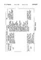
- FIG. 2shows a top-level view of the process flow of the present invention.
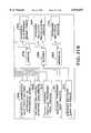
- FIG. 3shows the code at its highest level.
- FIGS. 4A and 4Bdescribe the subroutines that make up Generator 20.
- FIG. 5shows the program flow that makes up Generator 20.
- FIG. 6shows another view of the program flow, data structure, output and subroutine functions of Generator 20.
- FIGS. 7 through 15show detailed views of the program flow of each subroutine of Generator 20.
- FIGS. 16 through 19describe the subroutines that make up the All-Particle Tracker (MCXEC) 22.
- FIG. 20shows the program flow of the All-Particle Tracker 22.
- FIG. 21shows another view of the overall flow, data input, output and subroutine functions of the All-Particle Tracker 22.
- FIGS. 22 through 31show detailed views of the program flow of each subroutine of the All-Particle Tracker 22.
- FIG. 32shows the program flow of the postprocessor 24.
- FIG. 33shows the data flow for the postprocessor 24.
- the inventioncalculates the radiation dose in the body using three-dimensional Monte Carlo transport. Neutrons, protons, deuterons, tritons, helium-3, alpha particles, photons, electrons, and positrons are transported in a completely coupled manner, using the Lawrence Livermore National Laboratory (LLNL) developed Monte Carlo All-Particle Method (MCAPM).
- MCAPMMonte Carlo All-Particle Method
- the major elements of the inventioninclude: computer hardware and software, a digital data file containing the description of the patient (a CT scan), a description of the radiation source, nuclear and atomic interaction databases, Monte Carlo transport software, and digital output files containing dose distributions.
- An example of the type of computer hardware usable in accordance with the inventionincludes: a generic UNIX based workstation with one or more Central Processing Units (CPUs) operating under UNIX (the invention is currently operational on the following: Sun Microsystems SPARC series, Silicon Graphics SGI-R4000 series, IBM RS6000 series, etc.), 128 Mbytes of random access memory available to each processor, a 1 Gbyte internal hard disk to store input/output information required and generated by the invention, a color monitor to view the CT scan and dose results (preferably at least 17 inches) and a standard keyboard for program execution and control.
- CPUsCentral Processing Units
- SGI-R4000 seriesSilicon Graphics SGI-R4000 series
- IBM RS6000 seriesetc.
- 128 Mbytes of random access memoryavailable to each processor
- a 1 Gbyte internal hard diskto store input/output information required and generated by the invention
- a color monitorto view the CT scan and dose results (preferably at least 17 inches)
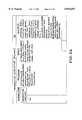
- FIG. 2shows a top-level view (12) of the process and the invention.
- a Cartesian transport meshis defined either manually or from a computed tomography (CT) scan (13) and input into a commercial patient treatment planning system (14). Additional data about the patient, the radiation source prescribed, and other user options are included with the CT data (15) and passed to the executive (16). In addition, nuclear and atomic data (17), are input into the Executive program (16), which manages the Monte Carlo dose calculations process (18) and returns to the Planning System (14) a 3-D map (19) of the dose delivered to the patient.
- CTcomputed tomography
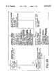
- FIG. 3is a structure diagram of the code at its highest level.
- the executive process, PGXEC (16)manages three major functions: (i) the generation process (Generator 20 called GENXEC) which sets up all the patient data, radiation source data, and physics data needed to do the dose calculation, (ii) the All Particle Monte Carlo physics dose calculation (All Particle Tracker 22 called MCXEC), and (iii) the final process of formatting the dose calculation and passing it back for viewing (Post Processor 24 called POSTXEC.
- the generator (20), all-particle tracker (22) and post-processor (24)are all controlled by the executive program (16).
- the executive program 16manages the three highest level subroutines; the generator (20), the all-particle tracker (22) and the post-processor (24).
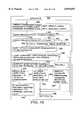
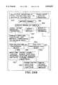
- Input to the generator (20)includes: (i) a CT scan data array, (ii) nuclear/atomic/electron data, (iii) the radiation source specification and (iv) user options.
- the output from the generator (20)includes: (i) a material specification array (muscle, bone, fat, etc.), (ii) material data (composition, density, etc.), (iii) particle data (mass, charge, etc.), (iv) a nuclear and atomic transport data array, (v) radiation source angular and energy distributions; and (vi) arrays describing beam delivery components.
- the output from generator (20)is input to the all-particle tracker (22) which executes the Monte Carlo calculation and provides output as a 3D map of energy deposited and a 3D map of standard deviation of energy deposited.
- the output from the all-particle trackeris provided as input to the post-processor 24, which formats a dose map for the appropriate output.
- Some of the output from Generator (20)is also provided as input to post-processor 24 in the form of a material specification array and material data (composition, density, etc.).
- the output from post-processor 24is a 3D map of the dose that has been delivered to the patient.
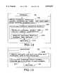
- FIGS. 4A and 4Bdescribe the subroutines that make up Generator 20.
- GENOPTIONS (32)determines user-specific options and sets switches for code control. Referring to FIG. 4A, input to GENOPTIONS (32) include user options: (i) Monte Carlo parameters, (ii) physics options and (iii) output options. The output of GENOPTIONS (32) sets the switches for code control.
- GENGRPS (34)determines the number of energy groups for transport of each particle type. Input to GENGRPS (34) is nuclear/atomic data.
- GENMESH (36)uses CT information to define dimensions and material composition for each CT voxel. The input to GENMESH (36) is a CT scan array and user options (user-specified thresholds for processing CT scan).
- the output from GENMESH (36)is a material specification array (muscle, bone, fat, etc. assigned to each zone).
- GENMATS (38)reads user-drawn contours that describe patient structures (control volumes) and modifies the material specification array. User options (user-drawn contours) and a material specification array (muscle, bone, fat, etc., assigned to each zone) are input into GENMATS (38).
- the output of GENMATS (38)is a modified material specification array and the standard deviation zone ID array.
- GENBEAM (40)reads user radiation source specification input specifying each radiation beam source.
- the output of GENBEAM (40)is the radiation source angular and energy distributions and arrays describing beam delivery components.
- GENSEED (42)reads user input specifying each internal (brachytherapy) source (if specified).
- the input to GENSEED (42)is the radiation source specification (internal sources).
- the output of GENSEED (42)is radiation source angular and energy distributions.
- GENMATSII (44)completes the final setup for the material arrays in preparation for calling the subroutine 46. Its input is material composition data which is data defined internally in the software.
- the output from 44is material data such as internal material lists, isotope data, etc.
- GENXSN (46)reads nuclear and atomic data and material and isotope data, and constructs arrays needed to do the physics of the transport.
- the output from GENXSN (46)is nuclear and atomic transport data arrays, heavy charged particle transport data arrays and an energy group structure for each particle type.
- GENELDAT (48)reads electron data and constructs transport arrays.
- Input to GENELDAT (48)includes electron data and material data and the output from the subroutine is electron transport data arrays.
- FIG. 5shows the program flow that makes up Generator (20).
- GENOPTIONS (32)reads files to determine user-specified options and to set switches for code control.
- GENGRPS (34)accesses nuclear and atomic data to determine the number of energy groups for transport of each particle type.
- GENMESH (36)uses CT information to define dimensions and material composition for each CT voxel.
- GENMATS (38)reads user-drawn contours that describe patient structures such as control volumes, modifies the material specification array, and defines standard deviation identification zones.
- GENSEED (42)reads user input specifying each internal brachytherapy source; and if there are any sources, establishes code controls for a brachytherapy problem.
- GENBEAM(40) reads user input specifying each radiation beam source and establishes code controls for an external problem.
- GENMATSII(44) completes the final setup for the material arrays in preparation for calling the nuclear and atomic data generator GENXSN (46). If photons, neutrons, and/or heavy charged particles were selected for tracking, GENXSN (46) reads nuclear and atomic data and constructs transport arrays. If electrons and/or positrons were selected for tracking, GENELDAT (48) reads electron data and constructs transport arrays.
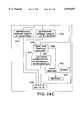
- FIG. 6Still another view of the program flow, data structure, data output and subroutine functions of GENERATOR (20) are shown in FIG. 6.
- Input 50comprising Monte Carlo parameters, physics options and output options is fed into GENOPTIONS (32), which determines user-specified options and sets switches for code control to produce output 66, which sets the switches for code control.
- Input 52comprising user options such as user-drawn contours, is fed into GENMATS (38), which reads user-drawn contours that describe patient structures (control volumes) and modifies the material specification array to provide output 70, comprising a material specification array, and output 72 comprising a standard deviation zone ID array.
- Input 54consisting of user options (user-specified thresholds for processing CT scan), is fed into GENMESH (36) which uses CT information to define dimensions and material compositions for each CT voxel.
- GENMESH (36)provides output 70 consisting of a material specification array (muscle, bone, fat, etc. assigned to each zone).
- Input 56comprising nuclear/atomic/electron data is fed into GENGRPS (34), GENXSN (46) and GENELDAT (48).
- GENGRPS (34)determines the number of energy groups for transport of the particle type and provides the output 68 of the number of energy groups for each particle type.
- Output 68is also fed into GENXSN (46) which reads nuclear and atomic data and constructs transport arrays.
- GENXSN(46) produces output 80, 82 and 84.
- Output 80consists of nuclear and atomic transport data arrays.
- Output 82consists of heavy-charged particle transport data arrays.
- Output 84consists of an energy group structure for each particle type.
- Input 56also fed into GENELDAT (48), produces output 86 which comprises electron transport data arrays.
- Input 58comprising a CT scan array is also fed into GENMESH (36) along with input 54.
- Input 60comprising radiation source specification (external beam characteristics and modifiers) is fed into GENBEAM (40) which reads user input specifying each radiation beam source to produce the output 74 and 76.
- Output 74consists of radiation source angular and energy distributions and output 76 consists of arrays describing beam delivery components.
- Input 62comprising radiation source specification (internal sources) is fed into GENSEED (42) and produces output 74 consisting of radiation source angular and energy distributions.
- Input 64consisting of material composition data (data defined internally in this code) is fed into GENMATSII (44) which completes the final setup for material arrays in preparation for calling GENXSN (46).
- Output 78 from GENMATSII (44)comprises problem-dependent material and isotope specification arrays and is fed into GENXSN (46) and GENELDAT (48).
- FIGS. 7 through 15Detailed views of the program flow of each subroutine of the Generator 20 are shown in FIGS. 7 through 15.
- GENOPTIONSbegins by setting the AAPM standard file reading options (100).
- AAPM standard filesparticle types to track, transport options, Monte Carlo sample size, random number seed and output options.
- the internal transport flagsare then set for code control (104).
- the minimum energyis then set for transport of each particle type (106).
- the program flowthen returns (108) to the calling routine (Generator 20).
- GENGRPS (34)previews nuclear and atomic data files (112) for each particle type (110) to obtain the number of energy groups.
- subroutine GENMESHobtains CT scan information (116) from AAPM Standard Interface files, which includes the CT numbers, user-defined material thresholds and zone dimensions.
- CT scan information(116) from AAPM Standard Interface files, which includes the CT numbers, user-defined material thresholds and zone dimensions.
- the existence of CT datais checked (118) and if there is no CT data, the program returns (126) to the calling routine (Generator 20).
- user-defined material thresholdsare used (120) to assign a material to each zone.
- Mesh-dependent transport parameters including internal/external material conversion arrays and tolerance factorsare then set (122).
- maximum transport mesh dimensions in the x, y, and z directionare set (124). The program flow then returns (126) to the calling routine.
- GENMATS (38)obtains contour (structure) information from AAPM Standard Interface files (128). The program then checks (130) for the existence of structures and if there are no structures, the program returns (148) to the calling routine (Generator 20). If structures exist, then GENMATS (38) checks (132) for the existence of a CT scan. If there is no CT scan, the transport mesh is first filled with air (134). Next the structures (136) are sorted from the foreground to the background. The routine then loops (138) through the structures, starting from background and ending in foreground. In this loop, the zone whose midpoint is the interior of the structure is identified. It is determined if user-specified material identification for this structure is less than 0 (142).
- a user-specified material IDis assigned (144) to each interior zone. Then, the user-specified material is added (146) to the internal material list if necessary. The program then returns to loop through the structures (138). If the user-specified material ID for the structure is less than 0, the interior zones are identified (150) as one standard deviation super-zone. The program flow then returns to loop through the structures (138). After looping through the structures, starting from the background to the foreground, until all the structures have been processed, GENMATS 38 returns (148) to the calling routine (Generator 20).
- GENBEAMbegins (152) by obtaining beam information from AAPM Standard Interface files. The existence of beams is determined (154) and if there are none, the routine returns (196) to the calling routine. If beams do exist, characteristics are read (158) from a user-specified machine description file. For each source at 160, it is determined if a block exists (162). If no block exists, a block is initialized (164) to not cover any part of the field. The existence of a compensator is then checked (166). If a block exists (162), a control flag is checked for filled or open block contours (163). If open contours are specified, the block is initialized (165) to cover the entire field.
- each block pixel that is interior to the contouris identified (168). All pixels that are in the block that are interior to the contour are cut out (169). If filled contours are specified (163), the block is initialized (170) to not cover any of the field.
- each block pixel that is interior to the contouris identified (172). All pixels that are in the block that are interior to the contour are filled (173).
- Block materialis then added (176) to the material list. The existence of the compensator is checked (166). The compensator raster is then filled (178) with user-supplied data, which specifies the thickness of the compensator at each pixel in the raster. Compensator material is added (180) to the material list.
- Collimator jaw materialis assigned (182) and added to the material list (184). For each source area segment (186), sampling arrays are set up for the energy distribution (188) and angular distribution (190). Miscellaneous transport parameters are then set up (192). When all source area segments are looped, then flow returns to (160) for additional sources. After all characterization of all beams is complete, temporary beam/machine arrays are closed (193) and the program flow returns (196).
- GENSEED (42)begins by obtaining internal radiation source information from AAPM Standard Interface files (198). For each radiation source (200), the energy distribution is selected (202) based on the internal source type. The maximum particle energy and group is then updated (204). Program flow then returns to step 200 for each internal radiation source until exhausted and then returns (206) to the calling routine.
- GENMATSII(44) obtains isotope lists that include only elements used in this problem (208). The average atomic number and the average ratio of atomic to mass number are then calculated (210), and the program flow returns (212) to the calling routine.
- GENXSNbegins (214) by initializing the cross sections and transport data arrays.
- step 216for each particle type except electrons and positrons, for each isotope at step 218, step 220 reads nuclear and atomic data, determines energy boundaries, microscopic total cross sections, and expected energy depositions.
- step 220returns to step 216 for each particle type except electrons and positrons and also returns to step 218 for each isotope.
- Step 224 for each particle type material and energy group identified at step 222calculates the mean free path, expected energy deposition, and, if photon and electron transport are on, the mean free path for Woodcock tracking.
- step 226a check is made for heavy charged particles and if none exist the routine continues with step 236. If charged particles do exist, the stopping power is calculated at step 230 for each isotope identified at step 228. At step 234 collision probability and range arrays are set up for each material identified at step 232. Step 236 sets up parameters and arrays for multiple scattering and energy straggling of charged particles (both heavy and light). The program returns to the calling routine (Generator 20) at step 238.
- the subroutine GENELDATcalculates the electron collision and radiative stopping power (step 242), converts (step 244) the electron collision stopping power to electron and positron restricted collision stopping power, for each identified material (step 240).
- Step 248calculates the mean free path to Moller event at step 246 for each energy group and material.
- Step 250obtains bremsstrahlung cross sections from the data file, and reintegrates them to reflect the correct the low-energy cutoff.
- Step 254sets up bremsstrahlung photon energy spectrum sampling arrays for each energy group isotope identified at step 252.
- Step 258calculates the mean free path to bremsstrahlung event for each energy group and material identified at step 256 and then returns to the calling routine at step 260.
- FIGS. 16 through 19describe the subroutines that make up the All-Particle Tracker 22 (MCXEC).
- BEAMSEL(262) selects particle attributes for a primary particle arising from an external radiation beam.
- the input to this routinecomprises (i) radiation source angular and energy distributions for external sources, (ii) arrays describing beam delivery components, (iii) material data (internal material lists, isotope data, etc.), (iv) nuclear and atomic transport data arrays and (v) number and energy group structure for each particle type.
- the output from this routineprovides attributes of one particle (energy, location, direction, type).
- the subroutine BRACHYSELselects particle attributes for a primary particle arising from an internal (brachytherapy) radiation source.
- the input to this subroutineis radiation source angular and energy distributions for internal sources.
- the subroutine outputprovides attributes of one particle (energy, location, direction, type).
- the subroutine SECSEL(266) selects a particle that has been previously created and stored by an interaction of another particle in the transport calculation, from an input of secondary particle arrays to provide an output of the attributes of one particle.
- NEUTRAN (268)tracks a neutron through a mesh (patient), and records the energy deposited by the neutron and stores the attributes of secondary particles produced in secondary particle arrays.
- the input to this routinecomprises (i) attributes of one particle, (ii) switches for code control, (iii) material specification array, (iv) material data, (v) nuclear transport data arrays and (vi) number and energy group structure for neutrons.
- the output of NEUTRAN (268)is secondary particle arrays and a 3-D energy deposit map (array).
- PHOTRAN (270)tracks the photon through the transport mesh and records the energy deposited by the photon. It also stores the attributes of secondary particles produced in secondary particle arrays.
- the input to PHOTRAN (270)comprises (i) the attributes of one particle, (ii) switches for code control, (iii) material specification array, (iv) material data, (v) atomic transport data arrays and (vi) number and energy group structure for photons. Secondary particle arrays and 3-D energy deposit map are output from PHOTRAN (270).
- CPTRAN (272)tracks a heavy charged particle through the patient transport mesh and records energy deposited by the heavy charged particle.
- CPTRAN (272)also stores attributes of secondary particles produced in secondary particle arrays.
- the input to CPTRAN (272)comprises (i) switches for code control, (ii) a material specification array, (iii) material data, (iv) nuclear transport data arrays, (v) heavy charged particle transport data arrays, and (vi) the number and energy group structure for heavy charged particles.
- the output from this subroutineis secondary particle arrays and a 3-D energy deposit map (array).
- ELTRAN (274)tracks a primary electron through the patient transport mesh, records energy deposited recorded by the electron and stores the attributes of secondary particles produced in secondary particle arrays.
- a primary electronis defined as an electron originating in a radiation source (created by BEAMSEL or BRAYCHYSEL).
- the input to ELTRAN (274)consists of (i) attributes of one particle which is energy, location, direction, type, (ii) switches for code control, (iii) material specification arrays, (iv) material data, (v) electron transport data arrays, and (vi) number and energy group structure for electrons.
- ELTRAN (274)outputs secondary particle arrays and a 3-D energy deposit map (array).
- ELTRANItracks a secondary electron through the transport mesh of the patient, records energy deposited by the electron and stores attributes of secondary particles produced in secondary particle arrays.
- the input to ELTRANI (276)is (i) attributes of one particle, (ii) switches for code control, (iii) a material specification array, (iv) material data, (v) electron transport data array, and (vi) number and energy group structure for electrons.
- ELTRANI (276)outputs secondary particle arrays and a 3-D energy deposit map (array).
- UPDATE (278)adds a 3-D energy deposit map calculated over batch to a 3-D energy deposit map for the problem.
- the input to this subroutineis a 3-D energy deposit map calculated for a single batch.
- the output of this subroutineis an integral 3-D energy deposit map (array).
- STDDEV (280)updates arrays necessary for a standard deviation calculation with energy deposit information determined from each batch.
- the inputis a 3-D energy deposit map and standard deviation zone ID array.
- the output from STDDEV (280)is standard deviation precalculation arrays.
- STDDEVX (282)calculates the standard deviation from standard deviation precalculation arrays to produce standard deviation arrays.
- FIG. 20shows the program flow of the All-Particle Tracker 22.
- Step 288samples particle attributes and step 290 tracks the particle through the patient, records the energy deposited by the particle, and stores the secondary particle produced.
- Step 286loops over each Monte Carlo particle in the batch.
- Step 292updates the standard deviation arrays and batch-dependent energy deposit arrays before looping back to step 284 to continue the next batch of Monte Carlo particles. When all batches are complete the standard deviation is calculated at step 294.
- FIG. 21Still another view of the flow, data input, output and subroutine functions are shown in FIG. 21.
- the inputs for the subroutines that make up the All-Particle Trackerare shown at 296, 298, 300, 302, 304, 306, 308, 310, 312 and 314 and comprise respectively radiation source angular and energy distributions, arrays describing beam delivery components, setting switches for code control, number and energy group structure for each particle type, material specification array, material composition data, nuclear and atomic transport data arrays, heavy charged particle transport data arrays, electron transport data arrays and a standard deviation zone ID array.
- Output 316provides attributes of one particle. Secondary particle arrays are provided by output 318.
- Output 320shows the 3-D energy deposit array and 322 comprises the integral 3-D energy deposit array.
- Output 324is a standard deviation precalculation array.
- Output 326provides the standard deviation arrays.
- FIGS. 16 through 19describe (supra) the subroutines that make up the All-Particle Track
- step 328selects the beam and particle attributes at the radiation point source.
- the initial position point sourceis sampled at step 330.
- the initial trajectory at the radiation point sourceis chosen at step 332.
- Step 334performs the following functions: (i) transport the particle to the beam definition plane, (ii) update the weight according to user-specified function and (iii) choose energy and direction from user-requested energy and angular displacement distributions.
- Step 336transport a particle through the collimator jaws.
- Step 338questions whether the particle emerged from the collimator, and if it did not, then the program flow returns to step 328, but if it did, step 340 determines the transport through the compensator.
- Step 342questions whether the particle emerged from the compensator and if it did not, the program flow returns again to step 328, but if it did, then a particle is transported through the block at step 334.
- Step 346questions whether the particle emerged from the block and if it did not, the program flow returns to step 328, but if it did, step 348 identifies the entrance position in the patient reference frame and step 350 returns to the calling routine (All-Particle Tracker 22).
- FIG. 23shows the program flow for BRACHYSEL 264 and begins with step 352 to select the internal source and determine the particle type.
- Step 354samples the energy and step 356 samples the particle trajectory.
- the patient reference frameis transformed and the initial position is identified in the patient at step 358.
- the program flowreturns to the calling routine at step 360.
- Subroutine SECSEL 266is shown in FIG. 24 with step 362 selecting the particle attributes for one particle from secondary particle arrays and returning to the calling routine at step 364.
- the subroutine NEUTRAN (268)begins with step 366 to locally absorb a particle if its energy is less than a cutoff energy and not absorb the particle if this is a BNCT calculation. If the particle is absorbed, the program flow moves to step 386, and returns to the calling routine. If the particle is not absorbed, step 368 determines the energy group of the particle and step 370 calculates the distance to the boundary. Step 372 calculates the distance to collision and step 374 selects the minimum distance of the boundary and the collision. If the minimum distance was a boundary step 376 determines the boundary that is crossed and step 378 advances the coordinates to the boundary intersection. Energy deposited is determined at step 380 and particle leakage is then tested at step 382.
- step 388advances the coordinates to the collision point and the energy deposited is determined at step 390.
- step 392samples the collision type and daughter products (secondary particles).
- Step 394locally absorbs low-energy secondaries except BNCT neutrons.
- Step 396stores all remaining secondary particles except one neutron before returning to the calling routine at 386.
- the subroutine PHOTRANbegins with step 398 which locally absorbs a particle if the energy of that particle is less than the cutoff. If the particle is absorbed, this routine returns to the calling routine at step 426. If the particle is not absorbed at step 398, step 402 determines the energy group of the particle and moves through step A at step 404 to begin at step 406 to test if electron/positron transport is needed. If the transport is off, then step 408 calculates the distance to boundary and the distance to the collision is determined at step 410. Step 412 selects the minimum distance of the boundary and the collision. If the minimum distance was a boundary then step 414 determines the boundary that is crossed. The coordinates are advanced in step 416.
- Step 418Energy is deposited in step 418.
- the particleis tested for leakage at step 420. If there is no leakage, the program flow returns to step 404, but if there is particle leakage, the leakage is tallied at step 424 before returning to the calling routine at step 426. If the minimum distance was a collision then step 428 advances coordinates to the collision distance.
- Step 430deposits energy. Collision type and daughter products (secondary particles) are sampled at step 434. Low energy secondaries are locally absorbed at step 436 and all remaining secondary particles except photons are stored at step 400. If the electron/positron transport is determined to be on at 406, step 433 calculates the maximum mean free path. The distance to collision is calculated at step 435 using the problem maximum mean free path.
- CPTRANbegins with step 450 where a particle is locally absorbed if its energy is less than the cutoff energy, and if the particle is absorbed, the program flow returns to the calling routine at step 480. If the particle is not absorbed, at steps 453, 454, 456, and 458 respectively, the program determines the energy group of the particle, determines the range of the particle, calculates the distance to the boundary and calculates the distance to collision. Step 460 sets minimum pathlength to minimum distance. Next, the variation in energy due to straggling is calculated at step 462. The variation in final trajectory due to multiple scattering is then calculated at step 464, and the energy deposit is made at step 466.
- Step 468updates the energy and the group and step 470 selects the minimum distance of the collision and the boundary.
- step 470if the minimum distance is the collision then step 472 advances the coordinates and 474 samples the collision type and daughter products. All remaining secondary particles are stored at 476 and the program flow returns to the calling routine at step 480.
- step 470if the minimum distance was the boundary then step 482 tests for particle leakage and if there is none, advances the coordinates at step 484 and returns to step 486. If particle leakage has occurred, step 488 tallies the leakage and the program flow returns to the calling routine at step 480.
- ELTRAN(274), shown in FIG. 28A, begins with step 500 which determines the energy group of a particle.
- Step 510sets the distance traveled to the minimum distance and sets the event switch.
- Step 512uses stopping power arrays and the distance traveled to calculate the energy loss over the path.
- Step 514calculates the variation in final trajectory due to multiple scattering.
- Step 516is the energy deposit calculation.
- the energy of the particleis updated at step 518, and the event switch at 520 determines if the event is equal to two where there is no boundary crossing or if the event is equal to one where there is a boundary crossing. If the event switch equals two, step 522 corrects for the path curvature and advances coordinates.
- a Moller scattering eventis then tested for in step 524 and if their is none, a bremsstrahlung event is tested for at 526.
- Step 536determines the energy group of the electron before returning to A at step 528. If the answer is yes when the Moller scattering event is tested at 524, step 530 stores a lower-energy electron and the routine continues tracking the higher-energy electron. The energy group of the electron is determined at step 532 before returning to A at step 528.
- particle leakageis tested at step 538. If particle leakage did not occur, coordinates are advanced at step 540 and the program flow goes to A at step 528. If particle leakage has occurred as determined at step 538, the leakage is tallied at step 542 and the program flow returns to the calling routine at step 544.
- the flow ELTRANI (276), shown in FIG. 28B,is equivalent to ELTRAN (274), shown in FIG. 28A, except that step 508' does not calculate the maximum distance allowed by multiple scattering theory assumptions.
- UPDATE (278)integrates batch-specific arrays to problem totals including (i) a 3-D energy deposited array, (ii) reaction tallies, (iii) a source energy tally, and (iv) a leakage tally, all at step 546. Batch-specific arrays are zeroed out at step 548 and the program flow returns to the calling routine at step 550.
- STDDEVsums batch-specific quantities for standard deviation calculations at 554 for each standard deviation zone at step 552 and returns to the calling routine at step 560.
- STDDEVXcalculates the standard deviation zone at step 564 for each standard deviation zone at 562 before returning to the calling routine at step 566 as shown in FIG. 31.
- Postprocessor (POSTXEC) 24 as shown in FIG. 32begins with step 568 by writing the problem summary into an ASCII output file.
- Step 570calculates the dose from 3-D map of energy deposited and writes out a 3-D dose map in ASCII or binary form at step 572.
- FIG. 33is a data flow diagram of POSTXEC (24).
- Inputs (574, 576, 578, 580)comprising the material array, material composition data, the integral 3-D energy deposit array, and the standard deviation arrays are passed to the post-processor, POSTXEC (24).
- the output of POSTXEC (24)is a 3-D map of dose delivered to the patient (582).
Landscapes
- Health & Medical Sciences (AREA)
- Engineering & Computer Science (AREA)
- Biomedical Technology (AREA)
- Pathology (AREA)
- Nuclear Medicine, Radiotherapy & Molecular Imaging (AREA)
- Radiology & Medical Imaging (AREA)
- Life Sciences & Earth Sciences (AREA)
- Animal Behavior & Ethology (AREA)
- General Health & Medical Sciences (AREA)
- Public Health (AREA)
- Veterinary Medicine (AREA)
- Radiation-Therapy Devices (AREA)
Abstract
Description
Claims (6)
Priority Applications (7)
| Application Number | Priority Date | Filing Date | Title |
|---|---|---|---|
| US08/610,917US5870697A (en) | 1996-03-05 | 1996-03-05 | Calculation of radiation therapy dose using all particle Monte Carlo transport |
| PCT/US1997/003328WO1997032630A1 (en) | 1996-03-05 | 1997-02-28 | Calculation of radiation therapy dose using all particle monte carlo transport |
| JP9531883AJP2000507848A (en) | 1996-03-05 | 1997-02-28 | Calculation of radiotherapy dose using whole-particle Monte Carlo transport |
| EP97915859AEP0956099A4 (en) | 1996-03-05 | 1997-02-28 | CALCULATION OF DOSIMETRY IN RADIATION THERAPY USING ALL-PARTICLE TRANSPORT ACCORDING TO THE MONTE CARLO PROCESS |
| CA002248862ACA2248862C (en) | 1996-03-05 | 1997-02-28 | Calculation of radiation therapy dose using all particle monte carlo transport |
| AU23178/97AAU2317897A (en) | 1996-03-05 | 1997-02-28 | Calculation of radiation therapy dose using all particle monte carlo transport |
| US09/247,653US6260005B1 (en) | 1996-03-05 | 1999-02-09 | Falcon: automated optimization method for arbitrary assessment criteria |
Applications Claiming Priority (1)
| Application Number | Priority Date | Filing Date | Title |
|---|---|---|---|
| US08/610,917US5870697A (en) | 1996-03-05 | 1996-03-05 | Calculation of radiation therapy dose using all particle Monte Carlo transport |
Related Child Applications (1)
| Application Number | Title | Priority Date | Filing Date |
|---|---|---|---|
| US09/247,653Continuation-In-PartUS6260005B1 (en) | 1996-03-05 | 1999-02-09 | Falcon: automated optimization method for arbitrary assessment criteria |
Publications (1)
| Publication Number | Publication Date |
|---|---|
| US5870697Atrue US5870697A (en) | 1999-02-09 |
Family
ID=24446922
Family Applications (1)
| Application Number | Title | Priority Date | Filing Date |
|---|---|---|---|
| US08/610,917Expired - LifetimeUS5870697A (en) | 1996-03-05 | 1996-03-05 | Calculation of radiation therapy dose using all particle Monte Carlo transport |
Country Status (6)
| Country | Link |
|---|---|
| US (1) | US5870697A (en) |
| EP (1) | EP0956099A4 (en) |
| JP (1) | JP2000507848A (en) |
| AU (1) | AU2317897A (en) |
| CA (1) | CA2248862C (en) |
| WO (1) | WO1997032630A1 (en) |
Cited By (58)
| Publication number | Priority date | Publication date | Assignee | Title |
|---|---|---|---|---|
| US6029079A (en)* | 1997-05-22 | 2000-02-22 | Regents Of The University Of California | Evaluated teletherapy source library |
| WO2000015299A1 (en) | 1998-09-10 | 2000-03-23 | The Regents Of The University Of California | Falcon: automated optimization method for arbitrary assessment criteria |
| WO2000053262A1 (en)* | 1999-03-08 | 2000-09-14 | The Regents Of The University Of California | Correlated histogram representation of monte carlo derived medical accelerator photon-output phase space |
| US6148272A (en)* | 1998-11-12 | 2000-11-14 | The Regents Of The University Of California | System and method for radiation dose calculation within sub-volumes of a monte carlo based particle transport grid |
| US6175761B1 (en)* | 1998-04-21 | 2001-01-16 | Bechtel Bwxt Idaho, Llc | Methods and computer executable instructions for rapidly calculating simulated particle transport through geometrically modeled treatment volumes having uniform volume elements for use in radiotherapy |
| USD445774S1 (en) | 2000-11-02 | 2001-07-31 | Sony Corporation | Disc player |
| US6278761B1 (en)* | 1998-10-28 | 2001-08-21 | Electronics And Telecommunications Research Institute | Method of establishing range of somatic fat by Gaussian function approximation in computerized tomography |
| US6285969B1 (en)* | 1998-05-22 | 2001-09-04 | The Regents Of The University Of California | Use of single scatter electron monte carlo transport for medical radiation sciences |
| USD448012S1 (en) | 2000-11-02 | 2001-09-18 | Sony Corporation | Disc player |
| US6301329B1 (en)* | 1998-02-09 | 2001-10-09 | The University Of Southampton | Treatment planning method and apparatus for radiation therapy |
| US6327490B1 (en) | 1998-02-27 | 2001-12-04 | Varian Medical Systems, Inc. | Brachytherapy system for prostate cancer treatment with computer implemented systems and processes to facilitate pre-implantation planning and post-implantation evaluations with storage of multiple plan variations for a single patient |
| US6360116B1 (en) | 1998-02-27 | 2002-03-19 | Varian Medical Systems, Inc. | Brachytherapy system for prostate cancer treatment with computer implemented systems and processes to facilitate pre-operative planning and post-operative evaluations |
| US20020046010A1 (en)* | 1998-04-21 | 2002-04-18 | Bechtel Bwxt Idaho, Llc | Methods and computer readable medium for improved radiotherapy dosimetry planning |
| US20020106054A1 (en)* | 2000-09-22 | 2002-08-08 | Numerix, Llc. | Radiation therapy treatment method |
| US20030095695A1 (en)* | 2001-11-21 | 2003-05-22 | Arnold Ben A. | Hybrid calibration of tissue densities in computerized tomography |
| US20030206610A1 (en)* | 2002-05-01 | 2003-11-06 | Collins William F. | Patient positioning system |
| US20030206612A1 (en)* | 2002-05-01 | 2003-11-06 | Carvalho Siochi Ramon Alfredo | System to present focused radiation treatmewnt area |
| US20030206613A1 (en)* | 2002-05-01 | 2003-11-06 | Collins William F. | Focused radiation visualization |
| US20040103130A1 (en)* | 2002-08-06 | 2004-05-27 | Igor Ivanisevic | System and method for matching diffraction patterns |
| US20040179648A1 (en)* | 2003-03-12 | 2004-09-16 | Siemens Medical Solutions Usa, Inc. | Optimal configuration of photon and electron multileaf collimators in mixed beam radiotherapy |
| US20050143965A1 (en)* | 2003-03-14 | 2005-06-30 | Failla Gregory A. | Deterministic computation of radiation doses delivered to tissues and organs of a living organism |
| US20060193441A1 (en)* | 2005-02-28 | 2006-08-31 | Cadman Patrick F | Method and apparatus for modulating a radiation beam |
| US20060259282A1 (en)* | 2003-03-14 | 2006-11-16 | Failla Gregory A | Deterministic computation of radiation transport for radiotherapy dose calculations and scatter correction for image reconstruction |
| US7346144B2 (en)* | 2002-03-14 | 2008-03-18 | Siemens Medical Solutions Usa, Inc. | In vivo planning and treatment of cancer therapy |
| US20080089480A1 (en)* | 2006-10-16 | 2008-04-17 | Oraya Therapeutics, Inc. | Portable orthovoltage radiotherapy |
| US20080212738A1 (en)* | 2006-12-13 | 2008-09-04 | Oraya Therapeutics, Inc. | Orthovoltage radiotherapy |
| US20080249753A1 (en)* | 2006-12-11 | 2008-10-09 | U.S Of America As Represented By The Administrator Of The National Aeronautics &Space Administration | Apparatus, method and program storage device for determining high-energy neutron/ion transport to a target of interest |
| CN100432699C (en)* | 2006-12-29 | 2008-11-12 | 成都川大奇林科技有限责任公司 | Method for measuring photon beam energy spectrum of medical accelerator |
| US20090110145A1 (en)* | 2007-10-25 | 2009-04-30 | Tomotherapy Incorporated | Method for adapting fractionation of a radiation therapy dose |
| US20090116616A1 (en)* | 2007-10-25 | 2009-05-07 | Tomotherapy Incorporated | System and method for motion adaptive optimization for radiation therapy delivery |
| US20090161826A1 (en)* | 2007-12-23 | 2009-06-25 | Oraya Therapeutics, Inc. | Methods and devices for orthovoltage ocular radiotherapy and treatment planning |
| US20090161827A1 (en)* | 2007-12-23 | 2009-06-25 | Oraya Therapeutics, Inc. | Methods and devices for detecting, controlling, and predicting radiation delivery |
| US20090163898A1 (en)* | 2007-06-04 | 2009-06-25 | Oraya Therapeutics, Inc. | Method and device for ocular alignment and coupling of ocular structures |
| US20090182310A1 (en)* | 2008-01-11 | 2009-07-16 | Oraya Therapeutics, Inc. | System and method for performing an ocular irradiation procedure |
| US20090252291A1 (en)* | 2007-10-25 | 2009-10-08 | Weiguo Lu | System and method for motion adaptive optimization for radiation therapy delivery |
| US20100054413A1 (en)* | 2008-08-28 | 2010-03-04 | Tomotherapy Incorporated | System and method of calculating dose uncertainty |
| US20100094202A1 (en)* | 2005-06-17 | 2010-04-15 | Bayer Technology Services Gmbh | Device for the Time-controlled Intravenous Administering of the Anesthetic Propofol |
| US8767917B2 (en) | 2005-07-22 | 2014-07-01 | Tomotherapy Incorpoated | System and method of delivering radiation therapy to a moving region of interest |
| US20150154374A1 (en)* | 2013-12-04 | 2015-06-04 | Elekta Ltd. | Method and system for dose calculation based on continuous material indexing |
| US9091628B2 (en) | 2012-12-21 | 2015-07-28 | L-3 Communications Security And Detection Systems, Inc. | 3D mapping with two orthogonal imaging views |
| US9097642B2 (en) | 2012-10-11 | 2015-08-04 | General Electric Company | X-ray dose estimation technique |
| US20160022241A1 (en)* | 2014-07-23 | 2016-01-28 | General Electric Company | System and method for use in mapping a radiation dose applied in an angiography imaging procedure of a patient |
| US9443633B2 (en) | 2013-02-26 | 2016-09-13 | Accuray Incorporated | Electromagnetically actuated multi-leaf collimator |
| US20170036037A1 (en)* | 2014-04-30 | 2017-02-09 | Stc.Unm | Optimization methods for radiation therapy planning |
| US20170087385A1 (en)* | 2015-09-25 | 2017-03-30 | Varian Medical Systems, Inc. | Accounting for imaging-based radiation doses |
| US20170228860A1 (en)* | 2010-12-08 | 2017-08-10 | Bayer Healthcare Llc | Dose estimation service system configured to support multiple computerized medical imaging scan providers |
| US9757084B2 (en) | 2011-12-22 | 2017-09-12 | The Johns Hopkins University | Method and system for administering radiopharmaceutical therapy (RPT) |
| US10610176B2 (en)* | 2009-01-05 | 2020-04-07 | Mobius Imaging, Llc | Medical imaging system and methods |
| US20210178191A1 (en)* | 2018-06-29 | 2021-06-17 | Medical Intelligence Medizintechnik Gmbh | Radiotherapy system |
| CN113109860A (en)* | 2021-04-08 | 2021-07-13 | 西北核技术研究所 | Method for predicting section curve of heavy ion single event effect of device |
| CN113536651A (en)* | 2021-06-17 | 2021-10-22 | 中科超精(南京)科技有限公司 | Radiation source intensity reconstruction method based on reverse particle transport |
| US11173323B2 (en)* | 2018-07-27 | 2021-11-16 | Reshma Munbodh | Computer-implemented method of evaluating a protocol for radiation therapy including a pre-treatment physics chart review (TPCR) |
| CN113850010A (en)* | 2021-07-28 | 2021-12-28 | 中科超精(南京)科技有限公司 | A method and system for calculating scattered rays based on Monte Carlo perturbation calculation |
| CN114681815A (en)* | 2020-12-31 | 2022-07-01 | 中硼(厦门)医疗器械有限公司 | Radiation irradiation system and control method thereof |
| CN114997035A (en)* | 2022-06-20 | 2022-09-02 | 西北核技术研究所 | A Global Variance Reduction Method Based on Volume and Non-Counting Region Correction |
| CN115856992A (en)* | 2022-12-08 | 2023-03-28 | 中国原子能科学研究院 | Correction method, storage medium, correction device, and correction system for gamma energy fluence spectrum |
| US20230390587A1 (en)* | 2020-10-16 | 2023-12-07 | The Johns Hopkins University | Ultra-high dose rate x-ray cabinet irradiator |
| US20240050770A1 (en)* | 2022-08-09 | 2024-02-15 | Shanghai United Imaging Healthcare Co., Ltd. | Particle transport simulation method and device |
Families Citing this family (13)
| Publication number | Priority date | Publication date | Assignee | Title |
|---|---|---|---|---|
| AU8656598A (en)* | 1997-05-22 | 1998-12-11 | Regents Of The University Of California, The | Use of single scatter electron monte carlo transport for medical radiation sciences |
| EP1229832A4 (en)* | 1999-12-21 | 2005-12-28 | Bechtel Bwxt Idaho Llc | Monte carlo simulation of neutron transport for use in radiotherapy |
| FR2839894A1 (en) | 2002-05-21 | 2003-11-28 | Chabunda Christophe Mwanza | Integrated radiotherapy equipment for obtaining instant diagnostic images, comprises five sources of photon beams on rotating frames and six sources of photon beams on fixed porticos |
| FR2864328B1 (en)* | 2003-12-22 | 2006-02-24 | Framatome Anp | METHOD FOR ESTIMATING FAST NEUTRON FLOWS, METHOD FOR DESIGNING NUCLEAR ASSEMBLIES, SYSTEMS, COMPUTER PROGRAM, AND COMPUTER-USABLE SUPPORT FOR CORRESPONDING COMPUTERS |
| JP5156548B2 (en)* | 2008-09-03 | 2013-03-06 | 株式会社Ihi | Quality and calculation method and program of biological effects of heavy particle beam |
| USD771089S1 (en) | 2014-07-23 | 2016-11-08 | General Electric Company | Display screen or portion thereof with graphical user interface for a radiation dose mapping system |
| KR101568938B1 (en) | 2014-07-30 | 2015-11-12 | 가톨릭대학교 산학협력단 | The radiation therapy and diagnosis device using proton boron fusion reaction |
| US9649079B1 (en) | 2014-10-09 | 2017-05-16 | General Electric Company | System and method to illustrate a radiation dose applied to different anatomical stages of an exposed subject |
| KR20170060698A (en) | 2015-11-25 | 2017-06-02 | 삼성전자주식회사 | Computed tomography apparatus and control method for the same |
| EP3324318B1 (en)* | 2016-11-17 | 2024-06-05 | RaySearch Laboratories AB | System and method for ion based radiotherapy treatment plan evaluation |
| US11850449B2 (en)* | 2017-09-14 | 2023-12-26 | Australian Nuclear Science And Technology Organisation | Irradiation method and system |
| KR102143063B1 (en)* | 2019-02-27 | 2020-08-11 | 서울대학교산학협력단 | Apparatus and method for dose calculation of neutron beam and recording medium storing dose calculation program |
| RU2709682C1 (en)* | 2019-03-29 | 2019-12-19 | Федеральное государственное автономное образовательное учреждение высшего образования "Новосибирский национальный исследовательский государственный университет" (Новосибирский государственный университет, НГУ) | Method for determining absorbed dose from thermal neutrons in boron-neutron capture therapy of malignant tumors |
Citations (8)
| Publication number | Priority date | Publication date | Assignee | Title |
|---|---|---|---|---|
| US3987281A (en)* | 1974-07-29 | 1976-10-19 | The United States Of America As Represented By The Department Of Health, Education And Welfare | Method of radiation therapy treatment planning |
| US5291404A (en)* | 1990-04-18 | 1994-03-01 | Mitsubishi Denki Kabushiki Kaisha | Radiotherapy treatment planning system |
| US5297037A (en)* | 1990-07-31 | 1994-03-22 | Kabushiki Kaisha Toshiba | X-ray simulating method and system for radiotherapy plan |
| US5341292A (en)* | 1992-06-04 | 1994-08-23 | New England Medical Center Hospitals, Inc. | Monte Carlo based treatment planning for neutron capture therapy |
| US5418827A (en)* | 1993-06-18 | 1995-05-23 | Wisconsin Alumino Research Foundation | Method for radiation therapy planning |
| US5418715A (en)* | 1994-03-15 | 1995-05-23 | Wisconsin Alumni Research Foundation | Method of electron beam radiotherapy |
| US5430308A (en)* | 1993-10-27 | 1995-07-04 | Accuray, Inc. | 3-dimensional radiation dosimeter |
| US5513238A (en)* | 1994-10-11 | 1996-04-30 | Radionics, Inc. | Automatic planning for radiation dosimetry |
- 1996
- 1996-03-05USUS08/610,917patent/US5870697A/ennot_activeExpired - Lifetime
- 1997
- 1997-02-28WOPCT/US1997/003328patent/WO1997032630A1/ennot_activeApplication Discontinuation
- 1997-02-28EPEP97915859Apatent/EP0956099A4/ennot_activeWithdrawn
- 1997-02-28AUAU23178/97Apatent/AU2317897A/ennot_activeAbandoned
- 1997-02-28JPJP9531883Apatent/JP2000507848A/ennot_activeCeased
- 1997-02-28CACA002248862Apatent/CA2248862C/ennot_activeExpired - Fee Related
Patent Citations (8)
| Publication number | Priority date | Publication date | Assignee | Title |
|---|---|---|---|---|
| US3987281A (en)* | 1974-07-29 | 1976-10-19 | The United States Of America As Represented By The Department Of Health, Education And Welfare | Method of radiation therapy treatment planning |
| US5291404A (en)* | 1990-04-18 | 1994-03-01 | Mitsubishi Denki Kabushiki Kaisha | Radiotherapy treatment planning system |
| US5297037A (en)* | 1990-07-31 | 1994-03-22 | Kabushiki Kaisha Toshiba | X-ray simulating method and system for radiotherapy plan |
| US5341292A (en)* | 1992-06-04 | 1994-08-23 | New England Medical Center Hospitals, Inc. | Monte Carlo based treatment planning for neutron capture therapy |
| US5418827A (en)* | 1993-06-18 | 1995-05-23 | Wisconsin Alumino Research Foundation | Method for radiation therapy planning |
| US5430308A (en)* | 1993-10-27 | 1995-07-04 | Accuray, Inc. | 3-dimensional radiation dosimeter |
| US5418715A (en)* | 1994-03-15 | 1995-05-23 | Wisconsin Alumni Research Foundation | Method of electron beam radiotherapy |
| US5513238A (en)* | 1994-10-11 | 1996-04-30 | Radionics, Inc. | Automatic planning for radiation dosimetry |
Cited By (172)
| Publication number | Priority date | Publication date | Assignee | Title |
|---|---|---|---|---|
| US6029079A (en)* | 1997-05-22 | 2000-02-22 | Regents Of The University Of California | Evaluated teletherapy source library |
| US6301329B1 (en)* | 1998-02-09 | 2001-10-09 | The University Of Southampton | Treatment planning method and apparatus for radiation therapy |
| US6539247B2 (en) | 1998-02-27 | 2003-03-25 | Varian Medical Systems, Inc. | Brachytherapy system for prostate cancer treatment with computer implemented systems and processes to facilitate pre-implantation planning and post-implantation evaluations with storage of multiple plan variations for a single patient |
| US6360116B1 (en) | 1998-02-27 | 2002-03-19 | Varian Medical Systems, Inc. | Brachytherapy system for prostate cancer treatment with computer implemented systems and processes to facilitate pre-operative planning and post-operative evaluations |
| US6327490B1 (en) | 1998-02-27 | 2001-12-04 | Varian Medical Systems, Inc. | Brachytherapy system for prostate cancer treatment with computer implemented systems and processes to facilitate pre-implantation planning and post-implantation evaluations with storage of multiple plan variations for a single patient |
| US20020046010A1 (en)* | 1998-04-21 | 2002-04-18 | Bechtel Bwxt Idaho, Llc | Methods and computer readable medium for improved radiotherapy dosimetry planning |
| US6175761B1 (en)* | 1998-04-21 | 2001-01-16 | Bechtel Bwxt Idaho, Llc | Methods and computer executable instructions for rapidly calculating simulated particle transport through geometrically modeled treatment volumes having uniform volume elements for use in radiotherapy |
| US6965847B2 (en) | 1998-04-21 | 2005-11-15 | Battelle Energy Alliance, Llc | Methods and computer readable medium for improved radiotherapy dosimetry planning |
| US6285969B1 (en)* | 1998-05-22 | 2001-09-04 | The Regents Of The University Of California | Use of single scatter electron monte carlo transport for medical radiation sciences |
| WO2000015299A1 (en) | 1998-09-10 | 2000-03-23 | The Regents Of The University Of California | Falcon: automated optimization method for arbitrary assessment criteria |
| US6278761B1 (en)* | 1998-10-28 | 2001-08-21 | Electronics And Telecommunications Research Institute | Method of establishing range of somatic fat by Gaussian function approximation in computerized tomography |
| US6148272A (en)* | 1998-11-12 | 2000-11-14 | The Regents Of The University Of California | System and method for radiation dose calculation within sub-volumes of a monte carlo based particle transport grid |
| US6535837B1 (en)* | 1999-03-08 | 2003-03-18 | The Regents Of The University Of California | Correlated histogram representation of Monte Carlo derived medical accelerator photon-output phase space |
| WO2000053262A1 (en)* | 1999-03-08 | 2000-09-14 | The Regents Of The University Of California | Correlated histogram representation of monte carlo derived medical accelerator photon-output phase space |
| WO2001074440A3 (en)* | 2000-03-21 | 2003-01-30 | Bechtel Bwxt Idaho Llc | Methods and computer readable medium for improved radiotherapy dosimetry planning |
| US20020106054A1 (en)* | 2000-09-22 | 2002-08-08 | Numerix, Llc. | Radiation therapy treatment method |
| US6714620B2 (en)* | 2000-09-22 | 2004-03-30 | Numerix, Llc | Radiation therapy treatment method |
| USD445774S1 (en) | 2000-11-02 | 2001-07-31 | Sony Corporation | Disc player |
| USD448012S1 (en) | 2000-11-02 | 2001-09-18 | Sony Corporation | Disc player |
| US20030095695A1 (en)* | 2001-11-21 | 2003-05-22 | Arnold Ben A. | Hybrid calibration of tissue densities in computerized tomography |
| US7424142B2 (en) | 2001-11-21 | 2008-09-09 | Arnold Ben A | Display and analysis of medical images using calibrated pixel values in units of known properties of reference materials |
| US7292721B2 (en) | 2001-11-21 | 2007-11-06 | Arnold Ben A | Calibration of tissue densities in computerized tomography |
| US6990222B2 (en) | 2001-11-21 | 2006-01-24 | Arnold Ben A | Calibration of tissue densities in computerized tomography |
| US7346144B2 (en)* | 2002-03-14 | 2008-03-18 | Siemens Medical Solutions Usa, Inc. | In vivo planning and treatment of cancer therapy |
| US20030206613A1 (en)* | 2002-05-01 | 2003-11-06 | Collins William F. | Focused radiation visualization |
| US20030206612A1 (en)* | 2002-05-01 | 2003-11-06 | Carvalho Siochi Ramon Alfredo | System to present focused radiation treatmewnt area |
| US20030206610A1 (en)* | 2002-05-01 | 2003-11-06 | Collins William F. | Patient positioning system |
| US6968035B2 (en)* | 2002-05-01 | 2005-11-22 | Siemens Medical Solutions Usa, Inc. | System to present focused radiation treatment area |
| US7070327B2 (en) | 2002-05-01 | 2006-07-04 | Siemens Medical Solutions Usa, Inc. | Focused radiation visualization |
| US20080120051A1 (en)* | 2002-08-06 | 2008-05-22 | Ssci, Inc. | System and Method for Matching Diffraction Patterns |
| US7715527B2 (en) | 2002-08-06 | 2010-05-11 | Aptuit (Kansas City), Llc | System and method for matching diffraction patterns |
| US20040103130A1 (en)* | 2002-08-06 | 2004-05-27 | Igor Ivanisevic | System and method for matching diffraction patterns |
| US7372941B2 (en)* | 2002-08-06 | 2008-05-13 | Ssci, Inc. | System and method for matching diffraction patterns |
| US6937693B2 (en)* | 2003-03-12 | 2005-08-30 | Siemens Medical Solutions Usa, Inc. | Optimal configuration of photon and electron multileaf collimators in mixed beam radiotherapy |
| US20040179648A1 (en)* | 2003-03-12 | 2004-09-16 | Siemens Medical Solutions Usa, Inc. | Optimal configuration of photon and electron multileaf collimators in mixed beam radiotherapy |
| US20060259282A1 (en)* | 2003-03-14 | 2006-11-16 | Failla Gregory A | Deterministic computation of radiation transport for radiotherapy dose calculations and scatter correction for image reconstruction |
| US20050143965A1 (en)* | 2003-03-14 | 2005-06-30 | Failla Gregory A. | Deterministic computation of radiation doses delivered to tissues and organs of a living organism |
| US7957507B2 (en)* | 2005-02-28 | 2011-06-07 | Cadman Patrick F | Method and apparatus for modulating a radiation beam |
| US20060193441A1 (en)* | 2005-02-28 | 2006-08-31 | Cadman Patrick F | Method and apparatus for modulating a radiation beam |
| US8038645B2 (en)* | 2005-06-17 | 2011-10-18 | Bayer Technology Services Gmbh | Device for the time-controlled intravenous administering of the anesthetic propofol |
| US20100094202A1 (en)* | 2005-06-17 | 2010-04-15 | Bayer Technology Services Gmbh | Device for the Time-controlled Intravenous Administering of the Anesthetic Propofol |
| US8767917B2 (en) | 2005-07-22 | 2014-07-01 | Tomotherapy Incorpoated | System and method of delivering radiation therapy to a moving region of interest |
| US7912178B2 (en) | 2006-10-16 | 2011-03-22 | Oraya Therapeutics, Inc. | Orthovoltage radiotherapy |
| US20080144771A1 (en)* | 2006-10-16 | 2008-06-19 | Oraya Therapeutics, Inc. | Portable orthovoltage radiotherapy |
| US20080187101A1 (en)* | 2006-10-16 | 2008-08-07 | Oraya Therapeutics, Inc. | Orthovoltage radiotherapy |
| US20080192893A1 (en)* | 2006-10-16 | 2008-08-14 | Oraya Therapeutics, Inc. | Orthovoltage radiotherapy |
| US8995618B2 (en) | 2006-10-16 | 2015-03-31 | Oraya Therapeutics, Inc. | Portable orthovoltage radiotherapy |
| US20080187102A1 (en)* | 2006-10-16 | 2008-08-07 | Oraya Therapeutics, Inc. | Orthovoltage radiotherapy |
| US8855267B2 (en) | 2006-10-16 | 2014-10-07 | Oraya Therapeutics, Inc. | Orthovoltage radiosurgery |
| US20100254513A1 (en)* | 2006-10-16 | 2010-10-07 | Oraya Therapeutics, Inc. | Orthovoltage radiotherapy |
| US8837675B2 (en) | 2006-10-16 | 2014-09-16 | Oraya Therapeutics, Inc. | Ocular radiosurgery |
| US20080089480A1 (en)* | 2006-10-16 | 2008-04-17 | Oraya Therapeutics, Inc. | Portable orthovoltage radiotherapy |
| US8761336B2 (en) | 2006-10-16 | 2014-06-24 | Oraya Therapeutics, Inc. | Orthovoltage radiotherapy |
| US7496174B2 (en) | 2006-10-16 | 2009-02-24 | Oraya Therapeutics, Inc. | Portable orthovoltage radiotherapy |
| US8611497B2 (en) | 2006-10-16 | 2013-12-17 | Oraya Therapeutics, Inc. | Portable orthovoltage radiotherapy |
| US8442185B2 (en) | 2006-10-16 | 2013-05-14 | Oraya Therapeutics, Inc. | Orthovoltage radiosurgery |
| US7535991B2 (en) | 2006-10-16 | 2009-05-19 | Oraya Therapeutics, Inc. | Portable orthovoltage radiotherapy |
| US8320524B2 (en) | 2006-10-16 | 2012-11-27 | Oraya Therapeutics, Inc. | Orthovoltage radiotherapy |
| US20080187100A1 (en)* | 2006-10-16 | 2008-08-07 | Oraya Therapeutics, Inc. | Orthovoltage radiotherapy |
| US8189739B2 (en) | 2006-10-16 | 2012-05-29 | Oraya Therapeutics, Inc. | Orthovoltage radiotherapy |
| US8180021B2 (en) | 2006-10-16 | 2012-05-15 | Oraya Therapeutics, Inc. | Orthovoltage radiotherapy |
| US20080187099A1 (en)* | 2006-10-16 | 2008-08-07 | Oraya Therapeutics, Inc. | Orthovoltage radiotherapy |
| US8094779B2 (en) | 2006-10-16 | 2012-01-10 | Oraya Therapeutics, Inc. | Orthovoltage radiotherapy |
| US7564946B2 (en) | 2006-10-16 | 2009-07-21 | Oraya Therapeutics, Inc. | Orthovoltage radiotherapy |
| US8073105B2 (en) | 2006-10-16 | 2011-12-06 | Oraya Therapeutics, Inc. | Ocular radiosurgery |
| US8059784B2 (en) | 2006-10-16 | 2011-11-15 | Oraya Therapeutics, Inc. | Portable orthovoltage radiotherapy |
| US20080089481A1 (en)* | 2006-10-16 | 2008-04-17 | Oraya Therapeutics, Inc. | Portable orthovoltage radiotherapy |
| US20110170664A1 (en)* | 2006-10-16 | 2011-07-14 | Oraya Therapeutics, Inc. | Orthovoltage radiosurgery |
| US7680245B2 (en) | 2006-10-16 | 2010-03-16 | Oraya Therapeutics, Inc. | Orthovoltage radiotherapy |
| US7680244B2 (en) | 2006-10-16 | 2010-03-16 | Oraya Therapeutics, Inc. | Ocular radiosurgery |
| US20100195794A1 (en)* | 2006-10-16 | 2010-08-05 | Oraya Therapeutics, Inc. | Orthovoltage radiotherapy |
| US20080181362A1 (en)* | 2006-10-16 | 2008-07-31 | Oraya Therapeutics, Inc. | Orthovoltage radiotherapy |
| US7822175B2 (en) | 2006-10-16 | 2010-10-26 | Oraya Therapeutics, Inc. | Portable orthovoltage radiotherapy |
| US20100260320A1 (en)* | 2006-10-16 | 2010-10-14 | Oraya Therapeutics, Inc. | Orthovoltage radiotherapy |
| US7693258B2 (en) | 2006-10-16 | 2010-04-06 | Oraya Therapeutics, Inc. | Orthovoltage radiotherapy |
| US7693259B2 (en) | 2006-10-16 | 2010-04-06 | Oraya Therapeutics, Inc. | Orthovoltage radiotherapy |
| US7697663B2 (en) | 2006-10-16 | 2010-04-13 | Oraya Therapeutics, Inc. | Orthovoltage radiotherapy |
| US20080187098A1 (en)* | 2006-10-16 | 2008-08-07 | Oraya Therapeutics, Inc. | Ocular radiosurgery |
| US8117013B2 (en) | 2006-12-11 | 2012-02-14 | The United States Of America As Represented By The Adminstrator Of The National Aeronautics And Space Administration | Apparatus, method and program storage device for determining high-energy neutron/ion transport to a target of interest |
| US20080249753A1 (en)* | 2006-12-11 | 2008-10-09 | U.S Of America As Represented By The Administrator Of The National Aeronautics &Space Administration | Apparatus, method and program storage device for determining high-energy neutron/ion transport to a target of interest |
| US8238517B2 (en) | 2006-12-13 | 2012-08-07 | Oraya Therapeutics, Inc. | Orthovoltage radiotherapy |
| US7978819B2 (en) | 2006-12-13 | 2011-07-12 | Oraya Therapeutics, Inc. | Orthovoltage radiotherapy |
| US9272161B2 (en) | 2006-12-13 | 2016-03-01 | Oraya Therapeutics, Inc. | Orthovoltage radiotherapy |
| US20100166148A1 (en)* | 2006-12-13 | 2010-07-01 | Oraya Therapeutics, Inc. | Orthovoltage radiotherapy |
| US8229069B2 (en) | 2006-12-13 | 2012-07-24 | Oraya Therapeutics, Inc. | Orthovoltage radiotherapy |
| US20100067657A1 (en)* | 2006-12-13 | 2010-03-18 | Oraya Therapeutics, Inc. | Orthovoltage radiotherapy |
| US8295437B2 (en) | 2006-12-13 | 2012-10-23 | Oraya Therapeutics, Inc. | Orthovoltage radiotherapy |
| US20100067656A1 (en)* | 2006-12-13 | 2010-03-18 | Oraya Therapeutics, Inc. | Orthovoltage radiotherapy |
| US20080212738A1 (en)* | 2006-12-13 | 2008-09-04 | Oraya Therapeutics, Inc. | Orthovoltage radiotherapy |
| US8306186B2 (en) | 2006-12-13 | 2012-11-06 | Oraya Therapeutics, Inc. | Orthovoltage radiotherapy |
| US8787524B2 (en) | 2006-12-13 | 2014-07-22 | Oraya Therapeutics, Inc. | Orthovoltage radiotherapy |
| US20100067658A1 (en)* | 2006-12-13 | 2010-03-18 | Oraya Therapeutics, Inc. | Orthovoltage radiotherapy |
| US7961845B2 (en) | 2006-12-13 | 2011-06-14 | Oraya Therapeutics, Inc. | Orthovoltage radiotherapy |
| US8229073B2 (en) | 2006-12-13 | 2012-07-24 | Oraya Therapeutics, Inc. | Orthovoltage radiotherapy |
| US7978818B2 (en) | 2006-12-13 | 2011-07-12 | Oraya Therapeutics, Inc. | Orthovoltage radiotherapy |
| US7620147B2 (en) | 2006-12-13 | 2009-11-17 | Oraya Therapeutics, Inc. | Orthovoltage radiotherapy |
| US20100002837A1 (en)* | 2006-12-13 | 2010-01-07 | Oraya Therapeutics, Inc. | Orthovoltage radiotherapy |
| CN100432699C (en)* | 2006-12-29 | 2008-11-12 | 成都川大奇林科技有限责任公司 | Method for measuring photon beam energy spectrum of medical accelerator |
| US7953203B2 (en) | 2007-04-09 | 2011-05-31 | Oraya Therapeutics, Inc. | Orthovoltage radiosurgery |
| US20090003525A1 (en)* | 2007-04-09 | 2009-01-01 | Oraya Therapeutics, Inc. | Orthovoltage radiosurgery |
| US20090022274A1 (en)* | 2007-04-09 | 2009-01-22 | Oraya Therapeutics, Inc. | Orthovoltage radiosurgery |
| US8457277B2 (en) | 2007-04-09 | 2013-06-04 | Oraya Therapeutics, Inc. | Orthovoltage radiosurgery |
| US8184772B2 (en) | 2007-04-09 | 2012-05-22 | Oraya Therapeutics, Inc. | Orthovoltage radiosurgery |
| US20080247510A1 (en)* | 2007-04-09 | 2008-10-09 | Oraya Therapeutics, Inc. | Orthovoltage radiosurgery |
| US7912179B2 (en) | 2007-04-09 | 2011-03-22 | Oraya Therapeutics, Inc. | Orthovoltage radiosurgery |
| US7693260B2 (en) | 2007-04-09 | 2010-04-06 | Oraya Therapeutics, Inc. | Orthovoltage radiosurgery |
| US20090163898A1 (en)* | 2007-06-04 | 2009-06-25 | Oraya Therapeutics, Inc. | Method and device for ocular alignment and coupling of ocular structures |
| US8923479B2 (en) | 2007-06-04 | 2014-12-30 | Oraya Therapeutics, Inc. | Method and device for ocular alignment and coupling of ocular structures |
| US8363783B2 (en) | 2007-06-04 | 2013-01-29 | Oraya Therapeutics, Inc. | Method and device for ocular alignment and coupling of ocular structures |
| US8630388B2 (en) | 2007-06-04 | 2014-01-14 | Oraya Therapeutics, Inc. | Method and device for ocular alignment and coupling of ocular structures |
| US8509383B2 (en) | 2007-10-25 | 2013-08-13 | Tomotherapy Incorporated | System and method for motion adaptive optimization for radiation therapy delivery |
| US8222616B2 (en) | 2007-10-25 | 2012-07-17 | Tomotherapy Incorporated | Method for adapting fractionation of a radiation therapy dose |
| US20090252291A1 (en)* | 2007-10-25 | 2009-10-08 | Weiguo Lu | System and method for motion adaptive optimization for radiation therapy delivery |
| US20090116616A1 (en)* | 2007-10-25 | 2009-05-07 | Tomotherapy Incorporated | System and method for motion adaptive optimization for radiation therapy delivery |
| US20090110145A1 (en)* | 2007-10-25 | 2009-04-30 | Tomotherapy Incorporated | Method for adapting fractionation of a radiation therapy dose |
| US8467497B2 (en) | 2007-10-25 | 2013-06-18 | Tomotherapy Incorporated | System and method for motion adaptive optimization for radiation therapy delivery |
| US20110081001A1 (en)* | 2007-12-23 | 2011-04-07 | Oraya Therapeutics, Inc. | Methods and devices for orthovoltage ocular radiotherapy and treatment planning |
| US8848869B2 (en) | 2007-12-23 | 2014-09-30 | Oraya Therapeutics, Inc. | Methods and devices for detecting, controlling, and predicting radiation delivery |
| US20090161827A1 (en)* | 2007-12-23 | 2009-06-25 | Oraya Therapeutics, Inc. | Methods and devices for detecting, controlling, and predicting radiation delivery |
| US8494116B2 (en) | 2007-12-23 | 2013-07-23 | Oraya Therapeutics, Inc. | Methods and devices for orthovoltage ocular radiotherapy and treatment planning |
| US9025727B2 (en) | 2007-12-23 | 2015-05-05 | Oraya Therapeutics, Inc. | Methods and devices for orthovoltage ocular radiotherapy and treatment planning |
| US8503609B2 (en) | 2007-12-23 | 2013-08-06 | Oraya Therapeutics, Inc. | Methods and devices for detecting, controlling, and predicting radiation delivery |
| US20110081000A1 (en)* | 2007-12-23 | 2011-04-07 | Oraya Therapeutics, Inc. | Methods and devices for detecting, controlling, and predicting radiation delivery |
| US7792249B2 (en) | 2007-12-23 | 2010-09-07 | Oraya Therapeutics, Inc. | Methods and devices for detecting, controlling, and predicting radiation delivery |
| US7801271B2 (en) | 2007-12-23 | 2010-09-21 | Oraya Therapeutics, Inc. | Methods and devices for orthovoltage ocular radiotherapy and treatment planning |
| US20090161826A1 (en)* | 2007-12-23 | 2009-06-25 | Oraya Therapeutics, Inc. | Methods and devices for orthovoltage ocular radiotherapy and treatment planning |
| US20090182310A1 (en)* | 2008-01-11 | 2009-07-16 | Oraya Therapeutics, Inc. | System and method for performing an ocular irradiation procedure |
| US20090182312A1 (en)* | 2008-01-11 | 2009-07-16 | Oraya Therapeutics, Inc. | Device and assembly for positioning and stabilizing an eye |
| US8920406B2 (en) | 2008-01-11 | 2014-12-30 | Oraya Therapeutics, Inc. | Device and assembly for positioning and stabilizing an eye |
| US20090182311A1 (en)* | 2008-01-11 | 2009-07-16 | Oraya Therapeutics, Inc. | System and method for positioning and stabilizing an eye |
| US8512236B2 (en) | 2008-01-11 | 2013-08-20 | Oraya Therapeutics, Inc. | System and method for positioning and stabilizing an eye |
| US8506558B2 (en) | 2008-01-11 | 2013-08-13 | Oraya Therapeutics, Inc. | System and method for performing an ocular irradiation procedure |
| US20100054413A1 (en)* | 2008-08-28 | 2010-03-04 | Tomotherapy Incorporated | System and method of calculating dose uncertainty |
| US8913716B2 (en) | 2008-08-28 | 2014-12-16 | Tomotherapy Incorporated | System and method of calculating dose uncertainty |
| US8363784B2 (en) | 2008-08-28 | 2013-01-29 | Tomotherapy Incorporated | System and method of calculating dose uncertainty |
| US10869643B2 (en) | 2009-01-05 | 2020-12-22 | Mobius Imaging Llc | Medical imaging system and methods |
| US12245880B2 (en) | 2009-01-05 | 2025-03-11 | Mobius Imaging, Llc | Medical imaging system and methods |
| US11903756B2 (en) | 2009-01-05 | 2024-02-20 | Mobius Imaging, Llc | Medical imaging system and methods |
| US11504081B2 (en) | 2009-01-05 | 2022-11-22 | Mobius Imaging Llc | Medical imaging system and methods |
| US10610176B2 (en)* | 2009-01-05 | 2020-04-07 | Mobius Imaging, Llc | Medical imaging system and methods |
| US10546376B2 (en)* | 2010-12-08 | 2020-01-28 | Bayer Healthcare Llc | Dose estimation service system configured to support multiple computerized medical imaging scan providers |
| US10546375B2 (en)* | 2010-12-08 | 2020-01-28 | Bayer Healthcare Llc | Generating a suitable model for estimating patient radiation dose from medical imaging scans |
| US20170228860A1 (en)* | 2010-12-08 | 2017-08-10 | Bayer Healthcare Llc | Dose estimation service system configured to support multiple computerized medical imaging scan providers |
| US20170243350A1 (en)* | 2010-12-08 | 2017-08-24 | Bayer Healthcare Llc | Generating a suitable model for estimating patient radiation does from medical imaging scans |
| US9757084B2 (en) | 2011-12-22 | 2017-09-12 | The Johns Hopkins University | Method and system for administering radiopharmaceutical therapy (RPT) |
| US9097642B2 (en) | 2012-10-11 | 2015-08-04 | General Electric Company | X-ray dose estimation technique |
| US9091628B2 (en) | 2012-12-21 | 2015-07-28 | L-3 Communications Security And Detection Systems, Inc. | 3D mapping with two orthogonal imaging views |
| US9443633B2 (en) | 2013-02-26 | 2016-09-13 | Accuray Incorporated | Electromagnetically actuated multi-leaf collimator |
| EP3077940A4 (en)* | 2013-12-04 | 2017-06-14 | Elekta Ltd. | Method and system for dose calculation based on continuous material indexing |
| US20150154374A1 (en)* | 2013-12-04 | 2015-06-04 | Elekta Ltd. | Method and system for dose calculation based on continuous material indexing |
| US10292670B2 (en)* | 2013-12-04 | 2019-05-21 | Elekta Ltd. | Method and system for dose calculation based on continuous material indexing |
| CN105979873B (en)* | 2013-12-04 | 2019-09-27 | 医科达有限公司 | Method and system for continuous material index based dose calculation |
| WO2015083064A1 (en) | 2013-12-04 | 2015-06-11 | Elekta Ltd. | Method and system for dose calculation based on continuous material indexing |
| CN105979873A (en)* | 2013-12-04 | 2016-09-28 | 医科达有限公司 | Method and system for dose calculation based on continuous material indexing |
| US20170036037A1 (en)* | 2014-04-30 | 2017-02-09 | Stc.Unm | Optimization methods for radiation therapy planning |
| US9844684B2 (en)* | 2014-04-30 | 2017-12-19 | Stc.Unm | Optimization methods for radiation therapy planning |
| US9480448B2 (en)* | 2014-07-23 | 2016-11-01 | General Electric Company | System and method for use in mapping a radiation dose applied in an angiography imaging procedure of a patient |
| US20160022241A1 (en)* | 2014-07-23 | 2016-01-28 | General Electric Company | System and method for use in mapping a radiation dose applied in an angiography imaging procedure of a patient |
| US10702708B2 (en)* | 2015-09-25 | 2020-07-07 | Varian Medical Systems, Inc. | Accounting for imaging-based radiation doses |
| US20170087385A1 (en)* | 2015-09-25 | 2017-03-30 | Varian Medical Systems, Inc. | Accounting for imaging-based radiation doses |
| US20210178191A1 (en)* | 2018-06-29 | 2021-06-17 | Medical Intelligence Medizintechnik Gmbh | Radiotherapy system |
| US11173323B2 (en)* | 2018-07-27 | 2021-11-16 | Reshma Munbodh | Computer-implemented method of evaluating a protocol for radiation therapy including a pre-treatment physics chart review (TPCR) |
| US20230390587A1 (en)* | 2020-10-16 | 2023-12-07 | The Johns Hopkins University | Ultra-high dose rate x-ray cabinet irradiator |
| CN114681815A (en)* | 2020-12-31 | 2022-07-01 | 中硼(厦门)医疗器械有限公司 | Radiation irradiation system and control method thereof |
| CN113109860B (en)* | 2021-04-08 | 2023-12-15 | 西北核技术研究所 | Method for predicting heavy ion single event effect section curve of device |
| CN113109860A (en)* | 2021-04-08 | 2021-07-13 | 西北核技术研究所 | Method for predicting section curve of heavy ion single event effect of device |
| CN113536651A (en)* | 2021-06-17 | 2021-10-22 | 中科超精(南京)科技有限公司 | Radiation source intensity reconstruction method based on reverse particle transport |
| CN113536651B (en)* | 2021-06-17 | 2024-04-02 | 中科超精(南京)科技有限公司 | Radiation source intensity reconstruction method based on reverse particle transport |
| CN113850010A (en)* | 2021-07-28 | 2021-12-28 | 中科超精(南京)科技有限公司 | A method and system for calculating scattered rays based on Monte Carlo perturbation calculation |
| CN114997035A (en)* | 2022-06-20 | 2022-09-02 | 西北核技术研究所 | A Global Variance Reduction Method Based on Volume and Non-Counting Region Correction |
| US20240050770A1 (en)* | 2022-08-09 | 2024-02-15 | Shanghai United Imaging Healthcare Co., Ltd. | Particle transport simulation method and device |
| CN115856992A (en)* | 2022-12-08 | 2023-03-28 | 中国原子能科学研究院 | Correction method, storage medium, correction device, and correction system for gamma energy fluence spectrum |
Also Published As
| Publication number | Publication date |
|---|---|
| CA2248862A1 (en) | 1997-09-12 |
| WO1997032630A1 (en) | 1997-09-12 |
| EP0956099A1 (en) | 1999-11-17 |
| EP0956099A4 (en) | 2000-04-26 |
| CA2248862C (en) | 2005-01-04 |
| AU2317897A (en) | 1997-09-22 |
| JP2000507848A (en) | 2000-06-27 |
Similar Documents
| Publication | Publication Date | Title |
|---|---|---|
| US5870697A (en) | Calculation of radiation therapy dose using all particle Monte Carlo transport | |
| US6029079A (en) | Evaluated teletherapy source library | |
| US6260005B1 (en) | Falcon: automated optimization method for arbitrary assessment criteria | |
| US6965847B2 (en) | Methods and computer readable medium for improved radiotherapy dosimetry planning | |
| Furhang et al. | A Monte Carlo approach to patient‐specific dosimetry | |
| WO2022078175A1 (en) | Boron neutron capture therapy system and treatment plan generation method therefor | |
| US20060259282A1 (en) | Deterministic computation of radiation transport for radiotherapy dose calculations and scatter correction for image reconstruction | |
| WO2005052721A2 (en) | Deterministic computation of radiation doses delivered to tissues and organs of a living organism | |
| Cornelius et al. | Benchmarking and validation of a Geant4–SHADOW Monte Carlo simulation for dose calculations in microbeam radiation therapy | |
| Sarrut et al. | Region‐oriented CT image representation for reducing computing time of Monte Carlo simulations | |
| Padilla-Cabal et al. | Implementation of a dose calculation algorithm based on Monte Carlo simulations for treatment planning towards MRI guided ion beam therapy | |
| EP1658878A1 (en) | BNCT treatment planning | |
| CN117982812A (en) | Boron neutron capture therapy system and treatment plan generation method thereof | |
| Van der Zee et al. | ORANGE: a Monte Carlo dose engine for radiotherapy | |
| US5418715A (en) | Method of electron beam radiotherapy | |
| US5995864A (en) | Dose factor entry and display tool for BNCT radiotherapy | |
| Difilippo | Forward and adjoint methods for radiotherapy planning | |
| Witte et al. | A deep learning based dynamic arc radiotherapy photon dose engine trained on Monte Carlo dose distributions | |
| Hartmann Siantar et al. | PEREGRINE: an all-particle Monte Carlo code for radiation therapy | |
| Liu et al. | Monte Carlo Techniques in Medical Physics | |
| Ai-Dong et al. | Effect of CT image-based voxel size on Monte Carlo dose calculation | |
| Amato et al. | Applications of the monte carlo method in medical physics | |
| Parodi | Monte Carlo methods for dose calculations | |
| Bravin | A Hybrid Approach for Fast Simulation of Dose Deposition in Stereotactic Synchrotron Radiotherapy | |
| Mein | 17 Dose Calculation Algorithms |
Legal Events
| Date | Code | Title | Description |
|---|---|---|---|
| AS | Assignment | Owner name:CALIFORNIA, UNIVERSITY OF, REGENTS, THE, CALIFORNI Free format text:ASSIGNMENT OF ASSIGNORS INTEREST;ASSIGNORS:CHANDLER, WILLIAM P.;HARTMANN-SIANTAR, CHRISTINE L.;RATHKOPF, JAMES A.;REEL/FRAME:007903/0801 Effective date:19960228 Owner name:REGENTS OF THE UNIVERSITY OF CALIFORNIA, THE, CALI Free format text:ASSIGNMENT OF ASSIGNORS INTEREST;ASSIGNORS:CHANDLER, WILLIAM P.;HARTMANN-SIANTAR, CHRISTINE L.;RATHKOPF, JAMES A.;REEL/FRAME:007903/0801 Effective date:19960228 | |
| STCF | Information on status: patent grant | Free format text:PATENTED CASE | |
| AS | Assignment | Owner name:ENERGY, U.S. DEPARTMENT OF, CALIFORNIA Free format text:CONFIRMATORY LICENSE;ASSIGNOR:CALIFORNIA, UNIVERSITY OF;REEL/FRAME:009839/0796 Effective date:19990106 | |
| FPAY | Fee payment | Year of fee payment:4 | |
| FPAY | Fee payment | Year of fee payment:8 | |
| AS | Assignment | Owner name:LAWRENCE LIVERMORE NATIONAL SECURITY LLC, CALIFORN Free format text:ASSIGNMENT OF ASSIGNORS INTEREST;ASSIGNOR:THE REGENTS OF THE UNIVERSITY OF CALIFORNIA;REEL/FRAME:021217/0050 Effective date:20080623 | |
| FEPP | Fee payment procedure | Free format text:PAT HOLDER NO LONGER CLAIMS SMALL ENTITY STATUS, ENTITY STATUS SET TO UNDISCOUNTED (ORIGINAL EVENT CODE: STOL); ENTITY STATUS OF PATENT OWNER: LARGE ENTITY | |
| FPAY | Fee payment | Year of fee payment:12 |