US5868745A - Spinal protection device - Google Patents
Spinal protection deviceDownload PDFInfo
- Publication number
- US5868745A US5868745AUS08/769,508US76950896AUS5868745AUS 5868745 AUS5868745 AUS 5868745AUS 76950896 AUS76950896 AUS 76950896AUS 5868745 AUS5868745 AUS 5868745A
- Authority
- US
- United States
- Prior art keywords
- shield
- dura
- spinal
- adhesions
- surgery
- Prior art date
- Legal status (The legal status is an assumption and is not a legal conclusion. Google has not performed a legal analysis and makes no representation as to the accuracy of the status listed.)
- Expired - Fee Related
Links
Images


Classifications
- A—HUMAN NECESSITIES
- A61—MEDICAL OR VETERINARY SCIENCE; HYGIENE
- A61B—DIAGNOSIS; SURGERY; IDENTIFICATION
- A61B17/00—Surgical instruments, devices or methods
- A—HUMAN NECESSITIES
- A61—MEDICAL OR VETERINARY SCIENCE; HYGIENE
- A61B—DIAGNOSIS; SURGERY; IDENTIFICATION
- A61B17/00—Surgical instruments, devices or methods
- A61B17/56—Surgical instruments or methods for treatment of bones or joints; Devices specially adapted therefor
- A61B17/58—Surgical instruments or methods for treatment of bones or joints; Devices specially adapted therefor for osteosynthesis, e.g. bone plates, screws or setting implements
- A61B17/68—Internal fixation devices, including fasteners and spinal fixators, even if a part thereof projects from the skin
- A61B17/70—Spinal positioners or stabilisers, e.g. stabilisers comprising fluid filler in an implant
- A—HUMAN NECESSITIES
- A61—MEDICAL OR VETERINARY SCIENCE; HYGIENE
- A61B—DIAGNOSIS; SURGERY; IDENTIFICATION
- A61B90/00—Instruments, implements or accessories specially adapted for surgery or diagnosis and not covered by any of the groups A61B1/00 - A61B50/00, e.g. for luxation treatment or for protecting wound edges
- A61B90/08—Accessories or related features not otherwise provided for
- A61B2090/0815—Implantable devices for insertion in between organs or other soft tissues
- A61B2090/0816—Implantable devices for insertion in between organs or other soft tissues for preventing adhesion
- Y—GENERAL TAGGING OF NEW TECHNOLOGICAL DEVELOPMENTS; GENERAL TAGGING OF CROSS-SECTIONAL TECHNOLOGIES SPANNING OVER SEVERAL SECTIONS OF THE IPC; TECHNICAL SUBJECTS COVERED BY FORMER USPC CROSS-REFERENCE ART COLLECTIONS [XRACs] AND DIGESTS
- Y10—TECHNICAL SUBJECTS COVERED BY FORMER USPC
- Y10S—TECHNICAL SUBJECTS COVERED BY FORMER USPC CROSS-REFERENCE ART COLLECTIONS [XRACs] AND DIGESTS
- Y10S606/00—Surgery
- Y10S606/907—Composed of particular material or coated
- Y10S606/91—Polymer
Definitions
- This inventionrelates generally to surgical devices that faciliatate revision surgeries by minimizing adhesion formation following surgery. Specifically, this invention relates to a device useful for preventing postoperative adhesion formation between the heart and the sternum.
- Adhesionscommonly form between an organ and surrounding connective tissue and bone after a surgical procedure. Following surgical trauma, connective tissue surrounding the organ proliferates to from a fibrous mass that binds the organ to neighboring organs, viscera, muscle, or bone. Depending on the type of surgery and the location of the incision, the adhesions may produce negligible discomfort or severe pain. However, adhesion formation can significantly complicate subsequent surgical procedures at the same or adjacent sites. Repeat surgical procedures are fairly frequent in the back, cardiac, abdomen and cranium. The presence of post-operative adhesions from a prior surgery complicates the second surgery because the contacts between the target organ and the neighboring bone and connective tissue must be carefully dissected away before the surgeon can initiate the corrective surgical procedure. The surgeon risks damaging the target organ during the dissection and the time required for the dissection procedures adds to the total time that the patient is under general anesthesia.
- methods to minimize the amount of scar tissueinclude the use of autogenous fat grafts, gelatin foams or sponges, or microfibullary collagen as an interposing protective layer between the spinal dura and the adjacent viscera.
- Other biological substances and chemical compounds that have been tested experimentally for their usefulness in animalsinclude bone grafts, microfibrillar collagen, elastase, polyethylene, mylar, dacron, teflon and methylmethacrylate.
- Gelatin foamsuch as Gelfoam® sponge, supplied by Upjohn Company Inc., Kalamazoo, Mich.
- PVApolylactic acid
- gelatin foams or spongesmay move out of position following surgery.
- fat and gelatin foamsmay form a barrier between the visceral tissue and the dura, there is a propensity for both fat and gelatin foam or sponge to adhere to the dura. Neither fat nor gelatin foam provides adequate physical protection to the cauda equina.
- U.S. Pat. No. 4,013,078 to Feilddiscloses a device for preventing adhesions between the patient's dura and spinal nerves and other anatomic structures following spinal surgery.
- the deviceincludes a conduit sheath of teflon or silicone that is positioned in close proximity to the nerve root.
- teflon or siliconethat is positioned in close proximity to the nerve root.
- a spinal cord protection deviceshould be simple to insert, non-invasive to the dura and maintain a distance from the neural tissues.
- anchoring meansshould contact bone instead of tissue prone to scar formation to minimize post-operative epidural fibrosis.
- the optimal mechanical deviceis readily contoured to provide a customized mechanical barrier to prevent dural or nerve root injury.
- the deviceis adaptable in design to accommodate other surgical devices used in back surgery. Such a device is provided in the detailed description of this invention.
- Adhesionsalso form between the heart and the anterior thoracic skeleton following cardiac surgery.
- adhesionsform between the posterior surface of the sternum and the anterior surfaces of the heart.
- Repeat open heart surgeriesare complicated by adhesion formation because the scar tissue must be dissected away before the sternum can be cut lengthwise and before the anterior thoracic skeleton can be retracted to expose the heart.
- Adhesionsform between the greater vessels of the heart and the posterior surface of the sternum. The adhesions make the separation of the pericardium from the sternum difficult and thus create severe complications during revision surgeries.
- the present devicefulfills this need. Moreover, the device is simple to insert, easy to remove and prevents the formation of adhesions between the heart and the posterior surface of the sternum.
- the present inventioncomprises methods and apparatus for spinal protection following spinal surgeries.
- the inventioncomprises a biocompatible protection device comprising a substantially pliable shield adapted to cover a bony dissection in the spine of a vertebrate.
- the shieldmay comprise a woven metal fiber, and may include attachment ports for attaching the shield to bone such that contact between the spinal dura and the entire protection device is substantially avoided.
- the inventionalso comprises methods for spinal protection.
- the inventioncomprises a method of minimizing the post operative formation of adhesions to a spinal dura exposed by spinal surgery comprising the step of attaching a device comprising a substantially pliable shield to a vertebra such that the shield is positioned over the exposed spinal dura but is not in contact with the exposed spinal dura.
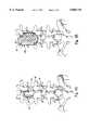
- FIG. 1is a right-front perspective view of the preferred embodiment, showing the arched shield and attachment arms;
- FIG. 2is a bottom plan view showing the arched shield and attachment arms of FIG. 1;
- FIG. 3Ais a partial perspective view of lumbar vertebrae illustrating the bony dissection associated with a laminectomy
- FIG. 3Bis a partial perspective view, similar to FIG. 3A, showing use of the shield device to cover the laminectomy defect in accordance with the present invention
- FIG. 3Cis a partial perspective view showing the use of an elongated shield device to cover a laminectomy defect associated with two vertebrae;
- FIG. 4Ais a partial perspective view of lumbar vertebrae illustrating the bony dissection associated with a hemilaminectomy
- FIG. 4Bis a partial perspective view, similar to FIG. 4A, showing use of the shield device to cover the hemilaminectomy defect in accordance with the present invention
- FIG. 5is a partial side perspective view of the lumbar vertebrae showing another preferred embodiment of the present invention.
- FIG. 6is a sectional view taken substantially along the line 6--6 of FIG. 5, showing the positioning of the arched shield in accordance with a preferred embodiment of the present invention
- FIG. 7is a top plan view showing an exemplary cardiac protector device of this invention.
- FIG. 8Ais a longitudinal cross-section of the device taken substantially along the line 8--8 of FIG. 7 illustrating the curved shield embodiment
- FIG. 8Bis a longitudinal cross-section of the device illustrating the flat shield embodiment
- FIG. 9is a transverse cross-section of the device taken substantially along the line 9--9 of FIG. 7 illustrating the longitudinal guide;
- FIG. 10is a perspective view illustrating the position of the device relative to the heart and the anterior thoracic skeleton.
- FIG. 11is an exploded perspective view further illustrating the position of the device relative to the heart and rib cage.
- the device of this inventionadvantageously operates to prevent adhesion formation and to physically protect the dura, now exposed by surgery. In addition, the device of this invention facilitates future revision surgery.
- the shield of this inventionis suitable as a protective covering for any bony dissection in a vertebrate. Therefore, while a preferred embodiment of this invention relates to the use of the shield to cover a bony dissection of a vertebrae, the shield device could similarly be used to cover a bony dissection associated with open heart surgery, the bony dissection of the cranium, or the like.
- the shield devicecould similarly be used to cover a bony dissection associated with open heart surgery, the bony dissection of the cranium, or the like.
- orthopaedics or neurosurgerywill be able to generate formed shields, anchorable to bone, that will accommodate bony dissections in a variety of skeletal tissues.
- FIG. 1provides an exemplary drawing of a preferred embodiment of this invention.
- the inventionprovides a formed shield 10 adapted to fit onto at least one vertebral facet (not shown in FIG. 3) to cover the bony dissection associated with a hemilaminectomy procedure, a laminectomy procedure or the like.
- a main body 11 of the shield 10is molded or formed preferably as an arch or semi-cylinder. The arch flattens out on each side of the main body 11, forming a pair of support planes 12.
- the support planes 12provide a surface area suitable for resting the shield 10 against the vertebral bone.
- the arched contours of the main body 11prevent contact of the main body 11 with the spinal dura or nerve roots, thereby minimizing further trauma to the spinal dura and preventing the formation of adhesions and scar tissue between-the underside of the device and the spinal dura.
- the removal of vertebral bone along the spinal columnleaves the cauda equina susceptible to physical trauma.
- Productssuch as gelatin foam or fat do not provide a sufficient barrier to prevent potential physical trauma.
- the shield 10 of the present inventionadvantageously rests on bone to minimize the contact between the device and the spinal dura and surrounding neural tissues. Such reduced contact minimizes adhesion formation between the neural tissues and the device itself.
- a plurality of fenestrations 14are formed in the surface of the shield 10. It is contemplated within the scope of this invention that the fenestrations 14 may take any number of forms or shapes, and those depicted in the figures are to be viewed as exemplary.
- the fenestrations 14are distributed across the surface of the shield 10 and are designed to prevent the retention of fluid within the vertebral canal. Absent such openings, liquids such as cerebral spinal fluid, blood, and irrigation fluid associated with surgery could potentially accumulate beneath the shield 10 and create harmful pressure on the cauda equina and associated neural tissue. Following surgery the fenestrations 14 serve as out-flow ports to relieve pressure associated with the accumulation of fluid beneath the device during the healing process.
- the fenestrations 14may be covered with a filter, mesh, or the like (not shown), to prevent the extension of scar tissue formation into the fenestrations 14 while permitting the passage of fluid through the shield 10.
- the deviceis associated with a surgical drain (not shown) to further facilitate fluid egress from the surgical site.
- a surgical drain(not shown) to further facilitate fluid egress from the surgical site.
- Such surgical drainsare well known in the art and it is contemplated that the device of this invention is adapted to accommodate a drain or in another embodiment a drain is directly incorporated into the device.
- the attachment means contemplated for use with the shield 10 of the present inventioncan take any number of forms.
- the attachmentis to bone.
- Bone attachment contemplated within the scope of this inventioninclude, but are not limited to, both attachment to adjacent spinous processes and lateral attachments such as to facets, transverse processes, articulating processes or the like. It is desirable that no contact is made with the neural elements.
- the attachment meansdoes not extend into the spinal canal or neuroforamen to an extent that would make contact with the dura or nerve roots likely.
- the attachment meansis a pair of attachment arms 16 attached to the main body 11 of the shield 10.
- each of the attachment arms 16comprise a rod 17 that is attached to and extends, at an acute angle, from the main body 11 of the shield 10.
- the rods 17are used to attach the shield 10 to an inferior or superior spinous process (see FIGS. 4A and 4B).
- the actual attachment to the spinous processmay take any form known in the art.
- the rods 17terminate in a pair of attachment flags 18. As shown in FIG. 1, the flags 18 are provided with a plurality of pin holes 19 to facilitate their attachment to an adjacent spinous process.
- the attachment meansis through the use of surgical wire, staples or the like.
- FIG. 2clearly illustrates the manner in which the attachment means laterally extend beyond the "footprint" of the main body 11.
- the fenestrations 14may also serve as attachment means.
- suture or wireis useful for attaching the shield 10 to bone by being passed through the fenestrations 14, binding the shield 10 to adjacent bone.
- the shield 10is stabilized to cartilage ligament or adjacent muscle tissue.
- the fenestrations 14ably serve as anchoring points over the entire surface of the shield 10, as required.
- the deviceis prepared in a size and shape to accommodate a laminectomy.
- Laminectomiesare used for spinal stenosis surgery, for central disk herniation, for an osteophyte centrally, for intradural tumors or for other conditions such as epidural abscesses. Laminectomies result in the exposure of the right and left nerve root axilla and the central cauda.
- FIGS. 3A, 3B and 3CThe application of the device to a bony dissection following a laminectomy procedure is illustrated in FIGS. 3A, 3B and 3C.
- FIG. 3Ais an illustration of a laminectomy of the fifth lumbar vertebrae.
- a spinal cord 24is schematically depicted, surrounded by a vertebral column composed of individual lumbar vertebrae 26, and the sacrum 28. Although the shape of the individual vertebrae 28 does vary, each are composed of a transverse process 30 and a spinous process 32.
- FIG. 3BA preferred application of the shield 10 is provided in FIG. 3B.
- the shield 10is positioned over the laminectomy site and the attachment arms 16, terminating here, in attachment flags 18, are used to attach the device to the spinous process 32 of the fourth lumbar vertebrae 26.
- a set of four attachment pins 20are used to anchor the four corners of the shield 10 in place to the surrounding vertebrae tissue.
- FIG. 3Cillustrates another preferred embodiment of the invention.
- the shield 10is designed to span a laminectomy defect involving two vertebrae. Attachment arms 16 and attachment flags 18 are similarly positioned on the shield 10 of FIG. 3C.
- the shield 10is adapted to span at least two vertebrae. It is contemplated that the shield 10 is prepared in a length and width to accommodate laminectomies involving three or more vertebrae.
- the overall length of the shield 10will be at least as long as a lateral length l of the lamina surface of the vertebrae 26 containing the laminotomy defect, and wide enough to stably mate with the remaining lateral lamina surfaces of bone following the laminectomy procedure. Therefore, it is contemplated that the overall length of the shield 10 for use in spanning one laminectomy defect (or hemilaminectomy defect, see FIGS. 4A and 4B) ranges in size from about 2.0 cm to 6.0 cm and preferably between 2.5 cm to 4.5 cm, with a final width ranging from about 1.5 cm to 4.5 cm, and preferably between about 2.0 cm to 3.5 cm.
- the optimal thickness of the shield 10will vary depending on the plasticity or moldability of the device, which in turn will depend on the choice of shield material. However, it is contemplated that a preferred thickness of the shield 10 should be approximately 0.5 mm to 8 mm and more preferably, about 0.5 mm to 5 mm.
- FIG. 4Aprovides an illustration of the bony dissection associated with a hemilaminectomy. Since the dissection for a hemilaminectomy is smaller than that of a laminectomy, it is contemplated that the surgeon, using a scalpel, or the like will be able to readily customize the shield 10 as sized for the laminectomy to overlie the exposed dura after a hemilaminectomy procedure.
- An example of a customized shield 10a contemplated for use in a hemilaminectomyis provided in FIG. 4B.
- the customized shield 10ais attached to the vertebrae 26 using the set of attachment pins 20.
- the shield 10 of FIG. 1could be customized for a hemilaminectomy by removing one of the attachment arms 16.
- the remaining attachment arm 16serves as an attachment means to anchor the shield 10 to a superior or an inferior spinous process.
- the overall length of the shield 10can be varied to accommodate laminectomies or hemilaminectomies involving more than one vertebrae.
- the shieldis preferably about 6.0 cm to about 12 cm in length and more preferably about 6.5 cm to about 9 cm in length.
- the widthis preferably about 2.0 cm to 5.0 cm and more preferably about 2.0 cm to 3.5 cm.
- the main body 11 of the shield 10preferably has an arch shape, and height h that prevents contact between the spinal dura and the surface of the shield 10.
- the height h of the archis between about 0.2 cm and 4.5 cm from the support planes 12, although within this range, it is contemplated that the shield 10 will be manufactured in at least two separate ranges of arch heights.
- the arch height hcan be varied to accommodate other medical devices known in the art.
- the height h of the archis between about 0.2 cm to 1.5 cm, or about one-half the height of an adjacent spinous process when positioned on the patient, and is used with medical devices such as a Dynamic Transverse Traction ("DTT") unit or other instrumentation systems employing a plurality of pedicle screws 34, (see FIG. 5) such as the Steffee-VSP system (AcroMed Corporation), Isola instrumentation (AcroMed Corporation, Cleveland, Ohio), or the like.
- DTTDynamic Transverse Traction
- FIG. 5Steffee-VSP system
- Isola instrumentationAcroMed Corporation, Cleveland, Ohio
- the shield 10that is suitable for a DTT device, or the like, preferably has a height h smaller than the dimension of the height between the DTT construct and the exposed dura. The reduced arch size permits the positioning of the spinal cord protection device beneath the pedical screws 34 and the transverse rods 36 without impingement.
- the shield 10is anchored to the vertebrae 26 through the fenestrations 14.
- the shield 10may be anchored at its periphery to the adjacent facets or spinous processes 32 above or below the decompression, using anchoring pins together with the anchoring means associated with the fenestrations 14.
- the shield 10 of this inventionmay be prepared from any number of materials known in the art. It is contemplated that the device could be prepared from surgical steel including a woven metal fiber or other similar material. In a preferred embodiment of this invention, the device is prepared from a thermoplastic polymer such as polypropylene, polyethylene, polymethacrylate or the like. Other materials contemplated for use in this invention include, but are not limited to, tungsten, titanium, polytetrafluoroethylene, silicone, bioerodable polylactic acid, hydroxylapatite, regenerated collagen or the like. Those with skill in the art of medical devices will be readily able to select and formulate a composition having the preferred characteristics herein described.
- the materialis biocompatible and is capable of being cut with a standard surgical tool, such as a scalpel, knife or scissors to permit customization of the device in the operating room to the shape and size of the bone defect for each individual patient.
- a standard surgical toolsuch as a scalpel, knife or scissors
- Methods for manufacturing the device of this inventionwill depend on the choice of material. Those with skill in the art of manufacturing implantable medical devices will be readily able to use the description of the invention provided herein to produce the contemplated spinal cord protection device.
- the device of this inventioncan be tooled, molded, heat pressed or the like.
- the devicemay have some pliability, such that the surgeon can customize the device to fit the desired bony dissection and, in addition, the shield 10 can be further bent or molded by the surgeon to accommodate the particular topography of the patient's spine.
- the device of this inventionmay be prepared from a solid sheet of material, or the device can advantageously be prepared as a laminate.
- the deviceis a composite of laminated sheets
- the shield materialcan be impregnated with a radiopaque substance or incorporate a radiopaque material into the edges of the device.
- Suitable radiopaque materialsinclude metals or halogenated compounds such as iodinated or brominated compounds.
- Other compoundsinclude barium containing substances, renografin or commercially available, Isovue® (Squibb Diagnostics, Princeton, N.J.).
- the polymers contemplated for use in preparing the shield of this inventionare preferably halogenated or are prepared in combination with halogenated polymers.
- a radiopaque material in the shield 10permits visualization of the shield 10 by X-ray radiation or the like. In situations where the patients back pain persists or where revision surgery is contemplated, the surgeon is able to determine the position of the shield device of this invention prior to or during the revision surgery.
- the radiopaque substancealso allows the surgeon to verify the location of the bone dissection as determined from the position of the shield.
- the shield 10 of the inventioncan advantageously be impregnated with, or otherwise positioned in place in association with, a drug suitable for inhibiting the formation of adhesions. Therefore, in another preferred embodiment, the shield contains an absorptive, saturatable or impregnatable material suitable for acting as a carrier for an adhesion-inhibiting substance.
- Suitable adhesion-inhibiting drugs contemplated for use in association with the shield 10 of this inventioninclude, but are not limited to, heparin salts and analogs of heparin salts, such as Pentosan Polysulfate ("PPS", available, for example, from Sigma Chemical Company, St.
- the devicefacilitates revision surgery.
- Revision surgeryis complicated by the formation of adhesions to the spinal dura. Dissections of adhesions and scar formation increase the time the patient must be under anaesthesia. Moreover, dissection of the scar tissue can result in inadvertent pierces or tears in the dura and the release of spinal fluid into the surgical area that can further complicate surgery.
- the spinal cord protection deviceprevents adhesions with the dura. During revision surgery, the surgeon can cut through the muscle and facia to the device quickly without the potential of piercing or tearing the spinal dura.
- the shield 10is colored. It is contemplated that the selected dye will contrast in color with bone, blood or internal tissues, and thus further facilitate revision surgery since the surgeon can rapidly identify the shield 10 during the dissection process.
- contrasting colors contemplated for use with this deviceinclude shades of blue, green, black, purple, yellow, orange or the like.
- the shield 10 of this inventionis suitable for the cervical and thoracic regions of the spine as well. Further, as disclosed supra, the shield 10 is contemplated for use in any location in the body associated with a bony dissection.
- the shield 10 of the present inventionis contemplated to be commercially available in a number of different sizes, shapes and include various attachment means.
- the shieldspreferably are packaged in separate sterile packaging and can be arranged on a tray that includes single and multiple protector devices in different sizes and embodiments.
- Example 1An exemplary surgical procedure employing the shield 10 of this invention is provided in Example 1, below. This procedure is only exemplary. Surgeons skilled in the art of orthopaedics and neurosurgery will be readily able to adapt their surgical techniques and surgical procedures to include the use of this shield and in particular, those surgeons skilled in spinal surgery will readily appreciate the variations discussed herein that do not detract from the scope of this invention.
- the deviceis positioned between a target organ or tissue and the dermis to protect that organ or tissue from damage during the accessing stage of a subsequent surgical procedure.
- the protector deviceis positioned over the organ or tissue and is preferably anchored to bone, cartilage or muscle. When revision surgery is necessary, the surgeon can rapidly access the protector device without the risk of nicking or damaging the underlying organ or tissue. The protector device can then be removed to expose the target tissue or organ.
- the devicewill also facilitate revision surgery by minimizing post-operative adhesion formation.
- Post-operative adhesion formationcomplicates a wide variety of revision surgeries. As discussed above, these adhesions make dissection tedious because the adjacent bone or tissue is now adhered to the target organ. This increases the likelihood that the organ will be inadvertently damaged during the surgical procedure. The presence of adhesions dramatically increases the time that a person is under anesthesia.
- the present inventionovercomes these problems by creating a barrier that minimizes adhesion formation between adjacent tissue plans and protects the surgical area from damage during the accessing phase of a subsequent revision surgery.
- Adhesionsform between the pericardium and the posterior surface of the sternum following a variety of open-heart cardiac procedures that disrupt the pericardium and the linings of the greater vessels of the heart.
- children who have congenital cardiac defectsoften require multiple surgical procedures over their lifetimes.
- a significant percentage of individuals who receive cardiac bypass surgerywill require a second cardiac procedure months or years after their original bypass surgery.
- a common problem associated with these surgeriesis that adhesions form between the posterior portion of the sternum and the anterior portion of the pericardium of the heart following surgery. These adhesions complicate subsequent surgeries because the heart is affixed to the sternum during the second sternotomy procedure. The surgeon must tediously dissect both pericardial adhesions and adhesions forming between the greater vessels of the heart and the sternum before performing the sternotomy.
- Adhesionsare particularly a problem for those revision cardiac surgeries that employ portions of the internal mammary artery for bypass tissue. Since the internal mammary artery has a higher degree of patency than the saphenous vein, or other veins of the lower extremity typically used for the bypass procedure, the dissection process required to release the heart from the sternum in a revision surgery is relatively complex. However, for a number of other reasons, the internal mammary artery is the vessel of choice for current bypass surgeries. These dissections are an obligate step of the revision surgery and increase the amount of time required to perform the second surgery and complicate the surgical procedure by increasing the risk that the heart will be inadvertently lacerated, nicked or otherwise damaged. The lacerations or nicks to the heart and greater vessels may result in serious complications or catastrophic results.
- FIG. 7An example of the type of adhesion-minimizing protector device contemplated in this invention is illustrated in FIG. 7.
- the body of the device 40also known as a shield, is preferably substantially oblong in shape. It is also contemplated that the device can be substantially circular or substantially rectangular in shape; however, as illustrated in FIG. 7, the edges of the device are preferably rounded and smoothed to reduce abrasion and bruising of the surrounding tissue after implantation.
- substantially oblongis used herein to mean that the overall conformation of the anterior surface of the device is generally similar to a geometric oblong.
- the deviceis generally flat (FIG. 8B) such that both its anterior surface 42 (see FIG. 11), that is adjacent to the sternum, and the posterior surface 44, that portion of the device that is adjacent the heart, form a substantially flat plane.
- the body of the deviceis curved along at least one axis, such that the body forms a portion of the face of a cylinder, with the posterior surface 44 of the device forming a concavity.
- the concavityis preferably slight such that the longitudinal edges 46 of the device are minimally raised relative to a central longitudinal line 48 that spatially divides the device lengthwise.
- the arch height h of the deviceis defined as the distance between the plane which connects the opposing longitudinal edges 46 of the device and the parallel plane tangent to the device at the central longitudinal line 48. It is anticipated that the degree of curvature or concavity will be no more than that required for the device to rest comfortably between the heart and the sternum.
- Anchoring meansare preferably provided along the edges of the device 40.
- the anchoring meanscomprise fenestrations or anchoring ports 50 (see FIG. 7).
- the anchoring portsare large enough to accommodate suture or wire.
- the cardiac protector devicewill be anchored to the central anterior portion of the thoracic skeleton.
- the devicecan be anchored to bone, cartilage, muscle or supportive elements associated with the central anterior portion of the thoracic skeleton, including the sternum.
- the anchoring meansstabilize the device and prevent it from moving with the normal pulsation of the heart. Further, the anchoring means prevent the device from moving during normal thoracic movement.
- FIG. 7provides one example of a device having fenestrations circumscribing the periphery of the apparatus; it is further contemplated that the fenestrations can be limited to the cranial and caudal aspects of the device.
- a method for attaching the device in place following open heart surgeryis provided in Example 2.
- a longitudinal guide 52extends along the length of the anterior surface of the device following the central longitudinal line 48.
- This longitudinal guideis positioned beneath the sternum along the sternotomy line.
- the guideforms a longitudinal cutting guide extending along the length of the device.
- the longitudinal guideadvantageously facilitates revision surgery by providing a recess to further distance the device from the sternum midline. This recess facilitates the positioning of the oscillating saw, or other equivalent surgical tool, along the sternum.
- the longitudinal guideserves as a groove for the surgeon to follow as he or she cuts through the sternum.
- the dimensions of the devicemay vary in length such that the device extends the full length of the sternum, or alternatively the device may be just a few inches in length. Therefore, it is contemplated that the device will range in length l from about 3.0 to 10 inches and more preferably from about 4.0 to 8.5 inches.
- the width w of the deviceis preferably from about 0.5 to 4.0 inches and more preferably from about 1.0 to 3.0 inches.
- the thickness of the devicemay vary from 0.125 to 0.5 inches and preferably from between 0.125 to 0.375 inches. In those embodiments where the body of the device is curved, the arch height h of the device is preferable no greater than 0.5 inches.
- the dimensionscan be selected and readily optimized by one of skill in the art in view of the disclosure herein, depending upon the particular surgical site and patient size. It is further contemplated that the dimensions of the apparatus may be scaled down even further to accommodate infant and pediatric applications for cardiac procedures were multiple surgeries are likely.
- the body of the cardiac protection device 40is preferably prepared from a biocompatible material that is capable of being cut with a standard surgical tool, such as a scalpel, knife, or scissors (such as a Mayo or Metsenbaum type scissor) to permit customization of the device in the operating room.
- a standard surgical toolsuch as a scalpel, knife, or scissors (such as a Mayo or Metsenbaum type scissor) to permit customization of the device in the operating room.
- the deviceis sufficiently malleable so that the device can be molded and contoured by the surgeon at the time of the surgery to accommodate the coronary artery bypass graft employing the internal mammary artery as its primary vessel.
- the devicecan be preformed in its final configuration from any of a variety of materials such as stainless steel, injection moldable polymers, and the like as will be apparent to one of skill in the art.
- the deviceis prepared from a thermoplastic polymer such as polypropylene, polyethylene, polymethacrylate or the like. It is further contemplated that the device, once formed, is somewhat flexible. Therefore, other materials also contemplated for use in this invention include, but are not limited to, silicone, bioerodable polylactic acid, poly-HEMA, or the like. Those with skill in the art will be able to select a suitable biocompatible material.
- the deviceis provided without anchoring ports.
- the deviceis prepared from a material that can be punctured by a sharp object to permit the surgeon to form his or her own anchoring ports during the surgical procedure.
- the devicecan be tooled, molded, heat pressed or the like.
- the methods of manufacturing the devicewill depend on the choice of material. Those with skill in the art of manufacturing implantable medical devices will be readily able to use this description of the contemplated invention together with the figures to produce the protector device of this invention.
- the cardiac protection devicecan be prepared from a solid sheet of material, or the device can be prepared as a laminate.
- a radiopaque materialis preferably incorporated into the device either as a laminate or the radiopaque substance can be impregnated either throughout the device or along the periphery. Suitable radiopaque materials are disclosed in the discussion relating to the spinal cord protection device (supra).
- FIG. 7illustrates a preferred embodiment having a radiopaque material such as barium or the like incorporated into the device as a peripheral ring 54.
- the body of the devicecan be impregnated with, coated with, or otherwise positioned in place in association with a drug or other substance suitable for inhibiting the formation of adhesions.
- Suitable adhesion-inhibiting drugs contemplated for use in association with the cardiac protection deviceinclude, but are not limited to, heparin salts and analogs of heparin salts, such as Pentosan Polysulfate, hyaluronic acid, dextran, growth factor inhibitors, or other compounds recognized in the art to inhibit adhesion formation such as gelatin foams, or the like.
- the body of the cardiac protection deviceis colored to contrast with the color of bone, fascia, blood and heart tissue.
- contrasting colors contemplated for use with this deviceinclude shades of blue, green, black, purple, yellow, pink, or the like.
- the surgical tools disclosed hereinare standard surgical equipment well known to those skilled in the art of orthopaedic and neurosurgery.
- the patientis positioned on an Andrew's frame or operating table and prepped and draped in the fashion standard for back surgery.
- the incisionis made over the spinous process of the area to be decompressed.
- the incisionis carried down through the dorsal lumbar fascia and the fascia is then incised down to the spinal lamina junction.
- Dissectionis continued out to the tips of the transverse processes and is accomplished using the electrocautery and Cobb dissection tool.
- Self retaining retractorsare then placed into the wound to allow clear visualization of the structures which have been denuded of their soft tissue.
- a Lexzell rongeuris then used to remove the bone of the spinous process 32 and that portion of the lamina 40 (see FIG. 3A).
- a Kerrison rongeuris used to remove bone from the lamina as well as ligamentum flavum and epidural fat.
- the dissectionis carried out to the facet joints. If nerve root entrapment, either by disk or soft tissue is noted lateral to the facet, then a partial medial facetectomy is performed. The origin of the nerve roots are then identified and traced into their corresponding neural foramen.
- a neural foraminal probeis placed into the neural foramen at each level and if it is met with any impedance, a partial foraminotomy is performed at each level to facilitate the passage of the probe. Once this is completed, hemostasis is achieved using the Malis bipolar coagulator or electrocautery device.
- Dissection into the neural foramenmany times can result in increased instability by weakening the facet region.
- a 4 mm burris used to do the dissection in the opening of the neural foramen to minimize the destruction with the Kerrison rongeur.
- the operative areais then irrigated and suction dried, and once again hemostasis is achieved using electrocautery and a Malis bipolar coagulator.
- the spinal cord protection shield 10is positioned over the laminectomy defect (see FIG. 3B).
- Customization of the shield 10is performed with a scalpel and scissors thereby molding the shield 10 to conform with the individual contours of the spinal column.
- the angle of the attachment arms relative to the protector deviceis adjusted by manually deforming the attachment arms to facilitate their attachment to an adjacent spinous process.
- the armsare sutured in place onto the spinous process and the fenestrations on the shield body are additionally used to suture the device in place (see FIG. 3B).
- the woundis closed using standard operating procedures, a drain is preferably placed into the wound and, as one example of wound closure, the wound is closed in layers using a #1 Vicryl (Ethicon, Piscataway, N.J.) suture for the dorsal lumbar fascia, a 2-0 Vicryl for the deep subcutaneous tissue, and a 3-0 subcuticular stitch.
- a #1 VicrylEsthicon, Piscataway, N.J.
- the patientis positioned in a supine manner on the operating table and the chest, upper abdomen and lower extremities are prepped for surgery.
- a sterile drapeis applied to the chest, lower extremities, abdomen and the perineum.
- the chest and lower extremitiesare draped out separately and kept sterile through the entire procedure to facilitate vein harvesting in the lower extremities.
- One team of physiciansopens the heart and a second team harvests the graft of the saphenous vein from the lower extremity.
- the veinis usually harvested in toto.
- the chestis opened using an oscillating saw along the midportion of the sternum. Once the sternum is divided, the anterior portion of the chest is opened to expose the anterior portions of the right and left chest cavities. The surgeon then performs the bypass procedure.
- the cardiac protection deviceis attached to the posterior aspect of the sternum.
- the deviceincorporates an adhesion-inhibiting compound such as a heparin analog, or the like.
- the attachmentis accomplished as the lateral aspects of the sternum are brought together.
- the deviceis attached to the sternum by running anchoring sutures or wires through the sternum to the device at its periphery. No more than one or two securing stitches positioned at the cranial and caudal aspects of the device are generally required to position the device in place.
- the sternum halvesare wired together and the incision is closed using standard procedures for cardiac surgery wound closure.
- the following methodis useful for accessing the thoracic cavity for a revision cardiac bypass surgery in a patient having a cardiac protection device in place.
- a Stryker sternal sawsuch as a straight Stryker sternal saw (298-97-100) or a half moon Stryker sagittal saw (2108-137) (Stryker Medical Supply) is positioned along the manubrium of the sternum with the blade positioned between the sternum and the cardiac protection device.
- the half moon Stryker sagittal sawis preferably used in revision coronary artery bypass surgery.
- the longitudinal guide in the deviceis used as a path to move the saw down the sternum.
- the suturesare clipped to separate the device from the sternum and the rib cage is separated to expose the thoracic cavity.
- the protector deviceis removed to expose the heart.
Landscapes
- Health & Medical Sciences (AREA)
- Surgery (AREA)
- Life Sciences & Earth Sciences (AREA)
- Orthopedic Medicine & Surgery (AREA)
- Heart & Thoracic Surgery (AREA)
- Veterinary Medicine (AREA)
- Engineering & Computer Science (AREA)
- Biomedical Technology (AREA)
- Nuclear Medicine, Radiotherapy & Molecular Imaging (AREA)
- Medical Informatics (AREA)
- Molecular Biology (AREA)
- Animal Behavior & Ethology (AREA)
- General Health & Medical Sciences (AREA)
- Public Health (AREA)
- Neurology (AREA)
- Pathology (AREA)
- Oral & Maxillofacial Surgery (AREA)
- Prostheses (AREA)
Abstract
Description
Claims (20)
Priority Applications (3)
| Application Number | Priority Date | Filing Date | Title |
|---|---|---|---|
| US08/769,508US5868745A (en) | 1992-11-12 | 1996-12-19 | Spinal protection device |
| US09/246,274US6454767B2 (en) | 1992-11-12 | 1999-02-08 | Protection device |
| US10/236,032US20030078588A1 (en) | 1992-11-12 | 2002-09-03 | Protection device |
Applications Claiming Priority (4)
| Application Number | Priority Date | Filing Date | Title |
|---|---|---|---|
| US97510692A | 1992-11-12 | 1992-11-12 | |
| US15243393A | 1993-11-12 | 1993-11-12 | |
| US08/440,363US5611354A (en) | 1992-11-12 | 1995-05-12 | Cardiac protection device |
| US08/769,508US5868745A (en) | 1992-11-12 | 1996-12-19 | Spinal protection device |
Related Parent Applications (1)
| Application Number | Title | Priority Date | Filing Date |
|---|---|---|---|
| US08/440,363ContinuationUS5611354A (en) | 1992-11-12 | 1995-05-12 | Cardiac protection device |
Related Child Applications (1)
| Application Number | Title | Priority Date | Filing Date |
|---|---|---|---|
| US09/246,274ContinuationUS6454767B2 (en) | 1992-11-12 | 1999-02-08 | Protection device |
Publications (1)
| Publication Number | Publication Date |
|---|---|
| US5868745Atrue US5868745A (en) | 1999-02-09 |
Family
ID=26849557
Family Applications (4)
| Application Number | Title | Priority Date | Filing Date |
|---|---|---|---|
| US08/440,363Expired - LifetimeUS5611354A (en) | 1992-11-12 | 1995-05-12 | Cardiac protection device |
| US08/769,508Expired - Fee RelatedUS5868745A (en) | 1992-11-12 | 1996-12-19 | Spinal protection device |
| US09/246,274Expired - Fee RelatedUS6454767B2 (en) | 1992-11-12 | 1999-02-08 | Protection device |
| US10/236,032AbandonedUS20030078588A1 (en) | 1992-11-12 | 2002-09-03 | Protection device |
Family Applications Before (1)
| Application Number | Title | Priority Date | Filing Date |
|---|---|---|---|
| US08/440,363Expired - LifetimeUS5611354A (en) | 1992-11-12 | 1995-05-12 | Cardiac protection device |
Family Applications After (2)
| Application Number | Title | Priority Date | Filing Date |
|---|---|---|---|
| US09/246,274Expired - Fee RelatedUS6454767B2 (en) | 1992-11-12 | 1999-02-08 | Protection device |
| US10/236,032AbandonedUS20030078588A1 (en) | 1992-11-12 | 2002-09-03 | Protection device |
Country Status (1)
| Country | Link |
|---|---|
| US (4) | US5611354A (en) |
Cited By (157)
| Publication number | Priority date | Publication date | Assignee | Title |
|---|---|---|---|---|
| US6419703B1 (en) | 2001-03-01 | 2002-07-16 | T. Wade Fallin | Prosthesis for the replacement of a posterior element of a vertebra |
| US6475219B1 (en) | 2001-06-07 | 2002-11-05 | Alexis P. Shelokov | Anterior vertebral protection method and device |
| US20030004572A1 (en)* | 2001-03-02 | 2003-01-02 | Goble E. Marlowe | Method and apparatus for spine joint replacement |
| WO2003007829A1 (en)* | 2001-07-20 | 2003-01-30 | Spinal Concepts, Inc. | Spinal stabilization system and method |
| US20030028250A1 (en)* | 1999-10-22 | 2003-02-06 | Archus Orthopedics, Inc. | Prostheses, systems and methods for replacement of natural facet joints with artifical facet joint surfaces |
| US20030045878A1 (en)* | 1999-12-03 | 2003-03-06 | Dominique Petit | Connecting assembly for spinal osteosynthesis |
| US20030073998A1 (en)* | 2000-08-01 | 2003-04-17 | Endius Incorporated | Method of securing vertebrae |
| US6565605B2 (en) | 2000-12-13 | 2003-05-20 | Medicinelodge, Inc. | Multiple facet joint replacement |
| US6579319B2 (en) | 2000-11-29 | 2003-06-17 | Medicinelodge, Inc. | Facet joint replacement |
| US6605090B1 (en) | 2000-10-25 | 2003-08-12 | Sdgi Holdings, Inc. | Non-metallic implant devices and intra-operative methods for assembly and fixation |
| US6613089B1 (en) | 2000-10-25 | 2003-09-02 | Sdgi Holdings, Inc. | Laterally expanding intervertebral fusion device |
| US20040034351A1 (en)* | 2002-08-14 | 2004-02-19 | Sherman Michael C. | Techniques for spinal surgery and attaching constructs to vertebral elements |
| US20040049277A1 (en)* | 1999-10-22 | 2004-03-11 | Archus Orthopedics, Inc. | Facet arthroplasty devices and methods |
| US20040057730A1 (en)* | 2002-09-18 | 2004-03-25 | Harry Littlejohn | Control processor for use with a transceiver in an optical wireless network |
| US6719794B2 (en) | 2001-05-03 | 2004-04-13 | Synthes (U.S.A.) | Intervertebral implant for transforaminal posterior lumbar interbody fusion procedure |
| US6730127B2 (en)* | 2000-07-10 | 2004-05-04 | Gary K. Michelson | Flanged interbody spinal fusion implants |
| US20040088053A1 (en)* | 2002-10-30 | 2004-05-06 | Hassan Serhan | Regenerative implants for stabilizing the spine and devices for attachment of said implants |
| US6758863B2 (en) | 2000-10-25 | 2004-07-06 | Sdgi Holdings, Inc. | Vertically expanding intervertebral body fusion device |
| US20040133201A1 (en)* | 2000-08-01 | 2004-07-08 | Alan Shluzas | Methods and apparatuses for treating the spine through an access device |
| WO2004021866A3 (en)* | 2002-09-06 | 2004-08-05 | Neville D Alleyne | Seal for posterior lateral vertebral disk cavity |
| US20040230304A1 (en)* | 2003-05-14 | 2004-11-18 | Archus Orthopedics Inc. | Prostheses, tools and methods for replacement of natural facet joints with artifical facet joint surfaces |
| US20050010291A1 (en)* | 2003-07-08 | 2005-01-13 | Archus Orthopedics Inc. | Prostheses, tools and methods for replacement of natural facet joints with artificial facet joint surfaces |
| US20050027361A1 (en)* | 1999-10-22 | 2005-02-03 | Reiley Mark A. | Facet arthroplasty devices and methods |
| US20050049590A1 (en)* | 2003-03-07 | 2005-03-03 | Neville Alleyne | Spinal implant with securement spikes |
| US20050080486A1 (en)* | 2000-11-29 | 2005-04-14 | Fallin T. Wade | Facet joint replacement |
| US20050131409A1 (en)* | 2003-12-10 | 2005-06-16 | Alan Chervitz | Linked bilateral spinal facet implants and methods of use |
| US20050131406A1 (en)* | 2003-12-15 | 2005-06-16 | Archus Orthopedics, Inc. | Polyaxial adjustment of facet joint prostheses |
| US20050126576A1 (en)* | 2003-11-04 | 2005-06-16 | Ferree Bret A. | Protecting biological structures, including the great vessels, particularly during spinal surgery |
| US20050177155A1 (en)* | 2003-10-28 | 2005-08-11 | Neville Alleyne | Anterior adhesion resistant barrier for spine |
| US20050235508A1 (en)* | 2004-04-22 | 2005-10-27 | Archus Orthopedics, Inc. | Facet joint prosthesis measurement and implant tools |
| US20050240264A1 (en)* | 2004-04-22 | 2005-10-27 | Archus Orthopedics, Inc. | Anti-rotation fixation element for spinal prostheses |
| US6974480B2 (en) | 2001-05-03 | 2005-12-13 | Synthes (Usa) | Intervertebral implant for transforaminal posterior lumbar interbody fusion procedure |
| US20060036211A1 (en)* | 2004-07-29 | 2006-02-16 | X-Sten, Inc. | Spinal ligament modification kit |
| US20060041311A1 (en)* | 2004-08-18 | 2006-02-23 | Mcleer Thomas J | Devices and methods for treating facet joints |
| US20060052785A1 (en)* | 2004-08-18 | 2006-03-09 | Augostino Teena M | Adjacent level facet arthroplasty devices, spine stabilization systems, and methods |
| US20060058791A1 (en)* | 2004-08-18 | 2006-03-16 | Richard Broman | Implantable spinal device revision system |
| US20060079895A1 (en)* | 2004-09-30 | 2006-04-13 | Mcleer Thomas J | Methods and devices for improved bonding of devices to bone |
| US20060085075A1 (en)* | 2004-10-04 | 2006-04-20 | Archus Orthopedics, Inc. | Polymeric joint complex and methods of use |
| US20060085072A1 (en)* | 2004-04-22 | 2006-04-20 | Archus Orthopedics, Inc. | Implantable orthopedic device component selection instrument and methods |
| US20060149254A1 (en)* | 2004-12-13 | 2006-07-06 | St. Francis Medical Technologies, Inc. | Inter-cervical facet implant and method for preserving the tissues surrounding the facet joint |
| US20060149272A1 (en)* | 2004-12-13 | 2006-07-06 | St. Francis Medical Technologies, Inc. | Inter-cervical facet implant and method |
| US20060200149A1 (en)* | 2005-02-22 | 2006-09-07 | Hoy Robert W | Polyaxial orhtopedic fastening apparatus |
| US20060200137A1 (en)* | 2001-02-16 | 2006-09-07 | St. Francis Medical Technologies, Inc. | Method and device for treating ailments of the spine |
| US20060217718A1 (en)* | 2005-03-28 | 2006-09-28 | Facet Solutions, Inc. | Facet joint implant crosslinking apparatus and method |
| US20060241597A1 (en)* | 2004-12-13 | 2006-10-26 | St. Francis Medical Technologies, Inc. | Inter-cervical facet joint implant with locking screw system |
| US20060241601A1 (en)* | 2005-04-08 | 2006-10-26 | Trautwein Frank T | Interspinous vertebral and lumbosacral stabilization devices and methods of use |
| US20060241770A1 (en)* | 2005-04-21 | 2006-10-26 | Rhoda William S | Expandable vertebral prosthesis |
| US20060247791A1 (en)* | 2005-04-29 | 2006-11-02 | Mckay William F | Multi-purpose medical implant devices |
| US20060247632A1 (en)* | 2004-12-13 | 2006-11-02 | St. Francis Medical Technologies, Inc. | Inter-cervical facet implant with surface enhancements |
| US20060276801A1 (en)* | 2005-04-04 | 2006-12-07 | Yerby Scott A | Inter-cervical facet implant distraction tool |
| US20070016218A1 (en)* | 2005-05-10 | 2007-01-18 | Winslow Charles J | Inter-cervical facet implant with implantation tool |
| US20070016296A1 (en)* | 2004-06-02 | 2007-01-18 | Triplett Daniel J | Surgical measurement systems and methods |
| US20070016195A1 (en)* | 2005-05-10 | 2007-01-18 | Winslow Charles J | Inter-cervical facet implant with implantation tool |
| US20070055111A1 (en)* | 2005-09-06 | 2007-03-08 | Morgan Mickey D | Methods and apparatus for vascular protection in spinal surgery |
| US20070071790A1 (en)* | 2005-09-28 | 2007-03-29 | Northwestern University | Biodegradable nanocomposites with enhance mechanical properties for soft tissue |
| US20070088358A1 (en)* | 2005-03-22 | 2007-04-19 | Hansen Yuan | Minimally Invasive Spine Restoration Systems, Devices, Methods and Kits |
| US20070093833A1 (en)* | 2004-05-03 | 2007-04-26 | Kuiper Mark K | Crossbar spinal prosthesis having a modular design and related implantation methods |
| US20070123890A1 (en)* | 2005-11-04 | 2007-05-31 | X-Sten, Corp. | Tissue retrieval devices and methods |
| US20070123863A1 (en)* | 2004-12-13 | 2007-05-31 | St. Francis Medical Technologies, Inc. | Inter-cervical facet implant with multiple direction articulation joint and method for implanting |
| US20070191834A1 (en)* | 2006-01-27 | 2007-08-16 | Sdgi Holdings, Inc. | Artificial spinous process for the sacrum and methods of use |
| US20070233256A1 (en)* | 2006-03-15 | 2007-10-04 | Ohrt John A | Facet and disc arthroplasty system and method |
| US20070249044A1 (en)* | 2006-01-30 | 2007-10-25 | University Of Illinois At Chicago | Microstructures in three dimensional gel suspensions for growth of cells |
| US20070276374A1 (en)* | 2005-03-02 | 2007-11-29 | Richard Broman | Arthroplasty revision system and method |
| US20070299459A1 (en)* | 2006-06-26 | 2007-12-27 | X-Sten Corp. | Percutaneous Tissue Access Device |
| US20080009823A1 (en)* | 2005-04-30 | 2008-01-10 | Mckay William F | Syringe devices and methods useful for delivering osteogenic material |
| US20080082171A1 (en)* | 2004-04-22 | 2008-04-03 | Kuiper Mark K | Crossbar spinal prosthesis having a modular design and systems for treating spinal pathologies |
| US20080097612A1 (en)* | 1999-10-22 | 2008-04-24 | Reiley Mark A | Facet Arthroplasty Devices and Methods |
| US20080097613A1 (en)* | 1999-10-22 | 2008-04-24 | Reiley Mark A | Prostheses, Systems and Methods for Replacement of Natural Facet Joints With Artificial Facet Joint Surfaces |
| US20080103501A1 (en)* | 2006-08-11 | 2008-05-01 | Ralph Christopher R | Angled Washer Polyaxial Connection for Dynamic Spine Prosthesis |
| US20080119851A1 (en)* | 2006-11-20 | 2008-05-22 | Depuy Spine, Inc. | Anterior spinal vessel protector |
| US20080125814A1 (en)* | 2003-05-14 | 2008-05-29 | Archus Orthopedics, Inc. | Prostheses, tools and methods for replacement of natural facet joints with artificial facet joint surfaces |
| US20080140115A1 (en)* | 2004-08-17 | 2008-06-12 | Stopek Joshua B | Stapling Support Structures |
| US20080161847A1 (en)* | 2006-12-28 | 2008-07-03 | Orthovita, Inc. | Non-resorbable implantable guides and methods of use |
| US20080167688A1 (en)* | 2005-02-22 | 2008-07-10 | Facet Solutions, Inc. | Taper-Locking Fixation System |
| US20080177310A1 (en)* | 2000-10-20 | 2008-07-24 | Archus Orthopedics, Inc. | Facet arthroplasty devices and methods |
| US20080177311A1 (en)* | 2006-10-30 | 2008-07-24 | St. Francis Medical Technologies, Inc. | Facet joint implant sizing tool |
| US20080208249A1 (en)* | 2007-02-22 | 2008-08-28 | Jason Blain | Vertebral facet joint drill and method of use |
| US20080221383A1 (en)* | 2007-02-12 | 2008-09-11 | Vertos Medical, Inc. | Tissue excision devices and methods |
| US20080221622A1 (en)* | 2007-01-10 | 2008-09-11 | Facet Solutions, Inc. | Facet Joint Replacement |
| US20080228225A1 (en)* | 2006-11-30 | 2008-09-18 | Paradigm Spine, Llc | Interlaminar-Interspinous Vertebral Stabilization System |
| US20080300595A1 (en)* | 2007-06-04 | 2008-12-04 | Brau Salvador A | Encasement prevention barrier for use in anterior lumbar surgery |
| US20090024169A1 (en)* | 2004-06-02 | 2009-01-22 | Facet Solutions, Inc. | System and method for multiple level facet joint arthroplasty and fusion |
| US20090036936A1 (en)* | 2006-05-09 | 2009-02-05 | Vertos Medical, Inc. | Translaminar approach to minimally invasive ligament decompression procedure |
| US20090118709A1 (en)* | 2007-11-06 | 2009-05-07 | Vertos Medical, Inc. A Delaware Corporation | Tissue Excision Tool, Kits and Methods of Using the Same |
| US20090177220A1 (en)* | 2008-01-08 | 2009-07-09 | Med-El Elektromedizinische Geraete Gmbh | Surgical Tool for Implantation of a Device with a Convex Element |
| US20090204152A1 (en)* | 2004-02-06 | 2009-08-13 | Spinal Elements, Inc. | Vertebral facet joint prosthesis and method of fixation |
| USD598549S1 (en) | 2008-09-16 | 2009-08-18 | Vertos Medical, Inc. | Surgical trocar |
| USD606654S1 (en) | 2006-07-31 | 2009-12-22 | Vertos Medical, Inc. | Tissue excision device |
| US20100030279A1 (en)* | 2008-02-26 | 2010-02-04 | Spartek Medical, Inc. | Load-sharing bone anchor having a deflectable post and axial spring and method for dynamic stabilization of the spine |
| US20100030224A1 (en)* | 2008-02-26 | 2010-02-04 | Spartek Medical, Inc. | Surgical tool and method for connecting a dynamic bone anchor and dynamic vertical rod |
| US20100030270A1 (en)* | 2007-06-05 | 2010-02-04 | Spartek Medical, Inc. | Dynamic spinal rod assembly and method for dynamic stabilization of the spine |
| US20100030273A1 (en)* | 2008-02-26 | 2010-02-04 | Spartek Medical, Inc. | Versatile polyaxial connector assembly and method for dynamic stabilization of the spine |
| US20100030267A1 (en)* | 2007-06-05 | 2010-02-04 | Spartek Medical, Inc. | Surgical tool and method for implantation of a dynamic bone anchor |
| US20100030274A1 (en)* | 2007-06-05 | 2010-02-04 | Spartek Medical, Inc. | Dynamic spinal rod and method for dynamic stabilization of the spine |
| US20100030271A1 (en)* | 2008-02-26 | 2010-02-04 | Spartek Medical, Inc. | Modular in-line deflection rod and bone anchor system and method for dynamic stabilization of the spine |
| US20100036435A1 (en)* | 2008-02-26 | 2010-02-11 | Spartek Medical, Inc. | Load-sharing bone anchor having a deflectable post and method for dynamic stabilization of the spine |
| US20100036436A1 (en)* | 2008-02-26 | 2010-02-11 | Spartek Medical, Inc. | Load-sharing bone anchor having a durable compliant member and method for dynamic stabilization of the spine |
| US20100036437A1 (en)* | 2008-02-26 | 2010-02-11 | Spartek Medical, Inc. | Load-sharing bone anchor having a deflectable post with a compliant ring and method for stabilization of the spine |
| USD610259S1 (en) | 2008-10-23 | 2010-02-16 | Vertos Medical, Inc. | Tissue modification device |
| USD611146S1 (en) | 2008-10-23 | 2010-03-02 | Vertos Medical, Inc. | Tissue modification device |
| US20100057140A1 (en)* | 2007-06-05 | 2010-03-04 | Spartek Medical, Inc. | Bone anchor for receiving a rod for stabilization and motion preservation spinal implantation system and method |
| US7722647B1 (en) | 2005-03-14 | 2010-05-25 | Facet Solutions, Inc. | Apparatus and method for posterior vertebral stabilization |
| US20100158979A1 (en)* | 2007-07-18 | 2010-06-24 | The Board Of Trustees Of The University Of Illinois | Temporal release of growth factors from 3d micro rod scaffolds for tissue regeneration |
| US20100168795A1 (en)* | 2008-02-26 | 2010-07-01 | Spartek Medical, Inc. | Load-sharing bone anchor having a natural center of rotation and method for dynamic stabilization of the spine |
| USD619253S1 (en) | 2008-10-23 | 2010-07-06 | Vertos Medical, Inc. | Tissue modification device |
| USD619252S1 (en) | 2008-10-23 | 2010-07-06 | Vertos Medical, Inc. | Tissue modification device |
| US20100179655A1 (en)* | 2009-01-12 | 2010-07-15 | Noah Hansell | Expandable Vertebral Prosthesis |
| WO2010078647A1 (en)* | 2009-01-06 | 2010-07-15 | Jean-Marc Mac-Thiong | Spinal covering device |
| US7758654B2 (en) | 2004-05-20 | 2010-07-20 | Kensey Nash Corporation | Anti-adhesion device |
| USD620593S1 (en) | 2006-07-31 | 2010-07-27 | Vertos Medical, Inc. | Tissue excision device |
| USD621939S1 (en) | 2008-10-23 | 2010-08-17 | Vertos Medical, Inc. | Tissue modification device |
| US20100222881A1 (en)* | 2008-10-03 | 2010-09-02 | Ann Prewett | Vessel protection device |
| US20100286703A1 (en)* | 2007-12-28 | 2010-11-11 | Mickey Morgan | tack or drive screw for securing a prosthesis to bone and associated instrumentation and method |
| US20110040301A1 (en)* | 2007-02-22 | 2011-02-17 | Spinal Elements, Inc. | Vertebral facet joint drill and method of use |
| USD635671S1 (en) | 2008-10-23 | 2011-04-05 | Vertos Medical, Inc. | Tissue modification device |
| US20110118783A1 (en)* | 2009-11-16 | 2011-05-19 | Spartek Medical, Inc. | Load-sharing bone anchor having a flexible post and method for dynamic stabilization of the spine |
| US8021396B2 (en) | 2007-06-05 | 2011-09-20 | Spartek Medical, Inc. | Configurable dynamic spinal rod and method for dynamic stabilization of the spine |
| US8057515B2 (en) | 2008-02-26 | 2011-11-15 | Spartek Medical, Inc. | Load-sharing anchor having a deflectable post and centering spring and method for dynamic stabilization of the spine |
| US8066749B2 (en) | 2004-12-13 | 2011-11-29 | Warsaw Orthopedic, Inc. | Implant for stabilizing a bone graft during spinal fusion |
| US8097024B2 (en) | 2008-02-26 | 2012-01-17 | Spartek Medical, Inc. | Load-sharing bone anchor having a deflectable post and method for stabilization of the spine |
| US8114134B2 (en) | 2007-06-05 | 2012-02-14 | Spartek Medical, Inc. | Spinal prosthesis having a three bar linkage for motion preservation and dynamic stabilization of the spine |
| US8221461B2 (en) | 2004-10-25 | 2012-07-17 | Gmedelaware 2 Llc | Crossbar spinal prosthesis having a modular design and systems for treating spinal pathologies |
| US8257397B2 (en) | 2009-12-02 | 2012-09-04 | Spartek Medical, Inc. | Low profile spinal prosthesis incorporating a bone anchor having a deflectable post and a compound spinal rod |
| US8282683B2 (en) | 2010-04-12 | 2012-10-09 | Globus Medical, Inc. | Expandable vertebral implant |
| US8337536B2 (en) | 2008-02-26 | 2012-12-25 | Spartek Medical, Inc. | Load-sharing bone anchor having a deflectable post with a compliant ring and method for stabilization of the spine |
| US8430916B1 (en) | 2012-02-07 | 2013-04-30 | Spartek Medical, Inc. | Spinal rod connectors, methods of use, and spinal prosthesis incorporating spinal rod connectors |
| US8496686B2 (en) | 2005-03-22 | 2013-07-30 | Gmedelaware 2 Llc | Minimally invasive spine restoration systems, devices, methods and kits |
| US8518085B2 (en) | 2010-06-10 | 2013-08-27 | Spartek Medical, Inc. | Adaptive spinal rod and methods for stabilization of the spine |
| US8540746B2 (en) | 1998-08-20 | 2013-09-24 | Zimmer Spine, Inc. | Cannula for receiving surgical instruments |
| US8696671B2 (en) | 2005-07-29 | 2014-04-15 | Vertos Medical Inc. | Percutaneous tissue excision devices |
| US20140128916A1 (en)* | 2005-05-03 | 2014-05-08 | Onike Technologies | Bone anchored surgical mesh |
| US8740949B2 (en) | 2011-02-24 | 2014-06-03 | Spinal Elements, Inc. | Methods and apparatus for stabilizing bone |
| USD724733S1 (en) | 2011-02-24 | 2015-03-17 | Spinal Elements, Inc. | Interbody bone implant |
| WO2015152738A1 (en)* | 2014-04-04 | 2015-10-08 | Ossis Limited | Surgical shield |
| US9271765B2 (en) | 2011-02-24 | 2016-03-01 | Spinal Elements, Inc. | Vertebral facet joint fusion implant and method for fusion |
| US9421044B2 (en) | 2013-03-14 | 2016-08-23 | Spinal Elements, Inc. | Apparatus for bone stabilization and distraction and methods of use |
| USD765853S1 (en) | 2013-03-14 | 2016-09-06 | Spinal Elements, Inc. | Flexible elongate member with a portion configured to receive a bone anchor |
| US9456855B2 (en) | 2013-09-27 | 2016-10-04 | Spinal Elements, Inc. | Method of placing an implant between bone portions |
| USD790062S1 (en) | 2011-10-26 | 2017-06-20 | Spinal Elements, Inc. | Interbody bone implant |
| USD792588S1 (en)* | 2015-10-30 | 2017-07-18 | Ossis Limited | Surgical shield |
| US9707091B2 (en) | 2010-04-12 | 2017-07-18 | Globus Medical, Inc. | Expandable vertebral implant |
| US9820784B2 (en) | 2013-03-14 | 2017-11-21 | Spinal Elements, Inc. | Apparatus for spinal fixation and methods of use |
| US9839450B2 (en) | 2013-09-27 | 2017-12-12 | Spinal Elements, Inc. | Device and method for reinforcement of a facet |
| US9913735B2 (en) | 2010-04-12 | 2018-03-13 | Globus Medical, Inc. | Angling inserter tool for expandable vertebral implant |
| US9931142B2 (en) | 2004-06-10 | 2018-04-03 | Spinal Elements, Inc. | Implant and method for facet immobilization |
| US10130489B2 (en) | 2010-04-12 | 2018-11-20 | Globus Medical, Inc. | Expandable vertebral implant |
| US10278686B2 (en) | 2010-09-20 | 2019-05-07 | DePuy Synthes Products, Inc. | Spinal access retractor |
| US10327910B2 (en) | 2013-03-14 | 2019-06-25 | X-Spine Systems, Inc. | Spinal implant and assembly |
| US10758361B2 (en) | 2015-01-27 | 2020-09-01 | Spinal Elements, Inc. | Facet joint implant |
| US11304733B2 (en) | 2020-02-14 | 2022-04-19 | Spinal Elements, Inc. | Bone tie methods |
| US11457959B2 (en) | 2019-05-22 | 2022-10-04 | Spinal Elements, Inc. | Bone tie and bone tie inserter |
| US11464552B2 (en) | 2019-05-22 | 2022-10-11 | Spinal Elements, Inc. | Bone tie and bone tie inserter |
| US11478275B2 (en) | 2014-09-17 | 2022-10-25 | Spinal Elements, Inc. | Flexible fastening band connector |
| US12102348B2 (en) | 2016-09-07 | 2024-10-01 | Vertos Medical, Inc. | Percutaneous lateral recess resection methods and instruments |
| US12324572B2 (en) | 2022-06-16 | 2025-06-10 | Vertos Medical, Inc. | Integrated instrument assembly |
| US12369952B2 (en) | 2021-12-10 | 2025-07-29 | Spinal Elements, Inc. | Bone tie and portal |
| US12440242B2 (en) | 2024-04-29 | 2025-10-14 | Spinal Elements, Inc. | Flexible fastening band connector |
Families Citing this family (62)
| Publication number | Priority date | Publication date | Assignee | Title |
|---|---|---|---|---|
| US6068630A (en)* | 1997-01-02 | 2000-05-30 | St. Francis Medical Technologies, Inc. | Spine distraction implant |
| US6221109B1 (en)* | 1999-09-15 | 2001-04-24 | Ed. Geistlich Söhne AG fur Chemische Industrie | Method of protecting spinal area |
| CZ301649B6 (en)* | 2000-06-28 | 2010-05-12 | Ed. Geistlich Soehne Ag Fur Chemische Industrie Incorporated Under The Laws Of Switzerland | Tube for regeneration of nerves and process for producing thereof |
| DE10107369B4 (en)* | 2001-02-16 | 2016-03-24 | Ernst Wiedemann | implant plate |
| US6872210B2 (en) | 2001-02-23 | 2005-03-29 | James P. Hearn | Sternum fixation device |
| US6887270B2 (en)* | 2002-02-08 | 2005-05-03 | Boston Scientific Scimed, Inc. | Implantable or insertable medical device resistant to microbial growth and biofilm formation |
| US8133501B2 (en) | 2002-02-08 | 2012-03-13 | Boston Scientific Scimed, Inc. | Implantable or insertable medical devices for controlled drug delivery |
| US8685427B2 (en)* | 2002-07-31 | 2014-04-01 | Boston Scientific Scimed, Inc. | Controlled drug delivery |
| US7993390B2 (en)* | 2002-02-08 | 2011-08-09 | Boston Scientific Scimed, Inc. | Implantable or insertable medical device resistant to microbial growth and biofilm formation |
| US6989012B2 (en)* | 2002-07-16 | 2006-01-24 | Sdgi Holdings, Inc. | Plating system for stabilizing a bony segment |
| US8920826B2 (en)* | 2002-07-31 | 2014-12-30 | Boston Scientific Scimed, Inc. | Medical imaging reference devices |
| US7578819B2 (en) | 2005-05-16 | 2009-08-25 | Baxano, Inc. | Spinal access and neural localization |
| US8613745B2 (en) | 2004-10-15 | 2013-12-24 | Baxano Surgical, Inc. | Methods, systems and devices for carpal tunnel release |
| US8430881B2 (en) | 2004-10-15 | 2013-04-30 | Baxano, Inc. | Mechanical tissue modification devices and methods |
| US7963915B2 (en) | 2004-10-15 | 2011-06-21 | Baxano, Inc. | Devices and methods for tissue access |
| US7938830B2 (en) | 2004-10-15 | 2011-05-10 | Baxano, Inc. | Powered tissue modification devices and methods |
| US8257356B2 (en) | 2004-10-15 | 2012-09-04 | Baxano, Inc. | Guidewire exchange systems to treat spinal stenosis |
| US20100331883A1 (en) | 2004-10-15 | 2010-12-30 | Schmitz Gregory P | Access and tissue modification systems and methods |
| JP5243034B2 (en) | 2004-10-15 | 2013-07-24 | バクサノ,インク. | Tissue removal device |
| US8048080B2 (en) | 2004-10-15 | 2011-11-01 | Baxano, Inc. | Flexible tissue rasp |
| US7857813B2 (en) | 2006-08-29 | 2010-12-28 | Baxano, Inc. | Tissue access guidewire system and method |
| US7887538B2 (en) | 2005-10-15 | 2011-02-15 | Baxano, Inc. | Methods and apparatus for tissue modification |
| US8062300B2 (en) | 2006-05-04 | 2011-11-22 | Baxano, Inc. | Tissue removal with at least partially flexible devices |
| US8221397B2 (en) | 2004-10-15 | 2012-07-17 | Baxano, Inc. | Devices and methods for tissue modification |
| US9101386B2 (en) | 2004-10-15 | 2015-08-11 | Amendia, Inc. | Devices and methods for treating tissue |
| US20110190772A1 (en) | 2004-10-15 | 2011-08-04 | Vahid Saadat | Powered tissue modification devices and methods |
| US9247952B2 (en) | 2004-10-15 | 2016-02-02 | Amendia, Inc. | Devices and methods for tissue access |
| US7738969B2 (en) | 2004-10-15 | 2010-06-15 | Baxano, Inc. | Devices and methods for selective surgical removal of tissue |
| US7959577B2 (en) | 2007-09-06 | 2011-06-14 | Baxano, Inc. | Method, system, and apparatus for neural localization |
| US8016887B1 (en)* | 2005-03-24 | 2011-09-13 | Cardinal Spine, Llc | Spinal implant with overlay |
| US20070068536A1 (en)* | 2005-09-29 | 2007-03-29 | Rawski Mark V | Surgical incision protection device |
| US8366712B2 (en) | 2005-10-15 | 2013-02-05 | Baxano, Inc. | Multiple pathways for spinal nerve root decompression from a single access point |
| US8092456B2 (en) | 2005-10-15 | 2012-01-10 | Baxano, Inc. | Multiple pathways for spinal nerve root decompression from a single access point |
| US8062298B2 (en) | 2005-10-15 | 2011-11-22 | Baxano, Inc. | Flexible tissue removal devices and methods |
| US8083743B2 (en) | 2007-01-29 | 2011-12-27 | Polaris Biotechnology, Inc. | Craniospinal fusion method and apparatus |
| US8182511B2 (en)* | 2007-01-29 | 2012-05-22 | Polaris Biotechnology, Inc. | Craniospinal fusion method and apparatus |
| US9827023B2 (en) | 2007-01-29 | 2017-11-28 | Life Spine, Inc. | Craniospinal fusion method and apparatus |
| US8403965B2 (en)* | 2007-01-29 | 2013-03-26 | Polaris Biotechnology, Inc. | Vertebra attachment method and system |
| CN101801277A (en)* | 2007-06-18 | 2010-08-11 | 马根医疗解决方案有限公司 | Temporary implantable medical device |
| US8192436B2 (en) | 2007-12-07 | 2012-06-05 | Baxano, Inc. | Tissue modification devices |
| US8398641B2 (en) | 2008-07-01 | 2013-03-19 | Baxano, Inc. | Tissue modification devices and methods |
| US8409206B2 (en) | 2008-07-01 | 2013-04-02 | Baxano, Inc. | Tissue modification devices and methods |
| US9314253B2 (en) | 2008-07-01 | 2016-04-19 | Amendia, Inc. | Tissue modification devices and methods |
| AU2009271047B2 (en) | 2008-07-14 | 2014-04-17 | Baxano Surgical, Inc. | Tissue modification devices |
| US8403966B2 (en)* | 2008-11-24 | 2013-03-26 | Mbd Medical Llc | Clavicle plate and screws |
| US8444679B2 (en)* | 2008-11-24 | 2013-05-21 | Mbd Medical Llc | Clavicle plate and screws |
| EP2405823A4 (en) | 2009-03-13 | 2012-07-04 | Baxano Inc | Flexible neural localization devices and methods |
| US9095380B2 (en) | 2009-03-31 | 2015-08-04 | Hamid R. Mir | Spinous process cross-link |
| US8394102B2 (en) | 2009-06-25 | 2013-03-12 | Baxano, Inc. | Surgical tools for treatment of spinal stenosis |
| USD626655S1 (en)* | 2009-12-11 | 2010-11-02 | John Rambo | Gravity-operated spine stretcher |
| WO2011116096A1 (en)* | 2010-03-16 | 2011-09-22 | Falci Scott P | Method and device for tenting dura mater in spinal surgery |
| US20110238058A1 (en)* | 2010-03-29 | 2011-09-29 | Estech, Inc. (Endoscopic Technologies, Inc.) | Indifferent electrode pad systems and methods for tissue ablation |
| US20140005723A1 (en)* | 2011-03-11 | 2014-01-02 | IINN, Inc. | Intra spinous process and method of bone graft placement |
| FR2975582B1 (en)* | 2011-05-23 | 2013-06-07 | Ass Marie Lannelongue | OSTEOSYNTHESIS IMPLANT |
| US9138325B2 (en)* | 2012-07-11 | 2015-09-22 | Globus Medical, Inc. | Lamina implant and method |
| BR202014007055Y1 (en)* | 2014-03-24 | 2020-12-29 | Eduardo Coelho De Souza | constructive disposition applied in cardiac protector |
| WO2016033106A1 (en)* | 2014-08-25 | 2016-03-03 | Bioventrix, Inc. | Tissue protecting devices for treatment of congestive heart failure and other conditions |
| US9526533B1 (en)* | 2014-09-12 | 2016-12-27 | Roberto J. Aranibar | Spinal repair implants and related methods |
| US10792077B2 (en) | 2015-10-01 | 2020-10-06 | Orion Spine Inc. | Spine protection device |
| EP3644880B1 (en) | 2017-06-26 | 2024-04-24 | Dignity Health | Systems for a spinal shield for protecting the spinal cord and dura during surgical procedures |
| US11523824B2 (en)* | 2019-12-12 | 2022-12-13 | Covidien Lp | Anvil buttress loading for a surgical stapling apparatus |
| US12042337B2 (en) | 2020-03-13 | 2024-07-23 | Ashish Patel | Dura shield |
Citations (32)
| Publication number | Priority date | Publication date | Assignee | Title |
|---|---|---|---|---|
| FR52880E (en)* | 1943-05-22 | 1945-08-13 | Device for wound protection | |
| US2443481A (en)* | 1942-10-19 | 1948-06-15 | Sene Leon Paul | Device for the treatment of wounds and the like lesions |
| FR1004625A (en)* | 1949-12-21 | 1952-04-01 | Prosthesis for blocking the lumbosacral joint | |
| US3044497A (en)* | 1944-05-11 | 1962-07-17 | Bodin Girin & Cie Soc | Tubular members provided with corrugated walls |
| US3108399A (en)* | 1961-02-13 | 1963-10-29 | Russell M Fraser | Plant growth cabinets |
| US3256877A (en)* | 1961-12-11 | 1966-06-21 | Edward J Haboush | Adjustable nail plate joint |
| US3693616A (en)* | 1970-06-26 | 1972-09-26 | Robert Roaf | Device for correcting scoliotic curves |
| US3993078A (en)* | 1974-11-04 | 1976-11-23 | Gambro Ag | Insert for use preferably in vascular surgery |
| US4013078A (en)* | 1974-11-25 | 1977-03-22 | Feild James Rodney | Intervertebral protector means |
| US4023569A (en)* | 1974-12-05 | 1977-05-17 | Tuwa-Plastik Dr. Herbert Warnecke Erzeugung Von Kunststoffartikeln Gesellschaft M.B.H. | Device for the protection of wounds |
| FR2386301A1 (en)* | 1977-04-06 | 1978-11-03 | English Biomech Ta | IMPLANTS IMPROVEMENTS |
| US4134399A (en)* | 1977-06-13 | 1979-01-16 | Alfred Halderson | Skin protective device |
| US4164794A (en)* | 1977-04-14 | 1979-08-21 | Union Carbide Corporation | Prosthetic devices having coatings of selected porous bioengineering thermoplastics |
| US4401112A (en)* | 1980-09-15 | 1983-08-30 | Rezaian Seyed M | Spinal fixator |
| US4583541A (en)* | 1984-05-07 | 1986-04-22 | Barry Joseph P | Sternal stabilization device |
| US4599085A (en)* | 1979-07-11 | 1986-07-08 | Neodontics, Inc. | Bone implant member for prostheses and bone connecting elements and process for the production thereof |
| US4599086A (en)* | 1985-06-07 | 1986-07-08 | Doty James R | Spine stabilization device and method |
| US4705031A (en)* | 1985-05-06 | 1987-11-10 | Dietmar Wolter | Osteosynthesis plate for pressure stabilization |
| US4773402A (en)* | 1985-09-13 | 1988-09-27 | Isola Implants, Inc. | Dorsal transacral surgical implant |
| US4794918A (en)* | 1985-05-06 | 1989-01-03 | Dietmar Wolter | Bone plate arrangement |
| US4892545A (en)* | 1988-07-14 | 1990-01-09 | Ohio Medical Instrument Company, Inc. | Vertebral lock |
| US4973332A (en)* | 1988-09-12 | 1990-11-27 | Hospital For Joint Diseases | Attachment for femur sliding screw plate |
| US5000166A (en)* | 1988-04-27 | 1991-03-19 | Sulzer Brothers Limited | Implant kit for stabilizing regions of a spine |
| US5041113A (en)* | 1989-07-20 | 1991-08-20 | Lutz Biedermann | Stabilization member for stabilizing bones |
| US5052373A (en)* | 1988-07-29 | 1991-10-01 | Michelson Gary K | Spinal retractor |
| US5108395A (en)* | 1989-09-18 | 1992-04-28 | Societe De Fabrication De Materiel Orthopedique - Sofamor | Implant for anterior dorsolumbar spinal osteosynthesis, intended for the correction of kyphoses |
| US5113685A (en)* | 1991-01-28 | 1992-05-19 | Acromed Corporation | Apparatus for contouring spine plates and/or rods |
| US5139498A (en)* | 1988-10-18 | 1992-08-18 | Astudillo Ley Freddy R | Device for closing sternum in heart surgery |
| US5190759A (en)* | 1989-02-21 | 1993-03-02 | Kabi Pharmacia Ab | Composition and method for prevention of adhesions between body tissues |
| US5242443A (en)* | 1991-08-15 | 1993-09-07 | Smith & Nephew Dyonics, Inc. | Percutaneous fixation of vertebrae |
| US5261908A (en)* | 1989-04-14 | 1993-11-16 | Campbell Robert M Jr | Expandable vertical prosthetic rib |
| US5366455A (en)* | 1988-11-04 | 1994-11-22 | Surgicraft Limited | Pedicle engaging means |
Family Cites Families (2)
| Publication number | Priority date | Publication date | Assignee | Title |
|---|---|---|---|---|
| JPS6014861A (en)* | 1983-07-05 | 1985-01-25 | 株式会社日本メデイカル・サプライ | Adhesion preventing material |
| US4554914A (en)* | 1983-10-04 | 1985-11-26 | Kapp John P | Prosthetic vertebral body |
- 1995
- 1995-05-12USUS08/440,363patent/US5611354A/ennot_activeExpired - Lifetime
- 1996
- 1996-12-19USUS08/769,508patent/US5868745A/ennot_activeExpired - Fee Related
- 1999
- 1999-02-08USUS09/246,274patent/US6454767B2/ennot_activeExpired - Fee Related
- 2002
- 2002-09-03USUS10/236,032patent/US20030078588A1/ennot_activeAbandoned
Patent Citations (32)
| Publication number | Priority date | Publication date | Assignee | Title |
|---|---|---|---|---|
| US2443481A (en)* | 1942-10-19 | 1948-06-15 | Sene Leon Paul | Device for the treatment of wounds and the like lesions |
| FR52880E (en)* | 1943-05-22 | 1945-08-13 | Device for wound protection | |
| US3044497A (en)* | 1944-05-11 | 1962-07-17 | Bodin Girin & Cie Soc | Tubular members provided with corrugated walls |
| FR1004625A (en)* | 1949-12-21 | 1952-04-01 | Prosthesis for blocking the lumbosacral joint | |
| US3108399A (en)* | 1961-02-13 | 1963-10-29 | Russell M Fraser | Plant growth cabinets |
| US3256877A (en)* | 1961-12-11 | 1966-06-21 | Edward J Haboush | Adjustable nail plate joint |
| US3693616A (en)* | 1970-06-26 | 1972-09-26 | Robert Roaf | Device for correcting scoliotic curves |
| US3993078A (en)* | 1974-11-04 | 1976-11-23 | Gambro Ag | Insert for use preferably in vascular surgery |
| US4013078A (en)* | 1974-11-25 | 1977-03-22 | Feild James Rodney | Intervertebral protector means |
| US4023569A (en)* | 1974-12-05 | 1977-05-17 | Tuwa-Plastik Dr. Herbert Warnecke Erzeugung Von Kunststoffartikeln Gesellschaft M.B.H. | Device for the protection of wounds |
| FR2386301A1 (en)* | 1977-04-06 | 1978-11-03 | English Biomech Ta | IMPLANTS IMPROVEMENTS |
| US4164794A (en)* | 1977-04-14 | 1979-08-21 | Union Carbide Corporation | Prosthetic devices having coatings of selected porous bioengineering thermoplastics |
| US4134399A (en)* | 1977-06-13 | 1979-01-16 | Alfred Halderson | Skin protective device |
| US4599085A (en)* | 1979-07-11 | 1986-07-08 | Neodontics, Inc. | Bone implant member for prostheses and bone connecting elements and process for the production thereof |
| US4401112A (en)* | 1980-09-15 | 1983-08-30 | Rezaian Seyed M | Spinal fixator |
| US4583541A (en)* | 1984-05-07 | 1986-04-22 | Barry Joseph P | Sternal stabilization device |
| US4794918A (en)* | 1985-05-06 | 1989-01-03 | Dietmar Wolter | Bone plate arrangement |
| US4705031A (en)* | 1985-05-06 | 1987-11-10 | Dietmar Wolter | Osteosynthesis plate for pressure stabilization |
| US4599086A (en)* | 1985-06-07 | 1986-07-08 | Doty James R | Spine stabilization device and method |
| US4773402A (en)* | 1985-09-13 | 1988-09-27 | Isola Implants, Inc. | Dorsal transacral surgical implant |
| US5000166A (en)* | 1988-04-27 | 1991-03-19 | Sulzer Brothers Limited | Implant kit for stabilizing regions of a spine |
| US4892545A (en)* | 1988-07-14 | 1990-01-09 | Ohio Medical Instrument Company, Inc. | Vertebral lock |
| US5052373A (en)* | 1988-07-29 | 1991-10-01 | Michelson Gary K | Spinal retractor |
| US4973332A (en)* | 1988-09-12 | 1990-11-27 | Hospital For Joint Diseases | Attachment for femur sliding screw plate |
| US5139498A (en)* | 1988-10-18 | 1992-08-18 | Astudillo Ley Freddy R | Device for closing sternum in heart surgery |
| US5366455A (en)* | 1988-11-04 | 1994-11-22 | Surgicraft Limited | Pedicle engaging means |
| US5190759A (en)* | 1989-02-21 | 1993-03-02 | Kabi Pharmacia Ab | Composition and method for prevention of adhesions between body tissues |
| US5261908A (en)* | 1989-04-14 | 1993-11-16 | Campbell Robert M Jr | Expandable vertical prosthetic rib |
| US5041113A (en)* | 1989-07-20 | 1991-08-20 | Lutz Biedermann | Stabilization member for stabilizing bones |
| US5108395A (en)* | 1989-09-18 | 1992-04-28 | Societe De Fabrication De Materiel Orthopedique - Sofamor | Implant for anterior dorsolumbar spinal osteosynthesis, intended for the correction of kyphoses |
| US5113685A (en)* | 1991-01-28 | 1992-05-19 | Acromed Corporation | Apparatus for contouring spine plates and/or rods |
| US5242443A (en)* | 1991-08-15 | 1993-09-07 | Smith & Nephew Dyonics, Inc. | Percutaneous fixation of vertebrae |
Non-Patent Citations (36)
| Title |
|---|
| Barbera, et al. J. Neurosurg 49: 419 424 (1978).* |
| Barbera, et al. J. Neurosurg 49: 419-424 (1978). |
| Boot, et al. Clin. Orthopedics & Related Res. 215: 296 302 (1987).* |
| Boot, et al. Clin. Orthopedics & Related Res. 215: 296-302 (1987). |
| Cooper, et al. Spine 16(9): 1044 1048 (1991).* |
| Cooper, et al. Spine 16(9): 1044-1048 (1991). |
| Gill, et al. Spine 4(2) : 176 186 (1979).* |
| Gill, et al. Spine 4(2) : 176-186 (1979). |
| Gill, et al. Spine 6(2) : 191 192 (1981).* |
| Gill, et al. Spine 6(2) : 191-192 (1981). |
| Jacobs, et al. Spine 5(3) : 223 229 (1980).* |
| Jacobs, et al. Spine 5(3) : 223-229 (1980). |
| Keller, et al. J. Neurosurg. 49: 412 418 (1978).* |
| Keller, et al. J. Neurosurg. 49: 412-418 (1978). |
| Kiviluoto, O. "Use of Free Fat Transplants to Prevent Epidural Scar Formation" Acta Orthopaedica Scandinavica 164: Chapters 1-6 (1976). |
| Kiviluoto, O. Use of Free Fat Transplants to Prevent Epidural Scar Formation Acta Orthopaedica Scandinavica 164: Chapters 1 6 (1976).* |
| Kuivila, et. Clin. Orthopedics & Related Res. 236: 166 174 (1988).* |
| Kuivila, et. Clin. Orthopedics & Related Res. 236: 166-174 (1988). |
| LaRocca, et al. J. Bone Joint Surg. 56B: 545 550 (1974).* |
| LaRocca, et al. J. Bone Joint Surg. 56B: 545-550 (1974). |
| Lawson, et al. Spine 16(6) : S222 S226 (1991).* |
| Lawson, et al. Spine 16(6) : S222-S226 (1991). |
| Lee, et al. Spine 9(3) : 305 312 (1984).* |
| Lee, et al. Spine 9(3) : 305-312 (1984). |
| Mayfield, F. "Autologous Fat Transplants for the Protection and Repair of the Spinal Dura" Clin. Neurosurgery Chapter 19: 349-361. |
| Mayfield, F. Autologous Fat Transplants for the Protection and Repair of the Spinal Dura Clin. Neurosurgery Chapter 19: 349 361.* |
| Mikawa, et al. Spine 11(8) : 843 846 (1986).* |
| Mikawa, et al. Spine 11(8) : 843-846 (1986). |
| Nussbaum, et al. Neurosurgery 26(4) : 649 654 (1990).* |
| Nussbaum, et al. Neurosurgery 26(4) : 649-654 (1990). |
| Songer, et al. Spine 15(6) : 550 554 (1990).* |
| Songer, et al. Spine 15(6) : 550-554 (1990). |
| Verardi, G. Chir. Organi. Mov. 65: 147 151 (1990).* |
| Verardi, G. Chir. Organi. Mov. 65: 147-151 (1990). |
| Wujek, et al. Exp. Neurology 114: 237 245 (1991).* |
| Wujek, et al. Exp. Neurology 114: 237-245 (1991). |
Cited By (469)
| Publication number | Priority date | Publication date | Assignee | Title |
|---|---|---|---|---|
| US8540746B2 (en) | 1998-08-20 | 2013-09-24 | Zimmer Spine, Inc. | Cannula for receiving surgical instruments |
| US20090018585A1 (en)* | 1999-10-22 | 2009-01-15 | Reiley Mark A | Facet arthroplasty devices and methods |
| US20080097612A1 (en)* | 1999-10-22 | 2008-04-24 | Reiley Mark A | Facet Arthroplasty Devices and Methods |
| US20080015583A1 (en)* | 1999-10-22 | 2008-01-17 | Reiley Mark A | Facet arthroplasty devices and methods |
| US20030028250A1 (en)* | 1999-10-22 | 2003-02-06 | Archus Orthopedics, Inc. | Prostheses, systems and methods for replacement of natural facet joints with artifical facet joint surfaces |
| US20080086213A1 (en)* | 1999-10-22 | 2008-04-10 | Reiley Mark A | Facet arthroplasty devices and methods |
| US8066740B2 (en) | 1999-10-22 | 2011-11-29 | Gmedelaware 2 Llc | Facet joint prostheses |
| US20080091202A1 (en)* | 1999-10-22 | 2008-04-17 | Reiley Mark A | Facet Arthroplasty Devices and Methods |
| US20080091268A1 (en)* | 1999-10-22 | 2008-04-17 | Archus Orthopedics, Inc. | Facet arthroplasty devices and methods |
| US20080091210A1 (en)* | 1999-10-22 | 2008-04-17 | Archus Orthopedics, Inc. | Facet arthroplasty devices and methods |
| US20080097438A1 (en)* | 1999-10-22 | 2008-04-24 | Reiley Mark A | Facet Arthroplasty Devices and Methods |
| US20050149190A1 (en)* | 1999-10-22 | 2005-07-07 | Reiley Mark A. | Facet arthroplasty devices and methods |
| US20080097439A1 (en)* | 1999-10-22 | 2008-04-24 | Reiley Mark A | Facet Arthroplasty Devices and Methods |
| US20050234552A1 (en)* | 1999-10-22 | 2005-10-20 | Reiley Mark A | Facet arthroplasty devices and methods |
| US20080097609A1 (en)* | 1999-10-22 | 2008-04-24 | Archus Orthopedics, Inc. | Facet arthroplasty devices and methods |
| US20080097437A1 (en)* | 1999-10-22 | 2008-04-24 | Archus Orthopedics, Inc. | Facet arthroplasty devices and methods |
| US20040049277A1 (en)* | 1999-10-22 | 2004-03-11 | Archus Orthopedics, Inc. | Facet arthroplasty devices and methods |
| US20040049278A1 (en)* | 1999-10-22 | 2004-03-11 | Archus Orthopedics, Inc. | Facet arthroplasty devices and methods |
| US20040049281A1 (en)* | 1999-10-22 | 2004-03-11 | Archus Orthopedics, Inc. | Facet arthroplasty devices and methods |
| US20080097613A1 (en)* | 1999-10-22 | 2008-04-24 | Reiley Mark A | Prostheses, Systems and Methods for Replacement of Natural Facet Joints With Artificial Facet Joint Surfaces |
| US20050137706A1 (en)* | 1999-10-22 | 2005-06-23 | Reiley Mark A. | Facet arthroplasty devices and methods |
| US20080200953A1 (en)* | 1999-10-22 | 2008-08-21 | Reiley Mark A | Facet Joint Prostheses |
| US20050137705A1 (en)* | 1999-10-22 | 2005-06-23 | Reiley Mark A. | Facet arthroplasty devices and methods |
| US20080015696A1 (en)* | 1999-10-22 | 2008-01-17 | Reiley Mark A | Facet arthroplasty devices and methods |
| US8163017B2 (en) | 1999-10-22 | 2012-04-24 | Gmedelaware 2 Llc | Facet arthroplasty devices and methods |
| US8092532B2 (en) | 1999-10-22 | 2012-01-10 | Gmedelaware 2 Llc | Facet arthroplasty devices and methods |
| US7608106B2 (en) | 1999-10-22 | 2009-10-27 | Archus Orthopedics, Inc. | Facet arthroplasty devices and methods |
| US8066771B2 (en) | 1999-10-22 | 2011-11-29 | Gmedelaware 2 Llc | Facet arthroplasty devices and methods |
| US7087084B2 (en) | 1999-10-22 | 2006-08-08 | Archus Orthopedics, Inc. | Method for replacing a natural facet joint with a prosthesis having an artificial facet joint structure |
| US8070811B2 (en) | 1999-10-22 | 2011-12-06 | Gmedelaware 2 Llc | Facet arthroplasty devices and methods |
| US7691145B2 (en) | 1999-10-22 | 2010-04-06 | Facet Solutions, Inc. | Prostheses, systems and methods for replacement of natural facet joints with artificial facet joint surfaces |
| US20070282445A1 (en)* | 1999-10-22 | 2007-12-06 | Reiley Mark A | Facet arthroplasty devices and methods |
| US20070255411A1 (en)* | 1999-10-22 | 2007-11-01 | Reiley Mark A | Facet arthroplasty devices and methods |
| US20060009848A1 (en)* | 1999-10-22 | 2006-01-12 | Reiley Mark A | Facet arthroplasty device and methods |
| US20050027361A1 (en)* | 1999-10-22 | 2005-02-03 | Reiley Mark A. | Facet arthroplasty devices and methods |
| US20060009849A1 (en)* | 1999-10-22 | 2006-01-12 | Reiley Mark A | Facet arthroplasty devices and methods |
| US20060009847A1 (en)* | 1999-10-22 | 2006-01-12 | Reiley Mark A | Facet arthroplasty devices and methods |
| US20050283238A1 (en)* | 1999-10-22 | 2005-12-22 | Reiley Mark A | Facet arthroplasty devices and methods |
| US6974478B2 (en) | 1999-10-22 | 2005-12-13 | Archus Orthopedics, Inc. | Prostheses, systems and methods for replacement of natural facet joints with artificial facet joint surfaces |
| US20050267579A1 (en)* | 1999-10-22 | 2005-12-01 | Reiley Mark A | Implantable device for facet joint replacement |
| US20050251256A1 (en)* | 1999-10-22 | 2005-11-10 | Archus Orthopedics, Inc. | Facet arthroplasty devices and methods |
| US20030045878A1 (en)* | 1999-12-03 | 2003-03-06 | Dominique Petit | Connecting assembly for spinal osteosynthesis |
| US8323343B2 (en) | 2000-07-10 | 2012-12-04 | Warsaw Orthopedic, Inc. | Flanged interbody spinal fusion implants |
| US20040210314A1 (en)* | 2000-07-10 | 2004-10-21 | Michelson Gary K. | Flanged interbody spinal fusion implants |
| US8882843B2 (en) | 2000-07-10 | 2014-11-11 | Warsaw Orthopedic, Inc. | Interbody spinal fusion implant having a trailing end with at least one stabilization element |
| US6730127B2 (en)* | 2000-07-10 | 2004-05-04 | Gary K. Michelson | Flanged interbody spinal fusion implants |
| US7163561B2 (en) | 2000-07-10 | 2007-01-16 | Warsaw Orthopedic, Inc. | Flanged interbody spinal fusion implants |
| US20070106388A1 (en)* | 2000-07-10 | 2007-05-10 | Sdgi Holdings, Inc. | Flanged interbody spinal fusion implants |
| US9101353B2 (en) | 2000-08-01 | 2015-08-11 | Zimmer Spine, Inc. | Method of securing vertebrae |
| US7056321B2 (en) | 2000-08-01 | 2006-06-06 | Endius, Incorporated | Method of securing vertebrae |
| US20030073998A1 (en)* | 2000-08-01 | 2003-04-17 | Endius Incorporated | Method of securing vertebrae |
| US7722530B2 (en) | 2000-08-01 | 2010-05-25 | Zimmer Spine, Inc. | Method of securing vertebrae |
| US7699877B2 (en) | 2000-08-01 | 2010-04-20 | Zimmer Spine, Inc. | Method of securing vertebrae |
| US20040236317A1 (en)* | 2000-08-01 | 2004-11-25 | Davison Thomas W. | Method of securing vertebrae |
| US8277486B2 (en) | 2000-08-01 | 2012-10-02 | Zimmer Spine, Inc. | System for performing a procedure at a spinal location |
| US8864785B2 (en) | 2000-08-01 | 2014-10-21 | Zimmer Spine, Inc. | Method for securing vertebrae |
| US20050113833A1 (en)* | 2000-08-01 | 2005-05-26 | Davison Thomas W. | Method of securing vertebrae |
| US7850695B2 (en) | 2000-08-01 | 2010-12-14 | Zimmer Spine, Inc. | Method of securing vertebrae |
| US20040082960A1 (en)* | 2000-08-01 | 2004-04-29 | Davison Thomas W. | Method of securing vertebrae |
| US9622735B2 (en) | 2000-08-01 | 2017-04-18 | Zimmer Spine, Inc. | Method for securing vertebrae |
| US20050021030A1 (en)* | 2000-08-01 | 2005-01-27 | Endius Incorporated | Method of securing vertebrae |
| US8777997B2 (en) | 2000-08-01 | 2014-07-15 | Zimmer Spine, Inc. | Method for securing vertebrae |
| US7985247B2 (en) | 2000-08-01 | 2011-07-26 | Zimmer Spine, Inc. | Methods and apparatuses for treating the spine through an access device |
| US20040133201A1 (en)* | 2000-08-01 | 2004-07-08 | Alan Shluzas | Methods and apparatuses for treating the spine through an access device |
| US20080177310A1 (en)* | 2000-10-20 | 2008-07-24 | Archus Orthopedics, Inc. | Facet arthroplasty devices and methods |
| US7951177B2 (en) | 2000-10-25 | 2011-05-31 | Warsaw Orthopedic, Inc. | Non-metallic implant devices and intra-operative methods for assembly and fixation |
| US6758863B2 (en) | 2000-10-25 | 2004-07-06 | Sdgi Holdings, Inc. | Vertically expanding intervertebral body fusion device |
| US20050080489A1 (en)* | 2000-10-25 | 2005-04-14 | Estes Bradley T. | Laterally expanding intervertebral fusion device |
| US7828845B2 (en) | 2000-10-25 | 2010-11-09 | Warsaw Orthopedic, Inc. | Laterally expanding intervertebral fusion device |
| US7172593B2 (en) | 2000-10-25 | 2007-02-06 | Sdgi Holdings, Inc. | Non-metallic implant devices and intra-operative methods for assembly and fixation |
| US20040030342A1 (en)* | 2000-10-25 | 2004-02-12 | Trieu Hai H. | Non-metallic implant devices and intra-operative methods for assembly and fixation |
| US6613089B1 (en) | 2000-10-25 | 2003-09-02 | Sdgi Holdings, Inc. | Laterally expanding intervertebral fusion device |
| US6605090B1 (en) | 2000-10-25 | 2003-08-12 | Sdgi Holdings, Inc. | Non-metallic implant devices and intra-operative methods for assembly and fixation |
| US7618453B2 (en) | 2000-11-29 | 2009-11-17 | Facet Solutions, Inc | Facet joint replacement |
| US20030191532A1 (en)* | 2000-11-29 | 2003-10-09 | Goble E. Marlowe | Facet joint replacement |
| US20050080486A1 (en)* | 2000-11-29 | 2005-04-14 | Fallin T. Wade | Facet joint replacement |
| US8313511B2 (en) | 2000-11-29 | 2012-11-20 | Gmedelaware 2 Llc | Facet joint replacement |
| US20070185576A1 (en)* | 2000-11-29 | 2007-08-09 | Goble E Marlowe | Facet Joint Replacement |
| US7041136B2 (en) | 2000-11-29 | 2006-05-09 | Facet Solutions, Inc. | Facet joint replacement |
| US7621955B2 (en) | 2000-11-29 | 2009-11-24 | Facet Solutions, Inc. | Facet joint replacement |
| US6579319B2 (en) | 2000-11-29 | 2003-06-17 | Medicinelodge, Inc. | Facet joint replacement |
| US20030204259A1 (en)* | 2000-12-13 | 2003-10-30 | Goble E. Marlowe | Multiple facet joint replacement |
| US6565605B2 (en) | 2000-12-13 | 2003-05-20 | Medicinelodge, Inc. | Multiple facet joint replacement |
| US7074237B2 (en) | 2000-12-13 | 2006-07-11 | Facet Solutions, Inc. | Multiple facet joint replacement |
| US20060200137A1 (en)* | 2001-02-16 | 2006-09-07 | St. Francis Medical Technologies, Inc. | Method and device for treating ailments of the spine |
| US8070777B2 (en) | 2001-02-16 | 2011-12-06 | Queen's University At Kingston | Method and device for treating abnormal curvature of a spine |
| US20080287996A1 (en)* | 2001-02-16 | 2008-11-20 | Soboleski Donald A | Method and device for treating scoliosis |
| US6419703B1 (en) | 2001-03-01 | 2002-07-16 | T. Wade Fallin | Prosthesis for the replacement of a posterior element of a vertebra |
| US7566345B1 (en) | 2001-03-01 | 2009-07-28 | Facet Solutions, Inc | Prosthesis for the replacement of a posterior element of a vertebra |
| US6902580B2 (en) | 2001-03-01 | 2005-06-07 | Facet Solutions, Inc. | Prosthesis for the replacement of a posterior element of a vertebra |
| US7090698B2 (en) | 2001-03-02 | 2006-08-15 | Facet Solutions | Method and apparatus for spine joint replacement |
| US7955390B2 (en) | 2001-03-02 | 2011-06-07 | GME Delaware 2 LLC | Method and apparatus for spine joint replacement |
| US20050234551A1 (en)* | 2001-03-02 | 2005-10-20 | Facet Solutions, Inc. | Method and apparatus for spine joint replacement |
| US20030004572A1 (en)* | 2001-03-02 | 2003-01-02 | Goble E. Marlowe | Method and apparatus for spine joint replacement |
| US7445635B2 (en) | 2001-03-02 | 2008-11-04 | Facet Solutions | Method and apparatus for spine joint replacement |
| USRE46647E1 (en) | 2001-05-03 | 2017-12-26 | DePuy Synthes Products, Inc. | Intervertebral implant for transforaminal posterior lumbar interbody fusion procedure |
| US20040167538A1 (en)* | 2001-05-03 | 2004-08-26 | Synthes (U.S.A.) | Method of performing a transforaminal posterior lumbar interbody fusion procedure |
| US8690949B2 (en) | 2001-05-03 | 2014-04-08 | DePuy Synthes Products, LLC | Intervertebral implant for transforaminal posterior lumbar interbody fusion procedure |
| US20070208423A1 (en)* | 2001-05-03 | 2007-09-06 | Dominique Messerli | Intervertebral Implant for Transforminal Posterior Lumbar Interbody Fusion Procedure |
| US20060106460A1 (en)* | 2001-05-03 | 2006-05-18 | Synthes (Usa) | Intervertebral implant for transforaminal posterior lumbar interbody fusion procedure |
| US6719794B2 (en) | 2001-05-03 | 2004-04-13 | Synthes (U.S.A.) | Intervertebral implant for transforaminal posterior lumbar interbody fusion procedure |
| US6974480B2 (en) | 2001-05-03 | 2005-12-13 | Synthes (Usa) | Intervertebral implant for transforaminal posterior lumbar interbody fusion procedure |
| US8435300B2 (en) | 2001-05-03 | 2013-05-07 | DePuy Synthes Products, LLC | Intervertebral implant for transforaminal posterior lumbar interbody fusion procedure |
| US7226483B2 (en) | 2001-05-03 | 2007-06-05 | Synthes (U.S.A.) | Method of performing a transforaminal posterior lumber interbody fusion procedure |
| US20040172133A1 (en)* | 2001-05-03 | 2004-09-02 | Synthes(U.S.A.) | Intervertebral Implant for transforaminal posterior lumbar interbody fusion procedure |
| US7223292B2 (en) | 2001-05-03 | 2007-05-29 | Synthes (U.S.A.) | Intervertebral implant for transforaminal posterior lumbar interbody fusion procedure |
| US20110160864A1 (en)* | 2001-05-03 | 2011-06-30 | Dominique Messerli | Intervertebral implant for transforaminal posterior lumbar interbody fusion procedure |
| US6475219B1 (en) | 2001-06-07 | 2002-11-05 | Alexis P. Shelokov | Anterior vertebral protection method and device |
| US20080140125A1 (en)* | 2001-07-20 | 2008-06-12 | Mitchell Margaret E | Spinal stabilization system and method |
| US20030040746A1 (en)* | 2001-07-20 | 2003-02-27 | Mitchell Margaret E. | Spinal stabilization system and method |
| US9636150B2 (en) | 2001-07-20 | 2017-05-02 | Zimmer Spine, Inc. | Spinal stabilization system and method |
| US9107707B2 (en) | 2001-07-20 | 2015-08-18 | Zimmer Spine, Inc. | Spinal stabilization system and method |
| WO2003007829A1 (en)* | 2001-07-20 | 2003-01-30 | Spinal Concepts, Inc. | Spinal stabilization system and method |
| US8709047B2 (en) | 2001-07-20 | 2014-04-29 | Zimmer Spine, Inc. | Spinal stabilization system and method |
| US8439953B2 (en) | 2001-07-20 | 2013-05-14 | Zimmer Spine, Inc. | Spinal stabilization system and method |
| US9968382B2 (en) | 2001-07-20 | 2018-05-15 | Zimmer Spine, Inc. | Spinal stabilization system and method |
| US20080097440A1 (en)* | 2001-09-25 | 2008-04-24 | Reiley Mark A | Prostheses, Systems and Methods for Replacement of Natural Facet Joints With Artificial Facet Joint Surfaces |
| US20080097446A1 (en)* | 2001-09-25 | 2008-04-24 | Reiley Mark A | Prostheses, Systems and Methods for Replacement of Natural Facet Joints With Artificial Facet Joint Surfaces |
| US20110276138A1 (en)* | 2002-08-14 | 2011-11-10 | Sherman Michael C | Techniques for spinal surgery and attaching constructs to vertebral elements |
| US20040034351A1 (en)* | 2002-08-14 | 2004-02-19 | Sherman Michael C. | Techniques for spinal surgery and attaching constructs to vertebral elements |
| US8012187B2 (en) | 2002-08-14 | 2011-09-06 | Warsaw Orthopedic, Inc. | Techniques for spinal surgery and attaching constructs to vertebral elements |
| US7052497B2 (en) | 2002-08-14 | 2006-05-30 | Sdgi Holdings, Inc. | Techniques for spinal surgery and attaching constructs to vertebral elements |
| US20060189986A1 (en)* | 2002-08-14 | 2006-08-24 | Sherman Michael C | Techniques for spinal surgery and attaching constructs to vertebral elements |
| US8303632B2 (en)* | 2002-08-14 | 2012-11-06 | Warsaw Orthopedic, Inc. | Techniques for spinal surgery and attaching constructs to vertebral elements |
| WO2004021866A3 (en)* | 2002-09-06 | 2004-08-05 | Neville D Alleyne | Seal for posterior lateral vertebral disk cavity |
| US20040057730A1 (en)* | 2002-09-18 | 2004-03-25 | Harry Littlejohn | Control processor for use with a transceiver in an optical wireless network |
| US7682392B2 (en) | 2002-10-30 | 2010-03-23 | Depuy Spine, Inc. | Regenerative implants for stabilizing the spine and devices for attachment of said implants |
| US20040088053A1 (en)* | 2002-10-30 | 2004-05-06 | Hassan Serhan | Regenerative implants for stabilizing the spine and devices for attachment of said implants |
| US20050049590A1 (en)* | 2003-03-07 | 2005-03-03 | Neville Alleyne | Spinal implant with securement spikes |
| US20070168029A1 (en)* | 2003-05-14 | 2007-07-19 | Yuan Hansen A | Prostheses, tools and methods for replacement of natural facet joints with artificial facet joint surfaces |
| US20060149375A1 (en)* | 2003-05-14 | 2006-07-06 | Yuan Hansen A | Prostheses, Tools And Methods For Replacement Of Natural Facet Joints With Artificial Facet Joint Surfaces |
| US20040230304A1 (en)* | 2003-05-14 | 2004-11-18 | Archus Orthopedics Inc. | Prostheses, tools and methods for replacement of natural facet joints with artifical facet joint surfaces |
| US20080125814A1 (en)* | 2003-05-14 | 2008-05-29 | Archus Orthopedics, Inc. | Prostheses, tools and methods for replacement of natural facet joints with artificial facet joint surfaces |
| US20080275505A1 (en)* | 2003-05-14 | 2008-11-06 | Hansen Yuan | Prostheses, Tools and Methods for Replacement of Natural Facet Joints With Artificial Facet Joint Surfaces |
| US9198766B2 (en) | 2003-05-14 | 2015-12-01 | Gmedelaware 2 Llc | Prostheses, tools, and methods for replacement of natural facet joints with artificial facet joint surfaces |
| US7608104B2 (en) | 2003-05-14 | 2009-10-27 | Archus Orthopedics, Inc. | Prostheses, tools and methods for replacement of natural facet joints with artifical facet joint surfaces |
| US8409254B2 (en) | 2003-05-14 | 2013-04-02 | Gmedelaware 2 Llc | Prostheses, tools and methods for replacement of natural facet joints with artificial facet joint surfaces |
| US20050010291A1 (en)* | 2003-07-08 | 2005-01-13 | Archus Orthopedics Inc. | Prostheses, tools and methods for replacement of natural facet joints with artificial facet joint surfaces |
| US20060265070A1 (en)* | 2003-07-08 | 2006-11-23 | David Stinson | Prostheses and methods for replacement of natural facet joints with artificial facet joint surfaces |
| US20060100707A1 (en)* | 2003-07-08 | 2006-05-11 | David Stinson | Prostheses, tools and methods for replacement of natural facet joints with artificial facet joint surfaces |
| US7074238B2 (en) | 2003-07-08 | 2006-07-11 | Archus Orthopedics, Inc. | Prostheses, tools and methods for replacement of natural facet joints with artificial facet joint surfaces |
| US8231655B2 (en) | 2003-07-08 | 2012-07-31 | Gmedelaware 2 Llc | Prostheses and methods for replacement of natural facet joints with artificial facet joint surfaces |
| US8523907B2 (en) | 2003-07-08 | 2013-09-03 | Gmedelaware 2 Llc | Prostheses, tools and methods for replacement of natural facet joints with artificial facet joint surfaces |
| US20050177155A1 (en)* | 2003-10-28 | 2005-08-11 | Neville Alleyne | Anterior adhesion resistant barrier for spine |
| US20050126576A1 (en)* | 2003-11-04 | 2005-06-16 | Ferree Bret A. | Protecting biological structures, including the great vessels, particularly during spinal surgery |
| US20050131537A1 (en)* | 2003-12-10 | 2005-06-16 | Hoy Robert W. | Spinal facet joint implant |
| US8926700B2 (en) | 2003-12-10 | 2015-01-06 | Gmedelware 2 LLC | Spinal facet joint implant |
| US20050131409A1 (en)* | 2003-12-10 | 2005-06-16 | Alan Chervitz | Linked bilateral spinal facet implants and methods of use |
| US7753937B2 (en) | 2003-12-10 | 2010-07-13 | Facet Solutions Inc. | Linked bilateral spinal facet implants and methods of use |
| US7588590B2 (en) | 2003-12-10 | 2009-09-15 | Facet Solutions, Inc | Spinal facet implant with spherical implant apposition surface and bone bed and methods of use |
| US8419770B2 (en) | 2003-12-10 | 2013-04-16 | Gmedelaware 2 Llc | Spinal facet implants with mating articulating bearing surface and methods of use |
| US20050131406A1 (en)* | 2003-12-15 | 2005-06-16 | Archus Orthopedics, Inc. | Polyaxial adjustment of facet joint prostheses |
| US9056016B2 (en) | 2003-12-15 | 2015-06-16 | Gmedelaware 2 Llc | Polyaxial adjustment of facet joint prostheses |
| US20080177332A1 (en)* | 2003-12-15 | 2008-07-24 | Archus Orthopedics, Inc. | Polyaxial adjustment of facet joint prostheses |
| US10085776B2 (en) | 2004-02-06 | 2018-10-02 | Spinal Elements, Inc. | Vertebral facet joint prosthesis and method of fixation |
| US8882804B2 (en) | 2004-02-06 | 2014-11-11 | Spinal Elements, Inc. | Vertebral facet joint prosthesis and method of fixation |
| US7998172B2 (en) | 2004-02-06 | 2011-08-16 | Spinal Elements, Inc. | Vertebral facet joint prosthesis and method of fixation |
| US8998953B2 (en) | 2004-02-06 | 2015-04-07 | Spinal Elements, Inc. | Vertebral facet joint prosthesis and method of fixation |
| US20090204152A1 (en)* | 2004-02-06 | 2009-08-13 | Spinal Elements, Inc. | Vertebral facet joint prosthesis and method of fixation |
| US20110082503A1 (en)* | 2004-02-06 | 2011-04-07 | Spinal Elements, Inc. | Vertebral facet joint prosthesis and method of fixation |
| US8858597B2 (en) | 2004-02-06 | 2014-10-14 | Spinal Elements, Inc. | Vertebral facet joint prosthesis and method of fixation |
| US8740942B2 (en) | 2004-02-06 | 2014-06-03 | Spinal Elements, Inc. | Vertebral facet joint prosthesis and method of fixation |
| US7846183B2 (en) | 2004-02-06 | 2010-12-07 | Spinal Elements, Inc. | Vertebral facet joint prosthesis and method of fixation |
| US9675387B2 (en) | 2004-02-06 | 2017-06-13 | Spinal Elements, Inc. | Vertebral facet joint prosthesis and method of fixation |
| US7914560B2 (en) | 2004-02-17 | 2011-03-29 | Gmedelaware 2 Llc | Spinal facet implant with spherical implant apposition surface and bone bed and methods of use |
| US8906063B2 (en) | 2004-02-17 | 2014-12-09 | Gmedelaware 2 Llc | Spinal facet joint implant |
| US8579941B2 (en) | 2004-02-17 | 2013-11-12 | Alan Chervitz | Linked bilateral spinal facet implants and methods of use |
| US8562649B2 (en) | 2004-02-17 | 2013-10-22 | Gmedelaware 2 Llc | System and method for multiple level facet joint arthroplasty and fusion |
| US20090030459A1 (en)* | 2004-02-17 | 2009-01-29 | Facet Solutions, Inc. | Spinal facet implant with spherical implant apposition surface and bone bed and methods of use |
| US20070185492A1 (en)* | 2004-02-17 | 2007-08-09 | Alan Chervitz | Linked Bilateral Spinal Facet Implants and Methods of Use |
| US20090030461A1 (en)* | 2004-02-17 | 2009-01-29 | Facet Solutions, Inc. | Spinal Facet Joint Implant |
| US20090024167A1 (en)* | 2004-02-17 | 2009-01-22 | Facet Solutions, Inc. | Spinal facet implants with mating articulating bearing surface and methods of use |
| US20090024168A1 (en)* | 2004-02-17 | 2009-01-22 | Facet Solutions, Inc. | Linked bilateral spinal facet implants and methods of use |
| US7998177B2 (en) | 2004-02-17 | 2011-08-16 | Gmedelaware 2 Llc | Linked bilateral spinal facet implants and methods of use |
| US7998178B2 (en) | 2004-02-17 | 2011-08-16 | Gmedelaware 2 Llc | Linked bilateral spinal facet implants and methods of use |
| US8425557B2 (en) | 2004-04-22 | 2013-04-23 | Gmedelaware 2 Llc | Crossbar spinal prosthesis having a modular design and related implantation methods |
| US20050235508A1 (en)* | 2004-04-22 | 2005-10-27 | Archus Orthopedics, Inc. | Facet joint prosthesis measurement and implant tools |
| US20080091205A1 (en)* | 2004-04-22 | 2008-04-17 | Kuiper Mark K | Crossbar Spinal Prosthesis Having a Modular Design and Related Implantation Methods |
| US20080091204A1 (en)* | 2004-04-22 | 2008-04-17 | Kuiper Mark K | Crossbar spinal prosthesis having a modular design and related implantation methods |
| US20080292161A1 (en)* | 2004-04-22 | 2008-11-27 | Funk Michael J | Implantable orthopedic device component selection instrument and methods |
| US20050240264A1 (en)* | 2004-04-22 | 2005-10-27 | Archus Orthopedics, Inc. | Anti-rotation fixation element for spinal prostheses |
| US20060184180A1 (en)* | 2004-04-22 | 2006-08-17 | Augostino Teena M | Facet Joint Prosthesis Measurement and Implant Tools |
| US8491635B2 (en) | 2004-04-22 | 2013-07-23 | Gmedelaware 2 Llc | Crossbar spinal prosthesis having a modular design and related implantation methods |
| US20050240265A1 (en)* | 2004-04-22 | 2005-10-27 | Kuiper Mark K | Crossbar spinal prosthesis having a modular design and related implantation methods |
| US7290347B2 (en) | 2004-04-22 | 2007-11-06 | Archus Orthopedics, Inc. | Facet joint prosthesis measurement and implant tools |
| US7674293B2 (en) | 2004-04-22 | 2010-03-09 | Facet Solutions, Inc. | Crossbar spinal prosthesis having a modular design and related implantation methods |
| US7051451B2 (en) | 2004-04-22 | 2006-05-30 | Archus Orthopedics, Inc. | Facet joint prosthesis measurement and implant tools |
| US20080082171A1 (en)* | 2004-04-22 | 2008-04-03 | Kuiper Mark K | Crossbar spinal prosthesis having a modular design and systems for treating spinal pathologies |
| US7406775B2 (en) | 2004-04-22 | 2008-08-05 | Archus Orthopedics, Inc. | Implantable orthopedic device component selection instrument and methods |
| US8675930B2 (en) | 2004-04-22 | 2014-03-18 | Gmedelaware 2 Llc | Implantable orthopedic device component selection instrument and methods |
| US8187303B2 (en) | 2004-04-22 | 2012-05-29 | Gmedelaware 2 Llc | Anti-rotation fixation element for spinal prostheses |
| US8496687B2 (en) | 2004-04-22 | 2013-07-30 | Gmedelaware 2 Llc | Crossbar spinal prosthesis having a modular design and related implantation methods |
| US20060085072A1 (en)* | 2004-04-22 | 2006-04-20 | Archus Orthopedics, Inc. | Implantable orthopedic device component selection instrument and methods |
| US20070093833A1 (en)* | 2004-05-03 | 2007-04-26 | Kuiper Mark K | Crossbar spinal prosthesis having a modular design and related implantation methods |
| US7758654B2 (en) | 2004-05-20 | 2010-07-20 | Kensey Nash Corporation | Anti-adhesion device |
| US20110035026A1 (en)* | 2004-05-20 | 2011-02-10 | Hoganson David M | Anti-adhesion device |
| US8361164B2 (en) | 2004-05-20 | 2013-01-29 | Kensey Nash Corporation | Anti-adhesion device |
| US20090024134A1 (en)* | 2004-06-02 | 2009-01-22 | Facet Solutions, Inc. | Surgical measurement and resection framework |
| US7815648B2 (en) | 2004-06-02 | 2010-10-19 | Facet Solutions, Inc | Surgical measurement systems and methods |
| US7588578B2 (en) | 2004-06-02 | 2009-09-15 | Facet Solutions, Inc | Surgical measurement systems and methods |
| US7507242B2 (en) | 2004-06-02 | 2009-03-24 | Facet Solutions | Surgical measurement and resection framework |
| US20090024135A1 (en)* | 2004-06-02 | 2009-01-22 | Facet Solutions, Inc. | Surgical measurement systems and methods |
| US8777994B2 (en) | 2004-06-02 | 2014-07-15 | Gmedelaware 2 Llc | System and method for multiple level facet joint arthroplasty and fusion |
| US20070016296A1 (en)* | 2004-06-02 | 2007-01-18 | Triplett Daniel J | Surgical measurement systems and methods |
| US20090024169A1 (en)* | 2004-06-02 | 2009-01-22 | Facet Solutions, Inc. | System and method for multiple level facet joint arthroplasty and fusion |
| US9931142B2 (en) | 2004-06-10 | 2018-04-03 | Spinal Elements, Inc. | Implant and method for facet immobilization |
| US20060206115A1 (en)* | 2004-07-29 | 2006-09-14 | X-Sten | Ligament decompression kit with contrast |
| US7896879B2 (en) | 2004-07-29 | 2011-03-01 | Vertos Medical, Inc. | Spinal ligament modification |
| US20060184175A1 (en)* | 2004-07-29 | 2006-08-17 | X-Sten, Inc. | Spinal ligament modification devices |
| US20060235452A1 (en)* | 2004-07-29 | 2006-10-19 | X-Sten | Ligament Decompression Tool with Tissue Removal Device |
| US20060036211A1 (en)* | 2004-07-29 | 2006-02-16 | X-Sten, Inc. | Spinal ligament modification kit |
| US20070198019A1 (en)* | 2004-07-29 | 2007-08-23 | X-Sten Corp. | Spinal ligament modification devices |
| US20060036271A1 (en)* | 2004-07-29 | 2006-02-16 | X-Sten, Inc. | Spinal ligament modification devices |
| US20080140115A1 (en)* | 2004-08-17 | 2008-06-12 | Stopek Joshua B | Stapling Support Structures |
| US20060058791A1 (en)* | 2004-08-18 | 2006-03-16 | Richard Broman | Implantable spinal device revision system |
| US20060041311A1 (en)* | 2004-08-18 | 2006-02-23 | Mcleer Thomas J | Devices and methods for treating facet joints |
| US20060052785A1 (en)* | 2004-08-18 | 2006-03-09 | Augostino Teena M | Adjacent level facet arthroplasty devices, spine stabilization systems, and methods |
| US8398681B2 (en) | 2004-08-18 | 2013-03-19 | Gmedelaware 2 Llc | Adjacent level facet arthroplasty devices, spine stabilization systems, and methods |
| US20060079895A1 (en)* | 2004-09-30 | 2006-04-13 | Mcleer Thomas J | Methods and devices for improved bonding of devices to bone |
| US20080140121A1 (en)* | 2004-10-04 | 2008-06-12 | Archus Orthopedics, Inc. | Polymeric joint complex and methods of use |
| US20090024219A1 (en)* | 2004-10-04 | 2009-01-22 | Archus Orthopedics, Inc. | Polymeric joint complex and methods of use |
| US20080177309A1 (en)* | 2004-10-04 | 2008-07-24 | Archus Orthopedics, Inc. | Polymeric joint complex and methods of use |
| US20060085075A1 (en)* | 2004-10-04 | 2006-04-20 | Archus Orthopedics, Inc. | Polymeric joint complex and methods of use |
| US8221461B2 (en) | 2004-10-25 | 2012-07-17 | Gmedelaware 2 Llc | Crossbar spinal prosthesis having a modular design and systems for treating spinal pathologies |
| US7591851B2 (en) | 2004-12-13 | 2009-09-22 | Kyphon Sarl | Inter-cervical facet implant and method |
| US20060247632A1 (en)* | 2004-12-13 | 2006-11-02 | St. Francis Medical Technologies, Inc. | Inter-cervical facet implant with surface enhancements |
| US20060149239A1 (en)* | 2004-12-13 | 2006-07-06 | St. Francis Medical Technologies, Inc. | Inter-cervical facet implant with locking screw and method |
| US20060149272A1 (en)* | 2004-12-13 | 2006-07-06 | St. Francis Medical Technologies, Inc. | Inter-cervical facet implant and method |
| US20060247650A1 (en)* | 2004-12-13 | 2006-11-02 | St. Francis Medical Technologies, Inc. | Inter-cervical facet joint fusion implant |
| US20060149254A1 (en)* | 2004-12-13 | 2006-07-06 | St. Francis Medical Technologies, Inc. | Inter-cervical facet implant and method for preserving the tissues surrounding the facet joint |
| US8066749B2 (en) | 2004-12-13 | 2011-11-29 | Warsaw Orthopedic, Inc. | Implant for stabilizing a bone graft during spinal fusion |
| US8172877B2 (en) | 2004-12-13 | 2012-05-08 | Kyphon Sarl | Inter-cervical facet implant with surface enhancements |
| US8100944B2 (en) | 2004-12-13 | 2012-01-24 | Kyphon Sarl | Inter-cervical facet implant and method for preserving the tissues surrounding the facet joint |
| US20060149374A1 (en)* | 2004-12-13 | 2006-07-06 | St. Francis Medical Technologies, Inc. | Inter-cervical facet implant and method |
| US20060149373A1 (en)* | 2004-12-13 | 2006-07-06 | St. Francis Medical Technologies, Inc. | Inter-cervical facet implant and method |
| US20060241597A1 (en)* | 2004-12-13 | 2006-10-26 | St. Francis Medical Technologies, Inc. | Inter-cervical facet joint implant with locking screw system |
| US20070123863A1 (en)* | 2004-12-13 | 2007-05-31 | St. Francis Medical Technologies, Inc. | Inter-cervical facet implant with multiple direction articulation joint and method for implanting |
| US8425530B2 (en) | 2004-12-13 | 2013-04-23 | Warsaw Orthopedic, Inc. | Apparatus for sizing a facet joint |
| US7763050B2 (en) | 2004-12-13 | 2010-07-27 | Warsaw Orthopedic, Inc. | Inter-cervical facet implant with locking screw and method |
| US8128660B2 (en) | 2004-12-13 | 2012-03-06 | Kyphon Sarl | Inter-cervical facet joint implant with locking screw system |
| US7601170B2 (en) | 2004-12-13 | 2009-10-13 | Kyphon Sarl | Inter-cervical facet implant and method |
| US7776090B2 (en) | 2004-12-13 | 2010-08-17 | Warsaw Orthopedic, Inc. | Inter-cervical facet implant and method |
| US8118838B2 (en) | 2004-12-13 | 2012-02-21 | Kyphon Sarl | Inter-cervical facet implant with multiple direction articulation joint and method for implanting |
| US20080167688A1 (en)* | 2005-02-22 | 2008-07-10 | Facet Solutions, Inc. | Taper-Locking Fixation System |
| US20060200149A1 (en)* | 2005-02-22 | 2006-09-07 | Hoy Robert W | Polyaxial orhtopedic fastening apparatus |
| US7993373B2 (en) | 2005-02-22 | 2011-08-09 | Hoy Robert W | Polyaxial orthopedic fastening apparatus |
| US8900273B2 (en) | 2005-02-22 | 2014-12-02 | Gmedelaware 2 Llc | Taper-locking fixation system |
| US8062336B2 (en) | 2005-02-22 | 2011-11-22 | Gmedelaware 2 Llc | Polyaxial orthopedic fastening apparatus with independent locking modes |
| US7914556B2 (en) | 2005-03-02 | 2011-03-29 | Gmedelaware 2 Llc | Arthroplasty revision system and method |
| US20070276374A1 (en)* | 2005-03-02 | 2007-11-29 | Richard Broman | Arthroplasty revision system and method |
| US7722647B1 (en) | 2005-03-14 | 2010-05-25 | Facet Solutions, Inc. | Apparatus and method for posterior vertebral stabilization |
| US8496686B2 (en) | 2005-03-22 | 2013-07-30 | Gmedelaware 2 Llc | Minimally invasive spine restoration systems, devices, methods and kits |
| US20070088358A1 (en)* | 2005-03-22 | 2007-04-19 | Hansen Yuan | Minimally Invasive Spine Restoration Systems, Devices, Methods and Kits |
| US20060217718A1 (en)* | 2005-03-28 | 2006-09-28 | Facet Solutions, Inc. | Facet joint implant crosslinking apparatus and method |
| US8764801B2 (en) | 2005-03-28 | 2014-07-01 | Gmedelaware 2 Llc | Facet joint implant crosslinking apparatus and method |
| US20060276801A1 (en)* | 2005-04-04 | 2006-12-07 | Yerby Scott A | Inter-cervical facet implant distraction tool |
| JP2008535583A (en)* | 2005-04-08 | 2008-09-04 | パラダイム・スパイン・リミテッド・ライアビリティ・カンパニー | Interspinous vertebrae and lumbosacral stabilization device and method of use |
| WO2006110578A3 (en)* | 2005-04-08 | 2007-01-18 | Paradigm Spine Llc | Interspinous vertebral and lumbosacral stabilization devices and methods of use |
| CN101217917B (en)* | 2005-04-08 | 2013-07-17 | 帕拉迪格脊骨有限责任公司 | Interspinous vertebral and lumbosacral stabilization devices and methods of use |
| CN101217917A (en)* | 2005-04-08 | 2008-07-09 | 帕拉迪格脊骨有限责任公司 | Interspinous spinal and lumbosacral stabilization device and method of use |
| US8470000B2 (en) | 2005-04-08 | 2013-06-25 | Paradigm Spine, Llc | Interspinous vertebral and lumbosacral stabilization devices and methods of use |
| US20160331417A1 (en)* | 2005-04-08 | 2016-11-17 | Paradigm Spine, Llc | Interspinous vertebral and lumbosacral stabilization devices and methods of use |
| US10194956B2 (en)* | 2005-04-08 | 2019-02-05 | Paradigm Spine, Llc | Interspinous vertebral and lumbosacral stabilization devices and methods of use |
| EP2992844A1 (en)* | 2005-04-08 | 2016-03-09 | Paradigm Spine, LLC | Interspinous vertebral and lumbosacral stabilization devices |
| JP2012101118A (en)* | 2005-04-08 | 2012-05-31 | Paradigm Spine Llc | Interspinous vertebral and lumbosacral stabilization device and method of use |
| US9402657B2 (en) | 2005-04-08 | 2016-08-02 | Paradigm Spine, Llc | Interspinous vertebral and lumbosacral stabilization devices and methods of use |
| KR101346095B1 (en)* | 2005-04-08 | 2013-12-31 | 패러다임 스파인, 엘엘씨 | Interspinous vertebral and lumbosacral stabilization devices and methods of use |
| US20060241601A1 (en)* | 2005-04-08 | 2006-10-26 | Trautwein Frank T | Interspinous vertebral and lumbosacral stabilization devices and methods of use |
| US20060241770A1 (en)* | 2005-04-21 | 2006-10-26 | Rhoda William S | Expandable vertebral prosthesis |
| US7674296B2 (en) | 2005-04-21 | 2010-03-09 | Globus Medical, Inc. | Expandable vertebral prosthesis |
| US8506646B2 (en) | 2005-04-29 | 2013-08-13 | Warsaw Orthopedic, Inc. | Multi-purpose medical implant devices |
| US20060247791A1 (en)* | 2005-04-29 | 2006-11-02 | Mckay William F | Multi-purpose medical implant devices |
| US20080009823A1 (en)* | 2005-04-30 | 2008-01-10 | Mckay William F | Syringe devices and methods useful for delivering osteogenic material |
| US8092464B2 (en) | 2005-04-30 | 2012-01-10 | Warsaw Orthopedic, Inc. | Syringe devices and methods useful for delivering osteogenic material |
| US20140128916A1 (en)* | 2005-05-03 | 2014-05-08 | Onike Technologies | Bone anchored surgical mesh |
| US8940027B2 (en)* | 2005-05-03 | 2015-01-27 | Onike Technologies | Bone anchored surgical mesh |
| US8029540B2 (en) | 2005-05-10 | 2011-10-04 | Kyphon Sarl | Inter-cervical facet implant with implantation tool |
| US20070016196A1 (en)* | 2005-05-10 | 2007-01-18 | Winslow Charles J | Inter-cervical facet implant with implantation tool |
| US20070016195A1 (en)* | 2005-05-10 | 2007-01-18 | Winslow Charles J | Inter-cervical facet implant with implantation tool |
| US20070016218A1 (en)* | 2005-05-10 | 2007-01-18 | Winslow Charles J | Inter-cervical facet implant with implantation tool |
| US20070244483A9 (en)* | 2005-05-10 | 2007-10-18 | Winslow Charles J | Inter-cervical facet implant with implantation tool |
| US8696671B2 (en) | 2005-07-29 | 2014-04-15 | Vertos Medical Inc. | Percutaneous tissue excision devices |
| US8894653B2 (en) | 2005-07-29 | 2014-11-25 | Vertos Medical, Inc. | Percutaneous tissue excision devices and methods |
| US8882772B2 (en) | 2005-07-29 | 2014-11-11 | Vertos Medical, Inc. | Percutaneous tissue excision devices and methods |
| US20070055111A1 (en)* | 2005-09-06 | 2007-03-08 | Morgan Mickey D | Methods and apparatus for vascular protection in spinal surgery |
| US7886741B2 (en) | 2005-09-06 | 2011-02-15 | Synthes Usa, Llc | Methods and apparatus for vascular protection in spinal surgery |
| KR101214208B1 (en) | 2005-09-06 | 2012-12-20 | 신세스 게엠바하 | methods and apparatus for vascular protection in spinal surgery |
| US9161785B2 (en) | 2005-09-06 | 2015-10-20 | DePuy Synthes Products, Inc. | Methods and apparatus for vascular protection in spinal surgery |
| US8162980B2 (en) | 2005-09-06 | 2012-04-24 | Synthes Usa, Llc | Methods and apparatus for vascular protection in spinal surgery |
| US20110098744A1 (en)* | 2005-09-06 | 2011-04-28 | Mickey Dean Morgan | Methods and apparatus for vascular protection in spinal surgery |
| WO2007030242A1 (en)* | 2005-09-06 | 2007-03-15 | Synthes (U.S.A.) | Methods and apparatus for vascular protection in spinal surgery |
| US20090198283A1 (en)* | 2005-09-06 | 2009-08-06 | Mickey Dean Morgan | Methods and apparatus for vascular protection in spinal surgery |
| CN101316561B (en)* | 2005-09-06 | 2011-03-09 | 新特斯有限责任公司 | Methods and apparatus for vascular protection in spinal surgery |
| US7533672B2 (en) | 2005-09-06 | 2009-05-19 | Synthes Usa, Llc | Methods and apparatus for vascular protection in spinal surgery |
| JP2009506868A (en)* | 2005-09-06 | 2009-02-19 | ジンテス ゲゼルシャフト ミット ベシュレンクテル ハフツング | Vascular protection device during spinal surgery |
| US20070071790A1 (en)* | 2005-09-28 | 2007-03-29 | Northwestern University | Biodegradable nanocomposites with enhance mechanical properties for soft tissue |
| US9750845B2 (en) | 2005-09-28 | 2017-09-05 | Northwestern University | Biodegradable nanocomposites with enhanced mechanical properties for soft tissue engineering |
| US20070123890A1 (en)* | 2005-11-04 | 2007-05-31 | X-Sten, Corp. | Tissue retrieval devices and methods |
| US7837711B2 (en) | 2006-01-27 | 2010-11-23 | Warsaw Orthopedic, Inc. | Artificial spinous process for the sacrum and methods of use |
| US20070191834A1 (en)* | 2006-01-27 | 2007-08-16 | Sdgi Holdings, Inc. | Artificial spinous process for the sacrum and methods of use |
| US20070249044A1 (en)* | 2006-01-30 | 2007-10-25 | University Of Illinois At Chicago | Microstructures in three dimensional gel suspensions for growth of cells |
| US20070233256A1 (en)* | 2006-03-15 | 2007-10-04 | Ohrt John A | Facet and disc arthroplasty system and method |
| US8608762B2 (en) | 2006-05-09 | 2013-12-17 | Vertos Medical, Inc. | Translaminar approach to minimally invasive ligament decompression procedure |
| US20090036936A1 (en)* | 2006-05-09 | 2009-02-05 | Vertos Medical, Inc. | Translaminar approach to minimally invasive ligament decompression procedure |
| US8734477B2 (en) | 2006-05-09 | 2014-05-27 | Vertos Medical, Inc. | Translaminar approach to minimally invasive ligament decompression procedure |
| US7942830B2 (en) | 2006-05-09 | 2011-05-17 | Vertos Medical, Inc. | Ipsilateral approach to minimally invasive ligament decompression procedure |
| US20070299459A1 (en)* | 2006-06-26 | 2007-12-27 | X-Sten Corp. | Percutaneous Tissue Access Device |
| USD620593S1 (en) | 2006-07-31 | 2010-07-27 | Vertos Medical, Inc. | Tissue excision device |
| USD606654S1 (en) | 2006-07-31 | 2009-12-22 | Vertos Medical, Inc. | Tissue excision device |
| US20080103501A1 (en)* | 2006-08-11 | 2008-05-01 | Ralph Christopher R | Angled Washer Polyaxial Connection for Dynamic Spine Prosthesis |
| US8702755B2 (en) | 2006-08-11 | 2014-04-22 | Gmedelaware 2 Llc | Angled washer polyaxial connection for dynamic spine prosthesis |
| US20080177311A1 (en)* | 2006-10-30 | 2008-07-24 | St. Francis Medical Technologies, Inc. | Facet joint implant sizing tool |
| US8114159B2 (en) | 2006-11-20 | 2012-02-14 | Depuy Spine, Inc. | Anterior spinal vessel protector |
| US8734517B2 (en) | 2006-11-20 | 2014-05-27 | DePuy Synthes Products, LLP | Medical procedure involving protective pad |
| US20080119851A1 (en)* | 2006-11-20 | 2008-05-22 | Depuy Spine, Inc. | Anterior spinal vessel protector |
| US20080228225A1 (en)* | 2006-11-30 | 2008-09-18 | Paradigm Spine, Llc | Interlaminar-Interspinous Vertebral Stabilization System |
| US7922750B2 (en) | 2006-11-30 | 2011-04-12 | Paradigm Spine, Llc | Interlaminar-interspinous vertebral stabilization system |
| US8652154B2 (en) | 2006-12-28 | 2014-02-18 | Orthovita, Inc. | Non-resorbable implantable guides |
| US20080161847A1 (en)* | 2006-12-28 | 2008-07-03 | Orthovita, Inc. | Non-resorbable implantable guides and methods of use |
| US8308768B2 (en) | 2007-01-10 | 2012-11-13 | Gmedelaware 2 Llc | System and method for facet joint replacement |
| US20080319488A1 (en)* | 2007-01-10 | 2008-12-25 | Facet Solutions, Inc. | System and method for facet joint replacement |
| US8206418B2 (en) | 2007-01-10 | 2012-06-26 | Gmedelaware 2 Llc | System and method for facet joint replacement with detachable coupler |
| US8333789B2 (en) | 2007-01-10 | 2012-12-18 | Gmedelaware 2 Llc | Facet joint replacement |
| US8211147B2 (en) | 2007-01-10 | 2012-07-03 | Gmedelaware 2 Llc | System and method for facet joint replacement |
| US20080221622A1 (en)* | 2007-01-10 | 2008-09-11 | Facet Solutions, Inc. | Facet Joint Replacement |
| US8252027B2 (en) | 2007-01-10 | 2012-08-28 | Gmedelaware 2 Llc | System and method for facet joint replacement |
| US20090012566A1 (en)* | 2007-01-10 | 2009-01-08 | Facet Solutions, Inc. | System and method for facet joint replacement |
| US20080221383A1 (en)* | 2007-02-12 | 2008-09-11 | Vertos Medical, Inc. | Tissue excision devices and methods |
| US8992533B2 (en) | 2007-02-22 | 2015-03-31 | Spinal Elements, Inc. | Vertebral facet joint drill and method of use |
| US9743937B2 (en) | 2007-02-22 | 2017-08-29 | Spinal Elements, Inc. | Vertebral facet joint drill and method of use |
| US20110040301A1 (en)* | 2007-02-22 | 2011-02-17 | Spinal Elements, Inc. | Vertebral facet joint drill and method of use |
| US8652137B2 (en) | 2007-02-22 | 2014-02-18 | Spinal Elements, Inc. | Vertebral facet joint drill and method of use |
| US9060787B2 (en) | 2007-02-22 | 2015-06-23 | Spinal Elements, Inc. | Method of using a vertebral facet joint drill |
| US20080208249A1 (en)* | 2007-02-22 | 2008-08-28 | Jason Blain | Vertebral facet joint drill and method of use |
| US9517077B2 (en) | 2007-02-22 | 2016-12-13 | Spinal Elements, Inc. | Vertebral facet joint drill and method of use |
| US20080319489A1 (en)* | 2007-04-17 | 2008-12-25 | Facet Solutions, Inc. | System and method for bone anchorage |
| US20080319485A1 (en)* | 2007-04-17 | 2008-12-25 | Facet Solutions, Inc. | System and method for implant anchorage with anti-rotation features |
| US8353933B2 (en) | 2007-04-17 | 2013-01-15 | Gmedelaware 2 Llc | Facet joint replacement |
| US9050144B2 (en) | 2007-04-17 | 2015-06-09 | Gmedelaware 2 Llc | System and method for implant anchorage with anti-rotation features |
| US8702759B2 (en) | 2007-04-17 | 2014-04-22 | Gmedelaware 2 Llc | System and method for bone anchorage |
| US20080300595A1 (en)* | 2007-06-04 | 2008-12-04 | Brau Salvador A | Encasement prevention barrier for use in anterior lumbar surgery |
| US8157819B2 (en)* | 2007-06-04 | 2012-04-17 | Brau Salvador A | Encasement prevention barrier for use in anterior lumbar surgery |
| US20100057140A1 (en)* | 2007-06-05 | 2010-03-04 | Spartek Medical, Inc. | Bone anchor for receiving a rod for stabilization and motion preservation spinal implantation system and method |
| US20100057139A1 (en)* | 2007-06-05 | 2010-03-04 | Spartek Medical, Inc. | Bone anchor for receiving a rod for stabilization and motion preservation spinal implantation system and method |
| US8114134B2 (en) | 2007-06-05 | 2012-02-14 | Spartek Medical, Inc. | Spinal prosthesis having a three bar linkage for motion preservation and dynamic stabilization of the spine |
| US20100030270A1 (en)* | 2007-06-05 | 2010-02-04 | Spartek Medical, Inc. | Dynamic spinal rod assembly and method for dynamic stabilization of the spine |
| US20100030274A1 (en)* | 2007-06-05 | 2010-02-04 | Spartek Medical, Inc. | Dynamic spinal rod and method for dynamic stabilization of the spine |
| US8317836B2 (en) | 2007-06-05 | 2012-11-27 | Spartek Medical, Inc. | Bone anchor for receiving a rod for stabilization and motion preservation spinal implantation system and method |
| US8568451B2 (en) | 2007-06-05 | 2013-10-29 | Spartek Medical, Inc. | Bone anchor for receiving a rod for stabilization and motion preservation spinal implantation system and method |
| US8083772B2 (en) | 2007-06-05 | 2011-12-27 | Spartek Medical, Inc. | Dynamic spinal rod assembly and method for dynamic stabilization of the spine |
| US8092501B2 (en) | 2007-06-05 | 2012-01-10 | Spartek Medical, Inc. | Dynamic spinal rod and method for dynamic stabilization of the spine |
| US8048115B2 (en) | 2007-06-05 | 2011-11-01 | Spartek Medical, Inc. | Surgical tool and method for implantation of a dynamic bone anchor |
| US8021396B2 (en) | 2007-06-05 | 2011-09-20 | Spartek Medical, Inc. | Configurable dynamic spinal rod and method for dynamic stabilization of the spine |
| US20100030267A1 (en)* | 2007-06-05 | 2010-02-04 | Spartek Medical, Inc. | Surgical tool and method for implantation of a dynamic bone anchor |
| US8591933B2 (en) | 2007-07-18 | 2013-11-26 | The Board Of Trustees Of The University Of Illinois | Temporal release of growth factors from 3D micro rod scaffolds for tissue regeneration |
| US20100158979A1 (en)* | 2007-07-18 | 2010-06-24 | The Board Of Trustees Of The University Of Illinois | Temporal release of growth factors from 3d micro rod scaffolds for tissue regeneration |
| US20090118709A1 (en)* | 2007-11-06 | 2009-05-07 | Vertos Medical, Inc. A Delaware Corporation | Tissue Excision Tool, Kits and Methods of Using the Same |
| US20100286703A1 (en)* | 2007-12-28 | 2010-11-11 | Mickey Morgan | tack or drive screw for securing a prosthesis to bone and associated instrumentation and method |
| US8323293B2 (en) | 2007-12-28 | 2012-12-04 | Synthes Gmbh | Tack or drive screw for securing a prosthesis to bone and associated instrumentation and method |
| US9474537B2 (en)* | 2008-01-08 | 2016-10-25 | Med-El Elektromedizinische Geraete Gmbh | Surgical tool for implantation of a device with a convex element |
| US20090177220A1 (en)* | 2008-01-08 | 2009-07-09 | Med-El Elektromedizinische Geraete Gmbh | Surgical Tool for Implantation of a Device with a Convex Element |
| US8048125B2 (en) | 2008-02-26 | 2011-11-01 | Spartek Medical, Inc. | Versatile offset polyaxial connector and method for dynamic stabilization of the spine |
| US20100030271A1 (en)* | 2008-02-26 | 2010-02-04 | Spartek Medical, Inc. | Modular in-line deflection rod and bone anchor system and method for dynamic stabilization of the spine |
| US8016861B2 (en) | 2008-02-26 | 2011-09-13 | Spartek Medical, Inc. | Versatile polyaxial connector assembly and method for dynamic stabilization of the spine |
| US20100168795A1 (en)* | 2008-02-26 | 2010-07-01 | Spartek Medical, Inc. | Load-sharing bone anchor having a natural center of rotation and method for dynamic stabilization of the spine |
| US8057515B2 (en) | 2008-02-26 | 2011-11-15 | Spartek Medical, Inc. | Load-sharing anchor having a deflectable post and centering spring and method for dynamic stabilization of the spine |
| US8337536B2 (en) | 2008-02-26 | 2012-12-25 | Spartek Medical, Inc. | Load-sharing bone anchor having a deflectable post with a compliant ring and method for stabilization of the spine |
| US20100036421A1 (en)* | 2008-02-26 | 2010-02-11 | Spartek Medical, Inc. | Load-sharing component having a deflectable post and method for dynamic stabilization of the spine |
| US20100036437A1 (en)* | 2008-02-26 | 2010-02-11 | Spartek Medical, Inc. | Load-sharing bone anchor having a deflectable post with a compliant ring and method for stabilization of the spine |
| US20100036436A1 (en)* | 2008-02-26 | 2010-02-11 | Spartek Medical, Inc. | Load-sharing bone anchor having a durable compliant member and method for dynamic stabilization of the spine |
| US20100036435A1 (en)* | 2008-02-26 | 2010-02-11 | Spartek Medical, Inc. | Load-sharing bone anchor having a deflectable post and method for dynamic stabilization of the spine |
| US20100036426A1 (en)* | 2008-02-26 | 2010-02-11 | Spartek Medical, Inc. | Versatile offset polyaxial connector and method for dynamic stabilization of the spine |
| US8012181B2 (en) | 2008-02-26 | 2011-09-06 | Spartek Medical, Inc. | Modular in-line deflection rod and bone anchor system and method for dynamic stabilization of the spine |
| US8057517B2 (en) | 2008-02-26 | 2011-11-15 | Spartek Medical, Inc. | Load-sharing component having a deflectable post and centering spring and method for dynamic stabilization of the spine |
| US8333792B2 (en) | 2008-02-26 | 2012-12-18 | Spartek Medical, Inc. | Load-sharing bone anchor having a deflectable post and method for dynamic stabilization of the spine |
| US20100030273A1 (en)* | 2008-02-26 | 2010-02-04 | Spartek Medical, Inc. | Versatile polyaxial connector assembly and method for dynamic stabilization of the spine |
| US8007518B2 (en) | 2008-02-26 | 2011-08-30 | Spartek Medical, Inc. | Load-sharing component having a deflectable post and method for dynamic stabilization of the spine |
| US20100030224A1 (en)* | 2008-02-26 | 2010-02-04 | Spartek Medical, Inc. | Surgical tool and method for connecting a dynamic bone anchor and dynamic vertical rod |
| US20100030279A1 (en)* | 2008-02-26 | 2010-02-04 | Spartek Medical, Inc. | Load-sharing bone anchor having a deflectable post and axial spring and method for dynamic stabilization of the spine |
| US8083775B2 (en) | 2008-02-26 | 2011-12-27 | Spartek Medical, Inc. | Load-sharing bone anchor having a natural center of rotation and method for dynamic stabilization of the spine |
| US8097024B2 (en) | 2008-02-26 | 2012-01-17 | Spartek Medical, Inc. | Load-sharing bone anchor having a deflectable post and method for stabilization of the spine |
| US8267979B2 (en) | 2008-02-26 | 2012-09-18 | Spartek Medical, Inc. | Load-sharing bone anchor having a deflectable post and axial spring and method for dynamic stabilization of the spine |
| US8211155B2 (en) | 2008-02-26 | 2012-07-03 | Spartek Medical, Inc. | Load-sharing bone anchor having a durable compliant member and method for dynamic stabilization of the spine |
| USD598549S1 (en) | 2008-09-16 | 2009-08-18 | Vertos Medical, Inc. | Surgical trocar |
| US20100222881A1 (en)* | 2008-10-03 | 2010-09-02 | Ann Prewett | Vessel protection device |
| USD621939S1 (en) | 2008-10-23 | 2010-08-17 | Vertos Medical, Inc. | Tissue modification device |
| USD611146S1 (en) | 2008-10-23 | 2010-03-02 | Vertos Medical, Inc. | Tissue modification device |
| USD610259S1 (en) | 2008-10-23 | 2010-02-16 | Vertos Medical, Inc. | Tissue modification device |
| USD619252S1 (en) | 2008-10-23 | 2010-07-06 | Vertos Medical, Inc. | Tissue modification device |
| USD635671S1 (en) | 2008-10-23 | 2011-04-05 | Vertos Medical, Inc. | Tissue modification device |
| USD619253S1 (en) | 2008-10-23 | 2010-07-06 | Vertos Medical, Inc. | Tissue modification device |
| USD676964S1 (en) | 2008-10-23 | 2013-02-26 | Vertos Medical, Inc. | Tissue modification device |
| US8216281B2 (en) | 2008-12-03 | 2012-07-10 | Spartek Medical, Inc. | Low profile spinal prosthesis incorporating a bone anchor having a deflectable post and a compound spinal rod |
| WO2010078647A1 (en)* | 2009-01-06 | 2010-07-15 | Jean-Marc Mac-Thiong | Spinal covering device |
| US10314717B2 (en) | 2009-01-12 | 2019-06-11 | Globus Medical, Inc. | Expandable vertebral prosthesis |
| US9962268B2 (en) | 2009-01-12 | 2018-05-08 | Globus Medical, Inc. | Expandable vertebral prosthesis |
| US11399951B2 (en) | 2009-01-12 | 2022-08-02 | Globus Medical, Inc. | Expandable vertebral prosthesis |
| US8721723B2 (en) | 2009-01-12 | 2014-05-13 | Globus Medical, Inc. | Expandable vertebral prosthesis |
| US12220324B2 (en) | 2009-01-12 | 2025-02-11 | Globus Medical, Inc. | Expandable vertebral prosthesis |
| US20100179655A1 (en)* | 2009-01-12 | 2010-07-15 | Noah Hansell | Expandable Vertebral Prosthesis |
| US20110118783A1 (en)* | 2009-11-16 | 2011-05-19 | Spartek Medical, Inc. | Load-sharing bone anchor having a flexible post and method for dynamic stabilization of the spine |
| US8257397B2 (en) | 2009-12-02 | 2012-09-04 | Spartek Medical, Inc. | Low profile spinal prosthesis incorporating a bone anchor having a deflectable post and a compound spinal rod |
| US8372122B2 (en) | 2009-12-02 | 2013-02-12 | Spartek Medical, Inc. | Low profile spinal prosthesis incorporating a bone anchor having a deflectable post and a compound spinal rod |
| US8394127B2 (en) | 2009-12-02 | 2013-03-12 | Spartek Medical, Inc. | Low profile spinal prosthesis incorporating a bone anchor having a deflectable post and a compound spinal rod |
| US11298243B2 (en) | 2010-04-12 | 2022-04-12 | Globus Medical, Inc. | Angling inserter tool for expandable vertebral implant |
| US10130489B2 (en) | 2010-04-12 | 2018-11-20 | Globus Medical, Inc. | Expandable vertebral implant |
| US8282683B2 (en) | 2010-04-12 | 2012-10-09 | Globus Medical, Inc. | Expandable vertebral implant |
| US10492928B2 (en) | 2010-04-12 | 2019-12-03 | Globus Medical, Inc. | Angling inserter tool for expandable vertebral implant |
| US9707091B2 (en) | 2010-04-12 | 2017-07-18 | Globus Medical, Inc. | Expandable vertebral implant |
| US9913735B2 (en) | 2010-04-12 | 2018-03-13 | Globus Medical, Inc. | Angling inserter tool for expandable vertebral implant |
| US12279971B2 (en) | 2010-04-12 | 2025-04-22 | Globus Medical, Inc. | Angling inserter tool for expandable vertebral implant |
| US8518085B2 (en) | 2010-06-10 | 2013-08-27 | Spartek Medical, Inc. | Adaptive spinal rod and methods for stabilization of the spine |
| US10278686B2 (en) | 2010-09-20 | 2019-05-07 | DePuy Synthes Products, Inc. | Spinal access retractor |
| US11103227B2 (en) | 2010-09-20 | 2021-08-31 | DePuy Synthes Products, Inc. | Spinal access retractor |
| US12023016B2 (en) | 2010-09-20 | 2024-07-02 | DePuy Synthes Products, Inc. | Spine access retractor |
| US10022161B2 (en) | 2011-02-24 | 2018-07-17 | Spinal Elements, Inc. | Vertebral facet joint fusion implant and method for fusion |
| US9179943B2 (en) | 2011-02-24 | 2015-11-10 | Spinal Elements, Inc. | Methods and apparatus for stabilizing bone |
| US12343048B2 (en) | 2011-02-24 | 2025-07-01 | Spinal Elements, Inc. | Methods and apparatus for stabilizing bone |
| US9808294B2 (en) | 2011-02-24 | 2017-11-07 | Spinal Elements, Inc. | Methods and apparatus for stabilizing bone |
| US10368921B2 (en) | 2011-02-24 | 2019-08-06 | Spinal Elements, Inc. | Methods and apparatus for stabilizing bone |
| US8740949B2 (en) | 2011-02-24 | 2014-06-03 | Spinal Elements, Inc. | Methods and apparatus for stabilizing bone |
| US9271765B2 (en) | 2011-02-24 | 2016-03-01 | Spinal Elements, Inc. | Vertebral facet joint fusion implant and method for fusion |
| US11464551B2 (en) | 2011-02-24 | 2022-10-11 | Spinal Elements, Inc. | Methods and apparatus for stabilizing bone |
| USD748793S1 (en) | 2011-02-24 | 2016-02-02 | Spinal Elements, Inc. | Interbody bone implant |
| US9301786B2 (en) | 2011-02-24 | 2016-04-05 | Spinal Elements, Inc. | Methods and apparatus for stabilizing bone |
| USD724733S1 (en) | 2011-02-24 | 2015-03-17 | Spinal Elements, Inc. | Interbody bone implant |
| US9572602B2 (en) | 2011-02-24 | 2017-02-21 | Spinal Elements, Inc. | Vertebral facet joint fusion implant and method for fusion |
| USD748262S1 (en) | 2011-02-24 | 2016-01-26 | Spinal Elements, Inc. | Interbody bone implant |
| USD777921S1 (en) | 2011-02-24 | 2017-01-31 | Spinal Elements, Inc. | Interbody bone implant |
| USD958366S1 (en) | 2011-10-26 | 2022-07-19 | Spinal Elements, Inc. | Interbody bone implant |
| USD834194S1 (en) | 2011-10-26 | 2018-11-20 | Spinal Elements, Inc. | Interbody bone implant |
| USD979062S1 (en) | 2011-10-26 | 2023-02-21 | Spinal Elements, Inc. | Interbody bone implant |
| USD790062S1 (en) | 2011-10-26 | 2017-06-20 | Spinal Elements, Inc. | Interbody bone implant |
| USD926982S1 (en) | 2011-10-26 | 2021-08-03 | Spinal Elements, Inc. | Interbody bone implant |
| USD810942S1 (en) | 2011-10-26 | 2018-02-20 | Spinal Elements, Inc. | Interbody bone implant |
| USD857900S1 (en) | 2011-10-26 | 2019-08-27 | Spinal Elements, Inc. | Interbody bone implant |
| USD884896S1 (en) | 2011-10-26 | 2020-05-19 | Spinal Elements, Inc. | Interbody bone implant |
| US8430916B1 (en) | 2012-02-07 | 2013-04-30 | Spartek Medical, Inc. | Spinal rod connectors, methods of use, and spinal prosthesis incorporating spinal rod connectors |
| US11857434B2 (en) | 2013-03-14 | 2024-01-02 | X-Spine Systems, Inc. | Spinal implant and assembly |
| US10251679B2 (en) | 2013-03-14 | 2019-04-09 | Spinal Elements, Inc. | Apparatus for bone stabilization and distraction and methods of use |
| USD780315S1 (en) | 2013-03-14 | 2017-02-28 | Spinal Elements, Inc. | Flexible elongate member with a portion configured to receive a bone anchor |
| US10426524B2 (en) | 2013-03-14 | 2019-10-01 | Spinal Elements, Inc. | Apparatus for spinal fixation and methods of use |
| US9820784B2 (en) | 2013-03-14 | 2017-11-21 | Spinal Elements, Inc. | Apparatus for spinal fixation and methods of use |
| US10327910B2 (en) | 2013-03-14 | 2019-06-25 | X-Spine Systems, Inc. | Spinal implant and assembly |
| US9421044B2 (en) | 2013-03-14 | 2016-08-23 | Spinal Elements, Inc. | Apparatus for bone stabilization and distraction and methods of use |
| US11272961B2 (en) | 2013-03-14 | 2022-03-15 | Spinal Elements, Inc. | Apparatus for bone stabilization and distraction and methods of use |
| USD765853S1 (en) | 2013-03-14 | 2016-09-06 | Spinal Elements, Inc. | Flexible elongate member with a portion configured to receive a bone anchor |
| USD812754S1 (en) | 2013-03-14 | 2018-03-13 | Spinal Elements, Inc. | Flexible elongate member with a portion configured to receive a bone anchor |
| US10194955B2 (en) | 2013-09-27 | 2019-02-05 | Spinal Elements, Inc. | Method of placing an implant between bone portions |
| US9456855B2 (en) | 2013-09-27 | 2016-10-04 | Spinal Elements, Inc. | Method of placing an implant between bone portions |
| US9839450B2 (en) | 2013-09-27 | 2017-12-12 | Spinal Elements, Inc. | Device and method for reinforcement of a facet |
| US11918258B2 (en) | 2013-09-27 | 2024-03-05 | Spinal Elements, Inc. | Device and method for reinforcement of a facet |
| US11517354B2 (en) | 2013-09-27 | 2022-12-06 | Spinal Elements, Inc. | Method of placing an implant between bone portions |
| US10624680B2 (en) | 2013-09-27 | 2020-04-21 | Spinal Elements, Inc. | Device and method for reinforcement of a facet |
| WO2015152738A1 (en)* | 2014-04-04 | 2015-10-08 | Ossis Limited | Surgical shield |
| US10548581B2 (en) | 2014-04-04 | 2020-02-04 | Ossis Limited | Surgical shield |
| US11478275B2 (en) | 2014-09-17 | 2022-10-25 | Spinal Elements, Inc. | Flexible fastening band connector |
| US11998240B2 (en) | 2014-09-17 | 2024-06-04 | Spinal Elements, Inc. | Flexible fastening band connector |
| US10758361B2 (en) | 2015-01-27 | 2020-09-01 | Spinal Elements, Inc. | Facet joint implant |
| USD792588S1 (en)* | 2015-10-30 | 2017-07-18 | Ossis Limited | Surgical shield |
| US12102348B2 (en) | 2016-09-07 | 2024-10-01 | Vertos Medical, Inc. | Percutaneous lateral recess resection methods and instruments |
| US11464552B2 (en) | 2019-05-22 | 2022-10-11 | Spinal Elements, Inc. | Bone tie and bone tie inserter |
| US11457959B2 (en) | 2019-05-22 | 2022-10-04 | Spinal Elements, Inc. | Bone tie and bone tie inserter |
| US12232778B2 (en) | 2020-02-14 | 2025-02-25 | Spinal Elements, Inc. | Bone tie methods |
| US11304733B2 (en) | 2020-02-14 | 2022-04-19 | Spinal Elements, Inc. | Bone tie methods |
| US12369952B2 (en) | 2021-12-10 | 2025-07-29 | Spinal Elements, Inc. | Bone tie and portal |
| US12324572B2 (en) | 2022-06-16 | 2025-06-10 | Vertos Medical, Inc. | Integrated instrument assembly |
| US12342999B2 (en) | 2022-06-16 | 2025-07-01 | Vertos Medical, Inc. | Integrated instrument assembly |
| US12440242B2 (en) | 2024-04-29 | 2025-10-14 | Spinal Elements, Inc. | Flexible fastening band connector |
Also Published As
| Publication number | Publication date |
|---|---|
| US6454767B2 (en) | 2002-09-24 |
| US5611354A (en) | 1997-03-18 |
| US20020010466A1 (en) | 2002-01-24 |
| US20030078588A1 (en) | 2003-04-24 |
Similar Documents
| Publication | Publication Date | Title |
|---|---|---|
| US5868745A (en) | Spinal protection device | |
| EP0668747B1 (en) | Cardiac protection device | |
| US8940027B2 (en) | Bone anchored surgical mesh | |
| US8206292B2 (en) | Working channel for minimally invasive spine surgery | |
| JP2009525766A (en) | System and method for spinal implants | |
| JP2003511201A (en) | Distraction device and method for intervertebral disc space | |
| Shores | Thoracolumbar hemilaminectomy | |
| US8652154B2 (en) | Non-resorbable implantable guides | |
| US8734517B2 (en) | Medical procedure involving protective pad | |
| US20110015736A1 (en) | Temporary Implantable Medical Device | |
| US8043212B1 (en) | Methods for treating cervical vertebrae through an access device | |
| Strong | Frontal sinus fractures | |
| Mayer | The microsurgical interlaminar, paramedian approach | |
| Brisson | Thoracolumbar Decompression: Hemilaminectomy and Mini‐Hemilaminectomy (Pediculectomy) | |
| FINGEROTH et al. | Laminotomy of the axis for surgical access to the cervical spinal cord a case report | |
| Gierthmuehlen et al. | Microsurgical approach to spinal canal in rats | |
| Meena et al. | Conus Dorsal Root Entry Zone Rhizotomy for Lower Limb Spasticity | |
| Ratha et al. | Core Surgical Skills and Postoperative Care: Answers and Explanations | |
| Gierthmuehlen et al. | Microsurgical approach to the spinal canal in rats | |
| Bajamal | Eyebrow Keyhole Approach in Aneurysm Surgery | |
| Mirkovic | Spinal Lumbar Decompression | |
| Raimondi | Surgical Considerations for Access to the Spinal Canal | |
| Papavero | The translaminar approach | |
| Hussein et al. | Juxtamedullary tumors of the ventral, thoracic, or upper lumbar spine: a posterolateral, extracavital operative approach |
Legal Events
| Date | Code | Title | Description |
|---|---|---|---|
| AS | Assignment | Owner name:PROSPINE, INC., CALIFORNIA Free format text:ASSIGNMENT OF ASSIGNORS INTEREST;ASSIGNOR:ALLEYNE, NEVILLE;REEL/FRAME:011675/0588 Effective date:20001229 | |
| FEPP | Fee payment procedure | Free format text:PAT HOLDER NO LONGER CLAIMS SMALL ENTITY STATUS, ENTITY STATUS SET TO UNDISCOUNTED (ORIGINAL EVENT CODE: STOL); ENTITY STATUS OF PATENT OWNER: LARGE ENTITY | |
| FPAY | Fee payment | Year of fee payment:4 | |
| AS | Assignment | Owner name:MEDTRONIC INTERNATIONAL, LTD., MINNESOTA Free format text:SECURITY AGREEMENT;ASSIGNOR:PROSPINE, INC.;REEL/FRAME:013184/0235 Effective date:20020731 | |
| AS | Assignment | Owner name:MEDTRONIC INTERNATIONAL, LTD., HONG KONG Free format text:ASSIGNMENT OF ASSIGNORS INTEREST;ASSIGNOR:PROSPINE, INC.;REEL/FRAME:017400/0097 Effective date:20060328 | |
| REMI | Maintenance fee reminder mailed | ||
| FEPP | Fee payment procedure | Free format text:PETITION RELATED TO MAINTENANCE FEES FILED (ORIGINAL EVENT CODE: PMFP); ENTITY STATUS OF PATENT OWNER: LARGE ENTITY | |
| FEPP | Fee payment procedure | Free format text:PETITION RELATED TO MAINTENANCE FEES GRANTED (ORIGINAL EVENT CODE: PMFG); ENTITY STATUS OF PATENT OWNER: LARGE ENTITY | |
| REIN | Reinstatement after maintenance fee payment confirmed | ||
| LAPS | Lapse for failure to pay maintenance fees | Free format text:PATENT EXPIRED FOR FAILURE TO PAY MAINTENANCE FEES (ORIGINAL EVENT CODE: EXP.); ENTITY STATUS OF PATENT OWNER: LARGE ENTITY | |
| FP | Lapsed due to failure to pay maintenance fee | Effective date:20070209 | |
| AS | Assignment | Owner name:WARSAW ORTHOPEDIC, INC., INDIANA Free format text:ASSIGNMENT OF ASSIGNORS INTEREST;ASSIGNOR:MEDTRONICS INTERNATIONAL, LTD.;REEL/FRAME:019458/0379 Effective date:20070618 | |
| FPAY | Fee payment | Year of fee payment:8 | |
| SULP | Surcharge for late payment | ||
| PRDP | Patent reinstated due to the acceptance of a late maintenance fee | Effective date:20070928 | |
| REMI | Maintenance fee reminder mailed | ||
| LAPS | Lapse for failure to pay maintenance fees | ||
| STCH | Information on status: patent discontinuation | Free format text:PATENT EXPIRED DUE TO NONPAYMENT OF MAINTENANCE FEES UNDER 37 CFR 1.362 | |
| FP | Lapsed due to failure to pay maintenance fee | Effective date:20110209 |