US5867210A - Stereoscopic on-screen surgical microscope systems - Google Patents
Stereoscopic on-screen surgical microscope systemsDownload PDFInfo
- Publication number
- US5867210A US5867210AUS08/598,902US59890296AUS5867210AUS 5867210 AUS5867210 AUS 5867210AUS 59890296 AUS59890296 AUS 59890296AUS 5867210 AUS5867210 AUS 5867210A
- Authority
- US
- United States
- Prior art keywords
- images
- participant
- stereoscopic
- eye
- video signal
- Prior art date
- Legal status (The legal status is an assumption and is not a legal conclusion. Google has not performed a legal analysis and makes no representation as to the accuracy of the status listed.)
- Expired - Lifetime
Links
- 238000001356surgical procedureMethods0.000claimsdescription25
- 230000003287optical effectEffects0.000claimsdescription23
- 238000000034methodMethods0.000claimsdescription15
- 230000000903blocking effectEffects0.000claimsdescription4
- 238000009420retrofittingMethods0.000claimsdescription4
- 230000008878couplingEffects0.000claims1
- 238000010168coupling processMethods0.000claims1
- 238000005859coupling reactionMethods0.000claims1
- 230000001360synchronised effectEffects0.000claims1
- 230000000694effectsEffects0.000description8
- 238000002406microsurgeryMethods0.000description5
- 238000010586diagramMethods0.000description4
- 230000009977dual effectEffects0.000description3
- ZCJJIQHVZCFSGZ-UHFFFAOYSA-N2,8-bis(diphenylphosphoryl)dibenzothiopheneChemical compoundC=1C=CC=CC=1P(C=1C=C2C3=CC(=CC=C3SC2=CC=1)P(=O)(C=1C=CC=CC=1)C=1C=CC=CC=1)(=O)C1=CC=CC=C1ZCJJIQHVZCFSGZ-UHFFFAOYSA-N0.000description2
- 230000000712assemblyEffects0.000description2
- 238000000429assemblyMethods0.000description2
- 239000004973liquid crystal related substanceSubstances0.000description2
- 230000033001locomotionEffects0.000description2
- 238000012544monitoring processMethods0.000description2
- 241000766026Coregonus nasusSpecies0.000description1
- 101000860173Myxococcus xanthus C-factorProteins0.000description1
- 241000050660VasconcelleaSpecies0.000description1
- 230000003213activating effectEffects0.000description1
- VREFGVBLTWBCJP-UHFFFAOYSA-NalprazolamChemical compoundC12=CC(Cl)=CC=C2N2C(C)=NN=C2CN=C1C1=CC=CC=C1VREFGVBLTWBCJP-UHFFFAOYSA-N0.000description1
- 238000013459approachMethods0.000description1
- 238000004891communicationMethods0.000description1
- 230000002708enhancing effectEffects0.000description1
- 239000011521glassSubstances0.000description1
- 210000003128headAnatomy0.000description1
- 230000003993interactionEffects0.000description1
- 238000012423maintenanceMethods0.000description1
- 238000004519manufacturing processMethods0.000description1
- 230000008447perceptionEffects0.000description1
- 230000003239periodontal effectEffects0.000description1
- 230000002093peripheral effectEffects0.000description1
- 230000010287polarizationEffects0.000description1
- 238000012545processingMethods0.000description1
- 238000002278reconstructive surgeryMethods0.000description1
- 238000000926separation methodMethods0.000description1
- 238000012549trainingMethods0.000description1
- 238000007631vascular surgeryMethods0.000description1
Images


Classifications
- G—PHYSICS
- G02—OPTICS
- G02B—OPTICAL ELEMENTS, SYSTEMS OR APPARATUS
- G02B21/00—Microscopes
- G02B21/18—Arrangements with more than one light path, e.g. for comparing two specimens
- G02B21/20—Binocular arrangements
- G02B21/22—Stereoscopic arrangements
- H—ELECTRICITY
- H04—ELECTRIC COMMUNICATION TECHNIQUE
- H04N—PICTORIAL COMMUNICATION, e.g. TELEVISION
- H04N13/00—Stereoscopic video systems; Multi-view video systems; Details thereof
- H04N13/10—Processing, recording or transmission of stereoscopic or multi-view image signals
- H04N13/106—Processing image signals
- H04N13/139—Format conversion, e.g. of frame-rate or size
- H—ELECTRICITY
- H04—ELECTRIC COMMUNICATION TECHNIQUE
- H04N—PICTORIAL COMMUNICATION, e.g. TELEVISION
- H04N13/00—Stereoscopic video systems; Multi-view video systems; Details thereof
- H04N13/10—Processing, recording or transmission of stereoscopic or multi-view image signals
- H04N13/106—Processing image signals
- H04N13/161—Encoding, multiplexing or demultiplexing different image signal components
- H—ELECTRICITY
- H04—ELECTRIC COMMUNICATION TECHNIQUE
- H04N—PICTORIAL COMMUNICATION, e.g. TELEVISION
- H04N13/00—Stereoscopic video systems; Multi-view video systems; Details thereof
- H04N13/10—Processing, recording or transmission of stereoscopic or multi-view image signals
- H04N13/106—Processing image signals
- H04N13/167—Synchronising or controlling image signals
- H—ELECTRICITY
- H04—ELECTRIC COMMUNICATION TECHNIQUE
- H04N—PICTORIAL COMMUNICATION, e.g. TELEVISION
- H04N13/00—Stereoscopic video systems; Multi-view video systems; Details thereof
- H04N13/20—Image signal generators
- H04N13/204—Image signal generators using stereoscopic image cameras
- H04N13/239—Image signal generators using stereoscopic image cameras using two 2D image sensors having a relative position equal to or related to the interocular distance
- H—ELECTRICITY
- H04—ELECTRIC COMMUNICATION TECHNIQUE
- H04N—PICTORIAL COMMUNICATION, e.g. TELEVISION
- H04N13/00—Stereoscopic video systems; Multi-view video systems; Details thereof
- H04N13/20—Image signal generators
- H04N13/296—Synchronisation thereof; Control thereof
- H—ELECTRICITY
- H04—ELECTRIC COMMUNICATION TECHNIQUE
- H04N—PICTORIAL COMMUNICATION, e.g. TELEVISION
- H04N13/00—Stereoscopic video systems; Multi-view video systems; Details thereof
- H04N13/30—Image reproducers
- H04N13/332—Displays for viewing with the aid of special glasses or head-mounted displays [HMD]
- H04N13/337—Displays for viewing with the aid of special glasses or head-mounted displays [HMD] using polarisation multiplexing
- H—ELECTRICITY
- H04—ELECTRIC COMMUNICATION TECHNIQUE
- H04N—PICTORIAL COMMUNICATION, e.g. TELEVISION
- H04N13/00—Stereoscopic video systems; Multi-view video systems; Details thereof
- H04N13/30—Image reproducers
- H04N13/332—Displays for viewing with the aid of special glasses or head-mounted displays [HMD]
- H04N13/341—Displays for viewing with the aid of special glasses or head-mounted displays [HMD] using temporal multiplexing
- H—ELECTRICITY
- H04—ELECTRIC COMMUNICATION TECHNIQUE
- H04N—PICTORIAL COMMUNICATION, e.g. TELEVISION
- H04N13/00—Stereoscopic video systems; Multi-view video systems; Details thereof
- H04N13/30—Image reproducers
- H04N13/366—Image reproducers using viewer tracking
- H04N13/368—Image reproducers using viewer tracking for two or more viewers
- A—HUMAN NECESSITIES
- A61—MEDICAL OR VETERINARY SCIENCE; HYGIENE
- A61G—TRANSPORT, PERSONAL CONVEYANCES, OR ACCOMMODATION SPECIALLY ADAPTED FOR PATIENTS OR DISABLED PERSONS; OPERATING TABLES OR CHAIRS; CHAIRS FOR DENTISTRY; FUNERAL DEVICES
- A61G12/00—Accommodation for nursing, e.g. in hospitals, not covered by groups A61G1/00 - A61G11/00, e.g. trolleys for transport of medicaments or food; Prescription lists
- H—ELECTRICITY
- H04—ELECTRIC COMMUNICATION TECHNIQUE
- H04N—PICTORIAL COMMUNICATION, e.g. TELEVISION
- H04N13/00—Stereoscopic video systems; Multi-view video systems; Details thereof
- H04N13/10—Processing, recording or transmission of stereoscopic or multi-view image signals
- H04N13/106—Processing image signals
- H04N13/15—Processing image signals for colour aspects of image signals
- H—ELECTRICITY
- H04—ELECTRIC COMMUNICATION TECHNIQUE
- H04N—PICTORIAL COMMUNICATION, e.g. TELEVISION
- H04N13/00—Stereoscopic video systems; Multi-view video systems; Details thereof
- H04N13/10—Processing, recording or transmission of stereoscopic or multi-view image signals
- H04N13/189—Recording image signals; Reproducing recorded image signals
- H—ELECTRICITY
- H04—ELECTRIC COMMUNICATION TECHNIQUE
- H04N—PICTORIAL COMMUNICATION, e.g. TELEVISION
- H04N13/00—Stereoscopic video systems; Multi-view video systems; Details thereof
- H04N13/20—Image signal generators
- H04N13/257—Colour aspects
- H—ELECTRICITY
- H04—ELECTRIC COMMUNICATION TECHNIQUE
- H04N—PICTORIAL COMMUNICATION, e.g. TELEVISION
- H04N13/00—Stereoscopic video systems; Multi-view video systems; Details thereof
- H04N13/30—Image reproducers
- H04N13/349—Multi-view displays for displaying three or more geometrical viewpoints without viewer tracking
- H—ELECTRICITY
- H04—ELECTRIC COMMUNICATION TECHNIQUE
- H04N—PICTORIAL COMMUNICATION, e.g. TELEVISION
- H04N13/00—Stereoscopic video systems; Multi-view video systems; Details thereof
- H04N13/30—Image reproducers
- H04N13/398—Synchronisation thereof; Control thereof
- H—ELECTRICITY
- H04—ELECTRIC COMMUNICATION TECHNIQUE
- H04N—PICTORIAL COMMUNICATION, e.g. TELEVISION
- H04N19/00—Methods or arrangements for coding, decoding, compressing or decompressing digital video signals
- H04N19/50—Methods or arrangements for coding, decoding, compressing or decompressing digital video signals using predictive coding
- H04N19/597—Methods or arrangements for coding, decoding, compressing or decompressing digital video signals using predictive coding specially adapted for multi-view video sequence encoding
Definitions
- the present inventionrelates to systems and apparatus for providing stereoscopic displays in clinical, laboratory, industrial or educational settings and, more particularly, to techniques for three-dimensional viewing of surgical procedures performed with aid of a surgical microscope to provide a display for participants or observers at the procedure.
- Microsurgerytypically involves operating on a structure while viewing it through a microscope. It is routinely used to operate on all areas of the body. Applications have been found in such varied fields as ophthalmology, otology, peripheral vascular surgery, urology, obstetrics, gynecology, neurosurgery, reconstructive surgery, periodontics and endodontics.
- Binocular or stereoscopic visioninvolves the recognition of spatial relationships among objects. This relationship, commonly referred to as "depth perception", is essential for performing fine manual exercises. Nowhere is this interplay between stereoscopic vision and motion more acutely illustrated than the fine motions magnified and guided with the microscope during the microsurgery.
- a stereoscopic television monitoring systemwas developed as early as 1985. Using two monitors and a special mirror box located directly in front of the eyes of the surgeon, assistant, or medical student, the stereoscopic image could only be seen by a limited number of observers. Sugita et al. "Stereoscopic television system for use with the operating microscope. Technical note. J. Neurosurg 62:610-611 (1985).
- a stereo operating microscopeis disclosed in Reinhardt et al. "Stereo-Microvision", Kunststoff domestic 1993; 60:105-109. As shown in FIGS. 1 and 3, a small camera microscope is substituted for the usual operating microscope. The stereoscopic image appears on a single monitor suspended above the patient. The surgeon wears passive spectacles having slightly tinted, circularly polarized lenses.
- the systems of the prior artsuffer from a number of shortcomings.
- One major shortcomingis illustrated by considering the many surgical procedures involving more than one physician working in the same microscopic field view, but accessed from different directions.
- two physiciansparticipate, one as an "operator” or attending surgeon, the other as an assistant.
- Prior art systemsfailed to provide correctly oriented images through the microscope eyepieces or on a single monitor for physicians at different locations. This is a major impediment to the performance of the surgical technique. In maximizing access and visibility for the attending surgeon, the assistant must accept whatever position and viewpoint is left available.
- eyepiece adaptersto convert conventional surgical microscopes for stereoscopic television monitoring. These systems have the advantage of permitting use of familiar and tried surgical microscopes. However, these systems have a number of possible disadvantages.
- the eyepiece adaptersmay not provide adequate parfocal operation of the microscope; the surgeon must refocus at the extremes of magnification when zooming in and out.
- Such eyepiece adaptersmay have a different field of view or different effective magnification or different depth of focus, requiring retraining of surgical personal to accommodate such changes.
- eyepiece adaptersit is also difficult to maintain the necessary precise alignment between the two separate cameras mounted on separate eyepieces. The stereoscopic effect may be destroyed by either camera slipping along its optical axis, slipping laterally across its optical axis or rotating about its optical axis.
- eyepiece adaptersdisable the eyepieces from conventional use.
- a preferred embodiment of the present inventionis a method for displaying images from a stereoscopic surgical microscope to at least two participants in a surgical procedure located in different positions in relation to the patient and, consequently, having different viewpoints or perspectives, in particular, two surgeons located on opposite sides of the patient.
- a video camera podis mounted on a custom built or retrofitted stereoscopic surgical microscope.
- the podproduces a first video signal corresponding to a left eye viewpoint of the first participant and a second video signal corresponding to a right eye viewpoint of the first participant.
- a first display monitor and image selective eyewearare provided for the first participant. Images corresponding to the first video signal are displayed to the left eye of the first participant and images corresponding to the second video signal are displayed to the right eye of the first participant.
- a second display monitor and image selective eyewearare provided for the second participant.
- Inverted images corresponding to the first video signalare displayed to the right eye of the second participant and inverted images corresponding to the second video signal are displayed to the left eye of the second participant, whereby both participants receive a magnified, stereoscopic view of the surgical procedure oriented to correspond to the viewpoint of each participant.
- the image selective eyewearis an active shutter system driven in synchronization to the respective display monitor.
- the first participant's eyeweartransmits images corresponding to the first video signal to the left eye and blocks images corresponding to the second video signal from the left eye; and the first participant's eyewear transmits images corresponding to the second video signal to the right eye and blocks images corresponding to the first video signal from the right eye.
- the second participant's eyeweartransmits images corresponding to the first video signal to the right eye and blocks images corresponding to the second video signals from the right eye; and the second participant's eyewear transmits images corresponding to the second video signal to the left eye and blocks images corresponding to the first video signal from the left eye.
- a stereoscopic on-screen surgical microscope apparatusmay include a binocular surgical microscope, a video camera pod optically coupled to the microscope, a display monitor for displaying stereoscopic video images to each of the participants in the surgery, eyewear for the surgery participant(s) for separating left and right images appearing on the display monitor(s), and a switch on each monitor for selectively inverting the image displayed on the display monitor to orient the image to correspond to the viewpoint of the participant.
- a switchis provided for selectively reversing the images viewed by the right and left eyes of the participant to achieve a natural stereoscopic effect.
- the stereoscopic image orientation switching systemmay include a first switch for selectively up-down reversing the first and second images, a second, independent switch for selectively right-left reversing the first and second images, and a third, independent switch for selecting which eye observes which of the first or second images.
- the display monitormay be a modified cathode ray tube display with vertical and horizontal magnetic yokes for beam deflection.
- the first switchmay reverse the polarity of a signal applied to the vertical yoke and said second switch may reverse the polarity of a signal applied to the horizontal yoke.
- a video camera pod of a preferred embodiment of the present inventionis employed to retrofit a surgical microscope for use in a stereoscopic on-screen display system.
- the podmay include a flange for releasably mounting the pod to a binocular surgical microscope so that the pod is positioned to receive light corresponding to the left perspective viewpoint and right perspective viewpoint images from the microscope.
- At least one optical detectorconverts the received light into left and right video signals.
- a lens systemis located in the optical paths between the microscope and the detector and is selected and positioned to match the field of view and magnification of the pod with that which would be seen through the eyepieces of the surgical microscope. The system maintains focus of the images over the range of magnifications of the host microscope.
- FIG. 1is a pictorial view of a stereoscopic on-screen surgical microscope system of a preferred embodiment of the present invention employing two display monitors for use by two participants in a surgical procedure;
- FIG. 2is a schematic block diagram of an apparatus which may be employed in a stereoscopic on-screen surgical microscope system
- FIG. 3ais a schematic diagram of switching circuitry employed in preferred embodiments of the present invention.
- FIG. 3bis a schematic diagram showing the connection of switching circuitry of FIG. 3a to a CRT display monitor
- FIG. 4is a diagram illustrating the functioning of the switching circuitry of FIG. 3a;
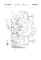
- FIG. 5ais a side view of a stereo camera pod of a preferred embodiment of the present invention adapted for attachment to a surgical microscope;
- FIG. 5bis a top view of the stereo camera pod of FIG. 5a
- FIG. 5cis a schematic illustration of optical elements of a preferred embodiment of the camera pod of FIG. 5a.
- FIG. 1illustrates, in pictorial form, an operating room equipped with a stereoscopic on-screen surgical microscope system.
- a surgical procedureis depicted involving a patient 10, an attending surgeon 12 and an assisting surgeon 14.
- the two surgeonsare located across the operating table 15 from one another, and thus have diametrically opposite views of surgery field 16.
- a surgical microscope 18is mounted above the patient on a boom 20, and is selectively positionable to view the surgery field 16.
- the boommay be a remotely operated positioner.
- the surgical microscopemay be a conventional microscope such as a Zeiss or Wild M600-700 stereo microscope, retrofitted for use in practicing the present invention.
- a stereo camera pod 22which may be configured as described in connection with FIG. 5, is mounted to the surgical microscope 18. Images from the stereo camera pod 22 may be displayed on the attending surgeon's display monitor 24 and the assisting surgeon's display monitor 26, as well as on other display monitors available to other participants and observers (not shown). Camera controllers, distribution amplifiers, stereo image encoder/multiplexers and switching circuitry, described in detail in connection with FIG. 2, may be located in an equipment rack 28 adjacent the operating table.
- the surgeons 12 and 14are outfitted with stereoscopic eyewear 30 and 32, respectively.
- the eyewearis designed to select different images displayed by a single monitor to replicate stereoscopic vision of the magnified field of surgery.
- a number of systems for providing stereoscope television displaysare known in the art. Such systems include passive systems, in which polarized goggles selectively transmit or block optically combined left and right perspective images from a television display. Also known are active systems for multiplexing and demultiplexing signals representing left and right perspective viewpoints. See, e.g. U.S. Pat. Nos. 4,967,268 and 5,193,000, the disclosures of which are incorporated herein by reference. A system found particularly useful in practicing the present invention is known as CRYSTAL-EYES®, a product of Stereographics, San Rafael, Calif. This system employs electronic combinations of left and right video signals and active shutters for producing stereoscopic displays. Left and right perspective views are field sequentially displayed on a television monitor.
- the viewerwears active shutter goggles with a liquid crystal assembly for selectively transmitting or blocking images from the monitor to the left or right eye.
- a remote emitterin electronic communication with left and right video signal multiplexing circuitry, transmits an infrared synchronization signal to the goggles.
- Drive circuitry in the gogglesselectively activates the liquid crystal assembly to transmit or block the image currently displayed on the monitor so that the left eye is exposed to a sequence of left perspective image fields and the right eye is exposed to a sequence of right perspective image fields.
- the gogglesturn clear when the wearer looks away from the display monitor, for example, to select an instrument for use in the surgical procedure.
- Systemsare commercially available which have resolution and refresh rate acceptable for use in microsurgery.
- a camera pod 22may contain a right camera 40 and a left camera 42, located in optical paths 44 and 46 of the objective lens system of the surgical microscope 18. Electrical output signals from the cameras are applied to the dual camera controller 48. A first video signal from the dual camera controller corresponding to a left eye view point is applied to a stereo image encoder/multiplexer 50. Likewise, a second video signal from the dual camera controller is applied to the encoder/multiplexer 50.
- the encoder/multiplexer 50is of the type described above for multiplexing the left and right channels of picture information into a single video channel output signal and for encoding a synchronization signal which is transmitted by emitters 34 and 36 to the stereoscopic eyewear of participants or real-time observers.
- Signal processing in this systemproceeds as follows.
- the left and right cameraseach produce a standard Y/C (luminance/chrominance) video signal, which comprises 525 horizontal lines per video frame.
- the encoder/multiplexer 50interlaces these two signals into a custom signal containing 1050 lines per complete video frame.
- the signal that the encoder/multiplexer sends to the display monitoris transmitted as a sequence of parts, or "fields", of a complete frame interlaced in the following manner:
- Each fieldis shown on the display monitor for 1/120th of a second.
- One complete cycle of fourfold interlacingtakes 1/30th of a second.
- one complete stereoscopic video imageappears on the monitor each 1/30th of a second.
- the processor's synchronizing signal sent via the infrared emitters 34 to the stereoscopic eyewearcauses a left lens eyewear to be transparent and a right lens to be opaque during steps 1 and 3 of the cycle.
- the right lensis transparent and the left lens is opaque during steps 2 and 4 of the cycle. Therefore, the attending surgeon's left and right eyes see only the images from the corresponding cameras. Because each eye sees half of the total video signal, the effective resolution of the stereoscopic image on the monitor is 525 horizontal lines.
- a signal sent by the process to a video tape recorder 54may be compressed by half to be recorded onto standard S-VHS videotape (which is formatted to accept a standard Y/C signal).
- the multiplexed video signal from the encoder/multiplexer 50may be applied to a distribution amplifier 52.
- the amplifier 52is a two-way distribution amplifier for providing the multiplexed video signal to the attending surgeon's display monitor 24 and simultaneously, to the assisting surgeon's display monitor 26.
- a switching module 38is located in the signal path of the multiplexed video signal from the distribution amplifier. It is also in the signal path of the encoder synchronization signal first applied to emitter 34 and thereafter to emitter 36.
- the structure and operation of such a switching moduleis described in greater detail in connection with FIGS. 3 and 4. It should be noted here, that for simplicity a switching module has been included only for signals used in the assisting surgeon's monitor 26 and emitter 36. It will be readily understood that maximum system flexibility is achieved where a switching module is associated with each monitor display.
- a switching modulecan be used in system configurations having a single monitor. For example, in the use of a surgical microscope for periodontal work by a single dentist, the most effective image orientation may require inversion. In circumstances where a mirror is used in the procedure, left-right reversal, with or without up-down reversal, may be achieved with the aid of the switching module 38.
- the switching moduleprovides the necessary flexibility to use the stereoscopic microscope system in widely varied configurations involving one or more display monitors and various orientations corresponding to the location and points of view of various participants and observers.
- the multiplexed stereoscopic video signalcan be recorded on the videotape recorder 54 as noted above.
- a replay system(or auxiliary real time display system) may be connected to the videotape recorder 54.
- Such a systemmay include a stereo-videotape playback unit 56 which derives encoded synchronization signals for auxiliary emitters, such as emitter 58.
- a distribution amplifier 60receives the multiplexed stereoscopic video signal from the playback unit and applies it to one or more auxiliary display monitors 62. Viewers of these monitors may be provided with stereoscopic eyewear 64, of the type previously described for selectively transmitting and blocking images presented on the display monitor 62.
- Inversion of the video imageis achieved by DPDT switches S1 and S2.
- a third DPDT switch S3is employed to achieve emitter phase reversal.
- FIG. 3bAn advantageous means of connecting the switching module to a conventional CRT display monitor 100 is illustrated in FIG. 3b.
- a video yoke drive circuit 102is connected directly to the CRT vertical yoke 104 and horizontal yoke 106.
- the polarity of the connectionis such that the conventional up-down/right-left orientation of images is displayed.
- the display monitor 100may be modified by interrupting the circuit paths 108 connecting the video yoke drive circuit to the respective yokes, and inserting switches S1 and S2 into the circuit.
- a socket 110 and cabling 112may be provided for this purpose
- switches S1, S2 and S3are illustrated in FIG. 4.
- two observers 150 and 152are shown, viewing a reference object 154.
- a stereoscopic surgical microscope and camera pod assemblyis directed at the reference object 154 in such a way that the orientation and prospective of the image produced corresponds to that which would be seen by the first participant either directly or through the microscope eyepieces.
- This image pairas it would be seen by the left and right eye of the first participant, is identified by the numeral 156.
- Switches S1 and S2produce a vertical inversion and a horizontal inversion, respectively, by reversing the polarity of the vertical and horizontal yoke signals.
- the effectis both an up-down reversal and a left-right reversal. This is referred to collectively as an "inversion", the effects of which are illustrated by the image pair 158.
- a mirror imagecan be produced by activating only switch S2 thereby producing a left-right reversal without an up-down reversal (not shown).
- a switch S3is shown for performing emitter phase reversal.
- the positive pole of an emitter phase circuitis connected to a positive pole of an infrared LED array which transmits the synchronization signal to the eyewear.
- the negative polesare likewise connected.
- a signalis sent by the emitter and received by the eyewear which shutters the wearer's eyes in such a way that the left eye views image fields corresponding to the normal left eye viewpoint, and the right eye views image fields corresponding to the normal right eye viewpoint.
- Switch S3is provided to selectively reverse the output of the emitter such that the images viewed by each eye are reversed.
- the effect of this eye reversalis shown in the pair of images 160 of FIG. 4. Were this not done, the perceived stereoscopic effect of the images would be incorrect, making distant objects appear close and close objects appear distant.
- the images 160represent a view which is displayed to the second participant (for example the assisting surgeon shown in FIG. 1).
- the net effect of the inversion and eye reversalis to provide the second participant with essentially the same stereoscopic images as would be present if the second participant directly viewed the reference object from the position and orientation shown at the top of FIG. 4, thus enhancing such things as handeye coordination which may be required in a surgical procedure.
- FIGS. 5a and 5bare, respectively, side and top views of the internal structure of a video camera pod 200 for retrofitting a surgical microscope for use in a stereoscopic on-screen display system.
- a male flange 202is provided for releasably mounting the pod to a binocular surgical microscope.
- the podcan be mounted in the still camera mount of the microscope, thus leaving both eyepieces available for use.
- the dimensions and configuration of the flangeare selected to adapt, for example, to the still camera port of one or more conventional surgical microscopes.
- the podWhen mounted, the pod is positioned to receive light corresponding to the left and right perspective viewpoint images of the microscope.
- the podemploys at least one optical detector for converting left and right optical image information into electrical signals.
- two optical detectors 204may be mounted in the pod and separated by a distance corresponding to the axial separation of the microscope.
- these detectorsmay be CCD camera elements such as Micro camera IK-M41A marketed by Toshiba or color video camera module XC-999 marketed by Sony Corporation. Such camera elements are rigidly held in position in the pod at their respective optical axes of the microscope by upright members 206.
- Lenses 208are placed in the optical paths of the pod in a base member 210.
- the lensesdirect the magnified images on the plane of focus of the CCD chip(s).
- the robustness of CCD chip and lens alignment within the podassures production of stable, properly focused images.
- FIG. 5cis an illustration of optical elements employed in a preferred embodiment of the video camera pod of FIG. 5a, especially adapted for use with typical configurations of Wild M600 and M700 series surgical microscopes and the Vasconcellas M900 series surgical microscopes.
- Two optical detectors 204' and 204"are held in fixed position on the optical axes of two lens assemblies 220 and 222, respectively.
- Each lens assemblyis made up of two back-to-back oriented, precision optimized achromats 224.
- Each achromatis formed by cementing one plano concave lens 226 to one double convex lens 228.
- the achromatsare 19 mm in diameter and have a focal length of 90 mm.
- the optical detectorsare positioned coaxially above the left and right lens assemblies as indicated in FIG. 5c.
- optical systemused in the video camera pod.
- Other such optical systemsmay be designed which match the field of view and magnification of the pod with that which would be seen through the eyepieces of a particular microscope, and also to insure parfocality, i.e. the maintenance of focus over the range of magnification of the host microscope. This facilitates training of surgical personnel already familiar with performing micro-surgery while viewing the procedures through the eyepieces of the surgical microscope being retrofitted.
Landscapes
- Engineering & Computer Science (AREA)
- Multimedia (AREA)
- Signal Processing (AREA)
- Physics & Mathematics (AREA)
- Chemical & Material Sciences (AREA)
- Analytical Chemistry (AREA)
- General Physics & Mathematics (AREA)
- Optics & Photonics (AREA)
- Microscoopes, Condenser (AREA)
- Testing, Inspecting, Measuring Of Stereoscopic Televisions And Televisions (AREA)
Abstract
Description
Claims (15)
Priority Applications (1)
| Application Number | Priority Date | Filing Date | Title |
|---|---|---|---|
| US08/598,902US5867210A (en) | 1996-02-09 | 1996-02-09 | Stereoscopic on-screen surgical microscope systems |
Applications Claiming Priority (1)
| Application Number | Priority Date | Filing Date | Title |
|---|---|---|---|
| US08/598,902US5867210A (en) | 1996-02-09 | 1996-02-09 | Stereoscopic on-screen surgical microscope systems |
Publications (1)
| Publication Number | Publication Date |
|---|---|
| US5867210Atrue US5867210A (en) | 1999-02-02 |
Family
ID=24397407
Family Applications (1)
| Application Number | Title | Priority Date | Filing Date |
|---|---|---|---|
| US08/598,902Expired - LifetimeUS5867210A (en) | 1996-02-09 | 1996-02-09 | Stereoscopic on-screen surgical microscope systems |
Country Status (1)
| Country | Link |
|---|---|
| US (1) | US5867210A (en) |
Cited By (100)
| Publication number | Priority date | Publication date | Assignee | Title |
|---|---|---|---|---|
| WO2000000934A3 (en)* | 1982-08-31 | 2000-03-23 | Koninkl Philips Electronics Nv | Filter for transforming 3d data in a hardware accelerated rendering architecture |
| US20020008757A1 (en)* | 2000-05-17 | 2002-01-24 | Seiji Sato | Stereoscopic picture image forming apparatus |
| US6466432B1 (en)* | 1999-07-12 | 2002-10-15 | Frank Beger | Instrument and service unit for a surgical operating area |
| US6471363B2 (en) | 1999-12-23 | 2002-10-29 | Hill-Rom Services, Inc. | Surgical theater system having light, monitors, and cameras |
| US6496183B1 (en) | 1998-06-30 | 2002-12-17 | Koninklijke Philips Electronics N.V. | Filter for transforming 3D data in a hardware accelerated rendering architecture |
| US20030053202A1 (en)* | 2001-09-17 | 2003-03-20 | Iekado Sibata Yoshikatsu Seiki | Surgical microscope system |
| US20030071893A1 (en)* | 2001-10-05 | 2003-04-17 | David Miller | System and method of providing visual documentation during surgery |
| US6639789B2 (en) | 2000-07-12 | 2003-10-28 | Karl Storz Gmbh & Co. Kg | Instrument and service unit for a surgical operating area |
| WO2003100501A1 (en)* | 2002-05-23 | 2003-12-04 | Enrique De Font-Reaulx Rojas | System for stereoscopic viewing of real-time images |
| US20040017607A1 (en)* | 2002-02-04 | 2004-01-29 | Christoph Hauger | Stereo-examination systems and stereo-image generation apparatus as well as a method for operating the same |
| US20040070823A1 (en)* | 2002-10-10 | 2004-04-15 | Radna Richard J. | Head-mount recording of three-dimensional stereo video images |
| WO2004066014A1 (en)* | 2003-01-21 | 2004-08-05 | Andreas Obrebski | Visualization device for a surgical microscope having a display unit that is supported by a support frame on the body of the viewer |
| DE10203215B4 (en)* | 2002-01-28 | 2004-09-09 | Carl Zeiss Jena Gmbh | Microscope, in particular surgical microscope |
| US20040224279A1 (en)* | 2003-05-06 | 2004-11-11 | Ormco Corporation | Dental display system |
| US20050046933A1 (en)* | 2003-08-08 | 2005-03-03 | Carl Zeiss Ag | Microscopy system and method |
| US20050058962A1 (en)* | 2002-12-05 | 2005-03-17 | Ormco Corporation | Remote dental display system |
| US20050063047A1 (en)* | 2003-08-04 | 2005-03-24 | Carl Zeiss Ag | Microscopy system and method |
| US20050117118A1 (en)* | 2001-10-05 | 2005-06-02 | David Miller | Digital ophthalmic workstation |
| WO2005066690A1 (en)* | 2003-12-19 | 2005-07-21 | De Font-Reaulx-Rojas Enrique | System for the stereoscopic viewing of real-time or static images |
| WO2005074303A1 (en)* | 2004-01-23 | 2005-08-11 | Henry Ford Health System | Stereoscopic projection-based display for surgical robot |
| WO2006000072A1 (en)* | 2004-06-29 | 2006-01-05 | Jorge Mitre | System and process to capture, store and show stereoscopic images |
| US20060055786A1 (en)* | 2004-03-09 | 2006-03-16 | Viosport | Portable camera and wiring harness |
| US7091416B1 (en) | 2005-12-30 | 2006-08-15 | Steris Inc. | Cover assembly for video monitor |
| US20070035831A1 (en)* | 2003-12-19 | 2007-02-15 | Gutierrez Novelo Manuel R | 3D Videogame system |
| US7190392B1 (en)* | 1997-10-23 | 2007-03-13 | Maguire Jr Francis J | Telepresence system and active/passive mode display for use therein |
| US20070121202A1 (en)* | 2004-10-21 | 2007-05-31 | Truevision Systems, Inc. | Stereoscopic electronic microscope workstation |
| US20070121203A1 (en)* | 2005-10-21 | 2007-05-31 | Truevision Systems, Inc. | Stereoscopic electronic microscope workstation |
| USD547346S1 (en) | 2004-03-09 | 2007-07-24 | V.I.O., Inc. | Camera |
| US20070188603A1 (en)* | 2005-10-21 | 2007-08-16 | Riederer Thomas P | Stereoscopic display cart and system |
| US20070263003A1 (en)* | 2006-04-03 | 2007-11-15 | Sony Computer Entertainment Inc. | Screen sharing method and apparatus |
| US20080004610A1 (en)* | 2006-06-30 | 2008-01-03 | David Miller | System for calculating IOL power |
| EP1804106A3 (en)* | 2002-02-04 | 2008-05-07 | Carl Zeiss Surgical GmbH | Stereoscopic examination system, stereoscopic image processing apparatus and operating method |
| EP1925962A1 (en)* | 2006-11-21 | 2008-05-28 | Swiss Medical Technology GmbH | Stereo video microscope system |
| WO2008061738A1 (en) | 2006-11-21 | 2008-05-29 | Swiss Medical Technology Gmbh | Stereo video microscope system |
| US20080215065A1 (en)* | 1996-02-20 | 2008-09-04 | Intuitive Surgical | Medical robotic arm that is attached to an operating table |
| US7443417B1 (en)* | 1999-12-13 | 2008-10-28 | Geoffrey W Heinrich | Method of performing dental work and apparatus providing vision enhancement dentistry |
| US20080278571A1 (en)* | 2007-05-10 | 2008-11-13 | Mora Assad F | Stereoscopic three dimensional visualization system and method of use |
| US20090048608A1 (en)* | 2007-08-13 | 2009-02-19 | Mikhail Boukhny | Toric lenses alignment using pre-operative images |
| US20090099576A1 (en)* | 2002-01-16 | 2009-04-16 | Intuitive Surgical, Inc. | Minimally invasive surgical training using robotics and telecollaboration |
| US20090173846A1 (en)* | 2008-01-09 | 2009-07-09 | Allan Katz | Medical boom |
| US20090190209A1 (en)* | 2006-06-29 | 2009-07-30 | Mitaka Kohki Co., Ltd. | Surgical microscope system |
| US20090254070A1 (en)* | 2008-04-04 | 2009-10-08 | Ashok Burton Tripathi | Apparatus and methods for performing enhanced visually directed procedures under low ambient light conditions |
| US20100094262A1 (en)* | 2008-10-10 | 2010-04-15 | Ashok Burton Tripathi | Real-time surgical reference indicium apparatus and methods for surgical applications |
| USD616486S1 (en) | 2008-10-20 | 2010-05-25 | X6D Ltd. | 3D glasses |
| US20100149636A1 (en)* | 2008-11-17 | 2010-06-17 | Macnaughton Boyd | Housing And Frame For 3D Glasses |
| US20100208199A1 (en)* | 2009-02-19 | 2010-08-19 | Ilias Levis | Intraocular lens alignment |
| US20100217278A1 (en)* | 2009-02-20 | 2010-08-26 | Ashok Burton Tripathi | Real-time surgical reference indicium apparatus and methods for intraocular lens implantation |
| US20100277485A1 (en)* | 2006-04-03 | 2010-11-04 | Sony Computer Entertainment America Llc | System and method of displaying multiple video feeds |
| EP2278818A2 (en)* | 2009-07-16 | 2011-01-26 | Sony Computer Entertainment America LLC | Display viewing system and methods for optimizing display view based on active tracking |
| USRE42091E1 (en) | 1998-11-20 | 2011-02-01 | Jerry Moscovitch | Computer display screen system and adjustable screen mount, and swinging screens therefor |
| US20110032335A1 (en)* | 2009-08-07 | 2011-02-10 | Leica Microsystems (Schweiz) Ag | Video stereomicroscope |
| US20110092984A1 (en)* | 2009-10-20 | 2011-04-21 | Ashok Burton Tripathi | Real-time Surgical Reference Indicium Apparatus and Methods for Astigmatism Correction |
| US20110137322A1 (en)* | 1998-11-20 | 2011-06-09 | Intuitive Surgical Operations | Cooperative Minimally Invasive Telesurgical System |
| GB2476245A (en)* | 2009-12-15 | 2011-06-22 | Hospital Authority | Stereoscopic display system for surgery |
| US20110205347A1 (en)* | 2008-11-17 | 2011-08-25 | X6D Limited | Universal 3d glasses for use with televisions |
| US20110213342A1 (en)* | 2010-02-26 | 2011-09-01 | Ashok Burton Tripathi | Real-time Virtual Indicium Apparatus and Methods for Guiding an Implant into an Eye |
| US20110216252A1 (en)* | 2008-11-17 | 2011-09-08 | Macnaughton Boyd | 3D Shutter Glasses For Use With LCD Displays |
| US20110216176A1 (en)* | 2008-11-17 | 2011-09-08 | Macnaughton Boyd | 3D Glasses With RF Synchronization |
| USD646451S1 (en) | 2009-03-30 | 2011-10-04 | X6D Limited | Cart for 3D glasses |
| USD650956S1 (en) | 2009-05-13 | 2011-12-20 | X6D Limited | Cart for 3D glasses |
| USD652860S1 (en) | 2008-10-20 | 2012-01-24 | X6D Limited | 3D glasses |
| WO2012047715A1 (en)* | 2010-10-05 | 2012-04-12 | Echostar Technologies L.L.C. | Apparatus, systems and methods for synchronization of 3-d shutter glasses to one of a plurality of presentation devices |
| USD662965S1 (en) | 2010-02-04 | 2012-07-03 | X6D Limited | 3D glasses |
| USD664183S1 (en) | 2010-08-27 | 2012-07-24 | X6D Limited | 3D glasses |
| USD666663S1 (en) | 2008-10-20 | 2012-09-04 | X6D Limited | 3D glasses |
| USD669522S1 (en) | 2010-08-27 | 2012-10-23 | X6D Limited | 3D glasses |
| USD671590S1 (en) | 2010-09-10 | 2012-11-27 | X6D Limited | 3D glasses |
| CN102809808A (en)* | 2012-08-15 | 2012-12-05 | 深圳市麟静科技有限公司 | Medical 3-dimensional imaging operating microscope system |
| USD672804S1 (en) | 2009-05-13 | 2012-12-18 | X6D Limited | 3D glasses |
| US8462103B1 (en) | 1998-12-23 | 2013-06-11 | Jerry Moscovitch | Computer display screen system and adjustable screen mount, and swinging screens therefor |
| CN103340686A (en)* | 2013-07-01 | 2013-10-09 | 中山大学 | General surgery three-dimensional micrography camera shooting presentation device |
| USD692941S1 (en) | 2009-11-16 | 2013-11-05 | X6D Limited | 3D glasses |
| USD711959S1 (en) | 2012-08-10 | 2014-08-26 | X6D Limited | Glasses for amblyopia treatment |
| US8870900B2 (en) | 1999-11-09 | 2014-10-28 | Intuitive Surgical Operations, Inc. | Endoscopic beating-heart stabilizer and vessel occlusion fastener |
| USRE45394E1 (en) | 2008-10-20 | 2015-03-03 | X6D Limited | 3D glasses |
| US8982203B2 (en) | 2007-06-06 | 2015-03-17 | Karl Storz Gmbh & Co. Kg | Video system for viewing an object on a body |
| US9119654B2 (en) | 1998-11-20 | 2015-09-01 | Intuitive Surgical Operations, Inc. | Stabilizer for robotic beating-heart surgery |
| US20150301326A1 (en)* | 2014-04-21 | 2015-10-22 | Mitaka Kohki Co., Ltd. | Surgical microscope system |
| WO2015124699A3 (en)* | 2014-02-19 | 2015-11-19 | Carl Zeiss Meditec Ag | Surgical microscope, and creation of an observation image of an object region in a surgical microscope |
| DE102014223181B3 (en)* | 2014-11-13 | 2016-01-28 | Carl Zeiss Meditec Ag | Visualization device for a surgical site |
| US9271798B2 (en) | 1998-11-20 | 2016-03-01 | Intuitive Surgical Operations, Inc. | Multi-user medical robotic system for collaboration or training in minimally invasive surgical procedures |
| US9402619B2 (en) | 1996-11-22 | 2016-08-02 | Intuitive Surgical Operation, Inc. | Rigidly-linked articulating wrist with decoupled motion transmission |
| US9480539B2 (en) | 2011-11-03 | 2016-11-01 | James Ortlieb | Viewing system and viewing method for assisting user in carrying out surgery by identifying a target image |
| US9552660B2 (en) | 2012-08-30 | 2017-01-24 | Truevision Systems, Inc. | Imaging system and methods displaying a fused multidimensional reconstructed image |
| DE10262230B4 (en)* | 2002-01-28 | 2017-03-16 | Carl Zeiss Jena Gmbh | Microscope, especially surgical microscope |
| TWI581003B (en)* | 2015-09-04 | 2017-05-01 | Show Chwan Memorial Hospital | Dimensional image display device for surgical microscope |
| TWI582462B (en)* | 2015-09-04 | 2017-05-11 | Show Chwan Memorial Hospital | Lightweight 3D stereoscopic surgical microscope device |
| US9655775B2 (en) | 2007-08-13 | 2017-05-23 | Novartis Ag | Toric lenses alignment using pre-operative images |
| DE102017105941B3 (en) | 2017-03-20 | 2018-05-17 | Carl Zeiss Meditec Ag | Surgical microscope with an image sensor and a display and method for operating a surgical microscope |
| US10117721B2 (en) | 2008-10-10 | 2018-11-06 | Truevision Systems, Inc. | Real-time surgical reference guides and methods for surgical applications |
| WO2017192996A3 (en)* | 2016-05-05 | 2019-04-04 | Watson Robert D | Surgical stereoscopic visualization system with movable head mounted display |
| US10299880B2 (en) | 2017-04-24 | 2019-05-28 | Truevision Systems, Inc. | Stereoscopic visualization camera and platform |
| US10917543B2 (en) | 2017-04-24 | 2021-02-09 | Alcon Inc. | Stereoscopic visualization camera and integrated robotics platform |
| US11083537B2 (en) | 2017-04-24 | 2021-08-10 | Alcon Inc. | Stereoscopic camera with fluorescence visualization |
| US11340441B2 (en) | 2019-07-09 | 2022-05-24 | Lightech Fiberoptics, Inc. | Microscope made with CMOS camera(s) |
| CN114786000A (en)* | 2022-04-12 | 2022-07-22 | 嘉兴智瞳科技有限公司 | Microsurgery 3D digital imaging system and 3D microsurgery camera |
| US11516437B2 (en)* | 2018-03-26 | 2022-11-29 | Bhs Technologies Gmbh | Stereo microscope for use in microsurgical operations on a patient and method for controlling the stereo microscope |
| DE102022105090B3 (en) | 2022-03-03 | 2023-06-29 | Schölly Fiberoptic GmbH | Stereoscopic arrangement and surgical microscope with stereoscopic arrangement |
| EP4242720A1 (en)* | 2022-03-08 | 2023-09-13 | Leica Instruments (Singapore) Pte Ltd | Microscope system and system, method and computer program for a microscope system |
| EP4212125A4 (en)* | 2020-09-23 | 2024-03-13 | Zumax Medical Co., Ltd. | Operating microscope for two surgeons |
Citations (5)
| Publication number | Priority date | Publication date | Assignee | Title |
|---|---|---|---|---|
| US4967268A (en)* | 1989-07-31 | 1990-10-30 | Stereographics | Liquid crystal shutter system for stereoscopic and other applications |
| US4987488A (en)* | 1988-03-07 | 1991-01-22 | George Berci | Video system for visualizing microsurgical images with enhanced depth of field |
| US5193000A (en)* | 1991-08-28 | 1993-03-09 | Stereographics Corporation | Multiplexing technique for stereoscopic video system |
| US5545120A (en)* | 1995-01-18 | 1996-08-13 | Medical Media Systems | Endoscopic viewing system for maintaining a surgeon's normal sense of kinesthesia during endoscopic surgery regardless of the orientation of the endoscope vis-a-vis the surgeon |
| US5652676A (en)* | 1995-04-24 | 1997-07-29 | Grinblat; Avi | Microscope-television camera adapter |
- 1996
- 1996-02-09USUS08/598,902patent/US5867210A/ennot_activeExpired - Lifetime
Patent Citations (5)
| Publication number | Priority date | Publication date | Assignee | Title |
|---|---|---|---|---|
| US4987488A (en)* | 1988-03-07 | 1991-01-22 | George Berci | Video system for visualizing microsurgical images with enhanced depth of field |
| US4967268A (en)* | 1989-07-31 | 1990-10-30 | Stereographics | Liquid crystal shutter system for stereoscopic and other applications |
| US5193000A (en)* | 1991-08-28 | 1993-03-09 | Stereographics Corporation | Multiplexing technique for stereoscopic video system |
| US5545120A (en)* | 1995-01-18 | 1996-08-13 | Medical Media Systems | Endoscopic viewing system for maintaining a surgeon's normal sense of kinesthesia during endoscopic surgery regardless of the orientation of the endoscope vis-a-vis the surgeon |
| US5652676A (en)* | 1995-04-24 | 1997-07-29 | Grinblat; Avi | Microscope-television camera adapter |
Non-Patent Citations (25)
| Title |
|---|
| Berisch Strauch, et al., Three Dimensional Imaging In Microvascular Surgery, The Journal of Medicine and Virtual Reality, pp. 34 35.* |
| Berisch Strauch, et al., Three-Dimensional Imaging In Microvascular Surgery, The Journal of Medicine and Virtual Reality, pp. 34-35. |
| Crystal Eyes Video System User s Manual, Crystal Eyes Stereographics, pp. 56 61.* |
| Crystal Eyes Video System User's Manual, Crystal Eyes Stereographics, pp. 56-61. |
| European Surgical Research Clinical and Experimental Surgery, 30th Congress of the European Society for Surgical Research, May 10 13, 1995, The Netherlands.* |
| European Surgical Research Clinical and Experimental Surgery, 30th Congress of the European Society for Surgical Research, May 10-13, 1995, The Netherlands. |
| Gene E. Schlueter, et al., The stereomicroscope Instrumentation And Techniques, Reprinted from American Laboratory, Apr. 1976.* |
| H. Kobayashi, et al., Three Dimensional videomonitor in microsurgery, Neuroschicurgis 36, pp. 129 130.* |
| H. Kobayashi, et al., Three Dimensional videomonitor in microsurgery, Neuroschicurgis 36, pp. 129-130. |
| H.F. Reinhardt, et al., Stereo Microvision Development of an Opto Electronic Operating Microscope, Bildgebung 1993, pp. 105 109.* |
| H.F. Reinhardt, et al., Stereo-Microvision Development of an Opto-Electronic Operating Microscope, Bildgebung 1993, pp. 105-109. |
| Hiroshi Okudera, et al., Introduction of high definition television system to neurosurgical documentation, Neurological Research, 1992, vol. 14, Dec. pp. 386 388.* |
| Hiroshi Okudera, et al., Introduction of high definition television system to neurosurgical documentation, Neurological Research, 1992, vol. 14, Dec. pp. 386-388. |
| Hiroshi Okudera, et al., Three dimensional Hi Vision System for Microneurosurgical Documentation Based on Wide vision Telepresence System Using One Camera and One Monitor, Med. Chir (Tokyo) 33, (1993) pp. 719 721.* |
| Hiroshi Okudera, et al., Three-dimensional Hi-Vision System for Microneurosurgical Documentation Based on Wide-vision Telepresence System Using One Camera and One Monitor, Med. Chir (Tokyo) 33, (1993) pp. 719-721. |
| J. Reiner, Possible Methods of Reproducing Stereoscopic Images on Monitors in Combination with the Operating Microscope, Klin. Mbl. Augenheilk. 196 (1990), pp. 51 53.* |
| J. Reiner, Possible Methods of Reproducing Stereoscopic Images on Monitors in Combination with the Operating Microscope,Klin. Mbl. Augenheilk. 196 (1990), pp. 51-53. |
| Kenichiro Sugita, et al., Stereoscopic television system for use with the operating microscope, J. Neurosug 62, 1983, pp. 610 611.* |
| Kenichiro Sugita, et al., Stereoscopic television system for use with the operating microscope, J. Neurosug 62, 1983, pp. 610-611. |
| Microsurgery Without A Microscope: Development Of A Three Dimensional On Screen Microsurgery System ( TOMS ), The Journal Of Medicine And Virtual Reality, vol. 1, No. 1, Spring 1995, pp. 2, 26 32.* |
| Microsurgery Without A Microscope: Development Of A Three-Dimensional On-Screen Microsurgery System (TOMS), The Journal Of Medicine And Virtual Reality, vol. 1, No. 1, Spring 1995, pp. 2, 26-32. |
| Ralph JPM Franken, MD., Three Dimensional Applications In Surgery Past Present Future, First VR in Med and Developers Expo, Jun. 1 4, 1995.* |
| Ralph JPM Franken, MD., Three-Dimensional Applications In Surgery Past-Present-Future, First VR in Med and Developers' Expo, Jun. 1-4, 1995. |
| Samuel E. Rod, Combined Stereoscopic Telepresence and Virtual Reality in Surgical Training, presented at the Virtual Reality in Medicine and Developers Expo, Jun. 1 4, 1995, Cambridge, Massachusetts, Bristlecone Corp., Jun. 1995, pp. 1 7.* |
| Samuel E. Rod, Combined Stereoscopic Telepresence and Virtual Reality in Surgical Training, presented at the Virtual Reality in Medicine and Developers' Expo, Jun. 1-4, 1995, Cambridge, Massachusetts, Bristlecone Corp., Jun. 1995, pp. 1-7. |
Cited By (188)
| Publication number | Priority date | Publication date | Assignee | Title |
|---|---|---|---|---|
| WO2000000934A3 (en)* | 1982-08-31 | 2000-03-23 | Koninkl Philips Electronics Nv | Filter for transforming 3d data in a hardware accelerated rendering architecture |
| US20080215065A1 (en)* | 1996-02-20 | 2008-09-04 | Intuitive Surgical | Medical robotic arm that is attached to an operating table |
| US7695481B2 (en)* | 1996-02-20 | 2010-04-13 | Intuitive Surgical, Inc. | Medical robotic system with different scaling factors |
| US9402619B2 (en) | 1996-11-22 | 2016-08-02 | Intuitive Surgical Operation, Inc. | Rigidly-linked articulating wrist with decoupled motion transmission |
| US7587747B2 (en) | 1997-10-23 | 2009-09-08 | Maguire Jr Francis J | Telepresence method and apparatus for simultaneous use by multiple active/passive users |
| US7190392B1 (en)* | 1997-10-23 | 2007-03-13 | Maguire Jr Francis J | Telepresence system and active/passive mode display for use therein |
| US6496183B1 (en) | 1998-06-30 | 2002-12-17 | Koninklijke Philips Electronics N.V. | Filter for transforming 3D data in a hardware accelerated rendering architecture |
| US8914150B2 (en) | 1998-11-20 | 2014-12-16 | Intuitive Surgical Operations, Inc. | Cooperative minimally invasive telesurgical system |
| US9666101B2 (en) | 1998-11-20 | 2017-05-30 | Intuitive Surgical Operations, Inc. | Multi-user medical robotic system for collaboration or training in minimally invasive surgical procedures |
| USRE42091E1 (en) | 1998-11-20 | 2011-02-01 | Jerry Moscovitch | Computer display screen system and adjustable screen mount, and swinging screens therefor |
| US20110137322A1 (en)* | 1998-11-20 | 2011-06-09 | Intuitive Surgical Operations | Cooperative Minimally Invasive Telesurgical System |
| US8489235B2 (en) | 1998-11-20 | 2013-07-16 | Intuitive Surgical Operations, Inc. | Cooperative minimally invasive telesurgical system |
| US8504201B2 (en) | 1998-11-20 | 2013-08-06 | Intuitive Sugrical Operations, Inc. | Cooperative minimally invasive telesurgical system |
| US8666544B2 (en) | 1998-11-20 | 2014-03-04 | Intuitive Surgical Operations, Inc. | Cooperative minimally invasive telesurgical system |
| US9119654B2 (en) | 1998-11-20 | 2015-09-01 | Intuitive Surgical Operations, Inc. | Stabilizer for robotic beating-heart surgery |
| US9271798B2 (en) | 1998-11-20 | 2016-03-01 | Intuitive Surgical Operations, Inc. | Multi-user medical robotic system for collaboration or training in minimally invasive surgical procedures |
| US9867671B2 (en) | 1998-11-20 | 2018-01-16 | Intuitive Surgical Operations, Inc. | Multi-user medical robotic system for collaboration or training in minimally invasive surgical procedures |
| US9636186B2 (en) | 1998-11-20 | 2017-05-02 | Intuitive Surgical Operations, Inc. | Multi-user medical robotic system for collaboration or training in minimally invasive surgical procedures |
| US8462103B1 (en) | 1998-12-23 | 2013-06-11 | Jerry Moscovitch | Computer display screen system and adjustable screen mount, and swinging screens therefor |
| US6466432B1 (en)* | 1999-07-12 | 2002-10-15 | Frank Beger | Instrument and service unit for a surgical operating area |
| US8870900B2 (en) | 1999-11-09 | 2014-10-28 | Intuitive Surgical Operations, Inc. | Endoscopic beating-heart stabilizer and vessel occlusion fastener |
| US7443417B1 (en)* | 1999-12-13 | 2008-10-28 | Geoffrey W Heinrich | Method of performing dental work and apparatus providing vision enhancement dentistry |
| US6899442B2 (en) | 1999-12-23 | 2005-05-31 | Hill-Rom Services, Inc. | Surgical theater system having light, monitors, and cameras |
| US6471363B2 (en) | 1999-12-23 | 2002-10-29 | Hill-Rom Services, Inc. | Surgical theater system having light, monitors, and cameras |
| US6639623B2 (en) | 1999-12-23 | 2003-10-28 | Hill-Rom Services, Inc. | Controls for a surgical theater system |
| US20030021107A1 (en)* | 1999-12-23 | 2003-01-30 | Howell Charles A. | Surgical theater system having light, monitors, and cameras |
| US20020008757A1 (en)* | 2000-05-17 | 2002-01-24 | Seiji Sato | Stereoscopic picture image forming apparatus |
| US6639789B2 (en) | 2000-07-12 | 2003-10-28 | Karl Storz Gmbh & Co. Kg | Instrument and service unit for a surgical operating area |
| EP1293816A3 (en)* | 2001-09-17 | 2004-11-10 | Iekado Sibata | Surgical microscope system |
| US20030053202A1 (en)* | 2001-09-17 | 2003-03-20 | Iekado Sibata Yoshikatsu Seiki | Surgical microscope system |
| US20030071893A1 (en)* | 2001-10-05 | 2003-04-17 | David Miller | System and method of providing visual documentation during surgery |
| US20050117118A1 (en)* | 2001-10-05 | 2005-06-02 | David Miller | Digital ophthalmic workstation |
| US20090099576A1 (en)* | 2002-01-16 | 2009-04-16 | Intuitive Surgical, Inc. | Minimally invasive surgical training using robotics and telecollaboration |
| US9039681B2 (en) | 2002-01-16 | 2015-05-26 | Intuitive Surgical Operations, Inc. | Minimally invasive surgical training using robotics and telecollaboration |
| US9786203B2 (en) | 2002-01-16 | 2017-10-10 | Intuitive Surgical Operations, Inc. | Minimally invasive surgical training using robotics and telecollaboration |
| DE10203215B4 (en)* | 2002-01-28 | 2004-09-09 | Carl Zeiss Jena Gmbh | Microscope, in particular surgical microscope |
| DE10262230B4 (en)* | 2002-01-28 | 2017-03-16 | Carl Zeiss Jena Gmbh | Microscope, especially surgical microscope |
| US20040017607A1 (en)* | 2002-02-04 | 2004-01-29 | Christoph Hauger | Stereo-examination systems and stereo-image generation apparatus as well as a method for operating the same |
| EP1333305A3 (en)* | 2002-02-04 | 2004-07-07 | Carl Zeiss | Stereoscopic examination system, stereoscopic image processing apparatus and operating method |
| EP1804106A3 (en)* | 2002-02-04 | 2008-05-07 | Carl Zeiss Surgical GmbH | Stereoscopic examination system, stereoscopic image processing apparatus and operating method |
| US7180660B2 (en) | 2002-02-04 | 2007-02-20 | Carl-Zeiss-Stiftung Trading As Carl Zeiss | Stereo-examination systems and stereo-image generation apparatus as well as a method for operating the same |
| WO2003100501A1 (en)* | 2002-05-23 | 2003-12-04 | Enrique De Font-Reaulx Rojas | System for stereoscopic viewing of real-time images |
| US20040070823A1 (en)* | 2002-10-10 | 2004-04-15 | Radna Richard J. | Head-mount recording of three-dimensional stereo video images |
| US20050058962A1 (en)* | 2002-12-05 | 2005-03-17 | Ormco Corporation | Remote dental display system |
| WO2004066014A1 (en)* | 2003-01-21 | 2004-08-05 | Andreas Obrebski | Visualization device for a surgical microscope having a display unit that is supported by a support frame on the body of the viewer |
| US20040224279A1 (en)* | 2003-05-06 | 2004-11-11 | Ormco Corporation | Dental display system |
| US7190513B2 (en) | 2003-08-04 | 2007-03-13 | Carl Zeiss Ag | Microscopy system and method |
| US20050063047A1 (en)* | 2003-08-04 | 2005-03-24 | Carl Zeiss Ag | Microscopy system and method |
| DE10335644B3 (en)* | 2003-08-04 | 2005-12-29 | Carl Zeiss | microscopy system |
| DE10335644B9 (en)* | 2003-08-04 | 2006-06-01 | Carl Zeiss | microscopy system |
| US9279974B2 (en) | 2003-08-08 | 2016-03-08 | Carl Zeiss Meditic Ag | Microscopy system and method |
| DE10336475A1 (en)* | 2003-08-08 | 2005-03-10 | Zeiss Carl | microscopy system |
| US7468835B2 (en) | 2003-08-08 | 2008-12-23 | Carl Zeiss Surgical Gmbh | Microscopy system and method |
| DE10336475B9 (en)* | 2003-08-08 | 2006-09-07 | Carl Zeiss | microscopy system |
| DE10336475B4 (en)* | 2003-08-08 | 2006-04-06 | Carl Zeiss | microscopy system |
| US20050046933A1 (en)* | 2003-08-08 | 2005-03-03 | Carl Zeiss Ag | Microscopy system and method |
| US8206218B2 (en) | 2003-12-19 | 2012-06-26 | Tdvision Corporation S.A. De C.V. | 3D videogame system |
| WO2005066690A1 (en)* | 2003-12-19 | 2005-07-21 | De Font-Reaulx-Rojas Enrique | System for the stereoscopic viewing of real-time or static images |
| US20070274577A1 (en)* | 2003-12-19 | 2007-11-29 | Enrique De Font-Reaulx-Rojas | "System for the stereoscopic viewing of real time or static images" |
| US7666096B2 (en)* | 2003-12-19 | 2010-02-23 | Tdvision Corporation S.A. De C.V. | Method for generating the left and right perspectives in a 3D videogame |
| US20070035831A1 (en)* | 2003-12-19 | 2007-02-15 | Gutierrez Novelo Manuel R | 3D Videogame system |
| US20100151944A1 (en)* | 2003-12-19 | 2010-06-17 | Manuel Rafael Gutierrez Novelo | 3d videogame system |
| US20120264515A1 (en)* | 2003-12-19 | 2012-10-18 | Tdvision Corporation S.A. De C.V. | 3d videogame system |
| WO2005074303A1 (en)* | 2004-01-23 | 2005-08-11 | Henry Ford Health System | Stereoscopic projection-based display for surgical robot |
| US20060055786A1 (en)* | 2004-03-09 | 2006-03-16 | Viosport | Portable camera and wiring harness |
| USD547346S1 (en) | 2004-03-09 | 2007-07-24 | V.I.O., Inc. | Camera |
| WO2006000072A1 (en)* | 2004-06-29 | 2006-01-05 | Jorge Mitre | System and process to capture, store and show stereoscopic images |
| US8339447B2 (en) | 2004-10-21 | 2012-12-25 | Truevision Systems, Inc. | Stereoscopic electronic microscope workstation |
| US20070121202A1 (en)* | 2004-10-21 | 2007-05-31 | Truevision Systems, Inc. | Stereoscopic electronic microscope workstation |
| US20070188603A1 (en)* | 2005-10-21 | 2007-08-16 | Riederer Thomas P | Stereoscopic display cart and system |
| US8358330B2 (en) | 2005-10-21 | 2013-01-22 | True Vision Systems, Inc. | Stereoscopic electronic microscope workstation |
| US20070121203A1 (en)* | 2005-10-21 | 2007-05-31 | Truevision Systems, Inc. | Stereoscopic electronic microscope workstation |
| US7091416B1 (en) | 2005-12-30 | 2006-08-15 | Steris Inc. | Cover assembly for video monitor |
| US20100182407A1 (en)* | 2006-04-03 | 2010-07-22 | Sony Computer Entertainment Inc. | Display device with 3d shutter control unit |
| US20100177172A1 (en)* | 2006-04-03 | 2010-07-15 | Sony Computer Entertainment Inc. | Stereoscopic screen sharing method and apparatus |
| US8325222B2 (en) | 2006-04-03 | 2012-12-04 | Sony Computer Entertainment Inc. | Stereoscopic screen sharing method and apparatus |
| US20100277485A1 (en)* | 2006-04-03 | 2010-11-04 | Sony Computer Entertainment America Llc | System and method of displaying multiple video feeds |
| US8325223B2 (en) | 2006-04-03 | 2012-12-04 | Sony Computer Entertainment Inc. | 3D shutter glasses with mode switching based on orientation to display device |
| US20100177174A1 (en)* | 2006-04-03 | 2010-07-15 | Sony Computer Entertainment Inc. | 3d shutter glasses with mode switching based on orientation to display device |
| US8466954B2 (en)* | 2006-04-03 | 2013-06-18 | Sony Computer Entertainment Inc. | Screen sharing method and apparatus |
| US8665291B2 (en) | 2006-04-03 | 2014-03-04 | Sony Computer Entertainment America Llc | System and method of displaying multiple video feeds |
| US8310527B2 (en) | 2006-04-03 | 2012-11-13 | Sony Computer Entertainment Inc. | Display device with 3D shutter control unit |
| US20070263003A1 (en)* | 2006-04-03 | 2007-11-15 | Sony Computer Entertainment Inc. | Screen sharing method and apparatus |
| US8786946B2 (en)* | 2006-06-29 | 2014-07-22 | Mitaka Kohki Co., Ltd. | Surgical microscope system |
| US20090190209A1 (en)* | 2006-06-29 | 2009-07-30 | Mitaka Kohki Co., Ltd. | Surgical microscope system |
| US20080004610A1 (en)* | 2006-06-30 | 2008-01-03 | David Miller | System for calculating IOL power |
| EP1925962A1 (en)* | 2006-11-21 | 2008-05-28 | Swiss Medical Technology GmbH | Stereo video microscope system |
| US8791995B2 (en) | 2006-11-21 | 2014-07-29 | Swiss Medical Technology Gmbh | Stereo video microscope system |
| US20100141739A1 (en)* | 2006-11-21 | 2010-06-10 | Swiss Medical Technology Gmbh | Stereo video microscope system |
| WO2008061738A1 (en) | 2006-11-21 | 2008-05-29 | Swiss Medical Technology Gmbh | Stereo video microscope system |
| US8619127B2 (en) | 2007-05-10 | 2013-12-31 | Assad F. Mora | Stereoscopic three dimensional visualization system and method of use |
| US20080278571A1 (en)* | 2007-05-10 | 2008-11-13 | Mora Assad F | Stereoscopic three dimensional visualization system and method of use |
| US8982203B2 (en) | 2007-06-06 | 2015-03-17 | Karl Storz Gmbh & Co. Kg | Video system for viewing an object on a body |
| US9468360B2 (en) | 2007-06-06 | 2016-10-18 | Karl Storz Gmbh & Co. Kg | Video system for viewing an object on a body |
| US20090048608A1 (en)* | 2007-08-13 | 2009-02-19 | Mikhail Boukhny | Toric lenses alignment using pre-operative images |
| US9655775B2 (en) | 2007-08-13 | 2017-05-23 | Novartis Ag | Toric lenses alignment using pre-operative images |
| US8414123B2 (en) | 2007-08-13 | 2013-04-09 | Novartis Ag | Toric lenses alignment using pre-operative images |
| US20090173846A1 (en)* | 2008-01-09 | 2009-07-09 | Allan Katz | Medical boom |
| US20090254070A1 (en)* | 2008-04-04 | 2009-10-08 | Ashok Burton Tripathi | Apparatus and methods for performing enhanced visually directed procedures under low ambient light conditions |
| US10398598B2 (en) | 2008-04-04 | 2019-09-03 | Truevision Systems, Inc. | Apparatus and methods for performing enhanced visually directed procedures under low ambient light conditions |
| US9168173B2 (en) | 2008-04-04 | 2015-10-27 | Truevision Systems, Inc. | Apparatus and methods for performing enhanced visually directed procedures under low ambient light conditions |
| US10117721B2 (en) | 2008-10-10 | 2018-11-06 | Truevision Systems, Inc. | Real-time surgical reference guides and methods for surgical applications |
| US11051884B2 (en) | 2008-10-10 | 2021-07-06 | Alcon, Inc. | Real-time surgical reference indicium apparatus and methods for surgical applications |
| US20100094262A1 (en)* | 2008-10-10 | 2010-04-15 | Ashok Burton Tripathi | Real-time surgical reference indicium apparatus and methods for surgical applications |
| US9226798B2 (en)* | 2008-10-10 | 2016-01-05 | Truevision Systems, Inc. | Real-time surgical reference indicium apparatus and methods for surgical applications |
| USD666663S1 (en) | 2008-10-20 | 2012-09-04 | X6D Limited | 3D glasses |
| USD616486S1 (en) | 2008-10-20 | 2010-05-25 | X6D Ltd. | 3D glasses |
| USRE45394E1 (en) | 2008-10-20 | 2015-03-03 | X6D Limited | 3D glasses |
| USD652860S1 (en) | 2008-10-20 | 2012-01-24 | X6D Limited | 3D glasses |
| USD650003S1 (en) | 2008-10-20 | 2011-12-06 | X6D Limited | 3D glasses |
| US20100157028A1 (en)* | 2008-11-17 | 2010-06-24 | Macnaughton Boyd | Warm Up Mode For 3D Glasses |
| US8233103B2 (en) | 2008-11-17 | 2012-07-31 | X6D Limited | System for controlling the operation of a pair of 3D glasses having left and right liquid crystal viewing shutters |
| US20110216176A1 (en)* | 2008-11-17 | 2011-09-08 | Macnaughton Boyd | 3D Glasses With RF Synchronization |
| US20110216252A1 (en)* | 2008-11-17 | 2011-09-08 | Macnaughton Boyd | 3D Shutter Glasses For Use With LCD Displays |
| US20110205347A1 (en)* | 2008-11-17 | 2011-08-25 | X6D Limited | Universal 3d glasses for use with televisions |
| US20110199464A1 (en)* | 2008-11-17 | 2011-08-18 | Macnaughton Boyd | 3D Glasses |
| US20100245693A1 (en)* | 2008-11-17 | 2010-09-30 | X6D Ltd. | 3D Glasses |
| US8542326B2 (en) | 2008-11-17 | 2013-09-24 | X6D Limited | 3D shutter glasses for use with LCD displays |
| US20100177254A1 (en)* | 2008-11-17 | 2010-07-15 | Macnaughton Boyd | 3D Glasses |
| US20100165085A1 (en)* | 2008-11-17 | 2010-07-01 | Macnaughton Boyd | Encoding Method for 3D Glasses |
| US20100157029A1 (en)* | 2008-11-17 | 2010-06-24 | Macnaughton Boyd | Test Method for 3D Glasses |
| US20100157031A1 (en)* | 2008-11-17 | 2010-06-24 | Macnaughton Boyd | Synchronization for 3D Glasses |
| US20100157027A1 (en)* | 2008-11-17 | 2010-06-24 | Macnaughton Boyd | Clear Mode for 3D Glasses |
| US20100149320A1 (en)* | 2008-11-17 | 2010-06-17 | Macnaughton Boyd | Power Conservation System for 3D Glasses |
| US20100149636A1 (en)* | 2008-11-17 | 2010-06-17 | Macnaughton Boyd | Housing And Frame For 3D Glasses |
| US20100208199A1 (en)* | 2009-02-19 | 2010-08-19 | Ilias Levis | Intraocular lens alignment |
| US9119565B2 (en) | 2009-02-19 | 2015-09-01 | Alcon Research, Ltd. | Intraocular lens alignment |
| US8529060B2 (en) | 2009-02-19 | 2013-09-10 | Alcon Research, Ltd. | Intraocular lens alignment using corneal center |
| US20100208200A1 (en)* | 2009-02-19 | 2010-08-19 | Ilias Levis | Intraocular lens alignment using corneal center |
| US10398300B2 (en) | 2009-02-19 | 2019-09-03 | Alcon Research, Ltd. | Intraocular lens alignment |
| US9173717B2 (en)* | 2009-02-20 | 2015-11-03 | Truevision Systems, Inc. | Real-time surgical reference indicium apparatus and methods for intraocular lens implantation |
| US11039901B2 (en) | 2009-02-20 | 2021-06-22 | Alcon, Inc. | Real-time surgical reference indicium apparatus and methods for intraocular lens implantation |
| US20100217278A1 (en)* | 2009-02-20 | 2010-08-26 | Ashok Burton Tripathi | Real-time surgical reference indicium apparatus and methods for intraocular lens implantation |
| USD646451S1 (en) | 2009-03-30 | 2011-10-04 | X6D Limited | Cart for 3D glasses |
| USD650956S1 (en) | 2009-05-13 | 2011-12-20 | X6D Limited | Cart for 3D glasses |
| USD672804S1 (en) | 2009-05-13 | 2012-12-18 | X6D Limited | 3D glasses |
| EP2278818A2 (en)* | 2009-07-16 | 2011-01-26 | Sony Computer Entertainment America LLC | Display viewing system and methods for optimizing display view based on active tracking |
| DE102010003640A1 (en) | 2009-08-07 | 2011-02-17 | Leica Microsystems (Schweiz) Ag | Video stereomicroscope |
| US20110032335A1 (en)* | 2009-08-07 | 2011-02-10 | Leica Microsystems (Schweiz) Ag | Video stereomicroscope |
| US10368948B2 (en) | 2009-10-20 | 2019-08-06 | Truevision Systems, Inc. | Real-time surgical reference indicium apparatus and methods for astigmatism correction |
| US9414961B2 (en) | 2009-10-20 | 2016-08-16 | Truevision Systems, Inc. | Real-time surgical reference indicium apparatus and methods for astigmatism correction |
| US20110092984A1 (en)* | 2009-10-20 | 2011-04-21 | Ashok Burton Tripathi | Real-time Surgical Reference Indicium Apparatus and Methods for Astigmatism Correction |
| US8784443B2 (en) | 2009-10-20 | 2014-07-22 | Truevision Systems, Inc. | Real-time surgical reference indicium apparatus and methods for astigmatism correction |
| USD692941S1 (en) | 2009-11-16 | 2013-11-05 | X6D Limited | 3D glasses |
| GB2476245A (en)* | 2009-12-15 | 2011-06-22 | Hospital Authority | Stereoscopic display system for surgery |
| USD662965S1 (en) | 2010-02-04 | 2012-07-03 | X6D Limited | 3D glasses |
| US20110213342A1 (en)* | 2010-02-26 | 2011-09-01 | Ashok Burton Tripathi | Real-time Virtual Indicium Apparatus and Methods for Guiding an Implant into an Eye |
| USD664183S1 (en) | 2010-08-27 | 2012-07-24 | X6D Limited | 3D glasses |
| USD669522S1 (en) | 2010-08-27 | 2012-10-23 | X6D Limited | 3D glasses |
| USD671590S1 (en) | 2010-09-10 | 2012-11-27 | X6D Limited | 3D glasses |
| US9654769B2 (en) | 2010-10-05 | 2017-05-16 | Echostar Technologies L.L.C. | Apparatus, systems and methods for synchronization of 3-D shutter glasses to one of a plurality of presentation devices |
| WO2012047715A1 (en)* | 2010-10-05 | 2012-04-12 | Echostar Technologies L.L.C. | Apparatus, systems and methods for synchronization of 3-d shutter glasses to one of a plurality of presentation devices |
| US9001196B2 (en) | 2010-10-05 | 2015-04-07 | Echostar Technologies L.L.C. | Apparatus, systems and methods for synchronization of 3-D shutter glasses to one of a plurality of presentation devices |
| US9480539B2 (en) | 2011-11-03 | 2016-11-01 | James Ortlieb | Viewing system and viewing method for assisting user in carrying out surgery by identifying a target image |
| USD711959S1 (en) | 2012-08-10 | 2014-08-26 | X6D Limited | Glasses for amblyopia treatment |
| CN102809808A (en)* | 2012-08-15 | 2012-12-05 | 深圳市麟静科技有限公司 | Medical 3-dimensional imaging operating microscope system |
| US10019819B2 (en) | 2012-08-30 | 2018-07-10 | Truevision Systems, Inc. | Imaging system and methods displaying a fused multidimensional reconstructed image |
| US9552660B2 (en) | 2012-08-30 | 2017-01-24 | Truevision Systems, Inc. | Imaging system and methods displaying a fused multidimensional reconstructed image |
| US10740933B2 (en) | 2012-08-30 | 2020-08-11 | Alcon Inc. | Imaging system and methods displaying a fused multidimensional reconstructed image |
| CN103340686A (en)* | 2013-07-01 | 2013-10-09 | 中山大学 | General surgery three-dimensional micrography camera shooting presentation device |
| CN103340686B (en)* | 2013-07-01 | 2016-06-29 | 上海昌宁医疗科技有限公司 | A kind of general surgical operation stereomicroscopy shooting apparatus for demonstrating |
| US10274714B2 (en) | 2014-02-19 | 2019-04-30 | Carl Zeiss Meditec Ag | Surgical microscope for generating an observation image of an object region |
| WO2015124699A3 (en)* | 2014-02-19 | 2015-11-19 | Carl Zeiss Meditec Ag | Surgical microscope, and creation of an observation image of an object region in a surgical microscope |
| US20150301326A1 (en)* | 2014-04-21 | 2015-10-22 | Mitaka Kohki Co., Ltd. | Surgical microscope system |
| US9746659B2 (en)* | 2014-04-21 | 2017-08-29 | Mitaka Kohki Co., Ltd. | Surgical microscope system |
| US20160142683A1 (en)* | 2014-11-13 | 2016-05-19 | Carl Zeiss Meditec Ag | Visualization apparatus for a surgical site |
| US9912917B2 (en)* | 2014-11-13 | 2018-03-06 | Carl Zeiss Meditec Ag | Visualization apparatus for a surgical site |
| DE102014223181B3 (en)* | 2014-11-13 | 2016-01-28 | Carl Zeiss Meditec Ag | Visualization device for a surgical site |
| TWI581003B (en)* | 2015-09-04 | 2017-05-01 | Show Chwan Memorial Hospital | Dimensional image display device for surgical microscope |
| TWI582462B (en)* | 2015-09-04 | 2017-05-11 | Show Chwan Memorial Hospital | Lightweight 3D stereoscopic surgical microscope device |
| WO2017192996A3 (en)* | 2016-05-05 | 2019-04-04 | Watson Robert D | Surgical stereoscopic visualization system with movable head mounted display |
| US10578852B2 (en) | 2016-05-05 | 2020-03-03 | Robert D. Watson | Surgical stereoscopic visualization system with movable head mounted display |
| US20180267287A1 (en)* | 2017-03-20 | 2018-09-20 | Carl Zeiss Meditec Ag | Operating microscope having an image sensor and a display, and method for operating an operating microscope |
| CN108627962A (en)* | 2017-03-20 | 2018-10-09 | 卡尔蔡司医疗技术股份公司 | Surgical operation microscope with imaging sensor and display |
| CN108627962B (en)* | 2017-03-20 | 2019-12-03 | 卡尔蔡司医疗技术股份公司 | The microscopical method of surgical operation microscope and operation with imaging sensor and display |
| US10838189B2 (en)* | 2017-03-20 | 2020-11-17 | Carl Zeiss Meditec Ag | Operating microscope having an image sensor and a display, and method for operating an operating microscope |
| DE102017105941B3 (en) | 2017-03-20 | 2018-05-17 | Carl Zeiss Meditec Ag | Surgical microscope with an image sensor and a display and method for operating a surgical microscope |
| US11058513B2 (en) | 2017-04-24 | 2021-07-13 | Alcon, Inc. | Stereoscopic visualization camera and platform |
| US10917543B2 (en) | 2017-04-24 | 2021-02-09 | Alcon Inc. | Stereoscopic visualization camera and integrated robotics platform |
| US10299880B2 (en) | 2017-04-24 | 2019-05-28 | Truevision Systems, Inc. | Stereoscopic visualization camera and platform |
| US11083537B2 (en) | 2017-04-24 | 2021-08-10 | Alcon Inc. | Stereoscopic camera with fluorescence visualization |
| US11516437B2 (en)* | 2018-03-26 | 2022-11-29 | Bhs Technologies Gmbh | Stereo microscope for use in microsurgical operations on a patient and method for controlling the stereo microscope |
| US11340441B2 (en) | 2019-07-09 | 2022-05-24 | Lightech Fiberoptics, Inc. | Microscope made with CMOS camera(s) |
| US11592655B2 (en) | 2019-07-09 | 2023-02-28 | Lightech Fiberoptics, Inc. | Microscope made with CMOS camera(s) |
| EP4212125A4 (en)* | 2020-09-23 | 2024-03-13 | Zumax Medical Co., Ltd. | Operating microscope for two surgeons |
| DE102022105090B3 (en) | 2022-03-03 | 2023-06-29 | Schölly Fiberoptic GmbH | Stereoscopic arrangement and surgical microscope with stereoscopic arrangement |
| EP4242720A1 (en)* | 2022-03-08 | 2023-09-13 | Leica Instruments (Singapore) Pte Ltd | Microscope system and system, method and computer program for a microscope system |
| CN114786000A (en)* | 2022-04-12 | 2022-07-22 | 嘉兴智瞳科技有限公司 | Microsurgery 3D digital imaging system and 3D microsurgery camera |
Similar Documents
| Publication | Publication Date | Title |
|---|---|---|
| US5867210A (en) | Stereoscopic on-screen surgical microscope systems | |
| US10310283B2 (en) | Stereoscopic optics | |
| US5222477A (en) | Endoscope or borescope stereo viewing system | |
| US5825532A (en) | Microscopic system integrated with wide-screen television | |
| Durrani et al. | Three-dimensional video imaging for endoscopic surgery | |
| JP2004309930A (en) | Stereoscopic observation system | |
| JPH09508765A (en) | Synthetic stereoscopic image processing system and method | |
| CN215839550U (en) | Surgical microscope system for displaying 3D images | |
| US20040070823A1 (en) | Head-mount recording of three-dimensional stereo video images | |
| US20250017464A1 (en) | Surgery 3d visualization apparatus | |
| JPH0735989A (en) | Stereoscopic viewing endoscope | |
| EP0703747A1 (en) | Medical video endoscope system | |
| US12349975B2 (en) | Retina imaging apparatus | |
| Dodgson et al. | Autostereoscopic 3D display in laparoscopic surgery | |
| Kwon et al. | High-definition 3D stereoscopic microscope display system for biomedical applications | |
| WO2021090335A1 (en) | An apparatus for a real time 3d view of an operating site | |
| Chios et al. | The potential use of an autostereoscopic 3D display in microsurgery | |
| US20230379596A1 (en) | Light field endoscope with look around capability | |
| JPH06300985A (en) | Stereoscopic optical adapter and stereoscopic system | |
| Jones Jr et al. | Three-dimensional imaging laparoscope | |
| Butterfield | A Teaching Stereo-Video Microscope | |
| Ilgner | (Invited talk) Stereo Video Vision in Surgery-Benefits and Obstacles for Doctors and Students | |
| Lu et al. | Stereoscopic imaging technology for medical video endoscope | |
| HK1196986A (en) | Stereoscopic optics | |
| JP2607828C (en) |
Legal Events
| Date | Code | Title | Description |
|---|---|---|---|
| FEPP | Fee payment procedure | Free format text:PAYOR NUMBER ASSIGNED (ORIGINAL EVENT CODE: ASPN); ENTITY STATUS OF PATENT OWNER: SMALL ENTITY | |
| STCF | Information on status: patent grant | Free format text:PATENTED CASE | |
| FPAY | Fee payment | Year of fee payment:4 | |
| REMI | Maintenance fee reminder mailed | ||
| FPAY | Fee payment | Year of fee payment:8 | |
| AS | Assignment | Owner name:BRISTLECONE LLC, NEW YORK Free format text:ASSIGNMENT OF ASSIGNORS INTEREST;ASSIGNOR:ROD, ALYSON K., LEGAL REPRESENTATIVE OF SAMUEL R. ROD (DECEASED);REEL/FRAME:019382/0545 Effective date:20070527 | |
| AS | Assignment | Owner name:TRUEVISION SYSTEMS, INC., CALIFORNIA Free format text:ASSIGNMENT OF ASSIGNORS INTEREST;ASSIGNOR:BRISTLECONE, LLC;REEL/FRAME:019580/0898 Effective date:20070712 | |
| FPAY | Fee payment | Year of fee payment:12 | |
| AS | Assignment | Owner name:BARELS, LARRY, CALIFORNIA Free format text:COLLATERAL ASSIGNMENT OF PATENTS;ASSIGNOR:TRUEVISIONSYSTEMS, INC.;REEL/FRAME:026010/0732 Effective date:20110318 | |
| AS | Assignment | Owner name:AGILITY CAPITAL II, LLC, CALIFORNIA Free format text:SECURITY AGREEMENT;ASSIGNOR:TRUEVISION SYSTEMS, INC.;REEL/FRAME:030777/0279 Effective date:20130705 | |
| AS | Assignment | Owner name:TRUEVISION SYSTEMS, INC., CALIFORNIA Free format text:RELEASE BY SECURED PARTY;ASSIGNOR:AGILITY CAPITAL II, LLC;REEL/FRAME:037960/0525 Effective date:20160311 | |
| AS | Assignment | Owner name:TRUEVISION SYSTEMS, INC., CALIFORNIA Free format text:RELEASE BY SECURED PARTY;ASSIGNOR:BARELS, LARRY;REEL/FRAME:040791/0647 Effective date:20161228 |