US5865723A - Method and apparatus for forming vascular prostheses - Google Patents
Method and apparatus for forming vascular prosthesesDownload PDFInfo
- Publication number
- US5865723A US5865723AUS08/580,582US58058295AUS5865723AUS 5865723 AUS5865723 AUS 5865723AUS 58058295 AUS58058295 AUS 58058295AUS 5865723 AUS5865723 AUS 5865723A
- Authority
- US
- United States
- Prior art keywords
- tissue
- frame
- prosthesis
- frame component
- attaching
- Prior art date
- Legal status (The legal status is an assumption and is not a legal conclusion. Google has not performed a legal analysis and makes no representation as to the accuracy of the status listed.)
- Expired - Fee Related
Links
Images


Classifications
- A—HUMAN NECESSITIES
- A61—MEDICAL OR VETERINARY SCIENCE; HYGIENE
- A61L—METHODS OR APPARATUS FOR STERILISING MATERIALS OR OBJECTS IN GENERAL; DISINFECTION, STERILISATION OR DEODORISATION OF AIR; CHEMICAL ASPECTS OF BANDAGES, DRESSINGS, ABSORBENT PADS OR SURGICAL ARTICLES; MATERIALS FOR BANDAGES, DRESSINGS, ABSORBENT PADS OR SURGICAL ARTICLES
- A61L27/00—Materials for grafts or prostheses or for coating grafts or prostheses
- A61L27/36—Materials for grafts or prostheses or for coating grafts or prostheses containing ingredients of undetermined constitution or reaction products thereof, e.g. transplant tissue, natural bone, extracellular matrix
- A61L27/3683—Materials for grafts or prostheses or for coating grafts or prostheses containing ingredients of undetermined constitution or reaction products thereof, e.g. transplant tissue, natural bone, extracellular matrix subjected to a specific treatment prior to implantation, e.g. decellularising, demineralising, grinding, cellular disruption/non-collagenous protein removal, anti-calcification, crosslinking, supercritical fluid extraction, enzyme treatment
- A61L27/3687—Materials for grafts or prostheses or for coating grafts or prostheses containing ingredients of undetermined constitution or reaction products thereof, e.g. transplant tissue, natural bone, extracellular matrix subjected to a specific treatment prior to implantation, e.g. decellularising, demineralising, grinding, cellular disruption/non-collagenous protein removal, anti-calcification, crosslinking, supercritical fluid extraction, enzyme treatment characterised by the use of chemical agents in the treatment, e.g. specific enzymes, detergents, capping agents, crosslinkers, anticalcification agents
- A—HUMAN NECESSITIES
- A61—MEDICAL OR VETERINARY SCIENCE; HYGIENE
- A61F—FILTERS IMPLANTABLE INTO BLOOD VESSELS; PROSTHESES; DEVICES PROVIDING PATENCY TO, OR PREVENTING COLLAPSING OF, TUBULAR STRUCTURES OF THE BODY, e.g. STENTS; ORTHOPAEDIC, NURSING OR CONTRACEPTIVE DEVICES; FOMENTATION; TREATMENT OR PROTECTION OF EYES OR EARS; BANDAGES, DRESSINGS OR ABSORBENT PADS; FIRST-AID KITS
- A61F2/00—Filters implantable into blood vessels; Prostheses, i.e. artificial substitutes or replacements for parts of the body; Appliances for connecting them with the body; Devices providing patency to, or preventing collapsing of, tubular structures of the body, e.g. stents
- A61F2/02—Prostheses implantable into the body
- A61F2/04—Hollow or tubular parts of organs, e.g. bladders, tracheae, bronchi or bile ducts
- A61F2/06—Blood vessels
- A61F2/07—Stent-grafts
- A—HUMAN NECESSITIES
- A61—MEDICAL OR VETERINARY SCIENCE; HYGIENE
- A61F—FILTERS IMPLANTABLE INTO BLOOD VESSELS; PROSTHESES; DEVICES PROVIDING PATENCY TO, OR PREVENTING COLLAPSING OF, TUBULAR STRUCTURES OF THE BODY, e.g. STENTS; ORTHOPAEDIC, NURSING OR CONTRACEPTIVE DEVICES; FOMENTATION; TREATMENT OR PROTECTION OF EYES OR EARS; BANDAGES, DRESSINGS OR ABSORBENT PADS; FIRST-AID KITS
- A61F2/00—Filters implantable into blood vessels; Prostheses, i.e. artificial substitutes or replacements for parts of the body; Appliances for connecting them with the body; Devices providing patency to, or preventing collapsing of, tubular structures of the body, e.g. stents
- A61F2/82—Devices providing patency to, or preventing collapsing of, tubular structures of the body, e.g. stents
- A61F2/86—Stents in a form characterised by the wire-like elements; Stents in the form characterised by a net-like or mesh-like structure
- A61F2/88—Stents in a form characterised by the wire-like elements; Stents in the form characterised by a net-like or mesh-like structure the wire-like elements formed as helical or spiral coils
- A—HUMAN NECESSITIES
- A61—MEDICAL OR VETERINARY SCIENCE; HYGIENE
- A61L—METHODS OR APPARATUS FOR STERILISING MATERIALS OR OBJECTS IN GENERAL; DISINFECTION, STERILISATION OR DEODORISATION OF AIR; CHEMICAL ASPECTS OF BANDAGES, DRESSINGS, ABSORBENT PADS OR SURGICAL ARTICLES; MATERIALS FOR BANDAGES, DRESSINGS, ABSORBENT PADS OR SURGICAL ARTICLES
- A61L27/00—Materials for grafts or prostheses or for coating grafts or prostheses
- A61L27/36—Materials for grafts or prostheses or for coating grafts or prostheses containing ingredients of undetermined constitution or reaction products thereof, e.g. transplant tissue, natural bone, extracellular matrix
- A61L27/3604—Materials for grafts or prostheses or for coating grafts or prostheses containing ingredients of undetermined constitution or reaction products thereof, e.g. transplant tissue, natural bone, extracellular matrix characterised by the human or animal origin of the biological material, e.g. hair, fascia, fish scales, silk, shellac, pericardium, pleura, renal tissue, amniotic membrane, parenchymal tissue, fetal tissue, muscle tissue, fat tissue, enamel
- A—HUMAN NECESSITIES
- A61—MEDICAL OR VETERINARY SCIENCE; HYGIENE
- A61F—FILTERS IMPLANTABLE INTO BLOOD VESSELS; PROSTHESES; DEVICES PROVIDING PATENCY TO, OR PREVENTING COLLAPSING OF, TUBULAR STRUCTURES OF THE BODY, e.g. STENTS; ORTHOPAEDIC, NURSING OR CONTRACEPTIVE DEVICES; FOMENTATION; TREATMENT OR PROTECTION OF EYES OR EARS; BANDAGES, DRESSINGS OR ABSORBENT PADS; FIRST-AID KITS
- A61F2/00—Filters implantable into blood vessels; Prostheses, i.e. artificial substitutes or replacements for parts of the body; Appliances for connecting them with the body; Devices providing patency to, or preventing collapsing of, tubular structures of the body, e.g. stents
- A61F2/0077—Special surfaces of prostheses, e.g. for improving ingrowth
- A—HUMAN NECESSITIES
- A61—MEDICAL OR VETERINARY SCIENCE; HYGIENE
- A61F—FILTERS IMPLANTABLE INTO BLOOD VESSELS; PROSTHESES; DEVICES PROVIDING PATENCY TO, OR PREVENTING COLLAPSING OF, TUBULAR STRUCTURES OF THE BODY, e.g. STENTS; ORTHOPAEDIC, NURSING OR CONTRACEPTIVE DEVICES; FOMENTATION; TREATMENT OR PROTECTION OF EYES OR EARS; BANDAGES, DRESSINGS OR ABSORBENT PADS; FIRST-AID KITS
- A61F2/00—Filters implantable into blood vessels; Prostheses, i.e. artificial substitutes or replacements for parts of the body; Appliances for connecting them with the body; Devices providing patency to, or preventing collapsing of, tubular structures of the body, e.g. stents
- A61F2/82—Devices providing patency to, or preventing collapsing of, tubular structures of the body, e.g. stents
- A61F2/848—Devices providing patency to, or preventing collapsing of, tubular structures of the body, e.g. stents having means for fixation to the vessel wall, e.g. barbs
- A—HUMAN NECESSITIES
- A61—MEDICAL OR VETERINARY SCIENCE; HYGIENE
- A61F—FILTERS IMPLANTABLE INTO BLOOD VESSELS; PROSTHESES; DEVICES PROVIDING PATENCY TO, OR PREVENTING COLLAPSING OF, TUBULAR STRUCTURES OF THE BODY, e.g. STENTS; ORTHOPAEDIC, NURSING OR CONTRACEPTIVE DEVICES; FOMENTATION; TREATMENT OR PROTECTION OF EYES OR EARS; BANDAGES, DRESSINGS OR ABSORBENT PADS; FIRST-AID KITS
- A61F2/00—Filters implantable into blood vessels; Prostheses, i.e. artificial substitutes or replacements for parts of the body; Appliances for connecting them with the body; Devices providing patency to, or preventing collapsing of, tubular structures of the body, e.g. stents
- A61F2/82—Devices providing patency to, or preventing collapsing of, tubular structures of the body, e.g. stents
- A61F2/852—Two or more distinct overlapping stents
- A—HUMAN NECESSITIES
- A61—MEDICAL OR VETERINARY SCIENCE; HYGIENE
- A61F—FILTERS IMPLANTABLE INTO BLOOD VESSELS; PROSTHESES; DEVICES PROVIDING PATENCY TO, OR PREVENTING COLLAPSING OF, TUBULAR STRUCTURES OF THE BODY, e.g. STENTS; ORTHOPAEDIC, NURSING OR CONTRACEPTIVE DEVICES; FOMENTATION; TREATMENT OR PROTECTION OF EYES OR EARS; BANDAGES, DRESSINGS OR ABSORBENT PADS; FIRST-AID KITS
- A61F2/00—Filters implantable into blood vessels; Prostheses, i.e. artificial substitutes or replacements for parts of the body; Appliances for connecting them with the body; Devices providing patency to, or preventing collapsing of, tubular structures of the body, e.g. stents
- A61F2/02—Prostheses implantable into the body
- A61F2/04—Hollow or tubular parts of organs, e.g. bladders, tracheae, bronchi or bile ducts
- A61F2/06—Blood vessels
- A61F2/07—Stent-grafts
- A61F2002/072—Encapsulated stents, e.g. wire or whole stent embedded in lining
- A—HUMAN NECESSITIES
- A61—MEDICAL OR VETERINARY SCIENCE; HYGIENE
- A61F—FILTERS IMPLANTABLE INTO BLOOD VESSELS; PROSTHESES; DEVICES PROVIDING PATENCY TO, OR PREVENTING COLLAPSING OF, TUBULAR STRUCTURES OF THE BODY, e.g. STENTS; ORTHOPAEDIC, NURSING OR CONTRACEPTIVE DEVICES; FOMENTATION; TREATMENT OR PROTECTION OF EYES OR EARS; BANDAGES, DRESSINGS OR ABSORBENT PADS; FIRST-AID KITS
- A61F2/00—Filters implantable into blood vessels; Prostheses, i.e. artificial substitutes or replacements for parts of the body; Appliances for connecting them with the body; Devices providing patency to, or preventing collapsing of, tubular structures of the body, e.g. stents
- A61F2/02—Prostheses implantable into the body
- A61F2/04—Hollow or tubular parts of organs, e.g. bladders, tracheae, bronchi or bile ducts
- A61F2/06—Blood vessels
- A61F2/07—Stent-grafts
- A61F2002/075—Stent-grafts the stent being loosely attached to the graft material, e.g. by stitching
- A—HUMAN NECESSITIES
- A61—MEDICAL OR VETERINARY SCIENCE; HYGIENE
- A61F—FILTERS IMPLANTABLE INTO BLOOD VESSELS; PROSTHESES; DEVICES PROVIDING PATENCY TO, OR PREVENTING COLLAPSING OF, TUBULAR STRUCTURES OF THE BODY, e.g. STENTS; ORTHOPAEDIC, NURSING OR CONTRACEPTIVE DEVICES; FOMENTATION; TREATMENT OR PROTECTION OF EYES OR EARS; BANDAGES, DRESSINGS OR ABSORBENT PADS; FIRST-AID KITS
- A61F2/00—Filters implantable into blood vessels; Prostheses, i.e. artificial substitutes or replacements for parts of the body; Appliances for connecting them with the body; Devices providing patency to, or preventing collapsing of, tubular structures of the body, e.g. stents
- A61F2/02—Prostheses implantable into the body
- A61F2/30—Joints
- A61F2002/30001—Additional features of subject-matter classified in A61F2/28, A61F2/30 and subgroups thereof
- A61F2002/30003—Material related properties of the prosthesis or of a coating on the prosthesis
- A61F2002/3006—Properties of materials and coating materials
- A61F2002/30062—(bio)absorbable, biodegradable, bioerodable, (bio)resorbable, resorptive
- A—HUMAN NECESSITIES
- A61—MEDICAL OR VETERINARY SCIENCE; HYGIENE
- A61F—FILTERS IMPLANTABLE INTO BLOOD VESSELS; PROSTHESES; DEVICES PROVIDING PATENCY TO, OR PREVENTING COLLAPSING OF, TUBULAR STRUCTURES OF THE BODY, e.g. STENTS; ORTHOPAEDIC, NURSING OR CONTRACEPTIVE DEVICES; FOMENTATION; TREATMENT OR PROTECTION OF EYES OR EARS; BANDAGES, DRESSINGS OR ABSORBENT PADS; FIRST-AID KITS
- A61F2/00—Filters implantable into blood vessels; Prostheses, i.e. artificial substitutes or replacements for parts of the body; Appliances for connecting them with the body; Devices providing patency to, or preventing collapsing of, tubular structures of the body, e.g. stents
- A61F2/02—Prostheses implantable into the body
- A61F2/30—Joints
- A61F2002/30001—Additional features of subject-matter classified in A61F2/28, A61F2/30 and subgroups thereof
- A61F2002/30316—The prosthesis having different structural features at different locations within the same prosthesis; Connections between prosthetic parts; Special structural features of bone or joint prostheses not otherwise provided for
- A61F2002/30329—Connections or couplings between prosthetic parts, e.g. between modular parts; Connecting elements
- A—HUMAN NECESSITIES
- A61—MEDICAL OR VETERINARY SCIENCE; HYGIENE
- A61F—FILTERS IMPLANTABLE INTO BLOOD VESSELS; PROSTHESES; DEVICES PROVIDING PATENCY TO, OR PREVENTING COLLAPSING OF, TUBULAR STRUCTURES OF THE BODY, e.g. STENTS; ORTHOPAEDIC, NURSING OR CONTRACEPTIVE DEVICES; FOMENTATION; TREATMENT OR PROTECTION OF EYES OR EARS; BANDAGES, DRESSINGS OR ABSORBENT PADS; FIRST-AID KITS
- A61F2210/00—Particular material properties of prostheses classified in groups A61F2/00 - A61F2/26 or A61F2/82 or A61F9/00 or A61F11/00 or subgroups thereof
- A61F2210/0004—Particular material properties of prostheses classified in groups A61F2/00 - A61F2/26 or A61F2/82 or A61F9/00 or A61F11/00 or subgroups thereof bioabsorbable
- A—HUMAN NECESSITIES
- A61—MEDICAL OR VETERINARY SCIENCE; HYGIENE
- A61F—FILTERS IMPLANTABLE INTO BLOOD VESSELS; PROSTHESES; DEVICES PROVIDING PATENCY TO, OR PREVENTING COLLAPSING OF, TUBULAR STRUCTURES OF THE BODY, e.g. STENTS; ORTHOPAEDIC, NURSING OR CONTRACEPTIVE DEVICES; FOMENTATION; TREATMENT OR PROTECTION OF EYES OR EARS; BANDAGES, DRESSINGS OR ABSORBENT PADS; FIRST-AID KITS
- A61F2220/00—Fixations or connections for prostheses classified in groups A61F2/00 - A61F2/26 or A61F2/82 or A61F9/00 or A61F11/00 or subgroups thereof
- A61F2220/0008—Fixation appliances for connecting prostheses to the body
- A61F2220/0016—Fixation appliances for connecting prostheses to the body with sharp anchoring protrusions, e.g. barbs, pins, spikes
- A—HUMAN NECESSITIES
- A61—MEDICAL OR VETERINARY SCIENCE; HYGIENE
- A61F—FILTERS IMPLANTABLE INTO BLOOD VESSELS; PROSTHESES; DEVICES PROVIDING PATENCY TO, OR PREVENTING COLLAPSING OF, TUBULAR STRUCTURES OF THE BODY, e.g. STENTS; ORTHOPAEDIC, NURSING OR CONTRACEPTIVE DEVICES; FOMENTATION; TREATMENT OR PROTECTION OF EYES OR EARS; BANDAGES, DRESSINGS OR ABSORBENT PADS; FIRST-AID KITS
- A61F2220/00—Fixations or connections for prostheses classified in groups A61F2/00 - A61F2/26 or A61F2/82 or A61F9/00 or A61F11/00 or subgroups thereof
- A61F2220/0025—Connections or couplings between prosthetic parts, e.g. between modular parts; Connecting elements
- A61F2220/005—Connections or couplings between prosthetic parts, e.g. between modular parts; Connecting elements using adhesives
- A—HUMAN NECESSITIES
- A61—MEDICAL OR VETERINARY SCIENCE; HYGIENE
- A61F—FILTERS IMPLANTABLE INTO BLOOD VESSELS; PROSTHESES; DEVICES PROVIDING PATENCY TO, OR PREVENTING COLLAPSING OF, TUBULAR STRUCTURES OF THE BODY, e.g. STENTS; ORTHOPAEDIC, NURSING OR CONTRACEPTIVE DEVICES; FOMENTATION; TREATMENT OR PROTECTION OF EYES OR EARS; BANDAGES, DRESSINGS OR ABSORBENT PADS; FIRST-AID KITS
- A61F2220/00—Fixations or connections for prostheses classified in groups A61F2/00 - A61F2/26 or A61F2/82 or A61F9/00 or A61F11/00 or subgroups thereof
- A61F2220/0025—Connections or couplings between prosthetic parts, e.g. between modular parts; Connecting elements
- A61F2220/0058—Connections or couplings between prosthetic parts, e.g. between modular parts; Connecting elements soldered or brazed or welded
- A—HUMAN NECESSITIES
- A61—MEDICAL OR VETERINARY SCIENCE; HYGIENE
- A61F—FILTERS IMPLANTABLE INTO BLOOD VESSELS; PROSTHESES; DEVICES PROVIDING PATENCY TO, OR PREVENTING COLLAPSING OF, TUBULAR STRUCTURES OF THE BODY, e.g. STENTS; ORTHOPAEDIC, NURSING OR CONTRACEPTIVE DEVICES; FOMENTATION; TREATMENT OR PROTECTION OF EYES OR EARS; BANDAGES, DRESSINGS OR ABSORBENT PADS; FIRST-AID KITS
- A61F2220/00—Fixations or connections for prostheses classified in groups A61F2/00 - A61F2/26 or A61F2/82 or A61F9/00 or A61F11/00 or subgroups thereof
- A61F2220/0025—Connections or couplings between prosthetic parts, e.g. between modular parts; Connecting elements
- A61F2220/0066—Connections or couplings between prosthetic parts, e.g. between modular parts; Connecting elements stapled
- A—HUMAN NECESSITIES
- A61—MEDICAL OR VETERINARY SCIENCE; HYGIENE
- A61F—FILTERS IMPLANTABLE INTO BLOOD VESSELS; PROSTHESES; DEVICES PROVIDING PATENCY TO, OR PREVENTING COLLAPSING OF, TUBULAR STRUCTURES OF THE BODY, e.g. STENTS; ORTHOPAEDIC, NURSING OR CONTRACEPTIVE DEVICES; FOMENTATION; TREATMENT OR PROTECTION OF EYES OR EARS; BANDAGES, DRESSINGS OR ABSORBENT PADS; FIRST-AID KITS
- A61F2220/00—Fixations or connections for prostheses classified in groups A61F2/00 - A61F2/26 or A61F2/82 or A61F9/00 or A61F11/00 or subgroups thereof
- A61F2220/0025—Connections or couplings between prosthetic parts, e.g. between modular parts; Connecting elements
- A61F2220/0075—Connections or couplings between prosthetic parts, e.g. between modular parts; Connecting elements sutured, ligatured or stitched, retained or tied with a rope, string, thread, wire or cable
- A—HUMAN NECESSITIES
- A61—MEDICAL OR VETERINARY SCIENCE; HYGIENE
- A61F—FILTERS IMPLANTABLE INTO BLOOD VESSELS; PROSTHESES; DEVICES PROVIDING PATENCY TO, OR PREVENTING COLLAPSING OF, TUBULAR STRUCTURES OF THE BODY, e.g. STENTS; ORTHOPAEDIC, NURSING OR CONTRACEPTIVE DEVICES; FOMENTATION; TREATMENT OR PROTECTION OF EYES OR EARS; BANDAGES, DRESSINGS OR ABSORBENT PADS; FIRST-AID KITS
- A61F2230/00—Geometry of prostheses classified in groups A61F2/00 - A61F2/26 or A61F2/82 or A61F9/00 or A61F11/00 or subgroups thereof
- A61F2230/0002—Two-dimensional shapes, e.g. cross-sections
- A61F2230/0017—Angular shapes
- A—HUMAN NECESSITIES
- A61—MEDICAL OR VETERINARY SCIENCE; HYGIENE
- A61F—FILTERS IMPLANTABLE INTO BLOOD VESSELS; PROSTHESES; DEVICES PROVIDING PATENCY TO, OR PREVENTING COLLAPSING OF, TUBULAR STRUCTURES OF THE BODY, e.g. STENTS; ORTHOPAEDIC, NURSING OR CONTRACEPTIVE DEVICES; FOMENTATION; TREATMENT OR PROTECTION OF EYES OR EARS; BANDAGES, DRESSINGS OR ABSORBENT PADS; FIRST-AID KITS
- A61F2240/00—Manufacturing or designing of prostheses classified in groups A61F2/00 - A61F2/26 or A61F2/82 or A61F9/00 or A61F11/00 or subgroups thereof
- A61F2240/001—Designing or manufacturing processes
- A—HUMAN NECESSITIES
- A61—MEDICAL OR VETERINARY SCIENCE; HYGIENE
- A61F—FILTERS IMPLANTABLE INTO BLOOD VESSELS; PROSTHESES; DEVICES PROVIDING PATENCY TO, OR PREVENTING COLLAPSING OF, TUBULAR STRUCTURES OF THE BODY, e.g. STENTS; ORTHOPAEDIC, NURSING OR CONTRACEPTIVE DEVICES; FOMENTATION; TREATMENT OR PROTECTION OF EYES OR EARS; BANDAGES, DRESSINGS OR ABSORBENT PADS; FIRST-AID KITS
- A61F2250/00—Special features of prostheses classified in groups A61F2/00 - A61F2/26 or A61F2/82 or A61F9/00 or A61F11/00 or subgroups thereof
- A61F2250/0058—Additional features; Implant or prostheses properties not otherwise provided for
- A61F2250/006—Additional features; Implant or prostheses properties not otherwise provided for modular
- A61F2250/0063—Nested prosthetic parts
- Y—GENERAL TAGGING OF NEW TECHNOLOGICAL DEVELOPMENTS; GENERAL TAGGING OF CROSS-SECTIONAL TECHNOLOGIES SPANNING OVER SEVERAL SECTIONS OF THE IPC; TECHNICAL SUBJECTS COVERED BY FORMER USPC CROSS-REFERENCE ART COLLECTIONS [XRACs] AND DIGESTS
- Y10—TECHNICAL SUBJECTS COVERED BY FORMER USPC
- Y10S—TECHNICAL SUBJECTS COVERED BY FORMER USPC CROSS-REFERENCE ART COLLECTIONS [XRACs] AND DIGESTS
- Y10S623/00—Prosthesis, i.e. artificial body members, parts thereof, or aids and accessories therefor
- Y10S623/901—Method of manufacturing prosthetic device
Definitions
- the present inventionrelates generally to medical methods and devices, and more particularly to a method and apparatus for forming vascular prostheses from host tissue sources.
- Coronary and peripheral atherosclerosisare characterized by partial or total occlusion of the arteries resulting from the accumulation of lipids, smooth muscle cells, connective tissue, and glycosaminoglycans on the arterial wall.
- Atherosclerosis of the coronary arteriesis a particular problem and can cause angina and myocardial infarction (heart attack).
- many coronary lesionscan be treated with percutaneous techniques, such as angioplasty and atherectomy, more tortuous and severely diseased arteries frequently require surgical intervention and bypass, commonly referred to as coronary artery bypass graft (CABG) surgery.
- CABGcoronary artery bypass graft
- CABG surgeryrelies on the surgical attachment of a vascular graft to bypass the arterial occlusion in order to restore blood flow to the coronary vasculature.
- a preferred vascular graftis formed from autologous internal mammary artery (IMA), where the resulting grafts have a patency rate approaching 95% ten years following the procedure.
- IMA graftsare limited by their length, and the need to harvest the artery from the patient can result in post-surgical complications.
- the autologous saphenous veinis a second common source for vascular grafts. While generally available in the necessary lengths, the saphenous vein is not ideally suited for replacement as an arterial vessel, and patency rates at ten years are often below 50%. Moreover, removal of the saphenous vein from the leg can also cause post-surgical complications.
- Non-autologous biological conduits which have been utilized as vascular prosthesesinclude human umbilical vein grafts and bovine internal mammary arteries. Synthetic grafts have also been seeded with human and other mammalian cells or proteins, e.g., collagens, in an effort to improve their long-term patency rate. Presently, however, none of these approaches has demonstrated long-term patency, particularly in smaller diameter grafts.
- vascular prostheses from autologous pericardiumpreparation of vascular prostheses from autologous pericardium.
- Pericardial tissueis harvested from the patient and formed into a tubular graft by suturing along a longitudinal line. While promising, the use of sutures can result in an irregular seam which, in turn, can cause turbulent blood flow and result in clot formation. Moreover, such grafts are unsupported and subject to kinking and collapse. The grafts further lack an inherently round geometry and are subject to dimensional changes, e.g., elongation and aneurysmal formation. Because of the dimensional uncertainty, it is difficult to match such grafts to the precise dimensional requirements of the particular application, e.g, caliber and length. The suturing of vascular prostheses from pericardium is labor intensive and time consuming, and the resulting structures are subject to rupture and other structural failure. Thus, the outcome of using sutured pericardial tissue grafts is uncertain at best.
- vascular prosthesesfor use in CABG and other procedures.
- Such prosthesesshould be biocompatible with the patient, resistant to kinking and collapse, and easy to implant.
- the prosthesesshould be non-thrombogenic, resistant to infection, and easy to sterilize and store.
- the vascular prosthesesshould be readily assemblable, preferably without suturing, in a manner that allows precise and uniform dimensions and preferably be available in a kit form to facilitate assembly.
- U.S. Pat. No. 4,502,159describes a vascular prosthesis made by suturing glutaraldehyde-treated pericardial tissue along a longitudinal seam.
- SU 1217362(Abstract) describes reinforcing arteries by securing pericardial tissue over the artery.
- U.S. Pat. No. 3,562,820describes forming tissue-containing prostheses over removable mandrels.
- the use of glutaraldehyde and other agents for treating tissue and prosthetic devices to reduce antigenicityis described in U.S. Pat. Nos. 3,988,782; 4,801,299; 5,215,541, and Brazilian applications 89/03621 and 90/03762.
- 4,539,716describes the fabrication of an artificial blood vessel from collagen and other natural materials.
- U.S. Pat. Nos. 3,894,530 and 3,974,526describe the formation of vascular prostheses from the arteries or veins present in the umbilical cord.
- U.S. Pat. No. 5,372,821describes the use of tissue for forming artificial ligament grafts for use in orthopedic procedures.
- U.S. Pat. No. 3,408,659describes the preparation of vascular artificial prostheses from other body lumens.
- French application FR 2,714,816,(Abstract) discloses a helically supported vascular prosthesis.
- a number of medical literature publicationsdescribe the use of vascular prostheses formed form tissue. See, for example, Rendina et al.
- the present inventionprovides improved vascular prostheses and methods for their preparation.
- the vascular prosthesesare formed in part from animal tissue, usually autologous tissue from the patient receiving the prostheses, which is supported on a separate support frame.
- the tissueis pericardial, fascial, rectus sheath, venous tissue, or other tissue harvested from the patient immediately before the CABG or other implantation procedure.
- the tissueis usually but not necessarily treated in a stabilizing medium, such as a cross-linking agent, and then attached to the frame in the operating room.
- the frameprecisely defines the length and dimensions of the vascular graft and inhibits kinking and collapse of the graft after implantation.
- the tissuewill be rolled or otherwise formed over the frame so that adjacent longitudinal edges are overlapped to seal the resulting lumen of the graft and prevent blood leakage. In this way, suturing of the graft can be avoided.
- vascular prostheseshave a number of advantages.
- the graftsare biocompatible and non-immunogenic.
- the graftsare durable, and use of the separate frame provides dimensional stability and inhibits unintended dilation, rupture, elongation, and kinking.
- the vascular prosthesismay be prepared in a range of diameters and lengths, with the tissue sources providing a relatively unlimited source of prosthetic material.
- the vascular prosthesesare relatively easy to fabricate, with attachment of the tissue to the frame being readily performable in an operating room environment.
- the frame components of the graftare easy to store and sterilize prior to use.
- Other advantagesinclude non-thrombogenicity and a compliance which approximates that of natural blood vessels.
- a tubular vascular prosthesisis formed by providing a sheet of tissue and a tubular support frame.
- the tissueis then attached to the tubular support frame to define a substantially unrestricted blood flow lumen therethrough.
- the tissue sheetmay be obtained from the host or from other human or animal (non-autologous) sources.
- the tissueis trimmed into a shape to facilitate rolling onto the frame, usually a rectangular shape.
- the tissuewill usually be pericardium, fascia, rectus sheath, venous tissue, or the like, and will preferably but not necessarily be treated with a cross-linking agent or other stabilizing agent (preservative) prior to formation.
- the tubular support framemay have a variety of configurations.
- the tubular support frameincludes at least an inner frame component and an outer frame component, where the attaching step comprises capturing the tissue sheet between the inner component and the outer frame component.
- the inner and outer frame componentsmay be in the form of helices, longitudinally spaced-apart rings, or other conventional intravascular stent structures and the like.
- the inner and outer frame componentscomprise concentric mating structure which clamp the tissue therebetween without suturing. The frame thus supports the clamped tissue along the entire length of the graft to provide support and precise dimensional control.
- the tubular support framemay include a single frame member having a plurality of fasteners disposed thereover.
- the attaching stepcomprises attaching the tissue to the fasteners, for example by penetrating the fasteners through the tissue.
- the tissuemay be attached to a single frame using separate fasteners, such as staples which are penetrated through the tissue and into the frame.
- the attaching stepmay comprise disposing a sleeve over the tissue which in turn is disposed over the tubular frame.
- Systems for forming tubular prosthesescomprise a cutter and a tubular frame.
- the cutteris designed to trim the sheet of harvested tissue into a predetermined pattern, typically a rectangular pattern.
- the tubular frameis capable of supporting the tissue trimmed by the cutter and a tubular geometry having a substantially unrestricted flow lumen therethrough.
- a plurality of cutters and a plurality of tubular frameswill be provided with matched pairs of cutters and frames used for forming tubular prostheses having different dimensions.
- the systemmay further include a mandrel for holding the tissue as the tissue is attached to the frame, and may still further include a cross-linking agent or other stabilizing agent or preservative for treating the tissue prior to attachment to the frame.
- the framemay comprise any of the structures described above.
- a tubular frame for supporting tissue in a tubular geometry with a substantially unrestricted flow lumen therethroughcomprises a first tubular frame component having tissue attachment means thereon.
- the tubular frametypically has a diameter in the range from 1 mm to 30 mm, preferably from 3 mm to 25 mm, and a length in the range from 1 cm to 30 cm, preferably from 5 cm to 15 cm.
- the lengthwill be determined at least in part by the length and amount of tissue available from an individual patient. In some cases, frames even longer than 30 cm might find use, but the resulting longer grafts will rarely be needed.
- the length of the tubular framewill be adjustable, for example by cutting a desired length of frame or frame components from a relatively long frame stock.
- the framewill usually be composed of a resilient metal, and may comprise either a helical element or a plurality of longitudinally spaced-apart ring elements.
- Attachment meansmay comprise any one of a second tubular frame component configured to mate with the first tubular frame component, e.g., a pair of nesting helical frame elements, a plurality of fasteners disposed over the first tubular frame component, a sleeve which is received over the exterior of the first tubular frame component, staples for attaching the tissue to the frame component, or the like.
- two or more frames or frame segmentsmay be linked together to create longer grafts, with the frames or frame segments being interlocked and/or overlapped to create a continuous lumen through the resulting assembly.
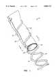
- FIG. 1is a perspective view of a vascular prostheses constructed in accordance with the principles of the present invention, shown with portions broken away.
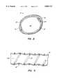
- FIG. 2is a cross-sectional view taken along line 2--2 of FIG. 1.
- FIG. 3is a partial, longitudinal cross-sectional view of the prostheses of FIG. 1.
- FIG. 4is a partial cross-sectional view of an alternative embodiment of the prosthesis of the present invention.
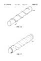
- FIGS. 5-8illustrate a method for preparing the vascular prosthesis of FIG. 1.
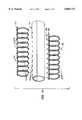
- FIG. 9illustrates an alternative construction of a vascular prostheses constructed in accordance with the principles of the present invention, shown in an exploded view.
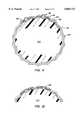
- FIG. 10illustrates yet another alternative environment of a vascular prosthesis constructed in accordance with the principles of the present invention.
- FIGS. 11 and 12illustrate another method for attaching tissue to a tubular frame member according to the method of the present invention.
- vascular prosthesesalso referred to as vascular grafts, intended for use in medical procedures requiring replacement or bypass of a patient's blood vessels.
- vascular prostheseswill be used in peripheral vascular bypass, coronary artery bypass (CABG) procedures, but they also may find use in aneurysm repair; vascular access shunts; vessel reconstruction, such as pulmonary outflow tract and aortic outflow tract; as a conduit for valvular repair; and the like.
- CABGcoronary artery bypass
- the tissue employed in the vascular prosthesiswill be obtained from a human or other animal source, usually but not necessarily being obtained from the patient or host into which the prosthesis is to be implanted.
- the tissuemay comprise any body tissue having sufficient strength and elasticity to act as the primary component of the prosthesis, usually being obtained from the pericardium or a fascial layer, such as the fascia lata.
- Other tissue sourcesinclude rectus sheath and venous tissues.
- the tissuewill be harvested by conventional techniques, such as those described in Love, Autologous Tissue Heart Valves, R. G. Landes Co., Austin, Tex., 1993, Chapter 8.
- the amount of tissue harvestedwill depend on the size of the vascular prosthesis to be prepared.
- the sheet of tissue initially obtainedwill be generally rectangular, having a length in the range from about 5 cm to 35 cm, usually 5 cm to 15 cm for pericardium, and a width in the range from about 2 cm to 20 cm, usually about 2 cm to 5 cm for pericardium.
- the tissuewill be trimmed to size, usually using a cutting die, stencil, or other pattern-forming device capable of trimming the tissue to the precise dimensions required.
- the precise dimensionswill depend on the dimensions of the vascular prosthesis to be formed.
- the sheetwill be cut into a rectangular pattern having a length and width in the ranges set forth above.
- the tissueAfter harvesting but usually before trimming, the tissue will be treated by conventional methods to enhance its stability and durability. For example, the tissue may be briefly immersed in a cross-linking solution, such as glutaraldehyde, in order to fix the tissue. It has been found that glutaraldehyde-treated tissue remains antigenically compatible with the host from which it has been harvested. Suitable techniques for treating the harvested tissue with glutaraldehyde are described in Love, supra., Chapter 5.
- tissuebe obtained from the patient in which the vascular prosthesis is to be implanted (referred to as "autologous" tissue)
- tissuecould be obtained from human cadavers, including frozen (cryo-preserved) cadaver tissue, treated with the cross-linking or other preserving agent, and then employed to make vascular prostheses according to the teachings herein.
- Tissuecould also be obtained from non-human animal sources, such as bovine tissue, porcine tissue, and the like. It would also be possible to use luminal tissues, such as venous tissues, e.g., human and non-human saphenous veins.
- the use of the frames of the present inventionwould also be advantageous in supporting saphenous vein grafts along their lengths.
- the saphenous or other veinscan either be split longitudinally, and formed as described hereinafter for other flat tissue sources, or alternatively could be placed intact over an inner stent with a second stent or sheath then being placed over the exterior of the vein.
- the grafts of the present inventionwill be formed from a single piece of tissue having a length which is generally equal to the length of the graft and having a single overlap extending longitudinally down the length of the graft.
- Other graft constructionswill be possible.
- a single long, relatively narrow strip of tissuecould be wrapped spirally around the graft, thus having a spiral overlap extending down the length of the graft.
- two or more tissue sectionscould be wrapped around the frame to form the graft of the present invention in a variety of geometries. While the preferred tissue geometry will be illustrated and described hereinafter, it is appreciated that the present invention is not so limited.
- the tubular support frame of the vascular prosthesiswill typically be composed of a non-biologic material having sufficient strength to maintain the rolled tissue in a tubular geometry with a substantially unrestricted lumen therethrough, but with sufficient flexibility to allow the prosthesis to be bent and with sufficient compliance to allow the prosthesis to accommodate pulsatile blood flow.
- the tubular supportwill be made from a spring metal, such as a spring stainless steel.
- a preferred materialis alloy MP35N (Maryland Specialty Wire, Inc., Cockeysville, Md. 21030).
- the framecould also be formed from plastic materials having the requisite strength and flexibility requirements, such as thermoplastics.
- a third alternativewould be thermoplastic-covered metal wires.
- thermoplastic-covered wiresare advantageous if the frame is trimmed prior to use since the plastic materials will reduce the formation of sharp edges on the frame.
- Thermoplastic frame materialscan also permit sewing or suturing through the frame.
- Both metal and plastic frame componentsmay optionally be covered with polyester (Dacron®) in order to enhance biocompatibility and non-immunogenicity.
- the dimensions of the tubular support framewill define the dimensions of the vascular prosthesis.
- the support framewill have a diameter in the range from about 1 mm to 30 mm, usually from 3 mm to 25 mm, and a length in the range from 1 cm to 35 cm, usually from 1 cm to 25 cm, and may usually range from 5 cm to 15 cm.
- the rolled tissue supported by the framewill often extend slightly beyond the ends of the frame, typically by a distance in the range from 1 mm to 10 mm, usually from 2 mm to 6 mm. Such tissue extensions can facilitate suturing of the prosthesis to form end-to-end and end-to-side anastomoses in performing CABG and other procedures.
- the tubular support framewill usually include at least two components, such as an inner frame component and an outer frame component, as described in more detail below. Generally, however, at least one of the frame components will extend continuously from a proximal end of the graft to a distal end of the graft. It is possible, however, that the tubular support frame will include two or more separate, longitudinally-separated segments or components which may be unattached or attached by overlapping or by other non-permanent fastening. The use of multiple, longitudinal segments may be advantageous in enhancing flexibility and/or facilitating the design and fabrication of longer tubular grafts.
- tissuewill be rolled into the desired tubular configuration and attached to the tubular support frame so that the tissue is maintained in its desired tubular geometry.
- Tissue attachmentmay be provided in a variety of ways.
- the tissuewill be attached to the frame without suturing or otherwise penetrating the sheet of tissue. In that way, integrity of the tissue is enhanced and leakage of blood or other fluids through the prostheses is reduced.
- the tissuemay be attached to the tubular support frame using penetrating attachment means, such as hooks, barbs, staples, or the like.
- the tissuewill not be sutured to the frame or otherwise to enhance closure of the tubular tissue structure.
- leakage from the tubular tissue structurewill be prevented by overlapping the adjacent (rolled) edges of the tissue by an arc of at least 35°, usually being in the range from 45° to 135°, preferably being about 120°.
- the tissuewill be overlapped by the requisite amount and will be held together by the tubular support frame, as described in detail hereinafter.
- adhesivessuch as fibrin glues, biological adhesives, synthetic glues (cyanoacrylates), or the like, to bond the overlapping layers.
- suture the layers togetheralthough this will generally be less preferred for the reasons set forth above in the Background section. It would further be possible when employing an adhesive to join the adjacent tissue edges together in an abutting fashion, forming an axially extending butt-joint, although this method is not presently preferred.
- a preferred tubular support framewill comprise an inner frame component and an outer frame component, where the rolled sheet of tissue is captured between the inner and outer components.
- both the inner and outer frame componentsare helical elements, usually having identical diameters and pitches.
- the sheet of tissueis rolled over a first of the helical support elements, which acts as the inner support.
- the second helical componentis then placed over the tissue, typically so that the outer helical support runs between the turns of the inner helical support.
- Other embodimentsutilize longitudinally spaced-apart support rings or other structures, such as those conventionally used in vascular stents.
- a vascular prosthesis 10comprising a rolled sheet of tissue 12, an outer helical support element 14, and an inner helical support element 16, is illustrated.
- the tissue 12is rolled from a rectangular sheet so that longitudinal edges 18 and 20 are parallel to each other and overlap by an arc in the range set forth above. Such overlapping will inhibit the leakage of blood or other body fluids which are being carried through lumen 22 of the graft 10.
- the helical support elements 14 and 16will usually have identical dimensions, i.e., diameter, length, and pitch.
- the diameter and lengthwill be within the ranges set forth above, and the pitch, i.e., distance between successive turns of the helix, will usually be in the range from 1 mm to 10 mm, usually being from 1 mm to 6 mm, and preferably being from about 2 mm to 4 mm. It is desirable to increase the pitch as much as possible, while maintaining sufficient capture of the tissue therebetween to prevent leakage of fluent from the prosthesis. Thus, it will frequently be possible to increase the pitch of the helical support elements 14 and 16 by also increasing the amount of overlap between the parallel edges 18 and 20.
- an outer sleeve 30may be placed over the tissue layer 12, as illustrated in FIG. 4.
- the inner helical support element 16, and other features of the graftmay be identical to those of the vascular prosthesis 10 of FIGS. 1-3.
- Use of an outer sleevemay have certain advantages. For example, use of an elastic material may facilitate placement of the sleeve over the tissue and underlying frame component. Porous membrane materials may also be employed in the sleeve in order to enhance tissue ingrowth. Finally, the use of elastic sleeve materials may enhance the compliance of the tubular prosthesis.
- a sheet of tissue Tis harvested from the patient or other animal source, as described previously.
- the sheetwill usually be treated with glutaraldehyde or other fixative or cross-linking agent, as also described previously. It is desirable that the tissue be treated prior to trimming since trimming can cause a slight shrinkage.
- the tissue sheet Twill then be trimmed, preferably using a cutter 40 or similar device capable of cutting the tissue into a rectangular pattern P, as shown on the tissue in broken line.
- the inner helical support element 16is typically placed over a mandrel 50, as shown in FIG. 6.
- the trimmed sheet of tissue 12is then rolled over the mandrel, as shown in FIG. 7.
- the outer helical support element 14may then be placed over the tissue 12, typically by expanding the diameter of the helix and, after properly positioning over the tissue 12, allowing the helix to contract onto the tubular form of the tissue, as shown in FIG. 8.
- the mandrel 50is then removed.
- the prosthesis 10is then ready to be used in a conventional vascular bypass or replacement procedure.
- the outer helical support elementmay be applied by screwing the helices together or by wrapping the coils of the outer helical support element over the tissue wrapped over the inner helical support element 16 and mandrel 50.
- the prosthesiscomprises a tubular support frame including an inner frame member 60 and an outer frame member 62.
- the inner frame member 60includes a plurality of ring elements 64 which are longitudinally spaced-apart along a longitudinal spine 66.
- a plurality of pins 68are disposed along the upper surface of the spine 66 and are disposed in a radially outward direction.
- the outer frame member 62also comprises a longitudinal spine 70 and includes a plurality of ring elements 72 longitudinally spaced-apart on the spine.
- the ring elements 72are open so that each ring forms a C-clamp.
- Tissue 74is rolled over the inner frame member 60, with a plurality of apertures 76 formed to receive the pins 68.
- the outer frame member 70is then placed over the rolled tissue 74, with apertures 78 and the spine 70 also being received over pins 68.
- the rings 72are spaced so that they are received between each of the rings 64 in the inner frame member 60.
- Prosthesis 80comprises a plurality of independent ring elements 82, each of which includes a plurality of "mushroom” fasteners disposed about its inner periphery.
- the fasteners 84project radially inward so that a rolled tissue can be pressed onto the fasteners 84, as illustrated.
- the tissuecould be perforated prior to placement over the ring elements 82 to facilitate placement over the fasteners 84. It would also be possible to connect the ring members 82 with one or more longitudinal members if it is desired to increase the column strength.
- FIGS. 11 and 12A further alternative approach for attaching a tissue layer 90 to a supporting ring element 92 is illustrated in FIGS. 11 and 12.
- the tissue 90is placed over a mandrel having a cross-sectional shape which matches that of the ring element 92.
- the ring element 92includes a plurality of peaks 94, each of which includes a pair of channels 96 therein.
- the channels 96are aligned so that staples 98 may be inserted therethrough, allowing stapling of the ring element 92 to the tissue, as shown in FIG. 12.
- the supporting mandrel 100is shaped to conform to the ring elements 92.
Landscapes
- Health & Medical Sciences (AREA)
- Life Sciences & Earth Sciences (AREA)
- Engineering & Computer Science (AREA)
- Biomedical Technology (AREA)
- General Health & Medical Sciences (AREA)
- Chemical & Material Sciences (AREA)
- Oral & Maxillofacial Surgery (AREA)
- Transplantation (AREA)
- Animal Behavior & Ethology (AREA)
- Public Health (AREA)
- Veterinary Medicine (AREA)
- Cardiology (AREA)
- Heart & Thoracic Surgery (AREA)
- Vascular Medicine (AREA)
- Chemical Kinetics & Catalysis (AREA)
- Pulmonology (AREA)
- Molecular Biology (AREA)
- Botany (AREA)
- Gastroenterology & Hepatology (AREA)
- Dermatology (AREA)
- Medicinal Chemistry (AREA)
- Epidemiology (AREA)
- Urology & Nephrology (AREA)
- Zoology (AREA)
- General Chemical & Material Sciences (AREA)
- Prostheses (AREA)
- Materials For Medical Uses (AREA)
Abstract
Description
1. Field of the Invention
The present invention relates generally to medical methods and devices, and more particularly to a method and apparatus for forming vascular prostheses from host tissue sources.
Coronary and peripheral atherosclerosis are characterized by partial or total occlusion of the arteries resulting from the accumulation of lipids, smooth muscle cells, connective tissue, and glycosaminoglycans on the arterial wall. Atherosclerosis of the coronary arteries is a particular problem and can cause angina and myocardial infarction (heart attack). Although many coronary lesions can be treated with percutaneous techniques, such as angioplasty and atherectomy, more tortuous and severely diseased arteries frequently require surgical intervention and bypass, commonly referred to as coronary artery bypass graft (CABG) surgery.
CABG surgery relies on the surgical attachment of a vascular graft to bypass the arterial occlusion in order to restore blood flow to the coronary vasculature. The nature of the vascular graft can have a significant impact on the ultimate success of the procedure. A preferred vascular graft is formed from autologous internal mammary artery (IMA), where the resulting grafts have a patency rate approaching 95% ten years following the procedure. The use of IMA grafts, however, is limited by their length, and the need to harvest the artery from the patient can result in post-surgical complications. The autologous saphenous vein is a second common source for vascular grafts. While generally available in the necessary lengths, the saphenous vein is not ideally suited for replacement as an arterial vessel, and patency rates at ten years are often below 50%. Moreover, removal of the saphenous vein from the leg can also cause post-surgical complications.
Because of the limitations on autologous vascular sources, a variety of synthetic and non-autologous biological prostheses have been proposed. Common synthetic prostheses are formed from Dacron® and PTFE, and can perform well when employed in larger diameters, i.e., above 6 mm. Smaller synthetic prostheses, however, occlude at a relatively high rate. Non-autologous biological conduits which have been utilized as vascular prostheses include human umbilical vein grafts and bovine internal mammary arteries. Synthetic grafts have also been seeded with human and other mammalian cells or proteins, e.g., collagens, in an effort to improve their long-term patency rate. Presently, however, none of these approaches has demonstrated long-term patency, particularly in smaller diameter grafts.
Of particular interest to the present invention, preparation of vascular prostheses from autologous pericardium has been proposed. Pericardial tissue is harvested from the patient and formed into a tubular graft by suturing along a longitudinal line. While promising, the use of sutures can result in an irregular seam which, in turn, can cause turbulent blood flow and result in clot formation. Moreover, such grafts are unsupported and subject to kinking and collapse. The grafts further lack an inherently round geometry and are subject to dimensional changes, e.g., elongation and aneurysmal formation. Because of the dimensional uncertainty, it is difficult to match such grafts to the precise dimensional requirements of the particular application, e.g, caliber and length. The suturing of vascular prostheses from pericardium is labor intensive and time consuming, and the resulting structures are subject to rupture and other structural failure. Thus, the outcome of using sutured pericardial tissue grafts is uncertain at best.
For these reasons, it would be desirable to provide improved vascular prostheses for use in CABG and other procedures. Such prostheses should be biocompatible with the patient, resistant to kinking and collapse, and easy to implant. Moreover, the prostheses should be non-thrombogenic, resistant to infection, and easy to sterilize and store. It would be particularly desirable to provide improved methods and apparatus for preparing vascular prostheses from autologous tissue sources, where the prostheses can be prepared in a range of diameters and lengths, and can be readily assembled in the operating room after the tissue has been harvested. In particular, the vascular prostheses should be readily assemblable, preferably without suturing, in a manner that allows precise and uniform dimensions and preferably be available in a kit form to facilitate assembly.
2. Description of the Back ground Art
U.S. Pat. No. 4,502,159, describes a vascular prosthesis made by suturing glutaraldehyde-treated pericardial tissue along a longitudinal seam. SU 1217362 (Abstract) describes reinforcing arteries by securing pericardial tissue over the artery. U.S. Pat. No. 3,562,820, describes forming tissue-containing prostheses over removable mandrels. The use of glutaraldehyde and other agents for treating tissue and prosthetic devices to reduce antigenicity is described in U.S. Pat. Nos. 3,988,782; 4,801,299; 5,215,541, and Brazilian applications 89/03621 and 90/03762. U.S. Pat. No. 4,539,716, describes the fabrication of an artificial blood vessel from collagen and other natural materials. U.S. Pat. Nos. 3,894,530 and 3,974,526, describe the formation of vascular prostheses from the arteries or veins present in the umbilical cord. U.S. Pat. No. 5,372,821, describes the use of tissue for forming artificial ligament grafts for use in orthopedic procedures. U.S. Pat. No. 3,408,659, describes the preparation of vascular artificial prostheses from other body lumens. French application FR 2,714,816, (Abstract) discloses a helically supported vascular prosthesis. A number of medical literature publications describe the use of vascular prostheses formed form tissue. See, for example, Rendina et al. (1995) J. Thorac. Cardiovasc. Surg. 110:867-868; Hvass et al. (1987) La Presse Medicale 16:441-443; Allen and Cole (1977) J. Ped. Surg. 12:287-294; and Sako (1951) Surgery 30:148-160. Other patents and published applications relating to synthetic vascular grafts include U.S. Pat. Nos. 4,728,328; 4,731,073; 4,798,606; 4,820,298; 4,822,361; and 4,842,575; and PCT publications WO 94/22505 and WO 95/25547. Patents and published applications relating to kits for preparing replacement heart valves from pericardial and other autologous tissue sources are described in U.S. Pat. Nos. 5,163,955; 5,297,564; 5,326,370; 5,326,371; 5,423,887; and 5,425,741.
The present invention provides improved vascular prostheses and methods for their preparation. The vascular prostheses are formed in part from animal tissue, usually autologous tissue from the patient receiving the prostheses, which is supported on a separate support frame. Typically, the tissue is pericardial, fascial, rectus sheath, venous tissue, or other tissue harvested from the patient immediately before the CABG or other implantation procedure. After harvesting, the tissue is usually but not necessarily treated in a stabilizing medium, such as a cross-linking agent, and then attached to the frame in the operating room. The frame precisely defines the length and dimensions of the vascular graft and inhibits kinking and collapse of the graft after implantation. Preferably, the tissue will be rolled or otherwise formed over the frame so that adjacent longitudinal edges are overlapped to seal the resulting lumen of the graft and prevent blood leakage. In this way, suturing of the graft can be avoided.
Such vascular prostheses have a number of advantages. When using autologous tissue, the grafts are biocompatible and non-immunogenic. The grafts are durable, and use of the separate frame provides dimensional stability and inhibits unintended dilation, rupture, elongation, and kinking. Moreover, the vascular prosthesis may be prepared in a range of diameters and lengths, with the tissue sources providing a relatively unlimited source of prosthetic material. The vascular prostheses are relatively easy to fabricate, with attachment of the tissue to the frame being readily performable in an operating room environment. The frame components of the graft are easy to store and sterilize prior to use. Other advantages include non-thrombogenicity and a compliance which approximates that of natural blood vessels.
According to the method of the present invention, a tubular vascular prosthesis is formed by providing a sheet of tissue and a tubular support frame. The tissue is then attached to the tubular support frame to define a substantially unrestricted blood flow lumen therethrough. The tissue sheet may be obtained from the host or from other human or animal (non-autologous) sources. Typically, the tissue is trimmed into a shape to facilitate rolling onto the frame, usually a rectangular shape. The tissue will usually be pericardium, fascia, rectus sheath, venous tissue, or the like, and will preferably but not necessarily be treated with a cross-linking agent or other stabilizing agent (preservative) prior to formation.
The tubular support frame may have a variety of configurations. In a first embodiment, the tubular support frame includes at least an inner frame component and an outer frame component, where the attaching step comprises capturing the tissue sheet between the inner component and the outer frame component. The inner and outer frame components may be in the form of helices, longitudinally spaced-apart rings, or other conventional intravascular stent structures and the like. In a preferred aspect of the present invention, the inner and outer frame components comprise concentric mating structure which clamp the tissue therebetween without suturing. The frame thus supports the clamped tissue along the entire length of the graft to provide support and precise dimensional control.
Alternatively, the tubular support frame may include a single frame member having a plurality of fasteners disposed thereover. In such case, the attaching step comprises attaching the tissue to the fasteners, for example by penetrating the fasteners through the tissue. As yet another alternative, the tissue may be attached to a single frame using separate fasteners, such as staples which are penetrated through the tissue and into the frame. In yet another alternative, the attaching step may comprise disposing a sleeve over the tissue which in turn is disposed over the tubular frame.
Systems for forming tubular prostheses according to the present invention comprise a cutter and a tubular frame. The cutter is designed to trim the sheet of harvested tissue into a predetermined pattern, typically a rectangular pattern. The tubular frame is capable of supporting the tissue trimmed by the cutter and a tubular geometry having a substantially unrestricted flow lumen therethrough. Usually, a plurality of cutters and a plurality of tubular frames will be provided with matched pairs of cutters and frames used for forming tubular prostheses having different dimensions. The system may further include a mandrel for holding the tissue as the tissue is attached to the frame, and may still further include a cross-linking agent or other stabilizing agent or preservative for treating the tissue prior to attachment to the frame. The frame may comprise any of the structures described above.
In another aspect of the present invention, a tubular frame for supporting tissue in a tubular geometry with a substantially unrestricted flow lumen therethrough comprises a first tubular frame component having tissue attachment means thereon. The tubular frame typically has a diameter in the range from 1 mm to 30 mm, preferably from 3 mm to 25 mm, and a length in the range from 1 cm to 30 cm, preferably from 5 cm to 15 cm. The length will be determined at least in part by the length and amount of tissue available from an individual patient. In some cases, frames even longer than 30 cm might find use, but the resulting longer grafts will rarely be needed. In some cases, the length of the tubular frame will be adjustable, for example by cutting a desired length of frame or frame components from a relatively long frame stock.
The frame will usually be composed of a resilient metal, and may comprise either a helical element or a plurality of longitudinally spaced-apart ring elements. Attachment means may comprise any one of a second tubular frame component configured to mate with the first tubular frame component, e.g., a pair of nesting helical frame elements, a plurality of fasteners disposed over the first tubular frame component, a sleeve which is received over the exterior of the first tubular frame component, staples for attaching the tissue to the frame component, or the like. Optionally, two or more frames or frame segments may be linked together to create longer grafts, with the frames or frame segments being interlocked and/or overlapped to create a continuous lumen through the resulting assembly.
FIG. 1 is a perspective view of a vascular prostheses constructed in accordance with the principles of the present invention, shown with portions broken away.
FIG. 2 is a cross-sectional view taken alongline 2--2 of FIG. 1.
FIG. 3 is a partial, longitudinal cross-sectional view of the prostheses of FIG. 1.
FIG. 4 is a partial cross-sectional view of an alternative embodiment of the prosthesis of the present invention.
FIGS. 5-8 illustrate a method for preparing the vascular prosthesis of FIG. 1.
FIG. 9 illustrates an alternative construction of a vascular prostheses constructed in accordance with the principles of the present invention, shown in an exploded view.
FIG. 10 illustrates yet another alternative environment of a vascular prosthesis constructed in accordance with the principles of the present invention.
FIGS. 11 and 12 illustrate another method for attaching tissue to a tubular frame member according to the method of the present invention.
The present invention provides vascular prostheses, also referred to as vascular grafts, intended for use in medical procedures requiring replacement or bypass of a patient's blood vessels. Most commonly, vascular prostheses will be used in peripheral vascular bypass, coronary artery bypass (CABG) procedures, but they also may find use in aneurysm repair; vascular access shunts; vessel reconstruction, such as pulmonary outflow tract and aortic outflow tract; as a conduit for valvular repair; and the like.
The tissue employed in the vascular prosthesis will be obtained from a human or other animal source, usually but not necessarily being obtained from the patient or host into which the prosthesis is to be implanted. The tissue may comprise any body tissue having sufficient strength and elasticity to act as the primary component of the prosthesis, usually being obtained from the pericardium or a fascial layer, such as the fascia lata. Other tissue sources include rectus sheath and venous tissues. The tissue will be harvested by conventional techniques, such as those described in Love, Autologous Tissue Heart Valves, R. G. Landes Co., Austin, Tex., 1993, Chapter 8.
The amount of tissue harvested will depend on the size of the vascular prosthesis to be prepared. Typically, the sheet of tissue initially obtained will be generally rectangular, having a length in the range from about 5 cm to 35 cm, usually 5 cm to 15 cm for pericardium, and a width in the range from about 2 cm to 20 cm, usually about 2 cm to 5 cm for pericardium. After harvesting, the tissue will be trimmed to size, usually using a cutting die, stencil, or other pattern-forming device capable of trimming the tissue to the precise dimensions required. The precise dimensions, of course, will depend on the dimensions of the vascular prosthesis to be formed. Typically, the sheet will be cut into a rectangular pattern having a length and width in the ranges set forth above.
After harvesting but usually before trimming, the tissue will be treated by conventional methods to enhance its stability and durability. For example, the tissue may be briefly immersed in a cross-linking solution, such as glutaraldehyde, in order to fix the tissue. It has been found that glutaraldehyde-treated tissue remains antigenically compatible with the host from which it has been harvested. Suitable techniques for treating the harvested tissue with glutaraldehyde are described in Love, supra., Chapter 5.
While it is preferred that the tissue be obtained from the patient in which the vascular prosthesis is to be implanted (referred to as "autologous" tissue), it is also possible to obtain tissue from other human and animal sources. For example, tissue could be obtained from human cadavers, including frozen (cryo-preserved) cadaver tissue, treated with the cross-linking or other preserving agent, and then employed to make vascular prostheses according to the teachings herein. Tissue could also be obtained from non-human animal sources, such as bovine tissue, porcine tissue, and the like. It would also be possible to use luminal tissues, such as venous tissues, e.g., human and non-human saphenous veins. While a particular advantage of the present invention is it allows the use of non-luminal tissues to form vascular and other graft structures, the use of the frames of the present invention would also be advantageous in supporting saphenous vein grafts along their lengths. The saphenous or other veins can either be split longitudinally, and formed as described hereinafter for other flat tissue sources, or alternatively could be placed intact over an inner stent with a second stent or sheath then being placed over the exterior of the vein.
Preferably, the grafts of the present invention will be formed from a single piece of tissue having a length which is generally equal to the length of the graft and having a single overlap extending longitudinally down the length of the graft. Other graft constructions, however, will be possible. For example, a single long, relatively narrow strip of tissue could be wrapped spirally around the graft, thus having a spiral overlap extending down the length of the graft. As a further alternative, two or more tissue sections could be wrapped around the frame to form the graft of the present invention in a variety of geometries. While the preferred tissue geometry will be illustrated and described hereinafter, it is appreciated that the present invention is not so limited.
The tubular support frame of the vascular prosthesis will typically be composed of a non-biologic material having sufficient strength to maintain the rolled tissue in a tubular geometry with a substantially unrestricted lumen therethrough, but with sufficient flexibility to allow the prosthesis to be bent and with sufficient compliance to allow the prosthesis to accommodate pulsatile blood flow. Usually, the tubular support will be made from a spring metal, such as a spring stainless steel. A preferred material is alloy MP35N (Maryland Specialty Wire, Inc., Cockeysville, Md. 21030). The frame could also be formed from plastic materials having the requisite strength and flexibility requirements, such as thermoplastics. A third alternative would be thermoplastic-covered metal wires. The use of both plastics and thermoplastic-covered wires is advantageous if the frame is trimmed prior to use since the plastic materials will reduce the formation of sharp edges on the frame. Thermoplastic frame materials can also permit sewing or suturing through the frame. Both metal and plastic frame components may optionally be covered with polyester (Dacron®) in order to enhance biocompatibility and non-immunogenicity.
The dimensions of the tubular support frame will define the dimensions of the vascular prosthesis. Typically, the support frame will have a diameter in the range from about 1 mm to 30 mm, usually from 3 mm to 25 mm, and a length in the range from 1 cm to 35 cm, usually from 1 cm to 25 cm, and may usually range from 5 cm to 15 cm. The rolled tissue supported by the frame will often extend slightly beyond the ends of the frame, typically by a distance in the range from 1 mm to 10 mm, usually from 2 mm to 6 mm. Such tissue extensions can facilitate suturing of the prosthesis to form end-to-end and end-to-side anastomoses in performing CABG and other procedures.
The tubular support frame will usually include at least two components, such as an inner frame component and an outer frame component, as described in more detail below. Generally, however, at least one of the frame components will extend continuously from a proximal end of the graft to a distal end of the graft. It is possible, however, that the tubular support frame will include two or more separate, longitudinally-separated segments or components which may be unattached or attached by overlapping or by other non-permanent fastening. The use of multiple, longitudinal segments may be advantageous in enhancing flexibility and/or facilitating the design and fabrication of longer tubular grafts.
The tissue will be rolled into the desired tubular configuration and attached to the tubular support frame so that the tissue is maintained in its desired tubular geometry. Tissue attachment may be provided in a variety of ways. Preferably, the tissue will be attached to the frame without suturing or otherwise penetrating the sheet of tissue. In that way, integrity of the tissue is enhanced and leakage of blood or other fluids through the prostheses is reduced. In alternative embodiments, the tissue may be attached to the tubular support frame using penetrating attachment means, such as hooks, barbs, staples, or the like. Preferably, the tissue will not be sutured to the frame or otherwise to enhance closure of the tubular tissue structure. Usually, leakage from the tubular tissue structure will be prevented by overlapping the adjacent (rolled) edges of the tissue by an arc of at least 35°, usually being in the range from 45° to 135°, preferably being about 120°.
In the exemplary embodiment, the tissue will be overlapped by the requisite amount and will be held together by the tubular support frame, as described in detail hereinafter. In some cases, however, it may be further desirable to provide adhesives, such as fibrin glues, biological adhesives, synthetic glues (cyanoacrylates), or the like, to bond the overlapping layers. It may also be possible to provide laser welding of the tissue layers together, also to enhance the bonding. It would also be possible to suture the layers together, although this will generally be less preferred for the reasons set forth above in the Background section. It would further be possible when employing an adhesive to join the adjacent tissue edges together in an abutting fashion, forming an axially extending butt-joint, although this method is not presently preferred.
A preferred tubular support frame will comprise an inner frame component and an outer frame component, where the rolled sheet of tissue is captured between the inner and outer components. In a particularly preferred embodiment, both the inner and outer frame components are helical elements, usually having identical diameters and pitches. The sheet of tissue is rolled over a first of the helical support elements, which acts as the inner support. The second helical component is then placed over the tissue, typically so that the outer helical support runs between the turns of the inner helical support. Other embodiments utilize longitudinally spaced-apart support rings or other structures, such as those conventionally used in vascular stents.
Referring now to FIGS. 1-3, avascular prosthesis 10 comprising a rolled sheet oftissue 12, an outerhelical support element 14, and an innerhelical support element 16, is illustrated. Thetissue 12 is rolled from a rectangular sheet so thatlongitudinal edges lumen 22 of thegraft 10.
Thehelical support elements helical support elements parallel edges
As an alternative to employing the outerhelical support element 14, anouter sleeve 30 may be placed over thetissue layer 12, as illustrated in FIG. 4. The innerhelical support element 16, and other features of the graft, may be identical to those of thevascular prosthesis 10 of FIGS. 1-3. Use of an outer sleeve may have certain advantages. For example, use of an elastic material may facilitate placement of the sleeve over the tissue and underlying frame component. Porous membrane materials may also be employed in the sleeve in order to enhance tissue ingrowth. Finally, the use of elastic sleeve materials may enhance the compliance of the tubular prosthesis.
Referring now to FIGS. 5-8, a method for preparing thevascular prosthesis 10 of FIGS. 1-3 will be described. A sheet of tissue T is harvested from the patient or other animal source, as described previously. The sheet will usually be treated with glutaraldehyde or other fixative or cross-linking agent, as also described previously. It is desirable that the tissue be treated prior to trimming since trimming can cause a slight shrinkage. The tissue sheet T will then be trimmed, preferably using acutter 40 or similar device capable of cutting the tissue into a rectangular pattern P, as shown on the tissue in broken line.
The innerhelical support element 16 is typically placed over amandrel 50, as shown in FIG. 6. The trimmed sheet oftissue 12 is then rolled over the mandrel, as shown in FIG. 7. The outerhelical support element 14 may then be placed over thetissue 12, typically by expanding the diameter of the helix and, after properly positioning over thetissue 12, allowing the helix to contract onto the tubular form of the tissue, as shown in FIG. 8. Themandrel 50 is then removed. Theprosthesis 10 is then ready to be used in a conventional vascular bypass or replacement procedure. Optionally, the outer helical support element may be applied by screwing the helices together or by wrapping the coils of the outer helical support element over the tissue wrapped over the innerhelical support element 16 andmandrel 50.
Referring to FIG. 9, an alternative embodiment of the vascular prosthesis of the present invention will be described. The prosthesis comprises a tubular support frame including an inner frame member 60 and anouter frame member 62. The inner frame member 60 includes a plurality ofring elements 64 which are longitudinally spaced-apart along a longitudinal spine 66. A plurality ofpins 68 are disposed along the upper surface of the spine 66 and are disposed in a radially outward direction. Theouter frame member 62 also comprises a longitudinal spine 70 and includes a plurality ofring elements 72 longitudinally spaced-apart on the spine. Thering elements 72 are open so that each ring forms a C-clamp.Tissue 74 is rolled over the inner frame member 60, with a plurality ofapertures 76 formed to receive thepins 68. The outer frame member 70 is then placed over the rolledtissue 74, with apertures 78 and the spine 70 also being received overpins 68. Therings 72 are spaced so that they are received between each of therings 64 in the inner frame member 60.
Anothervascular prosthesis 80 is illustrated in FIG. 10.Prosthesis 80 comprises a plurality ofindependent ring elements 82, each of which includes a plurality of "mushroom" fasteners disposed about its inner periphery. Thefasteners 84 project radially inward so that a rolled tissue can be pressed onto thefasteners 84, as illustrated. Optionally, the tissue could be perforated prior to placement over thering elements 82 to facilitate placement over thefasteners 84. It would also be possible to connect thering members 82 with one or more longitudinal members if it is desired to increase the column strength.
A further alternative approach for attaching atissue layer 90 to a supportingring element 92 is illustrated in FIGS. 11 and 12. Thetissue 90 is placed over a mandrel having a cross-sectional shape which matches that of thering element 92. Thering element 92 includes a plurality ofpeaks 94, each of which includes a pair ofchannels 96 therein. Thechannels 96 are aligned so thatstaples 98 may be inserted therethrough, allowing stapling of thering element 92 to the tissue, as shown in FIG. 12. The supportingmandrel 100 is shaped to conform to thering elements 92.
Although the foregoing invention has been described in some detail by way of illustration and example, for purposes of clarity of understanding, it will be obvious that certain changes and modifications may be practiced within the scope of the appended claims.
Claims (38)
1. A method for forming a tubular prosthesis, said method comprising:
providing a sheet of biological tissue;
providing a flexible tubular support frame wherein the tubular support frame includes at least an inner frame component and an outer frame component; and
attaching the tissue to the tubular support frame, wherein the frame captures the sheet of biological tissue between the inner frame component and the outer component to hold the tissue in a tubular geometry having ends suitable for anastomotic attachment with a substantially unrestricted lumen therethrough.
2. A method as in claim 1, wherein the sheet providing step comprises:
harvesting tissue from an animal host; and
trimming the tissue into a shape selected to permit attachment to the frame.
3. A method as in claim 2, wherein the tissue is trimmed into a rectangular shape.
4. A method as in claim 1, wherein the tissue is selected from the group consisting of pericardium, fascia, rectus sheath, and venous tissue.
5. A method as in claim 1, further comprising treating the tissue with a cross-linking agent.
6. A method as in claim 5, wherein the tissue is treated by immersion in glutaraldehyde prior to attaching to the tubular support frame.
7. A method as in claim 1, further comprising implanting the prosthesis in a host.
8. A method as in claim 7, wherein the tissue was obtained from the same host into which the prosthesis is implanted.
9. A method as in claim 7, wherein the tissue was obtained from a host other than the host into which the prosthesis is implanted.
10. A method as in claim 1, wherein the tissue sheet is disposed on the frame with parallel edges overlapping along a longitudinal line.
11. A method as in claim 10, wherein the edges are not connected other than by the frame.
12. A method as in claim 11, wherein the edges overlap by an arc of at least 30°.
13. A method as in claim 1, further comprising forming the tissue over a mandrel prior to attaching the tissue to the frame.
14. A method as in claim 1, wherein the inner frame component and the outer frame component each comprise a helix, wherein the capturing step comprises aligning the inner helix and the outer helix so that the turns of each helix run in parallel.
15. A method as in claim 14, wherein the helical turns are positioned to be evenly spaced from each other in the longitudinal direction.
16. A method as in claim 1, wherein the inner frame component and outer frame component each comprise a plurality of longitudinally spaced-apart ring elements, wherein the capturing step comprises aligning the inner and outer rings in a selected pattern relative to each other.
17. A method as in claim 16, wherein the inner and outer rings are aligned in an alternating pattern.
18. A method as in claim 16, wherein the inner and outer rings are aligned to lie over each other.
19. A method as in claim 1, wherein the tubular support frame includes a plurality of fasteners disposed thereover and wherein the attaching step comprises attaching the tissue to said fasteners.
20. A method as in claim 19, wherein the fasteners are disposed over an inner luminal surface of the frame.
21. A method, as in claim 20, wherein the attaching step comprises penetrating the fasteners through the tissue.
22. A method as in claim 1, wherein the attaching step comprises stapling the tissue to the frame.
23. A method as in claim 1, wherein the outer frame component comprises a sleeve, wherein the attaching step comprises disposing the sleeve over the tissue which is disposed over the inner frame component.
24. A method for forming a tubular prosthesis, said method comprising:
providing a sheet of biological tissue;
providing a flexible tubular support frame including an inner frame component and an outer frame component; and
capturing the tissue between the inner frame component and the outer frame component of the tubular support frame, wherein the frame holds the tissue in a tubular geometry with a substantially unrestricted lumen therethrough and wherein at least one of the frame components is in the form of a helix which extends continuously from a proximal end of the prosthesis to a distal end of the prosthesis.
25. A method as in claim 24, wherein the sheet providing step comprises:
harvesting tissue from an animal host; and
trimming the tissue into a shape selected to permit attachment to the frame.
26. A method as in claim 25, wherein the tissue is trimmed into a rectangular shape.
27. A method as in claim 24, wherein the tissue is selected from the group consisting of pericardium, fascia, rectus sheath, and venous tissue.
28. A method as in claim 24, further comprising treating the tissue with a cross-linking agent.
29. A method as in claim 28, wherein the tissue is treated by immersion in glutaraldehyde prior to attaching to the tubular support frame.
30. A method as in claim 24, further comprising implanting the prosthesis in a host.
31. A method as in claim 30, wherein the tissue was obtained from the same host into which the prosthesis is implanted.
32. A method as in claim 30, wherein the tissue was obtained from a host other than the host into which the prosthesis is implanted.
33. A method as in claim 24, wherein the tissue sheet is rolled between the components of the frame with parallel edges overlapping along a longitudinal line.
34. A method as in claim 33, wherein the edges are not connected other than by the frame.
35. A method as in claim 34, wherein the edges overlap by an arc of at least 30°.
36. A method as in claim 24, further comprising forming the tissue over a mandrel and the inner frame component prior to attaching the tissue to the frame.
37. A method as in claim 24, wherein the inner frame component and the outer frame component each comprise a helix, wherein the capturing step comprises aligning the inner helix and the outer helix so that the turns of each helix run in parallel.
38. A method as in claim 37, wherein the helical turns are positioned to be evenly spaced from each other in the longitudinal direction.
Priority Applications (7)
| Application Number | Priority Date | Filing Date | Title |
|---|---|---|---|
| US08/580,582US5865723A (en) | 1995-12-29 | 1995-12-29 | Method and apparatus for forming vascular prostheses |
| DE69636438TDE69636438D1 (en) | 1995-12-29 | 1996-12-27 | METHOD AND DEVICE FOR DESIGNING VASCOSIS THERAPIES |
| CA002240989ACA2240989A1 (en) | 1995-12-29 | 1996-12-27 | Method and apparatus for forming vascular prostheses |
| PCT/US1996/020868WO1997024081A1 (en) | 1995-12-29 | 1996-12-27 | Method and apparatus for forming vascular prostheses |
| EP96945441AEP0874603B1 (en) | 1995-12-29 | 1996-12-27 | Method and apparatus for forming vascular prostheses |
| AU15692/97AAU720362B2 (en) | 1995-12-29 | 1996-12-27 | Method and apparatus for forming vascular prostheses |
| JP9524612AJP2000502586A (en) | 1995-12-29 | 1996-12-27 | Method and apparatus for forming a vascular prosthesis |
Applications Claiming Priority (1)
| Application Number | Priority Date | Filing Date | Title |
|---|---|---|---|
| US08/580,582US5865723A (en) | 1995-12-29 | 1995-12-29 | Method and apparatus for forming vascular prostheses |
Publications (1)
| Publication Number | Publication Date |
|---|---|
| US5865723Atrue US5865723A (en) | 1999-02-02 |
Family
ID=24321680
Family Applications (1)
| Application Number | Title | Priority Date | Filing Date |
|---|---|---|---|
| US08/580,582Expired - Fee RelatedUS5865723A (en) | 1995-12-29 | 1995-12-29 | Method and apparatus for forming vascular prostheses |
Country Status (7)
| Country | Link |
|---|---|
| US (1) | US5865723A (en) |
| EP (1) | EP0874603B1 (en) |
| JP (1) | JP2000502586A (en) |
| AU (1) | AU720362B2 (en) |
| CA (1) | CA2240989A1 (en) |
| DE (1) | DE69636438D1 (en) |
| WO (1) | WO1997024081A1 (en) |
Cited By (129)
| Publication number | Priority date | Publication date | Assignee | Title |
|---|---|---|---|---|
| US5957975A (en)* | 1997-12-15 | 1999-09-28 | The Cleveland Clinic Foundation | Stent having a programmed pattern of in vivo degradation |
| US6080198A (en)* | 1996-03-14 | 2000-06-27 | Meadox Medicals, Inc. | Method for forming a yarn wrapped PTFE tubular prosthesis |
| WO2000047136A1 (en)* | 1999-02-12 | 2000-08-17 | Johns Hopkins University | Venous valve implant bioprosthesis and endovascular treatment for venous insufficiency |
| US6196230B1 (en) | 1998-09-10 | 2001-03-06 | Percardia, Inc. | Stent delivery system and method of use |
| US6214040B1 (en)* | 1997-10-20 | 2001-04-10 | Iowa-India Investments Company Limited | Sandwich stent with spiraling bands on an outer surface |
| US6253768B1 (en) | 1999-08-04 | 2001-07-03 | Percardia, Inc. | Vascular graft bypass |
| US6254564B1 (en) | 1998-09-10 | 2001-07-03 | Percardia, Inc. | Left ventricular conduit with blood vessel graft |
| US6290728B1 (en) | 1998-09-10 | 2001-09-18 | Percardia, Inc. | Designs for left ventricular conduit |
| EP1138278A1 (en)* | 2000-03-22 | 2001-10-04 | Zuli Holdings Ltd. | Method and apparatus for covering a stent |
| US6331188B1 (en) | 1994-08-31 | 2001-12-18 | Gore Enterprise Holdings, Inc. | Exterior supported self-expanding stent-graft |
| US6352561B1 (en) | 1996-12-23 | 2002-03-05 | W. L. Gore & Associates | Implant deployment apparatus |
| US6352553B1 (en) | 1995-12-14 | 2002-03-05 | Gore Enterprise Holdings, Inc. | Stent-graft deployment apparatus and method |
| US6355063B1 (en) | 2000-01-20 | 2002-03-12 | Impra, Inc. | Expanded PTFE drug delivery graft |
| US6361637B2 (en) | 1995-12-14 | 2002-03-26 | Gore Enterprise Holdings, Inc. | Method of making a kink resistant stent-graft |
| US6379382B1 (en) | 2000-03-13 | 2002-04-30 | Jun Yang | Stent having cover with drug delivery capability |
| WO2001082833A3 (en)* | 2000-04-28 | 2002-06-13 | Cardiovasc Inc | Stent graft assembly and method |
| US20020099436A1 (en)* | 1996-12-23 | 2002-07-25 | Troy Thornton | Kink-resistant bifurcated prosthesis |
| US20020103529A1 (en)* | 2000-03-01 | 2002-08-01 | Gregory Pinchasik | Longitudinally flexible stent |
| US6468300B1 (en)* | 1997-09-23 | 2002-10-22 | Diseno Y Desarrollo Medico, S.A. De C.V. | Stent covered heterologous tissue |
| US6494904B1 (en)* | 1996-12-27 | 2002-12-17 | Ramus Medical Technologies | Method and apparatus for forming vascular prostheses |
| EP1275344A1 (en)* | 2001-07-09 | 2003-01-15 | Bioring SA | Device for vascular anastomosis, maintenance and protection |
| WO2003007781A2 (en) | 2001-07-19 | 2003-01-30 | The Cleveland Clinic Foundation | Endovascular prosthesis having a layer of biological tissue |
| US6520984B1 (en) | 2000-04-28 | 2003-02-18 | Cardiovasc, Inc. | Stent graft assembly and method |
| US20030065383A1 (en)* | 2000-03-01 | 2003-04-03 | Gregory Pinchasik | Longitudinally flexible stent |
| US20030088309A1 (en)* | 2001-10-09 | 2003-05-08 | Olympus Optical Co., Ltd. | Stent |
| US20030114918A1 (en)* | 2000-04-28 | 2003-06-19 | Garrison Michi E. | Stent graft assembly and method |
| US20030138945A1 (en)* | 2001-12-11 | 2003-07-24 | Mcallister Todd N. | Tissue control rods for sheet-based tissue engineering |
| US20030139797A1 (en)* | 2002-01-24 | 2003-07-24 | Kirk Johnson | Covered segmented stent |
| US6605114B1 (en)* | 1997-04-24 | 2003-08-12 | Advanced Cardiovascular Systems, Inc. | Heparin delivery method |
| US6605053B1 (en) | 1999-09-10 | 2003-08-12 | Percardia, Inc. | Conduit designs and related methods for optimal flow control |
| US6613072B2 (en) | 1994-09-08 | 2003-09-02 | Gore Enterprise Holdings, Inc. | Procedures for introducing stents and stent-grafts |
| US6613082B2 (en) | 2000-03-13 | 2003-09-02 | Jun Yang | Stent having cover with drug delivery capability |
| US20030171800A1 (en)* | 2001-10-10 | 2003-09-11 | Scimed Life Systems, Inc. | Stent design with sheath attachment members |
| US6638237B1 (en) | 1999-08-04 | 2003-10-28 | Percardia, Inc. | Left ventricular conduits and methods for delivery |
| US6641610B2 (en) | 1998-09-10 | 2003-11-04 | Percardia, Inc. | Valve designs for left ventricular conduits |
| US20030208258A1 (en)* | 2002-05-02 | 2003-11-06 | Tom Reilly | Vascular grafts surrounding an expanded stent for transplantation |
| US20030229393A1 (en)* | 2001-03-15 | 2003-12-11 | Kutryk Michael J. B. | Medical device with coating that promotes cell adherence and differentiation |
| US6699277B1 (en) | 2000-03-09 | 2004-03-02 | Diseno Y Desarrollo Medica, S.A. De C.V. | Stent with cover connectors |
| US20040049263A1 (en)* | 2000-03-01 | 2004-03-11 | Gregory Pinchasik | Longitudinally flexible stent |
| US6709453B2 (en) | 2000-03-01 | 2004-03-23 | Medinol Ltd. | Longitudinally flexible stent |
| US20040147868A1 (en)* | 2003-01-27 | 2004-07-29 | Earl Bardsley | Myocardial implant with collar |
| US20040151924A1 (en)* | 2000-04-27 | 2004-08-05 | Sankar Sambasivan | Highly anisotropic ceramic thermal barrier coating materials and related composites |
| US20040176833A1 (en)* | 2002-11-22 | 2004-09-09 | Cook Incorporated | Stent tissue graft prosthesis |
| WO2004082534A1 (en)* | 2003-03-18 | 2004-09-30 | Veryan Medical Limited | Helical graft |
| WO2004082537A1 (en)* | 2003-03-21 | 2004-09-30 | Raymond Andrieu | Intraparietal aortic valve reinforcement device and reinforced aortic valve |
| US20040215324A1 (en)* | 2000-03-09 | 2004-10-28 | Diseno Y Desarrollo Medico, S.A. De C.V. | Stent assembly |
| US20040230291A1 (en)* | 2000-03-01 | 2004-11-18 | Jacob Richter | Longitudinally flexible stent |
| US20040236411A1 (en)* | 2001-07-19 | 2004-11-25 | The Cleveland Clinic Foundation | Prosthetic cardiac valve and method for making same |
| US20050033399A1 (en)* | 1998-12-03 | 2005-02-10 | Jacob Richter | Hybrid stent |
| US6854467B2 (en) | 2000-05-04 | 2005-02-15 | Percardia, Inc. | Methods and devices for delivering a ventricular stent |
| US20050113905A1 (en)* | 2003-10-10 | 2005-05-26 | Greenberg Roy K. | Endoluminal prosthesis with interconnectable modules |
| US20050197689A1 (en)* | 2004-03-02 | 2005-09-08 | Masoud Molaei | Medical devices including metallic films and methods for making same |
| US20050197687A1 (en)* | 2004-03-02 | 2005-09-08 | Masoud Molaei | Medical devices including metallic films and methods for making same |
| US20050197690A1 (en)* | 2004-03-02 | 2005-09-08 | Masoud Molaei | Medical devices including metallic films and methods for making same |
| US6945949B2 (en) | 1998-01-30 | 2005-09-20 | Percardia, Inc. | Left ventricular conduits to coronary arteries and methods for coronary bypass |
| US20050209679A1 (en)* | 2004-03-08 | 2005-09-22 | Cook Incorporated | Retainer for a stent-graft |
| US20050251244A1 (en)* | 1997-09-23 | 2005-11-10 | Design & Performance - Cyprus Limited | Non-thrombogenic stent jacket |
| WO2005122960A3 (en)* | 2004-06-09 | 2006-02-02 | Boston Scient Scimed Inc | Overlapped stents for scaffolding, flexibility and mri compatibility |
| US7033372B1 (en) | 1999-08-04 | 2006-04-25 | Percardia, Inc. | Corkscrew reinforced left ventricle to coronary artery channel |
| US20060100717A1 (en)* | 1998-06-05 | 2006-05-11 | Organogenesis, Inc. | Bioengineered vascular graft prostheses |
| US20060122691A1 (en)* | 1998-12-03 | 2006-06-08 | Jacob Richter | Hybrid stent |
| US20060121012A1 (en)* | 2000-03-15 | 2006-06-08 | Orbus Medical Technologies, Inc. | Medical device with coating for capturing genetically-altered cells and methods of using same |
| US20060142838A1 (en)* | 2004-12-29 | 2006-06-29 | Masoud Molaei | Medical devices including metallic films and methods for loading and deploying same |
| US20060142842A1 (en)* | 2004-12-29 | 2006-06-29 | Masoud Molaei | Medical devices including metallic films and methods for making same |
| US20060142845A1 (en)* | 2004-12-29 | 2006-06-29 | Masoud Molaei | Medical devices including metallic films and methods for making same |
| US20060142851A1 (en)* | 2004-12-29 | 2006-06-29 | Masoud Molaei | Medical devices including metallic films and methods for making same |
| WO2006071244A1 (en)* | 2004-12-29 | 2006-07-06 | Boston Scientific Limited | Medical devices including metallic films and methods for making the same |
| US20060178727A1 (en)* | 1998-12-03 | 2006-08-10 | Jacob Richter | Hybrid amorphous metal alloy stent |
| US20060259131A1 (en)* | 2005-05-16 | 2006-11-16 | Masoud Molaei | Medical devices including metallic films and methods for making same |
| US20060286664A1 (en)* | 1999-11-22 | 2006-12-21 | Cytograft Tissue Engineering, Inc. | Bioreactor for the Manufacture of Tissue Engineered Blood Vessels |
| GB2427668A (en)* | 2005-06-24 | 2007-01-03 | Veryan Medical Ltd | Method of forming artificial graft tubing |
| US20070042017A1 (en)* | 2000-03-15 | 2007-02-22 | Orbus Medical Technologies, Inc. | Medical device with coating that promotes endothelial cell adherence and differentiation |
| US20070050006A1 (en)* | 2005-08-31 | 2007-03-01 | Cook Ireland Limited | Coaxial dilatation method for stent implantation |
| US20070129789A1 (en)* | 2000-03-15 | 2007-06-07 | Orbusneich Medical, Inc. | Progenitor Endothelial Cell Capturing with a Drug Eluting Implantable Medical Device |
| US20070128723A1 (en)* | 2000-03-15 | 2007-06-07 | Orbusneich Medical, Inc. | Progenitor Endothelial Cell Capturing with a Drug Eluting Implantable Medical Device |
| US20070141107A1 (en)* | 2000-03-15 | 2007-06-21 | Orbusneich Medical, Inc. | Progenitor Endothelial Cell Capturing with a Drug Eluting Implantable Medical Device |
| US20070219642A1 (en)* | 1998-12-03 | 2007-09-20 | Jacob Richter | Hybrid stent having a fiber or wire backbone |
| US20070276466A1 (en)* | 2005-08-31 | 2007-11-29 | Vance Products Inc., D/B/A/ Cook Urological Inc. | Stent for implantation |
| US7318836B2 (en) | 2003-03-11 | 2008-01-15 | Boston Scientific Scimed, Inc. | Covered stent |
| US20080215133A1 (en)* | 2000-03-01 | 2008-09-04 | Jacob Richter | Longitudinally flexible stent |
| US20080221657A1 (en)* | 2007-03-09 | 2008-09-11 | Novostent Corporation | Delivery system and method for vascular prosthesis |
| US20090054977A1 (en)* | 2003-06-27 | 2009-02-26 | Zuli Holdings, Ltd. | Amorphous metal alloy medical devices |
| US20090069880A1 (en)* | 2006-02-03 | 2009-03-12 | Design & Performance - Cyprus Limited | Implantable graft assembly and aneurysm treatment |
| US20090105810A1 (en)* | 2007-10-17 | 2009-04-23 | Hancock Jaffe Laboratories | Biological valve for venous valve insufficiency |
| US20090234433A1 (en)* | 1998-12-03 | 2009-09-17 | Medinol Ltd. | Helical hybrid stent |
| US20100040663A1 (en)* | 2008-06-16 | 2010-02-18 | Cytograft Tissue Engineering, Inc. | Arterial Implants |
| US20100094403A1 (en)* | 2008-10-10 | 2010-04-15 | Kevin Heraty | Medical device |
| US20100173065A1 (en)* | 2001-07-30 | 2010-07-08 | Advanced Cardiovascular Systems, Inc. | Methods For Immobilizing Anti-Thrombogenic Material Onto A Medical Device Or Into A Coating Thereon |
| US7763064B2 (en) | 2004-06-08 | 2010-07-27 | Medinol, Ltd. | Stent having struts with reverse direction curvature |
| US20100189712A1 (en)* | 2006-11-17 | 2010-07-29 | Cytograft Tissue Engineering, Inc. | Preparation And Use Of Cell-Synthesized Threads |
| US20100198333A1 (en)* | 2009-01-31 | 2010-08-05 | Macatangay Edwin E | Preform for and an endoluminal prosthesis |
| US20100222894A1 (en)* | 2007-10-11 | 2010-09-02 | Peter Forsell | Implantable tissue connector |
| US7803183B2 (en) | 2000-03-15 | 2010-09-28 | Orbusneich Medical, Inc. | Medical device with coating that promotes endothelial cell adherence |
| US7815677B2 (en) | 2007-07-09 | 2010-10-19 | Leman Cardiovascular Sa | Reinforcement device for a biological valve and reinforced biological valve |
| US20100274350A1 (en)* | 2009-04-22 | 2010-10-28 | Medinol Ltd. | Helical hybrid stent |
| US7828835B2 (en) | 2000-03-01 | 2010-11-09 | Medinol Ltd. | Longitudinally flexible stent |
| US20110022156A1 (en)* | 2000-03-01 | 2011-01-27 | Medinol Ltd. | Longitudinally flexible stent |
| US7927369B2 (en) | 2005-03-01 | 2011-04-19 | Leman Cardiovascular Sa | Intraparietal reinforcing device for biological cardiac prosthesis and reinforced biological heart valve prosthesis |
| US8088060B2 (en) | 2000-03-15 | 2012-01-03 | Orbusneich Medical, Inc. | Progenitor endothelial cell capturing with a drug eluting implantable medical device |
| JP2014503276A (en)* | 2010-12-22 | 2014-02-13 | ベントレー イノメッド ゲーエムベーハー | Stent graft |
| US8920487B1 (en) | 2000-03-01 | 2014-12-30 | Medinol Ltd. | Longitudinally flexible stent |
| US9039755B2 (en) | 2003-06-27 | 2015-05-26 | Medinol Ltd. | Helical hybrid stent |
| US9498356B2 (en) | 2012-12-19 | 2016-11-22 | Cook Medical Technologies, LLC | Flexible stent and delivery system |
| US9522217B2 (en) | 2000-03-15 | 2016-12-20 | Orbusneich Medical, Inc. | Medical device with coating for capturing genetically-altered cells and methods for using same |
| US9561309B2 (en) | 2004-05-27 | 2017-02-07 | Advanced Cardiovascular Systems, Inc. | Antifouling heparin coatings |
| US9572694B2 (en) | 2003-03-18 | 2017-02-21 | Veryan Medical Limited | Helical graft |
| RU2615861C2 (en)* | 2015-06-16 | 2017-04-11 | Валерий Вильгельмович Петрашкевич | Implant device for human and animal blood vessels |
| GB2543648A (en)* | 2015-09-21 | 2017-04-26 | Univ Of Bolton | Vascular graft |
| US9763814B2 (en) | 2014-10-24 | 2017-09-19 | Cook Medical Technologies Llc | Elongate medical device |
| US9925031B2 (en) | 2009-12-28 | 2018-03-27 | Cook Medical Technologies Llc | Endoluminal device with kink-resistant regions |
| US10271974B2 (en) | 2011-06-24 | 2019-04-30 | Cook Medical Technologies Llc | Helical stent |
| CN109966012A (en)* | 2019-05-07 | 2019-07-05 | 吉林省一汽总医院 | A kind of membrane with serrated adhesive structure and its membrane stent |
| US10940167B2 (en) | 2012-02-10 | 2021-03-09 | Cvdevices, Llc | Methods and uses of biological tissues for various stent and other medical applications |
| US10993805B2 (en) | 2008-02-26 | 2021-05-04 | Jenavalve Technology, Inc. | Stent for the positioning and anchoring of a valvular prosthesis in an implantation site in the heart of a patient |
| US11065138B2 (en) | 2016-05-13 | 2021-07-20 | Jenavalve Technology, Inc. | Heart valve prosthesis delivery system and method for delivery of heart valve prosthesis with introducer sheath and loading system |
| US11185405B2 (en) | 2013-08-30 | 2021-11-30 | Jenavalve Technology, Inc. | Radially collapsible frame for a prosthetic valve and method for manufacturing such a frame |
| US11197754B2 (en) | 2017-01-27 | 2021-12-14 | Jenavalve Technology, Inc. | Heart valve mimicry |
| US11197977B2 (en) | 2017-12-15 | 2021-12-14 | Perfuze Limited | Catheters and devices and systems incorporating such catheters |
| US11337800B2 (en) | 2015-05-01 | 2022-05-24 | Jenavalve Technology, Inc. | Device and method with reduced pacemaker rate in heart valve replacement |
| US11357624B2 (en) | 2007-04-13 | 2022-06-14 | Jenavalve Technology, Inc. | Medical device for treating a heart valve insufficiency |
| US11406495B2 (en) | 2013-02-11 | 2022-08-09 | Cook Medical Technologies Llc | Expandable support frame and medical device |
| US11446469B2 (en) | 2016-07-13 | 2022-09-20 | Perfuze Limited | High flexibility, kink resistant catheter shaft |
| US11517431B2 (en) | 2005-01-20 | 2022-12-06 | Jenavalve Technology, Inc. | Catheter system for implantation of prosthetic heart valves |
| US11564794B2 (en) | 2008-02-26 | 2023-01-31 | Jenavalve Technology, Inc. | Stent for the positioning and anchoring of a valvular prosthesis in an implantation site in the heart of a patient |
| US11589981B2 (en) | 2010-05-25 | 2023-02-28 | Jenavalve Technology, Inc. | Prosthetic heart valve and transcatheter delivered endoprosthesis comprising a prosthetic heart valve and a stent |
| WO2023086763A1 (en)* | 2021-11-09 | 2023-05-19 | Atrium Medical Corporation | Vascular graft with pulsation damping |
| US12121461B2 (en) | 2015-03-20 | 2024-10-22 | Jenavalve Technology, Inc. | Heart valve prosthesis delivery system and method for delivery of heart valve prosthesis with introducer sheath |
| US12171658B2 (en) | 2022-11-09 | 2024-12-24 | Jenavalve Technology, Inc. | Catheter system for sequential deployment of an expandable implant |
| US12414854B2 (en) | 2010-05-20 | 2025-09-16 | Jenavalve Technology, Inc. | Catheter system for introducing an expandable stent into the body of a patient |
Families Citing this family (28)
| Publication number | Priority date | Publication date | Assignee | Title |
|---|---|---|---|---|
| DE19720115C2 (en)* | 1997-05-14 | 1999-05-20 | Jomed Implantate Gmbh | Stent graft |
| US6626939B1 (en)* | 1997-12-18 | 2003-09-30 | Boston Scientific Scimed, Inc. | Stent-graft with bioabsorbable structural support |
| US6280467B1 (en)* | 1998-02-26 | 2001-08-28 | World Medical Manufacturing Corporation | Delivery system for deployment and endovascular assembly of a multi-stage stented graft |
| US6887268B2 (en) | 1998-03-30 | 2005-05-03 | Cordis Corporation | Extension prosthesis for an arterial repair |
| US6626938B1 (en) | 2000-11-16 | 2003-09-30 | Cordis Corporation | Stent graft having a pleated graft member |
| US6540780B1 (en)* | 1998-11-23 | 2003-04-01 | Medtronic, Inc. | Porous synthetic vascular grafts with oriented ingrowth channels |
| GB9828696D0 (en) | 1998-12-29 | 1999-02-17 | Houston J G | Blood-flow tubing |
| US6872226B2 (en) | 2001-01-29 | 2005-03-29 | 3F Therapeutics, Inc. | Method of cutting material for use in implantable medical device |
| US6296661B1 (en) | 2000-02-01 | 2001-10-02 | Luis A. Davila | Self-expanding stent-graft |
| US6245100B1 (en) | 2000-02-01 | 2001-06-12 | Cordis Corporation | Method for making a self-expanding stent-graft |
| AU6120001A (en)* | 2000-05-04 | 2001-11-12 | Univ Oregon Health Sciences | Endovascular stent graft |
| US7229472B2 (en) | 2000-11-16 | 2007-06-12 | Cordis Corporation | Thoracic aneurysm repair prosthesis and system |
| US6843802B1 (en) | 2000-11-16 | 2005-01-18 | Cordis Corporation | Delivery apparatus for a self expanding retractable stent |
| US7267685B2 (en) | 2000-11-16 | 2007-09-11 | Cordis Corporation | Bilateral extension prosthesis and method of delivery |
| US6942692B2 (en) | 2000-11-16 | 2005-09-13 | Cordis Corporation | Supra-renal prosthesis and renal artery bypass |
| US20010044650A1 (en)* | 2001-01-12 | 2001-11-22 | Simso Eric J. | Stent for in-stent restenosis |
| GB2379996B (en) | 2001-06-05 | 2004-05-19 | Tayside Flow Technologies Ltd | Flow means |
| US7326237B2 (en) | 2002-01-08 | 2008-02-05 | Cordis Corporation | Supra-renal anchoring prosthesis |
| BRPI0408418A (en) | 2003-03-18 | 2006-03-21 | Veryan Medical Ltd | stent |
| DE602004018169D1 (en)* | 2003-10-10 | 2009-01-15 | Cook William Europ | STENT WINDOW |
| CA2558128C (en)* | 2004-03-02 | 2013-01-08 | Boston Scientific Scimed, Inc. | Medical devices including metallic films and polymer layers |
| US8663308B2 (en)* | 2005-09-19 | 2014-03-04 | Cook Medical Technologies Llc | Graft with bioabsorbable support frame |
| GB0908614D0 (en)* | 2009-05-19 | 2009-06-24 | Tayside Flow Technologies Ltd | A vascular graft |
| EP2380526B1 (en)* | 2010-04-23 | 2019-01-02 | Biotronik AG | Implant and method for producing the same |
| JP5767494B2 (en)* | 2011-03-28 | 2015-08-19 | 株式会社カネカ | Covered stent, stent delivery catheter, and method for manufacturing covered stent |
| WO2014045426A1 (en)* | 2012-09-24 | 2014-03-27 | テルモ株式会社 | Indwelling device and indwelling device assembled body |
| JP6745264B2 (en)* | 2015-06-02 | 2020-08-26 | 株式会社Adeka | Sheet of biological tissue, tubular structure obtained from the sheet, and artificial blood vessel comprising the tubular structure |
| DE102019106488A1 (en)* | 2019-03-14 | 2020-09-17 | Advanced Angioneers UG (haftungsbeschränkt) | Endoluminally insertable exovascular (perivascular) device and device for improving the elasticity of a stiffened blood vessel |
Citations (29)
| Publication number | Priority date | Publication date | Assignee | Title |
|---|---|---|---|---|
| US3408659A (en)* | 1964-07-25 | 1968-11-05 | Thiele Heinrich | Artificial blood vessels and process of making same |
| US3514791A (en)* | 1967-07-25 | 1970-06-02 | Charles H Sparks | Tissue grafts |
| US3562820A (en)* | 1966-08-22 | 1971-02-16 | Bernhard Braun | Tubular sheet and strip form prostheses on a basis of biological tissue |
| US3894530A (en)* | 1973-07-06 | 1975-07-15 | Irving I Dardik | Method for repairing, augmenting, or replacing a body conduit or organ |
| US3974526A (en)* | 1973-07-06 | 1976-08-17 | Dardik Irving I | Vascular prostheses and process for producing the same |
| US3988782A (en)* | 1973-07-06 | 1976-11-02 | Dardik Irving I | Non-antigenic, non-thrombogenic infection-resistant grafts from umbilical cord vessels and process for preparing and using same |
| US4502159A (en)* | 1982-08-12 | 1985-03-05 | Shiley Incorporated | Tubular prostheses prepared from pericardial tissue |
| US4539716A (en)* | 1981-03-19 | 1985-09-10 | Massachusetts Institute Of Technology | Fabrication of living blood vessels and glandular tissues |
| US4666442A (en)* | 1984-03-03 | 1987-05-19 | Sorin Biomedica S.P.A. | Cardiac valve prosthesis with valve flaps of biological tissue |
| US4728328A (en)* | 1984-10-19 | 1988-03-01 | Research Corporation | Cuffed tubular organic prostheses |
| US4731073A (en)* | 1981-02-13 | 1988-03-15 | Thoratec Laboratories Corporation | Arterial graft prosthesis |
| US4798606A (en)* | 1985-02-26 | 1989-01-17 | Corvita Corporation | Reinforcing structure for cardiovascular graft |
| US4801299A (en)* | 1983-06-10 | 1989-01-31 | University Patents, Inc. | Body implants of extracellular matrix and means and methods of making and using such implants |
| US4820298A (en)* | 1987-11-20 | 1989-04-11 | Leveen Eric G | Internal vascular prosthesis |
| US4822361A (en)* | 1985-12-24 | 1989-04-18 | Sumitomo Electric Industries, Ltd. | Tubular prosthesis having a composite structure |
| US4842575A (en)* | 1984-01-30 | 1989-06-27 | Meadox Medicals, Inc. | Method for forming impregnated synthetic vascular grafts |
| WO1990014804A1 (en)* | 1989-05-31 | 1990-12-13 | Baxter International Inc. | Biological valvular prosthesis |
| US5131908A (en)* | 1987-09-01 | 1992-07-21 | Herbert Dardik | Tubular prosthesis for vascular reconstructive surgery and process for preparing same |
| US5163955A (en)* | 1991-01-24 | 1992-11-17 | Autogenics | Rapid assembly, concentric mating stent, tissue heart valve with enhanced clamping and tissue alignment |
| US5192311A (en)* | 1988-04-25 | 1993-03-09 | Angeion Corporation | Medical implant and method of making |
| US5215541A (en)* | 1982-11-12 | 1993-06-01 | Baxter International Inc. | Surfactant treatment of implantable biological tissue to inhibit calcification |
| WO1994004099A1 (en)* | 1992-08-13 | 1994-03-03 | Autogenics | Tissue heart valve with concentric mating stents |
| US5297564A (en)* | 1992-08-27 | 1994-03-29 | Love Jack W | Catheter technique for creation of an aortico-pulmonary shunt |
| US5372821A (en)* | 1991-09-24 | 1994-12-13 | Purdue Research Foundation | Graft for promoting autogenous tissue growth |
| US5425741A (en)* | 1993-12-17 | 1995-06-20 | Autogenics | Tissue cutting die |
| WO1995016412A1 (en)* | 1993-12-17 | 1995-06-22 | Autogenics | Stents for autologous tissue heart valve |
| WO1995016411A1 (en)* | 1993-12-17 | 1995-06-22 | Autogenics | Stentless heart valve |
| WO1995016410A1 (en)* | 1993-12-17 | 1995-06-22 | Autogenics | Heart valve measurement tool |
| WO1995025547A1 (en)* | 1994-03-24 | 1995-09-28 | University Of Washington | Devices and methods for implanting transduced cells |
Family Cites Families (4)
| Publication number | Priority date | Publication date | Assignee | Title |
|---|---|---|---|---|
| US4300244A (en)* | 1979-09-19 | 1981-11-17 | Carbomedics, Inc. | Cardiovascular grafts |
| CA2087132A1 (en)* | 1992-01-31 | 1993-08-01 | Michael S. Williams | Stent capable of attachment within a body lumen |
| US5556414A (en)* | 1995-03-08 | 1996-09-17 | Wayne State University | Composite intraluminal graft |
| WO1997009006A1 (en)* | 1995-09-01 | 1997-03-13 | Emory University | Endovascular support device and method of use |
- 1995
- 1995-12-29USUS08/580,582patent/US5865723A/ennot_activeExpired - Fee Related
- 1996
- 1996-12-27DEDE69636438Tpatent/DE69636438D1/ennot_activeExpired - Lifetime
- 1996-12-27EPEP96945441Apatent/EP0874603B1/ennot_activeExpired - Lifetime
- 1996-12-27AUAU15692/97Apatent/AU720362B2/ennot_activeCeased
- 1996-12-27JPJP9524612Apatent/JP2000502586A/enactivePending
- 1996-12-27WOPCT/US1996/020868patent/WO1997024081A1/enactiveIP Right Grant
- 1996-12-27CACA002240989Apatent/CA2240989A1/ennot_activeAbandoned
Patent Citations (32)
| Publication number | Priority date | Publication date | Assignee | Title |
|---|---|---|---|---|
| US3408659A (en)* | 1964-07-25 | 1968-11-05 | Thiele Heinrich | Artificial blood vessels and process of making same |
| US3562820A (en)* | 1966-08-22 | 1971-02-16 | Bernhard Braun | Tubular sheet and strip form prostheses on a basis of biological tissue |
| US3514791A (en)* | 1967-07-25 | 1970-06-02 | Charles H Sparks | Tissue grafts |
| US3894530A (en)* | 1973-07-06 | 1975-07-15 | Irving I Dardik | Method for repairing, augmenting, or replacing a body conduit or organ |
| US3974526A (en)* | 1973-07-06 | 1976-08-17 | Dardik Irving I | Vascular prostheses and process for producing the same |
| US3988782A (en)* | 1973-07-06 | 1976-11-02 | Dardik Irving I | Non-antigenic, non-thrombogenic infection-resistant grafts from umbilical cord vessels and process for preparing and using same |
| US4731073A (en)* | 1981-02-13 | 1988-03-15 | Thoratec Laboratories Corporation | Arterial graft prosthesis |
| US4539716A (en)* | 1981-03-19 | 1985-09-10 | Massachusetts Institute Of Technology | Fabrication of living blood vessels and glandular tissues |
| US4502159A (en)* | 1982-08-12 | 1985-03-05 | Shiley Incorporated | Tubular prostheses prepared from pericardial tissue |
| US5215541A (en)* | 1982-11-12 | 1993-06-01 | Baxter International Inc. | Surfactant treatment of implantable biological tissue to inhibit calcification |
| US4801299A (en)* | 1983-06-10 | 1989-01-31 | University Patents, Inc. | Body implants of extracellular matrix and means and methods of making and using such implants |
| US4842575A (en)* | 1984-01-30 | 1989-06-27 | Meadox Medicals, Inc. | Method for forming impregnated synthetic vascular grafts |
| US4666442A (en)* | 1984-03-03 | 1987-05-19 | Sorin Biomedica S.P.A. | Cardiac valve prosthesis with valve flaps of biological tissue |
| US4728328A (en)* | 1984-10-19 | 1988-03-01 | Research Corporation | Cuffed tubular organic prostheses |
| US4798606A (en)* | 1985-02-26 | 1989-01-17 | Corvita Corporation | Reinforcing structure for cardiovascular graft |
| US4822361A (en)* | 1985-12-24 | 1989-04-18 | Sumitomo Electric Industries, Ltd. | Tubular prosthesis having a composite structure |
| US5131908A (en)* | 1987-09-01 | 1992-07-21 | Herbert Dardik | Tubular prosthesis for vascular reconstructive surgery and process for preparing same |
| US4820298A (en)* | 1987-11-20 | 1989-04-11 | Leveen Eric G | Internal vascular prosthesis |
| US5192311A (en)* | 1988-04-25 | 1993-03-09 | Angeion Corporation | Medical implant and method of making |
| WO1990014804A1 (en)* | 1989-05-31 | 1990-12-13 | Baxter International Inc. | Biological valvular prosthesis |
| US5163955A (en)* | 1991-01-24 | 1992-11-17 | Autogenics | Rapid assembly, concentric mating stent, tissue heart valve with enhanced clamping and tissue alignment |
| US5326370A (en)* | 1991-01-24 | 1994-07-05 | Autogenics | Prefabricated sterile and disposable kits for the rapid assembly of a tissue heart valve |
| US5326371A (en)* | 1991-01-24 | 1994-07-05 | Autogenics | Rapid assembly, concentric mating stent, tissue heart valve with enhanced clamping and tissue alignment |
| US5423887A (en)* | 1991-01-24 | 1995-06-13 | Autogenics | Rapid assembly, concentric mating stent, tissue heart valve with enhanced clamping and tissue alignment |
| US5372821A (en)* | 1991-09-24 | 1994-12-13 | Purdue Research Foundation | Graft for promoting autogenous tissue growth |
| WO1994004099A1 (en)* | 1992-08-13 | 1994-03-03 | Autogenics | Tissue heart valve with concentric mating stents |
| US5297564A (en)* | 1992-08-27 | 1994-03-29 | Love Jack W | Catheter technique for creation of an aortico-pulmonary shunt |
| US5425741A (en)* | 1993-12-17 | 1995-06-20 | Autogenics | Tissue cutting die |
| WO1995016412A1 (en)* | 1993-12-17 | 1995-06-22 | Autogenics | Stents for autologous tissue heart valve |
| WO1995016411A1 (en)* | 1993-12-17 | 1995-06-22 | Autogenics | Stentless heart valve |
| WO1995016410A1 (en)* | 1993-12-17 | 1995-06-22 | Autogenics | Heart valve measurement tool |
| WO1995025547A1 (en)* | 1994-03-24 | 1995-09-28 | University Of Washington | Devices and methods for implanting transduced cells |
Non-Patent Citations (10)
| Title |
|---|
| Allen, R. et al., "Modified Blalock Shunts Utilizing Pericardial Tube Grafts," (1977) J. Ped. Surg., 12(3):287-294. |
| Allen, R. et al., Modified Blalock Shunts Utilizing Pericardial Tube Grafts, (1977) J. Ped. Surg., 12(3):287 294.* |
| Angelini, Gianni D. et al. "External Stenting Reduces Early Medial and Neointimal Thickening in a Pig Model of Arteriovenous Bypass Grafting" (Jul. 1996) Journal of Thoracic and Cardiovascular Surgery, vol. 112, No. 1, pp. 79-84. |
| Angelini, Gianni D. et al. External Stenting Reduces Early Medial and Neointimal Thickening in a Pig Model of Arteriovenous Bypass Grafting (Jul. 1996) Journal of Thoracic and Cardiovascular Surgery, vol. 112, No. 1, pp. 79 84.* |
| Fayad J et al., Prefabricated microvascular autograft in tracheal reconstruction, Otolaryngology Head and Neck Surgery, 111(4), 396 406, Oct. 1994.* |
| Fayad J et al., Prefabricated microvascular autograft in tracheal reconstruction, Otolaryngology--Head and Neck Surgery, 111(4), 396-406, Oct. 1994. |
| Redina, Erino A. et al. "Reconstruction of the Pulmonary Artery by a conduit of Autologous Pericardium," (1995) J. Thorac Cardiovasc Surg, 110:867-868. |
| Redina, Erino A. et al. Reconstruction of the Pulmonary Artery by a conduit of Autologous Pericardium, (1995) J. Thorac Cardiovasc Surg, 110:867 868.* |
| Sako, Y. "Prevention of Dilatation in Autogenous Venous and Pericardial Grafts In the Thoracic Aorta," (1951) Surgery, 30:148-160. |
| Sako, Y. Prevention of Dilatation in Autogenous Venous and Pericardial Grafts In the Thoracic Aorta, (1951) Surgery, 30:148 160.* |
Cited By (264)
| Publication number | Priority date | Publication date | Assignee | Title |
|---|---|---|---|---|
| US8623065B2 (en) | 1994-08-31 | 2014-01-07 | W. L. Gore & Associates, Inc. | Exterior supported self-expanding stent-graft |
| US6331188B1 (en) | 1994-08-31 | 2001-12-18 | Gore Enterprise Holdings, Inc. | Exterior supported self-expanding stent-graft |
| US6517570B1 (en) | 1994-08-31 | 2003-02-11 | Gore Enterprise Holdings, Inc. | Exterior supported self-expanding stent-graft |
| US20030208260A1 (en)* | 1994-09-08 | 2003-11-06 | Lilip Lau | Procedures for introducing stents and stent-grafts |
| US6613072B2 (en) | 1994-09-08 | 2003-09-02 | Gore Enterprise Holdings, Inc. | Procedures for introducing stents and stent-grafts |
| US20030130721A1 (en)* | 1995-12-14 | 2003-07-10 | Martin Gerald Ray | Kink resistant stent-graft |
| US8323328B2 (en) | 1995-12-14 | 2012-12-04 | W. L. Gore & Associates, Inc. | Kink resistant stent-graft |
| US6520986B2 (en) | 1995-12-14 | 2003-02-18 | Gore Enterprise Holdings, Inc. | Kink resistant stent-graft |
| US6352553B1 (en) | 1995-12-14 | 2002-03-05 | Gore Enterprise Holdings, Inc. | Stent-graft deployment apparatus and method |
| US6361637B2 (en) | 1995-12-14 | 2002-03-26 | Gore Enterprise Holdings, Inc. | Method of making a kink resistant stent-graft |
| US6080198A (en)* | 1996-03-14 | 2000-06-27 | Meadox Medicals, Inc. | Method for forming a yarn wrapped PTFE tubular prosthesis |
| US6551350B1 (en) | 1996-12-23 | 2003-04-22 | Gore Enterprise Holdings, Inc. | Kink resistant bifurcated prosthesis |
| US20020099436A1 (en)* | 1996-12-23 | 2002-07-25 | Troy Thornton | Kink-resistant bifurcated prosthesis |
| US20100145434A1 (en)* | 1996-12-23 | 2010-06-10 | Troy Thornton | Kink resistant bifurcated prosthesis |
| US6352561B1 (en) | 1996-12-23 | 2002-03-05 | W. L. Gore & Associates | Implant deployment apparatus |
| US7682380B2 (en) | 1996-12-23 | 2010-03-23 | Gore Enterprise Holdings, Inc. | Kink-resistant bifurcated prosthesis |
| US6494904B1 (en)* | 1996-12-27 | 2002-12-17 | Ramus Medical Technologies | Method and apparatus for forming vascular prostheses |
| US6605114B1 (en)* | 1997-04-24 | 2003-08-12 | Advanced Cardiovascular Systems, Inc. | Heparin delivery method |
| US20060178738A1 (en)* | 1997-04-24 | 2006-08-10 | Yan John Y | Coated endovascular stent |
| US20050251244A1 (en)* | 1997-09-23 | 2005-11-10 | Design & Performance - Cyprus Limited | Non-thrombogenic stent jacket |
| US6468300B1 (en)* | 1997-09-23 | 2002-10-22 | Diseno Y Desarrollo Medico, S.A. De C.V. | Stent covered heterologous tissue |
| US8882822B2 (en) | 1997-09-23 | 2014-11-11 | Design & Performance-Cyprus Limited | Non-thrombogenic stent jacket |
| US7108717B2 (en)* | 1997-09-23 | 2006-09-19 | Design & Performance - Cyprus Limited | Stent covered with heterologous tissue |
| US6214040B1 (en)* | 1997-10-20 | 2001-04-10 | Iowa-India Investments Company Limited | Sandwich stent with spiraling bands on an outer surface |
| US5957975A (en)* | 1997-12-15 | 1999-09-28 | The Cleveland Clinic Foundation | Stent having a programmed pattern of in vivo degradation |
| US6949080B2 (en) | 1998-01-30 | 2005-09-27 | Percardia, Inc. | Left ventricular conduits to coronary arteries and methods for coronary bypass |
| US7294115B1 (en) | 1998-01-30 | 2007-11-13 | Percardia, Inc. | Methods of providing direct blood flow between a heart chamber and a coronary vessel |
| US6945949B2 (en) | 1998-01-30 | 2005-09-20 | Percardia, Inc. | Left ventricular conduits to coronary arteries and methods for coronary bypass |
| US20060100717A1 (en)* | 1998-06-05 | 2006-05-11 | Organogenesis, Inc. | Bioengineered vascular graft prostheses |
| US7736327B2 (en) | 1998-09-10 | 2010-06-15 | Jenavalve Technology, Inc. | Methods and conduits for flowing blood from a heart chamber to a blood vessel |
| US6196230B1 (en) | 1998-09-10 | 2001-03-06 | Percardia, Inc. | Stent delivery system and method of use |
| US6953481B2 (en) | 1998-09-10 | 2005-10-11 | Percardia, Inc. | Designs for left ventricular conduit |
| US7101402B2 (en) | 1998-09-10 | 2006-09-05 | Percardia, Inc. | Designs for left ventricular conduit |
| US7704222B2 (en)* | 1998-09-10 | 2010-04-27 | Jenavalve Technology, Inc. | Methods and conduits for flowing blood from a heart chamber to a blood vessel |
| US7347867B2 (en) | 1998-09-10 | 2008-03-25 | Wilk Patent And Development Corporation | Designs for left ventricular conduit |
| US7011095B2 (en) | 1998-09-10 | 2006-03-14 | Percardia, Inc. | Valve designs for left ventricular conduits |
| US20030216679A1 (en)* | 1998-09-10 | 2003-11-20 | Percardia, Inc. | Valve designs for left ventricular conduits |
| US6254564B1 (en) | 1998-09-10 | 2001-07-03 | Percardia, Inc. | Left ventricular conduit with blood vessel graft |
| US6290728B1 (en) | 1998-09-10 | 2001-09-18 | Percardia, Inc. | Designs for left ventricular conduit |
| US20050033220A1 (en)* | 1998-09-10 | 2005-02-10 | Percardia, Inc. | Left ventricular conduit with blood vessel graft |
| US6881199B2 (en) | 1998-09-10 | 2005-04-19 | Percardia, Inc. | Left ventricular conduit with blood vessel graft |
| US6610100B2 (en) | 1998-09-10 | 2003-08-26 | Percardia, Inc. | Designs for left ventricular conduit |
| US6409751B1 (en) | 1998-09-10 | 2002-06-25 | Percardia, Inc. | Stent delivery system and method of use |
| US8597226B2 (en) | 1998-09-10 | 2013-12-03 | Jenavalve Technology, Inc. | Methods and conduits for flowing blood from a heart chamber to a blood vessel |
| US6641610B2 (en) | 1998-09-10 | 2003-11-04 | Percardia, Inc. | Valve designs for left ventricular conduits |
| US20040186507A1 (en)* | 1998-09-10 | 2004-09-23 | Percardia, Inc. | Stent delivery system and method of use |
| US8216174B2 (en) | 1998-09-10 | 2012-07-10 | Jenavalve Technology, Inc. | Methods and conduits for flowing blood from a heart chamber to a blood vessel |
| US20060122691A1 (en)* | 1998-12-03 | 2006-06-08 | Jacob Richter | Hybrid stent |
| US20050033399A1 (en)* | 1998-12-03 | 2005-02-10 | Jacob Richter | Hybrid stent |
| US8382821B2 (en) | 1998-12-03 | 2013-02-26 | Medinol Ltd. | Helical hybrid stent |
| US20090234433A1 (en)* | 1998-12-03 | 2009-09-17 | Medinol Ltd. | Helical hybrid stent |
| US20060178727A1 (en)* | 1998-12-03 | 2006-08-10 | Jacob Richter | Hybrid amorphous metal alloy stent |
| US20070219642A1 (en)* | 1998-12-03 | 2007-09-20 | Jacob Richter | Hybrid stent having a fiber or wire backbone |
| WO2000047136A1 (en)* | 1999-02-12 | 2000-08-17 | Johns Hopkins University | Venous valve implant bioprosthesis and endovascular treatment for venous insufficiency |
| US6638237B1 (en) | 1999-08-04 | 2003-10-28 | Percardia, Inc. | Left ventricular conduits and methods for delivery |
| US6964652B2 (en) | 1999-08-04 | 2005-11-15 | Percardia, Inc. | Left ventricular conduits and methods for delivery |
| US7033372B1 (en) | 1999-08-04 | 2006-04-25 | Percardia, Inc. | Corkscrew reinforced left ventricle to coronary artery channel |
| US6253768B1 (en) | 1999-08-04 | 2001-07-03 | Percardia, Inc. | Vascular graft bypass |
| US20040106931A1 (en)* | 1999-08-04 | 2004-06-03 | Percardia, Inc. | Left ventricular conduits and methods for delivery |
| US6605113B2 (en) | 1999-08-04 | 2003-08-12 | Percardia Inc. | Vascular graft bypass |
| US6605053B1 (en) | 1999-09-10 | 2003-08-12 | Percardia, Inc. | Conduit designs and related methods for optimal flow control |
| US7744526B2 (en) | 1999-11-22 | 2010-06-29 | Cytograft Tissue Engineering, Inc. | Bioreactor for the manufacture of tissue engineered blood vessels |
| US20060286664A1 (en)* | 1999-11-22 | 2006-12-21 | Cytograft Tissue Engineering, Inc. | Bioreactor for the Manufacture of Tissue Engineered Blood Vessels |
| US6355063B1 (en) | 2000-01-20 | 2002-03-12 | Impra, Inc. | Expanded PTFE drug delivery graft |
| US8202312B2 (en) | 2000-03-01 | 2012-06-19 | Medinol Ltd. | Longitudinally flexible stent |
| US7758627B2 (en) | 2000-03-01 | 2010-07-20 | Medinol, Ltd. | Longitudinally flexible stent |
| US9968471B1 (en) | 2000-03-01 | 2018-05-15 | Medinol Ltd. | Longitudinally flexible stent |
| US7722658B2 (en) | 2000-03-01 | 2010-05-25 | Medinol Ltd. | Longitudinally flexible stent |
| US20030065383A1 (en)* | 2000-03-01 | 2003-04-03 | Gregory Pinchasik | Longitudinally flexible stent |
| US20040230291A1 (en)* | 2000-03-01 | 2004-11-18 | Jacob Richter | Longitudinally flexible stent |
| US7141062B1 (en) | 2000-03-01 | 2006-11-28 | Medinol, Ltd. | Longitudinally flexible stent |
| US8317851B2 (en) | 2000-03-01 | 2012-11-27 | Medinol Ltd. | Longitudinally flexible stent |
| US20060178724A1 (en)* | 2000-03-01 | 2006-08-10 | Gregory Pinchasik | Longitudinally flexible stent |
| US20080215133A1 (en)* | 2000-03-01 | 2008-09-04 | Jacob Richter | Longitudinally flexible stent |
| US20110022156A1 (en)* | 2000-03-01 | 2011-01-27 | Medinol Ltd. | Longitudinally flexible stent |
| US8496699B2 (en) | 2000-03-01 | 2013-07-30 | Medinol Ltd. | Longitudinally flexible stent |
| US20040106983A1 (en)* | 2000-03-01 | 2004-06-03 | Gregory Pinchasik | Longitudinally flexible stent |
| US20020103529A1 (en)* | 2000-03-01 | 2002-08-01 | Gregory Pinchasik | Longitudinally flexible stent |
| US7828835B2 (en) | 2000-03-01 | 2010-11-09 | Medinol Ltd. | Longitudinally flexible stent |
| US7621947B2 (en) | 2000-03-01 | 2009-11-24 | Medinol, Ltd. | Longitudinally flexible stent |
| US20100228339A1 (en)* | 2000-03-01 | 2010-09-09 | Medinol, Ltd. | Longitudinally flexible stent |
| US20040049263A1 (en)* | 2000-03-01 | 2004-03-11 | Gregory Pinchasik | Longitudinally flexible stent |
| US6709453B2 (en) | 2000-03-01 | 2004-03-23 | Medinol Ltd. | Longitudinally flexible stent |
| US9161849B1 (en) | 2000-03-01 | 2015-10-20 | Medinol Ltd. | Longitudinally flexible stent |
| US8920487B1 (en) | 2000-03-01 | 2014-12-30 | Medinol Ltd. | Longitudinally flexible stent |
| US6723119B2 (en) | 2000-03-01 | 2004-04-20 | Medinol Ltd. | Longitudinally flexible stent |
| US20040215324A1 (en)* | 2000-03-09 | 2004-10-28 | Diseno Y Desarrollo Medico, S.A. De C.V. | Stent assembly |
| US20040215329A1 (en)* | 2000-03-09 | 2004-10-28 | Diseno Y Desarrollo Medico, S.A. De C.V. | Stent with cover connectors |
| US6929658B1 (en) | 2000-03-09 | 2005-08-16 | Design & Performance-Cyprus Limited | Stent with cover connectors |
| US6699277B1 (en) | 2000-03-09 | 2004-03-02 | Diseno Y Desarrollo Medica, S.A. De C.V. | Stent with cover connectors |
| US6613082B2 (en) | 2000-03-13 | 2003-09-02 | Jun Yang | Stent having cover with drug delivery capability |
| US6379382B1 (en) | 2000-03-13 | 2002-04-30 | Jun Yang | Stent having cover with drug delivery capability |
| US6613084B2 (en) | 2000-03-13 | 2003-09-02 | Jun Yang | Stent having cover with drug delivery capability |
| US8088060B2 (en) | 2000-03-15 | 2012-01-03 | Orbusneich Medical, Inc. | Progenitor endothelial cell capturing with a drug eluting implantable medical device |
| US20070141107A1 (en)* | 2000-03-15 | 2007-06-21 | Orbusneich Medical, Inc. | Progenitor Endothelial Cell Capturing with a Drug Eluting Implantable Medical Device |
| US7803183B2 (en) | 2000-03-15 | 2010-09-28 | Orbusneich Medical, Inc. | Medical device with coating that promotes endothelial cell adherence |
| US9522217B2 (en) | 2000-03-15 | 2016-12-20 | Orbusneich Medical, Inc. | Medical device with coating for capturing genetically-altered cells and methods for using same |
| US20070055367A1 (en)* | 2000-03-15 | 2007-03-08 | Orbus Medical Technologies, Inc. | Medical device with coating that promotes endothelial cell adherence and differentiation |
| US20060121012A1 (en)* | 2000-03-15 | 2006-06-08 | Orbus Medical Technologies, Inc. | Medical device with coating for capturing genetically-altered cells and methods of using same |
| US20060135476A1 (en)* | 2000-03-15 | 2006-06-22 | Orbus Medical Technologies, Inc. | Medical device with coating that promotes endothelial cell adherence and differentiation |
| US20070042017A1 (en)* | 2000-03-15 | 2007-02-22 | Orbus Medical Technologies, Inc. | Medical device with coating that promotes endothelial cell adherence and differentiation |
| US20070129789A1 (en)* | 2000-03-15 | 2007-06-07 | Orbusneich Medical, Inc. | Progenitor Endothelial Cell Capturing with a Drug Eluting Implantable Medical Device |
| US8460367B2 (en) | 2000-03-15 | 2013-06-11 | Orbusneich Medical, Inc. | Progenitor endothelial cell capturing with a drug eluting implantable medical device |
| US20070128723A1 (en)* | 2000-03-15 | 2007-06-07 | Orbusneich Medical, Inc. | Progenitor Endothelial Cell Capturing with a Drug Eluting Implantable Medical Device |
| US9364565B2 (en) | 2000-03-15 | 2016-06-14 | Orbusneich Medical, Inc. | Medical device with coating for capturing genetically-altered cells and methods of using same |
| US7384428B2 (en) | 2000-03-22 | 2008-06-10 | Medinol, Ltd. | Method and apparatus for covering a stent |
| US20040062788A1 (en)* | 2000-03-22 | 2004-04-01 | Jacob Richter | Method and apparatus for covering a stent |
| SG85225A1 (en)* | 2000-03-22 | 2001-12-19 | Zuli Holdings Ltd | Method and apparatus for covering a stent |
| EP1138278A1 (en)* | 2000-03-22 | 2001-10-04 | Zuli Holdings Ltd. | Method and apparatus for covering a stent |
| US6736838B1 (en) | 2000-03-22 | 2004-05-18 | Zuli Holdings Ltd. | Method and apparatus for covering a stent |
| US20040151924A1 (en)* | 2000-04-27 | 2004-08-05 | Sankar Sambasivan | Highly anisotropic ceramic thermal barrier coating materials and related composites |
| US6520984B1 (en) | 2000-04-28 | 2003-02-18 | Cardiovasc, Inc. | Stent graft assembly and method |
| WO2001082833A3 (en)* | 2000-04-28 | 2002-06-13 | Cardiovasc Inc | Stent graft assembly and method |
| US20030114918A1 (en)* | 2000-04-28 | 2003-06-19 | Garrison Michi E. | Stent graft assembly and method |
| US8747450B2 (en) | 2000-04-28 | 2014-06-10 | Covidien Lp | Stent graft assembly and method |
| US6854467B2 (en) | 2000-05-04 | 2005-02-15 | Percardia, Inc. | Methods and devices for delivering a ventricular stent |
| US20030229393A1 (en)* | 2001-03-15 | 2003-12-11 | Kutryk Michael J. B. | Medical device with coating that promotes cell adherence and differentiation |
| WO2003005910A1 (en)* | 2001-07-09 | 2003-01-23 | Bioring Sa | Device for vascular anastomosis, retention and protection |
| EP1275344A1 (en)* | 2001-07-09 | 2003-01-15 | Bioring SA | Device for vascular anastomosis, maintenance and protection |
| US7377938B2 (en) | 2001-07-19 | 2008-05-27 | The Cleveland Clinic Foundation | Prosthetic cardiac value and method for making same |
| US20050119728A1 (en)* | 2001-07-19 | 2005-06-02 | The Cleveland Clinic Foundation | Method for fixing tissue |
| US20040236411A1 (en)* | 2001-07-19 | 2004-11-25 | The Cleveland Clinic Foundation | Prosthetic cardiac valve and method for making same |
| US7137947B2 (en) | 2001-07-19 | 2006-11-21 | The Cleveland Clinic Foundation | Endovascular prosthesis having a layer of biological tissue |
| WO2003007781A3 (en)* | 2001-07-19 | 2003-05-01 | Cleveland Clinic Foundation | Endovascular prosthesis having a layer of biological tissue |
| AU2002315435B2 (en)* | 2001-07-19 | 2004-11-25 | The Cleveland Clinic Foundation | Endovascular prosthesis having a layer of biological tissue |
| US20040260390A1 (en)* | 2001-07-19 | 2004-12-23 | The Cleveland Clinic Foundation | Prosthetic valve and method for making same |
| US6579307B2 (en) | 2001-07-19 | 2003-06-17 | The Cleveland Clinic Foundation | Endovascular prosthesis having a layer of biological tissue |
| WO2004091365A3 (en)* | 2001-07-19 | 2005-05-19 | Cleveland Clinic Foundation | Endovascular prosthesis having a layer of biological tissue |
| WO2003007781A2 (en) | 2001-07-19 | 2003-01-30 | The Cleveland Clinic Foundation | Endovascular prosthesis having a layer of biological tissue |
| US7547322B2 (en) | 2001-07-19 | 2009-06-16 | The Cleveland Clinic Foundation | Prosthetic valve and method for making same |
| EP1406558A4 (en)* | 2001-07-19 | 2009-09-02 | Cleveland Clinic Foundation | Endovascular prosthesis having a layer of biological tissue |
| US7559953B2 (en) | 2001-07-19 | 2009-07-14 | The Cleveland Clinic Foundation | Method for fixing tissue |
| US20100173065A1 (en)* | 2001-07-30 | 2010-07-08 | Advanced Cardiovascular Systems, Inc. | Methods For Immobilizing Anti-Thrombogenic Material Onto A Medical Device Or Into A Coating Thereon |
| US8263170B2 (en) | 2001-07-30 | 2012-09-11 | Advanced Cardiovascular Systems, Inc. | Methods for immobilizing anti-thrombogenic material onto a medical device or into a coating thereon |
| US20030088309A1 (en)* | 2001-10-09 | 2003-05-08 | Olympus Optical Co., Ltd. | Stent |
| US7131992B2 (en) | 2001-10-09 | 2006-11-07 | Olympus Corporation | Stent |
| EP1350485A3 (en)* | 2001-10-09 | 2004-02-04 | Olympus Optical Co., Ltd. | Stent |
| US20030171800A1 (en)* | 2001-10-10 | 2003-09-11 | Scimed Life Systems, Inc. | Stent design with sheath attachment members |
| US7399312B2 (en)* | 2001-10-10 | 2008-07-15 | Scimed Life Systems, Inc. | Stent design with sheath attachment members |
| US8641750B2 (en) | 2001-10-10 | 2014-02-04 | Lifeshield Sciences Llc | Stent design with sheath attachment members |
| US20090233356A1 (en)* | 2001-12-11 | 2009-09-17 | Cytograft Tissue Engineering, Inc. | Tissue Engineered Cellular Sheets, Methods of Making and Use Thereof |
| US8076137B2 (en) | 2001-12-11 | 2011-12-13 | Cytograft Tissue Engineering, Inc. | Tissue engineered cellular sheets, and methods of making same |
| US7166464B2 (en) | 2001-12-11 | 2007-01-23 | Cytograft Tissue Engineering, Inc. | Method of culturing cells to produce a tissue sheet |
| US20030138945A1 (en)* | 2001-12-11 | 2003-07-24 | Mcallister Todd N. | Tissue control rods for sheet-based tissue engineering |
| US7504258B2 (en) | 2001-12-11 | 2009-03-17 | Cytograft Tissue Engineering, Inc. | Tissue engineered cellular sheets, methods of making and use thereof |
| US20070178588A1 (en)* | 2001-12-11 | 2007-08-02 | Cytograft Tissue Engineering, Inc. | Tissue control rods for sheet-based tissue engineering |
| AU2003200089B2 (en)* | 2002-01-24 | 2007-09-20 | Cordis Corporation | Covered segmented stent |
| US6911040B2 (en) | 2002-01-24 | 2005-06-28 | Cordis Corporation | Covered segmented stent |
| US20030139797A1 (en)* | 2002-01-24 | 2003-07-24 | Kirk Johnson | Covered segmented stent |
| EP1330993A1 (en)* | 2002-01-24 | 2003-07-30 | Cordis Corporation | Covered segmented stent |
| US20030208258A1 (en)* | 2002-05-02 | 2003-11-06 | Tom Reilly | Vascular grafts surrounding an expanded stent for transplantation |
| US7914567B2 (en)* | 2002-11-22 | 2011-03-29 | Oregon Health & Science University | Stent tissue graft prosthesis |
| US20040176833A1 (en)* | 2002-11-22 | 2004-09-09 | Cook Incorporated | Stent tissue graft prosthesis |
| AU2003295797B2 (en)* | 2002-11-22 | 2009-01-29 | Cook Biotech, Inc. | Stent tissue graft prosthesis |
| US20040147868A1 (en)* | 2003-01-27 | 2004-07-29 | Earl Bardsley | Myocardial implant with collar |
| US20080114447A1 (en)* | 2003-03-11 | 2008-05-15 | Boston Scientific Scimed, Inc. | Covered stent |
| US7318836B2 (en) | 2003-03-11 | 2008-01-15 | Boston Scientific Scimed, Inc. | Covered stent |
| US8147538B2 (en) | 2003-03-11 | 2012-04-03 | Boston Scientific Scimed, Inc. | Covered stent |
| WO2004082534A1 (en)* | 2003-03-18 | 2004-09-30 | Veryan Medical Limited | Helical graft |
| US9572694B2 (en) | 2003-03-18 | 2017-02-21 | Veryan Medical Limited | Helical graft |
| AU2004222512B2 (en)* | 2003-03-21 | 2009-07-30 | Leman Cardiovascular Sa | Intraparietal aortic valve reinforcement device and reinforced aortic valve |
| RU2314777C2 (en)* | 2003-03-21 | 2008-01-20 | Леман Кардиоваскулар Са | Intraparietal reinforcing assembly for biological prosthetic appliance and biological prosthetic appliance |
| US7854763B2 (en)* | 2003-03-21 | 2010-12-21 | Leman Cardiovascular Sa | Intraparietal aortic valve reinforcement device and reinforced aortic valve |
| WO2004082537A1 (en)* | 2003-03-21 | 2004-09-30 | Raymond Andrieu | Intraparietal aortic valve reinforcement device and reinforced aortic valve |
| JP4838707B2 (en)* | 2003-03-21 | 2011-12-14 | リーマン カーディオヴァスキュラー ソシエテ アノニム | Body cavity wall reinforcement device for biological prosthesis and reinforced biological prosthesis |
| US20060184239A1 (en)* | 2003-03-21 | 2006-08-17 | Raymond Andrieu | Intraparietal aortic valve reinforcement device and reinforced aortic valve |
| JP2006523114A (en)* | 2003-03-21 | 2006-10-12 | レイモン アンドリュー | Body cavity wall reinforcement device for biological prosthesis and reinforced biological prosthesis |
| US9456910B2 (en) | 2003-06-27 | 2016-10-04 | Medinol Ltd. | Helical hybrid stent |
| US10363152B2 (en) | 2003-06-27 | 2019-07-30 | Medinol Ltd. | Helical hybrid stent |
| US9603731B2 (en) | 2003-06-27 | 2017-03-28 | Medinol Ltd. | Helical hybrid stent |
| US9039755B2 (en) | 2003-06-27 | 2015-05-26 | Medinol Ltd. | Helical hybrid stent |
| US7887584B2 (en) | 2003-06-27 | 2011-02-15 | Zuli Holdings, Ltd. | Amorphous metal alloy medical devices |
| US20090054977A1 (en)* | 2003-06-27 | 2009-02-26 | Zuli Holdings, Ltd. | Amorphous metal alloy medical devices |
| US20050113905A1 (en)* | 2003-10-10 | 2005-05-26 | Greenberg Roy K. | Endoluminal prosthesis with interconnectable modules |
| US20050197689A1 (en)* | 2004-03-02 | 2005-09-08 | Masoud Molaei | Medical devices including metallic films and methods for making same |
| US20050197690A1 (en)* | 2004-03-02 | 2005-09-08 | Masoud Molaei | Medical devices including metallic films and methods for making same |
| US8998973B2 (en) | 2004-03-02 | 2015-04-07 | Boston Scientific Scimed, Inc. | Medical devices including metallic films |
| US20050197687A1 (en)* | 2004-03-02 | 2005-09-08 | Masoud Molaei | Medical devices including metallic films and methods for making same |
| US8591568B2 (en) | 2004-03-02 | 2013-11-26 | Boston Scientific Scimed, Inc. | Medical devices including metallic films and methods for making same |
| US20050209679A1 (en)* | 2004-03-08 | 2005-09-22 | Cook Incorporated | Retainer for a stent-graft |
| WO2005087138A1 (en)* | 2004-03-08 | 2005-09-22 | Cook Incorporated | Great retainer for a stent-graft |
| US7497872B2 (en)* | 2004-03-08 | 2009-03-03 | Cook Incorporated | Retainer for a stent-graft |
| US9561309B2 (en) | 2004-05-27 | 2017-02-07 | Advanced Cardiovascular Systems, Inc. | Antifouling heparin coatings |
| US7763064B2 (en) | 2004-06-08 | 2010-07-27 | Medinol, Ltd. | Stent having struts with reverse direction curvature |
| US7942923B2 (en) | 2004-06-09 | 2011-05-17 | Boston Scientific Scimed, Inc. | Overlapped stents for scaffolding, flexibility and MRI compatibility |
| WO2006001904A3 (en)* | 2004-06-09 | 2006-02-16 | Boston Scient Scimed Inc | Overlapped stents for scaffolding, flexibility and mri compatibility |
| JP2008502447A (en)* | 2004-06-09 | 2008-01-31 | ボストン サイエンティフィック リミテッド | Duplicate stent with lumen retention properties, flexibility, and MRI compatibility |
| WO2005122960A3 (en)* | 2004-06-09 | 2006-02-02 | Boston Scient Scimed Inc | Overlapped stents for scaffolding, flexibility and mri compatibility |
| JP4846722B2 (en)* | 2004-06-09 | 2011-12-28 | ボストン サイエンティフィック リミテッド | Tubular insert for vessels with inner and outer stents |
| US8992592B2 (en) | 2004-12-29 | 2015-03-31 | Boston Scientific Scimed, Inc. | Medical devices including metallic films |
| US8864815B2 (en) | 2004-12-29 | 2014-10-21 | Boston Scientific Scimed, Inc. | Medical devices including metallic film and at least one filament |
| US7901447B2 (en) | 2004-12-29 | 2011-03-08 | Boston Scientific Scimed, Inc. | Medical devices including a metallic film and at least one filament |
| US8632580B2 (en) | 2004-12-29 | 2014-01-21 | Boston Scientific Scimed, Inc. | Flexible medical devices including metallic films |
| US20060142838A1 (en)* | 2004-12-29 | 2006-06-29 | Masoud Molaei | Medical devices including metallic films and methods for loading and deploying same |
| US20110144740A1 (en)* | 2004-12-29 | 2011-06-16 | Boston Scientific Scimed, Inc. | Medical Devices Including Metallic Film and at Least One Filament |
| US20060142842A1 (en)* | 2004-12-29 | 2006-06-29 | Masoud Molaei | Medical devices including metallic films and methods for making same |
| WO2006071244A1 (en)* | 2004-12-29 | 2006-07-06 | Boston Scientific Limited | Medical devices including metallic films and methods for making the same |
| US20060142851A1 (en)* | 2004-12-29 | 2006-06-29 | Masoud Molaei | Medical devices including metallic films and methods for making same |
| US20060142845A1 (en)* | 2004-12-29 | 2006-06-29 | Masoud Molaei | Medical devices including metallic films and methods for making same |
| US11517431B2 (en) | 2005-01-20 | 2022-12-06 | Jenavalve Technology, Inc. | Catheter system for implantation of prosthetic heart valves |
| US7927369B2 (en) | 2005-03-01 | 2011-04-19 | Leman Cardiovascular Sa | Intraparietal reinforcing device for biological cardiac prosthesis and reinforced biological heart valve prosthesis |
| US8152841B2 (en) | 2005-05-16 | 2012-04-10 | Boston Scientific Scimed, Inc. | Medical devices including metallic films |
| US20060259131A1 (en)* | 2005-05-16 | 2006-11-16 | Masoud Molaei | Medical devices including metallic films and methods for making same |
| US7854760B2 (en) | 2005-05-16 | 2010-12-21 | Boston Scientific Scimed, Inc. | Medical devices including metallic films |
| US20100204784A1 (en)* | 2005-05-16 | 2010-08-12 | Boston Scientific Scimed, Inc. | Medical devices including metallic films |
| GB2427668A (en)* | 2005-06-24 | 2007-01-03 | Veryan Medical Ltd | Method of forming artificial graft tubing |
| US7789915B2 (en) | 2005-08-31 | 2010-09-07 | Vance Products Incorporated | Stent for implantation |
| US20070276466A1 (en)* | 2005-08-31 | 2007-11-29 | Vance Products Inc., D/B/A/ Cook Urological Inc. | Stent for implantation |
| US20070050006A1 (en)* | 2005-08-31 | 2007-03-01 | Cook Ireland Limited | Coaxial dilatation method for stent implantation |
| US20090069880A1 (en)* | 2006-02-03 | 2009-03-12 | Design & Performance - Cyprus Limited | Implantable graft assembly and aneurysm treatment |
| US20100189712A1 (en)* | 2006-11-17 | 2010-07-29 | Cytograft Tissue Engineering, Inc. | Preparation And Use Of Cell-Synthesized Threads |
| US8002815B2 (en) | 2007-03-09 | 2011-08-23 | Novostent Corporation | Delivery system and method for vascular prosthesis |
| US20080221657A1 (en)* | 2007-03-09 | 2008-09-11 | Novostent Corporation | Delivery system and method for vascular prosthesis |
| WO2008112499A3 (en)* | 2007-03-09 | 2009-12-30 | Novostent Corporation | Delivery system and method for vascular prosthesis |
| US11357624B2 (en) | 2007-04-13 | 2022-06-14 | Jenavalve Technology, Inc. | Medical device for treating a heart valve insufficiency |
| US7815677B2 (en) | 2007-07-09 | 2010-10-19 | Leman Cardiovascular Sa | Reinforcement device for a biological valve and reinforced biological valve |
| US11389288B2 (en)* | 2007-10-11 | 2022-07-19 | Peter Forsell | Implantable tissue connector |
| US20220409358A1 (en)* | 2007-10-11 | 2022-12-29 | Peter Forsell | Implantable tissue connector |
| US20100222894A1 (en)* | 2007-10-11 | 2010-09-02 | Peter Forsell | Implantable tissue connector |
| US11338064B2 (en) | 2007-10-17 | 2022-05-24 | Envveno Medical Corporation | Biological valve for venous insufficiency |
| US11577004B2 (en) | 2007-10-17 | 2023-02-14 | Envveno Medical Corporation | Biological valve for venous insufficiency |
| US12329881B2 (en) | 2007-10-17 | 2025-06-17 | Envveno Medical Corporation | Biological valve for venous insufficiency |
| US11471568B2 (en) | 2007-10-17 | 2022-10-18 | Envveno Medical Corporation | Biological valve for venous insufficiency |
| US11285243B2 (en) | 2007-10-17 | 2022-03-29 | Envveno Medical Corporation | Biological valve for venous insufficiency |
| US20090105810A1 (en)* | 2007-10-17 | 2009-04-23 | Hancock Jaffe Laboratories | Biological valve for venous valve insufficiency |
| US20100234939A1 (en)* | 2007-10-17 | 2010-09-16 | Hancock Jaffe Laboratories | Biological valve for venous valve insufficiency |
| US10993805B2 (en) | 2008-02-26 | 2021-05-04 | Jenavalve Technology, Inc. | Stent for the positioning and anchoring of a valvular prosthesis in an implantation site in the heart of a patient |
| US11564794B2 (en) | 2008-02-26 | 2023-01-31 | Jenavalve Technology, Inc. | Stent for the positioning and anchoring of a valvular prosthesis in an implantation site in the heart of a patient |
| US11154398B2 (en) | 2008-02-26 | 2021-10-26 | JenaValve Technology. Inc. | Stent for the positioning and anchoring of a valvular prosthesis in an implantation site in the heart of a patient |
| US12232957B2 (en) | 2008-02-26 | 2025-02-25 | Jenavalve Technology, Inc. | Stent for the positioning and anchoring of a valvular prosthesis in an implantation site in the heart of a patient |
| US20100040663A1 (en)* | 2008-06-16 | 2010-02-18 | Cytograft Tissue Engineering, Inc. | Arterial Implants |
| US9883961B2 (en) | 2008-10-10 | 2018-02-06 | Veryan Medical Limited | Medical device |
| US20100094403A1 (en)* | 2008-10-10 | 2010-04-15 | Kevin Heraty | Medical device |
| US9597214B2 (en) | 2008-10-10 | 2017-03-21 | Kevin Heraty | Medical device |
| US20100198333A1 (en)* | 2009-01-31 | 2010-08-05 | Macatangay Edwin E | Preform for and an endoluminal prosthesis |
| US8641753B2 (en) | 2009-01-31 | 2014-02-04 | Cook Medical Technologies Llc | Preform for and an endoluminal prosthesis |
| US8926687B2 (en) | 2009-01-31 | 2015-01-06 | Cook Medical Technologies Llc | Preform for and an endoluminal prosthesis |
| US20100274350A1 (en)* | 2009-04-22 | 2010-10-28 | Medinol Ltd. | Helical hybrid stent |
| US9155639B2 (en) | 2009-04-22 | 2015-10-13 | Medinol Ltd. | Helical hybrid stent |
| US9925031B2 (en) | 2009-12-28 | 2018-03-27 | Cook Medical Technologies Llc | Endoluminal device with kink-resistant regions |
| US12414854B2 (en) | 2010-05-20 | 2025-09-16 | Jenavalve Technology, Inc. | Catheter system for introducing an expandable stent into the body of a patient |
| US11589981B2 (en) | 2010-05-25 | 2023-02-28 | Jenavalve Technology, Inc. | Prosthetic heart valve and transcatheter delivered endoprosthesis comprising a prosthetic heart valve and a stent |
| JP2014503276A (en)* | 2010-12-22 | 2014-02-13 | ベントレー イノメッド ゲーエムベーハー | Stent graft |
| US10271974B2 (en) | 2011-06-24 | 2019-04-30 | Cook Medical Technologies Llc | Helical stent |
| US10940167B2 (en) | 2012-02-10 | 2021-03-09 | Cvdevices, Llc | Methods and uses of biological tissues for various stent and other medical applications |
| US9498356B2 (en) | 2012-12-19 | 2016-11-22 | Cook Medical Technologies, LLC | Flexible stent and delivery system |
| US11406495B2 (en) | 2013-02-11 | 2022-08-09 | Cook Medical Technologies Llc | Expandable support frame and medical device |
| US12318281B2 (en) | 2013-08-30 | 2025-06-03 | Jenavalve Technology, Inc. | Radially collapsible frame for a prosthetic valve and method for manufacturing such a frame |
| US11185405B2 (en) | 2013-08-30 | 2021-11-30 | Jenavalve Technology, Inc. | Radially collapsible frame for a prosthetic valve and method for manufacturing such a frame |
| US9763814B2 (en) | 2014-10-24 | 2017-09-19 | Cook Medical Technologies Llc | Elongate medical device |
| US12121461B2 (en) | 2015-03-20 | 2024-10-22 | Jenavalve Technology, Inc. | Heart valve prosthesis delivery system and method for delivery of heart valve prosthesis with introducer sheath |
| US11337800B2 (en) | 2015-05-01 | 2022-05-24 | Jenavalve Technology, Inc. | Device and method with reduced pacemaker rate in heart valve replacement |
| US12343255B2 (en) | 2015-05-01 | 2025-07-01 | Jenavalve Technology, Inc. | Device and method with reduced pacemaker rate in heart valve replacement |
| RU2615861C2 (en)* | 2015-06-16 | 2017-04-11 | Валерий Вильгельмович Петрашкевич | Implant device for human and animal blood vessels |
| GB2543648A (en)* | 2015-09-21 | 2017-04-26 | Univ Of Bolton | Vascular graft |
| GB2543648B (en)* | 2015-09-21 | 2021-03-10 | Univ Of Bolton | Vascular graft |
| US11065138B2 (en) | 2016-05-13 | 2021-07-20 | Jenavalve Technology, Inc. | Heart valve prosthesis delivery system and method for delivery of heart valve prosthesis with introducer sheath and loading system |
| US11446469B2 (en) | 2016-07-13 | 2022-09-20 | Perfuze Limited | High flexibility, kink resistant catheter shaft |
| US11197754B2 (en) | 2017-01-27 | 2021-12-14 | Jenavalve Technology, Inc. | Heart valve mimicry |
| US12433745B2 (en) | 2017-01-27 | 2025-10-07 | Jenavalve Technology, Inc. | Heart valve mimicry |
| US11197977B2 (en) | 2017-12-15 | 2021-12-14 | Perfuze Limited | Catheters and devices and systems incorporating such catheters |
| CN109966012A (en)* | 2019-05-07 | 2019-07-05 | 吉林省一汽总医院 | A kind of membrane with serrated adhesive structure and its membrane stent |
| WO2023086763A1 (en)* | 2021-11-09 | 2023-05-19 | Atrium Medical Corporation | Vascular graft with pulsation damping |
| US12171658B2 (en) | 2022-11-09 | 2024-12-24 | Jenavalve Technology, Inc. | Catheter system for sequential deployment of an expandable implant |
Also Published As
| Publication number | Publication date |
|---|---|
| CA2240989A1 (en) | 1997-07-10 |
| DE69636438D1 (en) | 2006-09-21 |
| AU1569297A (en) | 1997-07-28 |
| EP0874603A1 (en) | 1998-11-04 |
| EP0874603A4 (en) | 2000-04-26 |
| AU720362B2 (en) | 2000-06-01 |
| EP0874603B1 (en) | 2006-08-09 |
| WO1997024081A1 (en) | 1997-07-10 |
| JP2000502586A (en) | 2000-03-07 |
Similar Documents
| Publication | Publication Date | Title |
|---|---|---|
| US5865723A (en) | Method and apparatus for forming vascular prostheses | |
| US6494904B1 (en) | Method and apparatus for forming vascular prostheses | |
| WO1997024081A9 (en) | Method and apparatus for forming vascular prostheses | |
| US6077217A (en) | System and method for assembling graft structures | |
| US5639278A (en) | Expandable supportive bifurcated endoluminal grafts | |
| US5855598A (en) | Expandable supportive branched endoluminal grafts | |
| US7452371B2 (en) | Implantable vascular device | |
| US7137947B2 (en) | Endovascular prosthesis having a layer of biological tissue | |
| US5948018A (en) | Expandable supportive endoluminal grafts | |
| EP1348403B1 (en) | Expandable supportive branched endoluminal grafts | |
| CA2397746C (en) | Implantable vascular device | |
| US20080221668A1 (en) | Expandable supportive branched endoluminal grafts | |
| AU2002315435A1 (en) | Endovascular prosthesis having a layer of biological tissue | |
| AU736081B2 (en) | Expandable supportive bifurcated endoluminal grafts | |
| AU736766B2 (en) | Endoluminal component with indent |
Legal Events
| Date | Code | Title | Description |
|---|---|---|---|
| AS | Assignment | Owner name:RAMUS MEDICAL TECHNOLOGIES, CALIFORNIA Free format text:ASSIGNMENT OF ASSIGNORS INTEREST;ASSIGNOR:LOVE, CHARLES S.;REEL/FRAME:008016/0644 Effective date:19960120 | |
| FPAY | Fee payment | Year of fee payment:4 | |
| REMI | Maintenance fee reminder mailed | ||
| FPAY | Fee payment | Year of fee payment:8 | |
| SULP | Surcharge for late payment | Year of fee payment:7 | |
| REMI | Maintenance fee reminder mailed | ||
| LAPS | Lapse for failure to pay maintenance fees | ||
| STCH | Information on status: patent discontinuation | Free format text:PATENT EXPIRED DUE TO NONPAYMENT OF MAINTENANCE FEES UNDER 37 CFR 1.362 | |
| FP | Lapsed due to failure to pay maintenance fee | Effective date:20110202 |