US5861004A - Hemostatic puncture closure system including closure locking means and method of use - Google Patents
Hemostatic puncture closure system including closure locking means and method of useDownload PDFInfo
- Publication number
- US5861004A US5861004AUS08/921,270US92127097AUS5861004AUS 5861004 AUS5861004 AUS 5861004AUS 92127097 AUS92127097 AUS 92127097AUS 5861004 AUS5861004 AUS 5861004A
- Authority
- US
- United States
- Prior art keywords
- closure
- puncture
- vessel
- lumen
- duct
- Prior art date
- Legal status (The legal status is an assumption and is not a legal conclusion. Google has not performed a legal analysis and makes no representation as to the accuracy of the status listed.)
- Expired - Lifetime
Links
Images


Classifications
- A—HUMAN NECESSITIES
- A61—MEDICAL OR VETERINARY SCIENCE; HYGIENE
- A61B—DIAGNOSIS; SURGERY; IDENTIFICATION
- A61B17/00—Surgical instruments, devices or methods
- A61B17/0057—Implements for plugging an opening in the wall of a hollow or tubular organ, e.g. for sealing a vessel puncture or closing a cardiac septal defect
- F—MECHANICAL ENGINEERING; LIGHTING; HEATING; WEAPONS; BLASTING
- F16—ENGINEERING ELEMENTS AND UNITS; GENERAL MEASURES FOR PRODUCING AND MAINTAINING EFFECTIVE FUNCTIONING OF MACHINES OR INSTALLATIONS; THERMAL INSULATION IN GENERAL
- F16G—BELTS, CABLES, OR ROPES, PREDOMINANTLY USED FOR DRIVING PURPOSES; CHAINS; FITTINGS PREDOMINANTLY USED THEREFOR
- F16G11/00—Means for fastening cables or ropes to one another or to other objects; Caps or sleeves for fixing on cables or ropes
- F16G11/10—Quick-acting fastenings; Clamps holding in one direction only
- F16G11/105—Clamps holding in one direction only
- F16G11/108—Clamps holding in one direction only using a ball or a cylinder
- A—HUMAN NECESSITIES
- A61—MEDICAL OR VETERINARY SCIENCE; HYGIENE
- A61B—DIAGNOSIS; SURGERY; IDENTIFICATION
- A61B17/00—Surgical instruments, devices or methods
- A61B2017/00004—(bio)absorbable, (bio)resorbable or resorptive
- A—HUMAN NECESSITIES
- A61—MEDICAL OR VETERINARY SCIENCE; HYGIENE
- A61B—DIAGNOSIS; SURGERY; IDENTIFICATION
- A61B17/00—Surgical instruments, devices or methods
- A61B17/0057—Implements for plugging an opening in the wall of a hollow or tubular organ, e.g. for sealing a vessel puncture or closing a cardiac septal defect
- A61B2017/00637—Implements for plugging an opening in the wall of a hollow or tubular organ, e.g. for sealing a vessel puncture or closing a cardiac septal defect for sealing trocar wounds through abdominal wall
- A—HUMAN NECESSITIES
- A61—MEDICAL OR VETERINARY SCIENCE; HYGIENE
- A61B—DIAGNOSIS; SURGERY; IDENTIFICATION
- A61B17/00—Surgical instruments, devices or methods
- A61B17/0057—Implements for plugging an opening in the wall of a hollow or tubular organ, e.g. for sealing a vessel puncture or closing a cardiac septal defect
- A61B2017/00646—Type of implements
- A61B2017/00654—Type of implements entirely comprised between the two sides of the opening
- A—HUMAN NECESSITIES
- A61—MEDICAL OR VETERINARY SCIENCE; HYGIENE
- A61B—DIAGNOSIS; SURGERY; IDENTIFICATION
- A61B17/00—Surgical instruments, devices or methods
- A61B17/0057—Implements for plugging an opening in the wall of a hollow or tubular organ, e.g. for sealing a vessel puncture or closing a cardiac septal defect
- A61B2017/00646—Type of implements
- A61B2017/00659—Type of implements located only on one side of the opening
- A—HUMAN NECESSITIES
- A61—MEDICAL OR VETERINARY SCIENCE; HYGIENE
- A61B—DIAGNOSIS; SURGERY; IDENTIFICATION
- A61B17/00—Surgical instruments, devices or methods
- A61B17/0057—Implements for plugging an opening in the wall of a hollow or tubular organ, e.g. for sealing a vessel puncture or closing a cardiac septal defect
- A61B2017/00672—Locating means therefor, e.g. bleed back lumen
- A—HUMAN NECESSITIES
- A61—MEDICAL OR VETERINARY SCIENCE; HYGIENE
- A61B—DIAGNOSIS; SURGERY; IDENTIFICATION
- A61B17/00—Surgical instruments, devices or methods
- A61B17/04—Surgical instruments, devices or methods for suturing wounds; Holders or packages for needles or suture materials
- A61B2017/0496—Surgical instruments, devices or methods for suturing wounds; Holders or packages for needles or suture materials for tensioning sutures
- A—HUMAN NECESSITIES
- A61—MEDICAL OR VETERINARY SCIENCE; HYGIENE
- A61B—DIAGNOSIS; SURGERY; IDENTIFICATION
- A61B90/00—Instruments, implements or accessories specially adapted for surgery or diagnosis and not covered by any of the groups A61B1/00 - A61B50/00, e.g. for luxation treatment or for protecting wound edges
- A61B90/03—Automatic limiting or abutting means, e.g. for safety
- A61B2090/032—Automatic limiting or abutting means, e.g. for safety pressure limiting, e.g. hydrostatic
- A—HUMAN NECESSITIES
- A61—MEDICAL OR VETERINARY SCIENCE; HYGIENE
- A61B—DIAGNOSIS; SURGERY; IDENTIFICATION
- A61B90/00—Instruments, implements or accessories specially adapted for surgery or diagnosis and not covered by any of the groups A61B1/00 - A61B50/00, e.g. for luxation treatment or for protecting wound edges
- A61B90/08—Accessories or related features not otherwise provided for
- A61B2090/0807—Indication means
- A61B2090/0811—Indication means for the position of a particular part of an instrument with respect to the rest of the instrument, e.g. position of the anvil of a stapling instrument
- A—HUMAN NECESSITIES
- A61—MEDICAL OR VETERINARY SCIENCE; HYGIENE
- A61B—DIAGNOSIS; SURGERY; IDENTIFICATION
- A61B90/00—Instruments, implements or accessories specially adapted for surgery or diagnosis and not covered by any of the groups A61B1/00 - A61B50/00, e.g. for luxation treatment or for protecting wound edges
- A61B90/03—Automatic limiting or abutting means, e.g. for safety
- A—HUMAN NECESSITIES
- A61—MEDICAL OR VETERINARY SCIENCE; HYGIENE
- A61B—DIAGNOSIS; SURGERY; IDENTIFICATION
- A61B90/00—Instruments, implements or accessories specially adapted for surgery or diagnosis and not covered by any of the groups A61B1/00 - A61B50/00, e.g. for luxation treatment or for protecting wound edges
- A61B90/39—Markers, e.g. radio-opaque or breast lesions markers
- Y—GENERAL TAGGING OF NEW TECHNOLOGICAL DEVELOPMENTS; GENERAL TAGGING OF CROSS-SECTIONAL TECHNOLOGIES SPANNING OVER SEVERAL SECTIONS OF THE IPC; TECHNICAL SUBJECTS COVERED BY FORMER USPC CROSS-REFERENCE ART COLLECTIONS [XRACs] AND DIGESTS
- Y10—TECHNICAL SUBJECTS COVERED BY FORMER USPC
- Y10S—TECHNICAL SUBJECTS COVERED BY FORMER USPC CROSS-REFERENCE ART COLLECTIONS [XRACs] AND DIGESTS
- Y10S604/00—Surgery
- Y10S604/90—Telltale showing entry of blood into body inserted conduit
Definitions
- the instrumentis arranged to expel the anchor member through the puncture, e.g., into the artery, and to draw its tissue engaging portion into engagement with the tissue contiguous with the puncture.
- the filamentextends through the instrument to a point outside the body of the being and is arranged to be drawn in the proximal direction, whereupon the portion of the filament connecting the anchor member and the sealing member causes the tissue engaging portion of the sealing member to move with respect to said anchor member and into engagement with the tissue contiguous with the puncture on the opposite side thereof from said anchor member. This action causes the tissue engaging portion of the sealing member to seal the puncture from the flow of fluid therethrough.
- FIG. 26is an isometric view of a second embodiment of a introducer sheath position indicating device forming a portion of the system of this invention.
- the pulley-like connection between the anchor member and the plug memberis effected by threading the filament 34 from a remote point (which is located outside the deployment instrument 20 when the closure device is in place in that instrument) through the transverse aperture 42, down the plug to the aperture 46, through that aperture to the opposite side of the plug and from there to the anchor member where it is threaded through the slot 56 and about the "pin" as described earlier. From there the filament 34 extends back to the plug where it enters into aperture 44, passes through the aperture to the opposite side of the plug, where it terminates in a loop 66 extending around the annular recess 40. The loop is secured by a knot 68, whose details are shown in FIG. 6.
- FIG. 28there is shown a third embodiment of a positioning device 400 for effecting the proper positioning of the introducer sheath 28 within the artery.
- the device 400basically comprises a conventional obturator having a passageway 402 extending longitudinally down substantially the length of the device.
- An entrance port 404extends radially inward into the device communicating with the distal end of the passageway 402, while an outlet port extends radially inward into the device communicating with the proximal end of the passageway 402.
- the device 400is arranged to be fully inserted within the introducer sheath 28 as shown in FIG. 29.
Landscapes
- Health & Medical Sciences (AREA)
- Engineering & Computer Science (AREA)
- Surgery (AREA)
- General Engineering & Computer Science (AREA)
- Life Sciences & Earth Sciences (AREA)
- Biomedical Technology (AREA)
- Nuclear Medicine, Radiotherapy & Molecular Imaging (AREA)
- Cardiology (AREA)
- Mechanical Engineering (AREA)
- Heart & Thoracic Surgery (AREA)
- Medical Informatics (AREA)
- Molecular Biology (AREA)
- Animal Behavior & Ethology (AREA)
- General Health & Medical Sciences (AREA)
- Public Health (AREA)
- Veterinary Medicine (AREA)
- Surgical Instruments (AREA)
Abstract
Description
This application is a Continuation of our earlier United States patent application, Ser. No. 08/608,428 filed on Feb. 28, 1996, now U.S. Pat. No. 5,676,689which is a Continuation of our United States patent application Ser .No. 08/604,205, filed on Feb. 21, 1996, now U.S. Pat. No. 5,707,393, which is a File-Wrapper-Continuation of application Ser. No. 08/426,371, filed on Apr. 21, 1995, entitled Hemostatic Puncture Closure System And Method Of Use, now abandoned, which in turn is a Continuation of our earlier filed United States patent application Ser. No. 08/154,882, filed on Nov. 18, 1993, entitled Radio-opaque Hemostatic Puncture Closure System and Delivery System Therefore, now U.S. Pat. No. 5,441,517, which in turn is a Continuation of our earlier filed United States patent application Ser. No. 07/846,322 filed on Mar. 5, 1992, entitled Hemostatic Puncture Closure System and Method of Use, now U.S. Pat. No. 5,282,827, which in turn is a Continuation-In-Part of our earlier filed United States patent application Ser. No. 07/789,704, filed on Nov. 8, 1991, entitled Hemostatic Puncture Closure System and Method of Use, now U.S. Pat. No. 5,222,974, all of whose disclosures are incorporated by reference herein and which are assigned to the same assignee as this invention.
In U.S. Pat. No. 5,021,059, which has been assigned to the same assignee as this invention, there is disclosed a closure device and method of use for sealing a small incision or puncture in tissue separating one portion of the body of a living being from another portion thereof, e.g., a percutaneous puncture in an artery, to prevent the flow of a body fluid, e.g., blood, through the puncture. The closure device is arranged to be used with (deployed by) an instrument which comprises a carrier in the form of a tubular member. The tubular member has a proximally located portion and a distally located portion. The latter includes an open free end arranged to be introduced through the incision or puncture. The proximately located portion of the tubular member is arranged to be located out of the body of the being when the distally located portion is extended through the incision or puncture.
The closure device comprises three components, namely, an anchor member, a sealing member, and a filament, e.g., suture. The sealing member is formed of a hemostatic material, e.g., compressed collagen foam. The anchor member includes a tissue engaging portion configured to pass through the puncture in one direction but resistant to passage therethrough in the opposite direction. The sealing member includes a tissue engaging portion. The filament is connected between the anchor member and the sealing member in a pulley-like arrangement so that they may be moved relative to each other by the application of a pulling force on the filament.
The instrument is arranged to expel the anchor member through the puncture, e.g., into the artery, and to draw its tissue engaging portion into engagement with the tissue contiguous with the puncture. The filament extends through the instrument to a point outside the body of the being and is arranged to be drawn in the proximal direction, whereupon the portion of the filament connecting the anchor member and the sealing member causes the tissue engaging portion of the sealing member to move with respect to said anchor member and into engagement with the tissue contiguous with the puncture on the opposite side thereof from said anchor member. This action causes the tissue engaging portion of the sealing member to seal the puncture from the flow of fluid therethrough.
The closure device and deploying instrument in that patent leave something to be desired from the standpoints of effectiveness and efficiency of use.
Accordingly, it is a general object of this invention to provide a system including a closure device and methods of use thereof which overcomes the disadvantages of the prior art.
It is another object of this invention to provide a system including a closure and method of use thereof for quickly, easily and effectively sealing the puncture.
It is another object of this invention to provide a system including a closure, a deployment instrument, and a positioning member for deploying the closure within a percutaneous puncture at an operative position and locking it in place therein to effectively seal the puncture.
It is another object of this invention to provide a system including a closure, a deployment instrument and a positioning member which are simple in construction and easy to use.
These and other objects of this invention are achieved by providing a system for for sealing a percutaneous puncture in the wall of a vessel, duct or lumen of a living being. The vessel, duct or lumen has a fluid, e.g., blood, therein. The puncture comprises an opening in the wall of the vessel, duct or lumen and a tract contiguous with the opening extending through tissue overlying the vessel, duct or lumen.
The system comprises a deployment member, positioning means, and a puncture closure. The closure has at least one filament portion and locking means. The deployment member has a portion arranged for introduction through the puncture and the opening in the wall of the vessel, duct or lumen into the interior of the vessel, duct or lumen to deploy the closure in the puncture, with at least a portion of the closure within the interior of the vessel, duct or lumen. The closure is arranged to be disposed at an operative position in the puncture to seal the puncture.
The locking means is arranged to be coupled to the at least one filament portion for cooperation with it to hold the closure at the operative position.
The positioning means is adapted for insertion within the tract and slidably disposed on the at least one filament portion to thereby effect the positioning of the locking means with respect to the at least one filament portion and to secure the closure in place at the operative position.
Other objects and many of the attendant advantages of this invention will readily be appreciated as the same becomes better understood by reference to the following detailed description when considered in connection with the accompanying drawings wherein:
FIG. 1 is a side elevational view, partially in section, showing a deploying instrument and a closure device of the system of the subject invention;
FIG. 2 is an enlarged top plan view of the closure device shown in FIG. 1, with the sealing component shown in an uncompressed state;
FIG. 3 is a top plan view, like that of FIG. 2, but showing the sealing component in its compressed state ready for deployment by the instrument of FIG. 1;
FIG. 4 is an enlarged top plan view of the anchor component of the closure device;
FIG. 5 is an enlarged side elevational view of the anchor component of closure device;
FIG. 6 is a greatly enlarged plan view showing the knot used to effect the securement of a filament component of the closure device to the sealing component thereof;
FIG. 7 is a top plan view of one embodiment of an introducer sheath position indicating device forming a portion of the system of this invention;
FIG. 8 is an enlarged sectional view taken alongline 8--8 of FIG. 7;
FIG. 9 is a front elevational view of a torsion spring used with the deployment instrument;
FIG. 10 is a side elevational view of the spring shown in FIG. 9;
FIG. 11 is an isometric view of the deployment instrument shown in FIG. 1;
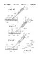
FIG. 12 is an illustration showing a preliminary step in the positioning of a conventional introducer sheath through a percutaneous puncture in an artery using the position indicating device shown in FIG. 7;
FIG. 13 is an illustration similar to that of FIG. 12 showing desired position of the introducer sheath within the artery as established by the use of the position indicating device shown in FIG. 7;
FIG. 14 is an illustration showing the introduction of the deployment instrument into the properly located introducer sheath;
FIGS. 15-23 are illustrations, similar to FIGS. 11 and 12, but showing the sequential steps in the use of the instrument to deploy the closure device to seal the percutaneous puncture in the artery;
FIG. 24 is an enlarged illustration showing the closure device in place after it has sealed the percutaneous puncture in the artery;
FIG. 25 is an isometric view of a position indicating clip of the system of this invention;
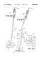
FIG. 26 is an isometric view of a second embodiment of a introducer sheath position indicating device forming a portion of the system of this invention;
FIG. 27 is an illustration similar to that of FIG. 12 showing desired position of a conventional introducer sheath within the artery as established by the use of the second embodiment of the position indicating device shown in FIG. 26;
FIG. 28 is an isometric view of a third embodiment of a introducer sheath position indicating device forming a portion of the system of this invention;
FIG. 29 is an illustration similar to that of FIG. 12 showing desired position of a conventional introducer sheath within the artery as established by the use of the third embodiment of the position indicating device shown in FIG. 28;
FIG. 30 is an isometric view of a conventional dilator;
FIG. 31 is an isometric view of a modified introducer sheath forming a position indicating device of the system of this invention;
FIG. 32 is an enlarged sectional view taken alongline 32--32 of FIG. 31;
FIG. 33 is an illustration similar to that of FIG. 12 showing desired position of the modified introducer sheath of FIG. 32 located within the artery;
FIG. 34 is an enlarged top plan view of an alternative anchor component to that shown in FIG. 4;
FIG. 35 is an enlarged side elevational view of the alternative anchor shown in FIG. 34;
FIG. 36 is an enlarged sectional view of an alternative tamping means to that shown in FIG. 1;
FIG. 37 is an enlarged illustration similar to FIG. 23 but showing the use of the alternative tamping means of FIG. 36; and
FIG. 38 is an enlarged illustration similar to FIG. 24 but showing the closure device in place after it has sealed the percutaneous puncture in the artery using the alternative tamping means.
Referring now in greater detail to the various figures of the drawings wherein like reference characters refer to like parts, there is shown at 20 an instrument forming a portion of a system for deploying aclosure device 22 to seal apercutaneous puncture 24 within ablood vessel 26, e.g., the femoral artery, constructed in accordance with this invention. Thepuncture 24 includes not only the opening in the wall of the vessel but also thetract 24A, i.e., the passageway in the tissue located between the vessel and the skin of the being formed when the vessel is punctured.
Theinstrument 20 andclosure device 22 have particular utility when used in connection with intravascular procedures, such as angiographic dye injection, cardiac catheterization, balloon angioplasty and other types of recanalizing of atherosclerotic arteries, etc. since theclosure 22 is designed to cause immediate hemostasis of the blood vessel, e.g., arterial, puncture. However, it is to be understood that while the description of the preferred embodiment instrument and closure contained herein is directed to the closing off of percutaneous incisions or punctures in arteries, they have much more wide-spread applications. Thus, the sealing of a percutaneous opening in an artery shown herein is merely exemplary.
Before describing theclosure 22 and theinstrument 20 for inserting it to seal the opening, a brief description of a typical, conventional, intravascular surgical procedure, e.g., catheter instrumentation of an artery, utilizing a percutaneous opening will be given to best appreciate the features of the invention. In such a procedure a cannula of an instrument, such as an angiographic needle (not shown), is inserted percutaneously through the skin into the artery, such as the femoral artery, at the situs for the instrument's insertion. The needle cannula is held in place and the flexible end of a mini-guidewire (not shown) is then passed through the cannula into the artery to the desired depth (i.e., longitudinal position therealong). Once the mini-guide wire is in place the needle cannula is removed, leaving the guidewire in place. An introducer sheath 28 (FIGS. 12 and 13) and an arterial dilator (not shown) are then passed over the guidewire, through the puncture or incision and into the artery. The guidewire and then the dilator are removed leaving the introducer sheath in place. A catheter, or other intravascular instrument (not shown) is then inserted through theintroducer sheath 28 and threaded down theartery 26 to the desired intravascular location, e.g., the situs of the atherosclerotic occlusion.
Once the intravascular procedure (e.g., angioplasty) has been completed, the catheter is removed. Thereafter, the sheath is removed and the surgeon or other trained person applies manual, digital pressure to the percutaneous puncture until hemostasis has occurred. In particular, the current standard of care for puncture hemostasis is to apply digital or mechanical pressure on the puncture site for twenty minutes to an hour, depending on the puncture size and the degree of hemolytic therapy. Obviously this results in wasted time for the physicians and other catheter lab personnel, and causes inconvenience and discomfort for the patient. In addition serious complications arise from persistent bleeding and hematoma formation in approximately five percent of the patients.
In accordance with the method of this invention theintroducer sheath 28 is left in place within the artery (although it is moved so that its distal end is at a desired position therein, as will be described later). Thedeployment instrument 20 having theclosure device 22 therein is inserted into the introducer sheath. The closure device is then deployed (ejected) and operated to immediately seal thearterial puncture site 24 and plug thetract 24A. Moreover, as will be appreciated from the description to follow theclosure device 22 is designed to reduce post-procedure puncture complications, cause minimal inflammatory reaction and resorb completely within a relatively short period of time, e.g., sixty to ninety days.
The details of theclosure 22 andinstrument 20 for introducing it will be described in detail later. Suffice it for now to briefly describe the closure and its method of deployment and use. Thus, as will be seen later the closure has three basic components, namely, a sealingmember 30, anintraarterial anchor member 32, and a positioningmember 34. The sealing member is in the form of an elongated rod-like plug, e.g., a hemostatic, resorbable collagen sponge or foam. This member is arranged for sealing the puncture. Theanchor member 32 is an elongated, stiff, low-profile, resorbable member which is arranged to be seated inside the artery against the artery wall contiguous with thepuncture 24. Theanchor member 32 is made of non-hemostatic resorbable polymer similar to resorbable suture. The positioningmember 34 comprises a filament, e.g., a resorbable suture. The suture connects the anchor member and the collagen plug (sealing member) via a pulley-like arrangement which serves to move the anchor and plug together, to sandwich and lock the artery wall between the anchor and plug.
Theclosure device 22 is used after the interventional procedure is finished. In particular, the physician inserts the delivery ordeployment instrument 20 containing theclosure device 22 into the patients'introducer sheath 28. On insertion, theanchor member 32 passes out of the distal end of the introducer sheath and deploys into the artery lumen. Thedeployment instrument 20 is then withdrawn from the introducer sheath until resistance is felt when the anchor member catches on the distal end thereof. Once this occurs (and assuming that the anchor is in the correct orientation when it catches on the end of the introducer sheath, as will be described later) the deployment instrument and the introducer sheath are then immediately withdrawn together. This withdrawing action causes theanchor member 32 to engage (catch) on the artery wall contiguous with the puncture. Continued withdrawal of the instrument and introducer sheath causes the pulley-like configuration of the filament to pull thecollagen plug 30 toward theanchor member 32, thereby depositing the plug in thepuncture tract 24A against the exterior of the artery contiguous with the puncture. The pulling on the filament to bring the plug into engagement with the puncture site also has the effect of deforming the plug into a larger diameter body to aid in holding it in place. Moreover, since the plug is formed of a compressed collagen it also expands automatically in the presence of blood within the puncture tract when deployed, thereby further contributing to the plug's enlargement. Theinstrument 20 also includes a tamper (to be described later) which is mounted on the suture and which is slidable thereon. The deployment of the plug member also effects the deployment of the tamper into the puncture tract proximally of the plug member. The tamper is then used to gently compress and lock the collagen plug on the outside of the artery.
The closure is now locked in place through the clotting of the hemostatic collagen plug and by spring tension (to be described later) on thefilament 34 attached to theintraarterial anchor 32. Thus the artery wall is sandwiched between thecollagen plug 30 andanchor 32. Within a few hours after deployment, theanchor 32 will be coated with fibrin and thus attached firmly to the arterial wall, thereby eliminating the possibility of distal embolization. After approximately thirty days, only a small deposit of anchor material will remain. In fact, resorption of all components will have occurred after approximately sixty days.
Theanchor member 32 is non-hemostatic and is sized to be hemodynamically insignificant in comparison to the size of the femoral artery. Thus, the resorbable anchor has an insignificant hemodynamic effect on blood flow.
As will be appreciated by the description to follow deployment of theclosure device 22 by theinstrument 20 is easy, quick and reliable. Anchoring is repeatable, safe, and effective to deploy the collagen plug. Hemostasis occurs almost instantaneously, e.g., in 15 seconds or less, when the closure device is deployed properly.
Referring now to FIGS. 2-5 the details of theclosure device 22 will now be described. As can be seen in FIG. 2 the sealing member or plug 30 comprises a cylindrical member formed of a compressible, resorbable, collagen foam, such as that sold by Collatec, Inc. of Plainsboro, N.J. Theplug 30 is arranged to be compressed from the large diameter configuration shown in FIG. 2 to the small diameter, elongated configuration shown in FIG. 3. In the configuration of FIG. 3 the diameter of the plug is very small, e.g., 1.32 mm, and therefor suitable for disposition within theinstrument 20 as will be described later. Theplug 30 includes anannular recess 40 extending about its outer periphery adjacent its proximal end. Threeapertures aperture 42 is located close to therecess 40 and diametrically through the centerline of the plug. Theaperture 46 is located close to the distal end of the plug and extends transversely through the plug on one side of the centerline. Theaperture 44 is located betweenapertures filament 34 extends to connect the anchor member to the plug and are spaced apart to preclude tearing of the plug.
The manner of connection of the plug to the anchor will be described later. Suffice it for now to state that thefilament 34 of theclosure device 22 serves to couple the plug component to the anchor component in an arrangement to effect the movement of the plug component toward the anchor component, once the anchor component is in its desired position in the artery at the puncture or incision. In particular the coupling of the plug component to the anchor component simulates a pulley to achieve a desired mechanical advantage.
In accordance with a preferred embodiment of this invention the filament is formed of resorbable, flexible, strong material, e.g., a resorbable suture.
As can be seen in FIGS. 4 and 5 theanchor member 32 basically comprises a thin, narrow, strip or bar of material, such as a resorbable lactide/glycolide polymer sold by Medisorb Technologies International L.P. under the trade designation MEDISORB. The strip is sufficiently rigid such that once it is in position within the artery (as will be described later) it is resistant to deformation to preclude it from bending to pass back through the puncture through which it was first introduced. Themember 32 has a generally planartop surface 48, a generally planarbottom surface 50 and aperipheral side surface 52. Each end of themember 32 is rounded. Theside surface 52 of theanchor member 32 tapers inward from its top surface to its bottom surface as shown in FIG. 5 to facilitate the removal of the plug from the mold for making it. Ahemispherical projection 54 is located at the center of thetop surface 48. Thehemispherical projection 54 includes alongitudinally extending slot 56 disposed perpendicularly to thetop surface 48 of themember 32. The bottom 58 of theslot 56 is arcuate (FIG. 5). Acylindrical opening 60 extends transversely across themember 32 through theprojection 54. Aloop 62 of suture material extends through theopening 60. Theloop 62 is closed by aknot 64. The portion of theloop 62 extending through theopening 60 overlies the bottom 58 of the slot and forms a "pin" about which thefilament 34 extends. In particular thefilament 34 is threaded through theslot 56, under the "pin" of theloop 60 and back out theslot 56 on the other side thereof as shown clearly in FIG. 5 to connect theplug member 30 to theanchor member 32.
In this regard the pulley-like connection between the anchor member and the plug member is effected by threading thefilament 34 from a remote point (which is located outside thedeployment instrument 20 when the closure device is in place in that instrument) through thetransverse aperture 42, down the plug to theaperture 46, through that aperture to the opposite side of the plug and from there to the anchor member where it is threaded through theslot 56 and about the "pin" as described earlier. From there thefilament 34 extends back to the plug where it enters intoaperture 44, passes through the aperture to the opposite side of the plug, where it terminates in aloop 66 extending around theannular recess 40. The loop is secured by aknot 68, whose details are shown in FIG. 6.
In FIGS. 34 and 35 there is shown an alternative anchor member 32'. That anchor member is virtually identical to theanchor member 32 except that member 32' includes means to enable it to be imaged radiographically to facilitate the placement of the closure at the desired situs within the patient's body. Thus, as can be seen therein the alternative anchor member 32' includes a pair ofwells 32A in thetop surface 48 adjacent the respective ends of the anchor member. A plug or powder of a conventional radio-opaque material, which is preferably biocompatible and which is excretable, e.g., solid agents of sodium diatrizoate, iohexal, etc., is located within eachwell 32A. A respective cover orcap 32B, preferably formed of a thin disk of a bioresorbable material, e.g., PGA, is disposed over each well to seal the material within the well. Each cover is secured to thetop surface 48 of the anchor 32' by a seal line extending about the periphery of the well. That seal line can be formed in various ways, e.g., by heat sealing.
Referring now to FIGS. 1 and 11 the details of thedeployment instrument 20 will now be described. As can be seen the instrument basically comprises acarrier 100 in the form of anelongated tube 102 formed of a somewhat flexible material, such as polyethylene or polyvinyl chloride, so that the carrier may be freely passed through the introducer sheath into an operative position within the patient's artery, notwithstanding any curvature of the introducer sleeve which may exist.
In accordance with a preferred embodiment of this invention the outside diameter of thetubular carrier 100 is 8 French. The distal end of thetube 102 includes a rigid, e.g., stainless steel, sleeve orbypass tube 104 mounted thereon, to enable it to be inserted through aconventional hemostasis valve 28A (FIGS. 12-14) forming a portion of theintroducer sheath 28, through the sheath, and out the distal end thereof into theartery 26. The distal end of theflexible tube 102 necks down into a generally hemicylindrical configuration (See FIG. 1) which includes a longitudinally extending slit (not shown) therein to enable it to be fit within thebypass tube 104 without buckling.
As can be seen in FIG. 11, theclosure device 22 is located within the distal end of thetubular carrier 100. In particular theanchor member 32 is disposed longitudinally within thebypass tube 104 laterally of the centrallongitudinal axis 106 of the carrier. Theplug member 30 is located within thetube 102 just behind (proximally) of the anchor member and on the opposite side of the central longitudinal axis. In fact the distal end of the plug member overlies the proximal end of the anchor member. Thebypass tube 104 includes areference detent 108 in its periphery located diametrically opposite to the position of the anchor member. Thedetent 108 serves as a visual guide to help the user orient the instrument to a proper yaw angle with respect to the central longitudinal axis for insertion within the introducer sheath as will be described later.
As can be seen in FIGS. 1 and 11, theinstrument 20 includes aconventional luer fitting 110. The proximal end of thecarrier tube 102 extends into an opening in the fitting 110 and is secured in place therein by any suitable means. Another conventional luer fitting 112 is threadedly secured to the threadeddistal end 114 of the fitting 110. Thefittings filament 34 extends. A tensioning assembly is located within that body and basically comprises aball 116, a cup shapedball seat 118, acompression spring 120, and aspring seat 122. The spring seat is a disk-like member located within an annular recess within the center of theluer fitting 110. The ball seat includes a conicalinner surface 124 having acentral opening 126. The spring is a helical member interposed between thespring seat 122 and theball 116 to bias the ball toward theconical surface 124 of theball seat 118. The proximally located portion of thefilament 34 extends through the space between theball 116 and its seat. The amount of force applied to the ball is established by aspacer sleeve 128 located between theluer fittings sleeve 128 any desired preload can be applied to the spring.
As will be appreciated by those skilled in the art the tensioning assembly just described will tend to hold the filament in place with respect thereto until the force applied to the filament exceeds the preload force applied by the compression spring, whereupon the filament will be freed to slide through the instrument.
Thecarrier 100 also includes a tampingmember 130. This member is an elongated rod-like member formed of any suitable material, e.g., polyethylene, and is disposed within thecarrier tube 102 immediately proximally of theplug 32. The tampingmember 130 includes acentral passageway 132 extending down its length from itsdistal end 134 to itsproximal end 136. Thefilament 34 portion extending from theanchor member 32 passes through thepassageway 132 in the tamping member and from there into theluer fittings hole 126 at the proximal end of theinstrument 20. A holding sleeve ortag 138, e.g., a stainless steel tube, is crimped onto the filament so that it engages the proximal end of the tampingmember 130 to hold that member in place. Thetag 138 is arranged to cooperate with a torsion spring 142 (FIGS. 9 and 10) to apply tension onto thefilament 34 after the closure device is in place to enable theinstrument 20 to be removed and the filament severed (as will be described later).
As mentioned earlier theinstrument 20 is arranged to be inserted into aconventional introducer sheath 28 to effect the deployment of theclosure device 20. Before describing that operation a brief description of the introducer sleeve and its method of location with respect to the percutaneous puncture is in order. As can be seen in FIGS. 12-14 thesheath 28 includes a body portion in which aconventional hemostasis valve 28A is located and atubular portion 28B extending from the body. Thetubular portion 28B terminates in an open distal orfree end 28C. The body portion of thesheath 28 includes asideport 28D having aconventional stopcock 28E located therein. The distal end of the body of the sheath includes anannular groove 28F which is arranged to receive aposition indicator clip 150 forming a portion of the system of this invention, for reasons to be described later.
Before the instrument can be inserted into theintroducer sheath 28, the sheath itself must be properly located within the artery. This action is accomplished via apositioning device 200. That device forms a portion of the system of this invention and is shown in FIGS. 7 and 8. As can be seen thedevice 200 basically comprises a conventional dilator whose outer periphery has been modified to include a longitudinally extending flat 202. Thedevice 200 is arranged to be fully inserted within theintroducer sheath 28 like shown in FIG. 12. The insertion of thedevice 200 within theintroducer sheath 28 forms a passageway between the flattedsurface 202 of thedevice 200 and the interior surface of thetubular portion 28B of the sheath disposed thereover. The length of the flattedportion 202 is selected so that when thedevice 200 is fully with the introducer sheath, and the distal end of the sheath within the interior of the artery, the distal end of the flatted surface extends just beyond thedistal end 28C of the introducer sheath to form awindow 204 into which blood may flow, while the proximal end of thesurface 202 is in fluid communication with the interior of the introducer body and thesideport 28D. Accordingly, blood may flow into thewindow 204 through the passageway formed by the flatted surface, into thesideport 28D and from there to thestopcock 28E when thewindow 204 is within the interior of the artery.
In order to correctly position the introducer sheath the location of the artery wall must be established. This is accomplished by inserting thedevice 200 within the introducer sheath as just described and then opening thestopcock 28E to observe the flow of blood therefrom. The blood will normally flow out of the opened stopcock by virtue of the pressure differential across the lumen wall. If however, there is insufficient pressure to cause such a flow of blood some means (not shown) can be used to create the desired differential pressure, e.g., suction can be used. In any event once the flow of blood is observed the introducer sheath with the device therein is then retracted (moved proximally) until the blood flow through the stopcock just stops, a position shown in FIG. 13. This indicates that thedistal end 28C of the introducer sheath has just left the artery lumen. The introducer sheath with the device therein is then reinserted approximately 10 mm into the puncture to ensure that the distal end of introducer sheath is at the desired position within the artery. Blood flow should be reestablished through the stopcock at this time. Then the stopcock is closed. From this point the introducer sheath must be kept fixed, i.e., it must not move axially relative to the patient. To achieve that end the user of the system should provide a continuous grasp on the introducer sheath, with the patient's groin as a position reference. Theposition indicating device 200 is then removed from the introducer sheath to ready the introducer sheath for receipt of thedeployment instrument 20 carrying theclosure device 22 as will be described later.
In FIG. 26 there is shown a second embodiment of apositioning device 300 for effecting the proper positioning of theintroducer sheath 28 within the artery. As can be seen thedevice 300 basically comprises a conventional obturator whose outer periphery has been modified to include anannular recess 302 extending thereabout. Like thedevice 200, thedevice 300 is arranged to be fully inserted within theintroducer sheath 28 as shown in FIG. 27. The insertion of thedevice 300 within theintroducer sheath 28 forms an annular passageway between theannular recess 302 of thedevice 300 and the interior surface of thetubular portion 28B of thesheath 28. A side opening orport 304 is provided in thesidewall 28B of theintroducer sheath 28 closely adjacent its opendistal end 28C.
The length of theannular recess 302 is selected so that when thedevice 300 is fully with theintroducer sheath 28, and theport 304 in the distal end of the sheath is located within the interior of the artery, the distal end of theannular recess 302 extends just beyond theport 304 while the proximal end of therecess 302 is in fluid communication with the interior of the introducer'ssideport 28D.
Theport 304 forms a window into which blood in the artery may flow when thedistal end 28C of the introducer is located therein. In particular, blood may flow into thewindow 304 through the annular passageway formed between therecess 302 and the inner surface of thetubular portion 28A of the introducer, into thesideport 28D and from there to thestopcock 28E when thewindow 304 is within the interior of the artery.
In FIG. 28 there is shown a third embodiment of apositioning device 400 for effecting the proper positioning of theintroducer sheath 28 within the artery. As can be seen thedevice 400 basically comprises a conventional obturator having apassageway 402 extending longitudinally down substantially the length of the device. Anentrance port 404 extends radially inward into the device communicating with the distal end of thepassageway 402, while an outlet port extends radially inward into the device communicating with the proximal end of thepassageway 402. Like thedevices device 400 is arranged to be fully inserted within theintroducer sheath 28 as shown in FIG. 29.
The length of theannular passageway 402 is selected so that when thedevice 400 is fully with theintroducer sheath 28 and the distal end of the sheath is located within the interior of the artery, theinlet port 404 of thepassageway 402 extends just beyond the free end of the sheath, while theoutlet port 406 is in fluid communication with the interior of the introducer'ssideport 28D. Theport 404 forms a window into which blood in the artery may flow when thedistal end 28C of the introducer is located therein.
In FIG. 31 there is shown alternative embodiment 28' of an introducer sheath. The sheath is similar tosheath 28 described earlier except that itstubular portion 28B includes a second passageway 502 (FIG. 31) extending therethrough. Thepassageway 502 serves as the passageway for blood to flow therethrough so that the sheath 28', itself, can act as a positioning device for effecting its proper positioning within the artery. As can be seen in FIG. 31 thepassageway 502 extends longitudinally down the sheath 28' within its wall and parallel to the central passageway 504 (the central passageway receives thedeployment instrument 20 to be described later). The distal end of thepassageway 502 includes aradially extending port 506. The proximal end of the passageway 502 (not shown) is in fluid communication with the interior of the introducer'ssideport 28D. The introducer sheath 28' is arranged to be used with a conventional obturator 600 (shown in FIG. 30).
The positioning of theintroducer sheath 28 utilizing either of thedevices obturator 600 is similar to that described with reference to thedevice 200. Thus, after the introducer sheath is positioned as described earlier thestopcock 28E is opened to observe the flow of blood therefrom (thereby indicating that the inlet port or window is within the artery). The introducer sheath is then retracted (moved proximally) until the blood flow through the stopcock just stops, thereby indicating that thedistal end 28C of the introducer sheath has just left the artery lumen. The introducer sheath with the device therein is then reinserted approximately 10 mm into the puncture to ensure that the distal end of introducer sheath is at the desired position within the artery. Blood flow should be reestablished through the stopcock at this time. Then the stopcock is closed. From this point the introducer sheath must be kept fixed (as described earlier) and theposition indicating device 300 or 400 (or the conventional obturator 600) removed to ready the introducer sheath for receipt of thedeployment instrument 20 carrying theclosure device 22 through the central passageway in the particular introducer sheath (that passageway is denoted by thereference number 504 in the embodiment 28').
The deployment of the closure will now be described with reference to FIGS. 14-23 and is as follows: Thereference detent 108 on the bypass tube is identified by the user and the bypass tube grasped by the user and oriented so that the detent faces up (away from the patient) as shown in FIG. 14. This ensures that the anchor member is located towards the patient. The bypass tube is then inserted into the sheath through thehemostasis valve 28A. The rigid nature of the bypass tube facilitates the passage of thecarrier 100 through the hemostasis valve and also protects the closure device from damage. The instrument is then pushed fully down the introducer sheath so that a stop surface 110A on the front (distal) luer fitting 110 (FIG. 11) engages the body of the introducer sheath housing the hemostasis valve. At this time the distal end of the carrier will be in the position shown in FIG. 16 and theanchor member 32 will be located in theartery 26 beyond the distal end of the introducer sheath. Thebypass tube 104 remains within the portion of the introducer sheath housing thehemostasis valve 28A.
Theposition indicator clip 150 is then mounted onto theannular recess 28F on theintroducer sheath 28 as shown in FIG. 17. As can be seen in FIG. 25 theclip 150 includes alinear section 150A from which ayoke 150B projects perpendicularly. Theyoke 150B includes acircular mouth 150C for receipt of theannular recess 28F of the introducer sheath. When mounted in place on the introducer sheath thefree end 150D of the indicator clip will extend beyond the distal end of the instrument 20 (beyond the tensioner assembly).
Thesystem 20 is then operated to determine if theanchor member 32 has been properly deployed. To that end the introducer sheath is then held by the user to prevent axial movement and theinstrument 20 is carefully withdrawn from it. This action causes theanchor member 32 to engage or catch on to the distal end of the introducer. As the anchor member catches on the distal end of the introducer, resistance will be felt by the user. This resistance must be noted by the time the luer fitting 112 housing the tensioner assembly reaches thefree end 150D of theindicator clip 150 as shown in FIG. 18. If so, then the anchor member will have caught on the distal end of the introducer at the location of its hemispherical projection 54 (the desired occurrence).
If, however, no resistance is noted by the time that the luer fitting 112 passes (extends proximally of) the free end of the indicator clip, this will indicate that the anchor has re-entered the introducer sheath, and that the anchor will not catch onto the artery as required. Thus, if no resistance is felt at this point, theinstrument 20 must be reinserted within the introducer sheath and the foregoing procedure retried, this time by turning theinstrument 20 about itsaxis 106 by 1/4 turns to each side before it is again withdrawn.
If the resistance is felt before the luer fitting reaches the free end of the indicator clip this will indicate that one of the curved ends of the anchor member has caught on the free end of the introducer sheath, an undesired occurrence. Accordingly, theinstrument 20 must be withdrawn then reinserted within the introducer sheath and the foregoing procedure retried, this time by turning theinstrument 20 about itsaxis 106 by 1/4 turns to each side before it is again withdrawn.
Once the anchor member has been properly deployed, as shown in FIG. 18, the collagen plug is deployed. To that end theintroducer sheath 28 and theinstrument 20 are held together and withdrawn as a unit from the puncture, whilst swinging the unit toward the vertical as shown in FIG. 19. This action causes theanchor 32 to engage or catch onto the inner surface of theartery 26 contiguous with thepuncture 24. The introducer sheath and the instrument are pulled further outward as shown in FIG. 20. Inasmuch as the anchor member is trapped against the interior of the artery wall the continued retraction of the introducer sheath and instrument causes thefilament 34 to pull the collagen plug out of thecarrier tube 102 and into thepuncture tract 24A. As the introducer and instrument come out of the puncture tract, continuous steady resistance will be felt as the tensioner assembly described heretofore controls the force on thefilament 34 during the retraction procedure. Continued retraction of the introducer and the instrument brings the tampingmember 130 out of the free end of the instrument.
Moreover the pulley arrangement of thefilament 24 connecting the anchor member and the plug member ensures that during the retraction of the introducer and the instrument the plug member is moved into engagement with the exterior of the artery wall contiguous with thepuncture 24. In fact continued retraction causes the filament to somewhat deform the plug, i.e., cause it to deform radially outward. The existence of blood within the puncture tract further contributes to the deformation of the plug member since the collagen foam expands in the presence of blood.
The retraction procedure continues to pull the introducer and instrument up the filament until thetag 138 is exposed as shown in FIG. 22. At this point the anchor member and collagen plug member have been deployed. At this time the collagen plug is tamped by the tampingmember 130. In particular the user quickly compacts the collagen of the plug by gently tensioning the filament by pulling on the introducer sheath and instrument in the proximal direction with one hand. The tamping member is then manually slid down the filament by the user's other hand so that it enters thepuncture tract 24A and engages the proximal end of theplug member 32. A few gentle compactions are adequate to achieve the desired result, i.e. , to assist theplug member 30 to conform to the artery contiguous with the puncture and to assist to lock the plug in place until hemostasis occurs (which happens very quickly, thereby locking the closure in place). It should be noted that during the tamping action care must be taken to maintain tension on thefilament 34 at a load greater than that used on the tampingmember 130 to ensure that the tamping action doesn't propel theplug member 30 into the interior of the artery.
After the tamping action is completed thetorsion spring 142 is mounted on thefilament 34 as shown in FIG. 23. This action is necessary to maintain appropriate tension on the filament while theinstrument 20 is removed (the filament severed). In FIGS. 9 and 10 the torsion spring is shown. As can be seen therein thespring 142 includes a pair oflegs central section 142C. Each leg includes a slot 142D at its free end. One of the slots is arranged to receive thefilament 34 therein and to engage thetag 138. The other of the slots is arranged to receive thefilament 34 therein and to engage the proximal end of the tampingmember 130. Thelegs intermediate section 142C so that when the spring is mounted on the filament as just described they will bias the tamping means towards theplug member 30 to hold it in place so that the filament can be severed (as is necessary to remove the instrument and the introducer from the closure device). Thus, once the spring is in place the filament on the proximal side of thetag 138 is cut and the spring applies a light controlled pressure to the collagen plug and anchor. The closure is left in this condition without being disturbed for approximately 30 minutes. After that time thespring 142 is removed and the filament is then severed at the top of the tampingmember 130. The tampingmember 130 is then removed and the remaining portion of the filament is taped to the skin at 160 as shown in FIG. 24. The tape (not shown) should be removed and the filament cut subcutaneously prior to the discharge of the patient.
With the closure in final position as shown in FIG. 24 the anchor member 32 (the only portion within the artery) does not take up a substantial portion of the interior of the artery and thus does not block off or otherwise impede the flow of blood therethrough. Since the components of the closure are all formed of resorbable materials the closure can be left in place within the body until it is absorbed.
In FIG. 36 there is shown analternative embodiment 700 of tamping means constructed in accordance with this invention. The tamping means 700 basically comprises an assembly of two components, whereas the tamping means 130 described earlier is composed of only a single component. Thus, as can be seen in FIG. 36 theassembly 700 comprises a firsttubular component 702 and a secondtubular component 704. Thecomponent 702 includes acentral passageway 706 and is formed of any suitable material, e.g., the same material as used to form thetamping component 130 described earlier. Thesecond component 704 also includes acentral passageway 708 extending therethrough.
Thecomponent 704 is mounted on the front or distal end of thecomponent 702. To that end thecomponent 704 includes anannular recess 710 about its periphery at the proximal end thereof. This recess is arranged to receive thedistal end 712 of thecomponent 702, with the twopassageways filament 34 to extend therethrough.
Thecomponent 704 is preferably formed of a compressed collagen foam, e.g., the same type of material used for the sealing portion or plug 30 of the closure. Thedistal end 714 of thecomponent 704 is arranged to engage theplug 30 to tamp it down in the same manner as that accomplished by thedistal end 134 of tampingmember 130. Once the tamping action is completed thetorsion spring 142 is mounted on the filament as shown in FIG. 37 so that it is located between thetag 138 and the proximal end of the component 702 (in the same manner as described with respect to tampingmember 130 shown in FIG. 23). Thus, the filament on the proximal side of thetag 138 can be cut, while the spring applies light controlled pressure to thecollagen plug 30 andanchor 32. The closure is left in this condition in the same manner as described earlier after which time the spring is removed and the filament severed at the top (proximal end) of thetamping component 702. That component can then be removed, leaving thetamping component 704 within the puncture tract as shown in FIG. 38. The remaining (exteriorly extending) portion of the filament is taped to the skin at 160 as also described earlier.
As should be appreciated by those skilled in the art the two sections of thefilament 34 between theanchor component 32 and theplug component 30 effectively form a "pulley" arrangement to increase the mechanical advantage of the force applied to the filament to move the two components toward each other. Accordingly, the closure can be properly seated without the application of a high pulling force. The use of the biased ball and associated seat between which the filament passes during the placing of the closure ensures that irrespective of how hard the instrument and the introducer are withdrawn from the puncture during the deployment and seating of the closure, the amount of force applied to thefilament 34, and hence to the closure device, will not exceed a predetermined maximum, e.g., one pound. This feature is of considerable importance to ensure that the anchor portion of the closure is not pulled through the opening (e.g., incision or puncture) once it is in place.
As should also be appreciated from the foregoing, the closure device, the instrument for deploying it, and their method of use enables the ready, effective and efficient sealing of a percutaneous puncture in an artery. Thus, it is expected that the hemostaticpuncture closure device 20 will be a significant advancement in the fields of cardiology and radiology. The device may allow continuance of anticoagulation post-procedure, more aggressive use of thrombolytic agents and safer use of large bore catheters. It should also reduce discomfort and complication rates for patients; allow many in-patient procedures to be performed safely on an out-patient basis; decrease the time and cost of interventional procedures; and reduce exposure of hospital personnel to human blood.
Without further elaboration the foregoing will so fully illustrate our invention that others may, by applying current or future knowledge, adopt the same for use under various conditions of service.
Claims (25)
1. A system for sealing a percutaneous puncture in the wall of a vessel, duct or lumen of a living being, the vessel, duct or lumen having a fluid therein, the puncture comprising an opening in the wall of the vessel, duct or lumen and a tract contiguous with the opening extending through tissue overlying the vessel, duct or lumen, said system comprising a deployment member, a pusher member, and a puncture closure, said closure comprising a first portion at least one extending filament portion and a filament lock, said deployment member having a portion arranged for introduction through the puncture and the opening in the wall of the vessel, duct or lumen into the interior of the vessel, duct or lumen to deploy said closure in the puncture with said first portion of said closure within the interior of the vessel, duct or lumen and with said at least one extending filament portion extending from said first portion of said closure into the puncture tract, said closure being arranged to be disposed at an operative position in the puncture sealing the puncture, said filament lock comprising a knot locatable on said at least one filament portion within the puncture tract for cooperation with a portion of said closure said knot, when in said puncture tract and when said first portion of said closure is within the interior of the vessel, duct or lumen, holding said closure at said operative position, said pusher member being adapted for insertion within the tract and slidably disposed on a portion of said closure to slide therealong to effect the positioning of said knot within the puncture tract with respect to said vessel, duct or lumen and to secure said closure in place at said operative position sealing the puncture.
2. The system of claim 1 wherein said first portion of said closure comprises an anchor arranged to be located within said vessel, duct or lumen, and wherein said closure additionally comprises a sealing member arranged to be located in the tract, said anchor member and said sealing member being coupled together by said at least one filament portion said knot being in engagement with said sealing member.
3. The system of claim 1 wherein said pusher member comprises a tamping member.
4. A method for sealing a percutaneous puncture in the wall of a vessel, duct or lumen of a living being utilizing a closure system, the vessel, duct or lumen having a fluid therein, the puncture comprising an opening in the wall of the vessel, duct or lumen and a tract contiguous with the opening extending through tissue overlying the vessel, duct or lumen, the closure system comprising a pusher and a puncture closure comprising a first portion, at least one extending filament portion and a filament lock, said filament lock being locatable on said one extending filament portion, said method comprising:
(A) introducing said closure into the puncture and through the opening in the wall of the vessel, duct or lumen so that said first portion of said closure is located within the interior of the vessel, duct or lumen while said at least one extending filament portion extends into the puncture tract from said first portion of said closure;
(B) locating said knot within the tract coupled to said at least one extending filament portion for cooperation therewith;
(C) inserting said pusher member within the tract so that said pusher member is disposed on a portion of said closure; and
(D) sliding said pusher member within the tract along said portion of said closure in a distal direction towards said vessel, duct or lumen to thereby effect the positioning of said knot with respect to said vessel, duct or lumen and to secure said closure in place at an operative position, whereupon said puncture is sealed by said closure to prevent the fluid in the vessel, duct or lumen from flowing through the puncture.
5. The method of claim 4 additionally comprising the step of:
(E) providing a deployment member for introducing said closure into the puncture so that said first portion of said closure is within the interior of the vessel, duct or lumen.
6. The method of claim 5 additionally comprising the step of:
(F) introducing at least a portion of said deployment member into the interior of the vessel, duct or lumen during the deployment of said closure.
7. The method of claim 6 wherein said at least a portion of said deployment member is introduced through the opening in the wall of the vessel, duct or lumen so that said at least a portion of said deployment member is located within the interior of the vessel, duct or lumen during the deployment of said closure.
8. The method of claim 4 wherein said vessel, duct, or lumen comprises an artery.
9. The method of claim 8 wherein said artery comprises the femoral artery.
10. A system for sealing a percutaneous puncture in the wall of a vessel, duct or lumen of a living being, the vessel, duct or lumen having a fluid therein, the puncture comprising an opening in the wall of the vessel, duct or lumen and a tract contiguous with the opening extending through tissue overlying the vessel, duct or lumen, said system comprising a deployment member, a pusher member, and a puncture closure, said closure comprising a first portion, at least one extending filament portion and a filament lock, said deployment member having a portion arranged for introduction through the puncture and the opening in the wall of the vessel, duct or lumen into the interior of the vessel, duct or lumen to deploy said closure in the puncture with said first portion of said closure within the interior of the vessel, duct or lumen and with said at least one extending filament portion extending from said first portion of said closure into the puncture tract, said closure being arranged to be disposed at an operative position in the puncture to seal the puncture, said filament lock comprising a knot coupled to and movable with respect to said at least one filament portion within the puncture tract to a position holding said closure at said operative position, said pusher member being adapted for insertion within the tract and slidably disposed on a portion of said closure to slide therealong to move said knot within the puncture tract with respect to the vessel, duct or lumen to the position holding said closure at said operative position, thereby sealing the puncture.
11. The system of claim 10 wherein said closure comprises two extending filament portions, wherein said knot is connected to one of said two extending filament portions and is slidably mounted on the other of said two extending filament portions.
12. A system for sealing a percutaneous puncture in the wall of a vessel, duct or lumen of a living being, the vessel, duct or lumen having a fluid therein, the puncture comprising an opening in the wall of the vessel, duct or lumen and a tract contiguous with the opening extending through tissue overlying the vessel, duct or lumen, said system comprising a deployment member, a pusher member, and a puncture closure, said closure comprising a first portion, a first extending filament portion, a second extending filament portion, and a filament lock, said deployment member having a portion arranged for introduction through the puncture and the opening in the wall of the vessel, duct or lumen into the interior of the vessel, duct or lumen to deploy said closure in the puncture with said first portion of said closure within the interior of the vessel, duct or lumen and with said first and second filament portions extending alongside each other within the tract from said first portion of said closure within the interior of the vessel, duct or lumen, said closure being arranged to be disposed at an operative position in the puncture to seal the puncture, said filament lock being coupled to one of said first and second filament portions and being movable with respect to the other of said first and second filament portions within the puncture tract to a position holding said closure at said operative position, said pusher member being adapted for insertion within the tract and slidably disposed on a portion of said closure to slide therealong to move said filament lock within the puncture tract with respect to the vessel, duct or lumen to the position holding said closure at said operative position, thereby sealing the puncture.
13. The system of claim 12 wherein said filament lock is connected to one of said first and second filament portions.
14. The system of claim 13 wherein said filament lock is slidable on the other of said first and second filament portions.
15. The system of claim 12 wherein said filament lock comprises a knot.
16. The system of claim 13 wherein said knot is connected to one of said first and second filament portions.
17. The system of claim 16 wherein said knot is slidable on the other of said first and second filament portions.
18. A method for sealing a percutaneous puncture in the wall of a vessel, duct or lumen of a living being utilizing a closure system, the vessel, duct or lumen having a fluid therein, the puncture comprising an opening in the wall of the vessel, duct or lumen and a tract contiguous with the opening extending through tissue overlying the vessel, duct or lumen, the closure system comprising a deployment member, a pusher member, and a puncture closure, the closure being arranged to be located at an operative position within the body of the being to seal the puncture and comprising a first portion, at least one extending filament portion and a filament lock comprising a knot coupled to and movable with respect to the at least one filament portion, said method comprising:
(A) introducing a portion of the deployment member through the puncture and the opening in the wall of the vessel, duct or lumen into the interior of the vessel, duct or lumen;
(B) using the deployment member to deploy the closure in the puncture with the first portion of the closure within the interior of the vessel, duct or lumen and with the at least one extending filament portion extending from the first portion of said closure into the puncture tract; and
(C) inserting the pusher member within the tract so that it is slidably disposed on a portion of the closure, and sliding the pusher member therealong to move the knot within the puncture tract with respect to the vessel, duct or lumen to the position holding the closure at the operative position sealing the puncture.
19. The method of claim 18 wherein the closure comprises two extending filament portions, wherein said knot is connected to one of said two extending filament portions and is slidably mounted on the other of said two extending filament portions.
20. A method for sealing a percutaneous puncture in the wall of a vessel, duct or lumen of a living being utilizing a closure system, the vessel, duct or lumen having a fluid therein, the puncture comprising an opening in the wall of the vessel, duct or lumen and a tract contiguous with the opening extending through tissue overlying the vessel, duct or lumen, the closure system comprising a deployment member, a pusher member, and a puncture closure, the closure being arranged to be located at an operative position within the body of the being to seal the puncture and comprising a first extending filament portion, a second extending filament portion, and a filament lock; said method comprising:
(A) introducing a portion of the deployment member through the puncture and the opening in the wall of the vessel, duct or lumen into the interior of the vessel, duct or lumen;
(B) using the deployment member to deploy the closure in the puncture with the first portion of the closure within the interior of the vessel, duct or lumen and with the first and second filament portions extending alongside each other within the tract from the first portion of the closure within the interior of the vessel, duct or lumen;
(C) coupling the filament lock to one of the first and second filament portions; and
(D) inserting the pusher member within the tract so that it is slidably disposed on a portion of the closure, and sliding the pusher member therealong to move the filament lock with respect to the other of the first and second filament portions within the puncture tract to the position holding the closure at the operative position sealing the puncture.
21. The method of claim 20 wherein the filament lock is connected to one of said first and second filament portions.
22. The method of claim 21 wherein the filament lock is slidable on the other of the first and second filament portions.
23. The method of claim 20 wherein the filament lock comprises a knot.
24. The method of claim 13 wherein the knot is connected to one of the first and second filament portions.
25. The method of claim 24 wherein said knot is slidable on the other of said first and second filament portions.
Priority Applications (4)
| Application Number | Priority Date | Filing Date | Title |
|---|---|---|---|
| US08/921,270US5861004A (en) | 1991-11-08 | 1997-08-29 | Hemostatic puncture closure system including closure locking means and method of use |
| US09/015,190US6045569A (en) | 1991-11-08 | 1998-01-29 | Hemostatic puncture closure system including closure locking means and methods of use |
| US09/104,321US6090130A (en) | 1991-11-08 | 1998-06-25 | Hemostatic puncture closure system including blood vessel locator and method of use |
| US09/168,429US6007563A (en) | 1991-11-08 | 1998-10-07 | Method of deploying percutaneous puncture closure |
Applications Claiming Priority (7)
| Application Number | Priority Date | Filing Date | Title |
|---|---|---|---|
| US07/789,704US5222974A (en) | 1991-11-08 | 1991-11-08 | Hemostatic puncture closure system and method of use |
| US07/846,322US5282827A (en) | 1991-11-08 | 1992-03-05 | Hemostatic puncture closure system and method of use |
| US08/154,882US5441517A (en) | 1991-11-08 | 1993-11-18 | Hemostatic puncture closure system and method of use |
| US42637195A | 1995-04-21 | 1995-04-21 | |
| US08/604,205US5707393A (en) | 1991-11-08 | 1996-02-21 | Hemostatic puncture closure system and method of use |
| US08/608,428US5676689A (en) | 1991-11-08 | 1996-02-28 | Hemostatic puncture closure system including vessel location device and method of use |
| US08/921,270US5861004A (en) | 1991-11-08 | 1997-08-29 | Hemostatic puncture closure system including closure locking means and method of use |
Related Parent Applications (1)
| Application Number | Title | Priority Date | Filing Date |
|---|---|---|---|
| US08/608,428ContinuationUS5676689A (en) | 1991-11-08 | 1996-02-28 | Hemostatic puncture closure system including vessel location device and method of use |
Related Child Applications (3)
| Application Number | Title | Priority Date | Filing Date |
|---|---|---|---|
| US09/015,190ContinuationUS6045569A (en) | 1991-11-08 | 1998-01-29 | Hemostatic puncture closure system including closure locking means and methods of use |
| US09/104,321ContinuationUS6090130A (en) | 1991-11-08 | 1998-06-25 | Hemostatic puncture closure system including blood vessel locator and method of use |
| US09/168,429ContinuationUS6007563A (en) | 1991-11-08 | 1998-10-07 | Method of deploying percutaneous puncture closure |
Publications (1)
| Publication Number | Publication Date |
|---|---|
| US5861004Atrue US5861004A (en) | 1999-01-19 |
Family
ID=27496164
Family Applications (4)
| Application Number | Title | Priority Date | Filing Date |
|---|---|---|---|
| US08/608,428Expired - LifetimeUS5676689A (en) | 1991-11-08 | 1996-02-28 | Hemostatic puncture closure system including vessel location device and method of use |
| US08/921,270Expired - LifetimeUS5861004A (en) | 1991-11-08 | 1997-08-29 | Hemostatic puncture closure system including closure locking means and method of use |
| US09/015,190Expired - LifetimeUS6045569A (en) | 1991-11-08 | 1998-01-29 | Hemostatic puncture closure system including closure locking means and methods of use |
| US09/104,321Expired - LifetimeUS6090130A (en) | 1991-11-08 | 1998-06-25 | Hemostatic puncture closure system including blood vessel locator and method of use |
Family Applications Before (1)
| Application Number | Title | Priority Date | Filing Date |
|---|---|---|---|
| US08/608,428Expired - LifetimeUS5676689A (en) | 1991-11-08 | 1996-02-28 | Hemostatic puncture closure system including vessel location device and method of use |
Family Applications After (2)
| Application Number | Title | Priority Date | Filing Date |
|---|---|---|---|
| US09/015,190Expired - LifetimeUS6045569A (en) | 1991-11-08 | 1998-01-29 | Hemostatic puncture closure system including closure locking means and methods of use |
| US09/104,321Expired - LifetimeUS6090130A (en) | 1991-11-08 | 1998-06-25 | Hemostatic puncture closure system including blood vessel locator and method of use |
Country Status (1)
| Country | Link |
|---|---|
| US (4) | US5676689A (en) |
Cited By (126)
| Publication number | Priority date | Publication date | Assignee | Title |
|---|---|---|---|---|
| US6045569A (en)* | 1991-11-08 | 2000-04-04 | Kensey Nash Corporation | Hemostatic puncture closure system including closure locking means and methods of use |
| US6306159B1 (en)* | 1998-12-23 | 2001-10-23 | Depuy Orthopaedics, Inc. | Meniscal repair device |
| US6319271B1 (en)* | 1998-12-30 | 2001-11-20 | Depuy Orthopaedics, Inc. | Suture locking device |
| US20020111688A1 (en)* | 1999-10-20 | 2002-08-15 | Cauthen Joseph C. | Intervertebral disc annulus stent |
| US20020123807A1 (en)* | 1999-10-20 | 2002-09-05 | Cauthen Joseph C. | Spinal disc annulus reconstruction method and spinal disc annulus stent |
| US6508828B1 (en) | 2000-11-03 | 2003-01-21 | Radi Medical Systems Ab | Sealing device and wound closure device |
| US20030021827A1 (en)* | 2001-07-16 | 2003-01-30 | Prasanna Malaviya | Hybrid biologic/synthetic porous extracellular matrix scaffolds |
| US20030033021A1 (en)* | 2001-07-16 | 2003-02-13 | Plouhar Pamela Lynn | Cartilage repair and regeneration scaffold and method |
| US20030036797A1 (en)* | 2001-07-16 | 2003-02-20 | Prasanna Malaviya | Meniscus regeneration device and method |
| US20030040760A1 (en)* | 1998-11-06 | 2003-02-27 | Neomend, Inc. | Systems, methods, and compositions for achieving closure of suture sites |
| US20030044444A1 (en)* | 2001-07-16 | 2003-03-06 | Prasanna Malaviya | Porous extracellular matrix scaffold and method |
| US20030050664A1 (en)* | 2001-09-07 | 2003-03-13 | Solem Jan O. | Apparatus and method for sealing a body vessel puncture |
| US20030078617A1 (en)* | 2001-07-16 | 2003-04-24 | Schwartz Herbert E. | Unitary surgical device and method |
| US6596012B2 (en) | 2000-04-19 | 2003-07-22 | Radi Medical Systems Ab | Intra-arterial occluder |
| US20030153976A1 (en)* | 1999-10-20 | 2003-08-14 | Cauthen Joseph C. | Spinal disc annulus reconstruction method and spinal disc annulus stent |
| US20040019328A1 (en)* | 2001-11-08 | 2004-01-29 | Sing Eduardo Chi | System and method for delivering hemostasis promoting material to a blood vessel puncture site by fluid pressure |
| US6726704B1 (en) | 1998-05-29 | 2004-04-27 | By-Pass, Inc. | Advanced closure device |
| US20040093025A1 (en)* | 2002-10-25 | 2004-05-13 | Radi Medical Systems Ab | Absorbable medical sealing device |
| US20040098046A1 (en)* | 2001-01-12 | 2004-05-20 | Radi Medical Systems Ab | Technique to confirm correct positioning of arterial wall sealing device |
| US20040127940A1 (en)* | 2000-12-14 | 2004-07-01 | Ginn Richard S. | Apparatus and methods for sealing vascular punctures |
| US20040143344A1 (en)* | 2001-07-16 | 2004-07-22 | Prasanna Malaviya | Implantable tissue repair device and method |
| US6786915B2 (en) | 2000-04-19 | 2004-09-07 | Radi Medical Systems Ab | Reinforced absorbable medical sealing device |
| US20040220574A1 (en)* | 2001-07-16 | 2004-11-04 | Pelo Mark Joseph | Device from naturally occuring biologically derived materials |
| US20050049637A1 (en)* | 2001-06-08 | 2005-03-03 | Morris Edward J. | Method and apparatus for sealing access |
| US20050085854A1 (en)* | 2003-10-17 | 2005-04-21 | Ensure Medical, Inc. | Locator and closure device and method of use |
| US20050085851A1 (en)* | 2003-10-15 | 2005-04-21 | William Fiehler | Tissue puncture closure device with automatic tamping |
| US20050085773A1 (en)* | 2003-10-15 | 2005-04-21 | Forsberg Andrew T. | Method and apparatus for locating vascular punctures |
| US20050085853A1 (en)* | 2003-10-15 | 2005-04-21 | Forsberg Andrew T. | Collagen delivery assembly with blood perfusion holes |
| US20050085852A1 (en)* | 2003-10-15 | 2005-04-21 | Theresa Ditter | Vascular sealing device with locking hub |
| US20050096697A1 (en)* | 2003-11-04 | 2005-05-05 | Forsberg Andrew T. | Vascular insertion sheath with stiffened tip |
| US20050096696A1 (en)* | 2003-11-04 | 2005-05-05 | Forsberg Andrew T. | Arteriotomy closure device with anti-roll anchor |
| US20050107827A1 (en)* | 2003-11-13 | 2005-05-19 | Loran Paprocki | Method and apparatus for sealing an internal tissue puncture incorporating a block and tackle |
| US20050113737A1 (en)* | 1998-05-01 | 2005-05-26 | Mark Ashby | Device and method for facilitating hemostasis of a biopsy tract |
| US20050125031A1 (en)* | 2003-12-03 | 2005-06-09 | Pipenhagen Catherine A. | Vascular sealing device with high surface area sealing plug |
| US20050121042A1 (en)* | 2003-12-03 | 2005-06-09 | Belhe Kedar R. | Suture based vascular closure apparatus and method incorporating a pre-tied knot |
| US20050125030A1 (en)* | 2003-12-03 | 2005-06-09 | Forsberg Andrew T. | Vascular puncture seal anchor nest |
| US20050155608A1 (en)* | 2001-07-26 | 2005-07-21 | Cook Incorporated | Bodily lumen closure apparatus and method |
| US20050249772A1 (en)* | 2004-05-04 | 2005-11-10 | Prasanna Malaviya | Hybrid biologic-synthetic bioabsorbable scaffolds |
| US20050267528A1 (en)* | 2000-12-14 | 2005-12-01 | Ensure Medical, Inc. | Vascular plug having composite construction |
| US20050273137A1 (en)* | 2000-12-14 | 2005-12-08 | Core Medical, Inc. | Apparatus and methods for sealing vascular punctures |
| US20050283246A1 (en)* | 1999-08-13 | 2005-12-22 | Cauthen Joseph C Iii | Method and apparatus for the treatment of the intervertebral disc annulus |
| US20050283188A1 (en)* | 1998-05-29 | 2005-12-22 | By-Pass, Inc. | Vascular closure device |
| US6979338B1 (en) | 1998-05-29 | 2005-12-27 | By-Pass Inc. | Low profile anastomosis connector |
| US20060004408A1 (en)* | 2001-06-08 | 2006-01-05 | Morris Edward J | Method and apparatus for sealing access |
| US20060030869A1 (en)* | 2002-11-14 | 2006-02-09 | By-Pass, Inc. | Adhesive anastomosis connection system |
| US20060058844A1 (en)* | 2004-09-13 | 2006-03-16 | St. Jude Medical Puerto Rico B.V. | Vascular sealing device with locking system |
| US7048710B1 (en) | 1998-05-01 | 2006-05-23 | Sub-Q, Inc. | System and method for facilitating hemostasis of blood vessel punctures with absorbable sponge |
| US20060116602A1 (en)* | 2004-12-01 | 2006-06-01 | Alden Dana A | Medical sensing device and system |
| US7060084B1 (en) | 1998-05-29 | 2006-06-13 | By-Pass, Inc. | Vascular closure device |
| US20060129156A1 (en)* | 1999-10-20 | 2006-06-15 | Anulex Technologies, Inc. | Methods and devices for spinal disc annulus reconstruction and repair |
| US20060135638A1 (en)* | 2004-12-22 | 2006-06-22 | Pedrozo Hugo A | Method for organizing the assembly of collagen fibers and compositions formed therefrom |
| US20060167553A1 (en)* | 1999-10-20 | 2006-07-27 | Anulex Technologies, Inc. | Spinal disc annulus reconstruction method and deformable spinal disc annulus stent |
| US20060229674A1 (en)* | 2005-04-12 | 2006-10-12 | St. Jude Medical Puerto Rico B.V. | Tissue puncture closure device with scroll gear transmission tamping system |
| US20060253037A1 (en)* | 2005-05-04 | 2006-11-09 | Ensure Medical, Inc. | Locator and closure device and method of use |
| US20070032823A1 (en)* | 2005-08-04 | 2007-02-08 | St. Jude Medical Puerto Rico B.V. | Tissue puncture closure device with coiled automatic tamping system |
| US20070038244A1 (en)* | 2001-06-08 | 2007-02-15 | Morris Edward J | Method and apparatus for sealing access |
| US20070100348A1 (en)* | 1999-10-20 | 2007-05-03 | Cauthen Joseph C Iii | Apparatus and methods for the treatment of the intervertebral disc |
| US20070156084A1 (en)* | 2006-01-04 | 2007-07-05 | St. Jude Medical Puerto Rico B.V. | Balloon insertion apparatus and method of sealing a tissue puncture |
| US20070198021A1 (en)* | 1999-10-20 | 2007-08-23 | Wales Lawrence W | Method and apparatus for enhanced delivery of treatment device to the intervertebral disc annulus |
| US20080065150A1 (en)* | 2006-09-12 | 2008-03-13 | Boston Scientific Scimed, Inc. | Method and Apparatus for Promoting Hemostasis of a Blood Vessel Puncture |
| US20080071297A1 (en)* | 2006-08-31 | 2008-03-20 | Thomas Kohl | Device For Inserting At Least One Anchor Piece Into A Hollow Space Of A Living Being |
| US20080140094A1 (en)* | 2006-12-11 | 2008-06-12 | Schwartz Herbert E | Unitary surgical device and method |
| US20080147114A1 (en)* | 1998-05-29 | 2008-06-19 | Bypass, Inc. | Vascular port device |
| US20090005777A1 (en)* | 2001-04-24 | 2009-01-01 | Vascular Closure Systems, Inc. | Arteriotomy closure devices and techniques |
| US20090024106A1 (en)* | 2007-07-17 | 2009-01-22 | Morris Edward J | Method and apparatus for maintaining access |
| US20090054926A1 (en)* | 2007-08-21 | 2009-02-26 | St. Jude Medical Puerto Rico B.V. | Extra-vascular sealing device and method |
| WO2009029914A1 (en)* | 2007-08-31 | 2009-03-05 | Ken Christopher G M | Closure medical device |
| US20090143789A1 (en)* | 2007-12-03 | 2009-06-04 | Houser Russell A | Vascular closure devices, systems, and methods of use |
| US20090143808A1 (en)* | 2001-04-24 | 2009-06-04 | Houser Russell A | Guided Tissue Cutting Device, Method of Use and Kits Therefor |
| US20090171281A1 (en)* | 2007-12-31 | 2009-07-02 | Pipenhagen Catherine A | Systems and methods for locating and closing a tissue puncture |
| US20090171282A1 (en)* | 2007-12-31 | 2009-07-02 | Pipenhagen Catherine A | Vascuar closure device having a flowable sealing material |
| US7569233B2 (en) | 2004-05-04 | 2009-08-04 | Depuy Products, Inc. | Hybrid biologic-synthetic bioabsorbable scaffolds |
| US7595062B2 (en) | 2005-07-28 | 2009-09-29 | Depuy Products, Inc. | Joint resurfacing orthopaedic implant and associated method |
| US20100076463A1 (en)* | 2008-04-04 | 2010-03-25 | Akshay Mavani | Implantable fistula closure device |
| US20100121358A1 (en)* | 2008-06-06 | 2010-05-13 | Vital Access Corporation | Tissue management methods, apparatus, and systems |
| US7753933B2 (en) | 2000-12-14 | 2010-07-13 | Ensure Medical, Inc. | Plug with detachable guidewire element and methods for use |
| US20100191166A1 (en)* | 2009-01-29 | 2010-07-29 | Vital Access Corporation | Subcutaneous vascular access ports and related systems and methods |
| US20100217309A1 (en)* | 2009-02-20 | 2010-08-26 | Boston Scientific Scimed, Inc. | Plug for arteriotomy closure and method of use |
| US20100217311A1 (en)* | 2009-02-20 | 2010-08-26 | Boston Scientific Scimed, Inc. | Tissue puncture closure device |
| US20100217308A1 (en)* | 2009-02-20 | 2010-08-26 | Boston Scientific Scimed, Inc. | Locking element for vascular closure device |
| US20100217310A1 (en)* | 2009-02-20 | 2010-08-26 | Boston Scientific Scimed, Inc. | Modified plug for arteriotomy closure |
| US20100249828A1 (en)* | 2008-09-04 | 2010-09-30 | Akshay Mavani | Inflatable device for enteric fistula treatement |
| US20100275432A1 (en)* | 2009-02-20 | 2010-11-04 | Boston Scientific Scimed, Inc. | Locking element for vascular closure device |
| US7850710B2 (en) | 2006-05-23 | 2010-12-14 | St. Jude Medical Puerto Rico Llc | Puncture closure apparatuses, sealing plugs, and related methods |
| US7857825B2 (en) | 1998-12-01 | 2010-12-28 | Cook Biotech Incorporated | Embolization device |
| US20110009901A1 (en)* | 2009-07-13 | 2011-01-13 | Tom Holman | Metal vascular aperture closure device |
| US20110046665A1 (en)* | 2007-09-12 | 2011-02-24 | Transluminal Technologies, Llc | Closure Device, Deployment Apparatus, and Method of Deploying a Closure Device |
| US20110066181A1 (en)* | 2009-02-20 | 2011-03-17 | Boston Scientific Scimed, Inc. | Tissue puncture closure device |
| EP2305324A1 (en) | 1999-03-25 | 2011-04-06 | Metabolix, Inc. | Medical devices and applications of polyhydroxyalkanoate polymers |
| US7951201B2 (en) | 1999-10-20 | 2011-05-31 | Anulex Technologies, Inc. | Method and apparatus for the treatment of the intervertebral disc annulus |
| US20110196388A1 (en)* | 2010-02-11 | 2011-08-11 | Boston Scientific Scimed, Inc. | Automatic vascular closure deployment devices and methods |
| US20110208219A1 (en)* | 1999-11-12 | 2011-08-25 | Boston Scientific Scimed, Inc. | Method and apparatus for endoscopic repair of the lower esophageal sphincter |
| US20110213309A1 (en)* | 2009-01-29 | 2011-09-01 | Vital Access Corporation | Vascular access ports and related methods |
| US8057510B2 (en) | 2000-12-14 | 2011-11-15 | Ensure Medical, Inc. | Plug with collet and apparatus and method for delivering such plugs |
| US8118832B1 (en) | 2008-06-16 | 2012-02-21 | Morris Innovative, Inc. | Method and apparatus for sealing access |
| US8137380B2 (en) | 2007-09-12 | 2012-03-20 | Transluminal Technologies, Llc | Closure device, deployment apparatus, and method of deploying a closure device |
| US8292918B2 (en) | 2009-02-20 | 2012-10-23 | Boston Scientific Scimed, Inc. | Composite plug for arteriotomy closure and method of use |
| US8579934B2 (en) | 2003-10-17 | 2013-11-12 | Ensure Medical, Inc. | Locator and delivery device and method of use |
| US20130303856A1 (en)* | 2010-10-29 | 2013-11-14 | Vectec S.A. | Single Use, Disposable, Tissue Suspender Device |
| US8597340B2 (en) | 2010-09-17 | 2013-12-03 | Boston Scientific Scimed, Inc. | Torque mechanism actuated bioabsorbable vascular closure device |
| US8758402B2 (en) | 2010-12-17 | 2014-06-24 | Boston Scientific Scimed, Inc. | Tissue puncture closure device |
| US8840640B2 (en) | 2007-12-31 | 2014-09-23 | St. Jude Medical Puerto Rico Llc | Vascular closure device having an improved plug |
| US8926654B2 (en) | 2005-05-04 | 2015-01-06 | Cordis Corporation | Locator and closure device and method of use |
| US8968336B2 (en) | 2011-12-07 | 2015-03-03 | Edwards Lifesciences Corporation | Self-cinching surgical clips and delivery system |
| US8992567B1 (en) | 2001-04-24 | 2015-03-31 | Cardiovascular Technologies Inc. | Compressible, deformable, or deflectable tissue closure devices and method of manufacture |
| US9017347B2 (en) | 2011-12-22 | 2015-04-28 | Edwards Lifesciences Corporation | Suture clip deployment devices |
| US9131939B1 (en) | 2008-02-27 | 2015-09-15 | Mitralign, Inc. | Device for percutaneously delivering a cardiac implant through the application of direct actuation forces external to the body |
| US9131941B2 (en) | 2011-06-17 | 2015-09-15 | Curaseal Inc. | Fistula treatment devices and methods |
| US9155530B2 (en) | 2010-11-09 | 2015-10-13 | Transluminal Technologies, Llc | Specially designed magnesium-aluminum alloys and medical uses thereof in a hemodynamic environment |
| US9211116B2 (en) | 2011-06-16 | 2015-12-15 | Curaseal Inc. | Fistula treatment devices and related methods |
| US9345460B2 (en) | 2001-04-24 | 2016-05-24 | Cardiovascular Technologies, Inc. | Tissue closure devices, device and systems for delivery, kits and methods therefor |
| US9456816B2 (en) | 2007-09-12 | 2016-10-04 | Transluminal Technologies, Llc | Closure device, deployment apparatus, and method of deploying a closure device |
| US9498202B2 (en) | 2012-07-10 | 2016-11-22 | Edwards Lifesciences Corporation | Suture securement devices |
| US9549715B2 (en) | 2011-08-09 | 2017-01-24 | Cook Regentec Llc | Vial useable in tissue extraction procedures |
| US9592047B2 (en) | 2012-12-21 | 2017-03-14 | Edwards Lifesciences Corporation | System for securing sutures |
| US9592048B2 (en) | 2013-07-11 | 2017-03-14 | Edwards Lifesciences Corporation | Knotless suture fastener installation system |
| US9861517B2 (en) | 2001-07-26 | 2018-01-09 | Cook Medical Technologies Llc | Vessel closure member, delivery apparatus, and method of inserting the member |
| US10016193B2 (en) | 2013-11-18 | 2018-07-10 | Edwards Lifesciences Ag | Multiple-firing crimp device and methods for using and manufacturing same |
| US10154835B2 (en) | 2013-05-09 | 2018-12-18 | Essential Medical, Inc. | Vascular closure device with conforming plug member |
| US10188383B2 (en) | 2013-07-09 | 2019-01-29 | Edwards Lifesciences Corporation | Suture clip deployment devices |
| US10470759B2 (en) | 2015-03-16 | 2019-11-12 | Edwards Lifesciences Corporation | Suture securement devices |
| US10624630B2 (en) | 2012-07-10 | 2020-04-21 | Edwards Lifesciences Ag | Multiple-firing securing device and methods for using and manufacturing same |
| US10786244B2 (en) | 2014-05-30 | 2020-09-29 | Edwards Lifesciences Corporation | Systems for securing sutures |
| US10863980B2 (en) | 2016-12-28 | 2020-12-15 | Edwards Lifesciences Corporation | Suture fastener having spaced-apart layers |
| US10939905B2 (en) | 2016-08-26 | 2021-03-09 | Edwards Lifesciences Corporation | Suture clips, deployment devices therefor, and methods of use |
| US11197952B2 (en) | 2009-01-29 | 2021-12-14 | Advent Access Pte. Ltd. | Vascular access ports and related methods |
Families Citing this family (325)
| Publication number | Priority date | Publication date | Assignee | Title |
|---|---|---|---|---|
| US6350274B1 (en)* | 1992-05-11 | 2002-02-26 | Regen Biologics, Inc. | Soft tissue closure systems |
| US6033401A (en)* | 1997-03-12 | 2000-03-07 | Advanced Closure Systems, Inc. | Vascular sealing device with microwave antenna |
| US6302898B1 (en) | 1994-06-24 | 2001-10-16 | Advanced Closure Systems, Inc. | Devices for sealing punctures in body vessels |
| US6071300A (en) | 1995-09-15 | 2000-06-06 | Sub-Q Inc. | Apparatus and method for percutaneous sealing of blood vessel punctures |
| US6183497B1 (en)* | 1998-05-01 | 2001-02-06 | Sub-Q, Inc. | Absorbable sponge with contrasting agent |
| US6524326B1 (en) | 1995-12-07 | 2003-02-25 | Loma Linda University Medical Center | Tissue opening locator and everter and method |
| US6287322B1 (en) | 1995-12-07 | 2001-09-11 | Loma Linda University Medical Center | Tissue opening locator and everter and method |
| US6004341A (en)* | 1996-12-05 | 1999-12-21 | Loma Linda University Medical Center | Vascular wound closure device |
| US6436109B1 (en) | 1996-06-11 | 2002-08-20 | X-Site, L.L.C. | Device and method for suturing blood vessels and the like |
| US5855585A (en)* | 1996-06-11 | 1999-01-05 | X-Site, L.L.C. | Device and method for suturing blood vessels and the like |
| US5728133A (en)* | 1996-07-09 | 1998-03-17 | Cardiologics, L.L.C. | Anchoring device and method for sealing percutaneous punctures in vessels |
| US5766183A (en)* | 1996-10-21 | 1998-06-16 | Lasersurge, Inc. | Vascular hole closure |
| US6743248B2 (en) | 1996-12-18 | 2004-06-01 | Neomend, Inc. | Pretreatment method for enhancing tissue adhesion |
| US6475182B1 (en) | 1997-03-12 | 2002-11-05 | Olexander Hnojewyj | Fluidic media introduction apparatus |
| US6371975B2 (en) | 1998-11-06 | 2002-04-16 | Neomend, Inc. | Compositions, systems, and methods for creating in situ, chemically cross-linked, mechanical barriers |
| US6733515B1 (en) | 1997-03-12 | 2004-05-11 | Neomend, Inc. | Universal introducer |
| US20030191496A1 (en)* | 1997-03-12 | 2003-10-09 | Neomend, Inc. | Vascular sealing device with microwave antenna |
| US6139556A (en)* | 1997-10-29 | 2000-10-31 | X-Site, L.L.C. | Device and method for suturing blood vessels and the like |
| US6159232A (en)* | 1997-12-16 | 2000-12-12 | Closys Corporation | Clotting cascade initiating apparatus and methods of use and methods of closing wounds |
| US6478808B2 (en) | 1997-12-17 | 2002-11-12 | Closys Corporation | Clotting cascade initiating apparatus and methods of use and methods of closing wounds |
| US6033427A (en)* | 1998-01-07 | 2000-03-07 | Lee; Benjamin I. | Method and device for percutaneous sealing of internal puncture sites |
| US7625352B1 (en) | 1998-05-01 | 2009-12-01 | Sub-Q, Inc. | Depth and puncture control for system for hemostasis of blood vessel |
| US6610026B2 (en) | 1998-05-01 | 2003-08-26 | Sub-Q, Inc. | Method of hydrating a sponge material for delivery to a body |
| US6315753B1 (en) | 1998-05-01 | 2001-11-13 | Sub-Q, Inc. | System and method for facilitating hemostasis of blood vessel punctures with absorbable sponge |
| US5980539A (en)* | 1998-05-06 | 1999-11-09 | X-Site L.L.C. | Device and method for suturing blood vessels and the like |
| US5910155A (en)* | 1998-06-05 | 1999-06-08 | United States Surgical Corporation | Vascular wound closure system |
| JP4087565B2 (en)* | 1998-07-02 | 2008-05-21 | バクスター・ヘルスケヤー・ソシエテ・アノニム | Device for introducing fibrin adhesive into perforation guide |
| US6056692A (en)* | 1998-07-08 | 2000-05-02 | Schwartz; John Q. | Apparatus and method for locating and marking blood vessels |
| US6994686B2 (en) | 1998-08-26 | 2006-02-07 | Neomend, Inc. | Systems for applying cross-linked mechanical barriers |
| US6458147B1 (en) | 1998-11-06 | 2002-10-01 | Neomend, Inc. | Compositions, systems, and methods for arresting or controlling bleeding or fluid leakage in body tissue |
| US7279001B2 (en)* | 1998-11-06 | 2007-10-09 | Neomend, Inc. | Systems, methods, and compositions for achieving closure of vascular puncture sites |
| US6830756B2 (en)* | 1998-11-06 | 2004-12-14 | Neomend, Inc. | Systems, methods, and compositions for achieving closure of vascular puncture sites |
| US6949114B2 (en) | 1998-11-06 | 2005-09-27 | Neomend, Inc. | Systems, methods, and compositions for achieving closure of vascular puncture sites |
| US6899889B1 (en)* | 1998-11-06 | 2005-05-31 | Neomend, Inc. | Biocompatible material composition adaptable to diverse therapeutic indications |
| CA2319443C (en)* | 1998-12-01 | 2009-09-29 | Cook Biotech, Inc. | Collagenous biomaterials formed with submucosal tissue |
| US20030069601A1 (en)* | 1998-12-15 | 2003-04-10 | Closys Corporation | Clotting cascade initiating apparatus and methods of use |
| JP4271375B2 (en) | 1999-02-10 | 2009-06-03 | サブ−キュー・インコーポレーテッド | Device and method for facilitating hemostasis in a biopsy duct |
| US20040092964A1 (en) | 1999-03-04 | 2004-05-13 | Modesitt D. Bruce | Articulating suturing device and method |
| US8137364B2 (en) | 2003-09-11 | 2012-03-20 | Abbott Laboratories | Articulating suturing device and method |
| US7001400B1 (en) | 1999-03-04 | 2006-02-21 | Abbott Laboratories | Articulating suturing device and method |
| US7235087B2 (en) | 1999-03-04 | 2007-06-26 | Abbott Park | Articulating suturing device and method |
| US6964668B2 (en) | 1999-03-04 | 2005-11-15 | Abbott Laboratories | Articulating suturing device and method |
| US7842048B2 (en) | 2006-08-18 | 2010-11-30 | Abbott Laboratories | Articulating suture device and method |
| US6482179B1 (en)* | 1999-05-28 | 2002-11-19 | Cohesion Technologies, Inc. | Apparatuses, methods and compositions for closing tissue puncture openings |
| EP1180977B1 (en) | 1999-06-01 | 2011-12-28 | Closys Corporation | Clotting cascade initiating apparatus and methods of use |
| US6964685B2 (en) | 1999-06-22 | 2005-11-15 | The Brigham And Women's Hospital, Inc. | Biologic replacement for fibrin clot |
| US7267679B2 (en) | 1999-09-13 | 2007-09-11 | Rex Medical, L.P | Vascular hole closure device |
| AU7373700A (en) | 1999-09-13 | 2001-04-17 | Rex Medical, Lp | Vascular closure |
| US7662161B2 (en) | 1999-09-13 | 2010-02-16 | Rex Medical, L.P | Vascular hole closure device |
| US7942888B2 (en) | 1999-09-13 | 2011-05-17 | Rex Medical, L.P. | Vascular hole closure device |
| US8083766B2 (en) | 1999-09-13 | 2011-12-27 | Rex Medical, Lp | Septal defect closure device |
| US7341595B2 (en)* | 1999-09-13 | 2008-03-11 | Rex Medical, L.P | Vascular hole closure device |
| US7695492B1 (en) | 1999-09-23 | 2010-04-13 | Boston Scientific Scimed, Inc. | Enhanced bleed back system |
| US6984219B2 (en) | 1999-09-23 | 2006-01-10 | Mark Ashby | Depth and puncture control for blood vessel hemostasis system |
| US6461364B1 (en) | 2000-01-05 | 2002-10-08 | Integrated Vascular Systems, Inc. | Vascular sheath with bioabsorbable puncture site closure apparatus and methods of use |
| US6942674B2 (en) | 2000-01-05 | 2005-09-13 | Integrated Vascular Systems, Inc. | Apparatus and methods for delivering a closure device |
| US6391048B1 (en) | 2000-01-05 | 2002-05-21 | Integrated Vascular Systems, Inc. | Integrated vascular device with puncture site closure component and sealant and methods of use |
| US7842068B2 (en) | 2000-12-07 | 2010-11-30 | Integrated Vascular Systems, Inc. | Apparatus and methods for providing tactile feedback while delivering a closure device |
| US8758400B2 (en) | 2000-01-05 | 2014-06-24 | Integrated Vascular Systems, Inc. | Closure system and methods of use |
| US9579091B2 (en) | 2000-01-05 | 2017-02-28 | Integrated Vascular Systems, Inc. | Closure system and methods of use |
| EP1259168B1 (en)* | 2000-02-24 | 2010-09-08 | Loma Linda University Medical Center | Patch and glue delivery system for closing tissue openings during surgery |
| US6451031B1 (en) | 2000-03-21 | 2002-09-17 | X-Site, L.L.C. | Blood vessel suturing device with single guide-wire/needle receiving lumen |
| US6729356B1 (en) | 2000-04-27 | 2004-05-04 | Endovascular Technologies, Inc. | Endovascular graft for providing a seal with vasculature |
| US6658288B1 (en) | 2000-05-05 | 2003-12-02 | Endovascular Technologies, Inc. | Apparatus and method for aiding thrombosis through the application of electric potential |
| US6540735B1 (en) | 2000-05-12 | 2003-04-01 | Sub-Q, Inc. | System and method for facilitating hemostasis of blood vessel punctures with absorbable sponge |
| US20020022822A1 (en)* | 2000-07-14 | 2002-02-21 | Cragg Andrew H. | Sheath-mounted arterial plug delivery device |
| US6890342B2 (en) | 2000-08-02 | 2005-05-10 | Loma Linda University | Method and apparatus for closing vascular puncture using hemostatic material |
| US6755842B2 (en) | 2000-09-01 | 2004-06-29 | Angiolink Corporation | Advanced wound site management systems and methods |
| DE60144328D1 (en)* | 2000-09-08 | 2011-05-12 | Abbott Vascular Inc | Surgical clamp |
| US7201725B1 (en) | 2000-09-25 | 2007-04-10 | Sub-Q, Inc. | Device and method for determining a depth of an incision |
| US6626918B1 (en) | 2000-10-06 | 2003-09-30 | Medical Technology Group | Apparatus and methods for positioning a vascular sheath |
| US7806904B2 (en) | 2000-12-07 | 2010-10-05 | Integrated Vascular Systems, Inc. | Closure device |
| US7905900B2 (en) | 2003-01-30 | 2011-03-15 | Integrated Vascular Systems, Inc. | Clip applier and methods of use |
| US8690910B2 (en) | 2000-12-07 | 2014-04-08 | Integrated Vascular Systems, Inc. | Closure device and methods for making and using them |
| US6695867B2 (en) | 2002-02-21 | 2004-02-24 | Integrated Vascular Systems, Inc. | Plunger apparatus and methods for delivering a closure device |
| US6623510B2 (en) | 2000-12-07 | 2003-09-23 | Integrated Vascular Systems, Inc. | Closure device and methods for making and using them |
| US7211101B2 (en) | 2000-12-07 | 2007-05-01 | Abbott Vascular Devices | Methods for manufacturing a clip and clip |
| US7285097B2 (en)* | 2001-01-12 | 2007-10-23 | Radi Medical System Ab | Technique to confirm correct positioning with respect to arterial wall |
| US6569185B2 (en)* | 2001-02-15 | 2003-05-27 | Scimed Life Systems Inc | Continuous infusion technique for arterial sealing |
| US8187625B2 (en) | 2001-03-12 | 2012-05-29 | Boston Scientific Scimed, Inc. | Cross-linked gelatin composition comprising a wetting agent |
| WO2002087636A1 (en) | 2001-03-12 | 2002-11-07 | Sub-Q, Inc. | Methods for sterilizing cross-linked gelatin compositions |
| JP4267867B2 (en)* | 2001-05-03 | 2009-05-27 | ラディ・メディカル・システムズ・アクチェボラーグ | Wound occlusion element guide device |
| US6863680B2 (en)* | 2001-11-08 | 2005-03-08 | Sub-Q, Inc. | System and method for delivering hemostasis promoting material to a blood vessel puncture site by fluid pressure |
| US7029489B1 (en) | 2001-05-18 | 2006-04-18 | Sub-Q, Inc. | System and method for delivering hemostasis promoting material to a blood vessel puncture site |
| IES20010547A2 (en)* | 2001-06-07 | 2002-12-11 | Christy Cummins | Surgical Staple |
| JP4159805B2 (en)* | 2001-06-15 | 2008-10-01 | ラディ・メディカル・システムズ・アクチェボラーグ | Pushing mechanism for closing method |
| IES20010749A2 (en)* | 2001-08-09 | 2003-02-19 | Christy Cummins | Surgical Stapling Device |
| US6592608B2 (en)* | 2001-12-07 | 2003-07-15 | Biopsy Sciences, Llc | Bioabsorbable sealant |
| IES20010748A2 (en)* | 2001-08-09 | 2003-02-19 | Christy Cummins | Surgical Stapling Device and Method |
| US20080249545A1 (en)* | 2007-04-06 | 2008-10-09 | Interventional Therapies | Suturing, crimping and cutting device |
| US20030078601A1 (en)* | 2001-10-22 | 2003-04-24 | Oleg Shikhman | Crimping and cutting device |
| AU2002349994A1 (en) | 2001-10-22 | 2003-05-06 | Interventional Therapies, L.L.C. | Removable sleeve |
| DE60239559D1 (en)* | 2001-10-22 | 2011-05-05 | Interventional Therapies Llc | WUNDNÄHVORRICHTUNG |
| US7037322B1 (en) | 2001-11-08 | 2006-05-02 | Sub-Q, Inc. | System and method for delivering hemostasis promoting material to a blood vessel puncture with a staging tube |
| US7037323B2 (en) | 2001-11-08 | 2006-05-02 | Sub-Q, Inc. | Pledget-handling system and method for delivering hemostasis promoting material to a blood vessel puncture site by fluid pressure |
| US7025748B2 (en)* | 2001-11-08 | 2006-04-11 | Boston Scientific Scimed, Inc. | Sheath based blood vessel puncture locator and depth indicator |
| US7192436B2 (en) | 2001-11-08 | 2007-03-20 | Sub-Q, Inc. | Pledget-handling system and method for delivering hemostasis promoting material to a blood vessel puncture site by fluid pressure |
| DE60235574D1 (en)* | 2001-12-26 | 2010-04-15 | Univ Yale | VESSEL shunt device |
| US6814743B2 (en)* | 2001-12-26 | 2004-11-09 | Origin Medsystems, Inc. | Temporary seal and method for facilitating anastomosis |
| US7192433B2 (en)* | 2002-03-15 | 2007-03-20 | Oscor Inc. | Locking vascular introducer assembly with adjustable hemostatic seal |
| IES20030424A2 (en) | 2002-06-04 | 2003-12-10 | Robert Stevenson | Blood vessel closure clip and delivery device |
| ATE342000T1 (en)* | 2002-06-14 | 2006-11-15 | Univ Loma Linda Med | DEVICE FOR CLOSING VASCULAR WOUNDS |
| EP1646320B1 (en)* | 2002-07-03 | 2009-02-25 | Abbott Vascular Inc | Surgical stapling device |
| US8317821B1 (en) | 2002-11-04 | 2012-11-27 | Boston Scientific Scimed, Inc. | Release mechanism |
| US7455680B1 (en) | 2002-11-04 | 2008-11-25 | Boston Scientific Scimed, Inc. | Apparatus and method for inhibiting blood loss |
| US7955353B1 (en) | 2002-11-04 | 2011-06-07 | Sub-Q, Inc. | Dissolvable closure device |
| US7108710B2 (en) | 2002-11-26 | 2006-09-19 | Abbott Laboratories | Multi-element biased suture clip |
| US7160309B2 (en) | 2002-12-31 | 2007-01-09 | Laveille Kao Voss | Systems for anchoring a medical device in a body lumen |
| US8382793B2 (en)* | 2003-01-14 | 2013-02-26 | Radi Medical Systems Ab | Introducer sheath |
| US7094209B2 (en)* | 2003-01-14 | 2006-08-22 | Radi Medical Systems Ab | Method for introducer replacement |
| EP1440656A3 (en)* | 2003-01-14 | 2004-10-06 | Radi Medical Systems Ab | Device for visually indicating a blood pressure |
| DE60324089D1 (en)* | 2003-01-14 | 2008-11-27 | Radi Medical Systems | introducer sheath |
| US8905937B2 (en) | 2009-02-26 | 2014-12-09 | Integrated Vascular Systems, Inc. | Methods and apparatus for locating a surface of a body lumen |
| US8398656B2 (en) | 2003-01-30 | 2013-03-19 | Integrated Vascular Systems, Inc. | Clip applier and methods of use |
| US8821534B2 (en) | 2010-12-06 | 2014-09-02 | Integrated Vascular Systems, Inc. | Clip applier having improved hemostasis and methods of use |
| US8758398B2 (en) | 2006-09-08 | 2014-06-24 | Integrated Vascular Systems, Inc. | Apparatus and method for delivering a closure element |
| US8202293B2 (en) | 2003-01-30 | 2012-06-19 | Integrated Vascular Systems, Inc. | Clip applier and methods of use |
| US7857828B2 (en) | 2003-01-30 | 2010-12-28 | Integrated Vascular Systems, Inc. | Clip applier and methods of use |
| US7223266B2 (en) | 2003-02-04 | 2007-05-29 | Cardiodex Ltd. | Methods and apparatus for hemostasis following arterial catheterization |
| US7850654B2 (en)* | 2003-04-24 | 2010-12-14 | St. Jude Medical Puerto Rico B.V. | Device and method for positioning a closure device |
| US7811274B2 (en)* | 2003-05-07 | 2010-10-12 | Portaero, Inc. | Method for treating chronic obstructive pulmonary disease |
| US7488340B2 (en)* | 2003-06-02 | 2009-02-10 | Vascular Solutions, Inc. | Vascular access closure system |
| US9289195B2 (en) | 2003-06-04 | 2016-03-22 | Access Closure, Inc. | Auto-retraction apparatus and methods for sealing a vascular puncture |
| US7331979B2 (en)* | 2003-06-04 | 2008-02-19 | Access Closure, Inc. | Apparatus and methods for sealing a vascular puncture |
| US8715303B2 (en) | 2003-06-06 | 2014-05-06 | Abbott Laboratories | Sizing and positioning adapter for medical instruments |
| DE602004031953D1 (en) | 2003-08-14 | 2011-05-05 | Univ Loma Linda Med | |
| US8187627B2 (en)* | 2003-09-05 | 2012-05-29 | Loma Linda University Medical Center | Dressing delivery system for internal wounds |
| US7462188B2 (en) | 2003-09-26 | 2008-12-09 | Abbott Laboratories | Device and method for suturing intracardiac defects |
| US20050107820A1 (en)* | 2003-11-13 | 2005-05-19 | Forsberg Andrew T. | Vascular puncture depth locator |
| CA2547999A1 (en)* | 2003-11-25 | 2005-06-09 | Boston Scientific Limited | Hemostatic pressure plug |
| US7875043B1 (en) | 2003-12-09 | 2011-01-25 | Sub-Q, Inc. | Cinching loop |
| US7449024B2 (en) | 2003-12-23 | 2008-11-11 | Abbott Laboratories | Suturing device with split arm and method of suturing tissue |
| US7648493B2 (en)* | 2004-04-20 | 2010-01-19 | St. Jude Medical Puerto Rico Llc | Method and apparatus for locating vascular punctures |
| IES20040368A2 (en) | 2004-05-25 | 2005-11-30 | James E Coleman | Surgical stapler |
| US9387010B2 (en) | 2004-10-28 | 2016-07-12 | Nico Corporation | Surgical access assembly and method of using same |
| US9161820B2 (en) | 2004-10-28 | 2015-10-20 | Nico Corporation | Surgical access assembly and method of using same |
| US9265523B2 (en) | 2011-10-24 | 2016-02-23 | Nico Corporation | Surgical access system with navigation element and method of using same |
| US9770261B2 (en)* | 2004-10-28 | 2017-09-26 | Nico Corporation | Surgical access assembly and method of using same |
| US9186175B2 (en) | 2004-10-28 | 2015-11-17 | Nico Corporation | Surgical access assembly and method of using same |
| EP1814478A4 (en) | 2004-11-22 | 2011-05-18 | Cardiodex Ltd | Techniques for heat-treating varicose veins |
| JP4366306B2 (en)* | 2004-12-17 | 2009-11-18 | テルモ株式会社 | In vivo tissue closure device and in vivo tissue closure device |
| US20060135946A1 (en)* | 2004-12-21 | 2006-06-22 | C. R. Bard, Inc. | Hemostasis cuff for catheter securement |
| EP1876972A4 (en)* | 2005-03-30 | 2009-04-01 | Access Scientific Inc | Vascular access |
| US7713283B2 (en) | 2005-04-11 | 2010-05-11 | St. Jude Medical Puerto Rico, Llc | Tissue puncture closure device with magazine fed tamping system |
| US7250057B2 (en)* | 2005-04-11 | 2007-07-31 | St. Jude Medical Puerto Rico B.V. | Tissue puncture closure device with automatic torque sensing tamping system |
| US7988706B2 (en)* | 2005-04-11 | 2011-08-02 | St. Jude Medical Puerto Rico Llc | Tissue puncture closure device with automatic torque sensing tamping system |
| WO2006115689A1 (en) | 2005-04-22 | 2006-11-02 | Rex Medical, L.P. | Closure device for left atrial appendage |
| US7837705B2 (en) | 2005-05-17 | 2010-11-23 | St. Jude Medical Puerto Rico Llc | Tissue puncture closure system with retractable sheath |
| US7618438B2 (en) | 2005-05-17 | 2009-11-17 | St. Jude Medical Puerto Rico Llc | Tissue puncture closure device with disengagable automatic tamping system |
| US8038687B2 (en) | 2005-05-17 | 2011-10-18 | St. Jude Medical Puerto Rico Llc | Suture loop closure device |
| US8273094B2 (en)* | 2005-05-17 | 2012-09-25 | St. Jude Medical Puerto Rico Llc | Puncture locating device |
| US8926633B2 (en) | 2005-06-24 | 2015-01-06 | Abbott Laboratories | Apparatus and method for delivering a closure element |
| US8313497B2 (en) | 2005-07-01 | 2012-11-20 | Abbott Laboratories | Clip applier and methods of use |
| US20070032824A1 (en)* | 2005-08-04 | 2007-02-08 | St. Jude Medical Puerto Rico B.V. | Tissue puncture closure device with track plug |
| US8083754B2 (en) | 2005-08-08 | 2011-12-27 | Abbott Laboratories | Vascular suturing device with needle capture |
| US7883517B2 (en) | 2005-08-08 | 2011-02-08 | Abbott Laboratories | Vascular suturing device |
| US8267947B2 (en) | 2005-08-08 | 2012-09-18 | Abbott Laboratories | Vascular suturing device |
| US20070060895A1 (en) | 2005-08-24 | 2007-03-15 | Sibbitt Wilmer L Jr | Vascular closure methods and apparatuses |
| US9456811B2 (en) | 2005-08-24 | 2016-10-04 | Abbott Vascular Inc. | Vascular closure methods and apparatuses |
| US8920442B2 (en) | 2005-08-24 | 2014-12-30 | Abbott Vascular Inc. | Vascular opening edge eversion methods and apparatuses |
| JP5253170B2 (en) | 2005-10-05 | 2013-07-31 | ローマ リンダ ユニヴァーシティ メディカル センター | Vascular wound closure device and method |
| US7691127B2 (en)* | 2005-12-13 | 2010-04-06 | Cardiva Medical, Inc. | Drug eluting vascular closure devices and methods |
| US20100168767A1 (en)* | 2008-06-30 | 2010-07-01 | Cardiva Medical, Inc. | Apparatus and methods for delivering hemostatic materials for blood vessel closure |
| US8911472B2 (en) | 2005-12-13 | 2014-12-16 | Cardiva Medical, Inc. | Apparatus and methods for delivering hemostatic materials for blood vessel closure |
| US9179897B2 (en)* | 2005-12-13 | 2015-11-10 | Cardiva Medical, Inc. | Vascular closure devices and methods providing hemostatic enhancement |
| AU2007208243B2 (en) | 2006-01-25 | 2013-09-19 | Children's Medical Center Corporation | Methods and procedures for ligament repair |
| US8808310B2 (en) | 2006-04-20 | 2014-08-19 | Integrated Vascular Systems, Inc. | Resettable clip applier and reset tools |
| USD611144S1 (en) | 2006-06-28 | 2010-03-02 | Abbott Laboratories | Apparatus for delivering a closure element |
| US8556930B2 (en) | 2006-06-28 | 2013-10-15 | Abbott Laboratories | Vessel closure device |
| US7749248B2 (en) | 2006-09-18 | 2010-07-06 | St. Jude Medical Puerto Rico Llc | Flexible tamping device |
| CA2664866C (en)* | 2006-09-28 | 2018-08-14 | Children's Medical Center Coporation | Methods and collagen products for tissue repair |
| US7922696B2 (en)* | 2007-01-24 | 2011-04-12 | Access Scientific, Inc. | Access device |
| US8080034B2 (en)* | 2007-03-29 | 2011-12-20 | St. Jude Medical, Inc. | Vascular hemostasis device and deployment apparatus |
| EP2150187A2 (en)* | 2007-04-18 | 2010-02-10 | Access Scientific, Inc. | Access device |
| EP3093038B1 (en) | 2007-04-18 | 2019-05-22 | Access Scientific, Inc. | Access device |
| US9610070B2 (en) | 2007-06-15 | 2017-04-04 | Vivasure Medical Limited | Closure device |
| US8226681B2 (en) | 2007-06-25 | 2012-07-24 | Abbott Laboratories | Methods, devices, and apparatus for managing access through tissue |
| US8574244B2 (en) | 2007-06-25 | 2013-11-05 | Abbott Laboratories | System for closing a puncture in a vessel wall |
| EP2166954A1 (en) | 2007-07-13 | 2010-03-31 | Rex Medical, L.P. | Vascular hole closure device |
| EP2182875A4 (en) | 2007-08-15 | 2011-08-24 | Cardiodex Ltd | Systems and methods for puncture closure |
| US7993367B2 (en)* | 2007-09-28 | 2011-08-09 | Accessclosure, Inc. | Apparatus and methods for sealing a vascular puncture |
| US20090088723A1 (en)* | 2007-09-28 | 2009-04-02 | Accessclosure, Inc. | Apparatus and methods for treating pseudoaneurysms |
| AU2008318560B2 (en) | 2007-10-31 | 2014-12-04 | Cardinal Health 529, Llc | Method of making a vascular closure device |
| US8858591B2 (en)* | 2007-10-31 | 2014-10-14 | Radi Medical Systems Ab | Method and device for sealing a puncture hole in a bodily organ |
| US20090157101A1 (en) | 2007-12-17 | 2009-06-18 | Abbott Laboratories | Tissue closure system and methods of use |
| US8556931B2 (en)* | 2007-12-17 | 2013-10-15 | Abbott Laboratories | Methods for imaging a delivery system |
| US8893947B2 (en) | 2007-12-17 | 2014-11-25 | Abbott Laboratories | Clip applier and methods of use |
| US7841502B2 (en)* | 2007-12-18 | 2010-11-30 | Abbott Laboratories | Modular clip applier |
| US20110029013A1 (en) | 2008-02-15 | 2011-02-03 | Mcguckin James F | Vascular Hole Closure Device |
| US8070772B2 (en) | 2008-02-15 | 2011-12-06 | Rex Medical, L.P. | Vascular hole closure device |
| US9226738B2 (en) | 2008-02-15 | 2016-01-05 | Rex Medical, L.P. | Vascular hole closure delivery device |
| US8491629B2 (en) | 2008-02-15 | 2013-07-23 | Rex Medical | Vascular hole closure delivery device |
| US8920462B2 (en) | 2008-02-15 | 2014-12-30 | Rex Medical, L.P. | Vascular hole closure device |
| US8920463B2 (en) | 2008-02-15 | 2014-12-30 | Rex Medical, L.P. | Vascular hole closure device |
| US8241324B2 (en)* | 2008-03-03 | 2012-08-14 | Eilaz Babaev | Ultrasonic vascular closure device |
| US20090228002A1 (en)* | 2008-03-04 | 2009-09-10 | Rioux Robert F | Electromagnetic energy assisted tissue penetration device and method |
| US8202251B2 (en) | 2008-03-14 | 2012-06-19 | Access Scientific, Inc. | Access device |
| USD601242S1 (en) | 2008-03-14 | 2009-09-29 | Access Scientific, Inc. | Access device |
| USD615201S1 (en) | 2008-03-14 | 2010-05-04 | Access Scientific, Inc. | Combined guide wire cap and track |
| EP2319576A1 (en)* | 2008-03-14 | 2011-05-11 | Access Scientific, Inc. | Access device |
| USD594981S1 (en) | 2008-03-14 | 2009-06-23 | Access Scientific, Inc. | Needle hub |
| US8029533B2 (en) | 2008-04-04 | 2011-10-04 | Accessclosure, Inc. | Apparatus and methods for sealing a vascular puncture |
| US9364206B2 (en)* | 2008-04-04 | 2016-06-14 | Access Closure, Inc. | Apparatus and methods for sealing a vascular puncture |
| US9282965B2 (en) | 2008-05-16 | 2016-03-15 | Abbott Laboratories | Apparatus and methods for engaging tissue |
| US8486022B2 (en)* | 2008-07-03 | 2013-07-16 | Abbott Cardiovascular Systems Inc. | Needle catheter with an angled distal tip lumen |
| EP2330981B1 (en) | 2008-08-26 | 2013-11-27 | St. Jude Medical, Inc. | System for sealing percutaneous punctures |
| USD601243S1 (en) | 2008-09-10 | 2009-09-29 | Access Scientific, Inc. | Access device |
| USD600793S1 (en) | 2008-09-10 | 2009-09-22 | Access Scientific, Inc. | Access device |
| US8485969B2 (en) | 2008-09-18 | 2013-07-16 | Jeffrey Grayzel | Medical guide element with diameter transition |
| EP2348966A4 (en)* | 2008-10-24 | 2012-08-08 | Innerspace Inc | Catheter with pressure sensor |
| US8398676B2 (en) | 2008-10-30 | 2013-03-19 | Abbott Vascular Inc. | Closure device |
| CA2743578A1 (en)* | 2008-11-12 | 2010-05-20 | Access Scientific, Inc. | Access device |
| US8239004B2 (en)* | 2008-12-17 | 2012-08-07 | Abbott Laboratories | Methods for imaging an implant site |
| US8858594B2 (en) | 2008-12-22 | 2014-10-14 | Abbott Laboratories | Curved closure device |
| US8323312B2 (en) | 2008-12-22 | 2012-12-04 | Abbott Laboratories | Closure device |
| US9414820B2 (en) | 2009-01-09 | 2016-08-16 | Abbott Vascular Inc. | Closure devices, systems, and methods |
| US9089311B2 (en) | 2009-01-09 | 2015-07-28 | Abbott Vascular Inc. | Vessel closure devices and methods |
| US20100179589A1 (en) | 2009-01-09 | 2010-07-15 | Abbott Vascular Inc. | Rapidly eroding anchor |
| US9173644B2 (en) | 2009-01-09 | 2015-11-03 | Abbott Vascular Inc. | Closure devices, systems, and methods |
| US9486191B2 (en) | 2009-01-09 | 2016-11-08 | Abbott Vascular, Inc. | Closure devices |
| US20100185234A1 (en) | 2009-01-16 | 2010-07-22 | Abbott Vascular Inc. | Closure devices, systems, and methods |
| WO2010129042A1 (en) | 2009-05-05 | 2010-11-11 | St. Jude Medical Puerto Rico Llc | Tissue puncture closure device with actuatable automatic spool driven compaction system |
| CN102802720B (en) | 2009-05-12 | 2015-11-25 | 埃克赛斯科技有限公司 | With the access to plant of valve |
| US20100324597A1 (en)* | 2009-06-19 | 2010-12-23 | Oleg Shikhman | Crimping and cutting device |
| WO2011014244A1 (en) | 2009-07-28 | 2011-02-03 | St. Jude Medical Puerto Rico Llc | Vascular closure device with automatic suture cutter |
| US9078631B2 (en)* | 2009-07-28 | 2015-07-14 | St. Jude Medical Puerto Rico Llc | Dual hypotube suture cutting device and methods |
| US20110034802A1 (en)* | 2009-08-05 | 2011-02-10 | Abbott Laboratories | Systems, methods, and apparatus for imaging an implantable device and methods for manufacturing |
| EP2464296B1 (en) | 2009-08-14 | 2018-09-19 | Terumo Puerto Rico L.L.C. | Carrier tube for vascular closure device |
| US9820727B2 (en) | 2009-08-24 | 2017-11-21 | St. Jude Medical Puerto Rico Llc | Single piece, dual component sealing pad and methods |
| US9820726B2 (en) | 2009-08-24 | 2017-11-21 | St. Jude Medical Puerto Rico Llc | Polymer membrane locator with built-in stress relief structure |
| US20110054492A1 (en) | 2009-08-26 | 2011-03-03 | Abbott Laboratories | Medical device for repairing a fistula |
| WO2011025528A1 (en) | 2009-08-31 | 2011-03-03 | St. Jude Medical Puerto Rico Llc | Compressible arteriotomy locator for vascular closure devices and methods |
| US9408595B2 (en) | 2009-08-31 | 2016-08-09 | St. Jude Medical Puerto Rico Llc | Monorail system for vascular closure device and methods |
| US20110071479A1 (en)* | 2009-09-23 | 2011-03-24 | Tyco Healthcare Group Lp | Blood Vessel Access Instrument with Vessel Entry Indicator |
| US9282994B2 (en)* | 2009-09-25 | 2016-03-15 | St. Jude Medical Puerto Rico Llc | Vascular access to closure sheath and methods |
| US8845682B2 (en) | 2009-10-13 | 2014-09-30 | E-Pacing, Inc. | Vasculature closure devices and methods |
| US9179900B2 (en)* | 2009-12-08 | 2015-11-10 | Phillips Medical Llc | Hemostatic device and its methods of use |
| US9993236B2 (en) | 2009-12-08 | 2018-06-12 | Phillips Medical, LLC | Hemostatic device and its methods of use |
| WO2011080588A2 (en) | 2009-12-30 | 2011-07-07 | Vivasure Medical Limited | Closure system and uses thereof |
| AU2011213558A1 (en) | 2010-02-08 | 2012-09-27 | Access Scientific, Inc. | Access device |
| US8303624B2 (en) | 2010-03-15 | 2012-11-06 | Abbott Cardiovascular Systems, Inc. | Bioabsorbable plug |
| EP2560556B1 (en) | 2010-04-19 | 2016-10-12 | Phillips Medical LLC | A hemostatic device |
| WO2011142821A1 (en) | 2010-05-12 | 2011-11-17 | St. Jude Medical, Inc. | Bioadhesive applicator and methods of sealing tissue punctures using same |
| US10231721B2 (en) | 2010-06-24 | 2019-03-19 | St. Croix Surgical Systems, Llc | Failsafe percutaneous wound barrier |
| JP6300524B2 (en) | 2010-07-12 | 2018-03-28 | テルモ プエルトリコ エルエルシー | Tissue puncture closure device that does not require consolidation |
| US8758399B2 (en) | 2010-08-02 | 2014-06-24 | Abbott Cardiovascular Systems, Inc. | Expandable bioabsorbable plug apparatus and method |
| US8603116B2 (en) | 2010-08-04 | 2013-12-10 | Abbott Cardiovascular Systems, Inc. | Closure device with long tines |
| EP2605707B1 (en) | 2010-08-20 | 2014-10-08 | St. Jude Medical Puerto Rico LLC | Clutch release mechanism for vascular closure device |
| US9364207B2 (en)* | 2010-08-30 | 2016-06-14 | St. Jude Medical Puerto Rico Llc | Disengagable cam system for tissue puncture closure device |
| US9370353B2 (en) | 2010-09-01 | 2016-06-21 | Abbott Cardiovascular Systems, Inc. | Suturing devices and methods |
| US8663252B2 (en) | 2010-09-01 | 2014-03-04 | Abbott Cardiovascular Systems, Inc. | Suturing devices and methods |
| US9149264B2 (en) | 2010-10-08 | 2015-10-06 | St. Jude Medical Puerto Rico Llc | Cam driven compaction tube for vascular closure device |
| US8617184B2 (en) | 2011-02-15 | 2013-12-31 | Abbott Cardiovascular Systems, Inc. | Vessel closure system |
| US9149276B2 (en) | 2011-03-21 | 2015-10-06 | Abbott Cardiovascular Systems, Inc. | Clip and deployment apparatus for tissue closure |
| US8974476B2 (en) | 2011-03-23 | 2015-03-10 | St. Jude Medical Puerto Rico Llc | Vascular closure device with compaction tube suture cutting port and methods |
| US9402606B2 (en) | 2011-03-23 | 2016-08-02 | St. Jude Medical Puerto Rico Llc | Snap-on vascular closure device and methods |
| EP2699168B1 (en) | 2011-04-20 | 2018-08-29 | Terumo Puerto Rico L.L.C. | Tissue puncture closure device with limited force auto compaction |
| US9386968B2 (en) | 2011-05-11 | 2016-07-12 | Access Closure, Inc. | Apparatus and methods for sealing a vascular puncture |
| US20120296275A1 (en) | 2011-05-16 | 2012-11-22 | Vivasure Medical Limited | Sheath-dilator system and uses thereof |
| US9592039B2 (en) | 2011-05-16 | 2017-03-14 | St. Jude Medical Puerto Rico Llc | Filled balloon arteriotomy locator for vascular closure devices and methods |
| US10111648B2 (en) | 2011-05-19 | 2018-10-30 | Terumo Puerto Rico, Llc | Procedural sheath adapter for vascular closure device |
| US8556932B2 (en) | 2011-05-19 | 2013-10-15 | Abbott Cardiovascular Systems, Inc. | Collapsible plug for tissue closure |
| EP2744557B1 (en) | 2011-08-17 | 2021-11-10 | Smiths Medical ASD, Inc. | Access device with valve |
| US10485524B2 (en) | 2011-10-25 | 2019-11-26 | Essential Medical, Inc. | Instrument and methods for surgically closing percutaneous punctures |
| WO2013081905A1 (en) | 2011-11-28 | 2013-06-06 | St. Jude Medical Puerto Rico Llc | Anchor device for large bore vascular closure |
| US9332976B2 (en) | 2011-11-30 | 2016-05-10 | Abbott Cardiovascular Systems, Inc. | Tissue closure device |
| EP2809340B1 (en) | 2012-02-01 | 2021-11-24 | Children's Medical Center Corporation | Biomaterial for articular cartilage maintenance and treatment of arthritis |
| US9572558B2 (en) | 2012-02-29 | 2017-02-21 | Vivasure Medical Limited | Devices and methods for delivering implants for percutaneous perforation closure |
| US8858573B2 (en) | 2012-04-10 | 2014-10-14 | Abbott Cardiovascular Systems, Inc. | Apparatus and method for suturing body lumens |
| US8864778B2 (en) | 2012-04-10 | 2014-10-21 | Abbott Cardiovascular Systems, Inc. | Apparatus and method for suturing body lumens |
| US9757147B2 (en) | 2012-04-11 | 2017-09-12 | Nico Corporation | Surgical access system with navigation element and method of using same |
| US10010311B2 (en) | 2012-04-11 | 2018-07-03 | St. Jude Medical Puerto Rico Llc | Sealant mixing containers for extra vascular bioadhesive delivery systems and methods |
| US9642604B2 (en) | 2012-04-12 | 2017-05-09 | Phillips Medical Llc | Hemostatic system and its methods of use |
| US9241707B2 (en) | 2012-05-31 | 2016-01-26 | Abbott Cardiovascular Systems, Inc. | Systems, methods, and devices for closing holes in body lumens |
| US9468428B2 (en)* | 2012-06-13 | 2016-10-18 | Phillips Medical Llc | Hemostatic device and its methods of use |
| US9757104B2 (en)* | 2012-07-19 | 2017-09-12 | Essential Medical, Inc. | Multi-lumen tamper tube |
| US9307966B2 (en) | 2012-08-15 | 2016-04-12 | St. Jude Medical Puerto Rico Llc | Vascular closure device anchor |
| US9743920B2 (en) | 2012-08-20 | 2017-08-29 | Terumo Puerto Rico, L.L.C. | Flexible tamping member |
| US9585643B2 (en)* | 2012-08-21 | 2017-03-07 | St. Jude Medical Puerto Rico Llc | Carrier tubes for closure devices |
| US9468429B2 (en) | 2012-08-21 | 2016-10-18 | St. Jude Medical Puerto Rico Llc | Sealing mechanism for closure devices |
| WO2014031147A1 (en) | 2012-08-24 | 2014-02-27 | St. Jude Medical Puerto Rico Llc | Balloon bailout and bioadhesive delivery device for suture based closure and methods |
| WO2014077874A1 (en)* | 2012-11-14 | 2014-05-22 | St. Jude Medical Puerto Rico Llc | Automatic sealant delivery catheter retraction for vascular closure device and methods |
| WO2014077876A1 (en) | 2012-11-14 | 2014-05-22 | St. Jude Medical Puerto Rico Llc | Blocking sleeve for bioadhesive delivery device and methods |
| US9554785B2 (en) | 2012-12-21 | 2017-01-31 | Essential Medical, Inc. | Vascular locating systems and methods of use |
| US9364209B2 (en) | 2012-12-21 | 2016-06-14 | Abbott Cardiovascular Systems, Inc. | Articulating suturing device |
| US12239302B2 (en) | 2013-01-21 | 2025-03-04 | Cyndrx Llc | Vessel sealing device |
| US10307145B2 (en) | 2013-01-21 | 2019-06-04 | Cyndrx, Llc | Vessel sealing device |
| US9138215B2 (en) | 2013-01-21 | 2015-09-22 | Vi Bravoseal, Llc | Vessel sealing device |
| US11253242B2 (en) | 2013-01-21 | 2022-02-22 | Cyndrx, Llc | Vessel sealing device |
| US9131931B2 (en) | 2013-01-21 | 2015-09-15 | Vi Bravoseal, Llc | Vessel sealing device with automatic deployment |
| US9757495B2 (en) | 2013-02-01 | 2017-09-12 | Children's Medical Center Corporation | Collagen scaffolds |
| US9131932B2 (en)* | 2013-02-01 | 2015-09-15 | St. Jude Medical Puerto Rico Llc | Dual lumen carrier tube with retractable sleeve and methods |
| US9307967B2 (en) | 2013-03-08 | 2016-04-12 | St. Jude Medical Puerto Rico Llc | Linkage driven compaction device |
| US20140277328A1 (en) | 2013-03-14 | 2014-09-18 | St. Jude Medical Systems Ab | Composite material and uses thereof |
| US10639019B2 (en) | 2013-03-15 | 2020-05-05 | Arrow International, Inc. | Vascular closure devices and methods of use |
| US9642605B2 (en)* | 2013-03-15 | 2017-05-09 | Cook Medical Technologies Llc | Vascular closure device suture tension limiting and indication mechanism |
| US9566087B2 (en) | 2013-03-15 | 2017-02-14 | Access Scientific, Llc | Vascular access device |
| US9850013B2 (en) | 2013-03-15 | 2017-12-26 | Vivasure Medical Limited | Loading devices and methods for percutaneous perforation closure systems |
| DE212013000291U1 (en)* | 2013-04-01 | 2015-11-20 | Terumo Kabushiki Kaisha | Guide catheter assembly |
| US9724081B2 (en) | 2013-06-04 | 2017-08-08 | Phillips Medical Llc | Hemostatic system and its methods of use |
| US10085730B2 (en)* | 2013-07-12 | 2018-10-02 | Phillips Medical, LLC | Hemostatic device and its methods of use |
| US9839416B2 (en) | 2013-07-12 | 2017-12-12 | Phillips Medical, LLC | Hemostatic device and its methods of use |
| EP3021762B1 (en) | 2013-07-15 | 2020-03-04 | E-Pacing, Inc. | Vasculature closure devices |
| EP3858254B1 (en) | 2013-12-23 | 2024-04-24 | Teleflex Life Sciences LLC | Vascular closure device |
| EP4574200A3 (en)* | 2014-07-04 | 2025-09-10 | Abiomed Europe GmbH | Sheath |
| MA40946A (en) | 2014-11-14 | 2017-09-19 | Access Closure Inc | APPARATUS AND METHODS FOR MAKING A VASCULAR PUNCTURE WATERTIGHT |
| US10433826B2 (en) | 2014-12-15 | 2019-10-08 | Vivasure Medical Limited | Closure apparatus with flexible sealable member and flexible support member |
| US11141142B2 (en) | 2014-12-15 | 2021-10-12 | Vivasure Medical Limited | Implantable sealable member with mesh layer |
| EP4147649A1 (en)* | 2015-02-10 | 2023-03-15 | Teleflex Life Sciences Limited | Closure device for sealing percutaneous opening in a vessel |
| US11027099B2 (en) | 2015-04-30 | 2021-06-08 | Smiths Medical Asd, Inc. | Vascular access device |
| US10555727B2 (en) | 2015-06-26 | 2020-02-11 | Essential Medical, Inc. | Vascular closure device with removable guide member |
| US20180271445A1 (en) | 2015-10-14 | 2018-09-27 | St. Jude Medical, Cardiology Division, Inc. | Vascular sensor implantation devices and methods |
| USD865166S1 (en) | 2015-11-13 | 2019-10-29 | Access Closure, Inc. | Sheath adapter |
| USD847988S1 (en) | 2015-11-13 | 2019-05-07 | Access Closure, Inc. | Handle grip |
| USD843573S1 (en) | 2015-11-13 | 2019-03-19 | Access Closure, Inc. | Vascular closure apparatus |
| WO2017102941A1 (en) | 2015-12-15 | 2017-06-22 | Vivasure Medical Limited | Arteriotomy closure apparatus with slotted shoe for advantageous pressure distribution |
| CA3024196A1 (en) | 2016-07-06 | 2018-01-11 | Children's Medical Center Corporation | Indirect method of articular tissue repair |
| US10426449B2 (en) | 2017-02-16 | 2019-10-01 | Abbott Cardiovascular Systems, Inc. | Articulating suturing device with improved actuation and alignment mechanisms |
| WO2018191547A1 (en) | 2017-04-14 | 2018-10-18 | Access Scientific, Llc | Vascular access device |
| US10569059B2 (en) | 2018-03-01 | 2020-02-25 | Asspv, Llc | Guidewire retention device |
| US11504105B2 (en) | 2019-01-25 | 2022-11-22 | Rex Medical L.P. | Vascular hole closure device |
| US11350919B2 (en) | 2019-02-19 | 2022-06-07 | Teleflex Life Sciences Limited | Puncture locating system with blood pulsation indicator |
| US12390249B2 (en) | 2020-07-31 | 2025-08-19 | Teleflex Life Sciences Llc | Access sheath with valve assembly |
| US12383246B2 (en) | 2020-10-12 | 2025-08-12 | Abbott Cardiovascular Systems, Inc. | Vessel closure device with improved safety and tract hemostasis |
| WO2023227947A2 (en)* | 2022-05-26 | 2023-11-30 | Vasorum Ltd. | Wound closure and tissue coupling systems and methods |
Citations (21)
| Publication number | Priority date | Publication date | Assignee | Title |
|---|---|---|---|---|
| US3874388A (en)* | 1973-02-12 | 1975-04-01 | Ochsner Med Found Alton | Shunt defect closure system |
| US4235238A (en)* | 1978-05-11 | 1980-11-25 | Olympus Optical Co., Ltd. | Apparatus for suturing coeliac tissues |
| US4493323A (en)* | 1982-12-13 | 1985-01-15 | University Of Iowa Research Foundation | Suturing device and method for using same |
| US4519392A (en)* | 1982-10-12 | 1985-05-28 | Lingua Robert W | Hemostasing muscle clips for needleless surgery |
| US4602635A (en)* | 1983-11-14 | 1986-07-29 | Mulhollan James S | Remote surgical knot tier and method of use |
| US4669473A (en)* | 1985-09-06 | 1987-06-02 | Acufex Microsurgical, Inc. | Surgical fastener |
| US4705040A (en)* | 1985-11-18 | 1987-11-10 | Medi-Tech, Incorporated | Percutaneous fixation of hollow organs |
| US4741330A (en)* | 1983-05-19 | 1988-05-03 | Hayhurst John O | Method and apparatus for anchoring and manipulating cartilage |
| US4744364A (en)* | 1987-02-17 | 1988-05-17 | Intravascular Surgical Instruments, Inc. | Device for sealing percutaneous puncture in a vessel |
| US4749689A (en)* | 1984-11-19 | 1988-06-07 | Koken Co., Ltd. | Hemostatic agent composed of collagen/gelatin and protamine |
| US4750492A (en)* | 1985-02-27 | 1988-06-14 | Richards Medical Company | Absorbable suture apparatus, method and installer |
| US4890612A (en)* | 1987-02-17 | 1990-01-02 | Kensey Nash Corporation | Device for sealing percutaneous puncture in a vessel |
| WO1990014796A1 (en)* | 1989-05-29 | 1990-12-13 | Muijs Van De Moer Wouter Matth | Occlusion assembly for sealing openings in blood vessels and a method for sealing openings in blood vessels |
| US5037433A (en)* | 1990-05-17 | 1991-08-06 | Wilk Peter J | Endoscopic suturing device and related method and suture |
| US5041129A (en)* | 1990-07-02 | 1991-08-20 | Acufex Microsurgical, Inc. | Slotted suture anchor and method of anchoring a suture |
| US5053046A (en)* | 1988-08-22 | 1991-10-01 | Woodrow W. Janese | Dural sealing needle and method of use |
| US5108420A (en)* | 1991-02-01 | 1992-04-28 | Temple University | Aperture occlusion device |
| US5176691A (en)* | 1990-09-11 | 1993-01-05 | Pierce Instruments, Inc. | Knot pusher |
| US5236445A (en)* | 1990-07-02 | 1993-08-17 | American Cyanamid Company | Expandable bone anchor and method of anchoring a suture to a bone |
| US5269809A (en)* | 1990-07-02 | 1993-12-14 | American Cyanamid Company | Locking mechanism for use with a slotted suture anchor |
| US5364407A (en)* | 1994-03-21 | 1994-11-15 | Poll Wayne L | Laparoscopic suturing system |
Family Cites Families (57)
| Publication number | Priority date | Publication date | Assignee | Title |
|---|---|---|---|---|
| US3459189A (en)* | 1965-07-28 | 1969-08-05 | Brunswick Corp | Trocar catheter |
| US3675639A (en)* | 1970-05-11 | 1972-07-11 | Hugo S Cimber | Device for and method of temporary sterilizing a female |
| DE2117014A1 (en)* | 1971-04-02 | 1972-10-26 | Schering Ag, 1000 Berlin Und 4619 Bergkamen | Salts of iodomethanesulfonic acid with organic bases |
| US3859998A (en)* | 1972-06-05 | 1975-01-14 | Johnson & Johnson | Intravenous needle assembly |
| US4007743A (en)* | 1975-10-20 | 1977-02-15 | American Hospital Supply Corporation | Opening mechanism for umbrella-like intravascular shunt defect closure device |
| US4052989A (en)* | 1975-10-30 | 1977-10-11 | Medical Evaluation Devices And Instruments Corporation | Needle catheter |
| SU782814A1 (en)* | 1977-01-18 | 1980-11-30 | За витель | Prosthesis for closing defect in heart tissues |
| US4108175A (en)* | 1977-01-28 | 1978-08-22 | Orton Dale W | Catheter insertion apparatus |
| DE2717607C3 (en)* | 1977-04-20 | 1981-06-25 | Coloplast A/S, Espergaerde | Magnetic stopper for an anus |
| US4207870A (en)* | 1978-06-15 | 1980-06-17 | Becton, Dickinson And Company | Blood sampling assembly having porous vent means vein entry indicator |
| JPS5753566Y2 (en)* | 1979-02-20 | 1982-11-19 | ||
| US4317445A (en)* | 1980-03-31 | 1982-03-02 | Baxter Travenol Laboratories, Inc. | Catheter insertion unit with separate flashback indication for the cannula |
| EP0047618B1 (en)* | 1980-09-03 | 1984-08-22 | Crosfield Electronics Limited | A rotogravure printing press |
| SU1088709A1 (en)* | 1981-02-10 | 1984-04-30 | Институт Клинической И Экспериментальной Хирургии | Method of treatment of stomach fistula |
| US4468224A (en)* | 1982-01-28 | 1984-08-28 | Advanced Cardiovascular Systems, Inc. | System and method for catheter placement in blood vessels of a human patient |
| US4487605A (en)* | 1982-11-17 | 1984-12-11 | Critikon, Inc. | Flashback device for catheters |
| DE3302932C2 (en)* | 1983-01-28 | 1986-04-24 | Johnson & Johnson GmbH, 4000 Düsseldorf | Tampon pack |
| ES533509A1 (en)* | 1983-07-18 | 1985-09-01 | Abbott Lab | Catheter flashback indicator. |
| US4525157A (en)* | 1983-07-28 | 1985-06-25 | Manresa, Inc. | Closed system catheter with guide wire |
| US4774091A (en)* | 1983-10-14 | 1988-09-27 | Sumitomo Pharmaceuticals Company, Ltd. | Long-term sustained-release preparation |
| US4650488A (en)* | 1984-05-16 | 1987-03-17 | Richards Medical Company | Biodegradable prosthetic device |
| US4543086A (en)* | 1984-06-01 | 1985-09-24 | Kimberly-Clark Corporation | Compact tampon applicator |
| US4610671A (en)* | 1985-03-28 | 1986-09-09 | Luther Medical Products, Inc. | Assembly of stylet and catheter |
| US4654031A (en)* | 1985-04-15 | 1987-03-31 | Warner-Lambert Company | Flash chamber |
| US4787391A (en)* | 1985-06-17 | 1988-11-29 | Elefteriades John A | Anastomotic marking device and related method |
| US4652256A (en)* | 1985-10-29 | 1987-03-24 | Manresa, Inc. | Closed system catheter with guide wire |
| US4655750A (en)* | 1985-11-22 | 1987-04-07 | Manresa, Inc. | Closed system catheter with guide wire |
| DE3541744A1 (en)* | 1985-11-26 | 1987-05-27 | Heraeus Gmbh W C | GAS LASER |
| US4710173A (en)* | 1986-03-24 | 1987-12-01 | Mcfarlane Richard H | Flashback structure |
| US4772264A (en)* | 1986-06-23 | 1988-09-20 | Regents Of The University Of Minnesota | Catheter introduction set |
| US4770183A (en)* | 1986-07-03 | 1988-09-13 | Advanced Magnetics Incorporated | Biologically degradable superparamagnetic particles for use as nuclear magnetic resonance imaging agents |
| US4738658A (en)* | 1986-09-19 | 1988-04-19 | Aries Medical Incorporated | Tapered hemostatic device for use in conjunction with a catheter for alleviating blood leakage and method for using same |
| US4852568A (en)* | 1987-02-17 | 1989-08-01 | Kensey Nash Corporation | Method and apparatus for sealing an opening in tissue of a living being |
| US4792326A (en)* | 1987-03-30 | 1988-12-20 | Kimberly-Clark Corporation | Rapidly disintegrating paper tubes |
| US4863431A (en)* | 1988-03-03 | 1989-09-05 | Vaillancourt Vincent L | Catheter assembly |
| GB8808305D0 (en)* | 1988-04-08 | 1988-05-11 | Nycomed As | Compositions |
| US4904240A (en)* | 1988-06-09 | 1990-02-27 | Hoover Rocklin L | Method and apparatus for starting intravenous solutions |
| US5074840A (en)* | 1990-07-24 | 1991-12-24 | Inbae Yoon | Packing device and method of packing for endoscopic procedures |
| US4894052A (en)* | 1988-08-22 | 1990-01-16 | Becton, Dickinson And Company | Flash detection in an over the needle catheter with a restricted needle bore |
| US4961729A (en)* | 1988-12-13 | 1990-10-09 | Vaillancourt Vincent L | Catheter insertion assembly |
| FR2640870A1 (en)* | 1988-12-26 | 1990-06-29 | Kabbara Jamil | |
| FR2647742B1 (en)* | 1989-06-02 | 1991-09-13 | Nguyen Manh Khanh | RIGID AIR SUSPENSION CLOSURES FOR SURFACE-EFFECT VESSELS |
| DE3918431C1 (en)* | 1989-06-06 | 1990-07-26 | B. Braun Melsungen Ag, 3508 Melsungen, De | |
| US5120527A (en)* | 1989-10-19 | 1992-06-09 | King Chuen Peter Li | Paramagnetic oil emulsions as mri contrast agents |
| EP0513201A1 (en)* | 1990-02-01 | 1992-11-19 | University Of South Florida | Leukocyte-derived growth factor |
| US5021059A (en)* | 1990-05-07 | 1991-06-04 | Kensey Nash Corporation | Plug device with pulley for sealing punctures in tissue and methods of use |
| ES2030639T1 (en)* | 1990-09-21 | 1992-11-16 | Bioplex Medical B.V. | DEVICE FOR PLACING STYPTIC MATERIAL, IN CONCRETE IN A FIBROUS OR SPONJIFORM FORM ON PERFORATED BLOOD VESSELS, IN CONCRETE ARTERIES. |
| US5108421A (en)* | 1990-10-01 | 1992-04-28 | Quinton Instrument Company | Insertion assembly and method of inserting a vessel plug into the body of a patient |
| US5030207A (en)* | 1990-11-02 | 1991-07-09 | Becton, Dickinson And Company | Instantaneous vein entry indicator for intravenous needle |
| US5254133A (en)* | 1991-04-24 | 1993-10-19 | Seid Arnold S | Surgical implantation device and related method of use |
| US5120319A (en)* | 1991-06-26 | 1992-06-09 | Critikon, Inc. | Flash tube for intravenous catheter |
| US5312361A (en)* | 1991-09-13 | 1994-05-17 | Zadini Filiberto P | Automatic cannulation device |
| CA2078530A1 (en)* | 1991-09-23 | 1993-03-24 | Jay Erlebacher | Percutaneous arterial puncture seal device and insertion tool therefore |
| US5676689A (en)* | 1991-11-08 | 1997-10-14 | Kensey Nash Corporation | Hemostatic puncture closure system including vessel location device and method of use |
| US5222974A (en)* | 1991-11-08 | 1993-06-29 | Kensey Nash Corporation | Hemostatic puncture closure system and method of use |
| US5376376A (en)* | 1992-01-13 | 1994-12-27 | Li; Shu-Tung | Resorbable vascular wound dressings |
| US5314410A (en)* | 1992-02-10 | 1994-05-24 | Marks Ronald L | Entry indicator device for arterial or intravenous needle |
- 1996
- 1996-02-28USUS08/608,428patent/US5676689A/ennot_activeExpired - Lifetime
- 1997
- 1997-08-29USUS08/921,270patent/US5861004A/ennot_activeExpired - Lifetime
- 1998
- 1998-01-29USUS09/015,190patent/US6045569A/ennot_activeExpired - Lifetime
- 1998-06-25USUS09/104,321patent/US6090130A/ennot_activeExpired - Lifetime
Patent Citations (21)
| Publication number | Priority date | Publication date | Assignee | Title |
|---|---|---|---|---|
| US3874388A (en)* | 1973-02-12 | 1975-04-01 | Ochsner Med Found Alton | Shunt defect closure system |
| US4235238A (en)* | 1978-05-11 | 1980-11-25 | Olympus Optical Co., Ltd. | Apparatus for suturing coeliac tissues |
| US4519392A (en)* | 1982-10-12 | 1985-05-28 | Lingua Robert W | Hemostasing muscle clips for needleless surgery |
| US4493323A (en)* | 1982-12-13 | 1985-01-15 | University Of Iowa Research Foundation | Suturing device and method for using same |
| US4741330A (en)* | 1983-05-19 | 1988-05-03 | Hayhurst John O | Method and apparatus for anchoring and manipulating cartilage |
| US4602635A (en)* | 1983-11-14 | 1986-07-29 | Mulhollan James S | Remote surgical knot tier and method of use |
| US4749689A (en)* | 1984-11-19 | 1988-06-07 | Koken Co., Ltd. | Hemostatic agent composed of collagen/gelatin and protamine |
| US4750492A (en)* | 1985-02-27 | 1988-06-14 | Richards Medical Company | Absorbable suture apparatus, method and installer |
| US4669473A (en)* | 1985-09-06 | 1987-06-02 | Acufex Microsurgical, Inc. | Surgical fastener |
| US4705040A (en)* | 1985-11-18 | 1987-11-10 | Medi-Tech, Incorporated | Percutaneous fixation of hollow organs |
| US4744364A (en)* | 1987-02-17 | 1988-05-17 | Intravascular Surgical Instruments, Inc. | Device for sealing percutaneous puncture in a vessel |
| US4890612A (en)* | 1987-02-17 | 1990-01-02 | Kensey Nash Corporation | Device for sealing percutaneous puncture in a vessel |
| US5053046A (en)* | 1988-08-22 | 1991-10-01 | Woodrow W. Janese | Dural sealing needle and method of use |
| WO1990014796A1 (en)* | 1989-05-29 | 1990-12-13 | Muijs Van De Moer Wouter Matth | Occlusion assembly for sealing openings in blood vessels and a method for sealing openings in blood vessels |
| US5037433A (en)* | 1990-05-17 | 1991-08-06 | Wilk Peter J | Endoscopic suturing device and related method and suture |
| US5041129A (en)* | 1990-07-02 | 1991-08-20 | Acufex Microsurgical, Inc. | Slotted suture anchor and method of anchoring a suture |
| US5236445A (en)* | 1990-07-02 | 1993-08-17 | American Cyanamid Company | Expandable bone anchor and method of anchoring a suture to a bone |
| US5269809A (en)* | 1990-07-02 | 1993-12-14 | American Cyanamid Company | Locking mechanism for use with a slotted suture anchor |
| US5176691A (en)* | 1990-09-11 | 1993-01-05 | Pierce Instruments, Inc. | Knot pusher |
| US5108420A (en)* | 1991-02-01 | 1992-04-28 | Temple University | Aperture occlusion device |
| US5364407A (en)* | 1994-03-21 | 1994-11-15 | Poll Wayne L | Laparoscopic suturing system |
Cited By (301)
| Publication number | Priority date | Publication date | Assignee | Title |
|---|---|---|---|---|
| US6045569A (en)* | 1991-11-08 | 2000-04-04 | Kensey Nash Corporation | Hemostatic puncture closure system including closure locking means and methods of use |
| US7048710B1 (en) | 1998-05-01 | 2006-05-23 | Sub-Q, Inc. | System and method for facilitating hemostasis of blood vessel punctures with absorbable sponge |
| US20050113737A1 (en)* | 1998-05-01 | 2005-05-26 | Mark Ashby | Device and method for facilitating hemostasis of a biopsy tract |
| US8050741B2 (en) | 1998-05-01 | 2011-11-01 | Boston Scientific Scimed, Inc. | Device and method for facilitating hemostasis of a biopsy tract |
| US20080147114A1 (en)* | 1998-05-29 | 2008-06-19 | Bypass, Inc. | Vascular port device |
| US20050283188A1 (en)* | 1998-05-29 | 2005-12-22 | By-Pass, Inc. | Vascular closure device |
| US6726704B1 (en) | 1998-05-29 | 2004-04-27 | By-Pass, Inc. | Advanced closure device |
| US7396359B1 (en) | 1998-05-29 | 2008-07-08 | Bypass, Inc. | Vascular port device |
| US6979338B1 (en) | 1998-05-29 | 2005-12-27 | By-Pass Inc. | Low profile anastomosis connector |
| US7060084B1 (en) | 1998-05-29 | 2006-06-13 | By-Pass, Inc. | Vascular closure device |
| US8409249B2 (en) | 1998-11-06 | 2013-04-02 | Neomend, Inc. | Systems, methods, and compositions for achieving closure of suture sites |
| US20080215088A1 (en)* | 1998-11-06 | 2008-09-04 | Neomend, Inc. | Systems, Methods, and compositions for achieving closure of suture sites |
| US20030040760A1 (en)* | 1998-11-06 | 2003-02-27 | Neomend, Inc. | Systems, methods, and compositions for achieving closure of suture sites |
| US7351249B2 (en)* | 1998-11-06 | 2008-04-01 | Neomend, Inc. | Systems, methods, and compositions for achieving closure of suture sites |
| US7857825B2 (en) | 1998-12-01 | 2010-12-28 | Cook Biotech Incorporated | Embolization device |
| US6306159B1 (en)* | 1998-12-23 | 2001-10-23 | Depuy Orthopaedics, Inc. | Meniscal repair device |
| US20060135996A1 (en)* | 1998-12-30 | 2006-06-22 | Schwartz Herbert E | Suture locking device |
| US7846181B2 (en) | 1998-12-30 | 2010-12-07 | Depuy Mitek, Inc. | Suture locking device |
| US20040153103A1 (en)* | 1998-12-30 | 2004-08-05 | Schwartz Herbert E. | Soft tissue locking device |
| US6319271B1 (en)* | 1998-12-30 | 2001-11-20 | Depuy Orthopaedics, Inc. | Suture locking device |
| US8323315B2 (en) | 1998-12-30 | 2012-12-04 | Depuy Mitek, Inc. | Suture locking device |
| US8512374B2 (en) | 1998-12-30 | 2013-08-20 | Depuy Mitek, Llc | Soft tissue locking device |
| EP2305324A1 (en) | 1999-03-25 | 2011-04-06 | Metabolix, Inc. | Medical devices and applications of polyhydroxyalkanoate polymers |
| US20050283246A1 (en)* | 1999-08-13 | 2005-12-22 | Cauthen Joseph C Iii | Method and apparatus for the treatment of the intervertebral disc annulus |
| US7951201B2 (en) | 1999-10-20 | 2011-05-31 | Anulex Technologies, Inc. | Method and apparatus for the treatment of the intervertebral disc annulus |
| US20060167553A1 (en)* | 1999-10-20 | 2006-07-27 | Anulex Technologies, Inc. | Spinal disc annulus reconstruction method and deformable spinal disc annulus stent |
| US20070088438A1 (en)* | 1999-10-20 | 2007-04-19 | Cauthen Iii Joseph C | Spinal disc annulus reconstruction method and deformable spinal disc annulus stent |
| US7776096B2 (en) | 1999-10-20 | 2010-08-17 | Anulex Technologies, Inc. | Intervertebral disc annulus stent |
| US7828850B2 (en) | 1999-10-20 | 2010-11-09 | Anulex Technologies, Inc. | Methods and devices for spinal disc annulus reconstruction and repair |
| US9675347B2 (en) | 1999-10-20 | 2017-06-13 | Krt Investors, Inc. | Apparatus for the treatment of tissue |
| US7670379B2 (en) | 1999-10-20 | 2010-03-02 | Anulex Technologies, Inc. | Spinal disc annulus reconstruction method |
| US7670380B2 (en) | 1999-10-20 | 2010-03-02 | Anulex Technologies, Inc. | Intervertebral disc annulus stent |
| US7846208B2 (en)* | 1999-10-20 | 2010-12-07 | Anulex Technologies, Inc. | Spinal disc annulus reconstruction method and deformable spinal disc annulus stent |
| US20070073407A1 (en)* | 1999-10-20 | 2007-03-29 | Cauthen Joseph C Iii | Spinal disc annulus reconstruction method and spinal disc annulus stent |
| US9114025B2 (en) | 1999-10-20 | 2015-08-25 | Krt Investors, Inc. | Methods and devices for spinal disc annulus reconstruction and repair |
| US9095442B2 (en) | 1999-10-20 | 2015-08-04 | Krt Investors, Inc. | Method and apparatus for the treatment of the intervertebral disc annulus |
| US7615076B2 (en) | 1999-10-20 | 2009-11-10 | Anulex Technologies, Inc. | Method and apparatus for the treatment of the intervertebral disc annulus |
| US7909879B2 (en) | 1999-10-20 | 2011-03-22 | Anulex Technologies, Inc. | Intervertebral disc annulus stent |
| US7922768B2 (en) | 1999-10-20 | 2011-04-12 | Anulex Technologies, Inc. | Spinal disc annulus reconstruction method and deformable spinal disc annulus stent |
| US8632590B2 (en) | 1999-10-20 | 2014-01-21 | Anulex Technologies, Inc. | Apparatus and methods for the treatment of the intervertebral disc |
| US20070100348A1 (en)* | 1999-10-20 | 2007-05-03 | Cauthen Joseph C Iii | Apparatus and methods for the treatment of the intervertebral disc |
| US20050149197A1 (en)* | 1999-10-20 | 2005-07-07 | Anulex Technologies, Inc. | Spinal disc annulus repair device |
| US8556977B2 (en) | 1999-10-20 | 2013-10-15 | Anulex Technologies, Inc. | Tissue anchoring system and method |
| US7935147B2 (en) | 1999-10-20 | 2011-05-03 | Anulex Technologies, Inc. | Method and apparatus for enhanced delivery of treatment device to the intervertebral disc annulus |
| US7963992B2 (en) | 1999-10-20 | 2011-06-21 | Anulex Technologies, Inc. | Method and apparatus for the treatment of the intervertebral disc annulus |
| US7985257B2 (en) | 1999-10-20 | 2011-07-26 | Anulex Technologies, Inc. | Methods and devices for spinal disc annulus reconstruction and repair |
| US20090036989A1 (en)* | 1999-10-20 | 2009-02-05 | Anulex Technologies, Inc. | Method and Apparatus for the Treatment of the Intervertebral Disc Annulus |
| US8034112B2 (en) | 1999-10-20 | 2011-10-11 | Anulex Technologies, Inc. | Spinal disc annulus reconstruction method and spinal disc annulus stent |
| US20020111688A1 (en)* | 1999-10-20 | 2002-08-15 | Cauthen Joseph C. | Intervertebral disc annulus stent |
| US7993405B2 (en) | 1999-10-20 | 2011-08-09 | Anulex Technologies, Inc. | Spinal disc annulus repair system and methods |
| US20020123807A1 (en)* | 1999-10-20 | 2002-09-05 | Cauthen Joseph C. | Spinal disc annulus reconstruction method and spinal disc annulus stent |
| US20070061013A1 (en)* | 1999-10-20 | 2007-03-15 | Cauthen Iii Joseph C | Methods and devices for spinal disc annulus reconstruction and repair |
| US20070061012A1 (en)* | 1999-10-20 | 2007-03-15 | Cauthen Joseph C Iii | Intervertebral disc annulus stent |
| US20060100711A1 (en)* | 1999-10-20 | 2006-05-11 | Anulex Technologies, Inc. | Intervertebral disc annulus stent |
| US20030153976A1 (en)* | 1999-10-20 | 2003-08-14 | Cauthen Joseph C. | Spinal disc annulus reconstruction method and spinal disc annulus stent |
| US20030220694A1 (en)* | 1999-10-20 | 2003-11-27 | Cauthen Joseph C. | Intervertebral disc annulus repair devices and methods |
| US8088165B2 (en) | 1999-10-20 | 2012-01-03 | Anulex Technologies, Inc. | Spinal disc annulus reconstruction method and deformable spinal disc annulus stent |
| US20030220693A1 (en)* | 1999-10-20 | 2003-11-27 | Cauthen Joseph C. | Intervertebral disc annulus repair devices and methods |
| US20060129245A1 (en)* | 1999-10-20 | 2006-06-15 | Anulex Technologies, Inc. | Intervertebral disc annulus stent |
| US20060129156A1 (en)* | 1999-10-20 | 2006-06-15 | Anulex Technologies, Inc. | Methods and devices for spinal disc annulus reconstruction and repair |
| US20030181983A1 (en)* | 1999-10-20 | 2003-09-25 | Cauthen Joseph C. | Spinal disc annulus reconstruction method and spinal disc annulus stent |
| US20070288041A1 (en)* | 1999-10-20 | 2007-12-13 | Anulex Technologies, Inc. | Intervertebral disc annulus stent |
| US20060142864A1 (en)* | 1999-10-20 | 2006-06-29 | Anulex Technologies, Inc. | Intervertebral disc annulus stent |
| US20060161258A1 (en)* | 1999-10-20 | 2006-07-20 | Anulex Technologies, Inc. | Spinal disc annulus reconstruction method and spinal disc annulus stent |
| US20070100354A1 (en)* | 1999-10-20 | 2007-05-03 | Cauthen Iii Joseph C | Spinal Disc Annulus Repair System And Methods |
| US20060173545A1 (en)* | 1999-10-20 | 2006-08-03 | Anulex Technologies, Inc. | Spinal disc annulus reconstruction method and deformable spinal disc annulus stent |
| US20060190085A1 (en)* | 1999-10-20 | 2006-08-24 | Anulex Technologies, Inc. | Spinal disc annulus repair device |
| US8048160B2 (en) | 1999-10-20 | 2011-11-01 | Anulex Technologies, Inc. | Intervertebral disc annulus stent |
| US20060241773A1 (en)* | 1999-10-20 | 2006-10-26 | Anulex Technologies, Inc. | Spinal disc annulus reconstruction method and spinal disc annulus stent |
| US20070198021A1 (en)* | 1999-10-20 | 2007-08-23 | Wales Lawrence W | Method and apparatus for enhanced delivery of treatment device to the intervertebral disc annulus |
| US20060287731A1 (en)* | 1999-10-20 | 2006-12-21 | Cauthen Joseph C Iii | Spinal disc annulus reconstruction method and deformable spinal disc annulus stent |
| US20070156244A1 (en)* | 1999-10-20 | 2007-07-05 | Cauthen Joseph C | Intervertebral disc annulus stent |
| US7749273B2 (en) | 1999-10-20 | 2010-07-06 | Anulex Technologies, Inc. | Method and apparatus for the treatment of the intervertebral disc annulus |
| US20070156245A1 (en)* | 1999-10-20 | 2007-07-05 | Cauthen Joseph C Iii | Method and apparatus for the treatment of the intervertebral disc annulus |
| US20110208219A1 (en)* | 1999-11-12 | 2011-08-25 | Boston Scientific Scimed, Inc. | Method and apparatus for endoscopic repair of the lower esophageal sphincter |
| US8628543B2 (en)* | 1999-11-12 | 2014-01-14 | Boston Scientific Scimed, Inc. | Method and apparatus for endoscopic repair of the lower esophageal sphincter |
| US9247939B2 (en) | 1999-11-12 | 2016-02-02 | Boston Scientific Scimed, Inc. | Methods and devices for manipulating tissue |
| US6596012B2 (en) | 2000-04-19 | 2003-07-22 | Radi Medical Systems Ab | Intra-arterial occluder |
| US6786915B2 (en) | 2000-04-19 | 2004-09-07 | Radi Medical Systems Ab | Reinforced absorbable medical sealing device |
| US6508828B1 (en) | 2000-11-03 | 2003-01-21 | Radi Medical Systems Ab | Sealing device and wound closure device |
| US8075587B2 (en) | 2000-12-14 | 2011-12-13 | Ensure Medical, Inc. | Apparatus and methods for sealing vascular punctures |
| US8057510B2 (en) | 2000-12-14 | 2011-11-15 | Ensure Medical, Inc. | Plug with collet and apparatus and method for delivering such plugs |
| US7753933B2 (en) | 2000-12-14 | 2010-07-13 | Ensure Medical, Inc. | Plug with detachable guidewire element and methods for use |
| US20100274281A1 (en)* | 2000-12-14 | 2010-10-28 | Ensure Medical, Inc. | Plug with detachable guidewire element and methods for use |
| US9655602B2 (en) | 2000-12-14 | 2017-05-23 | CARDINAL HEALTH SWITZERLAND 515 GmbH | Vascular plug having composite construction |
| US8083768B2 (en) | 2000-12-14 | 2011-12-27 | Ensure Medical, Inc. | Vascular plug having composite construction |
| US9492148B2 (en) | 2000-12-14 | 2016-11-15 | CARDINAL HEALTH SWITZERLAND 515 GmbH | Apparatus and methods for sealing vascular punctures |
| US20050273137A1 (en)* | 2000-12-14 | 2005-12-08 | Core Medical, Inc. | Apparatus and methods for sealing vascular punctures |
| US20040127940A1 (en)* | 2000-12-14 | 2004-07-01 | Ginn Richard S. | Apparatus and methods for sealing vascular punctures |
| US8409248B2 (en) | 2000-12-14 | 2013-04-02 | Core Medical, Inc. | Plug with detachable guidewire element and methods for use |
| US8888812B2 (en) | 2000-12-14 | 2014-11-18 | Cordis Corporation | Plug with collet and apparatus and methods for delivering such plugs |
| US20050267528A1 (en)* | 2000-12-14 | 2005-12-01 | Ensure Medical, Inc. | Vascular plug having composite construction |
| US7044916B2 (en)* | 2001-01-12 | 2006-05-16 | Radi Medical Systems Ab | Technique to confirm correct positioning of arterial wall sealing device |
| US20040098046A1 (en)* | 2001-01-12 | 2004-05-20 | Radi Medical Systems Ab | Technique to confirm correct positioning of arterial wall sealing device |
| US8992567B1 (en) | 2001-04-24 | 2015-03-31 | Cardiovascular Technologies Inc. | Compressible, deformable, or deflectable tissue closure devices and method of manufacture |
| US9345460B2 (en) | 2001-04-24 | 2016-05-24 | Cardiovascular Technologies, Inc. | Tissue closure devices, device and systems for delivery, kits and methods therefor |
| US20090005777A1 (en)* | 2001-04-24 | 2009-01-01 | Vascular Closure Systems, Inc. | Arteriotomy closure devices and techniques |
| US20090143808A1 (en)* | 2001-04-24 | 2009-06-04 | Houser Russell A | Guided Tissue Cutting Device, Method of Use and Kits Therefor |
| US8518063B2 (en) | 2001-04-24 | 2013-08-27 | Russell A. Houser | Arteriotomy closure devices and techniques |
| US20060004408A1 (en)* | 2001-06-08 | 2006-01-05 | Morris Edward J | Method and apparatus for sealing access |
| US7993365B2 (en) | 2001-06-08 | 2011-08-09 | Morris Innovative, Inc. | Method and apparatus for sealing access |
| US20070038244A1 (en)* | 2001-06-08 | 2007-02-15 | Morris Edward J | Method and apparatus for sealing access |
| US20070038245A1 (en)* | 2001-06-08 | 2007-02-15 | Morris Edward J | Dilator |
| US20050049637A1 (en)* | 2001-06-08 | 2005-03-03 | Morris Edward J. | Method and apparatus for sealing access |
| US8025896B2 (en) | 2001-07-16 | 2011-09-27 | Depuy Products, Inc. | Porous extracellular matrix scaffold and method |
| US20030044444A1 (en)* | 2001-07-16 | 2003-03-06 | Prasanna Malaviya | Porous extracellular matrix scaffold and method |
| US7914808B2 (en) | 2001-07-16 | 2011-03-29 | Depuy Products, Inc. | Hybrid biologic/synthetic porous extracellular matrix scaffolds |
| US20040143344A1 (en)* | 2001-07-16 | 2004-07-22 | Prasanna Malaviya | Implantable tissue repair device and method |
| US20030021827A1 (en)* | 2001-07-16 | 2003-01-30 | Prasanna Malaviya | Hybrid biologic/synthetic porous extracellular matrix scaffolds |
| US20030036797A1 (en)* | 2001-07-16 | 2003-02-20 | Prasanna Malaviya | Meniscus regeneration device and method |
| US8012205B2 (en)* | 2001-07-16 | 2011-09-06 | Depuy Products, Inc. | Cartilage repair and regeneration device |
| US20030033022A1 (en)* | 2001-07-16 | 2003-02-13 | Plouhar Pamela Lynn | Cartilage repair and regeneration device and method |
| US8092529B2 (en) | 2001-07-16 | 2012-01-10 | Depuy Products, Inc. | Meniscus regeneration device |
| US7163563B2 (en) | 2001-07-16 | 2007-01-16 | Depuy Products, Inc. | Unitary surgical device and method |
| US20030033021A1 (en)* | 2001-07-16 | 2003-02-13 | Plouhar Pamela Lynn | Cartilage repair and regeneration scaffold and method |
| US8337537B2 (en) | 2001-07-16 | 2012-12-25 | Depuy Products, Inc. | Device from naturally occurring biologically derived materials |
| US7819918B2 (en) | 2001-07-16 | 2010-10-26 | Depuy Products, Inc. | Implantable tissue repair device |
| US20040220574A1 (en)* | 2001-07-16 | 2004-11-04 | Pelo Mark Joseph | Device from naturally occuring biologically derived materials |
| US20030078617A1 (en)* | 2001-07-16 | 2003-04-24 | Schwartz Herbert E. | Unitary surgical device and method |
| US20080215087A1 (en)* | 2001-07-26 | 2008-09-04 | Dusan Pavcnik | Bodily lumen closure apparatus and method |
| US20050155608A1 (en)* | 2001-07-26 | 2005-07-21 | Cook Incorporated | Bodily lumen closure apparatus and method |
| US9861517B2 (en) | 2001-07-26 | 2018-01-09 | Cook Medical Technologies Llc | Vessel closure member, delivery apparatus, and method of inserting the member |
| US8317823B2 (en) | 2001-07-26 | 2012-11-27 | Cook Biotech Incorporated | Bodily lumen closure apparatus and method |
| US8465516B2 (en) | 2001-07-26 | 2013-06-18 | Oregon Health Science University | Bodily lumen closure apparatus and method |
| US20030050664A1 (en)* | 2001-09-07 | 2003-03-13 | Solem Jan O. | Apparatus and method for sealing a body vessel puncture |
| US7008440B2 (en) | 2001-11-08 | 2006-03-07 | Sub-Q, Inc. | System and method for delivering hemostasis promoting material to a blood vessel puncture site by fluid pressure |
| US20040019328A1 (en)* | 2001-11-08 | 2004-01-29 | Sing Eduardo Chi | System and method for delivering hemostasis promoting material to a blood vessel puncture site by fluid pressure |
| US20040093025A1 (en)* | 2002-10-25 | 2004-05-13 | Radi Medical Systems Ab | Absorbable medical sealing device |
| US8398675B2 (en) | 2002-10-25 | 2013-03-19 | Radi Medical Systems Ab | Absorbable medical sealing device with retaining assembly having at least two loops |
| US20060030869A1 (en)* | 2002-11-14 | 2006-02-09 | By-Pass, Inc. | Adhesive anastomosis connection system |
| US20050085853A1 (en)* | 2003-10-15 | 2005-04-21 | Forsberg Andrew T. | Collagen delivery assembly with blood perfusion holes |
| US8337522B2 (en) | 2003-10-15 | 2012-12-25 | St. Jude Medical Puerto Rico Llc | Vascular sealing device with locking hub |
| US8647364B2 (en) | 2003-10-15 | 2014-02-11 | St. Jude Medical Puerto Rico Llc | Tissue puncture closure device with automatic tamping |
| US20110172702A1 (en)* | 2003-10-15 | 2011-07-14 | St. Jude Medical Puerto Rico Llc | Tissue puncture closure device with automatic tamping |
| US9999409B2 (en) | 2003-10-15 | 2018-06-19 | Terumo Puerto Rico, L.L.C. | Vascular sealing device with locking hub |
| US20050085851A1 (en)* | 2003-10-15 | 2005-04-21 | William Fiehler | Tissue puncture closure device with automatic tamping |
| US20050085773A1 (en)* | 2003-10-15 | 2005-04-21 | Forsberg Andrew T. | Method and apparatus for locating vascular punctures |
| US20050085852A1 (en)* | 2003-10-15 | 2005-04-21 | Theresa Ditter | Vascular sealing device with locking hub |
| US7931670B2 (en) | 2003-10-15 | 2011-04-26 | St. Jude Medical Puerto Rico Llc | Tissue puncture closure device with automatic tamping |
| US8852229B2 (en)* | 2003-10-17 | 2014-10-07 | Cordis Corporation | Locator and closure device and method of use |
| US20050085854A1 (en)* | 2003-10-17 | 2005-04-21 | Ensure Medical, Inc. | Locator and closure device and method of use |
| US8579934B2 (en) | 2003-10-17 | 2013-11-12 | Ensure Medical, Inc. | Locator and delivery device and method of use |
| US20050096697A1 (en)* | 2003-11-04 | 2005-05-05 | Forsberg Andrew T. | Vascular insertion sheath with stiffened tip |
| US20050096696A1 (en)* | 2003-11-04 | 2005-05-05 | Forsberg Andrew T. | Arteriotomy closure device with anti-roll anchor |
| US8128652B2 (en) | 2003-11-13 | 2012-03-06 | St. Jude Medical Puerto Rico Llc | Method and apparatus for sealing an internal tissue puncture incorporating a block and tackle |
| US20050107827A1 (en)* | 2003-11-13 | 2005-05-19 | Loran Paprocki | Method and apparatus for sealing an internal tissue puncture incorporating a block and tackle |
| US9750489B2 (en) | 2003-12-03 | 2017-09-05 | Terumo Puerto Rico, L.L.C. | Vascular sealing device with high surface area sealing plug |
| US8075589B2 (en) | 2003-12-03 | 2011-12-13 | St. Jude Medical Puerto Rico Llc | Vascular sealing device with high surface area sealing plug |
| US8382795B2 (en) | 2003-12-03 | 2013-02-26 | St. Jude Medical Puerto Rico Llc | Vascular puncture seal anchor nest |
| US9039738B2 (en) | 2003-12-03 | 2015-05-26 | St. Jude Medical Puerto Rico Llc | Vascular sealing device with high surface area sealing plug |
| US20050125031A1 (en)* | 2003-12-03 | 2005-06-09 | Pipenhagen Catherine A. | Vascular sealing device with high surface area sealing plug |
| US20050121042A1 (en)* | 2003-12-03 | 2005-06-09 | Belhe Kedar R. | Suture based vascular closure apparatus and method incorporating a pre-tied knot |
| US7731726B2 (en) | 2003-12-03 | 2010-06-08 | St. Jude Medical Puerto Rico Llc | Suture based vascular closure apparatus and method incorporating a pre-tied knot |
| US7621937B2 (en) | 2003-12-03 | 2009-11-24 | St. Jude Medical Puerto Rico LC | Vascular sealing device with high surface area sealing plug |
| US20090312790A1 (en)* | 2003-12-03 | 2009-12-17 | St. Jude Medical Puerto Rico Llc | Vascular puncture seal anchor nest |
| US20070073345A1 (en)* | 2003-12-03 | 2007-03-29 | St. Jude Medical Puerto Rico B.V. | Vascular sealing device with high surface area sealing plug |
| US7597705B2 (en) | 2003-12-03 | 2009-10-06 | St. Jude Medical Puerto Rico Llc | Vascular puncture seal anchor nest |
| US20050125030A1 (en)* | 2003-12-03 | 2005-06-09 | Forsberg Andrew T. | Vascular puncture seal anchor nest |
| US20050249772A1 (en)* | 2004-05-04 | 2005-11-10 | Prasanna Malaviya | Hybrid biologic-synthetic bioabsorbable scaffolds |
| US7569233B2 (en) | 2004-05-04 | 2009-08-04 | Depuy Products, Inc. | Hybrid biologic-synthetic bioabsorbable scaffolds |
| US20060058844A1 (en)* | 2004-09-13 | 2006-03-16 | St. Jude Medical Puerto Rico B.V. | Vascular sealing device with locking system |
| US20060116602A1 (en)* | 2004-12-01 | 2006-06-01 | Alden Dana A | Medical sensing device and system |
| US7354627B2 (en) | 2004-12-22 | 2008-04-08 | Depuy Products, Inc. | Method for organizing the assembly of collagen fibers and compositions formed therefrom |
| US20060135638A1 (en)* | 2004-12-22 | 2006-06-22 | Pedrozo Hugo A | Method for organizing the assembly of collagen fibers and compositions formed therefrom |
| US20060229674A1 (en)* | 2005-04-12 | 2006-10-12 | St. Jude Medical Puerto Rico B.V. | Tissue puncture closure device with scroll gear transmission tamping system |
| US7618436B2 (en) | 2005-04-12 | 2009-11-17 | St. Jude Medical Puerto Rico Llc | Tissue puncture closure device with scroll gear transmission tamping system |
| US9289198B2 (en) | 2005-05-04 | 2016-03-22 | Cordis Corporation | Locator and closure device and method of use |
| US8926654B2 (en) | 2005-05-04 | 2015-01-06 | Cordis Corporation | Locator and closure device and method of use |
| US20060253037A1 (en)* | 2005-05-04 | 2006-11-09 | Ensure Medical, Inc. | Locator and closure device and method of use |
| US8088144B2 (en) | 2005-05-04 | 2012-01-03 | Ensure Medical, Inc. | Locator and closure device and method of use |
| US7595062B2 (en) | 2005-07-28 | 2009-09-29 | Depuy Products, Inc. | Joint resurfacing orthopaedic implant and associated method |
| US7749247B2 (en) | 2005-08-04 | 2010-07-06 | St. Jude Medical Puerto Rico, Llc | Tissue puncture closure device with coiled automatic tamping system |
| US20070032823A1 (en)* | 2005-08-04 | 2007-02-08 | St. Jude Medical Puerto Rico B.V. | Tissue puncture closure device with coiled automatic tamping system |
| US20070156084A1 (en)* | 2006-01-04 | 2007-07-05 | St. Jude Medical Puerto Rico B.V. | Balloon insertion apparatus and method of sealing a tissue puncture |
| US8382794B2 (en) | 2006-01-04 | 2013-02-26 | St. Jude Medical Puerto Rico Llc | Balloon insertion apparatus and method of sealing a tissue puncture |
| US7850710B2 (en) | 2006-05-23 | 2010-12-14 | St. Jude Medical Puerto Rico Llc | Puncture closure apparatuses, sealing plugs, and related methods |
| US20110077683A1 (en)* | 2006-05-23 | 2011-03-31 | St. Jude Medical Puerto Rico Llc | Puncture closure apparatuses, sealing plugs, and related methods |
| US8142449B2 (en)* | 2006-08-31 | 2012-03-27 | Karl Storz Gmbh & Co. Kg | Device for inserting at least one anchor piece into a hollow space of a living being |
| US20080071297A1 (en)* | 2006-08-31 | 2008-03-20 | Thomas Kohl | Device For Inserting At Least One Anchor Piece Into A Hollow Space Of A Living Being |
| US7789893B2 (en) | 2006-09-12 | 2010-09-07 | Boston Scientific Scimed, Inc. | Method and apparatus for promoting hemostasis of a blood vessel puncture |
| US20080065150A1 (en)* | 2006-09-12 | 2008-03-13 | Boston Scientific Scimed, Inc. | Method and Apparatus for Promoting Hemostasis of a Blood Vessel Puncture |
| US20080140094A1 (en)* | 2006-12-11 | 2008-06-12 | Schwartz Herbert E | Unitary surgical device and method |
| US7871440B2 (en) | 2006-12-11 | 2011-01-18 | Depuy Products, Inc. | Unitary surgical device and method |
| US20090024106A1 (en)* | 2007-07-17 | 2009-01-22 | Morris Edward J | Method and apparatus for maintaining access |
| US9913635B2 (en) | 2007-08-21 | 2018-03-13 | St. Jude Medical Puerto Rico Llc | Vascular closure device having a flowable sealing material |
| US20090054926A1 (en)* | 2007-08-21 | 2009-02-26 | St. Jude Medical Puerto Rico B.V. | Extra-vascular sealing device and method |
| US9254346B2 (en) | 2007-08-21 | 2016-02-09 | St. Jude Medical Puerto Rico Llc | Vascular closure device having a flowable sealing material |
| US8568445B2 (en) | 2007-08-21 | 2013-10-29 | St. Jude Medical Puerto Rico Llc | Extra-vascular sealing device and method |
| WO2009029914A1 (en)* | 2007-08-31 | 2009-03-05 | Ken Christopher G M | Closure medical device |
| AU2008292790B2 (en)* | 2007-08-31 | 2014-11-06 | Christopher G.M. Ken | Closure medical device |
| US9456816B2 (en) | 2007-09-12 | 2016-10-04 | Transluminal Technologies, Llc | Closure device, deployment apparatus, and method of deploying a closure device |
| US8876861B2 (en) | 2007-09-12 | 2014-11-04 | Transluminal Technologies, Inc. | Closure device, deployment apparatus, and method of deploying a closure device |
| US20110046665A1 (en)* | 2007-09-12 | 2011-02-24 | Transluminal Technologies, Llc | Closure Device, Deployment Apparatus, and Method of Deploying a Closure Device |
| US8137380B2 (en) | 2007-09-12 | 2012-03-20 | Transluminal Technologies, Llc | Closure device, deployment apparatus, and method of deploying a closure device |
| US20090143789A1 (en)* | 2007-12-03 | 2009-06-04 | Houser Russell A | Vascular closure devices, systems, and methods of use |
| US8961541B2 (en) | 2007-12-03 | 2015-02-24 | Cardio Vascular Technologies Inc. | Vascular closure devices, systems, and methods of use |
| US20090171281A1 (en)* | 2007-12-31 | 2009-07-02 | Pipenhagen Catherine A | Systems and methods for locating and closing a tissue puncture |
| US8840640B2 (en) | 2007-12-31 | 2014-09-23 | St. Jude Medical Puerto Rico Llc | Vascular closure device having an improved plug |
| US9282953B2 (en) | 2007-12-31 | 2016-03-15 | St. Jude Medical Puerto Rico Llc | Systems and methods for locating and closing a tissue puncture |
| US8333787B2 (en) | 2007-12-31 | 2012-12-18 | St. Jude Medical Puerto Rico Llc | Vascular closure device having a flowable sealing material |
| US20090171282A1 (en)* | 2007-12-31 | 2009-07-02 | Pipenhagen Catherine A | Vascuar closure device having a flowable sealing material |
| US9131939B1 (en) | 2008-02-27 | 2015-09-15 | Mitralign, Inc. | Device for percutaneously delivering a cardiac implant through the application of direct actuation forces external to the body |
| US20100076463A1 (en)* | 2008-04-04 | 2010-03-25 | Akshay Mavani | Implantable fistula closure device |
| US20100082056A1 (en)* | 2008-04-04 | 2010-04-01 | Akshay Mavani | Implantable fistula closure device |
| US20100121358A1 (en)* | 2008-06-06 | 2010-05-13 | Vital Access Corporation | Tissue management methods, apparatus, and systems |
| US8409228B2 (en) | 2008-06-06 | 2013-04-02 | Duane D. Blatter | Tissue management methods, apparatus, and systems |
| US9039717B2 (en) | 2008-06-06 | 2015-05-26 | Vital Access Corporation | Tissue management apparatus for vascular access |
| US11134950B2 (en) | 2008-06-06 | 2021-10-05 | Advent Access Pte. Ltd. | Methods of attaching an implant to a vessel |
| US8118832B1 (en) | 2008-06-16 | 2012-02-21 | Morris Innovative, Inc. | Method and apparatus for sealing access |
| US8974493B2 (en) | 2008-06-16 | 2015-03-10 | Morris Innovative, Inc. | Method and apparatus for sealing access |
| US20100249827A1 (en)* | 2008-09-04 | 2010-09-30 | Akshay Mavani | Inflatable device for enteric fistula treatment |
| US9993235B2 (en) | 2008-09-04 | 2018-06-12 | Curaseal Inc. | Enteric fistula treatment devices |
| US20100249828A1 (en)* | 2008-09-04 | 2010-09-30 | Akshay Mavani | Inflatable device for enteric fistula treatement |
| US8177809B2 (en) | 2008-09-04 | 2012-05-15 | Curaseal Inc. | Inflatable device for enteric fistula treatment |
| US8206416B2 (en) | 2008-09-04 | 2012-06-26 | Curaseal Inc. | Inflatable device for enteric fistula treatment |
| US8221451B2 (en) | 2008-09-04 | 2012-07-17 | Curaseal Inc. | Inflatable device for enteric fistula treatment |
| US8377094B2 (en) | 2008-09-04 | 2013-02-19 | Curaseal Inc. | Enteric fistula treatment devices |
| US9033931B2 (en) | 2009-01-29 | 2015-05-19 | Vital Access Corporation | Subcutaneous vascular access ports having attachment features |
| US9179901B2 (en) | 2009-01-29 | 2015-11-10 | Vital Access Corporation | Vascular access ports and related methods |
| US11197952B2 (en) | 2009-01-29 | 2021-12-14 | Advent Access Pte. Ltd. | Vascular access ports and related methods |
| US10894120B2 (en) | 2009-01-29 | 2021-01-19 | Advent Access Pte. Ltd. | Vascular access port systems and methods |
| US10773010B2 (en) | 2009-01-29 | 2020-09-15 | Advent Access Pte. Ltd. | Subcutaneous vascular access ports and related systems and methods |
| US10265458B2 (en) | 2009-01-29 | 2019-04-23 | Advent Access Pte. Ltd. | Vascular access ports and related methods |
| US10226564B2 (en) | 2009-01-29 | 2019-03-12 | Advent Access Pte. Ltd. | Vascular access port systems and methods |
| US20100191166A1 (en)* | 2009-01-29 | 2010-07-29 | Vital Access Corporation | Subcutaneous vascular access ports and related systems and methods |
| US20110213309A1 (en)* | 2009-01-29 | 2011-09-01 | Vital Access Corporation | Vascular access ports and related methods |
| US9968726B2 (en) | 2009-01-29 | 2018-05-15 | Df Vital Holdings, Llc | Methods for implanting subcutaneous vascular access ports |
| US9072880B2 (en) | 2009-01-29 | 2015-07-07 | Vital Access Corporation | Subcutaneous vascular access ports and related systems and methods |
| US20100191191A1 (en)* | 2009-01-29 | 2010-07-29 | Vital Access Corporation | Subcutaneous vascular access ports and related systems, methods, and implantation features |
| US8337464B2 (en) | 2009-01-29 | 2012-12-25 | Vital Access Corporation | Vascular access ports and related methods |
| US9603988B2 (en) | 2009-01-29 | 2017-03-28 | Vital Access Corporation | Subcutaneous vascular access ports |
| US8337465B2 (en) | 2009-01-29 | 2012-12-25 | Vital Access Corporation | Subcutaneous vascular access ports and related systems, methods, and implantation features |
| US9913634B2 (en) | 2009-02-20 | 2018-03-13 | Boston Scientific Scimed, Inc. | Locking element for vascular closure device |
| US10363020B2 (en) | 2009-02-20 | 2019-07-30 | Boston Scientific Scimed Inc. | Composite plug for arteriotomy closure and method of use |
| US8375553B2 (en) | 2009-02-20 | 2013-02-19 | Boston Scientific Scimed, Inc. | Locking element for vascular closure device |
| US20100217310A1 (en)* | 2009-02-20 | 2010-08-26 | Boston Scientific Scimed, Inc. | Modified plug for arteriotomy closure |
| US20100217309A1 (en)* | 2009-02-20 | 2010-08-26 | Boston Scientific Scimed, Inc. | Plug for arteriotomy closure and method of use |
| US9282955B2 (en) | 2009-02-20 | 2016-03-15 | Boston Scientific Scimed, Inc. | Tissue puncture closure device |
| US8317824B2 (en) | 2009-02-20 | 2012-11-27 | Boston Scientific Scimed, Inc. | Tissue puncture closure device |
| US8292918B2 (en) | 2009-02-20 | 2012-10-23 | Boston Scientific Scimed, Inc. | Composite plug for arteriotomy closure and method of use |
| US20100275432A1 (en)* | 2009-02-20 | 2010-11-04 | Boston Scientific Scimed, Inc. | Locking element for vascular closure device |
| US20100217308A1 (en)* | 2009-02-20 | 2010-08-26 | Boston Scientific Scimed, Inc. | Locking element for vascular closure device |
| US8052914B2 (en) | 2009-02-20 | 2011-11-08 | Boston Scientific Scimed, Inc. | Modified plug for arteriotomy closure |
| US8529598B2 (en) | 2009-02-20 | 2013-09-10 | Boston Scientific Scimed, Inc. | Tissue puncture closure device |
| US20100217311A1 (en)* | 2009-02-20 | 2010-08-26 | Boston Scientific Scimed, Inc. | Tissue puncture closure device |
| US20110066181A1 (en)* | 2009-02-20 | 2011-03-17 | Boston Scientific Scimed, Inc. | Tissue puncture closure device |
| US8252022B2 (en) | 2009-07-13 | 2012-08-28 | Vascular Solutions, Inc. | Metal vascular aperture closure device |
| US20110009900A1 (en)* | 2009-07-13 | 2011-01-13 | Tom Holman | Metal vascular aperture closure device |
| US20110009901A1 (en)* | 2009-07-13 | 2011-01-13 | Tom Holman | Metal vascular aperture closure device |
| US8192456B2 (en) | 2009-07-13 | 2012-06-05 | Vascular Solutions, Inc. | Metal vascular aperture closure device |
| US20110196388A1 (en)* | 2010-02-11 | 2011-08-11 | Boston Scientific Scimed, Inc. | Automatic vascular closure deployment devices and methods |
| US9301740B2 (en) | 2010-02-11 | 2016-04-05 | Boston Scientific Scimed, Inc. | Automatic vascular closure deployment devices and methods |
| US8444673B2 (en) | 2010-02-11 | 2013-05-21 | Boston Scientific Scimed, Inc. | Automatic vascular closure deployment devices and methods |
| US8597340B2 (en) | 2010-09-17 | 2013-12-03 | Boston Scientific Scimed, Inc. | Torque mechanism actuated bioabsorbable vascular closure device |
| US20130303856A1 (en)* | 2010-10-29 | 2013-11-14 | Vectec S.A. | Single Use, Disposable, Tissue Suspender Device |
| US9737289B2 (en)* | 2010-10-29 | 2017-08-22 | Vectec S.A. | Single use, disposable, tissue suspender device |
| US9155530B2 (en) | 2010-11-09 | 2015-10-13 | Transluminal Technologies, Llc | Specially designed magnesium-aluminum alloys and medical uses thereof in a hemodynamic environment |
| US8758402B2 (en) | 2010-12-17 | 2014-06-24 | Boston Scientific Scimed, Inc. | Tissue puncture closure device |
| US9211116B2 (en) | 2011-06-16 | 2015-12-15 | Curaseal Inc. | Fistula treatment devices and related methods |
| US9131941B2 (en) | 2011-06-17 | 2015-09-15 | Curaseal Inc. | Fistula treatment devices and methods |
| US9549715B2 (en) | 2011-08-09 | 2017-01-24 | Cook Regentec Llc | Vial useable in tissue extraction procedures |
| US10245037B2 (en) | 2011-12-07 | 2019-04-02 | Edwards Lifesciences Corporation | Self-cinching surgical clips and delivery system |
| US9668739B2 (en) | 2011-12-07 | 2017-06-06 | Edwards Lifesciences Corporation | Self-cinching surgical clips and delivery system |
| US8968336B2 (en) | 2011-12-07 | 2015-03-03 | Edwards Lifesciences Corporation | Self-cinching surgical clips and delivery system |
| US11090053B2 (en) | 2011-12-07 | 2021-08-17 | Edwards Lifesciences Corporation | Methods of deploying self-cinching surgical clips |
| US11707280B2 (en) | 2011-12-07 | 2023-07-25 | Edwards Lifesciences Corporation | Methods of deploying self-cinching surgical clips |
| US9549730B2 (en) | 2011-12-22 | 2017-01-24 | Edwards Lifesciences Corporation | Suture clip deployment devices |
| US10314573B2 (en) | 2011-12-22 | 2019-06-11 | Edwards Lifesciences Corporation | Suture clip deployment devices |
| US11185321B2 (en) | 2011-12-22 | 2021-11-30 | Edwards Lifesciences Corporation | Suture clip deployment devices |
| US9017347B2 (en) | 2011-12-22 | 2015-04-28 | Edwards Lifesciences Corporation | Suture clip deployment devices |
| US9414837B2 (en) | 2011-12-22 | 2016-08-16 | Edwards Lifesciences Corporation | Suture clip deployment devices |
| US9498202B2 (en) | 2012-07-10 | 2016-11-22 | Edwards Lifesciences Corporation | Suture securement devices |
| US10624630B2 (en) | 2012-07-10 | 2020-04-21 | Edwards Lifesciences Ag | Multiple-firing securing device and methods for using and manufacturing same |
| USRE47209E1 (en) | 2012-07-10 | 2019-01-22 | Edwards Lifesciences Corporation | Suture securement devices |
| US11382616B2 (en) | 2012-12-21 | 2022-07-12 | Edwards Lifesciences Corporation | Systems for securing sutures |
| US9592047B2 (en) | 2012-12-21 | 2017-03-14 | Edwards Lifesciences Corporation | System for securing sutures |
| US10441275B2 (en) | 2012-12-21 | 2019-10-15 | Edwards Lifesciences Corporation | Systems for securing sutures |
| US10154835B2 (en) | 2013-05-09 | 2018-12-18 | Essential Medical, Inc. | Vascular closure device with conforming plug member |
| US10188383B2 (en) | 2013-07-09 | 2019-01-29 | Edwards Lifesciences Corporation | Suture clip deployment devices |
| US11553908B2 (en) | 2013-07-11 | 2023-01-17 | Edwards Lifesciences Corporation | Knotless suture fastener installation system |
| US10426458B2 (en) | 2013-07-11 | 2019-10-01 | Edwards Lifesciences Corporation | Knotless suture fastener installation system |
| US12239311B2 (en) | 2013-07-11 | 2025-03-04 | Edwards Lifesciences Corporation | Knotless suture fastener installation system |
| US9592048B2 (en) | 2013-07-11 | 2017-03-14 | Edwards Lifesciences Corporation | Knotless suture fastener installation system |
| US10327758B2 (en) | 2013-11-18 | 2019-06-25 | Edwards Lifesciences Ag | Multiple-firing suture fixation device and methods for using and manufacturing same |
| US10016193B2 (en) | 2013-11-18 | 2018-07-10 | Edwards Lifesciences Ag | Multiple-firing crimp device and methods for using and manufacturing same |
| US10327759B2 (en) | 2013-11-18 | 2019-06-25 | Edwards Lifesciences Ag | Multiple-firing suture fixation device and methods for using and manufacturing same |
| US11471150B2 (en) | 2013-11-18 | 2022-10-18 | Edwards Lifesciences Ag | Multiple-firing suture fixation device and methods for using and manufacturing same |
| US10786244B2 (en) | 2014-05-30 | 2020-09-29 | Edwards Lifesciences Corporation | Systems for securing sutures |
| US11395650B2 (en) | 2014-05-30 | 2022-07-26 | Edwards Life Sciences Corporation | Systems for securing sutures |
| US11172924B2 (en) | 2014-12-10 | 2021-11-16 | Edwards Lifesciences Ag | Multiple-firing suture fixation device and methods for using and manufacturing same |
| US12251097B2 (en) | 2014-12-10 | 2025-03-18 | Edwards Lifesciences Ag | Multiple-firing suture fixation device and methods for using and manufacturing same |
| US11690613B2 (en) | 2014-12-24 | 2023-07-04 | Edwards Lifesciences Corporation | Suture clip deployment device |
| US10966711B2 (en) | 2014-12-24 | 2021-04-06 | Edwards Lifesciences Corporation | Suture clip deployment devices |
| US12016552B2 (en) | 2014-12-24 | 2024-06-25 | Edwards Lifesciences Corporation | Suture clip deployment device |
| US10470759B2 (en) | 2015-03-16 | 2019-11-12 | Edwards Lifesciences Corporation | Suture securement devices |
| US11759200B2 (en) | 2015-03-16 | 2023-09-19 | Edwards Lifesciences Corporation | Suture securement devices |
| US12193661B2 (en) | 2016-08-26 | 2025-01-14 | Edwards Lifesciences Corporation | Suture clips, deployment devices therefor, and methods of use |
| US10939905B2 (en) | 2016-08-26 | 2021-03-09 | Edwards Lifesciences Corporation | Suture clips, deployment devices therefor, and methods of use |
| US11957332B2 (en) | 2016-12-28 | 2024-04-16 | Edwards Lifesciences Corporation | Suture fastener having spaced-apart layers |
| US12144499B2 (en) | 2016-12-28 | 2024-11-19 | Edwards Lifesciences Corporation | Suture fastener having spaced-apart layers |
| US10863980B2 (en) | 2016-12-28 | 2020-12-15 | Edwards Lifesciences Corporation | Suture fastener having spaced-apart layers |
Also Published As
| Publication number | Publication date |
|---|---|
| US6045569A (en) | 2000-04-04 |
| US6090130A (en) | 2000-07-18 |
| US5676689A (en) | 1997-10-14 |
Similar Documents
| Publication | Publication Date | Title |
|---|---|---|
| US5861004A (en) | Hemostatic puncture closure system including closure locking means and method of use | |
| US6007563A (en) | Method of deploying percutaneous puncture closure | |
| US5222974A (en) | Hemostatic puncture closure system and method of use | |
| US5411520A (en) | Hemostatic vessel puncture closure system utilizing a plug located within the puncture tract spaced from the vessel, and method of use | |
| EP0955902B1 (en) | Hemostatic puncture closure device | |
| EP0996369A1 (en) | Hemostasis promoting device for sealing a puncture in a patient | |
| HK1011276B (en) | Hemostatic puncture closure system | |
| HK1021128B (en) | Hemostatic puncture closure system | |
| AU2121099A (en) | Hemostatic puncture closure system and method of use |
Legal Events
| Date | Code | Title | Description |
|---|---|---|---|
| AS | Assignment | Owner name:KENSEY NASH CORPORATION, PENNSYLVANIA Free format text:ASSIGNMENT OF ASSIGNORS INTEREST;ASSIGNORS:KENSEY, KENNETH;NASH, JOHN E.;EVANS, DOUGLAS C.;REEL/FRAME:008841/0026 Effective date:19971113 | |
| STCF | Information on status: patent grant | Free format text:PATENTED CASE | |
| CC | Certificate of correction | ||
| FPAY | Fee payment | Year of fee payment:4 | |
| FPAY | Fee payment | Year of fee payment:8 | |
| FPAY | Fee payment | Year of fee payment:12 |