US5848121A - Method and apparatus for digital subtraction angiography - Google Patents
Method and apparatus for digital subtraction angiographyDownload PDFInfo
- Publication number
- US5848121A US5848121AUS08/738,860US73886096AUS5848121AUS 5848121 AUS5848121 AUS 5848121AUS 73886096 AUS73886096 AUS 73886096AUS 5848121 AUS5848121 AUS 5848121A
- Authority
- US
- United States
- Prior art keywords
- image
- opacified
- points
- image data
- mask
- Prior art date
- Legal status (The legal status is an assumption and is not a legal conclusion. Google has not performed a legal analysis and makes no representation as to the accuracy of the status listed.)
- Expired - Lifetime
Links
Images

Classifications
- G—PHYSICS
- G06—COMPUTING OR CALCULATING; COUNTING
- G06T—IMAGE DATA PROCESSING OR GENERATION, IN GENERAL
- G06T3/00—Geometric image transformations in the plane of the image
- G06T3/14—Transformations for image registration, e.g. adjusting or mapping for alignment of images
- G06T3/153—Transformations for image registration, e.g. adjusting or mapping for alignment of images using elastic snapping
- G—PHYSICS
- G06—COMPUTING OR CALCULATING; COUNTING
- G06T—IMAGE DATA PROCESSING OR GENERATION, IN GENERAL
- G06T7/00—Image analysis
- G06T7/30—Determination of transform parameters for the alignment of images, i.e. image registration
- G06T7/32—Determination of transform parameters for the alignment of images, i.e. image registration using correlation-based methods
- G—PHYSICS
- G06—COMPUTING OR CALCULATING; COUNTING
- G06T—IMAGE DATA PROCESSING OR GENERATION, IN GENERAL
- G06T7/00—Image analysis
- G06T7/30—Determination of transform parameters for the alignment of images, i.e. image registration
- G06T7/38—Registration of image sequences
- G—PHYSICS
- G06—COMPUTING OR CALCULATING; COUNTING
- G06T—IMAGE DATA PROCESSING OR GENERATION, IN GENERAL
- G06T2207/00—Indexing scheme for image analysis or image enhancement
- G06T2207/30—Subject of image; Context of image processing
- G06T2207/30004—Biomedical image processing
- G06T2207/30101—Blood vessel; Artery; Vein; Vascular
Definitions
- This inventionrelates generally to X-ray imaging and, more particularly, to digital subtraction angiography for imaging vasculature.
- Digital subtraction angiographyis a known X-ray procedure for observing vasculature.
- DSA imaging methodX-ray images of anatomy are taken before and after an X-ray opaque contrast agent is injected into the blood vessels.
- the X-ray image taken before injecting the contrast agentis sometimes referred to as the mask image and the X-ray image taken after injecting the contrast agent is sometimes referred to as the opacified image.
- Logarithmic subtraction of the mask image from the opacified imageshould remove all but the image data associated with the opacified blood vessels.
- a DSA imageIn principal, therefore, only the opacified vasculature should be visible in a DSA image.
- a DSA imageit is not unusual for a DSA image to contain artifacts and other image data in addition to the data associated with the opacified vasculature. For example, there typically is a time lag between the acquisition of pre-contrast and post-contrast images. Small patient motions during this interval cause misregistration between the two images, leading to motion artifacts in the final DSA image.
- involuntary motionse.g., cardiac
- involuntary motionhas restricted, or limited, the use of DSA imaging in several anatomical regions. For example, in cardiac procedures, only the opacified images are used.
- the X-ray imagerit is sometimes necessary to move the X-ray imager between pre-contrast and post-contrast exposures.
- the imagerusually cannot be repositioned at the exact same spot as the position in which the first image, i.e., the mask image, has been obtained.
- mechanical vibrationsoccur in the structural components of the imager.
- the mask image and the opacified imagetypically are taken under slightly different settings, respectively.
- the mask and opacified imagesshould be regarded as images taken by two highly correlated, but different, cameras.
- Hysteresisalso causes differences in the mask image and the opacified image.
- the mask and opacified imageswill be distorted differently due to changes in the electrical and magnetic environment.
- the mask imagesare acquired in a forward sweep of the imager gantry and opacified images are acquired in a reverse sweep of the gantry.
- Such electrical and magnetic differencesresult in artifacts in the DSA image due to a hysteresis effect
- Such accurate imagesshould preferably have minimum artifacts due, e.g., to motion and hysteresis, and should be corrected for any misregistration changes from one part of the image to another part of the image.
- a locally-adaptive method for sub-pixel registration of mask and opacified digital X-ray imageswhich enables accurate subtraction of the mask image from the opacified image, includes the steps of generating match points, generating a locally-adaptive image-to-warp image transform, and performing log subtraction for generating a DSA image.
- match point generationa set of two-dimensional points in the mask image and their corresponding points in the opacified image are derived.
- locally-adaptive image-to-image warp generationis performed using the image-to-image match points. That is, a transformation function is generated that maps the matched points in the mask image to their corresponding points in the opacified image.
- the generated transformationis then applied to the mask image data and the logarithm of the pixel value in the transformed (i.e., warped) mask image is subtracted from the logarithm of the corresponding pixel value in the opacified image. Such subtraction is performed for each pixel. Upon completion of such subtraction for the entire image, the resulting data represents a DSA image.
- the above described methodprovides accurate DSA images having fewer than the usual number of artifacts and is adaptive to correct for any misregistration changes from one part of an image to another part of the image.
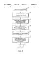
- FIG. 1is a flow chart illustrating processing steps in accordance with one embodiment of the present invention.
- FIG. 2is a flow chart illustrating processing steps for performing the match point generation processing step identified in the flow chart in FIG. 1.
- the flow chart of FIG. 1illustrates a DSA method or process 10 for imaging vasculature, in accordance with one embodiment of the invention.
- Method 10is a locally-adaptive method for sub-pixel registration of mask and opacified digital X-ray images which enables accurate subtraction of the mask image from the opacified image.
- mask image data and opacified image data for a region of interestare obtained prior to executing method 10.
- match point generation 14is performed.
- match point generation 14a set of two-dimensional points in the mask image, and their corresponding points in the opacified image, are derived.
- the procedure outlined in Hannah, "A Description of SRI's Baseline Stereo System", Technical Report Tech. Note 365, SRI International Artificial Intelligence Center, October, 1985, or any other match-point computation method that results in a dense grid of matches between the mask and the opacified images,can be used as step 14.
- locally-adaptive image-to-image warp generation 16is performed. More specifically, using the image-to-image match points generated at step 14, a transformation that maps the matched points in the mask image to their respective corresponding points in the opacified image is generated at step 16 and the generated transformation is applied to the mask image data. Such transformed mask image data is sometimes referred to as warped data.
- the logarithm of the pixel value in the transformed (i.e., warped) mask imageis subtracted from the logarithm of the corresponding pixel value in the opacified image at step 18. Such subtraction is performed for each pixel. Upon completion of such subtraction for the entire image, the resulting data at step 20 represents a DSA image.
- match-point generation 14is performed in accordance with the method described in Gupta-Srinivas, "Image Warping for Accurate Digital Subtraction Angiography", Proc. of AAAI, Spring Symposium on Applications of Computer Vision in Medical Image Processing, Stanford University, March 21-23, 1994.
- FIG. 2is a flow chart illustrating a method or process for performing match point generation 14 of FIG. 1.
- an image hierarchy, or pyramidis generated at step 54 in order to accelerate the computation of match points.
- Such hierarchyis generated by successively reducing both images in the stereo pair to half their size (and resolution) via subsampling using Gaussian convolution.
- the matching processthen begins with the lowest resolution image, i.e., at the bottom of the image pyramid, and works up to images with the highest resolution. More particularly, and starting with the lowest resolution image, a set of interesting points is identified in the mask image at step 56.
- the interesting pointsin the one embodiment, correspond to the pixels with the highest resolution. Points of high local intensity variation are considered interesting because they are easy to match.
- a small tile of imagery around the point in the mask imageis correlated with all tiles in the opacified image. This process proceeds hierarchically from the lowest resolution to the highest resolution. The center of the tile in the opacified image that gives the maximum correlation is identified as the corresponding match point.
- the image tiles in the mask and the opacified imagesmay be rotated or translated with respect to each other.
- the mismatch arising because of such rotationis corrected by a two-dimensional perspective transformation of the mask image tile to the neighborhood of its corresponding tile in the opacified image based, on user-provided rough match points.
- the method described in Gupta-Srinivas, "Image Warping for Accurate Digital Subtraction Angiography", Proc. of AAAI, Spring Symposium on Applications of Computer Vision in Medical Image Processing, Stanford University, March 21-23, 1994,may be used.
- the image-to-image transform for registering the image tiles between the mask and the opacified imagesis generated at step 60.
- the matching processis then repeated, as indicated at step 62, using this new transform which is more accurate than that obtained from user-provided match points.
- the end 64 of the match point generation processis reached, completing the process.
- the corresponding match pointsare identified in the opacified image. Due to patient motion, hysteresis, and other such effects, the grid in the opacified image is not square.
- the displacement (dx,dy) that should be added to each grid point in the mask image to find the coordinates of the corresponding point in the opacified imageis determined.
- dx and dyare considered as separable functions of the mask image coordinate (x, y). That is: ##EQU1## where f and g are two unknown functions whose values are known only at the grid points. The value off and g at any arbitrary point in the mask image is determined by interpolation. Importantly, the two displacements along x and y are treated separately, and the interpolation technique used preserves the values at the grid points.
- Interpolation techniquessuch as general polynomial transformation, bi-cubic or thin-plate splines, or other types of global splines, e.g., the global spline described in Wolberg, "Digital Image Warping", IEEE Computer Society Press, Los Alamitos, Calif., 1990, can be used for performing the interpolation.
- each pixel in the mask imageis transformed by the displacements given by interpolation to find the location of its corresponding pixel in the opacified image.
- the intensity of each pixel in the mask imageis then log-subtracted from its corresponding pixel in the opacified image.
- the above described methodprovides accurate DSA images having fewer artifacts, and is adaptive to correct for any misregistration changes from one part of an image to another part of the image.
- the methodis not limited to the specific implementation described above.
- other hierarchical matching techniquessuch as those described by Quam, "Hierarchical Warp Stereo", in M. A. Fischler and O. Firschein, editors, Readings In Computer Vision, pgs. 80-86, Morgan Kaufmann Publishers, Inc., 1987, can be used in match point generation. While a correlation-based matching scheme is described above, a feature-based matching scheme could alternatively be used.
- processorssuch as, for example, a general purpose computer or an application specific integrated circuit (ASIC) including a microcontroller.
- ASICapplication specific integrated circuit
- the particular type of processor selecteddepends, for example, on the manner in which the image data is provided and other desired operating characteristics.
Landscapes
- Engineering & Computer Science (AREA)
- Physics & Mathematics (AREA)
- General Physics & Mathematics (AREA)
- Theoretical Computer Science (AREA)
- Computer Vision & Pattern Recognition (AREA)
- Apparatus For Radiation Diagnosis (AREA)
- Image Processing (AREA)
Abstract
Description
This invention relates generally to X-ray imaging and, more particularly, to digital subtraction angiography for imaging vasculature.
Digital subtraction angiography (DSA) is a known X-ray procedure for observing vasculature. In one known DSA imaging method, X-ray images of anatomy are taken before and after an X-ray opaque contrast agent is injected into the blood vessels. The X-ray image taken before injecting the contrast agent is sometimes referred to as the mask image and the X-ray image taken after injecting the contrast agent is sometimes referred to as the opacified image. Logarithmic subtraction of the mask image from the opacified image should remove all but the image data associated with the opacified blood vessels.
In principal, therefore, only the opacified vasculature should be visible in a DSA image. However, in practice, it is not unusual for a DSA image to contain artifacts and other image data in addition to the data associated with the opacified vasculature. For example, there typically is a time lag between the acquisition of pre-contrast and post-contrast images. Small patient motions during this interval cause misregistration between the two images, leading to motion artifacts in the final DSA image.
In addition, while certain types of patient motions can be controlled and minimized, several other types of involuntary motions (e.g., cardiac) are much harder to control without major intervention. Such involuntary motion has restricted, or limited, the use of DSA imaging in several anatomical regions. For example, in cardiac procedures, only the opacified images are used.
Further, it is sometimes necessary to move the X-ray imager between pre-contrast and post-contrast exposures. The imager usually cannot be repositioned at the exact same spot as the position in which the first image, i.e., the mask image, has been obtained. Also, mechanical vibrations occur in the structural components of the imager. As a result, the mask image and the opacified image typically are taken under slightly different settings, respectively. In general, the mask and opacified images should be regarded as images taken by two highly correlated, but different, cameras.
Hysteresis also causes differences in the mask image and the opacified image. Particularly, the mask and opacified images will be distorted differently due to changes in the electrical and magnetic environment. For example, during image acquisition, the mask images are acquired in a forward sweep of the imager gantry and opacified images are acquired in a reverse sweep of the gantry. Such electrical and magnetic differences result in artifacts in the DSA image due to a hysteresis effect
In an attempt to address the problems associated with the different conditions under which the mask and opacified images are obtained, many angiography machines allow users to translate the mask image to better register the mask image with to the opacified image. A simple translation, however, can only correct for rigid patient motion aligned with the image plane. Such rigid patient motion, however, is rarely the type of motion which results in image artifacts.
Another known attempt to address such imaging problems is described in Gupta-Srinivas, "Image Warping for Accurate Digital Subtraction Angiography", Proc. of AAAI, Spring Symposium on Applications of Computer Vision in Medical Image Processing, Stanford University, March 21-23, 1994. The algorithm described by Gupta-Srinivas warps the mask image by using a global projective transformation to register the mask image to the opacified image. The global transformation applied to the mask image, however, applies the same transformation to all the pixels in the image. As a result, such algorithm does not correct for any misregistration changes from one part of the image to another part of the image. The causes of subtraction artifacts mentioned above, e.g., patient motion, involuntary motion, change in camera parameters, and hysteresis, generally result in misregistrations in the mask and opacified images which are different for different pixels.
It would be desirable to generate DSA images more accurately. Such accurate images should preferably have minimum artifacts due, e.g., to motion and hysteresis, and should be corrected for any misregistration changes from one part of the image to another part of the image.
Briefly, in accordance with a preferred embodiment of the invention, a locally-adaptive method for sub-pixel registration of mask and opacified digital X-ray images, which enables accurate subtraction of the mask image from the opacified image, includes the steps of generating match points, generating a locally-adaptive image-to-warp image transform, and performing log subtraction for generating a DSA image.
More specifically, in match point generation, a set of two-dimensional points in the mask image and their corresponding points in the opacified image are derived. After performing match point generation, locally-adaptive image-to-image warp generation is performed using the image-to-image match points. That is, a transformation function is generated that maps the matched points in the mask image to their corresponding points in the opacified image. The generated transformation is then applied to the mask image data and the logarithm of the pixel value in the transformed (i.e., warped) mask image is subtracted from the logarithm of the corresponding pixel value in the opacified image. Such subtraction is performed for each pixel. Upon completion of such subtraction for the entire image, the resulting data represents a DSA image.
The above described method provides accurate DSA images having fewer than the usual number of artifacts and is adaptive to correct for any misregistration changes from one part of an image to another part of the image.
FIG. 1 is a flow chart illustrating processing steps in accordance with one embodiment of the present invention.
FIG. 2 is a flow chart illustrating processing steps for performing the match point generation processing step identified in the flow chart in FIG. 1.
The flow chart of FIG. 1 illustrates a DSA method orprocess 10 for imaging vasculature, in accordance with one embodiment of the invention.Method 10 is a locally-adaptive method for sub-pixel registration of mask and opacified digital X-ray images which enables accurate subtraction of the mask image from the opacified image. Prior to executingmethod 10, mask image data and opacified image data for a region of interest are obtained.
After processing has been initiated atstep 12,match point generation 14 is performed. Inmatch point generation 14, a set of two-dimensional points in the mask image, and their corresponding points in the opacified image, are derived. The procedure outlined in Hannah, "A Description of SRI's Baseline Stereo System", Technical Report Tech. Note 365, SRI International Artificial Intelligence Center, October, 1985, or any other match-point computation method that results in a dense grid of matches between the mask and the opacified images, can be used asstep 14.
After performingmatch point generation 14, locally-adaptive image-to-image warp generation 16 is performed. More specifically, using the image-to-image match points generated atstep 14, a transformation that maps the matched points in the mask image to their respective corresponding points in the opacified image is generated atstep 16 and the generated transformation is applied to the mask image data. Such transformed mask image data is sometimes referred to as warped data.
Once the transformation has been applied to the mask image data, the logarithm of the pixel value in the transformed (i.e., warped) mask image is subtracted from the logarithm of the corresponding pixel value in the opacified image atstep 18. Such subtraction is performed for each pixel. Upon completion of such subtraction for the entire image, the resulting data atstep 20 represents a DSA image.
More specifically, in one embodiment ofprocess 10 described above, match-point generation 14 is performed in accordance with the method described in Gupta-Srinivas, "Image Warping for Accurate Digital Subtraction Angiography", Proc. of AAAI, Spring Symposium on Applications of Computer Vision in Medical Image Processing, Stanford University, March 21-23, 1994.
FIG. 2 is a flow chart illustrating a method or process for performingmatch point generation 14 of FIG. 1. Specifically, and as a pre-processingstep following initiation 52 of match point generation, an image hierarchy, or pyramid, is generated atstep 54 in order to accelerate the computation of match points. Such hierarchy is generated by successively reducing both images in the stereo pair to half their size (and resolution) via subsampling using Gaussian convolution.
The matching process then begins with the lowest resolution image, i.e., at the bottom of the image pyramid, and works up to images with the highest resolution. More particularly, and starting with the lowest resolution image, a set of interesting points is identified in the mask image atstep 56. The interesting points, in the one embodiment, correspond to the pixels with the highest resolution. Points of high local intensity variation are considered interesting because they are easy to match.
Initially, only the interesting points in the mask image are matched with their corresponding points in the opacified image, atstep 58. In order to match a point in the mask image to its corresponding point in the opacified image, and in one embodiment, a small tile of imagery around the point in the mask image is correlated with all tiles in the opacified image. This process proceeds hierarchically from the lowest resolution to the highest resolution. The center of the tile in the opacified image that gives the maximum correlation is identified as the corresponding match point.
The image tiles in the mask and the opacified images may be rotated or translated with respect to each other. The mismatch arising because of such rotation is corrected by a two-dimensional perspective transformation of the mask image tile to the neighborhood of its corresponding tile in the opacified image based, on user-provided rough match points. The method described in Gupta-Srinivas, "Image Warping for Accurate Digital Subtraction Angiography", Proc. of AAAI, Spring Symposium on Applications of Computer Vision in Medical Image Processing, Stanford University, March 21-23, 1994, may be used.
Once a set of match points has been computed, the image-to-image transform for registering the image tiles between the mask and the opacified images is generated atstep 60. The matching process is then repeated, as indicated atstep 62, using this new transform which is more accurate than that obtained from user-provided match points.
After processing the highest resolution image data, theend 64 of the match point generation process is reached, completing the process. For a set of points on a square grid in the mask image, the corresponding match points are identified in the opacified image. Due to patient motion, hysteresis, and other such effects, the grid in the opacified image is not square.
Once a grid of match points has been found, the displacement (dx,dy) that should be added to each grid point in the mask image to find the coordinates of the corresponding point in the opacified image is determined. Specifically, dx and dy are considered as separable functions of the mask image coordinate (x, y). That is: ##EQU1## where f and g are two unknown functions whose values are known only at the grid points. The value off and g at any arbitrary point in the mask image is determined by interpolation. Importantly, the two displacements along x and y are treated separately, and the interpolation technique used preserves the values at the grid points. Interpolation techniques such as general polynomial transformation, bi-cubic or thin-plate splines, or other types of global splines, e.g., the global spline described in Wolberg, "Digital Image Warping", IEEE Computer Society Press, Los Alamitos, Calif., 1990, can be used for performing the interpolation.
The location of each pixel in the mask image is transformed by the displacements given by interpolation to find the location of its corresponding pixel in the opacified image. The intensity of each pixel in the mask image is then log-subtracted from its corresponding pixel in the opacified image.
The above described method provides accurate DSA images having fewer artifacts, and is adaptive to correct for any misregistration changes from one part of an image to another part of the image. The method, moreover, is not limited to the specific implementation described above. For example, other hierarchical matching techniques, such as those described by Quam, "Hierarchical Warp Stereo", in M. A. Fischler and O. Firschein, editors, Readings In Computer Vision, pgs. 80-86, Morgan Kaufmann Publishers, Inc., 1987, can be used in match point generation. While a correlation-based matching scheme is described above, a feature-based matching scheme could alternatively be used.
The above described methods can be implemented by various types of processors such as, for example, a general purpose computer or an application specific integrated circuit (ASIC) including a microcontroller. The particular type of processor selected depends, for example, on the manner in which the image data is provided and other desired operating characteristics.
While only certain preferred features of the invention have been illustrated and described, many modifications and changes will occur to those skilled in the art. It is, therefore, to be understood that the appended claims are intended to cover all such modifications and changes as fall within the true spirit of the invention.
Claims (10)
1. A method for generating a digital subtraction angiography image from mask image data and opacified image data, comprising the steps of:
generating match points between interesting points in the mask image data and the opacified image data;
generating a locally-adaptive global image transform using the generated match points;
applying the global transform to the mask image data to generate warp image data; and
subtracting the logarithm of a warp image data value from the logarithm of the corresponding opacified image data value.
2. The method of claim 1 wherein the steps of generating match points comprises generating a set of two-dimensional points in the mask image data, and identifying corresponding points in the opacified image data.
3. The method of claim 1 wherein the steps of generating match points comprises:
generating an image resolution data hierarchy;
selecting the lowest resolution image data; and
for the selected image data:
(a) identifying a set of interesting points;
(b) matching the interesting points in the mask image data with corresponding points in the opacified image;
(c) determining an image-to-image transform for registering image tiles between the mask and the opacified images; and
(i) if the highest resolution image data has not been selected, selecting the next highest resolution image data and repeating steps (a)-(c); and
(ii) if the highest resolution image data has been selected, ending the match point generation.
4. The method of claim 3 wherein the image resolution data hierarchy is generated by successively reducing both the mask image and the opacified image in a stereo pair to about half their original size with subsampling using Gaussian convolution.
5. The method of claim 3 wherein each of the set of interesting points corresponds to a respective one of the pixels with the highest resolution.
6. The method of claim 3 wherein the step of matching the interesting points in the mask image data with corresponding points in the opacified image comprises correlating a small tile of imagery around a point in the mask image with all of the tiles in the opacified image and identifying the center of the tile in the opacified image in order to determine the maximum correlation as the corresponding match point.
7. The method of claim 1 wherein the step of generating match points comprises identifying a grid of match points, and said method further comprises determining a displacement (dx,dy) that should be added to each grid point in the mask image to find coordinates of the corresponding point in the opacified image.
8. The method of claim 7 wherein dx and dy are related by: ##EQU2## where f and g are two unknown functions whose values are known only at grid points.
9. The method of claim 8 further comprising the step of determining values of f and g at an arbitrary point in the mask image by interpolation.
10. The method of claim 9 wherein the location of each pixel in the mask image is transformed by the displacements given by interpolation to find the corresponding pixel in the opacified image.
Priority Applications (5)
| Application Number | Priority Date | Filing Date | Title |
|---|---|---|---|
| US08/738,860US5848121A (en) | 1996-10-28 | 1996-10-28 | Method and apparatus for digital subtraction angiography |
| JP9290205AJPH10191167A (en) | 1996-10-28 | 1997-10-23 | Method for generating digital subtraction angiography image |
| EP97308569AEP0840253B1 (en) | 1996-10-28 | 1997-10-28 | Methods and apparatus for digital subtraction angiography |
| DE69733238TDE69733238T2 (en) | 1996-10-28 | 1997-10-28 | Method and apparatus for digital subtraction angiography |
| US09/186,029US6154518A (en) | 1996-10-28 | 1998-11-04 | Three dimensional locally adaptive warping for volumetric registration of images |
Applications Claiming Priority (1)
| Application Number | Priority Date | Filing Date | Title |
|---|---|---|---|
| US08/738,860US5848121A (en) | 1996-10-28 | 1996-10-28 | Method and apparatus for digital subtraction angiography |
Related Child Applications (1)
| Application Number | Title | Priority Date | Filing Date |
|---|---|---|---|
| US09/186,029Continuation-In-PartUS6154518A (en) | 1996-10-28 | 1998-11-04 | Three dimensional locally adaptive warping for volumetric registration of images |
Publications (1)
| Publication Number | Publication Date |
|---|---|
| US5848121Atrue US5848121A (en) | 1998-12-08 |
Family
ID=24969798
Family Applications (2)
| Application Number | Title | Priority Date | Filing Date |
|---|---|---|---|
| US08/738,860Expired - LifetimeUS5848121A (en) | 1996-10-28 | 1996-10-28 | Method and apparatus for digital subtraction angiography |
| US09/186,029Expired - LifetimeUS6154518A (en) | 1996-10-28 | 1998-11-04 | Three dimensional locally adaptive warping for volumetric registration of images |
Family Applications After (1)
| Application Number | Title | Priority Date | Filing Date |
|---|---|---|---|
| US09/186,029Expired - LifetimeUS6154518A (en) | 1996-10-28 | 1998-11-04 | Three dimensional locally adaptive warping for volumetric registration of images |
Country Status (4)
| Country | Link |
|---|---|
| US (2) | US5848121A (en) |
| EP (1) | EP0840253B1 (en) |
| JP (1) | JPH10191167A (en) |
| DE (1) | DE69733238T2 (en) |
Cited By (68)
| Publication number | Priority date | Publication date | Assignee | Title |
|---|---|---|---|---|
| US20020156368A1 (en)* | 1999-01-21 | 2002-10-24 | Satoru Ohishi | X-ray diagnosis apparatus including a collimater |
| US20020167537A1 (en)* | 2001-05-11 | 2002-11-14 | Miroslav Trajkovic | Motion-based tracking with pan-tilt-zoom camera |
| US20030053670A1 (en)* | 2001-09-04 | 2003-03-20 | Hauper Sylvain Justin Georges Andre | Method of processing images for digital subtraction angiography |
| US6549798B2 (en) | 2001-02-07 | 2003-04-15 | Epix Medical, Inc. | Magnetic resonance angiography data |
| US6678399B2 (en)* | 2001-11-23 | 2004-01-13 | University Of Chicago | Subtraction technique for computerized detection of small lung nodules in computer tomography images |
| US6718054B1 (en) | 1999-06-23 | 2004-04-06 | Massachusetts Institute Of Technology | MRA segmentation using active contour models |
| US20050065430A1 (en)* | 2003-07-10 | 2005-03-24 | Andrea Wiethoff | Methods of cardiothoracic imaging - (MET-30) |
| US20050107691A1 (en)* | 2000-04-07 | 2005-05-19 | The General Hospital Corporation | Methods for digital bowel subtraction and polyp detection |
| US20050171420A1 (en)* | 2004-01-29 | 2005-08-04 | Siemens Aktiengesellschaft | Method and medical imaging system for compensating for patient motion |
| US20060239585A1 (en)* | 2005-04-04 | 2006-10-26 | Valadez Gerardo H | System and method for reducing artifacts in motion corrected dynamic image sequences |
| US20070016015A1 (en)* | 2005-07-15 | 2007-01-18 | Siemens Aktiengesellschaft | Method for the visualization of a vscular insert |
| US20080037844A1 (en)* | 2006-08-14 | 2008-02-14 | Siemens Medical Solutions Usa, Inc. | Precomputed Automatic Pixel Shift for Review of Digital Subtracted Angiography |
| US7374536B1 (en) | 2004-04-16 | 2008-05-20 | Taylor Colin R | Method for analysis of pain images |
| US20090268984A1 (en)* | 2008-04-29 | 2009-10-29 | Adobe Systems Incorporated | Subpixel Registration |
| US20100080435A1 (en)* | 2008-09-29 | 2010-04-01 | Guendel Lutz | Method and apparatus for registering tomographic volume data records of the intestine |
| US20120155737A1 (en)* | 2010-12-20 | 2012-06-21 | The Johns Hopkins University | Image processing apparatus and image processing method |
| CN101208721B (en)* | 2005-08-02 | 2012-07-04 | 卡西欧计算机株式会社 | Image processing device and image processing method |
| US20140016844A1 (en)* | 2012-07-10 | 2014-01-16 | Zakrytoe Akcionernoe Obshchestvo "Impul's" | Method for acquisition of subtraction angiograms |
| US9123100B2 (en)* | 2007-11-20 | 2015-09-01 | Olea Medical | Method and system for processing multiple series of biological images obtained from a patient |
| US9286673B2 (en) | 2012-10-05 | 2016-03-15 | Volcano Corporation | Systems for correcting distortions in a medical image and methods of use thereof |
| US9292918B2 (en) | 2012-10-05 | 2016-03-22 | Volcano Corporation | Methods and systems for transforming luminal images |
| US9301687B2 (en) | 2013-03-13 | 2016-04-05 | Volcano Corporation | System and method for OCT depth calibration |
| US9307926B2 (en) | 2012-10-05 | 2016-04-12 | Volcano Corporation | Automatic stent detection |
| US9324141B2 (en) | 2012-10-05 | 2016-04-26 | Volcano Corporation | Removal of A-scan streaking artifact |
| US9360630B2 (en) | 2011-08-31 | 2016-06-07 | Volcano Corporation | Optical-electrical rotary joint and methods of use |
| US9367965B2 (en) | 2012-10-05 | 2016-06-14 | Volcano Corporation | Systems and methods for generating images of tissue |
| US9383263B2 (en) | 2012-12-21 | 2016-07-05 | Volcano Corporation | Systems and methods for narrowing a wavelength emission of light |
| US9478940B2 (en) | 2012-10-05 | 2016-10-25 | Volcano Corporation | Systems and methods for amplifying light |
| US9486143B2 (en) | 2012-12-21 | 2016-11-08 | Volcano Corporation | Intravascular forward imaging device |
| US9596993B2 (en) | 2007-07-12 | 2017-03-21 | Volcano Corporation | Automatic calibration systems and methods of use |
| US9612105B2 (en) | 2012-12-21 | 2017-04-04 | Volcano Corporation | Polarization sensitive optical coherence tomography system |
| US9622706B2 (en) | 2007-07-12 | 2017-04-18 | Volcano Corporation | Catheter for in vivo imaging |
| US9709379B2 (en) | 2012-12-20 | 2017-07-18 | Volcano Corporation | Optical coherence tomography system that is reconfigurable between different imaging modes |
| US9730613B2 (en) | 2012-12-20 | 2017-08-15 | Volcano Corporation | Locating intravascular images |
| US9770172B2 (en) | 2013-03-07 | 2017-09-26 | Volcano Corporation | Multimodal segmentation in intravascular images |
| US9858668B2 (en) | 2012-10-05 | 2018-01-02 | Volcano Corporation | Guidewire artifact removal in images |
| US9867530B2 (en) | 2006-08-14 | 2018-01-16 | Volcano Corporation | Telescopic side port catheter device with imaging system and method for accessing side branch occlusions |
| US10058284B2 (en) | 2012-12-21 | 2018-08-28 | Volcano Corporation | Simultaneous imaging, monitoring, and therapy |
| US10070827B2 (en) | 2012-10-05 | 2018-09-11 | Volcano Corporation | Automatic image playback |
| US10166003B2 (en) | 2012-12-21 | 2019-01-01 | Volcano Corporation | Ultrasound imaging with variable line density |
| US10191220B2 (en) | 2012-12-21 | 2019-01-29 | Volcano Corporation | Power-efficient optical circuit |
| US10219887B2 (en) | 2013-03-14 | 2019-03-05 | Volcano Corporation | Filters with echogenic characteristics |
| US10219780B2 (en) | 2007-07-12 | 2019-03-05 | Volcano Corporation | OCT-IVUS catheter for concurrent luminal imaging |
| US10226597B2 (en) | 2013-03-07 | 2019-03-12 | Volcano Corporation | Guidewire with centering mechanism |
| US10238367B2 (en) | 2012-12-13 | 2019-03-26 | Volcano Corporation | Devices, systems, and methods for targeted cannulation |
| US10258301B2 (en) | 2015-01-05 | 2019-04-16 | Koninklijke Philips N.V. | Digital subtraction angiography |
| US10292677B2 (en) | 2013-03-14 | 2019-05-21 | Volcano Corporation | Endoluminal filter having enhanced echogenic properties |
| US10332228B2 (en) | 2012-12-21 | 2019-06-25 | Volcano Corporation | System and method for graphical processing of medical data |
| US10413317B2 (en) | 2012-12-21 | 2019-09-17 | Volcano Corporation | System and method for catheter steering and operation |
| US10420530B2 (en) | 2012-12-21 | 2019-09-24 | Volcano Corporation | System and method for multipath processing of image signals |
| US10426590B2 (en) | 2013-03-14 | 2019-10-01 | Volcano Corporation | Filters with echogenic characteristics |
| US10568586B2 (en) | 2012-10-05 | 2020-02-25 | Volcano Corporation | Systems for indicating parameters in an imaging data set and methods of use |
| US10595820B2 (en) | 2012-12-20 | 2020-03-24 | Philips Image Guided Therapy Corporation | Smooth transition catheters |
| US10638939B2 (en) | 2013-03-12 | 2020-05-05 | Philips Image Guided Therapy Corporation | Systems and methods for diagnosing coronary microvascular disease |
| US10724082B2 (en) | 2012-10-22 | 2020-07-28 | Bio-Rad Laboratories, Inc. | Methods for analyzing DNA |
| US10758207B2 (en) | 2013-03-13 | 2020-09-01 | Philips Image Guided Therapy Corporation | Systems and methods for producing an image from a rotational intravascular ultrasound device |
| US10939885B2 (en) | 2016-12-15 | 2021-03-09 | Koninklijke Philips N.V. | Visualizing vascular structures |
| US10939826B2 (en) | 2012-12-20 | 2021-03-09 | Philips Image Guided Therapy Corporation | Aspirating and removing biological material |
| US10942022B2 (en) | 2012-12-20 | 2021-03-09 | Philips Image Guided Therapy Corporation | Manual calibration of imaging system |
| US10993694B2 (en) | 2012-12-21 | 2021-05-04 | Philips Image Guided Therapy Corporation | Rotational ultrasound imaging catheter with extended catheter body telescope |
| US11026591B2 (en) | 2013-03-13 | 2021-06-08 | Philips Image Guided Therapy Corporation | Intravascular pressure sensor calibration |
| US11040140B2 (en) | 2010-12-31 | 2021-06-22 | Philips Image Guided Therapy Corporation | Deep vein thrombosis therapeutic methods |
| US11141063B2 (en) | 2010-12-23 | 2021-10-12 | Philips Image Guided Therapy Corporation | Integrated system architectures and methods of use |
| US11154313B2 (en) | 2013-03-12 | 2021-10-26 | The Volcano Corporation | Vibrating guidewire torquer and methods of use |
| US11272845B2 (en) | 2012-10-05 | 2022-03-15 | Philips Image Guided Therapy Corporation | System and method for instant and automatic border detection |
| US11406498B2 (en) | 2012-12-20 | 2022-08-09 | Philips Image Guided Therapy Corporation | Implant delivery system and implants |
| US12201477B2 (en) | 2012-10-05 | 2025-01-21 | Philips Image Guided Therapy Corporation | Methods and systems for establishing parameters for three-dimensional imaging |
| US12343198B2 (en) | 2013-03-14 | 2025-07-01 | Philips Image Guided Therapy Corporation | Delivery catheter having imaging capabilities |
Families Citing this family (29)
| Publication number | Priority date | Publication date | Assignee | Title |
|---|---|---|---|---|
| US6611615B1 (en)* | 1999-06-25 | 2003-08-26 | University Of Iowa Research Foundation | Method and apparatus for generating consistent image registration |
| AU6673600A (en)* | 1999-08-09 | 2001-03-05 | University Of British Columbia, The | Method and automated system for creating volumetric data sets |
| FR2799028B1 (en) | 1999-09-27 | 2002-05-03 | Ge Medical Syst Sa | METHOD FOR RECONSTRUCTING A THREE-DIMENSIONAL IMAGE OF ELEMENTS OF STRONG CONTRAST |
| US6909794B2 (en)* | 2000-11-22 | 2005-06-21 | R2 Technology, Inc. | Automated registration of 3-D medical scans of similar anatomical structures |
| US7072436B2 (en)* | 2001-08-24 | 2006-07-04 | The Board Of Trustees Of The Leland Stanford Junior University | Volumetric computed tomography (VCT) |
| FR2830962B1 (en)* | 2001-10-12 | 2004-01-30 | Inst Nat Rech Inf Automat | IMAGE PROCESSING DEVICE AND METHOD FOR DETECTION OF EVOLUTIVE LESIONS |
| US6738063B2 (en) | 2002-02-07 | 2004-05-18 | Siemens Corporate Research, Inc. | Object-correspondence identification without full volume registration |
| DE10210050A1 (en) | 2002-03-07 | 2003-12-04 | Siemens Ag | Method and device for repetitive relative positioning of a patient |
| GB2390792B (en)* | 2002-07-08 | 2005-08-31 | Vision Rt Ltd | Image processing system for use with a patient positioning device |
| US7074188B2 (en)* | 2002-08-26 | 2006-07-11 | The Cleveland Clinic Foundation | System and method of characterizing vascular tissue |
| US7787927B2 (en)* | 2003-06-20 | 2010-08-31 | Merge Cad Inc. | System and method for adaptive medical image registration |
| DE102004059182A1 (en) | 2004-12-08 | 2006-06-14 | Siemens Ag | Operating method for a computer and corresponding devices |
| US7605821B1 (en)* | 2005-09-29 | 2009-10-20 | Adobe Systems Incorporated | Poisson image-editing technique that matches texture contrast |
| WO2007054907A1 (en)* | 2005-11-10 | 2007-05-18 | Philips Intellectual Property & Standards Gmbh | Adaptive point-based elastic image registration |
| US8125498B2 (en)* | 2007-01-03 | 2012-02-28 | Siemens Medical Solutions Usa, Inc. | Generating a 3D volumetric mask from a closed surface mesh |
| US8285014B2 (en)* | 2007-04-06 | 2012-10-09 | Siemens Aktiengesellschaft | Measuring blood volume with C-arm computed tomography |
| US9367904B2 (en) | 2007-04-23 | 2016-06-14 | Koninklijke Philips Electronics N.V. | Spatial-temporal warping of different pre-captured medical images |
| US8094903B2 (en)* | 2007-06-28 | 2012-01-10 | Siemens Aktiengesellschaft | System and method for coronary digital subtraction angiography |
| WO2010015957A1 (en)* | 2008-08-04 | 2010-02-11 | Koninklijke Philips Electronics, N.V. | Automatic pre-alignment for registration of medical images |
| US8345944B2 (en)* | 2008-08-06 | 2013-01-01 | Siemens Aktiengesellschaft | System and method for coronary digital subtraction angiography |
| US8179460B2 (en)* | 2008-09-22 | 2012-05-15 | Aptina Imaging Corporation | System, method, and apparatus for variable rate pixel data transfer and storage |
| US8355557B2 (en)* | 2008-10-10 | 2013-01-15 | Siemens Aktiengesellschaft | System and method for decomposed temporal filtering for X-ray guided intervention application |
| JP5523791B2 (en) | 2008-10-27 | 2014-06-18 | 株式会社東芝 | X-ray diagnostic apparatus and image processing apparatus |
| CN102223843B (en)* | 2008-11-26 | 2013-10-23 | 皇家飞利浦电子股份有限公司 | Visualization of the Coronary Tree |
| JP5355074B2 (en)* | 2008-12-26 | 2013-11-27 | キヤノン株式会社 | 3D shape data processing apparatus, 3D shape data processing method and program |
| US8774481B2 (en)* | 2010-03-25 | 2014-07-08 | Emory University | Atlas-assisted synthetic computed tomography using deformable image registration |
| US8798227B2 (en)* | 2010-10-15 | 2014-08-05 | Kabushiki Kaisha Toshiba | Medical image processing apparatus and X-ray computed tomography apparatus |
| KR101880634B1 (en)* | 2011-08-09 | 2018-08-16 | 삼성전자주식회사 | Method and apparatus for generating 3d volume panorama |
| GB201502877D0 (en) | 2015-02-20 | 2015-04-08 | Cydar Ltd | Digital image remapping |
Citations (4)
| Publication number | Priority date | Publication date | Assignee | Title |
|---|---|---|---|---|
| US5048103A (en)* | 1989-03-29 | 1991-09-10 | General Electric Cgr S.A. | Method for the automatic resetting of images |
| US5210415A (en)* | 1990-04-04 | 1993-05-11 | Fuji Photo Film Co., Ltd. | Method and apparatus for forming energy subtraction images |
| US5647360A (en)* | 1995-06-30 | 1997-07-15 | Siemens Corporate Research, Inc. | Digital subtraction angiography for 3D diagnostic imaging |
| US5690106A (en)* | 1995-06-30 | 1997-11-25 | Siemens Corporate Research, Inc. | Flexible image registration for rotational angiography |
Family Cites Families (6)
| Publication number | Priority date | Publication date | Assignee | Title |
|---|---|---|---|---|
| CA2051939A1 (en)* | 1990-10-02 | 1992-04-03 | Gary A. Ransford | Digital data registration and differencing compression system |
| US5568384A (en)* | 1992-10-13 | 1996-10-22 | Mayo Foundation For Medical Education And Research | Biomedical imaging and analysis |
| EP0599345B1 (en)* | 1992-11-27 | 2002-06-05 | Fuji Photo Film Co., Ltd. | Method for adjusting positions of radiation images |
| WO1994018639A1 (en)* | 1993-02-09 | 1994-08-18 | Siemens Medical Systems, Inc. | Method and apparatus for generating well-registered subtraction images for digital subtraction angiography |
| JPH07152895A (en)* | 1993-11-29 | 1995-06-16 | Canon Inc | Image processing method and apparatus |
| US5839440A (en)* | 1994-06-17 | 1998-11-24 | Siemens Corporate Research, Inc. | Three-dimensional image registration method for spiral CT angiography |
- 1996
- 1996-10-28USUS08/738,860patent/US5848121A/ennot_activeExpired - Lifetime
- 1997
- 1997-10-23JPJP9290205Apatent/JPH10191167A/ennot_activeWithdrawn
- 1997-10-28EPEP97308569Apatent/EP0840253B1/ennot_activeExpired - Lifetime
- 1997-10-28DEDE69733238Tpatent/DE69733238T2/ennot_activeExpired - Lifetime
- 1998
- 1998-11-04USUS09/186,029patent/US6154518A/ennot_activeExpired - Lifetime
Patent Citations (4)
| Publication number | Priority date | Publication date | Assignee | Title |
|---|---|---|---|---|
| US5048103A (en)* | 1989-03-29 | 1991-09-10 | General Electric Cgr S.A. | Method for the automatic resetting of images |
| US5210415A (en)* | 1990-04-04 | 1993-05-11 | Fuji Photo Film Co., Ltd. | Method and apparatus for forming energy subtraction images |
| US5647360A (en)* | 1995-06-30 | 1997-07-15 | Siemens Corporate Research, Inc. | Digital subtraction angiography for 3D diagnostic imaging |
| US5690106A (en)* | 1995-06-30 | 1997-11-25 | Siemens Corporate Research, Inc. | Flexible image registration for rotational angiography |
Non-Patent Citations (4)
| Title |
|---|
| L.H. Quam, Hierarchial warp stereo, in M.A. Fischler and O. Firschein, editors, Readings in Computer Vision, pp. 80 86. Morgan Kaufman Publishers, Inc., 1987.* |
| L.H. Quam, Hierarchial warp stereo, in M.A. Fischler and O. Firschein, editors, Readings in Computer Vision, pp. 80-86. Morgan Kaufman Publishers, Inc., 1987. |
| R. Gupta et al., "Image Warping for Accurate Digital Subtraction Angiography", American Association for Artificial Intelligence, Spring Symposium Series, Mar. 21-23 1994, Stanford University, California. |
| R. Gupta et al., Image Warping for Accurate Digital Subtraction Angiography , American Association for Artificial Intelligence, Spring Symposium Series, Mar. 21 23 1994, Stanford University, California.* |
Cited By (93)
| Publication number | Priority date | Publication date | Assignee | Title |
|---|---|---|---|---|
| US20020156368A1 (en)* | 1999-01-21 | 2002-10-24 | Satoru Ohishi | X-ray diagnosis apparatus including a collimater |
| US7761136B2 (en) | 1999-01-21 | 2010-07-20 | Kabushiki Kaisha Toshiba | Medical image processing apparatus for scanning based on a set three-dimensional region of interest |
| US6721590B2 (en)* | 1999-01-21 | 2004-04-13 | Kabushiki Kaisha Toshiba | Medical image processing apparatus |
| US7505549B2 (en)* | 1999-01-21 | 2009-03-17 | Kabushiki Kaisha Toshiba | X-ray diagnosis apparatus |
| US6718054B1 (en) | 1999-06-23 | 2004-04-06 | Massachusetts Institute Of Technology | MRA segmentation using active contour models |
| US6947784B2 (en) | 2000-04-07 | 2005-09-20 | The General Hospital Corporation | System for digital bowel subtraction and polyp detection and related techniques |
| US7630529B2 (en) | 2000-04-07 | 2009-12-08 | The General Hospital Corporation | Methods for digital bowel subtraction and polyp detection |
| US20050107691A1 (en)* | 2000-04-07 | 2005-05-19 | The General Hospital Corporation | Methods for digital bowel subtraction and polyp detection |
| US6549798B2 (en) | 2001-02-07 | 2003-04-15 | Epix Medical, Inc. | Magnetic resonance angiography data |
| US6925321B2 (en) | 2001-02-07 | 2005-08-02 | Epix Pharmaceuticals, Inc. | Magnetic resonance angiography data |
| US20060116568A1 (en)* | 2001-02-07 | 2006-06-01 | Epix Pharmaceuticals, Inc., A Delaware Corporation | Magnetic resonance angiography data |
| US20020167537A1 (en)* | 2001-05-11 | 2002-11-14 | Miroslav Trajkovic | Motion-based tracking with pan-tilt-zoom camera |
| US7233689B2 (en)* | 2001-09-04 | 2007-06-19 | Koninklijke Philips Electronics N.V. | Method of processing images for digital subtraction angiography |
| US20030053670A1 (en)* | 2001-09-04 | 2003-03-20 | Hauper Sylvain Justin Georges Andre | Method of processing images for digital subtraction angiography |
| WO2003046831A3 (en)* | 2001-11-23 | 2004-01-22 | Univ Chicago | A novel subtraction technique for computerized detection of small lung nodules in computer tomography images |
| US6678399B2 (en)* | 2001-11-23 | 2004-01-13 | University Of Chicago | Subtraction technique for computerized detection of small lung nodules in computer tomography images |
| US20050065430A1 (en)* | 2003-07-10 | 2005-03-24 | Andrea Wiethoff | Methods of cardiothoracic imaging - (MET-30) |
| US20050171420A1 (en)* | 2004-01-29 | 2005-08-04 | Siemens Aktiengesellschaft | Method and medical imaging system for compensating for patient motion |
| US7421061B2 (en)* | 2004-01-29 | 2008-09-02 | Siemens Aktiengesellschaft | Method and medical imaging system for compensating for patient motion |
| US7374536B1 (en) | 2004-04-16 | 2008-05-20 | Taylor Colin R | Method for analysis of pain images |
| US20060239585A1 (en)* | 2005-04-04 | 2006-10-26 | Valadez Gerardo H | System and method for reducing artifacts in motion corrected dynamic image sequences |
| US20070016015A1 (en)* | 2005-07-15 | 2007-01-18 | Siemens Aktiengesellschaft | Method for the visualization of a vscular insert |
| CN101208721B (en)* | 2005-08-02 | 2012-07-04 | 卡西欧计算机株式会社 | Image processing device and image processing method |
| US8077952B2 (en) | 2006-08-14 | 2011-12-13 | Siemens Medical Solutions Usa, Inc. | Precomputed automatic pixel shift for review of digital subtracted angiography |
| US9867530B2 (en) | 2006-08-14 | 2018-01-16 | Volcano Corporation | Telescopic side port catheter device with imaging system and method for accessing side branch occlusions |
| US20080037844A1 (en)* | 2006-08-14 | 2008-02-14 | Siemens Medical Solutions Usa, Inc. | Precomputed Automatic Pixel Shift for Review of Digital Subtracted Angiography |
| US10219780B2 (en) | 2007-07-12 | 2019-03-05 | Volcano Corporation | OCT-IVUS catheter for concurrent luminal imaging |
| US11350906B2 (en) | 2007-07-12 | 2022-06-07 | Philips Image Guided Therapy Corporation | OCT-IVUS catheter for concurrent luminal imaging |
| US9596993B2 (en) | 2007-07-12 | 2017-03-21 | Volcano Corporation | Automatic calibration systems and methods of use |
| US9622706B2 (en) | 2007-07-12 | 2017-04-18 | Volcano Corporation | Catheter for in vivo imaging |
| US9123100B2 (en)* | 2007-11-20 | 2015-09-01 | Olea Medical | Method and system for processing multiple series of biological images obtained from a patient |
| US20090268984A1 (en)* | 2008-04-29 | 2009-10-29 | Adobe Systems Incorporated | Subpixel Registration |
| US8055101B2 (en) | 2008-04-29 | 2011-11-08 | Adobe Systems Incorporated | Subpixel registration |
| US8467586B2 (en)* | 2008-09-29 | 2013-06-18 | Siemens Aktiengesellschaft | Method and apparatus for registering tomographic volume data records of the intestine |
| US20100080435A1 (en)* | 2008-09-29 | 2010-04-01 | Guendel Lutz | Method and apparatus for registering tomographic volume data records of the intestine |
| US9072490B2 (en)* | 2010-12-20 | 2015-07-07 | Toshiba Medical Systems Corporation | Image processing apparatus and image processing method |
| US20120155737A1 (en)* | 2010-12-20 | 2012-06-21 | The Johns Hopkins University | Image processing apparatus and image processing method |
| US11141063B2 (en) | 2010-12-23 | 2021-10-12 | Philips Image Guided Therapy Corporation | Integrated system architectures and methods of use |
| US11040140B2 (en) | 2010-12-31 | 2021-06-22 | Philips Image Guided Therapy Corporation | Deep vein thrombosis therapeutic methods |
| US9360630B2 (en) | 2011-08-31 | 2016-06-07 | Volcano Corporation | Optical-electrical rotary joint and methods of use |
| US20140016844A1 (en)* | 2012-07-10 | 2014-01-16 | Zakrytoe Akcionernoe Obshchestvo "Impul's" | Method for acquisition of subtraction angiograms |
| US12201477B2 (en) | 2012-10-05 | 2025-01-21 | Philips Image Guided Therapy Corporation | Methods and systems for establishing parameters for three-dimensional imaging |
| US9367965B2 (en) | 2012-10-05 | 2016-06-14 | Volcano Corporation | Systems and methods for generating images of tissue |
| US9286673B2 (en) | 2012-10-05 | 2016-03-15 | Volcano Corporation | Systems for correcting distortions in a medical image and methods of use thereof |
| US11864870B2 (en) | 2012-10-05 | 2024-01-09 | Philips Image Guided Therapy Corporation | System and method for instant and automatic border detection |
| US9478940B2 (en) | 2012-10-05 | 2016-10-25 | Volcano Corporation | Systems and methods for amplifying light |
| US9292918B2 (en) | 2012-10-05 | 2016-03-22 | Volcano Corporation | Methods and systems for transforming luminal images |
| US11510632B2 (en) | 2012-10-05 | 2022-11-29 | Philips Image Guided Therapy Corporation | Systems for indicating parameters in an imaging data set and methods of use |
| US10568586B2 (en) | 2012-10-05 | 2020-02-25 | Volcano Corporation | Systems for indicating parameters in an imaging data set and methods of use |
| US9858668B2 (en) | 2012-10-05 | 2018-01-02 | Volcano Corporation | Guidewire artifact removal in images |
| US11890117B2 (en) | 2012-10-05 | 2024-02-06 | Philips Image Guided Therapy Corporation | Systems for indicating parameters in an imaging data set and methods of use |
| US11272845B2 (en) | 2012-10-05 | 2022-03-15 | Philips Image Guided Therapy Corporation | System and method for instant and automatic border detection |
| US10070827B2 (en) | 2012-10-05 | 2018-09-11 | Volcano Corporation | Automatic image playback |
| US9324141B2 (en) | 2012-10-05 | 2016-04-26 | Volcano Corporation | Removal of A-scan streaking artifact |
| US9307926B2 (en) | 2012-10-05 | 2016-04-12 | Volcano Corporation | Automatic stent detection |
| US12226189B2 (en) | 2012-10-05 | 2025-02-18 | Philips Image Guided Therapy Corporation | System and method for instant and automatic border detection |
| US10724082B2 (en) | 2012-10-22 | 2020-07-28 | Bio-Rad Laboratories, Inc. | Methods for analyzing DNA |
| US10238367B2 (en) | 2012-12-13 | 2019-03-26 | Volcano Corporation | Devices, systems, and methods for targeted cannulation |
| US9709379B2 (en) | 2012-12-20 | 2017-07-18 | Volcano Corporation | Optical coherence tomography system that is reconfigurable between different imaging modes |
| US10595820B2 (en) | 2012-12-20 | 2020-03-24 | Philips Image Guided Therapy Corporation | Smooth transition catheters |
| US11892289B2 (en) | 2012-12-20 | 2024-02-06 | Philips Image Guided Therapy Corporation | Manual calibration of imaging system |
| US9730613B2 (en) | 2012-12-20 | 2017-08-15 | Volcano Corporation | Locating intravascular images |
| US11406498B2 (en) | 2012-12-20 | 2022-08-09 | Philips Image Guided Therapy Corporation | Implant delivery system and implants |
| US11141131B2 (en) | 2012-12-20 | 2021-10-12 | Philips Image Guided Therapy Corporation | Smooth transition catheters |
| US10942022B2 (en) | 2012-12-20 | 2021-03-09 | Philips Image Guided Therapy Corporation | Manual calibration of imaging system |
| US10939826B2 (en) | 2012-12-20 | 2021-03-09 | Philips Image Guided Therapy Corporation | Aspirating and removing biological material |
| US10191220B2 (en) | 2012-12-21 | 2019-01-29 | Volcano Corporation | Power-efficient optical circuit |
| US11253225B2 (en) | 2012-12-21 | 2022-02-22 | Philips Image Guided Therapy Corporation | System and method for multipath processing of image signals |
| US9383263B2 (en) | 2012-12-21 | 2016-07-05 | Volcano Corporation | Systems and methods for narrowing a wavelength emission of light |
| US9486143B2 (en) | 2012-12-21 | 2016-11-08 | Volcano Corporation | Intravascular forward imaging device |
| US9612105B2 (en) | 2012-12-21 | 2017-04-04 | Volcano Corporation | Polarization sensitive optical coherence tomography system |
| US11786213B2 (en) | 2012-12-21 | 2023-10-17 | Philips Image Guided Therapy Corporation | System and method for multipath processing of image signals |
| US10332228B2 (en) | 2012-12-21 | 2019-06-25 | Volcano Corporation | System and method for graphical processing of medical data |
| US10993694B2 (en) | 2012-12-21 | 2021-05-04 | Philips Image Guided Therapy Corporation | Rotational ultrasound imaging catheter with extended catheter body telescope |
| US10413317B2 (en) | 2012-12-21 | 2019-09-17 | Volcano Corporation | System and method for catheter steering and operation |
| US10058284B2 (en) | 2012-12-21 | 2018-08-28 | Volcano Corporation | Simultaneous imaging, monitoring, and therapy |
| US10420530B2 (en) | 2012-12-21 | 2019-09-24 | Volcano Corporation | System and method for multipath processing of image signals |
| US10166003B2 (en) | 2012-12-21 | 2019-01-01 | Volcano Corporation | Ultrasound imaging with variable line density |
| US10226597B2 (en) | 2013-03-07 | 2019-03-12 | Volcano Corporation | Guidewire with centering mechanism |
| US9770172B2 (en) | 2013-03-07 | 2017-09-26 | Volcano Corporation | Multimodal segmentation in intravascular images |
| US10638939B2 (en) | 2013-03-12 | 2020-05-05 | Philips Image Guided Therapy Corporation | Systems and methods for diagnosing coronary microvascular disease |
| US12350018B2 (en) | 2013-03-12 | 2025-07-08 | Philips Image Guided Therapy Corporation | Systems and methods for diagnosing coronary microvascular disease |
| US11154313B2 (en) | 2013-03-12 | 2021-10-26 | The Volcano Corporation | Vibrating guidewire torquer and methods of use |
| US10758207B2 (en) | 2013-03-13 | 2020-09-01 | Philips Image Guided Therapy Corporation | Systems and methods for producing an image from a rotational intravascular ultrasound device |
| US11026591B2 (en) | 2013-03-13 | 2021-06-08 | Philips Image Guided Therapy Corporation | Intravascular pressure sensor calibration |
| US9301687B2 (en) | 2013-03-13 | 2016-04-05 | Volcano Corporation | System and method for OCT depth calibration |
| US10426590B2 (en) | 2013-03-14 | 2019-10-01 | Volcano Corporation | Filters with echogenic characteristics |
| US10292677B2 (en) | 2013-03-14 | 2019-05-21 | Volcano Corporation | Endoluminal filter having enhanced echogenic properties |
| US10219887B2 (en) | 2013-03-14 | 2019-03-05 | Volcano Corporation | Filters with echogenic characteristics |
| US12343198B2 (en) | 2013-03-14 | 2025-07-01 | Philips Image Guided Therapy Corporation | Delivery catheter having imaging capabilities |
| US10258301B2 (en) | 2015-01-05 | 2019-04-16 | Koninklijke Philips N.V. | Digital subtraction angiography |
| US11607186B2 (en) | 2016-12-15 | 2023-03-21 | Koninklijke Philips N.V. | Visualizing vascular structures |
| US10939885B2 (en) | 2016-12-15 | 2021-03-09 | Koninklijke Philips N.V. | Visualizing vascular structures |
Also Published As
| Publication number | Publication date |
|---|---|
| EP0840253B1 (en) | 2005-05-11 |
| DE69733238T2 (en) | 2006-02-09 |
| EP0840253A3 (en) | 1998-11-18 |
| JPH10191167A (en) | 1998-07-21 |
| US6154518A (en) | 2000-11-28 |
| EP0840253A2 (en) | 1998-05-06 |
| DE69733238D1 (en) | 2005-06-16 |
Similar Documents
| Publication | Publication Date | Title |
|---|---|---|
| US5848121A (en) | Method and apparatus for digital subtraction angiography | |
| JP3761094B2 (en) | Method for reconstructing a three-dimensional image of an object | |
| Bergen et al. | A three-frame algorithm for estimating two-component image motion | |
| JP4104054B2 (en) | Image alignment apparatus and image processing apparatus | |
| Van Tran et al. | Flexible mask subtraction for digital angiography | |
| JP2000051204A (en) | Method for reconstructing three-dimensional picture of object | |
| CN103544690A (en) | Method for acquisition of subtraction angiograms | |
| JPH11232464A (en) | Method for reconstituting three-dimensional object from closed loop sequence of uncorrected image taken by camera | |
| CN110211193B (en) | 3D CT inter-slice image interpolation restoration and super-resolution processing method and device | |
| Cox et al. | Automatic registration of temporal image pairs for digital subtraction angiography | |
| US12400345B2 (en) | Method and device for matching three-dimensional oral scan data via deep-learning based 3D feature detection | |
| Yang et al. | Multiresolution elastic registration of X-ray angiography images using thin-plate spline | |
| JPH0759763A (en) | 3D object measuring device | |
| JP2002109538A (en) | Method and device for aligning image | |
| CN112184569B (en) | Image restoration method and image restoration device | |
| CN112837392A (en) | CT image generation method, device, system, electronic device and storage medium | |
| WO2009040497A1 (en) | Image enhancement method | |
| Wang et al. | Depth-layer-based patient motion compensation for the overlay of 3D volumes onto X-ray sequences | |
| Tan et al. | Deformable Template Tracking in 1ms. | |
| Parchami et al. | A comparative study on 3-D stereo reconstruction from endoscopic images | |
| Gupta et al. | Image Warping for Accurate Digital Subtraction Angiography | |
| JP2004081424A (en) | Radiation imaging equipment | |
| KR102556057B1 (en) | Method and apparatus for reconstructing multi-view volumetric using local perspective warping | |
| Gregory et al. | The three dimensional reconstruction and monitoring of facial surfaces | |
| Meijering et al. | A fast technique for motion correction in DAS using a feature-based, irregular grid |
Legal Events
| Date | Code | Title | Description |
|---|---|---|---|
| AS | Assignment | Owner name:GENERAL ELECTRIC COMPANY, NEW YORK Free format text:ASSIGNMENT OF ASSIGNORS INTEREST;ASSIGNORS:GUPTA, RAJIV;SRINIVAS, CHUKKA;REEL/FRAME:008292/0468;SIGNING DATES FROM 19961011 TO 19961017 | |
| FEPP | Fee payment procedure | Free format text:PAYOR NUMBER ASSIGNED (ORIGINAL EVENT CODE: ASPN); ENTITY STATUS OF PATENT OWNER: LARGE ENTITY | |
| STCF | Information on status: patent grant | Free format text:PATENTED CASE | |
| FPAY | Fee payment | Year of fee payment:4 | |
| FPAY | Fee payment | Year of fee payment:8 | |
| FPAY | Fee payment | Year of fee payment:12 |