US5846796A - Blood-borne mesenchymal cells - Google Patents
Blood-borne mesenchymal cellsDownload PDFInfo
- Publication number
- US5846796A US5846796AUS08/488,241US48824195AUS5846796AUS 5846796 AUS5846796 AUS 5846796AUS 48824195 AUS48824195 AUS 48824195AUS 5846796 AUS5846796 AUS 5846796A
- Authority
- US
- United States
- Prior art keywords
- cells
- blood
- mesenchymal cells
- borne
- cell
- Prior art date
- Legal status (The legal status is an assumption and is not a legal conclusion. Google has not performed a legal analysis and makes no representation as to the accuracy of the status listed.)
- Expired - Lifetime
Links
- 210000004027cellAnatomy0.000claimsabstractdescription264
- 210000002950fibroblastAnatomy0.000claimsabstractdescription19
- 210000005259peripheral bloodAnatomy0.000claimsabstractdescription14
- 239000011886peripheral bloodSubstances0.000claimsabstractdescription14
- 210000004962mammalian cellAnatomy0.000claimsabstractdescription6
- 238000000034methodMethods0.000claimsdescription44
- 108090000623proteins and genesProteins0.000claimsdescription42
- 230000014509gene expressionEffects0.000claimsdescription12
- 102100031573Hematopoietic progenitor cell antigen CD34Human genes0.000claimsdescription4
- 101000777663Homo sapiens Hematopoietic progenitor cell antigen CD34Proteins0.000claimsdescription4
- 101000738771Homo sapiens Receptor-type tyrosine-protein phosphatase CProteins0.000claimsdescription3
- 102100037422Receptor-type tyrosine-protein phosphatase CHuman genes0.000claimsdescription3
- 239000013604expression vectorSubstances0.000claimsdescription3
- 210000003958hematopoietic stem cellAnatomy0.000claimsdescription3
- 230000001105regulatory effectEffects0.000claims1
- 208000027418Wounds and injuryDiseases0.000abstractdescription30
- 206010052428WoundDiseases0.000abstractdescription28
- 210000004369bloodAnatomy0.000abstractdescription22
- 239000008280bloodSubstances0.000abstractdescription22
- 230000029663wound healingEffects0.000abstractdescription22
- 238000002955isolationMethods0.000abstractdescription13
- 210000000265leukocyteAnatomy0.000abstractdescription9
- 238000012512characterization methodMethods0.000abstractdescription7
- 238000001727in vivoMethods0.000abstractdescription7
- 210000002808connective tissueAnatomy0.000abstractdescription6
- 230000007838tissue remodelingEffects0.000abstractdescription6
- 230000004071biological effectEffects0.000abstractdescription5
- 238000001415gene therapyMethods0.000abstractdescription4
- 238000010171animal modelMethods0.000abstractdescription3
- LOKCTEFSRHRXRJ-UHFFFAOYSA-Idipotassium trisodium dihydrogen phosphate hydrogen phosphate dichlorideChemical compoundP(=O)(O)(O)[O-].[K+].P(=O)(O)([O-])[O-].[Na+].[Na+].[Cl-].[K+].[Cl-].[Na+]LOKCTEFSRHRXRJ-UHFFFAOYSA-I0.000description22
- 239000002953phosphate buffered salineSubstances0.000description22
- 102000004127CytokinesHuman genes0.000description20
- 108090000695CytokinesProteins0.000description20
- 108091003079Bovine Serum AlbuminProteins0.000description17
- 108010017213Granulocyte-Macrophage Colony-Stimulating FactorProteins0.000description16
- 102100039620Granulocyte-macrophage colony-stimulating factorHuman genes0.000description16
- 210000001519tissueAnatomy0.000description14
- LFQSCWFLJHTTHZ-UHFFFAOYSA-NEthanolChemical compoundCCOLFQSCWFLJHTTHZ-UHFFFAOYSA-N0.000description12
- 229940098773bovine serum albuminDrugs0.000description11
- 238000000338in vitroMethods0.000description8
- 238000004519manufacturing processMethods0.000description8
- 230000007774longtermEffects0.000description7
- 230000008569processEffects0.000description7
- 239000000047productSubstances0.000description7
- 102000004169proteins and genesHuman genes0.000description7
- 102000013127VimentinHuman genes0.000description6
- 108010065472VimentinProteins0.000description6
- 238000004458analytical methodMethods0.000description6
- 239000000427antigenSubstances0.000description6
- 102000036639antigensHuman genes0.000description6
- 108091007433antigensProteins0.000description6
- 239000012091fetal bovine serumSubstances0.000description6
- 230000004044responseEffects0.000description6
- 210000005048vimentinAnatomy0.000description6
- 102000008186CollagenHuman genes0.000description5
- 108010035532CollagenProteins0.000description5
- 102000016359FibronectinsHuman genes0.000description5
- 108010067306FibronectinsProteins0.000description5
- 230000001464adherent effectEffects0.000description5
- 229920001436collagenPolymers0.000description5
- 238000012258culturingMethods0.000description5
- 238000001943fluorescence-activated cell sortingMethods0.000description5
- 239000003102growth factorSubstances0.000description5
- 238000010166immunofluorescenceMethods0.000description5
- 239000003550markerSubstances0.000description5
- 239000002609mediumSubstances0.000description5
- YBJHBAHKTGYVGT-ZKWXMUAHSA-N(+)-BiotinChemical compoundN1C(=O)N[C@@H]2[C@H](CCCCC(=O)O)SC[C@@H]21YBJHBAHKTGYVGT-ZKWXMUAHSA-N0.000description4
- 102000012422Collagen Type IHuman genes0.000description4
- 108010022452Collagen Type IProteins0.000description4
- 239000006144Dulbecco’s modified Eagle's mediumSubstances0.000description4
- CEAZRRDELHUEMR-URQXQFDESA-NGentamicinChemical compoundO1[C@H](C(C)NC)CC[C@@H](N)[C@H]1O[C@H]1[C@H](O)[C@@H](O[C@@H]2[C@@H]([C@@H](NC)[C@@](C)(O)CO2)O)[C@H](N)C[C@@H]1NCEAZRRDELHUEMR-URQXQFDESA-N0.000description4
- 229930182566GentamicinNatural products0.000description4
- 241000699670Mus sp.Species0.000description4
- 238000013459approachMethods0.000description4
- 230000001413cellular effectEffects0.000description4
- 239000003795chemical substances by applicationSubstances0.000description4
- 230000006870functionEffects0.000description4
- 229960002518gentamicinDrugs0.000description4
- 230000012010growthEffects0.000description4
- 208000015181infectious diseaseDiseases0.000description4
- 210000004698lymphocyteAnatomy0.000description4
- 210000004379membraneAnatomy0.000description4
- 239000012528membraneSubstances0.000description4
- 210000001616monocyteAnatomy0.000description4
- 230000000877morphologic effectEffects0.000description4
- 210000000056organAnatomy0.000description4
- 238000000926separation methodMethods0.000description4
- 210000000329smooth muscle myocyteAnatomy0.000description4
- 241000701161unidentified adenovirusSpecies0.000description4
- 102000007469ActinsHuman genes0.000description3
- 108010085238ActinsProteins0.000description3
- 102000018233Fibroblast Growth FactorHuman genes0.000description3
- 108050007372Fibroblast Growth FactorProteins0.000description3
- WSFSSNUMVMOOMR-UHFFFAOYSA-NFormaldehydeChemical compoundO=CWSFSSNUMVMOOMR-UHFFFAOYSA-N0.000description3
- PEDCQBHIVMGVHV-UHFFFAOYSA-NGlycerineChemical compoundOCC(O)COPEDCQBHIVMGVHV-UHFFFAOYSA-N0.000description3
- 241001529936MurinaeSpecies0.000description3
- 241000699666Mus <mouse, genus>Species0.000description3
- 102000010780Platelet-Derived Growth FactorHuman genes0.000description3
- 108010038512Platelet-Derived Growth FactorProteins0.000description3
- 102000004887Transforming Growth Factor betaHuman genes0.000description3
- 108090001012Transforming Growth Factor betaProteins0.000description3
- 241000700605VirusesSpecies0.000description3
- 238000005299abrasionMethods0.000description3
- 238000001042affinity chromatographyMethods0.000description3
- 210000003719b-lymphocyteAnatomy0.000description3
- 230000006378damageEffects0.000description3
- 210000002919epithelial cellAnatomy0.000description3
- 229940126864fibroblast growth factorDrugs0.000description3
- MHMNJMPURVTYEJ-UHFFFAOYSA-Nfluorescein-5-isothiocyanateChemical compoundO1C(=O)C2=CC(N=C=S)=CC=C2C21C1=CC=C(O)C=C1OC1=CC(O)=CC=C21MHMNJMPURVTYEJ-UHFFFAOYSA-N0.000description3
- 239000007850fluorescent dyeSubstances0.000description3
- 239000012634fragmentSubstances0.000description3
- 239000000499gelSubstances0.000description3
- 208000014674injuryDiseases0.000description3
- 230000010354integrationEffects0.000description3
- 230000005012migrationEffects0.000description3
- 238000013508migrationMethods0.000description3
- 230000004048modificationEffects0.000description3
- 238000012986modificationMethods0.000description3
- 239000013612plasmidSubstances0.000description3
- 238000002360preparation methodMethods0.000description3
- 230000008439repair processEffects0.000description3
- PYWVYCXTNDRMGF-UHFFFAOYSA-Nrhodamine BChemical compound[Cl-].C=12C=CC(=[N+](CC)CC)C=C2OC2=CC(N(CC)CC)=CC=C2C=1C1=CC=CC=C1C(O)=OPYWVYCXTNDRMGF-UHFFFAOYSA-N0.000description3
- 230000003248secreting effectEffects0.000description3
- 210000003491skinAnatomy0.000description3
- 238000001356surgical procedureMethods0.000description3
- MPLHNVLQVRSVEE-UHFFFAOYSA-Ntexas redChemical compound[O-]S(=O)(=O)C1=CC(S(Cl)(=O)=O)=CC=C1C(C1=CC=2CCCN3CCCC(C=23)=C1O1)=C2C1=C(CCC1)C3=[N+]1CCCC3=C2MPLHNVLQVRSVEE-UHFFFAOYSA-N0.000description3
- 238000013518transcriptionMethods0.000description3
- 230000035897transcriptionEffects0.000description3
- 239000013598vectorSubstances0.000description3
- 230000003442weekly effectEffects0.000description3
- 230000037314wound repairEffects0.000description3
- 108090001008AvidinProteins0.000description2
- 108020004414DNAProteins0.000description2
- 102100036912DesminHuman genes0.000description2
- 108010044052DesminProteins0.000description2
- YCAGGFXSFQFVQL-UHFFFAOYSA-NEndothionChemical compoundCOC1=COC(CSP(=O)(OC)OC)=CC1=OYCAGGFXSFQFVQL-UHFFFAOYSA-N0.000description2
- 102000010834Extracellular Matrix ProteinsHuman genes0.000description2
- 108010037362Extracellular Matrix ProteinsProteins0.000description2
- 206010016654FibrosisDiseases0.000description2
- 101000581981Homo sapiens Neural cell adhesion molecule 1Proteins0.000description2
- SIKJAQJRHWYJAI-UHFFFAOYSA-NIndoleChemical compoundC1=CC=C2NC=CC2=C1SIKJAQJRHWYJAI-UHFFFAOYSA-N0.000description2
- 102000011782KeratinsHuman genes0.000description2
- 108010076876KeratinsProteins0.000description2
- 241001465754MetazoaSpecies0.000description2
- 102100027347Neural cell adhesion molecule 1Human genes0.000description2
- 102000052812Ornithine decarboxylasesHuman genes0.000description2
- 108700005126Ornithine decarboxylasesProteins0.000description2
- 239000004793PolystyreneSubstances0.000description2
- ZTHYODDOHIVTJV-UHFFFAOYSA-NPropyl gallateChemical compoundCCCOC(=O)C1=CC(O)=C(O)C(O)=C1ZTHYODDOHIVTJV-UHFFFAOYSA-N0.000description2
- 108091008874T cell receptorsProteins0.000description2
- 102000016266T-Cell Antigen ReceptorsHuman genes0.000description2
- 210000001744T-lymphocyteAnatomy0.000description2
- 108060008682Tumor Necrosis FactorProteins0.000description2
- 208000025865UlcerDiseases0.000description2
- 239000002671adjuvantSubstances0.000description2
- 239000011324beadSubstances0.000description2
- 238000004166bioassayMethods0.000description2
- 229960002685biotinDrugs0.000description2
- 235000020958biotinNutrition0.000description2
- 239000011616biotinSubstances0.000description2
- 210000000601blood cellAnatomy0.000description2
- 210000001185bone marrowAnatomy0.000description2
- 238000004113cell cultureMethods0.000description2
- 238000001516cell proliferation assayMethods0.000description2
- 239000003153chemical reaction reagentSubstances0.000description2
- 238000004587chromatography analysisMethods0.000description2
- 210000000349chromosomeAnatomy0.000description2
- 230000001684chronic effectEffects0.000description2
- 239000002299complementary DNASubstances0.000description2
- 230000000875corresponding effectEffects0.000description2
- 210000004748cultured cellAnatomy0.000description2
- 230000016396cytokine productionEffects0.000description2
- 238000000432density-gradient centrifugationMethods0.000description2
- 210000005045desminAnatomy0.000description2
- 208000037265diseases, disorders, signs and symptomsDiseases0.000description2
- 238000012137double-stainingMethods0.000description2
- VLCYCQAOQCDTCN-UHFFFAOYSA-NeflornithineChemical compoundNCCCC(N)(C(F)F)C(O)=OVLCYCQAOQCDTCN-UHFFFAOYSA-N0.000description2
- 238000000799fluorescence microscopyMethods0.000description2
- 239000012737fresh mediumSubstances0.000description2
- 210000004408hybridomaAnatomy0.000description2
- 230000001939inductive effectEffects0.000description2
- 230000002757inflammatory effectEffects0.000description2
- 238000002347injectionMethods0.000description2
- 239000007924injectionSubstances0.000description2
- 238000001990intravenous administrationMethods0.000description2
- 210000002540macrophageAnatomy0.000description2
- 239000000463materialSubstances0.000description2
- 239000011159matrix materialSubstances0.000description2
- WSFSSNUMVMOOMR-NJFSPNSNSA-NmethanoneChemical compoundO=[14CH2]WSFSSNUMVMOOMR-NJFSPNSNSA-N0.000description2
- 238000010232migration assayMethods0.000description2
- 239000000203mixtureSubstances0.000description2
- 210000000440neutrophilAnatomy0.000description2
- 210000005105peripheral blood lymphocyteAnatomy0.000description2
- 239000004033plasticSubstances0.000description2
- 229920003023plasticPolymers0.000description2
- 229920002223polystyrenePolymers0.000description2
- 230000035755proliferationEffects0.000description2
- 230000001737promoting effectEffects0.000description2
- 238000011002quantificationMethods0.000description2
- 238000002415sodium dodecyl sulfate polyacrylamide gel electrophoresisMethods0.000description2
- 239000007787solidSubstances0.000description2
- 230000010473stable expressionEffects0.000description2
- 238000010186stainingMethods0.000description2
- 238000007920subcutaneous administrationMethods0.000description2
- 239000000126substanceSubstances0.000description2
- ZRKFYGHZFMAOKI-QMGMOQQFSA-NtgfbetaChemical compoundC([C@H](NC(=O)[C@H](C(C)C)NC(=O)CNC(=O)[C@H](CCC(O)=O)NC(=O)[C@H](CCCNC(N)=N)NC(=O)[C@H](CC(N)=O)NC(=O)[C@H](CC(C)C)NC(=O)[C@H]([C@@H](C)O)NC(=O)[C@H](CCC(O)=O)NC(=O)[C@H]([C@@H](C)O)NC(=O)[C@H](CC(C)C)NC(=O)CNC(=O)[C@H](C)NC(=O)[C@H](CO)NC(=O)[C@H](CCC(N)=O)NC(=O)[C@@H](NC(=O)[C@H](C)NC(=O)[C@H](C)NC(=O)[C@@H](NC(=O)[C@H](CC(C)C)NC(=O)[C@@H](N)CCSC)C(C)C)[C@@H](C)CC)C(=O)N[C@@H]([C@@H](C)O)C(=O)N[C@@H](C(C)C)C(=O)N[C@@H](CC=1C=CC=CC=1)C(=O)N[C@@H](C)C(=O)N1[C@@H](CCC1)C(=O)N[C@@H]([C@@H](C)O)C(=O)N[C@@H](CC(N)=O)C(=O)N[C@@H](CCC(O)=O)C(=O)N[C@@H](C)C(=O)N[C@@H](CC=1C=CC=CC=1)C(=O)N[C@@H](CCCNC(N)=N)C(=O)N[C@@H](C)C(=O)N[C@@H](CC(C)C)C(=O)N1[C@@H](CCC1)C(=O)N1[C@@H](CCC1)C(=O)N[C@@H](CCCNC(N)=N)C(=O)N[C@@H](CCC(O)=O)C(=O)N[C@@H](CCCNC(N)=N)C(=O)N[C@@H](CO)C(=O)N[C@@H](CCCNC(N)=N)C(=O)N[C@@H](CC(C)C)C(=O)N[C@@H](CC(C)C)C(O)=O)C1=CC=C(O)C=C1ZRKFYGHZFMAOKI-QMGMOQQFSA-N0.000description2
- 230000001225therapeutic effectEffects0.000description2
- 230000017423tissue regenerationEffects0.000description2
- 238000012546transferMethods0.000description2
- 102000003390tumor necrosis factorHuman genes0.000description2
- 231100000397ulcerToxicity0.000description2
- 230000003612virological effectEffects0.000description2
- 229960001134von willebrand factorDrugs0.000description2
- ASWBNKHCZGQVJV-UHFFFAOYSA-N(3-hexadecanoyloxy-2-hydroxypropyl) 2-(trimethylazaniumyl)ethyl phosphateChemical compoundCCCCCCCCCCCCCCCC(=O)OCC(O)COP([O-])(=O)OCC[N+](C)(C)CASWBNKHCZGQVJV-UHFFFAOYSA-N0.000description1
- UFBJCMHMOXMLKC-UHFFFAOYSA-N2,4-dinitrophenolChemical compoundOC1=CC=C([N+]([O-])=O)C=C1[N+]([O-])=OUFBJCMHMOXMLKC-UHFFFAOYSA-N0.000description1
- 102100031585ADP-ribosyl cyclase/cyclic ADP-ribose hydrolase 1Human genes0.000description1
- 102100029457Adenine phosphoribosyltransferaseHuman genes0.000description1
- 108010024223Adenine phosphoribosyltransferaseProteins0.000description1
- 102100022749Aminopeptidase NHuman genes0.000description1
- 102100024222B-lymphocyte antigen CD19Human genes0.000description1
- 241000701822Bovine papillomavirusSpecies0.000description1
- 102100032912CD44 antigenHuman genes0.000description1
- 108010001857Cell Surface ReceptorsProteins0.000description1
- 102000000844Cell Surface ReceptorsHuman genes0.000description1
- 108010012236ChemokinesProteins0.000description1
- 102000019034ChemokinesHuman genes0.000description1
- 208000017667Chronic DiseaseDiseases0.000description1
- 108091026890Coding regionProteins0.000description1
- 241000186216CorynebacteriumSpecies0.000description1
- 241000699800CricetinaeSpecies0.000description1
- 101150074155DHFR geneProteins0.000description1
- 102000004190EnzymesHuman genes0.000description1
- 108090000790EnzymesProteins0.000description1
- YQYJSBFKSSDGFO-UHFFFAOYSA-NEpihygromycinNatural productsOC1C(O)C(C(=O)C)OC1OC(C(=C1)O)=CC=C1C=C(C)C(=O)NC1C(O)C(O)C2OCOC2C1OYQYJSBFKSSDGFO-UHFFFAOYSA-N0.000description1
- 108010054218Factor VIIIProteins0.000description1
- 102000001690Factor VIIIHuman genes0.000description1
- 206010020118HistiocytosesDiseases0.000description1
- 101000777636Homo sapiens ADP-ribosyl cyclase/cyclic ADP-ribose hydrolase 1Proteins0.000description1
- 101000757160Homo sapiens Aminopeptidase NProteins0.000description1
- 101000980825Homo sapiens B-lymphocyte antigen CD19Proteins0.000description1
- 101000868273Homo sapiens CD44 antigenProteins0.000description1
- 101001046686Homo sapiens Integrin alpha-MProteins0.000description1
- 101000599852Homo sapiens Intercellular adhesion molecule 1Proteins0.000description1
- 101000917858Homo sapiens Low affinity immunoglobulin gamma Fc region receptor III-AProteins0.000description1
- 101000917839Homo sapiens Low affinity immunoglobulin gamma Fc region receptor III-BProteins0.000description1
- 101000946889Homo sapiens Monocyte differentiation antigen CD14Proteins0.000description1
- 101000934338Homo sapiens Myeloid cell surface antigen CD33Proteins0.000description1
- 101100321817Human parvovirus B19 (strain HV) 7.5K geneProteins0.000description1
- 108010091358Hypoxanthine PhosphoribosyltransferaseProteins0.000description1
- 102100029098Hypoxanthine-guanine phosphoribosyltransferaseHuman genes0.000description1
- -1IIIProteins0.000description1
- 102000001706Immunoglobulin Fab FragmentsHuman genes0.000description1
- 108010054477Immunoglobulin Fab FragmentsProteins0.000description1
- 102000008394Immunoglobulin FragmentsHuman genes0.000description1
- 108010021625Immunoglobulin FragmentsProteins0.000description1
- 102100022338Integrin alpha-MHuman genes0.000description1
- 102100022297Integrin alpha-XHuman genes0.000description1
- 102100037877Intercellular adhesion molecule 1Human genes0.000description1
- 108010002352Interleukin-1Proteins0.000description1
- 108090001005Interleukin-6Proteins0.000description1
- 108090001007Interleukin-8Proteins0.000description1
- 108010063738InterleukinsProteins0.000description1
- 102000015696InterleukinsHuman genes0.000description1
- FBOZXECLQNJBKD-ZDUSSCGKSA-NL-methotrexateChemical compoundC=1N=C2N=C(N)N=C(N)C2=NC=1CN(C)C1=CC=C(C(=O)N[C@@H](CCC(O)=O)C(O)=O)C=C1FBOZXECLQNJBKD-ZDUSSCGKSA-N0.000description1
- QIVBCDIJIAJPQS-VIFPVBQESA-NL-tryptophaneChemical compoundC1=CC=C2C(C[C@H](N)C(O)=O)=CNC2=C1QIVBCDIJIAJPQS-VIFPVBQESA-N0.000description1
- 108090001090LectinsProteins0.000description1
- 102000004856LectinsHuman genes0.000description1
- 102100029185Low affinity immunoglobulin gamma Fc region receptor III-BHuman genes0.000description1
- 102000008072LymphokinesHuman genes0.000description1
- 108010074338LymphokinesProteins0.000description1
- 102000043131MHC class II familyHuman genes0.000description1
- 108091054438MHC class II familyProteins0.000description1
- 101100261636Methanothermobacter marburgensis (strain ATCC BAA-927 / DSM 2133 / JCM 14651 / NBRC 100331 / OCM 82 / Marburg) trpB2 geneProteins0.000description1
- 102100035877Monocyte differentiation antigen CD14Human genes0.000description1
- 102100025243Myeloid cell surface antigen CD33Human genes0.000description1
- 241001045988NeogeneSpecies0.000description1
- 208000001388Opportunistic InfectionsDiseases0.000description1
- 229940122060Ornithine decarboxylase inhibitorDrugs0.000description1
- 241000283973Oryctolagus cuniculusSpecies0.000description1
- 206010033372Pain and discomfortDiseases0.000description1
- 102000057297Pepsin AHuman genes0.000description1
- 108090000284Pepsin AProteins0.000description1
- 101100124346Photorhabdus laumondii subsp. laumondii (strain DSM 15139 / CIP 105565 / TT01) hisCD geneProteins0.000description1
- 241000276498Pollachius virensSpecies0.000description1
- 239000004372Polyvinyl alcoholSubstances0.000description1
- 241000700159RattusSpecies0.000description1
- 108020004511Recombinant DNAProteins0.000description1
- 102000007056Recombinant Fusion ProteinsHuman genes0.000description1
- 108010008281Recombinant Fusion ProteinsProteins0.000description1
- 206010039710SclerodermaDiseases0.000description1
- 241000700584SimplexvirusSpecies0.000description1
- 108010090804StreptavidinProteins0.000description1
- 108090000190ThrombinProteins0.000description1
- 102000006601Thymidine KinaseHuman genes0.000description1
- 108020004440Thymidine kinaseProteins0.000description1
- QIVBCDIJIAJPQS-UHFFFAOYSA-NTryptophanNatural productsC1=CC=C2C(CC(N)C(O)=O)=CNC2=C1QIVBCDIJIAJPQS-UHFFFAOYSA-N0.000description1
- 241000700618Vaccinia virusSpecies0.000description1
- 230000009471actionEffects0.000description1
- 239000013543active substanceSubstances0.000description1
- 230000000735allogeneic effectEffects0.000description1
- WNROFYMDJYEPJX-UHFFFAOYSA-Kaluminium hydroxideChemical compound[OH-].[OH-].[OH-].[Al+3]WNROFYMDJYEPJX-UHFFFAOYSA-K0.000description1
- 229940126575aminoglycosideDrugs0.000description1
- 230000003367anti-collagen effectEffects0.000description1
- 230000000340anti-metaboliteEffects0.000description1
- 229930185229antidesminNatural products0.000description1
- 230000000890antigenic effectEffects0.000description1
- 229940100197antimetaboliteDrugs0.000description1
- 239000002256antimetaboliteSubstances0.000description1
- 238000000149argon plasma sinteringMethods0.000description1
- 230000003305autocrineEffects0.000description1
- 230000004888barrier functionEffects0.000description1
- 238000012742biochemical analysisMethods0.000description1
- 230000008512biological responseEffects0.000description1
- 230000006696biosynthetic metabolic pathwayEffects0.000description1
- 230000015572biosynthetic processEffects0.000description1
- 210000004204blood vesselAnatomy0.000description1
- 210000000988bone and boneAnatomy0.000description1
- 239000003710calcium ionophoreSubstances0.000description1
- 210000000845cartilageAnatomy0.000description1
- 230000012292cell migrationEffects0.000description1
- 230000004663cell proliferationEffects0.000description1
- 239000002458cell surface markerSubstances0.000description1
- 230000035605chemotaxisEffects0.000description1
- 208000019425cirrhosis of liverDiseases0.000description1
- 230000035602clottingEffects0.000description1
- 239000003086colorantSubstances0.000description1
- 238000004891communicationMethods0.000description1
- 230000000295complement effectEffects0.000description1
- 210000001608connective tissue cellAnatomy0.000description1
- 238000007796conventional methodMethods0.000description1
- 230000002596correlated effectEffects0.000description1
- 239000002537cosmeticSubstances0.000description1
- 238000004132cross linkingMethods0.000description1
- 238000012136culture methodMethods0.000description1
- 239000012228culture supernatantSubstances0.000description1
- 231100000599cytotoxic agentToxicity0.000description1
- 239000002619cytotoxinSubstances0.000description1
- 230000007123defenseEffects0.000description1
- 210000004443dendritic cellAnatomy0.000description1
- 210000004207dermisAnatomy0.000description1
- 230000001066destructive effectEffects0.000description1
- 238000001514detection methodMethods0.000description1
- 230000004069differentiationEffects0.000description1
- 230000029087digestionEffects0.000description1
- 201000010099diseaseDiseases0.000description1
- BFMYDTVEBKDAKJ-UHFFFAOYSA-Ldisodium;(2',7'-dibromo-3',6'-dioxido-3-oxospiro[2-benzofuran-1,9'-xanthene]-4'-yl)mercury;hydrateChemical compoundO.[Na+].[Na+].O1C(=O)C2=CC=CC=C2C21C1=CC(Br)=C([O-])C([Hg])=C1OC1=C2C=C(Br)C([O-])=C1BFMYDTVEBKDAKJ-UHFFFAOYSA-L0.000description1
- 208000035475disorderDiseases0.000description1
- 231100000673dose–response relationshipToxicity0.000description1
- 230000000694effectsEffects0.000description1
- 239000000839emulsionSubstances0.000description1
- 210000002889endothelial cellAnatomy0.000description1
- 230000003511endothelial effectEffects0.000description1
- 239000002158endotoxinSubstances0.000description1
- 239000003623enhancerSubstances0.000description1
- 230000002708enhancing effectEffects0.000description1
- 229940088598enzymeDrugs0.000description1
- 210000003743erythrocyteAnatomy0.000description1
- 238000002474experimental methodMethods0.000description1
- 210000002744extracellular matrixAnatomy0.000description1
- 229960000301factor viiiDrugs0.000description1
- 230000001605fetal effectEffects0.000description1
- 230000004761fibrosisEffects0.000description1
- 238000000684flow cytometryMethods0.000description1
- 239000012530fluidSubstances0.000description1
- 238000012757fluorescence stainingMethods0.000description1
- 210000001156gastric mucosaAnatomy0.000description1
- 238000010353genetic engineeringMethods0.000description1
- 239000011521glassSubstances0.000description1
- 210000003714granulocyteAnatomy0.000description1
- 239000000122growth hormoneSubstances0.000description1
- 239000001963growth mediumSubstances0.000description1
- 210000005003heart tissueAnatomy0.000description1
- 101150113423hisD geneProteins0.000description1
- HNDVDQJCIGZPNO-UHFFFAOYSA-NhistidineNatural productsOC(=O)C(N)CC1=CN=CN1HNDVDQJCIGZPNO-UHFFFAOYSA-N0.000description1
- 201000008298histiocytosisDiseases0.000description1
- 229940088597hormoneDrugs0.000description1
- 210000005260human cellAnatomy0.000description1
- 238000007654immersionMethods0.000description1
- 230000028993immune responseEffects0.000description1
- 230000000984immunochemical effectEffects0.000description1
- 238000010185immunofluorescence analysisMethods0.000description1
- 238000002513implantationMethods0.000description1
- PZOUSPYUWWUPPK-UHFFFAOYSA-NindoleNatural productsCC1=CC=CC2=C1C=CN2PZOUSPYUWWUPPK-UHFFFAOYSA-N0.000description1
- RKJUIXBNRJVNHR-UHFFFAOYSA-NindolenineNatural productsC1=CC=C2CC=NC2=C1RKJUIXBNRJVNHR-UHFFFAOYSA-N0.000description1
- 239000012678infectious agentSubstances0.000description1
- 230000004968inflammatory conditionEffects0.000description1
- 208000027866inflammatory diseaseDiseases0.000description1
- 230000005764inhibitory processEffects0.000description1
- 229910052500inorganic mineralInorganic materials0.000description1
- 238000003780insertionMethods0.000description1
- 230000037431insertionEffects0.000description1
- 230000003993interactionEffects0.000description1
- 229940047122interleukinsDrugs0.000description1
- 238000007918intramuscular administrationMethods0.000description1
- 238000004255ion exchange chromatographyMethods0.000description1
- 238000001155isoelectric focusingMethods0.000description1
- 208000011379keloid formationDiseases0.000description1
- 108010045069keyhole-limpet hemocyaninProteins0.000description1
- 238000002372labellingMethods0.000description1
- 239000002523lectinSubstances0.000description1
- 210000003041ligamentAnatomy0.000description1
- 210000004185liverAnatomy0.000description1
- 238000007885magnetic separationMethods0.000description1
- 229960000485methotrexateDrugs0.000description1
- HPNSFSBZBAHARI-UHFFFAOYSA-Nmicophenolic acidNatural productsOC1=C(CC=C(C)CCC(O)=O)C(OC)=C(C)C2=C1C(=O)OC2HPNSFSBZBAHARI-UHFFFAOYSA-N0.000description1
- 244000005700microbiomeSpecies0.000description1
- 238000000520microinjectionMethods0.000description1
- 239000011707mineralSubstances0.000description1
- 238000010369molecular cloningMethods0.000description1
- 210000005087mononuclear cellAnatomy0.000description1
- 210000003205muscleAnatomy0.000description1
- HPNSFSBZBAHARI-RUDMXATFSA-Nmycophenolic acidChemical compoundOC1=C(C\C=C(/C)CCC(O)=O)C(OC)=C(C)C2=C1C(=O)OC2HPNSFSBZBAHARI-RUDMXATFSA-N0.000description1
- 229960000951mycophenolic acidDrugs0.000description1
- 210000000822natural killer cellAnatomy0.000description1
- 101150091879neo geneProteins0.000description1
- 230000001537neural effectEffects0.000description1
- 230000003472neutralizing effectEffects0.000description1
- 238000011275oncology therapyMethods0.000description1
- 239000002818ornithine decarboxylase inhibitorSubstances0.000description1
- 229940111202pepsinDrugs0.000description1
- 230000002085persistent effectEffects0.000description1
- 150000004633phorbol derivativesChemical class0.000description1
- 239000002644phorbol esterSubstances0.000description1
- 229920001983poloxamerPolymers0.000description1
- 230000008488polyadenylationEffects0.000description1
- 229920000447polyanionic polymerPolymers0.000description1
- 229920005862polyolPolymers0.000description1
- 150000003077polyolsChemical class0.000description1
- 229920002451polyvinyl alcoholPolymers0.000description1
- 208000003476primary myelofibrosisDiseases0.000description1
- 102000004196processed proteins & peptidesHuman genes0.000description1
- 108090000765processed proteins & peptidesProteins0.000description1
- 230000002062proliferating effectEffects0.000description1
- 235000010388propyl gallateNutrition0.000description1
- 238000002731protein assayMethods0.000description1
- 230000002285radioactive effectEffects0.000description1
- 230000009257reactivityEffects0.000description1
- 238000005215recombinationMethods0.000description1
- 230000006798recombinationEffects0.000description1
- 238000007634remodelingMethods0.000description1
- 238000009877renderingMethods0.000description1
- 230000008521reorganizationEffects0.000description1
- 230000003252repetitive effectEffects0.000description1
- 230000010076replicationEffects0.000description1
- 230000000284resting effectEffects0.000description1
- 230000001177retroviral effectEffects0.000description1
- 230000036573scar formationEffects0.000description1
- 238000007790scrapingMethods0.000description1
- 238000010187selection methodMethods0.000description1
- 239000006152selective mediaSubstances0.000description1
- 210000002966serumAnatomy0.000description1
- 230000035939shockEffects0.000description1
- 229920000260silasticPolymers0.000description1
- 238000001179sorption measurementMethods0.000description1
- 241000894007speciesSpecies0.000description1
- 239000006228supernatantSubstances0.000description1
- 238000003786synthesis reactionMethods0.000description1
- 210000002435tendonAnatomy0.000description1
- 229960004072thrombinDrugs0.000description1
- 230000000451tissue damageEffects0.000description1
- 231100000827tissue damageToxicity0.000description1
- 238000010361transductionMethods0.000description1
- 230000026683transductionEffects0.000description1
- 238000001890transfectionMethods0.000description1
- 230000001131transforming effectEffects0.000description1
- 238000013519translationMethods0.000description1
- 230000008733traumaEffects0.000description1
- 101150081616trpB geneProteins0.000description1
- 101150111232trpB-1 geneProteins0.000description1
- 210000004881tumor cellAnatomy0.000description1
- 241001430294unidentified retrovirusSpecies0.000description1
- VBEQCZHXXJYVRD-GACYYNSASA-NuroantheloneChemical compoundC([C@@H](C(=O)N[C@H](C(=O)N[C@@H](CS)C(=O)N[C@@H](CC(N)=O)C(=O)N[C@@H](CS)C(=O)N[C@H](C(=O)N[C@@H]([C@@H](C)CC)C(=O)NCC(=O)N[C@@H](CC=1C=CC(O)=CC=1)C(=O)N[C@@H](CO)C(=O)NCC(=O)N[C@@H](CC(O)=O)C(=O)N[C@@H](CCCNC(N)=N)C(=O)N[C@@H](CS)C(=O)N[C@@H](CCC(N)=O)C(=O)N[C@@H]([C@@H](C)O)C(=O)N[C@@H](CCCNC(N)=N)C(=O)N[C@@H](CC(O)=O)C(=O)N[C@@H](CC(C)C)C(=O)N[C@@H](CCCNC(N)=N)C(=O)N[C@@H](CC=1C2=CC=CC=C2NC=1)C(=O)N[C@@H](CC=1C2=CC=CC=C2NC=1)C(=O)N[C@@H](CCC(O)=O)C(=O)N[C@@H](CC(C)C)C(=O)N[C@@H](CCCNC(N)=N)C(O)=O)C(C)C)[C@@H](C)O)NC(=O)[C@H](CO)NC(=O)[C@H](CC(O)=O)NC(=O)[C@H](CC(C)C)NC(=O)[C@H](CO)NC(=O)[C@H](CCC(O)=O)NC(=O)[C@@H](NC(=O)[C@H](CC=1NC=NC=1)NC(=O)[C@H](CCSC)NC(=O)[C@H](CS)NC(=O)[C@@H](NC(=O)CNC(=O)CNC(=O)[C@H](CC(N)=O)NC(=O)[C@H](CC(C)C)NC(=O)[C@H](CS)NC(=O)[C@H](CC=1C=CC(O)=CC=1)NC(=O)CNC(=O)[C@H](CC(O)=O)NC(=O)[C@H](CC=1C=CC(O)=CC=1)NC(=O)[C@H](CO)NC(=O)[C@H](CO)NC(=O)[C@H]1N(CCC1)C(=O)[C@H](CS)NC(=O)CNC(=O)[C@H]1N(CCC1)C(=O)[C@H](CC=1C=CC(O)=CC=1)NC(=O)[C@H](CO)NC(=O)[C@@H](N)CC(N)=O)C(C)C)[C@@H](C)CC)C1=CC=C(O)C=C1VBEQCZHXXJYVRD-GACYYNSASA-N0.000description1
- 230000002792vascularEffects0.000description1
- 210000003556vascular endothelial cellAnatomy0.000description1
- 210000001835visceraAnatomy0.000description1
- 230000009278visceral effectEffects0.000description1
- 238000012800visualizationMethods0.000description1
- 238000005406washingMethods0.000description1
- 210000001325yolk sacAnatomy0.000description1
Images

Classifications
- C—CHEMISTRY; METALLURGY
- C12—BIOCHEMISTRY; BEER; SPIRITS; WINE; VINEGAR; MICROBIOLOGY; ENZYMOLOGY; MUTATION OR GENETIC ENGINEERING
- C12N—MICROORGANISMS OR ENZYMES; COMPOSITIONS THEREOF; PROPAGATING, PRESERVING, OR MAINTAINING MICROORGANISMS; MUTATION OR GENETIC ENGINEERING; CULTURE MEDIA
- C12N5/00—Undifferentiated human, animal or plant cells, e.g. cell lines; Tissues; Cultivation or maintenance thereof; Culture media therefor
- C12N5/06—Animal cells or tissues; Human cells or tissues
- C12N5/0602—Vertebrate cells
- C12N5/0652—Cells of skeletal and connective tissues; Mesenchyme
- C12N5/0662—Stem cells
- C12N5/0665—Blood-borne mesenchymal stem cells, e.g. from umbilical cord blood
- A—HUMAN NECESSITIES
- A61—MEDICAL OR VETERINARY SCIENCE; HYGIENE
- A61K—PREPARATIONS FOR MEDICAL, DENTAL OR TOILETRY PURPOSES
- A61K35/00—Medicinal preparations containing materials or reaction products thereof with undetermined constitution
- A61K35/12—Materials from mammals; Compositions comprising non-specified tissues or cells; Compositions comprising non-embryonic stem cells; Genetically modified cells
- A61K2035/124—Materials from mammals; Compositions comprising non-specified tissues or cells; Compositions comprising non-embryonic stem cells; Genetically modified cells the cells being hematopoietic, bone marrow derived or blood cells
- A—HUMAN NECESSITIES
- A61—MEDICAL OR VETERINARY SCIENCE; HYGIENE
- A61K—PREPARATIONS FOR MEDICAL, DENTAL OR TOILETRY PURPOSES
- A61K39/00—Medicinal preparations containing antigens or antibodies
- A61K2039/51—Medicinal preparations containing antigens or antibodies comprising whole cells, viruses or DNA/RNA
- A61K2039/515—Animal cells
- A61K2039/5158—Antigen-pulsed cells, e.g. T-cells
- C—CHEMISTRY; METALLURGY
- C12—BIOCHEMISTRY; BEER; SPIRITS; WINE; VINEGAR; MICROBIOLOGY; ENZYMOLOGY; MUTATION OR GENETIC ENGINEERING
- C12N—MICROORGANISMS OR ENZYMES; COMPOSITIONS THEREOF; PROPAGATING, PRESERVING, OR MAINTAINING MICROORGANISMS; MUTATION OR GENETIC ENGINEERING; CULTURE MEDIA
- C12N2501/00—Active agents used in cell culture processes, e.g. differentation
- C12N2501/20—Cytokines; Chemokines
- C12N2501/22—Colony stimulating factors (G-CSF, GM-CSF)
Definitions
- the present inventionrelates to a population of blood-borne mammalian cells that express a unique profile of surface markers that includes certain markers typical of connective tissue fibroblasts, and are referred to herein as "blood-borne mesenchymal cells.”
- blood-borne mesenchymal cellsa unique profile of surface markers that includes certain markers typical of connective tissue fibroblasts.
- the cells of the present inventioncan be distinguished from peripheral blood leukocytes by their distinct size, morphology, cell surface phenotype and biologic activities, and are likewise distinguishable from connective tissue fibroblasts by other surface phenotypic markers. These cells proliferate in culture, and in vivo, as demonstrated in animal models, are capable of migrating into wound sites from the blood. Therefore, such blood-borne mesenchymal cells may have a wide range of applications, including, but not limited to, the promotion of wound healing, tissue remodeling, and for gene therapy.
- a woundcan be considered a physical interruption in the normal architecture of tissues, which can result from physical or chemical causes, such as burns, abrasions, cuts and surgical procedures.
- a cutaneous woundmay severely compromise an individual's ability to resist infectious agents, rendering the individual susceptible to opportunistic infections, in addition to pain and discomfort. Therefore, it is highly desirable to develop agents and methods for using them to promote a rapid wound healing response.
- a wound healingis a complex process of events involving both humoral and cellular elements, and which occurs over a time period of days to weeks.
- wound healingdepends on the interactions between specific cell types, cytokines and extracellular matrix (Clark, 1989, Curr. Opinion Cell Biol. 1:1000).
- a first step in wound healinginvolves the action of blood-borne cells known as platelets. These cells aggregate at wound sites and form a temporary barrier that prevents blood loss. Platelets achieve this function by secreting thrombin, which catalyzes blood clot formation, and other factors, which serve to attract other cells into the damaged area.
- Blood-borne neutrophils and monocytesmigrate into the wound site. These cells function in part by neutralizing invading microorganisms and secreting enzymes that clear away the initial clot.
- macrophagesplay a primary role by secreting a variety of inflammatory cytokines such as tumor necrosis factor (TNF), the interleukins such as IL-1, IL-6, IL-8, transforming growth factor- ⁇ (TGF- ⁇ ), etc., and growth factors such as epidermal growth factor (EGF), fibroblast growth factor (FGF) and platelet-derived growth factor (PDGF), that serve to combat infection and recruit additional cell types.
- TNFtumor necrosis factor
- TGF- ⁇tumor necrosis factor
- growth factorssuch as epidermal growth factor (EGF), fibroblast growth factor (FGF) and platelet-derived growth factor (PDGF)
- EGFepidermal growth factor
- FGFfibroblast growth factor
- PDGFplatelet-derived growth factor
- the final phase of tissue repairis tissue remodeling, involving collagen cross-linking, collagenolysis and collagen synthesis for increasing structural integrity within the wound. Unfortunately, this entire process takes a relatively long time to complete.
- cytokinesknown to be capable of promoting chemotaxis and cellular proliferation.
- cytokinesinclude PDGF, TGF ⁇ and FGF (Pierce et al., 1989, J. Cell. Biol. 109:429; Rappolee et al., 1988, Science 241:708).
- tissue repair and remodeling processescontinue to take place.
- epithelializationoccurs as neighboring, epithelial cells grow into the wound site to protect it while the subjacent dermis is repaired.
- Connective tissue mesenchymal cellsalso referred to as fibroblasts are the primary mediators of this later phase of wound healing. These cells proliferate within the wound site and produce collagens and other matrix components.
- fibroblastsconnective tissue mesenchymal cells, also referred to as fibroblasts.
- smooth muscle cells and vascular endothelial cellsalso repopulate the wound site. New blood vessels form to support and nourish the newly established tissue.
- the major cellular mediators of wound repairinclude blood-borne platelets and leukocytes such as neutrophils and monocytes which become macrophages as they migrate into the wound area. These blood-derived leukocytes combat infection, and secrete cytokines and growth factors. In addition, fibroblasts in the surrounding connective tissues also grow into the site of injury to provide additional cytokines and extracellular matrix proteins.
- a blood-borne population of fibroblast-like cellsthat possesses the capability of participating in and enhancing wound healing processes had never been described.
- the present inventionrelates to mammalian blood-borne mesenchymal cells involved in wound healing, methods of isolating the cells, and methods of using the cells in promoting wound healing processes and tissue remodeling.
- the inventionis based, in part, on the Applicants' discovery that a distinct population of relatively large, spindle-shaped, fibroblast-like cells can be isolated and cultured from the human and murine peripheral blood. Phenotypic analysis of these cells with antibodies specific for various known cell markers reveals that they are of mesenchymal origin, as they express typical fibroblast markers such as collagen, vimentin and fibronectin. In cell culture, the large spindle-shaped cells co-exist with small round cells that also display a fibroblast-like phenotype. Thus, these mesenchymal cells are distinguishable from peripheral blood leukocytes by their cell size, morphology and unique phenotype.
- blood-borne mesenchymal cellsBecause of the correspondence of this profile of surface markers to fibroblasts rather than known blood cell types, these cells are referred to herein as "blood-borne mesenchymal cells.”
- the inventionis described by way of examples in which human blood-borne mesenchymal cells are isolated, cultured and their cell surface phenotype characterized. In vitro, the cultured mesenchymal cells expand in numbers in response-to granulocyte-macrophage colony stimulating factor (GM-CSF) in a dose-dependent manner. In vivo, a corresponding murine cell population is observed to migrate into wound chambers that have been experimentally-implanted into animals.
- GM-CSFgranulocyte-macrophage colony stimulating factor
- the blood-borne mesenchymal cellsand factors produced by these cells, are encompassed by the invention described herein, particularly to improve wound healing, including, but not limited to, cutaneous-wounds, corneal wounds, wounds of epithelial-lined organs, resulting from physical abrasions, cuts, burns, chronic ulcers, inflammatory conditions and the like, as well as from any surgical procedure.
- the mesenchymal cellsmay be genetically engineered to express one or more desired gene products. The engineered cells may then be administered in vivo (e.g., either returned to the autologous host or administered to an appropriate recipient) to deliver their gene products locally or systemically.
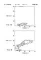
- FIGS. 1A and 1BForward and side scattering of incident light during cytofluorography demonstrating the increase in size and granularity of cell populations after time in culture.
- FIG. 1AFreshly isolated peripheral blood lymphocytes before culture.
- FIG. 1BCells recovered after 4 weeks in culture.
- FIG. 2Proliferation of blood-borne mesenchymal cells in response to granulocyte macrophage-colony stimulating factor.
- the present inventionrelates to mammalian blood-borne mesenchymal cells, to methods of isolating and characterizing the cells, and to methods of using the same for a variety of applications including but not limited to wound healing and gene therapy.
- the present inventionprovides for methods of enriching and/or purifying mesenchymal cells from peripheral blood or other physiological sources of these cells.
- the biologic activity of these cellsmay allow for their uses in settings where absolute purity is not achieved.
- the mesenchymal cells of the inventionmay be isolated from any tissue where they reside or from which they may mature, including but not limited to the bone marrow, fetal liver, or embryonic yolk sac.
- blood-borne mesenchymal cellsmay be isolated by separation based on the presence or absence of specific cell surface markers.
- These techniquesmay include flow cytometry using a fluorescence activated cell sorter or biotin-avidin or biotin-streptavidin separations using biotin-conjugated to marker-specific polyclonal or monoclonal antibodies and avidin or streptavidin bound to a solid support such as affinity column matrix or plastic surfaces, magnetic separations using antibody-coated magnetic beads, destructive separations such as antibody plus complement or antibody coupled to cytotoxins or radioactive isotopes for the removal of undesirable cell populations.
- mesenchymal cellsmay be isolated by procedures involving repetitive density gradient centrifugation, lectin chromatography, affinity chromatography involving positive selection and negative selection, or a combination thereof.
- Positive selection methodsmay utilize affinity chromatography with antibodies directed to mesenchymal cell-specific surface markers. For example, most mononuclear cells may be depleted first from the blood after density gradient centrifugation and plastic adhesion, then an antibody to vimentin antigen can be used to positively select for mesenchymal cells.
- Negative selectionincludes modifications of the protocol disclosed herein, infra. In essence, a mesenchymal cell preparation may be reacted with one or more antibodies directed at cell surface antigens not expressed by mesenchymal cells for their removal.
- Antibodies to any T cell, B cell, monocyte, natural killer (NK) cell, dendritic cell and granulocyte markersmay be used.
- examples of such antibodiesinclude anti-CD3, anti-CD4, anti-CD5, anti-CD8, anti- ⁇ and anti- ⁇ T cell receptor specific for T cells; anti-CD12, anti-CD19 and anti-CD20 specific for B cells; anti-CD14 specific for monocytes; and anti-CD16, and anti-CD56 specific for NK cells. These antibodies may be applied in any combination repeatedly or in a sequential manner for the enrichment of mesenchymal cells.
- the cellsmay be removed by adsorption to a solid surface coated with an anti-mouse antibody column, as the majority of monoclonal antibodies directed at cell surface markers are of mouse origin, or if the antibodies are conjugated with biotin, the antibody-bound cells can be removed by an avidin-coated surface; or if the antibodies are conjugated to magnetic beads, the cells expressing antigens recognized by the antibodies can be removed in a magnetic field.
- blood-borne cellsare detectable immunochemically in the peripheral blood, and may be purified to homogeneity by various procedures.
- the cells in short-term culturesfall into two distinct morphological profiles, a "round" cell type and "spindle-shaped" fibroblast-like cell type.
- the round cellsappear to be a mixture of lymphocytes and a small round cell type which, like the spindle-shaped cells, demonstrate a fibroblast-like phenotype.
- Long-term cultureappears to enhance the growth of the mesenchymal cells, i.e., both the round and spindle-shaped cells which display the fibroblast-like phenotype, until they become the dominant cell type in vitro.
- the small round mesenchymal cellsmay represent the mitotically active stage of the spindle-shaped mesenchymal cells.
- lymphocytespresent in the short-term cultures, i.e., cells which are known to have a finite life-span in culture in the absence of specific lymphokines, eventually yield to the more persistent mesenchymal cells.
- the cells of the present inventionare characterized to be of mesenchymal origin primarily because of their unusual cell surface phenotype for blood-derived cells.
- these cellsexpress vimentin, fibronectin, collagen I and III, which are typical markers for fibroblasts.
- these cellsdo not express cytokeratin, von Willebrand's factor, desmin, laminin and smooth muscle cell ⁇ -actin, all of which are commonly used markers for epithelial, endothelial or smooth muscle cells.
- the antigens that are typically expressed on peripheral blood leukocytes such as CD3, CD4, CD8, and CD56are also not present on the blood-borne mesenchymal cells.
- these cellsare positive for CD34, which is a marker on hematopoietic stem cells, suggesting that the mesenchymal cells described herein may be bone marrow-derived.
- the mesenchymal cells of the present inventionare larger and more granular than peripheral blood leukocytes when assessed by forward and side scattering of incident light during cytofluorography. They exhibit a unique spindle-shaped morphology which is typical for fibroblasts, but atypical for other blood-derived cells. Hence, taken collectively, the blood-borne mesenchymal cells appear to be a distinct cell type which is different from all previously described cell populations from the blood, based on their cell size, cell surface phenotype, and morphological properties.
- Isolated blood-borne mesenchymal cellsproliferate in vitro in culture media for extended periods of time using standard culture techniques that are well known to those skilled in the art.
- serum-enriched mediumshould be used, and more preferably medium containing 20% fetal bovine serum should be used, e.g. see Section 6.1.1, infra. It has been shown that their growth may be further enhanced by the addition of GM-CSF.
- short term cultures derived from peripheral blood lymphocytescontain a contaminating population of lymphocytes, whereas cells positive for fibroblast markers predominate in the long term cultures.
- GM-CSFaccelerates the time course over which the fibroblast-like cells dominate the culture.
- isolated cellsmay be engineered to express endogenous GM-CSF to sustain their long-term growth in an autocrine fashion (See Section 5.4, infra). Continuous cell lines or clones generated in this manner may facilitate further isolation of cell surface markers and cytokines and the genes encoding therefor.
- Long-term culture of blood-borne mesenchymal cellsmay be performed in tissue culture flasks, roller bottles, bioreactor systems and any culture methods known in the art. In fact, these mesenchymal cells may respond to a number of other conventional cytokines and growth factors.
- the ability of the blood-borne mesenchymal cells to proliferate in cultureindicates that they may be expanded in numbers for use in wound healing, or gene therapy applications, particularly in autologous and syngeneic hosts.
- the cellsmay be used directly after isolation, or after in vitro culture with or without the introduction of exogenous genes, and with or without expression in culture.
- a major impediment in the current attempts to achieve stable integration of foreign genes in eukaryotic host cells of different organsis the inability of most of these cells to proliferate in vitro. Since the mesenchymal cells proliferate in vitro, especially in response to GM-CSF, these cells may be ideal candidates as recipients for the introduction of exogenous genes in culture.
- cytokines normally synthesized by the blood-borne mesenchymal cellsa number of other cytokine or adhesion molecule genes may be engineered into these cells to further augment their ability to promote and accelerate wound healing and tissue remodeling, or to deliver products of any gene introduced into the mesenchymal cell for therapeutic purposes.
- genetic engineering of the cellsinvolves isolating blood-borne mesenchymal cells from an individual, transferring a gene of interest into these cells, confirming stable integration and expression of the desired gene products.
- Such genetically engineered cellsmay be transplanted into the same, or an HLA-matched, or otherwise suitable patient and/or used as a source of factors and/or genes encoding factors made by the cells.
- mesenchymal cells isolated by the procedures described in Section 6, inframay be used as recipients in gene transfer experiments.
- the cellsmay be grown in culture prior to, during, and after introduction of an exogenous gene.
- the proliferative activity of these cellsmay be enhanced by GM-CSF.
- any cloned genemay be transferred using conventional techniques, including, but not limited to, microinjection, transfection and transduction.
- a method of gene transferutilizes recombinant viruses, such as retroviruses or adenoviruses.
- virusessuch as retroviruses or adenoviruses.
- a coding sequencemay be ligated to an adenovirus transcription/translation control complex, e.g., the late promoter and tripartite leader sequence. This chimeric gene may then be inserted in the adenovirus genome by in vitro or in vivo recombination.
- Insertion in a nonessential region of the viral genomewill result in a recombinant virus that is viable and capable of expressing the gene product in infected mesenchymal cells (e.g., see Logan & Shenk, 1984, Proc. Natl. Acad. Sci. U.S.A. 81:3655-3659).
- the vaccinia virus 7.5K promotermay be used. (e.g., see, Mackett et al., 1982, Proc. Natl. Acad. Sci. U.S.A. 79: 7415-7419; Mackett et al., 1984, J. Virol.
- Vectors based on bovine papilloma virus which have the ability to replicate as extrachromosomal elementsare also candidates (Sarver, et al., 1981, Mol. Cell. Biol. 1: 486). Shortly after entry of this DNA into cells, the plasmid replicates to about 100 to 200 copies per cell. Transcription of the inserted cDNA does not require integration of the plasmid into the host's chromosome, thereby yielding a high level of expression.
- vectorscan be used for stable expression by including a selectable marker in the plasmid, such as, for example, the neo gene.
- a retroviral genomecan be modified for use as a vector capable of introducing and directing the expression of any gene of interest in the blood-borne mesenchymal cells (Cone & Mulligan, 1984, Proc. Natl. Acad. Sci. U.S.A. 81:6349-6353).
- High level expressionmay also be achieved using inducible promoters, including, but not limited to, the metallothionine IIA promoter and heat shock promoters.
- the mesenchymal cellscan be transformed with a cDNA controlled by appropriate expression control elements (e.g., promoter, enhancer, sequences, transcription terminators, polyadenylation sites, etc.), and a selectable marker.
- appropriate expression control elementse.g., promoter, enhancer, sequences, transcription terminators, polyadenylation sites, etc.
- the selectable markerconfers resistance to the selection and allows cells to stably integrate the recombinant DNA into their chromosomes and grow to form foci which in turn can be cloned and expanded into cell lines.
- engineered mesenchymal cellsmay be allowed to grow for 1-2 days in an enriched media, and then are switched to a selective media.
- a number of selection systemsmay be used, including but not limited to the herpes simplex virus thymidine kinase (Wigler, et al., 1977, Cell 11: 223), hypoxanthine-guanine phosphoribosyltransferase (Szybalska & Szybalski, 1962, Proc. Natl. Acad. Sci. U.S.A. 48: 2026), and adenine phosphoribosyltransferase (Lowy, et al., 1980, Cell 22: 817) genes.
- antimetabolite resistancecan be used as the basis of selection for dhfr, which confers resistance to methotrexate (Wigler, et al., 1980, Proc. Natl. Acad. Sci. U.S.A. 77: 3567; O'Hare, et al., 1981, Proc. Natl. Acad. Sci. U.S.A. 78: 1527); gpt, which confers resistance to mycophenolic acid (Mulligan & Berg, 1981, Proc. Natl. Acad. Sci. U.S.A.
- neowhich confers resistance to the aminoglycoside G-418 (Colberre-Garapin, et al., 1981, J. Mol. Biol. 150: 1); and hygro, which confers resistance to hygromycin (Santerre, et al., 1984, Gene 30: 147) genes.
- trpBwhich allows cells to utilize indole in place of tryptophan
- hisDwhich allows cells to utilize histinol in place of histidine (Hartman & Mulligan, 1988, Proc. Natl. Acad. Sci. U.S.A.
- ODCornithine decarboxylase
- Blood-borne mesenchymal cellsmay be isolated from the peripheral blood and expanded in culture for a variety of therapeutic purposes, including but not limited to the enhancement of wound healing. Isolated blood-borne mesenchymal cells which are purified or partially enriched with or without exogenous genes may be directly applied to external wound sites including, but not limited to, severe wounds, burns, cuts, abrasions, chronic ulcers and inflammatory diseases of skin. Cosmetic applications of these cells are also within the scope of this invention.
- the cellsmay be directly applied to damaged tissues or organs such as those resulting from trauma or in the course of surgery, for the repair of internal organs, e.g., gastric mucosa, cardiac tissue, bone, and vascular tissue, as well as tissues that are difficult to heal by traditional methods, e.g., joint cartilage, ligaments, tendons, and neural tissue.
- damaged tissues or organssuch as those resulting from trauma or in the course of surgery, for the repair of internal organs, e.g., gastric mucosa, cardiac tissue, bone, and vascular tissue, as well as tissues that are difficult to heal by traditional methods, e.g., joint cartilage, ligaments, tendons, and neural tissue.
- the cellsmay be administered to patients via any of a number of routes, including but not limited to intravenous, intramuscular, subcutaneous, intradermal, etc., for the treatment of external or internal visceral injuries.
- the method of administrationallows for cell migration (e.g., intravenous administration)
- the mesenchymal cellswill migrate in vivo to the site of the wound where they can enhance wound healing.
- Genetically engineered mesenchymal cellsmay be used in this fashion to deliver gene products to the site of the wound; e.g., genes for Factor VIII, growth factors, etc. may be useful in this regard.
- methods of administration which do not allow for migrationmay allow the mesenchymal cells, genetically engineered or otherwise, to take up residence at the site of administration where they can deliver gene products to the local environment, and/or systemically.
- protocolsmay be designed to inhibit or remove these cells in vivo such as by the administration of monoclonal antibodies to specific surface markers, in the treatment of chronic diseases with noted fibrosis, particularly conditions of excessive fibroses such as myelofibroses, histiocytoses, hepatic cirrhosis, keloid formation, scleroderma, etc.
- the inhibition of migration of these cells into wound sitesmay prevent excessive scar formation.
- the blood-borne mesenchymal cellsmay be quantified in peripheral blood samples obtained from individuals. Abnormally low or high concentrations of such cells (as compared to values determined for healthy individuals) may be correlated with diseases or disorders.
- the quantification of the blood-borne mesenchymal cellsmay be accomplished by morphological analysis, biological activities, or preferably by immunochemical means. For example, antibodies specific for vimentin, fibronectin, collagen I, or collagen III may be used individually or in combination for the detection and quantification of these cells.
- polyclonal and monoclonal antibodieswhich recognize novel antigenic markers expressed by the blood-borne mesenchymal cells; Such antibodies may have a variety of uses such as the isolation and characterization of blood-borne mesenchymal cells by affinity chromatography. Various procedures known in the art may be used for the production of antibodies to these mesenchymal cells.
- Various host animalscan be immunized by injection with viable isolated mesenchymal cells, fixed cells or membrane preparations, including but not limited to rabbits, hamsters, mice, rats, etc.
- adjuvantsmay be used to increase the immunological response, depending on the host species, including but not limited to Freund's (complete and incomplete), mineral gels such as aluminum hydroxide, surface active substances such as lysolecithin, pluronic polyols, polyanions, peptides, oil emulsions, keyhole limpet hemocyanin, dinitrophenol, and potentially useful human adjuvants such as BCG (bacille Calmette-Guerin) and Corynebacterium parvum.
- BCGBacille Calmette-Guerin
- Corynebacterium parvumbacille Calmette-Guerin
- Monoclonal antibodies to novel antigens on these mesenchymal cellsmay be prepared by using any technique which provides for the production of antibody molecules by continuous cell lines in culture. These include, but are not limited to, the hybridoma technique originally described by Kohler and Milstein (1975, Nature 256, 495-497), and the more recent human B-cell hybridoma technique (Kosbor et al., 1983, Immunology Today 4:72; Cote et al., 1983, Proc. Natl. Acad. Sci. 80:2026-2030) and the EBV-hybridoma technique (Cole et al., 1985, Monoclonal Antibodies and Cancer Therapy, Alan R. Liss, Inc., pp. 77-96).
- Syngeneic, allogeneic, and xenogeneic hostsmay be used for injection of blood-borne mesenchymal cells prepared in viable form, or in fixed form, or as extracted membrane preparations thereof. Monoclonal antibodies can be screened differentially by selective binding to mesenchymal cells, but not to other blood cells.
- Antibody fragments which contain the binding site of the moleculemay be generated by known techniques.
- such fragmentsinclude but are not limited to: the F(ab') 2 fragments which can be produced by pepsin digestion of the antibody molecule and the Fab fragments which can be generated by reducing the disulfide bridges of the F(ab') 2 fragments.
- mesenchymal cellsmay function through the release of cytokines and/or membrane-bound accessory molecules involved in cell-cell contact. Therefore, mesenchymal cells may be used as a source for identifying novel cytokines and cell surface accessory molecules and the genes encoding therefor.
- long-term mesenchymal cell culturesmay be established or continuous cell lines may be generated by transforming the cells to tumor cells using a virus or a chemical.
- Culture supernatantsmay be directly analyzed by applying them to various cell types or in various animal models, which can then be assayed for the appropriate desired biological response.
- the cellsmay be metabolically labelled and their supernatants subjected to biochemical analysis to identify candidate proteins responsible for the observed bioactivity.
- cytokinesmay be identified by inducing cytokine production in the cells. To this end, the cells may be exposed or contacted with an agent that induces the expression and production of a cytokine.
- agents known to induce cytokine production in other cellsmay be useful in this approach.
- agentsmay include but are not limited to calcium ionophores, endotoxins, phorbol esters, known cytokines, chemokines, growth factors, hormones and/or other mediators.
- the proteinmay be purified by a variety of techniques known in the art including but not limited to SDS-preparative gels, ion exchange chromatography, isoelectric focusing gels and other types of chromatography. Purity of the proteins can be verified by SDS-PAGE, quantified by protein assays, their activities confirmed in bioassays, and used as immunogens for the production of polyclonal and monoclonal antibodies.
- the purified proteinscan be further tested in bioassays to stimulate and/or inhibit proliferation and/or differentiation of a variety of indicator cell lines of diverse tissue types. Radiolabelled proteins may also be used to identify their cell surface receptors by methods such as affinity labelling. Specific antibodies to the cytokines may be used to identify and quantify membrane forms and secreted forms of the cytokines, to study their biosynthetic pathways, to affinity purify the proteins and to immunoscreen expression libraries for the molecular cloning of the coding sequences.
- Blood-borne mesenchymal cellswere isolated from whole blood, for instance human or mouse blood.
- human cells60 ml of blood was drawn by venipuncture into a heparinized syringe and diluted 1:1 with phosphate-buffered saline (PBS). Diluted blood then was layered on Ficoll-Hypaque density medium and centrifuged at room temperature for 30 minutes at 450 ⁇ g. Leukocytes that formed a band above the red blood cells were obtained and washed with PBS by centrifuging three additional times.
- PBSphosphate-buffered saline
- Pelleted cellswere resuspended in 25 ml Dulbecco's Modified Eagle's Medium/20% fetal bovine serum (FBS)/and 0.1% gentamicin. The cells then were plated onto a 150 mm tissue culture plate. After 24 hours, medium together with non-adherent cells was aspirated and replaced with fresh medium. Medium was replaced with fresh medium weekly and adherent cells enumerated at intervals.
- FBSfetal bovine serum
- cellswere seeded into wells that had microscope slide coverslips resting on the bottom of the wells. Spot immunofluorescence was then performed on cells cultured for 4 weeks on 13 mm glass coverslips.
- the slipswere removed from the plates, washed twice with PBS, and fixed by immersion in 3.5% formaldehyde for 20 minutes.
- the cellswere washed once with PBS, then immersed for 7 minutes at -20° C. in 70% ethanol.
- the 70% ethanolwas replaced with 100% ethanol in which the cells were immersed for an additional 7 minutes at -20° C.
- the cellsthen were immersed in 70% ethanol for 5 minutes at -20° C. and washed 3 ⁇ with PBS.
- Cytofluorographywas performed on 4 week cultured cells. Adherent cells were removed by gentle scraping and elutriation. After washing 3 times in PBS and enumeration, the cells were resuspended in 1% BSA in PBS at a concentration of 5 ⁇ 10 6 cells/ml. 3 ⁇ 10 5 cells were aliquoted into polystyrene tubes (10 ⁇ 75 mm) and 10 ⁇ l of undiluted primary antibody added for 45 minutes on ice in the dark, then washed 3 ⁇ in 1% BSA/PBS. Ten ⁇ l of a second antibody-fluorescent dye conjugate was added (if the primary antibody was not directly conjugated to a fluorescent dye) and the cells were incubated for 40 minutes on ice in the dark.
- cellswere incubated as above with two directly conjugated antibodies of different fluorescence properties (i.e., FITC and rhodamine or Texas Red).
- the cellswere washed 3 ⁇ in 1% BSA/PBS, resuspended in 25 ⁇ l 1% BSA/PBS and 100 ⁇ l 3.5% formaldehyde, and-stored at 4° C. in the dark until ready for cytofluorography.
- Cellswere analyzed with a Becton Dickinson FACS 440 and the Profile 2 by Coulter.
- Fluorescence-activated cell sortingwas performed to purify to homogeneity the spindle-shaped mesenchymal cell type in the blood-derived cultures. These studies utilized a Becton Dickinson FACs 440 to isolate the mesenchymal cells either by cell size (light scatter) or by specific cell surface marker expression, as determined by specific reactivity with antibodies.
- the cellswere to be double stained, they were incubated as above, substituting directly-conjugated antibodies of different fluorescence colors (i.e., FITC and rhodamine or Texas Red).
- the cellswere washed three times in 1% BSA/PBS, resuspended at a concentration of 5 ⁇ 10 5 /ml in 25 ⁇ l 1% BSA/PBS and 100 ⁇ l 3.5% formaldehyde, and stored at 4° C. in the dark until ready for sorting. If the cells were to be cultured after the sort, they were collected into DMEM/20% FBS/0.1% gentamicin and plated.
- the majority of the antibodies used in the studies described hereinwere purchased from Becton Dickinson (San Jose, Calif.). The exceptions are: anti-fibronectin, anti-desmin, anti-smooth muscle ⁇ -actin and anti-laminin, (Sigma, St. Louis, Mo.), anti-vimentin (Labsystems, Raleigh, N.C.), anti-collagen (Chemicon, Temecula, Calif.) and anti-von Willebrands factor (Accurate).
- GM-CSFGM-CSF
- cellswere cultured in 6-well plates such that each well contained white cells from 5 ml of whole blood. After two weeks in culture, the cells were subjected to one of four conditions: no GM-CSF (control), 25 U GM-CSF, 50 U GM-CSF or 1000 U GM-CSF per ml. The media and GM-CSF were replaced weekly. The cells were counted by marking off a set field on the plate and that area was manually counted each week. The percentage of fibroblasts per field reflected the number of cells that had the classical fibroblast morphology as compared to the total number of cells in the area being examined.
- Wound chamberswere implanted into subcutaneous pockets in the flanks of mice.
- the wound chambersconsisted of a perforated 3 cm length of silastic tubing (Dow Corning) that contained a piece of polyvinyl alcohol sponge (Unipoint, N.C.) that had been sterilized by autoclaving. Incisions were closed with wound clips and the mice were monitored for infection. Once weekly post implantation, the wound fluid was percutaneously aspirated using a 1 cc syringe with a 25 g needle. The cells obtained were cultured in DMEM/20% FBS/0.1% gentamicin. Cells were analyzed by morphology and fluorescence staining techniques for fibroblast-specific markers.
- the round cell populationwas a mixture of lymphocytes and cells that displayed the fibroblast-like phenotype.
- the vast majority of all the surviving cellshad the mesenchymal cell phenotype.
- Immunofluorescence analysis for selected cell surface markersthen was performed by direct visualization under fluorescence microscopy (spot immunofluorescence) and by cytofluorography. The two distinct cell types were further separated, purified and characterized by fluorescence-activated cell sorting (FACs).
- FACsfluorescence-activated cell sorting
- the large "spindle-shaped" cellwas identified by antibody staining to be a mesenchymal cell type that displayed typical fibroblast markers; i.e., collagen I, III, vimentin, and fibronectin. These results are summarized in Table 1. Cell-sorting by size or immunofluorescence, followed by specific staining, confirmed the cell type described above and the phenotypic analysis shown in Table 1. Further, these cells were shown to be larger and more granular than peripheral blood leukocytes (FIG. 1).
- Blood-borne mesenchymal cellscould be expanded in vitro by addition of granulocyte-macrophage colony stimulating factor (GM-CSF) at a concentration of 50 U/ml (FIG. 2).
- GM-CSFgranulocyte-macrophage colony stimulating factor
- micewere experimentally implanted with wound chambers in their back. The migration of a blood-borne murine cell population corresponding morphologically to the human blood-borne mesenchymal cells into the chambers was observed.
Landscapes
- Health & Medical Sciences (AREA)
- Life Sciences & Earth Sciences (AREA)
- Engineering & Computer Science (AREA)
- Biomedical Technology (AREA)
- Genetics & Genomics (AREA)
- Zoology (AREA)
- Organic Chemistry (AREA)
- Biotechnology (AREA)
- Chemical & Material Sciences (AREA)
- Bioinformatics & Cheminformatics (AREA)
- Developmental Biology & Embryology (AREA)
- Wood Science & Technology (AREA)
- Microbiology (AREA)
- Rheumatology (AREA)
- Cell Biology (AREA)
- Biochemistry (AREA)
- General Engineering & Computer Science (AREA)
- General Health & Medical Sciences (AREA)
- Hematology (AREA)
- Micro-Organisms Or Cultivation Processes Thereof (AREA)
- Medicines Containing Material From Animals Or Micro-Organisms (AREA)
Abstract
Description
TABLE I ______________________________________ Summary Of Cell Surface Phenotype Of Large Spindle-Shaped Mesenchymal Cells Derived from the Peripheral Blood POSITIVE EXPRESSION FOR: NEGATIVE EXPRESSION FOR: ______________________________________ MHC class II T cell receptor (αβ and γδ) CD11b CD3 CD11c CD4 CD13 CD8 CD34 CD11a CD45 CD14 Vimentin CD16 Fibronectin CD19 Collagen I CD25 Collagen III CD33 CD38 CD44 CD54 CD56 Cytokeratin Von Willebrand's factor Desmin smooth muscle cell α-actin Laminin ______________________________________
Claims (2)
Priority Applications (1)
| Application Number | Priority Date | Filing Date | Title |
|---|---|---|---|
| US08/488,241US5846796A (en) | 1993-02-26 | 1995-06-07 | Blood-borne mesenchymal cells |
Applications Claiming Priority (2)
| Application Number | Priority Date | Filing Date | Title |
|---|---|---|---|
| US08/023,290US5654186A (en) | 1993-02-26 | 1993-02-26 | Blood-borne mesenchymal cells |
| US08/488,241US5846796A (en) | 1993-02-26 | 1995-06-07 | Blood-borne mesenchymal cells |
Related Parent Applications (1)
| Application Number | Title | Priority Date | Filing Date |
|---|---|---|---|
| US08/023,290ContinuationUS5654186A (en) | 1993-02-26 | 1993-02-26 | Blood-borne mesenchymal cells |
Publications (1)
| Publication Number | Publication Date |
|---|---|
| US5846796Atrue US5846796A (en) | 1998-12-08 |
Family
ID=21814206
Family Applications (3)
| Application Number | Title | Priority Date | Filing Date |
|---|---|---|---|
| US08/023,290Expired - LifetimeUS5654186A (en) | 1993-02-26 | 1993-02-26 | Blood-borne mesenchymal cells |
| US08/487,116Expired - LifetimeUS6174526B1 (en) | 1993-02-26 | 1995-06-07 | Blood-borne mesenchymal cells |
| US08/488,241Expired - LifetimeUS5846796A (en) | 1993-02-26 | 1995-06-07 | Blood-borne mesenchymal cells |
Family Applications Before (2)
| Application Number | Title | Priority Date | Filing Date |
|---|---|---|---|
| US08/023,290Expired - LifetimeUS5654186A (en) | 1993-02-26 | 1993-02-26 | Blood-borne mesenchymal cells |
| US08/487,116Expired - LifetimeUS6174526B1 (en) | 1993-02-26 | 1995-06-07 | Blood-borne mesenchymal cells |
Country Status (1)
| Country | Link |
|---|---|
| US (3) | US5654186A (en) |
Cited By (47)
| Publication number | Priority date | Publication date | Assignee | Title |
|---|---|---|---|---|
| US20030003576A1 (en)* | 2001-06-04 | 2003-01-02 | Riichiro Abe | Peripheral blood fibrocytes differentiation pathway and migration to wound sites |
| WO2003053220A2 (en) | 2001-12-17 | 2003-07-03 | Corixa Corporation | Compositions and methods for the therapy and diagnosis of inflammatory bowel disease |
| WO2004062599A2 (en) | 2003-01-06 | 2004-07-29 | Corixa Corporation | Certain aminoalkyl glucosaminide phosphate compounds and their use |
| US6974571B2 (en)* | 1995-03-28 | 2005-12-13 | Thomas Jefferson University | Isolated stromal cells and methods of using the same |
| US20060002938A1 (en)* | 2002-12-23 | 2006-01-05 | Richard Gomer | Methods of detecting the inhibition of fibrocyte formation and methods and compositions for enhancing fibrocyte formation |
| EP1650221A2 (en) | 2000-02-23 | 2006-04-26 | GlaxoSmithKline Biologicals SA | Novel compounds |
| US20070065866A1 (en)* | 2002-12-23 | 2007-03-22 | William Marsh Rice University | Compositions and Methods for Suppressing Fibrocytes |
| US20070065368A1 (en)* | 2002-12-23 | 2007-03-22 | William Marsh Rice University | Compositions and Methods for Suppressing Fibrocytes and for Detecting Fibrocyte Differentiation |
| EP1961819A2 (en) | 2000-06-28 | 2008-08-27 | Corixa Corporation | Composition and methods for the therapy and diagnosis of lung cancer |
| EP1988097A1 (en) | 2001-05-09 | 2008-11-05 | Corixa Corporation | Compositions and methods for the therapy and diagnosis of prostate cancer |
| US20090074754A1 (en)* | 2007-07-06 | 2009-03-19 | Promedior, Inc. | Methods and compositions useful in the treatment of mucositis |
| US20090203144A1 (en)* | 2001-09-20 | 2009-08-13 | Andrew Beaton | Hiv-gag codon-optimised dna vaccines |
| EP2105502A1 (en) | 2000-12-12 | 2009-09-30 | Corixa Corporation | Compositions and methods for the therapy and diagnosis of lung cancer |
| EP2172476A2 (en) | 2001-10-30 | 2010-04-07 | Corixa Corporation | Compositions and methods for WT1 specific immunotherapy |
| EP2182055A1 (en) | 2008-10-14 | 2010-05-05 | Heinrich-Heine-Universität Düsseldorf | Human cord blood derived unrestricted somatic stem cells (USSC) |
| US20100111898A1 (en)* | 2006-12-04 | 2010-05-06 | Promedior, Inc | Conjoint therapy for treating fibrotic diseases |
| US20100172884A1 (en)* | 1997-07-14 | 2010-07-08 | Pittenger Mark F | Cardiac Muscle Repair Or Regeneration Using Bone Marrow-Derived Stem Cells |
| US20100260781A1 (en)* | 2009-03-11 | 2010-10-14 | Lynne Anne Murray | Treatment methods for autoimmune disorders |
| US20100266578A1 (en)* | 2009-03-11 | 2010-10-21 | Lynne Anne Murray | Treatment and diagnostic methods for hypersensitive disorders |
| US20100297074A1 (en)* | 2002-12-23 | 2010-11-25 | Richard Hans Gomer | Wound healing compositions, systems, and methods |
| US20100317596A1 (en)* | 2009-06-04 | 2010-12-16 | Willett W Scott | Serum amyloid p derivatives and their preparation and use |
| US20100323970A1 (en)* | 2009-06-17 | 2010-12-23 | Promedior, Inc. | Sap variants and their use |
| US20110020374A1 (en)* | 2007-10-15 | 2011-01-27 | The University Of Queensland | Expression system for modulating an immune response |
| EP2298340A1 (en) | 2004-09-22 | 2011-03-23 | GlaxoSmithKline Biologicals S.A. | Immunogenic composition for use in vaccination against staphylococcei |
| EP2441846A2 (en) | 2006-01-09 | 2012-04-18 | The Regents Of the University of California | Immunostimulatory combinations of TNFRSF, TLR, NLR, RHR, purinergic receptor, and cytokine receptor agoinsts for vaccines and tumor immunotherapy |
| WO2012177595A1 (en) | 2011-06-21 | 2012-12-27 | Oncofactor Corporation | Compositions and methods for the therapy and diagnosis of cancer |
| EP2662439A1 (en) | 2005-05-27 | 2013-11-13 | Viacord, LLC | Treatment of ischemia using stem cells |
| EP2940027A1 (en) | 2004-07-08 | 2015-11-04 | Corixa Corporation | Certain aminoalkyl glucosaminide phosphate compounds and their use |
| WO2016154010A1 (en) | 2015-03-20 | 2016-09-29 | Makidon Paul | Immunogenic compositions for use in vaccination against bordetella |
| WO2017096432A1 (en) | 2015-12-09 | 2017-06-15 | Admedus Vaccines Pty Ltd | Immunomodulating composition for treatment |
| US9795658B2 (en) | 2010-04-20 | 2017-10-24 | Admedus Vaccines Pty Ltd | Expression system for modulating an immune response |
| US9884899B2 (en) | 2007-07-06 | 2018-02-06 | Promedior, Inc. | Methods for treating fibrosis using CRP antagonists |
| WO2021195089A1 (en) | 2020-03-23 | 2021-09-30 | Sorrento Therapeutics, Inc. | Fc-coronavirus antigen fusion proteins, and nucleic acids, vectors, compositions and methods of use thereof |
| US11285191B2 (en) | 2016-07-26 | 2022-03-29 | The Board Of Supervisors Of Louisiana State University And Agricultural And Mechanical College | Immunostimulatory compositions and uses therefor |
| US11608486B2 (en) | 2015-07-02 | 2023-03-21 | Terumo Bct, Inc. | Cell growth with mechanical stimuli |
| US11613727B2 (en) | 2010-10-08 | 2023-03-28 | Terumo Bct, Inc. | Configurable methods and systems of growing and harvesting cells in a hollow fiber bioreactor system |
| US11624046B2 (en) | 2017-03-31 | 2023-04-11 | Terumo Bct, Inc. | Cell expansion |
| US11629332B2 (en) | 2017-03-31 | 2023-04-18 | Terumo Bct, Inc. | Cell expansion |
| US11634677B2 (en) | 2016-06-07 | 2023-04-25 | Terumo Bct, Inc. | Coating a bioreactor in a cell expansion system |
| US11667881B2 (en) | 2014-09-26 | 2023-06-06 | Terumo Bct, Inc. | Scheduled feed |
| US11667876B2 (en) | 2013-11-16 | 2023-06-06 | Terumo Bct, Inc. | Expanding cells in a bioreactor |
| US11685883B2 (en) | 2016-06-07 | 2023-06-27 | Terumo Bct, Inc. | Methods and systems for coating a cell growth surface |
| US11795432B2 (en) | 2014-03-25 | 2023-10-24 | Terumo Bct, Inc. | Passive replacement of media |
| US11965175B2 (en) | 2016-05-25 | 2024-04-23 | Terumo Bct, Inc. | Cell expansion |
| US12043823B2 (en) | 2021-03-23 | 2024-07-23 | Terumo Bct, Inc. | Cell capture and expansion |
| US12152699B2 (en) | 2022-02-28 | 2024-11-26 | Terumo Bct, Inc. | Multiple-tube pinch valve assembly |
| US12234441B2 (en) | 2017-03-31 | 2025-02-25 | Terumo Bct, Inc. | Cell expansion |
Families Citing this family (66)
| Publication number | Priority date | Publication date | Assignee | Title |
|---|---|---|---|---|
| US5804446A (en)* | 1993-02-26 | 1998-09-08 | The Picower Institute For Medical Research | Blood-borne mesenchymal cells |
| US6054121A (en)* | 1993-02-26 | 2000-04-25 | The Picower Institute For Medical Research | Modulation of immune responses in blood-borne mesenchymal cells |
| US5654186A (en)* | 1993-02-26 | 1997-08-05 | The Picower Institute For Medical Research | Blood-borne mesenchymal cells |
| US9068164B1 (en)* | 1995-02-02 | 2015-06-30 | Tristem Trading (Cyprus) Limited | Method of preparing an undifferentiated cell |
| GB9502022D0 (en)* | 1995-02-02 | 1995-03-22 | Abuljadayel Ilham M S | A method for preparing lymphohaematopoietic progenitor cells |
| AU731937B2 (en)* | 1996-12-10 | 2001-04-05 | Hadasit Medical Research Services & Development Company Ltd | Serum-derived factor inducing cell differentiation and medical uses thereof |
| US6759036B1 (en)* | 1997-03-28 | 2004-07-06 | The Picower Institute For Medical Research | Fibrocyte-base vaccine formulations |
| US6387367B1 (en)* | 1998-05-29 | 2002-05-14 | Osiris Therapeutics, Inc. | Human mesenchymal stem cells |
| BR9914465A (en)* | 1998-09-29 | 2001-10-09 | Gamida Cell Ltd | Methods to control the proliferation and differentiation of stem cells and progenitor cells and a pharmaceutical composition to induce differentiation in a cell population |
| ATE446767T1 (en)* | 1999-02-12 | 2009-11-15 | Univ Washington | GM-CSF FOR THE TREATMENT OF CROHN'S DISEASE |
| US6500418B1 (en)* | 1999-02-12 | 2002-12-31 | The Washington University | Stimulating neutrophil function to treat inflammatory bowel disease |
| US7311905B2 (en) | 2002-02-13 | 2007-12-25 | Anthrogenesis Corporation | Embryonic-like stem cells derived from post-partum mammalian placenta, and uses and methods of treatment using said cells |
| EP3299452A1 (en) | 2000-12-06 | 2018-03-28 | Anthrogenesis Corporation | Method of collecting placental stem cells |
| NZ541541A (en)* | 2001-02-14 | 2007-04-27 | Anthrogenesis Corp | A composition suitable for transplantation which comprises a population of hematopoietic stem cells comprising placental stem cells that are CD34+ and CD38- |
| IL159578A0 (en)* | 2001-07-06 | 2004-06-01 | Geron Corp | Messenchymal cells and osteoblasts from human embryonic stem cell |
| CA2469370C (en) | 2001-12-07 | 2014-07-08 | Macropore Biosurgery, Inc. | Adipose-derived cell processing unit |
| US9597395B2 (en)* | 2001-12-07 | 2017-03-21 | Cytori Therapeutics, Inc. | Methods of using adipose tissue-derived cells in the treatment of cardiovascular conditions |
| US7595043B2 (en)* | 2001-12-07 | 2009-09-29 | Cytori Therapeutics, Inc. | Method for processing and using adipose-derived stem cells |
| US8404229B2 (en)* | 2001-12-07 | 2013-03-26 | Cytori Therapeutics, Inc. | Methods of using adipose derived stem cells to treat acute tubular necrosis |
| US7585670B2 (en) | 2001-12-07 | 2009-09-08 | Cytori Therapeutics, Inc. | Automated methods for isolating and using clinically safe adipose derived regenerative cells |
| US8105580B2 (en) | 2001-12-07 | 2012-01-31 | Cytori Therapeutics, Inc. | Methods of using adipose derived stem cells to promote wound healing |
| US7771716B2 (en)* | 2001-12-07 | 2010-08-10 | Cytori Therapeutics, Inc. | Methods of using regenerative cells in the treatment of musculoskeletal disorders |
| US7651684B2 (en)* | 2001-12-07 | 2010-01-26 | Cytori Therapeutics, Inc. | Methods of using adipose tissue-derived cells in augmenting autologous fat transfer |
| US20050095228A1 (en) | 2001-12-07 | 2005-05-05 | Fraser John K. | Methods of using regenerative cells in the treatment of peripheral vascular disease and related disorders |
| IL152904A0 (en)* | 2002-01-24 | 2003-06-24 | Gamida Cell Ltd | Utilization of retinoid and vitamin d receptor antagonists for expansion of renewable stem cell populations |
| EP1465982A4 (en) | 2002-01-25 | 2006-06-07 | Gamida Cell Ltd | Methods of expanding stem and progenitor cells and expanded cell populations obtained thereby |
| WO2003078567A2 (en)* | 2002-03-18 | 2003-09-25 | Gamida-Cell Ltd. | Methods of inducing differentiation in ex vivo expanded stem cells |
| CA2493363A1 (en)* | 2002-07-29 | 2004-02-05 | Asahi Kasei Kabushiki Kaisha | Stem cells for treating pancreatic damage |
| EP1571910A4 (en) | 2002-11-26 | 2009-10-28 | Anthrogenesis Corp | Cytotherapeutics, cytotherapeutic units and methods for treatments using them |
| US7470538B2 (en)* | 2002-12-05 | 2008-12-30 | Case Western Reserve University | Cell-based therapies for ischemia |
| AU2003296338A1 (en)* | 2002-12-05 | 2004-06-30 | Case Western Reserve University | Cell-based therapies for ischemia |
| CN1770976A (en)* | 2003-02-13 | 2006-05-10 | 人类起源公司 | Use of umbilical cord blood to treat an individual suffering from a disease, disorder or condition |
| AU2004217699B2 (en)* | 2003-03-07 | 2008-07-03 | Gamida-Cell Ltd. | Expansion of renewable stem cell populations using modulators of PI 3-kinase |
| EP2329833A1 (en)* | 2004-01-27 | 2011-06-08 | The Hospital for Sick Children Research Institute | Methods of making and using skin-derived stem cells |
| JP2007536935A (en)* | 2004-05-14 | 2007-12-20 | ベクトン・ディキンソン・アンド・カンパニー | Cell culture environment for serum-free growth of mesenchymal stem cells |
| EP1799812A4 (en) | 2004-09-16 | 2009-09-09 | Gamida Cell Ltd | Methods of ex vivo progenitor and stem cell expansion by co-culture with mesenchymal cells |
| JP5138384B2 (en)* | 2005-01-10 | 2013-02-06 | サイトリ セラピューティクス インコーポレイテッド | Apparatus and method for monitoring, managing and servicing medical devices |
| ITMI20050205A1 (en)* | 2005-02-14 | 2006-08-15 | Pietro Prof Antonino Di | EXTRACTION PROCEDURE BY MEANS OF PURIFICATION OF STEM CELLS AND-OR OF FIBROBLASTS AND THEIR AUTOLOGOUS DERIVATIVES AND CELLULAR PRODUCT OBTAINED BY SUCH PROCEDURE |
| CA2624925C (en) | 2005-10-13 | 2014-09-30 | Anthrogenesis Corporation | Immunomodulation using placental stem cells |
| US8846393B2 (en) | 2005-11-29 | 2014-09-30 | Gamida-Cell Ltd. | Methods of improving stem cell homing and engraftment |
| WO2007079183A2 (en) | 2005-12-29 | 2007-07-12 | Anthrogenesis Corporation | Placental stem cell populations |
| US8455250B2 (en) | 2005-12-29 | 2013-06-04 | Anthrogenesis Corporation | Co-culture of placental stem cells and stem cells from a second source |
| US20090304644A1 (en)* | 2006-05-30 | 2009-12-10 | Cytori Therapeutics, Inc. | Systems and methods for manipulation of regenerative cells separated and concentrated from adipose tissue |
| EP2024493B1 (en)* | 2006-05-31 | 2014-07-02 | Cellect Biotechnology Ltd. | Methods of selecting stem cells and uses thereof |
| WO2008013863A2 (en)* | 2006-07-26 | 2008-01-31 | Cytori Therapeutics, Inc. | Generation of adipose tissue and adipocytes |
| WO2008085229A2 (en)* | 2006-11-15 | 2008-07-17 | Arteriocyte Inc. | Cell-based therapies for treating liver disease |
| NZ612888A (en) | 2007-02-12 | 2015-02-27 | Anthrogenesis Corp | Treatment of inflammatory diseases using placental stem cells |
| MX2009008559A (en) | 2007-02-12 | 2009-08-21 | Anthrogenesis Corp | Hepatocytes and chondrocytes from adherent placental stem cells; and cd34+, cd45- placental stem cell-enriched cell populations. |
| PT2203176E (en) | 2007-09-28 | 2015-03-02 | Anthrogenesis Corp | TUMOR SUPPRESSION USING HUMAN PLACENTARY PERFUSION AND INTERMEDIATE NATURAL MURDER CELLS DERIVED FROM HUMAN PLACENTA |
| WO2010021993A1 (en)* | 2008-08-19 | 2010-02-25 | Cytori Therapeutics, Inc. | Methods of using adipose tissue-derived cells in the treatment of the lymphatic system and malignant disease |
| KR101903049B1 (en) | 2008-08-20 | 2018-11-07 | 안트로제네시스 코포레이션 | Treatment of stroke using isolated placental cells |
| DK2330889T3 (en) | 2008-08-20 | 2017-01-30 | Anthrogenesis Corp | Improved cell composition and process for making the same |
| KR20110050688A (en) | 2008-08-22 | 2011-05-16 | 안트로제네시스 코포레이션 | Bone Defect Treatment Method and Bone Defect Therapeutic Composition Using Placental Cell Population |
| EP2367932B1 (en) | 2008-11-19 | 2019-06-12 | Celularity, Inc. | Amnion derived adherent cells |
| EP2424463B1 (en)* | 2009-05-01 | 2016-12-28 | Bimini Technologies LLC | Systems, methods and compositions for optimizing tissue and cell enriched grafts |
| CN102822330A (en) | 2010-01-26 | 2012-12-12 | 人类起源公司 | Treatment of bone-related cancers using placental stem cells |
| NZ630009A (en) | 2010-04-07 | 2016-05-27 | Anthrogenesis Corp | Angiogenesis using placental stem cells |
| NZ602798A (en) | 2010-04-08 | 2014-10-31 | Anthrogenesis Corp | Treatment of sarcoidosis using placental stem cells |
| BR112013000822B1 (en) | 2010-07-13 | 2021-02-23 | Anthrogenesis Corporation | TWO-STEP IN VITRO METHOD FOR THE PRODUCTION OF A POPULATION OF ACTIVATED NATURAL EXTERMINATOR (NK) PEOPLE |
| AR093183A1 (en) | 2010-12-31 | 2015-05-27 | Anthrogenesis Corp | INCREASE IN THE POWER OF PLACENTA MOTHER CELLS USING MODULATING RNA MOLECULES |
| CN113559126A (en) | 2011-06-01 | 2021-10-29 | 人类起源公司 | Treating pain with placental stem cells |
| WO2013055476A1 (en) | 2011-09-09 | 2013-04-18 | Anthrogenesis Corporation | Treatment of amyotrophic lateral sclerosis using placental stem cells |
| IN2014MN01680A (en) | 2012-02-13 | 2015-07-03 | Gamida Cell Ltd | |
| US9175266B2 (en) | 2012-07-23 | 2015-11-03 | Gamida Cell Ltd. | Enhancement of natural killer (NK) cell proliferation and activity |
| US9567569B2 (en) | 2012-07-23 | 2017-02-14 | Gamida Cell Ltd. | Methods of culturing and expanding mesenchymal stem cells |
| CN115282165A (en) | 2013-02-05 | 2022-11-04 | 细胞结构公司 | Natural killer cells from placenta |
Citations (5)
| Publication number | Priority date | Publication date | Assignee | Title |
|---|---|---|---|---|
| WO1992022584A1 (en)* | 1991-06-18 | 1992-12-23 | Caplan Arnold I | Monoclonal antibodies specific for marrow-derived mesenchymal cells |
| US5197985A (en)* | 1990-11-16 | 1993-03-30 | Caplan Arnold I | Method for enhancing the implantation and differentiation of marrow-derived mesenchymal cells |
| US5226914A (en)* | 1990-11-16 | 1993-07-13 | Caplan Arnold I | Method for treating connective tissue disorders |
| US5399346A (en)* | 1989-06-14 | 1995-03-21 | The United States Of America As Represented By The Department Of Health And Human Services | Gene therapy |
| US5654186A (en)* | 1993-02-26 | 1997-08-05 | The Picower Institute For Medical Research | Blood-borne mesenchymal cells |
- 1993
- 1993-02-26USUS08/023,290patent/US5654186A/ennot_activeExpired - Lifetime
- 1995
- 1995-06-07USUS08/487,116patent/US6174526B1/ennot_activeExpired - Lifetime
- 1995-06-07USUS08/488,241patent/US5846796A/ennot_activeExpired - Lifetime
Patent Citations (5)
| Publication number | Priority date | Publication date | Assignee | Title |
|---|---|---|---|---|
| US5399346A (en)* | 1989-06-14 | 1995-03-21 | The United States Of America As Represented By The Department Of Health And Human Services | Gene therapy |
| US5197985A (en)* | 1990-11-16 | 1993-03-30 | Caplan Arnold I | Method for enhancing the implantation and differentiation of marrow-derived mesenchymal cells |
| US5226914A (en)* | 1990-11-16 | 1993-07-13 | Caplan Arnold I | Method for treating connective tissue disorders |
| WO1992022584A1 (en)* | 1991-06-18 | 1992-12-23 | Caplan Arnold I | Monoclonal antibodies specific for marrow-derived mesenchymal cells |
| US5654186A (en)* | 1993-02-26 | 1997-08-05 | The Picower Institute For Medical Research | Blood-borne mesenchymal cells |
Non-Patent Citations (31)
| Title |
|---|
| Allgower, M. (1956) In: The Cellular Basis of Wound Repair, Charles C. Thomas Publisher, Springfield, Illinois, USA,. pp. 44 50 and 108.* |
| Allgower, M. (1956) In: The Cellular Basis of Wound Repair, Charles C. Thomas Publisher, Springfield, Illinois, USA,. pp. 44-50 and 108. |
| Allgower, M. (1963) Proceedings of a Workshop, The Committee on T, National Academy of Science National Research Council, 5 8, Dec., pp. 160 164.* |
| Allgower, M. (1963) Proceedings of a Workshop, The Committee on T, National Academy of Science--National Research Council, 5-8, Dec., pp. 160-164. |
| Allgower, M. (1974) Cellular Basis of Wound Repair, International Symposium on Wound Healing, Rotterdam, pp. 16 20.* |
| Allgower, M. (1974) Cellular Basis of Wound Repair, International Symposium on Wound Healing, Rotterdam, pp. 16-20. |
| Anderson, W.F. Gene Therapy. Scientific American Sep. 1995 p. 128 col. 1 Lines 50 58.* |
| Anderson, W.F. Gene Therapy. Scientific American Sep. 1995 p. 128 col. 1 Lines 50-58. |
| Andreoni et al. (1990) Exp. Hematol. 18:431.* |
| Bucala et al. (1993) The FASEB J. 7:A139.* |
| Caplan, A.I. (1990) Biomat 11:89.* |
| Certified English translation of Voskresenkii et al. (1988) Ontogenesis (19):247 52.* |
| Certified English translation of Voskresenkii et al. (1988) Ontogenesis (19):247-52. |
| Coghlan, New Scientists, vol. 148: 14 15, 1995.* |
| Coghlan, New Scientists, vol. 148: 14-15, 1995. |
| Correll, P.H. et al. Blood 80:331 336 1992.* |
| Correll, P.H. et al. Blood 80:331-336 1992. |
| Haynesworth et al. (1992) Bone 13:69 80.* |
| Haynesworth et al. (1992) Bone 13:69-80. |
| Haynesworth et al. (1992) Bone 13:81 88.* |
| Haynesworth et al. (1992) Bone 13:81-88. |
| Hodgson, C.P. The Vector Void. Biotechnology 13:222 col 1 Lines 1 20 1995.* |
| Hodgson, C.P. The Vector Void. Biotechnology 13:222 col 1 Lines 1-20 1995. |
| Nakahara et al. (1990) Bone 11:181 8.* |
| Nakahara et al. (1990) Bone 11:181-8. |
| Nakahara et al. (1991) Experimental Cell Res. 195:492 503.* |
| Nakahara et al. (1991) Experimental Cell Res. 195:492-503. |
| Ronald Crystal, Science, vol. 270: 404 409, 1995.* |
| Ronald Crystal, Science, vol. 270: 404-409, 1995. |
| Voskresensky et al., Ontogenesis, vol. 19, No. 3 pp. 247 252 (1988) in Russian plus an English translation.* |
| Voskresensky et al., Ontogenesis, vol. 19, No. 3 pp. 247-252 (1988) in Russian plus an English translation. |
Cited By (89)
| Publication number | Priority date | Publication date | Assignee | Title |
|---|---|---|---|---|
| US6974571B2 (en)* | 1995-03-28 | 2005-12-13 | Thomas Jefferson University | Isolated stromal cells and methods of using the same |
| US20090035347A1 (en)* | 1995-03-28 | 2009-02-05 | Thomas Jefferson University | Isolated stromal cells and methods of using the same |
| US8852574B2 (en) | 1997-07-14 | 2014-10-07 | Mesoblast International Sarl | Cardiac muscle repair or regeneration using bone marrow-derived stem cells |
| US8852575B2 (en) | 1997-07-14 | 2014-10-07 | Mesoblast International Sarl | Cardiac muscle repair or regeneration using bone marrow-derived stem cells |
| US8852573B2 (en) | 1997-07-14 | 2014-10-07 | Mesoblast International Sarl | Cardiac muscle repair or regeneration using bone marrow-derived stem cells |
| US20110158965A1 (en)* | 1997-07-14 | 2011-06-30 | Pittenger Mark F | Cardiac Muscle Regeneration Using Mesenchymal Stem Cells |
| US8852572B2 (en) | 1997-07-14 | 2014-10-07 | Mesoblast International Sarl | Cardiac muscle repair or regeneration using bone marrow-derived stem cells |
| US8852570B2 (en) | 1997-07-14 | 2014-10-07 | Mesoblast International Sarl | Cardiac muscle regeneration using mesenchymal stem cells |
| US8158121B2 (en) | 1997-07-14 | 2012-04-17 | Osiris Therapeutics, Inc. | Cardiac muscle regeneration using mesenchymal stem cells |
| US20100172884A1 (en)* | 1997-07-14 | 2010-07-08 | Pittenger Mark F | Cardiac Muscle Repair Or Regeneration Using Bone Marrow-Derived Stem Cells |
| US20110158964A1 (en)* | 1997-07-14 | 2011-06-30 | Pittenger Mark F | Cardiac Muscle Regeneration Using Mesenchymal Stem Cells |
| US8852571B2 (en) | 1997-07-14 | 2014-10-07 | Mesoblast International Sarl | Cardiac muscle regeneration using mesenchymal stem cells |
| US8158120B2 (en) | 1997-07-14 | 2012-04-17 | Osiris Therapeutics, Inc. | Cardiac muscle repair or regeneration using bone marrow-derived stem cells |
| EP1650221A2 (en) | 2000-02-23 | 2006-04-26 | GlaxoSmithKline Biologicals SA | Novel compounds |
| EP1961819A2 (en) | 2000-06-28 | 2008-08-27 | Corixa Corporation | Composition and methods for the therapy and diagnosis of lung cancer |
| EP2105502A1 (en) | 2000-12-12 | 2009-09-30 | Corixa Corporation | Compositions and methods for the therapy and diagnosis of lung cancer |
| EP1988097A1 (en) | 2001-05-09 | 2008-11-05 | Corixa Corporation | Compositions and methods for the therapy and diagnosis of prostate cancer |
| US20030003576A1 (en)* | 2001-06-04 | 2003-01-02 | Riichiro Abe | Peripheral blood fibrocytes differentiation pathway and migration to wound sites |
| US20090203144A1 (en)* | 2001-09-20 | 2009-08-13 | Andrew Beaton | Hiv-gag codon-optimised dna vaccines |
| EP2322626A1 (en) | 2001-09-20 | 2011-05-18 | Glaxo Group Limited | HIV-GAG codon-optimised DNA vaccines |
| EP2172476A2 (en) | 2001-10-30 | 2010-04-07 | Corixa Corporation | Compositions and methods for WT1 specific immunotherapy |
| EP2224012A1 (en) | 2001-12-17 | 2010-09-01 | Corixa Corporation | Compositions and methods for the therapy and diagnosis of inflammatory bowel disease |
| WO2003053220A2 (en) | 2001-12-17 | 2003-07-03 | Corixa Corporation | Compositions and methods for the therapy and diagnosis of inflammatory bowel disease |
| US8187599B2 (en) | 2002-12-23 | 2012-05-29 | William Marsh Rice University | Compositions and methods for suppressing fibrocytes |
| US8187608B2 (en) | 2002-12-23 | 2012-05-29 | William Marsh Rice University | Treatment of Fibrosis related disorders |
| US20060002938A1 (en)* | 2002-12-23 | 2006-01-05 | Richard Gomer | Methods of detecting the inhibition of fibrocyte formation and methods and compositions for enhancing fibrocyte formation |
| US20070065866A1 (en)* | 2002-12-23 | 2007-03-22 | William Marsh Rice University | Compositions and Methods for Suppressing Fibrocytes |
| US20070065368A1 (en)* | 2002-12-23 | 2007-03-22 | William Marsh Rice University | Compositions and Methods for Suppressing Fibrocytes and for Detecting Fibrocyte Differentiation |
| US7666432B2 (en) | 2002-12-23 | 2010-02-23 | William Marsh Rice University | Methods of suppressing fibrosis and fibrocyte formation |
| US7763256B2 (en) | 2002-12-23 | 2010-07-27 | William Marsh Rice University | Compositions and methods for suppressing fibrocytes and for detecting fibrocyte differentiation |
| US8057802B2 (en) | 2002-12-23 | 2011-11-15 | William Marsh Rice University | Treatment methods for fibrosis related disorders |
| US7935682B2 (en) | 2002-12-23 | 2011-05-03 | William Marsh Rice University | Wound healing dressing for enhancing fibrocyte formation |
| US20100297074A1 (en)* | 2002-12-23 | 2010-11-25 | Richard Hans Gomer | Wound healing compositions, systems, and methods |
| US8012472B2 (en) | 2002-12-23 | 2011-09-06 | William Marsh Rice University | Compositions and methods for suppressing fibrocytes |
| EP2940028A1 (en) | 2003-01-06 | 2015-11-04 | Corixa Corporation | Certain aminoalkyl glucosaminide phosphate compounds and their use |
| WO2004062599A2 (en) | 2003-01-06 | 2004-07-29 | Corixa Corporation | Certain aminoalkyl glucosaminide phosphate compounds and their use |
| EP2940027A1 (en) | 2004-07-08 | 2015-11-04 | Corixa Corporation | Certain aminoalkyl glucosaminide phosphate compounds and their use |
| EP2305296A1 (en) | 2004-09-22 | 2011-04-06 | GlaxoSmithKline Biologicals SA | Immunogenic composition for use in vaccination against staphylococcei |
| EP2305294A1 (en) | 2004-09-22 | 2011-04-06 | GlaxoSmithKline Biologicals SA | Immunogenic composition for use in vaccination against staphylococcei |
| EP2305295A1 (en) | 2004-09-22 | 2011-04-06 | GlaxoSmithKline Biologicals SA | Immunogenic composition for use in vaccination against staphylococcei |
| EP2893938A1 (en) | 2004-09-22 | 2015-07-15 | GlaxoSmithKline Biologicals SA | Immunogenic composition for use in vaccination against Staphylococcei |
| EP2298340A1 (en) | 2004-09-22 | 2011-03-23 | GlaxoSmithKline Biologicals S.A. | Immunogenic composition for use in vaccination against staphylococcei |
| EP2662439A1 (en) | 2005-05-27 | 2013-11-13 | Viacord, LLC | Treatment of ischemia using stem cells |
| EP2441846A2 (en) | 2006-01-09 | 2012-04-18 | The Regents Of the University of California | Immunostimulatory combinations of TNFRSF, TLR, NLR, RHR, purinergic receptor, and cytokine receptor agoinsts for vaccines and tumor immunotherapy |
| US8247370B2 (en) | 2006-12-04 | 2012-08-21 | Promedior, Inc. | Conjoint therapy for treating fibrotic diseases |
| US20100111898A1 (en)* | 2006-12-04 | 2010-05-06 | Promedior, Inc | Conjoint therapy for treating fibrotic diseases |
| US9884899B2 (en) | 2007-07-06 | 2018-02-06 | Promedior, Inc. | Methods for treating fibrosis using CRP antagonists |
| US8497243B2 (en) | 2007-07-06 | 2013-07-30 | Promedior, Inc. | Methods and compositions useful in the treatment of mucositis |
| US20090074754A1 (en)* | 2007-07-06 | 2009-03-19 | Promedior, Inc. | Methods and compositions useful in the treatment of mucositis |
| US9593340B2 (en) | 2007-10-15 | 2017-03-14 | Admedus Vaccines Pty Ltd. | Expression system for modulating an immune response |
| US20110020374A1 (en)* | 2007-10-15 | 2011-01-27 | The University Of Queensland | Expression system for modulating an immune response |
| EP2182055A1 (en) | 2008-10-14 | 2010-05-05 | Heinrich-Heine-Universität Düsseldorf | Human cord blood derived unrestricted somatic stem cells (USSC) |
| US20100260781A1 (en)* | 2009-03-11 | 2010-10-14 | Lynne Anne Murray | Treatment methods for autoimmune disorders |
| US10702583B2 (en) | 2009-03-11 | 2020-07-07 | Promedior, Inc. | Treatment methods for autoimmune disorders |
| US20100266578A1 (en)* | 2009-03-11 | 2010-10-21 | Lynne Anne Murray | Treatment and diagnostic methods for hypersensitive disorders |
| US9233140B2 (en) | 2009-03-11 | 2016-01-12 | Promedior, Inc. | Treatment methods for hypersensitive disorders |
| US20100317596A1 (en)* | 2009-06-04 | 2010-12-16 | Willett W Scott | Serum amyloid p derivatives and their preparation and use |
| US9296800B2 (en) | 2009-06-04 | 2016-03-29 | Promedior, Inc. | Serum amyloid P derivatives and their preparation and use |
| US8329659B2 (en) | 2009-06-17 | 2012-12-11 | Promedior, Inc. | SAP variants and their use |
| US9556246B2 (en) | 2009-06-17 | 2017-01-31 | Promedior, Inc. | SAP variants and their use |
| US20100323970A1 (en)* | 2009-06-17 | 2010-12-23 | Promedior, Inc. | Sap variants and their use |
| US9795658B2 (en) | 2010-04-20 | 2017-10-24 | Admedus Vaccines Pty Ltd | Expression system for modulating an immune response |
| US11613727B2 (en) | 2010-10-08 | 2023-03-28 | Terumo Bct, Inc. | Configurable methods and systems of growing and harvesting cells in a hollow fiber bioreactor system |
| US11773363B2 (en) | 2010-10-08 | 2023-10-03 | Terumo Bct, Inc. | Configurable methods and systems of growing and harvesting cells in a hollow fiber bioreactor system |
| US11746319B2 (en) | 2010-10-08 | 2023-09-05 | Terumo Bct, Inc. | Customizable methods and systems of growing and harvesting cells in a hollow fiber bioreactor system |
| WO2012177595A1 (en) | 2011-06-21 | 2012-12-27 | Oncofactor Corporation | Compositions and methods for the therapy and diagnosis of cancer |
| US11708554B2 (en) | 2013-11-16 | 2023-07-25 | Terumo Bct, Inc. | Expanding cells in a bioreactor |
| US11667876B2 (en) | 2013-11-16 | 2023-06-06 | Terumo Bct, Inc. | Expanding cells in a bioreactor |
| US11795432B2 (en) | 2014-03-25 | 2023-10-24 | Terumo Bct, Inc. | Passive replacement of media |
| US12065637B2 (en) | 2014-09-26 | 2024-08-20 | Terumo Bct, Inc. | Scheduled feed |
| US11667881B2 (en) | 2014-09-26 | 2023-06-06 | Terumo Bct, Inc. | Scheduled feed |
| WO2016154010A1 (en) | 2015-03-20 | 2016-09-29 | Makidon Paul | Immunogenic compositions for use in vaccination against bordetella |
| US11608486B2 (en) | 2015-07-02 | 2023-03-21 | Terumo Bct, Inc. | Cell growth with mechanical stimuli |
| WO2017096432A1 (en) | 2015-12-09 | 2017-06-15 | Admedus Vaccines Pty Ltd | Immunomodulating composition for treatment |
| US11965175B2 (en) | 2016-05-25 | 2024-04-23 | Terumo Bct, Inc. | Cell expansion |
| US12077739B2 (en) | 2016-06-07 | 2024-09-03 | Terumo Bct, Inc. | Coating a bioreactor in a cell expansion system |
| US11634677B2 (en) | 2016-06-07 | 2023-04-25 | Terumo Bct, Inc. | Coating a bioreactor in a cell expansion system |
| US11685883B2 (en) | 2016-06-07 | 2023-06-27 | Terumo Bct, Inc. | Methods and systems for coating a cell growth surface |
| US11999929B2 (en) | 2016-06-07 | 2024-06-04 | Terumo Bct, Inc. | Methods and systems for coating a cell growth surface |
| US11285191B2 (en) | 2016-07-26 | 2022-03-29 | The Board Of Supervisors Of Louisiana State University And Agricultural And Mechanical College | Immunostimulatory compositions and uses therefor |
| US12234441B2 (en) | 2017-03-31 | 2025-02-25 | Terumo Bct, Inc. | Cell expansion |
| US11624046B2 (en) | 2017-03-31 | 2023-04-11 | Terumo Bct, Inc. | Cell expansion |
| US11629332B2 (en) | 2017-03-31 | 2023-04-18 | Terumo Bct, Inc. | Cell expansion |
| US11702634B2 (en) | 2017-03-31 | 2023-07-18 | Terumo Bct, Inc. | Expanding cells in a bioreactor |
| US12359170B2 (en) | 2017-03-31 | 2025-07-15 | Terumo Bct, Inc. | Expanding cells in a bioreactor |
| WO2021195089A1 (en) | 2020-03-23 | 2021-09-30 | Sorrento Therapeutics, Inc. | Fc-coronavirus antigen fusion proteins, and nucleic acids, vectors, compositions and methods of use thereof |
| US12043823B2 (en) | 2021-03-23 | 2024-07-23 | Terumo Bct, Inc. | Cell capture and expansion |
| US12152699B2 (en) | 2022-02-28 | 2024-11-26 | Terumo Bct, Inc. | Multiple-tube pinch valve assembly |
| US12209689B2 (en) | 2022-02-28 | 2025-01-28 | Terumo Kabushiki Kaisha | Multiple-tube pinch valve assembly |
Also Published As
| Publication number | Publication date |
|---|---|
| US5654186A (en) | 1997-08-05 |
| US6174526B1 (en) | 2001-01-16 |
Similar Documents
| Publication | Publication Date | Title |
|---|---|---|
| US5846796A (en) | Blood-borne mesenchymal cells | |
| US5804446A (en) | Blood-borne mesenchymal cells | |
| US6054121A (en) | Modulation of immune responses in blood-borne mesenchymal cells | |
| JP4526186B2 (en) | Methods and compositions for maintaining hematopoietic stem cells in vitro | |
| US6261549B1 (en) | Human mesenchymal stem cells from peripheral blood | |
| JP4562816B2 (en) | Use and composition of mesenchymal stem cells for myocardial regeneration | |
| JP4767983B2 (en) | TGFβ1-responsive cells derived from bone marrow | |
| EP1876233A1 (en) | Cells capable of differentiating into cardiac muscle cells | |
| JP2008125540A (en) | Artificial skin | |
| AU693441B2 (en) | Blood-borne mesenchymal cells | |
| NZ265435A (en) | Blood-borne mesenchymal cells (fibroblasts) for wound healing and tissue remodelling | |
| CA2185539A1 (en) | Blood-borne mesenchymal cells | |
| CA2449466A1 (en) | Peripheral blood fibrocytes differentiation pathway and migration to wound sites | |
| US12116598B2 (en) | Thymic epithelial cells, exosomes derived therefrom, and methods of making and using same | |
| KR100274791B1 (en) | Monoclonal Antibodies Recognizing Interstitial Cell Surface Antigens | |
| US20040214323A1 (en) | Long lived keratinocytes | |
| AU2005200383B2 (en) | TGFbeta1-responsive cells from bone marrow | |
| BR122025001918A2 (en) | GAMMA DELTA T CELL EXPANSION METHODS, COMPOSITIONS, THEIR USE AND USE OF EXPANDED GAMMA DELTA T CELLS |
Legal Events
| Date | Code | Title | Description |
|---|---|---|---|
| STCF | Information on status: patent grant | Free format text:PATENTED CASE | |
| AS | Assignment | Owner name:MERRILL LYNCH BUSINESS FINANCIAL SERVICES INC, ILL Free format text:SECURITY INTEREST;ASSIGNOR:CYTOKINE PHARMASCIENCES, INC.;REEL/FRAME:010164/0083 Effective date:19990630 | |
| AS | Assignment | Owner name:PICOWER INSTITUTE FOR MEDICAL RESEARCH, THE, NEW Y Free format text:ASSIGNMENT OF ASSIGNORS INTEREST;ASSIGNORS:CERAMI, ANTHONY;BUCALA, RICHARD J.;REEL/FRAME:010570/0936;SIGNING DATES FROM 19991103 TO 19991129 | |
| FPAY | Fee payment | Year of fee payment:4 | |
| FPAY | Fee payment | Year of fee payment:8 | |
| AS | Assignment | Owner name:CYTOKINE PHARMASCIENCES, INC., PENNSYLVANIA Free format text:RELEASE BY SECURED PARTY;ASSIGNOR:MERRILL LYNCH BUSINESS FINANCIAL SERVICES, INC.;REEL/FRAME:021912/0458 Effective date:20081031 | |
| AS | Assignment | Owner name:CYTOKINE PHARMASCIENCES, INC., PENNSYLVANIA Free format text:ASSIGNMENT OF ASSIGNORS INTEREST;ASSIGNOR:THE PICOWER INSTITUTE FOR MEDICAL RESEARCH;REEL/FRAME:022575/0506 Effective date:20020415 | |
| FPAY | Fee payment | Year of fee payment:12 | |
| AS | Assignment | Owner name:FERRING BV, NETHERLANDS Free format text:ASSIGNMENT OF ASSIGNORS INTEREST;ASSIGNOR:CYTOKINE PHARMASCIENCES, INC.;REEL/FRAME:027827/0425 Effective date:20120302 |