US5824015A - Method for welding biological tissue - Google Patents
Method for welding biological tissueDownload PDFInfo
- Publication number
- US5824015A US5824015AUS08/461,227US46122795AUS5824015AUS 5824015 AUS5824015 AUS 5824015AUS 46122795 AUS46122795 AUS 46122795AUS 5824015 AUS5824015 AUS 5824015A
- Authority
- US
- United States
- Prior art keywords
- collagen
- filler material
- tissue
- energy
- biological tissue
- Prior art date
- Legal status (The legal status is an assumption and is not a legal conclusion. Google has not performed a legal analysis and makes no representation as to the accuracy of the status listed.)
- Expired - Fee Related
Links
- 238000000034methodMethods0.000titleclaimsabstractdescription60
- 238000003466weldingMethods0.000titledescription40
- 239000000463materialSubstances0.000claimsabstractdescription144
- 108010035532CollagenProteins0.000claimsabstractdescription83
- 102000008186CollagenHuman genes0.000claimsabstractdescription83
- 229920001436collagenPolymers0.000claimsabstractdescription83
- 239000000945fillerSubstances0.000claimsabstractdescription79
- 108010010803GelatinProteins0.000claimsabstractdescription20
- 239000008273gelatinSubstances0.000claimsabstractdescription20
- 229920000159gelatinPolymers0.000claimsabstractdescription20
- 235000019322gelatineNutrition0.000claimsabstractdescription20
- 235000011852gelatine dessertsNutrition0.000claimsabstractdescription20
- 230000003287optical effectEffects0.000claimsabstractdescription20
- 239000000203mixtureSubstances0.000claimsabstractdescription11
- 239000007787solidSubstances0.000claimsabstractdescription6
- 239000011261inert gasSubstances0.000claimsdescription6
- 239000004014plasticizerSubstances0.000claimsdescription4
- 239000003106tissue adhesiveSubstances0.000claimsdescription3
- 210000001519tissueAnatomy0.000description76
- XKRFYHLGVUSROY-UHFFFAOYSA-NArgonChemical compound[Ar]XKRFYHLGVUSROY-UHFFFAOYSA-N0.000description14
- XLYOFNOQVPJJNP-UHFFFAOYSA-NwaterSubstancesOXLYOFNOQVPJJNP-UHFFFAOYSA-N0.000description13
- 239000008280bloodSubstances0.000description12
- 210000004369bloodAnatomy0.000description12
- 210000004204blood vesselAnatomy0.000description11
- VYPSYNLAJGMNEJ-UHFFFAOYSA-NSilicium dioxideChemical compoundO=[Si]=OVYPSYNLAJGMNEJ-UHFFFAOYSA-N0.000description10
- 238000010521absorption reactionMethods0.000description10
- 238000002844meltingMethods0.000description8
- 230000008018meltingEffects0.000description8
- 102000004169proteins and genesHuman genes0.000description8
- 108090000623proteins and genesProteins0.000description8
- FAPWRFPIFSIZLT-UHFFFAOYSA-MSodium chlorideChemical compound[Na+].[Cl-]FAPWRFPIFSIZLT-UHFFFAOYSA-M0.000description7
- 229910052786argonInorganic materials0.000description7
- 230000003902lesionEffects0.000description7
- 239000011780sodium chlorideSubstances0.000description7
- 239000000975dyeSubstances0.000description6
- PEDCQBHIVMGVHV-UHFFFAOYSA-NGlycerineChemical compoundOCC(O)COPEDCQBHIVMGVHV-UHFFFAOYSA-N0.000description5
- 229920000642polymerPolymers0.000description5
- 230000008439repair processEffects0.000description5
- 239000000377silicon dioxideSubstances0.000description5
- 230000008901benefitEffects0.000description4
- 239000011248coating agentSubstances0.000description4
- 238000000576coating methodMethods0.000description4
- 230000007547defectEffects0.000description4
- 239000000126substanceSubstances0.000description4
- 229910019655synthetic inorganic crystalline materialInorganic materials0.000description4
- 241000282472Canis lupus familiarisSpecies0.000description3
- 108010080379Fibrin Tissue AdhesiveProteins0.000description3
- 206010052428WoundDiseases0.000description3
- 210000000709aortaAnatomy0.000description3
- 230000006378damageEffects0.000description3
- 230000000694effectsEffects0.000description3
- 239000000835fiberSubstances0.000description3
- 238000004108freeze dryingMethods0.000description3
- 150000004820halidesChemical class0.000description3
- 230000035876healingEffects0.000description3
- 238000010438heat treatmentMethods0.000description3
- 229910052751metalInorganic materials0.000description3
- 239000002184metalSubstances0.000description3
- 230000004048modificationEffects0.000description3
- 238000012986modificationMethods0.000description3
- 210000003205muscleAnatomy0.000description3
- 230000035515penetrationEffects0.000description3
- 241000283690Bos taurusSpecies0.000description2
- 229910052779NeodymiumInorganic materials0.000description2
- 208000027418Wounds and injuryDiseases0.000description2
- 238000002679ablationMethods0.000description2
- 239000002253acidSubstances0.000description2
- 230000003872anastomosisEffects0.000description2
- 239000003364biologic glueSubstances0.000description2
- 210000004027cellAnatomy0.000description2
- 238000001816coolingMethods0.000description2
- 229920001577copolymerPolymers0.000description2
- 239000008367deionised waterSubstances0.000description2
- 229910021641deionized waterInorganic materials0.000description2
- 238000004090dissolutionMethods0.000description2
- 238000001035dryingMethods0.000description2
- 235000011187glycerolNutrition0.000description2
- 238000003780insertionMethods0.000description2
- 230000037431insertionEffects0.000description2
- 239000007788liquidSubstances0.000description2
- 239000000155meltSubstances0.000description2
- 238000002156mixingMethods0.000description2
- QEFYFXOXNSNQGX-UHFFFAOYSA-Nneodymium atomChemical compound[Nd]QEFYFXOXNSNQGX-UHFFFAOYSA-N0.000description2
- 239000013307optical fiberSubstances0.000description2
- 239000000843powderSubstances0.000description2
- 230000008569processEffects0.000description2
- 230000037390scarringEffects0.000description2
- 210000003491skinAnatomy0.000description2
- 239000000758substrateSubstances0.000description2
- 210000002435tendonAnatomy0.000description2
- NQPDZGIKBAWPEJ-UHFFFAOYSA-Nvaleric acidChemical compoundCCCCC(O)=ONQPDZGIKBAWPEJ-UHFFFAOYSA-N0.000description2
- 230000002792vascularEffects0.000description2
- 206010003210ArteriosclerosisDiseases0.000description1
- 208000037260Atherosclerotic PlaqueDiseases0.000description1
- 102000012422Collagen Type IHuman genes0.000description1
- 108010022452Collagen Type IProteins0.000description1
- 102000000503Collagen Type IIHuman genes0.000description1
- 108010041390Collagen Type IIProteins0.000description1
- 102000001187Collagen Type IIIHuman genes0.000description1
- 108010069502Collagen Type IIIProteins0.000description1
- 229910052691ErbiumInorganic materials0.000description1
- 102000001554HemoglobinsHuman genes0.000description1
- 108010054147HemoglobinsProteins0.000description1
- 241000221931Hypomyces rosellusSpecies0.000description1
- 206010028980NeoplasmDiseases0.000description1
- 229910000831SteelInorganic materials0.000description1
- 208000007536ThrombosisDiseases0.000description1
- 108010077465TropocollagenProteins0.000description1
- 230000009471actionEffects0.000description1
- 229910052782aluminiumInorganic materials0.000description1
- XAGFODPZIPBFFR-UHFFFAOYSA-NaluminiumChemical compound[Al]XAGFODPZIPBFFR-UHFFFAOYSA-N0.000description1
- JNDMLEXHDPKVFC-UHFFFAOYSA-Naluminum;oxygen(2-);yttrium(3+)Chemical compound[O-2].[O-2].[O-2].[Al+3].[Y+3]JNDMLEXHDPKVFC-UHFFFAOYSA-N0.000description1
- 150000001413amino acidsChemical class0.000description1
- 210000001367arteryAnatomy0.000description1
- 239000012620biological materialSubstances0.000description1
- 230000000740bleeding effectEffects0.000description1
- 230000017531blood circulationEffects0.000description1
- 230000036770blood supplyEffects0.000description1
- 230000036760body temperatureEffects0.000description1
- 238000009835boilingMethods0.000description1
- 210000001715carotid arteryAnatomy0.000description1
- 230000007812deficiencyEffects0.000description1
- 230000010339dilationEffects0.000description1
- 239000000539dimerSubstances0.000description1
- 238000007598dipping methodMethods0.000description1
- 238000013171endarterectomyMethods0.000description1
- 238000005516engineering processMethods0.000description1
- 230000009144enzymatic modificationEffects0.000description1
- UYAHIZSMUZPPFV-UHFFFAOYSA-NerbiumChemical compound[Er]UYAHIZSMUZPPFV-UHFFFAOYSA-N0.000description1
- 210000003195fasciaAnatomy0.000description1
- 210000002950fibroblastAnatomy0.000description1
- 239000000834fixativeSubstances0.000description1
- 239000012530fluidSubstances0.000description1
- 230000004907fluxEffects0.000description1
- 238000009472formulationMethods0.000description1
- 239000000499gelSubstances0.000description1
- 239000003292glueSubstances0.000description1
- 150000004676glycansChemical class0.000description1
- 239000001046green dyeSubstances0.000description1
- 210000004013groinAnatomy0.000description1
- 229910052735hafniumInorganic materials0.000description1
- VBJZVLUMGGDVMO-UHFFFAOYSA-Nhafnium atomChemical compound[Hf]VBJZVLUMGGDVMO-UHFFFAOYSA-N0.000description1
- CPBQJMYROZQQJC-UHFFFAOYSA-Nhelium neonChemical compound[He].[Ne]CPBQJMYROZQQJC-UHFFFAOYSA-N0.000description1
- 208000015181infectious diseaseDiseases0.000description1
- 208000014674injuryDiseases0.000description1
- 210000004731jugular veinAnatomy0.000description1
- 238000013147laser angioplastyMethods0.000description1
- 229920002521macromoleculePolymers0.000description1
- 230000007246mechanismEffects0.000description1
- 229940127554medical productDrugs0.000description1
- 238000010309melting processMethods0.000description1
- 238000004021metal weldingMethods0.000description1
- 238000012544monitoring processMethods0.000description1
- 230000007886mutagenicityEffects0.000description1
- 231100000299mutagenicityToxicity0.000description1
- 210000005036nerveAnatomy0.000description1
- 238000013021overheatingMethods0.000description1
- 229920001282polysaccharidePolymers0.000description1
- 239000005017polysaccharideSubstances0.000description1
- 238000002360preparation methodMethods0.000description1
- 239000001044red dyeSubstances0.000description1
- 230000010076replicationEffects0.000description1
- 238000011160researchMethods0.000description1
- 230000000717retained effectEffects0.000description1
- 238000007789sealingMethods0.000description1
- 229910000679solderInorganic materials0.000description1
- 238000005507sprayingMethods0.000description1
- 239000010959steelSubstances0.000description1
- 238000007920subcutaneous administrationMethods0.000description1
- 238000001356surgical procedureMethods0.000description1
- 238000012360testing methodMethods0.000description1
- 230000003685thermal hair damageEffects0.000description1
- 230000000451tissue damageEffects0.000description1
- 231100000827tissue damageToxicity0.000description1
- 230000008733traumaEffects0.000description1
- WFKWXMTUELFFGS-UHFFFAOYSA-NtungstenChemical compound[W]WFKWXMTUELFFGS-UHFFFAOYSA-N0.000description1
- 229910052721tungstenInorganic materials0.000description1
- 239000010937tungstenSubstances0.000description1
- 229940005605valeric acidDrugs0.000description1
- 210000003462veinAnatomy0.000description1
- 229910019901yttrium aluminum garnetInorganic materials0.000description1
Images


Classifications
- A—HUMAN NECESSITIES
- A61—MEDICAL OR VETERINARY SCIENCE; HYGIENE
- A61B—DIAGNOSIS; SURGERY; IDENTIFICATION
- A61B17/00—Surgical instruments, devices or methods
- A61B17/00491—Surgical glue applicators
- A—HUMAN NECESSITIES
- A61—MEDICAL OR VETERINARY SCIENCE; HYGIENE
- A61B—DIAGNOSIS; SURGERY; IDENTIFICATION
- A61B18/00—Surgical instruments, devices or methods for transferring non-mechanical forms of energy to or from the body
- A61B18/18—Surgical instruments, devices or methods for transferring non-mechanical forms of energy to or from the body by applying electromagnetic radiation, e.g. microwaves
- A—HUMAN NECESSITIES
- A61—MEDICAL OR VETERINARY SCIENCE; HYGIENE
- A61L—METHODS OR APPARATUS FOR STERILISING MATERIALS OR OBJECTS IN GENERAL; DISINFECTION, STERILISATION OR DEODORISATION OF AIR; CHEMICAL ASPECTS OF BANDAGES, DRESSINGS, ABSORBENT PADS OR SURGICAL ARTICLES; MATERIALS FOR BANDAGES, DRESSINGS, ABSORBENT PADS OR SURGICAL ARTICLES
- A61L27/00—Materials for grafts or prostheses or for coating grafts or prostheses
- A61L27/14—Macromolecular materials
- A61L27/22—Polypeptides or derivatives thereof, e.g. degradation products
- A61L27/24—Collagen
- A—HUMAN NECESSITIES
- A61—MEDICAL OR VETERINARY SCIENCE; HYGIENE
- A61F—FILTERS IMPLANTABLE INTO BLOOD VESSELS; PROSTHESES; DEVICES PROVIDING PATENCY TO, OR PREVENTING COLLAPSING OF, TUBULAR STRUCTURES OF THE BODY, e.g. STENTS; ORTHOPAEDIC, NURSING OR CONTRACEPTIVE DEVICES; FOMENTATION; TREATMENT OR PROTECTION OF EYES OR EARS; BANDAGES, DRESSINGS OR ABSORBENT PADS; FIRST-AID KITS
- A61F2310/00—Prostheses classified in A61F2/28 or A61F2/30 - A61F2/44 being constructed from or coated with a particular material
- A61F2310/00005—The prosthesis being constructed from a particular material
- A61F2310/00365—Proteins; Polypeptides; Degradation products thereof
Definitions
- the present inventionrelates to the use of laser emitted optical energy or radio frequency ("RF") energy for joining, repairing or reconstructing biological tissue.
- RFradio frequency
- the present inventionrelates to a method of utilizing a welding rod filler material in combination with such optical or RF energy to join, repair or rebuild biological tissue.
- Optical energyin particular that generated by lasers, has been applied and utilized in the medical field for a variety of surgical purposes.
- the medical industryinitially utilized industrial lasers for the destruction of tumors or surface lesions in patients. At that time, the lasers were relatively crude, high powered and ineffective for delicate internal biological applications.
- Lasershave also been used to "glaze" the internal surfaces of blood vessels after balloon dilation and laser angioplasty in an attempt to prevent medical recollapse, intimal fibroplasia, and reobliteration.
- U.S. Pat. No. 4,672,969discloses one method and apparatus for utilizing laser emitted optical energy to effect wound closure or other reconstruction of biological tissue by applying the optical energy to produce thermal heating of the biological tissue to degree suitable for denaturing the tissue proteins such that the collagenous elements of the tissue form a "biological glue" which seals the tissue to effect the joining. This glue is later reabsorbed by the body during the healing process.
- the patentdiscloses a number of different types of lasers with preference stated for the Nd:YAG type, because its particular wavelength allows optical energy to propagate without substantial attenuation through water and/or blood for absorption in the tissue to be repaired.
- the present inventionrelates to a method of joining or reconstructing biological tissue which comprises applying energy to the biological tissue while providing a suitable filler material thereto; denaturing or melting the filler material and adjacent biological tissue with the energy to cause mixing of the denatured or melted filler material and biological tissue, thus joining or reconstructing such tissue.
- the filler materialis preferably collagen and one embodiment of the inventive method includes adhesively attaching the collagen filler material to the biological tissue to assure proper placement thereupon. This may be achieved by applying the collagen material adjacent the biological material with fibrin glue or other biological tissue adhesive.
- This methodmay also include applying an energy absorption aid to one of the filler materials or the biological tissue, or both, to facilitate absorption of the applied energy thereby.
- the energy absorbing aidis applied to preselected locations prior to the application of energy thereto, and it also assists in visually determining the areas to be joined or reconstructed.
- Preferred energy absorbing aidsinclude dyes, such as Vital Green or Basic Red, blood or water.
- the biological tissueincludes an incision and the method enables the surgeon to enclose the incision by the mixing and joining of the denatured or melted filler material and biological tissue.
- spaced suturesmay be placed in tissue surrounding the incision to fix the position of adjacent tissue.
- the filler materialmay be prepared by dissolving a predetermined amount of collagen material in water to form a solution, followed by drying or freeze drying of the solution in the desired form and shape of the collagen filler material.
- the collagen material used to prepare the filler materialis a mixture of an insoluble collagen material and a soluble collagen material in a weight ratio of about 1:3 to 3:1.
- the present methodalso contemplates applying a physiologically acceptable solution to one of the collagen filler materials or the biological tissue to control the temperature of the joint due to the energy imparted thereto.
- the applied energymay be provided as optical energy (i.e., by a laser), from an RF generator, or by an inert gas beam coagulator, since these devices have sufficient power dissipation to cause the energy or heat that they produce to be absorbed by the tissue and collagen filler material.
- the heat provided by the coagulator(or which is converted from the applied laser or RF energy) generally should be within a range bounded by the minimum absorption rate at which the protein elements of the tissue and collagen filler material are converted to melted collagen and by a maximum absorption rate which would cause water in the tissue or collagen filler material to boil.
- the RF energymay be provided by uni- or bipolar techniques, since each will melt the collagen filler material into the defect or joint area.
- the protein elements of the tissue and the collagen filler metalcan be melted or denatured, mixed or combined, and then cooled to form a weld joint.
- the methodfurther comprises forming a seal of collagen material near or upon the lesion.
- the lesioncomprises at least two separated segments of biological tissue
- the methodfurther comprises placing the two segments of tissue in close proximity, and guiding the energy source and collagen filler material into the area of their junction for joining or reconstruction thereof.
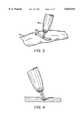
- FIG. 1is a perspective view of the use of a collagen welding rod for closing an incision in a blood vessel with the use of a laser or bipolar RF electrode;
- FIG. 2is a detail of the denatured or melted collagen material in the weld joint of FIG. 1;
- FIG. 3is a perspective view of the use of a collagen strip in the laser joining of an incision.
- FIG. 4is a detail of denatured or melted collagen material being applied upon a tissue defect or lesion.
- FIGS. 5-11are photomicrographs which show the morphology of various welded tissue joints.
- biological tissueincludes cell layers in a protein framework which provides tensile strength.
- the proteinsare amino acids, and it is known that the application of heat or optical energy can denature such proteins. When the source of heat or energy is removed, the proteins if not totally broken down cool and begin to reconfigure and form an approximate replication of the prior tissue structure.
- the prior artteaches that the application of either optical energy from a laser or RF energy from a suitable generator could be used to bring the temperature of the biological tissue above room temperature but below the boiling point of water (preferably between 45°-75° C. and more prefer 60°-70° C.).
- the denaturing of collagen, a major source of protein in the human bodycan also be achieved by the application of energy, and is believed to go into solution and form a type of "biological glue" which seals the incision or discontinuity in the biological tissue.
- biological gluewhich seals the incision or discontinuity in the biological tissue.
- Collagenis known for use in the medical field as a material for repairing tissue damage caused by thermal, chemical or mechanical trauma (see, e.g., "Collagen: Its Place In the Medical Industry” by J. M. Pachence, et al., Medical Device and Diagnostic Industry, January, 1987).
- this materialcan be used as a filler which can be placed in the path of the laser beam, melted or denatured, and directed into the incision or the tissue which is to be reconstructed.
- Bipolar or unipolar RF energywill also yield the same or substantially similar results. Based on qualitative observations, the additional collagen molecules provided by the filler material allows the tensile strength of the welded incision to be significantly increased.
- the application of optical energy and the use of additional collagen materialprovides several advantages in addition to increased tensile strength.
- the healing time of the woundis accelerated because blood supply to the affected tissue can be reestablished immediately after the surgical procedure.
- little or no scarringis produced because sutures are eliminated or substantially minimized.
- the various techniques disclosed hereinenhance the accuracy of the welding procedure thus avoiding optical or RF energy damage to adjacent or unintended areas of such tissue.
- a wide variety of materialsmay be used as a filler in this welding procedure.
- the most common sourceis collagen which may be obtained from bovine hides.
- Another materialwhich is ideal from the standpoint of melting, flowing, and compatibility with biological tissue, is a collagen-like substance which has been modified by dissolving collagen in water and modifying the thusly dissolved collagen to render its surface charge effectively more positive than prior to modification. This material is well known and is disclosed, e.g., in U.S. Pat. No. 4,238,480.
- the modified collagenis freeze-dried to form a solid mass of gelatin.
- this mass of gelatinalone or in combination with other collagen material, may be formed in the shape of a rod, strip, film or flake and utilized as a filler in a laser welding procedure.
- Semed Fa collagen preparation manufactured in native fiber form without any chemical or enzymatic modifications
- Semed Sa lyophilized collagen powder extracted from fresh bovine hides. Each of these products is available from Semex Medical, Frazer, Pa.
- the Semed F materialis a Type I collagen (greater than 95%)
- the Semed Sis a mixture of Type I and Type III collagen macromolecules in which the shape and dimension of tropocollagen in its natural helical orientation is retained.
- Either of the Semed S and Semed F collagen materialmay be formed into welding filler material by suspending a suitable amount (usually between about 0.5 and 10 weight percent) of the material in deionized water to form a viscous solution followed by drying the solution under the action of heat or by freeze-drying of the solution, followed by vacuum treating and heating steps.
- a suitable amountusually between about 0.5 and 10 weight percent
- the final shape of the materialcan be in the form of a rod, strip, powder, etc.
- a paste formulationmay also be formed by dissolving relatively large amounts of the material in relatively small amounts of saline or deionized water.
- the shapes of these formed materialsare solid and soft but firm. These materials may be readily sliced or cut to the desired sizes for use in the laser welding procedure. Also, the desired size and shape can be achieved by freeze-drying the material in a suitably sized mold which is configured to the desired size and shape of the product.
- the thicknesses of the rods or sheetscan be between 1/4 and 2 mm, depending upon the size of the incision to be joined or area of tissue to be reconstructed. When the paste form is utilized, it may be painted or dropped onto the areas of tissue to be joined or reconstructed.
- the surgeoncan choose from a wide variety of shapes, sizes, densities, thicknesses and configurations of such filler material depending upon the type of tissue to be welded.
- the concentration of the collagen in the liquid which is to be freeze-driedcan range from 0.5-10% and preferably 1-5%, with the lower concentrations forming less dense or discontinuous solids. At lower concentrations of 0.5 to 1%, the Semed F forms a structure which approximates dense cobwebs.
- Native collagen filmwherein the film strength is preserved and the triple-helix structure of the collagen polymer is maintained intact, can also be used, either alone or with a plasticizer incorporated therewith.
- a typical collagen sheetis cast from solution to a thickness of about 1.8 to 2 mm and contains the following composition by weight: collagen 70.3%, plasticizer (typically glycerol or glycerine) 16.9%, water 9%, other 3.8%.
- plasticizertypically glycerol or glycerine
- Such a materialis available from Norwood Medical Products Division of Norwood Industries, Inc., Malverne, Pa.
- gelatin or other water soluble forms of collagenWhen gelatin or other water soluble forms of collagen are utilized, certain advantages are provided in that the material will readily polymerize at body temperatures to form a stable subcutaneous gel. In addition, when implanted into the body as filler material in the weld joint, the polymerized material will become rapidly populated by host fibroblasts. Thus, the material becomes vascularized and can remain histologically, stable for up to 18 months.
- gelatin material per seis that it is highly soluble in blood such that the flow of blood across the material will cause it to dissolve.
- gelatin or other soluble collagen materialwhen used alone as laser weld filler should be limited to areas where direct contact with blood is avoided or minimized.
- mixtures of the various types of collagenit is more advantageous to use mixtures of the various types of collagen to obtain the most desirable features of each grade.
- a 50/50 mixture of Semed F and Semed Sallows the joint to obtain the higher tensile strength values of the F grade while retaining the superior flow properties of the S grade.
- Proportions ranging from 3:1 to 1:3also form useful mixtures.
- the gelatin material described abovecan be used in combination with the Semed F to achieve similar results.
- low melting polymers or polymeric materialssuch as copolymers of polyhydroxy buteric acid and valeric acid are useful in certain applications.
- Plasticizerssuch as polysaccharides may be included to further lower the melting point of these copolymers to below 200° F.
- These polymersmay also be mixed with the collagen or gelatin to increase the strength of the final weld joint. The melting
- a wide variety of energy sourcesmay be used to provide the desired energy for effecting the weld repair.
- Typical laser devicesare listed in Tables I and II.
- Low wattage laser energy devicessuch as those utilizing argon or CO 2 , would be the most useful for such welding because of their lower energy output.
- Higher energy output devicessuch as electrostatic and RF frequency coagulators (available from Everest, ValleyLab, or Medtronics) using bipolar tips can also be used to denature or melt the collagen filler materials. Since these devices have greater power input, they can burn the collagen to a greater extent than the argon or CO 2 lasers.
- One skilled in the artis able to control and successfully utilize these higher power devices in accordance with the teachings of the present invention.
- An argon beam coagulatorsuch as those made by Beacon Laboratories or Birtcher, are also suitable since they provide an easily controllable flame or heat source which can be utilized to melt the filler material and surrounding tissue to form the weld joint.
- FIG. 1An incision 10 in a blood vessel 20 is closed by first applying three approximating sutures 30a, 30b and 30 followed by heating the tissue on either side of the incision with the laser 40.
- Filler materiale.g., collagen
- the filler material 50is literally melted (i.e., denatured) to provide additional collagen which flows onto or over the incision, mixes with the melted or denatured tissue, and thereafter cools and fuses to the underlying tissue substrate.
- FIG. 2shows a detail of the joint as it is being made by this procedure.
- additional collagen materialallows the tensile strength of the joint to be significantly increased over weld joints which do not include additional collagen filler material.
- This difference in tensile strengthis due to the fact that the collagen filler material provides an additional collagen molecular substrate specifically in the area to be joined.
- the present techniquetherefore is analogous to the tungsten inert gas ("TIG") welding of metals such as steel or aluminum.
- TIGtungsten inert gas
- additional filler metalis almost always used on thin sections. Since the biological tissue to be joined is often relatively thin, similar improvements are obtained when using a filler material than by attempting to make the joint without such filter material.
- salinecan be used. This is accomplished by dipping the collagen welding rod into saline prior to placing the saline dipped collagen welding rod adjacent to joint area or by dripping saline into the weld. In actual testing, saline cooling makes a different of approximately 23° C. in the joint area (e.g., about 47° C. compared to about 70° C. without saline cooling).
- the present inventionresolves many of the problems of the prior art.
- This problemis due in part to the inability of the surgeon to uniformly melt the biological tissue on each side of the joint to obtain a satisfactory weld.
- additional collagen materialis supplied to the joint from the rod to compensate for any overmelting of tissue on either side of the joint.
- Thisalso provides an abundance of additional material to seal voids or other defects caused by overheating of tissue.
- All different types of biological tissuemay be treated according to the present procedures.
- all types of blood vessels, including veins, arteries, etc. in the vascular systemcan be connected or repaired, as can muscle, fascia, tendon, skin or even nerve material.
- FIG. 3Another procedure in accordance with the present invention is illustrated in FIG. 3.
- the incisionis covered with a flat strip of collagen material 60 along its entire length.
- the adjacent blood vessel walls 70 on each side of the incisionare overlapped by this strip 60 of collagen material.
- the laser 80heats the strip of material and the adjacent blood vessel walls 70 to denature those materials into a mass which then solidifies to form the laser welded joint.
- the use of the strip of collagen material 60facilitates the welding operation and improves the resultant tensile strength of the weld joint.
- FIG. 4shows a detail of the use of the strip material to fill a tissue defect or other lesion.
- the filler metalin order to insure that the placement of the welding rod remains in the appropriate position for allowing denatured collagen to flow into the joint area, it is possible to secure or attach the filler metal to the area to be joined.
- An easy way to accomplish thisis to dip the filler material into fibrin glue prior to applying the filler material to the area to be welded.
- the fibrin glue or other biological tissue adhesivealso appears to act as a flux which assists in directing the denatured or melted collagen material into the incision.
- the welding procedureis made easier by utilizing an energy absorbing aid in conjunction with the filler material.
- These aidsassist in the absorption of energy by the filler material so that the denaturing or melting process is more efficient, i.e., more of the energy is directly utilized to denature or melt the filler material rather than is scattered to other areas of the body near the tissue to be repaired.
- Preferred energy absorbing aidsinclude any of the numerous dyes, such as Vital Green or Basic Red.
- the color of the absorbing acid or dyeshould match the wavelength of the transmitted energy for optimum results.
- any substancepreferably which is in liquid form and which is capable of absorbing energy and transmitting the absorbed energy to the filler material, may be used.
- the blood or hemoglobin of the patientmay be used. Water or other physiologic solutions are also useful.
- the energy absorbing aidis applied to the filler material to form a coating thereon.
- the filler materialmay simply be dipped into a reservoir of the energy absorbing aid.
- More complex arrangementssuch as a spraying device or pump, can be used to apply the energy absorbing aid to the filler material, if desired.
- the energy absorbing aidcan be applied to the tissue to be repaired. This is easily accomplished, since the tissue is often cut and is bleeding to provide a suitable source of energy absorbing aid, i.e., blood. Also, the use of a dye is advantageous since it allows the joint to be easily viewed by the surgeon to determine exactly where the welding procedure must be conducted.
- the welding procedurecan be performed endoscopically: i.e., access to the area desired to be repaired or reconstructed can be made through multiple naturally or surgically created apertures: one aperture is used for insertion of the laser, another for the insertion of the filler material, and a third for monitoring the procedure with an optical fiber connected to an eye-piece or a video camera while the procedure can be visually observed through the eyepiece or camera, the presentation of the procedure on a monitor is preferred because the incision can be viewed in an enlarged mode so that the surgeon can accurately determine the proper placement of the filler material and completion of the joint.
- a dogwas anesthetized and its neck and groin area prepared for access.
- the carotid artery and jugular veinwere exposed and clamped, and a one inch incision was made in each one.
- An argon laser operated at about one-half wattwas used to reweld the clamped joints with one of Semed S, Semed F, and modified collagen material (i.e., gelatin) as described above.
- Sutureswere included at each end of the incision to prevent propagation of the incision during welding.
- the gelatin sampleswelded beautifully in that they readily melted, and simply and easily filled incision and rapidly formed a solid weld joint. However, upon exposure to blood, this material was solubilized by the blood which broke through the weld due to dissolution.
- the Semed F samplesdid not flow as readily into the joint, but once the joint was made, a very high tensile strength repair was obtained.
- the performance of the Semed Swas intermediate between the modified polymer and Semed F both with respect to joint strength and fluidity. Mixtures of either Semed S or modified collagen (gelatin) with the Semed F material, in a 50/50 ratio provides the benefits of each material are achieved in a single filler rod material.
- Vital Green dyewas applied to the tissue and filler material.
- the filler materialwas merely dipped into the dye.
- the dye coated filler and tissuegreatly facilitated the welding operation as it was easier to apply the optical energy to the desired locations.
- FIGS. 5-11illustrate the usefulness of the welding procedures of the present invention by showing its effects on various welded tissue joints. These were generated by operating on dogs to incise normal tissue, followed by welding to repair the incision.
- FIG. 5shows the results of a dog aorta which was welded with the mixed collagen filler material two days after welding.
- the nuclei and cell structure of the aortaappear normal and no karyolysis is evident.
- FIG. 6shows this filler material and the welded vena cava adjacent the aorta of FIG. 5 two weeks after welding.
- the welded vena cava and collagen filler materialare juxtaposed to form an intact weld joint across the incision. No evidence of thrombosis is seen at this joint or surrounding tissue.
- FIGS. 7 and 8further illustrate the weld joint of FIG. 6. These FIGS. show the filler material bridging the incision.
- the collagen filler materialappears as a large mass at the upper left hand corner of the photograph.
- the incision in the vena cavais just visible at the point where the material was transsected prior to placing in fixative.
- the fibrillar structure of the welding materialis evident.
- a low concentration of welding material as a thin bandbridges the incision which appears at the lower left corner of the photomicrograph. The incision is closed by this material, and the vena cava architecture is intact.
- FIG. 9illustrates welded skin tissue.
- the gelatin welding materialbridges the incision but has relatively poor tensile strength when tested about ten minutes after making the weld joint.
- the weld jointwas properly made and, as noted above, the strength of the welded joint can be improved by including collagen in the filler material.
- FIG. 10illustrates a welded coating of gelatin material placed under the skin.
- the coatingis able to hold the skin together for up to about ten minutes before losing strength due to saturation and dissolution in blood. Again, proper selection of a welding material which includes insoluble collagen will provide a higher strength coating.
- FIG. 11illustrates the welding of muscle tissue with a mixed collagen filler material. The incision is clearly filled and joined by the welding material to produce a strong joint.
- a wide variety of devicescan be used to place the welding material in the vicinity of the tissue to be repaired.
- a tube of collagen welding materialcan be placed concentrically around the laser.
- the surgeoncan urge the tube forward toward the distal end of the laser, where it can be melted by the energy.
- the tubecan be dyed with an energy absorbing aid to assist in the melting procedure.
- the surgeoncan urge further material into the path of the laser beam.
- a pair of grasping forcepscan also be used.
- a catheter or stentwhich includes a tubular covering of filler material can be introduced into the vessel beneath the area to be repaired. Thereafter, the laser welding procedure is conducted on the outside of the vessel, to melt both the vessel and the collagen material which is immediately below. Again, if desired, the collagen material can be dyed to increase its absorption of energy and melting efficiency.
- Both bi-polar and uni-polar RF electrodeswere also utilized to denature or melt various samples of modified gelatin, Semed F and Semed S, both alone and in combination, into arteriotomies and venotomies.
- a vascular anastomosiswas also crated using Semed F in accordance with the above-described welding technique.
- the weld jointwas observed to be of high tensile strength. Also, attempts at approximating muscle, tendon and skin have been successfully completed.
Landscapes
- Health & Medical Sciences (AREA)
- Life Sciences & Earth Sciences (AREA)
- Surgery (AREA)
- Animal Behavior & Ethology (AREA)
- Veterinary Medicine (AREA)
- Public Health (AREA)
- General Health & Medical Sciences (AREA)
- Heart & Thoracic Surgery (AREA)
- Molecular Biology (AREA)
- Medical Informatics (AREA)
- Biomedical Technology (AREA)
- Engineering & Computer Science (AREA)
- Nuclear Medicine, Radiotherapy & Molecular Imaging (AREA)
- Medicinal Chemistry (AREA)
- Chemical & Material Sciences (AREA)
- Dermatology (AREA)
- Biophysics (AREA)
- Oral & Maxillofacial Surgery (AREA)
- Transplantation (AREA)
- Epidemiology (AREA)
- Physics & Mathematics (AREA)
- Electromagnetism (AREA)
- Otolaryngology (AREA)
- Materials For Medical Uses (AREA)
- Surgical Instruments (AREA)
- Laser Surgery Devices (AREA)
- Lining Or Joining Of Plastics Or The Like (AREA)
Abstract
Description
The present application is a continuation of application Ser. No. 08/231,998, filed on Apr. 21, 1994, now abandoned, which was a continuation of application Ser. No. 07/832,171, filed on Feb. 6, 1992, now abandoned, which was a continuation-in-part of application Ser. No. 07/654,860, filed on Feb. 12, 1991, now U.S. Pat. No. 5,156,613.
1. Technical Field
The present invention relates to the use of laser emitted optical energy or radio frequency ("RF") energy for joining, repairing or reconstructing biological tissue. In particular, the present invention relates to a method of utilizing a welding rod filler material in combination with such optical or RF energy to join, repair or rebuild biological tissue.
2. Background Art
Optical energy, in particular that generated by lasers, has been applied and utilized in the medical field for a variety of surgical purposes. The medical industry initially utilized industrial lasers for the destruction of tumors or surface lesions in patients. At that time, the lasers were relatively crude, high powered and ineffective for delicate internal biological applications.
Subsequently, a variety of cauterization techniques were developed utilizing either laser or RF techniques. Laser optical energy was also utilized to reduce the flow of blood in an open wound or in a surgically created incision: the optical energy being supplied in sufficient quantity to sear or burn the blood vessels thus sealing the open ends of the capillaries and preventing blood flow. A typical use of laser cauterization is described in U.S. Pat. No. 4,122,853. Again, the types of lasers utilized at that time provided very high power application and very high wattage with the surrounding tissue also being destroyed, thus causing longer healing times, infection and scarring.
As newer, lower powered lasers were developed, techniques were developed for atheroma ablation or other endarterectomy procedures for blood vessels. One such procedure is disclosed in U.S. Pat. No. 4,878,492. The CO2, YAG and Excimer lasers all provided substantial improvements in these procedures due to their lower power output. These more sophisticated devices each provide better aiming of a narrower optical energy beam such that destruction of the walls of the blood vessels can be minimized. Also, advances in optical fiber technology allowed the surgeon to direct more accurately the optical energy to the desired location with greater precision.
Lasers have also been used to "glaze" the internal surfaces of blood vessels after balloon dilation and laser angioplasty in an attempt to prevent medical recollapse, intimal fibroplasia, and reobliteration.
Another procedure which has been developed includes the use of optical energy for welding or otherwise joining or connecting biological tissue. The original attempts to carry out these procedures began in the late 1960's and almost all universally met with failure not so much because of an inability to weld or join the tissue together, but because of the weakness of the resulting weld. The use of the lower powered laser devices, either alone or in combination with physiologic solutions, however, allowed the surgeon to cool the weld site sufficiently to obtain slight improvements in weld strength. Furthermore, RF energy has recently been utilized in both uni- and bi-polar generators to attempt to "weld" or "solder" biological tissue.
U.S. Pat. No. 4,672,969 discloses one method and apparatus for utilizing laser emitted optical energy to effect wound closure or other reconstruction of biological tissue by applying the optical energy to produce thermal heating of the biological tissue to degree suitable for denaturing the tissue proteins such that the collagenous elements of the tissue form a "biological glue" which seals the tissue to effect the joining. This glue is later reabsorbed by the body during the healing process. The patent discloses a number of different types of lasers with preference stated for the Nd:YAG type, because its particular wavelength allows optical energy to propagate without substantial attenuation through water and/or blood for absorption in the tissue to be repaired.
Despite these improvements, however, the weakness of the weld joint still remains as the primary disadvantage of this procedure and extensive current research is being conducted in an attempt to improve on that deficiency. I have now invented a simple yet elegant welding procedure for biological tissue utilizing laser or RF energy which overcomes the shortcomings of the prior art.
The present invention relates to a method of joining or reconstructing biological tissue which comprises applying energy to the biological tissue while providing a suitable filler material thereto; denaturing or melting the filler material and adjacent biological tissue with the energy to cause mixing of the denatured or melted filler material and biological tissue, thus joining or reconstructing such tissue.
The filler material is preferably collagen and one embodiment of the inventive method includes adhesively attaching the collagen filler material to the biological tissue to assure proper placement thereupon. This may be achieved by applying the collagen material adjacent the biological material with fibrin glue or other biological tissue adhesive.
This method may also include applying an energy absorption aid to one of the filler materials or the biological tissue, or both, to facilitate absorption of the applied energy thereby. Generally, the energy absorbing aid is applied to preselected locations prior to the application of energy thereto, and it also assists in visually determining the areas to be joined or reconstructed. Preferred energy absorbing aids include dyes, such as Vital Green or Basic Red, blood or water.
Often, the biological tissue includes an incision and the method enables the surgeon to enclose the incision by the mixing and joining of the denatured or melted filler material and biological tissue. If desired, spaced sutures may be placed in tissue surrounding the incision to fix the position of adjacent tissue.
The filler material may be prepared by dissolving a predetermined amount of collagen material in water to form a solution, followed by drying or freeze drying of the solution in the desired form and shape of the collagen filler material. Preferably, the collagen material used to prepare the filler material is a mixture of an insoluble collagen material and a soluble collagen material in a weight ratio of about 1:3 to 3:1.
The present method also contemplates applying a physiologically acceptable solution to one of the collagen filler materials or the biological tissue to control the temperature of the joint due to the energy imparted thereto. The applied energy may be provided as optical energy (i.e., by a laser), from an RF generator, or by an inert gas beam coagulator, since these devices have sufficient power dissipation to cause the energy or heat that they produce to be absorbed by the tissue and collagen filler material. The heat provided by the coagulator (or which is converted from the applied laser or RF energy) generally should be within a range bounded by the minimum absorption rate at which the protein elements of the tissue and collagen filler material are converted to melted collagen and by a maximum absorption rate which would cause water in the tissue or collagen filler material to boil. The RF energy may be provided by uni- or bipolar techniques, since each will melt the collagen filler material into the defect or joint area. Thus, the protein elements of the tissue and the collagen filler metal can be melted or denatured, mixed or combined, and then cooled to form a weld joint.
When the biological tissue includes a lesion, the method further comprises forming a seal of collagen material near or upon the lesion. When the lesion comprises at least two separated segments of biological tissue, the method further comprises placing the two segments of tissue in close proximity, and guiding the energy source and collagen filler material into the area of their junction for joining or reconstruction thereof.
The features and advantages of the present invention are more readily understood when read in conjunction with the attached drawing figures wherein
FIG. 1 is a perspective view of the use of a collagen welding rod for closing an incision in a blood vessel with the use of a laser or bipolar RF electrode;
FIG. 2 is a detail of the denatured or melted collagen material in the weld joint of FIG. 1;
FIG. 3 is a perspective view of the use of a collagen strip in the laser joining of an incision; and
FIG. 4 is a detail of denatured or melted collagen material being applied upon a tissue defect or lesion; and
FIGS. 5-11 are photomicrographs which show the morphology of various welded tissue joints.
It is well known that biological tissue includes cell layers in a protein framework which provides tensile strength. The proteins are amino acids, and it is known that the application of heat or optical energy can denature such proteins. When the source of heat or energy is removed, the proteins if not totally broken down cool and begin to reconfigure and form an approximate replication of the prior tissue structure.
The prior art teaches that the application of either optical energy from a laser or RF energy from a suitable generator could be used to bring the temperature of the biological tissue above room temperature but below the boiling point of water (preferably between 45°-75° C. and more prefer 60°-70° C.). The denaturing of collagen, a major source of protein in the human body, can also be achieved by the application of energy, and is believed to go into solution and form a type of "biological glue" which seals the incision or discontinuity in the biological tissue. Thus, it is possible to seal lesions, anastomose a severed or incised vessel or to reconstruct diseased or damaged tissue.
I have found that a major disadvantage of such laser welding procedures for rejoining incised tissue is that insufficient tissue material is present for completing a successful joint. When optical energy from the laser actually denatures or melts the tissue in the areas to be joined, a portion of the tissue thickness is reduced so that the denatured materials can flow towards each other and stick together to form the joint. On relatively thin sections of tissue to be joined, such as in repairing an incised blood vessel wall, there is insufficient denatured material in the joint area for providing a sound, high tensile strength connection.
Collagen is known for use in the medical field as a material for repairing tissue damage caused by thermal, chemical or mechanical trauma (see, e.g., "Collagen: Its Place In the Medical Industry" by J. M. Pachence, et al., Medical Device and Diagnostic Industry, January, 1987). I have found that this material can be used as a filler which can be placed in the path of the laser beam, melted or denatured, and directed into the incision or the tissue which is to be reconstructed. Bipolar or unipolar RF energy will also yield the same or substantially similar results. Based on qualitative observations, the additional collagen molecules provided by the filler material allows the tensile strength of the welded incision to be significantly increased.
The application of optical energy and the use of additional collagen material provides several advantages in addition to increased tensile strength. The healing time of the wound is accelerated because blood supply to the affected tissue can be reestablished immediately after the surgical procedure. In addition, little or no scarring is produced because sutures are eliminated or substantially minimized. Furthermore, the various techniques disclosed herein enhance the accuracy of the welding procedure thus avoiding optical or RF energy damage to adjacent or unintended areas of such tissue.
A wide variety of materials may be used as a filler in this welding procedure. The most common source is collagen which may be obtained from bovine hides. Another material, which is ideal from the standpoint of melting, flowing, and compatibility with biological tissue, is a collagen-like substance which has been modified by dissolving collagen in water and modifying the thusly dissolved collagen to render its surface charge effectively more positive than prior to modification. This material is well known and is disclosed, e.g., in U.S. Pat. No. 4,238,480. The modified collagen is freeze-dried to form a solid mass of gelatin. In accordance with the teachings of the present invention, this mass of gelatin, alone or in combination with other collagen material, may be formed in the shape of a rod, strip, film or flake and utilized as a filler in a laser welding procedure.
Other forms of collagen which are suitable for use in the present invention include Semed F, a collagen preparation manufactured in native fiber form without any chemical or enzymatic modifications, and Semed S, a lyophilized collagen powder extracted from fresh bovine hides. Each of these products is available from Semex Medical, Frazer, Pa. The Semed F material is a Type I collagen (greater than 95%), while the Semed S is a mixture of Type I and Type III collagen macromolecules in which the shape and dimension of tropocollagen in its natural helical orientation is retained.
Either of the Semed S and Semed F collagen material may be formed into welding filler material by suspending a suitable amount (usually between about 0.5 and 10 weight percent) of the material in deionized water to form a viscous solution followed by drying the solution under the action of heat or by freeze-drying of the solution, followed by vacuum treating and heating steps. As above with the gelatin material, the final shape of the material can be in the form of a rod, strip, powder, etc. A paste formulation may also be formed by dissolving relatively large amounts of the material in relatively small amounts of saline or deionized water.
The shapes of these formed materials are solid and soft but firm. These materials may be readily sliced or cut to the desired sizes for use in the laser welding procedure. Also, the desired size and shape can be achieved by freeze-drying the material in a suitably sized mold which is configured to the desired size and shape of the product. The thicknesses of the rods or sheets can be between 1/4 and 2 mm, depending upon the size of the incision to be joined or area of tissue to be reconstructed. When the paste form is utilized, it may be painted or dropped onto the areas of tissue to be joined or reconstructed. Thus, the surgeon can choose from a wide variety of shapes, sizes, densities, thicknesses and configurations of such filler material depending upon the type of tissue to be welded.
The concentration of the collagen in the liquid which is to be freeze-dried can range from 0.5-10% and preferably 1-5%, with the lower concentrations forming less dense or discontinuous solids. At lower concentrations of 0.5 to 1%, the Semed F forms a structure which approximates dense cobwebs.
Native collagen film, wherein the film strength is preserved and the triple-helix structure of the collagen polymer is maintained intact, can also be used, either alone or with a plasticizer incorporated therewith. A typical collagen sheet is cast from solution to a thickness of about 1.8 to 2 mm and contains the following composition by weight: collagen 70.3%, plasticizer (typically glycerol or glycerine) 16.9%, water 9%, other 3.8%. Such a material is available from Norwood Medical Products Division of Norwood Industries, Inc., Malverne, Pa.
When gelatin or other water soluble forms of collagen are utilized, certain advantages are provided in that the material will readily polymerize at body temperatures to form a stable subcutaneous gel. In addition, when implanted into the body as filler material in the weld joint, the polymerized material will become rapidly populated by host fibroblasts. Thus, the material becomes vascularized and can remain histologically, stable for up to 18 months. One problem with gelatin material per se, however, is that it is highly soluble in blood such that the flow of blood across the material will cause it to dissolve. Thus, gelatin or other soluble collagen material when used alone as laser weld filler should be limited to areas where direct contact with blood is avoided or minimized.
It is more advantageous to use mixtures of the various types of collagen to obtain the most desirable features of each grade. For example, a 50/50 mixture of Semed F and Semed S allows the joint to obtain the higher tensile strength values of the F grade while retaining the superior flow properties of the S grade. Proportions ranging from 3:1 to 1:3 also form useful mixtures. In addition, the gelatin material described above can be used in combination with the Semed F to achieve similar results.
In addition, low melting polymers or polymeric materials such as copolymers of polyhydroxy buteric acid and valeric acid are useful in certain applications. Plasticizers such as polysaccharides may be included to further lower the melting point of these copolymers to below 200° F. These polymers may also be mixed with the collagen or gelatin to increase the strength of the final weld joint. The melting
TABLE I __________________________________________________________________________TYPE WAVELENGTH (μ) F ENERGY RANGE/PHOTONS PENETRATION COMMENTS __________________________________________________________________________CO2 10.6 2.8 × 10.sup.13 3.7 × 10.sup.19 microns low penetration Helium-Neon .634 almost nil nil target laser Neodymium - Multiple Harmonics Yag 1.06 2.8 × 10.sup.14 5.3 × 10.sup.18 high yttrium-aluminum garnet 0.532 5.6 × 10.sup.14 2.7 × 10.sup.18 Welds tissue Increasing at low energy penetration increasing 0.353 8.4 × 10.sup.14 1.8 × 10.sup.18 0.266 1.1 × 10.sup.15 1.3 × 10.sup.18 Argon 4.8 1.1 × 10.sup.14 3.8 × 10.sup.19 2-400μ water absorption 5.12 Excimer (Excitable dimer) Xe CL .308 9.7 × 10.sup.14 1.6 × 10.sup.18 <20μ very short Xe F .351 8.6 × 10.sup.14 1.8 × 10.sup.16 gasifies operational Kr F .248 1.3 × 10.sup.15 1.3 × 10.sup.18 calcified distance Ar F .193 1.6 × 10.sup.15 9.7 × 10.sup.17 plaques increases __________________________________________________________________________ safety
TABLE II __________________________________________________________________________Proposed Laser-Fiberoptic Systems Wavelength Pulse Principal Plaque Ablation Operating Laser NM Duration Fiber Efficiency Calcified Range __________________________________________________________________________Excimer 248 H Y ? 308 2-200 nsec Silica H Y L 351 M-H Y(?) L Argon 488, 512 40 msec-CW Silica L-M N M-H Dye Laser 450-800 1-2 μsec Silica M ? M Nd:YAG 1,064 10.sup.-9 -10.sup.-12 sec None H N(?) O CW Silica L N M-H Ha:YLF 2,060 100 μsec Silica M ? M-H(?) Er:YAG 2,940 100 μsec ZnF.sub.4 H Y H CO.sub.2 10,600 1 μsec Halide (?) H Y(?) ? 10 msec Halide M-H N L CW Halide L N L __________________________________________________________________________ H, indicates high; Y, yes; L, low; M, medium; CW, continuous wave; N, no; Nd, Neodymium; Ha, Hafnium, Er, Erbium. 1, indicates extensive thermal damage; 2, strong water absorption; 3, possible mutagenicity; 4, nonthermal active mechanisms; 5, developmental fibers.
temperature of these polymers should be below about 212° F. and on the same order as the melting temperature of the collagen (i.e., between about 100°-200° F.).
A wide variety of energy sources may be used to provide the desired energy for effecting the weld repair. Typical laser devices are listed in Tables I and II. Low wattage laser energy devices, such as those utilizing argon or CO2, would be the most useful for such welding because of their lower energy output. Higher energy output devices, such as electrostatic and RF frequency coagulators (available from Everest, ValleyLab, or Medtronics) using bipolar tips can also be used to denature or melt the collagen filler materials. Since these devices have greater power input, they can burn the collagen to a greater extent than the argon or CO2 lasers. One skilled in the art, however, is able to control and successfully utilize these higher power devices in accordance with the teachings of the present invention.
An argon beam coagulator, such as those made by Beacon Laboratories or Birtcher, are also suitable since they provide an easily controllable flame or heat source which can be utilized to melt the filler material and surrounding tissue to form the weld joint.
The protocol for the process is further appreciated by reference to FIG. 1. Anincision 10 in ablood vessel 20 is closed by first applying three approximating sutures 30a, 30b and 30 followed by heating the tissue on either side of the incision with thelaser 40. Filler material (e.g., collagen) is applied to the incision by placing the tip ofwelding rod 50 into the laser beam near the heated portion of the incision. Thefiller material 50 is literally melted (i.e., denatured) to provide additional collagen which flows onto or over the incision, mixes with the melted or denatured tissue, and thereafter cools and fuses to the underlying tissue substrate. FIG. 2 shows a detail of the joint as it is being made by this procedure.
As noted above, the use of such additional collagen material allows the tensile strength of the joint to be significantly increased over weld joints which do not include additional collagen filler material. This difference in tensile strength is due to the fact that the collagen filler material provides an additional collagen molecular substrate specifically in the area to be joined. The present technique therefore is analogous to the tungsten inert gas ("TIG") welding of metals such as steel or aluminum. In the TIG process, additional filler metal is almost always used on thin sections. Since the biological tissue to be joined is often relatively thin, similar improvements are obtained when using a filler material than by attempting to make the joint without such filter material.
It has been found that a CO2 or argon laser with a half to one watt power is eminently suitable for making this type of joint. As noted above electrostatic generators can also be used. In addition, an argon beam electrocoagulator operated at 15-50 volts and 5-20 watts can also be used to denature and melt the collagen welding rod materials and surrounding tissue.
In an attempt to maintain the temperature of the tissue joint at a relatively low value, saline can be used. This is accomplished by dipping the collagen welding rod into saline prior to placing the saline dipped collagen welding rod adjacent to joint area or by dripping saline into the weld. In actual testing, saline cooling makes a different of approximately 23° C. in the joint area (e.g., about 47° C. compared to about 70° C. without saline cooling).
The present invention resolves many of the problems of the prior art. When welding biological tissues, it is difficult to achieve uniformly good results. This problem is due in part to the inability of the surgeon to uniformly melt the biological tissue on each side of the joint to obtain a satisfactory weld. With the use of collagen welding rod as proposed by the present invention, additional collagen material is supplied to the joint from the rod to compensate for any overmelting of tissue on either side of the joint. This also provides an abundance of additional material to seal voids or other defects caused by overheating of tissue. Thus, the reproducibility of the procedure and the attainment of uniform weld joints are significantly improved by the present invention.
All different types of biological tissue may be treated according to the present procedures. For example, all types of blood vessels, including veins, arteries, etc. in the vascular system can be connected or repaired, as can muscle, fascia, tendon, skin or even nerve material.
Another procedure in accordance with the present invention is illustrated in FIG. 3. In that FIG., the incision is covered with a flat strip ofcollagen material 60 along its entire length. The adjacentblood vessel walls 70 on each side of the incision are overlapped by thisstrip 60 of collagen material. Thelaser 80 heats the strip of material and the adjacentblood vessel walls 70 to denature those materials into a mass which then solidifies to form the laser welded joint. Again, the use of the strip ofcollagen material 60 facilitates the welding operation and improves the resultant tensile strength of the weld joint. FIG. 4 shows a detail of the use of the strip material to fill a tissue defect or other lesion.
In an alternate embodiment of the invention, in order to insure that the placement of the welding rod remains in the appropriate position for allowing denatured collagen to flow into the joint area, it is possible to secure or attach the filler metal to the area to be joined. An easy way to accomplish this is to dip the filler material into fibrin glue prior to applying the filler material to the area to be welded. In addition to retaining the filler in the appropriate area desired, the fibrin glue or other biological tissue adhesive also appears to act as a flux which assists in directing the denatured or melted collagen material into the incision.
The welding procedure is made easier by utilizing an energy absorbing aid in conjunction with the filler material. These aids assist in the absorption of energy by the filler material so that the denaturing or melting process is more efficient, i.e., more of the energy is directly utilized to denature or melt the filler material rather than is scattered to other areas of the body near the tissue to be repaired.
Preferred energy absorbing aids include any of the numerous dyes, such as Vital Green or Basic Red. The color of the absorbing acid or dye should match the wavelength of the transmitted energy for optimum results. However, any substance, preferably which is in liquid form and which is capable of absorbing energy and transmitting the absorbed energy to the filler material, may be used. Often, the blood or hemoglobin of the patient may be used. Water or other physiologic solutions are also useful.
Advantageously, the energy absorbing aid is applied to the filler material to form a coating thereon. The filler material may simply be dipped into a reservoir of the energy absorbing aid. More complex arrangements, such as a spraying device or pump, can be used to apply the energy absorbing aid to the filler material, if desired.
In addition, the energy absorbing aid can be applied to the tissue to be repaired. This is easily accomplished, since the tissue is often cut and is bleeding to provide a suitable source of energy absorbing aid, i.e., blood. Also, the use of a dye is advantageous since it allows the joint to be easily viewed by the surgeon to determine exactly where the welding procedure must be conducted.
In yet another embodiment, the welding procedure can be performed endoscopically: i.e., access to the area desired to be repaired or reconstructed can be made through multiple naturally or surgically created apertures: one aperture is used for insertion of the laser, another for the insertion of the filler material, and a third for monitoring the procedure with an optical fiber connected to an eye-piece or a video camera while the procedure can be visually observed through the eyepiece or camera, the presentation of the procedure on a monitor is preferred because the incision can be viewed in an enlarged mode so that the surgeon can accurately determine the proper placement of the filler material and completion of the joint.
The following examples illustrate applications of the welding procedures of the present invention. A dog was anesthetized and its neck and groin area prepared for access. The carotid artery and jugular vein were exposed and clamped, and a one inch incision was made in each one. An argon laser operated at about one-half watt was used to reweld the clamped joints with one of Semed S, Semed F, and modified collagen material (i.e., gelatin) as described above. Sutures were included at each end of the incision to prevent propagation of the incision during welding.
The gelatin samples welded beautifully in that they readily melted, and simply and easily filled incision and rapidly formed a solid weld joint. However, upon exposure to blood, this material was solubilized by the blood which broke through the weld due to dissolution. The Semed F samples did not flow as readily into the joint, but once the joint was made, a very high tensile strength repair was obtained. The performance of the Semed S was intermediate between the modified polymer and Semed F both with respect to joint strength and fluidity. Mixtures of either Semed S or modified collagen (gelatin) with the Semed F material, in a 50/50 ratio provides the benefits of each material are achieved in a single filler rod material.
To aid in the absorption of energy by the filler material and the tissue to be repaired, Vital Green dye was applied to the tissue and filler material. The filler material was merely dipped into the dye. The dye coated filler and tissue greatly facilitated the welding operation as it was easier to apply the optical energy to the desired locations.
FIGS. 5-11 illustrate the usefulness of the welding procedures of the present invention by showing its effects on various welded tissue joints. These were generated by operating on dogs to incise normal tissue, followed by welding to repair the incision.
FIG. 5 shows the results of a dog aorta which was welded with the mixed collagen filler material two days after welding. The nuclei and cell structure of the aorta appear normal and no karyolysis is evident.
FIG. 6 shows this filler material and the welded vena cava adjacent the aorta of FIG. 5 two weeks after welding. The welded vena cava and collagen filler material are juxtaposed to form an intact weld joint across the incision. No evidence of thrombosis is seen at this joint or surrounding tissue.
FIGS. 7 and 8 further illustrate the weld joint of FIG. 6. These FIGS. show the filler material bridging the incision. In FIG. 7, the collagen filler material appears as a large mass at the upper left hand corner of the photograph. The incision in the vena cava is just visible at the point where the material was transsected prior to placing in fixative. The fibrillar structure of the welding material is evident. In FIG. 8, a low concentration of welding material as a thin band bridges the incision which appears at the lower left corner of the photomicrograph. The incision is closed by this material, and the vena cava architecture is intact.
FIG. 9 illustrates welded skin tissue. The gelatin welding material bridges the incision but has relatively poor tensile strength when tested about ten minutes after making the weld joint. The weld joint was properly made and, as noted above, the strength of the welded joint can be improved by including collagen in the filler material.
FIG. 10 illustrates a welded coating of gelatin material placed under the skin. The coating is able to hold the skin together for up to about ten minutes before losing strength due to saturation and dissolution in blood. Again, proper selection of a welding material which includes insoluble collagen will provide a higher strength coating.
FIG. 11 illustrates the welding of muscle tissue with a mixed collagen filler material. The incision is clearly filled and joined by the welding material to produce a strong joint.
In the preceding FIGS., an Eximer CO2 laser was utilized as the energy source, with basic red dye or blood used as the energy absorbing aid. No difference in performance was seen using either fluid.
A wide variety of devices can be used to place the welding material in the vicinity of the tissue to be repaired. For example, in addition to the above-described arrangements, a tube of collagen welding material can be placed concentrically around the laser. Thus, the surgeon can urge the tube forward toward the distal end of the laser, where it can be melted by the energy. The tube can be dyed with an energy absorbing aid to assist in the melting procedure. As the end of the tube melts, the surgeon can urge further material into the path of the laser beam. To retain the area to be repaired in the proper position, a pair of grasping forceps can also be used.
In addition, for the repair of a blood vessel, a catheter or stent which includes a tubular covering of filler material can be introduced into the vessel beneath the area to be repaired. Thereafter, the laser welding procedure is conducted on the outside of the vessel, to melt both the vessel and the collagen material which is immediately below. Again, if desired, the collagen material can be dyed to increase its absorption of energy and melting efficiency.
Both bi-polar and uni-polar RF electrodes were also utilized to denature or melt various samples of modified gelatin, Semed F and Semed S, both alone and in combination, into arteriotomies and venotomies. A vascular anastomosis was also crated using Semed F in accordance with the above-described welding technique. The weld joint was observed to be of high tensile strength. Also, attempts at approximating muscle, tendon and skin have been successfully completed.
It is believed that numerous variations and modifications may be devised by those skilled in the art to the specifically disclosed invention, and it is intended that the appended claims cover all such modifications and embodiments as would fall within the true spirit and scope of the present invention.
Claims (12)
1. A method of joining or reconstructing biological tissue, said method comprising the following steps:
providing a solid filler material in the form of a preformed sheet, wherein the sheet comprises collagen, gelatin, or a mixture thereof;
placing the filler material over said biological tissue prior to applying energy, and
applying radiofrequency or optical energy to the filler material in an amount sufficient to melt or denature the filler material while said filler material is over the said biological tissue.
2. A method as in claim 1, wherein the filler material is cut to a desired size prior to placing on the tissue.
3. A method as in claim 1, wherein the energy is radiofrequency energy applied from an inert gas beam coagulator.
4. A method as in claim 1, further comprising applying a tissue adhesive to the tissue prior to placing the filler material.
5. A method of joining or restructuring biological tissue, said method comprising the following steps:
providing a filler material in the form of a preformed sheet which fuses to said biological tissue upon the application of energy, wherein the filler material comprises collagen, gelatin, or a mixture thereof;
placing the filler material over said biological tissue to be joined or restructured prior to applying energy; and
applying radiofrequency energy to the filler material in an amount sufficient to melt or denature the filler material and the said biological tissue after said filler material has been placed over the said biological tissue, wherein the radiofrequency energy is applied from an inert gas beam radiofrequency device.
6. A method as in claim 5, wherein the radiofrequency inert gas beam device is operated at between about 20 and 120 watts for about 1 to 60 seconds, so that about 20 to 1800 joules are delivered to the filler material and the biological tissue.
7. A method as in claim 6, wherein the radiofrequency inert gas beam device is operated at between about 35 and 80 watts for about 5 to 40 seconds, so that about 100 to 1200 joules are delivered to the filler material and the biological tissue.
8. An improved method for joining or reconstructing biological tissue wherein a collagen- or gelatin-containing material is placed over the tissue and energy is thereafter applied to fuse the material and tissue together, wherein the improvement comprises the step of applying radiofrequency energy to the material and said tissue under conditions which result in about 20 to 1800 joules of energy being delivered to the collagen or gelatin-containing material and the said tissue.
9. An improved method as in claim 8, wherein the collagen-containing material is in the form of a preformed a sheet.
10. An improved method as in claim 8, wherein the radiofrequency device is operated at between about 20 and 120 watts for about 1 to 60 seconds.
11. An improved method as in claim 8, wherein the radiofrequency device is operated at between about 35 and 80 watts for about 5 to 30 seconds, so that about 100 to 1200 joules are delivered to the collagen-containing material and the biological tissue.
12. An improved method as in claim 8, wherein the collagen-containing material is composed of a material selected from the group consisting of native collagen, collagen cast with a plasticizer, and gelatin.
Priority Applications (1)
| Application Number | Priority Date | Filing Date | Title |
|---|---|---|---|
| US08/461,227US5824015A (en) | 1991-02-13 | 1995-06-05 | Method for welding biological tissue |
Applications Claiming Priority (4)
| Application Number | Priority Date | Filing Date | Title |
|---|---|---|---|
| US07/654,860US5156613A (en) | 1991-02-13 | 1991-02-13 | Collagen welding rod material for use in tissue welding |
| US83217192A | 1992-02-06 | 1992-02-06 | |
| US23199894A | 1994-04-21 | 1994-04-21 | |
| US08/461,227US5824015A (en) | 1991-02-13 | 1995-06-05 | Method for welding biological tissue |
Related Parent Applications (1)
| Application Number | Title | Priority Date | Filing Date |
|---|---|---|---|
| US23199894AContinuation | 1991-02-13 | 1994-04-21 |
Publications (1)
| Publication Number | Publication Date |
|---|---|
| US5824015Atrue US5824015A (en) | 1998-10-20 |
Family
ID=27096836
Family Applications (1)
| Application Number | Title | Priority Date | Filing Date |
|---|---|---|---|
| US08/461,227Expired - Fee RelatedUS5824015A (en) | 1991-02-13 | 1995-06-05 | Method for welding biological tissue |
Country Status (6)
| Country | Link |
|---|---|
| US (1) | US5824015A (en) |
| EP (1) | EP0572526A4 (en) |
| JP (1) | JPH06505656A (en) |
| AU (1) | AU1444292A (en) |
| CA (1) | CA2103727A1 (en) |
| WO (1) | WO1992014513A1 (en) |
Cited By (135)
| Publication number | Priority date | Publication date | Assignee | Title |
|---|---|---|---|---|
| US5972007A (en)* | 1997-10-31 | 1999-10-26 | Ethicon Endo-Surgery, Inc. | Energy-base method applied to prosthetics for repairing tissue defects |
| US6217585B1 (en) | 1996-08-16 | 2001-04-17 | Converge Medical, Inc. | Mechanical stent and graft delivery system |
| US6221068B1 (en)* | 1998-01-15 | 2001-04-24 | Northwestern University | Method for welding tissue |
| US6251110B1 (en) | 1999-03-31 | 2001-06-26 | Ethicon Endo-Surgery, Inc. | Combined radio frequency and ultrasonic surgical device |
| US6257241B1 (en) | 1999-03-31 | 2001-07-10 | Ethicon Endo-Surgery, Inc. | Method for repairing tissue defects using ultrasonic radio frequency energy |
| US6287344B1 (en) | 1999-03-31 | 2001-09-11 | Ethicon Endo-Surgery, Inc. | Method for repairing tissue defects using an ultrasonic device |
| US6293955B1 (en) | 1996-09-20 | 2001-09-25 | Converge Medical, Inc. | Percutaneous bypass graft and securing system |
| US6310036B1 (en) | 1999-01-09 | 2001-10-30 | Last Chance Tissue Adhesives Corporation | High strength, Bio-compatible tissue adhesive and methods for treating vigorously bleeding surfaces |
| US6348679B1 (en) | 1998-03-17 | 2002-02-19 | Ameritherm, Inc. | RF active compositions for use in adhesion, bonding and coating |
| US6361559B1 (en) | 1998-06-10 | 2002-03-26 | Converge Medical, Inc. | Thermal securing anastomosis systems |
| US6371975B2 (en) | 1998-11-06 | 2002-04-16 | Neomend, Inc. | Compositions, systems, and methods for creating in situ, chemically cross-linked, mechanical barriers |
| US20020049503A1 (en)* | 2000-10-20 | 2002-04-25 | Michael Milbocker | Surgical repair of tissue defects |
| US6391049B1 (en) | 1999-10-06 | 2002-05-21 | Board Of Regents The University Of Texas System | Solid biodegradable device for use in tissue repair |
| US6398797B2 (en) | 1999-07-28 | 2002-06-04 | Cardica, Inc. | Tissue bonding system and method for controlling a tissue site during anastomosis |
| US6416486B1 (en) | 1999-03-31 | 2002-07-09 | Ethicon Endo-Surgery, Inc. | Ultrasonic surgical device having an embedding surface and a coagulating surface |
| US6458147B1 (en)* | 1998-11-06 | 2002-10-01 | Neomend, Inc. | Compositions, systems, and methods for arresting or controlling bleeding or fluid leakage in body tissue |
| US20020173809A1 (en)* | 1999-09-01 | 2002-11-21 | Fleischman Sidney D. | Sutureless anastomosis system deployment concepts |
| US6494889B1 (en) | 1999-09-01 | 2002-12-17 | Converge Medical, Inc. | Additional sutureless anastomosis embodiments |
| US20030014063A1 (en)* | 1996-11-08 | 2003-01-16 | Houser Russell A. | Percutaneous bypass graft and securing system |
| US20030023252A1 (en)* | 2001-07-05 | 2003-01-30 | Whayne James G. | Distal anastomosis system |
| US6547794B2 (en) | 2000-08-18 | 2003-04-15 | Auge', Ii Wayne K. | Method for fusing bone during endoscopy procedures |
| US20030093095A1 (en)* | 2001-07-05 | 2003-05-15 | Whayne James G. | Distal anastomosis system |
| US20030100921A1 (en)* | 1998-11-06 | 2003-05-29 | Neomend, Inc. | Systems, methods, and compositions for achieving closure of vascular puncture sites |
| US6607522B1 (en) | 2000-03-16 | 2003-08-19 | General Hospital Corporation | Methods for tissue welding using laser-activated protein solders |
| US6613348B1 (en)* | 2001-07-31 | 2003-09-02 | Manoj K. Jain | Process of controlling absorbency in collagen flakes |
| US6649888B2 (en) | 1999-09-23 | 2003-11-18 | Codaco, Inc. | Radio frequency (RF) heating system |
| US20030229365A1 (en)* | 2002-06-10 | 2003-12-11 | Whayne James G. | Angled vascular anastomosis system |
| US6685730B2 (en) | 2001-09-26 | 2004-02-03 | Rice University | Optically-absorbing nanoparticles for enhanced tissue repair |
| US6699245B2 (en) | 2001-02-05 | 2004-03-02 | A-Med Systems, Inc. | Anastomosis system and related methods |
| US20040068278A1 (en)* | 1999-12-06 | 2004-04-08 | Converge Medical Inc. | Anastomosis systems |
| US20040073256A1 (en)* | 2002-08-09 | 2004-04-15 | Kevin Marchitto | Activated surgical fasteners, devices therefor and uses thereof |
| US6743248B2 (en) | 1996-12-18 | 2004-06-01 | Neomend, Inc. | Pretreatment method for enhancing tissue adhesion |
| US20040106880A1 (en)* | 1999-10-25 | 2004-06-03 | Therus Corporation (Legal) | Use of focused ultrasound for vascular sealing |
| US6773699B1 (en) | 2001-10-09 | 2004-08-10 | Tissue Adhesive Technologies, Inc. | Light energized tissue adhesive conformal patch |
| US6780840B1 (en) | 2001-10-09 | 2004-08-24 | Tissue Adhesive Technologies, Inc. | Method for making a light energized tissue adhesive |
| US20040167244A1 (en)* | 2000-08-18 | 2004-08-26 | Auge Wayne K. | Methods and compositions for fusing bone during endoscopy procedures |
| US20040210282A1 (en)* | 2002-05-20 | 2004-10-21 | Flock Stephen T. | Electromagnetic treatment of tissues and cells |
| US6830756B2 (en) | 1998-11-06 | 2004-12-14 | Neomend, Inc. | Systems, methods, and compositions for achieving closure of vascular puncture sites |
| US6858035B2 (en) | 2001-07-05 | 2005-02-22 | Converge Medical, Inc. | Distal anastomosis system |
| US6875427B1 (en) | 2001-10-09 | 2005-04-05 | Tissue Adhesive Technologies, Inc. | Light energized tissue adhesive |
| US20050096542A1 (en)* | 1999-12-23 | 2005-05-05 | Lee Weng | Ultrasound transducers for imaging and therapy |
| US6899889B1 (en) | 1998-11-06 | 2005-05-31 | Neomend, Inc. | Biocompatible material composition adaptable to diverse therapeutic indications |
| US20050182449A1 (en)* | 2001-05-26 | 2005-08-18 | Map Technologies, Llc | Methods for electrosurgical electrolysis |
| US20050187542A1 (en)* | 2000-08-18 | 2005-08-25 | Map Technologies, Llc | Devices for electrosurgery |
| US6939348B2 (en) | 2003-03-27 | 2005-09-06 | Cierra, Inc. | Energy based devices and methods for treatment of patent foramen ovale |
| US6939364B1 (en) | 2001-10-09 | 2005-09-06 | Tissue Adhesive Technologies, Inc. | Composite tissue adhesive |
| US6949114B2 (en) | 1998-11-06 | 2005-09-27 | Neomend, Inc. | Systems, methods, and compositions for achieving closure of vascular puncture sites |
| US20050228369A1 (en)* | 2001-08-15 | 2005-10-13 | Map Technologies Llc | Devices for electrosurgery |
| US20050240170A1 (en)* | 1999-10-25 | 2005-10-27 | Therus Corporation | Insertable ultrasound probes, systems, and methods for thermal therapy |
| US20050251163A1 (en)* | 2001-07-05 | 2005-11-10 | Converge Medical, Inc. | Vascular anastomosis systems |
| US6994714B2 (en) | 2001-04-27 | 2006-02-07 | Cardica, Inc. | Anastomosis system |
| US6994686B2 (en) | 1998-08-26 | 2006-02-07 | Neomend, Inc. | Systems for applying cross-linked mechanical barriers |
| US7014644B1 (en) | 1999-07-28 | 2006-03-21 | Cardica, Inc. | Tissue bonding system and method for controlling a tissue site during anastomosis |
| US20060085031A1 (en)* | 2004-10-18 | 2006-04-20 | Michael Bettuchi | Extraluminal sealant applicator and method |
| US20060085033A1 (en)* | 2004-10-18 | 2006-04-20 | Criscuolo Christopher J | Surgical apparatus and structure for applying sprayable wound treatment material |
| US20060111738A1 (en)* | 2004-10-18 | 2006-05-25 | Thomas Wenchell | Apparatus for applying wound treatment material using tissue-penetrating needles |
| US7165552B2 (en) | 2003-03-27 | 2007-01-23 | Cierra, Inc. | Methods and apparatus for treatment of patent foramen ovale |
| US7186251B2 (en) | 2003-03-27 | 2007-03-06 | Cierra, Inc. | Energy based devices and methods for treatment of patent foramen ovale |
| US7238195B2 (en) | 2002-05-10 | 2007-07-03 | Tyco Healthcare Group Lp | Wound closure material applicator and stapler |
| US7257450B2 (en) | 2003-02-13 | 2007-08-14 | Coaptus Medical Corporation | Systems and methods for securing cardiovascular tissue |
| US7293562B2 (en) | 2003-03-27 | 2007-11-13 | Cierra, Inc. | Energy based devices and methods for treatment of anatomic tissue defects |
| US7300444B1 (en) | 1999-07-28 | 2007-11-27 | Cardica, Inc. | Surgical system and method for connecting hollow tissue structures |
| US7303570B2 (en) | 1999-07-28 | 2007-12-04 | Cardica, Inc. | Anastomosis tool having a connector holder |
| US7311701B2 (en) | 2003-06-10 | 2007-12-25 | Cierra, Inc. | Methods and apparatus for non-invasively treating atrial fibrillation using high intensity focused ultrasound |
| US7367975B2 (en) | 2004-06-21 | 2008-05-06 | Cierra, Inc. | Energy based devices and methods for treatment of anatomic tissue defects |
| US7371243B1 (en) | 1999-07-28 | 2008-05-13 | Cardica, Inc. | Surgical apparatus and method for anastomosis |
| WO2008079639A1 (en)* | 2006-12-20 | 2008-07-03 | Terumo Kabushiki Kaisha | Methods and apparatus for closing a layered tissue defect |
| US7473252B2 (en) | 2004-10-07 | 2009-01-06 | Coaptus Medical Corporation | Systems and methods for shrinking and/or securing cardiovascular tissue |
| US20090062639A1 (en)* | 2007-08-27 | 2009-03-05 | William Harrison Zurn | Automated vessel repair system, devices and methods |
| US20090216264A1 (en)* | 2005-09-19 | 2009-08-27 | Friedman Paul A | Implantable closure apparatus and methods |
| US7637924B2 (en) | 2003-03-27 | 2009-12-29 | Terumo Kabushiki Kaisha | Methods and apparatus for treatment of patent foramen ovale |
| US7682368B1 (en) | 1999-07-28 | 2010-03-23 | Cardica, Inc. | Anastomosis tool actuated with stored energy |
| US20100087815A1 (en)* | 2001-08-15 | 2010-04-08 | Nuortho Surgical Inc. | Electrosurgical Plenum |
| US7699859B2 (en) | 1999-07-28 | 2010-04-20 | Cardica, Inc. | Method of performing anastomosis |
| US20100127039A1 (en)* | 2008-11-24 | 2010-05-27 | Tyco Healthcare Group Lp | Pouch Used To Deliver Medication When Ruptured |
| US20100155454A1 (en)* | 2004-10-18 | 2010-06-24 | Tyco Healthcare Group Lp | Structure containing wound treatment material |
| US7771422B2 (en) | 2002-06-06 | 2010-08-10 | Nuortho Surgical, Inc. | Methods and devices for electrosurgery |
| US20100249763A1 (en)* | 2007-05-14 | 2010-09-30 | The Regents Of The University Of Colorado | Laser Tissue Fusion of Septal Membranes |
| US7850703B2 (en) | 1999-07-28 | 2010-12-14 | Cardica, Inc. | System for performing anastomosis |
| US20110036889A1 (en)* | 2004-10-18 | 2011-02-17 | Tyco Healthcare Group Lp | Structure for applying sprayable wound treatment material |
| US7923031B2 (en) | 2004-01-30 | 2011-04-12 | Ferrosan Medical Devices A/S | Haemostatic sprays and compositions |
| US7923431B2 (en) | 2001-12-21 | 2011-04-12 | Ferrosan Medical Devices A/S | Haemostatic kit, a method of preparing a haemostatic agent and a method of promoting haemostatis |
| US7955296B1 (en) | 2001-05-26 | 2011-06-07 | Nuortho Surgical, Inc. | Biologically enhanced irrigants |
| US7955288B2 (en) | 2002-12-11 | 2011-06-07 | Ferrosan Medical Devices A/S | Gelatine-based materials as swabs |
| US7963432B2 (en) | 2007-09-06 | 2011-06-21 | Cardica, Inc. | Driverless surgical stapler |
| US7972330B2 (en) | 2003-03-27 | 2011-07-05 | Terumo Kabushiki Kaisha | Methods and apparatus for closing a layered tissue defect |
| US20110184407A1 (en)* | 2010-01-26 | 2011-07-28 | Tyco Healthcare Group Lp | Hernia Repair System |
| US8021359B2 (en) | 2003-02-13 | 2011-09-20 | Coaptus Medical Corporation | Transseptal closure of a patent foramen ovale and other cardiac defects |
| US8021684B2 (en) | 2004-07-09 | 2011-09-20 | Ferrosan Medical Devices A/S | Haemostatic composition comprising hyaluronic acid |
| WO2011138347A3 (en)* | 2010-05-05 | 2012-01-12 | Aesculap Ag | Surgical system for connecting body tissue parts |
| US8109274B2 (en) | 2005-04-11 | 2012-02-07 | Terumo Kabushiki Kaisha | Methods and electrode apparatus to achieve a closure of a layered tissue defect |
| US8167805B2 (en) | 2005-10-20 | 2012-05-01 | Kona Medical, Inc. | Systems and methods for ultrasound applicator station keeping |
| US8221452B2 (en) | 1997-03-12 | 2012-07-17 | Neomend, Inc. | Systems and methods for sealing a vascular puncture |
| US8235979B2 (en) | 2001-08-15 | 2012-08-07 | Nuortho Surgical, Inc. | Interfacing media manipulation with non-ablation radiofrequency energy system and method |
| US8295912B2 (en) | 2009-10-12 | 2012-10-23 | Kona Medical, Inc. | Method and system to inhibit a function of a nerve traveling with an artery |
| US8374674B2 (en) | 2009-10-12 | 2013-02-12 | Kona Medical, Inc. | Nerve treatment system |
| US8388613B1 (en) | 2010-04-09 | 2013-03-05 | The United States Of America As Represented By The Administrator Of The National Aeronautics And Space Administration | Methods and apparatus for microwave tissue welding for wound closure |
| US8469904B2 (en) | 2009-10-12 | 2013-06-25 | Kona Medical, Inc. | Energetic modulation of nerves |
| US8512262B2 (en) | 2009-10-12 | 2013-08-20 | Kona Medical, Inc. | Energetic modulation of nerves |
| US8517962B2 (en) | 2009-10-12 | 2013-08-27 | Kona Medical, Inc. | Energetic modulation of nerves |
| US8622937B2 (en) | 1999-11-26 | 2014-01-07 | Kona Medical, Inc. | Controlled high efficiency lesion formation using high intensity ultrasound |
| US8642831B2 (en) | 2008-02-29 | 2014-02-04 | Ferrosan Medical Devices A/S | Device for promotion of hemostasis and/or wound healing |
| US8663209B2 (en) | 2012-01-24 | 2014-03-04 | William Harrison Zurn | Vessel clearing apparatus, devices and methods |
| US8734441B2 (en) | 2001-08-15 | 2014-05-27 | Nuortho Surgical, Inc. | Interfacing media manipulation with non-ablation radiofrequency energy system and method |
| US8986231B2 (en) | 2009-10-12 | 2015-03-24 | Kona Medical, Inc. | Energetic modulation of nerves |
| US8986211B2 (en) | 2009-10-12 | 2015-03-24 | Kona Medical, Inc. | Energetic modulation of nerves |
| US8992447B2 (en) | 2009-10-12 | 2015-03-31 | Kona Medical, Inc. | Energetic modulation of nerves |
| US9005143B2 (en) | 2009-10-12 | 2015-04-14 | Kona Medical, Inc. | External autonomic modulation |
| US9168039B1 (en) | 2007-09-06 | 2015-10-27 | Cardica, Inc. | Surgical stapler with staples of different sizes |
| US9265858B2 (en) | 2012-06-12 | 2016-02-23 | Ferrosan Medical Devices A/S | Dry haemostatic composition |
| US9345478B2 (en) | 2007-09-06 | 2016-05-24 | Cardica, Inc. | Method for surgical stapling |
| US9351729B2 (en) | 2002-06-17 | 2016-05-31 | Covidien Lp | Annular support structures |
| US9408658B2 (en) | 2011-02-24 | 2016-08-09 | Nuortho Surgical, Inc. | System and method for a physiochemical scalpel to eliminate biologic tissue over-resection and induce tissue healing |
| US9445817B2 (en) | 2004-10-18 | 2016-09-20 | Covidien Lp | Support structures and methods of using the same |
| US9522002B2 (en) | 2012-12-13 | 2016-12-20 | Covidien Lp | Surgical instrument with pressure distribution device |
| US9532827B2 (en) | 2009-06-17 | 2017-01-03 | Nuortho Surgical Inc. | Connection of a bipolar electrosurgical hand piece to a monopolar output of an electrosurgical generator |
| US9579142B1 (en) | 2012-12-13 | 2017-02-28 | Nuortho Surgical Inc. | Multi-function RF-probe with dual electrode positioning |
| US9724078B2 (en) | 2013-06-21 | 2017-08-08 | Ferrosan Medical Devices A/S | Vacuum expanded dry composition and syringe for retaining same |
| US9833207B2 (en) | 2012-08-08 | 2017-12-05 | William Harrison Zurn | Analysis and clearing module, system and method |
| US9962533B2 (en) | 2013-02-14 | 2018-05-08 | William Harrison Zurn | Module for treatment of medical conditions; system for making module and methods of making module |
| US10028731B2 (en) | 2013-11-12 | 2018-07-24 | Genzyme Corporation | Barrier application device |
| US10111980B2 (en) | 2013-12-11 | 2018-10-30 | Ferrosan Medical Devices A/S | Dry composition comprising an extrusion enhancer |
| US10271900B2 (en) | 2007-04-06 | 2019-04-30 | Rocky Mountain Biosystems, Inc | Inductive heating of tissues using alternating magnetic fields and uses thereof |
| US10441346B2 (en) | 2007-04-06 | 2019-10-15 | Rocky Mountain Biosystems, Inc | Inductive heating of tissues using alternating magnetic fields and uses thereof |
| US10653837B2 (en) | 2014-12-24 | 2020-05-19 | Ferrosan Medical Devices A/S | Syringe for retaining and mixing first and second substances |
| US10772681B2 (en) | 2009-10-12 | 2020-09-15 | Utsuka Medical Devices Co., Ltd. | Energy delivery to intraparenchymal regions of the kidney |
| US10813636B2 (en) | 2004-10-18 | 2020-10-27 | Covidien Lp | Annular adhesive structure |
| US10918796B2 (en) | 2015-07-03 | 2021-02-16 | Ferrosan Medical Devices A/S | Syringe for mixing two components and for retaining a vacuum in a storage condition |
| US10925579B2 (en) | 2014-11-05 | 2021-02-23 | Otsuka Medical Devices Co., Ltd. | Systems and methods for real-time tracking of a target tissue using imaging before and during therapy delivery |
| US11046818B2 (en) | 2014-10-13 | 2021-06-29 | Ferrosan Medical Devices A/S | Dry composition for use in haemostasis and wound healing |
| US11109849B2 (en) | 2012-03-06 | 2021-09-07 | Ferrosan Medical Devices A/S | Pressurized container containing haemostatic paste |
| WO2022010904A1 (en)* | 2020-07-06 | 2022-01-13 | The Regents Of The University Of California | Melt-and-meld approach to repair tissue defects |
| US11696929B2 (en) | 2017-09-20 | 2023-07-11 | The Regents Of The University Of California | Methods and systems for conserving highly expanded cells |
| US11801324B2 (en) | 2018-05-09 | 2023-10-31 | Ferrosan Medical Devices A/S | Method for preparing a haemostatic composition |
| US11998266B2 (en) | 2009-10-12 | 2024-06-04 | Otsuka Medical Devices Co., Ltd | Intravascular energy delivery |
Families Citing this family (8)
| Publication number | Priority date | Publication date | Assignee | Title |
|---|---|---|---|---|
| US5690675A (en)* | 1991-02-13 | 1997-11-25 | Fusion Medical Technologies, Inc. | Methods for sealing of staples and other fasteners in tissue |
| AU711199B2 (en)* | 1995-01-20 | 1999-10-07 | Macquarie Research Limited | Method of tissue repair |
| AUPN066795A0 (en) | 1995-01-20 | 1995-02-16 | Macquarie Research Limited | Method of repair |
| WO1998036707A1 (en)* | 1997-02-07 | 1998-08-27 | Sisters Of Providence In Oregon | Method of producing biomaterials |
| AU767057B2 (en)* | 1997-02-07 | 2003-10-30 | Kenton W. Gregory | Method of producing biomaterials |
| US6004333A (en)* | 1997-10-31 | 1999-12-21 | Ethicon Endo-Surgery, Inc. | Prosthetic with collagen for tissue repair |
| AU5434199A (en)* | 1998-08-21 | 2000-03-14 | Tissuemed Limited | Activatable sheet for topical, therapeutic use |
| GB9925380D0 (en)* | 1999-10-28 | 1999-12-29 | Tissuemed Ltd | Flexible sheets for use in therapy |
Citations (61)
| Publication number | Priority date | Publication date | Assignee | Title |
|---|---|---|---|---|
| US2011169A (en)* | 1932-04-13 | 1935-08-13 | Wappler Frederick Charles | Forcipated surgical electrode |
| US2510205A (en)* | 1949-02-11 | 1950-06-06 | Linde Air Prod Co | Inert-gas shielded continuous-feed metal-arc welding apparatus |
| US2618267A (en)* | 1949-03-11 | 1952-11-18 | Hanriot Raymond | Control means for electrosurgical cutting instruments |
| US2708933A (en)* | 1951-05-17 | 1955-05-24 | August William | Gas blanketed electro-surgical device |
| US2808833A (en)* | 1952-12-06 | 1957-10-08 | Birtcher Corp | Gas blanketed clotting instrument |
| US3527224A (en)* | 1967-09-05 | 1970-09-08 | American Cyanamid Co | Method of surgically bonding tissue together |
| US3578939A (en)* | 1968-10-15 | 1971-05-18 | Randolph E Green | Backing gas attachment saving device for heliarc welding |
| US3742955A (en)* | 1970-09-29 | 1973-07-03 | Fmc Corp | Fibrous collagen derived product having hemostatic and wound binding properties |
| US3834392A (en)* | 1973-02-01 | 1974-09-10 | Kli Inc | Laparoscopy system |
| US3858586A (en)* | 1971-03-11 | 1975-01-07 | Martin Lessen | Surgical method and electrode therefor |
| US3903891A (en)* | 1968-01-12 | 1975-09-09 | Hogle Kearns Int | Method and apparatus for generating plasma |
| US3991764A (en)* | 1973-11-28 | 1976-11-16 | Purdue Research Foundation | Plasma arc scalpel |
| US4040426A (en)* | 1976-01-16 | 1977-08-09 | Valleylab, Inc. | Electrosurgical method and apparatus for initiating an electrical discharge in an inert gas flow |
| US4060088A (en)* | 1976-01-16 | 1977-11-29 | Valleylab, Inc. | Electrosurgical method and apparatus for establishing an electrical discharge in an inert gas flow |
| US4100390A (en)* | 1975-11-17 | 1978-07-11 | Arcstart, Inc. | Control means for tig type torches and the like |
| SU1091933A1 (en)* | 1981-10-14 | 1984-05-15 | Рижский Медицинский Институт | Method of connecting vessels |
| US4618885A (en)* | 1983-09-05 | 1986-10-21 | Olympus Optical Co., Ltd. | Electromagnetic noise preventive means for an endscope with solid state image pickup element |
| US4633870A (en)* | 1985-06-26 | 1987-01-06 | Sauer Jude S | Apparatus for effecting anastomosis of tubular tissue by laser welding |
| US4638800A (en)* | 1985-02-08 | 1987-01-27 | Research Physics, Inc | Laser beam surgical system |
| US4640279A (en)* | 1985-08-08 | 1987-02-03 | Oximetrix, Inc. | Combination surgical scalpel and electrosurgical instrument |
| US4672969A (en)* | 1983-10-06 | 1987-06-16 | Sonomo Corporation | Laser healing method |
| US4708137A (en)* | 1985-05-20 | 1987-11-24 | Olympus Optical Co., Ltd. | High-frequency incision device |
| US4733660A (en)* | 1984-08-07 | 1988-03-29 | Medical Laser Research And Development Corporation | Laser system for providing target specific energy deposition and damage |
| US4781175A (en)* | 1986-04-08 | 1988-11-01 | C. R. Bard, Inc. | Electrosurgical conductive gas stream technique of achieving improved eschar for coagulation |
| US4854320A (en)* | 1983-10-06 | 1989-08-08 | Laser Surgery Software, Inc. | Laser healing method and apparatus |
| EP0330135A2 (en)* | 1988-02-25 | 1989-08-30 | LaserSurge, Inc. | Tubular tissue welding device without moving parts |
| US4878492A (en)* | 1987-10-08 | 1989-11-07 | C. R. Bard, Inc. | Laser balloon catheter |
| US4889120A (en)* | 1984-11-13 | 1989-12-26 | Gordon Robert T | Method for the connection of biological structures |
| US4901720A (en)* | 1986-04-08 | 1990-02-20 | C. R. Bard, Inc. | Power control for beam-type electrosurgical unit |
| US4929246A (en)* | 1988-10-27 | 1990-05-29 | C. R. Bard, Inc. | Method for closing and sealing an artery after removing a catheter |
| US4930504A (en)* | 1987-11-13 | 1990-06-05 | Diamantopoulos Costas A | Device for biostimulation of tissue and method for treatment of tissue |
| US4943290A (en)* | 1987-06-23 | 1990-07-24 | Concept Inc. | Electrolyte purging electrode tip |
| EP0405429A2 (en)* | 1989-06-30 | 1991-01-02 | Euroresearch S.R.L. | Phosthesis consisting of a heterologous collagen tube for use in hollow organ sutures |
| US5002051A (en)* | 1983-10-06 | 1991-03-26 | Lasery Surgery Software, Inc. | Method for closing tissue wounds using radiative energy beams |
| WO1991004073A1 (en)* | 1989-09-12 | 1991-04-04 | The Trustees Of Columbia University In The City Of New York | Laser tissue welding with dye enhanced solders |
| US5021452A (en)* | 1989-01-09 | 1991-06-04 | The Board Of Regents Of The University Of Washington | Process for enhancing wound healing |
| US5041110A (en)* | 1989-07-10 | 1991-08-20 | Beacon Laboratories, Inc. | Cart for mobilizing and interfacing use of an electrosurgical generator and inert gas supply |
| US5071417A (en)* | 1990-06-15 | 1991-12-10 | Rare Earth Medical Lasers, Inc. | Laser fusion of biological materials |
| WO1992002238A1 (en)* | 1990-07-27 | 1992-02-20 | Lawrence Samuel Bass | Tissue bonding and sealing composition and method of using the same |
| EP0480293A1 (en)* | 1990-10-11 | 1992-04-15 | LaserSurge, Inc. | Clamp for approximating tissue sections |
| US5140984A (en)* | 1983-10-06 | 1992-08-25 | Proclosure, Inc. | Laser healing method and apparatus |
| JPH04288165A (en)* | 1991-03-18 | 1992-10-13 | Terumo Corp | Organ implant and manufacture thereof |
| EP0526756A1 (en)* | 1991-07-10 | 1993-02-10 | C.R. Bard, Inc. | Composition for revitalizing scar tissue |
| WO1993003793A1 (en)* | 1991-08-22 | 1993-03-04 | Roberto Enzo Di Biaggio | Medical light treatment apparatus |
| US5201745A (en)* | 1988-03-15 | 1993-04-13 | Imedex | Visceral surgery patch |
| US5207691A (en)* | 1991-11-01 | 1993-05-04 | Medical Scientific, Inc. | Electrosurgical clip applicator |
| US5209776A (en)* | 1990-07-27 | 1993-05-11 | The Trustees Of Columbia University In The City Of New York | Tissue bonding and sealing composition and method of using the same |
| US5219895A (en)* | 1991-01-29 | 1993-06-15 | Autogenesis Technologies, Inc. | Collagen-based adhesives and sealants and methods of preparation and use thereof |
| WO1993017669A1 (en)* | 1992-02-28 | 1993-09-16 | Board Of Regents, The University Of Texas System | Photopolymerizable biodegradable hydrogels as tissue contacting materials and controlled-release carriers |
| US5272716A (en)* | 1991-10-15 | 1993-12-21 | Mcdonnell Douglas Corporation | Hand held laser apparatus |
| US5290272A (en)* | 1992-03-16 | 1994-03-01 | Helios Inc. | Method for the joining of ocular tissues using laser light |
| US5292253A (en)* | 1992-06-22 | 1994-03-08 | Laser Medical Technology, Inc. | Method for repairing tooth and bone tissue |
| US5330974A (en)* | 1993-03-01 | 1994-07-19 | Fibratek, Inc. | Therapeutic fibrinogen compositions |
| US5334191A (en)* | 1992-05-21 | 1994-08-02 | Dix Phillip Poppas | Laser tissue welding control system |
| US5336221A (en)* | 1992-10-14 | 1994-08-09 | Premier Laser Systems, Inc. | Method and apparatus for applying thermal energy to tissue using a clamp |
| US5354323A (en)* | 1992-10-20 | 1994-10-11 | Premier Laser Systems, Inc. | Optical heating system |
| US5372585A (en)* | 1992-04-09 | 1994-12-13 | Tiefenbrun; Jonathan | Instrument and associated method for applying biologically effective composition during laparoscopic operation |
| EP0640315A1 (en)* | 1993-07-22 | 1995-03-01 | Ethicon, Inc. | Electrosurgical stapling device |
| US5403312A (en)* | 1993-07-22 | 1995-04-04 | Ethicon, Inc. | Electrosurgical hemostatic device |
| US5410016A (en)* | 1990-10-15 | 1995-04-25 | Board Of Regents, The University Of Texas System | Photopolymerizable biodegradable hydrogels as tissue contacting materials and controlled-release carriers |
| US5421923A (en)* | 1993-12-03 | 1995-06-06 | Baxter International, Inc. | Ultrasonic welding horn with sonics dampening insert |
- 1992
- 1992-02-12CACA002103727Apatent/CA2103727A1/ennot_activeAbandoned
- 1992-02-12AUAU14442/92Apatent/AU1444292A/ennot_activeAbandoned
- 1992-02-12EPEP92907093Apatent/EP0572526A4/ennot_activeWithdrawn
- 1992-02-12JPJP4506770Apatent/JPH06505656A/enactivePending
- 1992-02-12WOPCT/US1992/001154patent/WO1992014513A1/ennot_activeApplication Discontinuation
- 1995
- 1995-06-05USUS08/461,227patent/US5824015A/ennot_activeExpired - Fee Related
Patent Citations (66)
| Publication number | Priority date | Publication date | Assignee | Title |
|---|---|---|---|---|
| US2011169A (en)* | 1932-04-13 | 1935-08-13 | Wappler Frederick Charles | Forcipated surgical electrode |
| US2510205A (en)* | 1949-02-11 | 1950-06-06 | Linde Air Prod Co | Inert-gas shielded continuous-feed metal-arc welding apparatus |
| US2618267A (en)* | 1949-03-11 | 1952-11-18 | Hanriot Raymond | Control means for electrosurgical cutting instruments |
| US2708933A (en)* | 1951-05-17 | 1955-05-24 | August William | Gas blanketed electro-surgical device |
| US2808833A (en)* | 1952-12-06 | 1957-10-08 | Birtcher Corp | Gas blanketed clotting instrument |
| US3527224A (en)* | 1967-09-05 | 1970-09-08 | American Cyanamid Co | Method of surgically bonding tissue together |
| US3903891A (en)* | 1968-01-12 | 1975-09-09 | Hogle Kearns Int | Method and apparatus for generating plasma |
| US3578939A (en)* | 1968-10-15 | 1971-05-18 | Randolph E Green | Backing gas attachment saving device for heliarc welding |
| US3742955A (en)* | 1970-09-29 | 1973-07-03 | Fmc Corp | Fibrous collagen derived product having hemostatic and wound binding properties |
| US3858586A (en)* | 1971-03-11 | 1975-01-07 | Martin Lessen | Surgical method and electrode therefor |
| US3834392A (en)* | 1973-02-01 | 1974-09-10 | Kli Inc | Laparoscopy system |
| US3991764A (en)* | 1973-11-28 | 1976-11-16 | Purdue Research Foundation | Plasma arc scalpel |
| US4100390A (en)* | 1975-11-17 | 1978-07-11 | Arcstart, Inc. | Control means for tig type torches and the like |
| US4040426A (en)* | 1976-01-16 | 1977-08-09 | Valleylab, Inc. | Electrosurgical method and apparatus for initiating an electrical discharge in an inert gas flow |
| US4060088A (en)* | 1976-01-16 | 1977-11-29 | Valleylab, Inc. | Electrosurgical method and apparatus for establishing an electrical discharge in an inert gas flow |
| SU1091933A1 (en)* | 1981-10-14 | 1984-05-15 | Рижский Медицинский Институт | Method of connecting vessels |
| US4618885A (en)* | 1983-09-05 | 1986-10-21 | Olympus Optical Co., Ltd. | Electromagnetic noise preventive means for an endscope with solid state image pickup element |
| US4672969A (en)* | 1983-10-06 | 1987-06-16 | Sonomo Corporation | Laser healing method |
| US4854320A (en)* | 1983-10-06 | 1989-08-08 | Laser Surgery Software, Inc. | Laser healing method and apparatus |
| US5140984A (en)* | 1983-10-06 | 1992-08-25 | Proclosure, Inc. | Laser healing method and apparatus |
| US5002051A (en)* | 1983-10-06 | 1991-03-26 | Lasery Surgery Software, Inc. | Method for closing tissue wounds using radiative energy beams |
| US4733660A (en)* | 1984-08-07 | 1988-03-29 | Medical Laser Research And Development Corporation | Laser system for providing target specific energy deposition and damage |
| US4889120A (en)* | 1984-11-13 | 1989-12-26 | Gordon Robert T | Method for the connection of biological structures |
| US4638800A (en)* | 1985-02-08 | 1987-01-27 | Research Physics, Inc | Laser beam surgical system |
| US4708137A (en)* | 1985-05-20 | 1987-11-24 | Olympus Optical Co., Ltd. | High-frequency incision device |
| US4892098A (en)* | 1985-06-26 | 1990-01-09 | Sauer Jude S | Tubular tissue welding device without moving parts |
| US4633870A (en)* | 1985-06-26 | 1987-01-06 | Sauer Jude S | Apparatus for effecting anastomosis of tubular tissue by laser welding |
| US4640279A (en)* | 1985-08-08 | 1987-02-03 | Oximetrix, Inc. | Combination surgical scalpel and electrosurgical instrument |
| US4781175A (en)* | 1986-04-08 | 1988-11-01 | C. R. Bard, Inc. | Electrosurgical conductive gas stream technique of achieving improved eschar for coagulation |
| US4901720A (en)* | 1986-04-08 | 1990-02-20 | C. R. Bard, Inc. | Power control for beam-type electrosurgical unit |
| US4943290A (en)* | 1987-06-23 | 1990-07-24 | Concept Inc. | Electrolyte purging electrode tip |
| US4878492A (en)* | 1987-10-08 | 1989-11-07 | C. R. Bard, Inc. | Laser balloon catheter |
| US4930504A (en)* | 1987-11-13 | 1990-06-05 | Diamantopoulos Costas A | Device for biostimulation of tissue and method for treatment of tissue |
| EP0330135A2 (en)* | 1988-02-25 | 1989-08-30 | LaserSurge, Inc. | Tubular tissue welding device without moving parts |
| US5201745A (en)* | 1988-03-15 | 1993-04-13 | Imedex | Visceral surgery patch |
| US4929246A (en)* | 1988-10-27 | 1990-05-29 | C. R. Bard, Inc. | Method for closing and sealing an artery after removing a catheter |
| US5021452A (en)* | 1989-01-09 | 1991-06-04 | The Board Of Regents Of The University Of Washington | Process for enhancing wound healing |
| EP0405429A2 (en)* | 1989-06-30 | 1991-01-02 | Euroresearch S.R.L. | Phosthesis consisting of a heterologous collagen tube for use in hollow organ sutures |
| US5041110A (en)* | 1989-07-10 | 1991-08-20 | Beacon Laboratories, Inc. | Cart for mobilizing and interfacing use of an electrosurgical generator and inert gas supply |
| WO1991004073A1 (en)* | 1989-09-12 | 1991-04-04 | The Trustees Of Columbia University In The City Of New York | Laser tissue welding with dye enhanced solders |
| US5071417A (en)* | 1990-06-15 | 1991-12-10 | Rare Earth Medical Lasers, Inc. | Laser fusion of biological materials |
| US5207670A (en)* | 1990-06-15 | 1993-05-04 | Rare Earth Medical, Inc. | Photoreactive suturing of biological materials |
| WO1992002238A1 (en)* | 1990-07-27 | 1992-02-20 | Lawrence Samuel Bass | Tissue bonding and sealing composition and method of using the same |
| US5292362A (en)* | 1990-07-27 | 1994-03-08 | The Trustees Of Columbia University In The City Of New York | Tissue bonding and sealing composition and method of using the same |
| US5209776A (en)* | 1990-07-27 | 1993-05-11 | The Trustees Of Columbia University In The City Of New York | Tissue bonding and sealing composition and method of using the same |
| EP0480293A1 (en)* | 1990-10-11 | 1992-04-15 | LaserSurge, Inc. | Clamp for approximating tissue sections |
| US5410016A (en)* | 1990-10-15 | 1995-04-25 | Board Of Regents, The University Of Texas System | Photopolymerizable biodegradable hydrogels as tissue contacting materials and controlled-release carriers |
| US5219895A (en)* | 1991-01-29 | 1993-06-15 | Autogenesis Technologies, Inc. | Collagen-based adhesives and sealants and methods of preparation and use thereof |
| US5354336A (en)* | 1991-01-29 | 1994-10-11 | Autogenesis Technologies, Inc. | Method for bonding soft tissue with collagen-based adhesives and sealants |
| JPH04288165A (en)* | 1991-03-18 | 1992-10-13 | Terumo Corp | Organ implant and manufacture thereof |
| EP0526756A1 (en)* | 1991-07-10 | 1993-02-10 | C.R. Bard, Inc. | Composition for revitalizing scar tissue |
| WO1993003793A1 (en)* | 1991-08-22 | 1993-03-04 | Roberto Enzo Di Biaggio | Medical light treatment apparatus |
| US5272716A (en)* | 1991-10-15 | 1993-12-21 | Mcdonnell Douglas Corporation | Hand held laser apparatus |
| US5207691A (en)* | 1991-11-01 | 1993-05-04 | Medical Scientific, Inc. | Electrosurgical clip applicator |
| WO1993017669A1 (en)* | 1992-02-28 | 1993-09-16 | Board Of Regents, The University Of Texas System | Photopolymerizable biodegradable hydrogels as tissue contacting materials and controlled-release carriers |
| US5290272A (en)* | 1992-03-16 | 1994-03-01 | Helios Inc. | Method for the joining of ocular tissues using laser light |
| US5372585A (en)* | 1992-04-09 | 1994-12-13 | Tiefenbrun; Jonathan | Instrument and associated method for applying biologically effective composition during laparoscopic operation |
| US5334191A (en)* | 1992-05-21 | 1994-08-02 | Dix Phillip Poppas | Laser tissue welding control system |
| US5409481A (en)* | 1992-05-21 | 1995-04-25 | Laserscope | Laser tissue welding control system |
| US5292253A (en)* | 1992-06-22 | 1994-03-08 | Laser Medical Technology, Inc. | Method for repairing tooth and bone tissue |
| US5336221A (en)* | 1992-10-14 | 1994-08-09 | Premier Laser Systems, Inc. | Method and apparatus for applying thermal energy to tissue using a clamp |
| US5354323A (en)* | 1992-10-20 | 1994-10-11 | Premier Laser Systems, Inc. | Optical heating system |
| US5330974A (en)* | 1993-03-01 | 1994-07-19 | Fibratek, Inc. | Therapeutic fibrinogen compositions |
| EP0640315A1 (en)* | 1993-07-22 | 1995-03-01 | Ethicon, Inc. | Electrosurgical stapling device |
| US5403312A (en)* | 1993-07-22 | 1995-04-04 | Ethicon, Inc. | Electrosurgical hemostatic device |
| US5421923A (en)* | 1993-12-03 | 1995-06-06 | Baxter International, Inc. | Ultrasonic welding horn with sonics dampening insert |
Non-Patent Citations (55)
| Title |
|---|
| Barry et al., The Effect Of Radiofrequency Generated Thermal Energy On The Mechanical And Histologic Characteristics Of The Arterial Wall In Vivo, Rad. Ang. vol. 117, No 2, pp. 332 341, Feb., 1989.* |
| Barry et al., The Effect Of Radiofrequency -Generated Thermal Energy On The Mechanical And Histologic Characteristics Of The Arterial Wall In Vivo, Rad. Ang. vol. 117, No 2, pp. 332-341, Feb., 1989. |
| Bass et al., "Anastomosis of Biliary Tissue with High-Frequency Electrical Diathermy," Surgical Endoscopy, vol. 4, No. 2, pp. 94-96 (1990). |
| Bass et al., "Sutureless Microvascular Anastomosis Using the THC: YAG Laser: A Preliminay report," Microsurgery vol. 10, pp. 189-193 (1989). |
| Bass et al., Anastomosis of Biliary Tissue with High Frequency Electrical Diathermy, Surgical Endoscopy, vol. 4, No. 2, pp. 94 96 (1990).* |
| Bass et al., Sutureless Microvascular Anastomosis Using the THC: YAG Laser: A Preliminay report, Microsurgery vol. 10, pp. 189 193 (1989).* |
| Becker et al., Radiofrequency Balloon Angioplasty Rationale And Proof Of Principle, Invest. Rad. vol. 23, No. 11, pp. 810 817 1988.* |
| Becker et al., Radiofrequency Balloon Angioplasty Rationale And Proof Of Principle, Invest. Rad. vol. 23, No. 11, pp. 810-817 1988. |
| Chemical Abstacts, vol. 114, No. 5, 38493y, (1990).* |
| Chuck et al, "Dye-Enhanced Laser Tissue Welding", Lasers in Surgery and Medicine, vol. 9, pp. 471-477 (1989). |
| Chuck et al, Dye Enhanced Laser Tissue Welding , Lasers in Surgery and Medicine, vol. 9, pp. 471 477 (1989).* |
| Costello et al., "Sutureless End-to-End bowel Anastomosis Using Nd:YAG and Water-Soluble Intraluminal Stent," (1990), Laser in Surgery and Medicine, vol, 10, pp. 179-184. |
| Costello et al., Sutureless End to End bowel Anastomosis Using Nd:YAG and Water Soluble Intraluminal Stent, (1990), Laser in Surgery and Medicine , vol, 10, pp. 179 184.* |
| Dennis et al., (1979) Evaluation of Elecrofulguration in Control of Bleeding of Experimental Gastric Ulsers, Digestive Diseases and Sciences 24:11:845 848.* |
| Dennis et al., (1979) Evaluation of Elecrofulguration in Control of Bleeding of Experimental Gastric Ulsers, Digestive Diseases and Sciences 24:11:845-848. |
| Hendry et al., (1993) Argon Beam Coagulation Compared with Cryoablation of Ventricular Subendocardium, Ann. Thorac. Surg. 55:135 139.* |
| Hendry et al., (1993) Argon Beam Coagulation Compared with Cryoablation of Ventricular Subendocardium, Ann. Thorac. Surg. 55:135-139. |
| Khadem et al., "Photoynaic Biologic Tissue Glue", 1994, Cornea, vol. 13, No. 5, pp. 406-410. |
| Khadem et al., Photoynaic Biologic Tissue Glue , 1994, Cornea , vol. 13, No. 5, pp. 406 410.* |
| Kirsch et al., "Preliminary Results of Laser Tissue Welding in Extravesical Reimplantation of the Ureters", 1994, The Journal of Urology, vol. 151, pp. 514-517. |
| Kirsch et al., Preliminary Results of Laser Tissue Welding in Extravesical Reimplantation of the Ureters , 1994, The Journal of Urology , vol. 151, pp. 514 517.* |
| Lee et al., Thermal Compression And Molding Of Atherosclerotic Vascular Tissue With Use Of Radiofrequency Energy, JACC vol. 13, No. 5, pp. 1167 1175, Apr. 1989.* |
| Lee et al., Thermal Compression And Molding Of Atherosclerotic Vascular Tissue With Use Of Radiofrequency Energy, JACC vol. 13, No. 5, pp. 1167-1175, Apr. 1989. |
| Libutti et al., "Canine Colonic Anastomoses Reinforced With Dye-enhanced Fibrinogen and a Diode Laser," Surgical Endoscopy, vol. 4, No. 2, pp. 97-99 (1990). |
| Libutti et al., Canine Colonic Anastomoses Reinforced With Dye enhanced Fibrinogen and a Diode Laser, Surgical Endoscopy, vol. 4, No. 2, pp. 97 99 (1990).* |
| Oz et al, "A Fiberoptic Compatable Midinfrared Laser with CO2 Laser-like Effect: Application to Atherosclerosis", Journal of Surgical Research , vol. 17, No. 6, pp. 493-501 (1989). |
| Oz et al, A Fiberoptic Compatable Midinfrared Laser with CO 2 Laser like Effect: Application to Atherosclerosis , Journal of Surgical Research , vol. 17, No. 6, pp. 493 501 (1989).* |
| Oz et al. "Strength of Laser Vascular Fusion: Preliminary Observations on the role of Thrombus," Lasers in Surgery and Medicine vol 10, pp. 393-395 (1990). |
| Oz et al. "Tissue Soldering by Use of Indocyanine Green Dye-enhanced Fibrinogin With the Near Infrared Diode Laser" Journal of Vascular Surgery vol 11, No.5, pp. 718-725 (1990). |
| Oz et al. Strength of Laser Vascular Fusion: Preliminary Observations on the role of Thrombus, Lasers in Surgery and Medicine vol 10, pp. 393 395 (1990).* |
| Oz et al. Tissue Soldering by Use of Indocyanine Green Dye enhanced Fibrinogin With the Near Infrared Diode Laser Journal of Vascular Surgery vol 11, No.5, pp. 718 725 (1990).* |
| Oz et al., "Effects of a 2.15 -Micron Laser on Human Atheroscherotic Xenografts in Vivo," Angiology, The Journal of Vascular Diseases, vol. 41, pp. 772-776 (1990). |
| Oz et al., "In Vitro Comparison of Thulium-Holmium-Chromium: YAG AND Argon Ion Lasers for Welding of Biliary Tissue," Lasers in Surgery and Medicine, vol. 9, pp. 248-253 (1989). |
| Oz et al., (1990) Progress in Biomedical Optics SPIE 1200:55 59.* |
| Oz et al., (1990) Progress in Biomedical Optics SPIE 1200:55-59. |
| Oz et al., Effects of a 2.15 Micron Laser on Human Atheroscherotic Xenografts in Vivo, Angiology, The Journal of Vascular Diseases, vol. 41, pp. 772 776 (1990).* |
| Oz et al., In Vitro Comparison of Thulium Holmium Chromium: YAG AND Argon Ion Lasers for Welding of Biliary Tissue, Lasers in Surgery and Medicine, vol. 9, pp. 248 253 (1989).* |
| Popp et al. "Welding of Gallbladder Tissue With a Pulsed 2.15 μm Thulium-Holmium -Chromium: YAG Laser," Lasers in Surgery and Medicine, 9:155-159 (1989). |
| Popp et al. Welding of Gallbladder Tissue With a Pulsed 2.15 m Thulium Holmium Chromium: YAG Laser, Lasers in Surgery and Medicine, 9:155 159 (1989).* |
| Poppas et al., "Laser Welding in Urethral Surgery: Improved Results with a Protein Solder," (1988), J. Urology, vol. 139(2), pp. 415-417. |
| Poppas et al., "Patch Graft Urethroplasty Using Dye Enhanced Laser Tissue Welding With A Human Protein Solder: A Preclinical Canine Model", 1993, The Journal of Urology, vol. 150, pp. 648-650. |
| Poppas et al., "Preparation of Human Albumin Solder for Laser Tissue Welding", 1993, Lasers in Surgery and Medicine, vol. 13, pp. 577-580. |
| Poppas et al., Laser Welding in Urethral Surgery: Improved Results with a Protein Solder, (1988), J. Urology , vol. 139(2), pp. 415 417.* |
| Poppas et al., Patch Graft Urethroplasty Using Dye Enhanced Laser Tissue Welding With A Human Protein Solder: A Preclinical Canine Model , 1993, The Journal of Urology , vol. 150, pp. 648 650.* |
| Poppas et al., Preparation of Human Albumin Solder for Laser Tissue Welding , 1993, Lasers in Surgery and Medicine , vol. 13, pp. 577 580.* |
| S. Shapiro et al., "Microvascular End-to-side Arterial Anastomosis Using the Nd:YAG Laser," Neurosurgery, vol. 25, No. 4 (1989) pp. 584-589. |
| S. Shapiro et al., Microvascular End to side Arterial Anastomosis Using the Nd:YAG Laser, Neurosurgery, vol. 25, No. 4 (1989) pp. 584 589.* |
| Sensaki et al., "Laser Patch Welding: Experimental Study for Application to Endoscopic Closure of Bronchopleural Fistula, a Preliminary Report", 1995, Laser in Surgery and Medicine, vol. 16, pp. 24-33. |
| Sensaki et al., Laser Patch Welding: Experimental Study for Application to Endoscopic Closure of Bronchopleural Fistula, a Preliminary Report , 1995, Laser in Surgery and Medicine , vol. 16, pp. 24 33.* |
| T. Benke et al. "Comparative Study of Suture and Laser-Assisted Anastomoses in Rat Sciatic Nerves," Lasers in Surgery and Medicine, 9:602-615 (1989). |
| T. Benke et al. Comparative Study of Suture and Laser Assisted Anastomoses in Rat Sciatic Nerves, Lasers in Surgery and Medicine, 9:602 615 (1989).* |
| Treat et al., "Preliminary Evaluation of a Pulsed 2.15 μm Laser System for fiberoptic Endoscopic Surgery," Lasers in Surgery and Medicine, vol. 8, pp. 322-326 (1988). |
| Treat et al., Preliminary Evaluation of a Pulsed 2.15 m Laser System for fiberoptic Endoscopic Surgery, Lasers in Surgery and Medicine, vol. 8, pp. 322 326 (1988).* |
| Wilder et al., "Skin Closure with Dye-Enhanced Laser Welding and Fibrinogen", 1991, Plastic and Reconstructive Surgery, vol. 88, No. 6, pp. 1018-1025. |
| Wilder et al., Skin Closure with Dye Enhanced Laser Welding and Fibrinogen , 1991, Plastic and Reconstructive Surgery , vol. 88, No. 6, pp. 1018 1025.* |
Cited By (290)
| Publication number | Priority date | Publication date | Assignee | Title |
|---|---|---|---|---|
| US6217585B1 (en) | 1996-08-16 | 2001-04-17 | Converge Medical, Inc. | Mechanical stent and graft delivery system |
| US6293955B1 (en) | 1996-09-20 | 2001-09-25 | Converge Medical, Inc. | Percutaneous bypass graft and securing system |
| US7083631B2 (en) | 1996-11-08 | 2006-08-01 | Converge Medical, Inc. | Percutaneous bypass graft and securing system |
| US20030014063A1 (en)* | 1996-11-08 | 2003-01-16 | Houser Russell A. | Percutaneous bypass graft and securing system |
| US6652544B2 (en) | 1996-11-08 | 2003-11-25 | Converge Medical, Inc. | Percutaneous bypass graft and securing system |
| US20030014061A1 (en)* | 1996-11-08 | 2003-01-16 | Houser Russell A. | Percutaneous bypass graft and securing system |
| US20030014062A1 (en)* | 1996-11-08 | 2003-01-16 | Houser Russell A. | Percutaneous bypass graft and securing system |
| US6743248B2 (en) | 1996-12-18 | 2004-06-01 | Neomend, Inc. | Pretreatment method for enhancing tissue adhesion |
| US8221452B2 (en) | 1997-03-12 | 2012-07-17 | Neomend, Inc. | Systems and methods for sealing a vascular puncture |
| US5972007A (en)* | 1997-10-31 | 1999-10-26 | Ethicon Endo-Surgery, Inc. | Energy-base method applied to prosthetics for repairing tissue defects |
| US6221068B1 (en)* | 1998-01-15 | 2001-04-24 | Northwestern University | Method for welding tissue |
| US6348679B1 (en) | 1998-03-17 | 2002-02-19 | Ameritherm, Inc. | RF active compositions for use in adhesion, bonding and coating |
| US6600142B2 (en) | 1998-03-17 | 2003-07-29 | Codaco, Inc. | RF active compositions for use in adhesion, bonding and coating |
| US6740101B2 (en) | 1998-06-10 | 2004-05-25 | Converge Medical, Inc. | Sutureless anastomosis systems |
| US6361559B1 (en) | 1998-06-10 | 2002-03-26 | Converge Medical, Inc. | Thermal securing anastomosis systems |
| US20020099394A1 (en)* | 1998-06-10 | 2002-07-25 | Houser Russell A. | Sutureless anastomosis systems |
| US6887249B1 (en) | 1998-06-10 | 2005-05-03 | Converge Medical Inc. | Positioning systems for sutureless anastomosis systems |
| US6843795B1 (en) | 1998-06-10 | 2005-01-18 | Converge Medical, Inc. | Anastomotic connector for sutureless anastomosis systems |
| US6648901B2 (en) | 1998-06-10 | 2003-11-18 | Converge Medical, Inc. | Anastomosis systems |
| US6648900B2 (en) | 1998-06-10 | 2003-11-18 | Converge Medical, Inc. | Anastomosis systems |
| US6599302B2 (en) | 1998-06-10 | 2003-07-29 | Converge Medical, Inc. | Aortic aneurysm treatment systems |
| US6994686B2 (en) | 1998-08-26 | 2006-02-07 | Neomend, Inc. | Systems for applying cross-linked mechanical barriers |
| US6830756B2 (en) | 1998-11-06 | 2004-12-14 | Neomend, Inc. | Systems, methods, and compositions for achieving closure of vascular puncture sites |
| US6458147B1 (en)* | 1998-11-06 | 2002-10-01 | Neomend, Inc. | Compositions, systems, and methods for arresting or controlling bleeding or fluid leakage in body tissue |
| US7247314B2 (en) | 1998-11-06 | 2007-07-24 | Neomend, Inc | Biocompatible material composition adaptable to diverse therapeutic indications |
| US7318933B2 (en) | 1998-11-06 | 2008-01-15 | Neomend, Inc. | Systems, methods, and compositions for achieving closure of vascular puncture sites |
| US20030100921A1 (en)* | 1998-11-06 | 2003-05-29 | Neomend, Inc. | Systems, methods, and compositions for achieving closure of vascular puncture sites |
| US20080038313A1 (en)* | 1998-11-06 | 2008-02-14 | Neomend, Inc. | Systems, methods, and compositions for mixing and applying lyophilized biomaterials |
| US7279001B2 (en) | 1998-11-06 | 2007-10-09 | Neomend, Inc. | Systems, methods, and compositions for achieving closure of vascular puncture sites |
| US20080108577A1 (en)* | 1998-11-06 | 2008-05-08 | Neomend, Inc. | Tissue adhering compositions |
| US20080175817A1 (en)* | 1998-11-06 | 2008-07-24 | Neomend, Inc. | Biocompatible material composition adaptable to diverse therapeutic indications |
| US8383144B2 (en) | 1998-11-06 | 2013-02-26 | Neomend, Inc. | Tissue adhering compositions |
| US8409605B2 (en) | 1998-11-06 | 2013-04-02 | Neomend, Inc. | Biocompatible material composition adaptable to diverse therapeutic indications |
| US20060024371A1 (en)* | 1998-11-06 | 2006-02-02 | Neomend, Inc. | Biocompatible material composition adaptable to diverse therapeutic indications |
| US6949114B2 (en) | 1998-11-06 | 2005-09-27 | Neomend, Inc. | Systems, methods, and compositions for achieving closure of vascular puncture sites |
| US20100173843A1 (en)* | 1998-11-06 | 2010-07-08 | Neomend, Inc. | Tissue adhering compositions |
| US20050085858A1 (en)* | 1998-11-06 | 2005-04-21 | Neomend, Inc. | Systems, methods, and compositions for achieving closure of vascular puncture sites |
| US6371975B2 (en) | 1998-11-06 | 2002-04-16 | Neomend, Inc. | Compositions, systems, and methods for creating in situ, chemically cross-linked, mechanical barriers |
| US8034367B2 (en) | 1998-11-06 | 2011-10-11 | Neomend, Inc. | Tissue adhering compositions |
| US6899889B1 (en) | 1998-11-06 | 2005-05-31 | Neomend, Inc. | Biocompatible material composition adaptable to diverse therapeutic indications |
| US6310036B1 (en) | 1999-01-09 | 2001-10-30 | Last Chance Tissue Adhesives Corporation | High strength, Bio-compatible tissue adhesive and methods for treating vigorously bleeding surfaces |
| US6416486B1 (en) | 1999-03-31 | 2002-07-09 | Ethicon Endo-Surgery, Inc. | Ultrasonic surgical device having an embedding surface and a coagulating surface |
| US6287344B1 (en) | 1999-03-31 | 2001-09-11 | Ethicon Endo-Surgery, Inc. | Method for repairing tissue defects using an ultrasonic device |
| US6257241B1 (en) | 1999-03-31 | 2001-07-10 | Ethicon Endo-Surgery, Inc. | Method for repairing tissue defects using ultrasonic radio frequency energy |
| US6251110B1 (en) | 1999-03-31 | 2001-06-26 | Ethicon Endo-Surgery, Inc. | Combined radio frequency and ultrasonic surgical device |
| US9622748B2 (en) | 1999-07-28 | 2017-04-18 | Dextera Surgical Inc. | Anastomosis system with flexible shaft |
| US6478804B2 (en) | 1999-07-28 | 2002-11-12 | Cardica, Inc. | Anastomosis system and method for controlling a tissue site |
| US7682368B1 (en) | 1999-07-28 | 2010-03-23 | Cardica, Inc. | Anastomosis tool actuated with stored energy |
| US7699859B2 (en) | 1999-07-28 | 2010-04-20 | Cardica, Inc. | Method of performing anastomosis |
| US7300444B1 (en) | 1999-07-28 | 2007-11-27 | Cardica, Inc. | Surgical system and method for connecting hollow tissue structures |
| US8475474B2 (en) | 1999-07-28 | 2013-07-02 | Cardica, Inc. | Anastomosis method utilizing tool with fluid-driven actuator |
| US7014644B1 (en) | 1999-07-28 | 2006-03-21 | Cardica, Inc. | Tissue bonding system and method for controlling a tissue site during anastomosis |
| US7371243B1 (en) | 1999-07-28 | 2008-05-13 | Cardica, Inc. | Surgical apparatus and method for anastomosis |
| US7303570B2 (en) | 1999-07-28 | 2007-12-04 | Cardica, Inc. | Anastomosis tool having a connector holder |
| US7217285B2 (en) | 1999-07-28 | 2007-05-15 | Cardica, Inc. | Apparatus for performing anastomosis |
| US7850703B2 (en) | 1999-07-28 | 2010-12-14 | Cardica, Inc. | System for performing anastomosis |
| US6398797B2 (en) | 1999-07-28 | 2002-06-04 | Cardica, Inc. | Tissue bonding system and method for controlling a tissue site during anastomosis |
| US6494889B1 (en) | 1999-09-01 | 2002-12-17 | Converge Medical, Inc. | Additional sutureless anastomosis embodiments |
| US20020173809A1 (en)* | 1999-09-01 | 2002-11-21 | Fleischman Sidney D. | Sutureless anastomosis system deployment concepts |
| US6649888B2 (en) | 1999-09-23 | 2003-11-18 | Codaco, Inc. | Radio frequency (RF) heating system |
| US6391049B1 (en) | 1999-10-06 | 2002-05-21 | Board Of Regents The University Of Texas System | Solid biodegradable device for use in tissue repair |
| US20070179379A1 (en)* | 1999-10-25 | 2007-08-02 | Therus Corporation | Use of focused ultrasound for vascular sealing |
| US20040106880A1 (en)* | 1999-10-25 | 2004-06-03 | Therus Corporation (Legal) | Use of focused ultrasound for vascular sealing |
| US20050240170A1 (en)* | 1999-10-25 | 2005-10-27 | Therus Corporation | Insertable ultrasound probes, systems, and methods for thermal therapy |
| US8137274B2 (en) | 1999-10-25 | 2012-03-20 | Kona Medical, Inc. | Methods to deliver high intensity focused ultrasound to target regions proximate blood vessels |
| US8277398B2 (en) | 1999-10-25 | 2012-10-02 | Kona Medical, Inc. | Methods and devices to target vascular targets with high intensity focused ultrasound |
| US8388535B2 (en) | 1999-10-25 | 2013-03-05 | Kona Medical, Inc. | Methods and apparatus for focused ultrasound application |
| US8622937B2 (en) | 1999-11-26 | 2014-01-07 | Kona Medical, Inc. | Controlled high efficiency lesion formation using high intensity ultrasound |
| US20040068278A1 (en)* | 1999-12-06 | 2004-04-08 | Converge Medical Inc. | Anastomosis systems |
| US20050096542A1 (en)* | 1999-12-23 | 2005-05-05 | Lee Weng | Ultrasound transducers for imaging and therapy |
| US20060235300A1 (en)* | 1999-12-23 | 2006-10-19 | Lee Weng | Ultrasound transducers for imaging and therapy |
| US7063666B2 (en) | 1999-12-23 | 2006-06-20 | Therus Corporation | Ultrasound transducers for imaging and therapy |
| US6607522B1 (en) | 2000-03-16 | 2003-08-19 | General Hospital Corporation | Methods for tissue welding using laser-activated protein solders |
| US20030216718A1 (en)* | 2000-03-16 | 2003-11-20 | Hamblin Michael R. | Methods for tissue welding using laser-activated protein solders |
| US7077839B2 (en) | 2000-03-16 | 2006-07-18 | General Hospital Corporation | Methods for tissue welding using laser-activated protein solders |
| US20110034914A1 (en)* | 2000-08-18 | 2011-02-10 | Auge Ii Wayne K | Devices for Electrosurgery |
| US7105011B2 (en) | 2000-08-18 | 2006-09-12 | Auge Ii Wayne K | Method for achieving tissue changes in bone or bone-derived tissue |
| US20050187542A1 (en)* | 2000-08-18 | 2005-08-25 | Map Technologies, Llc | Devices for electrosurgery |
| US20040167244A1 (en)* | 2000-08-18 | 2004-08-26 | Auge Wayne K. | Methods and compositions for fusing bone during endoscopy procedures |
| US7713269B2 (en) | 2000-08-18 | 2010-05-11 | Nuortho Surgical, Inc. | Devices for electrosurgery |
| US20030175251A1 (en)* | 2000-08-18 | 2003-09-18 | Auge Wayne K. | Method for achieving tissue changes in bone or bone-derived tissue |
| US6547794B2 (en) | 2000-08-18 | 2003-04-15 | Auge', Ii Wayne K. | Method for fusing bone during endoscopy procedures |
| US7445619B2 (en) | 2000-08-18 | 2008-11-04 | Map Technologies Llc | Devices for electrosurgery |
| US7927619B2 (en) | 2000-09-29 | 2011-04-19 | Promethean Surgical Devices Llc | In situ bulking composition |
| US20060198816A1 (en)* | 2000-09-29 | 2006-09-07 | Milbocker Michael T | In situ bulking composition |
| US7044982B2 (en) | 2000-10-20 | 2006-05-16 | Michael Milbocker | Surgical repair of tissue defects |
| US20020049503A1 (en)* | 2000-10-20 | 2002-04-25 | Michael Milbocker | Surgical repair of tissue defects |
| US20110135700A1 (en)* | 2000-10-20 | 2011-06-09 | Promethean Surgical Devices, Inc. | In situ bulking composition |
| US6699245B2 (en) | 2001-02-05 | 2004-03-02 | A-Med Systems, Inc. | Anastomosis system and related methods |
| US6994714B2 (en) | 2001-04-27 | 2006-02-07 | Cardica, Inc. | Anastomosis system |
| US7063712B2 (en) | 2001-04-27 | 2006-06-20 | Cardica, Inc. | Anastomosis method |
| US20100036446A9 (en)* | 2001-05-26 | 2010-02-11 | Map Technologies, Llc | Methods for electrosurgical electrolysis |
| US7955296B1 (en) | 2001-05-26 | 2011-06-07 | Nuortho Surgical, Inc. | Biologically enhanced irrigants |
| US7819861B2 (en) | 2001-05-26 | 2010-10-26 | Nuortho Surgical, Inc. | Methods for electrosurgical electrolysis |
| US20050182449A1 (en)* | 2001-05-26 | 2005-08-18 | Map Technologies, Llc | Methods for electrosurgical electrolysis |
| WO2002102438A3 (en)* | 2001-06-19 | 2003-11-13 | Wayne K Ii Auge | Methods and compositions for fusing bone during endoscopy procedures |
| US20030023252A1 (en)* | 2001-07-05 | 2003-01-30 | Whayne James G. | Distal anastomosis system |
| US6626920B2 (en) | 2001-07-05 | 2003-09-30 | Converge Medical, Inc. | Distal anastomosis system |
| US20050251163A1 (en)* | 2001-07-05 | 2005-11-10 | Converge Medical, Inc. | Vascular anastomosis systems |
| US6858035B2 (en) | 2001-07-05 | 2005-02-22 | Converge Medical, Inc. | Distal anastomosis system |
| US6972023B2 (en) | 2001-07-05 | 2005-12-06 | Converge Medical, Inc. | Distal anastomosis system |
| US20060064119A9 (en)* | 2001-07-05 | 2006-03-23 | Converge Medical, Inc. | Vascular anastomosis systems |
| US20030093095A1 (en)* | 2001-07-05 | 2003-05-15 | Whayne James G. | Distal anastomosis system |
| US6613348B1 (en)* | 2001-07-31 | 2003-09-02 | Manoj K. Jain | Process of controlling absorbency in collagen flakes |
| US20100087815A1 (en)* | 2001-08-15 | 2010-04-08 | Nuortho Surgical Inc. | Electrosurgical Plenum |
| US20070293852A1 (en)* | 2001-08-15 | 2007-12-20 | Map Technologies Llc | Electrosurgery devices |
| US7819864B2 (en) | 2001-08-15 | 2010-10-26 | Nuortho Surgical, Inc. | Electrosurgery devices |
| US8235979B2 (en) | 2001-08-15 | 2012-08-07 | Nuortho Surgical, Inc. | Interfacing media manipulation with non-ablation radiofrequency energy system and method |
| US8591508B2 (en) | 2001-08-15 | 2013-11-26 | Nuortho Surgical, Inc. | Electrosurgical plenum |
| US7549989B2 (en) | 2001-08-15 | 2009-06-23 | Nuortho Surgical, Inc. | Electrosurgery devices |
| US8734441B2 (en) | 2001-08-15 | 2014-05-27 | Nuortho Surgical, Inc. | Interfacing media manipulation with non-ablation radiofrequency energy system and method |
| US20050228369A1 (en)* | 2001-08-15 | 2005-10-13 | Map Technologies Llc | Devices for electrosurgery |
| US20090306645A1 (en)* | 2001-08-15 | 2009-12-10 | Nuortho Surgical Inc. | Electrosurgery Devices |
| US7354438B2 (en) | 2001-08-15 | 2008-04-08 | Map Technologies, Llc | Devices for electrosurgery |
| US6685730B2 (en) | 2001-09-26 | 2004-02-03 | Rice University | Optically-absorbing nanoparticles for enhanced tissue repair |
| US6875427B1 (en) | 2001-10-09 | 2005-04-05 | Tissue Adhesive Technologies, Inc. | Light energized tissue adhesive |
| US6780840B1 (en) | 2001-10-09 | 2004-08-24 | Tissue Adhesive Technologies, Inc. | Method for making a light energized tissue adhesive |
| US6773699B1 (en) | 2001-10-09 | 2004-08-10 | Tissue Adhesive Technologies, Inc. | Light energized tissue adhesive conformal patch |
| US6939364B1 (en) | 2001-10-09 | 2005-09-06 | Tissue Adhesive Technologies, Inc. | Composite tissue adhesive |
| US7923431B2 (en) | 2001-12-21 | 2011-04-12 | Ferrosan Medical Devices A/S | Haemostatic kit, a method of preparing a haemostatic agent and a method of promoting haemostatis |
| US8283320B2 (en) | 2001-12-21 | 2012-10-09 | Ferrosan Medical Devices A/S | Haemostatic kit, a method of preparing a haemostatic agent and a method of promoting haemostasis |
| US7571845B2 (en) | 2002-05-10 | 2009-08-11 | Tyco Healthcare Group Lp | Surgical stapling apparatus having a wound closure material applicator assembly |
| US7740160B2 (en) | 2002-05-10 | 2010-06-22 | Tyco Healthcare Group Lp | Surgical stapling apparatus having a wound closure material applicator assembly |
| US7238195B2 (en) | 2002-05-10 | 2007-07-03 | Tyco Healthcare Group Lp | Wound closure material applicator and stapler |
| US8286850B2 (en) | 2002-05-10 | 2012-10-16 | Tyco Healthcare Group Lp | Surgical stapling apparatus having a wound closure material applicator assembly |
| US7967839B2 (en)* | 2002-05-20 | 2011-06-28 | Rocky Mountain Biosystems, Inc. | Electromagnetic treatment of tissues and cells |
| US20040210282A1 (en)* | 2002-05-20 | 2004-10-21 | Flock Stephen T. | Electromagnetic treatment of tissues and cells |
| US7771422B2 (en) | 2002-06-06 | 2010-08-10 | Nuortho Surgical, Inc. | Methods and devices for electrosurgery |
| US20030229365A1 (en)* | 2002-06-10 | 2003-12-11 | Whayne James G. | Angled vascular anastomosis system |
| US9351729B2 (en) | 2002-06-17 | 2016-05-31 | Covidien Lp | Annular support structures |
| US20040073256A1 (en)* | 2002-08-09 | 2004-04-15 | Kevin Marchitto | Activated surgical fasteners, devices therefor and uses thereof |
| US7955288B2 (en) | 2002-12-11 | 2011-06-07 | Ferrosan Medical Devices A/S | Gelatine-based materials as swabs |
| US8021359B2 (en) | 2003-02-13 | 2011-09-20 | Coaptus Medical Corporation | Transseptal closure of a patent foramen ovale and other cardiac defects |
| US7257450B2 (en) | 2003-02-13 | 2007-08-14 | Coaptus Medical Corporation | Systems and methods for securing cardiovascular tissue |
| US8052677B2 (en) | 2003-02-13 | 2011-11-08 | Coaptus Medical Corporation | Transseptal left atrial access and septal closure |
| US8852181B2 (en) | 2003-03-27 | 2014-10-07 | Terumo Kabushiki Kaisha | Energy based devices and methods for treatment of anatomic tissue defects |
| US8075554B2 (en) | 2003-03-27 | 2011-12-13 | Terumo Kabushiki Kaisha | Energy based devices and methods for treatment of patent foramen ovale |
| US8038671B2 (en) | 2003-03-27 | 2011-10-18 | Terumo Kabushiki Kaisha | Energy based devices and methods for treatment of patent foramen ovale |
| US7914527B2 (en) | 2003-03-27 | 2011-03-29 | Terumo Kabushiki Kaisha | Energy based devices and methods for treatment of patent foramen ovale |
| US8021362B2 (en) | 2003-03-27 | 2011-09-20 | Terumo Kabushiki Kaisha | Methods and apparatus for closing a layered tissue defect |
| US8465485B2 (en) | 2003-03-27 | 2013-06-18 | Terumo Kabushiki Kaisha | Energy based devices and methods for treatment of patent foramen ovale |
| US7922716B2 (en) | 2003-03-27 | 2011-04-12 | Terumo Kabushiki Kaisha | Energy based devices and methods for treatment of anatomic tissue defects |
| US8038673B2 (en) | 2003-03-27 | 2011-10-18 | Terumo Kabushiki Kaisha | Energy based devices and methods for treatment of patent foramen ovale |
| US7165552B2 (en) | 2003-03-27 | 2007-01-23 | Cierra, Inc. | Methods and apparatus for treatment of patent foramen ovale |
| US8038669B2 (en) | 2003-03-27 | 2011-10-18 | Terumo Kabushiki Kaisha | Energy based devices and methods for treatment of patent foramen ovale |
| US8070747B2 (en) | 2003-03-27 | 2011-12-06 | Terumo Kabushiki Kaisha | Energy based devices and methods for treatment of patent foramen ovale |
| US7186251B2 (en) | 2003-03-27 | 2007-03-06 | Cierra, Inc. | Energy based devices and methods for treatment of patent foramen ovale |
| US8038672B2 (en) | 2003-03-27 | 2011-10-18 | Terumo Kabushiki Kaisha | Energy based devices and methods for treatment of patent foramen ovale |
| US7293562B2 (en) | 2003-03-27 | 2007-11-13 | Cierra, Inc. | Energy based devices and methods for treatment of anatomic tissue defects |
| US8066701B2 (en) | 2003-03-27 | 2011-11-29 | Terumo Kabushiki Kaisha | Energy based devices and methods for treatment of patent foramen ovale |
| US8057469B2 (en) | 2003-03-27 | 2011-11-15 | Terumo Kabushiki Kaisha | Methods and apparatus for treatment of patent foramen ovale |
| US6939348B2 (en) | 2003-03-27 | 2005-09-06 | Cierra, Inc. | Energy based devices and methods for treatment of patent foramen ovale |
| US7637924B2 (en) | 2003-03-27 | 2009-12-29 | Terumo Kabushiki Kaisha | Methods and apparatus for treatment of patent foramen ovale |
| US8052678B2 (en) | 2003-03-27 | 2011-11-08 | Terumo Kabushiki Kaisha | Energy based devices and methods for treatment of patent foramen ovale |
| US7972330B2 (en) | 2003-03-27 | 2011-07-05 | Terumo Kabushiki Kaisha | Methods and apparatus for closing a layered tissue defect |
| US7311701B2 (en) | 2003-06-10 | 2007-12-25 | Cierra, Inc. | Methods and apparatus for non-invasively treating atrial fibrillation using high intensity focused ultrasound |
| US8915934B2 (en) | 2003-11-24 | 2014-12-23 | Cardica, Inc. | Anastomosis system with anvil entry hole sealer |
| US7923031B2 (en) | 2004-01-30 | 2011-04-12 | Ferrosan Medical Devices A/S | Haemostatic sprays and compositions |
| US8133221B2 (en) | 2004-06-21 | 2012-03-13 | Terumo Kabushiki Kaisha | Energy based devices and methods for treatment of anatomic tissue defects |
| US7367975B2 (en) | 2004-06-21 | 2008-05-06 | Cierra, Inc. | Energy based devices and methods for treatment of anatomic tissue defects |
| US8021684B2 (en) | 2004-07-09 | 2011-09-20 | Ferrosan Medical Devices A/S | Haemostatic composition comprising hyaluronic acid |
| US7473252B2 (en) | 2004-10-07 | 2009-01-06 | Coaptus Medical Corporation | Systems and methods for shrinking and/or securing cardiovascular tissue |
| US20100204641A1 (en)* | 2004-10-18 | 2010-08-12 | Tyco Healthcare Group Lp | Apparatus for Applying Wound Treatment Material Using Tissue-Penetrating Needles |
| US8225981B2 (en) | 2004-10-18 | 2012-07-24 | Tyco Healthcare Group Lp | Surgical apparatus and structure for applying sprayable wound treatment material |
| US8016849B2 (en) | 2004-10-18 | 2011-09-13 | Tyco Healthcare Group Lp | Apparatus for applying wound treatment material using tissue-penetrating needles |
| US8951282B2 (en) | 2004-10-18 | 2015-02-10 | Covidien Lp | Extraluminal sealant applicator and method |
| US8945176B2 (en) | 2004-10-18 | 2015-02-03 | Covidien Lp | Extraluminal sealant applicator and method |
| US7972357B2 (en) | 2004-10-18 | 2011-07-05 | Tyco Healthcare Group Lp | Extraluminal sealant applicator and method |
| US20110147435A1 (en)* | 2004-10-18 | 2011-06-23 | Tyco Healthcare Group Lp D/B/A Covidien | Structure for applying sprayable wound treatment material |
| US20110147432A1 (en)* | 2004-10-18 | 2011-06-23 | Tyco Healthcare Group Lp D/B/A Covidien | Structure for applying sprayable wound treatment material |
| US20060111738A1 (en)* | 2004-10-18 | 2006-05-25 | Thomas Wenchell | Apparatus for applying wound treatment material using tissue-penetrating needles |
| US8066169B2 (en) | 2004-10-18 | 2011-11-29 | Tyco Healthcare Group Lp | Structure containing wound treatment material |
| US10813636B2 (en) | 2004-10-18 | 2020-10-27 | Covidien Lp | Annular adhesive structure |
| US10932782B2 (en) | 2004-10-18 | 2021-03-02 | Covidien Lp | Surgical apparatus and structure for applying sprayable wound treatment material |
| US8875970B2 (en) | 2004-10-18 | 2014-11-04 | Covidien Lp | Structure containing wound treatment material |
| US20060085033A1 (en)* | 2004-10-18 | 2006-04-20 | Criscuolo Christopher J | Surgical apparatus and structure for applying sprayable wound treatment material |
| US7717313B2 (en) | 2004-10-18 | 2010-05-18 | Tyco Healthcare Group Lp | Surgical apparatus and structure for applying sprayable wound treatment material |
| US7922743B2 (en) | 2004-10-18 | 2011-04-12 | Tyco Healthcare Group Lp | Structure for applying sprayable wound treatment material |
| US8821523B2 (en) | 2004-10-18 | 2014-09-02 | Covidien Lp | Structure for applying sprayable wound treatment material |
| US8157830B2 (en) | 2004-10-18 | 2012-04-17 | Tyco Healthcare Group Lp | Apparatus for applying wound treatment material using tissue-penetrating needles |
| US20080125811A1 (en)* | 2004-10-18 | 2008-05-29 | Tyco Healthcare Group Lp | Extraluminal sealant applicator and method |
| US10076333B2 (en) | 2004-10-18 | 2018-09-18 | Covidien Lp | Surgical apparatus and structure for applying sprayable wound treatment material |
| US20110036889A1 (en)* | 2004-10-18 | 2011-02-17 | Tyco Healthcare Group Lp | Structure for applying sprayable wound treatment material |
| US9456821B2 (en) | 2004-10-18 | 2016-10-04 | Covidien Lp | Surgical apparatus and structure for applying sprayable wound treatment material |
| US11045200B2 (en) | 2004-10-18 | 2021-06-29 | Covidien Lp | Support structures and methods of using the same |
| US20060085031A1 (en)* | 2004-10-18 | 2006-04-20 | Michael Bettuchi | Extraluminal sealant applicator and method |
| US8616429B2 (en) | 2004-10-18 | 2013-12-31 | Covidien Lp | Structure containing wound treatment material |
| US8449571B2 (en) | 2004-10-18 | 2013-05-28 | Covidien Lp | Extraluminal sealant applicator and method |
| US8281975B2 (en) | 2004-10-18 | 2012-10-09 | Tyco Healthcare Group Lp | Surgical apparatus and structure for applying sprayable wound treatment material |
| US20100230467A1 (en)* | 2004-10-18 | 2010-09-16 | Tyco Healthcare Group Lp | Surgical Apparatus and Structure for Applying Sprayable Wound Treatment Material |
| US20100230466A1 (en)* | 2004-10-18 | 2010-09-16 | Tyco Healthcare Group Lp | Surgical apparatus and structure for applying sprayable wound treatment material |
| US9445817B2 (en) | 2004-10-18 | 2016-09-20 | Covidien Lp | Support structures and methods of using the same |
| US8322588B2 (en) | 2004-10-18 | 2012-12-04 | Covidien Lp | Structure containing wound treatment material |
| US8353930B2 (en) | 2004-10-18 | 2013-01-15 | Covidien Lp | Structure for applying sprayable wound treatment material |
| US8490853B2 (en) | 2004-10-18 | 2013-07-23 | Covidien Lp | Surgical apparatus and structure for applying sprayable wound treatment material |
| US20110238097A1 (en)* | 2004-10-18 | 2011-09-29 | Tyco Healthcare Group Lp | Extraluminal Sealant Applicator And Method |
| US20100204728A1 (en)* | 2004-10-18 | 2010-08-12 | Tyco Healthcare Group Lp | Extraluminal Sealant Applicator and Method |
| US7744624B2 (en) | 2004-10-18 | 2010-06-29 | Tyco Healthcare Group Lp | Extraluminal sealant applicator and method |
| US20100155454A1 (en)* | 2004-10-18 | 2010-06-24 | Tyco Healthcare Group Lp | Structure containing wound treatment material |
| US8485414B2 (en) | 2004-10-18 | 2013-07-16 | Covidien Lp | Surgical apparatus and structure for applying sprayable wound treatment material |
| US8430291B2 (en) | 2004-10-18 | 2013-04-30 | Covidien Lp | Structure for applying sprayable wound treatment material |
| US8480705B2 (en) | 2004-10-18 | 2013-07-09 | Covidien Lp | Extraluminal sealant applicator and method |
| US8109274B2 (en) | 2005-04-11 | 2012-02-07 | Terumo Kabushiki Kaisha | Methods and electrode apparatus to achieve a closure of a layered tissue defect |
| US20090216264A1 (en)* | 2005-09-19 | 2009-08-27 | Friedman Paul A | Implantable closure apparatus and methods |
| US8372009B2 (en) | 2005-10-20 | 2013-02-12 | Kona Medical, Inc. | System and method for treating a therapeutic site |
| US9220488B2 (en) | 2005-10-20 | 2015-12-29 | Kona Medical, Inc. | System and method for treating a therapeutic site |
| US8167805B2 (en) | 2005-10-20 | 2012-05-01 | Kona Medical, Inc. | Systems and methods for ultrasound applicator station keeping |
| WO2008079639A1 (en)* | 2006-12-20 | 2008-07-03 | Terumo Kabushiki Kaisha | Methods and apparatus for closing a layered tissue defect |
| US10441346B2 (en) | 2007-04-06 | 2019-10-15 | Rocky Mountain Biosystems, Inc | Inductive heating of tissues using alternating magnetic fields and uses thereof |
| US11612427B2 (en) | 2007-04-06 | 2023-03-28 | Rocky Mountain Biosystems, Inc. | Inductive heating of tissues using alternating magnetic fields and uses thereof |
| US10271900B2 (en) | 2007-04-06 | 2019-04-30 | Rocky Mountain Biosystems, Inc | Inductive heating of tissues using alternating magnetic fields and uses thereof |
| US20100249763A1 (en)* | 2007-05-14 | 2010-09-30 | The Regents Of The University Of Colorado | Laser Tissue Fusion of Septal Membranes |
| US8251983B2 (en) | 2007-05-14 | 2012-08-28 | The Regents Of The University Of Colorado, A Body Corporate | Laser tissue fusion of septal membranes |
| US9993302B2 (en) | 2007-08-27 | 2018-06-12 | William Harrison Zurn | Automated vessel repair system, devices and methods |
| US20110040177A1 (en)* | 2007-08-27 | 2011-02-17 | William Harrison Zum | Automated vessel repair system, devices and methods |
| US7979108B2 (en)* | 2007-08-27 | 2011-07-12 | William Harrison Zurn | Automated vessel repair system, devices and methods |
| US9049988B2 (en)* | 2007-08-27 | 2015-06-09 | William Harrison Zurn | Automated vessel repair system, devices and methods |
| US20090062639A1 (en)* | 2007-08-27 | 2009-03-05 | William Harrison Zurn | Automated vessel repair system, devices and methods |
| US9168039B1 (en) | 2007-09-06 | 2015-10-27 | Cardica, Inc. | Surgical stapler with staples of different sizes |
| US9345478B2 (en) | 2007-09-06 | 2016-05-24 | Cardica, Inc. | Method for surgical stapling |
| US9655618B2 (en) | 2007-09-06 | 2017-05-23 | Dextera Surgical Inc. | Surgical method utilizing a true multiple-fire surgical stapler |
| US9144427B2 (en) | 2007-09-06 | 2015-09-29 | Cardica, Inc. | Surgical method utilizing a true multiple-fire surgical stapler |
| US8679155B2 (en) | 2007-09-06 | 2014-03-25 | Cardica, Inc. | Surgical method utilizing a true multiple-fire surgical stapler |
| US8789738B2 (en) | 2007-09-06 | 2014-07-29 | Cardica, Inc. | Surgical method for stapling tissue |
| US10405856B2 (en) | 2007-09-06 | 2019-09-10 | Aesculap Ag | Method for surgical stapling |
| US7963432B2 (en) | 2007-09-06 | 2011-06-21 | Cardica, Inc. | Driverless surgical stapler |
| US8272551B2 (en) | 2007-09-06 | 2012-09-25 | Cardica, Inc. | Method of utilizing a driverless surgical stapler |
| US8439245B2 (en) | 2007-09-06 | 2013-05-14 | Cardica, Inc. | True multi-fire endocutter |
| US8642831B2 (en) | 2008-02-29 | 2014-02-04 | Ferrosan Medical Devices A/S | Device for promotion of hemostasis and/or wound healing |
| US9533069B2 (en) | 2008-02-29 | 2017-01-03 | Ferrosan Medical Devices A/S | Device for promotion of hemostasis and/or wound healing |
| US20110089219A1 (en)* | 2008-11-24 | 2011-04-21 | Tyco Healthcare Group Lp | Pouch used to deliver medication when ruptured |
| US8096458B2 (en) | 2008-11-24 | 2012-01-17 | Tyco Healthcare Group Lp | Pouch used to deliver medication when ruptured |
| US7886951B2 (en) | 2008-11-24 | 2011-02-15 | Tyco Healthcare Group Lp | Pouch used to deliver medication when ruptured |
| US20110114701A1 (en)* | 2008-11-24 | 2011-05-19 | Tyco Healthcare Group Lp | Pouch Used To Deliver Medication When Ruptured |
| US8201720B2 (en) | 2008-11-24 | 2012-06-19 | Tyco Healthcare Group Lp | Pouch used to deliver medication when ruptured |
| US20100127039A1 (en)* | 2008-11-24 | 2010-05-27 | Tyco Healthcare Group Lp | Pouch Used To Deliver Medication When Ruptured |
| US9532827B2 (en) | 2009-06-17 | 2017-01-03 | Nuortho Surgical Inc. | Connection of a bipolar electrosurgical hand piece to a monopolar output of an electrosurgical generator |
| US9358401B2 (en) | 2009-10-12 | 2016-06-07 | Kona Medical, Inc. | Intravascular catheter to deliver unfocused energy to nerves surrounding a blood vessel |
| US9005143B2 (en) | 2009-10-12 | 2015-04-14 | Kona Medical, Inc. | External autonomic modulation |
| US9199097B2 (en) | 2009-10-12 | 2015-12-01 | Kona Medical, Inc. | Energetic modulation of nerves |
| US8469904B2 (en) | 2009-10-12 | 2013-06-25 | Kona Medical, Inc. | Energetic modulation of nerves |
| US11998266B2 (en) | 2009-10-12 | 2024-06-04 | Otsuka Medical Devices Co., Ltd | Intravascular energy delivery |
| US8556834B2 (en) | 2009-10-12 | 2013-10-15 | Kona Medical, Inc. | Flow directed heating of nervous structures |
| US9352171B2 (en) | 2009-10-12 | 2016-05-31 | Kona Medical, Inc. | Nerve treatment system |
| US8517962B2 (en) | 2009-10-12 | 2013-08-27 | Kona Medical, Inc. | Energetic modulation of nerves |
| US8295912B2 (en) | 2009-10-12 | 2012-10-23 | Kona Medical, Inc. | Method and system to inhibit a function of a nerve traveling with an artery |
| US8715209B2 (en) | 2009-10-12 | 2014-05-06 | Kona Medical, Inc. | Methods and devices to modulate the autonomic nervous system with ultrasound |
| US8512262B2 (en) | 2009-10-12 | 2013-08-20 | Kona Medical, Inc. | Energetic modulation of nerves |
| US9174065B2 (en) | 2009-10-12 | 2015-11-03 | Kona Medical, Inc. | Energetic modulation of nerves |
| US10772681B2 (en) | 2009-10-12 | 2020-09-15 | Utsuka Medical Devices Co., Ltd. | Energy delivery to intraparenchymal regions of the kidney |
| US11154356B2 (en) | 2009-10-12 | 2021-10-26 | Otsuka Medical Devices Co., Ltd. | Intravascular energy delivery |
| US8992447B2 (en) | 2009-10-12 | 2015-03-31 | Kona Medical, Inc. | Energetic modulation of nerves |
| US9579518B2 (en) | 2009-10-12 | 2017-02-28 | Kona Medical, Inc. | Nerve treatment system |
| US9125642B2 (en) | 2009-10-12 | 2015-09-08 | Kona Medical, Inc. | External autonomic modulation |
| US9119952B2 (en) | 2009-10-12 | 2015-09-01 | Kona Medical, Inc. | Methods and devices to modulate the autonomic nervous system via the carotid body or carotid sinus |
| US8986211B2 (en) | 2009-10-12 | 2015-03-24 | Kona Medical, Inc. | Energetic modulation of nerves |
| US8374674B2 (en) | 2009-10-12 | 2013-02-12 | Kona Medical, Inc. | Nerve treatment system |
| US9119951B2 (en) | 2009-10-12 | 2015-09-01 | Kona Medical, Inc. | Energetic modulation of nerves |
| US8986231B2 (en) | 2009-10-12 | 2015-03-24 | Kona Medical, Inc. | Energetic modulation of nerves |
| US8617157B2 (en)* | 2010-01-26 | 2013-12-31 | Covidien Lp | Hernia repair system |
| US20110184407A1 (en)* | 2010-01-26 | 2011-07-28 | Tyco Healthcare Group Lp | Hernia Repair System |
| US10070915B2 (en) | 2010-01-26 | 2018-09-11 | Covidien Lp | Hernia repair system |
| US8388613B1 (en) | 2010-04-09 | 2013-03-05 | The United States Of America As Represented By The Administrator Of The National Aeronautics And Space Administration | Methods and apparatus for microwave tissue welding for wound closure |
| US9642667B2 (en) | 2010-05-05 | 2017-05-09 | Aesculap Ag | Surgical system for connecting body tissue parts |
| WO2011138347A3 (en)* | 2010-05-05 | 2012-01-12 | Aesculap Ag | Surgical system for connecting body tissue parts |
| US10016230B2 (en) | 2011-02-24 | 2018-07-10 | Nuortho Surgical, Inc. | System and method for a physiochemical scalpel to eliminate biologic tissue over-resection and induce tissue healing |
| US9408658B2 (en) | 2011-02-24 | 2016-08-09 | Nuortho Surgical, Inc. | System and method for a physiochemical scalpel to eliminate biologic tissue over-resection and induce tissue healing |
| US8663209B2 (en) | 2012-01-24 | 2014-03-04 | William Harrison Zurn | Vessel clearing apparatus, devices and methods |
| US11109849B2 (en) | 2012-03-06 | 2021-09-07 | Ferrosan Medical Devices A/S | Pressurized container containing haemostatic paste |
| US10799611B2 (en) | 2012-06-12 | 2020-10-13 | Ferrosan Medical Devices A/S | Dry haemostatic composition |
| US9999703B2 (en) | 2012-06-12 | 2018-06-19 | Ferrosan Medical Devices A/S | Dry haemostatic composition |
| US9265858B2 (en) | 2012-06-12 | 2016-02-23 | Ferrosan Medical Devices A/S | Dry haemostatic composition |
| US9833207B2 (en) | 2012-08-08 | 2017-12-05 | William Harrison Zurn | Analysis and clearing module, system and method |
| US9522002B2 (en) | 2012-12-13 | 2016-12-20 | Covidien Lp | Surgical instrument with pressure distribution device |
| US10786251B2 (en) | 2012-12-13 | 2020-09-29 | Covidien Lp | Surgical instrument with pressure distribution device |
| US9579142B1 (en) | 2012-12-13 | 2017-02-28 | Nuortho Surgical Inc. | Multi-function RF-probe with dual electrode positioning |
| US9962533B2 (en) | 2013-02-14 | 2018-05-08 | William Harrison Zurn | Module for treatment of medical conditions; system for making module and methods of making module |
| US9724078B2 (en) | 2013-06-21 | 2017-08-08 | Ferrosan Medical Devices A/S | Vacuum expanded dry composition and syringe for retaining same |
| US10595837B2 (en) | 2013-06-21 | 2020-03-24 | Ferrosan Medical Devices A/S | Vacuum expanded dry composition and syringe for retaining same |
| US10028731B2 (en) | 2013-11-12 | 2018-07-24 | Genzyme Corporation | Barrier application device |
| US10111980B2 (en) | 2013-12-11 | 2018-10-30 | Ferrosan Medical Devices A/S | Dry composition comprising an extrusion enhancer |
| US11103616B2 (en) | 2013-12-11 | 2021-08-31 | Ferrosan Medical Devices A/S | Dry composition comprising an extrusion enhancer |
| US11046818B2 (en) | 2014-10-13 | 2021-06-29 | Ferrosan Medical Devices A/S | Dry composition for use in haemostasis and wound healing |
| US10925579B2 (en) | 2014-11-05 | 2021-02-23 | Otsuka Medical Devices Co., Ltd. | Systems and methods for real-time tracking of a target tissue using imaging before and during therapy delivery |
| US12133765B2 (en) | 2014-11-05 | 2024-11-05 | Otsuka Medical Devices Co., Ltd. | Systems and methods for real-time tracking of a target tissue using imaging before and during therapy delivery |
| US10653837B2 (en) | 2014-12-24 | 2020-05-19 | Ferrosan Medical Devices A/S | Syringe for retaining and mixing first and second substances |
| US10918796B2 (en) | 2015-07-03 | 2021-02-16 | Ferrosan Medical Devices A/S | Syringe for mixing two components and for retaining a vacuum in a storage condition |
| US11696929B2 (en) | 2017-09-20 | 2023-07-11 | The Regents Of The University Of California | Methods and systems for conserving highly expanded cells |
| US11801324B2 (en) | 2018-05-09 | 2023-10-31 | Ferrosan Medical Devices A/S | Method for preparing a haemostatic composition |
| WO2022010904A1 (en)* | 2020-07-06 | 2022-01-13 | The Regents Of The University Of California | Melt-and-meld approach to repair tissue defects |
Also Published As
| Publication number | Publication date |
|---|---|
| EP0572526A4 (en) | 1995-12-06 |
| CA2103727A1 (en) | 1992-08-14 |
| AU1444292A (en) | 1992-09-15 |
| JPH06505656A (en) | 1994-06-30 |
| EP0572526A1 (en) | 1993-12-08 |
| WO1992014513A1 (en) | 1992-09-03 |
Similar Documents
| Publication | Publication Date | Title |
|---|---|---|
| US5824015A (en) | Method for welding biological tissue | |
| US5156613A (en) | Collagen welding rod material for use in tissue welding | |
| US5669934A (en) | Methods for joining tissue by applying radiofrequency energy to performed collagen films and sheets | |
| US5209776A (en) | Tissue bonding and sealing composition and method of using the same | |
| EP0542880B1 (en) | Tissue bonding and sealing composition and method of using the same | |
| IL95664A (en) | Dye enhanced biologically active compatible tissue soldering compositions and method of ex vivo welding tissues therewith | |
| MXPA00012716A (en) | Method of tissue repair ii. | |
| JPS61500104A (en) | Laser healing method and device | |
| Pribil et al. | Carotid artery end-to-end anastomosis in the rat using the argon laser | |
| Talmor et al. | Laser tissue welding: a biotechnological advance for the future | |
| Bleustein et al. | Semi‐solid albumin solder improved mechanical properties for laser tissue welding | |
| Scherr et al. | Laser tissue welding | |
| US6239190B1 (en) | Enhancement of activation for ‘biological’ tissue adhesives, bonding agents and sealants using “color change” chromophores | |
| McNally et al. | Optimal parameters for laser tissue soldering: II. Premixed versus separate dye‐solder techniques | |
| Kopchok et al. | Thermal studies of in-vivo vascular tissue fusion by argon laser | |
| Samonte et al. | Laser‐assisted microvascular anastomosis using CO2 and KTP/532 lasers | |
| Bass et al. | Soldering is a superior alternative to fibrin sealant | |
| USRE43134E1 (en) | Biocompatible denatured albumin lamina | |
| Bass et al. | Laparoscopic applications of laser-activated tissue glues | |
| Shohet et al. | Prevention of pharyngocutaneous fistulas by means of laser‐weld techniques | |
| Chan et al. | Laser assisted soldering: Effects of hydration on solder-tissue adhesion | |
| Libutti et al. | Dye-enhanced tissue welding using fibrinogen and continuous-wave argon lasers | |
| Thomsen et al. | Pathology of laser tissue welding | |
| AU768533C (en) | Method of tissue repair II | |
| Oz et al. | Preliminary experience with laser reinforcement of vascular anastomoses |
Legal Events
| Date | Code | Title | Description |
|---|---|---|---|
| FPAY | Fee payment | Year of fee payment:4 | |
| LAPS | Lapse for failure to pay maintenance fees | ||
| STCH | Information on status: patent discontinuation | Free format text:PATENT EXPIRED DUE TO NONPAYMENT OF MAINTENANCE FEES UNDER 37 CFR 1.362 | |
| FP | Lapsed due to failure to pay maintenance fee | Effective date:20061020 | |
| AS | Assignment | Owner name:BAXTER HEALTHCARE CORPORATION,ILLINOIS Free format text:MERGER;ASSIGNOR:FUSION MEDICAL TECHNOLOGIES, INC.;REEL/FRAME:024381/0522 Effective date:20020627 Owner name:BAXTER HEALTHCARE S.A.,SWITZERLAND Free format text:ASSIGNMENT OF ASSIGNORS INTEREST;ASSIGNOR:BAXTER HEALTHCARE CORPORATION;REEL/FRAME:024383/0257 Effective date:20100422 Owner name:BAXTER INTERNATIONAL INC.,ILLINOIS Free format text:ASSIGNMENT OF ASSIGNORS INTEREST;ASSIGNOR:BAXTER HEALTHCARE CORPORATION;REEL/FRAME:024383/0257 Effective date:20100422 |