US5795308A - Apparatus for coaxial breast biopsy - Google Patents
Apparatus for coaxial breast biopsyDownload PDFInfo
- Publication number
- US5795308A US5795308AUS08/690,185US69018596AUS5795308AUS 5795308 AUS5795308 AUS 5795308AUS 69018596 AUS69018596 AUS 69018596AUS 5795308 AUS5795308 AUS 5795308A
- Authority
- US
- United States
- Prior art keywords
- cannula
- rod
- breast
- tube
- biopsy
- Prior art date
- Legal status (The legal status is an assumption and is not a legal conclusion. Google has not performed a legal analysis and makes no representation as to the accuracy of the status listed.)
- Expired - Fee Related
Links
- 210000000481breastAnatomy0.000titleclaimsabstractdescription72
- 238000001574biopsyMethods0.000titleclaimsabstractdescription57
- 206010028980NeoplasmDiseases0.000claimsabstractdescription53
- 201000011510cancerDiseases0.000claimsabstractdescription51
- 238000003780insertionMethods0.000claimsabstract11
- 230000037431insertionEffects0.000claimsabstract11
- 230000033001locomotionEffects0.000claimsdescription9
- 230000006835compressionEffects0.000claimsdescription6
- 238000007906compressionMethods0.000claimsdescription6
- 230000006641stabilisationEffects0.000claimsdescription2
- 238000011105stabilizationMethods0.000claimsdescription2
- 238000002347injectionMethods0.000claims1
- 239000007924injectionSubstances0.000claims1
- 239000012858resilient materialSubstances0.000claims1
- 238000000034methodMethods0.000abstractdescription15
- 230000007246mechanismEffects0.000abstractdescription11
- 230000000087stabilizing effectEffects0.000abstractdescription11
- 239000002874hemostatic agentSubstances0.000abstractdescription4
- 238000002560therapeutic procedureMethods0.000abstractdescription3
- 230000000149penetrating effectEffects0.000description10
- 238000003384imaging methodMethods0.000description8
- 238000002604ultrasonographyMethods0.000description7
- 239000000853adhesiveSubstances0.000description6
- 230000001070adhesive effectEffects0.000description6
- 239000002184metalSubstances0.000description6
- 229910000639Spring steelInorganic materials0.000description5
- 230000009471actionEffects0.000description5
- 210000003811fingerAnatomy0.000description4
- 241000251468ActinopterygiiSpecies0.000description3
- 238000002594fluoroscopyMethods0.000description3
- 230000006872improvementEffects0.000description3
- 230000003902lesionEffects0.000description3
- 230000004807localizationEffects0.000description3
- 231100000252nontoxicToxicity0.000description3
- 230000003000nontoxic effectEffects0.000description3
- 238000012285ultrasound imagingMethods0.000description3
- 229910000831SteelInorganic materials0.000description2
- 238000006073displacement reactionMethods0.000description2
- 230000000694effectsEffects0.000description2
- 230000004048modificationEffects0.000description2
- 238000012986modificationMethods0.000description2
- 238000012544monitoring processMethods0.000description2
- 230000008569processEffects0.000description2
- 239000010959steelSubstances0.000description2
- 206010006187Breast cancerDiseases0.000description1
- 206010006272Breast massDiseases0.000description1
- 208000026310Breast neoplasmDiseases0.000description1
- 238000002266amputationMethods0.000description1
- 230000003444anaesthetic effectEffects0.000description1
- 230000000740bleeding effectEffects0.000description1
- 229910003460diamondInorganic materials0.000description1
- 239000010432diamondSubstances0.000description1
- 238000005516engineering processMethods0.000description1
- 238000011156evaluationMethods0.000description1
- ZZUFCTLCJUWOSV-UHFFFAOYSA-NfurosemideChemical compoundC1=C(Cl)C(S(=O)(=O)N)=CC(C(O)=O)=C1NCC1=CC=CO1ZZUFCTLCJUWOSV-UHFFFAOYSA-N0.000description1
- 239000003292glueSubstances0.000description1
- 238000009607mammographyMethods0.000description1
- 210000002445nippleAnatomy0.000description1
- MFOUDYKPLGXPGO-UHFFFAOYSA-NpropachlorChemical compoundClCC(=O)N(C(C)C)C1=CC=CC=C1MFOUDYKPLGXPGO-UHFFFAOYSA-N0.000description1
- 238000011084recoveryMethods0.000description1
- 230000009467reductionEffects0.000description1
- 230000002040relaxant effectEffects0.000description1
- 230000000284resting effectEffects0.000description1
- 238000005096rolling processMethods0.000description1
- 238000004904shorteningMethods0.000description1
- 230000002269spontaneous effectEffects0.000description1
- 230000001954sterilising effectEffects0.000description1
- 210000003813thumbAnatomy0.000description1
- 238000012800visualizationMethods0.000description1
Images


Classifications
- A—HUMAN NECESSITIES
- A61—MEDICAL OR VETERINARY SCIENCE; HYGIENE
- A61B—DIAGNOSIS; SURGERY; IDENTIFICATION
- A61B10/00—Instruments for taking body samples for diagnostic purposes; Other methods or instruments for diagnosis, e.g. for vaccination diagnosis, sex determination or ovulation-period determination; Throat striking implements
- A61B10/02—Instruments for taking cell samples or for biopsy
- A61B10/0233—Pointed or sharp biopsy instruments
- A61B10/0266—Pointed or sharp biopsy instruments means for severing sample
- A—HUMAN NECESSITIES
- A61—MEDICAL OR VETERINARY SCIENCE; HYGIENE
- A61B—DIAGNOSIS; SURGERY; IDENTIFICATION
- A61B17/00—Surgical instruments, devices or methods
- A61B17/28—Surgical forceps
- A61B17/2812—Surgical forceps with a single pivotal connection
- A61B17/282—Jaws
- A—HUMAN NECESSITIES
- A61—MEDICAL OR VETERINARY SCIENCE; HYGIENE
- A61B—DIAGNOSIS; SURGERY; IDENTIFICATION
- A61B17/00—Surgical instruments, devices or methods
- A61B17/32—Surgical cutting instruments
- A61B17/320016—Endoscopic cutting instruments, e.g. arthroscopes, resectoscopes
- A61B17/32002—Endoscopic cutting instruments, e.g. arthroscopes, resectoscopes with continuously rotating, oscillating or reciprocating cutting instruments
- A—HUMAN NECESSITIES
- A61—MEDICAL OR VETERINARY SCIENCE; HYGIENE
- A61B—DIAGNOSIS; SURGERY; IDENTIFICATION
- A61B90/00—Instruments, implements or accessories specially adapted for surgery or diagnosis and not covered by any of the groups A61B1/00 - A61B50/00, e.g. for luxation treatment or for protecting wound edges
- A61B90/10—Instruments, implements or accessories specially adapted for surgery or diagnosis and not covered by any of the groups A61B1/00 - A61B50/00, e.g. for luxation treatment or for protecting wound edges for stereotaxic surgery, e.g. frame-based stereotaxis
- A61B90/14—Fixators for body parts, e.g. skull clamps; Constructional details of fixators, e.g. pins
- A61B90/17—Fixators for body parts, e.g. skull clamps; Constructional details of fixators, e.g. pins for soft tissue, e.g. breast-holding devices
- A—HUMAN NECESSITIES
- A61—MEDICAL OR VETERINARY SCIENCE; HYGIENE
- A61B—DIAGNOSIS; SURGERY; IDENTIFICATION
- A61B17/00—Surgical instruments, devices or methods
- A61B17/32—Surgical cutting instruments
- A61B17/3205—Excision instruments
- A61B17/32056—Surgical snare instruments
- A—HUMAN NECESSITIES
- A61—MEDICAL OR VETERINARY SCIENCE; HYGIENE
- A61B—DIAGNOSIS; SURGERY; IDENTIFICATION
- A61B17/00—Surgical instruments, devices or methods
- A61B17/34—Trocars; Puncturing needles
- A61B17/3417—Details of tips or shafts, e.g. grooves, expandable, bendable; Multiple coaxial sliding cannulas, e.g. for dilating
- A61B17/3421—Cannulas
- A—HUMAN NECESSITIES
- A61—MEDICAL OR VETERINARY SCIENCE; HYGIENE
- A61B—DIAGNOSIS; SURGERY; IDENTIFICATION
- A61B17/00—Surgical instruments, devices or methods
- A61B17/32—Surgical cutting instruments
- A61B17/320016—Endoscopic cutting instruments, e.g. arthroscopes, resectoscopes
- A61B2017/32004—Endoscopic cutting instruments, e.g. arthroscopes, resectoscopes having a laterally movable cutting member at its most distal end which remains within the contours of said end
- A—HUMAN NECESSITIES
- A61—MEDICAL OR VETERINARY SCIENCE; HYGIENE
- A61B—DIAGNOSIS; SURGERY; IDENTIFICATION
- A61B90/00—Instruments, implements or accessories specially adapted for surgery or diagnosis and not covered by any of the groups A61B1/00 - A61B50/00, e.g. for luxation treatment or for protecting wound edges
- A61B90/39—Markers, e.g. radio-opaque or breast lesions markers
- A61B2090/3904—Markers, e.g. radio-opaque or breast lesions markers specially adapted for marking specified tissue
- A61B2090/3908—Soft tissue, e.g. breast tissue
- A—HUMAN NECESSITIES
- A61—MEDICAL OR VETERINARY SCIENCE; HYGIENE
- A61B—DIAGNOSIS; SURGERY; IDENTIFICATION
- A61B90/00—Instruments, implements or accessories specially adapted for surgery or diagnosis and not covered by any of the groups A61B1/00 - A61B50/00, e.g. for luxation treatment or for protecting wound edges
- A61B90/39—Markers, e.g. radio-opaque or breast lesions markers
Definitions
- a rigid rodis inserted through the suspected cancer with guidance provided by radiologic imaging.
- the rigid rodis a K-wire, a common surgical tool, which may have a diameter three to six times greater than the standard flexible hook-wire used for needle localization of non-palpable breast masses prior to open surgical excision.
- the rodestablishes a mechanical guide over which the biopsy device is advanced, like a train rolling down a rail, and stabilizes the cancer in the path of the cutting instrument so that it will be removed by the biopsy process.
- the concept of rigidity of the rodis central to Coaxial Biopsy as described in the parent application. While a flexible hook-wire may indicate the location of the suspected cancer, one skilled in the art would know that a hook-wire can not provide mechanical guidance for the cutting tool, nor can it stabilize the cancer in the path of the cutting device.
- the difference between hook-wire guided biopsy and Coaxial Biopsyis perhaps best understood by analogy: one can catch fish with a hook on the end of a flexible fishing line, or one can spear the fish with a barbed harpoon on a long handle. Both systems are superficially similar in that they hook into the fish at some point, but one is flexible and the other is rigid.
- the cutting device(the cannula) is concentrically positioned along the rod; the rod guides the cannula to the cancer.
- What has been disclosed previously in the parent applicationis a method of marking the location of a suspected cancer so it can be identified with radiologic imaging (either by x-ray fluoroscopy or by ultrasound); normally the cancer is not clearly visible except by mammographic imaging.
- a metal target implanted into the immediate vicinity of the suspected cancercan be seen by fluoroscopy or ultrasound.
- the advance of a scalpel blade cutting a path down the rod to a point just proximal to the metal target and the progress of the cannula as it slides over the rod and incises the specimen containing the targetare monitored radiologically, eliminating the guess work associated with standard hook-wire guided biopsy.
- This methodis advantageous compared with stereotactically-guided biopsy because it can be performed without the need for a special stereotactic x-ray table. All that is required is a mammography machine for placement of the rod and the target, and an ultrasound or fluoroscopic machine for monitoring the biopsy in the operating room. While this is preferable to stereotactic biopsy and standard surgical biopsy with hook-wire guidance, the requirement for an operative ultrasound or fluoroscopic machine is a relative disadvantage, since not all surgeons are familiar with fluoroscopic or ultrasound imaging.
- the snare previously disclosed as part of the Coaxial Biopsy systemis an external snare slipped over the outside of the cannula by a modification of an existing tonsil-snare device. While this works effectively, it requires an initial relaxing incision to be made along the cannula to pass the device through, and it is a separate tool. If a simpler internal snare mechanism could be devised, this would incorporate two tools into one by combining the cannula and the snare device.
- a Coaxial Biopsy systemenables biopsy without need for intra-operative radiologic imaging.
- the systemcomprises:
- a microscalpel with a tube handleto slip over the rod with anchor hooks, to incise a short radial incision along the rod to a point just proximal to the location of the suspected cancer;
- An inserting tool for the spiked collarconsisting of a tube with a collar welded near one end.
- the spiked collarslips over the end of the tube, but is stabilized by the welded collar so that when the spiked collar-inserting tool assembly is slid over the anchor-rod, the spiked collar can be advanced over the anchor-rod into the breast.
- the inserting toolis withdrawn the spikes embed in the adjacent breast tissue and maintain the spiked collar in position on the rod just proximal to the cancer;
- a motor in the handleprovides the arm with a reciprocating motion, which causes the blade to rotate in a short arc in a back and forth motion.
- the bladehas a diameter of 1-3 cm and is useful for creating an incision centered on the rod from the skin down to the embedded collar with spikes;
- a self-attaching spring clipmade of flexible steel wire for embedding into breast tissue to mark the site of a coaxial biopsy within the breast;
- a clip injector toolto position the self-attaching clip alongside the cannula at the site of the biopsy;
- a breast stabilizing devicewith a flat disc on one tong and two finger-like tines on the other, handles on the proximal ends, to form tongs joined by a central pivot and governed by a ratchet mechanism. The action of this hinged tong device compresses and stabilizes the breast to facilitate the incision along the rod for passage of the coaxial cannula.
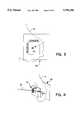
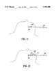
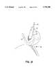
- FIG. 1is an isometric illustration of a plurality of instruments according to the present invention
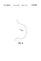
- FIG. 2is a side view of a suspected cancer in a human breast as seen on a mammogram
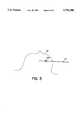
- FIG. 3is a side view of the breast as seen on a mammogram with the breast compressed by a plastic plate with a rectangular window with localization coordinates. A sharpened rod has been passed through the center of the suspected cancer, and is seen end-on as a small circular density;
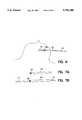
- FIG. 4is a partial front elevation view of the breast compressed in a mammogram machine. A sharpened rod passes though the suspected cancer;
- FIG. 5is a view of the breast as seen on a mammogram taken with the breast compressed from above and below (cc projection). The sharpened rod is now seen penetrating the suspected cancer in profile;
- FIG. 6is a view of the breast as seen on a mammogram (cc projection) with a bevelled tube passed coaxially over the sharpened rod. It is positioned just beyond the suspected cancer;
- FIG. 7(a)is a partial frontal view of the penetrating end of the bevelled tube.
- the bevel on the penetrating endenables smooth passage;
- FIG. 7(b)illustrates the sharpened rod extending through the bevelled tube
- FIG. 8is a view of the breast as seen on a mammogram (cc projection) with the sharpened rod withdrawn leaving the bevelled tube in position just beyond (distal) to the suspected cancer;
- FIG. 9is a view of the breast as seen on a mammogram (cc projection).
- a rod with anchor-hooksis passed through the tube.
- the anchor-hooksare made of flexible, resilient steel wire. As they are extruded out the penetrating end of the bevelled tube, they embed in breast tissue and resume their hooked shape. They form an anchor just distal to the suspected cancer;
- FIG. 10is a side elevation view, partially broken away, of the rod with anchor-hooks compressed within the lumen of the bevelled tube;
- FIG. 11is a view of the breast as seen on a mammogram (cc projection).
- the bevelled tubehas been withdrawn, leaving the rod with anchor-hooks in position;
- FIG. 12is a view of the breast as seen on a mammogram (cc projection).
- a microscalpelpreviously disclosed in original patent application of Coaxial Biopsy
- FIG. 13is a view of the breast as seen on a mammogram (cc projection).
- FIG. 14is a view of the breast as seen on a mammogram (cc projection).
- the inserting tubehas been withdrawn, leaving the spiked collar in position.
- the flexible spikeslike the anchor-hooks, extend into the adjacent tissue and become embedded;
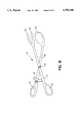
- FIG. 15is a frontal elevation view of stabilizing tongs to compress and stabilize the breast during the biopsy procedure. Holes for a surgeon's thumb and fingers, and a ratchet device to maintain selected compression are shown;
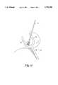
- FIG. 16is a partially frontal view of a breast compressed in a stabilizing tongs in the operating room. Compression is applied along the axis of the rod to foreshorten the distance from skin to lesion;
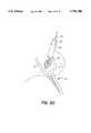
- FIG. 17is a partially frontal view of a breast compressed in a stabilizing tongs in the operating room.
- FIG. 18is a partially frontal view of a breast compressed in a stabilizing tongs.
- a scalpelmakes an incision centered on the rod;
- FIG. 19is a partially frontal view of a breast compressed in a stabilizing tongs. The incision is deepened until the spiked collar is exposed;
- FIG. 20is a partially frontal view of the cannula/obturator assembly advanced coaxially over the rod;
- FIG. 21is a partially frontal view of the cannula/obturator assembly advanced to the depth of the spiked collar. The obturator is then partially withdrawn from the cannula to reveal the cutting edge and the chamber within;
- FIG. 22is a partially frontal view.
- the cannulahas been advanced coaxially along the rod with a back-and-forth rotary cutting movement.
- a cylinder of tissue around the suspected canceris cut into the cannula. This is shown as if the cannula and the breast tissue were transparent;
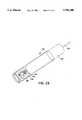
- FIG. 23is a side elevation view, partially in cross section, showing a cylindrical tissue specimen contained within the chamber of the cannula/obturator assembly.
- the tissue at the base of the cylindercan be amputated by the snare device disclosed in prior patent application or by an internal snare;
- FIG. 24is a partially frontal view. The cannula/obturator assembly have been removed. The incision is partly closed.
- FIGS. 25(a)-(c)is a side elevation view partially in cross section, showing the cannula/obturator assembly removed from the patient (FIG. 25(a)), and recovery of the tissue specimen with the suspected cancer centered within it (FIGS. 25(b)-(c));
- FIG. 26is a side elevation view of a power scalpel to deepen the skin incision along the axis of the rod;
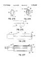
- FIGS. 27(a) and (b)are side elevation views of the penetrating end of the cannula, showing the snare wire nested in a circumferential internal groove near the cutting edge.
- the wireis maintained in place by the spring action of the loop or by a biologically safe adhesive.
- the wire strandspass through a hole to the exterior of the cannula.
- FIG. 27(c)is a plan view of the penetrating end of the cannula.
- the wire strandsare nested in a groove along the shaft where they are also maintained in position by an adhesive or by an external tube slipped over the cannula.
- FIG. 27(d)is a side cross-sectional view of the cannula showing the internal circumferential groove, the hole for the wire strands to pass through, and the groove along the shaft of the cannula.
- a tubeis postioned over the cannula to cover the wire strands in the groove along the shaft.
- FIG. 27(e)is a plan view of the cannula with obturator partially withdrawn and with the outer tube slipped over the cannula to cover the longitudinal groove along the shaft to maintain the wire strands in the groove.
- FIG. 27(f)is a side view in cross section showing the outer tube slipped over the cannula and covering the longitudinal groove.
- FIG. 28is a side elevation view partially in cross section showing a clip inserter with a spring steel clip compressed inside the lumen.
- a stylet with a flexible end-segmentis used as a ramrod to move the clip through the lumen and eject it out the penetrating end.
- FIG. 29is a side elevation view in partial cross section showing the spring clip ejected from the clip inserter in breast tissue adjacent to the suspected cancer.
- FIGS. 30(a)-(d)shows two configurations of spring clips, compressed within a tube and also uncompressed.
- the instruments employed in the practice of the inventioninclude a sharpened, rigid rod 10 having a conical or bevelled or flattened spear point 12 which can be positioned through a suspected cancer 14 within a breast 16, as shown in FIGS. 3, 4, and 5, with guidance of mammographic x-ray images.
- a conventional mammographic compression plate 18 and mammographic cassette 20are depicted in FIG. 4.
- a bevelled tube 22is comprised of a straight hollow cylinder with a sharpened bevelled edge 24 at one end to enable it to slide over the sharpened rod 10 through breast tissue without snagging.
- Index or depth markers 29are provided at the proximal end 31 of the rod 26.
- a microscalpel 32 as best shown in FIG. 12has a tubular handle 34 capable of sliding coaxially over the rigid rod 26 to create a short radial incision along the axis of the rigid rod by means of cutting blade 36.
- a spiked collar 38 as best shown in FIGS. 13 and 14fits loosely over the rigid rod 26.
- a plurality of spikes 40are attached to the collar. These spikes are formed of spring steel similar to the anchor-hooks 28 such that they can flex as the collar is inserted into the breast tissue, but straighten out to stabilize the collar within the breast when the collar is positioned along the rod.
- the rodcan move without displacing the collar once the collar is set into position within the breast.
- the spiked collaris advanced coaxially over the rod by means of an inserting tool 42 having a tube 44 and a collar seat 46.
- the collar 38is seated against collar seat 46 during advance over the rod, but it can disengage easily from the inserting tool 42 when the tool is withdrawn and the spikes embed into the breast tissue.
- a stabilizing tong instrument 48 best shown in FIG. 15is capable of stabilizing and compressing the breast so as to facilitate the process of coaxial biopsy.
- Tongs 50grasp the breast as would a surgeon's fingers, with compression against a flat spoon-shaped surface 52 maintained by a ratchet mechanism 54.
- Rivot 55secures the tongs 57 and 59 for relative rotational movement while finger apertures 61 provide access for the surgeons's fingers.
- a power scalpel 56best shown in FIG. 26.
- motor and battery(not shown) in the handle 58
- poweris delivered to a discoid scalpel blade 60 by a lever 62 with a short, reciprocating movement.
- the blademoves with a rapid back-and-forth oscillating motion.
- a cutting cannula 64similar to that previously disclosed in the parent application, but with an internal snare mechanism 66 best shown in FIGS. 1, 24 and 27(a) through 27(c).
- the snare wire 68rests in an internal circumferential groove 70 near the cutting edge 72 and is maintained therein by a nontoxic adhesive or by the spring action of the wire.
- the snare wire strands 74exit through a hole 76 and are maintained in a longitudinal groove 78 along the outer surface of the canula 64 by a nontoxic adhesive or by an outer tube 79 slipped over the external surface of the cutting cannula as shown in FIGS. 27(d)-27(f).
- An inner rim 81prevents the tube 79 from moving over the cutting edge of the cannula.
- Bevel 83prevents snagging at the distal end. Since the outer tube is maintained in place by the inner rim 81, a hemostat can be used to clamp on the wire strands resting in the groove along the shaft of the cannula. Thus, application of tension to the wire strands will not dislodge them from the groove as the snare loop is closed.
- the ends of the wire strandsare formed into loops which can be connected to a reel mechanism indicated generally by the reference numeral 80 in FIG. 1 to tighten the snare wire, dislodging it from the internal circumferential groove 70.
- the snare wirecan be tightened by the reel mechanism or by a clamped hemostat (not shown) to effect reduction of the size of the snare wire loop and consequently to effect severing of a tissue sample contained within the cannula 64.
- a blunt obturator 82 as disclosed in the parent application,has a central longitudinal hole 84 to engage the rigid rod.
- the obturatorfits inside the cannula and maintains it coaxially centered on the axis of the rod.
- FIG. 27(f)depicts the obturator partially withdrawn to expose the cutting edge of the cannula 72 and create a chamber 85.
- the central bore 84 for the rigid rodis illustrated together with an eccentric bore 83 that provides for equalization of pressure within the chamber 85.
- a spring clip inserter 86 as best shown in FIGS. 28 and 29comprises a tube 88 having a short bend 90 at the penetrating end, a plunger 92 with a flexible end 94, and a curved plate 96 to facilitate passage of the clip inserter into the breast along the outer surface of the cutting cannula 64.
- the spring clips 98are of spring steel formed into a shape which will lodge in breast tissue without likelihood of spontaneous displacement. When compressed, the clips can be passed through the tubular portion of the clip inserter.
- FIGS. 30(a) and 30(b)illustrate various configurations of the spring clips 98 in compressed form within a segment of the tube 88.
- FIGS. 30(c) and 30(d)depict the spring clips in uncompressed form.
- the method of Coaxial Biopsy previously disclosed in the parent applicationenables the surgeon to visualize the location of the suspected cancer along the axis of the penetrating rod by means of a metal target deposited in the lesion.
- This methodallows considerable improvement in precision and accuracy over conventional hook-wire guided biopsy because the surgeon can visualize the target within the specimen as it is removed.
- the disadvantage of this methodis that it requires fluoroscopy or ultrasound imaging during the biopsy, whereas conventional hook-wire guided biopsy is usually performed without such additional imaging technology.
- a biopsy methodshould enable confident localization of the suspected cancer without need for any imaging equipment (fluoroscopic, ultrasound, or stereotactic) during the biopsy.
- the method of the present invention depicted sequentially in FIGS. 2 through 25provides for the positioning of a sharpened rigid rod (16.5 ga, ideally) through a suspected cancer 100 (FIG. 2) in a manner analogous to the placement of a K-wire (which is a rigid, sharpened rod) through the lesion in the previous disclosure of Coaxial Biopsy.
- a difference between the present sharpened rod and the commercially available K-wireis that the sharpened points of conventionally available K-wires have a flattened "diamond" shape, like a spear point or a bevel point, but both of these are relatively blunt (without a sharply tapered point).
- the passage of the rod through breast tissueis facilitated by a long, tapered point which may have a conical, bevelled or spear point shape.
- the rodonce passed through the center of the suspected cancer, permits a 14 ga. tube to be positioned concentrically over it. With mammographic imaging guidance, the tube is advanced so that the distal end is approximately 1 cm beyond the suspected cancer.
- the tubeWhen the rod is removed (FIG. 8), the tube functions as a port to deliver another rod 26 with anchor hooks 28 into the breast.
- the anchor hooksspring out and embed in the breast tissue, stabilizing the rod such that it can neither advance farther into the breast nor be withdrawn through the suspected cancer.
- the tubeis then removed (FIG. 11).
- a microscalpelis passed over the rod and incises a short radial incision to the proximal location of the suspected cancer (FIG. 12). This opens a path along the axis of the rod.
- a loosely-fitting ring or "collar”is positioned concentrically over the rod just proximal to the suspected cancer.
- the collaris advanced over the rod with the breast still compressed in a mammographic machine. Its progress is monitored with sequential mammographic images as it is positioned approximately 1 cm proximal to the suspected cancer.
- the collarhas flexible short segments or "spikes" of 10 ga. wire radially welded to protrude into the breast tissue and maintain the collar in close relation to the suspected cancer. As the collar is advanced over the rod through the breast tissue, the wire spikes flex. When the collar is deposited in the desired position, the spikes straighten and become embedded in the adjacent breast tissue.
- the spiked collar proximally and the anchor hooksdistally bracket the suspected cancer between them on the axis of the rod.
- the rodprotrudes from the breast (FIG. 14).
- the rodis covered with a sterile dressing and the patient is transferred to the operating room.
- the surgeoncompresses the breast with the stabilizing tongs (FIG. 15), thus shortening the distance between the skin and the suspected cancer.
- a skin incisionis made (FIG. 18), centered on the rod. With the oscillating blade of the power scalpel (FIG.
- the incisionis deepened into the breast until the spiked collar is exposed under direct visualization (FIG. 19).
- the cannula/obturator assemblyis advanced coaxially over the rod (FIG. 20).
- the obturatoris partially withdrawn and the cannula is advanced with a rotary cutting motion to incise a cylinder of tissue around the rod, until the cutting edge is just beyond the anchor-hooks at the end of the rod (FIG. 21).
- the surgeoncan monitor the progress of the cannula by noting the relation of the proximal end of the obturator to etched markings on the shaft of the rod.
- the surgeoncan reliably begin the circular incision with the cutting cannula about 1 cm proximal to the suspected cancer, and end the circular incision about 1 cm distal to it.
- the coaxial cannulais equipped with an internal shallow groove near the cutting edge to retain a snare wire loop (FIGS. 27(a)-27(b)).
- the cannulais advanced far enough along the rod to ensure that the internally-mounted snare wire is located deep to the anchor-hooks at the end of the rod.
- the snare wireis made of spring steel and is bent into a loop shape which is slightly compressed as it is positioned inside the cannula. The spring action of the wire maintains it securely in the internal groove.
- the wirecan also be stabilized in the groove by a biologically non-toxic adhesive and the wire can be dislodged from the internal groove by applying tension to the ends of the wire, which pass through a small hole in the cannula. Tension is applied by means of a reel mechanism or by a clamped hemostat. As the snare wire is tightened, the base of the cylinder of tissue is amputated. This completes the incisions separating the specimen from the breast
- a metallic clipis then injected into the breast tissue adjacent to the cannula at the same depth as the suspected cancer, to mark the location of the biopsy (FIGS. 28-30).
- This clipis designed to spring into a self-retaining shape once it has been extruded from the tubing of the inserting tool, so that it will not dislodge from its position. It is inserted into the breast in a standardized location, e.g., oriented to the nipple. This will enable the radiologist to locate and scrutinize the biopsy site on follow-up mammographic evaluations, and will also indicate the original tumor site in case further therapy is required.
- the cannula containing the specimenis then withdrawn from the breast (FIGS. 25(a)-25(c).
- the specimenis extruded and then examined with a mammographic x-ray to confirm that the suspected cancer is present within it.
- the surgeonstops bleeding in the incision either by compression of the breast for several minutes, or by using electrocautery or suture ligatures.
- the skin incisionis then closed, as it is with standard hook-wire guided biopsy.
Landscapes
- Health & Medical Sciences (AREA)
- Life Sciences & Earth Sciences (AREA)
- Surgery (AREA)
- Medical Informatics (AREA)
- Animal Behavior & Ethology (AREA)
- Engineering & Computer Science (AREA)
- Biomedical Technology (AREA)
- Heart & Thoracic Surgery (AREA)
- Veterinary Medicine (AREA)
- Molecular Biology (AREA)
- Public Health (AREA)
- General Health & Medical Sciences (AREA)
- Nuclear Medicine, Radiotherapy & Molecular Imaging (AREA)
- Pathology (AREA)
- Orthopedic Medicine & Surgery (AREA)
- Ophthalmology & Optometry (AREA)
- Neurosurgery (AREA)
- Oral & Maxillofacial Surgery (AREA)
- Surgical Instruments (AREA)
Abstract
Description
The present application is a Continuation-in-Part application of application Ser. No. 08/386,111, filed Mar. 9, 1995 for Biopsy Device and Method, now abandoned.
As currently performed, open surgical biopsy of suspected breast cancer is guided by a flexible hook-wire which is embedded into the cancer by means of radiologic (mammographic or ultrasound) imaging. The surgeon makes an incision into the breast, finds the area of tissue where the hook-wire is embedded, and removes it as a biopsy specimen. Because the surgeon can not see the tip of the embedded hook-wire (it is considered undesirable to cut through the suspected cancer) and also because the hook-wire is a flimsy anchor which can be easily dislodged from its position within the breast, removal of the biopsy specimen involves some guess work which results in a degree of inaccuracy and imprecision. Suspected cancers are often incompletely excised, and occasionally are missed altogether.
With Coaxial Biopsy as described in patent application Ser. No. 08/386,111, a rigid rod is inserted through the suspected cancer with guidance provided by radiologic imaging. The rigid rod is a K-wire, a common surgical tool, which may have a diameter three to six times greater than the standard flexible hook-wire used for needle localization of non-palpable breast masses prior to open surgical excision. The rod establishes a mechanical guide over which the biopsy device is advanced, like a train rolling down a rail, and stabilizes the cancer in the path of the cutting instrument so that it will be removed by the biopsy process.
The concept of rigidity of the rod is central to Coaxial Biopsy as described in the parent application. While a flexible hook-wire may indicate the location of the suspected cancer, one skilled in the art would know that a hook-wire can not provide mechanical guidance for the cutting tool, nor can it stabilize the cancer in the path of the cutting device. The difference between hook-wire guided biopsy and Coaxial Biopsy is perhaps best understood by analogy: one can catch fish with a hook on the end of a flexible fishing line, or one can spear the fish with a barbed harpoon on a long handle. Both systems are superficially similar in that they hook into the fish at some point, but one is flexible and the other is rigid. The cutting device (the cannula) is concentrically positioned along the rod; the rod guides the cannula to the cancer.
What has been disclosed previously in the parent application is a method of marking the location of a suspected cancer so it can be identified with radiologic imaging (either by x-ray fluoroscopy or by ultrasound); normally the cancer is not clearly visible except by mammographic imaging. A metal target implanted into the immediate vicinity of the suspected cancer can be seen by fluoroscopy or ultrasound. The advance of a scalpel blade cutting a path down the rod to a point just proximal to the metal target and the progress of the cannula as it slides over the rod and incises the specimen containing the target are monitored radiologically, eliminating the guess work associated with standard hook-wire guided biopsy. This method is advantageous compared with stereotactically-guided biopsy because it can be performed without the need for a special stereotactic x-ray table. All that is required is a mammography machine for placement of the rod and the target, and an ultrasound or fluoroscopic machine for monitoring the biopsy in the operating room. While this is preferable to stereotactic biopsy and standard surgical biopsy with hook-wire guidance, the requirement for an operative ultrasound or fluoroscopic machine is a relative disadvantage, since not all surgeons are familiar with fluoroscopic or ultrasound imaging.
Also disclosed previously are cutting cannulae with internal snare mechanisms. However, these require at least two concentrically positioned cannulae to maintain the snare wire between them; the inner cannula in these prior patents is then withdrawn to reveal the snare wire, which is then tightened to amputate the specimen. These internal-snare devices require multiple moving parts, and share the disadvantage of complexity associated therewith.
The snare previously disclosed as part of the Coaxial Biopsy system is an external snare slipped over the outside of the cannula by a modification of an existing tonsil-snare device. While this works effectively, it requires an initial relaxing incision to be made along the cannula to pass the device through, and it is a separate tool. If a simpler internal snare mechanism could be devised, this would incorporate two tools into one by combining the cannula and the snare device.
Examples of prior art surgical apparati and procedures for performing biopsies can be found in U.S. Pat. No. 5,353,804 for Method and Device for Percutaneous Excisional Breast Biopsy issued Oct. 11, 1994 to Kornberg, et al and U.S. Pat. No. 5,133,360 for Spears Retriever issued Jul. 28, 1992 to Colin P. Spears and in the prior art cited these patents.
It will be appreciated that there exists a continuing need for improvement in breast biopsy which is partially satisfied by the Coaxial Biopsy system disclosed in the parent application. However, additional improvements of Coaxial Biopsy to enable intra-operative location of the suspected cancer without additional fluoroscopic or ultrasound imaging, and amputation of the specimen by an improved internal snare mechanism would be useful. Also, a method of injecting a metal clip into the breast to mark the location of the biopsy would be helpful in case further therapy such as wider surgical excision is needed. Finally, a stabilization device to position the breast most conveniently during Coaxial biopsy would be useful.
A Coaxial Biopsy system enables biopsy without need for intra-operative radiologic imaging. The system comprises:
(1) A stiff, sharpened rod with a conical point to penetrate and impale the suspected cancer;
(2) A tube with a bevelled edge at one end to slip over the sharpened rod, which establishes a portal into the breast after the rod is removed;
(3) An anchor-rod with flexible metal hook-wires welded to one end, which can be compressed and inserted into the proximal (external end) of the tube, and extruded through the other end of the tube to form an anchor in the breast just distal to the cancer which prevents forward or backward displacement of the rod;
(4) A microscalpel with a tube handle to slip over the rod with anchor hooks, to incise a short radial incision along the rod to a point just proximal to the location of the suspected cancer;
(5) A spiked metal collar with several short segments of flexible wire welded to it radially, to provide "spikes" which will embed into the breast tissue and stabilize the collar along the rod in the breast just proximal to the suspected cancer;
(6) An inserting tool for the spiked collar consisting of a tube with a collar welded near one end. The spiked collar slips over the end of the tube, but is stabilized by the welded collar so that when the spiked collar-inserting tool assembly is slid over the anchor-rod, the spiked collar can be advanced over the anchor-rod into the breast. When the inserting tool is withdrawn the spikes embed in the adjacent breast tissue and maintain the spiked collar in position on the rod just proximal to the cancer;
(7) A power scalpel with a circular blade attached to a handle and an arm attached to the blade by a pivot. A motor in the handle provides the arm with a reciprocating motion, which causes the blade to rotate in a short arc in a back and forth motion. The blade has a diameter of 1-3 cm and is useful for creating an incision centered on the rod from the skin down to the embedded collar with spikes;
(8) A cutting cannula sharpened at the distal end to form a circular knife with an internal circumferential groove near the cutting edge to accommodate a snare wire stabilized in place by the spring action of the wire or by an adhesive glue, with a hole for the snare wires to exit and be pulled when the snare is tightened to amputate the specimen within the cannula and with a longitudinal groove to lodge the wire strands in along the shaft of the cannula;
(9) An outer tube to slip over the shaft of the cannula to maintain the wire strands in the longitudinal groove prior to tightening the snare;
(10) A reel mechanism for drawing the ends of the snare sire tight to close the snare and amputate the tissue in the cannula;
(11) A self-attaching spring clip made of flexible steel wire for embedding into breast tissue to mark the site of a coaxial biopsy within the breast;
(12) A clip injector tool to position the self-attaching clip alongside the cannula at the site of the biopsy; and,
(13) A breast stabilizing device with a flat disc on one tong and two finger-like tines on the other, handles on the proximal ends, to form tongs joined by a central pivot and governed by a ratchet mechanism. The action of this hinged tong device compresses and stabilizes the breast to facilitate the incision along the rod for passage of the coaxial cannula.
FIG. 1 is an isometric illustration of a plurality of instruments according to the present invention;
FIG. 2 is a side view of a suspected cancer in a human breast as seen on a mammogram;
FIG. 3 is a side view of the breast as seen on a mammogram with the breast compressed by a plastic plate with a rectangular window with localization coordinates. A sharpened rod has been passed through the center of the suspected cancer, and is seen end-on as a small circular density;
FIG. 4 is a partial front elevation view of the breast compressed in a mammogram machine. A sharpened rod passes though the suspected cancer;
FIG. 5 is a view of the breast as seen on a mammogram taken with the breast compressed from above and below (cc projection). The sharpened rod is now seen penetrating the suspected cancer in profile;
FIG. 6 is a view of the breast as seen on a mammogram (cc projection) with a bevelled tube passed coaxially over the sharpened rod. It is positioned just beyond the suspected cancer;
FIG. 7(a) is a partial frontal view of the penetrating end of the bevelled tube. The bevel on the penetrating end enables smooth passage;
FIG. 7(b) illustrates the sharpened rod extending through the bevelled tube;
FIG. 8 is a view of the breast as seen on a mammogram (cc projection) with the sharpened rod withdrawn leaving the bevelled tube in position just beyond (distal) to the suspected cancer;
FIG. 9 is a view of the breast as seen on a mammogram (cc projection). A rod with anchor-hooks is passed through the tube. The anchor-hooks are made of flexible, resilient steel wire. As they are extruded out the penetrating end of the bevelled tube, they embed in breast tissue and resume their hooked shape. They form an anchor just distal to the suspected cancer;
FIG. 10 is a side elevation view, partially broken away, of the rod with anchor-hooks compressed within the lumen of the bevelled tube;
FIG. 11 is a view of the breast as seen on a mammogram (cc projection). The bevelled tube has been withdrawn, leaving the rod with anchor-hooks in position;
FIG. 12 is a view of the breast as seen on a mammogram (cc projection). A microscalpel (previously disclosed in original patent application of Coaxial Biopsy) passes over the rod and incises a short radial incision to a point proximal to the location of the suspected cancer. This opens a path along the axis of the rod;
FIG. 13 is a view of the breast as seen on a mammogram (cc projection). A loosely-fitting collar with flexible, resilient spikes, mounted on an inserting tube, passes coaxially over the rod and is positioned just proximal to the suspected cancer;
FIG. 14 is a view of the breast as seen on a mammogram (cc projection). The inserting tube has been withdrawn, leaving the spiked collar in position. The flexible spikes, like the anchor-hooks, extend into the adjacent tissue and become embedded;
FIG. 15 is a frontal elevation view of stabilizing tongs to compress and stabilize the breast during the biopsy procedure. Holes for a surgeon's thumb and fingers, and a ratchet device to maintain selected compression are shown;
FIG. 16 is a partially frontal view of a breast compressed in a stabilizing tongs in the operating room. Compression is applied along the axis of the rod to foreshorten the distance from skin to lesion;
FIG. 17 is a partially frontal view of a breast compressed in a stabilizing tongs in the operating room. The suspected cancer, bracketed by the spiked collar proximally and the anchor-hooks distally, is shown as if the breast tissue were transparent;
FIG. 18 is a partially frontal view of a breast compressed in a stabilizing tongs. A scalpel makes an incision centered on the rod;
FIG. 19 is a partially frontal view of a breast compressed in a stabilizing tongs. The incision is deepened until the spiked collar is exposed;
FIG. 20 is a partially frontal view of the cannula/obturator assembly advanced coaxially over the rod;
FIG. 21 is a partially frontal view of the cannula/obturator assembly advanced to the depth of the spiked collar. The obturator is then partially withdrawn from the cannula to reveal the cutting edge and the chamber within;
FIG. 22 is a partially frontal view. The cannula has been advanced coaxially along the rod with a back-and-forth rotary cutting movement. A cylinder of tissue around the suspected cancer is cut into the cannula. This is shown as if the cannula and the breast tissue were transparent;
FIG. 23 is a side elevation view, partially in cross section, showing a cylindrical tissue specimen contained within the chamber of the cannula/obturator assembly. The tissue at the base of the cylinder can be amputated by the snare device disclosed in prior patent application or by an internal snare;
FIG. 24 is a partially frontal view. The cannula/obturator assembly have been removed. The incision is partly closed.
FIGS. 25(a)-(c) is a side elevation view partially in cross section, showing the cannula/obturator assembly removed from the patient (FIG. 25(a)), and recovery of the tissue specimen with the suspected cancer centered within it (FIGS. 25(b)-(c));
FIG. 26 is a side elevation view of a power scalpel to deepen the skin incision along the axis of the rod;
FIGS. 27(a) and (b) are side elevation views of the penetrating end of the cannula, showing the snare wire nested in a circumferential internal groove near the cutting edge. The wire is maintained in place by the spring action of the loop or by a biologically safe adhesive. The wire strands pass through a hole to the exterior of the cannula.
FIG. 27(c) is a plan view of the penetrating end of the cannula. The wire strands are nested in a groove along the shaft where they are also maintained in position by an adhesive or by an external tube slipped over the cannula.
FIG. 27(d) is a side cross-sectional view of the cannula showing the internal circumferential groove, the hole for the wire strands to pass through, and the groove along the shaft of the cannula. A tube is postioned over the cannula to cover the wire strands in the groove along the shaft.
FIG. 27(e) is a plan view of the cannula with obturator partially withdrawn and with the outer tube slipped over the cannula to cover the longitudinal groove along the shaft to maintain the wire strands in the groove.
FIG. 27(f) is a side view in cross section showing the outer tube slipped over the cannula and covering the longitudinal groove.
FIG. 28 is a side elevation view partially in cross section showing a clip inserter with a spring steel clip compressed inside the lumen. A stylet with a flexible end-segment is used as a ramrod to move the clip through the lumen and eject it out the penetrating end.
FIG. 29 is a side elevation view in partial cross section showing the spring clip ejected from the clip inserter in breast tissue adjacent to the suspected cancer.
FIGS. 30(a)-(d) shows two configurations of spring clips, compressed within a tube and also uncompressed.
The instruments employed in the practice of the invention include a sharpened,rigid rod 10 having a conical or bevelled or flattenedspear point 12 which can be positioned through a suspected cancer 14 within abreast 16, as shown in FIGS. 3, 4, and 5, with guidance of mammographic x-ray images. A conventionalmammographic compression plage 18 andmammographic cassette 20 are depicted in FIG. 4.
Abevelled tube 22, as best shown in FIG. 7, is comprised of a straight hollow cylinder with a sharpened bevellededge 24 at one end to enable it to slide over the sharpenedrod 10 through breast tissue without snagging.
Arigid rod 26 with a plurality of spring steel anchor-hooks 28, as best shown in FIG. 9 which can be compressed within aloading tube 30 shown in FIG. 10 and advanced into the bevelledtube 22, and finally extruded through the penetrating end of the bevelledtube 22 so as to become embedded in adjacent breast tissue. Index ordepth markers 29 are provided at theproximal end 31 of therod 26.
Amicroscalpel 32 as best shown in FIG. 12 has atubular handle 34 capable of sliding coaxially over therigid rod 26 to create a short radial incision along the axis of the rigid rod by means of cuttingblade 36.
Aspiked collar 38 as best shown in FIGS. 13 and 14 fits loosely over therigid rod 26. A plurality ofspikes 40 are attached to the collar. These spikes are formed of spring steel similar to the anchor-hooks 28 such that they can flex as the collar is inserted into the breast tissue, but straighten out to stabilize the collar within the breast when the collar is positioned along the rod. The rod can move without displacing the collar once the collar is set into position within the breast. The spiked collar is advanced coaxially over the rod by means of an insertingtool 42 having atube 44 and acollar seat 46. Thecollar 38 is seated againstcollar seat 46 during advance over the rod, but it can disengage easily from the insertingtool 42 when the tool is withdrawn and the spikes embed into the breast tissue.
A stabilizingtong instrument 48 best shown in FIG. 15 is capable of stabilizing and compressing the breast so as to facilitate the process of coaxial biopsy.Tongs 50 grasp the breast as would a surgeon's fingers, with compression against a flat spoon-shapedsurface 52 maintained by aratchet mechanism 54.Rivot 55 secures thetongs finger apertures 61 provide access for the surgeons's fingers.
Apower scalpel 56 best shown in FIG. 26. With motor and battery (not shown) in thehandle 58, power is delivered to adiscoid scalpel blade 60 by alever 62 with a short, reciprocating movement. The blade moves with a rapid back-and-forth oscillating motion.
A cuttingcannula 64 similar to that previously disclosed in the parent application, but with aninternal snare mechanism 66 best shown in FIGS. 1, 24 and 27(a) through 27(c). Thesnare wire 68 rests in an internalcircumferential groove 70 near thecutting edge 72 and is maintained therein by a nontoxic adhesive or by the spring action of the wire. Thesnare wire strands 74 exit through ahole 76 and are maintained in alongitudinal groove 78 along the outer surface of thecanula 64 by a nontoxic adhesive or by anouter tube 79 slipped over the external surface of the cutting cannula as shown in FIGS. 27(d)-27(f). Aninner rim 81 prevents thetube 79 from moving over the cutting edge of the cannula.Bevel 83 prevents snagging at the distal end. Since the outer tube is maintained in place by theinner rim 81, a hemostat can be used to clamp on the wire strands resting in the groove along the shaft of the cannula. Thus, application of tension to the wire strands will not dislodge them from the groove as the snare loop is closed.
The ends of the wire strands are formed into loops which can be connected to a reel mechanism indicated generally by thereference numeral 80 in FIG. 1 to tighten the snare wire, dislodging it from the internalcircumferential groove 70. The snare wire can be tightened by the reel mechanism or by a clamped hemostat (not shown) to effect reduction of the size of the snare wire loop and consequently to effect severing of a tissue sample contained within thecannula 64.
Ablunt obturator 82 as disclosed in the parent application, has a centrallongitudinal hole 84 to engage the rigid rod. The obturator fits inside the cannula and maintains it coaxially centered on the axis of the rod. FIG. 27(f) depicts the obturator partially withdrawn to expose the cutting edge of thecannula 72 and create achamber 85. Thecentral bore 84 for the rigid rod is illustrated together with aneccentric bore 83 that provides for equalization of pressure within thechamber 85.
Aspring clip inserter 86 as best shown in FIGS. 28 and 29 comprises atube 88 having ashort bend 90 at the penetrating end, aplunger 92 with aflexible end 94, and acurved plate 96 to facilitate passage of the clip inserter into the breast along the outer surface of the cuttingcannula 64. The spring clips 98 are of spring steel formed into a shape which will lodge in breast tissue without likelihood of spontaneous displacement. When compressed, the clips can be passed through the tubular portion of the clip inserter. FIGS. 30(a) and 30(b) illustrate various configurations of the spring clips 98 in compressed form within a segment of thetube 88. FIGS. 30(c) and 30(d) depict the spring clips in uncompressed form.
The method of Coaxial Biopsy previously disclosed in the parent application enables the surgeon to visualize the location of the suspected cancer along the axis of the penetrating rod by means of a metal target deposited in the lesion. This method allows considerable improvement in precision and accuracy over conventional hook-wire guided biopsy because the surgeon can visualize the target within the specimen as it is removed. However, the disadvantage of this method is that it requires fluoroscopy or ultrasound imaging during the biopsy, whereas conventional hook-wire guided biopsy is usually performed without such additional imaging technology. Ideally, a biopsy method should enable confident localization of the suspected cancer without need for any imaging equipment (fluoroscopic, ultrasound, or stereotactic) during the biopsy.
The method of the present invention depicted sequentially in FIGS. 2 through 25 provides for the positioning of a sharpened rigid rod (16.5 ga, ideally) through a suspected cancer 100 (FIG. 2) in a manner analogous to the placement of a K-wire (which is a rigid, sharpened rod) through the lesion in the previous disclosure of Coaxial Biopsy. A difference between the present sharpened rod and the commercially available K-wire is that the sharpened points of conventionally available K-wires have a flattened "diamond" shape, like a spear point or a bevel point, but both of these are relatively blunt (without a sharply tapered point). The passage of the rod through breast tissue is facilitated by a long, tapered point which may have a conical, bevelled or spear point shape. The rod, once passed through the center of the suspected cancer, permits a 14 ga. tube to be positioned concentrically over it. With mammographic imaging guidance, the tube is advanced so that the distal end is approximately 1 cm beyond the suspected cancer. When the rod is removed (FIG. 8), the tube functions as a port to deliver anotherrod 26 with anchor hooks 28 into the breast. When the distal end of the rod with anchor hooks is extruded from the tube, the anchor hooks spring out and embed in the breast tissue, stabilizing the rod such that it can neither advance farther into the breast nor be withdrawn through the suspected cancer. The tube is then removed (FIG. 11). A microscalpel is passed over the rod and incises a short radial incision to the proximal location of the suspected cancer (FIG. 12). This opens a path along the axis of the rod.
With the suspected cancer securely anchored in place near the distal end of the rigid rod with anchor hooks, a loosely-fitting ring or "collar" is positioned concentrically over the rod just proximal to the suspected cancer. The collar is advanced over the rod with the breast still compressed in a mammographic machine. Its progress is monitored with sequential mammographic images as it is positioned approximately 1 cm proximal to the suspected cancer. The collar has flexible short segments or "spikes" of 10 ga. wire radially welded to protrude into the breast tissue and maintain the collar in close relation to the suspected cancer. As the collar is advanced over the rod through the breast tissue, the wire spikes flex. When the collar is deposited in the desired position, the spikes straighten and become embedded in the adjacent breast tissue.
The spiked collar proximally and the anchor hooks distally bracket the suspected cancer between them on the axis of the rod. The rod protrudes from the breast (FIG. 14). As with conventional hook-wire guided biopsy, the rod is covered with a sterile dressing and the patient is transferred to the operating room. There, after sterilizing the skin and injecting anesthetic, the surgeon compresses the breast with the stabilizing tongs (FIG. 15), thus shortening the distance between the skin and the suspected cancer. Then a skin incision is made (FIG. 18), centered on the rod. With the oscillating blade of the power scalpel (FIG. 26) or with conventional scalpel or electrocantery, the incision is deepened into the breast until the spiked collar is exposed under direct visualization (FIG. 19). The cannula/obturator assembly is advanced coaxially over the rod (FIG. 20). The obturator is partially withdrawn and the cannula is advanced with a rotary cutting motion to incise a cylinder of tissue around the rod, until the cutting edge is just beyond the anchor-hooks at the end of the rod (FIG. 21). The surgeon can monitor the progress of the cannula by noting the relation of the proximal end of the obturator to etched markings on the shaft of the rod. Thus, without fluoroscopic, ultrasound, or stereotactic monitoring, the surgeon can reliably begin the circular incision with the cutting cannula about 1 cm proximal to the suspected cancer, and end the circular incision about 1 cm distal to it.
The coaxial cannula is equipped with an internal shallow groove near the cutting edge to retain a snare wire loop (FIGS. 27(a)-27(b)). The cannula is advanced far enough along the rod to ensure that the internally-mounted snare wire is located deep to the anchor-hooks at the end of the rod. The snare wire is made of spring steel and is bent into a loop shape which is slightly compressed as it is positioned inside the cannula. The spring action of the wire maintains it securely in the internal groove. The wire can also be stabilized in the groove by a biologically non-toxic adhesive and the wire can be dislodged from the internal groove by applying tension to the ends of the wire, which pass through a small hole in the cannula. Tension is applied by means of a reel mechanism or by a clamped hemostat. As the snare wire is tightened, the base of the cylinder of tissue is amputated. This completes the incisions separating the specimen from the breast.
A metallic clip is then injected into the breast tissue adjacent to the cannula at the same depth as the suspected cancer, to mark the location of the biopsy (FIGS. 28-30). This clip is designed to spring into a self-retaining shape once it has been extruded from the tubing of the inserting tool, so that it will not dislodge from its position. It is inserted into the breast in a standardized location, e.g., oriented to the nipple. This will enable the radiologist to locate and scrutinize the biopsy site on follow-up mammographic evaluations, and will also indicate the original tumor site in case further therapy is required.
The cannula containing the specimen is then withdrawn from the breast (FIGS. 25(a)-25(c). The specimen is extruded and then examined with a mammographic x-ray to confirm that the suspected cancer is present within it. The surgeon stops bleeding in the incision either by compression of the breast for several minutes, or by using electrocautery or suture ligatures. The skin incision is then closed, as it is with standard hook-wire guided biopsy.
Having described in detail a preferred embodiment of the invention, it will now be apparent to those skilled in the art that numerous modifications can be made therein without departing from the scope of the invention as defined in the following claims.
Claims (12)
1. A biopsy kit comprising:
a) a stiff, point-sharpened rod;
b) a bevel-ended tube adapted to pass coaxially over the stiff, point-sharpened rod;
c) a stiff anchor rod having a plurality of flexible, resilient anchor hooks located at a distal end thereof, said stiff anchor rod and plurality of flexible anchor hooks being adapted to pass through the bevel-ended tube until the anchor hooks extend beyond the bevel-ended tube;
d) an insertion tube having a collar seat at an insertion and thereof, said insertion tube being adapted to pass coaxially over the stiff anchor rod; and,
e) a spiked collar having a plurality of flexible, resilient radially extending spikes adapted to be positioned removably against the collar seat of said insertion tube whereby the stiff, point-sharpened rod can penetrate and impale a suspected cancer with the bevel-ended tube thereafter positioned coaxially over the stiff, point-sharpened rod which can then be withdrawn leaving the bevel-ended tube located with respect to the suspected cancer to provide a guide for the stiff anchor rod that passes through the bevel-ended tube with the anchor hooks extending beyond the bevel-ended tube which can then be withdrawn so that the insertion tube can pass coaxially over the anchor rod with the spiked collar positioned removably against the collar seat of the insertion tube.
2. The biopsy kit of claim 1 further comprising:
a microscalpel having a longitudinally extending tube adapted to pass coaxially over said stiff anchor rod and an arcuate cutting blade mounted on said longitudingly extending tube at a distal end thereof.
3. The biopsy kit of claim 1 further comprising:
a powered microscalpel comprising:
a) a housing having a longitudinal axis;
b) an electrically powered motor mounted within said housing, said motor providing reciprocatory motion in a direction parallel to the longitudinal axis of the housing;
c) a circular scalpel having a cutting portion along at least a segment of the circumference of the circular scalpel;
d) means for rotationally mounting said circular scalpel with respect to said housing; and,
e) means for imparting the reciprocatory motion of said motor to said circular scalpel to rotate the circular scalpel clockwise and counter-clockwise.
4. The biopsy kit of claim 1 further comprising:
a breast compression and stabilization instrument having:
a) a first tong having an intermediate section, a finger receiving aperture at one end of the intermediate section and a pair of tines at the other end thereof;
b) a second tong having and intermediate section, a finger receiving aperture at one end of the intermediate section and a relatively flat spoon portion at the other end thereof;
c) means for rotationally securing said first and second tongs together in a scissor-like manner; and,
d) means for releasably holding said first and second tongs at a fixed position relative to each other so that a desired open distance can be releasably maintained between said tines and said relatively flat spoon portion during breast biopsy.
5. The biopsy kit of claim 1 further comprising:
a cannula having a cutting end and being adapted to coaxially advance along the stiff anchor rod.
6. The biopsy kit of claim 5 wherein said cannula has a snare loop located within the cannula and wherein the snare loop includes two standing ends that are parallel with respect to said stiff anchor rod and extend along said cannula away from its cutting end whereby when said standing ends are pulled in a direction away from the cutting end of the cannula, the snare loop is closed.
7. The biopsy kit of claim 6 wherein said snare loop standing ends are positioned externally to said cannula and further comprising an outer tube adapted to slip over the cannula and hold the standing ends with respect to the cannula.
8. The biopsy kit of claim 6 wherein the cannula has an internal circumferential groove adjacent to the cutting end of said cannula and wherein said snare loop rests in said internal circumferential groove.
9. The biopsy kit of claim 8 wherein said snare loop is a resilient material having sufficient resiliency to maintain the snare loop in said internal circumferential groove prior to pulling said standing ends.
10. The biopsy kit of claim 8 wherein the cannula internal circumferential groove has an aperture and wherein said snare loop standing ends are positioned externally by passing them through said aperture.
11. The biopsy kit of claim 5 wherein said cannula has an external longitudinal groove and wherein said standing ends are positioned therein.
12. The biopsy kit of claim 5 further comprising:
a) a flexible, resilient metallic target clip;
b) a longitudinally extending target clip insertion tube having a longitudinal axis and an arcuate base member adapted to fit on the cannula so that the longitudinal axis of the target clip insertion tube is parallel to the stiff anchor rod; and,
c) a target clip insertion rod adapted to fit within and move along the longitudinal axis of the target clip insertion tube to drive a flexible, resilient metallic target clip positioned therein along said axis for injection into tissue of a patient.
Priority Applications (4)
| Application Number | Priority Date | Filing Date | Title |
|---|---|---|---|
| US08/690,185US5795308A (en) | 1995-03-09 | 1996-07-26 | Apparatus for coaxial breast biopsy |
| US09/084,731US6080114A (en) | 1995-03-09 | 1998-05-26 | Method for coaxial breast biopsy |
| PCT/US1998/016747WO2000009005A1 (en) | 1996-07-26 | 1998-08-12 | Method and apparati for coaxial breast biopsy |
| AU87811/98AAU8781198A (en) | 1996-07-26 | 1998-08-12 | Method and apparati for coaxial breast biopsy |
Applications Claiming Priority (3)
| Application Number | Priority Date | Filing Date | Title |
|---|---|---|---|
| US38611195A | 1995-03-09 | 1995-03-09 | |
| US08/690,185US5795308A (en) | 1995-03-09 | 1996-07-26 | Apparatus for coaxial breast biopsy |
| PCT/US1998/016747WO2000009005A1 (en) | 1996-07-26 | 1998-08-12 | Method and apparati for coaxial breast biopsy |
Related Parent Applications (1)
| Application Number | Title | Priority Date | Filing Date |
|---|---|---|---|
| US38611195AContinuation-In-Part | 1995-03-09 | 1995-03-09 |
Related Child Applications (1)
| Application Number | Title | Priority Date | Filing Date |
|---|---|---|---|
| US09/084,731DivisionUS6080114A (en) | 1995-03-09 | 1998-05-26 | Method for coaxial breast biopsy |
Publications (1)
| Publication Number | Publication Date |
|---|---|
| US5795308Atrue US5795308A (en) | 1998-08-18 |
Family
ID=26794298
Family Applications (2)
| Application Number | Title | Priority Date | Filing Date |
|---|---|---|---|
| US08/690,185Expired - Fee RelatedUS5795308A (en) | 1995-03-09 | 1996-07-26 | Apparatus for coaxial breast biopsy |
| US09/084,731Expired - Fee RelatedUS6080114A (en) | 1995-03-09 | 1998-05-26 | Method for coaxial breast biopsy |
Family Applications After (1)
| Application Number | Title | Priority Date | Filing Date |
|---|---|---|---|
| US09/084,731Expired - Fee RelatedUS6080114A (en) | 1995-03-09 | 1998-05-26 | Method for coaxial breast biopsy |
Country Status (3)
| Country | Link |
|---|---|
| US (2) | US5795308A (en) |
| AU (1) | AU8781198A (en) |
| WO (1) | WO2000009005A1 (en) |
Cited By (166)
| Publication number | Priority date | Publication date | Assignee | Title |
|---|---|---|---|---|
| US5916180A (en)* | 1997-10-03 | 1999-06-29 | Uromed Corporation | Calibrating pressure sensors |
| US5989199A (en)* | 1996-11-27 | 1999-11-23 | Assurance Medical, Inc. | Tissue examination |
| WO1999066834A1 (en)* | 1998-06-22 | 1999-12-29 | Richard Eustis Fulton, Iii | Biopsy localization method and device |
| WO2000010471A1 (en)* | 1998-08-19 | 2000-03-02 | Artemis Medical, Inc. | Target tissue localization device and method |
| US6063031A (en)* | 1997-10-14 | 2000-05-16 | Assurance Medical, Inc. | Diagnosis and treatment of tissue with instruments |
| US6080113A (en)* | 1998-09-11 | 2000-06-27 | Imagyn Medical Technologies California, Inc. | Incisional breast biopsy device |
| US6086544A (en)* | 1999-03-31 | 2000-07-11 | Ethicon Endo-Surgery, Inc. | Control apparatus for an automated surgical biopsy device |
| US6091981A (en)* | 1997-09-16 | 2000-07-18 | Assurance Medical Inc. | Clinical tissue examination |
| US6120462A (en)* | 1999-03-31 | 2000-09-19 | Ethicon Endo-Surgery, Inc. | Control method for an automated surgical biopsy device |
| US6136014A (en)* | 1998-09-01 | 2000-10-24 | Vivant Medical, Inc. | Percutaneous tissue removal device |
| WO2001005320A1 (en)* | 1999-07-16 | 2001-01-25 | Senorx, Inc. | Electrosurgical lesion location device |
| US6179790B1 (en) | 1997-10-20 | 2001-01-30 | Assurance Medical, Inc. | Layer of material for use with tissue examination device |
| US6221006B1 (en) | 1998-02-10 | 2001-04-24 | Artemis Medical Inc. | Entrapping apparatus and method for use |
| US6273862B1 (en) | 1998-10-23 | 2001-08-14 | Ethicon Endo-Surgery, Inc | Surgical device for the collection of soft tissue |
| US6277083B1 (en) | 1999-12-27 | 2001-08-21 | Neothermia Corporation | Minimally invasive intact recovery of tissue |
| US20010034528A1 (en)* | 1994-09-16 | 2001-10-25 | Foerster Seth A. | Methods and devices for defining and marking tissue |
| US6311691B1 (en)* | 1998-07-23 | 2001-11-06 | Lincoln D. Russin | Method for determining the shortest distance between the periphery of a lesion and the cut edge of the tissue specimen |
| US6344026B1 (en) | 1998-04-08 | 2002-02-05 | Senorx, Inc. | Tissue specimen encapsulation device and method thereof |
| US6356782B1 (en) | 1998-12-24 | 2002-03-12 | Vivant Medical, Inc. | Subcutaneous cavity marking device and method |
| US6371904B1 (en) | 1998-12-24 | 2002-04-16 | Vivant Medical, Inc. | Subcutaneous cavity marking device and method |
| US20020052564A1 (en)* | 1998-03-03 | 2002-05-02 | Burbank Fred H. | Methods and apparatus for securing medical instruments to desired locations in a patient's body |
| US6383145B1 (en) | 1997-09-12 | 2002-05-07 | Imagyn Medical Technologies California, Inc. | Incisional breast biopsy device |
| US6405733B1 (en) | 2000-02-18 | 2002-06-18 | Thomas J. Fogarty | Device for accurately marking tissue |
| US6471709B1 (en) | 1998-10-30 | 2002-10-29 | Vivant Medical, Inc. | Expandable ring percutaneous tissue removal device |
| US6471659B2 (en) | 1999-12-27 | 2002-10-29 | Neothermia Corporation | Minimally invasive intact recovery of tissue |
| US20020198554A1 (en)* | 2001-03-14 | 2002-12-26 | Whitman Michael P. | Trocar device |
| WO2002043563A3 (en)* | 2000-11-29 | 2003-02-06 | Senorx Inc | Sentinel node location and biopsy |
| US6530923B1 (en) | 1998-02-10 | 2003-03-11 | Artemis Medical, Inc. | Tissue removal methods and apparatus |
| US6540695B1 (en) | 1998-04-08 | 2003-04-01 | Senorx, Inc. | Biopsy anchor device with cutter |
| US6551253B2 (en) | 1997-09-12 | 2003-04-22 | Imagyn Medical Technologies | Incisional breast biopsy device |
| US6569176B2 (en) | 1999-10-18 | 2003-05-27 | Jerry M. Jesseph | Device and method for improved diagnosis and treatment of cancer |
| US6572578B1 (en) | 2000-08-25 | 2003-06-03 | Patrick A. Blanchard | Fluid-jet catheter and its application to flexible endoscopy |
| US6589254B2 (en) | 1999-09-14 | 2003-07-08 | Mark G. Fontenot | Breast bracket |
| US6602265B2 (en) | 1998-02-10 | 2003-08-05 | Artemis Medical, Inc. | Tissue separation medical device and method |
| US6602204B2 (en) | 1998-02-10 | 2003-08-05 | Artemis Medical, Inc | Intraoperative tissue treatment methods |
| US6635068B1 (en) | 1998-02-10 | 2003-10-21 | Artemis Medical, Inc. | Occlusion, anchoring, tensioning and flow direction apparatus and methods for use |
| US20030204137A1 (en)* | 1999-06-17 | 2003-10-30 | Inrad, Inc. | Apparatus for the percutaneous marking of a lesion |
| US20030204188A1 (en)* | 2001-11-07 | 2003-10-30 | Artemis Medical, Inc. | Tissue separating and localizing catheter assembly |
| US6659105B2 (en) | 1998-02-26 | 2003-12-09 | Senorx, Inc. | Tissue specimen isolating and damaging device and method |
| US20040006355A1 (en)* | 2002-07-03 | 2004-01-08 | Rubicor Medical, Inc. | Methods and devices for cutting and collecting soft tissue |
| US6679851B2 (en) | 1998-09-01 | 2004-01-20 | Senorx, Inc. | Tissue accessing and anchoring device and method |
| US20040034280A1 (en)* | 1998-10-23 | 2004-02-19 | Salvatore Privitera | Surgical device for the collection of soft tissue |
| US20040049224A1 (en)* | 2000-11-07 | 2004-03-11 | Buehlmann Eric L. | Target tissue localization assembly and method |
| US6722371B1 (en) | 2000-02-18 | 2004-04-20 | Thomas J. Fogarty | Device for accurately marking tissue |
| US6743228B2 (en) | 2001-09-12 | 2004-06-01 | Manoa Medical, Inc. | Devices and methods for tissue severing and removal |
| US6752767B2 (en) | 2002-04-16 | 2004-06-22 | Vivant Medical, Inc. | Localization element with energized tip |
| US6752154B2 (en) | 2000-02-18 | 2004-06-22 | Thomas J. Fogarty | Device for accurately marking tissue |
| US20040167432A1 (en)* | 1998-03-03 | 2004-08-26 | Senorx, Inc. | Breast biopsy system and methods |
| US20040167511A1 (en)* | 2003-02-25 | 2004-08-26 | Artemis Medical, Inc. | Tissue separating catheter assembly and method |
| WO2004075732A2 (en) | 2003-02-25 | 2004-09-10 | Artemis Medical, Inc. | Tissue localizing and separating assembly |
| US20040260332A1 (en)* | 1997-11-12 | 2004-12-23 | Genesis Technologies Llc | Body passageway occluder and method |
| US20050182339A1 (en)* | 1998-09-03 | 2005-08-18 | Roberta Lee | Excisional biopsy devices and methods |
| US20050222521A1 (en)* | 2002-10-16 | 2005-10-06 | Rubicor Medical, Inc. | Devices and methods for performing procedures on a breast |
| US6955676B2 (en) | 1999-06-22 | 2005-10-18 | Senorx, Inc. | Shaped scalpel |
| US6997885B2 (en) | 1998-04-08 | 2006-02-14 | Senorx, Inc. | Dilation devices and methods for removing tissue specimens |
| US20060095028A1 (en)* | 2004-10-15 | 2006-05-04 | Baxano, Inc. | Devices and methods for tissue access |
| US20060116573A1 (en)* | 2003-11-17 | 2006-06-01 | Inrad, Inc. | Self Contained, Self Piercing, Side-Expelling Marking Apparatus |
| US20060129185A1 (en)* | 1999-10-25 | 2006-06-15 | Boston Scientific Scimed, Inc. | Forceps for medical use |
| US20060211944A1 (en)* | 2005-03-15 | 2006-09-21 | Codman & Shurtleff, Inc. | Pressure sensing devices |
| US20060224083A1 (en)* | 2003-06-18 | 2006-10-05 | Rubicor Medical, Inc. | Methods and devices for cutting and collecting soft tissue |
| US20060229650A1 (en)* | 2002-07-03 | 2006-10-12 | Rubicor Medical, Inc. | Methods and devices for cutting and collecting soft tissue |
| US7160292B2 (en) | 1999-06-17 | 2007-01-09 | Vivant Medical, Inc. | Needle kit and method for microwave ablation, track coagulation, and biopsy |
| US20070038145A1 (en)* | 2004-11-22 | 2007-02-15 | Inrad, Inc. | Post Decompression Marker Introducer System |
| US7197363B2 (en) | 2002-04-16 | 2007-03-27 | Vivant Medical, Inc. | Microwave antenna having a curved configuration |
| US7311703B2 (en) | 2003-07-18 | 2007-12-25 | Vivant Medical, Inc. | Devices and methods for cooling microwave antennas |
| US7318824B2 (en) | 2001-11-02 | 2008-01-15 | Vivant Medical, Inc. | High-strength microwave antenna assemblies |
| US20080051812A1 (en)* | 2006-08-01 | 2008-02-28 | Baxano, Inc. | Multi-Wire Tissue Cutter |
| US20080103504A1 (en)* | 2006-10-30 | 2008-05-01 | Schmitz Gregory P | Percutaneous spinal stenosis treatment |
| US20080146965A1 (en)* | 2003-08-11 | 2008-06-19 | Salvatore Privitera | Surgical Device for The Collection of Soft Tissue |
| EP1967155A1 (en)* | 2007-03-08 | 2008-09-10 | CAS Innovations AG | Medical clamp, in particular spinal clamp |
| WO2008016886A3 (en)* | 2006-08-01 | 2008-12-04 | Baxano Inc | Tissue cutting devices and methods |
| US20090125035A1 (en)* | 2007-11-14 | 2009-05-14 | Kennedy John S | Surgical Cutting Instrument for Breast Surgery |
| US20090182366A1 (en)* | 2007-11-14 | 2009-07-16 | Kennedy John S | Surgical Instrument for Detecting, Isolating and Excising Tumors |
| US20090182478A1 (en)* | 2008-01-15 | 2009-07-16 | Gm Global Technology Operations, Inc. | Axle torque based cruise control |
| US7578819B2 (en) | 2005-05-16 | 2009-08-25 | Baxano, Inc. | Spinal access and neural localization |
| US20090221876A1 (en)* | 2008-01-08 | 2009-09-03 | Tyson Cobb | Surgical Release Apparatuses, Systems and Methods |
| US7588545B2 (en) | 2003-09-10 | 2009-09-15 | Boston Scientific Scimed, Inc. | Forceps and collection assembly with accompanying mechanisms and related methods of use |
| US20100030149A1 (en)* | 2006-10-23 | 2010-02-04 | C.R. Bard, Inc. | Breast marker |
| US20100094169A1 (en)* | 2002-06-17 | 2010-04-15 | Senorx, Inc. | Plugged tip delivery tube for marker placement |
| US20100140500A1 (en)* | 2007-06-19 | 2010-06-10 | Therapy Positioning Technologies, Llc | Apparatus and method for the treatment of breast cancer with particle beams |
| US7738969B2 (en) | 2004-10-15 | 2010-06-15 | Baxano, Inc. | Devices and methods for selective surgical removal of tissue |
| US7762960B2 (en) | 2005-05-13 | 2010-07-27 | Boston Scientific Scimed, Inc. | Biopsy forceps assemblies |
| US7857813B2 (en) | 2006-08-29 | 2010-12-28 | Baxano, Inc. | Tissue access guidewire system and method |
| US20100331893A1 (en)* | 2009-06-26 | 2010-12-30 | Wyatt Drake Geist | K-Wire And Method For Surgical Procedures |
| US7887538B2 (en) | 2005-10-15 | 2011-02-15 | Baxano, Inc. | Methods and apparatus for tissue modification |
| US7918849B2 (en) | 2004-10-15 | 2011-04-05 | Baxano, Inc. | Devices and methods for tissue access |
| US7938830B2 (en) | 2004-10-15 | 2011-05-10 | Baxano, Inc. | Powered tissue modification devices and methods |
| US7942896B2 (en) | 2003-11-25 | 2011-05-17 | Scimed Life Systems, Inc. | Forceps and collection assembly and related methods of use and manufacture |
| US7959577B2 (en) | 2007-09-06 | 2011-06-14 | Baxano, Inc. | Method, system, and apparatus for neural localization |
| US20110144533A1 (en)* | 2009-12-15 | 2011-06-16 | Rafal Chudzik | Introducer cannula having a tissue anchor for use with a medical instrument |
| US7993329B2 (en) | 2002-08-13 | 2011-08-09 | Cook Medical Technologies Llc | ERCP catheter with a removable handle for lithotriptor compatible basket |
| US20110213432A1 (en)* | 2009-06-26 | 2011-09-01 | Wyatt Drake Geist | Guidewire And Method For Surgical Procedures |
| US8048080B2 (en) | 2004-10-15 | 2011-11-01 | Baxano, Inc. | Flexible tissue rasp |
| US8062300B2 (en) | 2006-05-04 | 2011-11-22 | Baxano, Inc. | Tissue removal with at least partially flexible devices |
| US8062298B2 (en) | 2005-10-15 | 2011-11-22 | Baxano, Inc. | Flexible tissue removal devices and methods |
| US8068921B2 (en) | 2006-09-29 | 2011-11-29 | Vivant Medical, Inc. | Microwave antenna assembly and method of using the same |
| US8092456B2 (en) | 2005-10-15 | 2012-01-10 | Baxano, Inc. | Multiple pathways for spinal nerve root decompression from a single access point |
| US8157862B2 (en) | 1997-10-10 | 2012-04-17 | Senorx, Inc. | Tissue marking implant |
| US8192436B2 (en) | 2007-12-07 | 2012-06-05 | Baxano, Inc. | Tissue modification devices |
| US8219182B2 (en) | 1999-02-02 | 2012-07-10 | Senorx, Inc. | Cavity-filling biopsy site markers |
| US8221397B2 (en) | 2004-10-15 | 2012-07-17 | Baxano, Inc. | Devices and methods for tissue modification |
| US8224424B2 (en) | 1999-02-02 | 2012-07-17 | Senorx, Inc. | Tissue site markers for in vivo imaging |
| US8257356B2 (en) | 2004-10-15 | 2012-09-04 | Baxano, Inc. | Guidewire exchange systems to treat spinal stenosis |
| US8292880B2 (en) | 2007-11-27 | 2012-10-23 | Vivant Medical, Inc. | Targeted cooling of deployable microwave antenna |
| US8311610B2 (en) | 2008-01-31 | 2012-11-13 | C. R. Bard, Inc. | Biopsy tissue marker |
| US8361082B2 (en) | 1999-02-02 | 2013-01-29 | Senorx, Inc. | Marker delivery device with releasable plug |
| US8366712B2 (en) | 2005-10-15 | 2013-02-05 | Baxano, Inc. | Multiple pathways for spinal nerve root decompression from a single access point |
| US8394102B2 (en) | 2009-06-25 | 2013-03-12 | Baxano, Inc. | Surgical tools for treatment of spinal stenosis |
| US8398641B2 (en) | 2008-07-01 | 2013-03-19 | Baxano, Inc. | Tissue modification devices and methods |
| US8401622B2 (en) | 2006-12-18 | 2013-03-19 | C. R. Bard, Inc. | Biopsy marker with in situ-generated imaging properties |
| US8409206B2 (en) | 2008-07-01 | 2013-04-02 | Baxano, Inc. | Tissue modification devices and methods |
| US8430881B2 (en) | 2004-10-15 | 2013-04-30 | Baxano, Inc. | Mechanical tissue modification devices and methods |
| US8447386B2 (en) | 2003-05-23 | 2013-05-21 | Senorx, Inc. | Marker or filler forming fluid |
| US8486028B2 (en) | 2005-10-07 | 2013-07-16 | Bard Peripheral Vascular, Inc. | Tissue marking apparatus having drug-eluting tissue marker |
| US8498693B2 (en) | 1999-02-02 | 2013-07-30 | Senorx, Inc. | Intracorporeal marker and marker delivery device |
| US20130253370A1 (en)* | 2002-10-16 | 2013-09-26 | Encapsule Medical, Llc | Devices and methods for performing procedures on a breast |
| US8568416B2 (en) | 2004-10-15 | 2013-10-29 | Baxano Surgical, Inc. | Access and tissue modification systems and methods |
| US8613745B2 (en) | 2004-10-15 | 2013-12-24 | Baxano Surgical, Inc. | Methods, systems and devices for carpal tunnel release |
| US8626269B2 (en) | 2003-05-23 | 2014-01-07 | Senorx, Inc. | Fibrous marker and intracorporeal delivery thereof |
| US8634899B2 (en) | 2003-11-17 | 2014-01-21 | Bard Peripheral Vascular, Inc. | Multi mode imaging marker |
| CN103584899A (en)* | 2013-10-24 | 2014-02-19 | 苏州美都医用设备有限公司 | Forked hemostat |
| US8670818B2 (en) | 2008-12-30 | 2014-03-11 | C. R. Bard, Inc. | Marker delivery device for tissue marker placement |
| US8668737B2 (en) | 1997-10-10 | 2014-03-11 | Senorx, Inc. | Tissue marking implant |
| US8718745B2 (en) | 2000-11-20 | 2014-05-06 | Senorx, Inc. | Tissue site markers for in vivo imaging |
| US8801626B2 (en) | 2004-10-15 | 2014-08-12 | Baxano Surgical, Inc. | Flexible neural localization devices and methods |
| US8845639B2 (en) | 2008-07-14 | 2014-09-30 | Baxano Surgical, Inc. | Tissue modification devices |
| USD715442S1 (en) | 2013-09-24 | 2014-10-14 | C. R. Bard, Inc. | Tissue marker for intracorporeal site identification |
| USD715942S1 (en) | 2013-09-24 | 2014-10-21 | C. R. Bard, Inc. | Tissue marker for intracorporeal site identification |
| USD716450S1 (en) | 2013-09-24 | 2014-10-28 | C. R. Bard, Inc. | Tissue marker for intracorporeal site identification |
| USD716451S1 (en) | 2013-09-24 | 2014-10-28 | C. R. Bard, Inc. | Tissue marker for intracorporeal site identification |
| US20140358158A1 (en)* | 2012-01-30 | 2014-12-04 | Jon Einarsson | Functional uterine manipulator |
| US20150126903A1 (en)* | 2012-05-03 | 2015-05-07 | The Johns Hopkins University | integrated punch biopsy device |
| US9101386B2 (en) | 2004-10-15 | 2015-08-11 | Amendia, Inc. | Devices and methods for treating tissue |
| US9149341B2 (en) | 1999-02-02 | 2015-10-06 | Senorx, Inc | Deployment of polysaccharide markers for treating a site within a patient |
| US9149259B1 (en)* | 2011-08-21 | 2015-10-06 | Cynthia E. Allen | Patient safety and wellbeing device for covering wires and needles used in mammography or ultrasound guided needle localization |
| US9186487B2 (en) | 1997-11-12 | 2015-11-17 | Genesis Technologies Llc | Medical device and method |
| US9186177B2 (en) | 2001-03-14 | 2015-11-17 | Covidien Lp | Trocar device |
| US9247952B2 (en) | 2004-10-15 | 2016-02-02 | Amendia, Inc. | Devices and methods for tissue access |
| WO2016036558A1 (en)* | 2014-09-02 | 2016-03-10 | Mayo Foundation For Medical Education And Research | Methods and devices for localizing, delineating and anchoring liver lesions for resection |
| US9314253B2 (en) | 2008-07-01 | 2016-04-19 | Amendia, Inc. | Tissue modification devices and methods |
| US9327061B2 (en) | 2008-09-23 | 2016-05-03 | Senorx, Inc. | Porous bioabsorbable implant |
| CN105796186A (en)* | 2016-04-19 | 2016-07-27 | 上海市肺科医院 | Intrathoracic L-shaped positioning device adopting 3D printing in endoscopy and manufacturing method thereof |
| US9456829B2 (en) | 2004-10-15 | 2016-10-04 | Amendia, Inc. | Powered tissue modification devices and methods |
| US9498604B2 (en) | 1997-11-12 | 2016-11-22 | Genesis Technologies Llc | Medical device and method |
| US9561094B2 (en) | 2010-07-23 | 2017-02-07 | Nfinium Vascular Technologies, Llc | Devices and methods for treating venous diseases |
| US9579077B2 (en) | 2006-12-12 | 2017-02-28 | C.R. Bard, Inc. | Multiple imaging mode tissue marker |
| WO2017040064A1 (en)* | 2015-08-28 | 2017-03-09 | SiteSelect Inc. | Tissue excision device with anchor stability rod and anchor stability rod |
| US9669113B1 (en) | 1998-12-24 | 2017-06-06 | Devicor Medical Products, Inc. | Device and method for safe location and marking of a biopsy cavity |
| US20170245886A1 (en)* | 2014-12-03 | 2017-08-31 | Neil Jeffrey WITCOMB | Tumor stabilizing apparatus for a medical procedure |
| US9820824B2 (en) | 1999-02-02 | 2017-11-21 | Senorx, Inc. | Deployment of polysaccharide markers for treating a site within a patent |
| US9877743B2 (en)* | 2013-01-17 | 2018-01-30 | Natural Nipple Knife Llc | Circular surgical scalpel and marker |
| US10004483B2 (en) | 2012-10-11 | 2018-06-26 | Pave, Llc | Scoop cannula for a coring biopsy device |
| US10251630B2 (en) | 2015-08-28 | 2019-04-09 | SiteSelect Inc. | Tissue excision device with anchor stability rod and anchor stability rod |
| WO2019093914A1 (en)* | 2017-11-13 | 2019-05-16 | Politechnika Gdańska | A method and a device to perform the biopsy of soft (usually human) tissue |
| EP3488810A1 (en)* | 2017-11-22 | 2019-05-29 | Laurent Fumex | Device for anchoring and locating suspected lesions on multiple anchoring means |
| US10342635B2 (en) | 2005-04-20 | 2019-07-09 | Bard Peripheral Vascular, Inc. | Marking device with retractable cannula |
| US10729466B2 (en) | 2017-08-21 | 2020-08-04 | Freyja Healthcare, Llc | Uterine manipulator with cutting head |
| EP3621509A4 (en)* | 2017-05-08 | 2021-05-19 | Platform Imaging, LLC | Laparoscopic device implantation and fixation system and method |
| US11185344B2 (en) | 2015-03-13 | 2021-11-30 | The Regents Of The University Of Michigan | Tool for neuroma treatment and nerve regeneration procedures |
| US11266414B2 (en) | 2014-06-04 | 2022-03-08 | Vascular Development Corp, Llc | Low radial force vascular device and method of occlusion |
| CN114176803A (en)* | 2022-01-06 | 2022-03-15 | 中南大学湘雅二医院 | Precise positioning resection device for breast conserving surgery for breast cancer |
| US20230032981A1 (en)* | 2018-02-15 | 2023-02-02 | Rls Interventional, Inc. | Full core biopsy device |
| EP4143738A4 (en)* | 2020-05-01 | 2024-03-13 | Endoglow Prime, Llc | NEAR-INFRARED BREAST TUMOR MARKER |
| US12096960B2 (en) | 2017-08-21 | 2024-09-24 | The Brigham And Women's Hospital, Inc. | Uterine manipulator |
| US12426860B2 (en) | 2018-04-18 | 2025-09-30 | C.R. Bard, Inc. | Dual lumen coaxial introducer having integrated tissue marker delivery |
| US12440650B2 (en) | 2023-02-27 | 2025-10-14 | Vascular Development Corp, Llc | Augmented delivery catheter and method |
Families Citing this family (24)
| Publication number | Priority date | Publication date | Assignee | Title |
|---|---|---|---|---|
| JP4255208B2 (en)* | 1997-07-24 | 2009-04-15 | レックス メディカル リミテッド パートナーシップ | Device for resecting subcutaneous target tissue mass |
| US6626903B2 (en) | 1997-07-24 | 2003-09-30 | Rex Medical, L.P. | Surgical biopsy device |
| US6945980B2 (en) | 1998-06-03 | 2005-09-20 | Medtronic, Inc. | Multiple loop tissue connector apparatus and methods |
| US6607541B1 (en) | 1998-06-03 | 2003-08-19 | Coalescent Surgical, Inc. | Tissue connector apparatus and methods |
| US6613059B2 (en) | 1999-03-01 | 2003-09-02 | Coalescent Surgical, Inc. | Tissue connector apparatus and methods |
| US6641593B1 (en) | 1998-06-03 | 2003-11-04 | Coalescent Surgical, Inc. | Tissue connector apparatus and methods |
| US8118822B2 (en) | 1999-03-01 | 2012-02-21 | Medtronic, Inc. | Bridge clip tissue connector apparatus and methods |
| US6695859B1 (en) | 1999-04-05 | 2004-02-24 | Coalescent Surgical, Inc. | Apparatus and methods for anastomosis |
| US8529583B1 (en) | 1999-09-03 | 2013-09-10 | Medtronic, Inc. | Surgical clip removal apparatus |
| US6926730B1 (en) | 2000-10-10 | 2005-08-09 | Medtronic, Inc. | Minimally invasive valve repair procedure and apparatus |
| US6551332B1 (en) | 2000-03-31 | 2003-04-22 | Coalescent Surgical, Inc. | Multiple bias surgical fastener |
| US8066724B2 (en) | 2002-09-12 | 2011-11-29 | Medtronic, Inc. | Anastomosis apparatus and methods |
| US8105345B2 (en) | 2002-10-04 | 2012-01-31 | Medtronic, Inc. | Anastomosis apparatus and methods |
| US8114102B2 (en)* | 2003-06-16 | 2012-02-14 | St. Jude Medical Atg, Inc. | Temporary hemostatic plug apparatus and method of use |
| US7182769B2 (en) | 2003-07-25 | 2007-02-27 | Medtronic, Inc. | Sealing clip, delivery systems, and methods |
| US20050043749A1 (en) | 2003-08-22 | 2005-02-24 | Coalescent Surgical, Inc. | Eversion apparatus and methods |
| US8394114B2 (en) | 2003-09-26 | 2013-03-12 | Medtronic, Inc. | Surgical connection apparatus and methods |
| US7879047B2 (en) | 2003-12-10 | 2011-02-01 | Medtronic, Inc. | Surgical connection apparatus and methods |
| US8992569B2 (en)* | 2007-06-29 | 2015-03-31 | Ethicon Endo-Surgery, Inc. | Insertion device and method of use |
| US8177836B2 (en) | 2008-03-10 | 2012-05-15 | Medtronic, Inc. | Apparatus and methods for minimally invasive valve repair |
| US8518060B2 (en) | 2009-04-09 | 2013-08-27 | Medtronic, Inc. | Medical clip with radial tines, system and method of using same |
| US8668704B2 (en) | 2009-04-24 | 2014-03-11 | Medtronic, Inc. | Medical clip with tines, system and method of using same |
| US8597201B2 (en) | 2010-03-30 | 2013-12-03 | Siteselect Medical Technologies, Inc. | Tissue excision device with a flexible transection blade |
| WO2011160686A1 (en)* | 2010-06-23 | 2011-12-29 | Renzo Marco Giovanni Brun Del Re | Biopsy alignment guide |
Citations (27)
| Publication number | Priority date | Publication date | Assignee | Title |
|---|---|---|---|---|
| US4007732A (en)* | 1975-09-02 | 1977-02-15 | Robert Carl Kvavle | Method for location and removal of soft tissue in human biopsy operations |
| US4461305A (en)* | 1981-09-04 | 1984-07-24 | Cibley Leonard J | Automated biopsy device |
| US4651752A (en)* | 1985-03-08 | 1987-03-24 | Fuerst Erwin J | Biopsy needle |
| US4682606A (en)* | 1986-02-03 | 1987-07-28 | Decaprio Vincent H | Localizing biopsy apparatus |
| US4691333A (en)* | 1985-12-27 | 1987-09-01 | Gabriele Joseph M | Breast compression and needle localization apparatus |
| US4727565A (en)* | 1983-11-14 | 1988-02-23 | Ericson Bjoern E | Method of localization |
| US4821727A (en)* | 1986-10-30 | 1989-04-18 | Elscint Ltd. | Mammographic biopsy needle holder system |
| US4837795A (en)* | 1987-07-27 | 1989-06-06 | Double D Double G Enterprises, Incorporated | Tissue specimen holding device and biopsy procedure |
| US4966583A (en)* | 1989-02-03 | 1990-10-30 | Elie Debbas | Apparatus for locating a breast mass |
| US5074311A (en)* | 1989-12-06 | 1991-12-24 | Hasson Harrith M | Biopsy device |
| US5083570A (en)* | 1990-06-18 | 1992-01-28 | Mosby Richard A | Volumetric localization/biopsy/surgical device |
| US5133360A (en)* | 1991-03-07 | 1992-07-28 | Spears Colin P | Spears retriever |
| US5148813A (en)* | 1990-11-20 | 1992-09-22 | Bucalo Brian D | Biopsy instrument with tissue specimen retaining and retrieval device |
| US5172702A (en)* | 1989-11-24 | 1992-12-22 | Medical Device Technologies, Inc. | Disposable spring-loaded soft tissue biopsy apparatus |
| US5183463A (en)* | 1989-02-03 | 1993-02-02 | Elie Debbas | Apparatus for locating a breast mass |
| US5188118A (en)* | 1990-11-07 | 1993-02-23 | Terwilliger Richard A | Automatic biopsy instrument with independently actuated stylet and cannula |
| US5267572A (en)* | 1990-11-20 | 1993-12-07 | Bucalo Brian D | Biopsy instrument with tissue specimen retaining and retrieval device |
| US5316014A (en)* | 1992-02-07 | 1994-05-31 | Livingston Products, Inc. | Biopsy locator and guide |
| US5353804A (en)* | 1990-09-18 | 1994-10-11 | Peb Biopsy Corporation | Method and device for percutaneous exisional breast biopsy |
| US5415169A (en)* | 1989-11-21 | 1995-05-16 | Fischer Imaging Corporation | Motorized mammographic biopsy apparatus |
| US5445645A (en)* | 1989-02-03 | 1995-08-29 | Debbas; Elie | Apparatus for locating a breast mass |
| US5452367A (en)* | 1993-11-29 | 1995-09-19 | Arch Development Corporation | Automated method and system for the segmentation of medical images |
| US5499989A (en)* | 1994-12-22 | 1996-03-19 | Labash; Stephen S. | Breast biopsy apparatus and method of use |
| US5507298A (en)* | 1994-09-23 | 1996-04-16 | M3 Systems, Inc., D/B/A/ Manan Medical Products, Inc. | Forward-fired automatic tissue sampling apparatus |
| US5526822A (en)* | 1994-03-24 | 1996-06-18 | Biopsys Medical, Inc. | Method and apparatus for automated biopsy and collection of soft tissue |
| US5570699A (en)* | 1994-04-26 | 1996-11-05 | Kass; Erik S. | Controlled surgical core biopsy system |
| US5573008A (en)* | 1993-10-29 | 1996-11-12 | Boston Scientific Corporation | Multiple biopsy sampling coring device |
Family Cites Families (1)
| Publication number | Priority date | Publication date | Assignee | Title |
|---|---|---|---|---|
| US5111828A (en)* | 1990-09-18 | 1992-05-12 | Peb Biopsy Corporation | Device for percutaneous excisional breast biopsy |
- 1996
- 1996-07-26USUS08/690,185patent/US5795308A/ennot_activeExpired - Fee Related
- 1998
- 1998-05-26USUS09/084,731patent/US6080114A/ennot_activeExpired - Fee Related
- 1998-08-12AUAU87811/98Apatent/AU8781198A/ennot_activeAbandoned
- 1998-08-12WOPCT/US1998/016747patent/WO2000009005A1/ennot_activeApplication Discontinuation
Patent Citations (27)
| Publication number | Priority date | Publication date | Assignee | Title |
|---|---|---|---|---|
| US4007732A (en)* | 1975-09-02 | 1977-02-15 | Robert Carl Kvavle | Method for location and removal of soft tissue in human biopsy operations |
| US4461305A (en)* | 1981-09-04 | 1984-07-24 | Cibley Leonard J | Automated biopsy device |
| US4727565A (en)* | 1983-11-14 | 1988-02-23 | Ericson Bjoern E | Method of localization |
| US4651752A (en)* | 1985-03-08 | 1987-03-24 | Fuerst Erwin J | Biopsy needle |
| US4691333A (en)* | 1985-12-27 | 1987-09-01 | Gabriele Joseph M | Breast compression and needle localization apparatus |
| US4682606A (en)* | 1986-02-03 | 1987-07-28 | Decaprio Vincent H | Localizing biopsy apparatus |
| US4821727A (en)* | 1986-10-30 | 1989-04-18 | Elscint Ltd. | Mammographic biopsy needle holder system |
| US4837795A (en)* | 1987-07-27 | 1989-06-06 | Double D Double G Enterprises, Incorporated | Tissue specimen holding device and biopsy procedure |
| US4966583A (en)* | 1989-02-03 | 1990-10-30 | Elie Debbas | Apparatus for locating a breast mass |
| US5445645A (en)* | 1989-02-03 | 1995-08-29 | Debbas; Elie | Apparatus for locating a breast mass |
| US5183463A (en)* | 1989-02-03 | 1993-02-02 | Elie Debbas | Apparatus for locating a breast mass |
| US5415169A (en)* | 1989-11-21 | 1995-05-16 | Fischer Imaging Corporation | Motorized mammographic biopsy apparatus |
| US5172702A (en)* | 1989-11-24 | 1992-12-22 | Medical Device Technologies, Inc. | Disposable spring-loaded soft tissue biopsy apparatus |
| US5074311A (en)* | 1989-12-06 | 1991-12-24 | Hasson Harrith M | Biopsy device |
| US5083570A (en)* | 1990-06-18 | 1992-01-28 | Mosby Richard A | Volumetric localization/biopsy/surgical device |
| US5353804A (en)* | 1990-09-18 | 1994-10-11 | Peb Biopsy Corporation | Method and device for percutaneous exisional breast biopsy |
| US5188118A (en)* | 1990-11-07 | 1993-02-23 | Terwilliger Richard A | Automatic biopsy instrument with independently actuated stylet and cannula |
| US5267572A (en)* | 1990-11-20 | 1993-12-07 | Bucalo Brian D | Biopsy instrument with tissue specimen retaining and retrieval device |
| US5148813A (en)* | 1990-11-20 | 1992-09-22 | Bucalo Brian D | Biopsy instrument with tissue specimen retaining and retrieval device |
| US5133360A (en)* | 1991-03-07 | 1992-07-28 | Spears Colin P | Spears retriever |
| US5316014A (en)* | 1992-02-07 | 1994-05-31 | Livingston Products, Inc. | Biopsy locator and guide |
| US5573008A (en)* | 1993-10-29 | 1996-11-12 | Boston Scientific Corporation | Multiple biopsy sampling coring device |
| US5452367A (en)* | 1993-11-29 | 1995-09-19 | Arch Development Corporation | Automated method and system for the segmentation of medical images |
| US5526822A (en)* | 1994-03-24 | 1996-06-18 | Biopsys Medical, Inc. | Method and apparatus for automated biopsy and collection of soft tissue |
| US5570699A (en)* | 1994-04-26 | 1996-11-05 | Kass; Erik S. | Controlled surgical core biopsy system |
| US5507298A (en)* | 1994-09-23 | 1996-04-16 | M3 Systems, Inc., D/B/A/ Manan Medical Products, Inc. | Forward-fired automatic tissue sampling apparatus |
| US5499989A (en)* | 1994-12-22 | 1996-03-19 | Labash; Stephen S. | Breast biopsy apparatus and method of use |
Cited By (374)
| Publication number | Priority date | Publication date | Assignee | Title |
|---|---|---|---|---|
| US7625397B2 (en) | 1994-09-16 | 2009-12-01 | Ethicon Endo-Surgery, Inc. | Methods for defining and marking tissue |
| US8277391B2 (en) | 1994-09-16 | 2012-10-02 | Devicor Medical Products, Inc. | Methods and devices for defining and marking tissue |
| US7044957B2 (en) | 1994-09-16 | 2006-05-16 | Ethicon Endo-Surgery, Inc. | Devices for defining and marking tissue |
| US20020193815A1 (en)* | 1994-09-16 | 2002-12-19 | Foerster Seth A. | Methods and devices for defining and marking tissue |
| US7229417B2 (en) | 1994-09-16 | 2007-06-12 | Ethicon Endo-Surgery, Inc. | Methods for marking a biopsy site |
| US20010034528A1 (en)* | 1994-09-16 | 2001-10-25 | Foerster Seth A. | Methods and devices for defining and marking tissue |
| US5989199A (en)* | 1996-11-27 | 1999-11-23 | Assurance Medical, Inc. | Tissue examination |
| US6551253B2 (en) | 1997-09-12 | 2003-04-22 | Imagyn Medical Technologies | Incisional breast biopsy device |
| US6383145B1 (en) | 1997-09-12 | 2002-05-07 | Imagyn Medical Technologies California, Inc. | Incisional breast biopsy device |
| US6267732B1 (en) | 1997-09-12 | 2001-07-31 | Imagyn Medical Technologies, Inc. | Incisional breast biopsy device |
| US6091981A (en)* | 1997-09-16 | 2000-07-18 | Assurance Medical Inc. | Clinical tissue examination |
| US5916180A (en)* | 1997-10-03 | 1999-06-29 | Uromed Corporation | Calibrating pressure sensors |
| US8668737B2 (en) | 1997-10-10 | 2014-03-11 | Senorx, Inc. | Tissue marking implant |
| US9039763B2 (en) | 1997-10-10 | 2015-05-26 | Senorx, Inc. | Tissue marking implant |
| US10058416B2 (en) | 1997-10-10 | 2018-08-28 | Senorx, Inc. | Tissue marking implant |
| US9480554B2 (en) | 1997-10-10 | 2016-11-01 | Senorx, Inc. | Tissue marking implant |
| US8157862B2 (en) | 1997-10-10 | 2012-04-17 | Senorx, Inc. | Tissue marking implant |
| US6063031A (en)* | 1997-10-14 | 2000-05-16 | Assurance Medical, Inc. | Diagnosis and treatment of tissue with instruments |
| US6179790B1 (en) | 1997-10-20 | 2001-01-30 | Assurance Medical, Inc. | Layer of material for use with tissue examination device |
| US9498604B2 (en) | 1997-11-12 | 2016-11-22 | Genesis Technologies Llc | Medical device and method |
| US20040260332A1 (en)* | 1997-11-12 | 2004-12-23 | Genesis Technologies Llc | Body passageway occluder and method |
| US9186487B2 (en) | 1997-11-12 | 2015-11-17 | Genesis Technologies Llc | Medical device and method |
| US20030199913A1 (en)* | 1998-02-10 | 2003-10-23 | Artemis Medical, Inc. | Occlusion, anchoring, tensioning and flow direction apparatus and methods for use |
| US6530923B1 (en) | 1998-02-10 | 2003-03-11 | Artemis Medical, Inc. | Tissue removal methods and apparatus |
| US6695858B1 (en) | 1998-02-10 | 2004-02-24 | Artemis Medical, Inc. | Medical device and methods for use |
| US20050033172A1 (en)* | 1998-02-10 | 2005-02-10 | Artemis Medical, Inc. | Tissue separation method |
| US7491210B2 (en) | 1998-02-10 | 2009-02-17 | Artemis Medical, Inc. | Medical device and methods for use |
| US7331980B2 (en) | 1998-02-10 | 2008-02-19 | Ethicon Endo-Surgery, Inc. | Occlusion, anchoring, tensioning and flow direction apparatus and methods for use |
| US6635068B1 (en) | 1998-02-10 | 2003-10-21 | Artemis Medical, Inc. | Occlusion, anchoring, tensioning and flow direction apparatus and methods for use |
| US20030195537A1 (en)* | 1998-02-10 | 2003-10-16 | Artemis Medical, Inc. | Medical device and methods for use |
| US6602204B2 (en) | 1998-02-10 | 2003-08-05 | Artemis Medical, Inc | Intraoperative tissue treatment methods |
| US6602265B2 (en) | 1998-02-10 | 2003-08-05 | Artemis Medical, Inc. | Tissue separation medical device and method |
| US20030109896A1 (en)* | 1998-02-10 | 2003-06-12 | Artemis Medical, Inc. | Tissue removal methods and apparatus |
| US6221006B1 (en) | 1998-02-10 | 2001-04-24 | Artemis Medical Inc. | Entrapping apparatus and method for use |
| US6659105B2 (en) | 1998-02-26 | 2003-12-09 | Senorx, Inc. | Tissue specimen isolating and damaging device and method |
| US20080077045A1 (en)* | 1998-03-03 | 2008-03-27 | Senorx, Inc. | Tissue specimen encapsulation device and method thereof |
| US20060094983A1 (en)* | 1998-03-03 | 2006-05-04 | Burbank Fred H | Methods and apparatus for securing medical instruments to desired locations in a patient's body |
| US20040167432A1 (en)* | 1998-03-03 | 2004-08-26 | Senorx, Inc. | Breast biopsy system and methods |
| US6540693B2 (en) | 1998-03-03 | 2003-04-01 | Senorx, Inc. | Methods and apparatus for securing medical instruments to desired locations in a patients body |
| US7229418B2 (en) | 1998-03-03 | 2007-06-12 | Senorx, Inc. | Tissue specimen encapsulation device and method thereof |
| US20050004492A1 (en)* | 1998-03-03 | 2005-01-06 | Senorx, Inc. | Breast biopsy system and methods |
| US20050245842A1 (en)* | 1998-03-03 | 2005-11-03 | Senorx, Inc. | Methods and apparatus for securing medical instruments to desired locations in a patient's body |
| US6958044B2 (en) | 1998-03-03 | 2005-10-25 | Senorx, Inc. | Methods and apparatus for securing medical instruments to desired locations in a patient's body |
| US20050010131A1 (en)* | 1998-03-03 | 2005-01-13 | Senorx, Inc. | Breast biopsy system and methods |
| US20050197593A1 (en)* | 1998-03-03 | 2005-09-08 | Senorx, Inc. | Breast biopsy system and methods |
| US6716179B2 (en) | 1998-03-03 | 2004-04-06 | Senorx, Inc. | Sentinel node location and biopsy |
| US8152737B2 (en) | 1998-03-03 | 2012-04-10 | Senorx, Inc. | Tissue specimen encapsulation device and method thereof |
| US20030176812A1 (en)* | 1998-03-03 | 2003-09-18 | Senorx, Inc. | Tissue specimen encapsulation device and method thereof |
| US7264596B2 (en) | 1998-03-03 | 2007-09-04 | Senorx, Inc. | Methods and apparatus for securing medical instruments to desired locations in a patient's body |
| US20020087095A1 (en)* | 1998-03-03 | 2002-07-04 | Senorx, Inc. | Methods and apparatus for securing medical instruments to desired locations in a patient's body |
| US20040167431A1 (en)* | 1998-03-03 | 2004-08-26 | Burbank Fred H. | Breast biopsy system and methods |
| US6638234B2 (en) | 1998-03-03 | 2003-10-28 | Senorx, Inc. | Sentinel node location and biopsy |
| US20020052564A1 (en)* | 1998-03-03 | 2002-05-02 | Burbank Fred H. | Methods and apparatus for securing medical instruments to desired locations in a patient's body |
| US20050187491A1 (en)* | 1998-03-03 | 2005-08-25 | Senorx, Inc. | Breast biopsy system and methods |
| US7329228B2 (en) | 1998-03-03 | 2008-02-12 | Senorx, Inc. | Methods and apparatus for securing medical instruments to desired locations in a patient's body |
| US7357801B2 (en) | 1998-04-08 | 2008-04-15 | Senorx, Inc. | Tissue specimen isolating and damaging device and method |
| US6540695B1 (en) | 1998-04-08 | 2003-04-01 | Senorx, Inc. | Biopsy anchor device with cutter |
| US6676658B2 (en) | 1998-04-08 | 2004-01-13 | Senorx, Inc. | Tissue specimen isolating and damaging device and method |
| US7377902B2 (en)* | 1998-04-08 | 2008-05-27 | Senorx, Inc. | Biopsy anchor device with cutter |
| US6344026B1 (en) | 1998-04-08 | 2002-02-05 | Senorx, Inc. | Tissue specimen encapsulation device and method thereof |
| US7651467B2 (en) | 1998-04-08 | 2010-01-26 | Senorx, Inc | Dilation devices and methods for removing tissue specimens |
| US6508773B2 (en) | 1998-04-08 | 2003-01-21 | Senorx, Inc. | Tissue specimen encapsulation device and method thereof |
| US6997885B2 (en) | 1998-04-08 | 2006-02-14 | Senorx, Inc. | Dilation devices and methods for removing tissue specimens |
| US6270464B1 (en) | 1998-06-22 | 2001-08-07 | Artemis Medical, Inc. | Biopsy localization method and device |
| US20050033195A1 (en)* | 1998-06-22 | 2005-02-10 | Fulton Richard E. | Biopsy localization method and device |
| US6699205B2 (en) | 1998-06-22 | 2004-03-02 | Artemis Medical, Inc. | Biopsy localization method and device |
| US20050045192A1 (en)* | 1998-06-22 | 2005-03-03 | Artemis Medical, Inc. | Biopsy localization method and device |
| WO1999066834A1 (en)* | 1998-06-22 | 1999-12-29 | Richard Eustis Fulton, Iii | Biopsy localization method and device |
| US10010380B2 (en) | 1998-06-22 | 2018-07-03 | Devicor Medical Products, Inc. | Biopsy localization method and device |
| US8292822B2 (en) | 1998-06-22 | 2012-10-23 | Devicor Medical Products, Inc. | Biopsy localization method and device |
| US6311691B1 (en)* | 1998-07-23 | 2001-11-06 | Lincoln D. Russin | Method for determining the shortest distance between the periphery of a lesion and the cut edge of the tissue specimen |
| US6409742B1 (en) | 1998-08-19 | 2002-06-25 | Artemis Medical, Inc. | Target tissue localization device and method |
| US6325816B1 (en) | 1998-08-19 | 2001-12-04 | Artemis Medical, Inc. | Target tissue localization method |
| US6758855B2 (en) | 1998-08-19 | 2004-07-06 | Artemis Medical, Inc. | Target tissue localization device |
| WO2000010471A1 (en)* | 1998-08-19 | 2000-03-02 | Artemis Medical, Inc. | Target tissue localization device and method |
| US6179860B1 (en) | 1998-08-19 | 2001-01-30 | Artemis Medical, Inc. | Target tissue localization device and method |
| US7479152B2 (en) | 1998-08-19 | 2009-01-20 | Artemis Medical, Inc. | Target tissue localization device |
| US8137346B2 (en) | 1998-09-01 | 2012-03-20 | Senorx, Inc. | Electrosurgical lesion location device |
| US6679851B2 (en) | 1998-09-01 | 2004-01-20 | Senorx, Inc. | Tissue accessing and anchoring device and method |
| US20050143674A1 (en)* | 1998-09-01 | 2005-06-30 | Burbank Fred H. | Methods and apparatus for securing medical instruments to desired locations in a patient's body |
| US6312429B1 (en) | 1998-09-01 | 2001-11-06 | Senorx, Inc. | Electrosurgical lesion location device |
| US9216012B2 (en) | 1998-09-01 | 2015-12-22 | Senorx, Inc | Methods and apparatus for securing medical instruments to desired locations in a patient's body |
| US8229553B2 (en) | 1998-09-01 | 2012-07-24 | Senorx, Inc. | Methods and apparatus for securing medical instruments to desired locations in a patient's body |
| US6136014A (en)* | 1998-09-01 | 2000-10-24 | Vivant Medical, Inc. | Percutaneous tissue removal device |
| US7261712B2 (en) | 1998-09-01 | 2007-08-28 | Senorx, Inc. | Electrosurgical lesion location device |
| US7282034B2 (en) | 1998-09-01 | 2007-10-16 | Senorx, Inc. | Tissue accessing and anchoring device and method |
| US20050182339A1 (en)* | 1998-09-03 | 2005-08-18 | Roberta Lee | Excisional biopsy devices and methods |
| US6080113A (en)* | 1998-09-11 | 2000-06-27 | Imagyn Medical Technologies California, Inc. | Incisional breast biopsy device |
| US8979768B2 (en) | 1998-10-23 | 2015-03-17 | Devicor Medical Products, Inc. | Surgical device for the collection of soft tissue |
| US10166010B2 (en) | 1998-10-23 | 2019-01-01 | Devicor Medical Products, Inc. | Surgical device for the collection of soft tissue |
| US9433402B2 (en) | 1998-10-23 | 2016-09-06 | Devicor Medical Products, Inc. | Surgical device for the collection of soft tissue |
| US8016844B2 (en) | 1998-10-23 | 2011-09-13 | Devicor Medical Products, Inc. | Surgical device for the collection of soft tissue |
| US20040034280A1 (en)* | 1998-10-23 | 2004-02-19 | Salvatore Privitera | Surgical device for the collection of soft tissue |
| US20110125055A1 (en)* | 1998-10-23 | 2011-05-26 | Devicor Medical Products, Inc. | Surgical device for the collection of soft tissue |
| US8206409B2 (en) | 1998-10-23 | 2012-06-26 | Devicor Medical Products, Inc. | Surgical device for the collection of soft tissue |
| US6273862B1 (en) | 1998-10-23 | 2001-08-14 | Ethicon Endo-Surgery, Inc | Surgical device for the collection of soft tissue |
| US6471709B1 (en) | 1998-10-30 | 2002-10-29 | Vivant Medical, Inc. | Expandable ring percutaneous tissue removal device |
| US9380998B2 (en) | 1998-12-24 | 2016-07-05 | Devicor Medical Products, Inc. | Subcutaneous cavity marking device and method |
| US6356782B1 (en) | 1998-12-24 | 2002-03-12 | Vivant Medical, Inc. | Subcutaneous cavity marking device and method |
| US8600481B2 (en) | 1998-12-24 | 2013-12-03 | Devicor Medical Products, Inc. | Subcutaneous cavity marking device |
| US6371904B1 (en) | 1998-12-24 | 2002-04-16 | Vivant Medical, Inc. | Subcutaneous cavity marking device and method |
| US9986974B2 (en) | 1998-12-24 | 2018-06-05 | Devicor Medical Products, Inc. | Biopsy cavity marking device |
| US7668582B2 (en) | 1998-12-24 | 2010-02-23 | Ethicon Endo-Surgery, Inc. | Biopsy site marker |
| US8320993B2 (en) | 1998-12-24 | 2012-11-27 | Devicor Medical Products, Inc. | Subcutaneous cavity marking device |
| US9669113B1 (en) | 1998-12-24 | 2017-06-06 | Devicor Medical Products, Inc. | Device and method for safe location and marking of a biopsy cavity |
| US8306602B2 (en) | 1998-12-24 | 2012-11-06 | Devicor Medical Products, Inc. | Biopsy cavity marking device |
| US9492570B2 (en) | 1998-12-24 | 2016-11-15 | Devicor Medical Products, Inc. | Device and method for safe location and marking of a biopsy cavity |
| US8320994B2 (en) | 1998-12-24 | 2012-11-27 | Devicor Medical Products, Inc. | Biopsy cavity marking device and method |
| US8636734B2 (en) | 1999-01-27 | 2014-01-28 | Senorx, Inc. | Tissue specimen isolating and damaging device and method |
| US9510809B2 (en) | 1999-01-27 | 2016-12-06 | Senorx, Inc. | Tissue specimen isolating and damaging device and method |
| US9149341B2 (en) | 1999-02-02 | 2015-10-06 | Senorx, Inc | Deployment of polysaccharide markers for treating a site within a patient |
| US8224424B2 (en) | 1999-02-02 | 2012-07-17 | Senorx, Inc. | Tissue site markers for in vivo imaging |
| US8498693B2 (en) | 1999-02-02 | 2013-07-30 | Senorx, Inc. | Intracorporeal marker and marker delivery device |
| US9044162B2 (en) | 1999-02-02 | 2015-06-02 | Senorx, Inc. | Marker delivery device with releasable plug |
| US8626270B2 (en) | 1999-02-02 | 2014-01-07 | Senorx, Inc. | Cavity-filling biopsy site markers |
| US8361082B2 (en) | 1999-02-02 | 2013-01-29 | Senorx, Inc. | Marker delivery device with releasable plug |
| US10172674B2 (en) | 1999-02-02 | 2019-01-08 | Senorx, Inc. | Intracorporeal marker and marker delivery device |
| US9237937B2 (en) | 1999-02-02 | 2016-01-19 | Senorx, Inc. | Cavity-filling biopsy site markers |
| US8219182B2 (en) | 1999-02-02 | 2012-07-10 | Senorx, Inc. | Cavity-filling biopsy site markers |
| US9649093B2 (en) | 1999-02-02 | 2017-05-16 | Senorx, Inc. | Cavity-filling biopsy site markers |
| US9861294B2 (en) | 1999-02-02 | 2018-01-09 | Senorx, Inc. | Marker delivery device with releasable plug |
| US8965486B2 (en) | 1999-02-02 | 2015-02-24 | Senorx, Inc. | Cavity filling biopsy site markers |
| US9820824B2 (en) | 1999-02-02 | 2017-11-21 | Senorx, Inc. | Deployment of polysaccharide markers for treating a site within a patent |
| US6120462A (en)* | 1999-03-31 | 2000-09-19 | Ethicon Endo-Surgery, Inc. | Control method for an automated surgical biopsy device |
| US6086544A (en)* | 1999-03-31 | 2000-07-11 | Ethicon Endo-Surgery, Inc. | Control apparatus for an automated surgical biopsy device |
| US7160292B2 (en) | 1999-06-17 | 2007-01-09 | Vivant Medical, Inc. | Needle kit and method for microwave ablation, track coagulation, and biopsy |
| US20090093714A1 (en)* | 1999-06-17 | 2009-04-09 | Bard Peripheral Vascular, Inc | Apparatus for the percutaneous marking of a lesion |
| US8052708B2 (en) | 1999-06-17 | 2011-11-08 | Bard Peripheral Vascular, Inc. | Apparatus for the percutaneous marking of a lesion |
| US7569065B2 (en) | 1999-06-17 | 2009-08-04 | Bard Peripheral Vascular, Inc. | Apparatus for the percutaneous marking of a lesion |
| US20030204137A1 (en)* | 1999-06-17 | 2003-10-30 | Inrad, Inc. | Apparatus for the percutaneous marking of a lesion |
| US8579931B2 (en) | 1999-06-17 | 2013-11-12 | Bard Peripheral Vascular, Inc. | Apparatus for the percutaneous marking of a lesion |
| US10463446B2 (en) | 1999-06-17 | 2019-11-05 | Bard Peripheral Vascular, Inc. | Apparatus for the percutaneous marking of a lesion |
| US9579159B2 (en) | 1999-06-17 | 2017-02-28 | Bard Peripheral Vascular, Inc. | Apparatus for the percutaneous marking of a lesion |
| US8690868B2 (en) | 1999-06-17 | 2014-04-08 | Covidien Lp | Needle kit and method for microwave ablation, track coagulation, and biopsy |
| US20060025795A1 (en)* | 1999-06-17 | 2006-02-02 | Inrad, Inc. | Apparatus for the percutaneous marking of a lesion |
| US6955676B2 (en) | 1999-06-22 | 2005-10-18 | Senorx, Inc. | Shaped scalpel |
| US7572256B2 (en) | 1999-06-22 | 2009-08-11 | Senorx, Inc. | Shaped scalpel |
| WO2001005320A1 (en)* | 1999-07-16 | 2001-01-25 | Senorx, Inc. | Electrosurgical lesion location device |
| US6589254B2 (en) | 1999-09-14 | 2003-07-08 | Mark G. Fontenot | Breast bracket |
| US20030233110A1 (en)* | 1999-10-18 | 2003-12-18 | Jesseph Jerry M. | Device and method for improved diagnosis and treatment of cancer |
| US6569176B2 (en) | 1999-10-18 | 2003-05-27 | Jerry M. Jesseph | Device and method for improved diagnosis and treatment of cancer |
| US20060129185A1 (en)* | 1999-10-25 | 2006-06-15 | Boston Scientific Scimed, Inc. | Forceps for medical use |
| US7909850B2 (en) | 1999-10-25 | 2011-03-22 | Boston Scientific Scimed, Inc. | Forceps for medical use |
| US6471659B2 (en) | 1999-12-27 | 2002-10-29 | Neothermia Corporation | Minimally invasive intact recovery of tissue |
| US6277083B1 (en) | 1999-12-27 | 2001-08-21 | Neothermia Corporation | Minimally invasive intact recovery of tissue |
| US7322360B2 (en) | 2000-02-18 | 2008-01-29 | Thomas J. Fogarty | Device for accurately marking tissue |
| US6564806B1 (en) | 2000-02-18 | 2003-05-20 | Thomas J. Fogarty | Device for accurately marking tissue |
| US6752154B2 (en) | 2000-02-18 | 2004-06-22 | Thomas J. Fogarty | Device for accurately marking tissue |
| US6722371B1 (en) | 2000-02-18 | 2004-04-20 | Thomas J. Fogarty | Device for accurately marking tissue |
| US6405733B1 (en) | 2000-02-18 | 2002-06-18 | Thomas J. Fogarty | Device for accurately marking tissue |
| EP1259155A4 (en)* | 2000-02-18 | 2008-04-02 | Fogarty Thomas J | Improved device for accurately marking tissue |
| US20040168692A1 (en)* | 2000-02-18 | 2004-09-02 | Thomas Fogarty | Device for accurately marking tissue |
| US6572578B1 (en) | 2000-08-25 | 2003-06-03 | Patrick A. Blanchard | Fluid-jet catheter and its application to flexible endoscopy |
| US7846159B2 (en) | 2000-11-07 | 2010-12-07 | Artemis Medical, Inc. | Tissue separating and localizing catheter assembly |
| US20040049224A1 (en)* | 2000-11-07 | 2004-03-11 | Buehlmann Eric L. | Target tissue localization assembly and method |
| US20060293654A1 (en)* | 2000-11-07 | 2006-12-28 | Artemis Medical, Inc. | Tissue separating and localizing catheter assembly |
| US7150712B2 (en) | 2000-11-07 | 2006-12-19 | Buehlmann Eric L | Target tissue localization assembly and method |
| US8718745B2 (en) | 2000-11-20 | 2014-05-06 | Senorx, Inc. | Tissue site markers for in vivo imaging |
| WO2002043563A3 (en)* | 2000-11-29 | 2003-02-06 | Senorx Inc | Sentinel node location and biopsy |
| US9192410B2 (en) | 2001-03-14 | 2015-11-24 | Covidien Lp | Trocar device |
| US9186177B2 (en) | 2001-03-14 | 2015-11-17 | Covidien Lp | Trocar device |
| US9055971B2 (en) | 2001-03-14 | 2015-06-16 | Covidien Lp | Trocar device |
| US20020198554A1 (en)* | 2001-03-14 | 2002-12-26 | Whitman Michael P. | Trocar device |
| US7905897B2 (en)* | 2001-03-14 | 2011-03-15 | Tyco Healthcare Group Lp | Trocar device |
| US6743228B2 (en) | 2001-09-12 | 2004-06-01 | Manoa Medical, Inc. | Devices and methods for tissue severing and removal |
| US7318824B2 (en) | 2001-11-02 | 2008-01-15 | Vivant Medical, Inc. | High-strength microwave antenna assemblies |
| US10154880B2 (en) | 2001-11-02 | 2018-12-18 | Covidien Lp | High-strength microwave antenna assemblies |
| US20030204188A1 (en)* | 2001-11-07 | 2003-10-30 | Artemis Medical, Inc. | Tissue separating and localizing catheter assembly |
| US7846108B2 (en) | 2002-04-16 | 2010-12-07 | Vivant Medical, Inc. | Localization element with energized tip |
| US6752767B2 (en) | 2002-04-16 | 2004-06-22 | Vivant Medical, Inc. | Localization element with energized tip |
| US7468042B2 (en) | 2002-04-16 | 2008-12-23 | Vivant Medical, Inc. | Localization element with energized tip |
| US8808282B2 (en) | 2002-04-16 | 2014-08-19 | Covidien Lp | Microwave antenna having a curved configuration |
| US7197363B2 (en) | 2002-04-16 | 2007-03-27 | Vivant Medical, Inc. | Microwave antenna having a curved configuration |
| US10363097B2 (en) | 2002-04-16 | 2019-07-30 | Coviden Lp | Ablation system having multiple energy sources |
| US10143520B2 (en) | 2002-04-16 | 2018-12-04 | Covidien Lp | Microwave antenna guide assembly |
| US10039602B2 (en) | 2002-04-16 | 2018-08-07 | Covidien Lp | Electrosurgical energy channel splitters and systems for delivering electrosurgical energy |
| US11045253B2 (en) | 2002-04-16 | 2021-06-29 | Covidien Lp | Electrosurgical energy channel splitters and systems for delivering electrosurgical energy |
| US8177792B2 (en) | 2002-06-17 | 2012-05-15 | Senorx, Inc. | Plugged tip delivery tube for marker placement |
| US8784433B2 (en) | 2002-06-17 | 2014-07-22 | Senorx, Inc. | Plugged tip delivery tube for marker placement |
| US20100094169A1 (en)* | 2002-06-17 | 2010-04-15 | Senorx, Inc. | Plugged tip delivery tube for marker placement |
| US20070203513A1 (en)* | 2002-07-03 | 2007-08-30 | Rubicor Medical, Inc. | Methods and devices for cutting and collecting soft tissue |
| US20070203512A1 (en)* | 2002-07-03 | 2007-08-30 | Rubicor Medical, Inc. | Methods and devices for cutting and collecting soft tissue |
| US20060229650A1 (en)* | 2002-07-03 | 2006-10-12 | Rubicor Medical, Inc. | Methods and devices for cutting and collecting soft tissue |
| US20070208273A1 (en)* | 2002-07-03 | 2007-09-06 | Rubicor Medical, Inc. | Methods and devices for cutting and collecting soft tissue |
| US8066727B2 (en) | 2002-07-03 | 2011-11-29 | Rubicor Medical Llc | Methods and devices for cutting and collecting soft tissue |
| US20040006355A1 (en)* | 2002-07-03 | 2004-01-08 | Rubicor Medical, Inc. | Methods and devices for cutting and collecting soft tissue |
| US7993329B2 (en) | 2002-08-13 | 2011-08-09 | Cook Medical Technologies Llc | ERCP catheter with a removable handle for lithotriptor compatible basket |
| US7438693B2 (en)* | 2002-10-16 | 2008-10-21 | Rubicor Medical, Inc. | Devices and methods for performing procedures on a breast |
| US20050222521A1 (en)* | 2002-10-16 | 2005-10-06 | Rubicor Medical, Inc. | Devices and methods for performing procedures on a breast |
| US20130253370A1 (en)* | 2002-10-16 | 2013-09-26 | Encapsule Medical, Llc | Devices and methods for performing procedures on a breast |
| US10813716B2 (en) | 2002-11-18 | 2020-10-27 | Bard Peripheral Vascular, Inc. | Self-contained, self-piercing, side-expelling marking apparatus |
| US9848956B2 (en) | 2002-11-18 | 2017-12-26 | Bard Peripheral Vascular, Inc. | Self-contained, self-piercing, side-expelling marking apparatus |
| US20040167511A1 (en)* | 2003-02-25 | 2004-08-26 | Artemis Medical, Inc. | Tissue separating catheter assembly and method |
| WO2004075732A3 (en)* | 2003-02-25 | 2005-12-29 | Artemis Medical Inc | Tissue localizing and separating assembly |
| EP1601282A4 (en)* | 2003-02-25 | 2008-05-07 | Artemis Medical Inc | Tissue localizing and separating assembly |
| US7534242B2 (en) | 2003-02-25 | 2009-05-19 | Artemis Medical, Inc. | Tissue separating catheter assembly and method |
| AU2004216108B2 (en)* | 2003-02-25 | 2008-06-12 | Artemis Medical Inc. | Tissue localizing and separating assembly |
| US20060030846A1 (en)* | 2003-02-25 | 2006-02-09 | Buehlmann Eric L | Tissue localizing and separating assembly |
| JP2006518656A (en)* | 2003-02-25 | 2006-08-17 | アーテミス・メディカル・インコーポレイテッド | Assembly for placing and separating tissue |
| US7351211B2 (en) | 2003-02-25 | 2008-04-01 | Artemis Medical, Inc. | Tissue localizing and separating assembly |
| US6994677B1 (en)* | 2003-02-25 | 2006-02-07 | Artemis Medical, Inc. | Tissue localizing and separating assembly |
| WO2004075732A2 (en) | 2003-02-25 | 2004-09-10 | Artemis Medical, Inc. | Tissue localizing and separating assembly |
| US8626269B2 (en) | 2003-05-23 | 2014-01-07 | Senorx, Inc. | Fibrous marker and intracorporeal delivery thereof |
| US10299881B2 (en) | 2003-05-23 | 2019-05-28 | Senorx, Inc. | Marker or filler forming fluid |
| US9801688B2 (en) | 2003-05-23 | 2017-10-31 | Senorx, Inc. | Fibrous marker and intracorporeal delivery thereof |
| US10045832B2 (en) | 2003-05-23 | 2018-08-14 | Senorx, Inc. | Marker or filler forming fluid |
| US8880154B2 (en) | 2003-05-23 | 2014-11-04 | Senorx, Inc. | Fibrous marker and intracorporeal delivery thereof |
| US8639315B2 (en) | 2003-05-23 | 2014-01-28 | Senorx, Inc. | Marker or filler forming fluid |
| US8447386B2 (en) | 2003-05-23 | 2013-05-21 | Senorx, Inc. | Marker or filler forming fluid |
| US7615013B2 (en) | 2003-06-18 | 2009-11-10 | Rubicor Medical, Inc. | Methods and devices for cutting and collecting soft tissue |
| US20060224083A1 (en)* | 2003-06-18 | 2006-10-05 | Rubicor Medical, Inc. | Methods and devices for cutting and collecting soft tissue |
| US7311703B2 (en) | 2003-07-18 | 2007-12-25 | Vivant Medical, Inc. | Devices and methods for cooling microwave antennas |
| US10405921B2 (en) | 2003-07-18 | 2019-09-10 | Covidien Lp | Devices and methods for cooling microwave antennas |
| US20080146965A1 (en)* | 2003-08-11 | 2008-06-19 | Salvatore Privitera | Surgical Device for The Collection of Soft Tissue |
| US7588545B2 (en) | 2003-09-10 | 2009-09-15 | Boston Scientific Scimed, Inc. | Forceps and collection assembly with accompanying mechanisms and related methods of use |
| US8083686B2 (en) | 2003-09-10 | 2011-12-27 | Boston Scientific Scimed, Inc. | Forceps and collection assembly with accompanying mechanisms and related methods of use |
| US8460205B2 (en) | 2003-09-10 | 2013-06-11 | Boston Scientific Scimed, Inc. | Forceps and collection assembly with accompanying mechanisms and related methods of use |
| US8634899B2 (en) | 2003-11-17 | 2014-01-21 | Bard Peripheral Vascular, Inc. | Multi mode imaging marker |
| US7819820B2 (en) | 2003-11-17 | 2010-10-26 | Bard Peripheral Vascular, Inc. | Self contained, self piercing, side-expelling marking apparatus |
| US20060116573A1 (en)* | 2003-11-17 | 2006-06-01 | Inrad, Inc. | Self Contained, Self Piercing, Side-Expelling Marking Apparatus |
| US7942896B2 (en) | 2003-11-25 | 2011-05-17 | Scimed Life Systems, Inc. | Forceps and collection assembly and related methods of use and manufacture |
| US7963915B2 (en) | 2004-10-15 | 2011-06-21 | Baxano, Inc. | Devices and methods for tissue access |
| US20060135882A1 (en)* | 2004-10-15 | 2006-06-22 | Baxano, Inc. | Devices and methods for selective surgical removal of tissue |
| US7738968B2 (en) | 2004-10-15 | 2010-06-15 | Baxano, Inc. | Devices and methods for selective surgical removal of tissue |
| US7738969B2 (en) | 2004-10-15 | 2010-06-15 | Baxano, Inc. | Devices and methods for selective surgical removal of tissue |
| US7918849B2 (en) | 2004-10-15 | 2011-04-05 | Baxano, Inc. | Devices and methods for tissue access |
| US7938830B2 (en) | 2004-10-15 | 2011-05-10 | Baxano, Inc. | Powered tissue modification devices and methods |
| US9463041B2 (en) | 2004-10-15 | 2016-10-11 | Amendia, Inc. | Devices and methods for tissue access |
| US9456829B2 (en) | 2004-10-15 | 2016-10-04 | Amendia, Inc. | Powered tissue modification devices and methods |
| US11382647B2 (en) | 2004-10-15 | 2022-07-12 | Spinal Elements, Inc. | Devices and methods for treating tissue |
| US20060095028A1 (en)* | 2004-10-15 | 2006-05-04 | Baxano, Inc. | Devices and methods for tissue access |
| US9345491B2 (en) | 2004-10-15 | 2016-05-24 | Amendia, Inc. | Flexible tissue rasp |
| US7740631B2 (en) | 2004-10-15 | 2010-06-22 | Baxano, Inc. | Devices and methods for tissue modification |
| US8568416B2 (en) | 2004-10-15 | 2013-10-29 | Baxano Surgical, Inc. | Access and tissue modification systems and methods |
| US9320618B2 (en) | 2004-10-15 | 2016-04-26 | Amendia, Inc. | Access and tissue modification systems and methods |
| US8579902B2 (en) | 2004-10-15 | 2013-11-12 | Baxano Signal, Inc. | Devices and methods for tissue modification |
| US9247952B2 (en) | 2004-10-15 | 2016-02-02 | Amendia, Inc. | Devices and methods for tissue access |
| US8048080B2 (en) | 2004-10-15 | 2011-11-01 | Baxano, Inc. | Flexible tissue rasp |
| US8613745B2 (en) | 2004-10-15 | 2013-12-24 | Baxano Surgical, Inc. | Methods, systems and devices for carpal tunnel release |
| US8617163B2 (en) | 2004-10-15 | 2013-12-31 | Baxano Surgical, Inc. | Methods, systems and devices for carpal tunnel release |
| US10052116B2 (en) | 2004-10-15 | 2018-08-21 | Amendia, Inc. | Devices and methods for treating tissue |
| US9101386B2 (en) | 2004-10-15 | 2015-08-11 | Amendia, Inc. | Devices and methods for treating tissue |
| US7555343B2 (en) | 2004-10-15 | 2009-06-30 | Baxano, Inc. | Devices and methods for selective surgical removal of tissue |
| US7553307B2 (en) | 2004-10-15 | 2009-06-30 | Baxano, Inc. | Devices and methods for tissue modification |
| US8192435B2 (en) | 2004-10-15 | 2012-06-05 | Baxano, Inc. | Devices and methods for tissue modification |
| US8647346B2 (en) | 2004-10-15 | 2014-02-11 | Baxano Surgical, Inc. | Devices and methods for tissue modification |
| US8652138B2 (en) | 2004-10-15 | 2014-02-18 | Baxano Surgical, Inc. | Flexible tissue rasp |
| US8221397B2 (en) | 2004-10-15 | 2012-07-17 | Baxano, Inc. | Devices and methods for tissue modification |
| US8430881B2 (en) | 2004-10-15 | 2013-04-30 | Baxano, Inc. | Mechanical tissue modification devices and methods |
| US8801626B2 (en) | 2004-10-15 | 2014-08-12 | Baxano Surgical, Inc. | Flexible neural localization devices and methods |
| US8257356B2 (en) | 2004-10-15 | 2012-09-04 | Baxano, Inc. | Guidewire exchange systems to treat spinal stenosis |
| US8419656B2 (en) | 2004-11-22 | 2013-04-16 | Bard Peripheral Vascular, Inc. | Post decompression marker introducer system |
| US20070038145A1 (en)* | 2004-11-22 | 2007-02-15 | Inrad, Inc. | Post Decompression Marker Introducer System |
| US10362947B2 (en)* | 2005-03-15 | 2019-07-30 | Integra LifeSciences Switzerland Sarl | Pressure sensing devices |
| US20060211944A1 (en)* | 2005-03-15 | 2006-09-21 | Codman & Shurtleff, Inc. | Pressure sensing devices |
| US10357328B2 (en) | 2005-04-20 | 2019-07-23 | Bard Peripheral Vascular, Inc. and Bard Shannon Limited | Marking device with retractable cannula |
| US11278370B2 (en) | 2005-04-20 | 2022-03-22 | Bard Peripheral Vascular, Inc. | Marking device with retractable cannula |
| US10342635B2 (en) | 2005-04-20 | 2019-07-09 | Bard Peripheral Vascular, Inc. | Marking device with retractable cannula |
| US7762960B2 (en) | 2005-05-13 | 2010-07-27 | Boston Scientific Scimed, Inc. | Biopsy forceps assemblies |
| US8317726B2 (en) | 2005-05-13 | 2012-11-27 | Boston Scientific Scimed, Inc. | Biopsy forceps assemblies |
| US8672859B2 (en) | 2005-05-13 | 2014-03-18 | Boston Scientific Scimed, Inc. | Biopsy forceps assemblies |
| US7578819B2 (en) | 2005-05-16 | 2009-08-25 | Baxano, Inc. | Spinal access and neural localization |
| US8419653B2 (en) | 2005-05-16 | 2013-04-16 | Baxano, Inc. | Spinal access and neural localization |
| US8486028B2 (en) | 2005-10-07 | 2013-07-16 | Bard Peripheral Vascular, Inc. | Tissue marking apparatus having drug-eluting tissue marker |
| US7887538B2 (en) | 2005-10-15 | 2011-02-15 | Baxano, Inc. | Methods and apparatus for tissue modification |
| US8366712B2 (en) | 2005-10-15 | 2013-02-05 | Baxano, Inc. | Multiple pathways for spinal nerve root decompression from a single access point |
| US8062298B2 (en) | 2005-10-15 | 2011-11-22 | Baxano, Inc. | Flexible tissue removal devices and methods |
| US9125682B2 (en) | 2005-10-15 | 2015-09-08 | Amendia, Inc. | Multiple pathways for spinal nerve root decompression from a single access point |
| US9492151B2 (en) | 2005-10-15 | 2016-11-15 | Amendia, Inc. | Multiple pathways for spinal nerve root decompression from a single access point |
| US8092456B2 (en) | 2005-10-15 | 2012-01-10 | Baxano, Inc. | Multiple pathways for spinal nerve root decompression from a single access point |
| US8062300B2 (en) | 2006-05-04 | 2011-11-22 | Baxano, Inc. | Tissue removal with at least partially flexible devices |
| US9351741B2 (en) | 2006-05-04 | 2016-05-31 | Amendia, Inc. | Flexible tissue removal devices and methods |
| US8585704B2 (en) | 2006-05-04 | 2013-11-19 | Baxano Surgical, Inc. | Flexible tissue removal devices and methods |
| WO2008016886A3 (en)* | 2006-08-01 | 2008-12-04 | Baxano Inc | Tissue cutting devices and methods |
| US20080051812A1 (en)* | 2006-08-01 | 2008-02-28 | Baxano, Inc. | Multi-Wire Tissue Cutter |
| US8845637B2 (en) | 2006-08-29 | 2014-09-30 | Baxano Surgical, Inc. | Tissue access guidewire system and method |
| US8551097B2 (en) | 2006-08-29 | 2013-10-08 | Baxano Surgical, Inc. | Tissue access guidewire system and method |
| US7857813B2 (en) | 2006-08-29 | 2010-12-28 | Baxano, Inc. | Tissue access guidewire system and method |
| US8068921B2 (en) | 2006-09-29 | 2011-11-29 | Vivant Medical, Inc. | Microwave antenna assembly and method of using the same |
| US9333032B2 (en) | 2006-09-29 | 2016-05-10 | Covidien Lp | Microwave antenna assembly and method of using the same |
| US8064987B2 (en) | 2006-10-23 | 2011-11-22 | C. R. Bard, Inc. | Breast marker |
| US20100030149A1 (en)* | 2006-10-23 | 2010-02-04 | C.R. Bard, Inc. | Breast marker |
| US8437834B2 (en) | 2006-10-23 | 2013-05-07 | C. R. Bard, Inc. | Breast marker |
| US20080103504A1 (en)* | 2006-10-30 | 2008-05-01 | Schmitz Gregory P | Percutaneous spinal stenosis treatment |
| US10682200B2 (en) | 2006-12-12 | 2020-06-16 | C. R. Bard, Inc. | Multiple imaging mode tissue marker |
| US9579077B2 (en) | 2006-12-12 | 2017-02-28 | C.R. Bard, Inc. | Multiple imaging mode tissue marker |
| US11471244B2 (en) | 2006-12-12 | 2022-10-18 | C.R. Bard, Inc. | Multiple imaging mode tissue marker |
| US9901415B2 (en) | 2006-12-12 | 2018-02-27 | C. R. Bard, Inc. | Multiple imaging mode tissue marker |
| US9042965B2 (en) | 2006-12-18 | 2015-05-26 | C. R. Bard, Inc. | Biopsy marker with in situ-generated imaging properties |
| US8401622B2 (en) | 2006-12-18 | 2013-03-19 | C. R. Bard, Inc. | Biopsy marker with in situ-generated imaging properties |
| US20080221625A1 (en)* | 2007-03-08 | 2008-09-11 | Cas Innovations Ag | Medical Clamp, in Particular Spinal Clamp, Device for Providing a Reference Position and Method for Determining a Reference Position |
| US8323295B2 (en) | 2007-03-08 | 2012-12-04 | Siemens Ag | Medical clamp, in particular spinal clamp, device for providing a reference position and method for determining a reference position |
| EP1967155A1 (en)* | 2007-03-08 | 2008-09-10 | CAS Innovations AG | Medical clamp, in particular spinal clamp |
| US20100140500A1 (en)* | 2007-06-19 | 2010-06-10 | Therapy Positioning Technologies, Llc | Apparatus and method for the treatment of breast cancer with particle beams |
| US7959577B2 (en) | 2007-09-06 | 2011-06-14 | Baxano, Inc. | Method, system, and apparatus for neural localization |
| US8303516B2 (en) | 2007-09-06 | 2012-11-06 | Baxano, Inc. | Method, system and apparatus for neural localization |
| US20090182366A1 (en)* | 2007-11-14 | 2009-07-16 | Kennedy John S | Surgical Instrument for Detecting, Isolating and Excising Tumors |
| US8128647B2 (en) | 2007-11-14 | 2012-03-06 | Kennedy John S | Surgical instrument for detecting, isolating and excising tumors |
| US20090125035A1 (en)* | 2007-11-14 | 2009-05-14 | Kennedy John S | Surgical Cutting Instrument for Breast Surgery |
| US8292880B2 (en) | 2007-11-27 | 2012-10-23 | Vivant Medical, Inc. | Targeted cooling of deployable microwave antenna |
| US9463029B2 (en) | 2007-12-07 | 2016-10-11 | Amendia, Inc. | Tissue modification devices |
| US8663228B2 (en) | 2007-12-07 | 2014-03-04 | Baxano Surgical, Inc. | Tissue modification devices |
| US8192436B2 (en) | 2007-12-07 | 2012-06-05 | Baxano, Inc. | Tissue modification devices |
| US8251899B2 (en)* | 2008-01-08 | 2012-08-28 | Integra Lifesciences Corporation | Surgical release apparatuses, systems and methods |
| US20090221876A1 (en)* | 2008-01-08 | 2009-09-03 | Tyson Cobb | Surgical Release Apparatuses, Systems and Methods |
| US20090182478A1 (en)* | 2008-01-15 | 2009-07-16 | Gm Global Technology Operations, Inc. | Axle torque based cruise control |
| US8311610B2 (en) | 2008-01-31 | 2012-11-13 | C. R. Bard, Inc. | Biopsy tissue marker |
| US8409206B2 (en) | 2008-07-01 | 2013-04-02 | Baxano, Inc. | Tissue modification devices and methods |
| US8398641B2 (en) | 2008-07-01 | 2013-03-19 | Baxano, Inc. | Tissue modification devices and methods |
| US9314253B2 (en) | 2008-07-01 | 2016-04-19 | Amendia, Inc. | Tissue modification devices and methods |
| US8845639B2 (en) | 2008-07-14 | 2014-09-30 | Baxano Surgical, Inc. | Tissue modification devices |
| US9327061B2 (en) | 2008-09-23 | 2016-05-03 | Senorx, Inc. | Porous bioabsorbable implant |
| US11833275B2 (en) | 2008-09-23 | 2023-12-05 | Senorx, Inc. | Porous bioabsorbable implant |
| US10786604B2 (en) | 2008-09-23 | 2020-09-29 | Senorx, Inc. | Porous bioabsorbable implant |
| US11779431B2 (en) | 2008-12-30 | 2023-10-10 | C. R. Bard, Inc. | Marker delivery device for tissue marker placement |
| US10258428B2 (en) | 2008-12-30 | 2019-04-16 | C. R. Bard, Inc. | Marker delivery device for tissue marker placement |
| US8670818B2 (en) | 2008-12-30 | 2014-03-11 | C. R. Bard, Inc. | Marker delivery device for tissue marker placement |
| US8394102B2 (en) | 2009-06-25 | 2013-03-12 | Baxano, Inc. | Surgical tools for treatment of spinal stenosis |
| US8361102B2 (en) | 2009-06-26 | 2013-01-29 | Safe Wire Holding, Llc | K-wire and method for surgical procedures |
| USRE47682E1 (en) | 2009-06-26 | 2019-11-05 | Orthovita, Inc. | Guidewire and method for surgical procedures |
| USRE46872E1 (en) | 2009-06-26 | 2018-05-29 | Orthovita, Inc. | Guidewire and method for surgical procedures |
| US8545531B2 (en) | 2009-06-26 | 2013-10-01 | Safe Wire Holding, Llc | Guidewire and method for surgical procedures |
| US8540747B2 (en) | 2009-06-26 | 2013-09-24 | Safe Wire Holdings, Llc | K-wire and method for surgical procedures |
| US8728111B2 (en) | 2009-06-26 | 2014-05-20 | Safe Wire Holdings, Llc | Guidewire and method for surgical procedures |
| US8715311B2 (en) | 2009-06-26 | 2014-05-06 | Safe Wire Holdings, Llc | K-wire and method for surgical procedures |
| US20110213432A1 (en)* | 2009-06-26 | 2011-09-01 | Wyatt Drake Geist | Guidewire And Method For Surgical Procedures |
| US20100331893A1 (en)* | 2009-06-26 | 2010-12-30 | Wyatt Drake Geist | K-Wire And Method For Surgical Procedures |
| USRE46855E1 (en) | 2009-06-26 | 2018-05-22 | Orthovita, Inc. | K-wire and method for surgical procedures |
| US8979889B2 (en) | 2009-06-26 | 2015-03-17 | Safe Wire Holding, Llc | K-wire and method for surgical procedures |
| US8974485B2 (en) | 2009-06-26 | 2015-03-10 | Safe Wire Holding, Llc | Guidewire and method for surgical procedures |
| USRE47683E1 (en) | 2009-06-26 | 2019-11-05 | Orthovita, Inc. | K-wire and method for surgical procedures |
| US8540676B2 (en) | 2009-06-26 | 2013-09-24 | Safe Wire Holdings, Llc | Guidewire and method for surgical procedures |
| US8298157B2 (en) | 2009-12-15 | 2012-10-30 | C. R. Bard, Inc. | Introducer cannula having a tissue anchor for use with a medical instrument |
| US20110144533A1 (en)* | 2009-12-15 | 2011-06-16 | Rafal Chudzik | Introducer cannula having a tissue anchor for use with a medical instrument |
| US9561094B2 (en) | 2010-07-23 | 2017-02-07 | Nfinium Vascular Technologies, Llc | Devices and methods for treating venous diseases |
| US9149259B1 (en)* | 2011-08-21 | 2015-10-06 | Cynthia E. Allen | Patient safety and wellbeing device for covering wires and needles used in mammography or ultrasound guided needle localization |
| US11413069B2 (en)* | 2012-01-30 | 2022-08-16 | The Brigham And Women's Hospital, Inc. | Functional uterine manipulator |
| US20170189065A1 (en)* | 2012-01-30 | 2017-07-06 | The Brigham And Women's Hospital | Functional uterine manipulator |
| US20200330130A1 (en)* | 2012-01-30 | 2020-10-22 | The Brigham And Women's Hospital, Inc. | Functional uterine manipulator |
| US20140358158A1 (en)* | 2012-01-30 | 2014-12-04 | Jon Einarsson | Functional uterine manipulator |
| US10722266B2 (en)* | 2012-01-30 | 2020-07-28 | The Brigham And Women's Hospital | Functional uterine manipulator |
| US9629660B2 (en)* | 2012-01-30 | 2017-04-25 | The Brigham And Women's Hospital | Functional uterine manipulator |
| US11730516B2 (en)* | 2012-01-30 | 2023-08-22 | The Brigham And Women's Hospital, Inc. | Functional uterine manipulator |
| US20150126903A1 (en)* | 2012-05-03 | 2015-05-07 | The Johns Hopkins University | integrated punch biopsy device |
| US10004483B2 (en) | 2012-10-11 | 2018-06-26 | Pave, Llc | Scoop cannula for a coring biopsy device |
| US9877743B2 (en)* | 2013-01-17 | 2018-01-30 | Natural Nipple Knife Llc | Circular surgical scalpel and marker |
| USD716451S1 (en) | 2013-09-24 | 2014-10-28 | C. R. Bard, Inc. | Tissue marker for intracorporeal site identification |
| USD715442S1 (en) | 2013-09-24 | 2014-10-14 | C. R. Bard, Inc. | Tissue marker for intracorporeal site identification |
| USD715942S1 (en) | 2013-09-24 | 2014-10-21 | C. R. Bard, Inc. | Tissue marker for intracorporeal site identification |
| USD716450S1 (en) | 2013-09-24 | 2014-10-28 | C. R. Bard, Inc. | Tissue marker for intracorporeal site identification |
| CN103584899A (en)* | 2013-10-24 | 2014-02-19 | 苏州美都医用设备有限公司 | Forked hemostat |
| US12357314B2 (en) | 2014-06-04 | 2025-07-15 | Vascular Development Corp, Llc | Low radial force vascular device and method of occlusion |
| US11266414B2 (en) | 2014-06-04 | 2022-03-08 | Vascular Development Corp, Llc | Low radial force vascular device and method of occlusion |
| WO2016036558A1 (en)* | 2014-09-02 | 2016-03-10 | Mayo Foundation For Medical Education And Research | Methods and devices for localizing, delineating and anchoring liver lesions for resection |
| US20170245886A1 (en)* | 2014-12-03 | 2017-08-31 | Neil Jeffrey WITCOMB | Tumor stabilizing apparatus for a medical procedure |
| US11185344B2 (en) | 2015-03-13 | 2021-11-30 | The Regents Of The University Of Michigan | Tool for neuroma treatment and nerve regeneration procedures |
| WO2017040064A1 (en)* | 2015-08-28 | 2017-03-09 | SiteSelect Inc. | Tissue excision device with anchor stability rod and anchor stability rod |
| US10251630B2 (en) | 2015-08-28 | 2019-04-09 | SiteSelect Inc. | Tissue excision device with anchor stability rod and anchor stability rod |
| EP3340889A4 (en)* | 2015-08-28 | 2019-05-08 | Siteselect Inc. | FABRIC EXCISION DEVICE WITH ANCHOR STABILITY ROD AND ANCHOR STABILITY ROD |
| CN105796186A (en)* | 2016-04-19 | 2016-07-27 | 上海市肺科医院 | Intrathoracic L-shaped positioning device adopting 3D printing in endoscopy and manufacturing method thereof |
| US11337603B2 (en) | 2017-05-08 | 2022-05-24 | Platform Imaging, LLC | Laparoscopic device implantation and fixation system and method |
| EP3621509A4 (en)* | 2017-05-08 | 2021-05-19 | Platform Imaging, LLC | Laparoscopic device implantation and fixation system and method |
| US12096960B2 (en) | 2017-08-21 | 2024-09-24 | The Brigham And Women's Hospital, Inc. | Uterine manipulator |
| US10729466B2 (en) | 2017-08-21 | 2020-08-04 | Freyja Healthcare, Llc | Uterine manipulator with cutting head |
| US11490929B2 (en) | 2017-08-21 | 2022-11-08 | Freyja Heathcare, LLC. | Uterine manipulator with cutting head |
| US12102355B2 (en) | 2017-08-21 | 2024-10-01 | Freyja Healthcare, Llc | Uterine manipulator with cutting head |
| WO2019093914A1 (en)* | 2017-11-13 | 2019-05-16 | Politechnika Gdańska | A method and a device to perform the biopsy of soft (usually human) tissue |
| EP3488810A1 (en)* | 2017-11-22 | 2019-05-29 | Laurent Fumex | Device for anchoring and locating suspected lesions on multiple anchoring means |
| US20230032981A1 (en)* | 2018-02-15 | 2023-02-02 | Rls Interventional, Inc. | Full core biopsy device |
| US12426860B2 (en) | 2018-04-18 | 2025-09-30 | C.R. Bard, Inc. | Dual lumen coaxial introducer having integrated tissue marker delivery |
| EP4143738A4 (en)* | 2020-05-01 | 2024-03-13 | Endoglow Prime, Llc | NEAR-INFRARED BREAST TUMOR MARKER |
| CN114176803A (en)* | 2022-01-06 | 2022-03-15 | 中南大学湘雅二医院 | Precise positioning resection device for breast conserving surgery for breast cancer |
| CN114176803B (en)* | 2022-01-06 | 2022-09-02 | 中南大学湘雅二医院 | Precise positioning resection device for breast conserving surgery for breast cancer |
| US12440650B2 (en) | 2023-02-27 | 2025-10-14 | Vascular Development Corp, Llc | Augmented delivery catheter and method |
Also Published As
| Publication number | Publication date |
|---|---|
| US6080114A (en) | 2000-06-27 |
| WO2000009005A1 (en) | 2000-02-24 |
| AU8781198A (en) | 2000-03-06 |
Similar Documents
| Publication | Publication Date | Title |
|---|---|---|
| US5795308A (en) | Apparatus for coaxial breast biopsy | |
| US7517348B2 (en) | Devices and methods for performing procedures on a breast | |
| US5353804A (en) | Method and device for percutaneous exisional breast biopsy | |
| US5197484A (en) | Method and device for precutaneous excisional breast biopsy | |
| US5782775A (en) | Apparatus and method for localizing and removing tissue | |
| US8414602B2 (en) | Biopsy devices and methods | |
| US6626848B2 (en) | Method and device to reduce needle insertion force | |
| EP0308482B1 (en) | Localization needle assembly | |
| US4931059A (en) | Needle/stylet combination | |
| US6428486B2 (en) | Methods and devices for automated biopsy and collection of soft tissue | |
| US5928164A (en) | Apparatus for automated biopsy and collection of soft tissue | |
| US5882316A (en) | Minimally invasive biopsy device | |
| EP1003428B1 (en) | Breast surgery apparatus | |
| US20090171198A1 (en) | Powdered marker | |
| JP2003518974A (en) | Apparatus and method for accessing a biopsy location | |
| WO2002013713A1 (en) | Localizer needle | |
| US20210282754A1 (en) | Biopsy system with end deploy needle | |
| CN117958999A (en) | Method and apparatus for direct marking | |
| EP1128762A1 (en) | Method and apparati for coaxial breast biopsy |
Legal Events
| Date | Code | Title | Description |
|---|---|---|---|
| FEPP | Fee payment procedure | Free format text:PETITION RELATED TO MAINTENANCE FEES FILED (ORIGINAL EVENT CODE: PMFP); ENTITY STATUS OF PATENT OWNER: SMALL ENTITY | |
| FEPP | Fee payment procedure | Free format text:PETITION RELATED TO MAINTENANCE FEES GRANTED (ORIGINAL EVENT CODE: PMFG); ENTITY STATUS OF PATENT OWNER: SMALL ENTITY | |
| REMI | Maintenance fee reminder mailed | ||
| REIN | Reinstatement after maintenance fee payment confirmed | ||
| FP | Lapsed due to failure to pay maintenance fee | Effective date:20020818 | |
| FPAY | Fee payment | Year of fee payment:4 | |
| SULP | Surcharge for late payment | ||
| PRDP | Patent reinstated due to the acceptance of a late maintenance fee | Effective date:20021209 | |
| FPAY | Fee payment | Year of fee payment:8 | |
| REMI | Maintenance fee reminder mailed | ||
| LAPS | Lapse for failure to pay maintenance fees | ||
| STCH | Information on status: patent discontinuation | Free format text:PATENT EXPIRED DUE TO NONPAYMENT OF MAINTENANCE FEES UNDER 37 CFR 1.362 | |
| FP | Lapsed due to failure to pay maintenance fee | Effective date:20100818 |