US5788636A - Method and system for forming an ultrasound image of a tissue while simultaneously ablating the tissue - Google Patents
Method and system for forming an ultrasound image of a tissue while simultaneously ablating the tissueDownload PDFInfo
- Publication number
- US5788636A US5788636AUS08/805,473US80547397AUS5788636AUS 5788636 AUS5788636 AUS 5788636AUS 80547397 AUS80547397 AUS 80547397AUS 5788636 AUS5788636 AUS 5788636A
- Authority
- US
- United States
- Prior art keywords
- therapy
- tissue
- ultrasonic
- detection phase
- current
- Prior art date
- Legal status (The legal status is an assumption and is not a legal conclusion. Google has not performed a legal analysis and makes no representation as to the accuracy of the status listed.)
- Expired - Lifetime
Links
Images


Classifications
- A—HUMAN NECESSITIES
- A61—MEDICAL OR VETERINARY SCIENCE; HYGIENE
- A61B—DIAGNOSIS; SURGERY; IDENTIFICATION
- A61B18/00—Surgical instruments, devices or methods for transferring non-mechanical forms of energy to or from the body
- A61B18/04—Surgical instruments, devices or methods for transferring non-mechanical forms of energy to or from the body by heating
- A61B18/12—Surgical instruments, devices or methods for transferring non-mechanical forms of energy to or from the body by heating by passing a current through the tissue to be heated, e.g. high-frequency current
- A61B18/1206—Generators therefor
- A—HUMAN NECESSITIES
- A61—MEDICAL OR VETERINARY SCIENCE; HYGIENE
- A61B—DIAGNOSIS; SURGERY; IDENTIFICATION
- A61B18/00—Surgical instruments, devices or methods for transferring non-mechanical forms of energy to or from the body
- A61B2018/00571—Surgical instruments, devices or methods for transferring non-mechanical forms of energy to or from the body for achieving a particular surgical effect
- A61B2018/00577—Ablation
- A—HUMAN NECESSITIES
- A61—MEDICAL OR VETERINARY SCIENCE; HYGIENE
- A61B—DIAGNOSIS; SURGERY; IDENTIFICATION
- A61B18/00—Surgical instruments, devices or methods for transferring non-mechanical forms of energy to or from the body
- A61B2018/00636—Sensing and controlling the application of energy
- A61B2018/00773—Sensed parameters
- A61B2018/00791—Temperature
- A—HUMAN NECESSITIES
- A61—MEDICAL OR VETERINARY SCIENCE; HYGIENE
- A61B—DIAGNOSIS; SURGERY; IDENTIFICATION
- A61B90/00—Instruments, implements or accessories specially adapted for surgery or diagnosis and not covered by any of the groups A61B1/00 - A61B50/00, e.g. for luxation treatment or for protecting wound edges
- A61B90/36—Image-producing devices or illumination devices not otherwise provided for
- A61B90/37—Surgical systems with images on a monitor during operation
- A61B2090/378—Surgical systems with images on a monitor during operation using ultrasound
- A61B2090/3782—Surgical systems with images on a monitor during operation using ultrasound transmitter or receiver in catheter or minimal invasive instrument
Definitions
- Radiofrequency energycan be applied to tissue in order to heat and thereby affect the tissue. This most often results in a region of necrotic or ablated tissue.
- a therapyis, for example, applied to the ablation of liver cancer, as described in "Radiofrequency Tissue Ablation: Increased Lesion Diameter with a Perfusion Electrode,” by S. N. Goldberg, G. S. Gazelle, L. Solbiati, W. J. Rittman, and P. R. Mueller, published in Academic Radiology, 3(8):636-44. It is also applied to the ablation of myocardium when the patient is suffering from tachycardia, as described in "Biophysics and Pathology of Catheter Energy Delivery Systems," by S. Nath and D. E. Haines, published in Progress in Cardiovascular Diseases, 37(4):185-204.
- This therapyis performed by passing a high-frequency (typically 200-500 kHz), high-amplitude (typically 0.5-1 A) electrical current through the tissue to be ablated.
- the therapy currentis generated by placing one or more metal therapy electrodes against the tissue to be ablated. When a single therapy electrode is used, therapy current is passed from this electrode into the tissue and then to a larger counter electrode contacting the tissue elsewhere. When more than one therapy electrode is used, the therapy current can be passed between these electrodes through the tissue. Ionic transport carries the current within the tissue and generates heat proportional to the square of the local current density. The maximum heating and the maximum temperature rise occur adjacent to the therapy electrode(s). This temperature rise will, if maintained for a sufficient time, alter the function of or kill the tissue.
- Ultrasoundcan be used to guide radiofrequency ablation therapies. Because the location of the regions to be treated either can be identified by ultrasound (often the case with liver tumors) or can be indicated by or referenced to known anatomic references (as in the case of cardiac ablation), ultrasound is increasingly being used in conjunction with ablation procedures to guide the placement of the therapy devices. Further, as heating tissue to therapeutic temperatures alters the echogenic nature of the tissue, ultrasound is now also being used to monitor the growth of the treated region. A description of ultrasound's role in cardiac radiofrequency ablation can be found in "Ultrasound Cardioscopy: Embarking on a New Journey," by J. B. Seward, D. L. Packer, R. C. Chan, M. G. Curley, and A. J. Tajik, published in Mayo Clinic Proceedings, 71(7) (1996).
- the present inventionis directed to a method and system for forming an ultrasound image of a tissue while simultaneously ablating the tissue.
- the application of therapy currentis sequenced with the performance of ultrasonic detection, resulting in essentially simultaneous application of current and ultrasonic detection. This avoids electrical interference on the ultrasound image caused by the radiofrequency therapy current without adding to the size of the transducer or to the cost of the transducer or system.
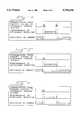
- FIG. 1is a block diagram of a preferred embodiment of a system of the present invention.
- FIG. 2is an illustration of a communication protocol of a second preferred embodiment.
- FIG. 3is an illustration of a communication protocol of a third preferred embodiment.
- FIG. 4is an illustration of a communication protocol of a fourth preferred embodiment.
- FIG. 5is an illustration of an alternative communication protocol of the fourth preferred embodiment.
- FIG. 6is an illustration of another alternative communication protocol of the fourth preferred embodiment.
- FIG. 7is a block diagram of an alternative embodiment of the system of FIG. 1.
- FIG. 8is a block diagram of another alternative embodiment of the system of FIG. 1.
- FIG. 1illustrates the components that can be used in the preferred embodiments described below.
- a therapeutic delivery device 14contains a therapeutic electrode 28 and a temperature sensor 30.
- the therapeutic delivery device 14connects to a therapy system 16, which contains a voltage generator 32 responsive to a voltage controller 34 and coupled to a current detector 36.
- the therapy system 16also contains a temperature monitoring system 38 coupled to the voltage controller 34 and responsive to the temperature sensor 30.
- one componentcan be "responsive” or “coupled” to another component either directly (as when two components are connected by a conductor) or indirectly (as when there is intermediate signal processing between two components).
- a communication link 18connects the therapy system 16 to an ultrasonic imaging system 20.
- the ultrasonic imaging system 20includes a central controller 41, a beam former 40 responsive to the central controller 41, and receiving circuitry and amplifiers 42 responsive to the central controller 41.
- the receiving circuitry and amplifiers 42are coupled to a scan converter 44, which is coupled to a display 46.
- the imaging system 20attaches to an ultrasound delivery device 22 containing an ultrasound transducer 24 responsive to the beam former 40 and coupled to the receiving circuitry and amplifiers 42. The function of these components will be described below.
- the voltage generator 32 in the therapy system 16generates a voltage and sends the resulting current to the therapy electrode 28.
- the voltage controller 34sets the frequency and amplitude of the voltage produced by the generator 32, as well as its sequencing in time. The voltage controller 34 makes this determination based on information received from the user via an input interface or from the temperature monitoring system 38 and the current detector 36.
- the temperature monitoring system 38receives thermal information from the temperature sensor 30 in the therapeutic delivery device 14, while the current detector 36 determines the amount of current sent to the therapy electrode 28.
- the current flowing from the therapy system 16 to the therapy electrode 28passes to the tissue 26 when the electrode 28 is placed adjacent to the tissue 26. This current spreads as it penetrates into the tissue 26 and propagates either to a second therapy electrode or to a distant, larger counter electrode (not shown). As it passes through the tissue 26, the current generates heat according to the local current density, ablating (i.e., causing thermal damage to) the tissue 26. As used herein, the term "ablation" refers to the process that affects the tissue by thermal means.
- the communication link 18connects the therapy system 16 to the ultrasonic imaging system 20 and is capable of sending signals between the two systems 16, 20.
- the ultrasonic imaging system 20controls the time during which the ultrasound transducer 24 is performing ultrasonic visualization of the tissue 26.
- the imaging system 20allows the user to adjust parameters which affect the time needed for ultrasonic visualization. As is well known in the art, these parameters include, but are not limited to, image depth, image width, and frame-rate, the number of frames per second that will be displayed to the user.
- the central controller 41interprets the set-up information entered by the user and configures the other components of the ultrasonic imaging system 20 accordingly.
- the term "ultrasonic visualization”refers to the well-known interrogating-and-imaging process which includes ultrasound generation, ultrasonic detection, image reconstruction, and image presentation phases.
- the beam former 40applies a voltage to the transducer 24 to cause it to vibrate and emit ultrasonic energy.
- the receiving circuitry 42measures the voltages created by the transducer 24 when ultrasonic energy reflected by the structures in the tissue 26 impinge on the transducer 24, creating a scan line. Additional ultrasonic energy is transmitted and received until enough scan lines are formed to create a frame.
- the image reconstruction phasebegins.
- the scan converter 44processes the amplified, sensed voltages to create an image of the tissue 26.
- the display 46presents the image to the user during the presentation phase.
- the desired frame-rateis usually 10-60 frames per second. Accordingly, the image is presented to the user in 1/60 to 1/60 of a second.
- the therapy currentneed not be applied throughout the entire ablation process. That is, continuous ablation of the tissue (i.e., continuous thermal damage to the tissue) will be maintained even if the therapy current is interrupted during the ablation process.
- Therapy currentcan be interrupted without significant effect on tissue heating if the resulting time-averaged current is sufficient to cause heating to therapeutic temperatures. This can be achieved by increasing the magnitude of the therapy current during its application or by increasing the duration of the ablation process to compensate for the interruption in therapy current. See S. A. Sapareto and W. C. Dewey, supra. If a sufficient amount of heat is generated within the tissue 26 before the therapy current is interrupted, ablation of the tissue 26 will continue even though therapy current is momentarily interrupted.
- the time the therapy current is offis short compared to the natural decay time of the heating field.
- This time scaleis approximately equal to the length scale of the heating field squared divided by the thermal diffusivity of tissue, or
- Lis the length scale of the heating field
- ⁇is the approximate time scale for cooling
- ⁇is the thermal diffusivity of tissue, typically 1.5 ⁇ 10 -3 cm 2 /sec.
- the length scalecan be taken as the diameter of the electrode, and so the time scale for cooling is about 106 seconds. Therefore, as long as the interruption of therapy current to the heating field is significantly less than 100 seconds and as long as the therapy current is increased during the "on" periods to compensate for the brief loss of heating energy, ablation of the tissue 26 will continue even though therapy current is interrupted. Instead of increasing the therapy current, the duration of the ablation process can be increased to compensate for the slight diminution in temperature resulting from the brief pauses in the application of the therapy current, if the resulting time-averaged current is sufficient to cause heating to therapeutic temperatures.
- This temporal averaging of the therapeutic heatingmay be used in a method and system for forming an ultrasound image of a tissue while simultaneously ablating the tissue, as will be illustrated below.
- Ultrasonic detectioncan be performed simultaneously with the ablation of tissue 26 if the application of therapy current is sequenced with ultrasonic detection. With this approach, no ultrasonic detection takes place while therapy current passes, and no therapy current passes while ultrasonic detection takes place. By sequencing these steps, the application of therapy current occurs virtually simultaneously with the performance of ultrasonic detection.
- a sufficient amount of therapy currentis applied to the tissue 26 to generate enough heat for ablation to continue during an interruption in therapy current, as can be detected by the temperature sensor 30 and the temperature monitoring system 38.
- ultrasonic detectionis performed by the ultrasonic imaging system 20 during an interruption in the application of the therapy current.
- the cyclebegins again, and therapy current is reapplied to the tissue 26, as before, to generate enough heat for continuous ablation.
- This methodallows the tissue 26 to be ultrasonically monitored while being simultaneously ablated without the previously mentioned problem of electrical interference on the ultrasound image.
- ultrasonic detectionoccurs in this method, no therapy current is flowing to the tissue (hence, no interference), yet the tissue is being ablated (from the heat built up before the therapy current was interrupted).
- this solutiondoes not significantly add to the size or cost of the transducer 24.
- Sequencingmay be implemented with a communication protocol, as described below, to determine when therapy current should be applied and when ultrasonic detection should be performed.
- each system 16, 20can use information entered by a user to determine when therapy current should be applied and when ultrasonic detection should be performed, without the use of a communication protocol between the systems 16, 20.
- FIG. 2illustrates the communication protocol used in the method of the second preferred embodiment.
- both the ultrasonic imaging system 20 and the therapy system 16signal each other, via the communication link 18, when done performing their functions.
- controlis passed between the two systems 16, 20. Once one system indicates that it is done with its function, it cannot proceed until it receives a signal from the other system signaling that the other system is done.
- the ultrasonic imaging system 20begins performing ultrasonic detection. When detection is complete, the ultrasonic imaging system 20 stops performing ultrasonic detection and signals the therapy system 16 that it is done with its function.
- the therapy system 16begins to apply therapy current to the tissue 26.
- the therapy system 16stops applying the therapy current to the tissue 26 and signals the ultrasonic imaging system 20 that it is done with its function. This would occur, for example, when a sufficient amount of heat is generated within the tissue 26 to continue ablation during the time when the therapy current is interrupted, as can be detected by the temperature sensor 30 and the temperature monitoring system 38. Ultrasonic detection of the tissue 26 resumes after the ultrasonic imaging system 20 receives the signal from the therapy system 16.
- FIG. 3illustrates the communication protocol used in the method of the third preferred embodiment.
- the ultrasonic imaging system 20retains exclusive control over when therapy current should be applied and when detection should be performed.
- the ultrasonic imaging system 20begins performing ultrasonic detection. When detection is complete, the ultrasonic imaging system 20 stops performing ultrasonic detection and signals the therapy system 16 that therapy current may be applied.
- the therapy system 16begins to apply therapy current to the tissue 26.
- the ultrasonic imaging system 20determines that a new ultrasound image needs to be formed, it sends a signal to the therapy system 16 to indicate that the therapy current should be turned off. This determination can be based on a minimum frame-rate entered by the user. For example, in cardiac ablation applications, the minimum frame-rate is usually 10-60 frames per second.
- the application of therapy currentis then interrupted, and ultrasonic detection is performed.
- the method using this protocolcan be used, for example, when careful monitoring of the growth of the treated tissue 26 is considered important.
- the ultrasonic imaging system 20ensures that the imaging is sufficiently continuous for tracking the growth of a lesion.
- FIG. 4illustrates the communication protocol used in the method of the fourth preferred embodiment.
- the therapy system 16retains exclusive control by determining when ultrasonic detection should begin and end. This protocol can be used to ensure a particular therapy current delivery.
- the therapy system 16begins applying therapy current to the tissue 26.
- the therapy system 16interrupts the application of therapy current to the tissue 26 and signals the ultrasonic imaging system 20 that detection may be performed. This would occur, for example, when a sufficient amount of heat is generated within the tissue 26 for ablation to continue during the time when the therapy current is interrupted, as can be detected by the temperature sensor 30 and the temperature monitoring system 38.
- the ultrasonic imaging system 20performs ultrasonic detection.
- the therapy system 16determines that therapy current needs to be applied to the tissue 26, it sends a signal to the ultrasonic imaging system 20 to interrupt ultrasonic detection. This would happen, for example, when more therapy current is needed to generate additional heat to maintain a continued ablation of the tissue 26.
- Ultrasonic detectionis then interrupted, and therapy current is applied to the tissue 26.
- This protocolmay produce a sub-optimal result given the complexity of the detection of ultrasound and given the delay needed for the ultrasound waves to travel into and return from the tissue 26. That is, the detection may not be complete when the therapy system 16 determines that detection must be interrupted and therapy current must be applied.
- the therapy system 16can apply current while the detection phase continues (see FIG. 5). Although there will be interference on the image caused by the simultaneous application of current and detection, this alternative allows the user to receive an image, even though it is of lesser quality.
- an alternative embodimentcan be used in which instead of sending two signals (one indicating that detection should begin and one indicating that it should be interrupted), the therapy system 16 sends only one signal, as shown in FIG. 6.
- This single signalgives the ultrasonic imaging system 20 a preset time interval to complete detection.
- the ultrasonic imaging system 20would then adjust imaging parameters (including, but not limited to, image depth and width) to ensure that detection is complete in the given amount of time.
- the detection phasewould be complete and therapy current would be applied automatically, without the need to send a second signal from the therapy system 16 to the ultrasonic imaging system 20.
- the application of therapy currentis sequenced with the performance of the ultrasonic detection because it is during the detection phase that the ultrasound system 20 is most susceptible to noise.
- the other phases of the ultrasonic visualization processi.e., ultrasound generation, image reconstruction, and image presentation
- the application of therapy currentcan be interrupted throughout the entire ultrasonic visualization process. This can be done for simplicity since the detection phase can consume the largest fraction of time for the entire ultrasonic visualization process.
- Ultrasound delivery devicesinclude all types of devices that contain a transducer. These devices include, but are not limited to, transthorasic, transabdominal, transesophageal, endorectal, endovaginal, and transluminal surgical devices and catheters.
- Therapy delivery devicesinclude several devices that contain an electrode. For example, in cardiac applications, the therapy electrodes can be delivered using a catheter, while for liver tumor applications, the therapy electrodes can be delivered using a catheter or a needle. Additionally, the therapy electrode can be a separate device delivered through an introducer needle or sheath.
- the ultrasound transducercan be of any type (e.g., mechanical array, ultrasound array, synthesized B-mode) as long as the ultrasonic imaging system 20 has the ability to control the time during which ultrasound energy is being generated or detected. While the ultrasonic imaging system can have duplicate receivers and amplifiers, it can also have a single receiver and amplifier.
- an additional communicationmay be sent from the ultrasonic imaging system 20 to the therapy system 16.
- the ultrasonic imaging system 20may communicate its duty cycle to the therapy system 16.
- the duty cyclecomprises the amount of time required for ultrasonic detection (or for completion of the entire ultrasonic visualization process) and the frame-rate.
- the time required for detection (or visualization)allows the therapy system 16 to know how long it must be off, while the frame-rate allows the therapy system 16 to know how long it can be on before another image needs to be generated.
- the voltage controller 34may then alter the intensity of therapy current or increase the duration of the ablation process accordingly to ensure that the time-average of the square of the therapy current, as determined by the current detector 36, is sufficient to generate enough heat for continuous ablation of the tissue 26.
- the duration of the ablation processincreasing, the number of times that the application of current and the performance of ultrasonic visualization (or detection) are sequenced is increased.
- the additional communicationcan be sent directly between the systems 16, 20, or it can be manually entered by the operator.
- the current detector 36can monitor the time-average of the square of the therapy current it is generating, and the voltage controller 34 can adjust the therapy current in real-time to maintain a particular value. Additionally, the voltage controller 34 can increase the duration of the ablation process.
- a combination of the therapy current-control alternatives described abovecan also be used. As mentioned earlier, the number of times that the application of current and the performance of ultrasonic visualization (or detection) are sequenced is increased as a result of the duration of the ablation process increasing.
- the temperature of the tissuecan be monitored by the temperature sensor 30 and the temperature monitoring system 38. With this information the voltage controller 34 or the user can increase the duration of the ablation process, instead of increasing the magnitude of the current, to ensure continuous ablation.
- the temperature sensor 30 and the temperature monitoring system 38are not strictly needed. For example, a user can use his experience to set the voltage or current level based on the electrode and tissue type.
- sequencing described in the embodimentscan begin with either application of therapy current or ultrasonic detection.
- ultrasound delivery device 22 and ultrasonic imaging system 20are separate components, it should be understood that they may be combined into one component. This is also true for the therapy delivery device 14 and the therapy system 16. Also, while the above preferred embodiments show the therapy system 16 and the ultrasonic imaging system 20 as being separate components, it should be understood that they may be subsystems of an integrated therapy/visualization system (See FIG. 7). Additionally, while two separate delivery devices 14, 22 are described above, a single delivery device can house the therapy electrode 28, the ultrasound transducer 24, and the temperature sensor 30 (See FIG. 8).
- the communication between the systems 16, 20can use conventional digital communication techniques--a short period of a high voltage to communicate that control is being passed. Other, possibly more sophisticated, communication modalities can also be used.
- communication linkis meant in its broadest sense to be any suitable communication technology and can include, but is not limited to, communication by wire, fiber optics, and radio waves, and it can be effected using either hardware or software.
- the communication protocols of the above embodiments and alternativesneed not be maintained throughout a particular therapy session.
- a communication protocolcan vary among any of the embodiments and alternatives as necessary or desired during a therapy session.
Landscapes
- Health & Medical Sciences (AREA)
- Surgery (AREA)
- Engineering & Computer Science (AREA)
- Life Sciences & Earth Sciences (AREA)
- Biomedical Technology (AREA)
- Molecular Biology (AREA)
- Nuclear Medicine, Radiotherapy & Molecular Imaging (AREA)
- Plasma & Fusion (AREA)
- Physics & Mathematics (AREA)
- Heart & Thoracic Surgery (AREA)
- Medical Informatics (AREA)
- Otolaryngology (AREA)
- Animal Behavior & Ethology (AREA)
- General Health & Medical Sciences (AREA)
- Public Health (AREA)
- Veterinary Medicine (AREA)
- Ultra Sonic Daignosis Equipment (AREA)
- Surgical Instruments (AREA)
Abstract
Description
τ=L.sup.2 /.sub.α,
Claims (35)
Priority Applications (5)
| Application Number | Priority Date | Filing Date | Title |
|---|---|---|---|
| US08/805,473US5788636A (en) | 1997-02-25 | 1997-02-25 | Method and system for forming an ultrasound image of a tissue while simultaneously ablating the tissue |
| DE19882137TDE19882137T1 (en) | 1997-02-25 | 1998-02-23 | Method and system for forming an ultrasound image of a tissue while ablating the tissue |
| JP53692298AJP2001513664A (en) | 1997-02-25 | 1998-02-23 | Method and system for forming ultrasound images while simultaneously cutting tissue |
| AU64376/98AAU6437698A (en) | 1997-02-25 | 1998-02-23 | Method and system for forming an ultrasound image of a tissue while simultaneously ablating the tissue |
| PCT/US1998/003409WO1998036679A2 (en) | 1997-02-25 | 1998-02-23 | Method and system for forming an ultrasound image of a tissue while simultaneously ablating the tissue |
Applications Claiming Priority (1)
| Application Number | Priority Date | Filing Date | Title |
|---|---|---|---|
| US08/805,473US5788636A (en) | 1997-02-25 | 1997-02-25 | Method and system for forming an ultrasound image of a tissue while simultaneously ablating the tissue |
Publications (1)
| Publication Number | Publication Date |
|---|---|
| US5788636Atrue US5788636A (en) | 1998-08-04 |
Family
ID=25191660
Family Applications (1)
| Application Number | Title | Priority Date | Filing Date |
|---|---|---|---|
| US08/805,473Expired - LifetimeUS5788636A (en) | 1997-02-25 | 1997-02-25 | Method and system for forming an ultrasound image of a tissue while simultaneously ablating the tissue |
Country Status (5)
| Country | Link |
|---|---|
| US (1) | US5788636A (en) |
| JP (1) | JP2001513664A (en) |
| AU (1) | AU6437698A (en) |
| DE (1) | DE19882137T1 (en) |
| WO (1) | WO1998036679A2 (en) |
Cited By (177)
| Publication number | Priority date | Publication date | Assignee | Title |
|---|---|---|---|---|
| WO2001071380A3 (en)* | 2000-03-17 | 2002-01-31 | Senorx Inc | System and method for managing intermittent interference on imaging systems |
| US20020042611A1 (en)* | 1996-10-22 | 2002-04-11 | Epicor, Inc. | Methods and devices for ablation |
| US20030073992A1 (en)* | 1996-10-22 | 2003-04-17 | Epicor, Inc. | Methods and devices for ablation |
| WO2002096508A3 (en)* | 2001-05-29 | 2003-07-24 | Ethicon Endo Surgery Inc | Ultrasound feedback in medically-treated patients |
| US6626855B1 (en) | 1999-11-26 | 2003-09-30 | Therus Corpoation | Controlled high efficiency lesion formation using high intensity ultrasound |
| US20030229283A1 (en)* | 2000-11-17 | 2003-12-11 | Craig Roger Kingdon | Ultrasound therapy |
| US20040106880A1 (en)* | 1999-10-25 | 2004-06-03 | Therus Corporation (Legal) | Use of focused ultrasound for vascular sealing |
| US6805129B1 (en) | 1996-10-22 | 2004-10-19 | Epicor Medical, Inc. | Apparatus and method for ablating tissue |
| US6805128B1 (en) | 1996-10-22 | 2004-10-19 | Epicor Medical, Inc. | Apparatus and method for ablating tissue |
| US20040258127A1 (en)* | 2003-06-23 | 2004-12-23 | Siemens Medical Solutions Usa, Inc. | Ultrasound transducer fault measurement method and system |
| US20050043726A1 (en)* | 2001-03-07 | 2005-02-24 | Mchale Anthony Patrick | Device II |
| US20050080336A1 (en)* | 2002-07-22 | 2005-04-14 | Ep Medsystems, Inc. | Method and apparatus for time gating of medical images |
| US20050096542A1 (en)* | 1999-12-23 | 2005-05-05 | Lee Weng | Ultrasound transducers for imaging and therapy |
| US20050124898A1 (en)* | 2002-01-16 | 2005-06-09 | Ep Medsystems, Inc. | Method and apparatus for isolating a catheter interface |
| US20050203410A1 (en)* | 2004-02-27 | 2005-09-15 | Ep Medsystems, Inc. | Methods and systems for ultrasound imaging of the heart from the pericardium |
| US6949095B2 (en) | 1996-10-22 | 2005-09-27 | Epicor Medical, Inc. | Apparatus and method for diagnosis and therapy of electrophysiological disease |
| US20050228290A1 (en)* | 2004-04-07 | 2005-10-13 | Ep Medsystems, Inc. | Steerable ultrasound catheter |
| US20050240105A1 (en)* | 2004-04-14 | 2005-10-27 | Mast T D | Method for reducing electronic artifacts in ultrasound imaging |
| US20050240103A1 (en)* | 2004-04-20 | 2005-10-27 | Ep Medsystems, Inc. | Method and apparatus for ultrasound imaging with autofrequency selection |
| US20050240170A1 (en)* | 1999-10-25 | 2005-10-27 | Therus Corporation | Insertable ultrasound probes, systems, and methods for thermal therapy |
| US20050245822A1 (en)* | 2002-07-22 | 2005-11-03 | Ep Medsystems, Inc. | Method and apparatus for imaging distant anatomical structures in intra-cardiac ultrasound imaging |
| US20050261571A1 (en)* | 2004-05-21 | 2005-11-24 | Willis Nathaniel P | 3-D ultrasound navigation during radio-frequency ablation |
| US20060122514A1 (en)* | 2004-11-23 | 2006-06-08 | Ep Medsystems, Inc. | Method and apparatus for localizing an ultrasound catheter |
| US7083620B2 (en) | 2002-10-30 | 2006-08-01 | Medtronic, Inc. | Electrosurgical hemostat |
| US7094235B2 (en) | 2001-04-26 | 2006-08-22 | Medtronic, Inc. | Method and apparatus for tissue ablation |
| US7118566B2 (en) | 2002-05-16 | 2006-10-10 | Medtronic, Inc. | Device and method for needle-less interstitial injection of fluid for ablation of cardiac tissue |
| US7128740B2 (en) | 1996-05-03 | 2006-10-31 | Jacobs Clemens J | Method for interrupting conduction paths within the heart |
| US7156845B2 (en) | 1998-07-07 | 2007-01-02 | Medtronic, Inc. | Method and apparatus for creating a bi-polar virtual electrode used for the ablation of tissue |
| US7166105B2 (en) | 1995-02-22 | 2007-01-23 | Medtronic, Inc. | Pen-type electrosurgical instrument |
| US7169144B2 (en) | 1998-07-07 | 2007-01-30 | Medtronic, Inc. | Apparatus and method for creating, maintaining, and controlling a virtual electrode used for the ablation of tissue |
| US20070049827A1 (en)* | 2005-08-16 | 2007-03-01 | General Electric Company | Clinical feedback of ablation efficacy during ablation procedure |
| WO2007000680A3 (en)* | 2005-06-29 | 2007-04-12 | Koninkl Philips Electronics Nv | Optimized temperature measurement in an ultrasound transducer |
| US20070083118A1 (en)* | 2002-07-22 | 2007-04-12 | Ep Medsystems, Inc. | Method and System For Estimating Cardiac Ejection Volume Using Ultrasound Spectral Doppler Image Data |
| US7211044B2 (en) | 2001-05-29 | 2007-05-01 | Ethicon Endo-Surgery, Inc. | Method for mapping temperature rise using pulse-echo ultrasound |
| US20070118035A1 (en)* | 2005-11-22 | 2007-05-24 | General Electric Company | Catheter tip |
| US20070167793A1 (en)* | 2005-12-14 | 2007-07-19 | Ep Medsystems, Inc. | Method and system for enhancing spectral doppler presentation |
| US20070167809A1 (en)* | 2002-07-22 | 2007-07-19 | Ep Medsystems, Inc. | Method and System For Estimating Cardiac Ejection Volume And Placing Pacemaker Electrodes Using Speckle Tracking |
| US20070167794A1 (en)* | 2005-12-14 | 2007-07-19 | Ep Medsystems, Inc. | Method and system for evaluating valvular function |
| US7250048B2 (en) | 2001-04-26 | 2007-07-31 | Medtronic, Inc. | Ablation system and method of use |
| US20070232949A1 (en)* | 2006-03-31 | 2007-10-04 | Ep Medsystems, Inc. | Method For Simultaneous Bi-Atrial Mapping Of Atrial Fibrillation |
| US7294143B2 (en) | 2002-05-16 | 2007-11-13 | Medtronic, Inc. | Device and method for ablation of cardiac tissue |
| US7309325B2 (en) | 1998-07-07 | 2007-12-18 | Medtronic, Inc. | Helical needle apparatus for creating a virtual electrode used for the ablation of tissue |
| US20070299436A1 (en)* | 2006-06-23 | 2007-12-27 | Podmore Jonathan L | Ablation device and method comprising movable ablation elements |
| US20070299479A1 (en)* | 2006-06-27 | 2007-12-27 | Ep Medsystems, Inc. | Method for Reversing Ventricular Dyssynchrony |
| US20080009733A1 (en)* | 2006-06-27 | 2008-01-10 | Ep Medsystems, Inc. | Method for Evaluating Regional Ventricular Function and Incoordinate Ventricular Contraction |
| US7347858B2 (en) | 2001-12-11 | 2008-03-25 | Medtronic, Inc. | Method and system for treatment of atrial tachyarrhythmias |
| US7364578B2 (en) | 2002-01-25 | 2008-04-29 | Medtronic, Inc. | System and method of performing an electrosurgical procedure |
| US7367972B2 (en) | 2001-04-26 | 2008-05-06 | Medtronic, Inc. | Ablation system |
| US7387126B2 (en) | 1996-10-22 | 2008-06-17 | St. Jude Medical, Atrial Fibrillation Division, Inc. | Surgical system and procedure for treatment of medically refractory atrial fibrillation |
| US20080146943A1 (en)* | 2006-12-14 | 2008-06-19 | Ep Medsystems, Inc. | Integrated Beam Former And Isolation For An Ultrasound Probe |
| US20080146942A1 (en)* | 2006-12-13 | 2008-06-19 | Ep Medsystems, Inc. | Catheter Position Tracking Methods Using Fluoroscopy and Rotational Sensors |
| US20080146928A1 (en)* | 2006-12-14 | 2008-06-19 | Ep Medsystems, Inc. | Method and System for Configuration of a Pacemaker and For Placement of Pacemaker Electrodes |
| US20080146940A1 (en)* | 2006-12-14 | 2008-06-19 | Ep Medsystems, Inc. | External and Internal Ultrasound Imaging System |
| US7435250B2 (en) | 2000-04-27 | 2008-10-14 | Medtronic, Inc. | Method and apparatus for tissue ablation |
| US7452357B2 (en) | 2004-10-22 | 2008-11-18 | Ethicon Endo-Surgery, Inc. | System and method for planning treatment of tissue |
| US7470272B2 (en) | 1997-07-18 | 2008-12-30 | Medtronic, Inc. | Device and method for ablating tissue |
| US7473250B2 (en) | 2004-05-21 | 2009-01-06 | Ethicon Endo-Surgery, Inc. | Ultrasound medical system and method |
| US20090030312A1 (en)* | 2007-07-27 | 2009-01-29 | Andreas Hadjicostis | Image-guided intravascular therapy catheters |
| US7494467B2 (en) | 2004-04-16 | 2009-02-24 | Ethicon Endo-Surgery, Inc. | Medical system having multiple ultrasound transducers or an ultrasound transducer and an RF electrode |
| US7497857B2 (en) | 2003-04-29 | 2009-03-03 | Medtronic, Inc. | Endocardial dispersive electrode for use with a monopolar RF ablation pen |
| WO2009028245A1 (en) | 2007-08-28 | 2009-03-05 | Olympus Medical Systems Corp. | Medical manual skill device |
| US20090076375A1 (en)* | 2007-09-13 | 2009-03-19 | Siemens Aktiengesellschaft | Myocardial tissue ablation device for treatment of cardiac arrhythmias by ablation of myocardial tissue in a patient as well as associated catheter and associated method |
| US7507235B2 (en) | 2001-01-13 | 2009-03-24 | Medtronic, Inc. | Method and system for organ positioning and stabilization |
| US20090093718A1 (en)* | 2007-07-12 | 2009-04-09 | Takao Jibiki | Ultrasonic diagnosis apparatus, radiofrequency wave cautery treatment device, ultrasonic diagnosis and treatment system, and ultrasonic diagnosis and treatment apparatus |
| US20090149754A1 (en)* | 2007-12-07 | 2009-06-11 | Masaki Tsuda | Ultrasonic diagnosis apparatus, radiofrequency wave cautery treatment device, ultrasonic diagnosis and treatment system, and ultrasonic diagnosis and treatment apparatus |
| US7566334B2 (en) | 2004-06-02 | 2009-07-28 | Medtronic, Inc. | Ablation device with jaws |
| US7615015B2 (en) | 2000-01-19 | 2009-11-10 | Medtronic, Inc. | Focused ultrasound ablation devices having selectively actuatable emitting elements and methods of using the same |
| US7628780B2 (en) | 2001-01-13 | 2009-12-08 | Medtronic, Inc. | Devices and methods for interstitial injection of biologic agents into tissue |
| US7648462B2 (en) | 2002-01-16 | 2010-01-19 | St. Jude Medical, Atrial Fibrillation Division, Inc. | Safety systems and methods for ensuring safe use of intra-cardiac ultrasound catheters |
| US7678108B2 (en) | 2004-06-02 | 2010-03-16 | Medtronic, Inc. | Loop ablation apparatus and method |
| US7706882B2 (en) | 2000-01-19 | 2010-04-27 | Medtronic, Inc. | Methods of using high intensity focused ultrasound to form an ablated tissue area |
| US7706894B2 (en) | 2000-10-10 | 2010-04-27 | Medtronic, Inc. | Heart wall ablation/mapping catheter and method |
| US20100121142A1 (en)* | 2008-11-12 | 2010-05-13 | Ouyang Xiaolong | Minimally Invasive Imaging Device |
| US20100121155A1 (en)* | 2008-11-12 | 2010-05-13 | Ouyang Xiaolong | Minimally Invasive Tissue Modification Systems With Integrated Visualization |
| US20100121139A1 (en)* | 2008-11-12 | 2010-05-13 | Ouyang Xiaolong | Minimally Invasive Imaging Systems |
| US20100145361A1 (en)* | 2004-06-18 | 2010-06-10 | Francischelli David E | Methods and Devices for Occlusion of an Atrial Appendage |
| US7740623B2 (en) | 2001-01-13 | 2010-06-22 | Medtronic, Inc. | Devices and methods for interstitial injection of biologic agents into tissue |
| US7744562B2 (en) | 2003-01-14 | 2010-06-29 | Medtronics, Inc. | Devices and methods for interstitial injection of biologic agents into tissue |
| US7758580B2 (en) | 2004-06-02 | 2010-07-20 | Medtronic, Inc. | Compound bipolar ablation device and method |
| US7758576B2 (en) | 2004-06-02 | 2010-07-20 | Medtronic, Inc. | Clamping ablation tool and method |
| US20100204568A1 (en)* | 2009-02-09 | 2010-08-12 | The Cleveland Clinic Foundation | Ultrasound-guided delivery of a therapy delivery device to a nerve target |
| US7806839B2 (en) | 2004-06-14 | 2010-10-05 | Ethicon Endo-Surgery, Inc. | System and method for ultrasound therapy using grating lobes |
| US7818039B2 (en) | 2000-04-27 | 2010-10-19 | Medtronic, Inc. | Suction stabilized epicardial ablation devices |
| US7824399B2 (en) | 2001-04-26 | 2010-11-02 | Medtronic, Inc. | Ablation system and method of use |
| US7824403B2 (en) | 1996-10-22 | 2010-11-02 | St. Jude Medical, Atrial Fibrillation Division, Inc. | Methods and devices for ablation |
| US7833221B2 (en) | 2004-10-22 | 2010-11-16 | Ethicon Endo-Surgery, Inc. | System and method for treatment of tissue using the tissue as a fiducial |
| US7846096B2 (en) | 2001-05-29 | 2010-12-07 | Ethicon Endo-Surgery, Inc. | Method for monitoring of medical treatment using pulse-echo ultrasound |
| US7883468B2 (en) | 2004-05-18 | 2011-02-08 | Ethicon Endo-Surgery, Inc. | Medical system having an ultrasound source and an acoustic coupling medium |
| EP2289444A1 (en)* | 2009-08-25 | 2011-03-02 | Tyco Healthcare Group, LP | System for performing an electrosurgical procedure using an imaging compatible electrosurgical system |
| US7951095B2 (en) | 2004-05-20 | 2011-05-31 | Ethicon Endo-Surgery, Inc. | Ultrasound medical system |
| US7959626B2 (en) | 2001-04-26 | 2011-06-14 | Medtronic, Inc. | Transmural ablation systems and methods |
| US7967816B2 (en) | 2002-01-25 | 2011-06-28 | Medtronic, Inc. | Fluid-assisted electrosurgical instrument with shapeable electrode |
| US8052607B2 (en) | 2008-04-22 | 2011-11-08 | St. Jude Medical, Atrial Fibrillation Division, Inc. | Ultrasound imaging catheter with pivoting head |
| US8057394B2 (en) | 2007-06-30 | 2011-11-15 | St. Jude Medical, Atrial Fibrillation Division, Inc. | Ultrasound image processing to render three-dimensional images from two-dimensional images |
| US8162933B2 (en) | 2000-04-27 | 2012-04-24 | Medtronic, Inc. | Vibration sensitive ablation device and method |
| US8167805B2 (en) | 2005-10-20 | 2012-05-01 | Kona Medical, Inc. | Systems and methods for ultrasound applicator station keeping |
| WO2012066472A1 (en)* | 2010-11-18 | 2012-05-24 | Koninklijke Philips Electronics N.V. | Interference reduction and signal to noise ratio improvement for ultrasound cardiac ablation monitoring |
| US8221402B2 (en) | 2000-01-19 | 2012-07-17 | Medtronic, Inc. | Method for guiding a medical device |
| US8295912B2 (en) | 2009-10-12 | 2012-10-23 | Kona Medical, Inc. | Method and system to inhibit a function of a nerve traveling with an artery |
| US8308719B2 (en) | 1998-09-21 | 2012-11-13 | St. Jude Medical, Atrial Fibrillation Division, Inc. | Apparatus and method for ablating tissue |
| US8317711B2 (en) | 2007-06-16 | 2012-11-27 | St. Jude Medical, Atrial Fibrillation Division, Inc. | Oscillating phased-array ultrasound imaging catheter system |
| US8333764B2 (en) | 2004-05-12 | 2012-12-18 | Medtronic, Inc. | Device and method for determining tissue thickness and creating cardiac ablation lesions |
| WO2013014583A1 (en)* | 2011-07-22 | 2013-01-31 | Koninklijke Philips Electronics N.V. | Ablation apparatus |
| US8374674B2 (en) | 2009-10-12 | 2013-02-12 | Kona Medical, Inc. | Nerve treatment system |
| US8409219B2 (en) | 2004-06-18 | 2013-04-02 | Medtronic, Inc. | Method and system for placement of electrical lead inside heart |
| US8469904B2 (en) | 2009-10-12 | 2013-06-25 | Kona Medical, Inc. | Energetic modulation of nerves |
| US8512262B2 (en) | 2009-10-12 | 2013-08-20 | Kona Medical, Inc. | Energetic modulation of nerves |
| US8512337B2 (en) | 2001-04-26 | 2013-08-20 | Medtronic, Inc. | Method and system for treatment of atrial tachyarrhythmias |
| US8517962B2 (en) | 2009-10-12 | 2013-08-27 | Kona Medical, Inc. | Energetic modulation of nerves |
| EP2103955A3 (en)* | 2008-03-20 | 2013-09-18 | Siemens Aktiengesellschaft | Device and method for synchronising ultrasound convertors in a motion system |
| US8568409B2 (en) | 2000-03-06 | 2013-10-29 | Medtronic Advanced Energy Llc | Fluid-assisted medical devices, systems and methods |
| FR2990138A1 (en)* | 2012-05-02 | 2013-11-08 | Siemens Medical Solutions | SYSTEM FOR THERAPY CONTROL WITH ULTRASONIC SCANNING DEVICE |
| US8632533B2 (en) | 2009-02-23 | 2014-01-21 | Medtronic Advanced Energy Llc | Fluid-assisted electrosurgical device |
| US8663245B2 (en) | 2004-06-18 | 2014-03-04 | Medtronic, Inc. | Device for occlusion of a left atrial appendage |
| US8709007B2 (en) | 1997-10-15 | 2014-04-29 | St. Jude Medical, Atrial Fibrillation Division, Inc. | Devices and methods for ablating cardiac tissue |
| US8750568B2 (en) | 2012-05-22 | 2014-06-10 | Covidien Lp | System and method for conformal ablation planning |
| US8801707B2 (en) | 2004-05-14 | 2014-08-12 | Medtronic, Inc. | Method and devices for treating atrial fibrillation by mass ablation |
| US8821488B2 (en) | 2008-05-13 | 2014-09-02 | Medtronic, Inc. | Tissue lesion evaluation |
| US8870864B2 (en) | 2011-10-28 | 2014-10-28 | Medtronic Advanced Energy Llc | Single instrument electrosurgery apparatus and its method of use |
| US8882756B2 (en) | 2007-12-28 | 2014-11-11 | Medtronic Advanced Energy Llc | Fluid-assisted electrosurgical devices, methods and systems |
| US8906012B2 (en) | 2010-06-30 | 2014-12-09 | Medtronic Advanced Energy Llc | Electrosurgical devices with wire electrode |
| US8920417B2 (en) | 2010-06-30 | 2014-12-30 | Medtronic Advanced Energy Llc | Electrosurgical devices and methods of use thereof |
| US8945015B2 (en) | 2012-01-31 | 2015-02-03 | Koninklijke Philips N.V. | Ablation probe with fluid-based acoustic coupling for ultrasonic tissue imaging and treatment |
| US8986231B2 (en) | 2009-10-12 | 2015-03-24 | Kona Medical, Inc. | Energetic modulation of nerves |
| US8986211B2 (en) | 2009-10-12 | 2015-03-24 | Kona Medical, Inc. | Energetic modulation of nerves |
| US8992447B2 (en) | 2009-10-12 | 2015-03-31 | Kona Medical, Inc. | Energetic modulation of nerves |
| US9005143B2 (en) | 2009-10-12 | 2015-04-14 | Kona Medical, Inc. | External autonomic modulation |
| US9023040B2 (en) | 2010-10-26 | 2015-05-05 | Medtronic Advanced Energy Llc | Electrosurgical cutting devices |
| US9089340B2 (en) | 2010-12-30 | 2015-07-28 | Boston Scientific Scimed, Inc. | Ultrasound guided tissue ablation |
| US9138289B2 (en) | 2010-06-28 | 2015-09-22 | Medtronic Advanced Energy Llc | Electrode sheath for electrosurgical device |
| US9227088B2 (en) | 2006-05-25 | 2016-01-05 | Medtronic, Inc. | Methods of using high intensity focused ultrasound to form an ablated tissue area containing a plurality of lesions |
| US9241687B2 (en) | 2011-06-01 | 2016-01-26 | Boston Scientific Scimed Inc. | Ablation probe with ultrasonic imaging capabilities |
| US9241761B2 (en) | 2011-12-28 | 2016-01-26 | Koninklijke Philips N.V. | Ablation probe with ultrasonic imaging capability |
| US9254168B2 (en) | 2009-02-02 | 2016-02-09 | Medtronic Advanced Energy Llc | Electro-thermotherapy of tissue using penetrating microelectrode array |
| US9333027B2 (en)* | 2010-05-28 | 2016-05-10 | Medtronic Advanced Energy Llc | Method of producing an electrosurgical device |
| US9345541B2 (en) | 2009-09-08 | 2016-05-24 | Medtronic Advanced Energy Llc | Cartridge assembly for electrosurgical devices, electrosurgical unit and methods of use thereof |
| US9370295B2 (en) | 2014-01-13 | 2016-06-21 | Trice Medical, Inc. | Fully integrated, disposable tissue visualization device |
| US9381061B2 (en) | 2000-03-06 | 2016-07-05 | Medtronic Advanced Energy Llc | Fluid-assisted medical devices, systems and methods |
| US9393072B2 (en) | 2009-06-30 | 2016-07-19 | Boston Scientific Scimed, Inc. | Map and ablate open irrigated hybrid catheter |
| US9427281B2 (en) | 2011-03-11 | 2016-08-30 | Medtronic Advanced Energy Llc | Bronchoscope-compatible catheter provided with electrosurgical device |
| US9439622B2 (en) | 2012-05-22 | 2016-09-13 | Covidien Lp | Surgical navigation system |
| US9439623B2 (en) | 2012-05-22 | 2016-09-13 | Covidien Lp | Surgical planning system and navigation system |
| US9439627B2 (en) | 2012-05-22 | 2016-09-13 | Covidien Lp | Planning system and navigation system for an ablation procedure |
| US9463064B2 (en) | 2011-09-14 | 2016-10-11 | Boston Scientific Scimed Inc. | Ablation device with multiple ablation modes |
| US9498231B2 (en) | 2011-06-27 | 2016-11-22 | Board Of Regents Of The University Of Nebraska | On-board tool tracking system and methods of computer assisted surgery |
| US9498182B2 (en) | 2012-05-22 | 2016-11-22 | Covidien Lp | Systems and methods for planning and navigation |
| US9592090B2 (en) | 2010-03-11 | 2017-03-14 | Medtronic Advanced Energy Llc | Bipolar electrosurgical cutter with position insensitive return electrode contact |
| US9603659B2 (en) | 2011-09-14 | 2017-03-28 | Boston Scientific Scimed Inc. | Ablation device with ionically conductive balloon |
| US9713497B2 (en)* | 2010-01-29 | 2017-07-25 | Covidien Lp | System and method for performing an electrosurgical procedure using an ablation device with an integrated imaging device |
| US9743854B2 (en) | 2014-12-18 | 2017-08-29 | Boston Scientific Scimed, Inc. | Real-time morphology analysis for lesion assessment |
| US9750565B2 (en) | 2011-09-30 | 2017-09-05 | Medtronic Advanced Energy Llc | Electrosurgical balloons |
| US9757191B2 (en) | 2012-01-10 | 2017-09-12 | Boston Scientific Scimed, Inc. | Electrophysiology system and methods |
| US9974599B2 (en) | 2014-08-15 | 2018-05-22 | Medtronic Ps Medical, Inc. | Multipurpose electrosurgical device |
| US10105149B2 (en) | 2013-03-15 | 2018-10-23 | Board Of Regents Of The University Of Nebraska | On-board tool tracking system and methods of computer assisted surgery |
| US10188368B2 (en) | 2017-06-26 | 2019-01-29 | Andreas Hadjicostis | Image guided intravascular therapy catheter utilizing a thin chip multiplexor |
| US10219811B2 (en) | 2011-06-27 | 2019-03-05 | Board Of Regents Of The University Of Nebraska | On-board tool tracking system and methods of computer assisted surgery |
| US10342579B2 (en) | 2014-01-13 | 2019-07-09 | Trice Medical, Inc. | Fully integrated, disposable tissue visualization device |
| US10405886B2 (en) | 2015-08-11 | 2019-09-10 | Trice Medical, Inc. | Fully integrated, disposable tissue visualization device |
| US10492760B2 (en) | 2017-06-26 | 2019-12-03 | Andreas Hadjicostis | Image guided intravascular therapy catheter utilizing a thin chip multiplexor |
| US10524684B2 (en) | 2014-10-13 | 2020-01-07 | Boston Scientific Scimed Inc | Tissue diagnosis and treatment using mini-electrodes |
| US10603105B2 (en) | 2014-10-24 | 2020-03-31 | Boston Scientific Scimed Inc | Medical devices with a flexible electrode assembly coupled to an ablation tip |
| US10716612B2 (en) | 2015-12-18 | 2020-07-21 | Medtronic Advanced Energy Llc | Electrosurgical device with multiple monopolar electrode assembly |
| US10772681B2 (en) | 2009-10-12 | 2020-09-15 | Utsuka Medical Devices Co., Ltd. | Energy delivery to intraparenchymal regions of the kidney |
| CN111870338A (en)* | 2019-05-01 | 2020-11-03 | 韦伯斯特生物官能(以色列)有限公司 | Temperature-controlled short-duration ablation with resistive heating |
| US10874449B2 (en) | 2012-04-19 | 2020-12-29 | Koninklijke Philips N.V. | Energy application apparatus |
| US10925579B2 (en) | 2014-11-05 | 2021-02-23 | Otsuka Medical Devices Co., Ltd. | Systems and methods for real-time tracking of a target tissue using imaging before and during therapy delivery |
| US11051875B2 (en) | 2015-08-24 | 2021-07-06 | Medtronic Advanced Energy Llc | Multipurpose electrosurgical device |
| US11109909B1 (en) | 2017-06-26 | 2021-09-07 | Andreas Hadjicostis | Image guided intravascular therapy catheter utilizing a thin ablation electrode |
| US11116574B2 (en) | 2006-06-16 | 2021-09-14 | Board Of Regents Of The University Of Nebraska | Method and apparatus for computer aided surgery |
| US11389227B2 (en) | 2015-08-20 | 2022-07-19 | Medtronic Advanced Energy Llc | Electrosurgical device with multivariate control |
| US11547446B2 (en) | 2014-01-13 | 2023-01-10 | Trice Medical, Inc. | Fully integrated, disposable tissue visualization device |
| US11622753B2 (en) | 2018-03-29 | 2023-04-11 | Trice Medical, Inc. | Fully integrated endoscope with biopsy capabilities and methods of use |
| US11684416B2 (en) | 2009-02-11 | 2023-06-27 | Boston Scientific Scimed, Inc. | Insulated ablation catheter devices and methods of use |
| US11707329B2 (en) | 2018-08-10 | 2023-07-25 | Covidien Lp | Systems and methods for ablation visualization |
| US11911117B2 (en) | 2011-06-27 | 2024-02-27 | Board Of Regents Of The University Of Nebraska | On-board tool tracking system and methods of computer assisted surgery |
| US11998266B2 (en) | 2009-10-12 | 2024-06-04 | Otsuka Medical Devices Co., Ltd | Intravascular energy delivery |
| US12023082B2 (en) | 2017-10-06 | 2024-07-02 | Medtronic Advanced Energy Llc | Hemostatic thermal sealer |
Families Citing this family (1)
| Publication number | Priority date | Publication date | Assignee | Title |
|---|---|---|---|---|
| WO2007023878A1 (en)* | 2005-08-24 | 2007-03-01 | Hitachi Medical Corporation | Curing system |
Citations (16)
| Publication number | Priority date | Publication date | Assignee | Title |
|---|---|---|---|---|
| US4517976A (en)* | 1981-10-20 | 1985-05-21 | Fuji Photo Film Co., Ltd. | High frequency scalpel and endoscope system and method of operating same |
| US4658828A (en)* | 1984-05-03 | 1987-04-21 | Jacques Dory | Apparatus for examining and localizing tumors using ultra sounds, comprising a device for localized hyperthermia treatment |
| US4936281A (en)* | 1989-04-13 | 1990-06-26 | Everest Medical Corporation | Ultrasonically enhanced RF ablation catheter |
| US5143074A (en)* | 1983-12-14 | 1992-09-01 | Edap International | Ultrasonic treatment device using a focussing and oscillating piezoelectric element |
| US5183048A (en)* | 1991-06-24 | 1993-02-02 | Endosonics Corporation | Method and apparatus for removing artifacts from an ultrasonically generated image of a small cavity |
| US5279299A (en)* | 1991-02-15 | 1994-01-18 | Cardiac Pathways Corporation | Endocardial mapping and ablation system and catheter probe |
| US5324284A (en)* | 1992-06-05 | 1994-06-28 | Cardiac Pathways, Inc. | Endocardial mapping and ablation system utilizing a separately controlled ablation catheter and method |
| US5385148A (en)* | 1993-07-30 | 1995-01-31 | The Regents Of The University Of California | Cardiac imaging and ablation catheter |
| US5409000A (en)* | 1993-09-14 | 1995-04-25 | Cardiac Pathways Corporation | Endocardial mapping and ablation system utilizing separately controlled steerable ablation catheter with ultrasonic imaging capabilities and method |
| US5429136A (en)* | 1993-04-21 | 1995-07-04 | Devices For Vascular Intervention, Inc. | Imaging atherectomy apparatus |
| US5435304A (en)* | 1992-04-24 | 1995-07-25 | Siemens Aktiengesellschaft | Method and apparatus for therapeutic treatment with focussed acoustic waves switchable between a locating mode and a therapy mode |
| US5454809A (en)* | 1989-01-06 | 1995-10-03 | Angioplasty Systems, Inc. | Electrosurgical catheter and method for resolving atherosclerotic plaque by radio frequency sparking |
| US5454370A (en)* | 1993-12-03 | 1995-10-03 | Avitall; Boaz | Mapping and ablation electrode configuration |
| US5526815A (en)* | 1993-01-29 | 1996-06-18 | Siemens Aktiengesellschat | Therapy apparatus for locating and treating a zone located in the body of a life form with acoustic waves |
| US5588432A (en)* | 1988-03-21 | 1996-12-31 | Boston Scientific Corporation | Catheters for imaging, sensing electrical potentials, and ablating tissue |
| US5657755A (en)* | 1993-03-11 | 1997-08-19 | Desai; Jawahar M. | Apparatus and method for cardiac ablation |
- 1997
- 1997-02-25USUS08/805,473patent/US5788636A/ennot_activeExpired - Lifetime
- 1998
- 1998-02-23JPJP53692298Apatent/JP2001513664A/ennot_activeCeased
- 1998-02-23AUAU64376/98Apatent/AU6437698A/ennot_activeAbandoned
- 1998-02-23DEDE19882137Tpatent/DE19882137T1/ennot_activeCeased
- 1998-02-23WOPCT/US1998/003409patent/WO1998036679A2/enactiveApplication Filing
Patent Citations (16)
| Publication number | Priority date | Publication date | Assignee | Title |
|---|---|---|---|---|
| US4517976A (en)* | 1981-10-20 | 1985-05-21 | Fuji Photo Film Co., Ltd. | High frequency scalpel and endoscope system and method of operating same |
| US5143074A (en)* | 1983-12-14 | 1992-09-01 | Edap International | Ultrasonic treatment device using a focussing and oscillating piezoelectric element |
| US4658828A (en)* | 1984-05-03 | 1987-04-21 | Jacques Dory | Apparatus for examining and localizing tumors using ultra sounds, comprising a device for localized hyperthermia treatment |
| US5588432A (en)* | 1988-03-21 | 1996-12-31 | Boston Scientific Corporation | Catheters for imaging, sensing electrical potentials, and ablating tissue |
| US5454809A (en)* | 1989-01-06 | 1995-10-03 | Angioplasty Systems, Inc. | Electrosurgical catheter and method for resolving atherosclerotic plaque by radio frequency sparking |
| US4936281A (en)* | 1989-04-13 | 1990-06-26 | Everest Medical Corporation | Ultrasonically enhanced RF ablation catheter |
| US5279299A (en)* | 1991-02-15 | 1994-01-18 | Cardiac Pathways Corporation | Endocardial mapping and ablation system and catheter probe |
| US5183048A (en)* | 1991-06-24 | 1993-02-02 | Endosonics Corporation | Method and apparatus for removing artifacts from an ultrasonically generated image of a small cavity |
| US5435304A (en)* | 1992-04-24 | 1995-07-25 | Siemens Aktiengesellschaft | Method and apparatus for therapeutic treatment with focussed acoustic waves switchable between a locating mode and a therapy mode |
| US5324284A (en)* | 1992-06-05 | 1994-06-28 | Cardiac Pathways, Inc. | Endocardial mapping and ablation system utilizing a separately controlled ablation catheter and method |
| US5526815A (en)* | 1993-01-29 | 1996-06-18 | Siemens Aktiengesellschat | Therapy apparatus for locating and treating a zone located in the body of a life form with acoustic waves |
| US5657755A (en)* | 1993-03-11 | 1997-08-19 | Desai; Jawahar M. | Apparatus and method for cardiac ablation |
| US5429136A (en)* | 1993-04-21 | 1995-07-04 | Devices For Vascular Intervention, Inc. | Imaging atherectomy apparatus |
| US5385148A (en)* | 1993-07-30 | 1995-01-31 | The Regents Of The University Of California | Cardiac imaging and ablation catheter |
| US5409000A (en)* | 1993-09-14 | 1995-04-25 | Cardiac Pathways Corporation | Endocardial mapping and ablation system utilizing separately controlled steerable ablation catheter with ultrasonic imaging capabilities and method |
| US5454370A (en)* | 1993-12-03 | 1995-10-03 | Avitall; Boaz | Mapping and ablation electrode configuration |
Non-Patent Citations (8)
| Title |
|---|
| "Biophysics and Pathology of Catheter Energy Delivery Systems", Nath & Haines, Progress in Cardiovascular Diseases, Jan./Feb. 1995, vol. XXXVII, No. 4, pp. 185-204. |
| "Radiofrequency Tissue Ablation: Increased Lesion Diameter with a Perfusion Electrode", Goldberg et al., Acad Radiol Aug. 1996; 3:636-644. |
| "Thermal Dose Determination In Cancer Therapy", S.A. Sapareto and W.C. Dewey, International Journal of Radiology, Oncology, Biology, and Physics 10(6): 787-800 Jun. 1984. |
| "Ultrasound Cardioscopy: Embarking on a New Journey", Seward, et al., Mayo Clinic Proceedings, Jul. 1996, vol. 71 No. 7, pp. 629-635. |
| Biophysics and Pathology of Catheter Energy Delivery Systems , Nath & Haines, Progress in Cardiovascular Diseases, Jan./Feb. 1995, vol. XXXVII, No. 4, pp. 185 204.* |
| Radiofrequency Tissue Ablation: Increased Lesion Diameter with a Perfusion Electrode , Goldberg et al., Acad Radiol Aug. 1996; 3:636 644.* |
| Thermal Dose Determination In Cancer Therapy , S.A. Sapareto and W.C. Dewey, International Journal of Radiology, Oncology, Biology, and Physics 10(6): 787 800 Jun. 1984.* |
| Ultrasound Cardioscopy: Embarking on a New Journey , Seward, et al., Mayo Clinic Proceedings, Jul. 1996, vol. 71 No. 7, pp. 629 635.* |
Cited By (287)
| Publication number | Priority date | Publication date | Assignee | Title |
|---|---|---|---|---|
| US7794460B2 (en) | 1995-02-22 | 2010-09-14 | Medtronic, Inc. | Method of ablating tissue |
| US7166105B2 (en) | 1995-02-22 | 2007-01-23 | Medtronic, Inc. | Pen-type electrosurgical instrument |
| US9770282B2 (en) | 1995-02-22 | 2017-09-26 | Medtronic, Inc. | Apparatus and method for creating, maintaining, and controlling a virtual electrode used for the ablation of tissue |
| US7247155B2 (en) | 1995-02-22 | 2007-07-24 | Medtronic, Inc. | Apparatus and method for creating, maintaining, and controlling a virtual electrode used for the ablation of tissue |
| US7422588B2 (en) | 1995-02-22 | 2008-09-09 | Medtronic, Inc. | Pen-type electrosurgical instrument |
| US7128740B2 (en) | 1996-05-03 | 2006-10-31 | Jacobs Clemens J | Method for interrupting conduction paths within the heart |
| US6858026B2 (en) | 1996-10-22 | 2005-02-22 | Epicor Medical, Inc. | Methods and devices for ablation |
| US7824403B2 (en) | 1996-10-22 | 2010-11-02 | St. Jude Medical, Atrial Fibrillation Division, Inc. | Methods and devices for ablation |
| US6805129B1 (en) | 1996-10-22 | 2004-10-19 | Epicor Medical, Inc. | Apparatus and method for ablating tissue |
| US6805128B1 (en) | 1996-10-22 | 2004-10-19 | Epicor Medical, Inc. | Apparatus and method for ablating tissue |
| US7387126B2 (en) | 1996-10-22 | 2008-06-17 | St. Jude Medical, Atrial Fibrillation Division, Inc. | Surgical system and procedure for treatment of medically refractory atrial fibrillation |
| US6840936B2 (en)* | 1996-10-22 | 2005-01-11 | Epicor Medical, Inc. | Methods and devices for ablation |
| US8535301B2 (en) | 1996-10-22 | 2013-09-17 | St. Jude Medical, Atrial Fibrillation Division, Inc. | Surgical system and procedure for treatment of medically refractory atrial fibrillation |
| US7338486B2 (en) | 1996-10-22 | 2008-03-04 | St. Jude Medical, Atrial Fibrillation Division, Inc. | Methods and devices for ablation |
| US7674257B2 (en) | 1996-10-22 | 2010-03-09 | St. Jude Medical, Atrial Fibrillation Division, Inc. | Apparatus and method for ablating tissue |
| US6971394B2 (en)* | 1996-10-22 | 2005-12-06 | Epicor Medical, Inc. | Methods and devices for ablation |
| US20020042611A1 (en)* | 1996-10-22 | 2002-04-11 | Epicor, Inc. | Methods and devices for ablation |
| US8002771B2 (en) | 1996-10-22 | 2011-08-23 | St. Jude Medical, Atrial Fibrillation Division, Inc. | Surgical system and procedure for treatment of medically refractory atrial fibrillation |
| US6949095B2 (en) | 1996-10-22 | 2005-09-27 | Epicor Medical, Inc. | Apparatus and method for diagnosis and therapy of electrophysiological disease |
| US8721636B2 (en) | 1996-10-22 | 2014-05-13 | St. Jude Medical, Atrial Fibrillation Division, Inc. | Apparatus and method for diagnosis and therapy of electrophysiological disease |
| US20030073992A1 (en)* | 1996-10-22 | 2003-04-17 | Epicor, Inc. | Methods and devices for ablation |
| US8057465B2 (en) | 1996-10-22 | 2011-11-15 | St. Jude Medical, Atrial Fibrillation Division, Inc. | Methods and devices for ablation |
| US8114069B2 (en) | 1996-10-22 | 2012-02-14 | St. Jude Medical, Atrial Fibrillation Division, Inc. | Methods and devices for ablation |
| US7470272B2 (en) | 1997-07-18 | 2008-12-30 | Medtronic, Inc. | Device and method for ablating tissue |
| US7678111B2 (en) | 1997-07-18 | 2010-03-16 | Medtronic, Inc. | Device and method for ablating tissue |
| US8709007B2 (en) | 1997-10-15 | 2014-04-29 | St. Jude Medical, Atrial Fibrillation Division, Inc. | Devices and methods for ablating cardiac tissue |
| US7169144B2 (en) | 1998-07-07 | 2007-01-30 | Medtronic, Inc. | Apparatus and method for creating, maintaining, and controlling a virtual electrode used for the ablation of tissue |
| US7699805B2 (en) | 1998-07-07 | 2010-04-20 | Medtronic, Inc. | Helical coil apparatus for ablation of tissue |
| US9113896B2 (en) | 1998-07-07 | 2015-08-25 | Medtronic, Inc. | Method and apparatus for creating a bi-polar virtual electrode used for the ablation of tissue |
| US7309325B2 (en) | 1998-07-07 | 2007-12-18 | Medtronic, Inc. | Helical needle apparatus for creating a virtual electrode used for the ablation of tissue |
| US7156845B2 (en) | 1998-07-07 | 2007-01-02 | Medtronic, Inc. | Method and apparatus for creating a bi-polar virtual electrode used for the ablation of tissue |
| US8308719B2 (en) | 1998-09-21 | 2012-11-13 | St. Jude Medical, Atrial Fibrillation Division, Inc. | Apparatus and method for ablating tissue |
| US9055959B2 (en) | 1999-07-19 | 2015-06-16 | St. Jude Medical, Atrial Fibrillation Division, Inc. | Methods and devices for ablation |
| US8137274B2 (en) | 1999-10-25 | 2012-03-20 | Kona Medical, Inc. | Methods to deliver high intensity focused ultrasound to target regions proximate blood vessels |
| US20050240170A1 (en)* | 1999-10-25 | 2005-10-27 | Therus Corporation | Insertable ultrasound probes, systems, and methods for thermal therapy |
| US8388535B2 (en) | 1999-10-25 | 2013-03-05 | Kona Medical, Inc. | Methods and apparatus for focused ultrasound application |
| US20040106880A1 (en)* | 1999-10-25 | 2004-06-03 | Therus Corporation (Legal) | Use of focused ultrasound for vascular sealing |
| US8277398B2 (en) | 1999-10-25 | 2012-10-02 | Kona Medical, Inc. | Methods and devices to target vascular targets with high intensity focused ultrasound |
| US20070179379A1 (en)* | 1999-10-25 | 2007-08-02 | Therus Corporation | Use of focused ultrasound for vascular sealing |
| US8622937B2 (en) | 1999-11-26 | 2014-01-07 | Kona Medical, Inc. | Controlled high efficiency lesion formation using high intensity ultrasound |
| US6626855B1 (en) | 1999-11-26 | 2003-09-30 | Therus Corpoation | Controlled high efficiency lesion formation using high intensity ultrasound |
| US7063666B2 (en) | 1999-12-23 | 2006-06-20 | Therus Corporation | Ultrasound transducers for imaging and therapy |
| US20060235300A1 (en)* | 1999-12-23 | 2006-10-19 | Lee Weng | Ultrasound transducers for imaging and therapy |
| US20050096542A1 (en)* | 1999-12-23 | 2005-05-05 | Lee Weng | Ultrasound transducers for imaging and therapy |
| US7706882B2 (en) | 2000-01-19 | 2010-04-27 | Medtronic, Inc. | Methods of using high intensity focused ultrasound to form an ablated tissue area |
| US8221402B2 (en) | 2000-01-19 | 2012-07-17 | Medtronic, Inc. | Method for guiding a medical device |
| US7615015B2 (en) | 2000-01-19 | 2009-11-10 | Medtronic, Inc. | Focused ultrasound ablation devices having selectively actuatable emitting elements and methods of using the same |
| US9381061B2 (en) | 2000-03-06 | 2016-07-05 | Medtronic Advanced Energy Llc | Fluid-assisted medical devices, systems and methods |
| US8568409B2 (en) | 2000-03-06 | 2013-10-29 | Medtronic Advanced Energy Llc | Fluid-assisted medical devices, systems and methods |
| WO2001071380A3 (en)* | 2000-03-17 | 2002-01-31 | Senorx Inc | System and method for managing intermittent interference on imaging systems |
| US6633658B1 (en) | 2000-03-17 | 2003-10-14 | Senorx, Inc. | System and method for managing intermittent interference on imaging systems |
| US7435250B2 (en) | 2000-04-27 | 2008-10-14 | Medtronic, Inc. | Method and apparatus for tissue ablation |
| US7818039B2 (en) | 2000-04-27 | 2010-10-19 | Medtronic, Inc. | Suction stabilized epicardial ablation devices |
| US8162933B2 (en) | 2000-04-27 | 2012-04-24 | Medtronic, Inc. | Vibration sensitive ablation device and method |
| US9693819B2 (en) | 2000-04-27 | 2017-07-04 | Medtronic, Inc. | Vibration sensitive ablation device and method |
| US8706260B2 (en) | 2000-10-10 | 2014-04-22 | Medtronic, Inc. | Heart wall ablation/mapping catheter and method |
| US7706894B2 (en) | 2000-10-10 | 2010-04-27 | Medtronic, Inc. | Heart wall ablation/mapping catheter and method |
| US20030229283A1 (en)* | 2000-11-17 | 2003-12-11 | Craig Roger Kingdon | Ultrasound therapy |
| US7481781B2 (en) | 2000-11-17 | 2009-01-27 | Gendel Limited | Ultrasound therapy |
| US7740623B2 (en) | 2001-01-13 | 2010-06-22 | Medtronic, Inc. | Devices and methods for interstitial injection of biologic agents into tissue |
| US7507235B2 (en) | 2001-01-13 | 2009-03-24 | Medtronic, Inc. | Method and system for organ positioning and stabilization |
| US7628780B2 (en) | 2001-01-13 | 2009-12-08 | Medtronic, Inc. | Devices and methods for interstitial injection of biologic agents into tissue |
| US20050043726A1 (en)* | 2001-03-07 | 2005-02-24 | Mchale Anthony Patrick | Device II |
| US8262649B2 (en) | 2001-04-26 | 2012-09-11 | Medtronic, Inc. | Method and apparatus for tissue ablation |
| US7367972B2 (en) | 2001-04-26 | 2008-05-06 | Medtronic, Inc. | Ablation system |
| US7824399B2 (en) | 2001-04-26 | 2010-11-02 | Medtronic, Inc. | Ablation system and method of use |
| US7959626B2 (en) | 2001-04-26 | 2011-06-14 | Medtronic, Inc. | Transmural ablation systems and methods |
| US7250051B2 (en) | 2001-04-26 | 2007-07-31 | Medtronic, Inc. | Method and apparatus for tissue ablation |
| US7250048B2 (en) | 2001-04-26 | 2007-07-31 | Medtronic, Inc. | Ablation system and method of use |
| US8221415B2 (en) | 2001-04-26 | 2012-07-17 | Medtronic, Inc. | Method and apparatus for tissue ablation |
| US7094235B2 (en) | 2001-04-26 | 2006-08-22 | Medtronic, Inc. | Method and apparatus for tissue ablation |
| US8512337B2 (en) | 2001-04-26 | 2013-08-20 | Medtronic, Inc. | Method and system for treatment of atrial tachyarrhythmias |
| US7473224B2 (en) | 2001-05-29 | 2009-01-06 | Ethicon Endo-Surgery, Inc. | Deployable ultrasound medical transducers |
| US9005144B2 (en) | 2001-05-29 | 2015-04-14 | Michael H. Slayton | Tissue-retaining systems for ultrasound medical treatment |
| WO2002096508A3 (en)* | 2001-05-29 | 2003-07-24 | Ethicon Endo Surgery Inc | Ultrasound feedback in medically-treated patients |
| US20160113620A1 (en)* | 2001-05-29 | 2016-04-28 | Guided Therapy Systems, Llc | Method for Monitoring of Medical Treatment Using Pulse-Echo Ultrasound |
| US9261596B2 (en) | 2001-05-29 | 2016-02-16 | T. Douglas Mast | Method for monitoring of medical treatment using pulse-echo ultrasound |
| US7211044B2 (en) | 2001-05-29 | 2007-05-01 | Ethicon Endo-Surgery, Inc. | Method for mapping temperature rise using pulse-echo ultrasound |
| US7806892B2 (en) | 2001-05-29 | 2010-10-05 | Ethicon Endo-Surgery, Inc. | Tissue-retaining system for ultrasound medical treatment |
| US7846096B2 (en) | 2001-05-29 | 2010-12-07 | Ethicon Endo-Surgery, Inc. | Method for monitoring of medical treatment using pulse-echo ultrasound |
| US7347858B2 (en) | 2001-12-11 | 2008-03-25 | Medtronic, Inc. | Method and system for treatment of atrial tachyarrhythmias |
| US20050124898A1 (en)* | 2002-01-16 | 2005-06-09 | Ep Medsystems, Inc. | Method and apparatus for isolating a catheter interface |
| US7648462B2 (en) | 2002-01-16 | 2010-01-19 | St. Jude Medical, Atrial Fibrillation Division, Inc. | Safety systems and methods for ensuring safe use of intra-cardiac ultrasound catheters |
| US7967816B2 (en) | 2002-01-25 | 2011-06-28 | Medtronic, Inc. | Fluid-assisted electrosurgical instrument with shapeable electrode |
| US8623010B2 (en) | 2002-01-25 | 2014-01-07 | Medtronic, Inc. | Cardiac mapping instrument with shapeable electrode |
| US7364578B2 (en) | 2002-01-25 | 2008-04-29 | Medtronic, Inc. | System and method of performing an electrosurgical procedure |
| US7975703B2 (en) | 2002-05-16 | 2011-07-12 | Medtronic, Inc. | Device and method for needle-less interstitial injection of fluid for ablation of cardiac tissue |
| US7294143B2 (en) | 2002-05-16 | 2007-11-13 | Medtronic, Inc. | Device and method for ablation of cardiac tissue |
| US7118566B2 (en) | 2002-05-16 | 2006-10-10 | Medtronic, Inc. | Device and method for needle-less interstitial injection of fluid for ablation of cardiac tissue |
| US8414573B2 (en) | 2002-05-16 | 2013-04-09 | Medtronic, Inc. | Device and method for ablation of cardiac tissue |
| US20050080336A1 (en)* | 2002-07-22 | 2005-04-14 | Ep Medsystems, Inc. | Method and apparatus for time gating of medical images |
| US20050245822A1 (en)* | 2002-07-22 | 2005-11-03 | Ep Medsystems, Inc. | Method and apparatus for imaging distant anatomical structures in intra-cardiac ultrasound imaging |
| US20070167809A1 (en)* | 2002-07-22 | 2007-07-19 | Ep Medsystems, Inc. | Method and System For Estimating Cardiac Ejection Volume And Placing Pacemaker Electrodes Using Speckle Tracking |
| US7314446B2 (en) | 2002-07-22 | 2008-01-01 | Ep Medsystems, Inc. | Method and apparatus for time gating of medical images |
| US20070083118A1 (en)* | 2002-07-22 | 2007-04-12 | Ep Medsystems, Inc. | Method and System For Estimating Cardiac Ejection Volume Using Ultrasound Spectral Doppler Image Data |
| US7963963B2 (en) | 2002-10-30 | 2011-06-21 | Medtronic, Inc. | Electrosurgical hemostat |
| US7083620B2 (en) | 2002-10-30 | 2006-08-01 | Medtronic, Inc. | Electrosurgical hemostat |
| US7744562B2 (en) | 2003-01-14 | 2010-06-29 | Medtronics, Inc. | Devices and methods for interstitial injection of biologic agents into tissue |
| US8273072B2 (en) | 2003-01-14 | 2012-09-25 | Medtronic, Inc. | Devices and methods for interstitial injection of biologic agents into tissue |
| US7497857B2 (en) | 2003-04-29 | 2009-03-03 | Medtronic, Inc. | Endocardial dispersive electrode for use with a monopolar RF ablation pen |
| US7871409B2 (en) | 2003-04-29 | 2011-01-18 | Medtronic, Inc. | Endocardial dispersive electrode for use with a monopolar RF ablation pen |
| US20070081576A1 (en)* | 2003-06-23 | 2007-04-12 | Siemens Medical Solutions Usa, Inc. | Ultrasound transducer fault measurement method and system |
| US7156551B2 (en)* | 2003-06-23 | 2007-01-02 | Siemens Medical Solutions Usa, Inc. | Ultrasound transducer fault measurement method and system |
| US7481577B2 (en)* | 2003-06-23 | 2009-01-27 | Siemens Medical Solutions Usa, Inc. | Ultrasound transducer fault measurement method and system |
| US20040258127A1 (en)* | 2003-06-23 | 2004-12-23 | Siemens Medical Solutions Usa, Inc. | Ultrasound transducer fault measurement method and system |
| US20050203410A1 (en)* | 2004-02-27 | 2005-09-15 | Ep Medsystems, Inc. | Methods and systems for ultrasound imaging of the heart from the pericardium |
| US7507205B2 (en) | 2004-04-07 | 2009-03-24 | St. Jude Medical, Atrial Fibrillation Division, Inc. | Steerable ultrasound catheter |
| US20050228290A1 (en)* | 2004-04-07 | 2005-10-13 | Ep Medsystems, Inc. | Steerable ultrasound catheter |
| US20050240105A1 (en)* | 2004-04-14 | 2005-10-27 | Mast T D | Method for reducing electronic artifacts in ultrasound imaging |
| US7494467B2 (en) | 2004-04-16 | 2009-02-24 | Ethicon Endo-Surgery, Inc. | Medical system having multiple ultrasound transducers or an ultrasound transducer and an RF electrode |
| US7654958B2 (en) | 2004-04-20 | 2010-02-02 | St. Jude Medical, Atrial Fibrillation Division, Inc. | Method and apparatus for ultrasound imaging with autofrequency selection |
| US20050240103A1 (en)* | 2004-04-20 | 2005-10-27 | Ep Medsystems, Inc. | Method and apparatus for ultrasound imaging with autofrequency selection |
| US8333764B2 (en) | 2004-05-12 | 2012-12-18 | Medtronic, Inc. | Device and method for determining tissue thickness and creating cardiac ablation lesions |
| US8801707B2 (en) | 2004-05-14 | 2014-08-12 | Medtronic, Inc. | Method and devices for treating atrial fibrillation by mass ablation |
| US7883468B2 (en) | 2004-05-18 | 2011-02-08 | Ethicon Endo-Surgery, Inc. | Medical system having an ultrasound source and an acoustic coupling medium |
| US7951095B2 (en) | 2004-05-20 | 2011-05-31 | Ethicon Endo-Surgery, Inc. | Ultrasound medical system |
| US20050261571A1 (en)* | 2004-05-21 | 2005-11-24 | Willis Nathaniel P | 3-D ultrasound navigation during radio-frequency ablation |
| US7473250B2 (en) | 2004-05-21 | 2009-01-06 | Ethicon Endo-Surgery, Inc. | Ultrasound medical system and method |
| US8172837B2 (en) | 2004-06-02 | 2012-05-08 | Medtronic, Inc. | Clamping ablation tool and method |
| US7875028B2 (en) | 2004-06-02 | 2011-01-25 | Medtronic, Inc. | Ablation device with jaws |
| US7566334B2 (en) | 2004-06-02 | 2009-07-28 | Medtronic, Inc. | Ablation device with jaws |
| US8162941B2 (en) | 2004-06-02 | 2012-04-24 | Medtronic, Inc. | Ablation device with jaws |
| US7678108B2 (en) | 2004-06-02 | 2010-03-16 | Medtronic, Inc. | Loop ablation apparatus and method |
| US7758576B2 (en) | 2004-06-02 | 2010-07-20 | Medtronic, Inc. | Clamping ablation tool and method |
| US7758580B2 (en) | 2004-06-02 | 2010-07-20 | Medtronic, Inc. | Compound bipolar ablation device and method |
| US7806839B2 (en) | 2004-06-14 | 2010-10-05 | Ethicon Endo-Surgery, Inc. | System and method for ultrasound therapy using grating lobes |
| US9132287B2 (en) | 2004-06-14 | 2015-09-15 | T. Douglas Mast | System and method for ultrasound treatment using grating lobes |
| US20100145361A1 (en)* | 2004-06-18 | 2010-06-10 | Francischelli David E | Methods and Devices for Occlusion of an Atrial Appendage |
| US8663245B2 (en) | 2004-06-18 | 2014-03-04 | Medtronic, Inc. | Device for occlusion of a left atrial appendage |
| US9656063B2 (en) | 2004-06-18 | 2017-05-23 | Medtronic, Inc. | Method and system for placement of electrical lead inside heart |
| US8409219B2 (en) | 2004-06-18 | 2013-04-02 | Medtronic, Inc. | Method and system for placement of electrical lead inside heart |
| US8926635B2 (en) | 2004-06-18 | 2015-01-06 | Medtronic, Inc. | Methods and devices for occlusion of an atrial appendage |
| US7833221B2 (en) | 2004-10-22 | 2010-11-16 | Ethicon Endo-Surgery, Inc. | System and method for treatment of tissue using the tissue as a fiducial |
| US7452357B2 (en) | 2004-10-22 | 2008-11-18 | Ethicon Endo-Surgery, Inc. | System and method for planning treatment of tissue |
| US20060122514A1 (en)* | 2004-11-23 | 2006-06-08 | Ep Medsystems, Inc. | Method and apparatus for localizing an ultrasound catheter |
| US7713210B2 (en) | 2004-11-23 | 2010-05-11 | St. Jude Medical, Atrial Fibrillation Division, Inc. | Method and apparatus for localizing an ultrasound catheter |
| US10639004B2 (en) | 2004-11-23 | 2020-05-05 | St. Jude Medical, Atrial Fibrillation Division, Inc. | Method and apparatus for localizing an ultrasound catheter |
| US20080214938A1 (en)* | 2005-06-29 | 2008-09-04 | Koninklijke Philips Electronics, N.V. | Optimized Temperature Measurement in an Ultrasound Transducer |
| WO2007000680A3 (en)* | 2005-06-29 | 2007-04-12 | Koninkl Philips Electronics Nv | Optimized temperature measurement in an ultrasound transducer |
| US20070049827A1 (en)* | 2005-08-16 | 2007-03-01 | General Electric Company | Clinical feedback of ablation efficacy during ablation procedure |
| US9220488B2 (en) | 2005-10-20 | 2015-12-29 | Kona Medical, Inc. | System and method for treating a therapeutic site |
| US8167805B2 (en) | 2005-10-20 | 2012-05-01 | Kona Medical, Inc. | Systems and methods for ultrasound applicator station keeping |
| US8372009B2 (en) | 2005-10-20 | 2013-02-12 | Kona Medical, Inc. | System and method for treating a therapeutic site |
| US20070118035A1 (en)* | 2005-11-22 | 2007-05-24 | General Electric Company | Catheter tip |
| US7819802B2 (en) | 2005-11-22 | 2010-10-26 | General Electric Company | Catheter tip |
| US20070167794A1 (en)* | 2005-12-14 | 2007-07-19 | Ep Medsystems, Inc. | Method and system for evaluating valvular function |
| US20070167793A1 (en)* | 2005-12-14 | 2007-07-19 | Ep Medsystems, Inc. | Method and system for enhancing spectral doppler presentation |
| US8070684B2 (en) | 2005-12-14 | 2011-12-06 | St. Jude Medical, Atrial Fibrillation Division, Inc. | Method and system for evaluating valvular function |
| US20070232949A1 (en)* | 2006-03-31 | 2007-10-04 | Ep Medsystems, Inc. | Method For Simultaneous Bi-Atrial Mapping Of Atrial Fibrillation |
| US10589130B2 (en) | 2006-05-25 | 2020-03-17 | Medtronic, Inc. | Methods of using high intensity focused ultrasound to form an ablated tissue area containing a plurality of lesions |
| US9227088B2 (en) | 2006-05-25 | 2016-01-05 | Medtronic, Inc. | Methods of using high intensity focused ultrasound to form an ablated tissue area containing a plurality of lesions |
| US9724119B2 (en) | 2006-05-25 | 2017-08-08 | Medtronic, Inc. | Methods of using high intensity focused ultrasound to form an ablated tissue area containing a plurality of lesions |
| US9931134B2 (en) | 2006-05-25 | 2018-04-03 | Medtronic, Inc. | Methods of using high intensity focused ultrasound to form an ablated tissue area containing a plurality of lesions |
| US11857265B2 (en) | 2006-06-16 | 2024-01-02 | Board Of Regents Of The University Of Nebraska | Method and apparatus for computer aided surgery |
| US11116574B2 (en) | 2006-06-16 | 2021-09-14 | Board Of Regents Of The University Of Nebraska | Method and apparatus for computer aided surgery |
| US20070299436A1 (en)* | 2006-06-23 | 2007-12-27 | Podmore Jonathan L | Ablation device and method comprising movable ablation elements |
| US7678109B2 (en) | 2006-06-23 | 2010-03-16 | St. Jude Medical, Atrial Fibrillation Division, Inc. | Ablation device and method comprising movable ablation elements |
| US20080009733A1 (en)* | 2006-06-27 | 2008-01-10 | Ep Medsystems, Inc. | Method for Evaluating Regional Ventricular Function and Incoordinate Ventricular Contraction |
| US20070299479A1 (en)* | 2006-06-27 | 2007-12-27 | Ep Medsystems, Inc. | Method for Reversing Ventricular Dyssynchrony |
| US20080146942A1 (en)* | 2006-12-13 | 2008-06-19 | Ep Medsystems, Inc. | Catheter Position Tracking Methods Using Fluoroscopy and Rotational Sensors |
| US20080146940A1 (en)* | 2006-12-14 | 2008-06-19 | Ep Medsystems, Inc. | External and Internal Ultrasound Imaging System |
| US20080146928A1 (en)* | 2006-12-14 | 2008-06-19 | Ep Medsystems, Inc. | Method and System for Configuration of a Pacemaker and For Placement of Pacemaker Electrodes |
| US8187190B2 (en) | 2006-12-14 | 2012-05-29 | St. Jude Medical, Atrial Fibrillation Division, Inc. | Method and system for configuration of a pacemaker and for placement of pacemaker electrodes |
| US20080146943A1 (en)* | 2006-12-14 | 2008-06-19 | Ep Medsystems, Inc. | Integrated Beam Former And Isolation For An Ultrasound Probe |
| US8317711B2 (en) | 2007-06-16 | 2012-11-27 | St. Jude Medical, Atrial Fibrillation Division, Inc. | Oscillating phased-array ultrasound imaging catheter system |
| US8057394B2 (en) | 2007-06-30 | 2011-11-15 | St. Jude Medical, Atrial Fibrillation Division, Inc. | Ultrasound image processing to render three-dimensional images from two-dimensional images |
| US11217000B2 (en) | 2007-06-30 | 2022-01-04 | St. Jude Medical, Atrial Fibrillation Division, Inc. | Ultrasound image processing to render three-dimensional images from two-dimensional images |
| US8622915B2 (en) | 2007-06-30 | 2014-01-07 | St. Jude Medical, Atrial Fibrillation Division, Inc. | Ultrasound image processing to render three-dimensional images from two-dimensional images |
| US9697634B2 (en) | 2007-06-30 | 2017-07-04 | St. Jude Medical, Atrial Fibrillation Division, Inc. | Ultrasound image processing to render three-dimensional images from two-dimensional images |
| US20090093718A1 (en)* | 2007-07-12 | 2009-04-09 | Takao Jibiki | Ultrasonic diagnosis apparatus, radiofrequency wave cautery treatment device, ultrasonic diagnosis and treatment system, and ultrasonic diagnosis and treatment apparatus |
| US20090030312A1 (en)* | 2007-07-27 | 2009-01-29 | Andreas Hadjicostis | Image-guided intravascular therapy catheters |
| US8702609B2 (en) | 2007-07-27 | 2014-04-22 | Meridian Cardiovascular Systems, Inc. | Image-guided intravascular therapy catheters |
| US20100106157A1 (en)* | 2007-08-28 | 2010-04-29 | Olympus Medical Systems Corp. | Medical manipulation apparatus |
| WO2009028245A1 (en) | 2007-08-28 | 2009-03-05 | Olympus Medical Systems Corp. | Medical manual skill device |
| EP2184026A4 (en)* | 2007-08-28 | 2010-12-22 | Olympus Medical Systems Corp | Medical manual skill device |
| US20090076375A1 (en)* | 2007-09-13 | 2009-03-19 | Siemens Aktiengesellschaft | Myocardial tissue ablation device for treatment of cardiac arrhythmias by ablation of myocardial tissue in a patient as well as associated catheter and associated method |
| US20090149754A1 (en)* | 2007-12-07 | 2009-06-11 | Masaki Tsuda | Ultrasonic diagnosis apparatus, radiofrequency wave cautery treatment device, ultrasonic diagnosis and treatment system, and ultrasonic diagnosis and treatment apparatus |
| US8262573B2 (en)* | 2007-12-07 | 2012-09-11 | Ge Medical Systems Global Technology Company, Llc | Ultrasonic diagnosis apparatus, radiofrequency wave cautery treatment device, ultrasonic diagnosis and treatment system, and ultrasonic diagnosis and treatment apparatus |
| US8882756B2 (en) | 2007-12-28 | 2014-11-11 | Medtronic Advanced Energy Llc | Fluid-assisted electrosurgical devices, methods and systems |
| EP2103955A3 (en)* | 2008-03-20 | 2013-09-18 | Siemens Aktiengesellschaft | Device and method for synchronising ultrasound convertors in a motion system |
| US8052607B2 (en) | 2008-04-22 | 2011-11-08 | St. Jude Medical, Atrial Fibrillation Division, Inc. | Ultrasound imaging catheter with pivoting head |
| US8821488B2 (en) | 2008-05-13 | 2014-09-02 | Medtronic, Inc. | Tissue lesion evaluation |
| US20100121155A1 (en)* | 2008-11-12 | 2010-05-13 | Ouyang Xiaolong | Minimally Invasive Tissue Modification Systems With Integrated Visualization |
| US20100121142A1 (en)* | 2008-11-12 | 2010-05-13 | Ouyang Xiaolong | Minimally Invasive Imaging Device |
| US10045686B2 (en) | 2008-11-12 | 2018-08-14 | Trice Medical, Inc. | Tissue visualization and modification device |
| US20100121139A1 (en)* | 2008-11-12 | 2010-05-13 | Ouyang Xiaolong | Minimally Invasive Imaging Systems |
| US9254168B2 (en) | 2009-02-02 | 2016-02-09 | Medtronic Advanced Energy Llc | Electro-thermotherapy of tissue using penetrating microelectrode array |
| US8870773B2 (en)* | 2009-02-09 | 2014-10-28 | The Cleveland Clinic Foundation | Ultrasound-guided delivery of a therapy delivery device to a nerve target |
| US20100204568A1 (en)* | 2009-02-09 | 2010-08-12 | The Cleveland Clinic Foundation | Ultrasound-guided delivery of a therapy delivery device to a nerve target |
| US11684416B2 (en) | 2009-02-11 | 2023-06-27 | Boston Scientific Scimed, Inc. | Insulated ablation catheter devices and methods of use |
| US9486283B2 (en) | 2009-02-23 | 2016-11-08 | Medtronic Advanced Energy Llc | Fluid-assisted electrosurgical device |
| US8632533B2 (en) | 2009-02-23 | 2014-01-21 | Medtronic Advanced Energy Llc | Fluid-assisted electrosurgical device |
| US9393072B2 (en) | 2009-06-30 | 2016-07-19 | Boston Scientific Scimed, Inc. | Map and ablate open irrigated hybrid catheter |
| US20110054457A1 (en)* | 2009-08-25 | 2011-03-03 | Tyco Healthcare Group Lp | System and Method for Performing an Electrosurgical Procedure Using an Imaging Compatible Electrosurgical System |
| EP2289444A1 (en)* | 2009-08-25 | 2011-03-02 | Tyco Healthcare Group, LP | System for performing an electrosurgical procedure using an imaging compatible electrosurgical system |
| US11751942B2 (en) | 2009-09-08 | 2023-09-12 | Medtronic Advanced Energy Llc | Surgical device |
| US9345541B2 (en) | 2009-09-08 | 2016-05-24 | Medtronic Advanced Energy Llc | Cartridge assembly for electrosurgical devices, electrosurgical unit and methods of use thereof |
| US10772681B2 (en) | 2009-10-12 | 2020-09-15 | Utsuka Medical Devices Co., Ltd. | Energy delivery to intraparenchymal regions of the kidney |
| US9352171B2 (en) | 2009-10-12 | 2016-05-31 | Kona Medical, Inc. | Nerve treatment system |
| US9174065B2 (en) | 2009-10-12 | 2015-11-03 | Kona Medical, Inc. | Energetic modulation of nerves |
| US9199097B2 (en) | 2009-10-12 | 2015-12-01 | Kona Medical, Inc. | Energetic modulation of nerves |
| US9119951B2 (en) | 2009-10-12 | 2015-09-01 | Kona Medical, Inc. | Energetic modulation of nerves |
| US11154356B2 (en) | 2009-10-12 | 2021-10-26 | Otsuka Medical Devices Co., Ltd. | Intravascular energy delivery |
| US9119952B2 (en) | 2009-10-12 | 2015-09-01 | Kona Medical, Inc. | Methods and devices to modulate the autonomic nervous system via the carotid body or carotid sinus |
| US9579518B2 (en) | 2009-10-12 | 2017-02-28 | Kona Medical, Inc. | Nerve treatment system |
| US8295912B2 (en) | 2009-10-12 | 2012-10-23 | Kona Medical, Inc. | Method and system to inhibit a function of a nerve traveling with an artery |
| US9005143B2 (en) | 2009-10-12 | 2015-04-14 | Kona Medical, Inc. | External autonomic modulation |
| US8715209B2 (en) | 2009-10-12 | 2014-05-06 | Kona Medical, Inc. | Methods and devices to modulate the autonomic nervous system with ultrasound |
| US8556834B2 (en) | 2009-10-12 | 2013-10-15 | Kona Medical, Inc. | Flow directed heating of nervous structures |
| US8986231B2 (en) | 2009-10-12 | 2015-03-24 | Kona Medical, Inc. | Energetic modulation of nerves |
| US9125642B2 (en) | 2009-10-12 | 2015-09-08 | Kona Medical, Inc. | External autonomic modulation |
| US9358401B2 (en) | 2009-10-12 | 2016-06-07 | Kona Medical, Inc. | Intravascular catheter to deliver unfocused energy to nerves surrounding a blood vessel |
| US8374674B2 (en) | 2009-10-12 | 2013-02-12 | Kona Medical, Inc. | Nerve treatment system |
| US8986211B2 (en) | 2009-10-12 | 2015-03-24 | Kona Medical, Inc. | Energetic modulation of nerves |
| US11998266B2 (en) | 2009-10-12 | 2024-06-04 | Otsuka Medical Devices Co., Ltd | Intravascular energy delivery |
| US8992447B2 (en) | 2009-10-12 | 2015-03-31 | Kona Medical, Inc. | Energetic modulation of nerves |
| US8469904B2 (en) | 2009-10-12 | 2013-06-25 | Kona Medical, Inc. | Energetic modulation of nerves |
| US8512262B2 (en) | 2009-10-12 | 2013-08-20 | Kona Medical, Inc. | Energetic modulation of nerves |
| US8517962B2 (en) | 2009-10-12 | 2013-08-27 | Kona Medical, Inc. | Energetic modulation of nerves |
| US9713497B2 (en)* | 2010-01-29 | 2017-07-25 | Covidien Lp | System and method for performing an electrosurgical procedure using an ablation device with an integrated imaging device |
| US10085796B2 (en) | 2010-03-11 | 2018-10-02 | Medtronic Advanced Energy Llc | Bipolar electrosurgical cutter with position insensitive return electrode contact |
| US9592090B2 (en) | 2010-03-11 | 2017-03-14 | Medtronic Advanced Energy Llc | Bipolar electrosurgical cutter with position insensitive return electrode contact |
| US9333027B2 (en)* | 2010-05-28 | 2016-05-10 | Medtronic Advanced Energy Llc | Method of producing an electrosurgical device |
| US9895191B2 (en) | 2010-06-28 | 2018-02-20 | Medtronic Advanced Energy Llc | Electrode sheath for electrosurgical device |
| US9138289B2 (en) | 2010-06-28 | 2015-09-22 | Medtronic Advanced Energy Llc | Electrode sheath for electrosurgical device |
| US9445858B2 (en) | 2010-06-30 | 2016-09-20 | Medtronic Advanced Energy Llc | Bipolar electrosurgical device |
| US8906012B2 (en) | 2010-06-30 | 2014-12-09 | Medtronic Advanced Energy Llc | Electrosurgical devices with wire electrode |
| US8920417B2 (en) | 2010-06-30 | 2014-12-30 | Medtronic Advanced Energy Llc | Electrosurgical devices and methods of use thereof |
| US9023040B2 (en) | 2010-10-26 | 2015-05-05 | Medtronic Advanced Energy Llc | Electrosurgical cutting devices |
| WO2012066472A1 (en)* | 2010-11-18 | 2012-05-24 | Koninklijke Philips Electronics N.V. | Interference reduction and signal to noise ratio improvement for ultrasound cardiac ablation monitoring |
| US9089340B2 (en) | 2010-12-30 | 2015-07-28 | Boston Scientific Scimed, Inc. | Ultrasound guided tissue ablation |
| US10517671B2 (en) | 2011-03-11 | 2019-12-31 | Medtronic Advanced Engery LLC | Broncoscope-compatible catheter provided with electrosurgical device |
| US9427281B2 (en) | 2011-03-11 | 2016-08-30 | Medtronic Advanced Energy Llc | Bronchoscope-compatible catheter provided with electrosurgical device |
| US9241687B2 (en) | 2011-06-01 | 2016-01-26 | Boston Scientific Scimed Inc. | Ablation probe with ultrasonic imaging capabilities |
| US11911117B2 (en) | 2011-06-27 | 2024-02-27 | Board Of Regents Of The University Of Nebraska | On-board tool tracking system and methods of computer assisted surgery |
| US12232828B2 (en) | 2011-06-27 | 2025-02-25 | Board Of Regents Of The University Of Nebraska | On-board tool tracking system and methods of computer assisted surgery |
| US10080617B2 (en) | 2011-06-27 | 2018-09-25 | Board Of Regents Of The University Of Nebraska | On-board tool tracking system and methods of computer assisted surgery |
| US10219811B2 (en) | 2011-06-27 | 2019-03-05 | Board Of Regents Of The University Of Nebraska | On-board tool tracking system and methods of computer assisted surgery |
| US9498231B2 (en) | 2011-06-27 | 2016-11-22 | Board Of Regents Of The University Of Nebraska | On-board tool tracking system and methods of computer assisted surgery |
| WO2013014583A1 (en)* | 2011-07-22 | 2013-01-31 | Koninklijke Philips Electronics N.V. | Ablation apparatus |
| US9463064B2 (en) | 2011-09-14 | 2016-10-11 | Boston Scientific Scimed Inc. | Ablation device with multiple ablation modes |
| US9603659B2 (en) | 2011-09-14 | 2017-03-28 | Boston Scientific Scimed Inc. | Ablation device with ionically conductive balloon |
| US9750565B2 (en) | 2011-09-30 | 2017-09-05 | Medtronic Advanced Energy Llc | Electrosurgical balloons |
| US10154878B2 (en) | 2011-09-30 | 2018-12-18 | Medtronic Advanced Energy Llc | Electrosurgical balloons |
| US8870864B2 (en) | 2011-10-28 | 2014-10-28 | Medtronic Advanced Energy Llc | Single instrument electrosurgery apparatus and its method of use |
| US9241761B2 (en) | 2011-12-28 | 2016-01-26 | Koninklijke Philips N.V. | Ablation probe with ultrasonic imaging capability |
| US9757191B2 (en) | 2012-01-10 | 2017-09-12 | Boston Scientific Scimed, Inc. | Electrophysiology system and methods |
| US8945015B2 (en) | 2012-01-31 | 2015-02-03 | Koninklijke Philips N.V. | Ablation probe with fluid-based acoustic coupling for ultrasonic tissue imaging and treatment |
| US10420605B2 (en) | 2012-01-31 | 2019-09-24 | Koninklijke Philips N.V. | Ablation probe with fluid-based acoustic coupling for ultrasonic tissue imaging |
| US10874449B2 (en) | 2012-04-19 | 2020-12-29 | Koninklijke Philips N.V. | Energy application apparatus |
| US11877786B2 (en) | 2012-04-19 | 2024-01-23 | Koninklijke Philips N.V. | Energy application apparatus |
| FR2990138A1 (en)* | 2012-05-02 | 2013-11-08 | Siemens Medical Solutions | SYSTEM FOR THERAPY CONTROL WITH ULTRASONIC SCANNING DEVICE |
| US9439623B2 (en) | 2012-05-22 | 2016-09-13 | Covidien Lp | Surgical planning system and navigation system |
| US8750568B2 (en) | 2012-05-22 | 2014-06-10 | Covidien Lp | System and method for conformal ablation planning |
| US9498182B2 (en) | 2012-05-22 | 2016-11-22 | Covidien Lp | Systems and methods for planning and navigation |
| US9439622B2 (en) | 2012-05-22 | 2016-09-13 | Covidien Lp | Surgical navigation system |
| US9439627B2 (en) | 2012-05-22 | 2016-09-13 | Covidien Lp | Planning system and navigation system for an ablation procedure |
| US10105149B2 (en) | 2013-03-15 | 2018-10-23 | Board Of Regents Of The University Of Nebraska | On-board tool tracking system and methods of computer assisted surgery |
| US10342579B2 (en) | 2014-01-13 | 2019-07-09 | Trice Medical, Inc. | Fully integrated, disposable tissue visualization device |
| US9610007B2 (en) | 2014-01-13 | 2017-04-04 | Trice Medical, Inc. | Fully integrated, disposable tissue visualization device |
| US10398298B2 (en) | 2014-01-13 | 2019-09-03 | Trice Medical, Inc. | Fully integrated, disposable tissue visualization device |
| US11547446B2 (en) | 2014-01-13 | 2023-01-10 | Trice Medical, Inc. | Fully integrated, disposable tissue visualization device |
| US10092176B2 (en) | 2014-01-13 | 2018-10-09 | Trice Medical, Inc. | Fully integrated, disposable tissue visualization device |
| US9370295B2 (en) | 2014-01-13 | 2016-06-21 | Trice Medical, Inc. | Fully integrated, disposable tissue visualization device |
| US9974599B2 (en) | 2014-08-15 | 2018-05-22 | Medtronic Ps Medical, Inc. | Multipurpose electrosurgical device |
| US11589768B2 (en) | 2014-10-13 | 2023-02-28 | Boston Scientific Scimed Inc. | Tissue diagnosis and treatment using mini-electrodes |
| US10524684B2 (en) | 2014-10-13 | 2020-01-07 | Boston Scientific Scimed Inc | Tissue diagnosis and treatment using mini-electrodes |
| US10603105B2 (en) | 2014-10-24 | 2020-03-31 | Boston Scientific Scimed Inc | Medical devices with a flexible electrode assembly coupled to an ablation tip |
| US12133765B2 (en) | 2014-11-05 | 2024-11-05 | Otsuka Medical Devices Co., Ltd. | Systems and methods for real-time tracking of a target tissue using imaging before and during therapy delivery |
| US10925579B2 (en) | 2014-11-05 | 2021-02-23 | Otsuka Medical Devices Co., Ltd. | Systems and methods for real-time tracking of a target tissue using imaging before and during therapy delivery |
| US9743854B2 (en) | 2014-12-18 | 2017-08-29 | Boston Scientific Scimed, Inc. | Real-time morphology analysis for lesion assessment |
| US10945588B2 (en) | 2015-08-11 | 2021-03-16 | Trice Medical, Inc. | Fully integrated, disposable tissue visualization device |
| US10405886B2 (en) | 2015-08-11 | 2019-09-10 | Trice Medical, Inc. | Fully integrated, disposable tissue visualization device |
| US11389227B2 (en) | 2015-08-20 | 2022-07-19 | Medtronic Advanced Energy Llc | Electrosurgical device with multivariate control |
| US11051875B2 (en) | 2015-08-24 | 2021-07-06 | Medtronic Advanced Energy Llc | Multipurpose electrosurgical device |
| US12082871B2 (en) | 2015-08-24 | 2024-09-10 | Medtronic Advanced Energy Llc | Multipurpose electrosurgical device |
| US10716612B2 (en) | 2015-12-18 | 2020-07-21 | Medtronic Advanced Energy Llc | Electrosurgical device with multiple monopolar electrode assembly |
| US11109909B1 (en) | 2017-06-26 | 2021-09-07 | Andreas Hadjicostis | Image guided intravascular therapy catheter utilizing a thin ablation electrode |
| US10492760B2 (en) | 2017-06-26 | 2019-12-03 | Andreas Hadjicostis | Image guided intravascular therapy catheter utilizing a thin chip multiplexor |
| US10188368B2 (en) | 2017-06-26 | 2019-01-29 | Andreas Hadjicostis | Image guided intravascular therapy catheter utilizing a thin chip multiplexor |
| US12023082B2 (en) | 2017-10-06 | 2024-07-02 | Medtronic Advanced Energy Llc | Hemostatic thermal sealer |
| US11622753B2 (en) | 2018-03-29 | 2023-04-11 | Trice Medical, Inc. | Fully integrated endoscope with biopsy capabilities and methods of use |
| US11707329B2 (en) | 2018-08-10 | 2023-07-25 | Covidien Lp | Systems and methods for ablation visualization |
| US12207891B2 (en) | 2018-08-10 | 2025-01-28 | Covidien Lp | Systems and methods for ablation visualization |
| US20200345414A1 (en)* | 2019-05-01 | 2020-11-05 | Biosense Webster (Israel) Ltd. | Temperature controlled short duration ablation with resistive heating |
| CN111870338A (en)* | 2019-05-01 | 2020-11-03 | 韦伯斯特生物官能(以色列)有限公司 | Temperature-controlled short-duration ablation with resistive heating |
| US12213728B2 (en)* | 2019-05-01 | 2025-02-04 | Biosense Webster (Israel) Ltd. | Temperature controlled short duration ablation with resistive heating |
Also Published As
| Publication number | Publication date |
|---|---|
| WO1998036679A3 (en) | 1998-10-01 |
| AU6437698A (en) | 1998-09-09 |
| DE19882137T1 (en) | 2000-05-18 |
| WO1998036679A2 (en) | 1998-08-27 |
| JP2001513664A (en) | 2001-09-04 |
Similar Documents
| Publication | Publication Date | Title |
|---|---|---|
| US5788636A (en) | Method and system for forming an ultrasound image of a tissue while simultaneously ablating the tissue | |
| US10729485B2 (en) | System for controlling tissue ablation using temperature sensors | |
| US5971980A (en) | System for controlling the energy delivered to a patient for ablation | |
| JP4649545B2 (en) | Surgical system and control method | |
| US10154831B2 (en) | Methods for imaging and ablating body tissue | |
| JP5453259B2 (en) | Ultrasound tissue treatment method and apparatus | |
| US7364546B2 (en) | Multi-functional medical catheter and methods of use | |
| JP5306603B2 (en) | Destructive assessment by pacing | |
| US6508774B1 (en) | Hifu applications with feedback control | |
| US6053868A (en) | Apparatus for a cardiological therapy | |
| RU2594809C2 (en) | Detector for object detection | |
| US20090287205A1 (en) | Systems and methods for preventing tissue popping caused by bubble expansion during tissue ablation | |
| US20050261571A1 (en) | 3-D ultrasound navigation during radio-frequency ablation | |
| EP2640274B1 (en) | Interference reduction and signal to noise ratio improvement for ultrasound cardiac ablation monitoring | |
| US6035226A (en) | Systems and methods for assessing stability of an operative instrument inside a body region | |
| KR20040074620A (en) | Externally-applied high intensity focused ultrasound (HIFU) for pulmonary vein isolation | |
| RU2669745C2 (en) | Energy supply apparatus | |
| CN116870385A (en) | An ultrasonic catheter and an ultrasonic treatment device integrating mapping and ablation | |
| JP2023044727A (en) | Ablation of an area of patient organs using selected ablation electrodes on an expandable catheter | |
| JP4085314B2 (en) | Ultrasonic diagnostic equipment | |
| US11160607B2 (en) | Hyper-apertured ablation electrode | |
| JPH01299553A (en) | Ultrasonic hyperthermia device | |
| CN120267392A (en) | High-voltage electric pulse treatment system and device based on impedance optimization treatment parameters | |
| HK1107240A (en) | Lesion assessment by pacing | |
| JPS61149142A (en) | Ultrasonic thermotherapy device |
Legal Events
| Date | Code | Title | Description |
|---|---|---|---|
| AS | Assignment | Owner name:ACUSON CORPORATION, CALIFORNIA Free format text:ASSIGNMENT OF ASSIGNORS INTEREST;ASSIGNOR:CURLEY, MICHAEL G.;REEL/FRAME:008397/0415 Effective date:19970219 | |
| STCF | Information on status: patent grant | Free format text:PATENTED CASE | |
| FEPP | Fee payment procedure | Free format text:PAYOR NUMBER ASSIGNED (ORIGINAL EVENT CODE: ASPN); ENTITY STATUS OF PATENT OWNER: LARGE ENTITY | |
| FPAY | Fee payment | Year of fee payment:4 | |
| FPAY | Fee payment | Year of fee payment:8 | |
| FEPP | Fee payment procedure | Free format text:PAYER NUMBER DE-ASSIGNED (ORIGINAL EVENT CODE: RMPN); ENTITY STATUS OF PATENT OWNER: LARGE ENTITY Free format text:PAYOR NUMBER ASSIGNED (ORIGINAL EVENT CODE: ASPN); ENTITY STATUS OF PATENT OWNER: LARGE ENTITY | |
| FPAY | Fee payment | Year of fee payment:12 | |
| AS | Assignment | Owner name:SIEMENS MEDICAL SOLUTIONS USA, INC.,PENNSYLVANIA Free format text:CHANGE OF NAME;ASSIGNOR:SIEMENS MEDICAL SYSTEMS, INC.;REEL/FRAME:024563/0051 Effective date:20010801 | |
| AS | Assignment | Owner name:SIEMENS MEDICAL SOLUTIONS USA, INC., PENNSYLVANIA Free format text:RE-RECORD TO CORRECT CONVEYING PARTY NAME PREVIOUSLY RECORDED AT REEL 024563 FRAME 0051;ASSIGNORS:ACUSON CORPORATION;ACUSON LLC;ACUSON CORPORATION;SIGNING DATES FROM 20021218 TO 20050926;REEL/FRAME:024651/0673 |