US5785674A - Device and method for treating glaucoma - Google Patents
Device and method for treating glaucomaDownload PDFInfo
- Publication number
- US5785674A US5785674AUS08/771,446US77144696AUS5785674AUS 5785674 AUS5785674 AUS 5785674AUS 77144696 AUS77144696 AUS 77144696AUS 5785674 AUS5785674 AUS 5785674A
- Authority
- US
- United States
- Prior art keywords
- globe
- medical device
- eye
- plate
- attached
- Prior art date
- Legal status (The legal status is an assumption and is not a legal conclusion. Google has not performed a legal analysis and makes no representation as to the accuracy of the status listed.)
- Expired - Lifetime
Links
Images

Classifications
- A—HUMAN NECESSITIES
- A61—MEDICAL OR VETERINARY SCIENCE; HYGIENE
- A61F—FILTERS IMPLANTABLE INTO BLOOD VESSELS; PROSTHESES; DEVICES PROVIDING PATENCY TO, OR PREVENTING COLLAPSING OF, TUBULAR STRUCTURES OF THE BODY, e.g. STENTS; ORTHOPAEDIC, NURSING OR CONTRACEPTIVE DEVICES; FOMENTATION; TREATMENT OR PROTECTION OF EYES OR EARS; BANDAGES, DRESSINGS OR ABSORBENT PADS; FIRST-AID KITS
- A61F9/00—Methods or devices for treatment of the eyes; Devices for putting in contact-lenses; Devices to correct squinting; Apparatus to guide the blind; Protective devices for the eyes, carried on the body or in the hand
- A61F9/007—Methods or devices for eye surgery
- A61F9/00781—Apparatus for modifying intraocular pressure, e.g. for glaucoma treatment
- A—HUMAN NECESSITIES
- A61—MEDICAL OR VETERINARY SCIENCE; HYGIENE
- A61M—DEVICES FOR INTRODUCING MEDIA INTO, OR ONTO, THE BODY; DEVICES FOR TRANSDUCING BODY MEDIA OR FOR TAKING MEDIA FROM THE BODY; DEVICES FOR PRODUCING OR ENDING SLEEP OR STUPOR
- A61M27/00—Drainage appliance for wounds or the like, i.e. wound drains, implanted drains
- A61M27/002—Implant devices for drainage of body fluids from one part of the body to another
Definitions
- This inventionrelates to a medical device and method for treating a patient suffering from glaucoma.
- glaucomais treated using a medical valve that employs a membrane in tension to control the flow of excess fluid from the intraocular chamber of the eye.
- the valveis attached to a distribution plate on which the fluid is deposited upon leaving the valve.
- This platemay be curved as a segment of a sphere in order to conform to the shape of the globe of the eye.
- the assembly of valve and distribution plateis placed beneath the layer of tissue forming the surface the globe. This tissue forms a bleb, which is a wall of tissue surrounding the assembly of the valve and distribution plate. The fluid collected on the distribution plate is absorbed by the patient's body through the wall of the bleb.
- these types of devicesare placed between adjacent rectus muscles. If the distribution plate is too large, or not properly shaped, the formation of the bleb may cause the rectus muscles to be pulled inward towards each other. This produces strabismus, that is, the patient's eyes become crossed.
- the first feature of medical device of this inventionis that it is adapted to be attached to the globe of the eye of a patient. It includes a distribution plate with a one-way flow valve adapted to be placed in communication with fluid to be drained from the intraocular chamber of the eye.
- the distribution platehas an anterior end, a posterior end, opposed sides extending between said anterior and posterior ends, a curved configuration so that said plate conforms essentially with the spherical shape of the globe of the eye ball, and opposed tab elements that extend outward from each side at said posterior end.
- the valveis attached to the anterior end of the distribution plate.
- the second featureis that the plate has a predetermined size and shape to maximize the area of the bleb, yet prevent strebismis.
- the width of the plateis sufficiently restrictive so that one side is close but not touching one rectus muscle and the other side is close but not touching the other rectus muscle, and the length of the plate is sufficiently restrictive so that the posterior end is at least 2 millimeters from the optic nerve of the eye.
- one tab elementis seated beneath one rectus muscle nearby the point where said one rectus muscle is attached to the globe and the other tab element is seated beneath the other rectus muscle nearby the point where said other rectus muscle is attached to the globe.
- the distribution platehas the following characteristics. Each side of the plate is less than 0.5 millimeter from an adjacent rectus muscles.
- the total area of the plate, inclusive of the surface area carrying the valve,is from 240 to 260 square millimeter.
- the valveoccupies less than 30 percent of the surface area of the distribution plate.
- the sides of the plateslope inward towards each other to a narrow section having a breath of from 13 to 14 millimeters, and then slope outward to form the tab elements.
- the anterior endhas a width from 13 to 15 millimeters, and the posterior end of the plate curves inward between the tab elements.
- the tab elementseach have a surface area of from 20 to 30 square millimeters, and they have rounded tips separated by a distance of from 16 to 18 millimeters.
- one-way flow valveincludes a body member holding a pair of overlying elastic membranes in tension to form therebetween a pressure chamber.
- the membranesprovide an elongated, slit-like opening therebetween, which is normally in a closed position and opens when the pressure in the pressure chamber exceeds a predetermined pressure and returns to the closed position when the pressure in the pressure chamber is below the predetermined pressure.
- An inlet tubein communication with and connected to the pressure chamber at a point remote from the opening places the valve in communication with the intraocular chamber of the eye.
- This inventionalso includes a method for treating glaucoma by draining fluid from the intraocular chamber of a patient's eye. This method includes the following steps:
- a distribution platehaving an anterior end, a posterior end, opposed sides extending between said anterior and posterior ends, a curved configuration conforming essentially to the spherical shape of the globe of the eye ball, and opposed tab elements that extend outward from each side at said posterior end, and
- a one-way flow valveattached to the anterior end of the distribution plate having a tube adapted to be placed in communication with the fluid to be drained from the intraocular chamber of the eye,
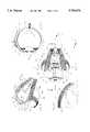
- FIG. 1is an exploded view of the medical device of this invention.
- FIG. 2is a cross-sectional view taken along line 2--2 of FIG. 3.
- FIG. 3is a side view of the globe of an eye having the medical device of this invention attached to the globe.
- FIG. 4is a perspective view taken along line 4--4 of FIG. 3.
- FIG. 2ais an enlarged, fragmentary view taken along line 2a--2a of FIG. 3.
- the medical device 10 of this inventionincludes an assembly of a one-way flow valve 12 and a distribution plate 14.
- the distribution plate 14has a unique shape, with the valve 12 attached to the anterior end 16 of the plate.
- the anterior end 16 of the plate 14has four posts 18 through 21 which fit through aligned holes 22 through 25 in a folded membrane 26, with a cover 28 covering the membrane, which is mounted on the plate 14.
- the folded membrane 26is sandwiched between the plate 14 and the cover 28, with the posts 18 through 21 extending into aligned holes 40 through 43 in the cover 28.
- This valve 12includes a tube 30 made of a flexible material, which has one end attached to the folded membrane 26 and another end adapted to be inserted into the intraocular chamber 32 of the eye, as illustrated in FIG. 2.
- the membrane 26Opposite the end where the tube 30 is attached, the membrane 26 forms a slit-like opening 34. With the cover 28 in place, the membrane 26 is in tension, and the slit-like opening 34 is normally closed until the pressure inside the intraocular chamber 32 exceeds a predetermined level. When this predetermined pressure is reached, the slit-like opening 34 expands to release fluid, which flows onto the distribution plate 14.
- This valve 12is disclosed in detail in U.S. Pat. Nos. 5,071,408 and 5,411,473.
- a pair of spaced apart fingers 44 and 46which have orifices 44a and 46a in them. These orifices 44a and 46a are used to hold sutures which are threaded through the orifices to attach the medical device 10 to the globe 48 of the patient's eye.
- the medical device 10is placed under this flap and the sutures are threaded through the orifices 44a and 46a and more sutures are used to sew the flap in place after it is positioned to cover the medical device.
- the medical device 10is now located on the globe in the correct position shown in FIGS. 2 through 4 with the flap (not shown) covering the device.
- a characteristic of this inventionis the size and shape of the distribution plate 14, which has been designed to maximize the surface area of the distribution plate 14 without causing strebismis.
- the anterior end 16 of the plate 14is a distance of 8 millimeters (D1) from the limbus 70 (FIG. 3) of the patient's eye.
- the posterior end 50 of the distribution plate 14has an inwardly curved edge 50a. This inwardly curved edge is desirable to ensure that, when the device 10 is properly placed in the correct position on the globe 48 of the patient's eye, this end 50 is at least 2 millimeters (D2) away from the optic nerve 52 (FIG. 4). This distance may range between 2 and 4 millimeters.
- the anterior end 16 of the distribution plate 14has a width (D3), as measured from the greatest separation of the sides 14 and 14b, of from 13 to 15 millimeters.
- the sides 14a and 14b of the plate 14curve outward slightly, and then inward to form a narrow section 54.
- the sides 14a and 14bthen expand outward to form at the posterior end 50 on the opposite sides 14a and 14b the tabs 60 and 62, respectively.
- the tips 60a and 62a of these tabs 60 and 62are rounded slightly and are separated a distance (D4) ranging between 16 and 18 millimeters.
- the area of these tabsis typically about 25 square millimeters.
- the total length (D5), not including the fingers 44 and 46, of the distribution plate 14ranges between 14 and 16 millimeters.
- the total area of the distribution plate 14is 250 square millimeters, inclusive of the area occupied by the valve 12. Typically the valve 12 does not occupy more than 30 percent of the surface of the plate 14.
- the unique shape of the distribution plate 14allows it to be seated between adjacent pairs of rectus muscles 72 and 74, with each tab 60 and 62, respectively, positioned beneath the rectus muscles 72 and 74 near the point where each rectus muscle joins the globe 48.
- the plate 14fills almost completely the space between the rectus muscles when it is correctly located on the globe 48, as depicted in FIG. 3.
- the bleb (not shown) formed over this medical device 10does not overlap the rectus muscles, or otherwise interfere with them, thus minimizing the likelihood of strebismis.
- the device 10is relatively simple to implant. As disclosed in U.S. Pat. No. 5071408, a special instrument may be used to insert the tube 30 into the intraocular chamber 32 of the eye so that the one end of the tube is in communication with the fluid in the chamber. When the pressure in the chamber 32 is to great, excess fluid flows through the tube 30 into the valve 12, causing the membrane 26 to expand, opening the slit 34 so the fluid flows onto the plate 14.
Landscapes
- Health & Medical Sciences (AREA)
- Ophthalmology & Optometry (AREA)
- Life Sciences & Earth Sciences (AREA)
- Animal Behavior & Ethology (AREA)
- Engineering & Computer Science (AREA)
- Biomedical Technology (AREA)
- Heart & Thoracic Surgery (AREA)
- Veterinary Medicine (AREA)
- Public Health (AREA)
- General Health & Medical Sciences (AREA)
- Surgery (AREA)
- Nuclear Medicine, Radiotherapy & Molecular Imaging (AREA)
- Vascular Medicine (AREA)
- Otolaryngology (AREA)
- Anesthesiology (AREA)
- Hematology (AREA)
- Prostheses (AREA)
Abstract
Description
This application is a continuation-in-part application of U. S. Ser. No. 08/269,839, filed Jul. 1, 1994, entitled "Uniquely Shaped Ophthalmological Device, now U.S. Pat. No. 5,616,118, which is a continuation-in-part application of U.S. Ser. No. 07/786,734, entitled "Medical Valve," filed Oct. 1, 1991, now U.S. Pat. No. 5,441,473, which is a divisional application of U.S. Ser. No. 07/478,655, filed Feb. 12, 1990, and entitled "Medical Valve," now U.S. Pat. No. 5,071,408, which is continuation-in-part application of U.S. patent application Ser. No. 07/255,070, entitled Self-Regulating Pressure Control Glaucoma Valve, filed Oct. 7, 1988 now abandoned. All of these related applications are incorporated herein by reference and made a part of this application.
1. Field of the Invention:
This invention relates to a medical device and method for treating a patient suffering from glaucoma.
2. Background Discussion:
As disclosed in U.S. Pat. Nos. 5,071,408, and 5,411,473, glaucoma is treated using a medical valve that employs a membrane in tension to control the flow of excess fluid from the intraocular chamber of the eye. The valve is attached to a distribution plate on which the fluid is deposited upon leaving the valve. This plate may be curved as a segment of a sphere in order to conform to the shape of the globe of the eye. The assembly of valve and distribution plate is placed beneath the layer of tissue forming the surface the globe. This tissue forms a bleb, which is a wall of tissue surrounding the assembly of the valve and distribution plate. The fluid collected on the distribution plate is absorbed by the patient's body through the wall of the bleb. Typically these types of devices are placed between adjacent rectus muscles. If the distribution plate is too large, or not properly shaped, the formation of the bleb may cause the rectus muscles to be pulled inward towards each other. This produces strabismus, that is, the patient's eyes become crossed.
It is the objective of this invention to provide an assembly of a one-way flow valve and a distribution plate which has a unique configuration that minimizes the likelihood of strebismis.
This invention has several features, no single one of which is solely responsible for its desirable attributes. Without limiting the scope of this invention as expressed by the claims which follow, its more prominent features will now be discussed briefly. After considering this discussion, and particularly after reading the section entitled, "DETAILED DESCRIPTION OF THE PREFERRED EMBODIMENT," one will understand how the features of this invention provide its benefits, which include ease of use and minimizing the likelihood of strebismis.
The first feature of medical device of this invention is that it is adapted to be attached to the globe of the eye of a patient. It includes a distribution plate with a one-way flow valve adapted to be placed in communication with fluid to be drained from the intraocular chamber of the eye. The distribution plate has an anterior end, a posterior end, opposed sides extending between said anterior and posterior ends, a curved configuration so that said plate conforms essentially with the spherical shape of the globe of the eye ball, and opposed tab elements that extend outward from each side at said posterior end. The valve is attached to the anterior end of the distribution plate.
The second feature is that the plate has a predetermined size and shape to maximize the area of the bleb, yet prevent strebismis. The larger the area of the bleb, the more rapidly the fluid will be absorbed by the patient's body, which is desirable to avoid high within the bleb. When attached to the globe between adjacent rectus muscles with the anterior end from 8 to 10 millimeters (mm) from the limbus of the eye, the width of the plate is sufficiently restrictive so that one side is close but not touching one rectus muscle and the other side is close but not touching the other rectus muscle, and the length of the plate is sufficiently restrictive so that the posterior end is at least 2 millimeters from the optic nerve of the eye. With the plate in this precise location on the globe, one tab element is seated beneath one rectus muscle nearby the point where said one rectus muscle is attached to the globe and the other tab element is seated beneath the other rectus muscle nearby the point where said other rectus muscle is attached to the globe.
Specifically, the distribution plate has the following characteristics. Each side of the plate is less than 0.5 millimeter from an adjacent rectus muscles. The total area of the plate, inclusive of the surface area carrying the valve, is from 240 to 260 square millimeter. The valve occupies less than 30 percent of the surface area of the distribution plate. The sides of the plate slope inward towards each other to a narrow section having a breath of from 13 to 14 millimeters, and then slope outward to form the tab elements. The anterior end has a width from 13 to 15 millimeters, and the posterior end of the plate curves inward between the tab elements. The tab elements each have a surface area of from 20 to 30 square millimeters, and they have rounded tips separated by a distance of from 16 to 18 millimeters.
The third feature is that one-way flow valve includes a body member holding a pair of overlying elastic membranes in tension to form therebetween a pressure chamber. The membranes provide an elongated, slit-like opening therebetween, which is normally in a closed position and opens when the pressure in the pressure chamber exceeds a predetermined pressure and returns to the closed position when the pressure in the pressure chamber is below the predetermined pressure. An inlet tube in communication with and connected to the pressure chamber at a point remote from the opening places the valve in communication with the intraocular chamber of the eye.
This invention also includes a method for treating glaucoma by draining fluid from the intraocular chamber of a patient's eye. This method includes the following steps:
(a) providing a medical device including
a distribution plate having an anterior end, a posterior end, opposed sides extending between said anterior and posterior ends, a curved configuration conforming essentially to the spherical shape of the globe of the eye ball, and opposed tab elements that extend outward from each side at said posterior end, and
a one-way flow valve attached to the anterior end of the distribution plate having a tube adapted to be placed in communication with the fluid to be drained from the intraocular chamber of the eye,
(b) positioning the device on the globe between adjacent rectus muscles with the anterior end from 8 to 10 millimeters from the limbus of the eye, with the width of the plate being sufficiently restrictive so that one side is close but not touching one rectus muscle and the other side is close but not touching the other rectus muscle, and the length of the plate being sufficiently restrictive so that the posterior end is at least 2 millimeters inch from the optic nerve of the eye,
(c) inserting one tab element beneath one rectus muscle nearby the point where said one rectus muscle is attached to the globe and inserting the other tab element beneath the other rectus muscle nearby the point where said other rectus muscle is attached to the globe,
(d) placing the tube in communication with the intraocular chamber of the eye so that the fluid drains from the intraocular chamber onto the distribution plate under the control of the valve, and
(e) attaching said device to the globe.
The preferred embodiment of this invention, illustrating all its features, will now be discussed in detail. This embodiment depicts the novel and non-obvious medical device and method of this invention as shown in the accompanying drawing, which is for illustrative purposes only. This drawing includes the following figures (FIGS.), with like numerals indicating like parts:
FIG. 1 is an exploded view of the medical device of this invention.
FIG. 2 is a cross-sectional view taken alongline 2--2 of FIG. 3.
FIG. 3 is a side view of the globe of an eye having the medical device of this invention attached to the globe.
FIG. 4 is a perspective view taken alongline 4--4 of FIG. 3.
FIG. 2a is an enlarged, fragmentary view taken alongline 2a--2a of FIG. 3.
As best shown in FIG. 1, themedical device 10 of this invention includes an assembly of a one-way flow valve 12 and adistribution plate 14. Thedistribution plate 14 has a unique shape, with thevalve 12 attached to theanterior end 16 of the plate. Theanterior end 16 of theplate 14 has fourposts 18 through 21 which fit through alignedholes 22 through 25 in a foldedmembrane 26, with acover 28 covering the membrane, which is mounted on theplate 14. The foldedmembrane 26 is sandwiched between theplate 14 and thecover 28, with theposts 18 through 21 extending into alignedholes 40 through 43 in thecover 28. Thisvalve 12 includes atube 30 made of a flexible material, which has one end attached to the foldedmembrane 26 and another end adapted to be inserted into theintraocular chamber 32 of the eye, as illustrated in FIG. 2. Opposite the end where thetube 30 is attached, themembrane 26 forms a slit-like opening 34. With thecover 28 in place, themembrane 26 is in tension, and the slit-like opening 34 is normally closed until the pressure inside theintraocular chamber 32 exceeds a predetermined level. When this predetermined pressure is reached, the slit-like opening 34 expands to release fluid, which flows onto thedistribution plate 14. Thisvalve 12 is disclosed in detail in U.S. Pat. Nos. 5,071,408 and 5,411,473.
At theanterior end 16 are a pair of spaced apartfingers orifices orifices medical device 10 to theglobe 48 of the patient's eye. After cutting a flap (not shown) of upper layer tissue in theglobe 48, themedical device 10 is placed under this flap and the sutures are threaded through theorifices medical device 10 is now located on the globe in the correct position shown in FIGS. 2 through 4 with the flap (not shown) covering the device.
A characteristic of this invention is the size and shape of thedistribution plate 14, which has been designed to maximize the surface area of thedistribution plate 14 without causing strebismis. In accordance with this invention, theanterior end 16 of theplate 14 is a distance of 8 millimeters (D1) from the limbus 70 (FIG. 3) of the patient's eye. Theposterior end 50 of thedistribution plate 14 has an inwardlycurved edge 50a. This inwardly curved edge is desirable to ensure that, when thedevice 10 is properly placed in the correct position on theglobe 48 of the patient's eye, thisend 50 is at least 2 millimeters (D2) away from the optic nerve 52 (FIG. 4). This distance may range between 2 and 4 millimeters. Theanterior end 16 of thedistribution plate 14 has a width (D3), as measured from the greatest separation of thesides 14 and 14b, of from 13 to 15 millimeters. Thesides 14a and 14b of theplate 14 curve outward slightly, and then inward to form anarrow section 54. Thesides 14a and 14b then expand outward to form at theposterior end 50 on theopposite sides 14a and 14b thetabs tips tabs fingers distribution plate 14 ranges between 14 and 16 millimeters. The total area of thedistribution plate 14 is 250 square millimeters, inclusive of the area occupied by thevalve 12. Typically thevalve 12 does not occupy more than 30 percent of the surface of theplate 14.
As depicted in FIG. 2a, the unique shape of thedistribution plate 14 allows it to be seated between adjacent pairs ofrectus muscles tab rectus muscles globe 48. Theplate 14 fills almost completely the space between the rectus muscles when it is correctly located on theglobe 48, as depicted in FIG. 3. The bleb (not shown) formed over thismedical device 10 does not overlap the rectus muscles, or otherwise interfere with them, thus minimizing the likelihood of strebismis.
Thedevice 10 is relatively simple to implant. As disclosed in U.S. Pat. No. 5071408, a special instrument may be used to insert thetube 30 into theintraocular chamber 32 of the eye so that the one end of the tube is in communication with the fluid in the chamber. When the pressure in thechamber 32 is to great, excess fluid flows through thetube 30 into thevalve 12, causing themembrane 26 to expand, opening theslit 34 so the fluid flows onto theplate 14.
The above presents a description of the best mode contemplated of carrying out the present invention, and of the manner and process of making and using it, in such full, clear, concise, and exact terms as to enable any person skilled in the art to which it pertains to make and use this invention. This invention is, however, susceptible to modifications and alternate constructions from that discussed above which are fully equivalent. Consequently, it is not the intention to limit this invention to the particular embodiment disclosed. On the contrary, the intention is to cover all modifications and alternate constructions coming within the spirit and scope of the invention as generally expressed by the following claims, which particularly point out and distinctly claim the subject matter of the invention:
Claims (20)
1. A medical device for treating a patient suffering from glaucoma and adapted to be attached to the globe of the eye of a patient, including
a distribution plate having an anterior end, a posterior end, opposed sides extending between said anterior and posterior ends, a curved configuration so that said plate conforms essentially with the spherical shape of the globe of the eye ball, and opposed tab elements that extend outward from each side at said posterior end,
said plate upon being attached to the globe one tab element being seated beneath one rectus muscle nearby the point where said one rectus muscle is attached to the globe and the other tab element being seated beneath the other rectus muscle nearby the point where said other rectus muscle is attached to the globe, and
a one-way flow valve attached to the anterior end of the distribution plate adapted to be placed in communication with fluid to be drained from the intraocular chamber of the eye.
2. The medical device of claim 1 where each side of the plate is less than 0.5 millimeter from an adjacent rectus muscles.
3. The medical device of claim 1 where the total area of the distribution plate inclusive of the surface area carrying the valve is from 240 to 260 square millimeters.
4. The medical device of claim 3 where the valve occupies less than 30 percent of the surface area of the distribution plate.
5. The medical device of claim 1 where the sides slope inward towards each other to a narrow section having a width of from 13 to 15 millimeters, and then slope outward to form said tab elements.
6. The medical device of claim 1 where the anterior end has a width from 13 to 15 millimeters.
7. The medical device of claim 1 where the posterior end of the plate curves inward between said tab elements.
8. The medical device of claim 1 where the tab elements each have a surface area of from 20 to 30 square millimeters.
9. The medical device of claim 1 where the tab elements have rounded tips, said tips being separated by a distance of from 16 to 18 millimeters.
10. The medical device of claim 1 where the one-way flow valve includes
a body member holding a pair of overlying elastic membranes in tension to form therebetween an pressure chamber,
said membranes providing an elongated, slit-like opening therebetween, which is normally in a closed position and opens when the pressure in the pressure chamber exceeds a predetermined pressure and returns to the closed position when the pressure in the pressure chamber is below said predetermined pressure, and
an inlet tube in communication with and connected to the pressure chamber at a point remote from the opening.
11. A medical device for treating a patient suffering from glaucoma and adapted to be attached to the globe of the eye of a patient, including
a distribution plate having an anterior end, a posterior end, opposed sides extending between said anterior and posterior ends, a curved configuration so that said plate conforms essentially with the spherical shape of the globe of the eye ball, and opposed tab elements that extend outward from each side at said posterior end, sides sloping inward towards each other to a narrow section having a width of from 13 to 15 millimeters, and then slope outward to form said tab elements,
said plate having total area of from 240 to 260 square millimeters and upon being attached to the globe one tab element being seated beneath one rectus muscle nearby the point where said one rectus muscle is attached to the globe and the other tab element being seated beneath the other rectus muscle nearby the point where said other rectus muscle is attached to the globe, and
a one-way flow valve attached to the anterior end of the distribution plate adapted to be placed in communication with fluid to be drained from the intraocular chamber of the eye,
said one-way flow valve including a body member holding a pair of overlying elastic membranes in tension to form therebetween an pressure chamber,
said membranes providing an elongated, slit-like opening therebetween, which is normally in a closed position and opens when the pressure in the pressure chamber exceeds a predetermined pressure and returns to the closed position when the pressure in the pressure chamber is below said predetermined pressure, and
an inlet tube in communication with and connected to the pressure chamber at a point remote from the opening.
12. The medical device of claim 11 where the valve occupies less than 30 percent of the total surface area of the distribution plate.
13. The medical device of claim 11 where the anterior end has a width from 13 to 15 millimeters.
14. The medical device of claim 11 where the posterior end of the plate curves inward between said tab elements.
15. The medical device of claim 11 where the tab elements each have a surface area of from 20 to 30 square millimeters.
16. The medical device of claim 11 where the tab elements have rounded tips, said tips being separated by a distance of from 16 to 18 millimeters.
17. A method for treating glaucoma by draining fluid from the intraocular chamber of a patient's eye, including the steps of
(a) providing a medical device including a distribution plate having an anterior end, a posterior end, opposed sides extending between said anterior and posterior ends, a curved configuration conforming essentially to the spherical shape of the globe of the eye ball, and opposed tab elements that extend outward from each side at said posterior end, and
a one-way flow valve attached to the anterior end of the distribution plate having a tube adapted to be placed in communication with the fluid to be drained from the intraocular chamber of the eye,
(b) positioning the device on the globe between adjacent rectus muscles with the anterior end from 8 to 10 millimeters from the limbus of the eye, with the width of the plate being sufficiently restrictive so that one side is close but not touching one rectus muscle and the other side is close but not touching the other rectus muscle, and the length of the plate being sufficiently restrictive so that the posterior end is at least 2 millimeters from the optic nerve of the eye,
(c) inserting one tab element beneath one rectus muscle nearby the point where said one rectus muscle is attached to the globe and inserting the other tab element beneath the other rectus muscle nearby the point where said other rectus muscle is attached to the globe,
(d) placing the tube in communication with the intraocular chamber of the eye so that the fluid drains from the intraocular chamber onto the distribution plate under the control of the valve, and
(e) attaching said device to the globe.
18. The method of claim 17 where the total area of the distribution plate inclusive of the surface area carrying the valve is from 240 to 260 square millimeters.
19. The method of claim 18 where the valve occupies less than 30 percent of the total surface area of the distribution plate.
20. The method of claim 19 where the one-way flow valve includes a body member holding a pair of overlying elastic membranes in tension to form therebetween an pressure chamber,
said membranes providing an elongated, slit-like opening therebetween, which is normally in a closed position and opens when the pressure in the pressure chamber exceeds a predetermined pressure and returns to the closed position when the pressure in the pressure chamber is below said predetermined pressure, and
an inlet tube in communication with and connected to the pressure chamber at a point remote from the opening.
Priority Applications (2)
| Application Number | Priority Date | Filing Date | Title |
|---|---|---|---|
| US08/771,446US5785674A (en) | 1988-10-07 | 1996-12-20 | Device and method for treating glaucoma |
| US08/943,453US6261256B1 (en) | 1996-12-20 | 1997-10-03 | Pocket medical valve & method |
Applications Claiming Priority (5)
| Application Number | Priority Date | Filing Date | Title |
|---|---|---|---|
| US25507088A | 1988-10-07 | 1988-10-07 | |
| US07/478,655US5071408A (en) | 1988-10-07 | 1990-02-12 | Medical valve |
| US07/786,734US5411473A (en) | 1988-10-07 | 1991-10-01 | Medical valve |
| US08/269,839US5616118A (en) | 1988-10-07 | 1994-07-01 | Uniquely shaped ophthalmological device |
| US08/771,446US5785674A (en) | 1988-10-07 | 1996-12-20 | Device and method for treating glaucoma |
Related Parent Applications (1)
| Application Number | Title | Priority Date | Filing Date |
|---|---|---|---|
| US08/269,839Continuation-In-PartUS5616118A (en) | 1988-10-07 | 1994-07-01 | Uniquely shaped ophthalmological device |
Related Child Applications (1)
| Application Number | Title | Priority Date | Filing Date |
|---|---|---|---|
| US08/943,453Continuation-In-PartUS6261256B1 (en) | 1996-12-20 | 1997-10-03 | Pocket medical valve & method |
Publications (1)
| Publication Number | Publication Date |
|---|---|
| US5785674Atrue US5785674A (en) | 1998-07-28 |
Family
ID=27500528
Family Applications (1)
| Application Number | Title | Priority Date | Filing Date |
|---|---|---|---|
| US08/771,446Expired - LifetimeUS5785674A (en) | 1988-10-07 | 1996-12-20 | Device and method for treating glaucoma |
Country Status (1)
| Country | Link |
|---|---|
| US (1) | US5785674A (en) |
Cited By (68)
| Publication number | Priority date | Publication date | Assignee | Title |
|---|---|---|---|---|
| US6261256B1 (en)* | 1996-12-20 | 2001-07-17 | Abdul Mateen Ahmed | Pocket medical valve & method |
| WO2002032343A2 (en) | 2000-10-18 | 2002-04-25 | Wilcox Michael J | C-shaped cross section tubular ophthalmic implant for reduction of intraocular pressure and method of use |
| US6450984B1 (en) | 1999-04-26 | 2002-09-17 | Gmp Vision Solutions, Inc. | Shunt device and method for treating glaucoma |
| US6471666B1 (en)* | 2000-02-24 | 2002-10-29 | Steven A. Odrich | Injectable glaucoma device |
| US20030055372A1 (en)* | 1999-04-26 | 2003-03-20 | Lynch Mary G. | Shunt device and method for treating glaucoma |
| US6595945B2 (en) | 2001-01-09 | 2003-07-22 | J. David Brown | Glaucoma treatment device and method |
| US20030181848A1 (en)* | 2000-04-14 | 2003-09-25 | Bergheim Olav B. | Implant with drug coating |
| US20040073156A1 (en)* | 2001-01-09 | 2004-04-15 | Brown J. David | Glaucoma treatment device and method |
| US20040215126A1 (en)* | 2003-04-22 | 2004-10-28 | Ahmed A. Mateen | Device for treating glaucoma & method of manufacture |
| US20040254521A1 (en)* | 2003-06-16 | 2004-12-16 | Solx, Inc. | Shunt for the treatment of glaucoma |
| US20050119636A1 (en)* | 2001-05-02 | 2005-06-02 | David Haffner | Implant with intraocular pressure sensor for glaucoma treatment |
| US20050165385A1 (en)* | 2004-01-22 | 2005-07-28 | Solx, Inc. | Glaucoma treatment method |
| US20050178394A1 (en)* | 2003-08-21 | 2005-08-18 | Intralens Vision, Inc. | Method for keratophakia surgery |
| US20050182350A1 (en)* | 1996-10-25 | 2005-08-18 | Alok Nigam | Sutureless implantable device and method for treatment of glaucoma |
| US6981958B1 (en) | 2001-05-02 | 2006-01-03 | Glaukos Corporation | Implant with pressure sensor for glaucoma treatment |
| US20060155300A1 (en)* | 2002-09-17 | 2006-07-13 | Iscience Surgical, Inc. | Apparatus and method for surgical bypass of aqueous humor |
| US20070249984A1 (en)* | 2004-03-26 | 2007-10-25 | Molteno Ophthalmic Ltd | Ophthalmic Implant for Treating Glaucoma |
| US20070280994A1 (en)* | 2006-06-01 | 2007-12-06 | Cunanan Crystal M | Ocular Tissue Separation Areas With Barrier Regions For Inlays Or Other Refractive Procedures |
| US20070293872A1 (en)* | 2006-06-20 | 2007-12-20 | Minu, L.L.C. | Ocular Drainage Device |
| US20070293807A1 (en)* | 2006-05-01 | 2007-12-20 | Lynch Mary G | Dual drainage pathway shunt device and method for treating glaucoma |
| US7431710B2 (en) | 2002-04-08 | 2008-10-07 | Glaukos Corporation | Ocular implants with anchors and methods thereof |
| US20080277332A1 (en)* | 2007-05-11 | 2008-11-13 | Becton, Dickinson And Company | Micromachined membrane filter device for a glaucoma implant and method for making the same |
| US7488303B1 (en) | 2002-09-21 | 2009-02-10 | Glaukos Corporation | Ocular implant with anchor and multiple openings |
| US20090043321A1 (en)* | 2004-04-29 | 2009-02-12 | Iscience Interventional Corporation | Apparatus And Method For Surgical Enhancement Of Aqueous Humor Drainage |
| US7563241B2 (en) | 2001-04-07 | 2009-07-21 | Glaukos Corporation | Implant and methods thereof for treatment of ocular disorders |
| US7600533B2 (en) | 2006-08-10 | 2009-10-13 | California Institute Of Technology | Microfluidic valve having free-floating member and method of fabrication |
| US20100056977A1 (en)* | 2008-08-26 | 2010-03-04 | Thaddeus Wandel | Trans-corneal shunt and method |
| US7708711B2 (en) | 2000-04-14 | 2010-05-04 | Glaukos Corporation | Ocular implant with therapeutic agents and methods thereof |
| US20100173866A1 (en)* | 2004-04-29 | 2010-07-08 | Iscience Interventional Corporation | Apparatus and method for ocular treatment |
| US20100191177A1 (en)* | 2009-01-23 | 2010-07-29 | Iscience Interventional Corporation | Device for aspirating fluids |
| US7776086B2 (en) | 2004-04-30 | 2010-08-17 | Revision Optics, Inc. | Aspherical corneal implant |
| US7867186B2 (en) | 2002-04-08 | 2011-01-11 | Glaukos Corporation | Devices and methods for treatment of ocular disorders |
| US7879079B2 (en) | 2001-08-28 | 2011-02-01 | Glaukos Corporation | Implant delivery system and methods thereof for treating ocular disorders |
| US7951155B2 (en) | 2002-03-15 | 2011-05-31 | Glaukos Corporation | Combined treatment for cataract and glaucoma treatment |
| US8057541B2 (en) | 2006-02-24 | 2011-11-15 | Revision Optics, Inc. | Method of using small diameter intracorneal inlays to treat visual impairment |
| US8162953B2 (en) | 2007-03-28 | 2012-04-24 | Revision Optics, Inc. | Insertion system for corneal implants |
| US8337445B2 (en) | 2001-05-03 | 2012-12-25 | Glaukos Corporation | Ocular implant with double anchor mechanism |
| US8425473B2 (en) | 2009-01-23 | 2013-04-23 | Iscience Interventional Corporation | Subretinal access device |
| US8469948B2 (en) | 2010-08-23 | 2013-06-25 | Revision Optics, Inc. | Methods and devices for forming corneal channels |
| US8506515B2 (en) | 2006-11-10 | 2013-08-13 | Glaukos Corporation | Uveoscleral shunt and methods for implanting same |
| US8617094B2 (en) | 2002-03-07 | 2013-12-31 | Glaukos Corporation | Fluid infusion methods for glaucoma treatment |
| US8668735B2 (en) | 2000-09-12 | 2014-03-11 | Revision Optics, Inc. | Corneal implant storage and delivery devices |
| US8900296B2 (en) | 2007-04-20 | 2014-12-02 | Revision Optics, Inc. | Corneal inlay design and methods of correcting vision |
| US9005280B2 (en) | 2000-09-12 | 2015-04-14 | Revision Optics, Inc. | System for packaging and handling an implant and method of use |
| US9271828B2 (en) | 2007-03-28 | 2016-03-01 | Revision Optics, Inc. | Corneal implant retaining devices and methods of use |
| US9301875B2 (en) | 2002-04-08 | 2016-04-05 | Glaukos Corporation | Ocular disorder treatment implants with multiple opening |
| US9341275B2 (en) | 2010-09-15 | 2016-05-17 | Minipumps, Llc | Molded and packaged elastomeric check valve |
| US9345569B2 (en) | 2011-10-21 | 2016-05-24 | Revision Optics, Inc. | Corneal implant storage and delivery devices |
| US9381112B1 (en) | 2011-10-06 | 2016-07-05 | William Eric Sponsell | Bleb drainage device, ophthalmological product and methods |
| US9539143B2 (en) | 2008-04-04 | 2017-01-10 | Revision Optics, Inc. | Methods of correcting vision |
| US9549848B2 (en) | 2007-03-28 | 2017-01-24 | Revision Optics, Inc. | Corneal implant inserters and methods of use |
| US9592151B2 (en) | 2013-03-15 | 2017-03-14 | Glaukos Corporation | Systems and methods for delivering an ocular implant to the suprachoroidal space within an eye |
| US9730638B2 (en) | 2013-03-13 | 2017-08-15 | Glaukos Corporation | Intraocular physiological sensor |
| US20170348151A1 (en)* | 2016-06-03 | 2017-12-07 | New World Medical, Inc. | Intraocular drainage device |
| US9845895B2 (en) | 2013-01-11 | 2017-12-19 | Minipumps, Llc | Diaphragm check valves and methods of manufacture thereof |
| US10271989B2 (en) | 2012-03-26 | 2019-04-30 | Glaukos Corporation | System and method for delivering multiple ocular implants |
| US10517759B2 (en) | 2013-03-15 | 2019-12-31 | Glaukos Corporation | Glaucoma stent and methods thereof for glaucoma treatment |
| US10555805B2 (en) | 2006-02-24 | 2020-02-11 | Rvo 2.0, Inc. | Anterior corneal shapes and methods of providing the shapes |
| US10583041B2 (en) | 2015-03-12 | 2020-03-10 | RVO 2.0 Inc. | Methods of correcting vision |
| US10736778B2 (en) | 2014-12-31 | 2020-08-11 | Microoptx Inc. | Glaucoma treatment devices and methods |
| US10835371B2 (en) | 2004-04-30 | 2020-11-17 | Rvo 2.0, Inc. | Small diameter corneal inlay methods |
| WO2020261184A1 (en) | 2019-06-26 | 2020-12-30 | Imvalv S.A. | Glaucoma drain implant system with pressure sensor and valve, and external reading unit |
| US10959941B2 (en) | 2014-05-29 | 2021-03-30 | Glaukos Corporation | Implants with controlled drug delivery features and methods of using same |
| US10980667B2 (en) | 2015-09-30 | 2021-04-20 | Microoptx Inc. | Eye treatment devices and methods |
| US11116625B2 (en) | 2017-09-28 | 2021-09-14 | Glaukos Corporation | Apparatus and method for controlling placement of intraocular implants |
| US11363951B2 (en) | 2011-09-13 | 2022-06-21 | Glaukos Corporation | Intraocular physiological sensor |
| US11925578B2 (en) | 2015-09-02 | 2024-03-12 | Glaukos Corporation | Drug delivery implants with bi-directional delivery capacity |
| US12440375B2 (en) | 2021-05-21 | 2025-10-14 | New World Medical, Inc. | Intraocular drainage device |
Citations (10)
| Publication number | Priority date | Publication date | Assignee | Title |
|---|---|---|---|---|
| US3288142A (en)* | 1964-04-27 | 1966-11-29 | Hakim Salomon | Hydrocephalus shunt with spring biased one-way valves |
| US3910283A (en)* | 1973-10-09 | 1975-10-07 | Harry H Leveen | Process for treatment of ascites and device to accomplish same |
| US4387715A (en)* | 1980-09-23 | 1983-06-14 | Hakim Company Limited | Shunt valve |
| US4521210A (en)* | 1982-12-27 | 1985-06-04 | Wong Vernon G | Eye implant for relieving glaucoma, and device and method for use therewith |
| US4560375A (en)* | 1983-06-30 | 1985-12-24 | Pudenz-Schulte Medical Research Corp. | Flow control valve |
| US4570901A (en)* | 1984-12-28 | 1986-02-18 | Keystone International, Inc. | Positioning assembly for use with rotatable valves |
| US4729761A (en)* | 1985-11-27 | 1988-03-08 | White Thomas C | Tissue-implantable, fluid-dissipating device |
| US4850955A (en)* | 1986-12-02 | 1989-07-25 | Codman & Shurtleff | Body fluid transfer device |
| US5454796A (en)* | 1991-04-09 | 1995-10-03 | Hood Laboratories | Device and method for controlling intraocular fluid pressure |
| US5476445A (en)* | 1990-05-31 | 1995-12-19 | Iovision, Inc. | Glaucoma implant with a temporary flow restricting seal |
- 1996
- 1996-12-20USUS08/771,446patent/US5785674A/ennot_activeExpired - Lifetime
Patent Citations (10)
| Publication number | Priority date | Publication date | Assignee | Title |
|---|---|---|---|---|
| US3288142A (en)* | 1964-04-27 | 1966-11-29 | Hakim Salomon | Hydrocephalus shunt with spring biased one-way valves |
| US3910283A (en)* | 1973-10-09 | 1975-10-07 | Harry H Leveen | Process for treatment of ascites and device to accomplish same |
| US4387715A (en)* | 1980-09-23 | 1983-06-14 | Hakim Company Limited | Shunt valve |
| US4521210A (en)* | 1982-12-27 | 1985-06-04 | Wong Vernon G | Eye implant for relieving glaucoma, and device and method for use therewith |
| US4560375A (en)* | 1983-06-30 | 1985-12-24 | Pudenz-Schulte Medical Research Corp. | Flow control valve |
| US4570901A (en)* | 1984-12-28 | 1986-02-18 | Keystone International, Inc. | Positioning assembly for use with rotatable valves |
| US4729761A (en)* | 1985-11-27 | 1988-03-08 | White Thomas C | Tissue-implantable, fluid-dissipating device |
| US4850955A (en)* | 1986-12-02 | 1989-07-25 | Codman & Shurtleff | Body fluid transfer device |
| US5476445A (en)* | 1990-05-31 | 1995-12-19 | Iovision, Inc. | Glaucoma implant with a temporary flow restricting seal |
| US5454796A (en)* | 1991-04-09 | 1995-10-03 | Hood Laboratories | Device and method for controlling intraocular fluid pressure |
Cited By (154)
| Publication number | Priority date | Publication date | Assignee | Title |
|---|---|---|---|---|
| US20050182350A1 (en)* | 1996-10-25 | 2005-08-18 | Alok Nigam | Sutureless implantable device and method for treatment of glaucoma |
| US6261256B1 (en)* | 1996-12-20 | 2001-07-17 | Abdul Mateen Ahmed | Pocket medical valve & method |
| US20030236484A1 (en)* | 1999-04-26 | 2003-12-25 | Gmp Vision Solutions, Inc. | Inflatable device and method for treating glaucoma |
| US9492320B2 (en) | 1999-04-26 | 2016-11-15 | Glaukos Corporation | Shunt device and method for treating ocular disorders |
| US9827143B2 (en) | 1999-04-26 | 2017-11-28 | Glaukos Corporation | Shunt device and method for treating ocular disorders |
| US6524275B1 (en) | 1999-04-26 | 2003-02-25 | Gmp Vision Solutions, Inc. | Inflatable device and method for treating glaucoma |
| US20030055372A1 (en)* | 1999-04-26 | 2003-03-20 | Lynch Mary G. | Shunt device and method for treating glaucoma |
| US8771217B2 (en) | 1999-04-26 | 2014-07-08 | Glaukos Corporation | Shunt device and method for treating ocular disorders |
| US8388568B2 (en) | 1999-04-26 | 2013-03-05 | Glaukos Corporation | Shunt device and method for treating ocular disorders |
| US6626858B2 (en) | 1999-04-26 | 2003-09-30 | Gmp Vision Solutions, Inc. | Shunt device and method for treating glaucoma |
| US6450984B1 (en) | 1999-04-26 | 2002-09-17 | Gmp Vision Solutions, Inc. | Shunt device and method for treating glaucoma |
| US6464724B1 (en) | 1999-04-26 | 2002-10-15 | Gmp Vision Solutions, Inc. | Stent device and method for treating glaucoma |
| US8152752B2 (en) | 1999-04-26 | 2012-04-10 | Glaukos Corporation | Shunt device and method for treating glaucoma |
| US6783544B2 (en) | 1999-04-26 | 2004-08-31 | Gmp Vision Solutions, Inc. | Stent device and method for treating glaucoma |
| US7850637B2 (en) | 1999-04-26 | 2010-12-14 | Glaukos Corporation | Shunt device and method for treating glaucoma |
| US6827699B2 (en) | 1999-04-26 | 2004-12-07 | Gmp Vision Solutions, Inc. | Shunt device and method for treating glaucoma |
| US6827700B2 (en) | 1999-04-26 | 2004-12-07 | Gmp Vision Solutions, Inc. | Shunt device and method for treating glaucoma |
| US10492950B2 (en) | 1999-04-26 | 2019-12-03 | Glaukos Corporation | Shunt device and method for treating ocular disorders |
| US10568762B2 (en) | 1999-04-26 | 2020-02-25 | Glaukos Corporation | Stent for treating ocular disorders |
| US20050119601A9 (en)* | 1999-04-26 | 2005-06-02 | Lynch Mary G. | Shunt device and method for treating glaucoma |
| US6471666B1 (en)* | 2000-02-24 | 2002-10-29 | Steven A. Odrich | Injectable glaucoma device |
| US8348877B2 (en) | 2000-04-14 | 2013-01-08 | Dose Medical Corporation | Ocular implant with therapeutic agents and methods thereof |
| US8333742B2 (en) | 2000-04-14 | 2012-12-18 | Glaukos Corporation | Method of delivering an implant for treating an ocular disorder |
| US7867205B2 (en) | 2000-04-14 | 2011-01-11 | Glaukos Corporation | Method of delivering an implant for treating an ocular disorder |
| US6955656B2 (en) | 2000-04-14 | 2005-10-18 | Glaukos Corporation | Apparatus and method for treating glaucoma |
| US6780164B2 (en) | 2000-04-14 | 2004-08-24 | Glaukos Corporation | L-shaped implant with bi-directional flow |
| US9993368B2 (en) | 2000-04-14 | 2018-06-12 | Glaukos Corporation | System and method for treating an ocular disorder |
| US8808219B2 (en) | 2000-04-14 | 2014-08-19 | Glaukos Corporation | Implant delivery device and methods thereof for treatment of ocular disorders |
| US9789001B2 (en) | 2000-04-14 | 2017-10-17 | Dose Medical Corporation | Ocular implant with therapeutic agents and methods thereof |
| US10485702B2 (en) | 2000-04-14 | 2019-11-26 | Glaukos Corporation | System and method for treating an ocular disorder |
| US8801648B2 (en) | 2000-04-14 | 2014-08-12 | Glaukos Corporation | Ocular implant with anchor and methods thereof |
| US20030181848A1 (en)* | 2000-04-14 | 2003-09-25 | Bergheim Olav B. | Implant with drug coating |
| US9066782B2 (en) | 2000-04-14 | 2015-06-30 | Dose Medical Corporation | Ocular implant with therapeutic agents and methods thereof |
| US7297130B2 (en) | 2000-04-14 | 2007-11-20 | Glaukos Corporation | Implant with anchor |
| US8273050B2 (en) | 2000-04-14 | 2012-09-25 | Glaukos Corporation | Ocular implant with anchor and therapeutic agent |
| US8814820B2 (en) | 2000-04-14 | 2014-08-26 | Glaukos Corporation | Ocular implant with therapeutic agent and methods thereof |
| US7708711B2 (en) | 2000-04-14 | 2010-05-04 | Glaukos Corporation | Ocular implant with therapeutic agents and methods thereof |
| US9005280B2 (en) | 2000-09-12 | 2015-04-14 | Revision Optics, Inc. | System for packaging and handling an implant and method of use |
| US8668735B2 (en) | 2000-09-12 | 2014-03-11 | Revision Optics, Inc. | Corneal implant storage and delivery devices |
| US9889000B2 (en) | 2000-09-12 | 2018-02-13 | Revision Optics, Inc. | Corneal implant applicators |
| WO2002032343A2 (en) | 2000-10-18 | 2002-04-25 | Wilcox Michael J | C-shaped cross section tubular ophthalmic implant for reduction of intraocular pressure and method of use |
| US20100168644A1 (en)* | 2001-01-09 | 2010-07-01 | Brown J David | Glaucoma Treatment Device and Method |
| US20060276739A1 (en)* | 2001-01-09 | 2006-12-07 | Brown J D | Glaucoma treatment device and method |
| US6881198B2 (en) | 2001-01-09 | 2005-04-19 | J. David Brown | Glaucoma treatment device and method |
| US6595945B2 (en) | 2001-01-09 | 2003-07-22 | J. David Brown | Glaucoma treatment device and method |
| US20040073156A1 (en)* | 2001-01-09 | 2004-04-15 | Brown J. David | Glaucoma treatment device and method |
| US20060079828A1 (en)* | 2001-01-09 | 2006-04-13 | Brown J D | Glaucoma treatment device and method |
| US8579846B2 (en) | 2001-04-07 | 2013-11-12 | Glaukos Corporation | Ocular implant systems |
| US8118768B2 (en) | 2001-04-07 | 2012-02-21 | Dose Medical Corporation | Drug eluting ocular implant with anchor and methods thereof |
| US9155654B2 (en) | 2001-04-07 | 2015-10-13 | Glaukos Corporation | Ocular system with anchoring implant and therapeutic agent |
| US7563241B2 (en) | 2001-04-07 | 2009-07-21 | Glaukos Corporation | Implant and methods thereof for treatment of ocular disorders |
| US8062244B2 (en) | 2001-04-07 | 2011-11-22 | Glaukos Corporation | Self-trephining implant and methods thereof for treatment of ocular disorders |
| US10828473B2 (en) | 2001-04-07 | 2020-11-10 | Glaukos Corporation | Ocular implant delivery system and methods thereof |
| US9987472B2 (en) | 2001-04-07 | 2018-06-05 | Glaukos Corporation | Ocular implant delivery systems |
| US9572963B2 (en) | 2001-04-07 | 2017-02-21 | Glaukos Corporation | Ocular disorder treatment methods and systems |
| US7857782B2 (en) | 2001-04-07 | 2010-12-28 | Glaukos Corporation | Ocular implant delivery system and method thereof |
| US10406029B2 (en) | 2001-04-07 | 2019-09-10 | Glaukos Corporation | Ocular system with anchoring implant and therapeutic agent |
| US8075511B2 (en) | 2001-04-07 | 2011-12-13 | Glaukos Corporation | System for treating ocular disorders and methods thereof |
| US7678065B2 (en) | 2001-05-02 | 2010-03-16 | Glaukos Corporation | Implant with intraocular pressure sensor for glaucoma treatment |
| US20050119636A1 (en)* | 2001-05-02 | 2005-06-02 | David Haffner | Implant with intraocular pressure sensor for glaucoma treatment |
| US8142364B2 (en) | 2001-05-02 | 2012-03-27 | Dose Medical Corporation | Method of monitoring intraocular pressure and treating an ocular disorder |
| US6981958B1 (en) | 2001-05-02 | 2006-01-03 | Glaukos Corporation | Implant with pressure sensor for glaucoma treatment |
| US8337445B2 (en) | 2001-05-03 | 2012-12-25 | Glaukos Corporation | Ocular implant with double anchor mechanism |
| US7879079B2 (en) | 2001-08-28 | 2011-02-01 | Glaukos Corporation | Implant delivery system and methods thereof for treating ocular disorders |
| US10285856B2 (en) | 2001-08-28 | 2019-05-14 | Glaukos Corporation | Implant delivery system and methods thereof for treating ocular disorders |
| US9561131B2 (en) | 2001-08-28 | 2017-02-07 | Glaukos Corporation | Implant delivery system and methods thereof for treating ocular disorders |
| US8617094B2 (en) | 2002-03-07 | 2013-12-31 | Glaukos Corporation | Fluid infusion methods for glaucoma treatment |
| US9220632B2 (en) | 2002-03-07 | 2015-12-29 | Glaukos Corporation | Fluid infusion methods for ocular disorder treatment |
| US7951155B2 (en) | 2002-03-15 | 2011-05-31 | Glaukos Corporation | Combined treatment for cataract and glaucoma treatment |
| US8882781B2 (en) | 2002-03-15 | 2014-11-11 | Glaukos Corporation | Combined treatment for cataract and glaucoma treatment |
| US7867186B2 (en) | 2002-04-08 | 2011-01-11 | Glaukos Corporation | Devices and methods for treatment of ocular disorders |
| US7431710B2 (en) | 2002-04-08 | 2008-10-07 | Glaukos Corporation | Ocular implants with anchors and methods thereof |
| US7879001B2 (en) | 2002-04-08 | 2011-02-01 | Glaukos Corporation | Devices and methods for treatment of ocular disorders |
| US9597230B2 (en) | 2002-04-08 | 2017-03-21 | Glaukos Corporation | Devices and methods for glaucoma treatment |
| US9301875B2 (en) | 2002-04-08 | 2016-04-05 | Glaukos Corporation | Ocular disorder treatment implants with multiple opening |
| US10485701B2 (en) | 2002-04-08 | 2019-11-26 | Glaukos Corporation | Devices and methods for glaucoma treatment |
| US20060155300A1 (en)* | 2002-09-17 | 2006-07-13 | Iscience Surgical, Inc. | Apparatus and method for surgical bypass of aqueous humor |
| US7699882B2 (en) | 2002-09-17 | 2010-04-20 | Iscience Interventional Corporation | Apparatus and method for surgical bypass of aqueous humor |
| US8007459B2 (en) | 2002-09-21 | 2011-08-30 | Glaukos Corporation | Ocular implant with anchoring mechanism and multiple outlets |
| US7488303B1 (en) | 2002-09-21 | 2009-02-10 | Glaukos Corporation | Ocular implant with anchor and multiple openings |
| US20040215126A1 (en)* | 2003-04-22 | 2004-10-28 | Ahmed A. Mateen | Device for treating glaucoma & method of manufacture |
| US7025740B2 (en)* | 2003-04-22 | 2006-04-11 | Ahmed A Mateen | Device for treating glaucoma & method of manufacture |
| US7207965B2 (en) | 2003-06-16 | 2007-04-24 | Solx, Inc. | Shunt for the treatment of glaucoma |
| US20040254521A1 (en)* | 2003-06-16 | 2004-12-16 | Solx, Inc. | Shunt for the treatment of glaucoma |
| US20050178394A1 (en)* | 2003-08-21 | 2005-08-18 | Intralens Vision, Inc. | Method for keratophakia surgery |
| US20050165385A1 (en)* | 2004-01-22 | 2005-07-28 | Solx, Inc. | Glaucoma treatment method |
| US7282046B2 (en) | 2004-01-22 | 2007-10-16 | Peter M. Adams, Doug P. Adams, and John Sullivan, Collectively as the Stockholder Representative Committee | Glaucoma treatment method |
| US20070249984A1 (en)* | 2004-03-26 | 2007-10-25 | Molteno Ophthalmic Ltd | Ophthalmic Implant for Treating Glaucoma |
| US7776002B2 (en) | 2004-03-26 | 2010-08-17 | Molteno Ophthalmic Ltd. | Ophthalmic implant for treating glaucoma |
| US20090043321A1 (en)* | 2004-04-29 | 2009-02-12 | Iscience Interventional Corporation | Apparatus And Method For Surgical Enhancement Of Aqueous Humor Drainage |
| US20100173866A1 (en)* | 2004-04-29 | 2010-07-08 | Iscience Interventional Corporation | Apparatus and method for ocular treatment |
| US10835371B2 (en) | 2004-04-30 | 2020-11-17 | Rvo 2.0, Inc. | Small diameter corneal inlay methods |
| US7776086B2 (en) | 2004-04-30 | 2010-08-17 | Revision Optics, Inc. | Aspherical corneal implant |
| US10555805B2 (en) | 2006-02-24 | 2020-02-11 | Rvo 2.0, Inc. | Anterior corneal shapes and methods of providing the shapes |
| US8057541B2 (en) | 2006-02-24 | 2011-11-15 | Revision Optics, Inc. | Method of using small diameter intracorneal inlays to treat visual impairment |
| US20070293807A1 (en)* | 2006-05-01 | 2007-12-20 | Lynch Mary G | Dual drainage pathway shunt device and method for treating glaucoma |
| US20070280994A1 (en)* | 2006-06-01 | 2007-12-06 | Cunanan Crystal M | Ocular Tissue Separation Areas With Barrier Regions For Inlays Or Other Refractive Procedures |
| US7458953B2 (en) | 2006-06-20 | 2008-12-02 | Gholam A. Peyman | Ocular drainage device |
| US20070293872A1 (en)* | 2006-06-20 | 2007-12-20 | Minu, L.L.C. | Ocular Drainage Device |
| US8141573B2 (en) | 2006-08-10 | 2012-03-27 | California Institute Of Technology | Microfluidic valve having free-floating member and method of fabrication |
| US7600533B2 (en) | 2006-08-10 | 2009-10-13 | California Institute Of Technology | Microfluidic valve having free-floating member and method of fabrication |
| US20100025613A1 (en)* | 2006-08-10 | 2010-02-04 | California Institute Of Technology | Microfluidic valve having free-floating member and method of fabrication |
| US8506515B2 (en) | 2006-11-10 | 2013-08-13 | Glaukos Corporation | Uveoscleral shunt and methods for implanting same |
| US10828195B2 (en) | 2006-11-10 | 2020-11-10 | Glaukos Corporation | Uveoscleral shunt and methods for implanting same |
| US9962290B2 (en) | 2006-11-10 | 2018-05-08 | Glaukos Corporation | Uveoscleral shunt and methods for implanting same |
| US12186237B2 (en) | 2006-11-10 | 2025-01-07 | Glaukos Corporation | Uveoscleral shunt and methods for implanting same |
| US8162953B2 (en) | 2007-03-28 | 2012-04-24 | Revision Optics, Inc. | Insertion system for corneal implants |
| US9549848B2 (en) | 2007-03-28 | 2017-01-24 | Revision Optics, Inc. | Corneal implant inserters and methods of use |
| US9271828B2 (en) | 2007-03-28 | 2016-03-01 | Revision Optics, Inc. | Corneal implant retaining devices and methods of use |
| US9877823B2 (en) | 2007-03-28 | 2018-01-30 | Revision Optics, Inc. | Corneal implant retaining devices and methods of use |
| US8540727B2 (en) | 2007-03-28 | 2013-09-24 | Revision Optics, Inc. | Insertion system for corneal implants |
| US8900296B2 (en) | 2007-04-20 | 2014-12-02 | Revision Optics, Inc. | Corneal inlay design and methods of correcting vision |
| US20080277332A1 (en)* | 2007-05-11 | 2008-11-13 | Becton, Dickinson And Company | Micromachined membrane filter device for a glaucoma implant and method for making the same |
| US9539143B2 (en) | 2008-04-04 | 2017-01-10 | Revision Optics, Inc. | Methods of correcting vision |
| US20100056977A1 (en)* | 2008-08-26 | 2010-03-04 | Thaddeus Wandel | Trans-corneal shunt and method |
| US8425473B2 (en) | 2009-01-23 | 2013-04-23 | Iscience Interventional Corporation | Subretinal access device |
| US20100191177A1 (en)* | 2009-01-23 | 2010-07-29 | Iscience Interventional Corporation | Device for aspirating fluids |
| US8469948B2 (en) | 2010-08-23 | 2013-06-25 | Revision Optics, Inc. | Methods and devices for forming corneal channels |
| US9341275B2 (en) | 2010-09-15 | 2016-05-17 | Minipumps, Llc | Molded and packaged elastomeric check valve |
| US11363951B2 (en) | 2011-09-13 | 2022-06-21 | Glaukos Corporation | Intraocular physiological sensor |
| US9381112B1 (en) | 2011-10-06 | 2016-07-05 | William Eric Sponsell | Bleb drainage device, ophthalmological product and methods |
| US11065154B1 (en) | 2011-10-06 | 2021-07-20 | William Eric Sponsel | Bleb drainage device, ophthalmic product and methods |
| US9345569B2 (en) | 2011-10-21 | 2016-05-24 | Revision Optics, Inc. | Corneal implant storage and delivery devices |
| US9987124B2 (en) | 2011-10-21 | 2018-06-05 | Revision Optics, Inc. | Corneal implant storage and delivery devices |
| US10271989B2 (en) | 2012-03-26 | 2019-04-30 | Glaukos Corporation | System and method for delivering multiple ocular implants |
| US11944573B2 (en) | 2012-03-26 | 2024-04-02 | Glaukos Corporation | System and method for delivering multiple ocular implants |
| US11197780B2 (en) | 2012-03-26 | 2021-12-14 | Glaukos Corporation | System and method for delivering multiple ocular implants |
| US12343288B2 (en) | 2012-03-26 | 2025-07-01 | Glaukos Corporation | System and method for delivering multiple ocular implants |
| US9845895B2 (en) | 2013-01-11 | 2017-12-19 | Minipumps, Llc | Diaphragm check valves and methods of manufacture thereof |
| US10849558B2 (en) | 2013-03-13 | 2020-12-01 | Glaukos Corporation | Intraocular physiological sensor |
| US9730638B2 (en) | 2013-03-13 | 2017-08-15 | Glaukos Corporation | Intraocular physiological sensor |
| US11523938B2 (en) | 2013-03-15 | 2022-12-13 | Glaukos Corporation | Systems and methods for delivering an ocular implant to the suprachoroidal space within an eye |
| US11559430B2 (en) | 2013-03-15 | 2023-01-24 | Glaukos Corporation | Glaucoma stent and methods thereof for glaucoma treatment |
| US10517759B2 (en) | 2013-03-15 | 2019-12-31 | Glaukos Corporation | Glaucoma stent and methods thereof for glaucoma treatment |
| US9592151B2 (en) | 2013-03-15 | 2017-03-14 | Glaukos Corporation | Systems and methods for delivering an ocular implant to the suprachoroidal space within an eye |
| US10285853B2 (en) | 2013-03-15 | 2019-05-14 | Glaukos Corporation | Systems and methods for delivering an ocular implant to the suprachoroidal space within an eye |
| US10188551B2 (en) | 2013-03-15 | 2019-01-29 | Glaukos Corporation | Systems and methods for delivering an ocular implant to the suprachoroidal space within an eye |
| US11992551B2 (en) | 2014-05-29 | 2024-05-28 | Glaukos Corporation | Implants with controlled drug delivery features and methods of using same |
| US10959941B2 (en) | 2014-05-29 | 2021-03-30 | Glaukos Corporation | Implants with controlled drug delivery features and methods of using same |
| US10736778B2 (en) | 2014-12-31 | 2020-08-11 | Microoptx Inc. | Glaucoma treatment devices and methods |
| US10583041B2 (en) | 2015-03-12 | 2020-03-10 | RVO 2.0 Inc. | Methods of correcting vision |
| US11925578B2 (en) | 2015-09-02 | 2024-03-12 | Glaukos Corporation | Drug delivery implants with bi-directional delivery capacity |
| US12350193B2 (en) | 2015-09-30 | 2025-07-08 | Microoptx Inc. | Dry eye treatment devices and methods |
| US10980667B2 (en) | 2015-09-30 | 2021-04-20 | Microoptx Inc. | Eye treatment devices and methods |
| US10154925B2 (en)* | 2016-06-03 | 2018-12-18 | New World Medical, Inc. | Intraocular drainage device |
| EP3871643A1 (en) | 2016-06-03 | 2021-09-01 | New World Medical, Inc. | Intraocular drainage device |
| US11096824B2 (en) | 2016-06-03 | 2021-08-24 | New World Medical, Inc. | Intraocular drainage device |
| EP3669836A1 (en) | 2016-06-03 | 2020-06-24 | New World Medical, Inc. | Intraocular drainage device |
| US10166145B2 (en) | 2016-06-03 | 2019-01-01 | New World Medical, Inc. | Intraocular drainage device |
| US20170348151A1 (en)* | 2016-06-03 | 2017-12-07 | New World Medical, Inc. | Intraocular drainage device |
| US11116625B2 (en) | 2017-09-28 | 2021-09-14 | Glaukos Corporation | Apparatus and method for controlling placement of intraocular implants |
| US12226308B2 (en) | 2017-09-28 | 2025-02-18 | Glaukos Corporation | Method for controlling placement of intraocular implants |
| WO2020261184A1 (en) | 2019-06-26 | 2020-12-30 | Imvalv S.A. | Glaucoma drain implant system with pressure sensor and valve, and external reading unit |
| US12440375B2 (en) | 2021-05-21 | 2025-10-14 | New World Medical, Inc. | Intraocular drainage device |
Similar Documents
| Publication | Publication Date | Title |
|---|---|---|
| US5785674A (en) | Device and method for treating glaucoma | |
| US6261256B1 (en) | Pocket medical valve & method | |
| US5681275A (en) | Ophthalmological device with adaptable multiple distribution plates | |
| US5616118A (en) | Uniquely shaped ophthalmological device | |
| US4604087A (en) | Aqueous humor drainage device | |
| US5626559A (en) | Ophthalmic device for draining excess intraocular fluid | |
| US7357778B2 (en) | Aqueous drainage and flow regulating implant | |
| US4402681A (en) | Artificial implant valve for the regulation of intraocular pressure | |
| US5454796A (en) | Device and method for controlling intraocular fluid pressure | |
| EP0773759B1 (en) | Glaucoma implant with a temporary flow-restricting seal | |
| US4750901A (en) | Implant for drainage of aqueous humour | |
| US5411473A (en) | Medical valve | |
| US5397300A (en) | Glaucoma implant | |
| US6050970A (en) | Method and apparatus for inserting a glaucoma implant in an anterior and posterior segment of the eye | |
| US8353856B2 (en) | Glaucoma drainage shunts and methods of use | |
| US4457757A (en) | Device for draining aqueous humour | |
| EP0532654B1 (en) | Glaucoma implant | |
| US5433701A (en) | Apparatus for reducing ocular pressure | |
| CA1295907C (en) | Glaucoma drainage in the lacrimal system | |
| EP0168201B1 (en) | Aqueous humour drainage device | |
| US20030236483A1 (en) | Dual drainage ocular shunt for glaucoma | |
| US20110105986A1 (en) | Uveoscleral drainage device | |
| EP0515489A1 (en) | Glaucoma valve | |
| WO1993020783A1 (en) | Glaucoma implant | |
| CA1238833A (en) | Aqueous humor drainage device |
Legal Events
| Date | Code | Title | Description |
|---|---|---|---|
| STCF | Information on status: patent grant | Free format text:PATENTED CASE | |
| CC | Certificate of correction | ||
| CC | Certificate of correction | ||
| FPAY | Fee payment | Year of fee payment:4 | |
| REMI | Maintenance fee reminder mailed | ||
| FPAY | Fee payment | Year of fee payment:8 | |
| FPAY | Fee payment | Year of fee payment:12 |