US5785658A - In vivo tissue analysis methods and apparatus - Google Patents
In vivo tissue analysis methods and apparatusDownload PDFInfo
- Publication number
- US5785658A US5785658AUS08/473,004US47300495AUS5785658AUS 5785658 AUS5785658 AUS 5785658AUS 47300495 AUS47300495 AUS 47300495AUS 5785658 AUS5785658 AUS 5785658A
- Authority
- US
- United States
- Prior art keywords
- tissue
- spectra
- library
- electromagnetic radiation
- detection
- Prior art date
- Legal status (The legal status is an assumption and is not a legal conclusion. Google has not performed a legal analysis and makes no representation as to the accuracy of the status listed.)
- Expired - Lifetime
Links
Images


Classifications
- A—HUMAN NECESSITIES
- A61—MEDICAL OR VETERINARY SCIENCE; HYGIENE
- A61B—DIAGNOSIS; SURGERY; IDENTIFICATION
- A61B17/00—Surgical instruments, devices or methods
- A61B17/34—Trocars; Puncturing needles
- A61B17/3417—Details of tips or shafts, e.g. grooves, expandable, bendable; Multiple coaxial sliding cannulas, e.g. for dilating
- A—HUMAN NECESSITIES
- A61—MEDICAL OR VETERINARY SCIENCE; HYGIENE
- A61B—DIAGNOSIS; SURGERY; IDENTIFICATION
- A61B17/00—Surgical instruments, devices or methods
- A61B17/34—Trocars; Puncturing needles
- A61B17/3494—Trocars; Puncturing needles with safety means for protection against accidental cutting or pricking, e.g. limiting insertion depth, pressure sensors
- A—HUMAN NECESSITIES
- A61—MEDICAL OR VETERINARY SCIENCE; HYGIENE
- A61B—DIAGNOSIS; SURGERY; IDENTIFICATION
- A61B18/00—Surgical instruments, devices or methods for transferring non-mechanical forms of energy to or from the body
- A—HUMAN NECESSITIES
- A61—MEDICAL OR VETERINARY SCIENCE; HYGIENE
- A61B—DIAGNOSIS; SURGERY; IDENTIFICATION
- A61B5/00—Measuring for diagnostic purposes; Identification of persons
- A61B5/0059—Measuring for diagnostic purposes; Identification of persons using light, e.g. diagnosis by transillumination, diascopy, fluorescence
- A61B5/0082—Measuring for diagnostic purposes; Identification of persons using light, e.g. diagnosis by transillumination, diascopy, fluorescence adapted for particular medical purposes
- A61B5/0084—Measuring for diagnostic purposes; Identification of persons using light, e.g. diagnosis by transillumination, diascopy, fluorescence adapted for particular medical purposes for introduction into the body, e.g. by catheters
- A61B5/0086—Measuring for diagnostic purposes; Identification of persons using light, e.g. diagnosis by transillumination, diascopy, fluorescence adapted for particular medical purposes for introduction into the body, e.g. by catheters using infrared radiation
- A—HUMAN NECESSITIES
- A61—MEDICAL OR VETERINARY SCIENCE; HYGIENE
- A61B—DIAGNOSIS; SURGERY; IDENTIFICATION
- A61B5/00—Measuring for diagnostic purposes; Identification of persons
- A61B5/68—Arrangements of detecting, measuring or recording means, e.g. sensors, in relation to patient
- A61B5/6846—Arrangements of detecting, measuring or recording means, e.g. sensors, in relation to patient specially adapted to be brought in contact with an internal body part, i.e. invasive
- A61B5/6847—Arrangements of detecting, measuring or recording means, e.g. sensors, in relation to patient specially adapted to be brought in contact with an internal body part, i.e. invasive mounted on an invasive device
- A61B5/6848—Needles
- G—PHYSICS
- G01—MEASURING; TESTING
- G01N—INVESTIGATING OR ANALYSING MATERIALS BY DETERMINING THEIR CHEMICAL OR PHYSICAL PROPERTIES
- G01N21/00—Investigating or analysing materials by the use of optical means, i.e. using sub-millimetre waves, infrared, visible or ultraviolet light
- G01N21/17—Systems in which incident light is modified in accordance with the properties of the material investigated
- G01N21/47—Scattering, i.e. diffuse reflection
- G01N21/4795—Scattering, i.e. diffuse reflection spatially resolved investigating of object in scattering medium
- A—HUMAN NECESSITIES
- A61—MEDICAL OR VETERINARY SCIENCE; HYGIENE
- A61B—DIAGNOSIS; SURGERY; IDENTIFICATION
- A61B17/00—Surgical instruments, devices or methods
- A61B17/068—Surgical staplers, e.g. containing multiple staples or clamps
- A—HUMAN NECESSITIES
- A61—MEDICAL OR VETERINARY SCIENCE; HYGIENE
- A61B—DIAGNOSIS; SURGERY; IDENTIFICATION
- A61B17/00—Surgical instruments, devices or methods
- A61B17/28—Surgical forceps
- A61B17/29—Forceps for use in minimally invasive surgery
- A—HUMAN NECESSITIES
- A61—MEDICAL OR VETERINARY SCIENCE; HYGIENE
- A61B—DIAGNOSIS; SURGERY; IDENTIFICATION
- A61B17/00—Surgical instruments, devices or methods
- A61B17/34—Trocars; Puncturing needles
- A61B17/3474—Insufflating needles, e.g. Veress needles
- A—HUMAN NECESSITIES
- A61—MEDICAL OR VETERINARY SCIENCE; HYGIENE
- A61B—DIAGNOSIS; SURGERY; IDENTIFICATION
- A61B18/00—Surgical instruments, devices or methods for transferring non-mechanical forms of energy to or from the body
- A61B18/04—Surgical instruments, devices or methods for transferring non-mechanical forms of energy to or from the body by heating
- A61B18/12—Surgical instruments, devices or methods for transferring non-mechanical forms of energy to or from the body by heating by passing a current through the tissue to be heated, e.g. high-frequency current
- A61B18/14—Probes or electrodes therefor
- A—HUMAN NECESSITIES
- A61—MEDICAL OR VETERINARY SCIENCE; HYGIENE
- A61B—DIAGNOSIS; SURGERY; IDENTIFICATION
- A61B17/00—Surgical instruments, devices or methods
- A61B2017/00017—Electrical control of surgical instruments
- A61B2017/00022—Sensing or detecting at the treatment site
- A—HUMAN NECESSITIES
- A61—MEDICAL OR VETERINARY SCIENCE; HYGIENE
- A61B—DIAGNOSIS; SURGERY; IDENTIFICATION
- A61B17/00—Surgical instruments, devices or methods
- A61B2017/00017—Electrical control of surgical instruments
- A61B2017/00022—Sensing or detecting at the treatment site
- A61B2017/00057—Light
- A61B2017/00061—Light spectrum
- A—HUMAN NECESSITIES
- A61—MEDICAL OR VETERINARY SCIENCE; HYGIENE
- A61B—DIAGNOSIS; SURGERY; IDENTIFICATION
- A61B17/00—Surgical instruments, devices or methods
- A61B2017/00017—Electrical control of surgical instruments
- A61B2017/00115—Electrical control of surgical instruments with audible or visual output
- A61B2017/00119—Electrical control of surgical instruments with audible or visual output alarm; indicating an abnormal situation
- A61B2017/00123—Electrical control of surgical instruments with audible or visual output alarm; indicating an abnormal situation and automatic shutdown
- A—HUMAN NECESSITIES
- A61—MEDICAL OR VETERINARY SCIENCE; HYGIENE
- A61B—DIAGNOSIS; SURGERY; IDENTIFICATION
- A61B17/00—Surgical instruments, devices or methods
- A61B17/00491—Surgical glue applicators
- A61B2017/00504—Tissue welding
- A61B2017/00508—Tissue welding using laser
- A—HUMAN NECESSITIES
- A61—MEDICAL OR VETERINARY SCIENCE; HYGIENE
- A61B—DIAGNOSIS; SURGERY; IDENTIFICATION
- A61B17/00—Surgical instruments, devices or methods
- A61B2017/00535—Surgical instruments, devices or methods pneumatically or hydraulically operated
- A61B2017/00557—Surgical instruments, devices or methods pneumatically or hydraulically operated inflatable
- A—HUMAN NECESSITIES
- A61—MEDICAL OR VETERINARY SCIENCE; HYGIENE
- A61B—DIAGNOSIS; SURGERY; IDENTIFICATION
- A61B90/00—Instruments, implements or accessories specially adapted for surgery or diagnosis and not covered by any of the groups A61B1/00 - A61B50/00, e.g. for luxation treatment or for protecting wound edges
- A61B90/36—Image-producing devices or illumination devices not otherwise provided for
- A61B90/361—Image-producing devices, e.g. surgical cameras
- A61B2090/3614—Image-producing devices, e.g. surgical cameras using optical fibre
- A—HUMAN NECESSITIES
- A61—MEDICAL OR VETERINARY SCIENCE; HYGIENE
- A61B—DIAGNOSIS; SURGERY; IDENTIFICATION
- A61B2560/00—Constructional details of operational features of apparatus; Accessories for medical measuring apparatus
- A61B2560/02—Operational features
- A61B2560/0266—Operational features for monitoring or limiting apparatus function
- A61B2560/0276—Determining malfunction
- A—HUMAN NECESSITIES
- A61—MEDICAL OR VETERINARY SCIENCE; HYGIENE
- A61B—DIAGNOSIS; SURGERY; IDENTIFICATION
- A61B2562/00—Details of sensors; Constructional details of sensor housings or probes; Accessories for sensors
- A61B2562/02—Details of sensors specially adapted for in-vivo measurements
- A61B2562/0233—Special features of optical sensors or probes classified in A61B5/00
- A61B2562/0242—Special features of optical sensors or probes classified in A61B5/00 for varying or adjusting the optical path length in the tissue
- A—HUMAN NECESSITIES
- A61—MEDICAL OR VETERINARY SCIENCE; HYGIENE
- A61B—DIAGNOSIS; SURGERY; IDENTIFICATION
- A61B2562/00—Details of sensors; Constructional details of sensor housings or probes; Accessories for sensors
- A61B2562/08—Sensors provided with means for identification, e.g. barcodes or memory chips
- A—HUMAN NECESSITIES
- A61—MEDICAL OR VETERINARY SCIENCE; HYGIENE
- A61B—DIAGNOSIS; SURGERY; IDENTIFICATION
- A61B5/00—Measuring for diagnostic purposes; Identification of persons
- A61B5/0059—Measuring for diagnostic purposes; Identification of persons using light, e.g. diagnosis by transillumination, diascopy, fluorescence
- A61B5/0075—Measuring for diagnostic purposes; Identification of persons using light, e.g. diagnosis by transillumination, diascopy, fluorescence by spectroscopy, i.e. measuring spectra, e.g. Raman spectroscopy, infrared absorption spectroscopy
Definitions
- the present inventionrelates to sensors for in vivo measurements of body tissues, more particularly to surgical tools and instruments capable of non-destructive interrogation of body tissues during surgical and/or diagnostic procedures.
- the terms “interrogate” and “interrogation”refer to the act and the process of determining a characteristic of one or more of the structure, function and composition of an in vivo sample of body tissue.
- non-destructiverefers to a determination of the characteristic, or more than one characteristic, by analysis of a portion of tissue, perhaps without having to penetrate into the tissue portion that is to be analyzed, and hence includes both non-invasive and touching contact, notwithstanding that the interrogation occurs using a member that may have penetrated into or through (i.e., is invasive to) some other tissue portion that is not the in vivo tissue portion to be interrogated.
- a portalis formed in the patient's skin and tools are inserted into the body cavity.
- a rigid laparoscopeis passed through the portal.
- the laparoscopeprovides direct visualization inside the body cavity, typically using a miniature video camera attached to the eyepiece.
- the alternate techniques for surgical interventionsuch as endoscopy, electrosurgical or hemostatic cutting and coagulation laser welding, cryosurgery, and other thermal techniques which have been developed, depend on the surgeons' skill in applying those techniques.
- the skill set of the surgeonis often of critical importance in the surgical procedure applied, and the successful outcome of the surgical intervention.
- the surgeon's skill levelis even more important.
- U.S. Pat. Nos. 5,131,398, 4,416,285, and othersrefer to using optics and needles or catheters, for distinguishing cancer from noncancerous tissue, or analyzing the content of fluids.
- International publication WO 92/17108 and U.S. Pat. No. 5,280,7880refer to analyzing tissue in an existing state to determine whether a tissue is or is not metastatic cancer, or to diagnose an existing condition of tissue.
- U.S. Pat. Nos. 4,633,885 and 4,502,487refer to an optical thermodetector for in vivo testing of tissue which contains both temperature and color sensors in a probe or needle-like extension.
- U.S. Pat. No. 4,311,138refers to an illuminated hypodermic needle to help guide its placement into a blood vessel.
- U.S. Pat. No. 5,030,207describes an intravenous needle with a fiber optic for showing the instantaneous entry into a vein.
- U.S. Pat. No. 4,658,825describes a combined spiral EKG electrode and fiber optic pH probe for use in monitoring a fetus during labor.
- U.S. Pat. No. 4,622,974refers to a fiber optic needle with a sample cavity assembly for making in-vivo measurement of chemical concentrations in the body, wherein fluids are aspirated into the sample cavity where light absorption measurements are made.
- 4,945,895refers to a small caliber fiber optic bundle disposed through a pre-positioned needle lumen for making direct observations of tissues.
- U.S. Pat. No. 4,566,438refers to a fiber-optic stylet for a surgical or diagnostic type needle that is used for determining the area in which the needle is positioned within the body during a diagnostic or surgical procedure.
- Edholm, et al., Med & Biol. Engrg., Vol. 6, pp. 409--13 (1968)refers to a fiber optic needle for tissue identification by reflection spectroscopy.
- U.S. Pat. Nos. 4,269,192, 4,410,020 and 4,556,438refer to placing optical fibers into a needle or syringe to report on the location of the tool or device as it is placed into the tissue.
- a problem with the invasive devices referred to in the foregoing referencesis that penetration of the tissue under interrogation is required. This act can create complications, such as tissue trauma and hemorrhage. Bleeding not only can be dangerous to the patient, but it can also disturb the proper visualization of tissue structures during laparoscopic-type (endoscopic) procedures, making the procedure more difficult and more time consuming.
- Pulse oximetersare widely used to monitor the blood oxygen saturation and pulse rate of patients. They utilize optical sensors, typically placed on a finger, nose or earlobe, to make measurements of blood constituents. See for example, U.S. Pat. No. 4,830,014. U.S. Pat. No. 4,938,218 refers to an exemplary perinatal pulse oximeter sensor for monitoring a fetus during labor.
- One problem with these non-invasive devicesis that they require sustained sensor contact with well perfused epidermal skin, without expressing venous blood, over long periods of time to monitor effectively blood oxygen saturation and pulse rate.
- Another problemis that they adjust the light intensity of multiple light frequencies for the tissue interrogation to obtain a signal that is insensitive to variations of tissue color (pigment), hair or other surface phenomenon and surface configuration from interrogation to interrogation.
- Another problem with these devicesis that, without some mechanism for maintaining the sensor in good touching contact against the tissue without affecting the normal perfusion of the tissue, which is itself difficult to achieve, they do not operate well over time.
- the present inventionis directed to methods and apparatus coupling surgical tools with optical sensors for non-destructively interrogating tissue to discriminate tissue types based on the spectral characteristics of the tissue in response to one or more wavelengths of radiation passed into the tissue.
- an instrument (tool) and method in accordance with the present inventioncan distinguish vital organs and vital tissues.
- the inventionidentifies the tissue being interrogated, for example, as presenting one or more of blood vessels (veins and arteries), urine containing structures (ureter and bladder), bile containing structures, nerve tissue (ganglia), lymph containing structures, inflamed tissue, fat, diseased tissue, endometrial tissue (lining of the uterus), as well as certain tissue, e.g., abdominal wall, and the tissue types of internal organs and structures, e.g., liver, pancreas, lung, kidneys, gall bladder, bony pelvis, colon, small bowels, diaphragm, etc.
- the inventionalso concerns sensing the temperature of tissue to be operated on during minimally invasive procedures, and identifying a characteristic of the sensed tissue in response to the sensed temperature.
- both spectral characteristics and temperaturecan be used to discriminate and identify the tissue.
- each tipincludes, on the one hand, optical components for interrogating tissue, and on the other hand, a structure for performing a selected surgical intervention procedure. More specifically, each tip is preferably a detachable component of a surgical intervention tool, and is replaceable and interchangeable as between tools, and also may have a disposable or reusable construction.
- a surgical instrumenthaving a probe member for contacting tissue and a plurality of optical components disposed on the member, for example, for non-destructive interrogation of tissue proximate to the member during a surgical intervention or other interrogation procedure.
- the membermay be configured with a tip that is one of a single body probe, having a blunt edge or a cutting edge (or both), or a multibody probe having two or more cooperating elements.
- the multibody probemay be a bi-valve structure having two lever elements cooperatively hinged together, a structure having two tissue contacting elements cooperatively arranged for grasping or cutting or more than two tissue contacting elements operating in cooperation.
- the plurality of optical componentsinclude one or more optical emitting windows through which light emitted by a light source is launched to illuminate tissue, and one or more optical detecting windows through which the light illumination in response to the launched emitted light is coupled and detected or sensed by a light detector.
- the emitted lightmay be at one or more discrete wavelengths or a broadband light source or a combination of broadband and discrete wavelengths.
- more than one wavelengthmay be launched using a plurality of discrete light sources, e.g., light emitting diodes ("LED") and synchronous or frequency multiplexing techniques, or using a broadband light source such as a Xenon or filament lamp, e.g., a krypton gas filled bulb, together with one or more light conducting waveguides.
- LEDlight emitting diodes
- a broadband light sourcesuch as a Xenon or filament lamp, e.g., a krypton gas filled bulb
- the plurality of wavelengthsare similarly coupled by the one optical detecting window and then demultiplexed into the desired wavelengths of the spectral characterization which are sensed by one or more photodetectors.
- the same multiplexing and demultiplexingmay be used when more than one optical window is used for launching and/or coupling.
- the sensed wavelengthsmay be one or more of the wavelengths emitted by the light emitting element and/or a wavelength emitted from the tissue in response to the emitted light, e.g., a fluorescent wavelength.
- Filters, gratings or other optical elements and one or more photodetectorsmay be used for sensing the selected wavelengths from the coupled light.
- the optical emitting and detecting windowsmay be fiber optic materials having suitably polished ends for launching and coupling light to and from the tissue.
- the fiber endsare preferably mounted on the member in one of any number of arrangements so that the light does not pass directly from the emitting fiber to the detecting fiber, but, rather, passes through the tissue to be interrogated.
- Other optical elementssuch as prisms, mirrors, light integrating spheres or wells, may also be used to facilitate light launching and coupling, more specifically, transmission through the tissue.
- elementand “windows” are used herein in connection with the launching and coupling (detection) of electromagnetic radiation, which terms are defined to include, without limitation, optical elements and optical windows which are capable of launching and coupling optical wavelengths of electromagnetic wavelengths, as well as non optical wavelengths.
- Another aspect of the present inventionconcerns a surgical instrument having a first tissue contacting member and a second tissue contacting member and containing a plurality of optical components for non-destructively interrogating body tissues interposed between the first and second members during surgical or other procedures.
- the light pathmay be transmissive in that it passes from one member through the tissue to the other member or it may be reflective in that it passes from an emitting window on one member into the tissue and is reflected back to a cooperating detecting window on the same member (or a different member).
- a combination of reflective and transmissive operationalso may be used.
- temporal variation of the emitted light intensitycan be used to help separate out the effects of absorbance and scattering.
- emitted and detected lightcan be measured at different points in space, and this can allow a similar separation of absorbance and scatter, or even generation of an image using the surgical tool.
- the light wavelengths launched into the tissueare transmitted, absorbed, remitted, and reflected and cause fluorescence in varying degrees by the tissue.
- the variationsproduce a light illumination at the detecting window (and coupled to the detector) that differs from the illumination launched, and thus indicates the spectral nature of the tissue illuminated.
- the coupled illuminationis sensed and processed to identify the spectral characteristics, i.e., composition, structure and/or function, of the tissue being interrogated.
- the plurality of optical componentsinclude one or more optical emitting windows on one member and one or more optical detecting windows facing a tissue contacting surface of the other member such that the optical emitting and detecting windows couple well and pass light through the tissue.
- the optical emitting and detecting windowsmay be lengths of fiber optic materials having polished ends in the tissue contacting surfaces and associated light sources and light detectors.
- the surgical instruments or “tools”may be any instrument adapted to contact, grasp or sever tissue. They include, without limitation: forceps, scissors, knives, staplers, clip appliers, and other like devices; probes, suction probes, and irrigation probes; and tissue penetrating devices such as insufflation devices, biopsy needles, trocars and the like. Tools of a known construction may be adapted, by incorporation of the optical elements and analysis functions, for use in the present invention.
- Another aspect of the present inventionis directed to examining the time-varying light intensity of the sensed light to determine whether pulsations are present. Pulsations of one type would be seen when interrogating tissue having an artery in or proximate to the tissue bed. Pulsations of a different type would be seen when interrogating tissue having a vein in the tissue bed.
- the interrogating toolalso may be provided with a grasping mechanism to induce pulses on the tissue and monitor the tissue response.
- a bi-valve structurehaving two contacting members that are pneumatically operated to compress tissue can induce pulses. Optical elements located in the members then can sense the tissue response.
- Another aspect of the present inventionis directed to analyzing the spectral characteristics of the coupled light from the tissue under interrogation in order to determine the probable composition and structure of the tissue.
- the sensed spectral characteristics at a selected number of wavelengthsmay be compared to a library containing stored spectral records which represent the spectral characteristic components corresponding to various known types of tissue and structures in the body.
- a probable tissue typeis then identified based on a correlation between the detected and stored spectral characteristics.
- the correlationmay be a match or a best match or based on a K Nearest Neighbors (KNN) or Soft Independent Modeling of Class Analogy (SIMCA) algorithm, as found in the Pirouette Multivariate data analysis for IBM PC systems, available from Infometrix, Seattle Wash., or some other matching routine.
- KNNK Nearest Neighbors
- SIMCASoft Independent Modeling of Class Analogy
- Another aspect of the present inventionis directed to interposing the two members of the surgical instrument between body tissues (or vise versa) for the purpose of illuminating the tissue with one or more wavelengths of radiation from the optical emitter and measuring the intensity and spectral characteristics of the coupled light at the optical detector.
- Another aspect of the present inventionis directed to a surgical instrument having a member for contacting tissue and a temperature sensing device on the contacting member. This provides for measuring the temperature of a tissue bed in contact with the conducting member.
- the instrumenthas a second tissue contacting member so that the temperature of the tissue interposed between the contacting members, e.g., two jaws of a surgical forceps, can be monitored.
- the temperature sensorcan be a contacting sensor, a proximity infrared temperature sensor or other temperature sensing device.
- the plurality of optical components, and optionally the temperature sensorare disposed on the electrodes of a bipolar electrocautery unit or on the opposing blades of an instrument such as forceps, a heated cutting instrument, hemostatic scissors or electrosurgical scissors or along the cutting edge of a scalpel or a tissue penetrating device.
- the present inventionalso is directed to processing the optical signals derived from the optical component to alert the operator, prior to the engagement of a surgical tool for performing a surgical intervention, that the tissue interposed between the instrument "jaws" contains a substantial artery, vein, bile duct or nerve tissue or other tissue type which is not the subject of the intended surgical intervention.
- This aspectis particularly applicable to tools, such as an electrosurgical unit, a cutting instrument, a stapling or clip applying unit, where the intervention is to be performed on tissue that cannot be directly visualized or felt, and is below the surface of the tissue that can be seen.
- the present inventionprovides an intelligent surgical tool to assist the surgeon in navigating the tool through the healthy tissue and around the tissue that is not the object of the surgical intervention, to reach the tissue that is to be treated.
- the toolsprovide feedback, in the form of cues and/or alarms, to aid the surgeon in the procedure. These aids result in reducing damage to the healthy tissue, and avoiding complications arising from inadvertent (and heretofore unavoidable) intervention of other tissue types.
- the present inventionalso permits the surgeon to operate more quickly and effectively. This results in further advantages to the patient, whether in major invasive or minimally invasive procedure.
- the tools of the present inventionare particularly directed to distinguishing tissue types to aid the surgeon in this navigation process.
- Another advantage of the present inventionis that a library of spectral records for appropriate tissue types can be constructed to identify the various types of tissue and organs that are likely to be confronted during a given tissue intervention.
- the variation in the spectral characteristics of a given tissue type or structurevary from one person to the next.
- those variationstend to remain within a typical range such that each given tissue type has a range of spectral characteristics that is identifiably different from the ranges of the other tissue types.
- tissue structures and tissue types of interestdiffer, and classifying tissue types by selected ranges of spectral characteristics that are unique for the different tissue types, e.g., for bile duct, urine containing structures, blood containing structures (arterial and venous), lymph containing structure, nerve tissue, muscle, fat, tissue exudate, inflammatory substances, drugs, toxins, metabolites, tumors, organs, air, water and other body components.
- fluorescently tagged monoclonal antibodiesselectively absorbed by particular tissue structures can be used to locate that tissue based on the fluorescent property.
- tissue structuree.g., a gastrointestinal duct, or a kidney
- Radioisotopes, chemiluminescent, electrochemiluminescent and magnetizable particulate tags that are selectively absorbed or adsorbedcan be similarly used.
- the surgical instruments supporting the plurality of optical componentsare designed to pass through a laparoscopic surgery portal or an endoscope, although the invention is not limited to such surgical instruments.
- the surgical instruments containing the optical components, more specifically the tipsare disposable in order to insure optimal performance for each type of interrogation as well as to minimize the possibility of cross-contamination.
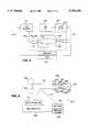
- FIG. 1is a block diagram of a tissue interrogator tool in accordance with a preferred embodiment of the present invention
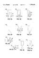
- FIG. 2is a plot of a normalized transmission versus wavelength of light (nm) of four tissue types collected in-vivo using a device in accordance with the present invention
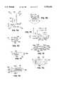
- FIG. 3is an elevated perspective view of a generic detachable tip for use with the tool of FIG. 1;
- FIGS. 3A-3Jare illustrations of various tip configurations for use with the interrogator tool of FIG. 1;
- FIGS. 4A-4Care illustrations of various embodiments of the optical emitting and detecting elements for use in the interrogator tool of FIG. 1;
- FIG. 5illustrates a schematic block diagram of an interrogator tool apparatus in accordance with an embodiment of the present invention
- FIG. 6illustrates a schematic diagram of another embodiment of a surgical interrogation tool in accordance with the present invention.
- FIG. 7Ais a front elevated view of a trocar tip tissue penetrating device in accordance with the present invention.
- FIG. 7Bis a tip end view taken along line 7B--7B of FIG. 7A;
- FIG. 7Cis a side view taken along line 7C--7C of FIG. 7A;
- FIG. 7Dis a base end view taken along line 7D--7D of FIG. 7A;
- FIGS. 7E-7Gare side sectional views of a trocar tip tissue penetrating device, taken along line 7F--7F of FIG. 7D, in accordance with different embodiments of the present invention.
- FIG. 7His a cross sectional view of the tip of a trocar tip in accordance with an embodiment of the present invention.
- FIG. 8Ais an elevated perspective view of the tip of an ellipsoid tipped needle-type tissue penetrating device in accordance with the present invention.
- FIG. 8Bis a front view of the tip of FIG. 8A taken along line 8B--8B;
- FIG. 8Cis a side view taken along line 8C--8C of FIG. 8B;
- FIG. 8Dis an end view taken along line 8D--8D of FIG. 8A;
- FIG. 8Eis a side view of an alternate embodiment of the device tip of FIG. 8A;
- FIG. 9is a schematic drawing of an embodiment of a synchronous multiplexed optical coupling system for the system of FIG. 1;
- FIG. 10is a schematic drawing of an embodiment of a wavelength multiplexed optical coupler for the optical system of FIG. 1;
- FIG. 11are a table of experimental results of tissue identification using a prototype tool in accordance with the present invention.
- An interrogation tool 10includes an analyzer 20 coupled to a surgical tool 30.
- the optical elements for emitting and detecting lightwhich are not shown in FIG. 1 for purposes of clarity, may be a part of analyzer 20, tool 30, a tool tip 40, a separate optical probe structure, or some combination of the foregoing.
- analyzer 20includes a light source controller 22, a spectrometer 24, a signal processor 26, and a display system 28. These elements are all controllably interconnected and operated under the control of a suitable state control machine, such as a microprocessor 29 executing software instructions.
- a suitable state control machinesuch as a microprocessor 29 executing software instructions.
- the light source controller 22provides a light control signal or signals on path 23 that control the generation of the light to be emitted by a light source and launched into the tissue.
- the light control signalthus can be suitable to operate a single light source (broadband or discrete), a plurality of discrete light sources (e.g., with a synchronous or frequency multiplexer where appropriate) or both.
- the light sourcecan be in the tool, i.e., physically inside, or merely coupled to the end of the tool using, e.g., fibers.
- Spectrometer 24receives signals corresponding to the detected light on path 25, performs a color analysis and ascertains the desired spectral characteristics of the detected light, and provides the spectral data to the signal processor 26.
- the signal processor 26receives the determined spectral characteristic data, and, using predetermined algorithms, evaluates the data to identify the type of tissue interrogated. Signal processor 26 provides the determined result to display system 28.
- signal processor 26also outputs an interlock control signal (illustrated by signal path 27) to tool 30.
- the interlock control signalis used by tool 30 in its operation.
- the interlock control signalmay be used to disable or enable the tool, override an attempted operation of the tool, interrupt an ongoing operation of the tool (e.g., in response to signal processor 26 identifying an undesired tissue type), or modify tool operating conditions if a desired tissue characteristic is identified (e.g., locating a blood vessel to be coagulated or determining that coagulation is complete).
- the interlock control signalmay be used by tool 30 in a manual response, semi-automatic control, or full automatic control, depending on the nature of the operation performed by the tool and the preferences of the surgeon. These operating parameters may be programmable or determined by manual adjustment of a switch on tool 30 or analyzer 20.
- Signal processor 26controls operation of the analyzer 20 to cause light to be emitted, to detect the coupled light and determine the spectral characteristics of the detected light, and to process the spectral characteristics to characterize the tissue, i.e., to identify the tissue by applying one or more algorithms to the spectral data until the data is determined to correspond to a known tissue type.
- Microprocessor 29performs various housekeeping, system management, diagnostic, and data storage and processing functions common to microcomputer-microprocessor controlled devices. This includes functions of the system such as are typical with microprocessor based devices, e.g., signal conditioning, digitization, communications, data storage and compilation, and control.
- microprocessor 29is a part of signal processor 26 (i.e., the functionalities of each exist within the same software controlled device).
- the functions of signal processor 26also may include various circuits for signal conditioning and processing in addition to hardware and software for digital data processing. It should be understood however, that microprocessor 29 could be replaced with solid state circuits, or a separate computer/microcontroller that is coupled to the analyzer 20.
- the display system 28may include one or more of text messages, audible indicators, and visual indicators mounted in a convenient location to be seen.
- the text messagesmay be displayed on an LCD type or other stand alone monitor associated with the analyzer 20 and/or on the surgeon's video monitor used to conduct the surgical procedure, e.g., the video monitor of an endoscope.
- Audible messagesmay be tones of selected frequency and/or pitch, volume and duty cycle to identify the intended message. Steady state and variable signals may be used.
- Visual indicatorsmay be a light panel with predetermined locations and/or colors assigned to represent different information.
- the surgical tool 30includes a handle 32, a shaft 34, and a detachable tip 40.
- the handle 32represents the controls for the manipulation and operation of the tool in accordance with the conventional manner. This includes manipulating the tool into and inside body tissue or a body cavity, as in the case of a tool for minimally invasive surgery, as well as the surgical procedure to be performed by the tools.
- the shaft 34is for passing the distal end of the tool 30 and the tip 40 into tissue, for example, through a portal and through or around internal body tissue in the case of minimally invasive surgery.
- the handle 32also contains the usual electronics, switches, fluids, valves, cables, levers, tubes, imaging equipment (in the case of endoscopes, laparoscopes and the like), signal paths, and other mechanisms, circuits, and/or logic for operating the tool to perform its intended functions.
- imaging equipmentin the case of endoscopes, laparoscopes and the like
- signal pathsand other mechanisms, circuits, and/or logic for operating the tool to perform its intended functions.
- Such toolsare known in the art. See, e.g., U.S. Pat. Nos. 5,192,280, 3,197,964, 5,201,732, 5,201,732, 5, 217,458, 5,250,047, 5,258,006, 5,290,286, 5,324,289, 5,330,471, 5,352,222 and 5,352,223.
- handlestypically include a scissor-like or pistol grip for moving a cable or wire 31 to actuate a moving member 48 relative to an opposing member 49 (see FIG. 3).
- the opposing membermay be fixed or movable, depending on the grasping mechanism.
- the toolmay be modified to receive the interlock control signal from signal processor 26 and to respond to the information content of the interlock control signal in an appropriate manner.
- Such modificationsare believed to be within the abilities of a person skilled in the art.
- Tip 40contains the optical emitting and detecting windows for launching light into the tissue and for coupling light after having passed through the tissue to be interrogated, and an end effector structure for performing the surgical procedure on the tissue, as will be described in more detail below with reference to FIGS. 3, 3A-3J, and 4A-4C.
- Tip 40is preferably detachable from tool 30 and operatively interconnects with the distal end of tool shaft 34 (see FIG. 3) so that tool 30 can control the tip 40 to perform its intended tissue interrogation and surgical intervention functions.
- tool 30is constructed with a common fitting 33 at distal end 34 to interfit detachably with more than one type of tip 40, wherein each tip 40 thus has a corresponding fitting to interconnect operatively with distal end 34.
- analyzer 20may be integrated into tool 30 to provide a single tool 10 with a plurality of useful tips 40.
- the system implemented by tool 10operates light source controller 22 to produce a control signal that causes a light source (not shown in FIG. 1) to emit light, so that light is launched into the tissue being interrogated at tip 40.
- a light sourcenot shown in FIG. 1
- spectrometer 24operates spectrometer 24 to collect the detected light (i.e., the light coupled at tip 40 after having passed through the tissue) at the wavelengths of interest and calculate the transmission spectra of the interrogated tissue.
- signal processor 26operates to process the spectral data determined by spectrometer 24 using one or more predetermined classification algorithms to determine the characteristic of the tissue being interrogated, and preferably to identify the tissue.
- the resultant tissue characteristic or the tissue identification (with a confidence limit) or bothcan then be displayed on a display system 28.
- the inability to identify a matching tissue typealso is considered an "identification," and an appropriate signal is displayed.
- the interlock control signalmay be provided on signal path 27, which signal is responsive to the identification of the interrogated tissue, as
- each tool 30is preferably provided with an information element 36 that can be read by analyzer 20 for recognizing the type of tool and its operational characteristics.
- the information element 36is a memory device such as a programmable read only memory (“PROM”) device or the like (EEPROM, flash memory, one or two dimensional bar code, coded resistor values, etc.), which contains appropriate use data and calibration information for the specific tool or a key word descriptor for the same.
- PROMprogrammable read only memory
- the calibration factorsthus can be determined, e.g., at the factory, and recorded in the information element 36 for access by analyzer 20, e.g., by signal path 27.
- the calibration informationalso can include structure information, e.g., how big are the components of the tip 40 end effector, for performing the surgical intervention procedure.
- structure informatione.g., how big are the components of the tip 40 end effector, for performing the surgical intervention procedure.
- spectral characteristicsfor example, a pair of jaws having a large space between optical elements would likely respond differently from a unipolar electrocautery device or even a retractor.
- Providing this information to the analyzer 20permits adjusting the response to the acquired transmission spectra accordingly.
- analyzer 20also can monitor the operating state of tool 30 during operation using signal path 27, so that it can provide suitable alarms and warnings, and produce an interlock control signal, based on a protocol of the procedure and the determined characteristic(s) of the tissue being interrogated.
- the analyzer 20may detect when the surgeon operates a switch to cause electrocautery cutting of tissue, and acts, for one example, to prevent cutting through tissue characterized as a bile duct by disabling the response (electrical and/or mechanical) of the tool to the activation of the trigger switch, and for another example to reduce power when the blood vessel is sufficiently coagulated so as to prevent char from forming.
- path 27can be constructed of a plurality of signal paths, rather than a single path.
- detachable tip 40may be configured as any surgical tool end effector incorporating optical elements and coupling to a male or female fitting (generically shown as 33) on tool distal end 34, as illustrated in FIG. 3.
- FIGS. 3A-3Jare shown generically, and thus may represent more than one type or kind of end effector tips as discussed herein.
- These structuresinclude, for example: a grasping mechanism (FIGS. 3, 3A, 3I, 3J) such as tongs, retractor, or a clamp; a bi-valve scissors (FIG. 3E) or a two member scissors or clamp (FIG. 3I); a suction or an irrigation probe (FIGS.
- FIG. 3B, 3Dan electrocautery tool, unipolar (FIG. 3H) or bipolar with electrodes on a single contacting surface (FIGS. 3C, 3H), a bi-valve structure (FIGS. 3, 3A, 3E) or other two member structure (FIG. 3J, viewing the two members as movable and fixed); a hemostatic tool that is thermally regulated or autoregulated (FIGS. 3, 3A, 3C, 3E, 3H); a tissue penetrating device such as a needle (FIG. 3D) or a trocar (FIG. 3F); and a surgical stapler or clip applier (FIG. 3G).
- a tissue penetrating devicesuch as a needle (FIG. 3D) or a trocar (FIG. 3F)
- FIG. 3Gsurgical stapler or clip applier
- the detachable tip 40is coupled to the shaft 34 at the distal end by a conventional mechanical interconnection 33 that securely engages the two parts (and preferably can be remotely coupled and decoupled at handle 32) and provides for the necessary optical, electrical and mechanical interconnections as appropriate for the given structure and function of the tip 40.
- a conventional mechanical interconnection 33that securely engages the two parts (and preferably can be remotely coupled and decoupled at handle 32) and provides for the necessary optical, electrical and mechanical interconnections as appropriate for the given structure and function of the tip 40.
- the dimensions of tip 40can be selected for use in major surgical intervention, wherein the patient is opened up, minimally invasive surgical intervention, wherein a portal is formed in the patent and the tools all pass through one or more ports, and other endoscopic or exploratory procedures.
- Portals for minimally invasive surgeryare typically 5 mm, 10 mm, or 12 mm in diameter.
- lightcan shine through plastic staples (and clips) to permit monitoring stapling as it occurs, with the staples themselves forming an optical conduit--such that the light is measured at the tip of the staples as they pass through tissue.
- lightcan enter the body from the outside and be detected by an internal tool with detection fiber, such that the light is launched from a different device than the light devicing device.
- a needlecan contain a light source, while another tool, e.g., another needle or one of the aforementioned end effector tools, contains a light coupling fiber connected to a detector.
- Each tip 40also may contain an information element 42 (FIG. 1) which identifies the end effector structure of the tip 40, and may contain any calibration information (or code) necessary for the tip 40, which information element 42 can be interrogated and read by analyzer 20 (or tool 30 depending on the configuration of tool 10 and the optical elements). This permits adjusting the operation of the system for the particular tool 30 and tip 40 used, more particularly, for automatically adjusting operation in the event that the tool and/or tip is changed during a procedure.
- the information element 42can include a resistor coding or a more complicated memory device, such as PROM or bar code.
- the information element 42also can include calibration data relating to the optical and physical operating characteristics of the tip 40, as already described, and other information such as the age of the tip or the number of times the tip has been used (or hours of use). The latter information can provide that a tip can be discarded (or not used until inspected) when its predicted useful life has been reached. In cases when both a tip information element 42 and a tool information element 32 are used, it is preferred that the stored calibration information not be redundant.
- tip 40in one embodiment, includes a broadband filament light source 43, which is electrically coupled to light source controller 22 by signal path 23 passing to and through tool 30 and signal path 44 in tip 40, and a light detector 47, such as a photodetector (e.g., a phototransistor, photodiode, charge coupled device (CCD), etc.), which is electrically connected to spectrometer 24 by signal path 25 in tool 30 and signal path 41 in tip 40.
- a photodetectore.g., a phototransistor, photodiode, charge coupled device (CCD), etc.
- Light source 43has an optical window 45 to couple light from source 43 and launch it into tissue
- light detector 47has an optical window 46 to couple light at the distal end of tip 40 and transmit it to detector 47, through wavelength selective filters (not shown in FIGS. 4A-4C).
- Optical windows 45 and 46may be glass or plastic inserts, or portions of fiber optic guides or some other optically transmissive material (e.g., an epoxy).
- photodetector 47may include four discrete photodetectors, each having a wavelength selective filter interposed to filter the coupled light. In this illustrative example, four electric signals corresponding to the four wavelengths of the spectra characteristics are transmitted to spectrometer 24 on path 25. More or fewer photodetectors and filters could be used.
- the light sourceis a plurality of discrete wavelength LED's mounted in tip 40 as light source 43 to launch multiplexed lights into tissue
- the aforementioned multi-element photodetector arrangementcould be used when there is a frequency multiplexed system, and a single photodetector could be used when there is a synchronous multiplexing system.
- Other variationsare believed apparent to those skilled in the art, subject to space and cost considerations in constructing tip 40.
- light source 43 and photodetector 47may be located in tool 30, shown secured at the distal end 34.
- the optical windows in tip 40extend completely therethrough to couple light between the light source and the light launching window and between the light coupling window and the light detector.
- the tool calibration information 36would account for the optical calibration characteristics of the detector 47, or in analyzer 20 (FIG. 4C), e.g., in spectrometer 24.
- the photodetector 47is disposed in the distal end of shaft 34 so that the mechanical interconnection couples the fiber optic guides 45 and 46 of tip 40 to the light source 43 and photodetector 47, whereby the photodetector 47 passes an output signal corresponding to the detected light to spectrometer 24 on signal path 25, and the light source 43 is controlled by signal path 23.
- the light source 43 and photodetector 47could be located elsewhere in tool 30, and coupled to the distal end 34 by suitable fiber optic waveguides (not shown).
- the light source 43is included in analyzer 20, e.g., as part of controller 22, and the light detector 47 is included in analyzer 20, e.g., as part of spectrometer 24, such that the light emitted and sensed are coupled between analyzer 20 and tool 30 over fiber optic signal paths 23 and 25, which mate with corresponding fiber optic elements 45 and 46 of tip 40, respectively.
- the light source 43is a broadband filament light source located in tip 40 and electrically coupled to light source control 22 by wire signal paths 23 and 45
- the light detector 47is located in analyzer 20, e.g., as a functional part of spectrometer 24, with the light coupled at tip 40 passed to detector 47 over fiber optic guides 46 and 25.
- the light detector 47can be easily constructed to detect multiple selected discrete frequencies, by use of filters, gratings and other frequency selective optical elements. This facilitates use of frequency selective demodulation of sensed broadband light.
- Rotating wavelength selective filters and a single photodetector or a plurality of photodetectors with fixed wavelength selective filterscould be used as a matter of design choice.
- This embodimentavoids the need to minimize the size and weight of the photodetecting equipment, which are more important concerns for tool 30 and tip 40.
- the light source 43can be located in tool 30, and the photodetector 47 located in analyzer 20, so that the calibration information of tool 30 includes the light source, and the light source can be replaced if necessary, during a surgical intervention with minimal risk to the patient.
- one or both of the fiber optic elements 45 and 46can be secured to the exterior surface of the end effector tip 401, for example, by a suitable adhesive, or in the form of a sleeve that the end effector tip 40 is passed into, such that the sleeve maintains the fibers 45 or 46 in a predetermined orientation relative to the tip 40 during use, once the sleeve is securely in place.
- a sleevemay be a body-compatible plastic (or a wrapping of suitable tape or a heat shrink material) that also serves to seal the coupling between tip 40 and the distal end 30 of tool 30, e.g., against wicking of body fluids which might interfere with an optical connection.
- a suitable adhesiveor in the form of a sleeve that the end effector tip 40 is passed into, such that the sleeve maintains the fibers 45 or 46 in a predetermined orientation relative to the tip 40 during use, once the sleeve is securely in place.
- a temperature sensor 52optionally is included in tip 40 and operatively connected to acquire and pass temperature related information to analyzer 20, e.g., over signal path 53 and signal path 27. It should be understood that this temperature sensor can be included in any tip 40. In the case that the temperature sensor is a near infrared based detection system, or other such light-based system, then the element shown as 52 could instead be an appropriate light emitting source, provided that the light detector 47 is modified to detect the appropriate wavelength from which the temperature information can be derived (in the conventional manner for such temperature monitoring systems). Indeed, the appropriate light illumination for the temperature sensing may be incorporated into the light source 43, or a second light source (not shown) may be included and multiplexed (by frequency or synchronously) with the light related to sensing spectral characteristics.
- the analyzer 20 and tool 30are constructed as rugged, reusable devices having relatively long useful lives, and the detachable tip 40 can be constructed as either durable reusable devices, or consumable devices having a more limited useful life.
- the foregoing interrogation tool 10operates as follows.
- the tissueis illuminated by a broadband light source located in tip 40. Some of the irradiating light is reflected, some is absorbed, and some is transmitted. There also may be some fluorescence.
- the light passing through the tissueis coupled into tip 40 and the particular spectral characteristics of a selected number of wavelengths of interest are then sensed.
- the relative values of transmitted and absorbed lightvary as a function of tissue type, composition, and histology, tissue thickness, and the wavelength of light. This permits discriminating tissue types and structures based on difference in the transmission spectra.
- FIG. 2an example of transmission spectra determined for four different tissue types are shown. These spectra are actual data collected by an experimental prototype bi-valve grasping tool, similar to that shown in FIG. 3, operating on tissue in-vivo in a living animal undergoing laporoscopic surgical intervention under an approved animal-study protocol.
- the tissue types illustratedare bile duct A, ureter B, artery C, and vein D. It is noted that the transmission spectra for these tissue types are clearly distinct from one another.
- a set of spectracan be selected which permit a wide variation from person to person, such that the different transmission spectra ranges for the different tissue types are sufficiently and clearly distinct. This enables the interrogation tool 10 of the present invention to discriminate and identify many different major tissue types, and in particular those tissue types and structures that are likely to lead to problematic complications if improperly subjected to the surgical intervention.
- a library of such tissue typescan be empirically created based on selected transmission spectra.
- detection spectrais used interchangeably herein with the term “transmission spectra”.
- the formeris believed to be a more accurate descriptor, and the latter is used to indicate that the radiation has been transmitted through the tissue to be characterized.
- Classification algorithmsalso can be established using well-known analysis techniques, such as K-Nearest Neighbors (KNN) or Soft Independent Modeling of Class Analogy (SIMCA).
- the acquired datais then processed using one or more of these algorithms, and the library of spectra records, and the interrogated tissue type can be determined within acceptable confidence limits or identified as not identifiable.
- the number of data points used to define a transmission spectra for a given tissue typemay vary from one type of tissue to another, and from one application to another, depending on the surgical intervention to be performed. In addition, the number of data points can vary depending on the computing power of analyzer 20, which often is a result of a cost-benefit decision, and is thus deemed to be matter of design choice.
- the library of tissue typescan include a set of spectra records corresponding to distinct types of tissue classes, including, for example, ureter, liver, kidney and other types such as those listed in FIGS. 11A and B.
- the libraryalso can include a set of spectra records corresponding to different pathological states of a selected tissue class, such as normal, precancerous, and cancerous states of a given tissue class, e.g., pancreas, liver, lung, or more than one given tissue.
- the libraryalso could include a set of spectra records corresponding to a sequence of effects of a surgical intervention on a given tissue type, for example, on a blood carrying tissue.
- the interventionis electrocautery of normal blood flow in tissue
- representative pathological statesinclude normal blood flow in the tissue, coagulated blood in the tissue, over-coagulated blood in the tissue, and char, wherein the blood carrying tissue contains at least one of an artery, a vein, and a capillary bed.
- these different setscan be combined in the same library, or in different libraries, such that the surgical procedure being performed determines which library or which parts of a library are to be used.
- the libraryincludes a plurality of predetermined spectra records corresponding to the plurality of known tissue types. It also is possible to provide for modifying the library by inputting data from a remote source, such as, for example, a CD-ROM, a magnetic tape, an IC ROM, a modem, a PCMCIA disk, a PCMCIA memory, a direct user input, and a previously obtained detection spectra.
- a remote sourcesuch as, for example, a CD-ROM, a magnetic tape, an IC ROM, a modem, a PCMCIA disk, a PCMCIA memory, a direct user input, and a previously obtained detection spectra.
- the signal processorfurther is configured to improve its accuracy of identification in response to continued operation of the signal processor in using classification algorithm to identify a detection spectra.
- classification algorithmcan be used to average multiple ones of detection spectras over time, to improve the accuracy of the first output signal.
- the classification algorithmalso can be used to provide an output signal corresponding to a probabilistic strength of the first output signal. This provides a confidence factor for the surgeon to continue with the surgical procedure or take additional detection spectra.
- the classification algorithmis able to provide an output signal or signals corresponding to multiple tissue types being identified.
- the spectrometeris preferably operable to detect a feature of the detection spectra, i.e., the detected electromagnetic radiation, selected from at least one of absorbance, scattering, fluorescence, optical rotation, elastic scattering, temperature, time resolved optical data, frequency resolved optical data, reflectance, opacity and turbidity. More specifically, the spectrometer is configured to separate an absorbance effect and a scattering effect of the electromagnetic radiation. This will permit one or more of performing a time-resolved analysis, a frequency-resolved analysis, and a multifiber spatial-resolved analysis (using multiple fibers and/or multiple detections covering the space to be analyzed).
- the optical sensor componentscan be built into a tip 40 of any configuration, from flat to cylindrical, single member tips to multi-member tips.
- the light source and detectormay be mounted side by side on a planar surface with parallel optical axes that are perpendicular (normal) to the tissue contacting surface (see FIGS. 4A-4C). This would form a reflective mode sensor.
- Another reflective mode sensoris configured with light source and detector on diametrically opposed surfaces, pointing away from each other (see FIG. 3B).
- Yet another reflective mode sensoris configured with the light source and detector (or the corresponding fiber optic windows) on the same surface with the optical axes at an angle to each other, to minimize light passing directly from the emitter to the detector without passing through tissue (see FIG. 3G).
- the reflective mode and transmissive mode sensormay be combined on a single instrument by integrating multiple light source/detectors or fiber optic elements.
- multiple sets or arrays of emitter and detector windowscan be provided (see FIG. 3C). This is particularly useful for tools having multiple functions, such as electrocautery cutting using a thin edge or wires, and electrocautery coagulation using a broad edge (not shown).
- an apparatus representative of a smart surgical tool in accordance with the present inventionincludes an electrocautery end effector function and a tissue monitoring function.
- the latterincludes a light emitting window 145 and a light detecting window 146, placed in opposite surfaces of a bivalve tip 140 for grasping a sample tissue 107.
- a light source 143preferably a broadband light source, such as a filament lamp located in tool 30 is coupled to emitting window 145 by an optical fiber guide 144.
- a commercial bipolar electrocautery unit 130e.g., model Force B available from ValleyLabs, Inc. was used to pass current through electrical contacts 131 and 132 disposed on opposite sides of tissue sample 107.
- Spectroscopic data obtained via fiber optic 141 coupling window 146 to detector 147was collected by a CCD spectrophotometer 124, which incorporates detector 147, in this case Ocean Optics model SD-1000, and the collected data was provided to a signal processor 126, in this case a Intel model 80486-DX66-microprocessor based system.
- Processor 126is used to record spectra from tissue 107 at discrete wavelengths ranging between 400 nm and 1100 nm in 1024 channels, with one spectrum recorded every 25 ms.
- Such datacan be processed in multiple ways. For example, a class analysis can be performed (Haaland, 1989) that would allow use of such data to generate a model for determining which selected spectra fall into each of the tissue types to be discriminated.
- the exact mathematical model usedis not important, provided only that a series of spectral characteristics can be identified that allow automated calibration of the identification method.
- the present inventionprovides a smart surgical tool that can perform a tissue heating function, using tips for electrocautery, thermally regulated heating and laser based heating, and control the heating function to inhibit intervention in the presence of tissue identified as inappropriate.
- the present inventionalso is applicable to controlling bleeding during a surgical intervention.
- tissue to bleedTwo conditions must be satisfied for tissue to bleed: 1) There must be a hole in a blood vessel, and 2) there must be blood flow.
- tissue weldingcan be used for closing a hole or for stopping the normal blood flow.
- tissue weldingcan be used for closing a hole or for stopping the normal blood flow.
- alternative methods of closing blood vesselsare being sought, such as electrocautery and thermally heated hemostatic devices. If done properly, using the right time, current, and pressure, larger vessels can be seared together, preventing bleeding during tissue removal. Further, such a sealed site will heal correctly because the tissue that is left will form a strong scar.
- This approachcan also be used to tack down tissue using spot-welds, and even to reseal open compartments, such as blood vessels.
- spectroscopic or optical methodsare used to detect either the loss of a spectral signal related to the presence of hemoglobin (e.g., the blood has been destroyed in the vessel) or that the flow has stopped or that the hole is cauterized or otherwise closed.
- the present inventionprovides an semi- or fully-automated tissue interrogation tool system to determine tissue doneness based on spectroscopic analysis, for assessment and control of progress during an electrocautery or thermal intervention.
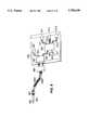
- a currentpasses between a pair of grasper members 201 and 203 which are opposed jaws having respective electrocautery jaws 241 and 242. Jaws 241 and 242 pass current to heat tissue 207 interposed between members 201 and 203.
- Optical fibers 245 and 246are disposed to terminate in mirrored wells 261 and 262 located in the jaws, and launch and collect light in the wells. Optical elements (not shown) may be inserted in wells 261 and/or 262 to enhance coupling light between the tissue and the optical fibers.
- Fiber 245is connected to light source 243 and fiber 246 is connected to spectrometer 224.
- signal processor 226sounds a first alarm (audibly sounded on display 228), to alert the surgeon that the tissue contains a significant blood vessel that must be sealed or cauterized (or some other tissue type that should be avoided).
- the surgeoncan initiate a first intervention by heating the tissue to coagulate the blood (or both).
- the processor 226then sounds a second alarm that instructs the surgeon that the coagulation is complete, e.g., based on detecting the disappearance of the spectral characteristics corresponding to blood flow or the appearance of the spectral characteristics corresponding to coagulated blood. Then the surgeon can sever the tissue and proceed with the intervention as appropriate.
- Signal processor 226includes a microprocessor chip, for example, model 486DX66 based computer or similar or equivalent device, having additional functionality for generating alarms when one or more predetermined criteria are met (or exceeded).
- signal processor 226also controls the delivery of electrocautery current through grasper members 201 and 203 in response to the change in spectral characteristics of the tissue corresponding to the coagulation of blood. The surgeon can change tips, if necessary, to sever the tissue, perhaps hemostatically, and monitor possible blood flow such that hemostasis can be applied to coagulate any blood flow found.
- the present inventionprovides both advantageous and surprising results.
- most surgeonsuse unipolar instruments that direct current from the tip of the electric knife through the body to a grounding plate electrode, commonly placed under the patient.
- a grounding plate electrodecommonly placed under the patient.
- An advantageous technique for tissue welding or searingknown as bipolar electrocautery, can be used that instead directs current between two tongs (or relatively closely positioned electrodes) of the device, e.g., as shown in FIG. 3A, 3E, 3I and 3J, thus sparing at least some of the nearby tissue from electric current.
- Electrocautery welding techniquesee e.g., Camran Nezhat et al., "Operative Laparoscopy (Minimally Invasive Surgery): State of the Art", Journal of Gynecological Surgery, Volume 8, Number 3, 1992, still find it difficult to control tissue welding.
- the skill barrier that must be crossed in order to perform this techniqueis effectively prohibitively high. This is particularly important during minimally-invasive surgery performed using an endoscope-type instrument (e.g., surgery performed through a periscope-like tube without opening up the patient).
- tissuesuch as an artery
- this tissuewill bleed profusely when cut, and may force emergency surgery (due to the blood loss and its attendant risks, as well as the fact that blood will block the endoscopic field of view, preventing further surgery via the endoscope in some cases).
- emergency surgerydue to the blood loss and its attendant risks, as well as the fact that blood will block the endoscopic field of view, preventing further surgery via the endoscope in some cases.
- bleedingleads to worsening of scarring, and lengthening of surgery; at the worst, such bleeding can be a life-threatening complication of surgery.
- the present inventionadvantageously overcomes these problems by automatically monitoring the tissue state and determining whether such blood vessels are present or adequately coagulated, in real time, even as electric current is passed through tissue between bipolar tong electrodes.
- a smart surgical tool in accordance with the inventioncould vary, but could include such measures as the disappearance of spectral signal corresponding to the presence of blood, or a class-analysis approach that searches for a color signature associated with properly browned tissue, or both.
- charhas a spectral signature that is different than tissue, whether based upon absorbance or scattering, or both.
- time-resolved methods, frequency resolved methods, or spatially-resolved methodscan separate absorbance from scatter, and allow an individual analysis of wavelength-dependent absorbance and wavelength-dependent scattering. Such an analysis may yield additional information, or for cost-effectiveness or other competitive reason, such a time-resolved, frequency-resolved, or space-resolved measurement may be avoided as well.
- a bipolar electrosurgical cutting tool in accordance with the inventioncould shut down when an artery is fully baked, and there is minimal danger of bleeding if the tissue is subsequently cut. This would prevent overcooking, which can cause bleeding well after surgery due to improper healing, as well as undercooking, which will lead to immediate bleeding when the tissue is cut by the surgeon.
- tissue in which arterial blood is flowingdemonstrates a well-known pulsatility of signal.
- tissueis thermally coagulated by a bipolar device, or even a unipolar probe exerting pressure on the tissue, this signal disappears.
- the pressure of the deviceis removed, the venous blood will flow back into the tissue.
- a spectroscopic methodcan be used to detect the flow of blood back into the tissue, demonstrating the patency of the venous system.
- spectroscopycan be used to verify if the blood flow has been adequately destroyed for intervention.
- the tool 210includes a pneumatic system such as an air bladder 271 to open and close controllably grasper jaws 241 and 242, or to close the jaws and then release some pressure and then reapply pressure, allowing blood to refill the tissue at times.
- a pneumatic systemsuch as an air bladder 271 to open and close controllably grasper jaws 241 and 242, or to close the jaws and then release some pressure and then reapply pressure, allowing blood to refill the tissue at times.
- This changecan detected by spectrometer 224, and the absence of spectral change as the pressure is modulated can be used to determine the time at which occlusion has occurred, and an alarm needs to sound.
- the opening and closing effectcan be achieved by having air bladder 271 inflated by a pump 272 and hose 273, using positive pressure or negative pressure under operative control of processor 226.
- An alternate structurecould use a conventional cable operated grasping tool to apply the pressure variations. Other structures could easily be considered for such use and are omitted without exclusion.
- the air bladder 271 illustrated in FIG. 6 and related structurecould be omitted from the tool and end effector tip.
- tissue interrogating toolincluding a penetrating device with an optical sensor for use in determining whether or not the penetrating device has penetrated to the desired body cavity.
- the puncturing toolmay be, for example, a trocar or surgical insufflation needle coupled with such an optical sensor.
- the optical sensormay have a multiplicity of optical components at the distal end of the puncturing tool for emitting and launching light and coupling and detecting light to provide a signal corresponding to the spectral characteristics of the tissue presented to the tool.
- this toolis able to generate an alarm when penetration into a desired tissue type has occurred or penetration into an undesired tissue type is about to occur and/or has occurred.
- the light emitting window element and the light detecting window elementmay be arranged in any configuration on the tip so that, in response to the light intensity launched by the light emitting window, the light detector produces a signal that corresponds to the spectral characteristics of the tissue being interrogated.
- the light emitting and detecting window elementsmay be oriented on the tool, in either (1) transmissive line of sight, wherein there is a line of sight light path between the light emitting window and the light detecting window and the presence of tissue reduces the intensity of light illumination sensed by the detecting element, (2) transmissive over the horizon, wherein there is no direct line of sight light path between the light emitting and detecting windows, the presence of tissue couples light to the light detecting window, and the absence of tissue reduces the intensity of light illumination coupled to the light detecting window, (3) reflective over the horizon, wherein there is no direct line of sight light path between the light detecting and light emitting window, the presence of tissue attenuates the light intensity sensed at the light detecting window and the absence of tissue results in an increased light intensity sensed at the light detecting window, or (4) some combination thereof.
- the selected wavelengthsmay be launched and the desired wavelengths, which may be different from the wavelengths launched, sensed using wavelengths multiplexed by frequency or by time for transmission to the tissue on one common optical waveguide.
- the discrete wavelengthsmay be separately delivered over corresponding dedicated optical waveguides.
- more than one fiber optic waveguidemay be used to deliver the same wavelength(s) simultaneously at different points on the device tip.
- the light detecting windowmay be one optical waveguide adapted to receive all light sensed, which detected light signal can be optically (frequency) or electrically (time) demultiplexed and separated into different sensed light intensity signals corresponding to the different desired wavelengths.
- the light detecting windowmay have separate optical waveguides for receiving the light and either selectively passing one discrete wavelength to a light detector or passing the light to a wavelength selective light detector, for generating the separate corresponding electrical signals.
- the light detecting windowmay have more than one optical waveguide to detect the light such that the detected light signals are averaged, summed, or otherwise related to yield a detected light level for each emitted frequency.
- the light emitting and detecting windowscomprise a plurality of optical components, such as optical fibers, at the device tip which are plugged into and thus coupled to an optical bench containing the light source(s) and the light detector(s) such as photodiodes, phototransistors, photomultiplier tubes, charge coupled devices, and the like.
- optical fiberscan be made of glass or plastic materials for reusable and disposable device tips. Glass fibers are preferred for reusable tips and plastic fibers are preferred for disposable tips.
- the optical benchcan be made in a durable case in the surgical tool or the analyzer and reused for both disposable and reusable device tips.
- the optical fibersmay be identical in construction and symmetrically secured in or to the tissue penetrating device tip so that either fiber (or group of fibers) may be used either to illuminate tissue or to couple light from the tissue.
- either fiberor group of fibers
- dedicated male and female interconnectionscan be used for coupling the emitter to the emitter window and the detector to the detector window.
- the optical benchcan be in the tip, the tool, or in the analyzer, and it can be separated into one bench for emitting light and another bench for sensing light, each being independently located in one of the analyzer, tool, or tip, as desired in the particular application.
- the present inventionprovides for a tissue intervention tool having tips with optical elements and control circuits that can be used to determine reliably and accurately the tissue type being or to be penetrated. It also provides for disposable components for trocars or surgical insufflation needles containing inexpensive optical elements to insure optimal performance of the invention and prevention of crosscontamination between patients. Alternately, the invention provides durable trocars and insufflation needles with optical elements that will withstand sterilization by autoclave, gas, and x-ray sterilization techniques.
- tip 40is illustrated as a conventional trocar tip for laparoscopic surgery, which has been modified as described below.
- the sheath and obturator tubesare not shown.
- the optical elementsare provided in the form of first and second lengths of optical fibers 745 and 746, and an optical coupler 790 connecting the tip 40 fiber 745 to a light source 43 for launching one or more of the selected illuminating wavelengths or a broadband light source as the case may be, onto fiber 745 and a light detector 47 for sensing light coupled at tip 40 by fiber 746.
- light source 43 and detector 47are part of an optical bench located in tool 30 (see FIG. 4B), and are not shown in FIGS. 7A-7D.
- the conventional surgical trocar appliance(reusable or disposable) is modified by the incorporation of two optical fibers 745 and 746 into or on the structure.
- Optical fibers 745 and 746are each a single fiber that is small in diameter and may be of conventional plastic or glass construction. The fiber diameter could be selected to be from between 0.05 and 0.25 mm, but should be selected such that the mechanical function of the appliance tip 40 is not compromised. Fibers 1.0 mm diameter or larger may be used on large trocar applications, i.e., a trocar having a 10.0 mm diameter. Glass optical fibers currently have superior optical transmission characteristics and are available in the smaller sizes.
- Glass optical fibersalso have better mechanical and thermal properties, such as the ability to withstand autoclave temperatures, although high-temperature plastics are being introduced and are expected to be useful in the present invention. Glass fibers also have a benefit in that they can act as an electrically isolating spacer between conductive elements such as are used in electrocautery applications discussed above. Plastic fibers are much less expensive, which makes them well suited to disposable devices and reusable devices where sterilization by autoclave is not required.
- the optical fibers 745 and 746are secured to tip 40 by being mounted into small holes formed in the tip 40 near the point of the tip and fixed in place with a medical grade adhesive or potting compound.
- the exact positions of the ends of fibers 745 and 746are preferably chosen to minimize the likelihood of a direct optical path between the two, which would allow optical crosstalk.
- the position of the fiber ends in the cutting faces of tip 40also are selected so that when the light illumination changes in response to the ends of fibers passing into different types of tissue and/or body cavities to acquire the spectral characteristics.
- coupler 790in which the fibers terminate (see FIG. 7E) a durable component that is plug-compatible with the optical bench, having secured optical fibers 745 and 746.
- coupler 790may have a "plug" that is keyed so that a selected optical element will be used for sensing (element 746) (or illuminating) (element 745) light, but not both. This is useful when an optical element, or its manner of mounting or its positioning, is specially designed for light illumination or sensing, but not both.
- tip 40 and coupler 790may be constructed with a coding system as described above to have a code that identifies the type of tip, i.e., its surgical end effector structure and the configuration of optical elements 745 and 746.
- This codeis read by analyzer 20, for example, when tip 40 is coupled to the distal end 34 of tool 30 and/or when the device is turned on.
- the codeis identified and can be used to select appropriate timing signals and signal levels for controlling the illumination and detection of light, and to select appropriate thresholds for processing the sensed light signals and making determinations regarding tissue spectral characteristics.
- analyzer 20may be provided with a memory device (not shown) containing a conventional look-up table of prerecorded information, for providing all of the necessary operating parameters that are associated with a given code. Further, if a code is not recognized, the device may automatically shut down so that incompatible appliances are not incorrectly used. Any method of coding tips 40 may be used, such as using selected resistor values or digital words in an integral circuit appliance 10 and corresponding contacts in coupler 790 for electrically sensing the code by analyzer 20. In addition, product labeling such as a bar code and a photodetector array and sensors may be used for optically sensing the code by analyzer 20. Coding optical sensing devices are discussed in, for example, U.S. Pat. Nos. 4,621,643, 4,700,708, and 4,770,179, the disclosures of which are hereby incorporated by reference.
- one construction of trocarhas a tip 40 that includes a 3-face pyramid.
- the facesare cut on essentially flat planes that intersect at an angle of 120° relative to each other (see angle ⁇ in FIG. 7B) and so that the midline of one facet and the edge of an adjacent facet form an angle of 45° (see angle ⁇ in FIG. 7C) with the apex being the point of tip 40.
- This trocar tip constructionis conventional.
- fibers 745 and 746are respectively inserted into two parallel 0.013-inch-diameter holes, which are spaced apart a distance d2 of 0.1 inches along a diameter of tip 40, and centered about the point of tip 40.
- the faces of fibers 745 and 746are flush with the plane of the facet of trocar tip 40, and terminate a distance d3 of 0.07 inches from the point of tip 40 as illustrated in FIG. 7A.
- the trocar tip 40may be constructed as follows. Two holes are drilled. The fibers 745 and 746 are coated with silicone adhesive and passed into the drilled holes so that the ends of fibers extend from the tip faces. The adhesive is allowed to set. The ends of fibers are then trimmed either separately or during the process for tipping the tip. The fiber ends are then polished. The inside of trocar tip 40 may be partially filled with a potting material to provide additional support for fibers 745 and 746.
- tip 40has two pairs of optical elements 745 and 746 namely optical fibers 745a and 746a, and optical fibers 745b and 746b.
- the placement of the ends of fibers 745b and 746bare selected so that element 746b will couple predominantly light illumination launched by optical element 745b, rather than 745a.
- the two pairsare separately operated to provide different signals of light (and dark) illumination corresponding to different sensing locations on tip 40.
- Pair of fibers 745a and 746aprovide the light (and dark) illumination at the point of tip 40 and fibers 745b and 746b provide the light (and dark) illumination closer to the base of tip 40. This provides a first signal responsive to tissue spectral characteristics at the leading point of tip 40, and a second signal responsive to spectral characteristics at the base of tip 40.
- the pairs of optical elements in FIG. 7Emay be connected so that optical elements 745a and 745b are coupled to a common light source and optical elements 745a and 745b are coupled to a common light detector (not shown).
- the optical elements 745 and 746may be constructed to terminate with their ends normal to the tissue contacting surface (see FIG. 7F) perpendicular to the longitudinal axis (see FIG. 7G), or various other angles or combinations of angles, taking into consideration the allowable radius of curvature of the optical fibers.
- yet another configuration of a trocar tip 40could include a single optical fiber 745 passing along the axis of tip 40 and terminating with its end in the leading point of tip 40 (or proximate thereto), and using two light detecting fibers 746-1 and 746-2 to couple light illumination in response to the light emitted by fiber 745.
- Fibers 746-1 and 746-2are shown as terminating at different distances from the point of tip 40 and on opposite sides of the axis of tip 42, thereby to provide different coupled light signals as described above.
- the endsmay terminate in different faces.
- the endsmay terminate on the same side of tip 40, e.g., in the same face of a pyramid tipped trocar tip.
- references to the optical elements being "at” the tissue contacting surfaceshould be understood to refer to the end(s) of the optical fiber or the emitting surface of a light source being one of flush with the plane of the tissue contacting surface, a short distance below the plane, or a short distance above the plane, such that the end does not interfere with the mechanical function of the tissue contacting surface.
- an elliptical tip 40 of a surgical appliance 10 that is a needleis shown.
- the ellipsoidis formed in a flat plane at a conventional angle ⁇ , for example, 15° relative to the longitudinal axis of needle tip 40.
- the optical elements 845 and 846are secured along the minor axis of the ellipsoid plane, on a diameter of the needle tip 40, disposed in parallel to the longitudinal axis.
- the elements 845 and 846are secured by being mounted to needle tip 40 by epoxy, e.g., Bipax brand optical grade epoxy available from Tra-Con, Milford, Mass.
- the ends of elements 845 and 846are polished flush with the flat ellipsoid tip 40.
- irrigation and suction probe tubeswhich may have a cylindrical tip in a plane perpendicular to the longitudinal axis of the tube.
- optical fibers 845 and 846are illustrated in FIGS. 8B and 8E as on a diameter of needle tip 40 and so that they will essentially enter the tissue at about the same time. Alternately, they could be disposed on a chord of appliance needle tip 40, e.g., in a plane perpendicular to the longitudinal axis or in a plane parallel to the tissue contacting face. Further, they may be oriented with one end (preferably the light emitting window) in the lead portion and the other end (preferably the light detecting window) in the trailing portion of the tissue contacting face as illustrated in FIG. 8E.
- elements 845 and 846may be secured to needle tip 40 by being wrapped inside a thin film of material, e.g., mylar.
- elements 845 and 846 in the form of optical fibersmay be inserted into channels formed in the material of a tip 40 and sealed in place with an adhesive.
- optical fibers 845 and 846where they emerge from the surgical appliance 10 to be coplaner with the appliance tissue penetrating tip 40, must be polished. Polishing insures proper mechanical function of the tip and improves the optical coupling of the fibers.
- the fibersmay be polished by the tip sharpening process or they may be polished after tipping by a secondary wet or dry process.
- the codeis preferably provided at the junction where the appliance tip 40 is coupled to a tool 30, and the information is then provided to the analyzer 20. For example, with reference to the embodiment illustrated in FIG.
- the codemay be installed in coupler 990 and sensed by a corresponding code reading circuit in coupler 991 (not shown), whereas in an embodiment where fibers 945 and 946 terminate into coupler 991, the code may be provided in another manner and sensed by a corresponding code reading circuit appropriately.
- fiber optics 945 and 946may fit into a coupler 995 which is an optics module (an optical bench) that transmits and receives monochromatic light over optic fibers at n different wavelengths to and from tip 40, where n is greater than or equal to one. It has a bulkhead mounted, duplex connector (female) 991 which mates with the male connector 990 at the end of tip 40 or alternatively with the ends of optical fibers 945 and 946. Fibers 946 and 945 are also illustrated as jacketed fibers 946A and 946B and incorporate strain relief 942 as appropriate.
- One optical fiber, or channel, illustrated as channel 992 in FIG. 9,is configured as a transmission channel, and the other channel, illustrated as channel 993 in FIG. 9, is configured as the reception channel. These channels are typically made of the same commercial fiber optic material.
- light sources 43-Nare preferably light emitting diodes (LEDs), although semiconductor lasers or broadband light sources (e.g., filament bulbs) combined with wavelength selective filters also may be used or combinations thereof, e.g., one broadband light source, and one infrared LED with one ultraviolet source (for fluorescent applications).
- LEDslight emitting diodes
- broadband light sourcese.g., filament bulbs
- wavelength selective filterse.g., one broadband light source, and one infrared LED with one ultraviolet source (for fluorescent applications).
- fibers 945 and 946typically will carry light that is other than monochromatic light.
- monochromaticalso should be understood to allow some variation of wavelength within a small bandwidth.
- the light sources 43-Nare energized according to timing control signals. Preferably, the timing of the operation of light sources 43-N and detector 947 provide for illuminating only one light source 43-N at a time, according to a selected sequence. Any suitable timing control signals sequence may be used.
- the light sources 43-Nare respectively connected by fiber pigtails 23-N (i.e., 23-1, 23-2 and 23-3) to an optical coupler 994 such as model #P92-7019-241-1 manufactured by AMP/Kaptron, Harrisburg, Pa. When necessary, optical elements such as lenses may be used to couple light into an optical fiber or to couple light from a fiber to a light detector.
- Coupler 994is a 2 ⁇ 3 coupler which combines the light provided at each of the input fibers 23-N in a symmetric homogeneous fashion and passes it along to each of the two output channels.
- One output channelis the transmission channel 992 of connector 991.
- the other output fiber optic channel, channel 995is directed to a photodetector 947A, preferably a photodiode such as model #BPX66 manufactured by Siemens Components, Inc., Cupertino, Calif.
- Detector 947Aprovides an electrical signal corresponding to the intensity of light emitted by each source 43-N. This is used to monitor and/or control the illumination level to avoid intensity drift and to maintain uniform illumination from pulse to pulse of each wavelength.
- Reception channel 993 of connector 991connects the sensed light from tip 40 and a second light detector 947, preferably a photodiode.
- Detector 947provides an electrical signal corresponding to the intensity of light sensed at tip 40 of the surgical tool 30 (for each monochromatic source 43-N in this embodiment). This light includes the light reflected by tissue 107 in response to the illumination by each source 43-N. When desired, it may include tissue fluorescence wavelengths.
- Coupler 994Mhas the same characteristic of combining light in a symmetric, homogeneous fashion as 2 ⁇ 3 coupler 994 illustrated in FIG. 9.
- the reception channel 993 of the bulkhead connector 990is directed to a wavelength demultiplexing coupler 994DM, such as models #99100-3 or 99103-3 available from AMP, Inc.
- a wavelength demultiplexing coupler 994DMsuch as models #99100-3 or 99103-3 available from AMP, Inc.
- Each output pigtail 25-N of demultiplexer 994DM, which contains one wavelength signal Nis routed to an individual light detector 47-N (photodiodes are preferred).
- a second output of coupler 994Mis passed on fiber 995 to a second wavelength demultiplexing coupler 997DM, and then by pigtails 995-N to detectors 947-N.
- This schemeprovides n discrete detectors 947-N to monitor sensed light and dark illumination at sources 43-N and n discrete detectors 47-N to monitor sensed light and dark illumination at tissue 107 at tip 40 of surgical appliance 10. No additional electronic signal multiplexing or conditioning is required.
- the timing functionis much simpler because each light source may be illuminated simultaneously. Hence, a single square wave denoting on and off states is adequate. The dark illumination may be sampled during each off state, following the aforementioned delay.
- the same light source drive circuit and photodetector preamplifiermay be used for both the synchronous and frequency multiplexer systems, except that the photodetector preamplifier must be repeated for each of the photodetectors in the frequency multiplexer system.
- the synchronous multiplexing techniqueuses fewer optical and detector components than the wavelength, but it requires additional electronic switching and processing.
- any one of the various structures for the optical elements shown in FIGS. 3, 3A-3J, 4A-4C, 7A-7H and 8A-8E as described hereinmay be applied in a straightforward manner to the other various structures of tip 40 including those not described herein, which variations are believed to be within the ability of a person skilled in the art.
- the sensed light signalsare best demultiplexed by a spectrometer-detector system as previously described.
- the demultiplexing by frequencycan be performed in the aforementioned manner.
- the demultiplexing by timecan be performed using frequency selective filters that are periodically interposed between the sensed light and a photodetector, to chop the sensed light into frequency selective segments, which segments can be separated in time to acquire the desired spectral characteristics.
- time-resolved, frequency- resolved, or spatially-resolved methodscan be used to extract additional information, in an analogous manner, which information can be used to generate similar tissue identifications. Modification of the foregoing to include one or more of the time/frequency/space resolved methods is believed to be within the abilities of a person of ordinary skill in the art, once knowledgeable about the present invention, and, therefore, within the scope of the invention.
- Another optical sensing embodimentcan be implemented without any surgical tool at all.
- the entire surgical fieldis illuminated by broadband energy.
- a cameracaptures an image of the field which is stored as pixels in a memory array.
- Various narrow band filterscan be sequentially interposed between the camera and the resulting sequence of monochromatic images stored in page memory arrays.
- the paged memorycan be analyzed by a computer to yield calorimetric information about each pixel of the image.
- the calorimetric informationcan be further analyzed for spectral fingerprints of various tissue qualities, e.g., blood vessels or ureters. Pixels which match the spectral fingerprint would be highlighted on a video system, perhaps by brightness control or flashing or labelling with a cursor.
- the present inventionalso is applicable to surgery where it is important to differentiate between normal and abnormal types of tissue, as well as different types of tissue. Examples include the following:
- the interrogatory tool of the present inventionincludes probes which can identify blood vessels (arterial and venous), as well as bile ducts and other lumens such as ureters, nerves, fallopian tubes, etc., from surrounding tissue.
- the colorimetry technique already describedcan be used for this tissue type analysis and identification. This application minimizes the likelihood of severing the bile duct, for example, which is a frequent complication requiring traditional surgical intervention, and/or cutting through the ureter, which is also a frequent type of injury.
- Bipolar electrocauteryis often used to control locally and separate blood vessels.
- optical elementscan be incorporated into the bipolar electrodes.
- the optical fiberscould be sandwiched in an array between electrodes of a unipolar device or mounted on or adjacent electrodes of bipolar devices in a transmissive or reflective configuration.
- the optical elementscan locate blood vessels as well as determine the coagulation status of the tissue by measuring the absolute transmission spectra and/or the color change over time and detecting any formation of char, by its spectral signature, at the tissue interface.
- the color changemay be relative to a baseline measurement or a preselected spectral criteria corresponding to a good quality coagulation.
- This real time informationis useful to the clinician, who can use it to adjust electrocautery parameters, such as power level and duration.
- the informationalso can be used for automatic parameter control.
- the same optical sensing technologyalso may be used for observing and controlling unipolar electrosurgical cautery, laser cautery, and laser welding of tissues.
- Staples or clipsare routinely used in place of sutures. occasionally, the staples are misplaced so that they obstruct or damage an underlying structure. For example, a blood vessel may be inadvertently occluded or a nerve may be accidentally severed after being "caught" in the jaws of a stapler. Alternately, an underlying structure may be situated in an unusual location and be caught unknowingly.
- the optical sensor of the present inventioncan be incorporated into the jaws of a stapler or clip applier to interrogate the tissue to be stapled or clipped.
- an alarmcan be sounded if there is important tissue trapped between the jaws (e.g., blood vessel or nerve) or if the tissue cannot be reliably classified as only the desired tissue free of undesired structures.
- the alarmalso could be configured as a safety interlock which prevents the stapler from actuating during an alarm condition (unless the alarm is overridden).
- a plastic light conducting staplecan be utilized in the light detection path to monitor the effectiveness of the closure.
- Endometriosisis often distributed throughout the surgical field.
- the colorimetry techniquecan be used to identify areas of endometriosis. For example, the spectral characteristics of normal tissue and the endometriosis can be ascertained, and the two or more characteristics can be used to decide whether the tissue being interrogated is normal or abnormal. Further, in combination with the cautery status indicator, the optical sensing technology of the present invention can be used to determine when the endometriosis has been effectively removed.
- an optical probecan be similarly used to identify obstructions or inflamed tissue beds.
- a pathological tissue classificationcan be used to distinguish cancerous tissue from noncancerous and other types of healthy tissue.
- the inventionalso overcomes the problems of the direct biopsy techniques.
- FIGS. 2, 3A, 3B and 11a preliminary study on a live pig was conducted using a grasper tool similar to FIGS. 3, 3A and a suction irrigator probe tool similar to FIG. 4C.
- Sixty-three sets of datawere obtained in a laparoscopic procedure using the aforementioned Ocean Optics Spectrometer, which was known to be faulty and in need of service, and with vacuum leaks in the prototype tooling which made sustained insufflation difficult.
- the prototype systemincluded a conventional fiber optic light source supplied by Wolff of West Germany and a fiber optic Spectrometer model PSD 1000 supplied by Ocean Optics, Inc., Dunedin, Fla.
- the light source fiber and detector fiberwere mounted on opposing jaws of a Kleppinger style Bipolar forceps, also supplied by Wolff.
- the jaws of the Kleppinger forcepsare typically 1 mm wide and 12 mm long.
- the suction irrigator and grasper toolsare thus preferably sized so that they will fit through the lumen of a 5 mm canula.
- the suction irrigatorshould have a large internal lumen (4.5 mm) to preserve its primary function.
- Each set of dataincludes a spectral measurement every one nm in the range of 400 to 1100 nm (although only four such spectra are illustrated in FIG. 2 for clarity of presentation).
- a librarywas empirically constructed for each of the categories listed on FIG. 11A and B: Abdominal Wall Fat, Liver, Gall Bladder, Bowel, Bladder, Ureter, Uterine Wall, Diaphragm, Small Bowel, Colon Fat, Artery, Vein, Ganglia, Bony Pelvis, and Bile Duct.
- An analysiswas conducted using KNN and the predictive algorithm has a 76% success rate. Of sixty-two determinations, with the prototype equipment, there were a total of 15 false position and false negative determinations. These are reported in FIG. 11 where the number of successful determination are on the diagonal, and the false determinations are indicated above or below the diagonal, for the various tools and tissue types in the study.
Landscapes
- Health & Medical Sciences (AREA)
- Life Sciences & Earth Sciences (AREA)
- Surgery (AREA)
- General Health & Medical Sciences (AREA)
- Public Health (AREA)
- Biomedical Technology (AREA)
- Heart & Thoracic Surgery (AREA)
- Medical Informatics (AREA)
- Molecular Biology (AREA)
- Animal Behavior & Ethology (AREA)
- Engineering & Computer Science (AREA)
- Pathology (AREA)
- Veterinary Medicine (AREA)
- Physics & Mathematics (AREA)
- Biophysics (AREA)
- Nuclear Medicine, Radiotherapy & Molecular Imaging (AREA)
- Otolaryngology (AREA)
- Optics & Photonics (AREA)
- Chemical & Material Sciences (AREA)
- Analytical Chemistry (AREA)
- Biochemistry (AREA)
- General Physics & Mathematics (AREA)
- Immunology (AREA)
- Investigating Or Analysing Materials By Optical Means (AREA)
- Measurement Of The Respiration, Hearing Ability, Form, And Blood Characteristics Of Living Organisms (AREA)
Abstract
Description
Claims (43)
Priority Applications (1)
| Application Number | Priority Date | Filing Date | Title |
|---|---|---|---|
| US08/473,004US5785658A (en) | 1992-09-14 | 1995-06-07 | In vivo tissue analysis methods and apparatus |
Applications Claiming Priority (4)
| Application Number | Priority Date | Filing Date | Title |
|---|---|---|---|
| US07/944,516US5460182A (en) | 1992-09-14 | 1992-09-14 | Tissue penetrating apparatus and methods |
| US08/024,278US5746210A (en) | 1993-02-26 | 1993-02-26 | Device and method for detection, localization, and characterization of inhomogeneities in turbid media |
| US08/437,327US5772597A (en) | 1992-09-14 | 1995-05-09 | Surgical tool end effector |
| US08/473,004US5785658A (en) | 1992-09-14 | 1995-06-07 | In vivo tissue analysis methods and apparatus |
Related Parent Applications (2)
| Application Number | Title | Priority Date | Filing Date |
|---|---|---|---|
| US08/024,278Continuation-In-PartUS5746210A (en) | 1992-09-14 | 1993-02-26 | Device and method for detection, localization, and characterization of inhomogeneities in turbid media |
| US08/437,327ContinuationUS5772597A (en) | 1992-09-14 | 1995-05-09 | Surgical tool end effector |
Publications (1)
| Publication Number | Publication Date |
|---|---|
| US5785658Atrue US5785658A (en) | 1998-07-28 |
Family
ID=46251364
Family Applications (4)
| Application Number | Title | Priority Date | Filing Date |
|---|---|---|---|
| US08/437,327Expired - LifetimeUS5772597A (en) | 1992-09-14 | 1995-05-09 | Surgical tool end effector |
| US08/472,863Expired - LifetimeUS5769791A (en) | 1992-09-14 | 1995-06-07 | Tissue interrogating device and methods |
| US08/486,604Expired - LifetimeUS5807261A (en) | 1992-09-14 | 1995-06-07 | Noninvasive system for characterizing tissue in vivo |
| US08/473,004Expired - LifetimeUS5785658A (en) | 1992-09-14 | 1995-06-07 | In vivo tissue analysis methods and apparatus |
Family Applications Before (3)
| Application Number | Title | Priority Date | Filing Date |
|---|---|---|---|
| US08/437,327Expired - LifetimeUS5772597A (en) | 1992-09-14 | 1995-05-09 | Surgical tool end effector |
| US08/472,863Expired - LifetimeUS5769791A (en) | 1992-09-14 | 1995-06-07 | Tissue interrogating device and methods |
| US08/486,604Expired - LifetimeUS5807261A (en) | 1992-09-14 | 1995-06-07 | Noninvasive system for characterizing tissue in vivo |
Country Status (1)
| Country | Link |
|---|---|
| US (4) | US5772597A (en) |
Cited By (146)
| Publication number | Priority date | Publication date | Assignee | Title |
|---|---|---|---|---|
| US5984861A (en)* | 1997-09-29 | 1999-11-16 | Boston Scientific Corporation | Endofluorescence imaging module for an endoscope |
| US6096065A (en)* | 1997-09-29 | 2000-08-01 | Boston Scientific Corporation | Sheath for tissue spectroscopy |
| US6119031A (en)* | 1996-11-21 | 2000-09-12 | Boston Scientific Corporation | Miniature spectrometer |
| US6129667A (en)* | 1998-02-02 | 2000-10-10 | General Electric Company | Luminal diagnostics employing spectral analysis |
| US6185443B1 (en) | 1997-09-29 | 2001-02-06 | Boston Scientific Corporation | Visible display for an interventional device |
| US6238348B1 (en)* | 1997-07-22 | 2001-05-29 | Scimed Life Systems, Inc. | Miniature spectrometer system and method |
| US6289229B1 (en) | 1998-01-20 | 2001-09-11 | Scimed Life Systems, Inc. | Readable probe array for in vivo use |
| US6296608B1 (en)* | 1996-07-08 | 2001-10-02 | Boston Scientific Corporation | Diagnosing and performing interventional procedures on tissue in vivo |
| WO2001087154A1 (en)* | 2000-05-18 | 2001-11-22 | Nuvasive, Inc. | Tissue discrimination and applications in medical procedures |
| US6324418B1 (en) | 1997-09-29 | 2001-11-27 | Boston Scientific Corporation | Portable tissue spectroscopy apparatus and method |
| US20020013572A1 (en)* | 2000-05-19 | 2002-01-31 | Berlin Michael S. | Delivery system and method of use for the eye |
| US20020041166A1 (en)* | 2000-09-26 | 2002-04-11 | Dragan Grubisic | Led light source-based instrument for non-invasive blood analyte determination |
| US20020169379A1 (en)* | 2001-05-10 | 2002-11-14 | Hospital For Special Surgery | Utilization of an infrared probe to discriminate between materials |
| US6505133B1 (en) | 2000-11-15 | 2003-01-07 | Datex-Ohmeda, Inc. | Simultaneous signal attenuation measurements utilizing code division multiplexing |
| US6519485B2 (en) | 2000-12-13 | 2003-02-11 | The General Hospital Corporation | Minimally invasive system for assessment of organ function |
| US20030045808A1 (en)* | 1999-11-24 | 2003-03-06 | Nuvasive, Inc. | Nerve proximity and status detection system and method |
| US6577884B1 (en) | 2000-06-19 | 2003-06-10 | The General Hospital Corporation | Detection of stroke events using diffuse optical tomagraphy |
| US20030195405A1 (en)* | 1998-12-23 | 2003-10-16 | Nuvasive, Inc. | Nerve surveillance cannulae systems |
| US20040047499A1 (en)* | 1998-02-07 | 2004-03-11 | Soheil Shams | Automated DNA array image segmentation and analysis |
| US20040059520A1 (en)* | 2002-09-25 | 2004-03-25 | Soheil Shams | Apparatus, method, and computer program product for determining confidence measures and combined confidence measures for assessing the quality of microarrays |
| US6731781B1 (en)* | 1999-09-30 | 2004-05-04 | Biodiscovery, Inc. | System and method for automatically processing microarrays |
| US20040109478A1 (en)* | 2002-12-09 | 2004-06-10 | Infraredx, Inc. | Tunable spectroscopic source with power stability and method of operation |
| US20040199084A1 (en)* | 1999-11-24 | 2004-10-07 | Nuvasive, Inc. | Electromyography system |
| US20040213446A1 (en)* | 1999-10-12 | 2004-10-28 | Soheil Shams | System and method for automatically processing microarrays |
| US20040225228A1 (en)* | 2003-05-08 | 2004-11-11 | Ferree Bret A. | Neurophysiological apparatus and procedures |
| US20040230132A1 (en)* | 2003-02-07 | 2004-11-18 | Alfred E. Mann Institute For Biomedical Engineering At The | Surgical drain with positioning and protective features |
| US20040236192A1 (en)* | 2003-02-07 | 2004-11-25 | Alfred E. Mann Inst. For Biomedical Engineering At The Univ. Of Southern California | Implantable device with sensors for differential monitoring of internal condition |
| US6869430B2 (en) | 2000-03-31 | 2005-03-22 | Rita Medical Systems, Inc. | Tissue biopsy and treatment apparatus and method |
| US6934576B2 (en) | 2000-05-12 | 2005-08-23 | Hospital For Special Surgery | Determination of the ultrastructure of connective tissue by an infrared fiber-optic spectroscopic probe |
| US20050187451A1 (en)* | 2004-02-25 | 2005-08-25 | Norris Mark A. | Simultaneous signal attenuation measurements utilizing frequency orthogonal random codes |
| US6975899B2 (en) | 1998-09-11 | 2005-12-13 | Spectrx, Inc. | Multi-modal optical tissue diagnostic system |
| US20060025703A1 (en)* | 2003-08-05 | 2006-02-02 | Nuvasive, Inc. | System and methods for performing dynamic pedicle integrity assessments |
| US20060069315A1 (en)* | 2003-09-25 | 2006-03-30 | Patrick Miles | Surgical access system and related methods |
| US20060200012A1 (en)* | 2005-02-14 | 2006-09-07 | Mansour Hebah N | Methods and sensors for monitoring internal tissue conditions |
| US20070015981A1 (en)* | 2003-08-29 | 2007-01-18 | Benaron David A | Device and methods for the detection of locally-weighted tissue ischemia |
| US7196783B2 (en) | 2002-11-15 | 2007-03-27 | Accurate Machining, Inc. | Optical fiber bundle utilizing electromagnetic radiation feedback |
| GB2431232A (en)* | 2005-12-14 | 2007-04-18 | Zinir Ltd | Spectrophotometer |
| US20070185397A1 (en)* | 2006-02-09 | 2007-08-09 | Assaf Govari | Two-stage calibration of medical probes |
| US20070299468A1 (en)* | 2006-06-22 | 2007-12-27 | Viola Frank J | Tissue vitality comparator with light pipe with fiber optic imaging bundle |
| US20080009689A1 (en)* | 2002-04-09 | 2008-01-10 | Benaron David A | Difference-weighted somatic spectroscopy |
| US20080082078A1 (en)* | 2001-05-21 | 2008-04-03 | Eyelight, Inc. | Glaucoma surgery methods and systems |
| US20080097164A1 (en)* | 2003-01-16 | 2008-04-24 | Nuvasive, Inc. | Surgical access system and related methods |
| US20080188727A1 (en)* | 2002-04-09 | 2008-08-07 | Benaron David A | Broadband solid-state spectroscopy illuminator and method |
| US20080188873A1 (en)* | 2005-01-21 | 2008-08-07 | Giovanni Speziali | Thorascopic Heart Valve Repair Method and Apparatus |
| US20080221455A1 (en)* | 2002-08-05 | 2008-09-11 | Infraredx, Inc. | Near-infrared spectroscopic analysis of blood vessel walls |
| US20080226137A1 (en)* | 2007-03-14 | 2008-09-18 | Benaron David A | Metabolism- or Biochemical-Based Anti-Spoofing Biometrics Devices, Systems, and Methods |
| US7428048B1 (en) | 2004-12-30 | 2008-09-23 | Spectral Molecular Imaging Inc. | Imaging elastic scattering spectroscopy |
| US20080304793A1 (en)* | 2004-06-16 | 2008-12-11 | Benaron David A | Optical and Electrical Hybrid Connector |
| US20080306380A1 (en)* | 2006-02-09 | 2008-12-11 | Yochai Parchak | Shielding of catheter handle |
| WO2009005850A1 (en) | 2007-06-29 | 2009-01-08 | Tyco Healthcare Group, Lp | Method and system for monitoring tissue during an electrosurgical procedure |
| US7522953B2 (en) | 2001-09-25 | 2009-04-21 | Nuvasive, Inc. | System and methods for performing surgical procedures and assessments |
| US20090124860A1 (en)* | 2003-02-27 | 2009-05-14 | Nuvasive, Inc. | Surgical access system and related methods |
| US7582058B1 (en) | 2002-06-26 | 2009-09-01 | Nuvasive, Inc. | Surgical access system and related methods |
| US20090234248A1 (en)* | 2008-03-15 | 2009-09-17 | Surgisense Corporation | Sensing Adjunct for Surgical Staplers |
| US20090312616A1 (en)* | 2008-06-12 | 2009-12-17 | Sabrina Paseman | Transmission Fluorometer |
| US7664544B2 (en) | 2002-10-30 | 2010-02-16 | Nuvasive, Inc. | System and methods for performing percutaneous pedicle integrity assessments |
| US7785253B1 (en) | 2005-01-31 | 2010-08-31 | Nuvasive, Inc. | Surgical access system and related methods |
| US20100312081A1 (en)* | 2005-07-29 | 2010-12-09 | Spectros Corporation | Implantable Tissue Ischemia Sensor |
| US7905840B2 (en) | 2003-10-17 | 2011-03-15 | Nuvasive, Inc. | Surgical access system and related methods |
| US7920922B2 (en) | 2001-07-11 | 2011-04-05 | Nuvasive, Inc. | System and methods for determining nerve proximity, direction, and pathology during surgery |
| US20110257478A1 (en)* | 2010-04-20 | 2011-10-20 | Spinewindow Llc | Method and apparatus for performing retro peritoneal dissection |
| US8137284B2 (en) | 2002-10-08 | 2012-03-20 | Nuvasive, Inc. | Surgical access system and related methods |
| US8147421B2 (en) | 2003-01-15 | 2012-04-03 | Nuvasive, Inc. | System and methods for determining nerve direction to a surgical instrument |
| US8206312B2 (en) | 2005-09-22 | 2012-06-26 | Nuvasive, Inc. | Multi-channel stimulation threshold detection algorithm for use in neurophysiology monitoring |
| US20120179010A1 (en)* | 2002-04-04 | 2012-07-12 | Maynard John D | Determination of a Measure of a Glycation End-Product or Disease State Using Tissue Fluorescence of Various Sites |
| US8255045B2 (en)* | 2007-04-03 | 2012-08-28 | Nuvasive, Inc. | Neurophysiologic monitoring system |
| US8287597B1 (en) | 2009-04-16 | 2012-10-16 | Nuvasive, Inc. | Method and apparatus for performing spine surgery |
| US8313430B1 (en) | 2006-01-11 | 2012-11-20 | Nuvasive, Inc. | Surgical access system and related methods |
| US20120296238A1 (en)* | 2011-05-16 | 2012-11-22 | Tyco Healthcare Group Lp | System and Methods for Energy-Based Sealing of Tissue with Optical Feedback |
| US8328851B2 (en) | 2005-07-28 | 2012-12-11 | Nuvasive, Inc. | Total disc replacement system and related methods |
| US8551088B2 (en) | 2008-03-31 | 2013-10-08 | Applied Medical Resources Corporation | Electrosurgical system |
| US8568317B1 (en) | 2005-09-27 | 2013-10-29 | Nuvasive, Inc. | System and methods for nerve monitoring |
| US8568331B2 (en) | 2005-02-02 | 2013-10-29 | Nuvasive, Inc. | System and methods for monitoring during anterior surgery |
| US8740783B2 (en) | 2005-07-20 | 2014-06-03 | Nuvasive, Inc. | System and methods for performing neurophysiologic assessments with pressure monitoring |
| US8790406B1 (en) | 2011-04-01 | 2014-07-29 | William D. Smith | Systems and methods for performing spine surgery |
| US8821397B2 (en) | 2010-09-28 | 2014-09-02 | Masimo Corporation | Depth of consciousness monitor including oximeter |
| US9044221B2 (en) | 2010-12-29 | 2015-06-02 | Neochord, Inc. | Exchangeable system for minimally invasive beating heart repair of heart valve leaflets |
| US9167959B1 (en)* | 2010-03-26 | 2015-10-27 | Optech Ventures, Llc | Illumination for enhanced contrast in debridement apparatus and method |
| US9174025B2 (en) | 2004-09-21 | 2015-11-03 | Boston Scientific Scimed, Inc. | Rapid exchange catheters having a sealed guidewire lumen and methods of making the same |
| US9192374B2 (en) | 2007-10-18 | 2015-11-24 | Neochord, Inc. | Minimally invasive repair of a valve leaflet in a beating heart |
| US9198765B1 (en) | 2011-10-31 | 2015-12-01 | Nuvasive, Inc. | Expandable spinal fusion implants and related methods |
| USD748259S1 (en) | 2014-12-29 | 2016-01-26 | Applied Medical Resources Corporation | Electrosurgical instrument |
| US9320563B2 (en) | 2010-10-01 | 2016-04-26 | Applied Medical Resources Corporation | Electrosurgical instruments and connections thereto |
| US9351845B1 (en) | 2009-04-16 | 2016-05-31 | Nuvasive, Inc. | Method and apparatus for performing spine surgery |
| US9392953B1 (en) | 2010-09-17 | 2016-07-19 | Nuvasive, Inc. | Neurophysiologic monitoring |
| US9603741B2 (en) | 2000-05-19 | 2017-03-28 | Michael S. Berlin | Delivery system and method of use for the eye |
| US9622732B2 (en) | 2004-10-08 | 2017-04-18 | Nuvasive, Inc. | Surgical access system and related methods |
| US9717403B2 (en) | 2008-12-05 | 2017-08-01 | Jeffrey B. Kleiner | Method and apparatus for performing retro peritoneal dissection |
| US9757072B1 (en) | 2013-02-11 | 2017-09-12 | Nuvasive, Inc. | Waveform marker placement algorithm for use in neurophysiologic monitoring |
| US9757067B1 (en) | 2012-11-09 | 2017-09-12 | Nuvasive, Inc. | Systems and methods for performing neurophysiologic monitoring during spine surgery |
| US9775545B2 (en) | 2010-09-28 | 2017-10-03 | Masimo Corporation | Magnetic electrical connector for patient monitors |
| US9827109B2 (en) | 1999-03-07 | 2017-11-28 | Nuvasive, Inc. | Methods and apparatus for performing spine surgery |
| US10064757B2 (en) | 2011-05-05 | 2018-09-04 | Michael S. Berlin | Methods and apparatuses for the treatment of glaucoma using visible and infrared ultrashort laser pulses |
| US10149713B2 (en) | 2014-05-16 | 2018-12-11 | Applied Medical Resources Corporation | Electrosurgical system |
| US10154815B2 (en) | 2014-10-07 | 2018-12-18 | Masimo Corporation | Modular physiological sensors |
| US10251600B2 (en) | 2014-03-25 | 2019-04-09 | Briteseed, Llc | Vessel detector and method of detection |
| US10363168B2 (en) | 2011-06-14 | 2019-07-30 | Ivantis, Inc. | Ocular implants for delivery into the eye |
| US10405838B2 (en) | 2014-08-28 | 2019-09-10 | Koninklijke Philips N.V. | Side-looking lung biopsy device |
| US10406025B2 (en) | 2009-07-09 | 2019-09-10 | Ivantis, Inc. | Ocular implants and methods for delivering ocular implants into the eye |
| US10420480B1 (en) | 2014-09-16 | 2019-09-24 | Nuvasive, Inc. | Systems and methods for performing neurophysiologic monitoring |
| US10420603B2 (en) | 2014-12-23 | 2019-09-24 | Applied Medical Resources Corporation | Bipolar electrosurgical sealer and divider |
| US10492949B2 (en) | 2009-07-09 | 2019-12-03 | Ivantis, Inc. | Single operator device for delivering an ocular implant |
| US10537474B2 (en) | 2008-03-05 | 2020-01-21 | Ivantis, Inc. | Methods and apparatus for treating glaucoma |
| US10588620B2 (en) | 2018-03-23 | 2020-03-17 | Neochord, Inc. | Device for suture attachment for minimally invasive heart valve repair |
| US10695178B2 (en) | 2011-06-01 | 2020-06-30 | Neochord, Inc. | Minimally invasive repair of heart valve leaflets |
| US10709547B2 (en) | 2014-07-14 | 2020-07-14 | Ivantis, Inc. | Ocular implant delivery system and method |
| US10716508B2 (en) | 2015-10-08 | 2020-07-21 | Briteseed, Llc | System and method for determining vessel size |
| US10765517B2 (en) | 2015-10-01 | 2020-09-08 | Neochord, Inc. | Ringless web for repair of heart valves |
| US10792092B2 (en) | 2014-05-30 | 2020-10-06 | Applied Medical Resources Corporation | Electrosurgical seal and dissection systems |
| US10820838B2 (en) | 2015-02-19 | 2020-11-03 | Briteseed, Llc | System for determining vessel size using light absorption |
| US10959860B2 (en) | 2008-12-26 | 2021-03-30 | Pantheon Spinal, Llc | Method of retroperitoneal lateral insertion of spinal implants |
| US10966709B2 (en) | 2018-09-07 | 2021-04-06 | Neochord, Inc. | Device for suture attachment for minimally invasive heart valve repair |
| US11026836B2 (en) | 2012-04-18 | 2021-06-08 | Ivantis, Inc. | Ocular implants for delivery into an anterior chamber of the eye |
| US11026627B2 (en) | 2013-03-15 | 2021-06-08 | Cadwell Laboratories, Inc. | Surgical instruments for determining a location of a nerve during a procedure |
| US11135088B2 (en) | 2011-12-19 | 2021-10-05 | Ivantis Inc. | Delivering ocular implants into the eye |
| WO2021206557A1 (en)* | 2020-04-09 | 2021-10-14 | ACADEMISCH ZIEKENHUIS LEIDEN (h.o.d.n. LUMC) | A surgical tool, system and method for tissue characterisation |
| US11177610B2 (en) | 2017-01-23 | 2021-11-16 | Cadwell Laboratories, ino. | Neuromonitoring connection system |
| US11173030B2 (en) | 2018-05-09 | 2021-11-16 | Neochord, Inc. | Suture length adjustment for minimally invasive heart valve repair |
| US11197779B2 (en) | 2015-08-14 | 2021-12-14 | Ivantis, Inc. | Ocular implant with pressure sensor and delivery system |
| US11253182B2 (en) | 2018-05-04 | 2022-02-22 | Cadwell Laboratories, Inc. | Apparatus and method for polyphasic multi-output constant-current and constant-voltage neurophysiological stimulation |
| US11253360B2 (en) | 2018-05-09 | 2022-02-22 | Neochord, Inc. | Low profile tissue anchor for minimally invasive heart valve repair |
| US11259737B2 (en) | 2012-11-06 | 2022-03-01 | Nuvasive, Inc. | Systems and methods for performing neurophysiologic monitoring during spine surgery |
| US11376126B2 (en) | 2019-04-16 | 2022-07-05 | Neochord, Inc. | Transverse helical cardiac anchor for minimally invasive heart valve repair |
| US11399898B2 (en) | 2012-03-06 | 2022-08-02 | Briteseed, Llc | User interface for a system used to determine tissue or artifact characteristics |
| US11443649B2 (en) | 2018-06-29 | 2022-09-13 | Cadwell Laboratories, Inc. | Neurophysiological monitoring training simulator |
| US11490820B2 (en) | 2015-02-19 | 2022-11-08 | Briteseed, Llc | System and method for determining vessel size and/or edge |
| US11540940B2 (en) | 2021-01-11 | 2023-01-03 | Alcon Inc. | Systems and methods for viscoelastic delivery |
| US11589989B2 (en) | 2017-03-31 | 2023-02-28 | Neochord, Inc. | Minimally invasive heart valve repair in a beating heart |
| US11589852B2 (en) | 2016-08-30 | 2023-02-28 | Briteseed, Llc | Optical surgical system having light sensor on its jaw and method for determining vessel size with angular distortion compensation |
| US11696777B2 (en) | 2017-12-22 | 2023-07-11 | Briteseed, Llc | Compact system used to determine tissue or artifact characteristics |
| US11696796B2 (en) | 2018-11-16 | 2023-07-11 | Applied Medical Resources Corporation | Electrosurgical system |
| US11712369B2 (en) | 2012-11-28 | 2023-08-01 | Alcon Inc. | Apparatus for delivering ocular implants into an anterior chamber of the eye |
| US11723600B2 (en) | 2017-09-05 | 2023-08-15 | Briteseed, Llc | System and method used to determine tissue and/or artifact characteristics |
| US11744734B2 (en) | 2007-09-24 | 2023-09-05 | Alcon Inc. | Method of implanting an ocular implant |
| US11793504B2 (en) | 2011-08-19 | 2023-10-24 | Nuvasive, Inc. | Surgical retractor system and methods of use |
| US11864812B2 (en) | 2018-09-05 | 2024-01-09 | Applied Medical Resources Corporation | Electrosurgical generator control system |
| US11877860B2 (en) | 2012-11-06 | 2024-01-23 | Nuvasive, Inc. | Systems and methods for performing neurophysiologic monitoring during spine surgery |
| US11938058B2 (en) | 2015-12-15 | 2024-03-26 | Alcon Inc. | Ocular implant and delivery system |
| US11992235B2 (en)* | 2016-02-12 | 2024-05-28 | Briteseed, Llc | System to differentiate and identify types of tissue within a region proximate to a working end of a surgical instrument |
| US11992339B2 (en) | 2018-05-04 | 2024-05-28 | Cadwell Laboratories, Inc. | Systems and methods for dynamic neurophysiological stimulation |
| US11992338B2 (en) | 2018-12-30 | 2024-05-28 | Briteseed, Llc | System and method used to detect or differentiate tissue or an artifact |
| US12029683B2 (en) | 2018-02-22 | 2024-07-09 | Alcon Inc. | Ocular implant and delivery system |
| US12208007B2 (en) | 2020-01-16 | 2025-01-28 | Neochord, Inc. | Helical cardiac anchors for minimally invasive heart valve repair |
| US12226309B2 (en) | 2013-03-15 | 2025-02-18 | Alcon Inc. | Intraocular lens storage and loading devices and methods of use |
| US12318279B2 (en) | 2007-07-23 | 2025-06-03 | Alcon Inc. | Lens delivery system |
| US12419550B2 (en) | 2014-04-05 | 2025-09-23 | Surgisense Corporation | Apparatus, systems, and methods for mapping of tissue oxygenation |
Families Citing this family (964)
| Publication number | Priority date | Publication date | Assignee | Title |
|---|---|---|---|---|
| US5803909A (en) | 1994-10-06 | 1998-09-08 | Hitachi, Ltd. | Optical system for measuring metabolism in a body and imaging method |
| US20040052730A1 (en)* | 1995-10-04 | 2004-03-18 | Cytoscan Sciences, L.L.C. | Methods and systems for assessing biological materials using optical and spectroscopic detection techniques |
| US6573063B2 (en) | 1995-10-04 | 2003-06-03 | Cytoscan Sciences, Llc | Methods and systems for assessing biological materials using optical and spectroscopic detection techniques |
| US6096510A (en)* | 1995-10-04 | 2000-08-01 | Cytoscan Sciences, Llc | Methods and systems for assessing biological materials using optical detection techniques |
| US6544193B2 (en)* | 1996-09-04 | 2003-04-08 | Marcio Marc Abreu | Noninvasive measurement of chemical substances |
| GB9703416D0 (en)* | 1997-02-19 | 1997-04-09 | Moor Instr Ltd | Apparatus for imaging blood flow in the microcirculation |
| US6575965B1 (en)* | 1997-03-06 | 2003-06-10 | The Regents Of The University Of California | Medical devices utilizing optical fibers for simultaneous power, communications and control |
| US5978695A (en) | 1997-08-18 | 1999-11-02 | Lucid Inc. | System for imaging mechanically stabilized tissue |
| US6459919B1 (en)* | 1997-08-26 | 2002-10-01 | Color Kinetics, Incorporated | Precision illumination methods and systems |
| KR100650818B1 (en) | 1997-11-17 | 2006-11-27 | 시게이트 테크놀로지 엘엘씨 | Dedicated frame buffer and method for loop initialization and response |
| US6834238B1 (en) | 1998-06-08 | 2004-12-21 | Cytoscan Sciences Llc | Method for identifying optical contrast enhancing agents |
| US6608671B2 (en)* | 1998-07-17 | 2003-08-19 | Vertex Pharmaceuticals (San Diego) Llc | Detector and screening device for ion channels |
| US6349160B2 (en) | 1998-07-24 | 2002-02-19 | Aurora Biosciences Corporation | Detector and screening device for ion channels |
| US7128073B1 (en) | 1998-11-06 | 2006-10-31 | Ev3 Endovascular, Inc. | Method and device for left atrial appendage occlusion |
| DE19851152A1 (en) | 1998-11-06 | 2000-05-11 | Storz Karl Gmbh & Co Kg | Instruments for implanting a cruciate ligament replacement in a knee joint |
| US7044134B2 (en) | 1999-11-08 | 2006-05-16 | Ev3 Sunnyvale, Inc | Method of implanting a device in the left atrial appendage |
| ATE404110T1 (en)* | 1998-11-18 | 2008-08-15 | Lea Medizintechnik Gmbh | DEVICE FOR THE NON-INVASIVE DETERMINATION OF OXYGEN CONVERSION IN TISSUES |
| US8722668B2 (en)* | 1998-12-23 | 2014-05-13 | Daryl W. Hochman | Methods and compositions for the treatment of neuropathic pain and neuropsychiatric disorders |
| US8008283B2 (en)* | 1998-12-23 | 2011-08-30 | Neurotherapeutics Pharma, Inc. | Methods and compositions for the treatment of neuropsychiatric disorders |
| US7214711B2 (en)* | 1998-12-23 | 2007-05-08 | Neurotherapeutics Pharma Llc | Method of treating migraine headache without aura |
| US6587702B1 (en)* | 1999-01-22 | 2003-07-01 | Instrumentation Metrics, Inc | Classification and characterization of tissue through features related to adipose tissue |
| US7041094B2 (en) | 1999-03-15 | 2006-05-09 | Cutera, Inc. | Tissue treatment device and method |
| JP4484373B2 (en)* | 1999-03-29 | 2010-06-16 | 株式会社日立メディコ | Biological light measurement device |
| US6138046A (en)* | 1999-04-20 | 2000-10-24 | Miravant Medical Technologies, Inc. | Dosimetry probe |
| US6716233B1 (en) | 1999-06-02 | 2004-04-06 | Power Medical Interventions, Inc. | Electromechanical driver and remote surgical instrument attachment having computer assisted control capabilities |
| US6981941B2 (en)* | 1999-06-02 | 2006-01-03 | Power Medical Interventions | Electro-mechanical surgical device |
| US6315184B1 (en) | 1999-06-02 | 2001-11-13 | Powermed, Inc. | Stapling device for use with an electromechanical driver device for use with anastomosing, stapling, and resecting instruments |
| US7032798B2 (en)* | 1999-06-02 | 2006-04-25 | Power Medical Interventions, Inc. | Electro-mechanical surgical device |
| US6424859B2 (en)* | 1999-06-17 | 2002-07-23 | Michael Jackson | Diagnosis of rheumatoid arthritis in vivo using a novel spectroscopic approach |
| US7637905B2 (en) | 2003-01-15 | 2009-12-29 | Usgi Medical, Inc. | Endoluminal tool deployment system |
| US7744613B2 (en) | 1999-06-25 | 2010-06-29 | Usgi Medical, Inc. | Apparatus and methods for forming and securing gastrointestinal tissue folds |
| GB2352512B (en)* | 1999-07-23 | 2002-03-13 | Toshiba Res Europ Ltd | A radiation probe and detecting tooth decay |
| JP3413131B2 (en)* | 1999-10-04 | 2003-06-03 | キヤノン株式会社 | Optical apparatus and device manufacturing method |
| US20040034340A1 (en)* | 1999-10-13 | 2004-02-19 | Spineco, Inc., An Ohio Corporation | Smart dissector |
| US6994092B2 (en) | 1999-11-08 | 2006-02-07 | Ev3 Sunnyvale, Inc. | Device for containing embolic material in the LAA having a plurality of tissue retention structures |
| US20040215235A1 (en)* | 1999-11-16 | 2004-10-28 | Barrx, Inc. | Methods and systems for determining physiologic characteristics for treatment of the esophagus |
| WO2001035846A1 (en) | 1999-11-16 | 2001-05-25 | Ganz Robert A | System and method of treating abnormal tissue in the human esophagus |
| US20060095032A1 (en) | 1999-11-16 | 2006-05-04 | Jerome Jackson | Methods and systems for determining physiologic characteristics for treatment of the esophagus |
| JP4447196B2 (en)* | 2000-02-22 | 2010-04-07 | パワー メディカル インターベンションズ, エルエルシー | Telesurgical instrument attachment with electromechanical driver and computer control capability |
| CZ20014306A3 (en)* | 2000-03-31 | 2002-05-15 | Japan As Represented By President Of Kobe Universi | Method and apparatus for detecting mastitis of cows |
| US8517923B2 (en) | 2000-04-03 | 2013-08-27 | Intuitive Surgical Operations, Inc. | Apparatus and methods for facilitating treatment of tissue via improved delivery of energy based and non-energy based modalities |
| US6468203B2 (en) | 2000-04-03 | 2002-10-22 | Neoguide Systems, Inc. | Steerable endoscope and improved method of insertion |
| US6610007B2 (en) | 2000-04-03 | 2003-08-26 | Neoguide Systems, Inc. | Steerable segmented endoscope and method of insertion |
| US6858005B2 (en) | 2000-04-03 | 2005-02-22 | Neo Guide Systems, Inc. | Tendon-driven endoscope and methods of insertion |
| US8888688B2 (en) | 2000-04-03 | 2014-11-18 | Intuitive Surgical Operations, Inc. | Connector device for a controllable instrument |
| US6650923B1 (en)* | 2000-04-13 | 2003-11-18 | Ev3 Sunnyvale, Inc. | Method for accessing the left atrium of the heart by locating the fossa ovalis |
| US7056294B2 (en)* | 2000-04-13 | 2006-06-06 | Ev3 Sunnyvale, Inc | Method and apparatus for accessing the left atrial appendage |
| GB2361994B (en)* | 2000-05-06 | 2004-12-08 | Astron Clinica Ltd | Apparatus and methods for analysing skin histology |
| US6494882B1 (en) | 2000-07-25 | 2002-12-17 | Verimetra, Inc. | Cutting instrument having integrated sensors |
| US7231243B2 (en) | 2000-10-30 | 2007-06-12 | The General Hospital Corporation | Optical methods for tissue analysis |
| US9295391B1 (en) | 2000-11-10 | 2016-03-29 | The General Hospital Corporation | Spectrally encoded miniature endoscopic imaging probe |
| IL156876A0 (en)* | 2001-01-11 | 2004-02-08 | Given Imaging Ltd | Device and system for in-vivo procedures |
| US20050010114A1 (en)* | 2001-01-29 | 2005-01-13 | Yissum Research Development Company Of The Hebrew University Of Jerusalem | Optical mammography |
| US6632183B2 (en) | 2001-02-12 | 2003-10-14 | Thermal Technologies, Inc. | Perfusion sensitive biopsy extractor |
| US20020115922A1 (en)* | 2001-02-12 | 2002-08-22 | Milton Waner | Infrared assisted monitoring of a catheter |
| JP4227415B2 (en)* | 2001-03-28 | 2009-02-18 | ヴァスキュラー・コントロール・システムズ・インコーポレーテッド | Method and apparatus for detecting and ligating uterine arteries |
| WO2002088684A1 (en) | 2001-04-30 | 2002-11-07 | The General Hospital Corporation | Method and apparatus for improving image clarity and sensitivity in optical coherence tomography using dynamic feedback to control focal properties and coherence gating |
| AT503309B1 (en)* | 2001-05-01 | 2011-08-15 | Gen Hospital Corp | DEVICE FOR DETERMINING ATHEROSCLEROTIC BEARING BY MEASURING OPTICAL TISSUE PROPERTIES |
| US7047057B2 (en)* | 2001-07-16 | 2006-05-16 | Art. Advanced Research Technologies Inc. | Simultaneous multiwavelength TPSF-based optical imaging |
| US9149175B2 (en)* | 2001-07-26 | 2015-10-06 | Given Imaging Ltd. | Apparatus and method for light control in an in-vivo imaging device |
| US20060009740A1 (en)* | 2001-08-28 | 2006-01-12 | Michael Higgins | Multiple lumen catheter having a soft tip |
| US20030055360A1 (en)* | 2001-09-05 | 2003-03-20 | Zeleznik Matthew A. | Minimally invasive sensing system for measuring rigidity of anatomical matter |
| US6939310B2 (en)* | 2001-10-10 | 2005-09-06 | Lifescan, Inc. | Devices for physiological fluid sampling and methods of using the same |
| US6980299B1 (en)* | 2001-10-16 | 2005-12-27 | General Hospital Corporation | Systems and methods for imaging a sample |
| EP1453432B1 (en) | 2001-12-04 | 2012-08-01 | Tyco Healthcare Group LP | System and method for calibrating a surgical instrument |
| CA2472207A1 (en) | 2002-01-09 | 2003-07-24 | Neoguide Systems, Inc. | Apparatus and method for endoscopic colectomy |
| US20030167007A1 (en)* | 2002-01-09 | 2003-09-04 | Amir Belson | Apparatus and method for spectroscopic examination of the colon |
| EP1468245B1 (en)* | 2002-01-11 | 2011-03-30 | The General Hospital Corporation | Apparatus for OCT imaging with axial line focus for improved resolution and depth of field |
| US7355716B2 (en) | 2002-01-24 | 2008-04-08 | The General Hospital Corporation | Apparatus and method for ranging and noise reduction of low coherence interferometry LCI and optical coherence tomography OCT signals by parallel detection of spectral bands |
| US20110306997A9 (en)* | 2002-02-21 | 2011-12-15 | Roschak Edmund J | Devices for creating passages and sensing for blood vessels |
| US6695837B2 (en)* | 2002-03-13 | 2004-02-24 | Starion Instruments Corporation | Power supply for identification and control of electrical surgical tools |
| US8614768B2 (en) | 2002-03-18 | 2013-12-24 | Raytheon Company | Miniaturized imaging device including GRIN lens optically coupled to SSID |
| US7591780B2 (en)* | 2002-03-18 | 2009-09-22 | Sterling Lc | Miniaturized imaging device with integrated circuit connector system |
| US7787939B2 (en)* | 2002-03-18 | 2010-08-31 | Sterling Lc | Miniaturized imaging device including utility aperture and SSID |
| US20060146172A1 (en)* | 2002-03-18 | 2006-07-06 | Jacobsen Stephen C | Miniaturized utility device having integrated optical capabilities |
| US6822413B2 (en)* | 2002-03-20 | 2004-11-23 | Fsi International, Inc. | Systems and methods incorporating an end effector with a rotatable and/or pivotable body and/or an optical sensor having a light path that extends along a length of the end effector |
| JP2003305050A (en)* | 2002-04-17 | 2003-10-28 | Olympus Optical Co Ltd | Ultrasonic operation apparatus |
| WO2003090630A2 (en) | 2002-04-25 | 2003-11-06 | Tyco Healthcare Group, Lp | Surgical instruments including micro-electromechanical systems (mems) |
| US7029467B2 (en)* | 2002-07-16 | 2006-04-18 | Edwards Lifesciences Corporation | Multiple lumen catheter having a soft tip |
| US6999809B2 (en)* | 2002-07-16 | 2006-02-14 | Edwards Lifesciences Corporation | Central venous catheter having a soft tip and fiber optics |
| US7250047B2 (en)* | 2002-08-16 | 2007-07-31 | Lumenis Ltd. | System and method for treating tissue |
| US7000321B1 (en)* | 2002-09-17 | 2006-02-21 | Rodgers Sandra J | Optical source and sensor for detecting living tissue within an animal nail |
| US7535935B2 (en)* | 2002-09-27 | 2009-05-19 | Infraredx, Inc. | Spectroscopic catheter system with widely tunable source and method of operation |
| DE10249674B4 (en)* | 2002-10-24 | 2014-12-24 | Carl Zeiss Meditec Ag | Surgical instrument for cutting, ablating or aspirating material in an operating area |
| US6879741B2 (en)* | 2002-11-04 | 2005-04-12 | C Technologies, Inc | Sampling end for fiber optic probe |
| US8054468B2 (en) | 2003-01-24 | 2011-11-08 | The General Hospital Corporation | Apparatus and method for ranging and noise reduction of low coherence interferometry LCI and optical coherence tomography OCT signals by parallel detection of spectral bands |
| CA2514189A1 (en) | 2003-01-24 | 2004-08-12 | The General Hospital Corporation | System and method for identifying tissue using low-coherence interferometry |
| US8882657B2 (en) | 2003-03-07 | 2014-11-11 | Intuitive Surgical Operations, Inc. | Instrument having radio frequency identification systems and methods for use |
| EP2436307B1 (en) | 2003-03-31 | 2015-10-21 | The General Hospital Corporation | Speckle reduction in optical coherence tomography by path length encoded angular compounding |
| US20070084897A1 (en) | 2003-05-20 | 2007-04-19 | Shelton Frederick E Iv | Articulating surgical stapling instrument incorporating a two-piece e-beam firing mechanism |
| US9060770B2 (en) | 2003-05-20 | 2015-06-23 | Ethicon Endo-Surgery, Inc. | Robotically-driven surgical instrument with E-beam driver |
| CH696516A5 (en)* | 2003-05-21 | 2007-07-31 | Asulab Sa | Portable instrument for measuring a physiological quantity comprising a device for illuminating the surface of an organic tissue. |
| KR101386971B1 (en) | 2003-06-06 | 2014-04-18 | 더 제너럴 하스피탈 코포레이션 | Process and apparatus for a wavelength tunning source |
| US7008222B2 (en)* | 2003-06-24 | 2006-03-07 | Suk Song Oh | Root canal plugging apparatus for dental work |
| DE10329915A1 (en)* | 2003-07-02 | 2005-03-17 | Manfred Holtkamp Elektronik Gmbh | Devices and methods for the determination of a permissible irradiation of the human skin with UV radiation |
| US7291140B2 (en) | 2003-07-18 | 2007-11-06 | Cutera, Inc. | System and method for low average power dermatologic light treatment device |
| US8308682B2 (en) | 2003-07-18 | 2012-11-13 | Broncus Medical Inc. | Devices for maintaining patency of surgically created channels in tissue |
| GB0318498D0 (en)* | 2003-08-07 | 2003-09-10 | Univ Dundee | Palpation device |
| US7079036B2 (en)* | 2003-08-20 | 2006-07-18 | Bed-Check Corporation | Method and apparatus for alarm volume control using pulse width modulation |
| US8870856B2 (en) | 2003-08-25 | 2014-10-28 | Cutera, Inc. | Method for heating skin using light to provide tissue treatment |
| US8915906B2 (en) | 2003-08-25 | 2014-12-23 | Cutera, Inc. | Method for treatment of post-partum abdominal skin redundancy or laxity |
| US7722600B2 (en) | 2003-08-25 | 2010-05-25 | Cutera, Inc. | System and method for heating skin using light to provide tissue treatment |
| US20120289859A9 (en)* | 2003-08-27 | 2012-11-15 | Nicoson Zachary R | System and method for minimally invasive disease therapy |
| US20050060109A1 (en)* | 2003-09-17 | 2005-03-17 | Analog Devices, Inc. | Measuring circuit and a method for determining a characteristic of the impedance of a complex impedance element for facilitating characterization of the impedance thereof |
| EP1682002A4 (en)* | 2003-10-15 | 2010-03-31 | Univ British Columbia | METHODS AND APPARATUS FOR URODYNAMIC ANALYSIS |
| US8260389B2 (en)* | 2003-10-15 | 2012-09-04 | Hegln (Dalian) Pharmaceuticals, Inc. | Bladder function monitoring methods, apparatuses, media and signals |
| US10041822B2 (en) | 2007-10-05 | 2018-08-07 | Covidien Lp | Methods to shorten calibration times for powered devices |
| EP2280256B1 (en) | 2003-10-27 | 2016-11-16 | The General Hospital Corporation | Method and apparatus for performing optical imaging using frequency-domain interferometry |
| US7431720B2 (en)* | 2003-11-25 | 2008-10-07 | Ethicon, Inc. | Multi-function clamping device with stapler and ablation heads |
| JP5214883B2 (en) | 2003-11-28 | 2013-06-19 | ザ ジェネラル ホスピタル コーポレイション | Method and apparatus for three-dimensional spectrally encoded imaging |
| US7326199B2 (en) | 2003-12-22 | 2008-02-05 | Cutera, Inc. | System and method for flexible architecture for dermatologic treatments utilizing multiple light sources |
| US7150745B2 (en) | 2004-01-09 | 2006-12-19 | Barrx Medical, Inc. | Devices and methods for treatment of luminal tissue |
| JP4578817B2 (en)* | 2004-02-06 | 2010-11-10 | オリンパス株式会社 | Surgical lesion identification system |
| US7462157B2 (en)* | 2004-03-15 | 2008-12-09 | Jarrell John F | Method and apparatus for detecting endometriosis |
| US20050234526A1 (en)* | 2004-04-14 | 2005-10-20 | Gilhuly Terence J | Systems and methods for detection of disease including oral scopes and ambient light management systems (ALMS) |
| US8801746B1 (en) | 2004-05-04 | 2014-08-12 | Covidien Lp | System and method for delivering a left atrial appendage containment device |
| KR101239250B1 (en) | 2004-05-29 | 2013-03-05 | 더 제너럴 하스피탈 코포레이션 | Process, system and software arrangement for a chromatic dispersion compensation using reflective layers in optical coherence tomography (oct) imaging |
| AU2005270037B2 (en) | 2004-07-02 | 2012-02-09 | The General Hospital Corporation | Endoscopic imaging probe comprising dual clad fibre |
| US7608073B2 (en)* | 2004-07-09 | 2009-10-27 | Tyco Healthcare Group Lp | Energy based partial circumferential hemorrhoid repair device |
| US8409167B2 (en) | 2004-07-19 | 2013-04-02 | Broncus Medical Inc | Devices for delivering substances through an extra-anatomic opening created in an airway |
| US9072535B2 (en) | 2011-05-27 | 2015-07-07 | Ethicon Endo-Surgery, Inc. | Surgical stapling instruments with rotatable staple deployment arrangements |
| US11998198B2 (en) | 2004-07-28 | 2024-06-04 | Cilag Gmbh International | Surgical stapling instrument incorporating a two-piece E-beam firing mechanism |
| US11890012B2 (en) | 2004-07-28 | 2024-02-06 | Cilag Gmbh International | Staple cartridge comprising cartridge body and attached support |
| US8215531B2 (en) | 2004-07-28 | 2012-07-10 | Ethicon Endo-Surgery, Inc. | Surgical stapling instrument having a medical substance dispenser |
| US20060184175A1 (en)* | 2004-07-29 | 2006-08-17 | X-Sten, Inc. | Spinal ligament modification devices |
| EP1782020B1 (en) | 2004-08-06 | 2012-10-03 | The General Hospital Corporation | Process, system and software arrangement for determining at least one location in a sample using an optical coherence tomography |
| EP2272421A1 (en) | 2004-08-24 | 2011-01-12 | The General Hospital Corporation | Method and apparatus for imaging of vessel segments |
| WO2006024014A2 (en) | 2004-08-24 | 2006-03-02 | The General Hospital Corporation | Process, system and software arrangement for measuring a mechanical strain and elastic properties of a sample |
| US7365859B2 (en) | 2004-09-10 | 2008-04-29 | The General Hospital Corporation | System and method for optical coherence imaging |
| KR101257100B1 (en) | 2004-09-29 | 2013-04-22 | 더 제너럴 하스피탈 코포레이션 | System and Method for Optical Coherence Imaging |
| WO2006039797A1 (en)* | 2004-10-12 | 2006-04-20 | Led Medical Diagnostics, Inc. | Systems and methods relating to colposcopic viewing tubes for enhanced viewing andexamination |
| US20060089637A1 (en) | 2004-10-14 | 2006-04-27 | Werneth Randell L | Ablation catheter |
| WO2006050453A1 (en) | 2004-11-02 | 2006-05-11 | The General Hospital Corporation | Fiber-optic rotational device, optical system and method for imaging a sample |
| US20060100523A1 (en)* | 2004-11-08 | 2006-05-11 | Ogle John S | Noninvasive blood vessel location device and method |
| US8617152B2 (en) | 2004-11-15 | 2013-12-31 | Medtronic Ablation Frontiers Llc | Ablation system with feedback |
| US7468062B2 (en) | 2004-11-24 | 2008-12-23 | Ablation Frontiers, Inc. | Atrial ablation catheter adapted for treatment of septal wall arrhythmogenic foci and method of use |
| WO2006058049A1 (en) | 2004-11-24 | 2006-06-01 | The General Hospital Corporation | Common-path interferometer for endoscopic oct |
| US7429261B2 (en) | 2004-11-24 | 2008-09-30 | Ablation Frontiers, Inc. | Atrial ablation catheter and method of use |
| WO2006058346A1 (en) | 2004-11-29 | 2006-06-01 | The General Hospital Corporation | Arrangements, devices, endoscopes, catheters and methods for performing optical imaging by simultaneously illuminating and detecting multiple points on a sample |
| US7239385B2 (en)* | 2004-11-30 | 2007-07-03 | Hutchinson Technology Incorporated | Method and apparatus for monitoring output signal instability in a light source |
| WO2006065271A2 (en)* | 2004-12-15 | 2006-06-22 | Embo-Optics, Llc | Point of infusion lighting device |
| WO2006076759A1 (en)* | 2005-01-21 | 2006-07-27 | Optiscan Pty Ltd | Fibre bundle for contact endomicroscopy |
| FR2881341B1 (en)* | 2005-02-02 | 2007-05-11 | Satelec Sa | DENTAL TREATMENT APPARATUS WITH AUTOMATIC INSERT RECOGNITION |
| JP4610369B2 (en)* | 2005-02-24 | 2011-01-12 | 株式会社リコー | Image forming apparatus |
| US8761865B2 (en)* | 2005-03-10 | 2014-06-24 | Anatoly Babchenko | Optical sensor and a method of its use |
| US9204830B2 (en) | 2005-04-15 | 2015-12-08 | Surgisense Corporation | Surgical instruments with sensors for detecting tissue properties, and system using such instruments |
| ES2337497T3 (en) | 2005-04-28 | 2010-04-26 | The General Hospital Corporation | EVALUATION OF CHARACTERISTICS OF THE IMAGE OF AN ANATOMICAL STRUCTURE IN IMAGES OF TOMOGRAPHY OF OPTICAL COHERENCE. |
| US7859679B2 (en) | 2005-05-31 | 2010-12-28 | The General Hospital Corporation | System, method and arrangement which can use spectral encoding heterodyne interferometry techniques for imaging |
| US9060689B2 (en) | 2005-06-01 | 2015-06-23 | The General Hospital Corporation | Apparatus, method and system for performing phase-resolved optical frequency domain imaging |
| EP2759276A1 (en) | 2005-06-20 | 2014-07-30 | Medtronic Ablation Frontiers LLC | Ablation catheter |
| AU2006268238A1 (en) | 2005-07-11 | 2007-01-18 | Medtronic Ablation Frontiers Llc | Low power tissue ablation system |
| US8627995B2 (en) | 2006-05-19 | 2014-01-14 | Ethicon Endo-Sugery, Inc. | Electrically self-powered surgical instrument with cryptographic identification of interchangeable part |
| US9662116B2 (en) | 2006-05-19 | 2017-05-30 | Ethicon, Llc | Electrically self-powered surgical instrument with cryptographic identification of interchangeable part |
| US8573462B2 (en)* | 2006-05-19 | 2013-11-05 | Ethicon Endo-Surgery, Inc. | Electrical surgical instrument with optimized power supply and drive |
| US9554803B2 (en) | 2005-07-26 | 2017-01-31 | Ethicon Endo-Surgery, Llc | Electrically self-powered surgical instrument with manual release |
| US10314583B2 (en) | 2005-07-26 | 2019-06-11 | Ethicon Llc | Electrically self-powered surgical instrument with manual release |
| EP2335600B1 (en) | 2005-07-29 | 2017-04-19 | Vertos Medical, Inc. | Percutaneous tissue excision devices |
| EP2267404B1 (en) | 2005-08-09 | 2016-10-05 | The General Hospital Corporation | Apparatus and method for performing polarization-based quadrature demodulation in optical coherence tomography |
| US8657814B2 (en)* | 2005-08-22 | 2014-02-25 | Medtronic Ablation Frontiers Llc | User interface for tissue ablation system |
| US7669746B2 (en) | 2005-08-31 | 2010-03-02 | Ethicon Endo-Surgery, Inc. | Staple cartridges for forming staples having differing formed staple heights |
| US11484312B2 (en) | 2005-08-31 | 2022-11-01 | Cilag Gmbh International | Staple cartridge comprising a staple driver arrangement |
| US10159482B2 (en) | 2005-08-31 | 2018-12-25 | Ethicon Llc | Fastener cartridge assembly comprising a fixed anvil and different staple heights |
| US11246590B2 (en) | 2005-08-31 | 2022-02-15 | Cilag Gmbh International | Staple cartridge including staple drivers having different unfired heights |
| US7934630B2 (en) | 2005-08-31 | 2011-05-03 | Ethicon Endo-Surgery, Inc. | Staple cartridges for forming staples having differing formed staple heights |
| US9237891B2 (en) | 2005-08-31 | 2016-01-19 | Ethicon Endo-Surgery, Inc. | Robotically-controlled surgical stapling devices that produce formed staples having different lengths |
| US7972359B2 (en) | 2005-09-16 | 2011-07-05 | Atritech, Inc. | Intracardiac cage and method of delivering same |
| US7843572B2 (en) | 2005-09-29 | 2010-11-30 | The General Hospital Corporation | Method and apparatus for optical imaging via spectral encoding |
| US7889348B2 (en) | 2005-10-14 | 2011-02-15 | The General Hospital Corporation | Arrangements and methods for facilitating photoluminescence imaging |
| US20070123890A1 (en)* | 2005-11-04 | 2007-05-31 | X-Sten, Corp. | Tissue retrieval devices and methods |
| US20070106317A1 (en) | 2005-11-09 | 2007-05-10 | Shelton Frederick E Iv | Hydraulically and electrically actuated articulation joints for surgical instruments |
| US20070161857A1 (en) | 2005-11-22 | 2007-07-12 | Neoguide Systems, Inc. | Method of determining the shape of a bendable instrument |
| US7959627B2 (en)* | 2005-11-23 | 2011-06-14 | Barrx Medical, Inc. | Precision ablating device |
| US8702694B2 (en)* | 2005-11-23 | 2014-04-22 | Covidien Lp | Auto-aligning ablating device and method of use |
| US7997278B2 (en) | 2005-11-23 | 2011-08-16 | Barrx Medical, Inc. | Precision ablating method |
| US8083879B2 (en) | 2005-11-23 | 2011-12-27 | Intuitive Surgical Operations, Inc. | Non-metallic, multi-strand control cable for steerable instruments |
| US8052715B2 (en)* | 2005-12-01 | 2011-11-08 | Atritech, Inc. | Method and apparatus for recapturing an implant from the left atrial appendage |
| EP1971848B1 (en) | 2006-01-10 | 2019-12-04 | The General Hospital Corporation | Systems and methods for generating data based on one or more spectrally-encoded endoscopy techniques |
| US8145018B2 (en) | 2006-01-19 | 2012-03-27 | The General Hospital Corporation | Apparatus for obtaining information for a structure using spectrally-encoded endoscopy techniques and methods for producing one or more optical arrangements |
| DK1973466T3 (en) | 2006-01-19 | 2021-02-01 | Massachusetts Gen Hospital | BALLOON IMAGING CATHETER |
| WO2007083991A1 (en)* | 2006-01-19 | 2007-07-26 | Erasmus University Medical Center Rotterdam | Apparatus and method for performing optical analysis |
| US20090012405A1 (en)* | 2006-01-20 | 2009-01-08 | Takemi Hasegawa | Imaging system |
| US8820603B2 (en) | 2006-01-31 | 2014-09-02 | Ethicon Endo-Surgery, Inc. | Accessing data stored in a memory of a surgical instrument |
| US20110295295A1 (en) | 2006-01-31 | 2011-12-01 | Ethicon Endo-Surgery, Inc. | Robotically-controlled surgical instrument having recording capabilities |
| US11793518B2 (en) | 2006-01-31 | 2023-10-24 | Cilag Gmbh International | Powered surgical instruments with firing system lockout arrangements |
| US8708213B2 (en) | 2006-01-31 | 2014-04-29 | Ethicon Endo-Surgery, Inc. | Surgical instrument having a feedback system |
| US7753904B2 (en) | 2006-01-31 | 2010-07-13 | Ethicon Endo-Surgery, Inc. | Endoscopic surgical instrument with a handle that can articulate with respect to the shaft |
| US20110024477A1 (en) | 2009-02-06 | 2011-02-03 | Hall Steven G | Driven Surgical Stapler Improvements |
| US11278279B2 (en) | 2006-01-31 | 2022-03-22 | Cilag Gmbh International | Surgical instrument assembly |
| US8186555B2 (en) | 2006-01-31 | 2012-05-29 | Ethicon Endo-Surgery, Inc. | Motor-driven surgical cutting and fastening instrument with mechanical closure system |
| US20120292367A1 (en) | 2006-01-31 | 2012-11-22 | Ethicon Endo-Surgery, Inc. | Robotically-controlled end effector |
| US11224427B2 (en) | 2006-01-31 | 2022-01-18 | Cilag Gmbh International | Surgical stapling system including a console and retraction assembly |
| US7845537B2 (en) | 2006-01-31 | 2010-12-07 | Ethicon Endo-Surgery, Inc. | Surgical instrument having recording capabilities |
| WO2007149602A2 (en) | 2006-02-01 | 2007-12-27 | The General Hospital Corporation | Methods and systems for providing electromagnetic radiation to at least one portion of a sample using conformal laser therapy procedures |
| JP2009537024A (en) | 2006-02-01 | 2009-10-22 | ザ ジェネラル ホスピタル コーポレイション | Apparatus for controlling at least one of at least two sites of at least one fiber |
| JP5680829B2 (en) | 2006-02-01 | 2015-03-04 | ザ ジェネラル ホスピタル コーポレイション | A device that irradiates a sample with multiple electromagnetic radiations |
| US9777053B2 (en) | 2006-02-08 | 2017-10-03 | The General Hospital Corporation | Methods, arrangements and systems for obtaining information associated with an anatomical sample using optical microscopy |
| WO2007106142A2 (en)* | 2006-02-24 | 2007-09-20 | Mallinckrodt Inc. | Process for using optical agents |
| EP2982929A1 (en) | 2006-02-24 | 2016-02-10 | The General Hospital Corporation | Methods and systems for performing angle-resolved fourier-domain optical coherence tomography |
| US8992422B2 (en) | 2006-03-23 | 2015-03-31 | Ethicon Endo-Surgery, Inc. | Robotically-controlled endoscopic accessory channel |
| JP5135324B2 (en) | 2006-04-05 | 2013-02-06 | ザ ジェネラル ホスピタル コーポレイション | Method, arrangement and system for polarization sensitive optical frequency domain imaging of samples |
| WO2007115406A1 (en)* | 2006-04-10 | 2007-10-18 | Led Medical Diagnostics, Inc. | Multipurpose diseased tissue detection devices, systems and methods |
| US7942830B2 (en) | 2006-05-09 | 2011-05-17 | Vertos Medical, Inc. | Ipsilateral approach to minimally invasive ligament decompression procedure |
| WO2007133961A2 (en) | 2006-05-10 | 2007-11-22 | The General Hospital Corporation | Processes, arrangements and systems for providing frequency domain imaging of a sample |
| US7782464B2 (en) | 2006-05-12 | 2010-08-24 | The General Hospital Corporation | Processes, arrangements and systems for providing a fiber layer thickness map based on optical coherence tomography images |
| US8568299B2 (en) | 2006-05-19 | 2013-10-29 | Intuitive Surgical Operations, Inc. | Methods and apparatus for displaying three-dimensional orientation of a steerable distal tip of an endoscope |
| US7686492B2 (en) | 2006-06-13 | 2010-03-30 | Invuity, Inc. | Film illumination system |
| US20070299459A1 (en)* | 2006-06-26 | 2007-12-27 | X-Sten Corp. | Percutaneous Tissue Access Device |
| US8322455B2 (en) | 2006-06-27 | 2012-12-04 | Ethicon Endo-Surgery, Inc. | Manually driven surgical cutting and fastening instrument |
| USD620593S1 (en) | 2006-07-31 | 2010-07-27 | Vertos Medical, Inc. | Tissue excision device |
| USD606654S1 (en) | 2006-07-31 | 2009-12-22 | Vertos Medical, Inc. | Tissue excision device |
| US20080097378A1 (en)* | 2006-08-02 | 2008-04-24 | Zuckerman Stephen D | Optical device for needle placement into a joint |
| US7920271B2 (en) | 2006-08-25 | 2011-04-05 | The General Hospital Corporation | Apparatus and methods for enhancing optical coherence tomography imaging using volumetric filtering techniques |
| US20080077122A1 (en)* | 2006-09-22 | 2008-03-27 | Searete Llc, A Limited Liability Corporation Of The State Of Delaware | Sterilizing cutting method |
| US20080077145A1 (en)* | 2006-09-22 | 2008-03-27 | Searete Llc, A Limited Liability Corporation Of The State Of Delaware | Sterilizing cutting system |
| US7506791B2 (en) | 2006-09-29 | 2009-03-24 | Ethicon Endo-Surgery, Inc. | Surgical stapling instrument with mechanical mechanism for limiting maximum tissue compression |
| US10568652B2 (en) | 2006-09-29 | 2020-02-25 | Ethicon Llc | Surgical staples having attached drivers of different heights and stapling instruments for deploying the same |
| US11980366B2 (en) | 2006-10-03 | 2024-05-14 | Cilag Gmbh International | Surgical instrument |
| US8838213B2 (en)* | 2006-10-19 | 2014-09-16 | The General Hospital Corporation | Apparatus and method for obtaining and providing imaging information associated with at least one portion of a sample, and effecting such portion(s) |
| US20080132917A1 (en)* | 2006-12-04 | 2008-06-05 | Gregory Paul Mueller | Surgical instrument docking device |
| US20080132931A1 (en)* | 2006-12-04 | 2008-06-05 | Gregory Paul Mueller | Skin puncturing device |
| US9033999B2 (en) | 2006-12-04 | 2015-05-19 | Implicitcare, Llc | Surgical threading device with removable suture |
| US8025671B2 (en)* | 2006-12-04 | 2011-09-27 | Implicitcare, Llc | Surgical threading device and method for using same |
| US7566340B2 (en)* | 2006-12-04 | 2009-07-28 | Implicitcare, Llc | Surgical threading device and method for using same |
| US20080132946A1 (en)* | 2006-12-04 | 2008-06-05 | Gregory Paul Mueller | Skin port |
| US8951271B2 (en) | 2006-12-04 | 2015-02-10 | Implicitcare, Llc | Surgical threading device and method for using same |
| US20140039343A1 (en)* | 2006-12-13 | 2014-02-06 | Devicor Medical Products, Inc. | Biopsy system |
| US8684253B2 (en) | 2007-01-10 | 2014-04-01 | Ethicon Endo-Surgery, Inc. | Surgical instrument with wireless communication between a control unit of a robotic system and remote sensor |
| US11291441B2 (en) | 2007-01-10 | 2022-04-05 | Cilag Gmbh International | Surgical instrument with wireless communication between control unit and remote sensor |
| US8652120B2 (en) | 2007-01-10 | 2014-02-18 | Ethicon Endo-Surgery, Inc. | Surgical instrument with wireless communication between control unit and sensor transponders |
| US8632535B2 (en) | 2007-01-10 | 2014-01-21 | Ethicon Endo-Surgery, Inc. | Interlock and surgical instrument including same |
| US20080169333A1 (en) | 2007-01-11 | 2008-07-17 | Shelton Frederick E | Surgical stapler end effector with tapered distal end |
| US11039836B2 (en) | 2007-01-11 | 2021-06-22 | Cilag Gmbh International | Staple cartridge for use with a surgical stapling instrument |
| EP2104968A1 (en) | 2007-01-19 | 2009-09-30 | The General Hospital Corporation | Rotating disk reflection for fast wavelength scanning of dispersed broadband light |
| WO2008089406A2 (en) | 2007-01-19 | 2008-07-24 | The General Hospital Corporation | Apparatus and method for simultaneous inspection at different depths based on the principle of frequency domain optical coherence tomography |
| EP2114268A4 (en)* | 2007-02-12 | 2010-03-03 | Vertos Medical Inc | DEVICES AND METHODS FOR TISSUE EXCISION |
| US7996068B2 (en)* | 2007-03-14 | 2011-08-09 | The Board Of Trustees Of The Leland Stanford Junior University | Surgical method and apparatus for identification of fluorescence |
| US7673782B2 (en) | 2007-03-15 | 2010-03-09 | Ethicon Endo-Surgery, Inc. | Surgical stapling instrument having a releasable buttress material |
| US9176319B2 (en) | 2007-03-23 | 2015-11-03 | The General Hospital Corporation | Methods, arrangements and apparatus for utilizing a wavelength-swept laser using angular scanning and dispersion procedures |
| US20080255461A1 (en)* | 2007-03-26 | 2008-10-16 | Robert Weersink | Real-time optical monitoring system and method for thermal therapy treatment |
| US9018724B2 (en)* | 2007-03-28 | 2015-04-28 | Advancedmems Llp | Method of producing optical MEMS |
| US8893946B2 (en) | 2007-03-28 | 2014-11-25 | Ethicon Endo-Surgery, Inc. | Laparoscopic tissue thickness and clamp load measuring devices |
| US10534129B2 (en) | 2007-03-30 | 2020-01-14 | The General Hospital Corporation | System and method providing intracoronary laser speckle imaging for the detection of vulnerable plaque |
| WO2009009175A2 (en)* | 2007-04-06 | 2009-01-15 | Cedars-Sinai Medical Center | Spectral imaging device for hirschsprung's disease |
| US8045177B2 (en) | 2007-04-17 | 2011-10-25 | The General Hospital Corporation | Apparatus and methods for measuring vibrations using spectrally-encoded endoscopy |
| US8641711B2 (en) | 2007-05-04 | 2014-02-04 | Covidien Lp | Method and apparatus for gastrointestinal tract ablation for treatment of obesity |
| WO2008137637A2 (en) | 2007-05-04 | 2008-11-13 | The General Hospital Corporation | Methods, arrangements and systems for obtaining information associated with a sample using brillouin microscopy |
| US8641704B2 (en) | 2007-05-11 | 2014-02-04 | Medtronic Ablation Frontiers Llc | Ablation therapy system and method for treating continuous atrial fibrillation |
| US7832611B2 (en)* | 2007-05-16 | 2010-11-16 | The Invention Science Fund I, Llc | Steerable surgical stapler |
| US7823761B2 (en) | 2007-05-16 | 2010-11-02 | The Invention Science Fund I, Llc | Maneuverable surgical stapler |
| US7798385B2 (en)* | 2007-05-16 | 2010-09-21 | The Invention Science Fund I, Llc | Surgical stapling instrument with chemical sealant |
| US8485411B2 (en) | 2007-05-16 | 2013-07-16 | The Invention Science Fund I, Llc | Gentle touch surgical stapler |
| US7922064B2 (en)* | 2007-05-16 | 2011-04-12 | The Invention Science Fund, I, LLC | Surgical fastening device with cutter |
| US7810691B2 (en) | 2007-05-16 | 2010-10-12 | The Invention Science Fund I, Llc | Gentle touch surgical stapler |
| US7869016B2 (en)* | 2007-05-17 | 2011-01-11 | Ams Research Corporation | Fiber damage detection and protection device |
| US20080296346A1 (en)* | 2007-05-31 | 2008-12-04 | Shelton Iv Frederick E | Pneumatically powered surgical cutting and fastening instrument with electrical control and recording mechanisms |
| US8157145B2 (en) | 2007-05-31 | 2012-04-17 | Ethicon Endo-Surgery, Inc. | Pneumatically powered surgical cutting and fastening instrument with electrical feedback |
| US11564682B2 (en) | 2007-06-04 | 2023-01-31 | Cilag Gmbh International | Surgical stapler device |
| US8931682B2 (en) | 2007-06-04 | 2015-01-13 | Ethicon Endo-Surgery, Inc. | Robotically-controlled shaft based rotary drive systems for surgical instruments |
| US7835074B2 (en) | 2007-06-05 | 2010-11-16 | Sterling Lc | Mini-scope for multi-directional imaging |
| US8784338B2 (en) | 2007-06-22 | 2014-07-22 | Covidien Lp | Electrical means to normalize ablational energy transmission to a luminal tissue surface of varying size |
| US7753245B2 (en) | 2007-06-22 | 2010-07-13 | Ethicon Endo-Surgery, Inc. | Surgical stapling instruments |
| US11849941B2 (en) | 2007-06-29 | 2023-12-26 | Cilag Gmbh International | Staple cartridge having staple cavities extending at a transverse angle relative to a longitudinal cartridge axis |
| FI119531B (en)* | 2007-06-29 | 2008-12-15 | Optomed Oy | Creating an image |
| WO2009009443A1 (en) | 2007-07-06 | 2009-01-15 | Barrx Medical, Inc. | Method and apparatus for gastrointestinal tract ablation to achieve loss of persistent and/or recurrent excess body weight following a weight-loss operation |
| CN102688092B (en) | 2007-07-06 | 2015-04-22 | 柯惠有限合伙公司 | Ablation in the gastrointestinal tract to achieve hemostasis and eradicate lesions with a propensity for bleeding |
| US8325337B2 (en)* | 2007-07-13 | 2012-12-04 | Purdue Research Foundation | Time resolved raman spectroscopy |
| US9375158B2 (en) | 2007-07-31 | 2016-06-28 | The General Hospital Corporation | Systems and methods for providing beam scan patterns for high speed doppler optical frequency domain imaging |
| US20090043180A1 (en)* | 2007-08-08 | 2009-02-12 | Nonin Medical, Inc. | Sensor and system providing physiologic data and biometric identification |
| WO2009029843A1 (en) | 2007-08-31 | 2009-03-05 | The General Hospital Corporation | System and method for self-interference fluoresence microscopy, and computer-accessible medium associated therewith |
| WO2009032168A1 (en)* | 2007-08-31 | 2009-03-12 | Children's Hospital Medical Center | Noninvasive fetal blood oxygen monitoring system and associated method |
| US20090088772A1 (en)* | 2007-09-27 | 2009-04-02 | Blumenkranz Stephen J | Fiber optic in-situ chemical analysis in a robotic surgical system |
| US10498269B2 (en) | 2007-10-05 | 2019-12-03 | Covidien Lp | Powered surgical stapling device |
| US8517241B2 (en) | 2010-04-16 | 2013-08-27 | Covidien Lp | Hand-held surgical devices |
| US8960520B2 (en) | 2007-10-05 | 2015-02-24 | Covidien Lp | Method and apparatus for determining parameters of linear motion in a surgical instrument |
| US10779818B2 (en)* | 2007-10-05 | 2020-09-22 | Covidien Lp | Powered surgical stapling device |
| US9220398B2 (en) | 2007-10-11 | 2015-12-29 | Intuitive Surgical Operations, Inc. | System for managing Bowden cables in articulating instruments |
| US7933021B2 (en) | 2007-10-30 | 2011-04-26 | The General Hospital Corporation | System and method for cladding mode detection |
| US20090118709A1 (en)* | 2007-11-06 | 2009-05-07 | Vertos Medical, Inc. A Delaware Corporation | Tissue Excision Tool, Kits and Methods of Using the Same |
| US9672471B2 (en) | 2007-12-18 | 2017-06-06 | Gearbox Llc | Systems, devices, and methods for detecting occlusions in a biological subject including spectral learning |
| US8636670B2 (en) | 2008-05-13 | 2014-01-28 | The Invention Science Fund I, Llc | Circulatory monitoring systems and methods |
| US20090287120A1 (en) | 2007-12-18 | 2009-11-19 | Searete Llc, A Limited Liability Corporation Of The State Of Delaware | Circulatory monitoring systems and methods |
| US9717896B2 (en) | 2007-12-18 | 2017-08-01 | Gearbox, Llc | Treatment indications informed by a priori implant information |
| US8280484B2 (en)* | 2007-12-18 | 2012-10-02 | The Invention Science Fund I, Llc | System, devices, and methods for detecting occlusions in a biological subject |
| US8249696B2 (en)* | 2007-12-19 | 2012-08-21 | Depuy Spine, Inc. | Smart pedicle tool |
| US20100286530A1 (en)* | 2007-12-19 | 2010-11-11 | Saurav Paul | Photodynamic-based tissue sensing device and method |
| US7969659B2 (en)* | 2008-01-11 | 2011-06-28 | Sterling Lc | Grin lens microscope system |
| US20110046637A1 (en)* | 2008-01-14 | 2011-02-24 | The University Of Western Ontario | Sensorized medical instrument |
| US9332942B2 (en) | 2008-01-28 | 2016-05-10 | The General Hospital Corporation | Systems, processes and computer-accessible medium for providing hybrid flourescence and optical coherence tomography imaging |
| US11123047B2 (en) | 2008-01-28 | 2021-09-21 | The General Hospital Corporation | Hybrid systems and methods for multi-modal acquisition of intravascular imaging data and counteracting the effects of signal absorption in blood |
| KR101583246B1 (en) | 2008-02-06 | 2016-01-12 | 인튜어티브 서지컬 오퍼레이션즈 인코포레이티드 | A segmented instrument having braking capabilities |
| US8221418B2 (en) | 2008-02-07 | 2012-07-17 | Tyco Healthcare Group Lp | Endoscopic instrument for tissue identification |
| US7819298B2 (en) | 2008-02-14 | 2010-10-26 | Ethicon Endo-Surgery, Inc. | Surgical stapling apparatus with control features operable with one hand |
| US8573465B2 (en) | 2008-02-14 | 2013-11-05 | Ethicon Endo-Surgery, Inc. | Robotically-controlled surgical end effector system with rotary actuated closure systems |
| JP5410110B2 (en) | 2008-02-14 | 2014-02-05 | エシコン・エンド−サージェリィ・インコーポレイテッド | Surgical cutting / fixing instrument with RF electrode |
| US8758391B2 (en) | 2008-02-14 | 2014-06-24 | Ethicon Endo-Surgery, Inc. | Interchangeable tools for surgical instruments |
| US9179912B2 (en) | 2008-02-14 | 2015-11-10 | Ethicon Endo-Surgery, Inc. | Robotically-controlled motorized surgical cutting and fastening instrument |
| US8636736B2 (en) | 2008-02-14 | 2014-01-28 | Ethicon Endo-Surgery, Inc. | Motorized surgical cutting and fastening instrument |
| US11986183B2 (en) | 2008-02-14 | 2024-05-21 | Cilag Gmbh International | Surgical cutting and fastening instrument comprising a plurality of sensors to measure an electrical parameter |
| US7866527B2 (en) | 2008-02-14 | 2011-01-11 | Ethicon Endo-Surgery, Inc. | Surgical stapling apparatus with interlockable firing system |
| US11272927B2 (en) | 2008-02-15 | 2022-03-15 | Cilag Gmbh International | Layer arrangements for surgical staple cartridges |
| US9585657B2 (en) | 2008-02-15 | 2017-03-07 | Ethicon Endo-Surgery, Llc | Actuator for releasing a layer of material from a surgical end effector |
| US8182418B2 (en) | 2008-02-25 | 2012-05-22 | Intuitive Surgical Operations, Inc. | Systems and methods for articulating an elongate body |
| US20090236541A1 (en)* | 2008-03-24 | 2009-09-24 | General Electric Company | System and Methods for Optical Imaging |
| EP2274572A4 (en) | 2008-05-07 | 2013-08-28 | Gen Hospital Corp | SYSTEM, METHOD AND COMPUTER MEDIUM FOR MONITORING THE MOVEMENT OF VESSELS DURING A THREE-DIMENSIONAL MICROSCOPY EXAMINATION OF CORONARY ARTERIES |
| US8983581B2 (en)* | 2008-05-27 | 2015-03-17 | Massachusetts Institute Of Technology | System and method for large field of view, single cell analysis |
| WO2009155441A2 (en) | 2008-06-18 | 2009-12-23 | Sterling Lc | Transparent endoscope head defining a focal length |
| US8861910B2 (en) | 2008-06-20 | 2014-10-14 | The General Hospital Corporation | Fused fiber optic coupler arrangement and method for use thereof |
| WO2010009136A2 (en) | 2008-07-14 | 2010-01-21 | The General Hospital Corporation | Apparatus and methods for color endoscopy |
| WO2010014792A2 (en) | 2008-07-30 | 2010-02-04 | Sterling Lc | Method and device for incremental wavelength variation to analyze tissue |
| USD598549S1 (en) | 2008-09-16 | 2009-08-18 | Vertos Medical, Inc. | Surgical trocar |
| EP2165671B1 (en)* | 2008-09-19 | 2011-06-01 | BrainLAB AG | Surgical pointer instrument with tip sensor |
| US11648005B2 (en) | 2008-09-23 | 2023-05-16 | Cilag Gmbh International | Robotically-controlled motorized surgical instrument with an end effector |
| US8210411B2 (en) | 2008-09-23 | 2012-07-03 | Ethicon Endo-Surgery, Inc. | Motor-driven surgical cutting instrument |
| US9386983B2 (en) | 2008-09-23 | 2016-07-12 | Ethicon Endo-Surgery, Llc | Robotically-controlled motorized surgical instrument |
| US9005230B2 (en) | 2008-09-23 | 2015-04-14 | Ethicon Endo-Surgery, Inc. | Motorized surgical instrument |
| US8608045B2 (en) | 2008-10-10 | 2013-12-17 | Ethicon Endo-Sugery, Inc. | Powered surgical cutting and stapling apparatus with manually retractable firing system |
| USD610259S1 (en) | 2008-10-23 | 2010-02-16 | Vertos Medical, Inc. | Tissue modification device |
| USD611146S1 (en) | 2008-10-23 | 2010-03-02 | Vertos Medical, Inc. | Tissue modification device |
| USD621939S1 (en) | 2008-10-23 | 2010-08-17 | Vertos Medical, Inc. | Tissue modification device |
| USD635671S1 (en) | 2008-10-23 | 2011-04-05 | Vertos Medical, Inc. | Tissue modification device |
| USD619253S1 (en) | 2008-10-23 | 2010-07-06 | Vertos Medical, Inc. | Tissue modification device |
| USD619252S1 (en) | 2008-10-23 | 2010-07-06 | Vertos Medical, Inc. | Tissue modification device |
| WO2010053916A2 (en) | 2008-11-04 | 2010-05-14 | Sterling Lc | Method and device for wavelength shifted imaging |
| JP5731394B2 (en) | 2008-12-10 | 2015-06-10 | ザ ジェネラル ホスピタル コーポレイション | System, apparatus and method for extending imaging depth range of optical coherence tomography through optical subsampling |
| US9693825B2 (en)* | 2008-12-14 | 2017-07-04 | C Laser, Inc. | Fiber embedded hollow needle for percutaneous delivery of laser energy |
| US9149647B2 (en)* | 2008-12-14 | 2015-10-06 | C Laser, Inc. | Method for deep tissue laser treatments using low intensity laser therapy causing selective destruction of Nociceptive nerves |
| US8267869B2 (en)* | 2008-12-31 | 2012-09-18 | Manua Kea Technologies | Multi-purpose biopsy forceps |
| RU2526929C2 (en)* | 2009-01-23 | 2014-08-27 | Конинклейке Филипс Электроникс Н.В. | Optical research device, made with possibility of, at least, partial placement into turbid medium |
| US8097864B2 (en) | 2009-01-26 | 2012-01-17 | The General Hospital Corporation | System, method and computer-accessible medium for providing wide-field superresolution microscopy |
| CA2749670A1 (en) | 2009-02-04 | 2010-08-12 | The General Hospital Corporation | Apparatus and method for utilization of a high-speed optical wavelength tuning source |
| US8517239B2 (en) | 2009-02-05 | 2013-08-27 | Ethicon Endo-Surgery, Inc. | Surgical stapling instrument comprising a magnetic element driver |
| RU2525225C2 (en) | 2009-02-06 | 2014-08-10 | Этикон Эндо-Серджери, Инк. | Improvement of drive surgical suturing instrument |
| US8444036B2 (en) | 2009-02-06 | 2013-05-21 | Ethicon Endo-Surgery, Inc. | Motor driven surgical fastener device with mechanisms for adjusting a tissue gap within the end effector |
| WO2010105197A2 (en) | 2009-03-12 | 2010-09-16 | The General Hospital Corporation | Non-contact optical system, computer-accessible medium and method for measuring at least one mechanical property of tissue using coherent speckle techniques(s) |
| US9155471B2 (en) | 2009-05-27 | 2015-10-13 | Lumicell, Inc'. | Methods and systems for spatially identifying abnormal cells |
| US10433917B2 (en)* | 2009-05-29 | 2019-10-08 | Jack Wade | System and method for enhanced data analysis with video enabled software tools for medical environments |
| US8812080B2 (en) | 2009-06-10 | 2014-08-19 | Koninklijke Philips N.V. | Algorithm for photonic needle console |
| JP5819823B2 (en) | 2009-07-14 | 2015-11-24 | ザ ジェネラル ホスピタル コーポレイション | Device for measuring the flow and pressure inside a blood vessel and method of operating the device |
| US8205779B2 (en)* | 2009-07-23 | 2012-06-26 | Tyco Healthcare Group Lp | Surgical stapler with tactile feedback system |
| CN102686181B (en)* | 2009-08-27 | 2016-08-10 | 新泽西理工学院 | Integrated fiber Raman spectrum and radio-frequency (RF) ablation |
| JP5544219B2 (en)* | 2009-09-24 | 2014-07-09 | 富士フイルム株式会社 | Endoscope system |
| WO2011041730A2 (en) | 2009-10-01 | 2011-04-07 | Jacobsen Stephen C | Light diffusion apparatus |
| WO2011041720A2 (en) | 2009-10-01 | 2011-04-07 | Jacobsen Stephen C | Method and apparatus for manipulating movement of a micro-catheter |
| WO2011041728A2 (en) | 2009-10-01 | 2011-04-07 | Jacobsen Stephen C | Needle delivered imaging device |
| US8828028B2 (en) | 2009-11-03 | 2014-09-09 | Raytheon Company | Suture device and method for closing a planar opening |
| US8220688B2 (en) | 2009-12-24 | 2012-07-17 | Ethicon Endo-Surgery, Inc. | Motor-driven surgical cutting instrument with electric actuator directional control assembly |
| US8851354B2 (en) | 2009-12-24 | 2014-10-07 | Ethicon Endo-Surgery, Inc. | Surgical cutting instrument that analyzes tissue thickness |
| US9603559B2 (en) | 2009-12-24 | 2017-03-28 | Children's Medical Center Corporation | Capillary refill time diagnostic apparatus and methods |
| GB2477353B (en)* | 2010-02-01 | 2015-12-02 | Gyrus Medical Ltd | Electrosurgical instrument |
| US9265576B2 (en) | 2010-02-21 | 2016-02-23 | C Laser, Inc. | Laser generator for medical treatment |
| US9044594B2 (en) | 2010-02-21 | 2015-06-02 | C Laser, Inc. | Laser generator for deep tissue laser treatments using low intensity laser therapy causing selective destruction of nociceptive nerves |
| US10206742B2 (en)* | 2010-02-21 | 2019-02-19 | C Laser, Inc. | Fiber embedded hollow spikes for percutaneous delivery of laser energy |
| KR20130004326A (en) | 2010-03-05 | 2013-01-09 | 더 제너럴 하스피탈 코포레이션 | Systems, methods and computer-accessible medium which provide microscopic images of at least one anatomical structure at a particular resolution |
| US8864761B2 (en)* | 2010-03-10 | 2014-10-21 | Covidien Lp | System and method for determining proximity relative to a critical structure |
| GB201005930D0 (en) | 2010-04-09 | 2010-05-26 | Univ St Andrews | Monolithic device |
| GB201005919D0 (en)* | 2010-04-09 | 2010-05-26 | Univ St Andrews | Optical backscattering diagnostics |
| US9069130B2 (en) | 2010-05-03 | 2015-06-30 | The General Hospital Corporation | Apparatus, method and system for generating optical radiation from biological gain media |
| US8958860B2 (en) | 2010-05-17 | 2015-02-17 | Covidien Lp | Optical sensors for intraoperative procedures |
| EP2575597B1 (en) | 2010-05-25 | 2022-05-04 | The General Hospital Corporation | Apparatus for providing optical imaging of structures and compositions |
| EP2575598A2 (en) | 2010-05-25 | 2013-04-10 | The General Hospital Corporation | Apparatus, systems, methods and computer-accessible medium for spectral analysis of optical coherence tomography images |
| JP6066901B2 (en) | 2010-06-03 | 2017-01-25 | ザ ジェネラル ホスピタル コーポレイション | Method for apparatus and device for imaging structures in or in one or more luminal organs |
| US8783543B2 (en) | 2010-07-30 | 2014-07-22 | Ethicon Endo-Surgery, Inc. | Tissue acquisition arrangements and methods for surgical stapling devices |
| US10945731B2 (en) | 2010-09-30 | 2021-03-16 | Ethicon Llc | Tissue thickness compensator comprising controlled release and expansion |
| US11925354B2 (en) | 2010-09-30 | 2024-03-12 | Cilag Gmbh International | Staple cartridge comprising staples positioned within a compressible portion thereof |
| US11812965B2 (en) | 2010-09-30 | 2023-11-14 | Cilag Gmbh International | Layer of material for a surgical end effector |
| US9351730B2 (en) | 2011-04-29 | 2016-05-31 | Ethicon Endo-Surgery, Llc | Tissue thickness compensator comprising channels |
| US9232941B2 (en) | 2010-09-30 | 2016-01-12 | Ethicon Endo-Surgery, Inc. | Tissue thickness compensator comprising a reservoir |
| US9016542B2 (en) | 2010-09-30 | 2015-04-28 | Ethicon Endo-Surgery, Inc. | Staple cartridge comprising compressible distortion resistant components |
| US9364233B2 (en) | 2010-09-30 | 2016-06-14 | Ethicon Endo-Surgery, Llc | Tissue thickness compensators for circular surgical staplers |
| US12213666B2 (en) | 2010-09-30 | 2025-02-04 | Cilag Gmbh International | Tissue thickness compensator comprising layers |
| US11298125B2 (en) | 2010-09-30 | 2022-04-12 | Cilag Gmbh International | Tissue stapler having a thickness compensator |
| US9386988B2 (en) | 2010-09-30 | 2016-07-12 | Ethicon End-Surgery, LLC | Retainer assembly including a tissue thickness compensator |
| US9629814B2 (en) | 2010-09-30 | 2017-04-25 | Ethicon Endo-Surgery, Llc | Tissue thickness compensator configured to redistribute compressive forces |
| US9788834B2 (en) | 2010-09-30 | 2017-10-17 | Ethicon Llc | Layer comprising deployable attachment members |
| US8695866B2 (en) | 2010-10-01 | 2014-04-15 | Ethicon Endo-Surgery, Inc. | Surgical instrument having a power control circuit |
| WO2012058381A2 (en) | 2010-10-27 | 2012-05-03 | The General Hospital Corporation | Apparatus, systems and methods for measuring blood pressure within at least one vessel |
| WO2012061584A2 (en) | 2010-11-03 | 2012-05-10 | University Of Washington Through Its Center For Commercialization | Deternimation of tissue oxygenation in vivo |
| US9314304B2 (en) | 2010-12-08 | 2016-04-19 | Lumicell, Inc. | Methods and system for image guided cell ablation with microscopic resolution |
| US9063074B2 (en)* | 2010-12-30 | 2015-06-23 | Empire Technology Development Llc | Analyzing tissue morphology in three dimensions |
| RU2013136493A (en)* | 2011-01-04 | 2015-02-10 | Конинклейке Филипс Электроникс Н.В. | DEVICE FOR OPTICAL ANALYSIS OF ASSOCIATED FABRIC |
| US10413349B2 (en) | 2011-03-04 | 2019-09-17 | Covidien Lp | System and methods for identifying tissue and vessels |
| US10278774B2 (en) | 2011-03-18 | 2019-05-07 | Covidien Lp | Selectively expandable operative element support structure and methods of use |
| CN103458759B (en)* | 2011-03-31 | 2015-10-14 | 奥林巴斯株式会社 | Fluorescence monitoring apparatus |
| AU2012250197B2 (en) | 2011-04-29 | 2017-08-10 | Ethicon Endo-Surgery, Inc. | Staple cartridge comprising staples positioned within a compressible portion thereof |
| JP6240064B2 (en) | 2011-04-29 | 2017-11-29 | ザ ジェネラル ホスピタル コーポレイション | Method for determining depth-resolved physical and / or optical properties of a scattering medium |
| US8709034B2 (en) | 2011-05-13 | 2014-04-29 | Broncus Medical Inc. | Methods and devices for diagnosing, monitoring, or treating medical conditions through an opening through an airway wall |
| US9345532B2 (en) | 2011-05-13 | 2016-05-24 | Broncus Medical Inc. | Methods and devices for ablation of tissue |
| US10117705B2 (en)* | 2011-05-16 | 2018-11-06 | Covidien Lp | Optical recognition of tissue and vessels |
| US9456870B2 (en) | 2011-05-16 | 2016-10-04 | Covidien Lp | Optical energy-based methods and apparatus for tissue sealing |
| US9113934B2 (en) | 2011-05-16 | 2015-08-25 | Covidien Lp | Optical energy-based methods and apparatus for tissue sealing |
| US9113933B2 (en) | 2011-05-16 | 2015-08-25 | Covidien Lp | Optical energy-based methods and apparatus for tissue sealing |
| US11207064B2 (en) | 2011-05-27 | 2021-12-28 | Cilag Gmbh International | Automated end effector component reloading system for use with a robotic system |
| CN103200858B (en)* | 2011-07-07 | 2015-05-20 | 奥林巴斯医疗株式会社 | Optical measuring device |
| US9330092B2 (en) | 2011-07-19 | 2016-05-03 | The General Hospital Corporation | Systems, methods, apparatus and computer-accessible-medium for providing polarization-mode dispersion compensation in optical coherence tomography |
| EP3835718B1 (en) | 2011-08-25 | 2023-07-26 | The General Hospital Corporation | Apparatus for providing micro-optical coherence tomography inside a respiratory system |
| DE102011053755A1 (en)* | 2011-09-19 | 2013-03-21 | Aesculap Ag | Temperature sensor, temperature measuring device and medical systems with a temperature sensor or a temperature measuring device |
| US20130085398A1 (en) | 2011-09-26 | 2013-04-04 | California Institute Of Technology | Brain-machine interface based on photonic neural probe arrays |
| JP2015502562A (en) | 2011-10-18 | 2015-01-22 | ザ ジェネラル ホスピタル コーポレイション | Apparatus and method for generating and / or providing recirculating optical delay |
| WO2013078235A1 (en) | 2011-11-23 | 2013-05-30 | Broncus Medical Inc | Methods and devices for diagnosing, monitoring, or treating medical conditions through an opening through an airway wall |
| US10064554B2 (en) | 2011-12-14 | 2018-09-04 | The Trustees Of The University Of Pennsylvania | Fiber optic flow and oxygenation monitoring using diffuse correlation and reflectance |
| US9861427B2 (en) | 2012-01-20 | 2018-01-09 | Koninklijke Philips N.V. | Electro-surgical system, an electro-surgical device, and a method for operating an electro-surgical system |
| US9044230B2 (en) | 2012-02-13 | 2015-06-02 | Ethicon Endo-Surgery, Inc. | Surgical cutting and fastening instrument with apparatus for determining cartridge and firing motion status |
| US20150066000A1 (en)* | 2012-03-06 | 2015-03-05 | Briteseed Llc | Surgical Tool With Integrated Sensor |
| MX358135B (en) | 2012-03-28 | 2018-08-06 | Ethicon Endo Surgery Inc | Tissue thickness compensator comprising a plurality of layers. |
| JP6224070B2 (en) | 2012-03-28 | 2017-11-01 | エシコン・エンド−サージェリィ・インコーポレイテッドEthicon Endo−Surgery,Inc. | Retainer assembly including tissue thickness compensator |
| BR112014024098B1 (en) | 2012-03-28 | 2021-05-25 | Ethicon Endo-Surgery, Inc. | staple cartridge |
| WO2013148306A1 (en) | 2012-03-30 | 2013-10-03 | The General Hospital Corporation | Imaging system, method and distal attachment for multidirectional field of view endoscopy |
| US9265514B2 (en) | 2012-04-17 | 2016-02-23 | Miteas Ltd. | Manipulator for grasping tissue |
| US11871901B2 (en) | 2012-05-20 | 2024-01-16 | Cilag Gmbh International | Method for situational awareness for surgical network or surgical network connected device capable of adjusting function based on a sensed situation or usage |
| JP2015517387A (en) | 2012-05-21 | 2015-06-22 | ザ ジェネラル ホスピタル コーポレイション | Apparatus, device and method for capsule microscopy |
| US9101358B2 (en) | 2012-06-15 | 2015-08-11 | Ethicon Endo-Surgery, Inc. | Articulatable surgical instrument comprising a firing drive |
| JP5861000B2 (en) | 2012-06-26 | 2016-02-16 | コーニンクレッカ フィリップス エヌ ヴェKoninklijke Philips N.V. | Biopsy needle with large fiber spacing at the tip |
| US20140005718A1 (en) | 2012-06-28 | 2014-01-02 | Ethicon Endo-Surgery, Inc. | Multi-functional powered surgical device with external dissection features |
| US12383267B2 (en) | 2012-06-28 | 2025-08-12 | Cilag Gmbh International | Robotically powered surgical device with manually-actuatable reversing system |
| US20140001231A1 (en) | 2012-06-28 | 2014-01-02 | Ethicon Endo-Surgery, Inc. | Firing system lockout arrangements for surgical instruments |
| US9282974B2 (en) | 2012-06-28 | 2016-03-15 | Ethicon Endo-Surgery, Llc | Empty clip cartridge lockout |
| US11278284B2 (en) | 2012-06-28 | 2022-03-22 | Cilag Gmbh International | Rotary drive arrangements for surgical instruments |
| JP6290201B2 (en) | 2012-06-28 | 2018-03-07 | エシコン・エンド−サージェリィ・インコーポレイテッドEthicon Endo−Surgery,Inc. | Lockout for empty clip cartridge |
| US9289256B2 (en) | 2012-06-28 | 2016-03-22 | Ethicon Endo-Surgery, Llc | Surgical end effectors having angled tissue-contacting surfaces |
| US9408606B2 (en) | 2012-06-28 | 2016-08-09 | Ethicon Endo-Surgery, Llc | Robotically powered surgical device with manually-actuatable reversing system |
| BR112014032776B1 (en) | 2012-06-28 | 2021-09-08 | Ethicon Endo-Surgery, Inc | SURGICAL INSTRUMENT SYSTEM AND SURGICAL KIT FOR USE WITH A SURGICAL INSTRUMENT SYSTEM |
| KR20140008546A (en) | 2012-07-05 | 2014-01-22 | 삼성전자주식회사 | Surgical tool, surgical robot having the same and remote control robot system |
| US9833285B2 (en) | 2012-07-17 | 2017-12-05 | Covidien Lp | Optical sealing device with cutting ability |
| US9113904B2 (en) | 2012-07-19 | 2015-08-25 | Covidien Lp | Surgical instrument with fiber bragg grating |
| JP6227652B2 (en) | 2012-08-22 | 2017-11-08 | ザ ジェネラル ホスピタル コーポレイション | System, method, and computer-accessible medium for fabricating a miniature endoscope using soft lithography |
| US10231782B2 (en) | 2012-09-06 | 2019-03-19 | Covidien Lp | Medical devices and methods incorporating frustrated total internal reflection for energy-efficient sealing and cutting of tissue using light energy |
| US10226297B2 (en) | 2012-09-06 | 2019-03-12 | Covidien Lp | Medical devices and methods incorporating frustrated total internal reflection for energy-efficient sealing and cutting of tissue using light energy |
| US9572529B2 (en)* | 2012-10-31 | 2017-02-21 | Covidien Lp | Surgical devices and methods utilizing optical coherence tomography (OCT) to monitor and control tissue sealing |
| US10206583B2 (en) | 2012-10-31 | 2019-02-19 | Covidien Lp | Surgical devices and methods utilizing optical coherence tomography (OCT) to monitor and control tissue sealing |
| WO2014081971A1 (en)* | 2012-11-21 | 2014-05-30 | Roukes Michael L | Highly multiplexed optogenetic neural stimulation using integrated optical technologies |
| WO2014120791A1 (en) | 2013-01-29 | 2014-08-07 | The General Hospital Corporation | Apparatus, systems and methods for providing information regarding the aortic valve |
| US11179028B2 (en) | 2013-02-01 | 2021-11-23 | The General Hospital Corporation | Objective lens arrangement for confocal endomicroscopy |
| US20140228643A1 (en)* | 2013-02-14 | 2014-08-14 | Marc Possover | System and method for implantation of lead and electrodes to the endopelvic portion of the pelvic nerves to treat pelvic floor/organ dysfunctions and pelvic neuropathic pain |
| RU2672520C2 (en) | 2013-03-01 | 2018-11-15 | Этикон Эндо-Серджери, Инк. | Hingedly turnable surgical instruments with conducting ways for signal transfer |
| BR112015021082B1 (en) | 2013-03-01 | 2022-05-10 | Ethicon Endo-Surgery, Inc | surgical instrument |
| AU2014236561B2 (en) | 2013-03-14 | 2018-08-16 | Lumicell, Inc. | Medical imaging device and methods of use |
| US9629629B2 (en) | 2013-03-14 | 2017-04-25 | Ethicon Endo-Surgey, LLC | Control systems for surgical instruments |
| US9808244B2 (en) | 2013-03-14 | 2017-11-07 | Ethicon Llc | Sensor arrangements for absolute positioning system for surgical instruments |
| US10478072B2 (en) | 2013-03-15 | 2019-11-19 | The General Hospital Corporation | Methods and system for characterizing an object |
| US9420967B2 (en) | 2013-03-19 | 2016-08-23 | Surgisense Corporation | Apparatus, systems and methods for determining tissue oxygenation |
| BR112015026109B1 (en) | 2013-04-16 | 2022-02-22 | Ethicon Endo-Surgery, Inc | surgical instrument |
| US9826976B2 (en) | 2013-04-16 | 2017-11-28 | Ethicon Llc | Motor driven surgical instruments with lockable dual drive shafts |
| EP2997354A4 (en) | 2013-05-13 | 2017-01-18 | The General Hospital Corporation | Detecting self-interefering fluorescence phase and amplitude |
| US10722292B2 (en)* | 2013-05-31 | 2020-07-28 | Covidien Lp | Surgical device with an end-effector assembly and system for monitoring of tissue during a surgical procedure |
| PL2815695T3 (en)* | 2013-06-20 | 2019-10-31 | Erbe Elektromedizin | Surgical instrument with tissue detection |
| EP2815713B1 (en) | 2013-06-20 | 2015-08-12 | Erbe Elektromedizin GmbH | Electrosurgical instrument with light guide |
| WO2015009932A1 (en) | 2013-07-19 | 2015-01-22 | The General Hospital Corporation | Imaging apparatus and method which utilizes multidirectional field of view endoscopy |
| EP3021735A4 (en) | 2013-07-19 | 2017-04-19 | The General Hospital Corporation | Determining eye motion by imaging retina. with feedback |
| WO2015013651A2 (en) | 2013-07-26 | 2015-01-29 | The General Hospital Corporation | System, apparatus and method utilizing optical dispersion for fourier-domain optical coherence tomography |
| MX369362B (en) | 2013-08-23 | 2019-11-06 | Ethicon Endo Surgery Llc | Firing member retraction devices for powered surgical instruments. |
| US9775609B2 (en) | 2013-08-23 | 2017-10-03 | Ethicon Llc | Tamper proof circuit for surgical instrument battery pack |
| US10610289B2 (en) | 2013-09-25 | 2020-04-07 | Covidien Lp | Devices, systems, and methods for grasping, treating, and dividing tissue |
| US20150148632A1 (en)* | 2013-11-26 | 2015-05-28 | David Alan Benaron | Calorie Monitoring Sensor And Method For Cell Phones, Smart Watches, Occupancy Sensors, And Wearables |
| WO2015105870A1 (en) | 2014-01-08 | 2015-07-16 | The General Hospital Corporation | Method and apparatus for microscopic imaging |
| US20150208925A1 (en)* | 2014-01-24 | 2015-07-30 | Actuated Medical, Inc. | Photoacoustic Needle Insertion Platform |
| US10736494B2 (en) | 2014-01-31 | 2020-08-11 | The General Hospital Corporation | System and method for facilitating manual and/or automatic volumetric imaging with real-time tension or force feedback using a tethered imaging device |
| US11471697B2 (en)* | 2015-02-10 | 2022-10-18 | Andrew Hewitson | Laser therapy device and method of use |
| US9962161B2 (en) | 2014-02-12 | 2018-05-08 | Ethicon Llc | Deliverable surgical instrument |
| US11033182B2 (en) | 2014-02-21 | 2021-06-15 | 3Dintegrated Aps | Set comprising a surgical instrument |
| US9301691B2 (en)* | 2014-02-21 | 2016-04-05 | Covidien Lp | Instrument for optically detecting tissue attributes |
| JP6462004B2 (en) | 2014-02-24 | 2019-01-30 | エシコン エルエルシー | Fastening system with launcher lockout |
| US20150272580A1 (en) | 2014-03-26 | 2015-10-01 | Ethicon Endo-Surgery, Inc. | Verification of number of battery exchanges/procedure count |
| US12232723B2 (en) | 2014-03-26 | 2025-02-25 | Cilag Gmbh International | Systems and methods for controlling a segmented circuit |
| US10004497B2 (en)* | 2014-03-26 | 2018-06-26 | Ethicon Llc | Interface systems for use with surgical instruments |
| BR112016021943B1 (en) | 2014-03-26 | 2022-06-14 | Ethicon Endo-Surgery, Llc | SURGICAL INSTRUMENT FOR USE BY AN OPERATOR IN A SURGICAL PROCEDURE |
| US10013049B2 (en) | 2014-03-26 | 2018-07-03 | Ethicon Llc | Power management through sleep options of segmented circuit and wake up control |
| WO2015153982A1 (en) | 2014-04-04 | 2015-10-08 | The General Hospital Corporation | Apparatus and method for controlling propagation and/or transmission of electromagnetic radiation in flexible waveguide(s) |
| US10470768B2 (en) | 2014-04-16 | 2019-11-12 | Ethicon Llc | Fastener cartridge including a layer attached thereto |
| CN106456159B (en) | 2014-04-16 | 2019-03-08 | 伊西康内外科有限责任公司 | Fastener Cartridge Assembly and Nail Retainer Cover Arrangement |
| CN106456176B (en) | 2014-04-16 | 2019-06-28 | 伊西康内外科有限责任公司 | Fastener Cartridge Including Extensions With Different Configurations |
| US20150297225A1 (en) | 2014-04-16 | 2015-10-22 | Ethicon Endo-Surgery, Inc. | Fastener cartridges including extensions having different configurations |
| BR112016023825B1 (en) | 2014-04-16 | 2022-08-02 | Ethicon Endo-Surgery, Llc | STAPLE CARTRIDGE FOR USE WITH A SURGICAL STAPLER AND STAPLE CARTRIDGE FOR USE WITH A SURGICAL INSTRUMENT |
| US10327764B2 (en) | 2014-09-26 | 2019-06-25 | Ethicon Llc | Method for creating a flexible staple line |
| US10448843B1 (en)* | 2014-07-16 | 2019-10-22 | Verily Life Sciences Llc | Flow detection |
| CN106572878B (en) | 2014-07-22 | 2020-04-10 | 皇家飞利浦有限公司 | Tissue sealing device with optical feedback |
| EP3020345A1 (en)* | 2014-11-14 | 2016-05-18 | Koninklijke Philips N.V. | Tissue sealing device with optical feedback |
| US10912462B2 (en) | 2014-07-25 | 2021-02-09 | The General Hospital Corporation | Apparatus, devices and methods for in vivo imaging and diagnosis |
| US9752935B2 (en)* | 2014-08-29 | 2017-09-05 | Marqmetrix, Inc. | Portable analytical equipment |
| US10135242B2 (en) | 2014-09-05 | 2018-11-20 | Ethicon Llc | Smart cartridge wake up operation and data retention |
| BR112017004361B1 (en) | 2014-09-05 | 2023-04-11 | Ethicon Llc | ELECTRONIC SYSTEM FOR A SURGICAL INSTRUMENT |
| US11311294B2 (en) | 2014-09-05 | 2022-04-26 | Cilag Gmbh International | Powered medical device including measurement of closure state of jaws |
| US10105142B2 (en) | 2014-09-18 | 2018-10-23 | Ethicon Llc | Surgical stapler with plurality of cutting elements |
| US11523821B2 (en) | 2014-09-26 | 2022-12-13 | Cilag Gmbh International | Method for creating a flexible staple line |
| CN107427300B (en) | 2014-09-26 | 2020-12-04 | 伊西康有限责任公司 | Surgical suture buttresses and auxiliary materials |
| US10076325B2 (en) | 2014-10-13 | 2018-09-18 | Ethicon Llc | Surgical stapling apparatus comprising a tissue stop |
| US9924944B2 (en) | 2014-10-16 | 2018-03-27 | Ethicon Llc | Staple cartridge comprising an adjunct material |
| US11141153B2 (en) | 2014-10-29 | 2021-10-12 | Cilag Gmbh International | Staple cartridges comprising driver arrangements |
| US10517594B2 (en) | 2014-10-29 | 2019-12-31 | Ethicon Llc | Cartridge assemblies for surgical staplers |
| US11504192B2 (en) | 2014-10-30 | 2022-11-22 | Cilag Gmbh International | Method of hub communication with surgical instrument systems |
| US9844376B2 (en) | 2014-11-06 | 2017-12-19 | Ethicon Llc | Staple cartridge comprising a releasable adjunct material |
| US10736636B2 (en) | 2014-12-10 | 2020-08-11 | Ethicon Llc | Articulatable surgical instrument system |
| US10085748B2 (en) | 2014-12-18 | 2018-10-02 | Ethicon Llc | Locking arrangements for detachable shaft assemblies with articulatable surgical end effectors |
| US9943309B2 (en) | 2014-12-18 | 2018-04-17 | Ethicon Llc | Surgical instruments with articulatable end effectors and movable firing beam support arrangements |
| US9987000B2 (en) | 2014-12-18 | 2018-06-05 | Ethicon Llc | Surgical instrument assembly comprising a flexible articulation system |
| US9844375B2 (en) | 2014-12-18 | 2017-12-19 | Ethicon Llc | Drive arrangements for articulatable surgical instruments |
| US9844374B2 (en) | 2014-12-18 | 2017-12-19 | Ethicon Llc | Surgical instrument systems comprising an articulatable end effector and means for adjusting the firing stroke of a firing member |
| US10188385B2 (en) | 2014-12-18 | 2019-01-29 | Ethicon Llc | Surgical instrument system comprising lockable systems |
| MX389118B (en) | 2014-12-18 | 2025-03-20 | Ethicon Llc | SURGICAL INSTRUMENT WITH AN ANVIL THAT CAN BE SELECTIVELY MOVED ON A DISCRETE, NON-MOBILE AXIS RELATIVE TO A STAPLE CARTRIDGE. |
| AU2016200084B2 (en)* | 2015-01-16 | 2020-01-16 | Covidien Lp | Powered surgical stapling device |
| WO2016117106A1 (en)* | 2015-01-23 | 2016-07-28 | オリンパス株式会社 | Surgical treatment device |
| US11154301B2 (en) | 2015-02-27 | 2021-10-26 | Cilag Gmbh International | Modular stapling assembly |
| US10159483B2 (en) | 2015-02-27 | 2018-12-25 | Ethicon Llc | Surgical apparatus configured to track an end-of-life parameter |
| US10180463B2 (en) | 2015-02-27 | 2019-01-15 | Ethicon Llc | Surgical apparatus configured to assess whether a performance parameter of the surgical apparatus is within an acceptable performance band |
| US10617412B2 (en) | 2015-03-06 | 2020-04-14 | Ethicon Llc | System for detecting the mis-insertion of a staple cartridge into a surgical stapler |
| US10687806B2 (en) | 2015-03-06 | 2020-06-23 | Ethicon Llc | Adaptive tissue compression techniques to adjust closure rates for multiple tissue types |
| US10441279B2 (en) | 2015-03-06 | 2019-10-15 | Ethicon Llc | Multiple level thresholds to modify operation of powered surgical instruments |
| US9808246B2 (en) | 2015-03-06 | 2017-11-07 | Ethicon Endo-Surgery, Llc | Method of operating a powered surgical instrument |
| US9901342B2 (en) | 2015-03-06 | 2018-02-27 | Ethicon Endo-Surgery, Llc | Signal and power communication system positioned on a rotatable shaft |
| US10245033B2 (en) | 2015-03-06 | 2019-04-02 | Ethicon Llc | Surgical instrument comprising a lockable battery housing |
| US9993248B2 (en) | 2015-03-06 | 2018-06-12 | Ethicon Endo-Surgery, Llc | Smart sensors with local signal processing |
| US9924961B2 (en) | 2015-03-06 | 2018-03-27 | Ethicon Endo-Surgery, Llc | Interactive feedback system for powered surgical instruments |
| JP2020121162A (en) | 2015-03-06 | 2020-08-13 | エシコン エルエルシーEthicon LLC | Time dependent evaluation of sensor data to determine stability element, creep element and viscoelastic element of measurement |
| US10548504B2 (en) | 2015-03-06 | 2020-02-04 | Ethicon Llc | Overlaid multi sensor radio frequency (RF) electrode system to measure tissue compression |
| US10433844B2 (en) | 2015-03-31 | 2019-10-08 | Ethicon Llc | Surgical instrument with selectively disengageable threaded drive systems |
| US11457987B2 (en)* | 2015-05-15 | 2022-10-04 | The Johns Hopkins University | Manipulator device and therapeutic and diagnostic methods |
| US11020144B2 (en) | 2015-07-21 | 2021-06-01 | 3Dintegrated Aps | Minimally invasive surgery system |
| CN108024806B (en)* | 2015-07-21 | 2022-07-01 | 3D集成公司 | Cannula assembly kit, trocar assembly kit, sleeve assembly, minimally invasive surgical system and method thereof |
| US10835249B2 (en) | 2015-08-17 | 2020-11-17 | Ethicon Llc | Implantable layers for a surgical instrument |
| US10363036B2 (en) | 2015-09-23 | 2019-07-30 | Ethicon Llc | Surgical stapler having force-based motor control |
| US10105139B2 (en) | 2015-09-23 | 2018-10-23 | Ethicon Llc | Surgical stapler having downstream current-based motor control |
| US10238386B2 (en) | 2015-09-23 | 2019-03-26 | Ethicon Llc | Surgical stapler having motor control based on an electrical parameter related to a motor current |
| US10327769B2 (en) | 2015-09-23 | 2019-06-25 | Ethicon Llc | Surgical stapler having motor control based on a drive system component |
| US10299878B2 (en) | 2015-09-25 | 2019-05-28 | Ethicon Llc | Implantable adjunct systems for determining adjunct skew |
| US11890015B2 (en) | 2015-09-30 | 2024-02-06 | Cilag Gmbh International | Compressible adjunct with crossing spacer fibers |
| US10980539B2 (en) | 2015-09-30 | 2021-04-20 | Ethicon Llc | Implantable adjunct comprising bonded layers |
| US10478188B2 (en) | 2015-09-30 | 2019-11-19 | Ethicon Llc | Implantable layer comprising a constricted configuration |
| US10433846B2 (en) | 2015-09-30 | 2019-10-08 | Ethicon Llc | Compressible adjunct with crossing spacer fibers |
| WO2017059870A1 (en)* | 2015-10-09 | 2017-04-13 | 3Dintegrated Aps | A laparoscopic tool system for minimally invasive surgery |
| DK178899B1 (en) | 2015-10-09 | 2017-05-08 | 3Dintegrated Aps | A depiction system |
| US10624616B2 (en)* | 2015-12-18 | 2020-04-21 | Covidien Lp | Surgical instruments including sensors |
| US10368865B2 (en) | 2015-12-30 | 2019-08-06 | Ethicon Llc | Mechanisms for compensating for drivetrain failure in powered surgical instruments |
| US10292704B2 (en) | 2015-12-30 | 2019-05-21 | Ethicon Llc | Mechanisms for compensating for battery pack failure in powered surgical instruments |
| US10265068B2 (en) | 2015-12-30 | 2019-04-23 | Ethicon Llc | Surgical instruments with separable motors and motor control circuits |
| US10413291B2 (en) | 2016-02-09 | 2019-09-17 | Ethicon Llc | Surgical instrument articulation mechanism with slotted secondary constraint |
| BR112018016098B1 (en) | 2016-02-09 | 2023-02-23 | Ethicon Llc | SURGICAL INSTRUMENT |
| US11213293B2 (en) | 2016-02-09 | 2022-01-04 | Cilag Gmbh International | Articulatable surgical instruments with single articulation link arrangements |
| US10448948B2 (en) | 2016-02-12 | 2019-10-22 | Ethicon Llc | Mechanisms for compensating for drivetrain failure in powered surgical instruments |
| US10258331B2 (en) | 2016-02-12 | 2019-04-16 | Ethicon Llc | Mechanisms for compensating for drivetrain failure in powered surgical instruments |
| US11224426B2 (en) | 2016-02-12 | 2022-01-18 | Cilag Gmbh International | Mechanisms for compensating for drivetrain failure in powered surgical instruments |
| TWI613442B (en) | 2016-02-23 | 2018-02-01 | 國立清華大學 | Tissue identification method and biosensor for tissue identification |
| US10617413B2 (en) | 2016-04-01 | 2020-04-14 | Ethicon Llc | Closure system arrangements for surgical cutting and stapling devices with separate and distinct firing shafts |
| US10413297B2 (en) | 2016-04-01 | 2019-09-17 | Ethicon Llc | Surgical stapling system configured to apply annular rows of staples having different heights |
| US11607239B2 (en) | 2016-04-15 | 2023-03-21 | Cilag Gmbh International | Systems and methods for controlling a surgical stapling and cutting instrument |
| US10828028B2 (en) | 2016-04-15 | 2020-11-10 | Ethicon Llc | Surgical instrument with multiple program responses during a firing motion |
| US10405859B2 (en) | 2016-04-15 | 2019-09-10 | Ethicon Llc | Surgical instrument with adjustable stop/start control during a firing motion |
| US10335145B2 (en) | 2016-04-15 | 2019-07-02 | Ethicon Llc | Modular surgical instrument with configurable operating mode |
| US10357247B2 (en) | 2016-04-15 | 2019-07-23 | Ethicon Llc | Surgical instrument with multiple program responses during a firing motion |
| US10492783B2 (en) | 2016-04-15 | 2019-12-03 | Ethicon, Llc | Surgical instrument with improved stop/start control during a firing motion |
| US10456137B2 (en) | 2016-04-15 | 2019-10-29 | Ethicon Llc | Staple formation detection mechanisms |
| US10426467B2 (en)* | 2016-04-15 | 2019-10-01 | Ethicon Llc | Surgical instrument with detection sensors |
| US11179150B2 (en) | 2016-04-15 | 2021-11-23 | Cilag Gmbh International | Systems and methods for controlling a surgical stapling and cutting instrument |
| US11317917B2 (en) | 2016-04-18 | 2022-05-03 | Cilag Gmbh International | Surgical stapling system comprising a lockable firing assembly |
| US20170296173A1 (en) | 2016-04-18 | 2017-10-19 | Ethicon Endo-Surgery, Llc | Method for operating a surgical instrument |
| US10363037B2 (en) | 2016-04-18 | 2019-07-30 | Ethicon Llc | Surgical instrument system comprising a magnetic lockout |
| EP3730075B1 (en) | 2016-05-18 | 2022-07-27 | DePuy Ireland Unlimited Company | System for preparing a patient's tibia in an orthopaedic joint replacement procedure |
| US10500000B2 (en) | 2016-08-16 | 2019-12-10 | Ethicon Llc | Surgical tool with manual control of end effector jaws |
| EP4368128A3 (en) | 2016-09-07 | 2024-07-17 | Vertos Medical, Inc. | Percutaneous lateral recess resection methods and instruments |
| US10090143B2 (en)* | 2016-12-13 | 2018-10-02 | R.J. Reynolds Tobacco Company | Real time measurement techniques combining light sources and mass spectrometer |
| EP3558147B1 (en)* | 2016-12-20 | 2025-05-07 | Cilag GmbH International | A 3d sensor system comprising an optical transmitter device, a detector and a computer system |
| US20180168615A1 (en) | 2016-12-21 | 2018-06-21 | Ethicon Endo-Surgery, Llc | Method of deforming staples from two different types of staple cartridges with the same surgical stapling instrument |
| JP7010957B2 (en) | 2016-12-21 | 2022-01-26 | エシコン エルエルシー | Shaft assembly with lockout |
| MX2019007295A (en) | 2016-12-21 | 2019-10-15 | Ethicon Llc | Surgical instrument system comprising an end effector lockout and a firing assembly lockout. |
| US20180168625A1 (en) | 2016-12-21 | 2018-06-21 | Ethicon Endo-Surgery, Llc | Surgical stapling instruments with smart staple cartridges |
| US10542982B2 (en) | 2016-12-21 | 2020-01-28 | Ethicon Llc | Shaft assembly comprising first and second articulation lockouts |
| US10898186B2 (en) | 2016-12-21 | 2021-01-26 | Ethicon Llc | Staple forming pocket arrangements comprising primary sidewalls and pocket sidewalls |
| US10980536B2 (en) | 2016-12-21 | 2021-04-20 | Ethicon Llc | No-cartridge and spent cartridge lockout arrangements for surgical staplers |
| US10758229B2 (en) | 2016-12-21 | 2020-09-01 | Ethicon Llc | Surgical instrument comprising improved jaw control |
| US10426471B2 (en) | 2016-12-21 | 2019-10-01 | Ethicon Llc | Surgical instrument with multiple failure response modes |
| US10695055B2 (en) | 2016-12-21 | 2020-06-30 | Ethicon Llc | Firing assembly comprising a lockout |
| US10973516B2 (en) | 2016-12-21 | 2021-04-13 | Ethicon Llc | Surgical end effectors and adaptable firing members therefor |
| US11419606B2 (en) | 2016-12-21 | 2022-08-23 | Cilag Gmbh International | Shaft assembly comprising a clutch configured to adapt the output of a rotary firing member to two different systems |
| US11134942B2 (en) | 2016-12-21 | 2021-10-05 | Cilag Gmbh International | Surgical stapling instruments and staple-forming anvils |
| JP7010956B2 (en) | 2016-12-21 | 2022-01-26 | エシコン エルエルシー | How to staple tissue |
| JP2020501815A (en) | 2016-12-21 | 2020-01-23 | エシコン エルエルシーEthicon LLC | Surgical stapling system |
| JP6983893B2 (en) | 2016-12-21 | 2021-12-17 | エシコン エルエルシーEthicon LLC | Lockout configuration for surgical end effectors and replaceable tool assemblies |
| US10568625B2 (en) | 2016-12-21 | 2020-02-25 | Ethicon Llc | Staple cartridges and arrangements of staples and staple cavities therein |
| CN110087565A (en) | 2016-12-21 | 2019-08-02 | 爱惜康有限责任公司 | Surgical stapling system |
| US11090048B2 (en) | 2016-12-21 | 2021-08-17 | Cilag Gmbh International | Method for resetting a fuse of a surgical instrument shaft |
| US10485543B2 (en) | 2016-12-21 | 2019-11-26 | Ethicon Llc | Anvil having a knife slot width |
| US10813638B2 (en) | 2016-12-21 | 2020-10-27 | Ethicon Llc | Surgical end effectors with expandable tissue stop arrangements |
| US10582928B2 (en) | 2016-12-21 | 2020-03-10 | Ethicon Llc | Articulation lock arrangements for locking an end effector in an articulated position in response to actuation of a jaw closure system |
| US10751044B2 (en) | 2017-02-02 | 2020-08-25 | Covidien Lp | Vaginal tissue closure |
| US10638944B2 (en) | 2017-02-22 | 2020-05-05 | Covidien Lp | Methods of determining tissue viability |
| US10687811B2 (en) | 2017-03-08 | 2020-06-23 | Covidien Lp | Surgical instruments including sensors |
| GB201704405D0 (en) | 2017-03-20 | 2017-05-03 | Univ Bristol | Probe |
| US11202590B2 (en)* | 2017-04-09 | 2021-12-21 | Intelligent Optical Systems, Inc. | Sensor integrated biopsy needle system |
| EP3614933A1 (en) | 2017-04-27 | 2020-03-04 | Boston Scientific Scimed, Inc. | Occlusive medical device with fabric retention barb |
| US10945616B2 (en) | 2017-05-12 | 2021-03-16 | Covidien Lp | Blood pressure measuring surgical instrument |
| CA3063187A1 (en)* | 2017-05-16 | 2018-11-22 | Research Development Foundation | Apparatus and methods for endometrial tissue identification |
| WO2018229771A1 (en)* | 2017-06-16 | 2018-12-20 | Yissum Research Development Company Of The Hebrew University Of Jerusalem Ltd. | Spectroscopic clamper for real time nerve detection |
| US10368864B2 (en) | 2017-06-20 | 2019-08-06 | Ethicon Llc | Systems and methods for controlling displaying motor velocity for a surgical instrument |
| US10888321B2 (en) | 2017-06-20 | 2021-01-12 | Ethicon Llc | Systems and methods for controlling velocity of a displacement member of a surgical stapling and cutting instrument |
| USD890784S1 (en) | 2017-06-20 | 2020-07-21 | Ethicon Llc | Display panel with changeable graphical user interface |
| US10779820B2 (en) | 2017-06-20 | 2020-09-22 | Ethicon Llc | Systems and methods for controlling motor speed according to user input for a surgical instrument |
| US11382638B2 (en) | 2017-06-20 | 2022-07-12 | Cilag Gmbh International | Closed loop feedback control of motor velocity of a surgical stapling and cutting instrument based on measured time over a specified displacement distance |
| US11071554B2 (en) | 2017-06-20 | 2021-07-27 | Cilag Gmbh International | Closed loop feedback control of motor velocity of a surgical stapling and cutting instrument based on magnitude of velocity error measurements |
| US11653914B2 (en) | 2017-06-20 | 2023-05-23 | Cilag Gmbh International | Systems and methods for controlling motor velocity of a surgical stapling and cutting instrument according to articulation angle of end effector |
| US11517325B2 (en) | 2017-06-20 | 2022-12-06 | Cilag Gmbh International | Closed loop feedback control of motor velocity of a surgical stapling and cutting instrument based on measured displacement distance traveled over a specified time interval |
| USD879809S1 (en) | 2017-06-20 | 2020-03-31 | Ethicon Llc | Display panel with changeable graphical user interface |
| US10624633B2 (en) | 2017-06-20 | 2020-04-21 | Ethicon Llc | Systems and methods for controlling motor velocity of a surgical stapling and cutting instrument |
| US10881399B2 (en) | 2017-06-20 | 2021-01-05 | Ethicon Llc | Techniques for adaptive control of motor velocity of a surgical stapling and cutting instrument |
| US10390841B2 (en) | 2017-06-20 | 2019-08-27 | Ethicon Llc | Control of motor velocity of a surgical stapling and cutting instrument based on angle of articulation |
| US11090046B2 (en) | 2017-06-20 | 2021-08-17 | Cilag Gmbh International | Systems and methods for controlling displacement member motion of a surgical stapling and cutting instrument |
| US10813639B2 (en) | 2017-06-20 | 2020-10-27 | Ethicon Llc | Closed loop feedback control of motor velocity of a surgical stapling and cutting instrument based on system conditions |
| US10307170B2 (en) | 2017-06-20 | 2019-06-04 | Ethicon Llc | Method for closed loop control of motor velocity of a surgical stapling and cutting instrument |
| US10646220B2 (en) | 2017-06-20 | 2020-05-12 | Ethicon Llc | Systems and methods for controlling displacement member velocity for a surgical instrument |
| USD879808S1 (en) | 2017-06-20 | 2020-03-31 | Ethicon Llc | Display panel with graphical user interface |
| US10980537B2 (en) | 2017-06-20 | 2021-04-20 | Ethicon Llc | Closed loop feedback control of motor velocity of a surgical stapling and cutting instrument based on measured time over a specified number of shaft rotations |
| US10327767B2 (en) | 2017-06-20 | 2019-06-25 | Ethicon Llc | Control of motor velocity of a surgical stapling and cutting instrument based on angle of articulation |
| US10881396B2 (en) | 2017-06-20 | 2021-01-05 | Ethicon Llc | Surgical instrument with variable duration trigger arrangement |
| US11266405B2 (en) | 2017-06-27 | 2022-03-08 | Cilag Gmbh International | Surgical anvil manufacturing methods |
| US11324503B2 (en) | 2017-06-27 | 2022-05-10 | Cilag Gmbh International | Surgical firing member arrangements |
| US10772629B2 (en) | 2017-06-27 | 2020-09-15 | Ethicon Llc | Surgical anvil arrangements |
| US10993716B2 (en) | 2017-06-27 | 2021-05-04 | Ethicon Llc | Surgical anvil arrangements |
| US10856869B2 (en) | 2017-06-27 | 2020-12-08 | Ethicon Llc | Surgical anvil arrangements |
| US11090049B2 (en) | 2017-06-27 | 2021-08-17 | Cilag Gmbh International | Staple forming pocket arrangements |
| US11484310B2 (en) | 2017-06-28 | 2022-11-01 | Cilag Gmbh International | Surgical instrument comprising a shaft including a closure tube profile |
| USD869655S1 (en) | 2017-06-28 | 2019-12-10 | Ethicon Llc | Surgical fastener cartridge |
| US11259805B2 (en) | 2017-06-28 | 2022-03-01 | Cilag Gmbh International | Surgical instrument comprising firing member supports |
| US10765427B2 (en) | 2017-06-28 | 2020-09-08 | Ethicon Llc | Method for articulating a surgical instrument |
| US10211586B2 (en) | 2017-06-28 | 2019-02-19 | Ethicon Llc | Surgical shaft assemblies with watertight housings |
| EP3420947B1 (en) | 2017-06-28 | 2022-05-25 | Cilag GmbH International | Surgical instrument comprising selectively actuatable rotatable couplers |
| US10903685B2 (en) | 2017-06-28 | 2021-01-26 | Ethicon Llc | Surgical shaft assemblies with slip ring assemblies forming capacitive channels |
| USD851762S1 (en) | 2017-06-28 | 2019-06-18 | Ethicon Llc | Anvil |
| US11246592B2 (en) | 2017-06-28 | 2022-02-15 | Cilag Gmbh International | Surgical instrument comprising an articulation system lockable to a frame |
| US10758232B2 (en) | 2017-06-28 | 2020-09-01 | Ethicon Llc | Surgical instrument with positive jaw opening features |
| US10716614B2 (en) | 2017-06-28 | 2020-07-21 | Ethicon Llc | Surgical shaft assemblies with slip ring assemblies with increased contact pressure |
| US11564686B2 (en) | 2017-06-28 | 2023-01-31 | Cilag Gmbh International | Surgical shaft assemblies with flexible interfaces |
| USD854151S1 (en) | 2017-06-28 | 2019-07-16 | Ethicon Llc | Surgical instrument shaft |
| USD906355S1 (en) | 2017-06-28 | 2020-12-29 | Ethicon Llc | Display screen or portion thereof with a graphical user interface for a surgical instrument |
| US10398434B2 (en) | 2017-06-29 | 2019-09-03 | Ethicon Llc | Closed loop velocity control of closure member for robotic surgical instrument |
| US10932772B2 (en) | 2017-06-29 | 2021-03-02 | Ethicon Llc | Methods for closed loop velocity control for robotic surgical instrument |
| US10898183B2 (en) | 2017-06-29 | 2021-01-26 | Ethicon Llc | Robotic surgical instrument with closed loop feedback techniques for advancement of closure member during firing |
| US11007022B2 (en) | 2017-06-29 | 2021-05-18 | Ethicon Llc | Closed loop velocity control techniques based on sensed tissue parameters for robotic surgical instrument |
| US10258418B2 (en) | 2017-06-29 | 2019-04-16 | Ethicon Llc | System for controlling articulation forces |
| US11944300B2 (en) | 2017-08-03 | 2024-04-02 | Cilag Gmbh International | Method for operating a surgical system bailout |
| US11974742B2 (en) | 2017-08-03 | 2024-05-07 | Cilag Gmbh International | Surgical system comprising an articulation bailout |
| US11304695B2 (en) | 2017-08-03 | 2022-04-19 | Cilag Gmbh International | Surgical system shaft interconnection |
| US11471155B2 (en) | 2017-08-03 | 2022-10-18 | Cilag Gmbh International | Surgical system bailout |
| CN111182841A (en) | 2017-08-04 | 2020-05-19 | 爱尔兰国家大学科克学院 | Tissue penetrating surgical systems and methods |
| EP3449815A1 (en)* | 2017-08-28 | 2019-03-06 | Fraunhofer-Gesellschaft zur Förderung der angewandten Forschung e.V. | Monitoring of tissue coagulation by optical reflectance signals |
| US11399829B2 (en) | 2017-09-29 | 2022-08-02 | Cilag Gmbh International | Systems and methods of initiating a power shutdown mode for a surgical instrument |
| USD917500S1 (en) | 2017-09-29 | 2021-04-27 | Ethicon Llc | Display screen or portion thereof with graphical user interface |
| US10796471B2 (en) | 2017-09-29 | 2020-10-06 | Ethicon Llc | Systems and methods of displaying a knife position for a surgical instrument |
| USD907648S1 (en) | 2017-09-29 | 2021-01-12 | Ethicon Llc | Display screen or portion thereof with animated graphical user interface |
| US10765429B2 (en) | 2017-09-29 | 2020-09-08 | Ethicon Llc | Systems and methods for providing alerts according to the operational state of a surgical instrument |
| US10743872B2 (en) | 2017-09-29 | 2020-08-18 | Ethicon Llc | System and methods for controlling a display of a surgical instrument |
| US10729501B2 (en) | 2017-09-29 | 2020-08-04 | Ethicon Llc | Systems and methods for language selection of a surgical instrument |
| USD907647S1 (en) | 2017-09-29 | 2021-01-12 | Ethicon Llc | Display screen or portion thereof with animated graphical user interface |
| US11311342B2 (en) | 2017-10-30 | 2022-04-26 | Cilag Gmbh International | Method for communicating with surgical instrument systems |
| US11801098B2 (en) | 2017-10-30 | 2023-10-31 | Cilag Gmbh International | Method of hub communication with surgical instrument systems |
| US11026687B2 (en) | 2017-10-30 | 2021-06-08 | Cilag Gmbh International | Clip applier comprising clip advancing systems |
| US11229436B2 (en) | 2017-10-30 | 2022-01-25 | Cilag Gmbh International | Surgical system comprising a surgical tool and a surgical hub |
| US11317919B2 (en) | 2017-10-30 | 2022-05-03 | Cilag Gmbh International | Clip applier comprising a clip crimping system |
| US11090075B2 (en) | 2017-10-30 | 2021-08-17 | Cilag Gmbh International | Articulation features for surgical end effector |
| US11291510B2 (en) | 2017-10-30 | 2022-04-05 | Cilag Gmbh International | Method of hub communication with surgical instrument systems |
| US11925373B2 (en) | 2017-10-30 | 2024-03-12 | Cilag Gmbh International | Surgical suturing instrument comprising a non-circular needle |
| US11510741B2 (en) | 2017-10-30 | 2022-11-29 | Cilag Gmbh International | Method for producing a surgical instrument comprising a smart electrical system |
| US11911045B2 (en) | 2017-10-30 | 2024-02-27 | Cllag GmbH International | Method for operating a powered articulating multi-clip applier |
| US11134944B2 (en) | 2017-10-30 | 2021-10-05 | Cilag Gmbh International | Surgical stapler knife motion controls |
| US11564756B2 (en) | 2017-10-30 | 2023-01-31 | Cilag Gmbh International | Method of hub communication with surgical instrument systems |
| US10842490B2 (en) | 2017-10-31 | 2020-11-24 | Ethicon Llc | Cartridge body design with force reduction based on firing completion |
| US10779903B2 (en) | 2017-10-31 | 2020-09-22 | Ethicon Llc | Positive shaft rotation lock activated by jaw closure |
| US10743874B2 (en) | 2017-12-15 | 2020-08-18 | Ethicon Llc | Sealed adapters for use with electromechanical surgical instruments |
| US10966718B2 (en) | 2017-12-15 | 2021-04-06 | Ethicon Llc | Dynamic clamping assemblies with improved wear characteristics for use in connection with electromechanical surgical instruments |
| US11006955B2 (en) | 2017-12-15 | 2021-05-18 | Ethicon Llc | End effectors with positive jaw opening features for use with adapters for electromechanical surgical instruments |
| US11033267B2 (en) | 2017-12-15 | 2021-06-15 | Ethicon Llc | Systems and methods of controlling a clamping member firing rate of a surgical instrument |
| US11071543B2 (en) | 2017-12-15 | 2021-07-27 | Cilag Gmbh International | Surgical end effectors with clamping assemblies configured to increase jaw aperture ranges |
| US10779825B2 (en) | 2017-12-15 | 2020-09-22 | Ethicon Llc | Adapters with end effector position sensing and control arrangements for use in connection with electromechanical surgical instruments |
| US10743875B2 (en) | 2017-12-15 | 2020-08-18 | Ethicon Llc | Surgical end effectors with jaw stiffener arrangements configured to permit monitoring of firing member |
| US10779826B2 (en) | 2017-12-15 | 2020-09-22 | Ethicon Llc | Methods of operating surgical end effectors |
| US10828033B2 (en) | 2017-12-15 | 2020-11-10 | Ethicon Llc | Handheld electromechanical surgical instruments with improved motor control arrangements for positioning components of an adapter coupled thereto |
| US10687813B2 (en) | 2017-12-15 | 2020-06-23 | Ethicon Llc | Adapters with firing stroke sensing arrangements for use in connection with electromechanical surgical instruments |
| US10869666B2 (en) | 2017-12-15 | 2020-12-22 | Ethicon Llc | Adapters with control systems for controlling multiple motors of an electromechanical surgical instrument |
| US11197670B2 (en) | 2017-12-15 | 2021-12-14 | Cilag Gmbh International | Surgical end effectors with pivotal jaws configured to touch at their respective distal ends when fully closed |
| US10729509B2 (en) | 2017-12-19 | 2020-08-04 | Ethicon Llc | Surgical instrument comprising closure and firing locking mechanism |
| US10835330B2 (en) | 2017-12-19 | 2020-11-17 | Ethicon Llc | Method for determining the position of a rotatable jaw of a surgical instrument attachment assembly |
| US10716565B2 (en) | 2017-12-19 | 2020-07-21 | Ethicon Llc | Surgical instruments with dual articulation drivers |
| US11045270B2 (en) | 2017-12-19 | 2021-06-29 | Cilag Gmbh International | Robotic attachment comprising exterior drive actuator |
| USD910847S1 (en) | 2017-12-19 | 2021-02-16 | Ethicon Llc | Surgical instrument assembly |
| US11020112B2 (en) | 2017-12-19 | 2021-06-01 | Ethicon Llc | Surgical tools configured for interchangeable use with different controller interfaces |
| US12336705B2 (en) | 2017-12-21 | 2025-06-24 | Cilag Gmbh International | Continuous use self-propelled stapling instrument |
| US11129680B2 (en) | 2017-12-21 | 2021-09-28 | Cilag Gmbh International | Surgical instrument comprising a projector |
| US11076853B2 (en) | 2017-12-21 | 2021-08-03 | Cilag Gmbh International | Systems and methods of displaying a knife position during transection for a surgical instrument |
| US11179151B2 (en) | 2017-12-21 | 2021-11-23 | Cilag Gmbh International | Surgical instrument comprising a display |
| AU2018389332A1 (en)* | 2017-12-21 | 2020-07-02 | Incube Labs, Llc | Devices and methods for sensing bladder fullness |
| US11311290B2 (en) | 2017-12-21 | 2022-04-26 | Cilag Gmbh International | Surgical instrument comprising an end effector dampener |
| US11076921B2 (en) | 2017-12-28 | 2021-08-03 | Cilag Gmbh International | Adaptive control program updates for surgical hubs |
| US11864728B2 (en) | 2017-12-28 | 2024-01-09 | Cilag Gmbh International | Characterization of tissue irregularities through the use of mono-chromatic light refractivity |
| US20190206569A1 (en) | 2017-12-28 | 2019-07-04 | Ethicon Llc | Method of cloud based data analytics for use with the hub |
| US11857152B2 (en) | 2017-12-28 | 2024-01-02 | Cilag Gmbh International | Surgical hub spatial awareness to determine devices in operating theater |
| US11903601B2 (en) | 2017-12-28 | 2024-02-20 | Cilag Gmbh International | Surgical instrument comprising a plurality of drive systems |
| US11166772B2 (en) | 2017-12-28 | 2021-11-09 | Cilag Gmbh International | Surgical hub coordination of control and communication of operating room devices |
| US11464559B2 (en) | 2017-12-28 | 2022-10-11 | Cilag Gmbh International | Estimating state of ultrasonic end effector and control system therefor |
| US11160605B2 (en) | 2017-12-28 | 2021-11-02 | Cilag Gmbh International | Surgical evacuation sensing and motor control |
| US11364075B2 (en) | 2017-12-28 | 2022-06-21 | Cilag Gmbh International | Radio frequency energy device for delivering combined electrical signals |
| US11257589B2 (en) | 2017-12-28 | 2022-02-22 | Cilag Gmbh International | Real-time analysis of comprehensive cost of all instrumentation used in surgery utilizing data fluidity to track instruments through stocking and in-house processes |
| US11529187B2 (en) | 2017-12-28 | 2022-12-20 | Cilag Gmbh International | Surgical evacuation sensor arrangements |
| US11311306B2 (en) | 2017-12-28 | 2022-04-26 | Cilag Gmbh International | Surgical systems for detecting end effector tissue distribution irregularities |
| US11253315B2 (en) | 2017-12-28 | 2022-02-22 | Cilag Gmbh International | Increasing radio frequency to create pad-less monopolar loop |
| US11678881B2 (en) | 2017-12-28 | 2023-06-20 | Cilag Gmbh International | Spatial awareness of surgical hubs in operating rooms |
| US11602393B2 (en) | 2017-12-28 | 2023-03-14 | Cilag Gmbh International | Surgical evacuation sensing and generator control |
| US11896322B2 (en) | 2017-12-28 | 2024-02-13 | Cilag Gmbh International | Sensing the patient position and contact utilizing the mono-polar return pad electrode to provide situational awareness to the hub |
| US11589888B2 (en) | 2017-12-28 | 2023-02-28 | Cilag Gmbh International | Method for controlling smart energy devices |
| US11304745B2 (en) | 2017-12-28 | 2022-04-19 | Cilag Gmbh International | Surgical evacuation sensing and display |
| US11423007B2 (en) | 2017-12-28 | 2022-08-23 | Cilag Gmbh International | Adjustment of device control programs based on stratified contextual data in addition to the data |
| US11896443B2 (en) | 2017-12-28 | 2024-02-13 | Cilag Gmbh International | Control of a surgical system through a surgical barrier |
| US11832899B2 (en) | 2017-12-28 | 2023-12-05 | Cilag Gmbh International | Surgical systems with autonomously adjustable control programs |
| US10892899B2 (en) | 2017-12-28 | 2021-01-12 | Ethicon Llc | Self describing data packets generated at an issuing instrument |
| US11571234B2 (en) | 2017-12-28 | 2023-02-07 | Cilag Gmbh International | Temperature control of ultrasonic end effector and control system therefor |
| US11278281B2 (en) | 2017-12-28 | 2022-03-22 | Cilag Gmbh International | Interactive surgical system |
| US11100631B2 (en) | 2017-12-28 | 2021-08-24 | Cilag Gmbh International | Use of laser light and red-green-blue coloration to determine properties of back scattered light |
| US11612444B2 (en) | 2017-12-28 | 2023-03-28 | Cilag Gmbh International | Adjustment of a surgical device function based on situational awareness |
| US10944728B2 (en) | 2017-12-28 | 2021-03-09 | Ethicon Llc | Interactive surgical systems with encrypted communication capabilities |
| US10758310B2 (en) | 2017-12-28 | 2020-09-01 | Ethicon Llc | Wireless pairing of a surgical device with another device within a sterile surgical field based on the usage and situational awareness of devices |
| US20190201142A1 (en) | 2017-12-28 | 2019-07-04 | Ethicon Llc | Automatic tool adjustments for robot-assisted surgical platforms |
| US11096693B2 (en) | 2017-12-28 | 2021-08-24 | Cilag Gmbh International | Adjustment of staple height of at least one row of staples based on the sensed tissue thickness or force in closing |
| US11576677B2 (en) | 2017-12-28 | 2023-02-14 | Cilag Gmbh International | Method of hub communication, processing, display, and cloud analytics |
| US11540855B2 (en) | 2017-12-28 | 2023-01-03 | Cilag Gmbh International | Controlling activation of an ultrasonic surgical instrument according to the presence of tissue |
| US10932872B2 (en) | 2017-12-28 | 2021-03-02 | Ethicon Llc | Cloud-based medical analytics for linking of local usage trends with the resource acquisition behaviors of larger data set |
| US11376002B2 (en) | 2017-12-28 | 2022-07-05 | Cilag Gmbh International | Surgical instrument cartridge sensor assemblies |
| US11432885B2 (en) | 2017-12-28 | 2022-09-06 | Cilag Gmbh International | Sensing arrangements for robot-assisted surgical platforms |
| US20190201090A1 (en) | 2017-12-28 | 2019-07-04 | Ethicon Llc | Capacitive coupled return path pad with separable array elements |
| US11696760B2 (en) | 2017-12-28 | 2023-07-11 | Cilag Gmbh International | Safety systems for smart powered surgical stapling |
| US11234756B2 (en) | 2017-12-28 | 2022-02-01 | Cilag Gmbh International | Powered surgical tool with predefined adjustable control algorithm for controlling end effector parameter |
| US20190201039A1 (en) | 2017-12-28 | 2019-07-04 | Ethicon Llc | Situational awareness of electrosurgical systems |
| US11179175B2 (en) | 2017-12-28 | 2021-11-23 | Cilag Gmbh International | Controlling an ultrasonic surgical instrument according to tissue location |
| US12376855B2 (en) | 2017-12-28 | 2025-08-05 | Cilag Gmbh International | Safety systems for smart powered surgical stapling |
| US11132462B2 (en) | 2017-12-28 | 2021-09-28 | Cilag Gmbh International | Data stripping method to interrogate patient records and create anonymized record |
| US11284936B2 (en) | 2017-12-28 | 2022-03-29 | Cilag Gmbh International | Surgical instrument having a flexible electrode |
| US11786245B2 (en) | 2017-12-28 | 2023-10-17 | Cilag Gmbh International | Surgical systems with prioritized data transmission capabilities |
| US11026751B2 (en) | 2017-12-28 | 2021-06-08 | Cilag Gmbh International | Display of alignment of staple cartridge to prior linear staple line |
| US10755813B2 (en) | 2017-12-28 | 2020-08-25 | Ethicon Llc | Communication of smoke evacuation system parameters to hub or cloud in smoke evacuation module for interactive surgical platform |
| US11147607B2 (en) | 2017-12-28 | 2021-10-19 | Cilag Gmbh International | Bipolar combination device that automatically adjusts pressure based on energy modality |
| US11291495B2 (en) | 2017-12-28 | 2022-04-05 | Cilag Gmbh International | Interruption of energy due to inadvertent capacitive coupling |
| US10695081B2 (en) | 2017-12-28 | 2020-06-30 | Ethicon Llc | Controlling a surgical instrument according to sensed closure parameters |
| US11559307B2 (en) | 2017-12-28 | 2023-01-24 | Cilag Gmbh International | Method of robotic hub communication, detection, and control |
| US10898622B2 (en) | 2017-12-28 | 2021-01-26 | Ethicon Llc | Surgical evacuation system with a communication circuit for communication between a filter and a smoke evacuation device |
| US11464535B2 (en) | 2017-12-28 | 2022-10-11 | Cilag Gmbh International | Detection of end effector emersion in liquid |
| US10892995B2 (en) | 2017-12-28 | 2021-01-12 | Ethicon Llc | Surgical network determination of prioritization of communication, interaction, or processing based on system or device needs |
| US11633237B2 (en) | 2017-12-28 | 2023-04-25 | Cilag Gmbh International | Usage and technique analysis of surgeon / staff performance against a baseline to optimize device utilization and performance for both current and future procedures |
| US11069012B2 (en) | 2017-12-28 | 2021-07-20 | Cilag Gmbh International | Interactive surgical systems with condition handling of devices and data capabilities |
| US10918310B2 (en) | 2018-01-03 | 2021-02-16 | Biosense Webster (Israel) Ltd. | Fast anatomical mapping (FAM) using volume filling |
| US11659023B2 (en) | 2017-12-28 | 2023-05-23 | Cilag Gmbh International | Method of hub communication |
| US11818052B2 (en) | 2017-12-28 | 2023-11-14 | Cilag Gmbh International | Surgical network determination of prioritization of communication, interaction, or processing based on system or device needs |
| US12396806B2 (en) | 2017-12-28 | 2025-08-26 | Cilag Gmbh International | Adjustment of a surgical device function based on situational awareness |
| US11969142B2 (en) | 2017-12-28 | 2024-04-30 | Cilag Gmbh International | Method of compressing tissue within a stapling device and simultaneously displaying the location of the tissue within the jaws |
| US11998193B2 (en) | 2017-12-28 | 2024-06-04 | Cilag Gmbh International | Method for usage of the shroud as an aspect of sensing or controlling a powered surgical device, and a control algorithm to adjust its default operation |
| US11304699B2 (en) | 2017-12-28 | 2022-04-19 | Cilag Gmbh International | Method for adaptive control schemes for surgical network control and interaction |
| US12127729B2 (en) | 2017-12-28 | 2024-10-29 | Cilag Gmbh International | Method for smoke evacuation for surgical hub |
| US11051876B2 (en) | 2017-12-28 | 2021-07-06 | Cilag Gmbh International | Surgical evacuation flow paths |
| US11109866B2 (en) | 2017-12-28 | 2021-09-07 | Cilag Gmbh International | Method for circular stapler control algorithm adjustment based on situational awareness |
| US12062442B2 (en) | 2017-12-28 | 2024-08-13 | Cilag Gmbh International | Method for operating surgical instrument systems |
| US20190201112A1 (en) | 2017-12-28 | 2019-07-04 | Ethicon Llc | Computer implemented interactive surgical systems |
| US11324557B2 (en) | 2017-12-28 | 2022-05-10 | Cilag Gmbh International | Surgical instrument with a sensing array |
| US11832840B2 (en) | 2017-12-28 | 2023-12-05 | Cilag Gmbh International | Surgical instrument having a flexible circuit |
| US10966791B2 (en) | 2017-12-28 | 2021-04-06 | Ethicon Llc | Cloud-based medical analytics for medical facility segmented individualization of instrument function |
| US11937769B2 (en) | 2017-12-28 | 2024-03-26 | Cilag Gmbh International | Method of hub communication, processing, storage and display |
| US10849697B2 (en) | 2017-12-28 | 2020-12-01 | Ethicon Llc | Cloud interface for coupled surgical devices |
| US11446052B2 (en) | 2017-12-28 | 2022-09-20 | Cilag Gmbh International | Variation of radio frequency and ultrasonic power level in cooperation with varying clamp arm pressure to achieve predefined heat flux or power applied to tissue |
| US10987178B2 (en) | 2017-12-28 | 2021-04-27 | Ethicon Llc | Surgical hub control arrangements |
| US11056244B2 (en) | 2017-12-28 | 2021-07-06 | Cilag Gmbh International | Automated data scaling, alignment, and organizing based on predefined parameters within surgical networks |
| US11424027B2 (en) | 2017-12-28 | 2022-08-23 | Cilag Gmbh International | Method for operating surgical instrument systems |
| US12096916B2 (en) | 2017-12-28 | 2024-09-24 | Cilag Gmbh International | Method of sensing particulate from smoke evacuated from a patient, adjusting the pump speed based on the sensed information, and communicating the functional parameters of the system to the hub |
| WO2019133144A1 (en) | 2017-12-28 | 2019-07-04 | Ethicon Llc | Detection and escalation of security responses of surgical instruments to increasing severity threats |
| US11273001B2 (en) | 2017-12-28 | 2022-03-15 | Cilag Gmbh International | Surgical hub and modular device response adjustment based on situational awareness |
| US10943454B2 (en) | 2017-12-28 | 2021-03-09 | Ethicon Llc | Detection and escalation of security responses of surgical instruments to increasing severity threats |
| US11308075B2 (en) | 2017-12-28 | 2022-04-19 | Cilag Gmbh International | Surgical network, instrument, and cloud responses based on validation of received dataset and authentication of its source and integrity |
| US11179208B2 (en) | 2017-12-28 | 2021-11-23 | Cilag Gmbh International | Cloud-based medical analytics for security and authentication trends and reactive measures |
| US11744604B2 (en) | 2017-12-28 | 2023-09-05 | Cilag Gmbh International | Surgical instrument with a hardware-only control circuit |
| US11266468B2 (en) | 2017-12-28 | 2022-03-08 | Cilag Gmbh International | Cooperative utilization of data derived from secondary sources by intelligent surgical hubs |
| US11410259B2 (en) | 2017-12-28 | 2022-08-09 | Cilag Gmbh International | Adaptive control program updates for surgical devices |
| US11317937B2 (en) | 2018-03-08 | 2022-05-03 | Cilag Gmbh International | Determining the state of an ultrasonic end effector |
| US11013563B2 (en) | 2017-12-28 | 2021-05-25 | Ethicon Llc | Drive arrangements for robot-assisted surgical platforms |
| US11419667B2 (en) | 2017-12-28 | 2022-08-23 | Cilag Gmbh International | Ultrasonic energy device which varies pressure applied by clamp arm to provide threshold control pressure at a cut progression location |
| US11419630B2 (en) | 2017-12-28 | 2022-08-23 | Cilag Gmbh International | Surgical system distributed processing |
| US11389164B2 (en) | 2017-12-28 | 2022-07-19 | Cilag Gmbh International | Method of using reinforced flexible circuits with multiple sensors to optimize performance of radio frequency devices |
| US11969216B2 (en) | 2017-12-28 | 2024-04-30 | Cilag Gmbh International | Surgical network recommendations from real time analysis of procedure variables against a baseline highlighting differences from the optimal solution |
| US11304763B2 (en) | 2017-12-28 | 2022-04-19 | Cilag Gmbh International | Image capturing of the areas outside the abdomen to improve placement and control of a surgical device in use |
| US11202570B2 (en) | 2017-12-28 | 2021-12-21 | Cilag Gmbh International | Communication hub and storage device for storing parameters and status of a surgical device to be shared with cloud based analytics systems |
| US11786251B2 (en) | 2017-12-28 | 2023-10-17 | Cilag Gmbh International | Method for adaptive control schemes for surgical network control and interaction |
| US11559308B2 (en) | 2017-12-28 | 2023-01-24 | Cilag Gmbh International | Method for smart energy device infrastructure |
| US11666331B2 (en) | 2017-12-28 | 2023-06-06 | Cilag Gmbh International | Systems for detecting proximity of surgical end effector to cancerous tissue |
| US11304720B2 (en) | 2017-12-28 | 2022-04-19 | Cilag Gmbh International | Activation of energy devices |
| EP3740117A1 (en)* | 2018-01-18 | 2020-11-25 | Briteseed, LLC | System and method for detecting and/or determining characteristics of tissue |
| US11534196B2 (en) | 2018-03-08 | 2022-12-27 | Cilag Gmbh International | Using spectroscopy to determine device use state in combo instrument |
| US12303159B2 (en) | 2018-03-08 | 2025-05-20 | Cilag Gmbh International | Methods for estimating and controlling state of ultrasonic end effector |
| US11986233B2 (en) | 2018-03-08 | 2024-05-21 | Cilag Gmbh International | Adjustment of complex impedance to compensate for lost power in an articulating ultrasonic device |
| US11259830B2 (en) | 2018-03-08 | 2022-03-01 | Cilag Gmbh International | Methods for controlling temperature in ultrasonic device |
| US11278280B2 (en) | 2018-03-28 | 2022-03-22 | Cilag Gmbh International | Surgical instrument comprising a jaw closure lockout |
| US11207067B2 (en) | 2018-03-28 | 2021-12-28 | Cilag Gmbh International | Surgical stapling device with separate rotary driven closure and firing systems and firing member that engages both jaws while firing |
| US11589865B2 (en) | 2018-03-28 | 2023-02-28 | Cilag Gmbh International | Methods for controlling a powered surgical stapler that has separate rotary closure and firing systems |
| US10973520B2 (en) | 2018-03-28 | 2021-04-13 | Ethicon Llc | Surgical staple cartridge with firing member driven camming assembly that has an onboard tissue cutting feature |
| US11219453B2 (en) | 2018-03-28 | 2022-01-11 | Cilag Gmbh International | Surgical stapling devices with cartridge compatible closure and firing lockout arrangements |
| US11471156B2 (en) | 2018-03-28 | 2022-10-18 | Cilag Gmbh International | Surgical stapling devices with improved rotary driven closure systems |
| US11090047B2 (en) | 2018-03-28 | 2021-08-17 | Cilag Gmbh International | Surgical instrument comprising an adaptive control system |
| US11213294B2 (en) | 2018-03-28 | 2022-01-04 | Cilag Gmbh International | Surgical instrument comprising co-operating lockout features |
| US11096688B2 (en) | 2018-03-28 | 2021-08-24 | Cilag Gmbh International | Rotary driven firing members with different anvil and channel engagement features |
| US10912559B2 (en) | 2018-08-20 | 2021-02-09 | Ethicon Llc | Reinforced deformable anvil tip for surgical stapler anvil |
| US11253256B2 (en) | 2018-08-20 | 2022-02-22 | Cilag Gmbh International | Articulatable motor powered surgical instruments with dedicated articulation motor arrangements |
| US11045192B2 (en) | 2018-08-20 | 2021-06-29 | Cilag Gmbh International | Fabricating techniques for surgical stapler anvils |
| US10842492B2 (en) | 2018-08-20 | 2020-11-24 | Ethicon Llc | Powered articulatable surgical instruments with clutching and locking arrangements for linking an articulation drive system to a firing drive system |
| US11083458B2 (en) | 2018-08-20 | 2021-08-10 | Cilag Gmbh International | Powered surgical instruments with clutching arrangements to convert linear drive motions to rotary drive motions |
| US11324501B2 (en) | 2018-08-20 | 2022-05-10 | Cilag Gmbh International | Surgical stapling devices with improved closure members |
| US11291440B2 (en) | 2018-08-20 | 2022-04-05 | Cilag Gmbh International | Method for operating a powered articulatable surgical instrument |
| USD914878S1 (en) | 2018-08-20 | 2021-03-30 | Ethicon Llc | Surgical instrument anvil |
| US20200054321A1 (en) | 2018-08-20 | 2020-02-20 | Ethicon Llc | Surgical instruments with progressive jaw closure arrangements |
| US11039834B2 (en) | 2018-08-20 | 2021-06-22 | Cilag Gmbh International | Surgical stapler anvils with staple directing protrusions and tissue stability features |
| US11207065B2 (en) | 2018-08-20 | 2021-12-28 | Cilag Gmbh International | Method for fabricating surgical stapler anvils |
| US10779821B2 (en) | 2018-08-20 | 2020-09-22 | Ethicon Llc | Surgical stapler anvils with tissue stop features configured to avoid tissue pinch |
| US10856870B2 (en) | 2018-08-20 | 2020-12-08 | Ethicon Llc | Switching arrangements for motor powered articulatable surgical instruments |
| CN112714632B (en) | 2018-08-21 | 2024-08-30 | 波士顿科学医学有限公司 | Barbed protruding member for cardiovascular device |
| US11497526B2 (en) | 2018-11-13 | 2022-11-15 | T & J Enterprises, Llc | Cervical tenaculum device |
| US11707603B2 (en)* | 2019-01-18 | 2023-07-25 | Becton, Dickinson And Company | Intravenous therapy system for blood vessel detection |
| US11317915B2 (en) | 2019-02-19 | 2022-05-03 | Cilag Gmbh International | Universal cartridge based key feature that unlocks multiple lockout arrangements in different surgical staplers |
| US11331100B2 (en) | 2019-02-19 | 2022-05-17 | Cilag Gmbh International | Staple cartridge retainer system with authentication keys |
| US11357503B2 (en) | 2019-02-19 | 2022-06-14 | Cilag Gmbh International | Staple cartridge retainers with frangible retention features and methods of using same |
| US11464511B2 (en) | 2019-02-19 | 2022-10-11 | Cilag Gmbh International | Surgical staple cartridges with movable authentication key arrangements |
| US11369377B2 (en) | 2019-02-19 | 2022-06-28 | Cilag Gmbh International | Surgical stapling assembly with cartridge based retainer configured to unlock a firing lockout |
| US11147551B2 (en) | 2019-03-25 | 2021-10-19 | Cilag Gmbh International | Firing drive arrangements for surgical systems |
| US11147553B2 (en) | 2019-03-25 | 2021-10-19 | Cilag Gmbh International | Firing drive arrangements for surgical systems |
| US11172929B2 (en) | 2019-03-25 | 2021-11-16 | Cilag Gmbh International | Articulation drive arrangements for surgical systems |
| US11696761B2 (en) | 2019-03-25 | 2023-07-11 | Cilag Gmbh International | Firing drive arrangements for surgical systems |
| US11406454B2 (en) | 2019-03-29 | 2022-08-09 | Gyrus Acmi, Inc. | Anti-perforation device |
| US11253254B2 (en) | 2019-04-30 | 2022-02-22 | Cilag Gmbh International | Shaft rotation actuator on a surgical instrument |
| US11903581B2 (en) | 2019-04-30 | 2024-02-20 | Cilag Gmbh International | Methods for stapling tissue using a surgical instrument |
| US11648009B2 (en) | 2019-04-30 | 2023-05-16 | Cilag Gmbh International | Rotatable jaw tip for a surgical instrument |
| US11426251B2 (en) | 2019-04-30 | 2022-08-30 | Cilag Gmbh International | Articulation directional lights on a surgical instrument |
| US11432816B2 (en) | 2019-04-30 | 2022-09-06 | Cilag Gmbh International | Articulation pin for a surgical instrument |
| US11471157B2 (en) | 2019-04-30 | 2022-10-18 | Cilag Gmbh International | Articulation control mapping for a surgical instrument |
| US11452528B2 (en) | 2019-04-30 | 2022-09-27 | Cilag Gmbh International | Articulation actuators for a surgical instrument |
| US12279876B2 (en)* | 2019-05-28 | 2025-04-22 | Neuro-Kinesis Corporation | Catheter for cardiac and renal nerve sensing and mediation |
| USD950728S1 (en) | 2019-06-25 | 2022-05-03 | Cilag Gmbh International | Surgical staple cartridge |
| USD952144S1 (en) | 2019-06-25 | 2022-05-17 | Cilag Gmbh International | Surgical staple cartridge retainer with firing system authentication key |
| USD964564S1 (en) | 2019-06-25 | 2022-09-20 | Cilag Gmbh International | Surgical staple cartridge retainer with a closure system authentication key |
| US11684434B2 (en) | 2019-06-28 | 2023-06-27 | Cilag Gmbh International | Surgical RFID assemblies for instrument operational setting control |
| US11051807B2 (en) | 2019-06-28 | 2021-07-06 | Cilag Gmbh International | Packaging assembly including a particulate trap |
| US11376098B2 (en) | 2019-06-28 | 2022-07-05 | Cilag Gmbh International | Surgical instrument system comprising an RFID system |
| US12004740B2 (en) | 2019-06-28 | 2024-06-11 | Cilag Gmbh International | Surgical stapling system having an information decryption protocol |
| US11426167B2 (en) | 2019-06-28 | 2022-08-30 | Cilag Gmbh International | Mechanisms for proper anvil attachment surgical stapling head assembly |
| US11246678B2 (en) | 2019-06-28 | 2022-02-15 | Cilag Gmbh International | Surgical stapling system having a frangible RFID tag |
| US11478241B2 (en) | 2019-06-28 | 2022-10-25 | Cilag Gmbh International | Staple cartridge including projections |
| US11660163B2 (en) | 2019-06-28 | 2023-05-30 | Cilag Gmbh International | Surgical system with RFID tags for updating motor assembly parameters |
| US11553971B2 (en) | 2019-06-28 | 2023-01-17 | Cilag Gmbh International | Surgical RFID assemblies for display and communication |
| US11771419B2 (en) | 2019-06-28 | 2023-10-03 | Cilag Gmbh International | Packaging for a replaceable component of a surgical stapling system |
| US11224497B2 (en) | 2019-06-28 | 2022-01-18 | Cilag Gmbh International | Surgical systems with multiple RFID tags |
| US11259803B2 (en) | 2019-06-28 | 2022-03-01 | Cilag Gmbh International | Surgical stapling system having an information encryption protocol |
| US11241235B2 (en) | 2019-06-28 | 2022-02-08 | Cilag Gmbh International | Method of using multiple RFID chips with a surgical assembly |
| US11291451B2 (en) | 2019-06-28 | 2022-04-05 | Cilag Gmbh International | Surgical instrument with battery compatibility verification functionality |
| US11298127B2 (en) | 2019-06-28 | 2022-04-12 | Cilag GmbH Interational | Surgical stapling system having a lockout mechanism for an incompatible cartridge |
| US11627959B2 (en) | 2019-06-28 | 2023-04-18 | Cilag Gmbh International | Surgical instruments including manual and powered system lockouts |
| US11298132B2 (en) | 2019-06-28 | 2022-04-12 | Cilag GmbH Inlernational | Staple cartridge including a honeycomb extension |
| US11638587B2 (en) | 2019-06-28 | 2023-05-02 | Cilag Gmbh International | RFID identification systems for surgical instruments |
| US11219455B2 (en) | 2019-06-28 | 2022-01-11 | Cilag Gmbh International | Surgical instrument including a lockout key |
| US11464601B2 (en) | 2019-06-28 | 2022-10-11 | Cilag Gmbh International | Surgical instrument comprising an RFID system for tracking a movable component |
| US11497492B2 (en) | 2019-06-28 | 2022-11-15 | Cilag Gmbh International | Surgical instrument including an articulation lock |
| US11523822B2 (en) | 2019-06-28 | 2022-12-13 | Cilag Gmbh International | Battery pack including a circuit interrupter |
| US11399837B2 (en) | 2019-06-28 | 2022-08-02 | Cilag Gmbh International | Mechanisms for motor control adjustments of a motorized surgical instrument |
| CN114126540A (en) | 2019-07-17 | 2022-03-01 | 波士顿科学医学有限公司 | Continuously covered left atrial appendage implant |
| US11877833B2 (en) | 2019-07-26 | 2024-01-23 | Covidien Lp | Systems and methods for monitoring blood pressure with a powered linear drive |
| EP4563099A3 (en) | 2019-08-30 | 2025-08-06 | Boston Scientific Scimed, Inc. | Left atrial appendage implant with sealing disk |
| FR3100340B1 (en)* | 2019-09-04 | 2021-07-30 | Arteion | Training device for an automatic analysis device for in vitro diagnostics |
| WO2021067622A1 (en)* | 2019-10-01 | 2021-04-08 | Najdahmadi Avid | Light-based medical device |
| US11819244B2 (en) | 2019-11-27 | 2023-11-21 | Gyrus Acmi, Inc. | Surgical device for treatment of endometriosis |
| US11234698B2 (en) | 2019-12-19 | 2022-02-01 | Cilag Gmbh International | Stapling system comprising a clamp lockout and a firing lockout |
| US11559304B2 (en) | 2019-12-19 | 2023-01-24 | Cilag Gmbh International | Surgical instrument comprising a rapid closure mechanism |
| US11911032B2 (en) | 2019-12-19 | 2024-02-27 | Cilag Gmbh International | Staple cartridge comprising a seating cam |
| US11576672B2 (en) | 2019-12-19 | 2023-02-14 | Cilag Gmbh International | Surgical instrument comprising a closure system including a closure member and an opening member driven by a drive screw |
| US11844520B2 (en) | 2019-12-19 | 2023-12-19 | Cilag Gmbh International | Staple cartridge comprising driver retention members |
| US11504122B2 (en) | 2019-12-19 | 2022-11-22 | Cilag Gmbh International | Surgical instrument comprising a nested firing member |
| US11931033B2 (en) | 2019-12-19 | 2024-03-19 | Cilag Gmbh International | Staple cartridge comprising a latch lockout |
| US12035913B2 (en) | 2019-12-19 | 2024-07-16 | Cilag Gmbh International | Staple cartridge comprising a deployable knife |
| US11304696B2 (en) | 2019-12-19 | 2022-04-19 | Cilag Gmbh International | Surgical instrument comprising a powered articulation system |
| US11464512B2 (en) | 2019-12-19 | 2022-10-11 | Cilag Gmbh International | Staple cartridge comprising a curved deck surface |
| US11446029B2 (en) | 2019-12-19 | 2022-09-20 | Cilag Gmbh International | Staple cartridge comprising projections extending from a curved deck surface |
| US11291447B2 (en) | 2019-12-19 | 2022-04-05 | Cilag Gmbh International | Stapling instrument comprising independent jaw closing and staple firing systems |
| US11607219B2 (en) | 2019-12-19 | 2023-03-21 | Cilag Gmbh International | Staple cartridge comprising a detachable tissue cutting knife |
| US11701111B2 (en) | 2019-12-19 | 2023-07-18 | Cilag Gmbh International | Method for operating a surgical stapling instrument |
| US11529139B2 (en) | 2019-12-19 | 2022-12-20 | Cilag Gmbh International | Motor driven surgical instrument |
| US11529137B2 (en) | 2019-12-19 | 2022-12-20 | Cilag Gmbh International | Staple cartridge comprising driver retention members |
| WO2021195085A1 (en) | 2020-03-24 | 2021-09-30 | Boston Scientific Scimed, Inc. | Medical system for treating a left atrial appendage |
| US12408970B2 (en) | 2020-04-02 | 2025-09-09 | Covidien Lp | Systems and methods for sealing and dissecting tissue using an electrosurgical forceps including a thermal cutting element |
| USD975850S1 (en) | 2020-06-02 | 2023-01-17 | Cilag Gmbh International | Staple cartridge |
| USD967421S1 (en) | 2020-06-02 | 2022-10-18 | Cilag Gmbh International | Staple cartridge |
| USD975278S1 (en) | 2020-06-02 | 2023-01-10 | Cilag Gmbh International | Staple cartridge |
| USD976401S1 (en) | 2020-06-02 | 2023-01-24 | Cilag Gmbh International | Staple cartridge |
| USD974560S1 (en) | 2020-06-02 | 2023-01-03 | Cilag Gmbh International | Staple cartridge |
| USD966512S1 (en) | 2020-06-02 | 2022-10-11 | Cilag Gmbh International | Staple cartridge |
| USD975851S1 (en) | 2020-06-02 | 2023-01-17 | Cilag Gmbh International | Staple cartridge |
| US11871925B2 (en) | 2020-07-28 | 2024-01-16 | Cilag Gmbh International | Surgical instruments with dual spherical articulation joint arrangements |
| EP3964822B1 (en)* | 2020-09-03 | 2023-11-01 | Erbe Elektromedizin GmbH | Device and method for tissue analysis |
| US11779330B2 (en) | 2020-10-29 | 2023-10-10 | Cilag Gmbh International | Surgical instrument comprising a jaw alignment system |
| US11931025B2 (en) | 2020-10-29 | 2024-03-19 | Cilag Gmbh International | Surgical instrument comprising a releasable closure drive lock |
| US11896217B2 (en) | 2020-10-29 | 2024-02-13 | Cilag Gmbh International | Surgical instrument comprising an articulation lock |
| US12053175B2 (en) | 2020-10-29 | 2024-08-06 | Cilag Gmbh International | Surgical instrument comprising a stowed closure actuator stop |
| US11617577B2 (en) | 2020-10-29 | 2023-04-04 | Cilag Gmbh International | Surgical instrument comprising a sensor configured to sense whether an articulation drive of the surgical instrument is actuatable |
| US11717289B2 (en) | 2020-10-29 | 2023-08-08 | Cilag Gmbh International | Surgical instrument comprising an indicator which indicates that an articulation drive is actuatable |
| USD980425S1 (en) | 2020-10-29 | 2023-03-07 | Cilag Gmbh International | Surgical instrument assembly |
| US11534259B2 (en) | 2020-10-29 | 2022-12-27 | Cilag Gmbh International | Surgical instrument comprising an articulation indicator |
| US11517390B2 (en) | 2020-10-29 | 2022-12-06 | Cilag Gmbh International | Surgical instrument comprising a limited travel switch |
| US11844518B2 (en) | 2020-10-29 | 2023-12-19 | Cilag Gmbh International | Method for operating a surgical instrument |
| US11452526B2 (en) | 2020-10-29 | 2022-09-27 | Cilag Gmbh International | Surgical instrument comprising a staged voltage regulation start-up system |
| USD1013170S1 (en) | 2020-10-29 | 2024-01-30 | Cilag Gmbh International | Surgical instrument assembly |
| JP7627761B2 (en) | 2020-11-30 | 2025-02-06 | ボストン サイエンティフィック サイムド,インコーポレイテッド | Implantable passive mean pressure sensor |
| US11737751B2 (en) | 2020-12-02 | 2023-08-29 | Cilag Gmbh International | Devices and methods of managing energy dissipated within sterile barriers of surgical instrument housings |
| US11653920B2 (en) | 2020-12-02 | 2023-05-23 | Cilag Gmbh International | Powered surgical instruments with communication interfaces through sterile barrier |
| US11744581B2 (en) | 2020-12-02 | 2023-09-05 | Cilag Gmbh International | Powered surgical instruments with multi-phase tissue treatment |
| US11944296B2 (en) | 2020-12-02 | 2024-04-02 | Cilag Gmbh International | Powered surgical instruments with external connectors |
| US11890010B2 (en) | 2020-12-02 | 2024-02-06 | Cllag GmbH International | Dual-sided reinforced reload for surgical instruments |
| US11678882B2 (en) | 2020-12-02 | 2023-06-20 | Cilag Gmbh International | Surgical instruments with interactive features to remedy incidental sled movements |
| US11653915B2 (en) | 2020-12-02 | 2023-05-23 | Cilag Gmbh International | Surgical instruments with sled location detection and adjustment features |
| US11627960B2 (en) | 2020-12-02 | 2023-04-18 | Cilag Gmbh International | Powered surgical instruments with smart reload with separately attachable exteriorly mounted wiring connections |
| US11849943B2 (en) | 2020-12-02 | 2023-12-26 | Cilag Gmbh International | Surgical instrument with cartridge release mechanisms |
| US12383201B2 (en) | 2021-02-03 | 2025-08-12 | Boston Scientific Scimed, Inc. | Medical system for treating a left atrial appendage |
| US11744583B2 (en) | 2021-02-26 | 2023-09-05 | Cilag Gmbh International | Distal communication array to tune frequency of RF systems |
| US11751869B2 (en) | 2021-02-26 | 2023-09-12 | Cilag Gmbh International | Monitoring of multiple sensors over time to detect moving characteristics of tissue |
| US11793514B2 (en) | 2021-02-26 | 2023-10-24 | Cilag Gmbh International | Staple cartridge comprising sensor array which may be embedded in cartridge body |
| US11696757B2 (en) | 2021-02-26 | 2023-07-11 | Cilag Gmbh International | Monitoring of internal systems to detect and track cartridge motion status |
| US11950779B2 (en) | 2021-02-26 | 2024-04-09 | Cilag Gmbh International | Method of powering and communicating with a staple cartridge |
| US11749877B2 (en) | 2021-02-26 | 2023-09-05 | Cilag Gmbh International | Stapling instrument comprising a signal antenna |
| US11980362B2 (en) | 2021-02-26 | 2024-05-14 | Cilag Gmbh International | Surgical instrument system comprising a power transfer coil |
| US11730473B2 (en) | 2021-02-26 | 2023-08-22 | Cilag Gmbh International | Monitoring of manufacturing life-cycle |
| US12108951B2 (en) | 2021-02-26 | 2024-10-08 | Cilag Gmbh International | Staple cartridge comprising a sensing array and a temperature control system |
| US11812964B2 (en) | 2021-02-26 | 2023-11-14 | Cilag Gmbh International | Staple cartridge comprising a power management circuit |
| US11950777B2 (en) | 2021-02-26 | 2024-04-09 | Cilag Gmbh International | Staple cartridge comprising an information access control system |
| US11701113B2 (en) | 2021-02-26 | 2023-07-18 | Cilag Gmbh International | Stapling instrument comprising a separate power antenna and a data transfer antenna |
| US12324580B2 (en) | 2021-02-26 | 2025-06-10 | Cilag Gmbh International | Method of powering and communicating with a staple cartridge |
| US11925349B2 (en) | 2021-02-26 | 2024-03-12 | Cilag Gmbh International | Adjustment to transfer parameters to improve available power |
| US11723657B2 (en) | 2021-02-26 | 2023-08-15 | Cilag Gmbh International | Adjustable communication based on available bandwidth and power capacity |
| US11723658B2 (en) | 2021-03-22 | 2023-08-15 | Cilag Gmbh International | Staple cartridge comprising a firing lockout |
| US11826042B2 (en) | 2021-03-22 | 2023-11-28 | Cilag Gmbh International | Surgical instrument comprising a firing drive including a selectable leverage mechanism |
| US11826012B2 (en) | 2021-03-22 | 2023-11-28 | Cilag Gmbh International | Stapling instrument comprising a pulsed motor-driven firing rack |
| US11737749B2 (en) | 2021-03-22 | 2023-08-29 | Cilag Gmbh International | Surgical stapling instrument comprising a retraction system |
| US11806011B2 (en) | 2021-03-22 | 2023-11-07 | Cilag Gmbh International | Stapling instrument comprising tissue compression systems |
| US11717291B2 (en) | 2021-03-22 | 2023-08-08 | Cilag Gmbh International | Staple cartridge comprising staples configured to apply different tissue compression |
| US11759202B2 (en) | 2021-03-22 | 2023-09-19 | Cilag Gmbh International | Staple cartridge comprising an implantable layer |
| US11849945B2 (en) | 2021-03-24 | 2023-12-26 | Cilag Gmbh International | Rotary-driven surgical stapling assembly comprising eccentrically driven firing member |
| US11849944B2 (en) | 2021-03-24 | 2023-12-26 | Cilag Gmbh International | Drivers for fastener cartridge assemblies having rotary drive screws |
| US11896218B2 (en) | 2021-03-24 | 2024-02-13 | Cilag Gmbh International | Method of using a powered stapling device |
| US11786243B2 (en) | 2021-03-24 | 2023-10-17 | Cilag Gmbh International | Firing members having flexible portions for adapting to a load during a surgical firing stroke |
| US11744603B2 (en) | 2021-03-24 | 2023-09-05 | Cilag Gmbh International | Multi-axis pivot joints for surgical instruments and methods for manufacturing same |
| US11944336B2 (en) | 2021-03-24 | 2024-04-02 | Cilag Gmbh International | Joint arrangements for multi-planar alignment and support of operational drive shafts in articulatable surgical instruments |
| US11857183B2 (en) | 2021-03-24 | 2024-01-02 | Cilag Gmbh International | Stapling assembly components having metal substrates and plastic bodies |
| US11903582B2 (en) | 2021-03-24 | 2024-02-20 | Cilag Gmbh International | Leveraging surfaces for cartridge installation |
| US12102323B2 (en) | 2021-03-24 | 2024-10-01 | Cilag Gmbh International | Rotary-driven surgical stapling assembly comprising a floatable component |
| US11786239B2 (en) | 2021-03-24 | 2023-10-17 | Cilag Gmbh International | Surgical instrument articulation joint arrangements comprising multiple moving linkage features |
| US11832816B2 (en) | 2021-03-24 | 2023-12-05 | Cilag Gmbh International | Surgical stapling assembly comprising nonplanar staples and planar staples |
| US11793516B2 (en) | 2021-03-24 | 2023-10-24 | Cilag Gmbh International | Surgical staple cartridge comprising longitudinal support beam |
| US11896219B2 (en) | 2021-03-24 | 2024-02-13 | Cilag Gmbh International | Mating features between drivers and underside of a cartridge deck |
| RU2767895C1 (en)* | 2021-05-13 | 2022-03-22 | Владимир Игоревич Федоренко | Optical-surgical device for detection and recognition of neurovascular structures in the volume of biological tissue |
| US11826047B2 (en) | 2021-05-28 | 2023-11-28 | Cilag Gmbh International | Stapling instrument comprising jaw mounts |
| EP4358872A1 (en) | 2021-06-22 | 2024-05-01 | Boston Scientific Scimed, Inc. | Left atrial appendage implant |
| CN117615720A (en) | 2021-07-08 | 2024-02-27 | 波士顿科学医学有限公司 | Left auricle closing device |
| WO2023038929A1 (en) | 2021-09-08 | 2023-03-16 | Boston Scientific Scimed, Inc. | Occlusive implant with multi-sharpness split tip soft tissue anchors |
| US12239317B2 (en) | 2021-10-18 | 2025-03-04 | Cilag Gmbh International | Anvil comprising an arrangement of forming pockets proximal to tissue stop |
| US11957337B2 (en) | 2021-10-18 | 2024-04-16 | Cilag Gmbh International | Surgical stapling assembly with offset ramped drive surfaces |
| US11980363B2 (en) | 2021-10-18 | 2024-05-14 | Cilag Gmbh International | Row-to-row staple array variations |
| US11877745B2 (en) | 2021-10-18 | 2024-01-23 | Cilag Gmbh International | Surgical stapling assembly having longitudinally-repeating staple leg clusters |
| US12432790B2 (en) | 2021-10-28 | 2025-09-30 | Cilag Gmbh International | Method and device for transmitting UART communications over a security short range wireless communication |
| US12089841B2 (en) | 2021-10-28 | 2024-09-17 | Cilag CmbH International | Staple cartridge identification systems |
| US11937816B2 (en) | 2021-10-28 | 2024-03-26 | Cilag Gmbh International | Electrical lead arrangements for surgical instruments |
| US20230404561A1 (en) | 2022-06-16 | 2023-12-21 | Vertos Medical, Inc. | Integrated instrument assembly |
| EP4342403A1 (en)* | 2022-09-22 | 2024-03-27 | Erbe Elektromedizin GmbH | Method for determining a state of an optical waveguide of an electrosurgical instrument, and system comprising an electrosurgical instrument |
| NL2033846B1 (en)* | 2022-12-27 | 2024-07-08 | Univ College Cork National Univ Of Ireland Cork | Bio-photonics based hand-held device |
Citations (6)
| Publication number | Priority date | Publication date | Assignee | Title |
|---|---|---|---|---|
| US4784150A (en)* | 1986-11-04 | 1988-11-15 | Research Corporation | Surgical retractor and blood flow monitor |
| US4975581A (en)* | 1989-06-21 | 1990-12-04 | University Of New Mexico | Method of and apparatus for determining the similarity of a biological analyte from a model constructed from known biological fluids |
| US4981138A (en)* | 1988-06-30 | 1991-01-01 | Yale University | Endoscopic fiberoptic fluorescence spectrometer |
| DE3920706A1 (en)* | 1989-06-24 | 1991-01-10 | Foerster Ernst | Catheter for carrying out a biopsy - has mini-endoscope and a forceps combined with an inner sheath which slides in an outer sheath |
| US5199431A (en)* | 1985-03-22 | 1993-04-06 | Massachusetts Institute Of Technology | Optical needle for spectroscopic diagnosis |
| US5348018A (en)* | 1991-11-25 | 1994-09-20 | Alfano Robert R | Method for determining if tissue is malignant as opposed to non-malignant using time-resolved fluorescence spectroscopy |
Family Cites Families (112)
| Publication number | Priority date | Publication date | Assignee | Title |
|---|---|---|---|---|
| US2437916A (en)* | 1943-12-11 | 1948-03-16 | Philip Morris & Company Ltd In | Apparatus for examining living tissue |
| US3674008A (en)* | 1970-07-13 | 1972-07-04 | Battelle Development Corp | Quantitative pulsed transilluminator and method of operation |
| AU463065B2 (en)* | 1972-02-01 | 1975-07-17 | Oximetrix Inc. | Oximeter and method |
| DK131542C (en)* | 1974-02-06 | 1976-02-09 | Akad Tekn Videnskaber | SURGICAL INSTRUMENT FOR SAMPLING BIOLOGICAL SAMPLES |
| US3963019A (en)* | 1974-11-25 | 1976-06-15 | Quandt Robert S | Ocular testing method and apparatus |
| US3941121A (en)* | 1974-12-20 | 1976-03-02 | The University Of Cincinnati | Focusing fiber-optic needle endoscope |
| US4000743A (en)* | 1975-07-09 | 1977-01-04 | Kenneth Weaver | Uterine anteverter |
| JPS5933388B2 (en)* | 1976-03-19 | 1984-08-15 | 高美 上原 | Single-operable biopsy fiberscope |
| US4295470A (en)* | 1976-10-18 | 1981-10-20 | Oximetrix, Inc. | Optical catheters and method for making same |
| US4322164A (en)* | 1976-10-18 | 1982-03-30 | Oximetrix, Inc. | Sterilizable, disposable optical scattering reference medium and container assembly |
| US4281645A (en)* | 1977-06-28 | 1981-08-04 | Duke University, Inc. | Method and apparatus for monitoring metabolism in body organs |
| US4269192A (en)* | 1977-12-02 | 1981-05-26 | Olympus Optical Co., Ltd. | Stabbing apparatus for diagnosis of living body |
| US4191191A (en)* | 1978-02-13 | 1980-03-04 | Auburn Robert M | Laproscopic trocar |
| US4273127A (en)* | 1978-10-12 | 1981-06-16 | Research Corporation | Method for cutting and coagulating tissue |
| US4416285A (en)* | 1978-11-29 | 1983-11-22 | Oximetrix, Inc. | Improved optical catheter and method for making same |
| US4299230A (en)* | 1979-05-09 | 1981-11-10 | Olympus Optical Co., Ltd. | Stabbing apparatus for diagnosis of living body |
| US4290433A (en)* | 1979-08-20 | 1981-09-22 | Alfano Robert R | Method and apparatus for detecting the presence of caries in teeth using visible luminescence |
| US4515165A (en)* | 1980-02-04 | 1985-05-07 | Energy Conversion Devices, Inc. | Apparatus and method for detecting tumors |
| US4311138A (en)* | 1980-03-10 | 1982-01-19 | Sugarman Edward D | Illuminated hypodermic needle |
| ATE3371T1 (en)* | 1980-04-23 | 1983-06-15 | Contraves Ag | SENSOR CANNULES. |
| US4356682A (en)* | 1980-08-04 | 1982-11-02 | Consumers Glass Company Limited | Counting mechanism for creamer filling machine |
| US4350148A (en)* | 1981-01-28 | 1982-09-21 | Cleveland Clinic Foundation | Esophageal varices injector |
| US4509368A (en)* | 1981-06-22 | 1985-04-09 | The Commonwealth Of Australia | Ultrasound tomography |
| US4759348A (en)* | 1981-09-28 | 1988-07-26 | Cawood Charles David | Endoscope assembly and surgical instrument for use therewith |
| US4471781A (en)* | 1982-02-03 | 1984-09-18 | Ethicon, Inc. | Surgical instrument with rotatable front housing and latch mechanism |
| US4448194A (en)* | 1982-02-03 | 1984-05-15 | Ethicon, Inc. | Full stroke compelling mechanism for surgical instrument with drum drive |
| US4658825A (en)* | 1982-09-24 | 1987-04-21 | International Biomedics, Inc. | Spiral probe for simultaneous electrical and chemical monitoring of a fetus |
| US4617940A (en)* | 1982-11-01 | 1986-10-21 | Ko Pen Wang | Bronchoscopic needle assembly |
| US4532935A (en)* | 1982-11-01 | 1985-08-06 | Wang Ko P | Bronchoscopic needle assembly |
| US4555179A (en)* | 1982-11-08 | 1985-11-26 | John Langerholc | Detection and imaging of objects in scattering media by light irradiation |
| US4527569A (en)* | 1982-11-26 | 1985-07-09 | South African Inventions Develop. Corp. | Device for guiding a surgical needle into a blood vessel |
| US4502487A (en)* | 1983-04-29 | 1985-03-05 | Dubrucq Denyse C | Optical thermodetector |
| US4601710B1 (en)* | 1983-08-24 | 1998-05-05 | United States Surgical Corp | Trocar assembly |
| US4854320A (en)* | 1983-10-06 | 1989-08-08 | Laser Surgery Software, Inc. | Laser healing method and apparatus |
| US5140984A (en)* | 1983-10-06 | 1992-08-25 | Proclosure, Inc. | Laser healing method and apparatus |
| US5002051A (en)* | 1983-10-06 | 1991-03-26 | Lasery Surgery Software, Inc. | Method for closing tissue wounds using radiative energy beams |
| US4539586A (en)* | 1983-10-07 | 1985-09-03 | Welch Allyn Inc. | Connector module for video endoscopic system |
| US5140989A (en)* | 1983-10-14 | 1992-08-25 | Somanetics Corporation | Examination instrument for optical-response diagnostic apparatus |
| US5139025A (en)* | 1983-10-14 | 1992-08-18 | Somanetics Corporation | Method and apparatus for in vivo optical spectroscopic examination |
| US4622974A (en)* | 1984-03-07 | 1986-11-18 | University Of Tennessee Research Corporation | Apparatus and method for in-vivo measurements of chemical concentrations |
| US4667229A (en)* | 1984-05-18 | 1987-05-19 | Fuji Photo Optical Company, Ltd. | Color video endoscope system |
| JPS60256443A (en)* | 1984-05-31 | 1985-12-18 | オムロン株式会社 | Image measuring apparatus |
| US4948974A (en)* | 1984-06-25 | 1990-08-14 | Nelson Robert S | High resolution imaging apparatus and method for approximating scattering effects |
| IT1206462B (en)* | 1984-08-07 | 1989-04-27 | Anic Spa | MULTI-WAVE LENGTH PULSED LIGHT PHOTOMETER FOR NON-INVASIVE MONITORING. |
| US4566438A (en)* | 1984-10-05 | 1986-01-28 | Liese Grover J | Fiber-optic stylet for needle tip localization |
| DE3506464A1 (en)* | 1985-02-23 | 1986-08-28 | Richard Wolf Gmbh, 7134 Knittlingen | ENDOSCOPOPTICS TO BE CARRIED OUT BY TROCAR SLEEVES OR THE LIKE |
| US4763662A (en)* | 1985-06-07 | 1988-08-16 | Olympus Optical Co., Ltd. | Ultrasonic biopsy endoscope with extensible guide sheath |
| US4650327A (en)* | 1985-10-28 | 1987-03-17 | Oximetrix, Inc. | Optical catheter calibrating assembly |
| US4930516B1 (en)* | 1985-11-13 | 1998-08-04 | Laser Diagnostic Instr Inc | Method for detecting cancerous tissue using visible native luminescence |
| GB2184021A (en)* | 1985-12-13 | 1987-06-17 | Micra Ltd | Laser treatment apparatus for port wine stains |
| DE3603800A1 (en)* | 1986-02-07 | 1987-08-13 | Philips Patentverwaltung | METHOD FOR TRANSMITTING AT LEAST TWO MEASURED VALUES ON AN OPTICAL TRANSMISSION RANGE |
| US4765736A (en)* | 1986-07-24 | 1988-08-23 | Electric Power Research Institute | Frequency modulation spectroscopy using dual frequency modulation and detection |
| US4887606A (en)* | 1986-09-18 | 1989-12-19 | Yock Paul G | Apparatus for use in cannulation of blood vessels |
| US4902280A (en)* | 1986-10-17 | 1990-02-20 | United States Surgical Corporation | Trocar |
| US5030206A (en)* | 1986-10-17 | 1991-07-09 | United States Surgical Corporation | Trocar |
| US4810875A (en)* | 1987-02-02 | 1989-03-07 | Wyatt Technology Corporation | Method and apparatus for examining the interior of semi-opaque objects |
| JP2645718B2 (en)* | 1988-02-17 | 1997-08-25 | 住友電気工業株式会社 | Optical CT device |
| US4872187A (en)* | 1987-02-27 | 1989-10-03 | Hitachi, Ltd. | X-ray tomographic imaging system and method |
| US4805623A (en)* | 1987-09-04 | 1989-02-21 | Vander Corporation | Spectrophotometric method for quantitatively determining the concentration of a dilute component in a light- or other radiation-scattering environment |
| US4917097A (en)* | 1987-10-27 | 1990-04-17 | Endosonics Corporation | Apparatus and method for imaging small cavities |
| US5053016A (en)* | 1987-12-31 | 1991-10-01 | United States Surgical Corporation | Valve seat for an insufflation cannula assembly |
| US4951677A (en)* | 1988-03-21 | 1990-08-28 | Prutech Research And Development Partnership Ii | Acoustic imaging catheter and the like |
| US4959063A (en)* | 1988-05-12 | 1990-09-25 | Osada Research Institute, Ltd. | Spinal needle with optical fiber means for radiating a laser beam |
| US4905668A (en)* | 1988-05-16 | 1990-03-06 | Olympus Optical Co., Ltd. | Endoscope apparatus |
| US5137355A (en)* | 1988-06-08 | 1992-08-11 | The Research Foundation Of State University Of New York | Method of imaging a random medium |
| GB8816033D0 (en)* | 1988-07-06 | 1988-08-10 | Ethicon Inc | Improved safety trocar |
| US5112330A (en)* | 1988-09-16 | 1992-05-12 | Olympus Optical Co., Ltd. | Resectoscope apparatus |
| JPH06103257B2 (en)* | 1988-12-19 | 1994-12-14 | 大塚電子株式会社 | Method and apparatus for measuring absorption coefficient of substance using light scattering |
| US5119815A (en)* | 1988-12-21 | 1992-06-09 | Nim, Incorporated | Apparatus for determining the concentration of a tissue pigment of known absorbance, in vivo, using the decay characteristics of scintered electromagnetic radiation |
| US5022757A (en)* | 1989-01-23 | 1991-06-11 | Modell Mark D | Heterodyne system and method for sensing a target substance |
| US4972827A (en)* | 1989-02-06 | 1990-11-27 | Fuji Photo Optical Co., Ltd. | Guide device for percutaneous insertion of endoscope |
| JP2779825B2 (en)* | 1989-02-15 | 1998-07-23 | 則雄 大工園 | Laser light emitting device |
| US4911148A (en)* | 1989-03-14 | 1990-03-27 | Intramed Laboratories, Inc. | Deflectable-end endoscope with detachable flexible shaft assembly |
| US4945895A (en)* | 1989-03-20 | 1990-08-07 | Vance Products Incorporated | Remote fiber optic medical procedure and device |
| US5002557A (en)* | 1989-04-06 | 1991-03-26 | Hasson Harrith M | Laparoscopic cannula |
| ATE80225T1 (en)* | 1989-05-23 | 1992-09-15 | Biosensors Technology Inc | METHOD OF DETERMINING SUBSTANCES IN ABSORBING AND SCATTERING MATRIX MATERIALS BY RADIATION ABSORPTION. |
| US4970757A (en)* | 1989-06-27 | 1990-11-20 | The United States Of America As Represented By The Secretary Of Agriculture | Automated excision of undesirable material and production of starting material for restructured meat |
| US4998527A (en)* | 1989-07-27 | 1991-03-12 | Percutaneous Technologies Inc. | Endoscopic abdominal, urological, and gynecological tissue removing device |
| US5104383A (en)* | 1989-10-17 | 1992-04-14 | United States Surgical Corporation | Trocar adapter seal and method of use |
| US5078712A (en)* | 1989-10-24 | 1992-01-07 | Surgical Technologies, Inc. | Tissue manipulator for use in vitreous surgery combining a fiber optic endoilluminator and membrane pic |
| US5131398A (en)* | 1990-01-22 | 1992-07-21 | Mediscience Technology Corp. | Method and apparatus for distinguishing cancerous tissue from benign tumor tissue, benign tissue or normal tissue using native fluorescence |
| US5070874A (en)* | 1990-01-30 | 1991-12-10 | Biocontrol Technology, Inc. | Non-invasive determination of glucose concentration in body of patients |
| US5219345A (en) | 1990-03-30 | 1993-06-15 | Health Research, Inc. | Backscatter monitoring system |
| US5146091A (en)* | 1990-04-19 | 1992-09-08 | Inomet, Inc. | Body fluid constituent measurement utilizing an interference pattern |
| US5037433A (en)* | 1990-05-17 | 1991-08-06 | Wilk Peter J | Endoscopic suturing device and related method and suture |
| US5152278A (en)* | 1990-08-28 | 1992-10-06 | Applied Medical Resources, Inc. | Surgical endoscope apparatus |
| US5114407A (en)* | 1990-08-30 | 1992-05-19 | Ethicon, Inc. | Safety mechanism for trocar |
| US5154709A (en)* | 1990-09-04 | 1992-10-13 | Johnson Gerald W | Vacuum hood attachment for electrosurgical instruments |
| US5116353B1 (en)* | 1990-10-05 | 1996-09-10 | Digital Voice Systems Inc | Safety trocar |
| US5100402A (en)* | 1990-10-05 | 1992-03-31 | Megadyne Medical Products, Inc. | Electrosurgical laparoscopic cauterization electrode |
| US5030207A (en)* | 1990-11-02 | 1991-07-09 | Becton, Dickinson And Company | Instantaneous vein entry indicator for intravenous needle |
| DE4035146A1 (en) | 1990-11-06 | 1992-05-07 | Riek Siegfried | INSTRUMENT FOR PENETRATING BODY TISSUE |
| US5213105A (en)* | 1990-12-04 | 1993-05-25 | Research Corporation Technologies, Inc. | Frequency domain optical imaging using diffusion of intensity modulated radiation |
| US5104382A (en)* | 1991-01-15 | 1992-04-14 | Ethicon, Inc. | Trocar |
| US5280788A (en) | 1991-02-26 | 1994-01-25 | Massachusetts Institute Of Technology | Devices and methods for optical diagnosis of tissue |
| US5318023A (en) | 1991-04-03 | 1994-06-07 | Cedars-Sinai Medical Center | Apparatus and method of use for a photosensitizer enhanced fluorescence based biopsy needle |
| US5147356A (en)* | 1991-04-16 | 1992-09-15 | Microsurge, Inc. | Surgical instrument |
| US5330471A (en) | 1991-06-07 | 1994-07-19 | Hemostatic Surgery Corporation | Bi-polar electrosurgical endoscopic instruments and methods of use |
| US5203339A (en)* | 1991-06-28 | 1993-04-20 | The Government Of The United States Of America As Represented By The Secretary Of The Department Health And Human Services | Method and apparatus for imaging a physical parameter in turbid media using diffuse waves |
| US5250047A (en) | 1991-10-21 | 1993-10-05 | Everest Medical Corporation | Bipolar laparoscopic instrument with replaceable electrode tip assembly |
| US5197964A (en)* | 1991-11-12 | 1993-03-30 | Everest Medical Corporation | Bipolar instrument utilizing one stationary electrode and one movable electrode |
| US5192280A (en)* | 1991-11-25 | 1993-03-09 | Everest Medical Corporation | Pivoting multiple loop bipolar cutting device |
| US5275168A (en) | 1992-03-31 | 1994-01-04 | The United States Of America As Represented By The Secretary Of The Navy | Time-gated imaging through dense-scattering materials using stimulated Raman amplification |
| US5217458A (en) | 1992-04-09 | 1993-06-08 | Everest Medical Corporation | Bipolar biopsy device utilizing a rotatable, single-hinged moving element |
| US5201732A (en)* | 1992-04-09 | 1993-04-13 | Everest Medical Corporation | Bipolar sphincterotomy utilizing side-by-side parallel wires |
| US5186714A (en)* | 1992-05-18 | 1993-02-16 | Yab Revo-Tech Inc. | Multifunctional surgical instrument |
| US5258006A (en) | 1992-08-21 | 1993-11-02 | Everest Medical Corporation | Bipolar electrosurgical forceps |
| US5460182A (en) | 1992-09-14 | 1995-10-24 | Sextant Medical Corporation | Tissue penetrating apparatus and methods |
| US5290278A (en) | 1992-10-20 | 1994-03-01 | Proclosure Inc. | Method and apparatus for applying thermal energy to luminal tissue |
| US5300065A (en) | 1992-11-06 | 1994-04-05 | Proclosure Inc. | Method and apparatus for simultaneously holding and sealing tissue |
| US5352223A (en) | 1993-07-13 | 1994-10-04 | Symbiosis Corporation | Endoscopic instruments having distally extending lever mechanisms |
| US5352222A (en) | 1994-03-15 | 1994-10-04 | Everest Medical Corporation | Surgical scissors with bipolar coagulation feature |
- 1995
- 1995-05-09USUS08/437,327patent/US5772597A/ennot_activeExpired - Lifetime
- 1995-06-07USUS08/472,863patent/US5769791A/ennot_activeExpired - Lifetime
- 1995-06-07USUS08/486,604patent/US5807261A/ennot_activeExpired - Lifetime
- 1995-06-07USUS08/473,004patent/US5785658A/ennot_activeExpired - Lifetime
Patent Citations (6)
| Publication number | Priority date | Publication date | Assignee | Title |
|---|---|---|---|---|
| US5199431A (en)* | 1985-03-22 | 1993-04-06 | Massachusetts Institute Of Technology | Optical needle for spectroscopic diagnosis |
| US4784150A (en)* | 1986-11-04 | 1988-11-15 | Research Corporation | Surgical retractor and blood flow monitor |
| US4981138A (en)* | 1988-06-30 | 1991-01-01 | Yale University | Endoscopic fiberoptic fluorescence spectrometer |
| US4975581A (en)* | 1989-06-21 | 1990-12-04 | University Of New Mexico | Method of and apparatus for determining the similarity of a biological analyte from a model constructed from known biological fluids |
| DE3920706A1 (en)* | 1989-06-24 | 1991-01-10 | Foerster Ernst | Catheter for carrying out a biopsy - has mini-endoscope and a forceps combined with an inner sheath which slides in an outer sheath |
| US5348018A (en)* | 1991-11-25 | 1994-09-20 | Alfano Robert R | Method for determining if tissue is malignant as opposed to non-malignant using time-resolved fluorescence spectroscopy |
Cited By (417)
| Publication number | Priority date | Publication date | Assignee | Title |
|---|---|---|---|---|
| US6296608B1 (en)* | 1996-07-08 | 2001-10-02 | Boston Scientific Corporation | Diagnosing and performing interventional procedures on tissue in vivo |
| US8135454B2 (en) | 1996-07-08 | 2012-03-13 | Boston Scientific Corporation | Diagnosing and performing interventional procedures on tissue in vivo |
| US20060036182A1 (en)* | 1996-07-08 | 2006-02-16 | Boston Scientific Corporation | Diagnosing and performing interventional procedures on tissue in vivo |
| US20010047135A1 (en)* | 1996-07-08 | 2001-11-29 | Daniels Douglas J. | Diagnosing and performing interventional procedures on tissue in vivo |
| US6343227B1 (en) | 1996-11-21 | 2002-01-29 | Boston Scientific Corporation | Miniature spectrometer |
| US20020115918A1 (en)* | 1996-11-21 | 2002-08-22 | Crowley Robert J. | Miniature spectrometer |
| US6119031A (en)* | 1996-11-21 | 2000-09-12 | Boston Scientific Corporation | Miniature spectrometer |
| US8660637B2 (en) | 1996-11-21 | 2014-02-25 | Boston Scientific Scimed, Inc. | Miniature spectrometer |
| US8126531B2 (en) | 1996-11-21 | 2012-02-28 | Boston Scientific Scimed, Inc. | Miniature spectrometer |
| US6238348B1 (en)* | 1997-07-22 | 2001-05-29 | Scimed Life Systems, Inc. | Miniature spectrometer system and method |
| US6405073B1 (en)* | 1997-07-22 | 2002-06-11 | Scimed Life Systems, Inc. | Miniature spectrometer system and method |
| US6185443B1 (en) | 1997-09-29 | 2001-02-06 | Boston Scientific Corporation | Visible display for an interventional device |
| US6324418B1 (en) | 1997-09-29 | 2001-11-27 | Boston Scientific Corporation | Portable tissue spectroscopy apparatus and method |
| US6364831B1 (en) | 1997-09-29 | 2002-04-02 | Boston Scientific Corporation | Endofluorescence imaging module for an endoscope |
| US6882875B1 (en) | 1997-09-29 | 2005-04-19 | Boston Scientific Corporation | Visible display for an interventional device |
| US6383209B1 (en) | 1997-09-29 | 2002-05-07 | Boston Scientific Corporation | Sheath for tissue spectroscopy |
| US5984861A (en)* | 1997-09-29 | 1999-11-16 | Boston Scientific Corporation | Endofluorescence imaging module for an endoscope |
| US6096065A (en)* | 1997-09-29 | 2000-08-01 | Boston Scientific Corporation | Sheath for tissue spectroscopy |
| US6289229B1 (en) | 1998-01-20 | 2001-09-11 | Scimed Life Systems, Inc. | Readable probe array for in vivo use |
| US7302289B2 (en) | 1998-01-20 | 2007-11-27 | Scimed Life Systems, Inc. | Readable probe array for in-vivo use |
| US8140148B2 (en) | 1998-01-20 | 2012-03-20 | Boston Scientific Scimed Ltd. | Readable probe array for in vivo use |
| US6129667A (en)* | 1998-02-02 | 2000-10-10 | General Electric Company | Luminal diagnostics employing spectral analysis |
| US6990221B2 (en) | 1998-02-07 | 2006-01-24 | Biodiscovery, Inc. | Automated DNA array image segmentation and analysis |
| US20040047499A1 (en)* | 1998-02-07 | 2004-03-11 | Soheil Shams | Automated DNA array image segmentation and analysis |
| US6975899B2 (en) | 1998-09-11 | 2005-12-13 | Spectrx, Inc. | Multi-modal optical tissue diagnostic system |
| US7079883B2 (en) | 1998-12-23 | 2006-07-18 | Nuvaslve, Inc. | Nerve surveillance cannulae systems |
| US20030195405A1 (en)* | 1998-12-23 | 2003-10-16 | Nuvasive, Inc. | Nerve surveillance cannulae systems |
| US8165653B2 (en) | 1998-12-23 | 2012-04-24 | Nuvasive, Inc. | Surgical access and nerve surveillance |
| US9014776B2 (en) | 1998-12-23 | 2015-04-21 | Nuvasive, Inc. | Surgical access and nerve surveillance |
| US7962191B2 (en) | 1998-12-23 | 2011-06-14 | Nuvasive, Inc. | Nerve surveillance cannulae systems |
| US9827109B2 (en) | 1999-03-07 | 2017-11-28 | Nuvasive, Inc. | Methods and apparatus for performing spine surgery |
| US6731781B1 (en)* | 1999-09-30 | 2004-05-04 | Biodiscovery, Inc. | System and method for automatically processing microarrays |
| US20040213446A1 (en)* | 1999-10-12 | 2004-10-28 | Soheil Shams | System and method for automatically processing microarrays |
| US7099502B2 (en) | 1999-10-12 | 2006-08-29 | Biodiscovery, Inc. | System and method for automatically processing microarrays |
| US20030045808A1 (en)* | 1999-11-24 | 2003-03-06 | Nuvasive, Inc. | Nerve proximity and status detection system and method |
| US7963927B2 (en) | 1999-11-24 | 2011-06-21 | Nuvasive, Inc. | Electromyography system |
| US7177677B2 (en) | 1999-11-24 | 2007-02-13 | Nuvasive, Inc. | Nerve proximity and status detection system and method |
| US7470236B1 (en) | 1999-11-24 | 2008-12-30 | Nuvasive, Inc. | Electromyography system |
| US20040199084A1 (en)* | 1999-11-24 | 2004-10-07 | Nuvasive, Inc. | Electromyography system |
| US9743853B2 (en) | 1999-11-24 | 2017-08-29 | Nuvasive, Inc. | Electromyography system |
| US6869430B2 (en) | 2000-03-31 | 2005-03-22 | Rita Medical Systems, Inc. | Tissue biopsy and treatment apparatus and method |
| US20060241577A1 (en)* | 2000-03-31 | 2006-10-26 | Rita Medical Systems, Inc. | Tissue biopsy and treatment apparatus and method |
| US7025765B2 (en) | 2000-03-31 | 2006-04-11 | Rita Medical Systems, Inc. | Tissue biopsy and treatment apparatus and method |
| US6934576B2 (en) | 2000-05-12 | 2005-08-23 | Hospital For Special Surgery | Determination of the ultrastructure of connective tissue by an infrared fiber-optic spectroscopic probe |
| US7050848B2 (en) | 2000-05-18 | 2006-05-23 | Nuvasive, Inc. | Tissue discrimination and applications in medical procedures |
| US8090436B2 (en) | 2000-05-18 | 2012-01-03 | Nuvasive, Inc. | Tissue discrimination and applications in medical procedures |
| US20100049081A1 (en)* | 2000-05-18 | 2010-02-25 | Nuvasive, Inc. | Tissue Discrimination and Applications in Medical Procedures |
| US20040181165A1 (en)* | 2000-05-18 | 2004-09-16 | Nuvasive, Inc. | Tissue discrimination and applications in medical procedures |
| US20060224078A1 (en)* | 2000-05-18 | 2006-10-05 | Nuvasive, Inc. | Tissue discrimination and applications in medical procedures |
| WO2001087154A1 (en)* | 2000-05-18 | 2001-11-22 | Nuvasive, Inc. | Tissue discrimination and applications in medical procedures |
| US20020013572A1 (en)* | 2000-05-19 | 2002-01-31 | Berlin Michael S. | Delivery system and method of use for the eye |
| US20080108934A1 (en)* | 2000-05-19 | 2008-05-08 | Berlin Michael S | Delivery system and method of use for the eye |
| US20040082939A1 (en)* | 2000-05-19 | 2004-04-29 | Berlin Michael S. | Delivery system and method of use for the eye |
| US9603741B2 (en) | 2000-05-19 | 2017-03-28 | Michael S. Berlin | Delivery system and method of use for the eye |
| US9820883B2 (en)* | 2000-05-19 | 2017-11-21 | Michael S. Berlin | Method for treating glaucoma |
| US10383689B2 (en) | 2000-05-19 | 2019-08-20 | Michael S. Berlin | Delivery system and method of use for the eye |
| US10390993B1 (en) | 2000-05-19 | 2019-08-27 | Ivantis, Inc. | Delivery system and method of use for the eye |
| US10687978B2 (en) | 2000-05-19 | 2020-06-23 | Ivantis, Inc. | Delivery system and method of use for the eye |
| US10335314B2 (en) | 2000-05-19 | 2019-07-02 | Ivantis, Inc. | Delivery system and method of use for the eye |
| US9833357B2 (en) | 2000-05-19 | 2017-12-05 | Michael S. Berlin | Delivery system and method of use for the eye |
| US8540659B2 (en) | 2000-05-19 | 2013-09-24 | Michael S. Berlin | Delivery system and method of use for the eye |
| US10195080B2 (en) | 2000-05-19 | 2019-02-05 | Michael S. Berlin | Glaucoma surgery methods and systems |
| US10159601B2 (en) | 2000-05-19 | 2018-12-25 | Ivantis, Inc. | Delivery system and method of use for the eye |
| US6577884B1 (en) | 2000-06-19 | 2003-06-10 | The General Hospital Corporation | Detection of stroke events using diffuse optical tomagraphy |
| US20020041166A1 (en)* | 2000-09-26 | 2002-04-11 | Dragan Grubisic | Led light source-based instrument for non-invasive blood analyte determination |
| US6816241B2 (en)* | 2000-09-26 | 2004-11-09 | Sensys Medical, Inc. | LED light source-based instrument for non-invasive blood analyte determination |
| US6778923B2 (en) | 2000-11-15 | 2004-08-17 | DatexθOhmeda, Inc. | Reduced cross talk pulse oximeter |
| US20030028357A1 (en)* | 2000-11-15 | 2003-02-06 | Norris Mark A. | Reduced cross talk pulse oximeter |
| US6505133B1 (en) | 2000-11-15 | 2003-01-07 | Datex-Ohmeda, Inc. | Simultaneous signal attenuation measurements utilizing code division multiplexing |
| WO2002047544A3 (en)* | 2000-12-13 | 2003-03-06 | Gen Hospital Corp | Minimally invasive system for assessment of organ function |
| US6519485B2 (en) | 2000-12-13 | 2003-02-11 | The General Hospital Corporation | Minimally invasive system for assessment of organ function |
| US20020169379A1 (en)* | 2001-05-10 | 2002-11-14 | Hospital For Special Surgery | Utilization of an infrared probe to discriminate between materials |
| US7167742B2 (en) | 2001-05-10 | 2007-01-23 | Hospital For Special Surgery | Utilization of an infrared probe to discriminate between materials |
| US8679089B2 (en) | 2001-05-21 | 2014-03-25 | Michael S. Berlin | Glaucoma surgery methods and systems |
| US9642746B2 (en) | 2001-05-21 | 2017-05-09 | Michael Berlin | Glaucoma surgery methods and systems |
| US20080082078A1 (en)* | 2001-05-21 | 2008-04-03 | Eyelight, Inc. | Glaucoma surgery methods and systems |
| US8634904B2 (en) | 2001-07-11 | 2014-01-21 | Nuvasive, Inc. | System and methods for determining nerve proximity, direction, and pathology during surgery |
| US7920922B2 (en) | 2001-07-11 | 2011-04-05 | Nuvasive, Inc. | System and methods for determining nerve proximity, direction, and pathology during surgery |
| US8812116B2 (en) | 2001-07-11 | 2014-08-19 | Nuvasive, Inc. | System and methods for determining nerve proximity, direction, and pathology during surgery |
| US9037250B2 (en) | 2001-07-11 | 2015-05-19 | Nuvasive, Inc. | System and methods for determining nerve proximity, direction and pathology during surgery |
| US10716509B2 (en) | 2001-07-11 | 2020-07-21 | Nuvasive, Inc. | System and methods for determining nerve proximity, direction and pathology during surgery |
| US8068912B2 (en) | 2001-07-11 | 2011-11-29 | Nuvasive, Inc. | System and methods for determining nerve proximity, direction, and pathology during surgery |
| US9931077B2 (en) | 2001-07-11 | 2018-04-03 | Nuvasive, Inc. | System and methods for determining nerve proximity, direction and pathology during surgery |
| US9456783B2 (en) | 2001-07-11 | 2016-10-04 | Nuvasive, Inc. | System and methods for determining nerve proximity, direction and pathology during surgery |
| US8050769B2 (en) | 2001-07-11 | 2011-11-01 | Nuvasive, Inc. | System and methods for determining nerve proximity, direction, and pathology during surgery |
| US10507120B2 (en) | 2001-09-25 | 2019-12-17 | Nuvasive, Inc. | Systems and methods for performing surgical procedures and assessments |
| US8265744B2 (en) | 2001-09-25 | 2012-09-11 | Nuvasive, Inc. | Systems and methods for performing surgical procedures and assessments |
| US8000782B2 (en) | 2001-09-25 | 2011-08-16 | Nuvasive, Inc. | System and methods for performing surgical procedures and assessments |
| US8005535B2 (en) | 2001-09-25 | 2011-08-23 | Nuvasive, Inc. | System and methods for performing surgical procedures and assessments |
| US8977352B2 (en) | 2001-09-25 | 2015-03-10 | Nuvasive, Inc. | Systems and methods for performing surgical procedures and assessments |
| US8244343B2 (en) | 2001-09-25 | 2012-08-14 | Nuvasive, Inc. | System and methods for performing surgical procedures and assessments |
| US8548579B2 (en) | 2001-09-25 | 2013-10-01 | Nuvasive, Inc. | System and methods for performing surgical procedures and assessments |
| US8027716B2 (en) | 2001-09-25 | 2011-09-27 | Nuvasive, Inc. | System and methods for performing surgical procedures and assessments |
| US8768450B2 (en) | 2001-09-25 | 2014-07-01 | Nuvasive, Inc. | System and methods for performing surgical procedures and assessments |
| US8738123B2 (en) | 2001-09-25 | 2014-05-27 | Nuvasive, Inc. | System and methods for performing surgical procedures and assessments |
| US7522953B2 (en) | 2001-09-25 | 2009-04-21 | Nuvasive, Inc. | System and methods for performing surgical procedures and assessments |
| US10470707B2 (en) | 2001-10-30 | 2019-11-12 | Nuvasive, Inc. | System and methods for performing percutaneous pedicle integrity assessments |
| US20120179010A1 (en)* | 2002-04-04 | 2012-07-12 | Maynard John D | Determination of a Measure of a Glycation End-Product or Disease State Using Tissue Fluorescence of Various Sites |
| US20090048502A1 (en)* | 2002-04-09 | 2009-02-19 | Benaron David A | Broadband illuminator for oximetry, hemoglobin, met-hemoglobin, carboxy-hemoglobin, and other blood component monitoring |
| US20100198030A1 (en)* | 2002-04-09 | 2010-08-05 | Spectros Corporation | Solid-State General Illumination With Broadband White LED And Integrated Heat Sink |
| US20080188727A1 (en)* | 2002-04-09 | 2008-08-07 | Benaron David A | Broadband solid-state spectroscopy illuminator and method |
| US20090187086A1 (en)* | 2002-04-09 | 2009-07-23 | Benaron David A | Integrated White LED Illuminator and Color Sensor Detector System and Method |
| US20080287758A1 (en)* | 2002-04-09 | 2008-11-20 | Benaron David A | Illuminator probe with memory device and method |
| US20080009689A1 (en)* | 2002-04-09 | 2008-01-10 | Benaron David A | Difference-weighted somatic spectroscopy |
| US9848861B2 (en) | 2002-06-26 | 2017-12-26 | Nuvasive, Inc. | Surgical access system and related methods |
| US9833227B2 (en) | 2002-06-26 | 2017-12-05 | Nuvasive, Inc. | Surgical access system and related methods |
| US10251633B2 (en) | 2002-06-26 | 2019-04-09 | Nuvasive, Inc. | Surgical access system and related methods |
| US7935051B2 (en) | 2002-06-26 | 2011-05-03 | Nuvasive, Inc. | Surgical access system and related methods |
| US10980524B2 (en) | 2002-06-26 | 2021-04-20 | Nuvasive, Inc. | Surgical access system and related methods |
| US8192356B2 (en) | 2002-06-26 | 2012-06-05 | Nuvasive, Inc. | Surgical access system and related methods |
| US8708899B2 (en) | 2002-06-26 | 2014-04-29 | Nuvasive, Inc. | Surgical access system and related methods |
| US8672840B2 (en) | 2002-06-26 | 2014-03-18 | Nuvasive, Inc. | Surgical access system and related methods |
| US8187179B2 (en) | 2002-06-26 | 2012-05-29 | Nuvasive, Inc. | Surgical access system and related methods |
| US8915846B2 (en) | 2002-06-26 | 2014-12-23 | Nuvasive, Inc. | Surgical access system and related methods |
| US9750490B2 (en) | 2002-06-26 | 2017-09-05 | Nuvasive, Inc. | Surgical access system and related methods |
| US7582058B1 (en) | 2002-06-26 | 2009-09-01 | Nuvasive, Inc. | Surgical access system and related methods |
| US8182423B2 (en) | 2002-06-26 | 2012-05-22 | Nuvasive, Inc. | Surgical access system and related methods |
| US9826968B2 (en) | 2002-06-26 | 2017-11-28 | Nuvasive, Inc. | Surgical access system and related methods |
| US8060187B2 (en)* | 2002-08-05 | 2011-11-15 | Infraredx, Inc. | Near-infrared spectroscopic analysis of blood vessel walls |
| US20080221455A1 (en)* | 2002-08-05 | 2008-09-11 | Infraredx, Inc. | Near-infrared spectroscopic analysis of blood vessel walls |
| US7512496B2 (en) | 2002-09-25 | 2009-03-31 | Soheil Shams | Apparatus, method, and computer program product for determining confidence measures and combined confidence measures for assessing the quality of microarrays |
| US20040059520A1 (en)* | 2002-09-25 | 2004-03-25 | Soheil Shams | Apparatus, method, and computer program product for determining confidence measures and combined confidence measures for assessing the quality of microarrays |
| US8137284B2 (en) | 2002-10-08 | 2012-03-20 | Nuvasive, Inc. | Surgical access system and related methods |
| US10695044B2 (en) | 2002-10-08 | 2020-06-30 | Nuvasive, Inc. | Surgical access system and related methods |
| US9820729B2 (en) | 2002-10-08 | 2017-11-21 | Nuvasive, Inc. | Surgical access system and related methods |
| US9572562B2 (en) | 2002-10-08 | 2017-02-21 | Nuvasive, Inc. | Surgical access system and related methods |
| US8512235B2 (en) | 2002-10-08 | 2013-08-20 | Nuvasive, Inc. | Surgical access system and related methods |
| US8679006B2 (en) | 2002-10-08 | 2014-03-25 | Nuvasive, Inc. | Surgical access system and related methods |
| US8663100B2 (en) | 2002-10-08 | 2014-03-04 | Nuvasive, Inc. | Surgical access system and related methods |
| US8956283B2 (en) | 2002-10-08 | 2015-02-17 | Nuvasive, Inc. | Surgical access system and related methods |
| US9204871B2 (en) | 2002-10-08 | 2015-12-08 | Nuvasive, Inc. | Surgical access system and related methods |
| US8192357B2 (en) | 2002-10-08 | 2012-06-05 | Nuvasive, Inc. | Surgical access system and related methods |
| US7664544B2 (en) | 2002-10-30 | 2010-02-16 | Nuvasive, Inc. | System and methods for performing percutaneous pedicle integrity assessments |
| US7196783B2 (en) | 2002-11-15 | 2007-03-27 | Accurate Machining, Inc. | Optical fiber bundle utilizing electromagnetic radiation feedback |
| US20050286571A1 (en)* | 2002-12-09 | 2005-12-29 | Infraredx, Inc. | Tunable spectroscopic source with power stability and method of operation |
| US20040109478A1 (en)* | 2002-12-09 | 2004-06-10 | Infraredx, Inc. | Tunable spectroscopic source with power stability and method of operation |
| US6980573B2 (en)* | 2002-12-09 | 2005-12-27 | Infraredx, Inc. | Tunable spectroscopic source with power stability and method of operation |
| US10993650B2 (en) | 2003-01-15 | 2021-05-04 | Nuvasive, Inc. | System for determining nerve direction to a surgical instrument |
| US8147421B2 (en) | 2003-01-15 | 2012-04-03 | Nuvasive, Inc. | System and methods for determining nerve direction to a surgical instrument |
| US8172750B2 (en) | 2003-01-16 | 2012-05-08 | Nuvasive, Inc. | Surgical access system and related methods |
| US8753270B2 (en) | 2003-01-16 | 2014-06-17 | Nuvasive, Inc. | Surgical access system and related methods |
| US8602982B2 (en) | 2003-01-16 | 2013-12-10 | Nuvasive, Inc. | Surgical access system and related methods |
| US7691057B2 (en) | 2003-01-16 | 2010-04-06 | Nuvasive, Inc. | Surgical access system and related methods |
| US9795371B2 (en) | 2003-01-16 | 2017-10-24 | Nuvasive, Inc. | Surgical access system and related methods |
| US8133173B2 (en) | 2003-01-16 | 2012-03-13 | Nuvasive, Inc. | Surgical access system and related methods |
| US8747307B2 (en) | 2003-01-16 | 2014-06-10 | Nuvasive, Inc. | Surgical access system and related methods |
| US8562521B2 (en) | 2003-01-16 | 2013-10-22 | Nuvasive, Inc. | Surgical access system and related methods |
| US8114019B2 (en) | 2003-01-16 | 2012-02-14 | Nuvasive, Inc. | Surgical access system and related methods |
| US9301743B2 (en) | 2003-01-16 | 2016-04-05 | Nuvasive, Inc. | Surgical access system and related methods |
| US10357238B2 (en) | 2003-01-16 | 2019-07-23 | Nuvasive, Inc. | Surgical access system and related methods |
| US8523768B2 (en) | 2003-01-16 | 2013-09-03 | Nuvasive, Inc. | Surgical access system and related methods |
| US11219440B2 (en) | 2003-01-16 | 2022-01-11 | Nuvasive, Inc. | Surgical access system and related methods |
| US8439832B2 (en) | 2003-01-16 | 2013-05-14 | Nuvasive, Inc. | Surgical access system and related methods |
| US8403841B2 (en) | 2003-01-16 | 2013-03-26 | Nuvasive, Inc. | Surgical access system and related methods |
| US8343046B2 (en) | 2003-01-16 | 2013-01-01 | Nuvasive, Inc. | Surgical access system and related methods |
| US20080097164A1 (en)* | 2003-01-16 | 2008-04-24 | Nuvasive, Inc. | Surgical access system and related methods |
| US20040236192A1 (en)* | 2003-02-07 | 2004-11-25 | Alfred E. Mann Inst. For Biomedical Engineering At The Univ. Of Southern California | Implantable device with sensors for differential monitoring of internal condition |
| US7241287B2 (en) | 2003-02-07 | 2007-07-10 | Alfred E. Mann Institute For Biomedical Engineering At The University Of Southern California | Implanted surgical drain with drain holes for monitoring internal tissue condition |
| US7322971B2 (en) | 2003-02-07 | 2008-01-29 | Alfred E. Mann Institute For Biomedical Engineering At The University Of Southern California | Surgical drain with sensors for monitoring internal tissue condition by transmittance |
| US7534207B2 (en) | 2003-02-07 | 2009-05-19 | Alfred E. Mann Institute For Biomedical Engineering At The University Of Southern California | Implantable device with sensors for differential monitoring of internal condition |
| US20040230132A1 (en)* | 2003-02-07 | 2004-11-18 | Alfred E. Mann Institute For Biomedical Engineering At The | Surgical drain with positioning and protective features |
| US7267671B2 (en) | 2003-02-07 | 2007-09-11 | Alfred E. Mann Institute For Biomedical Research At The University Of Southern California | Surgical drain with sensors for monitoring fluid lumen |
| US7419483B2 (en) | 2003-02-07 | 2008-09-02 | Alfred E. Mann Institute For Biomedical Engineering At The University Of Southern California | Surgical drain with positioning and protective features |
| US20040254432A1 (en)* | 2003-02-07 | 2004-12-16 | Alfred E. Mann Institute For Biomedical Engineering At The Univ. Of S. California | Surgical drain with sensors for differential monitoring of internal condition |
| US20060217684A1 (en)* | 2003-02-07 | 2006-09-28 | Alfred E. Mann Institute For Biomedical Engineering | Surgical Drain with Sensors for Monitoring Internal Tissue Condition |
| US7252659B2 (en) | 2003-02-07 | 2007-08-07 | Alfred E. Mann Institute For Biomedical Engineering At The University Of Southern California | Implanted surgical drain with sensing and transmitting elements for monitoring internal tissue condition |
| US7244251B2 (en) | 2003-02-07 | 2007-07-17 | Alfred E. Mann Institute For Biomedical Engineering | Implanted surgical drain with multiple sensing elements for monitoring internal tissue condition |
| US20060217685A1 (en)* | 2003-02-07 | 2006-09-28 | Alfred E. Mann Institute For Biomedical Engineering | Surgical Drain with Sensors for Monitoring Internal Tissue Condition |
| US20040230118A1 (en)* | 2003-02-07 | 2004-11-18 | Alfred E. Mann Institute For Biomedical Engineering At The University Of Southern Ca | Surgical drain with sensors for monitoring internal tissue condition |
| US20040254431A1 (en)* | 2003-02-07 | 2004-12-16 | Alfred E. Mann Institute For Biomedical Engineering At The University Of Southern Ca | Surgical drain with sensors for monitoring internal tissue condition by transmittance |
| US7264616B2 (en) | 2003-02-07 | 2007-09-04 | Alfred E. Mann Institute For Biomedical Engineering At The University Of Southern California | Method of utilizing a surgical drain with sensors for monitoring internal tissue condition |
| US20040230179A1 (en)* | 2003-02-07 | 2004-11-18 | Alfred E. Mann Institute For Biomedical Engineering | Surgical drain with sensors for monitoring fluid lumen |
| US20090163782A1 (en)* | 2003-02-26 | 2009-06-25 | Alfred E. Mann Institute For Biomedical Engineering At The University Of Southern Californ | Implantable device with sensors for differential monitoring of internal condition |
| US8550994B2 (en) | 2003-02-27 | 2013-10-08 | Nuvasive, Inc. | Surgical access system and related methods |
| US7819801B2 (en) | 2003-02-27 | 2010-10-26 | Nuvasive, Inc. | Surgical access system and related methods |
| US8696559B2 (en) | 2003-02-27 | 2014-04-15 | Nuvasive, Inc. | Surgical access system and related methods |
| US7892173B2 (en) | 2003-02-27 | 2011-02-22 | Nuvasive, Inc. | Surgical access system and related methods |
| US8303498B2 (en) | 2003-02-27 | 2012-11-06 | Nuvasive, Inc. | Surgical access system and related methods |
| US9468405B2 (en) | 2003-02-27 | 2016-10-18 | Nuvasive, Inc. | Surgical access system and related methods |
| US20090124860A1 (en)* | 2003-02-27 | 2009-05-14 | Nuvasive, Inc. | Surgical access system and related methods |
| US20040225228A1 (en)* | 2003-05-08 | 2004-11-11 | Ferree Bret A. | Neurophysiological apparatus and procedures |
| US9131947B2 (en) | 2003-05-08 | 2015-09-15 | Nuvasive, Inc. | Neurophysiological apparatus and procedures |
| US10695108B1 (en) | 2003-05-08 | 2020-06-30 | Nuvasive, Inc. | Neurophysiological apparatus and procedures |
| US8255044B2 (en) | 2003-08-05 | 2012-08-28 | Nuvasive, Inc. | System and methods for performing dynamic pedicle integrity assessments |
| US7657308B2 (en) | 2003-08-05 | 2010-02-02 | Nuvasive, Inc. | System and methods for performing dynamic pedicle integrity assessments |
| US20060025703A1 (en)* | 2003-08-05 | 2006-02-02 | Nuvasive, Inc. | System and methods for performing dynamic pedicle integrity assessments |
| US20100249644A1 (en)* | 2003-08-05 | 2010-09-30 | Patrick Miles | System and Methods for Performing Dynamic Pedicle Integrity Assessements |
| US20070015981A1 (en)* | 2003-08-29 | 2007-01-18 | Benaron David A | Device and methods for the detection of locally-weighted tissue ischemia |
| US7207949B2 (en) | 2003-09-25 | 2007-04-24 | Nuvasive, Inc. | Surgical access system and related methods |
| US8942801B2 (en) | 2003-09-25 | 2015-01-27 | Nuvasive, Inc. | Surgical access system and related methods |
| US9314152B2 (en) | 2003-09-25 | 2016-04-19 | Nuvasive, Inc. | Surgical access system and related methods |
| US8591432B2 (en) | 2003-09-25 | 2013-11-26 | Nuvasive, Inc. | Surgical access system and related methods |
| US8628469B2 (en) | 2003-09-25 | 2014-01-14 | Nuvasive, Inc. | Surgical access system and related methods |
| US20060069315A1 (en)* | 2003-09-25 | 2006-03-30 | Patrick Miles | Surgical access system and related methods |
| US8016767B2 (en) | 2003-09-25 | 2011-09-13 | Nuvasive, Inc. | Surgical access system and related methods |
| US9265493B2 (en) | 2003-09-25 | 2016-02-23 | Nuvasive, Inc. | Surgical access system and related methods |
| US9610071B2 (en) | 2003-09-25 | 2017-04-04 | Nuvasive, Inc. | Surgical access system and related methods |
| US8303515B2 (en) | 2003-09-25 | 2012-11-06 | Nuvasive, Inc. | Surgical access system and related methods |
| US8753271B1 (en) | 2003-09-25 | 2014-06-17 | Nuvasive, Inc. | Surgical access system and related methods |
| US10357233B2 (en) | 2003-09-25 | 2019-07-23 | Nuvasive, Inc. | Surgical access system and related methods |
| US8556808B2 (en) | 2003-09-25 | 2013-10-15 | Nuvasive, Inc. | Surgical access system and related methods |
| US8764649B2 (en) | 2003-09-25 | 2014-07-01 | Nuvasive, Inc. | Surgical access system and related methods |
| US9974531B2 (en) | 2003-09-25 | 2018-05-22 | Nuvasive, Inc. | Surgical access system and related methods |
| US11064934B2 (en) | 2003-09-25 | 2021-07-20 | Nuvasive, Inc. | Surgical access system and related methods |
| US8355780B2 (en) | 2003-09-25 | 2013-01-15 | Nuvasive, Inc. | Surgical access system and related methods |
| US8388527B2 (en) | 2003-09-25 | 2013-03-05 | Nuvasive, Inc. | Surgical access system and related method |
| US8821396B1 (en) | 2003-09-25 | 2014-09-02 | Nuvasive, Inc. | Surgical access system and related methods |
| US9788822B2 (en) | 2003-09-25 | 2017-10-17 | Nuvasive, Inc. | Surgical access system and related methods |
| US8945004B2 (en) | 2003-09-25 | 2015-02-03 | Nuvasive, Inc. | Surgical access system and related methods |
| US8500634B2 (en) | 2003-09-25 | 2013-08-06 | Nuvasive, Inc. | Surgical access system and related methods |
| US10653308B2 (en) | 2003-10-17 | 2020-05-19 | Nuvasive, Inc. | Surgical access system and related methods |
| US7905840B2 (en) | 2003-10-17 | 2011-03-15 | Nuvasive, Inc. | Surgical access system and related methods |
| US20050187451A1 (en)* | 2004-02-25 | 2005-08-25 | Norris Mark A. | Simultaneous signal attenuation measurements utilizing frequency orthogonal random codes |
| US7194292B2 (en) | 2004-02-25 | 2007-03-20 | General Electric Company | Simultaneous signal attenuation measurements utilizing frequency orthogonal random codes |
| US20080304793A1 (en)* | 2004-06-16 | 2008-12-11 | Benaron David A | Optical and Electrical Hybrid Connector |
| US10589073B2 (en) | 2004-09-21 | 2020-03-17 | Boston Scientific Scimed, Inc. | Rapid exchange catheters having a sealed guidewire lumen and methods of making the same |
| US9174025B2 (en) | 2004-09-21 | 2015-11-03 | Boston Scientific Scimed, Inc. | Rapid exchange catheters having a sealed guidewire lumen and methods of making the same |
| US11723644B2 (en) | 2004-10-08 | 2023-08-15 | Nuvasive, Inc. | Surgical access system and related methods |
| US9622732B2 (en) | 2004-10-08 | 2017-04-18 | Nuvasive, Inc. | Surgical access system and related methods |
| US7428048B1 (en) | 2004-12-30 | 2008-09-23 | Spectral Molecular Imaging Inc. | Imaging elastic scattering spectroscopy |
| US8968338B2 (en) | 2005-01-21 | 2015-03-03 | Mayo Foundation For Medical Education And Research | Thorascopic heart valve repair method and apparatus |
| US11534156B2 (en) | 2005-01-21 | 2022-12-27 | Mayo Foundation For Medical Education And Research | Thorascopic heart valve repair method and apparatus |
| US9364213B2 (en) | 2005-01-21 | 2016-06-14 | Mayo Foundation For Medical Education And Research | Thorascopic heart valve repair method |
| US10582924B2 (en) | 2005-01-21 | 2020-03-10 | Mayo Foundation For Medical Education And Research | Thorascopic heart valve repair method |
| US20080188873A1 (en)* | 2005-01-21 | 2008-08-07 | Giovanni Speziali | Thorascopic Heart Valve Repair Method and Apparatus |
| US8465500B2 (en)* | 2005-01-21 | 2013-06-18 | Mayo Foundation For Medical Education And Research | Thorascopic heart valve repair method and apparatus |
| US9700300B2 (en) | 2005-01-21 | 2017-07-11 | Mayo Foundation For Medical Education And Research | Thorascopic heart valve repair apparatus |
| US7785253B1 (en) | 2005-01-31 | 2010-08-31 | Nuvasive, Inc. | Surgical access system and related methods |
| US8568331B2 (en) | 2005-02-02 | 2013-10-29 | Nuvasive, Inc. | System and methods for monitoring during anterior surgery |
| US20060200012A1 (en)* | 2005-02-14 | 2006-09-07 | Mansour Hebah N | Methods and sensors for monitoring internal tissue conditions |
| US7930015B2 (en)* | 2005-02-14 | 2011-04-19 | Hebah Noshy Mansour | Methods and sensors for monitoring internal tissue conditions |
| US8740783B2 (en) | 2005-07-20 | 2014-06-03 | Nuvasive, Inc. | System and methods for performing neurophysiologic assessments with pressure monitoring |
| US9610171B2 (en) | 2005-07-28 | 2017-04-04 | Nuvasive, Inc. | Total disc replacement system and related methods |
| US9168149B2 (en) | 2005-07-28 | 2015-10-27 | NaVasive, Inc. | Total disc replacement system and related methods |
| US8870960B2 (en) | 2005-07-28 | 2014-10-28 | Nuvasive, Inc. | Total disc replacement system and related methods |
| US8328851B2 (en) | 2005-07-28 | 2012-12-11 | Nuvasive, Inc. | Total disc replacement system and related methods |
| US20100312081A1 (en)* | 2005-07-29 | 2010-12-09 | Spectros Corporation | Implantable Tissue Ischemia Sensor |
| US8206312B2 (en) | 2005-09-22 | 2012-06-26 | Nuvasive, Inc. | Multi-channel stimulation threshold detection algorithm for use in neurophysiology monitoring |
| US10441183B2 (en) | 2005-09-22 | 2019-10-15 | Nuvasive, Inc. | Multi-channel stimulation threshold detection algorithm for use with neurophysiology monitoring systems |
| US11457857B2 (en) | 2005-09-22 | 2022-10-04 | Nuvasive, Inc. | Multi-channel stimulation threshold detection algorithm for use with neurophysiology monitoring systems |
| US8500653B2 (en) | 2005-09-22 | 2013-08-06 | Nuvasive, Inc. | Neurophysiology monitoring system configured for rapid stimulation threshold acquisition |
| US11540804B2 (en) | 2005-09-27 | 2023-01-03 | Nuvasive, Inc. | System and methods for nerve monitoring |
| US12127877B2 (en) | 2005-09-27 | 2024-10-29 | Nuvasive Inc. | System and methods for nerve monitoring |
| US11617562B2 (en) | 2005-09-27 | 2023-04-04 | Nuvasive, Inc. | System and methods for nerve monitoring |
| US10299756B1 (en) | 2005-09-27 | 2019-05-28 | Nuvasive, Inc. | System and methods for nerve monitoring |
| US8568317B1 (en) | 2005-09-27 | 2013-10-29 | Nuvasive, Inc. | System and methods for nerve monitoring |
| US11653894B2 (en) | 2005-09-27 | 2023-05-23 | Nuvasive, Inc. | System and methods for nerve monitoring |
| US11712218B2 (en) | 2005-09-27 | 2023-08-01 | Nuvasive, Inc. | System and methods for nerve monitoring |
| GB2431232A (en)* | 2005-12-14 | 2007-04-18 | Zinir Ltd | Spectrophotometer |
| GB2431232B (en)* | 2005-12-14 | 2007-10-10 | Zinir Ltd | Spectrophotometer |
| US8313430B1 (en) | 2006-01-11 | 2012-11-20 | Nuvasive, Inc. | Surgical access system and related methods |
| US8827900B1 (en) | 2006-01-11 | 2014-09-09 | Nuvasive, Inc. | Surgical access system and related methods |
| US8798711B2 (en) | 2006-02-09 | 2014-08-05 | Biosense Webster, Inc. | Shielding of catheter handle |
| US20080306380A1 (en)* | 2006-02-09 | 2008-12-11 | Yochai Parchak | Shielding of catheter handle |
| US7860553B2 (en)* | 2006-02-09 | 2010-12-28 | Biosense Webster, Inc. | Two-stage calibration of medical probes |
| US20070185397A1 (en)* | 2006-02-09 | 2007-08-09 | Assaf Govari | Two-stage calibration of medical probes |
| US8758224B2 (en)* | 2006-06-22 | 2014-06-24 | Covidien Lp | Tissue vitality comparator with light pipe with fiber optic imaging bundle |
| US20120108898A1 (en)* | 2006-06-22 | 2012-05-03 | Tyco Healthcare Group Lp | Tissue Vitality Comparator with Light Pipe with Fiber Optic Imaging Bundle |
| US20070299468A1 (en)* | 2006-06-22 | 2007-12-27 | Viola Frank J | Tissue vitality comparator with light pipe with fiber optic imaging bundle |
| US8114121B2 (en)* | 2006-06-22 | 2012-02-14 | Tyco Healthcare Group Lp | Tissue vitality comparator with light pipe with fiber optic imaging bundle |
| US20080226137A1 (en)* | 2007-03-14 | 2008-09-18 | Benaron David A | Metabolism- or Biochemical-Based Anti-Spoofing Biometrics Devices, Systems, and Methods |
| US20220265189A1 (en)* | 2007-04-03 | 2022-08-25 | Nuvasive, Inc. | Neurophysiologic monitoring system |
| US9295396B2 (en) | 2007-04-03 | 2016-03-29 | Nuvasive, Inc. | Neurophysiologic monitoring system |
| US8255045B2 (en)* | 2007-04-03 | 2012-08-28 | Nuvasive, Inc. | Neurophysiologic monitoring system |
| WO2009005850A1 (en) | 2007-06-29 | 2009-01-08 | Tyco Healthcare Group, Lp | Method and system for monitoring tissue during an electrosurgical procedure |
| JP2010532213A (en)* | 2007-06-29 | 2010-10-07 | タイコ ヘルスケア グループ リミテッド パートナーシップ | Method and system for monitoring tissue during an electrosurgical procedure |
| US12318279B2 (en) | 2007-07-23 | 2025-06-03 | Alcon Inc. | Lens delivery system |
| US11744734B2 (en) | 2007-09-24 | 2023-09-05 | Alcon Inc. | Method of implanting an ocular implant |
| US12016796B2 (en) | 2007-09-24 | 2024-06-25 | Alcon Inc. | Methods and devices for increasing aqueous humor outflow |
| US9192374B2 (en) | 2007-10-18 | 2015-11-24 | Neochord, Inc. | Minimally invasive repair of a valve leaflet in a beating heart |
| US11419602B2 (en) | 2007-10-18 | 2022-08-23 | Neochord, Inc. | Minimally invasive repair of a valve leaflet in a beating heart |
| US12426874B2 (en) | 2007-10-18 | 2025-09-30 | Neochord, Inc. | Minimally invasive repair of a valve leaflet in a beating heart |
| US10507018B2 (en) | 2007-10-18 | 2019-12-17 | Neochord, Inc. | Minimally invasive repair of a valve leaflet in a beating heart |
| US10537474B2 (en) | 2008-03-05 | 2020-01-21 | Ivantis, Inc. | Methods and apparatus for treating glaucoma |
| US11504275B2 (en) | 2008-03-05 | 2022-11-22 | Alcon Inc. | Methods and apparatus for treating glaucoma |
| US20090234248A1 (en)* | 2008-03-15 | 2009-09-17 | Surgisense Corporation | Sensing Adjunct for Surgical Staplers |
| US8118206B2 (en) | 2008-03-15 | 2012-02-21 | Surgisense Corporation | Sensing adjunct for surgical staplers |
| US8568411B2 (en) | 2008-03-31 | 2013-10-29 | Applied Medical Resources Corporation | Electrosurgical system |
| US8551088B2 (en) | 2008-03-31 | 2013-10-08 | Applied Medical Resources Corporation | Electrosurgical system |
| US11660136B2 (en) | 2008-03-31 | 2023-05-30 | Applied Medical Resources Corporation | Electrosurgical system |
| US8562598B2 (en) | 2008-03-31 | 2013-10-22 | Applied Medical Resources Corporation | Electrosurgical system |
| US10342604B2 (en) | 2008-03-31 | 2019-07-09 | Applied Medical Resources Corporation | Electrosurgical system |
| US9566108B2 (en) | 2008-03-31 | 2017-02-14 | Applied Medical Resources Corporation | Electrosurgical system |
| US8579894B2 (en) | 2008-03-31 | 2013-11-12 | Applied Medical Resources Corporation | Electrosurgical system |
| US8915910B2 (en) | 2008-03-31 | 2014-12-23 | Applied Medical Resources Corporation | Electrosurgical system |
| US12295642B2 (en) | 2008-03-31 | 2025-05-13 | Applied Medical Resources Corporation | Electrosurgical system |
| US10888371B2 (en) | 2008-03-31 | 2021-01-12 | Applied Medical Resources Corporation | Electrosurgical system |
| US8306594B2 (en) | 2008-06-12 | 2012-11-06 | Paseman Sabrina K | Transmission fluorometer |
| US8594758B2 (en) | 2008-06-12 | 2013-11-26 | Sabrina K. Paseman | Transmission fluorometer |
| US20090312616A1 (en)* | 2008-06-12 | 2009-12-17 | Sabrina Paseman | Transmission Fluorometer |
| US10617293B2 (en) | 2008-12-05 | 2020-04-14 | Jeffrey B. Kleiner | Method and apparatus for performing retro peritoneal dissection |
| US9717403B2 (en) | 2008-12-05 | 2017-08-01 | Jeffrey B. Kleiner | Method and apparatus for performing retro peritoneal dissection |
| US10959860B2 (en) | 2008-12-26 | 2021-03-30 | Pantheon Spinal, Llc | Method of retroperitoneal lateral insertion of spinal implants |
| US11969359B2 (en) | 2008-12-26 | 2024-04-30 | Pantheon Spinal, Llc | Method of retroperitoneal lateral insertion of spinal implants |
| US12383411B2 (en) | 2008-12-26 | 2025-08-12 | Pantheon Spinal, Llc | Spinal surgery methods and devices |
| US8920500B1 (en) | 2009-04-16 | 2014-12-30 | Nuvasive, Inc. | Methods and apparatus for performing spine surgery |
| US11246713B2 (en) | 2009-04-16 | 2022-02-15 | Nuvasive, Inc. | Methods and apparatus for performing spine surgery |
| US9192482B1 (en) | 2009-04-16 | 2015-11-24 | Nuvasive, Inc. | Methods and apparatus for performing spine surgery |
| US11647999B1 (en) | 2009-04-16 | 2023-05-16 | Nuvasive, Inc. | Method and apparatus for performing spine surgery |
| US10327750B1 (en) | 2009-04-16 | 2019-06-25 | Nuvasive, Inc. | Method and apparatus for performing spine surgery |
| US9757246B1 (en) | 2009-04-16 | 2017-09-12 | Nuvasive, Inc. | Methods and apparatus for performing spine surgery |
| US12396863B2 (en) | 2009-04-16 | 2025-08-26 | Globus Medical Inc. | Methods and apparatus for performing spine surgery |
| US10426627B2 (en) | 2009-04-16 | 2019-10-01 | Nuvasive, Inc. | Methods and apparatus for performing spine surgery |
| US8287597B1 (en) | 2009-04-16 | 2012-10-16 | Nuvasive, Inc. | Method and apparatus for performing spine surgery |
| US9351845B1 (en) | 2009-04-16 | 2016-05-31 | Nuvasive, Inc. | Method and apparatus for performing spine surgery |
| US11596546B2 (en) | 2009-07-09 | 2023-03-07 | Alcon Inc. | Ocular implants and methods for delivering ocular implants into the eye |
| US12409067B2 (en) | 2009-07-09 | 2025-09-09 | Alcon Inc. | Single operator device for delivering an ocular implant |
| US10492949B2 (en) | 2009-07-09 | 2019-12-03 | Ivantis, Inc. | Single operator device for delivering an ocular implant |
| US10406025B2 (en) | 2009-07-09 | 2019-09-10 | Ivantis, Inc. | Ocular implants and methods for delivering ocular implants into the eye |
| US11464675B2 (en) | 2009-07-09 | 2022-10-11 | Alcon Inc. | Single operator device for delivering an ocular implant |
| US11918514B2 (en) | 2009-07-09 | 2024-03-05 | Alcon Inc. | Single operator device for delivering an ocular implant |
| US9167959B1 (en)* | 2010-03-26 | 2015-10-27 | Optech Ventures, Llc | Illumination for enhanced contrast in debridement apparatus and method |
| US8864654B2 (en)* | 2010-04-20 | 2014-10-21 | Jeffrey B. Kleiner | Method and apparatus for performing retro peritoneal dissection |
| US20110257478A1 (en)* | 2010-04-20 | 2011-10-20 | Spinewindow Llc | Method and apparatus for performing retro peritoneal dissection |
| US9392953B1 (en) | 2010-09-17 | 2016-07-19 | Nuvasive, Inc. | Neurophysiologic monitoring |
| US9775545B2 (en) | 2010-09-28 | 2017-10-03 | Masimo Corporation | Magnetic electrical connector for patient monitors |
| US8821397B2 (en) | 2010-09-28 | 2014-09-02 | Masimo Corporation | Depth of consciousness monitor including oximeter |
| US10531811B2 (en) | 2010-09-28 | 2020-01-14 | Masimo Corporation | Depth of consciousness monitor including oximeter |
| US9538949B2 (en) | 2010-09-28 | 2017-01-10 | Masimo Corporation | Depth of consciousness monitor including oximeter |
| US11717210B2 (en) | 2010-09-28 | 2023-08-08 | Masimo Corporation | Depth of consciousness monitor including oximeter |
| US9320563B2 (en) | 2010-10-01 | 2016-04-26 | Applied Medical Resources Corporation | Electrosurgical instruments and connections thereto |
| US10874452B2 (en) | 2010-10-01 | 2020-12-29 | Applied Medical Resources Corporation | Electrosurgical instruments and connections thereto |
| US11864823B2 (en) | 2010-10-01 | 2024-01-09 | Applied Medical Resources Corporation | Electrosurgical instruments and connections thereto |
| US12357374B2 (en) | 2010-10-01 | 2025-07-15 | Applied Medical Resources Corporation | Electrosurgical instruments and connections thereto |
| US10130474B2 (en) | 2010-12-29 | 2018-11-20 | Neochord, Inc. | Exchangeable system for minimally invasive beating heart repair of heart valve leaflets |
| US9044221B2 (en) | 2010-12-29 | 2015-06-02 | Neochord, Inc. | Exchangeable system for minimally invasive beating heart repair of heart valve leaflets |
| US10080659B1 (en) | 2010-12-29 | 2018-09-25 | Neochord, Inc. | Devices and methods for minimally invasive repair of heart valves |
| US8790406B1 (en) | 2011-04-01 | 2014-07-29 | William D. Smith | Systems and methods for performing spine surgery |
| US9949840B1 (en) | 2011-04-01 | 2018-04-24 | William D. Smith | Systems and methods for performing spine surgery |
| US10765559B2 (en) | 2011-05-05 | 2020-09-08 | Michael S. Berlin | Methods and apparatuses for the treatment of glaucoma using visible and infrared ultrashort laser pulses |
| US11974950B2 (en) | 2011-05-05 | 2024-05-07 | Michael S. Berlin | Methods and systems for the treatment of glaucoma using visible and infrared ultrashort laser pulses |
| US11857463B2 (en) | 2011-05-05 | 2024-01-02 | Michael S. Berlin | Methods for the treatment of glaucoma using visible and infrared ultrashort laser pulses |
| US12329684B2 (en) | 2011-05-05 | 2025-06-17 | Michael S. Berlin | Systems for the treatment of glaucoma using visible and infrared ultrashort laser pulses |
| US10064757B2 (en) | 2011-05-05 | 2018-09-04 | Michael S. Berlin | Methods and apparatuses for the treatment of glaucoma using visible and infrared ultrashort laser pulses |
| US11510813B2 (en) | 2011-05-05 | 2022-11-29 | Michael S. Berlin | Methods and apparatuses for the treatment of glaucoma using visible and infrared ultrashort laser pulses |
| US11039958B2 (en) | 2011-05-05 | 2021-06-22 | Michael S. Berlin | Methods and apparatuses for the treatment of glaucoma using visible and infrared ultrashort laser pulses |
| US20120296238A1 (en)* | 2011-05-16 | 2012-11-22 | Tyco Healthcare Group Lp | System and Methods for Energy-Based Sealing of Tissue with Optical Feedback |
| US10695178B2 (en) | 2011-06-01 | 2020-06-30 | Neochord, Inc. | Minimally invasive repair of heart valve leaflets |
| US11974920B2 (en) | 2011-06-01 | 2024-05-07 | Neochord, Inc. | Minimally invasive repair of heart valve leaflets |
| US10363168B2 (en) | 2011-06-14 | 2019-07-30 | Ivantis, Inc. | Ocular implants for delivery into the eye |
| US12256916B2 (en) | 2011-08-19 | 2025-03-25 | Nuvasive, Inc. | Surgical retractor system and methods of use |
| US11793504B2 (en) | 2011-08-19 | 2023-10-24 | Nuvasive, Inc. | Surgical retractor system and methods of use |
| US9655744B1 (en) | 2011-10-31 | 2017-05-23 | Nuvasive, Inc. | Expandable spinal fusion implants and related methods |
| US9198765B1 (en) | 2011-10-31 | 2015-12-01 | Nuvasive, Inc. | Expandable spinal fusion implants and related methods |
| US12076273B2 (en) | 2011-12-19 | 2024-09-03 | Alcon Inc. | Delivering ocular implants into the eye |
| US11135088B2 (en) | 2011-12-19 | 2021-10-05 | Ivantis Inc. | Delivering ocular implants into the eye |
| US11399898B2 (en) | 2012-03-06 | 2022-08-02 | Briteseed, Llc | User interface for a system used to determine tissue or artifact characteristics |
| US12201385B2 (en) | 2012-03-06 | 2025-01-21 | Briteseed, Llc | User interface for a system used to determine tissue or artifact characteristics |
| US11992437B2 (en) | 2012-04-18 | 2024-05-28 | Alcon Inc. | Ocular implants for delivery into an anterior chamber of the eye |
| US11026836B2 (en) | 2012-04-18 | 2021-06-08 | Ivantis, Inc. | Ocular implants for delivery into an anterior chamber of the eye |
| US11259737B2 (en) | 2012-11-06 | 2022-03-01 | Nuvasive, Inc. | Systems and methods for performing neurophysiologic monitoring during spine surgery |
| US11877860B2 (en) | 2012-11-06 | 2024-01-23 | Nuvasive, Inc. | Systems and methods for performing neurophysiologic monitoring during spine surgery |
| US9757067B1 (en) | 2012-11-09 | 2017-09-12 | Nuvasive, Inc. | Systems and methods for performing neurophysiologic monitoring during spine surgery |
| US12376988B2 (en) | 2012-11-28 | 2025-08-05 | Alcon Inc. | Apparatus for delivering ocular implants into an anterior chamber of the eye |
| US11712369B2 (en) | 2012-11-28 | 2023-08-01 | Alcon Inc. | Apparatus for delivering ocular implants into an anterior chamber of the eye |
| US9757072B1 (en) | 2013-02-11 | 2017-09-12 | Nuvasive, Inc. | Waveform marker placement algorithm for use in neurophysiologic monitoring |
| US12226309B2 (en) | 2013-03-15 | 2025-02-18 | Alcon Inc. | Intraocular lens storage and loading devices and methods of use |
| US11026627B2 (en) | 2013-03-15 | 2021-06-08 | Cadwell Laboratories, Inc. | Surgical instruments for determining a location of a nerve during a procedure |
| US12178606B2 (en) | 2013-03-15 | 2024-12-31 | Cadwell Laboratories, Inc. | Neuromonitoring systems and methods |
| US10251600B2 (en) | 2014-03-25 | 2019-04-09 | Briteseed, Llc | Vessel detector and method of detection |
| US12419550B2 (en) | 2014-04-05 | 2025-09-23 | Surgisense Corporation | Apparatus, systems, and methods for mapping of tissue oxygenation |
| US11672589B2 (en) | 2014-05-16 | 2023-06-13 | Applied Medical Resources Corporation | Electrosurgical system |
| US10149713B2 (en) | 2014-05-16 | 2018-12-11 | Applied Medical Resources Corporation | Electrosurgical system |
| US12239359B2 (en) | 2014-05-30 | 2025-03-04 | Applied Medical Resources Corporation | Electrosurgical seal and dissection systems |
| US10792092B2 (en) | 2014-05-30 | 2020-10-06 | Applied Medical Resources Corporation | Electrosurgical seal and dissection systems |
| US10709547B2 (en) | 2014-07-14 | 2020-07-14 | Ivantis, Inc. | Ocular implant delivery system and method |
| US10405838B2 (en) | 2014-08-28 | 2019-09-10 | Koninklijke Philips N.V. | Side-looking lung biopsy device |
| US10420480B1 (en) | 2014-09-16 | 2019-09-24 | Nuvasive, Inc. | Systems and methods for performing neurophysiologic monitoring |
| US11471086B2 (en) | 2014-09-16 | 2022-10-18 | Nuvasive, Inc. | Systems and methods for performing neurophysiologic monitoring |
| US11717218B2 (en) | 2014-10-07 | 2023-08-08 | Masimo Corporation | Modular physiological sensor |
| US10765367B2 (en) | 2014-10-07 | 2020-09-08 | Masimo Corporation | Modular physiological sensors |
| US10154815B2 (en) | 2014-10-07 | 2018-12-18 | Masimo Corporation | Modular physiological sensors |
| US10420603B2 (en) | 2014-12-23 | 2019-09-24 | Applied Medical Resources Corporation | Bipolar electrosurgical sealer and divider |
| US12029472B2 (en) | 2014-12-23 | 2024-07-09 | Applied Medical Resources Corporation | Bipolar electrosurgical sealer and divider |
| US11540871B2 (en) | 2014-12-23 | 2023-01-03 | Applied Medical Resources Corporation | Bipolar electrosurgical sealer and divider |
| USD748259S1 (en) | 2014-12-29 | 2016-01-26 | Applied Medical Resources Corporation | Electrosurgical instrument |
| US11490820B2 (en) | 2015-02-19 | 2022-11-08 | Briteseed, Llc | System and method for determining vessel size and/or edge |
| US10820838B2 (en) | 2015-02-19 | 2020-11-03 | Briteseed, Llc | System for determining vessel size using light absorption |
| US11197779B2 (en) | 2015-08-14 | 2021-12-14 | Ivantis, Inc. | Ocular implant with pressure sensor and delivery system |
| US11484409B2 (en) | 2015-10-01 | 2022-11-01 | Neochord, Inc. | Ringless web for repair of heart valves |
| US10765517B2 (en) | 2015-10-01 | 2020-09-08 | Neochord, Inc. | Ringless web for repair of heart valves |
| US11969258B2 (en) | 2015-10-08 | 2024-04-30 | Briteseed, Llc | System and method for determining vessel size |
| US10716508B2 (en) | 2015-10-08 | 2020-07-21 | Briteseed, Llc | System and method for determining vessel size |
| US11938058B2 (en) | 2015-12-15 | 2024-03-26 | Alcon Inc. | Ocular implant and delivery system |
| US11992235B2 (en)* | 2016-02-12 | 2024-05-28 | Briteseed, Llc | System to differentiate and identify types of tissue within a region proximate to a working end of a surgical instrument |
| US11589852B2 (en) | 2016-08-30 | 2023-02-28 | Briteseed, Llc | Optical surgical system having light sensor on its jaw and method for determining vessel size with angular distortion compensation |
| US12108944B2 (en) | 2016-08-30 | 2024-10-08 | Briteseed, Llc | Optical surgical system having light emitters and light sensors coupled to a controller configured to remove angular distortion via comparison of illumination pattern |
| US11177610B2 (en) | 2017-01-23 | 2021-11-16 | Cadwell Laboratories, ino. | Neuromonitoring connection system |
| US11949188B2 (en) | 2017-01-23 | 2024-04-02 | Cadwell Laboratories, Inc. | Methods for concurrently forming multiple electrical connections in a neuro-monitoring system |
| US12208009B2 (en) | 2017-03-31 | 2025-01-28 | Neochord, Inc. | Minimally invasive heart valve repair in a beating heart |
| US11589989B2 (en) | 2017-03-31 | 2023-02-28 | Neochord, Inc. | Minimally invasive heart valve repair in a beating heart |
| US11723600B2 (en) | 2017-09-05 | 2023-08-15 | Briteseed, Llc | System and method used to determine tissue and/or artifact characteristics |
| US11696777B2 (en) | 2017-12-22 | 2023-07-11 | Briteseed, Llc | Compact system used to determine tissue or artifact characteristics |
| US12029683B2 (en) | 2018-02-22 | 2024-07-09 | Alcon Inc. | Ocular implant and delivery system |
| US11612389B2 (en) | 2018-03-23 | 2023-03-28 | Neochord, Inc. | Device for suture attachment for minimally invasive heart valve repair |
| US12310577B2 (en) | 2018-03-23 | 2025-05-27 | Neochord, Inc. | Device for suture attachment for minimally invasive heart valve repair |
| US10588620B2 (en) | 2018-03-23 | 2020-03-17 | Neochord, Inc. | Device for suture attachment for minimally invasive heart valve repair |
| US11992339B2 (en) | 2018-05-04 | 2024-05-28 | Cadwell Laboratories, Inc. | Systems and methods for dynamic neurophysiological stimulation |
| US11998338B2 (en) | 2018-05-04 | 2024-06-04 | Cadwell Laboratories, Inc. | Systems and methods for dynamically switching output port cathode and anode designations |
| US11253182B2 (en) | 2018-05-04 | 2022-02-22 | Cadwell Laboratories, Inc. | Apparatus and method for polyphasic multi-output constant-current and constant-voltage neurophysiological stimulation |
| US11173030B2 (en) | 2018-05-09 | 2021-11-16 | Neochord, Inc. | Suture length adjustment for minimally invasive heart valve repair |
| US11957584B2 (en) | 2018-05-09 | 2024-04-16 | Neochord, Inc. | Suture length adjustment for minimally invasive heart valve repair |
| US11253360B2 (en) | 2018-05-09 | 2022-02-22 | Neochord, Inc. | Low profile tissue anchor for minimally invasive heart valve repair |
| US11443649B2 (en) | 2018-06-29 | 2022-09-13 | Cadwell Laboratories, Inc. | Neurophysiological monitoring training simulator |
| US11978360B2 (en) | 2018-06-29 | 2024-05-07 | Cadwell Laboratories, Inc. | Systems and methods for neurophysiological simulation |
| US11864812B2 (en) | 2018-09-05 | 2024-01-09 | Applied Medical Resources Corporation | Electrosurgical generator control system |
| US10966709B2 (en) | 2018-09-07 | 2021-04-06 | Neochord, Inc. | Device for suture attachment for minimally invasive heart valve repair |
| US12137897B2 (en) | 2018-09-07 | 2024-11-12 | Neochord, Inc. | Device for suture attachment for minimally invasive heart valve repair |
| US11696796B2 (en) | 2018-11-16 | 2023-07-11 | Applied Medical Resources Corporation | Electrosurgical system |
| US11992338B2 (en) | 2018-12-30 | 2024-05-28 | Briteseed, Llc | System and method used to detect or differentiate tissue or an artifact |
| US11918468B2 (en) | 2019-04-16 | 2024-03-05 | Neochord, Inc. | Transverse helical cardiac anchor for minimally invasive heart valve repair |
| US11376126B2 (en) | 2019-04-16 | 2022-07-05 | Neochord, Inc. | Transverse helical cardiac anchor for minimally invasive heart valve repair |
| US12208007B2 (en) | 2020-01-16 | 2025-01-28 | Neochord, Inc. | Helical cardiac anchors for minimally invasive heart valve repair |
| NL2025324B1 (en)* | 2020-04-09 | 2021-10-26 | Academisch Ziekenhuis Leiden | A Surgical Tool, System and Method for Tissue Characterisation |
| WO2021206557A1 (en)* | 2020-04-09 | 2021-10-14 | ACADEMISCH ZIEKENHUIS LEIDEN (h.o.d.n. LUMC) | A surgical tool, system and method for tissue characterisation |
| US12336933B2 (en) | 2021-01-11 | 2025-06-24 | Alcon Inc. | Systems and methods for viscoelastic delivery |
| US11540940B2 (en) | 2021-01-11 | 2023-01-03 | Alcon Inc. | Systems and methods for viscoelastic delivery |
Also Published As
| Publication number | Publication date |
|---|---|
| US5807261A (en) | 1998-09-15 |
| US5769791A (en) | 1998-06-23 |
| US5772597A (en) | 1998-06-30 |
Similar Documents
| Publication | Publication Date | Title |
|---|---|---|
| US5785658A (en) | In vivo tissue analysis methods and apparatus | |
| US5762609A (en) | Device and method for analysis of surgical tissue interventions | |
| EP3302327B1 (en) | Surgical device with an end effector assembly and system for monitoring of tissue before and after a surgical procedure | |
| US12076109B1 (en) | Using an oximeter probe to detect intestinal ischemia | |
| CN104856646B (en) | For being detected optically by the instrument of organizational attribution | |
| US5460182A (en) | Tissue penetrating apparatus and methods | |
| US6594518B1 (en) | Device and method for classification of tissue | |
| US10674947B1 (en) | Diagnosing intestinal ischemia based on oxygen saturation measurements | |
| AU2008271014B2 (en) | Method and system for monitoring tissue during an electrosurgical procedure | |
| JP4340392B2 (en) | Method and apparatus for detecting, locating and targeting an in-vivo interior field using an optical contrast factor | |
| US12102487B2 (en) | Instrument for optical tissue interrogation | |
| JP2009543663A (en) | Apparatus with integrated multi-fiber optical probe and method of use | |
| JP2009512539A (en) | System and method for non-endoscopic optical biopsy detection of diseased tissue | |
| JP2018516112A (en) | Detection of anisotropic biological tissue |
Legal Events
| Date | Code | Title | Description |
|---|---|---|---|
| AS | Assignment | Owner name:SEXTANT MEDICAL CORPORATION, COLORADO Free format text:ASSIGNMENT OF ASSIGNORS INTEREST;ASSIGNORS:GOLDBERGER, DANIEL S.;BENARON, DAVID;REEL/FRAME:007655/0775;SIGNING DATES FROM 19950725 TO 19950901 | |
| STCF | Information on status: patent grant | Free format text:PATENTED CASE | |
| FPAY | Fee payment | Year of fee payment:4 | |
| REMI | Maintenance fee reminder mailed | ||
| AS | Assignment | Owner name:SPECTROS CORPORATION, CALIFORNIA Free format text:ASSIGNMENT OF ASSIGNORS INTEREST;ASSIGNOR:SEXTANT MEDICAL CORPORATION;REEL/FRAME:013897/0639 Effective date:20030228 | |
| FEPP | Fee payment procedure | Free format text:PAYOR NUMBER ASSIGNED (ORIGINAL EVENT CODE: ASPN); ENTITY STATUS OF PATENT OWNER: SMALL ENTITY | |
| FPAY | Fee payment | Year of fee payment:8 | |
| FPAY | Fee payment | Year of fee payment:12 | |
| AS | Assignment | Owner name:ALIPHCOM, CALIFORNIA Free format text:ASSIGNMENT OF ASSIGNORS INTEREST;ASSIGNOR:SPECTROS CORPORATION;REEL/FRAME:036630/0699 Effective date:20150917 | |
| AS | Assignment | Owner name:BLACKROCK ADVISORS, LLC, NEW JERSEY Free format text:SECURITY INTEREST;ASSIGNOR:ALIPHCOM;REEL/FRAME:037196/0229 Effective date:20150917 | |
| AS | Assignment | Owner name:JB IP ACQUISITION LLC, NEW YORK Free format text:ASSIGNMENT OF ASSIGNORS INTEREST;ASSIGNORS:ALIPHCOM, LLC;BODYMEDIA, INC.;REEL/FRAME:049805/0582 Effective date:20180205 | |
| AS | Assignment | Owner name:J FITNESS LLC, NEW YORK Free format text:UCC FINANCING STATEMENT;ASSIGNOR:JB IP ACQUISITION, LLC;REEL/FRAME:049825/0718 Effective date:20180205 Owner name:J FITNESS LLC, NEW YORK Free format text:SECURITY INTEREST;ASSIGNOR:JB IP ACQUISITION, LLC;REEL/FRAME:049825/0907 Effective date:20180205 Owner name:J FITNESS LLC, NEW YORK Free format text:UCC FINANCING STATEMENT;ASSIGNOR:JAWBONE HEALTH HUB, INC.;REEL/FRAME:049825/0659 Effective date:20180205 | |
| AS | Assignment | Owner name:ALIPHCOM LLC, NEW YORK Free format text:RELEASE BY SECURED PARTY;ASSIGNOR:BLACKROCK ADVISORS, LLC;REEL/FRAME:050005/0095 Effective date:20190529 | |
| AS | Assignment | Owner name:J FITNESS LLC, NEW YORK Free format text:RELEASE BY SECURED PARTY;ASSIGNORS:JAWBONE HEALTH HUB, INC.;JB IP ACQUISITION, LLC;REEL/FRAME:050067/0286 Effective date:20190808 |