US5776062A - Enhanced breast imaging/biopsy system employing targeted ultrasound - Google Patents
Enhanced breast imaging/biopsy system employing targeted ultrasoundDownload PDFInfo
- Publication number
- US5776062A US5776062AUS08/730,107US73010796AUS5776062AUS 5776062 AUS5776062 AUS 5776062AUS 73010796 AUS73010796 AUS 73010796AUS 5776062 AUS5776062 AUS 5776062A
- Authority
- US
- United States
- Prior art keywords
- interest
- ultrasound
- ray
- body region
- image data
- Prior art date
- Legal status (The legal status is an assumption and is not a legal conclusion. Google has not performed a legal analysis and makes no representation as to the accuracy of the status listed.)
- Expired - Lifetime
Links
- 238000002604ultrasonographyMethods0.000titleclaimsabstractdescription79
- 210000000481breastAnatomy0.000titleclaimsabstractdescription62
- 238000001574biopsyMethods0.000titleclaimsabstractdescription59
- 238000003384imaging methodMethods0.000titleclaimsabstractdescription44
- 238000012285ultrasound imagingMethods0.000claimsabstractdescription56
- 210000000746body regionAnatomy0.000claimsabstractdescription36
- 238000000034methodMethods0.000claimsdescription37
- 239000000523sampleSubstances0.000claimsdescription28
- 230000006835compressionEffects0.000claimsdescription22
- 238000007906compressionMethods0.000claimsdescription22
- 230000000875corresponding effectEffects0.000claimsdescription20
- 230000002596correlated effectEffects0.000claimsdescription11
- 230000005855radiationEffects0.000claimsdescription10
- 230000003100immobilizing effectEffects0.000claimsdescription4
- 238000012552reviewMethods0.000claimsdescription4
- 230000000007visual effectEffects0.000claims1
- 230000003902lesionEffects0.000abstractdescription24
- 238000003745diagnosisMethods0.000description6
- 230000008901benefitEffects0.000description4
- 238000001514detection methodMethods0.000description3
- 238000009607mammographyMethods0.000description3
- 230000007246mechanismEffects0.000description3
- 238000012544monitoring processMethods0.000description3
- 238000012216screeningMethods0.000description3
- 239000007787solidSubstances0.000description3
- 206010028980NeoplasmDiseases0.000description2
- 201000011510cancerDiseases0.000description2
- 230000001276controlling effectEffects0.000description2
- 230000036210malignancyEffects0.000description2
- NJPPVKZQTLUDBO-UHFFFAOYSA-NnovaluronChemical compoundC1=C(Cl)C(OC(F)(F)C(OC(F)(F)F)F)=CC=C1NC(=O)NC(=O)C1=C(F)C=CC=C1FNJPPVKZQTLUDBO-UHFFFAOYSA-N0.000description2
- 230000003287optical effectEffects0.000description2
- 230000007704transitionEffects0.000description2
- 208000034656ContusionsDiseases0.000description1
- 206010011732CystDiseases0.000description1
- 206010018852HaematomaDiseases0.000description1
- 208000032843HemorrhageDiseases0.000description1
- 230000000712assemblyEffects0.000description1
- 238000000429assemblyMethods0.000description1
- 230000005540biological transmissionEffects0.000description1
- 208000034158bleedingDiseases0.000description1
- 230000000740bleeding effectEffects0.000description1
- 239000002537cosmeticSubstances0.000description1
- 208000031513cystDiseases0.000description1
- 238000011161developmentMethods0.000description1
- 230000018109developmental processEffects0.000description1
- 238000002059diagnostic imagingMethods0.000description1
- 239000012530fluidSubstances0.000description1
- 230000003118histopathologic effectEffects0.000description1
- 230000010354integrationEffects0.000description1
- 230000005865ionizing radiationEffects0.000description1
- 238000003825pressingMethods0.000description1
- 238000012545processingMethods0.000description1
- 230000009467reductionEffects0.000description1
- 238000004088simulationMethods0.000description1
- 230000001225therapeutic effectEffects0.000description1
- 238000012549trainingMethods0.000description1
- 238000012800visualizationMethods0.000description1
Images


Classifications
- A—HUMAN NECESSITIES
- A61—MEDICAL OR VETERINARY SCIENCE; HYGIENE
- A61B—DIAGNOSIS; SURGERY; IDENTIFICATION
- A61B6/00—Apparatus or devices for radiation diagnosis; Apparatus or devices for radiation diagnosis combined with radiation therapy equipment
- A61B6/50—Apparatus or devices for radiation diagnosis; Apparatus or devices for radiation diagnosis combined with radiation therapy equipment specially adapted for specific body parts; specially adapted for specific clinical applications
- A61B6/502—Apparatus or devices for radiation diagnosis; Apparatus or devices for radiation diagnosis combined with radiation therapy equipment specially adapted for specific body parts; specially adapted for specific clinical applications for diagnosis of breast, i.e. mammography
- A—HUMAN NECESSITIES
- A61—MEDICAL OR VETERINARY SCIENCE; HYGIENE
- A61B—DIAGNOSIS; SURGERY; IDENTIFICATION
- A61B6/00—Apparatus or devices for radiation diagnosis; Apparatus or devices for radiation diagnosis combined with radiation therapy equipment
- A61B6/04—Positioning of patients; Tiltable beds or the like
- A61B6/0407—Supports, e.g. tables or beds, for the body or parts of the body
- A61B6/0435—Supports, e.g. tables or beds, for the body or parts of the body with means for imaging suspended breasts
- A—HUMAN NECESSITIES
- A61—MEDICAL OR VETERINARY SCIENCE; HYGIENE
- A61B—DIAGNOSIS; SURGERY; IDENTIFICATION
- A61B6/00—Apparatus or devices for radiation diagnosis; Apparatus or devices for radiation diagnosis combined with radiation therapy equipment
- A61B6/44—Constructional features of apparatus for radiation diagnosis
- A61B6/4417—Constructional features of apparatus for radiation diagnosis related to combined acquisition of different diagnostic modalities
- A—HUMAN NECESSITIES
- A61—MEDICAL OR VETERINARY SCIENCE; HYGIENE
- A61B—DIAGNOSIS; SURGERY; IDENTIFICATION
- A61B8/00—Diagnosis using ultrasonic, sonic or infrasonic waves
- A61B8/08—Clinical applications
- A61B8/0825—Clinical applications for diagnosis of the breast, e.g. mammography
- A—HUMAN NECESSITIES
- A61—MEDICAL OR VETERINARY SCIENCE; HYGIENE
- A61B—DIAGNOSIS; SURGERY; IDENTIFICATION
- A61B8/00—Diagnosis using ultrasonic, sonic or infrasonic waves
- A61B8/40—Positioning of patients, e.g. means for holding or immobilising parts of the patient's body
- A61B8/406—Positioning of patients, e.g. means for holding or immobilising parts of the patient's body using means for diagnosing suspended breasts
- A—HUMAN NECESSITIES
- A61—MEDICAL OR VETERINARY SCIENCE; HYGIENE
- A61B—DIAGNOSIS; SURGERY; IDENTIFICATION
- A61B8/00—Diagnosis using ultrasonic, sonic or infrasonic waves
- A61B8/42—Details of probe positioning or probe attachment to the patient
- A61B8/4209—Details of probe positioning or probe attachment to the patient by using holders, e.g. positioning frames
- A61B8/4218—Details of probe positioning or probe attachment to the patient by using holders, e.g. positioning frames characterised by articulated arms
- A—HUMAN NECESSITIES
- A61—MEDICAL OR VETERINARY SCIENCE; HYGIENE
- A61B—DIAGNOSIS; SURGERY; IDENTIFICATION
- A61B8/00—Diagnosis using ultrasonic, sonic or infrasonic waves
- A61B8/52—Devices using data or image processing specially adapted for diagnosis using ultrasonic, sonic or infrasonic waves
- A61B8/5215—Devices using data or image processing specially adapted for diagnosis using ultrasonic, sonic or infrasonic waves involving processing of medical diagnostic data
- A61B8/5238—Devices using data or image processing specially adapted for diagnosis using ultrasonic, sonic or infrasonic waves involving processing of medical diagnostic data for combining image data of patient, e.g. merging several images from different acquisition modes into one image
- A—HUMAN NECESSITIES
- A61—MEDICAL OR VETERINARY SCIENCE; HYGIENE
- A61B—DIAGNOSIS; SURGERY; IDENTIFICATION
- A61B90/00—Instruments, implements or accessories specially adapted for surgery or diagnosis and not covered by any of the groups A61B1/00 - A61B50/00, e.g. for luxation treatment or for protecting wound edges
- A61B90/10—Instruments, implements or accessories specially adapted for surgery or diagnosis and not covered by any of the groups A61B1/00 - A61B50/00, e.g. for luxation treatment or for protecting wound edges for stereotaxic surgery, e.g. frame-based stereotaxis
- A61B90/14—Fixators for body parts, e.g. skull clamps; Constructional details of fixators, e.g. pins
- A61B90/17—Fixators for body parts, e.g. skull clamps; Constructional details of fixators, e.g. pins for soft tissue, e.g. breast-holding devices
- A—HUMAN NECESSITIES
- A61—MEDICAL OR VETERINARY SCIENCE; HYGIENE
- A61B—DIAGNOSIS; SURGERY; IDENTIFICATION
- A61B10/00—Instruments for taking body samples for diagnostic purposes; Other methods or instruments for diagnosis, e.g. for vaccination diagnosis, sex determination or ovulation-period determination; Throat striking implements
- A61B10/02—Instruments for taking cell samples or for biopsy
- A61B10/0233—Pointed or sharp biopsy instruments
- A—HUMAN NECESSITIES
- A61—MEDICAL OR VETERINARY SCIENCE; HYGIENE
- A61B—DIAGNOSIS; SURGERY; IDENTIFICATION
- A61B17/00—Surgical instruments, devices or methods
- A61B17/34—Trocars; Puncturing needles
- A61B17/3403—Needle locating or guiding means
- A—HUMAN NECESSITIES
- A61—MEDICAL OR VETERINARY SCIENCE; HYGIENE
- A61B—DIAGNOSIS; SURGERY; IDENTIFICATION
- A61B6/00—Apparatus or devices for radiation diagnosis; Apparatus or devices for radiation diagnosis combined with radiation therapy equipment
- A61B6/52—Devices using data or image processing specially adapted for radiation diagnosis
- A61B6/5211—Devices using data or image processing specially adapted for radiation diagnosis involving processing of medical diagnostic data
- A61B6/5229—Devices using data or image processing specially adapted for radiation diagnosis involving processing of medical diagnostic data combining image data of a patient, e.g. combining a functional image with an anatomical image
- A61B6/5247—Devices using data or image processing specially adapted for radiation diagnosis involving processing of medical diagnostic data combining image data of a patient, e.g. combining a functional image with an anatomical image combining images from an ionising-radiation diagnostic technique and a non-ionising radiation diagnostic technique, e.g. X-ray and ultrasound
- Y—GENERAL TAGGING OF NEW TECHNOLOGICAL DEVELOPMENTS; GENERAL TAGGING OF CROSS-SECTIONAL TECHNOLOGIES SPANNING OVER SEVERAL SECTIONS OF THE IPC; TECHNICAL SUBJECTS COVERED BY FORMER USPC CROSS-REFERENCE ART COLLECTIONS [XRACs] AND DIGESTS
- Y10—TECHNICAL SUBJECTS COVERED BY FORMER USPC
- Y10S—TECHNICAL SUBJECTS COVERED BY FORMER USPC CROSS-REFERENCE ART COLLECTIONS [XRACs] AND DIGESTS
- Y10S128/00—Surgery
- Y10S128/915—Ultrasound mammography
- Y—GENERAL TAGGING OF NEW TECHNOLOGICAL DEVELOPMENTS; GENERAL TAGGING OF CROSS-SECTIONAL TECHNOLOGIES SPANNING OVER SEVERAL SECTIONS OF THE IPC; TECHNICAL SUBJECTS COVERED BY FORMER USPC CROSS-REFERENCE ART COLLECTIONS [XRACs] AND DIGESTS
- Y10—TECHNICAL SUBJECTS COVERED BY FORMER USPC
- Y10S—TECHNICAL SUBJECTS COVERED BY FORMER USPC CROSS-REFERENCE ART COLLECTIONS [XRACs] AND DIGESTS
- Y10S128/00—Surgery
- Y10S128/916—Ultrasound 3-D imaging
Definitions
- the present inventionrelates to medical imaging/biopsy systems, and more particularly, to an enhanced system that employs x-ray imaging and targeted ultrasound imaging in a combinative, spatially correlatable manner that is particularly apt for breast imaging/biopsy procedures.
- Needle localized surgical biopsy meanshave recently been giving way to stereotactic x-ray biopsy with automated core needles and tissue removal systems.
- a patientis typically positioned prone (e.g., on a solid table) with the breast immobilized within a predetermined frame of reference (e.g., the breast passes through an opening in the table and is immobilized between opposing compression plates).
- Stereotactic X-ray imagesare then generated (e.g., via x-ray film or digital imaging) for review by medical personnel to identify a specific location of interest (e.g., corresponding with a potential lesion or suspicious mass) within the predetermined frame of reference.
- a puncture instrumentmounted in predetermined relation to the predetermined frame of reference, is then positioned/utilized to obtain a sample of tissue from the location of interest.
- current state-of-the-art breast biopsy systemsinclude the MAMMOTEST® and MAMMOVISION® products offered by Fischer Imaging Corporation of Denver, Colo. Such system is further described in U.S. Pat. Nos. 5,078,142, 5,240,011 and 5,415,169, hereby incorporated by reference in their entirety.
- ultrasoundmay be preferred due to the lack of ionizing radiation and the availability of real time imaging to reduce procedure time.
- a further objective of the present inventionis to provide an enhanced imaging/biopsy system for obtaining spatially correlated three-dimensional image information regarding a location of interest in the body, such system being apt for the obtainment of three-dimensional image information regarding a potential lesion or suspicious mass in a female patient's breast. It is a further objective to provide such information in a manner allowing for enhanced use of tissue removal systems used for obtaining tissue samples from the body, including specifically, tissue from a potential lesion or suspicious mass within a female patient's breast.
- the present inventionprovides for the transmission of x-ray radiation through a selected body region-of-interest within a predetermined, three-dimensional frame of reference to obtain x-ray image data corresponding with one or more x-ray images.
- an ultrasound signalis directed into a limited, selectively targeted portion of the x-rayed body region of interest to provide ultrasound image data corresponding with one or more ultrasound images of the targeted portion of the selected body region.
- the x-ray and ultrasound image dataare acquired in spatial co-relation by utilizing x-ray imaging means and ultrasound imaging means each supportably positioned in known co-relation to the predetermined, three-dimensional frame of reference.
- This arrangementallows the x-ray and ultrasound image data to combinatively provide correlated, three-dimensional image data corresponding with the body region of interest.
- the spatially correlated informationallows for an enhanced medical diagnosis of a given location of interest within the body region (e.g., potential lesion or suspicious mass in a breast application) and enhanced biopsy options in relation thereto.
- the ultrasound imaging meansis advantageously positionable in direct contact with the body region of interest for optimal ultrasound image acquisition. More particularly, in breast imaging applications, opposing compression plates may be employed to immobilize a patient's breast within the predetermined, three-dimensional frame of reference, wherein an opening is provided in one of the compression plates for selectively positioning an ultrasound imaging head (e.g., comprising a linear ultrasound transducer array) therethrough in contact with the patient's breast for imaging.
- an ultrasound imaging heade.g., comprising a linear ultrasound transducer array
- a locating meanse.g., an image data processor with display/user interface
- a biopsy meansis provided for obtaining a sample from the identified location of interest.
- the biopsy meansmay include positioning means for selectively and supportably positioning an elongated puncture instrument or other tissue removal system relative to the predetermined, three-dimensional frame of reference, including for example positioning at a desired entry angle.
- the ultrasound imaging meansmay also comprise a means for selectively positioning an elongated ultrasound imaging head in a known position relative to the predetermined, three-dimensional frame of reference, including angulation of the ultrasound imaging head relative to the predetermined frame of reference.
- the imaging headmay be angled to image a layer, or "slice," of the body region of interest from a direction orthogonal to a direction from which an angled puncture instrument or other tissue-removal system may be advanced within such layer (i.e., the longitudinal axes of the imaging head and puncture instrument are parallel).
- Such ultrasound imagingallows for processor simulation/display of a biopsy procedure using a tissue-removal system from a given biopsy position, as well as real-time imaging/control of a biopsy device as it is actually advanced into the body region of interest.
- the acquired x-ray imagesmay be employed to select a limited, or targeted, portion of the x-rayed body region of interest to be imaged utilizing the ultrasound signal.
- targeted ultrasound imagingavoids the acquisition, storage and processing of unneeded imaging data, and otherwise facilitates efficient use of medical personnel time, and otherwise advantageously accommodates direct contact with the body portion to be imaged.
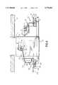
- FIG. 1is a top view of a stereotactic x-ray imaging system with integrated ultrasound imaging and biopsy components combinatively defining one embodiment of the present invention with a central patient/table portion cutaway to show key components.
- FIG. 2is a partial end cross-sectional view of the embodiment of FIG. 1 cut along AA.
- FIG. 3is a partial side cross-sectional view of the embodiment of FIG. 1 cut along BB.
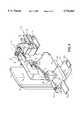
- FIG. 4is a perspective view of the immobilization, ultrasound imaging and biopsy assemblies of the embodiment of FIG. 1.
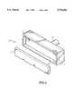
- FIG. 5is a perspective view of an ultrasound imaging head employable in the present invention.
- FIG. 6illustrates spatially correlated x-ray and ultrasound images of a potential breast lesion/suspicious mass obtainable with the present invention.
- FIGS. 1-6illustrate a diagnostic ultrasound/x-ray biopsy system embodiment of the present invention, as adapted for mammography/breast biopsy use.
- the systemcomprises a support assembly 10 having a patient table 12 with breast-opening 14 therethrough, an immobilization assembly 30 for immobilizing a patient's breast within a predetermined XYZ frame of reference under the opening 14 of table 12, an x-ray imaging assembly 40 for providing two-dimensional x-ray images (e.g., X-Y images) of the patient's immobilized breast in correlated spatial relation to the predetermined XYZ frame of reference, and an ultrasound imaging assembly 100 for providing orthogonal depth-profile images (e.g., X-Z, Y-Z and/or X,Y-Z images) of the immobilized breast in correlated spatial relation to the predetermined XYZ frame of reference.
- an immobilization assembly 30for immobilizing a patient's breast within a predetermined XYZ frame of reference under the opening 14 of table 12
- an x-ray imaging assembly 40for providing two-dimensional x-ray images (e.g., X-Y images) of the patient's immobil
- a biopsy assembly 50 having puncture instrument 52is also provided for obtaining samples from a patient's breast while the breast is immobilized in the predetermined XYZ frame of reference.
- a display/processor assembly 60is provided for recording/displaying the various images obtained/generated, for determining the coordinates of a user-identified location of interest within the breast and for monitoring/controlling/simulating the position of the various positionable assembly components.
- the illustrated embodimentmay utilize the x-ray, automated biopsy and other functionalities embodied in the current MAMMOTEST® and MAMMOVISION® products of Fischer Imaging Corp. of Denver, Colo., U.S.A.
- the present inventionallows for the integration and effective use of ultrasound imaging with such products, thereby allowing medical equipment cost efficiencies to be realized.
- the MAMMOTEST® and MAMMOVISION® productsinclude features corresponding with the disclosures in U.S. Pat. Nos. 5,078,142, 5,240,011 and 5,415,169, which are incorporated by reference in their entirety.
- Support assembly 10further includes pedestal 16 and cantilevered first and second support arms 20 and 22, respectively, for supportably interfacing the breast immobilization assembly 30, x-ray imaging assembly 40, ultrasound imaging assembly 100 and biopsy assembly 50 in a predetermined spatially correlated manner.
- First and second supports arms 20 and 22can be jointly pivoted relative to pedestal 16, thereby providing imaging/biopsy access to the breast from different directions (e.g., 0°, +90° and -90° relative to the table longitudinal axis).
- second support arm 22can be selectively pivoted relative to first support arm 20, to provide for stereotactic x-ray imaging (e.g., +15° and -15° relative to the first support arm longitudinal axis).
- Breast immobilization assembly 30is supported on first support arm 20 and includes a stationary face plate 32 and opposing compression paddle 34 for immobilizing a patient's breast therebetween.
- Compression paddle 34is x-ray transmittent and further includes a window 36 for direct breast access by the ultrasound imaging assembly 100 and/or biopsy assembly 50.
- Compression paddle 34is selectively positionable along first support arm 20 (e.g., via motorized and position sensor systems) for controlled, registered movement toward/away from face plate 32 to accommodate breast positioning/removal and differing breast sizes.
- Compression paddle 34can be readily removed from/interconnected to the first support arm 20 to accommodate the selective use of compression paddles of differing sizes, shapes, window positions, etc. As shown in FIG.
- compression assembly 30may further include selectively advanceable/retractable auxiliary side paddles 38, each having optional openings for breast access (e.g., by a puncture instrument or an ultrasound imaging head) for further compression/breast immobilization within the predetermined XYZ frame of reference, and particularly during use of biopsy assembly 50.
- compression paddle 34 and face plate 32are intended to define a breast imaging area of substantially common thickness and to immobilize such area during imaging/biopsy procedures, and to otherwise provide direct access to the breast for targeted ultrasound imaging/biopsy procedures.
- X-ray imaging assembly 40includes x-ray tube source 42 mounted on the end of second support arm 22 and x-ray receiver/imager 44 mounted on first support arm 20.
- x-ray tube source 42provides x-ray radiation having a center axis C substantially perpendicular to the fronts of face plate 34 and x-ray receiver/imager 44, such x-ray radiation having a focal point positioned along the center axis C at a determinable location between the face plate 32 and compression paddle 34 during use.
- the predetermined XYZ frame of referencecan be defined in the illustrated embodiment in relation to an X-Y plane corresponding with the front surface of the face plate 32 and/or parallel back surface of compression paddle 34, together with orthogonal X-Z and Y-Z planes within which the x-ray radiation center axis passes (i.e., all three planes being orthogonal).
- X-ray opaque markingscan be provided on compression paddle 34 and/or face plate 32 to facilitate spatial correlation of the radiation center axes and x-ray receiver/imager.
- the x-ray receiver/imager 44is disposed in abutting relation with the face plate 32.
- X-ray receiver/imager 44may comprise an image receptor consisting of a removable radiographic film cassette (e.g., for full-field breast imaging) and/or digital camera (e.g., for partial field, real-time imaging/display).
- a partial field, digital CCD camera 46e.g., having a 5 mm ⁇ 10 mm or 5 mm ⁇ 5 mm imaging area
- ultrasound imaging assembly 100 and biopsy assembly 50are selectively and alternatively connectable to opposing sides of first support arm 20 via connection/locking handles 102 and 55, respectively. Additionally, biopsy assembly 50 may be mounted in an axially aligned manner on first support arm 20 for breast access through window 36.
- a reference, or "home,” position for each assembly in a given mounted locationis known relative to the predetermine XYZ frame of reference. Further, positioning of the various components of each assembly during use is automatically determinable via position sensor/control systems. As will be appreciated, such positioning can be automated via corresponding processor-controlled, servo motors.
- Biopsy assembly 50comprises a punction sub-assembly 54, which includes puncture instrument 52, and positioner sub-assembly 56.
- Positioner sub-assembly 56includes horizontal axis and vertical control motors 58 and 60, respectively, for selective sideward movement and upward angulation of the punction instrument 52.
- punction sub-assembly 56may comprise the AUTOGUIDETM assembly of Fischer Imaging Corporation.
- the illustrated embodimentmay be particularly apt for use with punction subassemblies for obtaining samples having relatively large cross-sections, including, for example, the MAMMOTOME® from Biopsys Medical, Inc. of Irvine, Calif.
- Ultrasound imaging assembly 100comprises an ultrasound imaging head, or probe, 110 interconnected to arm assembly 130 and, in turn, to XYZ ultrasound positioning assembly 140.
- XYZ ultrasound positioning assembly 140is employed to selectively position ultrasound imaging head 110 through the window 36 of compression paddle 34 to establish direct breast contact for targeted ultrasound imaging in determinable spatial relation to the predetermined XYZ frame of reference.
- ultrasound probe 110may include an elongated housing 112 with an elongated ultrasound transducer module 114 positioned therein.
- Ultrasound transducer module 114provides an ultrasound signal having a focal point on a signal center axis at a location between compression paddle 34 and face plate 32.
- Ultrasound transducer module 114may include, for example, a phased linear array of ultrasound transducers positioned along a longitudinal axis of the ultrasound probe 110.
- the ultrasound probe 110emits signal pulses and detects corresponding echo pulses to generate the depth-profile images.
- detected echo pulseswill result from ultrasound transmissivity differences (i.e., ultrasound impedance mismatches) at tissue-type transition areas (e.g., transitions between healthy tissue and a potential lesion/suspicious mass) and at structural obstructions (e.g., the front surface of face plate 32).
- the housing 112 of ultrasound probe 110may include a recess 118 (exaggerated in FIG. 5) for receiving a cold-pack 120 for orthogonal application to a biopsy site after a biopsy procedure. Applying pressure and a cold medium directly over a biopsy site in the breast has been shown to reduce hematoma bleeding and bruising.
- XYZ ultrasound positioning assembly 140includes X, Y and Z platforms 142, 146 and 148, respectively, mounted for selective, registered movement on corresponding support members 152, 156 and 158 relative to the predetermined XYZ frame of reference.
- XYZ positioning assembly 140may include internal X, Y and Z optical position encoders.
- XYZ positioning assembly 140can further include X, Y and Z motor drives for automatic, selective positioning of ultrasound imaging head 110 in registered XYZ relation to the predetermined XYZ frame of reference.
- the XYZ positioning assembly 140may also include counterbalance and electro-lock components to accommodate ready manual positioning and to maintain a selected ultrasound imaging/biopsy position, respectively.
- Arm assembly 130is provided to allow the ultrasound imaging probe 110 to be rotated about one or more of selected X, Y and Z axes to obtain a desired pitch, roll and/or yaw orientation).
- arm assembly 130can be controlled to selectively rotate the longitudinal axis, or pitch, of probe 110 so that the ultrasound signal may be employed to obtain depth-profile image in a plane, or "slice," within which an upwardly angled punction instrument 52 of biopsy assembly 50 may be orthogonally advanced, as will be further discussed.
- arm assembly 130includes pivot arm 132 pivotally interconnected to XYZ ultrasound positioning assembly 140 via a lock/release mechanism (not shown) for selective, centered rotation of probe 116 about axis YY.
- Arm assembly 130further includes arm 134 rotably interconnected to arm 132 via a lock/release mechanism (not shown) for selective, centered rotation of probe 116 about axis XX, and arm 136 rotably interconnected to arm 134 via a lock/release mechanism (not shown) for selective, centered rotation of probe 116 about axis ZZ.
- Internal optical encodersmay be provided at the various arm interconnections, wherein the pitch, roll and/or yaw of probe 110 is automatically determinable in relation to the predetermined XYZ frame of reference.
- internal automated micro-positionersmay also be utilized under processor control.
- the ultrasound signalmay be scanned to obtain depth-profile information for a predetermined layer, or "slice," within the region of interest.
- scanningmay be provided electrically by driving a phased linear array of transducers comprising probe 110 in a known manner and/or via manual or automatic-driven control of XYZ positioning assembly 140 to mechanically move ultrasound imaging head 110.
- display/processor 60includes a display screen 62 for displaying the acquired x-ray images on a first portion 62a and displaying corresponding depth-profile ultrasound images on a second portion 62b, each in registered co-relation to the predetermined XYZ frame of reference.
- Display/processor 60may further include a user interface means 64, e.g., keyboard 65 and mouse 66 and screen point cursor 68 (e.g., on both display portions 62a, 62b), wherein a user may identify (e.g., click upon) a specific location-of-interest within both an x-ray image and corresponding ultrasound image (e.g., corresponding with a potential lesion or suspicious mass), e.g, for automatic processor determination of the three-dimensional coordinates of the location within the predetermined XYZ frame of reference.
- a user interface means 64e.g., keyboard 65 and mouse 66 and screen point cursor 68 (e.g., on both display portions 62a, 62b)
- a usermay identify (e.g., click upon) a specific location-of-interest within both an x-ray image and corresponding ultrasound image (e.g., corresponding with a potential lesion or suspicious mass), e.g, for automatic processor determination of the three-dimensional coordinates
- User interface meansmay further allow for user selection/display of a particular desired ultrasound depth-profile image, e.g., via mouse 66 and screen "slice" cursor 70 on the x-ray image display portion 62a. More particularly, screen "slice" cursor 70 may be employed to identify a particular slice, or layer, of an X-Y x-ray image for which a corresponding ultrasound depth-profile image is to be obtained (e.g., thereby resulting in processor-assisted positioning and imaging using probe 110) and/or accessed and displayed (e.g., where such ultrasound depth-profile image has been previously obtained/stored for selective subsequent review).
- screen "slice" cursor 70may be employed to identify a particular slice, or layer, of an X-Y x-ray image for which a corresponding ultrasound depth-profile image is to be obtained (e.g., thereby resulting in processor-assisted positioning and imaging using probe 110) and/or accessed and displayed (e.g., where such ultrasound depth-profile
- display/processor 60may be operatively interconnected (e.g., via electrical lines 80) to the various positionable assembly components for monitoring/controlling their respective positions relative to the predetermined XYZ frame of reference, including the positionable components of immobilization assembly 30, x-ray imaging assembly 40, ultrasound imaging assembly 110 and biopsy assembly 50.
- display/processor 60may determine the three-dimensional coordinates of a specific location of interest, as described above, and in turn assist/control the positioning of biopsy assembly 50 so as to position the assembly for obtainment of a tissue sample from the location of interest.
- the display/processor 60may also be employable to visually project, or simulate, the entry of a punction instrument 52 into a given location of interest, thereby allowing physicians the opportunity to insure an optimal positioning for biopsy entry prior to an actual biopsy procedure. Since three-dimensional visualization of a potential lesion/suspicious mass can be provided by the present invention, and since the disclosed arrangement allows for breast access by biopsy assembly 50 from a plurality of aspects (e.g., by selective mounting on either side of or coaxial along support arm 20), such simulated biopsy modeling may prone to be of particular advantage.
- the present inventionallows for spatial correlation of the x-ray and ultrasound images utilizing various techniques.
- the X-Y x-ray images obtainedcan be readily correlated to the predetermined XYZ frame of reference since the position and attributes of x-ray source 42 and x-ray receiver/imager 44 are each known in relation to the predetermined XYZ frame of reference.
- the two X-Y stereotactic x-ray imagescan be employed to obtain a Z location for particular location of interest relative to the predetermined XYZ frame of reference utilizing known triangulation techniques, as will be appreciated by those skilled in the art.
- the XYZ positioning of ultrasound imaging head 110is determinable relative to the predetermined XYZ frame of reference, as noted above.
- the ultrasound imaging head 110will emit/detect ultrasound signals in substantially the same plane as the surface of compression paddle 34 contacting the imaged breast.
- the position of such surface relative to the predetermined XYZ frame of referencee.g., the Z distance to face plate 32
- a depth profilecomprising a potential lesion/suspicious mass can be spatially related in a reliable manner to the acquired x-ray images.
- a patientcan be positioned on the table 12 with a breast positioned through opening 14.
- Compression paddle 34is then advanced along first support arm 20 to compress the breast to define a cross-sectional imaging area having a common thickness and to otherwise immobilize the breast in a set position within the predetermined XYZ frame of reference.
- X-ray imaging assembly 40is then selectively positioned to obtain the desired x-ray images. Such x-ray images are then reviewed using display/processor 60, to identify, analyze and or otherwise confirm the presence and location of a potential lesion or suspicious mass for ultrasound imaging. Alternatively, the general location of a potential lesion or suspicious mass may already be known due to prior x-ray screening.
- the patientshould be positioned/repositioned so that the potential lesion or suspicious mass is positioned within the accessible field of view of ultrasound imaging head 110 when it is maneuvered through the window 36 of compression paddle 34 in direct contact with the imaged breast.
- new x-ray and corresponding ultrasound imagesshould be generated for each position in which a breast is immobilized within the predetermined XYZ frame of reference.
- ultrasound imaging probe 110is positioned through the window 36 and a series of ultrasound images are obtained and displayed on display/processor 60.
- Cursor 66 control of the ultrasound images taken across the area of interestprovides additional, valuable information as to the type of potential lesion/suspicious mass originally noted on an original mammogram. For example, with proper training of ultrasound and x-ray imaging techniques, physicians may rule out the possibility of a solid mass in favor of a fluid-filled cyst. Or, additional ultrasound characteristics may aid the physician in making a definitive diagnosis.
- the specific location from which tissue is to be obtainedcan be identified using mouse 66 to position screen point cursor 68 on both the x-ray image and correlated ultrasound depth-profile image on display/processor 60. Three-dimensional coordinates can then be determined and utilized by display/processor 60 to control positioning of biopsy assembly 50.
- specific attributes of the particular punction subassembly 54 utilizedwill have been previously entered into by display/processor 60.
- display/processor 60may be employed to simulate the advancement of punction instrument 52 into the breast from a given potential position, thereby allowing the physician to determine if breast biopsy access from a different position may be more desirable.
- the ultrasound imaging head 110may be utilized to provide continuous, successive depth profile images, thereby allowing for real-time monitoring and user control of the advancement of the punction instrument 52 into the breast. More particularly, when the punction instrument is positioned at an angle ⁇ as illustrated in FIG. 2, ultrasound imaging head 110 may be similarly angled at ⁇ (e.g., relative to horizontal) so as to yield real-time ultrasound depth-profile images of the layer into which punction instrument 52 is advanced. After biopsy procedures are completed, ultrasound imaging head 110 may be repositioned so as to allow for pressure application of a cold pack 120.
Landscapes
- Health & Medical Sciences (AREA)
- Life Sciences & Earth Sciences (AREA)
- Engineering & Computer Science (AREA)
- Medical Informatics (AREA)
- Surgery (AREA)
- Heart & Thoracic Surgery (AREA)
- Animal Behavior & Ethology (AREA)
- Nuclear Medicine, Radiotherapy & Molecular Imaging (AREA)
- Veterinary Medicine (AREA)
- Pathology (AREA)
- Public Health (AREA)
- Biomedical Technology (AREA)
- General Health & Medical Sciences (AREA)
- Molecular Biology (AREA)
- Radiology & Medical Imaging (AREA)
- Biophysics (AREA)
- Physics & Mathematics (AREA)
- High Energy & Nuclear Physics (AREA)
- Optics & Photonics (AREA)
- Oral & Maxillofacial Surgery (AREA)
- Dentistry (AREA)
- Neurosurgery (AREA)
- Computer Vision & Pattern Recognition (AREA)
- Apparatus For Radiation Diagnosis (AREA)
- Ultra Sonic Daignosis Equipment (AREA)
Abstract
Description
Claims (18)
Priority Applications (8)
| Application Number | Priority Date | Filing Date | Title |
|---|---|---|---|
| US08/730,107US5776062A (en) | 1996-10-15 | 1996-10-15 | Enhanced breast imaging/biopsy system employing targeted ultrasound |
| AU49009/97AAU4900997A (en) | 1996-10-15 | 1997-10-14 | Enhanced breast imaging/biopsy system employing targeted ultrasound |
| PCT/US1997/018468WO1998016149A2 (en) | 1996-10-15 | 1997-10-14 | Enhanced breast imaging/biopsy system employing targeted ultrasound |
| EP97911697AEP0936889B8 (en) | 1996-10-15 | 1997-10-14 | Enhanced breast imaging/biopsy system employing targeted ultrasound |
| DE69739947TDE69739947D1 (en) | 1996-10-15 | 1997-10-14 | ARRANGEMENT FOR IMAGING AND BIOPSYING THE MOM WITH FOCUSED ULTRASOUND |
| US09/111,094US6102866A (en) | 1996-10-15 | 1998-07-06 | Enhanced breast imaging/biopsy system employing targeted ultrasound |
| US10/260,719US7496398B2 (en) | 1996-10-15 | 2002-09-27 | Spatially correlated x-ray and ultrasound mammographic imaging systems and method |
| US12/365,918US20090143674A1 (en) | 1996-10-15 | 2009-02-05 | User interface system for mammographic imager |
Applications Claiming Priority (1)
| Application Number | Priority Date | Filing Date | Title |
|---|---|---|---|
| US08/730,107US5776062A (en) | 1996-10-15 | 1996-10-15 | Enhanced breast imaging/biopsy system employing targeted ultrasound |
Related Child Applications (1)
| Application Number | Title | Priority Date | Filing Date |
|---|---|---|---|
| US09/111,094Continuation-In-PartUS6102866A (en) | 1996-10-15 | 1998-07-06 | Enhanced breast imaging/biopsy system employing targeted ultrasound |
Publications (1)
| Publication Number | Publication Date |
|---|---|
| US5776062Atrue US5776062A (en) | 1998-07-07 |
Family
ID=24933944
Family Applications (3)
| Application Number | Title | Priority Date | Filing Date |
|---|---|---|---|
| US08/730,107Expired - LifetimeUS5776062A (en) | 1996-10-15 | 1996-10-15 | Enhanced breast imaging/biopsy system employing targeted ultrasound |
| US09/111,094Expired - LifetimeUS6102866A (en) | 1996-10-15 | 1998-07-06 | Enhanced breast imaging/biopsy system employing targeted ultrasound |
| US12/365,918AbandonedUS20090143674A1 (en) | 1996-10-15 | 2009-02-05 | User interface system for mammographic imager |
Family Applications After (2)
| Application Number | Title | Priority Date | Filing Date |
|---|---|---|---|
| US09/111,094Expired - LifetimeUS6102866A (en) | 1996-10-15 | 1998-07-06 | Enhanced breast imaging/biopsy system employing targeted ultrasound |
| US12/365,918AbandonedUS20090143674A1 (en) | 1996-10-15 | 2009-02-05 | User interface system for mammographic imager |
Country Status (5)
| Country | Link |
|---|---|
| US (3) | US5776062A (en) |
| EP (1) | EP0936889B8 (en) |
| AU (1) | AU4900997A (en) |
| DE (1) | DE69739947D1 (en) |
| WO (1) | WO1998016149A2 (en) |
Cited By (74)
| Publication number | Priority date | Publication date | Assignee | Title |
|---|---|---|---|---|
| US5983123A (en) | 1993-10-29 | 1999-11-09 | United States Surgical Corporation | Methods and apparatus for performing ultrasound and enhanced X-ray imaging |
| WO2000009014A1 (en)* | 1998-08-17 | 2000-02-24 | Kari Richter | Combined ultrasound-radiography apparatuses |
| US6110112A (en)* | 1998-03-06 | 2000-08-29 | Siemens Aktiengesellschaft | Medical guide apparatus for breath-coordinated puncturing of the body or a body cavity |
| WO2000065989A1 (en)* | 1999-05-03 | 2000-11-09 | Biotrack, Inc. | Systems and methods for targeting a breast lesion |
| US6254538B1 (en)* | 1996-08-15 | 2001-07-03 | Life Imaging Systems, Inc. | System and process for performing percutaneous biopsy within the breast using three-dimensional ultrasonography |
| WO2001089384A3 (en)* | 2000-05-22 | 2002-03-21 | Wisconsin Alumni Res Found | Combined ultrasound-radionuclide device for percutaneous ultrasound-guided biopsy and method of use |
| US6396940B1 (en) | 1999-05-27 | 2002-05-28 | Litton Systems, Inc. | Optical correlator based automated pathologic region of interest selector for integrated 3D ultrasound and digital mammography |
| US6478739B1 (en) | 2001-05-11 | 2002-11-12 | The Procter & Gamble Company | Ultrasonic breast examination system |
| US6491632B1 (en)* | 2001-06-26 | 2002-12-10 | Geoffrey L. Taylor | Method and apparatus for photogrammetric orientation of ultrasound images |
| US20030007598A1 (en)* | 2000-11-24 | 2003-01-09 | U-Systems, Inc. | Breast cancer screening with adjunctive ultrasound mammography |
| US20030045798A1 (en)* | 2001-09-04 | 2003-03-06 | Richard Hular | Multisensor probe for tissue identification |
| US20030073895A1 (en)* | 1996-10-15 | 2003-04-17 | Nields Morgan W. | User interface system for mammographic imager |
| WO2003032837A1 (en)* | 2001-10-12 | 2003-04-24 | University Of Florida | Computer controlled guidance of a biopsy needle |
| US6558337B2 (en)* | 2001-04-17 | 2003-05-06 | Wisconsin Alumni Research Foundation | Positioner for medical devices such as biopsy needles |
| US6574499B1 (en) | 1998-11-25 | 2003-06-03 | Xdata Corporation | Mammography method and apparatus |
| FR2835421A1 (en)* | 2002-02-01 | 2003-08-08 | Gen Electric | METHODS, SYSTEM AND DEVICE FOR DIGITAL IMAGING. |
| US20030181801A1 (en)* | 2002-03-08 | 2003-09-25 | Imperium, Inc. | Apparatus for multimodal plane wave ultrasound imaging |
| US20030194048A1 (en)* | 2002-04-15 | 2003-10-16 | General Electric Company | Reprojection and backprojection methods and algorithms for implementation thereof |
| US20030194051A1 (en)* | 2002-04-15 | 2003-10-16 | General Electric | Tomosynthesis X-ray mammogram system and method with automatic drive system |
| US20030194121A1 (en)* | 2002-04-15 | 2003-10-16 | General Electric Company | Computer aided detection (CAD) for 3D digital mammography |
| US20030194115A1 (en)* | 2002-04-15 | 2003-10-16 | General Electric Company | Method and apparatus for providing mammographic image metrics to a clinician |
| US20030194050A1 (en)* | 2002-04-15 | 2003-10-16 | General Electric Company | Multi modality X-ray and nuclear medicine mammography imaging system and method |
| US20030199785A1 (en)* | 2002-04-23 | 2003-10-23 | Ethicon Endo-Surgery | Localization mechanism for an MRI compatible biopsy device |
| US20030215057A1 (en)* | 2002-05-15 | 2003-11-20 | General Electric Company | Scatter correction method for non-stationary X-ray acquisitions |
| US20040022447A1 (en)* | 2002-07-31 | 2004-02-05 | General Electric Company | Method and system for image compression and decompression using span of interest of an imaging sequence |
| US6707878B2 (en) | 2002-04-15 | 2004-03-16 | General Electric Company | Generalized filtered back-projection reconstruction in digital tomosynthesis |
| US20040068170A1 (en)* | 2000-11-24 | 2004-04-08 | U-Systems Inc.(Vii) | Breast cancer screening with ultrasound image overlays |
| US20040077972A1 (en)* | 2002-10-18 | 2004-04-22 | Mark Tsonton | Localization mechanism for an MRI compatible biopsy device |
| US20040082856A1 (en)* | 2002-07-16 | 2004-04-29 | Alfred E. Mann Institute For Biomedical Engineering, University Of Southern California | Support bra for ultrasonic breast scanner |
| US6731966B1 (en) | 1997-03-04 | 2004-05-04 | Zachary S. Spigelman | Systems and methods for targeting a lesion |
| WO2004051405A3 (en)* | 2002-11-27 | 2004-08-19 | U Systems Inc | Full-field breast image data processing and archiving |
| US20040249283A1 (en)* | 2001-05-09 | 2004-12-09 | Edward Kantorovich | Method and apparatus for breast imaging utilizing ultrasound |
| US6846289B2 (en) | 2003-06-06 | 2005-01-25 | Fischer Imaging Corporation | Integrated x-ray and ultrasound medical imaging system |
| US20050089205A1 (en)* | 2003-10-23 | 2005-04-28 | Ajay Kapur | Systems and methods for viewing an abnormality in different kinds of images |
| US20050165299A1 (en)* | 2004-01-23 | 2005-07-28 | Traxyz Medical, Inc. | Methods and apparatus for performing procedures on target locations in the body |
| US20060074287A1 (en)* | 2004-09-30 | 2006-04-06 | General Electric Company | Systems, methods and apparatus for dual mammography image detection |
| US20060084868A1 (en)* | 2003-01-17 | 2006-04-20 | Hee-Boong Park | Apparatus for ultrasonic examination of deformable object |
| US7035450B1 (en)* | 1996-07-09 | 2006-04-25 | Ge Medical Systems Sa | Method for locating an element of interest contained in a three-dimensional object, in particular during a stereotactic investigation in the X-ray examination of the breast |
| US20060159318A1 (en)* | 2003-11-26 | 2006-07-20 | Alyassin Abdalmajeid M | Method, system and computer program product for multi-modality registration using virtual cursors |
| US20060173303A1 (en)* | 2000-11-24 | 2006-08-03 | Zengpin Yu | Full-field breast image data processing and archiving |
| US20060251301A1 (en)* | 2004-11-02 | 2006-11-09 | Mcnamara Michael P Jr | Method and apparatus for determining correlation between spatial coordinates in breast |
| US20070167801A1 (en)* | 2005-12-02 | 2007-07-19 | Webler William E | Methods and apparatuses for image guided medical procedures |
| EP1977692A2 (en) | 2007-04-05 | 2008-10-08 | Kabushiki Kaisha Toshiba | Ultrasonic diagnosis apparatus, breast imaging system, and breast imaging method |
| WO2007130526A3 (en)* | 2006-05-02 | 2008-10-30 | U Systems Inc | Ultrasound scanning and ultrasound- assisted biopsy |
| US20090143674A1 (en)* | 1996-10-15 | 2009-06-04 | Nields Morgan W | User interface system for mammographic imager |
| US7615008B2 (en) | 2000-11-24 | 2009-11-10 | U-Systems, Inc. | Processing and displaying breast ultrasound information |
| WO2009050712A3 (en)* | 2007-10-15 | 2010-03-11 | Yissum Research Development Company Of The Hebrew Univercity Of Jerusalem | Method, system and computer program product for tissue characterization |
| US20110015542A1 (en)* | 2002-04-23 | 2011-01-20 | Devicor Medical Products, Inc. | Mri compatible biopsy device with detachable probe |
| US20110087098A1 (en)* | 2008-02-20 | 2011-04-14 | Siemens Aktiengesellschaft | Mammography system and method for sonographic and radiographic examination of a breast |
| US8133242B1 (en) | 2007-04-27 | 2012-03-13 | Q-Tech Medical Incorporated | Image-guided extraluminal occlusion |
| US8211121B1 (en) | 2010-03-06 | 2012-07-03 | Q-Tech Medical Incorporated | Methods and apparatus for image-guided extraluminal occlusion using clamping jaws |
| US20120238875A1 (en)* | 2004-11-30 | 2012-09-20 | Eric Savitsky | Embedded Motion Sensing Technology for Integration within Commercial Ultrasound Probes |
| DE102011006058A1 (en)* | 2011-03-24 | 2012-09-27 | Siemens Aktiengesellschaft | Tomosynthesis device and method for its operation |
| US8750568B2 (en) | 2012-05-22 | 2014-06-10 | Covidien Lp | System and method for conformal ablation planning |
| US9271686B2 (en) | 2013-03-14 | 2016-03-01 | West Virginia University | Endorectal prostate probe composed of a combined mini gamma camera and ultrasound sensor |
| US9439623B2 (en) | 2012-05-22 | 2016-09-13 | Covidien Lp | Surgical planning system and navigation system |
| US9439622B2 (en) | 2012-05-22 | 2016-09-13 | Covidien Lp | Surgical navigation system |
| US9439627B2 (en) | 2012-05-22 | 2016-09-13 | Covidien Lp | Planning system and navigation system for an ablation procedure |
| US9498182B2 (en) | 2012-05-22 | 2016-11-22 | Covidien Lp | Systems and methods for planning and navigation |
| US9950194B2 (en) | 2014-09-09 | 2018-04-24 | Mevion Medical Systems, Inc. | Patient positioning system |
| US10372876B2 (en)* | 2017-01-20 | 2019-08-06 | Agfa Healthcare Inc. | System and method for providing breast image data |
| US10568560B2 (en) | 2013-03-14 | 2020-02-25 | West Virginia University | Endorectal prostate probe with combined PET and US modalities |
| US10726741B2 (en) | 2004-11-30 | 2020-07-28 | The Regents Of The University Of California | System and method for converting handheld diagnostic ultrasound systems into ultrasound training systems |
| US11013492B1 (en)* | 2020-11-04 | 2021-05-25 | Philip B. Kivitz | Ultrasound sonographic imaging system and method |
| US11315439B2 (en) | 2013-11-21 | 2022-04-26 | SonoSim, Inc. | System and method for extended spectrum ultrasound training using animate and inanimate training objects |
| US11495142B2 (en) | 2019-01-30 | 2022-11-08 | The Regents Of The University Of California | Ultrasound trainer with internal optical tracking |
| US11600201B1 (en) | 2015-06-30 | 2023-03-07 | The Regents Of The University Of California | System and method for converting handheld diagnostic ultrasound systems into ultrasound training systems |
| US11631342B1 (en) | 2012-05-25 | 2023-04-18 | The Regents Of University Of California | Embedded motion sensing technology for integration within commercial ultrasound probes |
| US11627944B2 (en) | 2004-11-30 | 2023-04-18 | The Regents Of The University Of California | Ultrasound case builder system and method |
| US11707329B2 (en) | 2018-08-10 | 2023-07-25 | Covidien Lp | Systems and methods for ablation visualization |
| US11749137B2 (en) | 2017-01-26 | 2023-09-05 | The Regents Of The University Of California | System and method for multisensory psychomotor skill training |
| US11810473B2 (en) | 2019-01-29 | 2023-11-07 | The Regents Of The University Of California | Optical surface tracking for medical simulation |
| US20240299005A1 (en)* | 2023-03-10 | 2024-09-12 | Fujifilm Corporation | Control apparatus, control method, and control program |
| US12399923B1 (en) | 2023-09-15 | 2025-08-26 | Gabriele Nataneli | Multi-modal enhancement of large language models without retraining |
Families Citing this family (87)
| Publication number | Priority date | Publication date | Assignee | Title |
|---|---|---|---|---|
| FR2754612B1 (en)* | 1996-10-10 | 1998-11-20 | Ge Medical Syst Sa | SYSTEM FOR LOCATING DIGITAL IMAGE TAPE CASSETTES |
| AU2003203755B2 (en)* | 1998-11-25 | 2005-04-28 | Rubicor Medical, Inc. | Breast Stabilization Devices and Imaging and Interventional Methods Using Same |
| US6421454B1 (en)* | 1999-05-27 | 2002-07-16 | Litton Systems, Inc. | Optical correlator assisted detection of calcifications for breast biopsy |
| JP4133317B2 (en)* | 2000-07-07 | 2008-08-13 | メディカル ポジショニング アイエヌシー. | Patient support device and recumbent chest biopsy method |
| US6367104B1 (en)* | 2000-07-07 | 2002-04-09 | Medical Positioning, Inc. | Patient support apparatus and method for performing decubitus breast biopsy |
| US6678546B2 (en)* | 2001-01-30 | 2004-01-13 | Fischer Imaging Corporation | Medical instrument guidance using stereo radiolocation |
| US7443949B2 (en) | 2001-10-19 | 2008-10-28 | Hologic, Inc. | Mammography system and method employing offset compression paddles, automatic collimation, and retractable anti-scatter grid |
| US7609806B2 (en)* | 2004-10-18 | 2009-10-27 | Hologic Inc. | Mammography system and method employing offset compression paddles, automatic collimations, and retractable anti-scatter grid |
| EP1531730B1 (en) | 2002-05-31 | 2012-01-18 | U-Systems, Inc. | Apparatus for acquiring ultrasound scans of a breast |
| US8083678B2 (en)* | 2003-04-16 | 2011-12-27 | Eastern Virginia Medical School | System, method and medium for acquiring and generating standardized operator independent ultrasound images of fetal, neonatal and adult organs |
| MXPA05011120A (en)* | 2003-04-16 | 2005-12-15 | Eastern Viriginai Medical Scho | System and method for generating operator independent ultrasound images. |
| DE102004034240A1 (en)* | 2004-07-15 | 2006-02-16 | Siemens Ag | Medical imaging device |
| ES2253997B1 (en)* | 2004-07-29 | 2007-07-16 | Udiat Centre Diagnostic, S.A. | DIGITAL SYSTEM TO PERFORM STEREOTAXIC BIOPSY. |
| EP1804668B1 (en)* | 2004-10-18 | 2012-05-23 | Mobile Robotics Sweden AB | Robot for ultrasonic examination |
| EP1816965B1 (en) | 2004-11-26 | 2016-06-29 | Hologic, Inc. | Integrated multi-mode mammography/tomosynthesis x-ray system |
| DE102005039658B3 (en)* | 2005-08-22 | 2007-07-19 | Siemens Ag | Laser device for a mammography device |
| WO2007095330A2 (en) | 2006-02-15 | 2007-08-23 | Hologic Inc | Breast biopsy and needle localization using tomosynthesis systems |
| US20070282221A1 (en)* | 2006-06-02 | 2007-12-06 | U-Systems, Inc. | Ultrasound assisted and x-ray assisted biopsy devices |
| SE530549C2 (en)* | 2006-10-31 | 2008-07-08 | Xcounter Ab | System for imaging a breast through computed tomography |
| WO2008054279A1 (en)* | 2006-10-31 | 2008-05-08 | Xcounter Ab | Imaging arrangement and system for imaging |
| US8870771B2 (en) | 2007-05-04 | 2014-10-28 | Barbara Ann Karmanos Cancer Institute | Method and apparatus for categorizing breast density and assessing cancer risk utilizing acoustic parameters |
| US10201324B2 (en) | 2007-05-04 | 2019-02-12 | Delphinus Medical Technologies, Inc. | Patient interface system |
| US8066644B2 (en)* | 2007-05-17 | 2011-11-29 | Vanderbilt University | System, method and device for positioning a target located within soft tissue in a path of an instrument |
| WO2009026587A1 (en)* | 2007-08-23 | 2009-02-26 | Fischer Medical Technologies, Inc. | Improved computed tomography breast imaging and biopsy system |
| US8375054B2 (en)* | 2008-04-03 | 2013-02-12 | Siemens Aktiengesellschaft | Findings navigator |
| US10603008B2 (en)* | 2009-02-19 | 2020-03-31 | Tessonics Corporation | Ultrasonic device for assessment of internal tooth structure |
| JP5373450B2 (en)* | 2009-03-31 | 2013-12-18 | 富士フイルム株式会社 | Biopsy device and method of operating biopsy device |
| US8556815B2 (en)* | 2009-05-20 | 2013-10-15 | Laurent Pelissier | Freehand ultrasound imaging systems and methods for guiding fine elongate instruments |
| US10039527B2 (en) | 2009-05-20 | 2018-08-07 | Analogic Canada Corporation | Ultrasound systems incorporating spatial position sensors and associated methods |
| ES2862525T3 (en) | 2009-10-08 | 2021-10-07 | Hologic Inc | Needle Breast Biopsy System and Method of Use |
| JP5317933B2 (en)* | 2009-11-17 | 2013-10-16 | 富士フイルム株式会社 | Image display device and program thereof |
| EP2528509B1 (en) | 2010-01-29 | 2021-10-13 | University Of Virginia Patent Foundation | Ultrasound for locating anatomy or probe guidance |
| US9144403B2 (en) | 2010-02-12 | 2015-09-29 | Delphinus Medical Technologies, Inc. | Method of characterizing the pathological response of tissue to a treatment plan |
| CN102869301B (en) | 2010-02-12 | 2016-06-29 | 戴尔菲纳斯医疗科技公司 | The method characterizing the tissue of patient |
| US20130281840A1 (en)* | 2010-06-03 | 2013-10-24 | Caperay Medical (Pty) Ltd. | Dual-modality scanning system for detecting breast cancer |
| US20120133600A1 (en) | 2010-11-26 | 2012-05-31 | Hologic, Inc. | User interface for medical image review workstation |
| JP6057922B2 (en) | 2011-03-08 | 2017-01-11 | ホロジック, インコーポレイテッドHologic, Inc. | System and method for dual energy and / or contrast enhanced breast imaging for screening, diagnosis and biopsy |
| WO2012148985A1 (en) | 2011-04-26 | 2012-11-01 | University Of Virginia Patent Foundation | Bone surface image reconstruction using ultrasound |
| US12042134B2 (en) | 2011-09-16 | 2024-07-23 | Hologic, Inc. | Breast biopsy lateral arm system |
| ES2795416T3 (en) | 2011-09-16 | 2020-11-23 | Hologic Inc | Lateral Arm System for Breast Biopsy |
| US11284869B2 (en) | 2011-09-16 | 2022-03-29 | Hologic, Inc. | Breast biopsy lateral arm system |
| EP2782505B1 (en) | 2011-11-27 | 2020-04-22 | Hologic, Inc. | System and method for generating a 2d image using mammography and/or tomosynthesis image data |
| US9295449B2 (en) | 2012-01-23 | 2016-03-29 | Ultrasonix Medical Corporation | Landmarks for ultrasound imaging |
| US8914925B2 (en)* | 2012-02-08 | 2014-12-23 | Wayne County Employees' Retirement System | Mobile diagnostic assembly |
| JP6240097B2 (en) | 2012-02-13 | 2017-11-29 | ホロジック インコーポレイティッド | How to navigate a tomosynthesis stack using composite image data |
| US20130303895A1 (en)* | 2012-05-14 | 2013-11-14 | Delphinus Medical Technologies, Inc. | System and Method for Performing an Image-Guided Biopsy |
| CN102697539B (en)* | 2012-06-27 | 2016-04-27 | 青岛银泰医疗科技有限公司 | A kind of novel three-dimensional Needle localization system |
| US20140073907A1 (en) | 2012-09-12 | 2014-03-13 | Convergent Life Sciences, Inc. | System and method for image guided medical procedures |
| US9763641B2 (en) | 2012-08-30 | 2017-09-19 | Delphinus Medical Technologies, Inc. | Method and system for imaging a volume of tissue with tissue boundary detection |
| EP2895076B1 (en)* | 2012-09-13 | 2020-09-09 | Koninklijke Philips N.V. | Ultrasound imaging device operated by mobile display device and ultrasound imaging system |
| RU2015131134A (en) | 2012-12-28 | 2017-02-02 | Конинклейке Филипс Н.В. | REAL-TIME SCENE MODEL UNITING FORMATION OF THREE-DIMENSIONAL ULTRASONIC AND TWO-DIMENSIONAL X-RAY IMAGES |
| US10123770B2 (en) | 2013-03-13 | 2018-11-13 | Delphinus Medical Technologies, Inc. | Patient support system |
| CN105451657A (en) | 2013-03-15 | 2016-03-30 | 霍罗吉克公司 | System and method for navigating tomosynthesis stack including automatic focusing |
| US10092358B2 (en) | 2013-03-15 | 2018-10-09 | Hologic, Inc. | Tomosynthesis-guided biopsy apparatus and method |
| KR101656776B1 (en)* | 2013-09-03 | 2016-09-12 | 삼성전자주식회사 | Ultrasound diagnostic apparatus and operating method thereof |
| EP3060132B1 (en) | 2013-10-24 | 2019-12-04 | Hologic, Inc. | System and method for navigating x-ray guided breast biopsy |
| JP6506769B2 (en) | 2014-02-28 | 2019-04-24 | ホロジック, インコーポレイテッドHologic, Inc. | System and method for generating and displaying tomosynthesis image slabs |
| WO2015140782A1 (en)* | 2014-03-18 | 2015-09-24 | Doron Kwiat | Biopsy method and clinic for imaging and biopsy |
| EP3148471B1 (en)* | 2014-05-28 | 2021-11-03 | General Electric Company | Method and associated biopsy device |
| US10285667B2 (en) | 2014-08-05 | 2019-05-14 | Delphinus Medical Technologies, Inc. | Method for generating an enhanced image of a volume of tissue |
| US9855014B2 (en) | 2014-12-16 | 2018-01-02 | General Electric Company | Compression paddle for use in breast imaging |
| US9949719B2 (en) | 2014-12-16 | 2018-04-24 | General Electric Company | Breast imaging method and system |
| US10786224B2 (en) | 2016-04-21 | 2020-09-29 | Covidien Lp | Biopsy devices and methods of use thereof |
| EP3600047A1 (en) | 2017-03-30 | 2020-02-05 | Hologic, Inc. | System and method for hierarchical multi-level feature image synthesis and representation |
| EP3600052A1 (en) | 2017-03-30 | 2020-02-05 | Hologic, Inc. | System and method for targeted object enhancement to generate synthetic breast tissue images |
| CN110621233B (en) | 2017-03-30 | 2023-12-12 | 豪洛捷公司 | Method for processing breast tissue image data |
| WO2018236565A1 (en) | 2017-06-20 | 2018-12-27 | Hologic, Inc. | METHOD AND SYSTEM FOR MEDICAL IMAGING WITH DYNAMIC SELF-LEARNING |
| DE102017210604A1 (en) | 2017-06-23 | 2018-12-27 | Siemens Healthcare Gmbh | Compression unit for a combined X-ray / ultrasound examination device |
| GB2566942B (en)* | 2017-09-22 | 2020-06-03 | Caperay Medical Pty Ltd | Multimodal imaging system and method |
| EP3510930B1 (en) | 2018-01-15 | 2021-07-14 | Hologic, Inc. | Automated and configurable magnification stand |
| US11331161B2 (en) | 2018-03-23 | 2022-05-17 | Covidien Lp | Surgical assemblies facilitating tissue marking and methods of use thereof |
| CA3098900A1 (en)* | 2018-04-30 | 2019-11-07 | Memorial Sloan Kettering Cancer Center | Compression paddles for breast biopsies |
| EP3787520B1 (en) | 2018-05-04 | 2024-09-25 | Hologic, Inc. | Biopsy needle visualization |
| US12121304B2 (en) | 2018-05-04 | 2024-10-22 | Hologic, Inc. | Introducer and localization wire visualization |
| WO2020068851A1 (en) | 2018-09-24 | 2020-04-02 | Hologic, Inc. | Breast mapping and abnormality localization |
| JP7084291B2 (en) | 2018-12-07 | 2022-06-14 | 富士フイルム株式会社 | Tomosynthesis photography support equipment, methods and programs |
| US11517294B2 (en) | 2019-05-07 | 2022-12-06 | Covidien Lp | Biopsy devices and methods of use thereof |
| US11883206B2 (en) | 2019-07-29 | 2024-01-30 | Hologic, Inc. | Personalized breast imaging system |
| EP4439580A3 (en) | 2019-09-27 | 2024-12-25 | Hologic, Inc. | Ai system for predicting reading time and reading complexity for reviewing 2d/3d breast images |
| EP3832689A3 (en) | 2019-12-05 | 2021-08-11 | Hologic, Inc. | Systems and methods for improved x-ray tube life |
| US11471118B2 (en) | 2020-03-27 | 2022-10-18 | Hologic, Inc. | System and method for tracking x-ray tube focal spot position |
| US11481038B2 (en) | 2020-03-27 | 2022-10-25 | Hologic, Inc. | Gesture recognition in controlling medical hardware or software |
| KR102522627B1 (en)* | 2020-09-17 | 2023-04-17 | 주식회사 제이시스메디칼 | Ultrasonic medical instrument with adjusable focusing depth of ultrasonic wave generator |
| US12254586B2 (en) | 2021-10-25 | 2025-03-18 | Hologic, Inc. | Auto-focus tool for multimodality image review |
| WO2023097279A1 (en) | 2021-11-29 | 2023-06-01 | Hologic, Inc. | Systems and methods for correlating objects of interest |
| US12414217B2 (en) | 2022-02-07 | 2025-09-09 | Hologic, Inc. | Systems and methods for adaptively controlling filament current in an X-ray tube |
| US12251257B2 (en)* | 2022-10-19 | 2025-03-18 | GE Precision Healthcare LLC | System and method for analyzing breast support environment |
Citations (36)
| Publication number | Priority date | Publication date | Assignee | Title |
|---|---|---|---|---|
| US2707662A (en)* | 1949-05-27 | 1955-05-03 | Picker X Ray Corp Waite Mfg | Tiltably X-ray table with extension panel |
| US3165630A (en)* | 1961-06-13 | 1965-01-12 | Polaroid Corp | Table for holding and positioning a female subject and film during breast x-ray exposures |
| US3963933A (en)* | 1975-08-18 | 1976-06-15 | General Electric Company | Mammography fixture |
| US3973126A (en)* | 1975-07-31 | 1976-08-03 | General Electric Company | Mammography |
| US4051380A (en)* | 1976-03-31 | 1977-09-27 | Lasky Harold J | Apparatus and method for supporting and positioning the body to facilitate radiographic mammography procedures |
| US4099880A (en)* | 1976-08-11 | 1978-07-11 | Tsutomu Kano | Method and an apparatus for stereoscopic measurement utilizing a three-dimensional image |
| US4249539A (en)* | 1979-02-09 | 1981-02-10 | Technicare Corporation | Ultrasound needle tip localization system |
| US4341120A (en)* | 1979-11-09 | 1982-07-27 | Diasonics Cardio/Imaging, Inc. | Ultrasonic volume measuring system |
| US4346717A (en)* | 1979-09-07 | 1982-08-31 | Siemens Aktiengesellschaft | Device for punctuating internal body organs, vessels or the like |
| US4485819A (en)* | 1980-01-21 | 1984-12-04 | Wolfgang Igl | Mechanical accessory for commercially available compound apparatuses for echo mammography |
| US4567896A (en)* | 1984-01-20 | 1986-02-04 | Elscint, Inc. | Method and apparatus for calibrating a biopsy attachment for ultrasonic imaging apparatus |
| US4576175A (en)* | 1983-09-06 | 1986-03-18 | Moshe Epstein | Biopsy attachment for ultrasonic probe |
| US4613122A (en)* | 1983-08-19 | 1986-09-23 | Kabushiki Kaisha Toshiba | CT couch apparatus having a lift |
| US4618973A (en)* | 1985-11-01 | 1986-10-21 | Lasky Harold J | Mammographic X-ray apparatus |
| US4625555A (en)* | 1984-02-07 | 1986-12-02 | Terumo Kabushiki Kaisha | Ultrasonic measurement method, and apparatus therefor |
| US4727565A (en)* | 1983-11-14 | 1988-02-23 | Ericson Bjoern E | Method of localization |
| US4750487A (en)* | 1986-11-24 | 1988-06-14 | Zanetti Paul H | Stereotactic frame |
| US4791934A (en)* | 1986-08-07 | 1988-12-20 | Picker International, Inc. | Computer tomography assisted stereotactic surgery system and method |
| US4869247A (en)* | 1988-03-11 | 1989-09-26 | The University Of Virginia Alumni Patents Foundation | Video tumor fighting system |
| US4875478A (en)* | 1987-04-10 | 1989-10-24 | Chen Harry H | Portable compression grid & needle holder |
| US4890311A (en)* | 1987-06-30 | 1989-12-26 | Siemens Aktiengesellschaft | Biopsy means for an x-ray examination apparatus |
| US4930143A (en)* | 1986-09-19 | 1990-05-29 | Bengt Lundgren | Method and device for mammographic stereotactic punction of pathological lesions in the female breast |
| US5078142A (en)* | 1989-11-21 | 1992-01-07 | Fischer Imaging Corporation | Precision mammographic needle biopsy system |
| US5129911A (en)* | 1991-03-11 | 1992-07-14 | Siczek Bernard W | Orbital aiming device |
| US5285772A (en)* | 1991-10-24 | 1994-02-15 | Siemens Aktiengesellschaft | Therapy apparatus for treating a patient with focused acoustic waves |
| US5289520A (en)* | 1991-11-27 | 1994-02-22 | Lorad Corporation | Stereotactic mammography imaging system with prone position examination table and CCD camera |
| US5320111A (en)* | 1992-02-07 | 1994-06-14 | Livingston Products, Inc. | Light beam locator and guide for a biopsy needle |
| US5398690A (en)* | 1994-08-03 | 1995-03-21 | Batten; Bobby G. | Slaved biopsy device, analysis apparatus, and process |
| US5409497A (en)* | 1991-03-11 | 1995-04-25 | Fischer Imaging Corporation | Orbital aiming device for mammo biopsy |
| US5411026A (en)* | 1993-10-08 | 1995-05-02 | Nomos Corporation | Method and apparatus for lesion position verification |
| US5415169A (en)* | 1989-11-21 | 1995-05-16 | Fischer Imaging Corporation | Motorized mammographic biopsy apparatus |
| US5447154A (en)* | 1992-07-31 | 1995-09-05 | Universite Joseph Fourier | Method for determining the position of an organ |
| US5474072A (en)* | 1993-10-29 | 1995-12-12 | Neovision Corporation | Methods and apparatus for performing sonomammography |
| US5526394A (en)* | 1993-11-26 | 1996-06-11 | Fischer Imaging Corporation | Digital scan mammography apparatus |
| US5569266A (en)* | 1991-03-11 | 1996-10-29 | Fischer Imaging Corporation | Magnetic resonance imaging device useful for guiding a medical instrument |
| US5584292A (en)* | 1994-10-31 | 1996-12-17 | Grumman Aerospace Corporation | Digital X-ray camera for precision mammographic needle biopsy system |
Family Cites Families (13)
| Publication number | Priority date | Publication date | Assignee | Title |
|---|---|---|---|---|
| EP0096039A1 (en)* | 1981-12-14 | 1983-12-21 | The Commonwealth Of Australia | Apparatus for ultrasonic examination of deformable objects |
| US4671292A (en)* | 1985-04-30 | 1987-06-09 | Dymax Corporation | Concentric biopsy probe |
| US4899756A (en)* | 1988-07-18 | 1990-02-13 | Sonek Jiri D | Articulated needle guide for ultrasound imaging and method of using same |
| US5240011A (en)* | 1991-11-27 | 1993-08-31 | Fischer Imaging Corporation | Motorized biopsy needle positioner |
| US5662109A (en)* | 1990-12-14 | 1997-09-02 | Hutson; William H. | Method and system for multi-dimensional imaging and analysis for early detection of diseased tissue |
| DE4309597A1 (en)* | 1993-03-22 | 1994-09-29 | Kari Dr Richter | Process for imaging a part of the human body |
| IL107523A (en)* | 1993-11-07 | 2000-01-31 | Ultraguide Ltd | Articulated needle guide for ultrasound imaging and method of using same |
| JP2833456B2 (en)* | 1993-11-22 | 1998-12-09 | 株式会社東芝 | Insertable ultrasound system |
| US5660185A (en)* | 1995-04-13 | 1997-08-26 | Neovision Corporation | Image-guided biopsy apparatus with enhanced imaging and methods |
| US5640956A (en)* | 1995-06-07 | 1997-06-24 | Neovision Corporation | Methods and apparatus for correlating ultrasonic image data and radiographic image data |
| US5769086A (en)* | 1995-12-06 | 1998-06-23 | Biopsys Medical, Inc. | Control system and method for automated biopsy device |
| US5776062A (en)* | 1996-10-15 | 1998-07-07 | Fischer Imaging Corporation | Enhanced breast imaging/biopsy system employing targeted ultrasound |
| EP1143845A4 (en)* | 1998-11-25 | 2004-10-06 | Fischer Imaging Corp | User interface system for mammographic imager |
- 1996
- 1996-10-15USUS08/730,107patent/US5776062A/ennot_activeExpired - Lifetime
- 1997
- 1997-10-14EPEP97911697Apatent/EP0936889B8/ennot_activeExpired - Lifetime
- 1997-10-14DEDE69739947Tpatent/DE69739947D1/ennot_activeExpired - Lifetime
- 1997-10-14WOPCT/US1997/018468patent/WO1998016149A2/enactiveApplication Filing
- 1997-10-14AUAU49009/97Apatent/AU4900997A/ennot_activeAbandoned
- 1998
- 1998-07-06USUS09/111,094patent/US6102866A/ennot_activeExpired - Lifetime
- 2009
- 2009-02-05USUS12/365,918patent/US20090143674A1/ennot_activeAbandoned
Patent Citations (38)
| Publication number | Priority date | Publication date | Assignee | Title |
|---|---|---|---|---|
| US2707662A (en)* | 1949-05-27 | 1955-05-03 | Picker X Ray Corp Waite Mfg | Tiltably X-ray table with extension panel |
| US3165630A (en)* | 1961-06-13 | 1965-01-12 | Polaroid Corp | Table for holding and positioning a female subject and film during breast x-ray exposures |
| US3973126A (en)* | 1975-07-31 | 1976-08-03 | General Electric Company | Mammography |
| US3963933A (en)* | 1975-08-18 | 1976-06-15 | General Electric Company | Mammography fixture |
| US4051380A (en)* | 1976-03-31 | 1977-09-27 | Lasky Harold J | Apparatus and method for supporting and positioning the body to facilitate radiographic mammography procedures |
| US4099880A (en)* | 1976-08-11 | 1978-07-11 | Tsutomu Kano | Method and an apparatus for stereoscopic measurement utilizing a three-dimensional image |
| US4249539A (en)* | 1979-02-09 | 1981-02-10 | Technicare Corporation | Ultrasound needle tip localization system |
| US4346717A (en)* | 1979-09-07 | 1982-08-31 | Siemens Aktiengesellschaft | Device for punctuating internal body organs, vessels or the like |
| US4341120A (en)* | 1979-11-09 | 1982-07-27 | Diasonics Cardio/Imaging, Inc. | Ultrasonic volume measuring system |
| US4485819A (en)* | 1980-01-21 | 1984-12-04 | Wolfgang Igl | Mechanical accessory for commercially available compound apparatuses for echo mammography |
| US4613122A (en)* | 1983-08-19 | 1986-09-23 | Kabushiki Kaisha Toshiba | CT couch apparatus having a lift |
| US4576175A (en)* | 1983-09-06 | 1986-03-18 | Moshe Epstein | Biopsy attachment for ultrasonic probe |
| US4727565A (en)* | 1983-11-14 | 1988-02-23 | Ericson Bjoern E | Method of localization |
| US4567896A (en)* | 1984-01-20 | 1986-02-04 | Elscint, Inc. | Method and apparatus for calibrating a biopsy attachment for ultrasonic imaging apparatus |
| US4625555A (en)* | 1984-02-07 | 1986-12-02 | Terumo Kabushiki Kaisha | Ultrasonic measurement method, and apparatus therefor |
| US4618973A (en)* | 1985-11-01 | 1986-10-21 | Lasky Harold J | Mammographic X-ray apparatus |
| US4791934A (en)* | 1986-08-07 | 1988-12-20 | Picker International, Inc. | Computer tomography assisted stereotactic surgery system and method |
| US4930143A (en)* | 1986-09-19 | 1990-05-29 | Bengt Lundgren | Method and device for mammographic stereotactic punction of pathological lesions in the female breast |
| US4750487A (en)* | 1986-11-24 | 1988-06-14 | Zanetti Paul H | Stereotactic frame |
| US4875478A (en)* | 1987-04-10 | 1989-10-24 | Chen Harry H | Portable compression grid & needle holder |
| US4890311A (en)* | 1987-06-30 | 1989-12-26 | Siemens Aktiengesellschaft | Biopsy means for an x-ray examination apparatus |
| US4869247A (en)* | 1988-03-11 | 1989-09-26 | The University Of Virginia Alumni Patents Foundation | Video tumor fighting system |
| US5415169A (en)* | 1989-11-21 | 1995-05-16 | Fischer Imaging Corporation | Motorized mammographic biopsy apparatus |
| US5078142A (en)* | 1989-11-21 | 1992-01-07 | Fischer Imaging Corporation | Precision mammographic needle biopsy system |
| US5129911A (en)* | 1991-03-11 | 1992-07-14 | Siczek Bernard W | Orbital aiming device |
| US5569266A (en)* | 1991-03-11 | 1996-10-29 | Fischer Imaging Corporation | Magnetic resonance imaging device useful for guiding a medical instrument |
| US5409497A (en)* | 1991-03-11 | 1995-04-25 | Fischer Imaging Corporation | Orbital aiming device for mammo biopsy |
| US5285772A (en)* | 1991-10-24 | 1994-02-15 | Siemens Aktiengesellschaft | Therapy apparatus for treating a patient with focused acoustic waves |
| US5289520A (en)* | 1991-11-27 | 1994-02-22 | Lorad Corporation | Stereotactic mammography imaging system with prone position examination table and CCD camera |
| US5426685A (en)* | 1991-11-27 | 1995-06-20 | Thermotrex Corporation | Stereotactic mammography system imaging |
| US5320111A (en)* | 1992-02-07 | 1994-06-14 | Livingston Products, Inc. | Light beam locator and guide for a biopsy needle |
| US5447154A (en)* | 1992-07-31 | 1995-09-05 | Universite Joseph Fourier | Method for determining the position of an organ |
| US5411026A (en)* | 1993-10-08 | 1995-05-02 | Nomos Corporation | Method and apparatus for lesion position verification |
| US5474072A (en)* | 1993-10-29 | 1995-12-12 | Neovision Corporation | Methods and apparatus for performing sonomammography |
| US5479927A (en)* | 1993-10-29 | 1996-01-02 | Neovision Corporation | Methods and apparatus for performing sonomammography and enhanced x-ray imaging |
| US5526394A (en)* | 1993-11-26 | 1996-06-11 | Fischer Imaging Corporation | Digital scan mammography apparatus |
| US5398690A (en)* | 1994-08-03 | 1995-03-21 | Batten; Bobby G. | Slaved biopsy device, analysis apparatus, and process |
| US5584292A (en)* | 1994-10-31 | 1996-12-17 | Grumman Aerospace Corporation | Digital X-ray camera for precision mammographic needle biopsy system |
Non-Patent Citations (4)
| Title |
|---|
| Jan Bolmgraaen, Bertil Jacobson and Bjorn Nordenstrom, "Stereotaxic Instrument for Needle Biopsy of the Mamma" J Roenigenol 129:121-125, Jul. 1977. |
| Jan Bolmgraaen, Bertil Jacobson and Bjorn Nordenstrom, Stereotaxic Instrument for Needle Biopsy of the Mamma J Roenigenol 129:121 125, Jul. 1977.* |
| Kossoft, G. et al "Apparatus for UTS Examination" Intnl Pub. No. WO 83/02053 Published 23 Jun. 1983. |
| Kossoft, G. et al Apparatus for UTS Examination Intnl Pub. No. WO 83/02053 Published 23 Jun. 1983.* |
Cited By (119)
| Publication number | Priority date | Publication date | Assignee | Title |
|---|---|---|---|---|
| US5983123A (en) | 1993-10-29 | 1999-11-09 | United States Surgical Corporation | Methods and apparatus for performing ultrasound and enhanced X-ray imaging |
| US7035450B1 (en)* | 1996-07-09 | 2006-04-25 | Ge Medical Systems Sa | Method for locating an element of interest contained in a three-dimensional object, in particular during a stereotactic investigation in the X-ray examination of the breast |
| US6254538B1 (en)* | 1996-08-15 | 2001-07-03 | Life Imaging Systems, Inc. | System and process for performing percutaneous biopsy within the breast using three-dimensional ultrasonography |
| US20030073895A1 (en)* | 1996-10-15 | 2003-04-17 | Nields Morgan W. | User interface system for mammographic imager |
| US20090143674A1 (en)* | 1996-10-15 | 2009-06-04 | Nields Morgan W | User interface system for mammographic imager |
| US7496398B2 (en)* | 1996-10-15 | 2009-02-24 | Hologic Inc. | Spatially correlated x-ray and ultrasound mammographic imaging systems and method |
| US6731966B1 (en) | 1997-03-04 | 2004-05-04 | Zachary S. Spigelman | Systems and methods for targeting a lesion |
| US6110112A (en)* | 1998-03-06 | 2000-08-29 | Siemens Aktiengesellschaft | Medical guide apparatus for breath-coordinated puncturing of the body or a body cavity |
| WO2000009014A1 (en)* | 1998-08-17 | 2000-02-24 | Kari Richter | Combined ultrasound-radiography apparatuses |
| US6876879B2 (en) | 1998-11-25 | 2005-04-05 | Xdata Corporation | Mammography method and apparatus |
| EP1143845A4 (en)* | 1998-11-25 | 2004-10-06 | Fischer Imaging Corp | User interface system for mammographic imager |
| US20030167004A1 (en)* | 1998-11-25 | 2003-09-04 | Dines Kris A. | Mammography method and apparatus |
| US6574499B1 (en) | 1998-11-25 | 2003-06-03 | Xdata Corporation | Mammography method and apparatus |
| WO2000065989A1 (en)* | 1999-05-03 | 2000-11-09 | Biotrack, Inc. | Systems and methods for targeting a breast lesion |
| US6396940B1 (en) | 1999-05-27 | 2002-05-28 | Litton Systems, Inc. | Optical correlator based automated pathologic region of interest selector for integrated 3D ultrasound and digital mammography |
| US6512943B1 (en) | 2000-05-22 | 2003-01-28 | Wisconsin Alumni Research Foundation | Combined ultrasound-radionuclide device for percutaneous ultrasound-guided biopsy and method of use |
| WO2001089384A3 (en)* | 2000-05-22 | 2002-03-21 | Wisconsin Alumni Res Found | Combined ultrasound-radionuclide device for percutaneous ultrasound-guided biopsy and method of use |
| US7103205B2 (en) | 2000-11-24 | 2006-09-05 | U-Systems, Inc. | Breast cancer screening with ultrasound image overlays |
| US7940966B2 (en) | 2000-11-24 | 2011-05-10 | U-Systems, Inc. | Full-field breast image data processing and archiving |
| US9861342B2 (en) | 2000-11-24 | 2018-01-09 | U-Systems, Inc. | Adjunctive ultrasound processing and display for breast cancer screening |
| US7828732B2 (en) | 2000-11-24 | 2010-11-09 | U-Systems, Inc. | Breast cancer screening with adjunctive ultrasound mammography |
| US8162833B2 (en) | 2000-11-24 | 2012-04-24 | U-Systems, Inc. | Thick-slice ultrasound images derived from ultrasonic scans of a chestwardly-compressed breast of a supine patient |
| US7556602B2 (en) | 2000-11-24 | 2009-07-07 | U-Systems, Inc. | Breast cancer screening with adjunctive ultrasound mammography |
| US20090240150A1 (en)* | 2000-11-24 | 2009-09-24 | U-Systems, Inc. | Breast cancer screening with adjunctive ultrasound mammography |
| US7597663B2 (en) | 2000-11-24 | 2009-10-06 | U-Systems, Inc. | Adjunctive ultrasound processing and display for breast cancer screening |
| US7313260B2 (en) | 2000-11-24 | 2007-12-25 | U-Systems, Inc. | Controlling thick-slice viewing of breast ultrasound data |
| US7615008B2 (en) | 2000-11-24 | 2009-11-10 | U-Systems, Inc. | Processing and displaying breast ultrasound information |
| US20100040274A1 (en)* | 2000-11-24 | 2010-02-18 | U-Systems, Inc. | Processing and displaying breast ultrasound information |
| US20040068170A1 (en)* | 2000-11-24 | 2004-04-08 | U-Systems Inc.(Vii) | Breast cancer screening with ultrasound image overlays |
| US7828733B2 (en) | 2000-11-24 | 2010-11-09 | U-Systems Inc. | Coronal and axial thick-slice ultrasound images derived from ultrasonic scans of a chestwardly-compressed breast |
| US20060173303A1 (en)* | 2000-11-24 | 2006-08-03 | Zengpin Yu | Full-field breast image data processing and archiving |
| US20060257009A1 (en)* | 2000-11-24 | 2006-11-16 | Shih-Ping Wang | Controlling thick-slice viewing of breast ultrasound data |
| US20030007598A1 (en)* | 2000-11-24 | 2003-01-09 | U-Systems, Inc. | Breast cancer screening with adjunctive ultrasound mammography |
| US20110098566A1 (en)* | 2000-11-24 | 2011-04-28 | U-Systems, Inc. | Thick-slice ultrasound images derived from ultrasonic scans of a chestwardly-compressed breast of a supine patient |
| US6558337B2 (en)* | 2001-04-17 | 2003-05-06 | Wisconsin Alumni Research Foundation | Positioner for medical devices such as biopsy needles |
| US20040249283A1 (en)* | 2001-05-09 | 2004-12-09 | Edward Kantorovich | Method and apparatus for breast imaging utilizing ultrasound |
| US6478739B1 (en) | 2001-05-11 | 2002-11-12 | The Procter & Gamble Company | Ultrasonic breast examination system |
| US6491632B1 (en)* | 2001-06-26 | 2002-12-10 | Geoffrey L. Taylor | Method and apparatus for photogrammetric orientation of ultrasound images |
| US20030045798A1 (en)* | 2001-09-04 | 2003-03-06 | Richard Hular | Multisensor probe for tissue identification |
| US20050261568A1 (en)* | 2001-09-04 | 2005-11-24 | Bioluminate, Inc. | Multisensor probe for tissue identification |
| WO2003032837A1 (en)* | 2001-10-12 | 2003-04-24 | University Of Florida | Computer controlled guidance of a biopsy needle |
| FR2835421A1 (en)* | 2002-02-01 | 2003-08-08 | Gen Electric | METHODS, SYSTEM AND DEVICE FOR DIGITAL IMAGING. |
| US20030181801A1 (en)* | 2002-03-08 | 2003-09-25 | Imperium, Inc. | Apparatus for multimodal plane wave ultrasound imaging |
| US6971991B2 (en) | 2002-03-08 | 2005-12-06 | Imperium, Inc. | Apparatus for multimodal plane wave ultrasound imaging |
| US6724856B2 (en) | 2002-04-15 | 2004-04-20 | General Electric Company | Reprojection and backprojection methods and algorithms for implementation thereof |
| US20030194050A1 (en)* | 2002-04-15 | 2003-10-16 | General Electric Company | Multi modality X-ray and nuclear medicine mammography imaging system and method |
| US20030194048A1 (en)* | 2002-04-15 | 2003-10-16 | General Electric Company | Reprojection and backprojection methods and algorithms for implementation thereof |
| US6882700B2 (en) | 2002-04-15 | 2005-04-19 | General Electric Company | Tomosynthesis X-ray mammogram system and method with automatic drive system |
| US20030194051A1 (en)* | 2002-04-15 | 2003-10-16 | General Electric | Tomosynthesis X-ray mammogram system and method with automatic drive system |
| US20030194121A1 (en)* | 2002-04-15 | 2003-10-16 | General Electric Company | Computer aided detection (CAD) for 3D digital mammography |
| US20030194115A1 (en)* | 2002-04-15 | 2003-10-16 | General Electric Company | Method and apparatus for providing mammographic image metrics to a clinician |
| US7783089B2 (en) | 2002-04-15 | 2010-08-24 | General Electric Company | Method and apparatus for providing mammographic image metrics to a clinician |
| US7218766B2 (en) | 2002-04-15 | 2007-05-15 | General Electric Company | Computer aided detection (CAD) for 3D digital mammography |
| US6707878B2 (en) | 2002-04-15 | 2004-03-16 | General Electric Company | Generalized filtered back-projection reconstruction in digital tomosynthesis |
| US20030199785A1 (en)* | 2002-04-23 | 2003-10-23 | Ethicon Endo-Surgery | Localization mechanism for an MRI compatible biopsy device |
| US8808198B2 (en) | 2002-04-23 | 2014-08-19 | Devicor Medical Products, Inc. | MRI compatible biopsy device with detachable probe |
| US20110015542A1 (en)* | 2002-04-23 | 2011-01-20 | Devicor Medical Products, Inc. | Mri compatible biopsy device with detachable probe |
| US7826883B2 (en)* | 2002-04-23 | 2010-11-02 | Devicor Medical Products, Inc. | Localization mechanism for an MRI compatible biopsy device |
| US20030215057A1 (en)* | 2002-05-15 | 2003-11-20 | General Electric Company | Scatter correction method for non-stationary X-ray acquisitions |
| US6748047B2 (en) | 2002-05-15 | 2004-06-08 | General Electric Company | Scatter correction method for non-stationary X-ray acquisitions |
| US20040082856A1 (en)* | 2002-07-16 | 2004-04-29 | Alfred E. Mann Institute For Biomedical Engineering, University Of Southern California | Support bra for ultrasonic breast scanner |
| US20040022447A1 (en)* | 2002-07-31 | 2004-02-05 | General Electric Company | Method and system for image compression and decompression using span of interest of an imaging sequence |
| US7438692B2 (en)* | 2002-10-18 | 2008-10-21 | Mark Tsonton | Localization mechanism for an MRI compatible biopsy device |
| US20090156961A1 (en)* | 2002-10-18 | 2009-06-18 | Mark Tsonton | Localization mechanism for an mri compatible biopsy device |
| US20040077972A1 (en)* | 2002-10-18 | 2004-04-22 | Mark Tsonton | Localization mechanism for an MRI compatible biopsy device |
| WO2004051405A3 (en)* | 2002-11-27 | 2004-08-19 | U Systems Inc | Full-field breast image data processing and archiving |
| US20060084868A1 (en)* | 2003-01-17 | 2006-04-20 | Hee-Boong Park | Apparatus for ultrasonic examination of deformable object |
| US7963918B2 (en)* | 2003-01-17 | 2011-06-21 | Hee-Boong Park | Apparatus for ultrasonic examination of deformable object |
| US6846289B2 (en) | 2003-06-06 | 2005-01-25 | Fischer Imaging Corporation | Integrated x-ray and ultrasound medical imaging system |
| US20050089205A1 (en)* | 2003-10-23 | 2005-04-28 | Ajay Kapur | Systems and methods for viewing an abnormality in different kinds of images |
| US7313259B2 (en) | 2003-11-26 | 2007-12-25 | General Electric Company | Method, system and computer program product for multi-modality registration using virtual cursors |
| US20060159318A1 (en)* | 2003-11-26 | 2006-07-20 | Alyassin Abdalmajeid M | Method, system and computer program product for multi-modality registration using virtual cursors |
| US7787936B2 (en) | 2004-01-23 | 2010-08-31 | Traxyz Medical, Inc. | Methods and apparatus for performing procedures on target locations in the body |
| US20050165299A1 (en)* | 2004-01-23 | 2005-07-28 | Traxyz Medical, Inc. | Methods and apparatus for performing procedures on target locations in the body |
| US20100286509A1 (en)* | 2004-01-23 | 2010-11-11 | Traxyz Medical, Inc. | Methods and apparatus for performing procedures on target locations in the body |
| US20060074287A1 (en)* | 2004-09-30 | 2006-04-06 | General Electric Company | Systems, methods and apparatus for dual mammography image detection |
| US20060251301A1 (en)* | 2004-11-02 | 2006-11-09 | Mcnamara Michael P Jr | Method and apparatus for determining correlation between spatial coordinates in breast |
| JP2008518684A (en)* | 2004-11-02 | 2008-06-05 | メトロヘルス システム | Method and apparatus for determining correlation between spatial coordinates in the chest |
| US11627944B2 (en) | 2004-11-30 | 2023-04-18 | The Regents Of The University Of California | Ultrasound case builder system and method |
| US10026338B2 (en)* | 2004-11-30 | 2018-07-17 | The Regents Of The University Of California | Embedded motion sensing technology for integration within commercial ultrasound probes |
| US20120238875A1 (en)* | 2004-11-30 | 2012-09-20 | Eric Savitsky | Embedded Motion Sensing Technology for Integration within Commercial Ultrasound Probes |
| US11062624B2 (en) | 2004-11-30 | 2021-07-13 | The Regents Of University Of California | Embedded motion sensing technology for integration within commercial ultrasound probes |
| US10726741B2 (en) | 2004-11-30 | 2020-07-28 | The Regents Of The University Of California | System and method for converting handheld diagnostic ultrasound systems into ultrasound training systems |
| US8303505B2 (en) | 2005-12-02 | 2012-11-06 | Abbott Cardiovascular Systems Inc. | Methods and apparatuses for image guided medical procedures |
| US20070167801A1 (en)* | 2005-12-02 | 2007-07-19 | Webler William E | Methods and apparatuses for image guided medical procedures |
| US20100174185A1 (en)* | 2006-05-02 | 2010-07-08 | Shih-Ping Wang | Ultrasound scanning and ultrasound-assisted biopsy |
| US10561394B2 (en) | 2006-05-02 | 2020-02-18 | U-Systems, Inc. | Ultrasound scanning and ultrasound-assisted biopsy |
| WO2007130526A3 (en)* | 2006-05-02 | 2008-10-30 | U Systems Inc | Ultrasound scanning and ultrasound- assisted biopsy |
| EP1977692A3 (en)* | 2007-04-05 | 2008-12-10 | Kabushiki Kaisha Toshiba | Ultrasonic diagnosis apparatus, breast imaging system, and breast imaging method |
| EP1977692A2 (en) | 2007-04-05 | 2008-10-08 | Kabushiki Kaisha Toshiba | Ultrasonic diagnosis apparatus, breast imaging system, and breast imaging method |
| US20080249415A1 (en)* | 2007-04-05 | 2008-10-09 | Yoko Okamura | Ultrasonic diagnosis apparatus, breast imaging system, and breast imaging method |
| US8133242B1 (en) | 2007-04-27 | 2012-03-13 | Q-Tech Medical Incorporated | Image-guided extraluminal occlusion |
| WO2009050712A3 (en)* | 2007-10-15 | 2010-03-11 | Yissum Research Development Company Of The Hebrew Univercity Of Jerusalem | Method, system and computer program product for tissue characterization |
| US20110087098A1 (en)* | 2008-02-20 | 2011-04-14 | Siemens Aktiengesellschaft | Mammography system and method for sonographic and radiographic examination of a breast |
| US8211121B1 (en) | 2010-03-06 | 2012-07-03 | Q-Tech Medical Incorporated | Methods and apparatus for image-guided extraluminal occlusion using clamping jaws |
| DE102011006058A1 (en)* | 2011-03-24 | 2012-09-27 | Siemens Aktiengesellschaft | Tomosynthesis device and method for its operation |
| US8750568B2 (en) | 2012-05-22 | 2014-06-10 | Covidien Lp | System and method for conformal ablation planning |
| US9439622B2 (en) | 2012-05-22 | 2016-09-13 | Covidien Lp | Surgical navigation system |
| US9439623B2 (en) | 2012-05-22 | 2016-09-13 | Covidien Lp | Surgical planning system and navigation system |
| US9498182B2 (en) | 2012-05-22 | 2016-11-22 | Covidien Lp | Systems and methods for planning and navigation |
| US9439627B2 (en) | 2012-05-22 | 2016-09-13 | Covidien Lp | Planning system and navigation system for an ablation procedure |
| US11631342B1 (en) | 2012-05-25 | 2023-04-18 | The Regents Of University Of California | Embedded motion sensing technology for integration within commercial ultrasound probes |
| US9271686B2 (en) | 2013-03-14 | 2016-03-01 | West Virginia University | Endorectal prostate probe composed of a combined mini gamma camera and ultrasound sensor |
| US10178979B2 (en) | 2013-03-14 | 2019-01-15 | West Virginia University | Endorectal prostate probe composed of a combined mini gamma camera and ultrasound sensor |
| US10568560B2 (en) | 2013-03-14 | 2020-02-25 | West Virginia University | Endorectal prostate probe with combined PET and US modalities |
| US11594150B1 (en) | 2013-11-21 | 2023-02-28 | The Regents Of The University Of California | System and method for extended spectrum ultrasound training using animate and inanimate training objects |
| US11315439B2 (en) | 2013-11-21 | 2022-04-26 | SonoSim, Inc. | System and method for extended spectrum ultrasound training using animate and inanimate training objects |
| US9950194B2 (en) | 2014-09-09 | 2018-04-24 | Mevion Medical Systems, Inc. | Patient positioning system |
| US11600201B1 (en) | 2015-06-30 | 2023-03-07 | The Regents Of The University Of California | System and method for converting handheld diagnostic ultrasound systems into ultrasound training systems |
| US10372876B2 (en)* | 2017-01-20 | 2019-08-06 | Agfa Healthcare Inc. | System and method for providing breast image data |
| US11749137B2 (en) | 2017-01-26 | 2023-09-05 | The Regents Of The University Of California | System and method for multisensory psychomotor skill training |
| US11707329B2 (en) | 2018-08-10 | 2023-07-25 | Covidien Lp | Systems and methods for ablation visualization |
| US12207891B2 (en) | 2018-08-10 | 2025-01-28 | Covidien Lp | Systems and methods for ablation visualization |
| US11810473B2 (en) | 2019-01-29 | 2023-11-07 | The Regents Of The University Of California | Optical surface tracking for medical simulation |
| US11495142B2 (en) | 2019-01-30 | 2022-11-08 | The Regents Of The University Of California | Ultrasound trainer with internal optical tracking |
| US11324480B1 (en) | 2020-11-04 | 2022-05-10 | Focuswest Health Inc. | Ultrasound sonographic imaging system and method |
| US11013492B1 (en)* | 2020-11-04 | 2021-05-25 | Philip B. Kivitz | Ultrasound sonographic imaging system and method |
| US20240299005A1 (en)* | 2023-03-10 | 2024-09-12 | Fujifilm Corporation | Control apparatus, control method, and control program |
| US12399923B1 (en) | 2023-09-15 | 2025-08-26 | Gabriele Nataneli | Multi-modal enhancement of large language models without retraining |
Also Published As
| Publication number | Publication date |
|---|---|
| US6102866A (en) | 2000-08-15 |
| DE69739947D1 (en) | 2010-09-09 |
| EP0936889B1 (en) | 2010-07-28 |
| EP0936889B8 (en) | 2010-11-24 |
| EP0936889A2 (en) | 1999-08-25 |
| US20090143674A1 (en) | 2009-06-04 |
| AU4900997A (en) | 1998-05-11 |
| WO1998016149A2 (en) | 1998-04-23 |
| WO1998016149A3 (en) | 1998-10-22 |
| EP0936889A4 (en) | 1999-12-01 |
Similar Documents
| Publication | Publication Date | Title |
|---|---|---|
| US5776062A (en) | Enhanced breast imaging/biopsy system employing targeted ultrasound | |
| US7496398B2 (en) | Spatially correlated x-ray and ultrasound mammographic imaging systems and method | |
| US12419605B2 (en) | System for imaging a patient breast | |
| JP5143333B2 (en) | System and method for performing image processing for observing abnormal parts in different types of images | |
| US20230098305A1 (en) | Systems and methods to produce tissue imaging biomarkers | |
| US8123691B2 (en) | Ultrasonic diagnostic apparatus for fixedly displaying a puncture probe during 2D imaging | |
| DE10255856B4 (en) | Procedure and medical imaging system | |
| US6574499B1 (en) | Mammography method and apparatus | |
| AU694865B2 (en) | Image-guided biopsy apparatus with enhanced imaging and methods | |
| JP7538705B2 (en) | Ultrasound diagnostic system and operation support method | |
| JP2937344B2 (en) | Ultrasound therapy equipment | |
| CA2217976C (en) | Image-guided biopsy apparatus with enhanced imaging and methods |
Legal Events
| Date | Code | Title | Description |
|---|---|---|---|
| AS | Assignment | Owner name:FISCHER IMAGING CORPORATION, COLORADO Free format text:ASSIGNMENT OF ASSIGNORS INTEREST;ASSIGNOR:NIELDS, MORGAN W.;REEL/FRAME:008278/0586 Effective date:19961209 | |
| FEPP | Fee payment procedure | Free format text:ENTITY STATUS SET TO SMALL (ORIGINAL EVENT CODE: SMAL); ENTITY STATUS OF PATENT OWNER: LARGE ENTITY | |
| STCF | Information on status: patent grant | Free format text:PATENTED CASE | |
| FPAY | Fee payment | Year of fee payment:4 | |
| AS | Assignment | Owner name:SILICON VALLEY BANK, CALIFORNIA Free format text:SECURITY INTEREST;ASSIGNOR:FISCHER IMAGING CORPORATION;REEL/FRAME:014344/0008 Effective date:20030611 | |
| AS | Assignment | Owner name:COMVEST INVESTMENT PARTNERS II LLC, FLORIDA Free format text:SECURITY AGREEMENT;ASSIGNOR:FISCHER IMAGING CORPORATION;REEL/FRAME:015711/0085 Effective date:20050222 | |
| AS | Assignment | Owner name:FISCHER IMAGING CORPORATION, COLORADO Free format text:RELEASE;ASSIGNOR:SILICON VALLEY BANK;REEL/FRAME:016323/0708 Effective date:20050224 | |
| FEPP | Fee payment procedure | Free format text:PAT HOLDER NO LONGER CLAIMS SMALL ENTITY STATUS, ENTITY STATUS SET TO UNDISCOUNTED (ORIGINAL EVENT CODE: STOL); ENTITY STATUS OF PATENT OWNER: LARGE ENTITY | |
| REFU | Refund | Free format text:REFUND - PAYMENT OF MAINTENANCE FEE, 8TH YR, SMALL ENTITY (ORIGINAL EVENT CODE: R2552); ENTITY STATUS OF PATENT OWNER: LARGE ENTITY | |
| FPAY | Fee payment | Year of fee payment:8 | |
| FEPP | Fee payment procedure | Free format text:PAYER NUMBER DE-ASSIGNED (ORIGINAL EVENT CODE: RMPN); ENTITY STATUS OF PATENT OWNER: LARGE ENTITY Free format text:PAYOR NUMBER ASSIGNED (ORIGINAL EVENT CODE: ASPN); ENTITY STATUS OF PATENT OWNER: LARGE ENTITY | |
| FPAY | Fee payment | Year of fee payment:12 | |
| AS | Assignment | Owner name:SIEMENS AG,GERMANY Free format text:ASSIGNMENT OF ASSIGNORS INTEREST;ASSIGNOR:HOLOGIC, INC.;REEL/FRAME:024555/0616 Effective date:20060626 | |
| AS | Assignment | Owner name:HOLOGIC, INC., MASSACHUSETTS Free format text:ASSIGNMENT OF ASSIGNORS INTEREST;ASSIGNOR:FISCHER IMAGING CORPORATION;REEL/FRAME:024838/0542 Effective date:20050928 | |
| AS | Assignment | Owner name:DEVICOR MEDICAL PRODUCTS, INC., OHIO Free format text:ASSIGNMENT OF ASSIGNORS INTEREST;ASSIGNOR:SIEMENS AKTIENGESELLSCHAFT;REEL/FRAME:032855/0173 Effective date:20140429 | |
| AS | Assignment | Owner name:GENERAL ELECTRIC CAPITAL CORPORATION, AS AGENT, MA Free format text:SECURITY INTEREST;ASSIGNOR:DEVICOR MEDICAL PRODUCTS, INC.;REEL/FRAME:033501/0262 Effective date:20140213 | |
| AS | Assignment | Owner name:SIEMENS HEALTHCARE GMBH, GERMANY Free format text:ASSIGNMENT OF ASSIGNORS INTEREST;ASSIGNOR:SIEMENS AKTIENGESELLSCHAFT;REEL/FRAME:039271/0561 Effective date:20160610 |