US5769792A - Endoscopic imaging system for diseased tissue - Google Patents
Endoscopic imaging system for diseased tissueDownload PDFInfo
- Publication number
- US5769792A US5769792AUS08/632,018US63201896AUS5769792AUS 5769792 AUS5769792 AUS 5769792AUS 63201896 AUS63201896 AUS 63201896AUS 5769792 AUS5769792 AUS 5769792A
- Authority
- US
- United States
- Prior art keywords
- light
- autofluorescence
- tissue
- spectral band
- excitation light
- Prior art date
- Legal status (The legal status is an assumption and is not a legal conclusion. Google has not performed a legal analysis and makes no representation as to the accuracy of the status listed.)
- Expired - Lifetime
Links
- 238000003384imaging methodMethods0.000titleclaimsabstractdescription34
- 230000003595spectral effectEffects0.000claimsabstractdescription57
- 230000002159abnormal effectEffects0.000claimsabstractdescription48
- 230000005284excitationEffects0.000claimsabstractdescription44
- 230000003287optical effectEffects0.000claimsabstractdescription29
- 201000010099diseaseDiseases0.000claimsabstractdescription10
- 208000037265diseases, disorders, signs and symptomsDiseases0.000claimsabstractdescription10
- 229910052724xenonInorganic materials0.000claimsdescription8
- FHNFHKCVQCLJFQ-UHFFFAOYSA-Nxenon atomChemical compound[Xe]FHNFHKCVQCLJFQ-UHFFFAOYSA-N0.000claimsdescription8
- 239000000835fiberSubstances0.000claimsdescription5
- 206010028980NeoplasmDiseases0.000description16
- 238000005286illuminationMethods0.000description12
- 238000001914filtrationMethods0.000description10
- 210000004072lungAnatomy0.000description10
- 229940079593drugDrugs0.000description9
- 239000003814drugSubstances0.000description9
- 238000001228spectrumMethods0.000description7
- 208000009458Carcinoma in SituDiseases0.000description6
- AUNGANRZJHBGPY-SCRDCRAPSA-NRiboflavinChemical compoundOC[C@@H](O)[C@@H](O)[C@@H](O)CN1C=2C=C(C)C(C)=CC=2N=C2C1=NC(=O)NC2=OAUNGANRZJHBGPY-SCRDCRAPSA-N0.000description6
- 201000004933in situ carcinomaDiseases0.000description6
- 238000000034methodMethods0.000description6
- 238000002073fluorescence micrographMethods0.000description5
- 238000001727in vivoMethods0.000description5
- 238000012545processingMethods0.000description5
- 238000013276bronchoscopyMethods0.000description4
- 230000003247decreasing effectEffects0.000description4
- AUNGANRZJHBGPY-UHFFFAOYSA-ND-LyxoflavinNatural productsOCC(O)C(O)C(O)CN1C=2C=C(C)C(C)=CC=2N=C2C1=NC(=O)NC2=OAUNGANRZJHBGPY-UHFFFAOYSA-N0.000description3
- 206010058314DysplasiaDiseases0.000description3
- 201000011510cancerDiseases0.000description3
- 238000005259measurementMethods0.000description3
- 229960002477riboflavinDrugs0.000description3
- 235000019192riboflavinNutrition0.000description3
- 239000002151riboflavinSubstances0.000description3
- UZFPOOOQHWICKY-UHFFFAOYSA-N3-[13-[1-[1-[8,12-bis(2-carboxyethyl)-17-(1-hydroxyethyl)-3,7,13,18-tetramethyl-21,24-dihydroporphyrin-2-yl]ethoxy]ethyl]-18-(2-carboxyethyl)-8-(1-hydroxyethyl)-3,7,12,17-tetramethyl-22,23-dihydroporphyrin-2-yl]propanoic acidChemical compoundN1C(C=C2C(=C(CCC(O)=O)C(C=C3C(=C(C)C(C=C4N5)=N3)CCC(O)=O)=N2)C)=C(C)C(C(C)O)=C1C=C5C(C)=C4C(C)OC(C)C1=C(N2)C=C(N3)C(C)=C(C(O)C)C3=CC(C(C)=C3CCC(O)=O)=NC3=CC(C(CCC(O)=O)=C3C)=NC3=CC2=C1CUZFPOOOQHWICKY-UHFFFAOYSA-N0.000description2
- 206010061218InflammationDiseases0.000description2
- 230000008901benefitEffects0.000description2
- 238000001514detection methodMethods0.000description2
- 238000002189fluorescence spectrumMethods0.000description2
- 230000004054inflammatory processEffects0.000description2
- 230000003902lesionEffects0.000description2
- 208000020816lung neoplasmDiseases0.000description2
- 230000007935neutral effectEffects0.000description2
- 229960004293porfimer sodiumDrugs0.000description2
- 238000012360testing methodMethods0.000description2
- 230000009466transformationEffects0.000description2
- 238000012800visualizationMethods0.000description2
- KFKRXESVMDBTNQ-UHFFFAOYSA-N3-[18-(2-carboxylatoethyl)-8,13-bis(1-hydroxyethyl)-3,7,12,17-tetramethyl-22,23-dihydroporphyrin-21,24-diium-2-yl]propanoateChemical classN1C2=C(C)C(C(C)O)=C1C=C(N1)C(C)=C(C(O)C)C1=CC(C(C)=C1CCC(O)=O)=NC1=CC(C(CCC(O)=O)=C1C)=NC1=C2KFKRXESVMDBTNQ-UHFFFAOYSA-N0.000description1
- 206010054949MetaplasiaDiseases0.000description1
- 241001465754MetazoaSpecies0.000description1
- 208000026062Tissue diseaseDiseases0.000description1
- 238000013459approachMethods0.000description1
- 230000005540biological transmissionEffects0.000description1
- 230000000903blocking effectEffects0.000description1
- 210000000621bronchiAnatomy0.000description1
- UIZLQMLDSWKZGC-UHFFFAOYSA-Ncadmium heliumChemical compound[He].[Cd]UIZLQMLDSWKZGC-UHFFFAOYSA-N0.000description1
- 230000008859changeEffects0.000description1
- 230000001427coherent effectEffects0.000description1
- 238000000354decomposition reactionMethods0.000description1
- 238000010586diagramMethods0.000description1
- 230000000694effectsEffects0.000description1
- 238000001839endoscopyMethods0.000description1
- 238000000338in vitroMethods0.000description1
- 230000004807localizationEffects0.000description1
- 230000003211malignant effectEffects0.000description1
- 230000015689metaplastic ossificationEffects0.000description1
- 238000012986modificationMethods0.000description1
- 230000004048modificationEffects0.000description1
- 230000008569processEffects0.000description1
- 230000000717retained effectEffects0.000description1
- 239000000523sampleSubstances0.000description1
- 238000001429visible spectrumMethods0.000description1
- 230000000007visual effectEffects0.000description1
Images


Classifications
- A—HUMAN NECESSITIES
- A61—MEDICAL OR VETERINARY SCIENCE; HYGIENE
- A61B—DIAGNOSIS; SURGERY; IDENTIFICATION
- A61B5/00—Measuring for diagnostic purposes; Identification of persons
- A61B5/0059—Measuring for diagnostic purposes; Identification of persons using light, e.g. diagnosis by transillumination, diascopy, fluorescence
- A61B5/0082—Measuring for diagnostic purposes; Identification of persons using light, e.g. diagnosis by transillumination, diascopy, fluorescence adapted for particular medical purposes
- A61B5/0084—Measuring for diagnostic purposes; Identification of persons using light, e.g. diagnosis by transillumination, diascopy, fluorescence adapted for particular medical purposes for introduction into the body, e.g. by catheters
- A—HUMAN NECESSITIES
- A61—MEDICAL OR VETERINARY SCIENCE; HYGIENE
- A61B—DIAGNOSIS; SURGERY; IDENTIFICATION
- A61B5/00—Measuring for diagnostic purposes; Identification of persons
- A61B5/0059—Measuring for diagnostic purposes; Identification of persons using light, e.g. diagnosis by transillumination, diascopy, fluorescence
- A61B5/0071—Measuring for diagnostic purposes; Identification of persons using light, e.g. diagnosis by transillumination, diascopy, fluorescence by measuring fluorescence emission
- G—PHYSICS
- G01—MEASURING; TESTING
- G01N—INVESTIGATING OR ANALYSING MATERIALS BY DETERMINING THEIR CHEMICAL OR PHYSICAL PROPERTIES
- G01N21/00—Investigating or analysing materials by the use of optical means, i.e. using sub-millimetre waves, infrared, visible or ultraviolet light
- G01N21/62—Systems in which the material investigated is excited whereby it emits light or causes a change in wavelength of the incident light
- G01N21/63—Systems in which the material investigated is excited whereby it emits light or causes a change in wavelength of the incident light optically excited
- G01N21/64—Fluorescence; Phosphorescence
- G01N21/645—Specially adapted constructive features of fluorimeters
- G01N2021/6463—Optics
Definitions
- This inventionrelates to an apparatus for imaging abnormal tissues in the body to locate and identify areas that are otherwise not recognizable by white light endoscopy.
- the inventionis particularly suited for imaging abnormal bronchial tissues to detect conditions such as inflammation, denudation, dysplasia and noninvasive early cancer (carcinoma in situ).
- a flexible endoscopeFor examination of the air passages of the lung, a flexible endoscope is usually used, commonly referred to as a bronchoscope. Bronchoscopes, like all endoscopes, employs visible white light to illuminate the surface under examination. The illuminating light is brought into the air passages (bronchi) of the lungs via a fiber-optic illuminating light guide. The reflected and scattered light from the bronchial tissues is captured by a projection lens which focuses the image into the bronchoscope's imaging bundle. The imaging bundle is composed of several thousand individually wrapped fibers, which transmit a coherent image to the exterior of the body. This image is then projected through the ocular of the bronchoscope for human observation. A color video camera can be attached to the eyepiece of the bronchoscope such that color images of scattered/reflected white (broadband) light can be viewed on a color video monitor.
- Alfanodiscloses a method of detecting cancers on the basis that the fluorescence spectra of cancerous tissues is different from normal tissues in that the maximal fluorescence peak of tumor tissues is blue shifted to lower wavelengths (from 531 nm to 521 nm).
- the decreased green fluorescencemay be attributed to a reduced level of oxidized form of riboflavin.
- Riboflavinemits strongly in the green region and is believed to be predominantly responsible for the strong green fluorescence in normal human lung tissue. In the cancerous tissues, much less riboflavin was found (M.A. Pollack et al., Cancer Res. 2:739-743, 1942) and/or is present in the reduced state. This may account for the reduced autofluorescence in premalignant and malignant bronchial tissues.
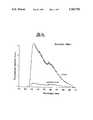
- Testswere conducted revealing examples of such decreased tissue autofluorescence for dysplastic bronchial tissue, and carcinoma in situ. It was determined that the main difference between abnormal and normal tissues is manifested by a greatly reduced fluorescence intensity in the region of the spectrum from 480 nm-600 nm. At wavelengths greater than approximately 635 nm, the tissue autofluorescence is approximately the same between abnormal and normal tissues. Tests were conducted using excitation light of 442 nm, 405 nm and 488 nm and abnormal tissue results were compared to normal tissue results. All of these data were obtained in vivo during standard fiber-optic bronchoscopy using the optical multichannel analyzer.
- the present inventionprovides an imaging apparatus that uses autofluorescence characteristics of tissues to detect and delineate the extent of abnormal tissues in human patients in vivo. Capture and analysis of the autofluorescence images is achieved using a highly sensitive detector such as an image intensified CCD camera. A pseudo image is generated by sending one image to the red channel and one image to the green channel of an RGB video monitor. By capturing the two images simultaneously or sequentially within a few milliseconds, pseudo image generation in real time can be achieved. The pseudo images can clearly delineate the diseased tissue from the surrounding normal tissue.
- the present inventionprovides an apparatus for imaging diseases in tissue comprising:
- a light source for generating excitation lightthat includes wavelengths capable of generating characteristic autofluorescence for abnormal and normal tissues
- optical meansfor intercepting said filtered autofluorescence light to acquire at least two filtered emitted autofluorescence images of the tissue
- display meansfor displaying said acquired images in such a manner as to delineate abnormal and normal tissue.
- the apparatus of the present inventionis used with a standard bronchoscope for imaging abnormal bronchial tissues.
- FIGS. 1A to 1Dprovide examples of autofluorescence spectrums at selected excitation wavelengths which indicate the difference between abnormal and normal tissue
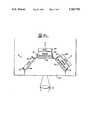
- FIG. 2is a schematic diagram showing the apparatus of the present invention useful for imaging abnormal lung tissue
- FIG. 3shows details of the illumination module
- FIG. 4Ashows the filtering and optical means of the present invention in which a single sensitive detector is used to acquire fluorescence images sequentially;
- FIG. 4Bshows alternative filtering and optical means m which fluorescence images are acquired simultaneously using two sensitive cameras
- FIG. 4Cshows a still further filtering and optical means in which a prism element is incorporated to allow two fluorescence images to be acquired simultaneously together with a reflected/scattered excitation light image.
- FIG. 1shows examples of decreased tissue autofluorescence for dysplastic bronchial tissue and carcinoma in situ.
- the main difference between abnormal and normal tissuesis manifested by a greatly reduced fluorescence intensity in the region of the spectrum from 480 nm-600 nm. At wavelengths greater than approximately 635 nm, the tissue autofluorescence is approximately the same between abnormal and normal tissues.
- a 442 nm, Helium Cadmium laser lightwas used to excite the tissues.
- FIG. 1Ashows tissue autofluorescence spectra of normal and dysplastic tissues and
- FIG. 1Bshows a carcinoma in situ (CIS) lesion compared to the normal tissue of a different patient.
- CIScarcinoma in situ
- the apparatus of the present inventionis designed to exploit the difference in fluorescence intensity in different regions of the spectrum to identify and delineate abnormal tissue.
- the apparatus of the present inventionadapted for use in examining bronchial tissues of the lung in patients is schematically illustrated in FIG. 2. As such, the apparatus is integrated with a conventional bronchoscope used for examining bronchial tissue of the lung.
- the light source 1for generating excitation light that includes wavelengths capable of generating characteristic autofluorescence spectra for abnormal and normal tissue.
- the light source 1is shown in greater detail in FIG. 3 and preferably includes a laser light source 7 capable of producing excitation light at a selected desirable wavelength.
- a white light sourcesuch as an incandescent Xenon light source 8 can be used for white light illumination when desired.
- the laser light source 7is used to generate pseudo images derived from tissue autofluorescence while the white light source is used to generate color images of reflected/scattered white light.
- the light from each light sourcepasses through synchronizing means that allow for alternate illumination of the tissue by the laser light and the white light source.
- the synchronizing meanscomprises blocking means in the form of electronically controlled shutters 9 and 13 associated with laser light source 7 and Xenon light source 8, respectively.
- shutter 9When shutter 9 is open to allow laser light to pass, shutter 13 is closed to prevent passage of white light and vice versa.
- the light from the laser light source 7passes through shutter 9 when open, a mirror with a pin hole 10, and a lens 11 which focuses the laser light onto means for illuminating the tissue with light comprising a conventional bronchoscope light guide 12.
- Light guide 12conducts the excitation light to the tissue area under examination.
- the tissueupon illumination with the laser light, emits its characteristic autofluorescence for abnormal and normal tissue.
- shutter 9is closed and previously closed shutter 13 is opened to allow the light from Xenon light source 8 to pass through shutter 13.
- the white lightis then filtered by a neutral density filter set 14, reflected by a mirror 15, and passes through a lens 16 which focuses the light onto bronchoscope light guide 12 after being reflected off mirror 10 and through lens 11.
- the neutral density filter set 14is used to condition the light from the Xenon source such that it is of the appropriate intensity for the light sensors used in the apparatus.
- Light guide 12ensures that the light is evenly dispersed over the area under examination.
- the bronchoscopeprovides the collecting means to gather images in the form of the bronchoscope lens (not shown) which collects scattered and reflected light, or emitted autofluorescence light from within the lung for transmission out of the body by imaging bundle 2 of the bronchoscope. This collected light is transmitted to a focusing lens 21 of the bronchoscope ocular coupled to the imaging bundle.
- the collected lightenters the image acquisition module 3 which includes means for filtering the autofluorescence light and optical means for intercepting the filtered light.
- image acquisition module 3includes means for filtering the autofluorescence light and optical means for intercepting the filtered light.
- image acquisition module 3includes means for filtering the autofluorescence light and optical means for intercepting the filtered light.
- FIG. 4Aillustrates an image acquisition module that includes filtering means and optical means that allow for acquisition of emitted autofluorescence images sequentially.
- the means for filtering the autofluorescence lightcomprises a series of filters that are sequentially insertable into the path of the emitted autofluorescence light to generate a sequence of filtered autofluorescence images.
- Filter wheel 18is provided and is rotatably mounted beneath the optical means of the image acquisition module.
- laser excitation light 7it is necessary to filter the autofluorescence light generated into at least two spectral bands. In one spectral band, the autofluorescence intensity for abnormal tissue is substantially different from that of normal tissue and in the other spectral band, the autofluorescence intensity is substantially similar to that of normal tissue.
- filter wheel 18would be fitted with two filters.
- a green filter of 500 ⁇ 20 nm and a red 630 nm long pass filterwould be used.
- the green filterwould filter the autofluorescence light into a spectral band in which the autofluorescence intensity for abnormal tissue is substantially different from that of normal tissue while the red long pass filter would filter the light into a spectral band in which the autofluorescence intensity is substantially similar for abnormal and normal tissue.
- the two filtersare mounted in filter wheel 18 such that each covers one-half of the filter surface. By rotating filter wheel 18 at an appropriate speed, red and green filtered autofluorescence images can be captured sequentially by optical means in the form of a single highly sensitive detector 17 such as an image intensified CCD camera.
- the foregoing image acquisition modulealso includes additional optical means for capturing reflected/scattered white light images when white light source 8 is providing illumination of the tissue.
- a movable mirror 20is provided that is insertable into the path of the collected light transmitted by ocular lens 21.
- Mirror 20is positionable to deflect white light into a color video camera 22 for acquisition of white light images. Necessarily, the movement of mirror 20 is controlled such that the mirror deflects the collected light into video camera 22 only when white light source 8 is providing illumination.
- white light source 8color images can be generated on a color monitor in the same way as in conventional bronchoscopy.
- laser light source 7is illuminating the tissue
- mirror 20is removed from the light path to allow for filtering of the autofluorescence light and subsequent acquisition by detector 17.
- FIG. 4Billustrates an alternative arrangement of image acquisition module 3 in which the optical means comprises at least two photodetectors that acquire filtered autofluorescence images simultaneously.
- Each photodetectorhas associated filtering means.
- filter wheel 18 of the embodiment of FIG. 4Ais replaced by beam splitting means in the form of a dichroic mirror 24 which allows the red light >600 nm to pass but reflects the shorter wavelengths.
- additional filters 25 and 26 for exact selection of the desired autofluorescence lightcan be employed and the respective images are focused onto two independent, sensitive photodetectors such as image intensified CCD cameras 17 and 23.
- filter 25is a red 630 nm long pass filter to further filter red light passed by dichroic mirror into a spectral band in which autofluorescence intensity is substantially similar for normal and abnormal tissue.
- Filter 26is a green filter of 500 ⁇ 20 nm for filtering the autofluorescence light into a spectral band in which the autofluorescence intensity for abnormal tissue is substantially different from that of normal tissue. Images acquired by the image intensified CCD camera 17 and/or image intensified CCD camera 23 are fed into red and green input channels of an RGB color monitor 5 (FIG. 1).
- reflected/scattered white light images created by white light source 8are captured by a color camera 22 and are displayed directly onto the color monitor for visualization of the examined site using an identical movable mirror 20 insertable into the light path whenever white light source 8 is providing illumination.
- FIG. 4Cillustrates a further embodiment of an image acquisition module for use with the apparatus of the present invention.
- a prism element 27is provided that simultaneously splits collected light into a plurality of directions.
- laser light source 7 and white light source 8it is possible to capture sequentially both autofluorescence images and white light images within a 33 millisecond cycle time, therefore allowing a view of white (broadband) light color images and pseudo fluorescence images at the same time on display means.
- a specially developed camera with three photodetectors 28, 29 and 30is provided.
- the prism 27splits the collected light into three images which are then captured by the three separate detectors.
- Photodetectors 28 and 29comprise CCD imaging devices that are provided with associated image intensifiers 37 and 38 and photodetector 30 is a regular CCD imaging device.
- Each photodetectorhas its own filter 32, 33 and 34, respectively, as well as an x,y,z micropositioner 31.
- Filters 32 and 33are the same as in the previous embodiments: a 500 ⁇ 20 nm green filter 33, and a 630 nm long pass filter 33.
- CCD imaging device 30has an associated broadband blue filter 34.
- associated camera control electronics 4are such that they generate three image signals, a red signal produced by red filter 32 and intensified CCD imaging device 28, a green signal produced by green filter 33 and intensified CCD imaging device 29 and a blue signal produced by blue filter 34 and nonintensified CCD imaging device 30.
- a specially designed CCD imaging deviceinstead of an image intensified detector.
- several pixels of a sensitive scientific CCD detectorcan be electronically combined into a single very large pixel which allows very low signals to be detected.
- All or some of the image signals produced by the various image acquisition modules of the present inventionmay be displayed directly on color monitor 5 or processed by image processing means prior to display.
- the apparatus of the present inventioncan switch between white (broadband) light illumination and laser illumination in one-thirtieth of a second.
- the image acquisition module of FIG. 4Ccan collect autofluorescence images of the tissue over two selected areas of the spectra and a blue scattered/reflected excitation light image all simultaneously. These images can be combined either visually or mathematically via image processing means to make distinguishable the various tissue types present in the image. With white light illumination, the apparatus can collect red, green and blue reflected/scattered light images so as to make possible a regular color image of the tissues.
- the color imagecan be combined with the autofluorescence blue laser illuminated images to enhance the detection, localization, and delineation of the various tissues.
- a different combination of filtersis employed to enhance the differences between normal and diseased tissues based on the characteristic emitted autofluorescence light of the diseased tissue under study.
- the present inventionis preferably provided with image processing means in the form of an imaging board 35 associated with a computer 6 that controls and coordinates operation of the apparatus.
- Imaging board 35allows images to be digitally captured if desired.
- Board 35acts to digitize the filtered images provided by the image acquisition modules and enhance the digitized images by application of transformational algorithms to produce pseudo computed images in real time for display on video monitor 5.
- the digitized imagescan be stored in computer memory.
- the pixel values in the digitized imagescan be used to calculate a value for each image pixel, using a mathematical transformation, so that all pixels covering the diseased tissue site are clearly different from those of the normal tissue. This process can be used to enhance the images, to enable the measurement of the degree of the disease, and make possible other applications and/or measurements.
- digitization of images and image processingis not required.
- color monitor 5 and the human visual systemit is possible to depict differences between the normal and diseased site as differences in perceived color.
- the image acquisition module of FIG. 4Bhaving two sensitive CCD cameras
- one camerafeeds the Red channel and the other feeds the Green channel of the RGB color monitor 5.
- the red tissue autofluorescence of the abnormal and normal bronchial tissuesis approximately the same.
- the green tissue autofluorescenceis dramatically decreased in the abnormal site compared to normal tissue. Therefore the abnormal site appears much less green and much more reddish and/or sandy colored compared to the surrounding normal tissue which looks bright green, as green fluorescence is much more dominant than red fluorescence in normal tissue.
- This preferred embodimentallows visualization of the diseased sites in real time without any processing of the images and is therefore very inexpensive.
- tissue autofluorescencetwo different spectral bands of tissue autofluorescence are acquired and interpreted as red and green signals for color display on a color monitor. This gives excellent pseudo images of inflamed tissue, dysplastic tissue and non-invasive cancer; clearly delineating these tissues from normal tissue. The decrease in diseased tissue autofluorescence, particularly in the green region, indicates the presence of the disease as well as the severity of the disease.
- the apparatus of the present inventioncan be used to visualize small and large tumors.
- drugssuch as Photoflin (Porfimer sodium)
- the same filterscan be used as the drug emits fluorescence at peak values of 630 nm and 690 nm. In this case all sites where the drug has localized will also be clearly delineated from the normal tissues.
Landscapes
- Life Sciences & Earth Sciences (AREA)
- Health & Medical Sciences (AREA)
- Medical Informatics (AREA)
- Biophysics (AREA)
- Pathology (AREA)
- Engineering & Computer Science (AREA)
- Biomedical Technology (AREA)
- Heart & Thoracic Surgery (AREA)
- Physics & Mathematics (AREA)
- Molecular Biology (AREA)
- Surgery (AREA)
- Animal Behavior & Ethology (AREA)
- General Health & Medical Sciences (AREA)
- Public Health (AREA)
- Veterinary Medicine (AREA)
- Endoscopes (AREA)
- Investigating, Analyzing Materials By Fluorescence Or Luminescence (AREA)
Abstract
Description
Claims (36)
Priority Applications (1)
| Application Number | Priority Date | Filing Date | Title |
|---|---|---|---|
| US08/632,018US5769792A (en) | 1991-07-03 | 1996-04-15 | Endoscopic imaging system for diseased tissue |
Applications Claiming Priority (4)
| Application Number | Priority Date | Filing Date | Title |
|---|---|---|---|
| US72528391A | 1991-07-03 | 1991-07-03 | |
| US8201993A | 1993-06-23 | 1993-06-23 | |
| US08/428,494US5507287A (en) | 1991-05-08 | 1995-04-27 | Endoscopic imaging system for diseased tissue |
| US08/632,018US5769792A (en) | 1991-07-03 | 1996-04-15 | Endoscopic imaging system for diseased tissue |
Related Parent Applications (1)
| Application Number | Title | Priority Date | Filing Date |
|---|---|---|---|
| US08/428,494ContinuationUS5507287A (en) | 1991-05-08 | 1995-04-27 | Endoscopic imaging system for diseased tissue |
Publications (1)
| Publication Number | Publication Date |
|---|---|
| US5769792Atrue US5769792A (en) | 1998-06-23 |
Family
ID=27374189
Family Applications (1)
| Application Number | Title | Priority Date | Filing Date |
|---|---|---|---|
| US08/632,018Expired - LifetimeUS5769792A (en) | 1991-07-03 | 1996-04-15 | Endoscopic imaging system for diseased tissue |
Country Status (1)
| Country | Link |
|---|---|
| US (1) | US5769792A (en) |
Cited By (106)
| Publication number | Priority date | Publication date | Assignee | Title |
|---|---|---|---|---|
| US6026319A (en)* | 1997-02-13 | 2000-02-15 | Fuji Photo Film Co., Ltd. | Fluorescence detecting system |
| US6070096A (en)* | 1996-03-06 | 2000-05-30 | Fuji Photo Film Co., Ltd. | Fluorescence detecting apparatus |
| WO2000042910A1 (en)* | 1999-01-26 | 2000-07-27 | Newton Laboratories, Inc. | Autofluorescence imaging system for endoscopy |
| US6241672B1 (en)* | 1990-08-10 | 2001-06-05 | University Of Washington | Method and apparatus for optically imaging solid tumor tissue |
| US6256530B1 (en)* | 1998-09-15 | 2001-07-03 | Denvu, L.L.C. | Optical instrument and technique for cancer diagnosis using in-vivo fluorescence emission of test tissue |
| WO2000037917A3 (en)* | 1998-12-23 | 2001-08-23 | Medispectra Inc | Systems and methods for optical examination of samples |
| US6289236B1 (en)* | 1997-10-10 | 2001-09-11 | The General Hospital Corporation | Methods and apparatus for distinguishing inflamed and tumorous bladder tissue |
| US20020103439A1 (en)* | 2000-12-19 | 2002-08-01 | Haishan Zeng | Methods and apparatus for fluorescence and reflectance imaging and spectroscopy and for contemporaneous measurements of electromagnetic radiation with multiple measuring devices |
| US6492646B1 (en) | 1999-09-29 | 2002-12-10 | Fuji Photo Film Co., Ltd. | Method of and apparatus for obtaining fluorescence image |
| US20020196337A1 (en)* | 1999-09-01 | 2002-12-26 | Kaneyoshi Takeyama | Weak light color imaging device |
| US20020197728A1 (en)* | 1999-12-15 | 2002-12-26 | Howard Kaufman | Methods of monitoring effects of chemical agents on a sample |
| US20030001104A1 (en)* | 2001-06-29 | 2003-01-02 | Fuji Photo Film Co., Ltd | Method and apparatus for obtaining fluorescence images, and computer executable program therefor |
| US6510338B1 (en)* | 1998-02-07 | 2003-01-21 | Karl Storz Gmbh & Co. Kg | Method of and devices for fluorescence diagnosis of tissue, particularly by endoscopy |
| US6516217B1 (en) | 1999-07-09 | 2003-02-04 | Fuji Photo Film Co., Ltd. | Fluorescence diagnosis system |
| US6529769B2 (en) | 2001-03-08 | 2003-03-04 | Apti, Inc. | Apparatus for performing hyperspectral endoscopy |
| US6537211B1 (en)* | 1998-01-26 | 2003-03-25 | Massachusetts Institute Of Technology | Flourescence imaging endoscope |
| US6571119B2 (en) | 1996-03-06 | 2003-05-27 | Fuji Photo Film Co., Ltd. | Fluorescence detecting apparatus |
| DE10153900A1 (en)* | 2001-11-02 | 2003-06-18 | Wolf Gmbh Richard | Tissue diagnostic imaging device |
| US20030216626A1 (en)* | 2002-03-28 | 2003-11-20 | Fuji Photo Film Co., Ltd. | Fluorescence judging method and apparatus |
| US20030220574A1 (en)* | 2002-03-18 | 2003-11-27 | Sarcos Investments Lc. | Miniaturized imaging device including utility aperture and SSID |
| US20030222325A1 (en)* | 2002-03-18 | 2003-12-04 | Sarcos Investments Lc. | Miniaturized imaging device with integrated circuit connector system |
| US6671540B1 (en)* | 1990-08-10 | 2003-12-30 | Daryl W. Hochman | Methods and systems for detecting abnormal tissue using spectroscopic techniques |
| US6766184B2 (en) | 2000-03-28 | 2004-07-20 | Board Of Regents, The University Of Texas System | Methods and apparatus for diagnostic multispectral digital imaging |
| US6768918B2 (en) | 2002-07-10 | 2004-07-27 | Medispectra, Inc. | Fluorescent fiberoptic probe for tissue health discrimination and method of use thereof |
| US20040152987A1 (en)* | 2002-11-11 | 2004-08-05 | Carl-Zeiss-Stiftung Trading As Carl Zeiss | Inspection system and inspection method |
| US20040186382A1 (en)* | 1997-01-13 | 2004-09-23 | Medispectra, Inc. | Spectral volume microprobe arrays |
| US20040225222A1 (en)* | 2003-05-08 | 2004-11-11 | Haishan Zeng | Real-time contemporaneous multimodal imaging and spectroscopy uses thereof |
| US6818903B2 (en) | 2002-07-09 | 2004-11-16 | Medispectra, Inc. | Method and apparatus for identifying spectral artifacts |
| US20040245350A1 (en)* | 2003-06-03 | 2004-12-09 | Haishan Zeng | Methods and apparatus for fluorescence imaging using multiple excitation-emission pairs and simultaneous multi-channel image detection |
| US6839661B2 (en) | 2000-12-15 | 2005-01-04 | Medispectra, Inc. | System for normalizing spectra |
| US6847490B1 (en) | 1997-01-13 | 2005-01-25 | Medispectra, Inc. | Optical probe accessory device for use in vivo diagnostic procedures |
| US20050154261A1 (en)* | 2000-04-03 | 2005-07-14 | Ohline Robert M. | Tendon-driven endoscope and methods of insertion |
| US6933154B2 (en) | 2002-07-09 | 2005-08-23 | Medispectra, Inc. | Optimal windows for obtaining optical data for characterization of tissue samples |
| EP1669019A3 (en)* | 2004-12-08 | 2006-08-30 | Olympus Corporation | Fluorescence endoscopy device |
| US7103401B2 (en) | 2002-07-10 | 2006-09-05 | Medispectra, Inc. | Colonic polyp discrimination by tissue fluorescence and fiberoptic probe |
| US20060217594A1 (en)* | 2005-03-24 | 2006-09-28 | Ferguson Gary W | Endoscopy device with removable tip |
| US7127282B2 (en) | 1998-12-23 | 2006-10-24 | Medispectra, Inc. | Optical methods and systems for rapid screening of the cervix |
| US20060247537A1 (en)* | 2005-04-19 | 2006-11-02 | Shinya Matsumoto | Endoscope apparatus |
| US7136518B2 (en) | 2003-04-18 | 2006-11-14 | Medispectra, Inc. | Methods and apparatus for displaying diagnostic data |
| DE10141559B4 (en)* | 2000-08-25 | 2007-01-25 | Pentax Corp. | Video endoscope system and illumination optics |
| US20070038126A1 (en)* | 2005-06-23 | 2007-02-15 | Pyle Jason L | System and method for monitoring of end organ oxygenation by measurement of in vivo cellular energy status |
| US7187810B2 (en) | 1999-12-15 | 2007-03-06 | Medispectra, Inc. | Methods and systems for correcting image misalignment |
| EP1759631A3 (en)* | 2000-06-06 | 2007-05-09 | FUJIFILM Corporation | Fluorescent-light image display method and apparatus therefor |
| US20070189443A1 (en)* | 2004-11-19 | 2007-08-16 | Walter Deborah J | Detection of thrombi in ct using energy discrimination |
| US7260248B2 (en) | 1999-12-15 | 2007-08-21 | Medispectra, Inc. | Image processing using measures of similarity |
| US20070213593A1 (en)* | 2006-02-28 | 2007-09-13 | Olympus Corporation | Endoscope system |
| US7282723B2 (en) | 2002-07-09 | 2007-10-16 | Medispectra, Inc. | Methods and apparatus for processing spectral data for use in tissue characterization |
| US7309867B2 (en) | 2003-04-18 | 2007-12-18 | Medispectra, Inc. | Methods and apparatus for characterization of tissue samples |
| US20080058649A1 (en)* | 2006-04-12 | 2008-03-06 | Searete Llc., A Limited Liability Corporation Of The State Of Delaware | Systems for autofluorescent imaging and target ablation |
| US20080100700A1 (en)* | 2006-10-27 | 2008-05-01 | Pentax Corporation | Electronic endoscope |
| US7459696B2 (en) | 2003-04-18 | 2008-12-02 | Schomacker Kevin T | Methods and apparatus for calibrating spectral data |
| US20080304143A1 (en)* | 2007-06-05 | 2008-12-11 | Jacobsen Stephen C | Mini-scope for multi-directional imaging |
| US7469160B2 (en) | 2003-04-18 | 2008-12-23 | Banks Perry S | Methods and apparatus for evaluating image focus |
| EP1575423A4 (en)* | 2002-10-31 | 2009-04-08 | Shanghai Shengda Medical Healt | A laser-induced fluorescence method for precancerous lesion diagnosis and an endoscope precancerous lesion diagnosis apparatus thereof |
| US20090180197A1 (en)* | 2008-01-11 | 2009-07-16 | Sterling Lc | Grin lens microscope system |
| US20090192390A1 (en)* | 2008-01-24 | 2009-07-30 | Lifeguard Surgical Systems | Common bile duct surgical imaging system |
| US20090192349A1 (en)* | 2008-01-24 | 2009-07-30 | Lifeguard Surgical Systems | Common bile duct surgical imaging system |
| US20090259101A1 (en)* | 2008-04-15 | 2009-10-15 | Olympus Medical Systems Corp. | Image pickup apparatus and endoscope apparatus incorporating the same |
| US20090268010A1 (en)* | 2008-04-26 | 2009-10-29 | Intuitive Surgical, Inc. | Augmented stereoscopic visualization for a surgical robot using a captured fluorescence image and captured stereoscopic visible images |
| DE10141527B4 (en)* | 2000-08-25 | 2010-01-14 | Hoya Corp. | Video endoscope system |
| US20100073930A1 (en)* | 2008-09-23 | 2010-03-25 | Lsi Industries, Inc. | Lighting Apparatus with Heat Dissipation System |
| US20100268025A1 (en)* | 2007-11-09 | 2010-10-21 | Amir Belson | Apparatus and methods for capsule endoscopy of the esophagus |
| US20100264313A1 (en)* | 2009-04-20 | 2010-10-21 | Lsi Industries, Inc. | Lighting Techniques for Wirelessly Controlling Lighting Elements |
| US20100264314A1 (en)* | 2009-04-20 | 2010-10-21 | Lsi Industries, Inc. | Lighting Techniques for Wirelessly Controlling Lighting Elements |
| US20110060189A1 (en)* | 2004-06-30 | 2011-03-10 | Given Imaging Ltd. | Apparatus and Methods for Capsule Endoscopy of the Esophagus |
| US20110117025A1 (en)* | 2008-05-20 | 2011-05-19 | Ralph Sebastian Dacosta | Device and method for fluorescence-based imaging and monitoring |
| US20110137179A1 (en)* | 2008-08-21 | 2011-06-09 | University Of Florida Research Foundation, Inc. | Differential laser-induced perturbation (dlip) for bioimaging and chemical sensing |
| US8062212B2 (en) | 2000-04-03 | 2011-11-22 | Intuitive Surgical Operations, Inc. | Steerable endoscope and improved method of insertion |
| US8083879B2 (en) | 2005-11-23 | 2011-12-27 | Intuitive Surgical Operations, Inc. | Non-metallic, multi-strand control cable for steerable instruments |
| US8182418B2 (en) | 2008-02-25 | 2012-05-22 | Intuitive Surgical Operations, Inc. | Systems and methods for articulating an elongate body |
| US20120184813A1 (en)* | 2011-01-19 | 2012-07-19 | Fujifilm Corporation | Endoscope system |
| US20120184812A1 (en)* | 2011-01-19 | 2012-07-19 | Fujifilm Corporation | Endoscope system |
| US8361090B2 (en) | 2002-01-09 | 2013-01-29 | Intuitive Surgical Operations, Inc. | Apparatus and method for endoscopic colectomy |
| US8486735B2 (en) | 2008-07-30 | 2013-07-16 | Raytheon Company | Method and device for incremental wavelength variation to analyze tissue |
| US8517923B2 (en) | 2000-04-03 | 2013-08-27 | Intuitive Surgical Operations, Inc. | Apparatus and methods for facilitating treatment of tissue via improved delivery of energy based and non-energy based modalities |
| US8568299B2 (en) | 2006-05-19 | 2013-10-29 | Intuitive Surgical Operations, Inc. | Methods and apparatus for displaying three-dimensional orientation of a steerable distal tip of an endoscope |
| US8614768B2 (en) | 2002-03-18 | 2013-12-24 | Raytheon Company | Miniaturized imaging device including GRIN lens optically coupled to SSID |
| US8690762B2 (en) | 2008-06-18 | 2014-04-08 | Raytheon Company | Transparent endoscope head defining a focal length |
| US8717428B2 (en) | 2009-10-01 | 2014-05-06 | Raytheon Company | Light diffusion apparatus |
| US8825140B2 (en) | 2001-05-17 | 2014-09-02 | Xenogen Corporation | Imaging system |
| US8828028B2 (en) | 2009-11-03 | 2014-09-09 | Raytheon Company | Suture device and method for closing a planar opening |
| US8845524B2 (en) | 2000-04-03 | 2014-09-30 | Intuitive Surgical Operations, Inc. | Steerable segmented endoscope and method of insertion |
| US8882657B2 (en) | 2003-03-07 | 2014-11-11 | Intuitive Surgical Operations, Inc. | Instrument having radio frequency identification systems and methods for use |
| US8888688B2 (en) | 2000-04-03 | 2014-11-18 | Intuitive Surgical Operations, Inc. | Connector device for a controllable instrument |
| US9060704B2 (en) | 2008-11-04 | 2015-06-23 | Sarcos Lc | Method and device for wavelength shifted imaging |
| US9144664B2 (en) | 2009-10-01 | 2015-09-29 | Sarcos Lc | Method and apparatus for manipulating movement of a micro-catheter |
| US20150359440A1 (en)* | 2013-01-28 | 2015-12-17 | Oslo Universitetssykehus Hf | Assessing circulatory failure |
| US9220398B2 (en) | 2007-10-11 | 2015-12-29 | Intuitive Surgical Operations, Inc. | System for managing Bowden cables in articulating instruments |
| US9354115B2 (en)* | 2012-06-05 | 2016-05-31 | Hypermed Imaging, Inc. | Methods and apparatus for coaxial imaging of multiple wavelengths |
| US9648254B2 (en) | 2014-03-21 | 2017-05-09 | Hypermed Imaging, Inc. | Compact light sensor |
| US9642532B2 (en) | 2008-03-18 | 2017-05-09 | Novadaq Technologies Inc. | Imaging system for combined full-color reflectance and near-infrared imaging |
| US9655519B2 (en) | 2014-03-21 | 2017-05-23 | Hypermed Imaging, Inc. | Systems and methods for performing an imaging test under constrained conditions |
| US9661996B2 (en) | 2009-10-01 | 2017-05-30 | Sarcos Lc | Needle delivered imaging device |
| US9814378B2 (en) | 2011-03-08 | 2017-11-14 | Novadaq Technologies Inc. | Full spectrum LED illuminator having a mechanical enclosure and heatsink |
| US20170367559A1 (en)* | 2015-03-26 | 2017-12-28 | Sony Corporation | Surgical system, information processing device, and method |
| US9952157B2 (en) | 2012-07-17 | 2018-04-24 | The Arizona Board Of Regents On Behalf Of The University Of Arizona | Tissue imaging and visualization of lesions using reflectance and autofluorescence measurements |
| US10438356B2 (en) | 2014-07-24 | 2019-10-08 | University Health Network | Collection and analysis of data for diagnostic purposes |
| US10512392B2 (en) | 2008-02-06 | 2019-12-24 | Intuitive Surgical Operations, Inc. | Segmented instrument having braking capabilities |
| US10694151B2 (en) | 2006-12-22 | 2020-06-23 | Novadaq Technologies ULC | Imaging system with a single color image sensor for simultaneous fluorescence and color video endoscopy |
| US10798310B2 (en) | 2016-05-17 | 2020-10-06 | Hypermed Imaging, Inc. | Hyperspectral imager coupled with indicator molecule tracking |
| US10869645B2 (en) | 2016-06-14 | 2020-12-22 | Stryker European Operations Limited | Methods and systems for adaptive imaging for low light signal enhancement in medical visualization |
| USD916294S1 (en) | 2016-04-28 | 2021-04-13 | Stryker European Operations Limited | Illumination and imaging device |
| US10980420B2 (en) | 2016-01-26 | 2021-04-20 | Stryker European Operations Limited | Configurable platform |
| US10992848B2 (en) | 2017-02-10 | 2021-04-27 | Novadaq Technologies ULC | Open-field handheld fluorescence imaging systems and methods |
| US11096563B2 (en) | 2005-11-22 | 2021-08-24 | Intuitive Surgical Operations, Inc. | Method of determining the shape of a bendable instrument |
| US11930278B2 (en) | 2015-11-13 | 2024-03-12 | Stryker Corporation | Systems and methods for illumination and imaging of a target |
Citations (42)
| Publication number | Priority date | Publication date | Assignee | Title |
|---|---|---|---|---|
| JPS5812675A (en)* | 1981-07-15 | 1983-01-24 | 松下電工株式会社 | Case of electric machinery |
| JPS58222331A (en)* | 1982-06-21 | 1983-12-24 | Sony Corp | Reproducer of character information |
| US4473841A (en)* | 1981-10-20 | 1984-09-25 | Fuji Photo Film Co., Ltd. | Video signal transmission system for endoscope using solid state image sensor |
| US4541438A (en)* | 1983-06-02 | 1985-09-17 | The Johns Hopkins University | Localization of cancerous tissue by monitoring infrared fluorescence emitted by intravenously injected porphyrin tumor-specific markers excited by long wavelength light |
| US4556057A (en)* | 1982-08-31 | 1985-12-03 | Hamamatsu Tv Co., Ltd. | Cancer diagnosis device utilizing laser beam pulses |
| WO1986002730A1 (en)* | 1984-10-22 | 1986-05-09 | Hightech Network Sci Ab | A fluorescence imaging system |
| EP0215772A2 (en)* | 1985-09-16 | 1987-03-25 | AVL Medical Instruments AG | Method and device for diagnosing tumours using sera |
| US4719508A (en)* | 1985-10-02 | 1988-01-12 | Olympus Optical Co., Ltd. | Endoscopic photographing apparatus |
| US4718417A (en)* | 1985-03-22 | 1988-01-12 | Massachusetts Institute Of Technology | Visible fluorescence spectral diagnostic for laser angiosurgery |
| US4768513A (en)* | 1986-04-21 | 1988-09-06 | Agency Of Industrial Science And Technology | Method and device for measuring and processing light |
| US4773097A (en)* | 1984-05-31 | 1988-09-20 | Omron Tateisi Electronics Co. | Image analyzing apparatus |
| US4774568A (en)* | 1986-01-27 | 1988-09-27 | Kabushiki Kaisha Toshiba | Endoscopic apparatus |
| GB2203831A (en)* | 1986-07-07 | 1988-10-26 | Academy Of Applied Sciences | Diagnosis of malignant tumours by fluorescence |
| US4805597A (en)* | 1986-09-30 | 1989-02-21 | Kabushiki Kaisha Toshiba | Endoscopic apparatus |
| US4821117A (en)* | 1986-11-12 | 1989-04-11 | Kabushiki Kaisha Toshiba | Endoscopic system for producing fluorescent and visible images |
| US4827908A (en)* | 1987-03-30 | 1989-05-09 | Kabushiki Kaisha Toshiba | Endoscopic apparatus |
| US4852579A (en)* | 1987-04-20 | 1989-08-01 | Karl Storz Endoscopy Gmbh And Company | Photocharacterization and treatment of normal abnormal and ectopic endometrium |
| US4858001A (en)* | 1987-10-08 | 1989-08-15 | High-Tech Medical Instrumentation, Inc. | Modular endoscopic apparatus with image rotation |
| US4860731A (en)* | 1987-12-17 | 1989-08-29 | Olympus Optical Co., Ltd. | Endoscope |
| US4867137A (en)* | 1987-03-19 | 1989-09-19 | Olympus Optical Co., Ltd. | Electronic endoscope |
| US4868647A (en)* | 1987-09-14 | 1989-09-19 | Olympus Optical Co., Ltd. | Electronic endoscopic apparatus isolated by differential type drive means |
| US4930516A (en)* | 1985-11-13 | 1990-06-05 | Alfano Robert R | Method for detecting cancerous tissue using visible native luminescence |
| US4938205A (en)* | 1988-05-27 | 1990-07-03 | The University Of Connecticut | Endoscope with traced raster and elemental photodetectors |
| WO1990010219A1 (en)* | 1989-02-22 | 1990-09-07 | Spectraphos Ab | Improvements in diagnosis by means of fluorescent light emission from tissue |
| US4957114A (en)* | 1985-04-01 | 1990-09-18 | Kun Zeng | Diagnostic apparatus for intrinsic fluorescence of malignant tumor |
| WO1990012536A1 (en)* | 1989-04-14 | 1990-11-01 | Massachusetts Institute Of Technology | Spectral diagnosis of diseased tissue |
| US4993404A (en)* | 1989-06-26 | 1991-02-19 | Lane Timothy G | Fluoroscopy switching device |
| US4998972A (en)* | 1988-04-28 | 1991-03-12 | Thomas J. Fogarty | Real time angioscopy imaging system |
| US5003977A (en)* | 1988-03-31 | 1991-04-02 | Agency Of Industrial Science And Technology | Device for analyzing fluorescent light signals |
| US5042494A (en)* | 1985-11-13 | 1991-08-27 | Alfano Robert R | Method and apparatus for detecting cancerous tissue using luminescence excitation spectra |
| US5071417A (en)* | 1990-06-15 | 1991-12-10 | Rare Earth Medical Lasers, Inc. | Laser fusion of biological materials |
| US5078150A (en)* | 1988-05-02 | 1992-01-07 | Olympus Optical Co., Ltd. | Spectral diagnosing apparatus with endoscope |
| US5090400A (en)* | 1987-03-31 | 1992-02-25 | Kabushiki Kaisha Toshiba | Measuring endoscope |
| US5091652A (en)* | 1990-01-12 | 1992-02-25 | The Regents Of The University Of California | Laser excited confocal microscope fluorescence scanner and method |
| US5117466A (en)* | 1991-04-30 | 1992-05-26 | The United States Of America As Represented By The United States Department Of Energy | Integrated fluorescence analysis system |
| US5125404A (en)* | 1985-03-22 | 1992-06-30 | Massachusetts Institute Of Technology | Apparatus and method for obtaining spectrally resolved spatial images of tissue |
| US5131398A (en)* | 1990-01-22 | 1992-07-21 | Mediscience Technology Corp. | Method and apparatus for distinguishing cancerous tissue from benign tumor tissue, benign tissue or normal tissue using native fluorescence |
| US5318023A (en)* | 1991-04-03 | 1994-06-07 | Cedars-Sinai Medical Center | Apparatus and method of use for a photosensitizer enhanced fluorescence based biopsy needle |
| US5318024A (en)* | 1985-03-22 | 1994-06-07 | Massachusetts Institute Of Technology | Laser endoscope for spectroscopic imaging |
| US5377676A (en)* | 1991-04-03 | 1995-01-03 | Cedars-Sinai Medical Center | Method for determining the biodistribution of substances using fluorescence spectroscopy |
| US5421337A (en)* | 1989-04-14 | 1995-06-06 | Massachusetts Institute Of Technology | Spectral diagnosis of diseased tissue |
| US5507287A (en)* | 1991-05-08 | 1996-04-16 | Xillix Technologies Corporation | Endoscopic imaging system for diseased tissue |
- 1996
- 1996-04-15USUS08/632,018patent/US5769792A/ennot_activeExpired - Lifetime
Patent Citations (46)
| Publication number | Priority date | Publication date | Assignee | Title |
|---|---|---|---|---|
| JPS5812675A (en)* | 1981-07-15 | 1983-01-24 | 松下電工株式会社 | Case of electric machinery |
| US4473841A (en)* | 1981-10-20 | 1984-09-25 | Fuji Photo Film Co., Ltd. | Video signal transmission system for endoscope using solid state image sensor |
| JPS58222331A (en)* | 1982-06-21 | 1983-12-24 | Sony Corp | Reproducer of character information |
| US4556057A (en)* | 1982-08-31 | 1985-12-03 | Hamamatsu Tv Co., Ltd. | Cancer diagnosis device utilizing laser beam pulses |
| US4541438A (en)* | 1983-06-02 | 1985-09-17 | The Johns Hopkins University | Localization of cancerous tissue by monitoring infrared fluorescence emitted by intravenously injected porphyrin tumor-specific markers excited by long wavelength light |
| US4773097A (en)* | 1984-05-31 | 1988-09-20 | Omron Tateisi Electronics Co. | Image analyzing apparatus |
| WO1986002730A1 (en)* | 1984-10-22 | 1986-05-09 | Hightech Network Sci Ab | A fluorescence imaging system |
| US4786813A (en)* | 1984-10-22 | 1988-11-22 | Hightech Network Sci Ab | Fluorescence imaging system |
| US4718417A (en)* | 1985-03-22 | 1988-01-12 | Massachusetts Institute Of Technology | Visible fluorescence spectral diagnostic for laser angiosurgery |
| US5318024A (en)* | 1985-03-22 | 1994-06-07 | Massachusetts Institute Of Technology | Laser endoscope for spectroscopic imaging |
| US5125404A (en)* | 1985-03-22 | 1992-06-30 | Massachusetts Institute Of Technology | Apparatus and method for obtaining spectrally resolved spatial images of tissue |
| US4957114A (en)* | 1985-04-01 | 1990-09-18 | Kun Zeng | Diagnostic apparatus for intrinsic fluorescence of malignant tumor |
| EP0215772A2 (en)* | 1985-09-16 | 1987-03-25 | AVL Medical Instruments AG | Method and device for diagnosing tumours using sera |
| US4719508A (en)* | 1985-10-02 | 1988-01-12 | Olympus Optical Co., Ltd. | Endoscopic photographing apparatus |
| US4930516B1 (en)* | 1985-11-13 | 1998-08-04 | Laser Diagnostic Instr Inc | Method for detecting cancerous tissue using visible native luminescence |
| US4930516A (en)* | 1985-11-13 | 1990-06-05 | Alfano Robert R | Method for detecting cancerous tissue using visible native luminescence |
| US5042494A (en)* | 1985-11-13 | 1991-08-27 | Alfano Robert R | Method and apparatus for detecting cancerous tissue using luminescence excitation spectra |
| US4774568A (en)* | 1986-01-27 | 1988-09-27 | Kabushiki Kaisha Toshiba | Endoscopic apparatus |
| US4768513A (en)* | 1986-04-21 | 1988-09-06 | Agency Of Industrial Science And Technology | Method and device for measuring and processing light |
| GB2203831A (en)* | 1986-07-07 | 1988-10-26 | Academy Of Applied Sciences | Diagnosis of malignant tumours by fluorescence |
| US4805597A (en)* | 1986-09-30 | 1989-02-21 | Kabushiki Kaisha Toshiba | Endoscopic apparatus |
| US4821117A (en)* | 1986-11-12 | 1989-04-11 | Kabushiki Kaisha Toshiba | Endoscopic system for producing fluorescent and visible images |
| US4867137A (en)* | 1987-03-19 | 1989-09-19 | Olympus Optical Co., Ltd. | Electronic endoscope |
| US4827908A (en)* | 1987-03-30 | 1989-05-09 | Kabushiki Kaisha Toshiba | Endoscopic apparatus |
| US5090400A (en)* | 1987-03-31 | 1992-02-25 | Kabushiki Kaisha Toshiba | Measuring endoscope |
| US4852579A (en)* | 1987-04-20 | 1989-08-01 | Karl Storz Endoscopy Gmbh And Company | Photocharacterization and treatment of normal abnormal and ectopic endometrium |
| US4868647A (en)* | 1987-09-14 | 1989-09-19 | Olympus Optical Co., Ltd. | Electronic endoscopic apparatus isolated by differential type drive means |
| US4858001B1 (en)* | 1987-10-08 | 1992-06-30 | High Tech Medical Instrumentat | |
| US4858001A (en)* | 1987-10-08 | 1989-08-15 | High-Tech Medical Instrumentation, Inc. | Modular endoscopic apparatus with image rotation |
| US4860731A (en)* | 1987-12-17 | 1989-08-29 | Olympus Optical Co., Ltd. | Endoscope |
| US5003977A (en)* | 1988-03-31 | 1991-04-02 | Agency Of Industrial Science And Technology | Device for analyzing fluorescent light signals |
| US4998972A (en)* | 1988-04-28 | 1991-03-12 | Thomas J. Fogarty | Real time angioscopy imaging system |
| US5078150A (en)* | 1988-05-02 | 1992-01-07 | Olympus Optical Co., Ltd. | Spectral diagnosing apparatus with endoscope |
| US4938205A (en)* | 1988-05-27 | 1990-07-03 | The University Of Connecticut | Endoscope with traced raster and elemental photodetectors |
| US5115137A (en)* | 1989-02-22 | 1992-05-19 | Spectraphos Ab | Diagnosis by means of fluorescent light emission from tissue |
| WO1990010219A1 (en)* | 1989-02-22 | 1990-09-07 | Spectraphos Ab | Improvements in diagnosis by means of fluorescent light emission from tissue |
| WO1990012536A1 (en)* | 1989-04-14 | 1990-11-01 | Massachusetts Institute Of Technology | Spectral diagnosis of diseased tissue |
| US5421337A (en)* | 1989-04-14 | 1995-06-06 | Massachusetts Institute Of Technology | Spectral diagnosis of diseased tissue |
| US4993404A (en)* | 1989-06-26 | 1991-02-19 | Lane Timothy G | Fluoroscopy switching device |
| US5091652A (en)* | 1990-01-12 | 1992-02-25 | The Regents Of The University Of California | Laser excited confocal microscope fluorescence scanner and method |
| US5131398A (en)* | 1990-01-22 | 1992-07-21 | Mediscience Technology Corp. | Method and apparatus for distinguishing cancerous tissue from benign tumor tissue, benign tissue or normal tissue using native fluorescence |
| US5071417A (en)* | 1990-06-15 | 1991-12-10 | Rare Earth Medical Lasers, Inc. | Laser fusion of biological materials |
| US5377676A (en)* | 1991-04-03 | 1995-01-03 | Cedars-Sinai Medical Center | Method for determining the biodistribution of substances using fluorescence spectroscopy |
| US5318023A (en)* | 1991-04-03 | 1994-06-07 | Cedars-Sinai Medical Center | Apparatus and method of use for a photosensitizer enhanced fluorescence based biopsy needle |
| US5117466A (en)* | 1991-04-30 | 1992-05-26 | The United States Of America As Represented By The United States Department Of Energy | Integrated fluorescence analysis system |
| US5507287A (en)* | 1991-05-08 | 1996-04-16 | Xillix Technologies Corporation | Endoscopic imaging system for diseased tissue |
Non-Patent Citations (74)
| Title |
|---|
| Alfano et al., "Fluorescence Spectra from Cancerous and Normal Human Breast and Lung Tissues," IEEE Journal of Quantum Electronics, vol. QE-23, No. 10, 1987, pp. 1806-1811. |
| Alfano et al., "Laser Induced Fluorescence Spectroscopy from Native Cancerous and Normal Tissue," IEEE Journal of Quantum Electronics, vol. QE-20, No. 12, Dec. 1984, pp. 1507-1511. |
| Alfano et al., Fluorescence Spectra from Cancerous and Normal Human Breast and Lung Tissues, IEEE Journal of Quantum Electronics, vol. QE 23, No. 10, 1987, pp. 1806 1811.* |
| Alfano et al., Laser Induced Fluorescence Spectroscopy from Native Cancerous and Normal Tissue, IEEE Journal of Quantum Electronics, vol. QE 20, No. 12, Dec. 1984, pp. 1507 1511.* |
| Andersson Engels et al., Fluorescence Characteristics of Atherosclorotic Plaque and Malignant Tumors, SPIE, vol. 1426, 1991, pp. 31 43.* |
| Andersson Engels et al., Tissue Diagnostics Using Laser Induced Fluorescence, Ber Bunsenges, Phys. Chem., vol. 93, 1989, pp. 335 342.* |
| Andersson-Engels et al., "Fluorescence Characteristics of Atherosclorotic Plaque and Malignant Tumors," SPIE, vol. 1426, 1991, pp. 31-43. |
| Andersson-Engels et al., "Tissue Diagnostics Using Laser-Induced Fluorescence," Ber Bunsenges, Phys. Chem., vol. 93, 1989, pp. 335-342. |
| Coffey et al., "Evaluation of Visual Acuity During Laser Photoradiation Therapy of Cancer," Lasers in Surgery and Medicine, vol. 4, pp. 65-71. |
| Coffey et al., Evaluation of Visual Acuity During Laser Photoradiation Therapy of Cancer, Lasers in Surgery and Medicine, vol. 4, pp. 65 71.* |
| Cothren et al., "Gastrointestinal Tissue Diagnosis by Laser-Induced Fluorescence Spectroscopy at Endoscopy," Gastrointestinal Endoscopy, vol. 36, No. 2, 1990, pp. 105-111. |
| Cothren et al., Gastrointestinal Tissue Diagnosis by Laser Induced Fluorescence Spectroscopy at Endoscopy, Gastrointestinal Endoscopy, vol. 36, No. 2, 1990, pp. 105 111.* |
| Dougherty et al., "Cutaneous Phototoxic Occurrences in Patients Receiving Photofrin," Lasers in Surgery and Medicine, vol. 10, 1990, pp. 485-488. |
| Dougherty et al., Cutaneous Phototoxic Occurrences in Patients Receiving Photofrin, Lasers in Surgery and Medicine, vol. 10, 1990, pp. 485 488.* |
| Hayata et al., "Fiberoptic Bronchoscopic Laser Photoradiation for Tumor Localization in Lung Cancer," Chest, vol. 82, 1982, pp. 10-14. |
| Hayata et al., Fiberoptic Bronchoscopic Laser Photoradiation for Tumor Localization in Lung Cancer, Chest, vol. 82, 1982, pp. 10 14.* |
| Hirano et al., "Photodynamic Cancer Diagnosis and Treatment System Consisting of Pulse Lasers and an Endoscopic Spectro-Image Analyzer," Laser in Life Sciences, vol. 3(1), 1989, pp. 1-18. |
| Hirano et al., Photodynamic Cancer Diagnosis and Treatment System Consisting of Pulse Lasers and an Endoscopic Spectro Image Analyzer, Laser in Life Sciences, vol. 3(1), 1989, pp. 1 18.* |
| Hung et al., "Autofluorescence of Normal and Malignant Bronchial Tissue," Lasers in Surgery and Medicine, vol. 11, 1991, pp. 99-105. |
| Hung et al., Autofluorescence of Normal and Malignant Bronchial Tissue, Lasers in Surgery and Medicine, vol. 11, 1991, pp. 99 105.* |
| Ikeda, "New Bronichial TV Endoscopy System," Elsevier Science Publishers B.V. Biomedical Press, 1988. |
| Ikeda, New Bronichial TV Endoscopy System, Elsevier Science Publishers B.V. Biomedical Press, 1988.* |
| Kapadia et al., "Laser-Induced Fluorescence Spectroscopy of Human Colonic Mucosa," Gastroenterology, vol. 99, 1990, pp. 150-157. |
| Kapadia et al., Laser Induced Fluorescence Spectroscopy of Human Colonic Mucosa, Gastroenterology, vol. 99, 1990, pp. 150 157.* |
| Kato et al., "Early Detection of Lung Cancer by Means of Hematoporphyrin Derivative Fluorescence and Laser Photoradiation," Clinics in Chest Medicine, vol. 6, No. 2, 1985, pp. 237-253. |
| Kato et al., Early Detection of Lung Cancer by Means of Hematoporphyrin Derivative Fluorescence and Laser Photoradiation, Clinics in Chest Medicine, vol. 6, No. 2, 1985, pp. 237 253.* |
| Kato et al., Photodynamic Diagnosis in Respiratory Tract Malignancy Using an Excimer Dye Laser System, Journal of Photochemistry and Photobiology, B:Biology, vol. 6, 1990, pp. 189 196.* |
| Kato et al., Photodynamic Diagnosis in Respiratory Tract Malignancy Using an Excimer Dye Laser System, Journal of Photochemistry and Photobiology, B:Biology, vol. 6, 1990, pp. 189-196. |
| Lam et al., "Detection of Lung Cancer by Ratio Fluorometry With and Without Photofrin II," SPIE Proc. vol. 1201, 1990, pp. 561-568. |
| Lam et al., "Fluorescence Detection," Advances in the Diagnosis and Therapy of Lung Cancer, Blackwell Scientific Publications Inc. |
| Lam et al., "Fluorescence Imaging of Early Lung Cancer," IEEE Eng. Med. Biology, vol. 12, 1990. |
| Lam et al., "Mechanism of Detection of Early Lung Cancer by Ratio Fluorometry," Lasers in Life Sciences, vol. 4(2), 1991, pp. 67-73. |
| Lam et al., Detection of Early Lung Cancer Using Low Dose Photofrin II, Chest, vol. 97, 1990, pp. 333 337.* |
| Lam et al., Detection of Early Lung Cancer Using Low Dose Photofrin II, Chest, vol. 97, 1990, pp. 333-337. |
| Lam et al., Detection of Lung Cancer by Ratio Fluorometry With and Without Photofrin II, SPIE Proc. vol. 1201, 1990, pp. 561 568.* |
| Lam et al., Fluorescence Detection, Advances in the Diagnosis and Therapy of Lung Cancer, Blackwell Scientific Publications Inc.* |
| Lam et al., Fluorescence Imaging of Early Lung Cancer, IEEE Eng. Med. Biology, vol. 12, 1990.* |
| Lam et al., Mechanism of Detection of Early Lung Cancer by Ratio Fluorometry, Lasers in Life Sciences, vol. 4(2), 1991, pp. 67 73.* |
| Montan et al., "Multicolor Imaging and Contrast Enhancement in Cancer-Tumor Localization Using Laser-Induced Fluorescence in Hematoporphyrin-derivative-bearing Tissue," Optics Letters, vol. 10(2), 1985, pp. 56-58. |
| Montan et al., Multicolor Imaging and Contrast Enhancement in Cancer Tumor Localization Using Laser Induced Fluorescence in Hematoporphyrin derivative bearing Tissue, Optics Letters, vol. 10(2), 1985, pp. 56 58.* |
| Mullooly et al., "Dihematoporphyrin Ether-Induced Photosensitivity in Laryngeal Papilloma Patients," Lasers in Surgery and Medicine, vol. 10, 1990, pp. 349-356. |
| Mullooly et al., Dihematoporphyrin Ether Induced Photosensitivity in Laryngeal Papilloma Patients, Lasers in Surgery and Medicine, vol. 10, 1990, pp. 349 356.* |
| Palcic et al. "Lung Imaging Fluorescence Endoscope: A Device for Detection of Occult Lung Cancer," Medical Design and Material, 1991. |
| Palcic et al. Lung Imaging Fluorescence Endoscope: A Device for Detection of Occult Lung Cancer, Medical Design and Material, 1991.* |
| Palcic et al., "Detection and Localization of Early Lung Cancer by Imaging Techniques," Chest, vol. 99, 1991, pp. 742-743. |
| Palcic et al., "Development of a Lung Imaging Fluorescence Endoscope," Proceedings of the 12th Annual Int'l Conference of the IEEE Engineering in Medicine and Biology Society, vol. 12, No. 1, 1990. |
| Palcic et al., "Lung Imaging Fluorescence Endoscope: Development and Experimental Prototype," SPIE, vol. 1448, 1991, pp. 113-117. |
| Palcic et al., "The Importance of Image Quality for Computing Texture Features in Biomedical Specimens," SPIE Proc., vol. 1205, 1990, pp. 155-162. |
| Palcic et al., Detection and Localization of Early Lung Cancer by Imaging Techniques, Chest, vol. 99, 1991, pp. 742 743.* |
| Palcic et al., Development of a Lung Imaging Fluorescence Endoscope, Proceedings of the 12th Annual Int l Conference of the IEEE Engineering in Medicine and Biology Society, vol. 12, No. 1, 1990.* |
| Palcic et al., Lung Imaging Fluorescence Endoscope: Development and Experimental Prototype, SPIE, vol. 1448, 1991, pp. 113 117.* |
| Palcic et al., The Importance of Image Quality for Computing Texture Features in Biomedical Specimens, SPIE Proc., vol. 1205, 1990, pp. 155 162.* |
| Peak et al., "DNA-to-Protein Crosslinks and Backbone Breaks Caused by FAR- and NEAR-Ultraviolet and Visible Light Radiations in Mammalian Cells," Mechanism of DNA Damage and Repair, Implications for Carcinogenesis and Risk Assessment, 1986, pp. 193-202. |
| Peak et al., DNA to Protein Crosslinks and Backbone Breaks Caused by FAR and NEAR Ultraviolet and Visible Light Radiations in Mammalian Cells, Mechanism of DNA Damage and Repair, Implications for Carcinogenesis and Risk Assessment, 1986, pp. 193 202.* |
| Profio et al., "Digital Background Subtraction for Fluorescence Imaging," Medical Physics, vol. 13(5), 1988, pp. 717-727. |
| Profio et al., "Endoscopic Fluorescence Detection of Early Lung Cancer," SPIE, vol. 1426, 1991, pp. 44-46. |
| Profio et al., "Fluorometer for Endoscopic Diagnosis of Tumors," Medical Physics, vol. 11(4), 1984, pp. 516-520. |
| Profio et al., "Laser Fluorescence Bronchoscope for Localization of Occult Lung Tumors," Medical Physics, vol. 6, 1979, pp. 523-525. |
| Profio et al., Digital Background Subtraction for Fluorescence Imaging, Medical Physics, vol. 13(5), 1988, pp. 717 727.* |
| Profio et al., Endoscopic Fluorescence Detection of Early Lung Cancer, SPIE, vol. 1426, 1991, pp. 44 46.* |
| Profio et al., Fluorometer for Endoscopic Diagnosis of Tumors, Medical Physics, vol. 11(4), 1984, pp. 516 520.* |
| Profio et al., Laser Fluorescence Bronchoscope for Localization of Occult Lung Tumors, Medical Physics, vol. 6, 1979, pp. 523 525.* |
| Rava et al., "Early Detection of Dysplasia in Colon and Bladder Tissue Using Laser Induced Fluorescence," SPIE, vol. 1426, 1991, pp. 68-78. |
| Rava et al., Early Detection of Dysplasia in Colon and Bladder Tissue Using Laser Induced Fluorescence, SPIE, vol. 1426, 1991, pp. 68 78.* |
| Razum et al., "Skin Photosensitivity: Duration and Intensity Following Intravenous Hematoporphyrin Derivatives, Hp D and DHE," Photochemistry and Photobiology, vol. 46, No. 5, 1987, pp. 925-928. |
| Razum et al., Skin Photosensitivity: Duration and Intensity Following Intravenous Hematoporphyrin Derivatives, H p D and DHE, Photochemistry and Photobiology, vol. 46, No. 5, 1987, pp. 925 928.* |
| Richards Kortum et al., Spectroscopic Diagnosis of Colonic Dysplasia: Spectroscopic Analysis, Biochemistry and Photobiology, vol. 53, No. 6, 1991, pp. 777 786.* |
| Richards-Kortum et al., "Spectroscopic Diagnosis of Colonic Dysplasia: Spectroscopic Analysis," Biochemistry and Photobiology, vol. 53, No. 6, 1991, pp. 777-786. |
| Tang et al., "Spectroscopic Differences Between Human Cancer and Normal Lung and Breast Tissues," Lasers in Surgery and Medicine, vol. 9, 1989, pp. 290-295. |
| Tang et al., Spectroscopic Differences Between Human Cancer and Normal Lung and Breast Tissues, Lasers in Surgery and Medicine, vol. 9, 1989, pp. 290 295.* |
| Wagnieres et al., "Photodetection of Early Cancer by Laser Induced Fluorescence of a Tumor-Selective Dye: Apparatus Design and Realization," SPIE Proc., vol. 1203, 1990, pp. 43-52. |
| Wagnieres et al., Photodetection of Early Cancer by Laser Induced Fluorescence of a Tumor Selective Dye: Apparatus Design and Realization, SPIE Proc., vol. 1203, 1990, pp. 43 52.* |
| Wooten et al., "Prospective Study of Cutaneous Phototoxicity After Systemic Hematoporphyrin Derivative," Lasers in Surgery and Medicine, vol. 8, 1988, pp. 294-300. |
| Wooten et al., Prospective Study of Cutaneous Phototoxicity After Systemic Hematoporphyrin Derivative, Lasers in Surgery and Medicine, vol. 8, 1988, pp. 294 300.* |
Cited By (221)
| Publication number | Priority date | Publication date | Assignee | Title |
|---|---|---|---|---|
| US6241672B1 (en)* | 1990-08-10 | 2001-06-05 | University Of Washington | Method and apparatus for optically imaging solid tumor tissue |
| US6671540B1 (en)* | 1990-08-10 | 2003-12-30 | Daryl W. Hochman | Methods and systems for detecting abnormal tissue using spectroscopic techniques |
| US6070096A (en)* | 1996-03-06 | 2000-05-30 | Fuji Photo Film Co., Ltd. | Fluorescence detecting apparatus |
| US6571119B2 (en) | 1996-03-06 | 2003-05-27 | Fuji Photo Film Co., Ltd. | Fluorescence detecting apparatus |
| US6847490B1 (en) | 1997-01-13 | 2005-01-25 | Medispectra, Inc. | Optical probe accessory device for use in vivo diagnostic procedures |
| US6826422B1 (en) | 1997-01-13 | 2004-11-30 | Medispectra, Inc. | Spectral volume microprobe arrays |
| US20040186382A1 (en)* | 1997-01-13 | 2004-09-23 | Medispectra, Inc. | Spectral volume microprobe arrays |
| US6026319A (en)* | 1997-02-13 | 2000-02-15 | Fuji Photo Film Co., Ltd. | Fluorescence detecting system |
| US6289236B1 (en)* | 1997-10-10 | 2001-09-11 | The General Hospital Corporation | Methods and apparatus for distinguishing inflamed and tumorous bladder tissue |
| US20030191368A1 (en)* | 1998-01-26 | 2003-10-09 | Massachusetts Institute Of Technology | Fluorescence imaging endoscope |
| US7235045B2 (en)* | 1998-01-26 | 2007-06-26 | Massachusetts Institute Of Technology | Fluorescence imaging endoscope |
| US6537211B1 (en)* | 1998-01-26 | 2003-03-25 | Massachusetts Institute Of Technology | Flourescence imaging endoscope |
| US6510338B1 (en)* | 1998-02-07 | 2003-01-21 | Karl Storz Gmbh & Co. Kg | Method of and devices for fluorescence diagnosis of tissue, particularly by endoscopy |
| US6256530B1 (en)* | 1998-09-15 | 2001-07-03 | Denvu, L.L.C. | Optical instrument and technique for cancer diagnosis using in-vivo fluorescence emission of test tissue |
| US7127282B2 (en) | 1998-12-23 | 2006-10-24 | Medispectra, Inc. | Optical methods and systems for rapid screening of the cervix |
| US6411838B1 (en) | 1998-12-23 | 2002-06-25 | Medispectra, Inc. | Systems and methods for optical examination of samples |
| US6385484B2 (en) | 1998-12-23 | 2002-05-07 | Medispectra, Inc. | Spectroscopic system employing a plurality of data types |
| WO2000037917A3 (en)* | 1998-12-23 | 2001-08-23 | Medispectra Inc | Systems and methods for optical examination of samples |
| US6760613B2 (en) | 1998-12-23 | 2004-07-06 | Medispectra, Inc. | Substantially monostatic, substantially confocal optical systems for examination of samples |
| EP1632173A1 (en)* | 1999-01-26 | 2006-03-08 | Newton Laboratories, Inc. | Autofluorescence imaging system for endoscopy |
| US7846091B2 (en) | 1999-01-26 | 2010-12-07 | Newton Laboratories, Inc. | Autofluorescence imaging system for endoscopy |
| WO2000042910A1 (en)* | 1999-01-26 | 2000-07-27 | Newton Laboratories, Inc. | Autofluorescence imaging system for endoscopy |
| US20110213252A1 (en)* | 1999-01-26 | 2011-09-01 | Fulghum Stephen F | Autofluorescence imaging system for endoscopy |
| US8764643B2 (en) | 1999-01-26 | 2014-07-01 | Hoya Corporation | Autofluorescence imaging system for endoscopy |
| US20020161282A1 (en)* | 1999-01-26 | 2002-10-31 | Newton Laboratories, Inc. | Autofluorescence imaging system for endoscopy |
| US6516217B1 (en) | 1999-07-09 | 2003-02-04 | Fuji Photo Film Co., Ltd. | Fluorescence diagnosis system |
| EP1227686A4 (en)* | 1999-09-01 | 2004-04-07 | Hamamatsu Photonics Kk | Feeble light color imaging device |
| US20020196337A1 (en)* | 1999-09-01 | 2002-12-26 | Kaneyoshi Takeyama | Weak light color imaging device |
| US6492646B1 (en) | 1999-09-29 | 2002-12-10 | Fuji Photo Film Co., Ltd. | Method of and apparatus for obtaining fluorescence image |
| US7187810B2 (en) | 1999-12-15 | 2007-03-06 | Medispectra, Inc. | Methods and systems for correcting image misalignment |
| US7260248B2 (en) | 1999-12-15 | 2007-08-21 | Medispectra, Inc. | Image processing using measures of similarity |
| US20020197728A1 (en)* | 1999-12-15 | 2002-12-26 | Howard Kaufman | Methods of monitoring effects of chemical agents on a sample |
| US6902935B2 (en) | 1999-12-15 | 2005-06-07 | Medispectra, Inc. | Methods of monitoring effects of chemical agents on a sample |
| US6766184B2 (en) | 2000-03-28 | 2004-07-20 | Board Of Regents, The University Of Texas System | Methods and apparatus for diagnostic multispectral digital imaging |
| US10105036B2 (en) | 2000-04-03 | 2018-10-23 | Intuitive Surgical Operations, Inc. | Connector device for a controllable instrument |
| US10736490B2 (en) | 2000-04-03 | 2020-08-11 | Intuitive Surgical Operations, Inc. | Connector device for a controllable instrument |
| US8888688B2 (en) | 2000-04-03 | 2014-11-18 | Intuitive Surgical Operations, Inc. | Connector device for a controllable instrument |
| US8517923B2 (en) | 2000-04-03 | 2013-08-27 | Intuitive Surgical Operations, Inc. | Apparatus and methods for facilitating treatment of tissue via improved delivery of energy based and non-energy based modalities |
| US12076102B2 (en) | 2000-04-03 | 2024-09-03 | Intuitive Surgical Operations, Inc. | Connector device for a controllable instrument |
| US9427282B2 (en) | 2000-04-03 | 2016-08-30 | Intuitive Surgical Operations, Inc. | Apparatus and methods for facilitating treatment of tissue via improved delivery of energy based and non-energy based modalities |
| US11026564B2 (en) | 2000-04-03 | 2021-06-08 | Intuitive Surgical Operations, Inc. | Apparatus and methods for facilitating treatment of tissue via improved delivery of energy based and non-energy based modalities |
| US8062212B2 (en) | 2000-04-03 | 2011-11-22 | Intuitive Surgical Operations, Inc. | Steerable endoscope and improved method of insertion |
| US8834354B2 (en) | 2000-04-03 | 2014-09-16 | Intuitive Surgical Operations, Inc. | Steerable endoscope and improved method of insertion |
| US10893794B2 (en) | 2000-04-03 | 2021-01-19 | Intuitive Surgical Operations, Inc. | Steerable endoscope and improved method of insertion |
| US20050154261A1 (en)* | 2000-04-03 | 2005-07-14 | Ohline Robert M. | Tendon-driven endoscope and methods of insertion |
| US8721530B2 (en) | 2000-04-03 | 2014-05-13 | Intuitive Surgical Operations, Inc. | Tendon-driven endoscope and methods of use |
| US9808140B2 (en) | 2000-04-03 | 2017-11-07 | Intuitive Surgical Operations, Inc. | Steerable segmented endoscope and method of insertion |
| US8827894B2 (en) | 2000-04-03 | 2014-09-09 | Intuitive Surgical Operations, Inc. | Steerable endoscope and improved method of insertion |
| US8845524B2 (en) | 2000-04-03 | 2014-09-30 | Intuitive Surgical Operations, Inc. | Steerable segmented endoscope and method of insertion |
| US10327625B2 (en) | 2000-04-03 | 2019-06-25 | Intuitive Surgical Operations, Inc. | Apparatus and methods for facilitating treatment of tissue via improved delivery of energy based and non-energy based modalities |
| US9138132B2 (en) | 2000-04-03 | 2015-09-22 | Intuitive Surgical Operations, Inc. | Steerable endoscope and improved method of insertion |
| US8641602B2 (en) | 2000-04-03 | 2014-02-04 | Intuitive Surgical Operations, Inc. | Steerable endoscope and improved method of insertion |
| US7283858B2 (en) | 2000-06-06 | 2007-10-16 | Fujifilm Corporation | Fluorescent-light image display method and apparatus therefor |
| EP1759631A3 (en)* | 2000-06-06 | 2007-05-09 | FUJIFILM Corporation | Fluorescent-light image display method and apparatus therefor |
| DE10141559B4 (en)* | 2000-08-25 | 2007-01-25 | Pentax Corp. | Video endoscope system and illumination optics |
| DE10141527B4 (en)* | 2000-08-25 | 2010-01-14 | Hoya Corp. | Video endoscope system |
| US6839661B2 (en) | 2000-12-15 | 2005-01-04 | Medispectra, Inc. | System for normalizing spectra |
| US7190452B2 (en) | 2000-12-19 | 2007-03-13 | Perceptronix Medical, Inc. | Imaging systems for fluorescence and reflectance imaging and spectroscopy and for contemporaneous measurements of electromagnetic radiation with multiple measuring devices |
| US6898458B2 (en) | 2000-12-19 | 2005-05-24 | Haishan Zeng | Methods and apparatus for fluorescence and reflectance imaging and spectroscopy and for contemporaneous measurements of electromagnetic radiation with multiple measuring devices |
| US20020103439A1 (en)* | 2000-12-19 | 2002-08-01 | Haishan Zeng | Methods and apparatus for fluorescence and reflectance imaging and spectroscopy and for contemporaneous measurements of electromagnetic radiation with multiple measuring devices |
| US20050203423A1 (en)* | 2000-12-19 | 2005-09-15 | Haishan Zeng | Imaging systems for fluorescence and reflectance imaging and spectroscopy and for contemporaneous measurements of electromagnetic radiation with multiple measuring devices |
| US20050167621A1 (en)* | 2000-12-19 | 2005-08-04 | Haishan Zeng | Imaging methods for fluorescence and reflectance imaging and spectroscopy and for contemporaneous measurements of electromagnetic radiation with multiple measuring devices |
| US20050203421A1 (en)* | 2000-12-19 | 2005-09-15 | Haishan Zeng | Image detection apparatus for fluorescence and reflectance imaging and spectroscopy and for contemporaneous measurements of electromagnetic radiation with multiple measuring devices |
| US6826424B1 (en) | 2000-12-19 | 2004-11-30 | Haishan Zeng | Methods and apparatus for fluorescence and reflectance imaging and spectroscopy and for contemporaneous measurements of electromagnetic radiation with multiple measuring devices |
| US7115841B2 (en) | 2000-12-19 | 2006-10-03 | Perceptronix Medical, Inc. | Imaging methods for fluorescence and reflectance imaging and spectroscopy and for contemporaneous measurements of electromagnetic radiation with multiple measuring devices |
| US7253894B2 (en) | 2000-12-19 | 2007-08-07 | Perceptronix Medical, Inc. | Image detection apparatus for fluorescence and reflectance imaging and spectroscopy and for contemporaneous measurements of electromagnetic radiation with multiple measuring devices |
| US6529769B2 (en) | 2001-03-08 | 2003-03-04 | Apti, Inc. | Apparatus for performing hyperspectral endoscopy |
| US8825140B2 (en) | 2001-05-17 | 2014-09-02 | Xenogen Corporation | Imaging system |
| EP1535569A1 (en)* | 2001-06-29 | 2005-06-01 | Fuji Photo Film Co., Ltd | Method and apparatus for obtaining fluorescence images, and computer executable program therefor |
| EP1535568A1 (en)* | 2001-06-29 | 2005-06-01 | Fuji Photo Film Co., Ltd | Method and apparatus for obtaining fluorescence images, and computer executable program therefor |
| EP1275338A3 (en)* | 2001-06-29 | 2004-01-02 | Fuji Photo Film Co., Ltd. | Method and apparatus for obtaining fluorescence images, and computer executable program therefor |
| US20030001104A1 (en)* | 2001-06-29 | 2003-01-02 | Fuji Photo Film Co., Ltd | Method and apparatus for obtaining fluorescence images, and computer executable program therefor |
| DE10153900B4 (en)* | 2001-11-02 | 2006-04-27 | Richard Wolf Gmbh | Device for the diagnostic diagnosis of tissue |
| DE10153900A1 (en)* | 2001-11-02 | 2003-06-18 | Wolf Gmbh Richard | Tissue diagnostic imaging device |
| US8696694B2 (en) | 2002-01-09 | 2014-04-15 | Intuitive Surgical Operations, Inc. | Apparatus and method for endoscopic colectomy |
| US9421016B2 (en) | 2002-01-09 | 2016-08-23 | Intuitive Surgical Operations, Inc. | Apparatus and method for endoscopic colectomy |
| US8361090B2 (en) | 2002-01-09 | 2013-01-29 | Intuitive Surgical Operations, Inc. | Apparatus and method for endoscopic colectomy |
| US10349816B2 (en) | 2002-01-09 | 2019-07-16 | Intuitive Surgical Operations, Inc. | Apparatus and method for endoscopic colectomy |
| US7629659B2 (en) | 2002-03-18 | 2009-12-08 | Sterling Lc | Miniaturized imaging device with integrated circuit connector system |
| US20080185672A1 (en)* | 2002-03-18 | 2008-08-07 | Jacobsen Stephen C | Miniaturized imaging device with integrated circuit connector system |
| US7787939B2 (en) | 2002-03-18 | 2010-08-31 | Sterling Lc | Miniaturized imaging device including utility aperture and SSID |
| US20030222325A1 (en)* | 2002-03-18 | 2003-12-04 | Sarcos Investments Lc. | Miniaturized imaging device with integrated circuit connector system |
| US20030220574A1 (en)* | 2002-03-18 | 2003-11-27 | Sarcos Investments Lc. | Miniaturized imaging device including utility aperture and SSID |
| US7591780B2 (en) | 2002-03-18 | 2009-09-22 | Sterling Lc | Miniaturized imaging device with integrated circuit connector system |
| US8614768B2 (en) | 2002-03-18 | 2013-12-24 | Raytheon Company | Miniaturized imaging device including GRIN lens optically coupled to SSID |
| US20030216626A1 (en)* | 2002-03-28 | 2003-11-20 | Fuji Photo Film Co., Ltd. | Fluorescence judging method and apparatus |
| US7282723B2 (en) | 2002-07-09 | 2007-10-16 | Medispectra, Inc. | Methods and apparatus for processing spectral data for use in tissue characterization |
| US6933154B2 (en) | 2002-07-09 | 2005-08-23 | Medispectra, Inc. | Optimal windows for obtaining optical data for characterization of tissue samples |
| US6818903B2 (en) | 2002-07-09 | 2004-11-16 | Medispectra, Inc. | Method and apparatus for identifying spectral artifacts |
| US6768918B2 (en) | 2002-07-10 | 2004-07-27 | Medispectra, Inc. | Fluorescent fiberoptic probe for tissue health discrimination and method of use thereof |
| US8005527B2 (en) | 2002-07-10 | 2011-08-23 | Luma Imaging Corporation | Method of determining a condition of a tissue |
| US7103401B2 (en) | 2002-07-10 | 2006-09-05 | Medispectra, Inc. | Colonic polyp discrimination by tissue fluorescence and fiberoptic probe |
| US7310547B2 (en) | 2002-07-10 | 2007-12-18 | Medispectra, Inc. | Fluorescent fiberoptic probe for tissue health discrimination |
| EP1575423A4 (en)* | 2002-10-31 | 2009-04-08 | Shanghai Shengda Medical Healt | A laser-induced fluorescence method for precancerous lesion diagnosis and an endoscope precancerous lesion diagnosis apparatus thereof |
| US20040152987A1 (en)* | 2002-11-11 | 2004-08-05 | Carl-Zeiss-Stiftung Trading As Carl Zeiss | Inspection system and inspection method |
| US9980778B2 (en) | 2003-03-07 | 2018-05-29 | Intuitive Surgical Operations, Inc. | Instrument having radio frequency identification systems and methods for use |
| US8882657B2 (en) | 2003-03-07 | 2014-11-11 | Intuitive Surgical Operations, Inc. | Instrument having radio frequency identification systems and methods for use |
| US10959807B2 (en) | 2003-03-07 | 2021-03-30 | Intuitive Surgical Operations, Inc. | Systems and methods for determining the state of motion of an instrument |
| US7459696B2 (en) | 2003-04-18 | 2008-12-02 | Schomacker Kevin T | Methods and apparatus for calibrating spectral data |
| US7469160B2 (en) | 2003-04-18 | 2008-12-23 | Banks Perry S | Methods and apparatus for evaluating image focus |
| US7136518B2 (en) | 2003-04-18 | 2006-11-14 | Medispectra, Inc. | Methods and apparatus for displaying diagnostic data |
| US7309867B2 (en) | 2003-04-18 | 2007-12-18 | Medispectra, Inc. | Methods and apparatus for characterization of tissue samples |
| US20040225222A1 (en)* | 2003-05-08 | 2004-11-11 | Haishan Zeng | Real-time contemporaneous multimodal imaging and spectroscopy uses thereof |
| US20040245350A1 (en)* | 2003-06-03 | 2004-12-09 | Haishan Zeng | Methods and apparatus for fluorescence imaging using multiple excitation-emission pairs and simultaneous multi-channel image detection |
| US7697975B2 (en) | 2003-06-03 | 2010-04-13 | British Colombia Cancer Agency | Methods and apparatus for fluorescence imaging using multiple excitation-emission pairs and simultaneous multi-channel image detection |
| US9968290B2 (en) | 2004-06-30 | 2018-05-15 | Given Imaging Ltd. | Apparatus and methods for capsule endoscopy of the esophagus |
| US20110060189A1 (en)* | 2004-06-30 | 2011-03-10 | Given Imaging Ltd. | Apparatus and Methods for Capsule Endoscopy of the Esophagus |
| US20070189443A1 (en)* | 2004-11-19 | 2007-08-16 | Walter Deborah J | Detection of thrombi in ct using energy discrimination |
| US7599465B2 (en)* | 2004-11-19 | 2009-10-06 | General Electric Company | Detection of thrombi in CT using energy discrimination |
| US20070016077A1 (en)* | 2004-12-08 | 2007-01-18 | Masaya Nakaoka | Fluorescent endoscope device |
| US7774048B2 (en) | 2004-12-08 | 2010-08-10 | Olympus Corporation | Fluorescent endoscope device |
| EP1669019A3 (en)* | 2004-12-08 | 2006-08-30 | Olympus Corporation | Fluorescence endoscopy device |
| EP2245979A1 (en) | 2004-12-08 | 2010-11-03 | Olympus Corporation | Fluorescence endoscopy device |
| US20060217594A1 (en)* | 2005-03-24 | 2006-09-28 | Ferguson Gary W | Endoscopy device with removable tip |
| US20060247537A1 (en)* | 2005-04-19 | 2006-11-02 | Shinya Matsumoto | Endoscope apparatus |
| US20070038126A1 (en)* | 2005-06-23 | 2007-02-15 | Pyle Jason L | System and method for monitoring of end organ oxygenation by measurement of in vivo cellular energy status |
| US11617499B2 (en) | 2005-11-22 | 2023-04-04 | Intuitive Surgical Operations, Inc. | System for determining the shape of a bendable instrument |
| US11096563B2 (en) | 2005-11-22 | 2021-08-24 | Intuitive Surgical Operations, Inc. | Method of determining the shape of a bendable instrument |
| US8083879B2 (en) | 2005-11-23 | 2011-12-27 | Intuitive Surgical Operations, Inc. | Non-metallic, multi-strand control cable for steerable instruments |
| US20070213593A1 (en)* | 2006-02-28 | 2007-09-13 | Olympus Corporation | Endoscope system |
| US20080058649A1 (en)* | 2006-04-12 | 2008-03-06 | Searete Llc., A Limited Liability Corporation Of The State Of Delaware | Systems for autofluorescent imaging and target ablation |
| US8936629B2 (en) | 2006-04-12 | 2015-01-20 | Invention Science Fund I Llc | Autofluorescent imaging and target ablation |
| US8145295B2 (en) | 2006-04-12 | 2012-03-27 | The Invention Science Fund I, Llc | Methods and systems for untethered autofluorescent imaging, target ablation, and movement of untethered device in a lumen |
| US9220917B2 (en) | 2006-04-12 | 2015-12-29 | The Invention Science Fund I, Llc | Systems for autofluorescent imaging and target ablation |
| US8160680B2 (en) | 2006-04-12 | 2012-04-17 | The Invention Science Fund I, Llc | Autofluorescent imaging and target ablation |
| US8180436B2 (en) | 2006-04-12 | 2012-05-15 | The Invention Science Fund I, Llc | Systems for autofluorescent imaging and target ablation |
| US8568299B2 (en) | 2006-05-19 | 2013-10-29 | Intuitive Surgical Operations, Inc. | Methods and apparatus for displaying three-dimensional orientation of a steerable distal tip of an endoscope |
| US12256891B2 (en) | 2006-05-19 | 2025-03-25 | Intuitive Surgical Operations, Inc. | Methods and apparatus for displaying three-dimensional orientation of a steerable distal tip of an endoscope |
| US10426412B2 (en) | 2006-05-19 | 2019-10-01 | Intuitive Surgical Operations, Inc. | Methods and apparatus for displaying three-dimensional orientation of a steerable distal tip of an endoscope |
| US9357901B2 (en) | 2006-05-19 | 2016-06-07 | Intuitive Surgical Operations, Inc. | Methods and apparatus for displaying three-dimensional orientation of a steerable distal tip of an endoscope |
| US20080100700A1 (en)* | 2006-10-27 | 2008-05-01 | Pentax Corporation | Electronic endoscope |
| US10694151B2 (en) | 2006-12-22 | 2020-06-23 | Novadaq Technologies ULC | Imaging system with a single color image sensor for simultaneous fluorescence and color video endoscopy |
| US11770503B2 (en) | 2006-12-22 | 2023-09-26 | Stryker European Operations Limited | Imaging systems and methods for displaying fluorescence and visible images |
| US11025867B2 (en) | 2006-12-22 | 2021-06-01 | Stryker European Operations Limited | Imaging systems and methods for displaying fluorescence and visible images |
| US10694152B2 (en) | 2006-12-22 | 2020-06-23 | Novadaq Technologies ULC | Imaging systems and methods for displaying fluorescence and visible images |
| US7835074B2 (en) | 2007-06-05 | 2010-11-16 | Sterling Lc | Mini-scope for multi-directional imaging |
| US20080304143A1 (en)* | 2007-06-05 | 2008-12-11 | Jacobsen Stephen C | Mini-scope for multi-directional imaging |
| US8358462B2 (en) | 2007-06-05 | 2013-01-22 | Jacobsen Stephen C | Mini-scope for multi-directional imaging |
| US9220398B2 (en) | 2007-10-11 | 2015-12-29 | Intuitive Surgical Operations, Inc. | System for managing Bowden cables in articulating instruments |
| US20100268025A1 (en)* | 2007-11-09 | 2010-10-21 | Amir Belson | Apparatus and methods for capsule endoscopy of the esophagus |
| US7969659B2 (en) | 2008-01-11 | 2011-06-28 | Sterling Lc | Grin lens microscope system |
| US20090180197A1 (en)* | 2008-01-11 | 2009-07-16 | Sterling Lc | Grin lens microscope system |
| US20090192390A1 (en)* | 2008-01-24 | 2009-07-30 | Lifeguard Surgical Systems | Common bile duct surgical imaging system |
| US20090192349A1 (en)* | 2008-01-24 | 2009-07-30 | Lifeguard Surgical Systems | Common bile duct surgical imaging system |
| US9072445B2 (en) | 2008-01-24 | 2015-07-07 | Lifeguard Surgical Systems Inc. | Common bile duct surgical imaging system |
| US10952594B2 (en) | 2008-02-06 | 2021-03-23 | Intuitive Surgical Operations, Inc. | Segmented instrument having braking capabilities |
| US10512392B2 (en) | 2008-02-06 | 2019-12-24 | Intuitive Surgical Operations, Inc. | Segmented instrument having braking capabilities |
| US8608647B2 (en) | 2008-02-25 | 2013-12-17 | Intuitive Surgical Operations, Inc. | Systems and methods for articulating an elongate body |
| US8182418B2 (en) | 2008-02-25 | 2012-05-22 | Intuitive Surgical Operations, Inc. | Systems and methods for articulating an elongate body |
| US9642532B2 (en) | 2008-03-18 | 2017-05-09 | Novadaq Technologies Inc. | Imaging system for combined full-color reflectance and near-infrared imaging |
| US10779734B2 (en) | 2008-03-18 | 2020-09-22 | Stryker European Operations Limited | Imaging system for combine full-color reflectance and near-infrared imaging |
| US8602972B2 (en)* | 2008-04-15 | 2013-12-10 | Olympus Medical Systems Corp. | Image pickup apparatus and endoscope apparatus incorporating the same |
| US20090259101A1 (en)* | 2008-04-15 | 2009-10-15 | Olympus Medical Systems Corp. | Image pickup apparatus and endoscope apparatus incorporating the same |
| US9775499B2 (en) | 2008-04-26 | 2017-10-03 | Intuitive Surgical Operations, Inc. | Augmented visualization for a surgical robot using a captured visible image combined with a fluorescence image and a captured visible image |
| US20090268011A1 (en)* | 2008-04-26 | 2009-10-29 | Intuitive Surgical, Inc. | Augmented stereoscopic visualization for a surgical robot using a camera unit with a modified prism |
| US8167793B2 (en) | 2008-04-26 | 2012-05-01 | Intuitive Surgical Operations, Inc. | Augmented stereoscopic visualization for a surgical robot using time duplexing |
| US8810631B2 (en) | 2008-04-26 | 2014-08-19 | Intuitive Surgical Operations, Inc. | Augmented stereoscopic visualization for a surgical robot using a captured visible image combined with a fluorescence image and a captured visible image |
| US20090268010A1 (en)* | 2008-04-26 | 2009-10-29 | Intuitive Surgical, Inc. | Augmented stereoscopic visualization for a surgical robot using a captured fluorescence image and captured stereoscopic visible images |
| US8803955B2 (en) | 2008-04-26 | 2014-08-12 | Intuitive Surgical Operations, Inc. | Augmented stereoscopic visualization for a surgical robot using a camera unit with a modified prism |
| US10524644B2 (en) | 2008-04-26 | 2020-01-07 | Intuitive Surgical Operations, Inc. | Augmented visualization for a surgical robot using a captured visible image combined with a fluorescence image and a captured visible image |
| US20090268015A1 (en)* | 2008-04-26 | 2009-10-29 | Intuitive Surgical, Inc. | Augmented stereoscopic visualization for a surgical robot |
| US20090268012A1 (en)* | 2008-04-26 | 2009-10-29 | Intuitive Surgical, Inc | Augmented stereoscopic visualization for a surgical robot using a captured visible image combined with a fluorescence image and a captured visible image |
| US8228368B2 (en) | 2008-04-26 | 2012-07-24 | Intuitive Surgical Operations, Inc. | Augmented stereoscopic visualization for a surgical robot using a captured fluorescence image and captured stereoscopic visible images |
| US20090270678A1 (en)* | 2008-04-26 | 2009-10-29 | Intuitive Surgical, Inc. | Augmented stereoscopic visualization for a surgical robot using time duplexing |
| US8169468B2 (en) | 2008-04-26 | 2012-05-01 | Intuitive Surgical Operations, Inc. | Augmented stereoscopic visualization for a surgical robot |
| US9042967B2 (en) | 2008-05-20 | 2015-05-26 | University Health Network | Device and method for wound imaging and monitoring |
| US11284800B2 (en) | 2008-05-20 | 2022-03-29 | University Health Network | Devices, methods, and systems for fluorescence-based endoscopic imaging and collection of data with optical filters with corresponding discrete spectral bandwidth |
| US11154198B2 (en) | 2008-05-20 | 2021-10-26 | University Health Network | Method and system for imaging and collection of data for diagnostic purposes |
| US11375898B2 (en) | 2008-05-20 | 2022-07-05 | University Health Network | Method and system with spectral filtering and thermal mapping for imaging and collection of data for diagnostic purposes from bacteria |
| US12226186B2 (en) | 2008-05-20 | 2025-02-18 | University Health Network | Devices, methods, and systems with spectral filtering for detecting wound and identifying bacteria based on fluorescence signature |
| US20110117025A1 (en)* | 2008-05-20 | 2011-05-19 | Ralph Sebastian Dacosta | Device and method for fluorescence-based imaging and monitoring |
| US12251191B2 (en) | 2008-05-20 | 2025-03-18 | University Health Network | Diagnostic method and system with optical and temperature sensors for imaging and mapping fluorescence intensities of tissue |
| US8690762B2 (en) | 2008-06-18 | 2014-04-08 | Raytheon Company | Transparent endoscope head defining a focal length |
| US9521946B2 (en) | 2008-06-18 | 2016-12-20 | Sarcos Lc | Transparent endoscope head defining a focal length |
| US9259142B2 (en) | 2008-07-30 | 2016-02-16 | Sarcos Lc | Method and device for incremental wavelength variation to analyze tissue |
| US8486735B2 (en) | 2008-07-30 | 2013-07-16 | Raytheon Company | Method and device for incremental wavelength variation to analyze tissue |
| US20110137179A1 (en)* | 2008-08-21 | 2011-06-09 | University Of Florida Research Foundation, Inc. | Differential laser-induced perturbation (dlip) for bioimaging and chemical sensing |
| US8939966B2 (en)* | 2008-08-21 | 2015-01-27 | University Of Florida Research Foundation, Inc. | Differential laser-induced perturbation (DLIP) for bioimaging and chemical sensing |
| US20100073930A1 (en)* | 2008-09-23 | 2010-03-25 | Lsi Industries, Inc. | Lighting Apparatus with Heat Dissipation System |
| US9717418B2 (en) | 2008-11-04 | 2017-08-01 | Sarcos Lc | Method and device for wavelength shifted imaging |
| US9060704B2 (en) | 2008-11-04 | 2015-06-23 | Sarcos Lc | Method and device for wavelength shifted imaging |
| US20100264313A1 (en)* | 2009-04-20 | 2010-10-21 | Lsi Industries, Inc. | Lighting Techniques for Wirelessly Controlling Lighting Elements |
| US20100264314A1 (en)* | 2009-04-20 | 2010-10-21 | Lsi Industries, Inc. | Lighting Techniques for Wirelessly Controlling Lighting Elements |
| US9144664B2 (en) | 2009-10-01 | 2015-09-29 | Sarcos Lc | Method and apparatus for manipulating movement of a micro-catheter |
| US9661996B2 (en) | 2009-10-01 | 2017-05-30 | Sarcos Lc | Needle delivered imaging device |
| US8717428B2 (en) | 2009-10-01 | 2014-05-06 | Raytheon Company | Light diffusion apparatus |
| US8828028B2 (en) | 2009-11-03 | 2014-09-09 | Raytheon Company | Suture device and method for closing a planar opening |
| US20120184813A1 (en)* | 2011-01-19 | 2012-07-19 | Fujifilm Corporation | Endoscope system |
| US20120184812A1 (en)* | 2011-01-19 | 2012-07-19 | Fujifilm Corporation | Endoscope system |
| US9814378B2 (en) | 2011-03-08 | 2017-11-14 | Novadaq Technologies Inc. | Full spectrum LED illuminator having a mechanical enclosure and heatsink |
| US9354115B2 (en)* | 2012-06-05 | 2016-05-31 | Hypermed Imaging, Inc. | Methods and apparatus for coaxial imaging of multiple wavelengths |
| US9952157B2 (en) | 2012-07-17 | 2018-04-24 | The Arizona Board Of Regents On Behalf Of The University Of Arizona | Tissue imaging and visualization of lesions using reflectance and autofluorescence measurements |
| US10987011B2 (en)* | 2013-01-28 | 2021-04-27 | Oslo Universitetssykehus Hf | Assessing circulatory failure |
| US20150359440A1 (en)* | 2013-01-28 | 2015-12-17 | Oslo Universitetssykehus Hf | Assessing circulatory failure |
| US10652481B2 (en) | 2014-03-21 | 2020-05-12 | Hypermed Imaging, Inc. | Compact light sensor |
| US9746377B2 (en) | 2014-03-21 | 2017-08-29 | Hypermed Imaging, Inc. | Compact light sensor |
| US9655519B2 (en) | 2014-03-21 | 2017-05-23 | Hypermed Imaging, Inc. | Systems and methods for performing an imaging test under constrained conditions |
| US10205892B2 (en) | 2014-03-21 | 2019-02-12 | Hypermed Imaging, Inc. | Compact light sensor |
| US9648254B2 (en) | 2014-03-21 | 2017-05-09 | Hypermed Imaging, Inc. | Compact light sensor |
| US11399716B2 (en) | 2014-03-21 | 2022-08-02 | Hypermed Imaging, Inc. | Systems and methods for performing an imaging test under constrained conditions |
| US11159750B2 (en) | 2014-03-21 | 2021-10-26 | Hypermed Imaging, Inc. | Compact light sensor |
| US11676276B2 (en) | 2014-07-24 | 2023-06-13 | University Health Network | Collection and analysis of data for diagnostic purposes |
| US11961236B2 (en) | 2014-07-24 | 2024-04-16 | University Health Network | Collection and analysis of data for diagnostic purposes |
| US10438356B2 (en) | 2014-07-24 | 2019-10-08 | University Health Network | Collection and analysis of data for diagnostic purposes |
| US12169935B2 (en) | 2014-07-24 | 2024-12-17 | University Health Network | Systems, devices, and methods for visualization of tissue and collection and analysis of data regarding same |
| US12387335B2 (en) | 2014-07-24 | 2025-08-12 | University Health Network | Systems, devices, and methods for visualization of tissue and collection and analysis of data regarding same |
| US11954861B2 (en) | 2014-07-24 | 2024-04-09 | University Health Network | Systems, devices, and methods for visualization of tissue and collection and analysis of data regarding same |
| US10966590B2 (en)* | 2015-03-26 | 2021-04-06 | Sony Corporation | Surgical system, information processing device, and method |
| US20170367559A1 (en)* | 2015-03-26 | 2017-12-28 | Sony Corporation | Surgical system, information processing device, and method |
| US11930278B2 (en) | 2015-11-13 | 2024-03-12 | Stryker Corporation | Systems and methods for illumination and imaging of a target |
| US10980420B2 (en) | 2016-01-26 | 2021-04-20 | Stryker European Operations Limited | Configurable platform |
| US11298024B2 (en) | 2016-01-26 | 2022-04-12 | Stryker European Operations Limited | Configurable platform |
| USD977480S1 (en) | 2016-04-28 | 2023-02-07 | Stryker European Operations Limited | Device for illumination and imaging of a target |
| USD916294S1 (en) | 2016-04-28 | 2021-04-13 | Stryker European Operations Limited | Illumination and imaging device |
| USD1065550S1 (en) | 2016-04-28 | 2025-03-04 | Stryker Corporation | Device for illumination and imaging of a target |
| US10798310B2 (en) | 2016-05-17 | 2020-10-06 | Hypermed Imaging, Inc. | Hyperspectral imager coupled with indicator molecule tracking |
| US11756674B2 (en) | 2016-06-14 | 2023-09-12 | Stryker European Operations Limited | Methods and systems for adaptive imaging for low light signal enhancement in medical visualization |
| US10869645B2 (en) | 2016-06-14 | 2020-12-22 | Stryker European Operations Limited | Methods and systems for adaptive imaging for low light signal enhancement in medical visualization |
| US12028600B2 (en) | 2017-02-10 | 2024-07-02 | Stryker Corporation | Open-field handheld fluorescence imaging systems and methods |
| US11140305B2 (en) | 2017-02-10 | 2021-10-05 | Stryker European Operations Limited | Open-field handheld fluorescence imaging systems and methods |
| US10992848B2 (en) | 2017-02-10 | 2021-04-27 | Novadaq Technologies ULC | Open-field handheld fluorescence imaging systems and methods |
Similar Documents
| Publication | Publication Date | Title |
|---|---|---|
| US5769792A (en) | Endoscopic imaging system for diseased tissue | |
| US5507287A (en) | Endoscopic imaging system for diseased tissue | |
| US5647368A (en) | Imaging system for detecting diseased tissue using native fluorsecence in the gastrointestinal and respiratory tract | |
| JP3683271B2 (en) | Apparatus and method for imaging an image of diseased tissue using integrated internal fluorescence | |
| US6678398B2 (en) | Dual mode real-time screening and rapid full-area, selective-spectral, remote imaging and analysis device and process | |
| US7697975B2 (en) | Methods and apparatus for fluorescence imaging using multiple excitation-emission pairs and simultaneous multi-channel image detection | |
| US5986271A (en) | Fluorescence imaging system | |
| Zeng et al. | Real‐time endoscopic fluorescence imaging for early cancer detection in the gastrointestinal tract | |
| JP3236085B2 (en) | Endoscope device | |
| JPH08224240A (en) | Fluorescent diagnosing device | |
| CA2472197A1 (en) | Apparatus and method for spectroscopic examination of the colon | |
| IL173288A (en) | Method and device for the detection and characterization of biological tissue | |
| JP3823096B2 (en) | Imaging device | |
| JP2001128925A (en) | Method and device for fluorescent character display | |
| JP4109132B2 (en) | Fluorescence determination device | |
| JP7538142B2 (en) | Medical equipment using narrowband light observation |
Legal Events
| Date | Code | Title | Description |
|---|---|---|---|
| AS | Assignment | Owner name:XILLIX TECHNOLOGIES CORP., CANADA Free format text:ASSIGNMENT OF ASSIGNORS INTEREST;ASSIGNORS:PALCIC, BRANKO;MACAULAY, CALUM E.;JAGGI, BRUNO W.;AND OTHERS;REEL/FRAME:008213/0119;SIGNING DATES FROM 19960802 TO 19961028 | |
| STCF | Information on status: patent grant | Free format text:PATENTED CASE | |
| CC | Certificate of correction | ||
| FPAY | Fee payment | Year of fee payment:4 | |
| FEPP | Fee payment procedure | Free format text:PAYOR NUMBER ASSIGNED (ORIGINAL EVENT CODE: ASPN); ENTITY STATUS OF PATENT OWNER: LARGE ENTITY Free format text:PAT HOLDER NO LONGER CLAIMS SMALL ENTITY STATUS, ENTITY STATUS SET TO UNDISCOUNTED (ORIGINAL EVENT CODE: STOL); ENTITY STATUS OF PATENT OWNER: LARGE ENTITY | |
| REFU | Refund | Free format text:REFUND - PAYMENT OF MAINTENANCE FEE, 8TH YR, SMALL ENTITY (ORIGINAL EVENT CODE: R2552); ENTITY STATUS OF PATENT OWNER: LARGE ENTITY | |
| FPAY | Fee payment | Year of fee payment:8 | |
| AS | Assignment | Owner name:NOVADAQ TECHNOLOGIES INC., CANADA Free format text:ASSIGNMENT OF ASSIGNORS INTEREST;ASSIGNOR:XILLIX TECHNOLOGIES CORP.;REEL/FRAME:019297/0691 Effective date:20070502 | |
| FPAY | Fee payment | Year of fee payment:12 | |
| AS | Assignment | Owner name:NOVADAQ CORP., CANADA Free format text:RELEASE BY SECURED PARTY;ASSIGNOR:MIDCAP FUNDING IV TRUST (AS SUCCESSOR AGENT TO MIDCAP FINANCIAL TRUST);REEL/FRAME:043786/0344 Effective date:20170901 Owner name:NOVADAQ TECHNOLOGIES INC., CANADA Free format text:RELEASE BY SECURED PARTY;ASSIGNOR:MIDCAP FUNDING IV TRUST (AS SUCCESSOR AGENT TO MIDCAP FINANCIAL TRUST);REEL/FRAME:043786/0344 Effective date:20170901 Owner name:NOVADAQ CORP., CANADA Free format text:RELEASE BY SECURED PARTY;ASSIGNOR:MIDCAP FINANCIAL TRUST;REEL/FRAME:043788/0799 Effective date:20170901 Owner name:NOVADAQ TECHNOLOGIES INC., CANADA Free format text:RELEASE BY SECURED PARTY;ASSIGNOR:MIDCAP FINANCIAL TRUST;REEL/FRAME:043788/0799 Effective date:20170901 |