US5759787A - Kinase assay - Google Patents
Kinase assayDownload PDFInfo
- Publication number
- US5759787A US5759787AUS08/702,970US70297096AUS5759787AUS 5759787 AUS5759787 AUS 5759787AUS 70297096 AUS70297096 AUS 70297096AUS 5759787 AUS5759787 AUS 5759787A
- Authority
- US
- United States
- Prior art keywords
- receptor
- kinase
- substrate
- peptide
- product
- Prior art date
- Legal status (The legal status is an assumption and is not a legal conclusion. Google has not performed a legal analysis and makes no representation as to the accuracy of the status listed.)
- Expired - Fee Related
Links
- 238000000021kinase assayMethods0.000titleabstractdescription9
- 239000000758substrateSubstances0.000claimsabstractdescription77
- 108090000765processed proteins & peptidesProteins0.000claimsabstractdescription76
- 108091000080PhosphotransferaseProteins0.000claimsabstractdescription54
- 102000020233phosphotransferaseHuman genes0.000claimsabstractdescription54
- 238000000034methodMethods0.000claimsabstractdescription52
- 230000000694effectsEffects0.000claimsabstractdescription44
- 238000006243chemical reactionMethods0.000claimsabstractdescription37
- 102000001253Protein KinaseHuman genes0.000claimsabstractdescription21
- 108060006633protein kinaseProteins0.000claimsabstractdescription21
- 108010090804StreptavidinProteins0.000claimsabstractdescription20
- 102000004196processed proteins & peptidesHuman genes0.000claimsabstractdescription20
- 238000006366phosphorylation reactionMethods0.000claimsabstractdescription19
- 230000026731phosphorylationEffects0.000claimsabstractdescription17
- 238000009739bindingMethods0.000claimsdescription24
- YBJHBAHKTGYVGT-ZKWXMUAHSA-N(+)-BiotinChemical compoundN1C(=O)N[C@@H]2[C@H](CCCCC(=O)O)SC[C@@H]21YBJHBAHKTGYVGT-ZKWXMUAHSA-N0.000claimsdescription18
- 102000004190EnzymesHuman genes0.000claimsdescription14
- 108090000790EnzymesProteins0.000claimsdescription14
- 239000007787solidSubstances0.000claimsdescription10
- 229960002685biotinDrugs0.000claimsdescription9
- 235000020958biotinNutrition0.000claimsdescription9
- 239000011616biotinSubstances0.000claimsdescription9
- 238000005406washingMethods0.000claimsdescription9
- 230000001419dependent effectEffects0.000claimsdescription7
- 239000002777nucleosideSubstances0.000claimsdescription4
- -1nucleoside triphosphateChemical class0.000claimsdescription4
- 239000001226triphosphateSubstances0.000claimsdescription4
- 235000011178triphosphateNutrition0.000claimsdescription4
- 230000008859changeEffects0.000claimsdescription3
- 125000002467phosphate groupChemical group[H]OP(=O)(O[H])O[*]0.000claimsdescription3
- 229920001184polypeptidePolymers0.000claimsdescription3
- 238000012546transferMethods0.000claimsdescription2
- 238000003556assayMethods0.000abstractdescription36
- 238000001514detection methodMethods0.000abstractdescription34
- 239000000203mixtureSubstances0.000abstractdescription5
- 239000007790solid phaseSubstances0.000abstractdescription5
- 102000005962receptorsHuman genes0.000description28
- 108020003175receptorsProteins0.000description28
- 230000002285radioactive effectEffects0.000description18
- 102000008130Cyclic AMP-Dependent Protein KinasesHuman genes0.000description16
- 108010049894Cyclic AMP-Dependent Protein KinasesProteins0.000description16
- 239000000047productSubstances0.000description15
- 230000035945sensitivityEffects0.000description14
- 102000003923Protein Kinase CHuman genes0.000description11
- 108090000315Protein Kinase CProteins0.000description11
- 102000004022Protein-Tyrosine KinasesHuman genes0.000description9
- 108090000412Protein-Tyrosine KinasesProteins0.000description9
- 150000001413amino acidsChemical class0.000description9
- 239000003795chemical substances by applicationSubstances0.000description9
- 239000003112inhibitorSubstances0.000description9
- 229940080469phosphocelluloseDrugs0.000description8
- 239000000243solutionSubstances0.000description8
- LLTWLOYZJCWIOT-PZLLXQLWSA-N(beta-D-mannosyl)methyl C32-phosphonomycoketideChemical compoundCCCCCCC[C@H](C)CCC[C@H](C)CCC[C@H](C)CCC[C@H](C)CCC[C@H](C)CCCOP(C[C@@H]([C@H]([C@H]1O)O)O[C@H](CO)[C@H]1O)(O)=OLLTWLOYZJCWIOT-PZLLXQLWSA-N0.000description7
- OBMZMSLWNNWEJA-XNCRXQDQSA-NC1=CC=2C(C[C@@H]3NC(=O)[C@@H](NC(=O)[C@H](NC(=O)N(CC#CCN(CCCC[C@H](NC(=O)[C@@H](CC4=CC=CC=C4)NC3=O)C(=O)N)CC=C)NC(=O)[C@@H](N)C)CC3=CNC4=C3C=CC=C4)C)=CNC=2C=C1Chemical compoundC1=CC=2C(C[C@@H]3NC(=O)[C@@H](NC(=O)[C@H](NC(=O)N(CC#CCN(CCCC[C@H](NC(=O)[C@@H](CC4=CC=CC=C4)NC3=O)C(=O)N)CC=C)NC(=O)[C@@H](N)C)CC3=CNC4=C3C=CC=C4)C)=CNC=2C=C1OBMZMSLWNNWEJA-XNCRXQDQSA-N0.000description7
- 101710176384Peptide 1Proteins0.000description7
- 102000004657Calcium-Calmodulin-Dependent Protein Kinase Type 2Human genes0.000description6
- 108010003721Calcium-Calmodulin-Dependent Protein Kinase Type 2Proteins0.000description6
- TWRXJAOTZQYOKJ-UHFFFAOYSA-LMagnesium chlorideChemical compound[Mg+2].[Cl-].[Cl-]TWRXJAOTZQYOKJ-UHFFFAOYSA-L0.000description6
- BZQFBWGGLXLEPQ-REOHCLBHSA-NphosphoserineChemical compoundOC(=O)[C@@H](N)COP(O)(O)=OBZQFBWGGLXLEPQ-REOHCLBHSA-N0.000description6
- 238000013459approachMethods0.000description5
- 239000003153chemical reaction reagentSubstances0.000description5
- 238000004737colorimetric analysisMethods0.000description5
- 238000010790dilutionMethods0.000description5
- 239000012895dilutionSubstances0.000description5
- 238000002474experimental methodMethods0.000description5
- 230000003834intracellular effectEffects0.000description5
- 102000004169proteins and genesHuman genes0.000description5
- 108090000623proteins and genesProteins0.000description5
- 238000007420radioactive assayMethods0.000description5
- VZQHRKZCAZCACO-PYJNHQTQSA-N(2s)-2-[[(2s)-2-[2-[[(2s)-2-[[(2s)-2-amino-5-(diaminomethylideneamino)pentanoyl]amino]propanoyl]amino]prop-2-enoylamino]-3-methylbutanoyl]amino]propanoic acidChemical compoundOC(=O)[C@H](C)NC(=O)[C@H](C(C)C)NC(=O)C(=C)NC(=O)[C@H](C)NC(=O)[C@@H](N)CCCNC(N)=NVZQHRKZCAZCACO-PYJNHQTQSA-N0.000description4
- QKNYBSVHEMOAJP-UHFFFAOYSA-N2-amino-2-(hydroxymethyl)propane-1,3-diol;hydron;chlorideChemical compoundCl.OCC(N)(CO)COQKNYBSVHEMOAJP-UHFFFAOYSA-N0.000description4
- BZQFBWGGLXLEPQ-UHFFFAOYSA-NO-phosphoryl-L-serineNatural productsOC(=O)C(N)COP(O)(O)=OBZQFBWGGLXLEPQ-UHFFFAOYSA-N0.000description4
- 239000012131assay bufferSubstances0.000description4
- 210000004027cellAnatomy0.000description4
- 229950006137dexfosfoserineDrugs0.000description4
- 238000007877drug screeningMethods0.000description4
- 238000001914filtrationMethods0.000description4
- 238000011534incubationMethods0.000description4
- 108010082683kemptideProteins0.000description4
- 229940043355kinase inhibitorDrugs0.000description4
- 239000003757phosphotransferase inhibitorSubstances0.000description4
- WIGDGIGALMYEBW-LLINQDLYSA-N2-[[(2s)-2-[[(2s)-2-[[(2s)-2-[[(2s)-2-[[(2s)-2-[[(2s)-2-amino-4-methylpentanoyl]amino]-5-(diaminomethylideneamino)pentanoyl]amino]-5-(diaminomethylideneamino)pentanoyl]amino]propanoyl]amino]-3-hydroxypropanoyl]amino]-4-methylpentanoyl]amino]acetic acidChemical compoundCC(C)C[C@H](N)C(=O)N[C@@H](CCCN=C(N)N)C(=O)N[C@@H](CCCN=C(N)N)C(=O)N[C@@H](C)C(=O)N[C@@H](CO)C(=O)N[C@@H](CC(C)C)C(=O)NCC(O)=OWIGDGIGALMYEBW-LLINQDLYSA-N0.000description3
- 102000002260Alkaline PhosphataseHuman genes0.000description3
- 108020004774Alkaline PhosphataseProteins0.000description3
- 102100022789Calcium/calmodulin-dependent protein kinase type IVHuman genes0.000description3
- 101710193519Glial fibrillary acidic proteinProteins0.000description3
- 101100287682Homo sapiens CAMK2G geneProteins0.000description3
- 101100126883Homo sapiens CAMK4 geneProteins0.000description3
- 239000011324beadSubstances0.000description3
- 150000001875compoundsChemical class0.000description3
- 210000005046glial fibrillary acidic proteinAnatomy0.000description3
- 238000001727in vivoMethods0.000description3
- 229910001629magnesium chlorideInorganic materials0.000description3
- 150000002894organic compoundsChemical class0.000description3
- 239000002901radioactive wasteSubstances0.000description3
- 239000011541reaction mixtureSubstances0.000description3
- 108090001008AvidinProteins0.000description2
- UXVMQQNJUSDDNG-UHFFFAOYSA-LCalcium chlorideChemical compound[Cl-].[Cl-].[Ca+2]UXVMQQNJUSDDNG-UHFFFAOYSA-L0.000description2
- 102000053171Glial Fibrillary AcidicHuman genes0.000description2
- OUYCCCASQSFEME-QMMMGPOBSA-NL-tyrosineChemical compoundOC(=O)[C@@H](N)CC1=CC=C(O)C=C1OUYCCCASQSFEME-QMMMGPOBSA-N0.000description2
- VYPSYNLAJGMNEJ-UHFFFAOYSA-NSilicium dioxideChemical compoundO=[Si]=OVYPSYNLAJGMNEJ-UHFFFAOYSA-N0.000description2
- QWXOJIDBSHLIFI-UHFFFAOYSA-N[3-(1-chloro-3'-methoxyspiro[adamantane-4,4'-dioxetane]-3'-yl)phenyl] dihydrogen phosphateChemical compoundO1OC2(C3CC4CC2CC(Cl)(C4)C3)C1(OC)C1=CC=CC(OP(O)(O)=O)=C1QWXOJIDBSHLIFI-UHFFFAOYSA-N0.000description2
- 230000015572biosynthetic processEffects0.000description2
- 239000000872bufferSubstances0.000description2
- 239000001110calcium chlorideSubstances0.000description2
- 229910001628calcium chlorideInorganic materials0.000description2
- 230000003197catalytic effectEffects0.000description2
- 238000012512characterization methodMethods0.000description2
- 230000007423decreaseEffects0.000description2
- 238000011161developmentMethods0.000description2
- 230000018109developmental processEffects0.000description2
- 238000007876drug discoveryMethods0.000description2
- 230000002349favourable effectEffects0.000description2
- 239000000835fiberSubstances0.000description2
- 230000014759maintenance of locationEffects0.000description2
- 239000000463materialSubstances0.000description2
- 239000011325microbeadSubstances0.000description2
- 230000003287optical effectEffects0.000description2
- USRGIUJOYOXOQJ-GBXIJSLDSA-NphosphothreonineChemical compoundOP(=O)(O)O[C@H](C)[C@H](N)C(O)=OUSRGIUJOYOXOQJ-GBXIJSLDSA-N0.000description2
- 230000009822protein phosphorylationEffects0.000description2
- 230000004044responseEffects0.000description2
- 238000004007reversed phase HPLCMethods0.000description2
- 229910052594sapphireInorganic materials0.000description2
- 239000010980sapphireSubstances0.000description2
- 238000012216screeningMethods0.000description2
- 239000000126substanceSubstances0.000description2
- 238000003786synthesis reactionMethods0.000description2
- 238000012360testing methodMethods0.000description2
- OUYCCCASQSFEME-UHFFFAOYSA-NtyrosineNatural productsOC(=O)C(N)CC1=CC=C(O)C=C1OUYCCCASQSFEME-UHFFFAOYSA-N0.000description2
- XLYOFNOQVPJJNP-UHFFFAOYSA-NwaterSubstancesOXLYOFNOQVPJJNP-UHFFFAOYSA-N0.000description2
- 125000003088(fluoren-9-ylmethoxy)carbonyl groupChemical group0.000description1
- TZCPCKNHXULUIY-RGULYWFUSA-N1,2-distearoyl-sn-glycero-3-phosphoserineChemical compoundCCCCCCCCCCCCCCCCCC(=O)OC[C@H](COP(O)(=O)OC[C@H](N)C(O)=O)OC(=O)CCCCCCCCCCCCCCCCCTZCPCKNHXULUIY-RGULYWFUSA-N0.000description1
- GEYOCULIXLDCMW-UHFFFAOYSA-N1,2-phenylenediamineChemical compoundNC1=CC=CC=C1NGEYOCULIXLDCMW-UHFFFAOYSA-N0.000description1
- QUDAEJXIMBXKMG-UHFFFAOYSA-N1-[2-[[2-[2-[[6-amino-2-[[2-[[2-amino-5-(diaminomethylideneamino)pentanoyl]amino]-5-(diaminomethylideneamino)pentanoyl]amino]hexanoyl]amino]propanoylamino]-3-hydroxypropanoyl]amino]acetyl]pyrrolidine-2-carboxylic acidChemical compoundNC(N)=NCCCC(N)C(=O)NC(CCCN=C(N)N)C(=O)NC(CCCCN)C(=O)NC(C)C(=O)NC(CO)C(=O)NCC(=O)N1CCCC1C(O)=OQUDAEJXIMBXKMG-UHFFFAOYSA-N0.000description1
- WIGDGIGALMYEBW-UHFFFAOYSA-N2-[[2-[[2-[2-[[2-[[2-[(2-amino-4-methylpentanoyl)amino]-5-(diaminomethylideneamino)pentanoyl]amino]-5-(diaminomethylideneamino)pentanoyl]amino]propanoylamino]-3-hydroxypropanoyl]amino]-4-methylpentanoyl]amino]acetic acidChemical compoundCC(C)CC(N)C(=O)NC(CCCN=C(N)N)C(=O)NC(CCCN=C(N)N)C(=O)NC(C)C(=O)NC(CO)C(=O)NC(CC(C)C)C(=O)NCC(O)=OWIGDGIGALMYEBW-UHFFFAOYSA-N0.000description1
- YEDUAINPPJYDJZ-UHFFFAOYSA-N2-hydroxybenzothiazoleChemical compoundC1=CC=C2SC(O)=NC2=C1YEDUAINPPJYDJZ-UHFFFAOYSA-N0.000description1
- SLXKOJJOQWFEFD-UHFFFAOYSA-N6-aminohexanoic acidChemical compoundNCCCCCC(O)=OSLXKOJJOQWFEFD-UHFFFAOYSA-N0.000description1
- 102000000584CalmodulinHuman genes0.000description1
- 108010041952CalmodulinProteins0.000description1
- 241000283707CapraSpecies0.000description1
- KCXVZYZYPLLWCC-UHFFFAOYSA-NEDTAChemical compoundOC(=O)CN(CC(O)=O)CCN(CC(O)=O)CC(O)=OKCXVZYZYPLLWCC-UHFFFAOYSA-N0.000description1
- 238000002965ELISAMethods0.000description1
- 238000012286ELISA AssayMethods0.000description1
- 241000196324EmbryophytaSpecies0.000description1
- 102100039289Glial fibrillary acidic proteinHuman genes0.000description1
- ZWZWYGMENQVNFU-UHFFFAOYSA-NGlycerophosphorylserinNatural productsOC(=O)C(N)COP(O)(=O)OCC(O)COZWZWYGMENQVNFU-UHFFFAOYSA-N0.000description1
- 108010001336Horseradish PeroxidaseProteins0.000description1
- AYFVYJQAPQTCCC-GBXIJSLDSA-NL-threonineChemical compoundC[C@@H](O)[C@H](N)C(O)=OAYFVYJQAPQTCCC-GBXIJSLDSA-N0.000description1
- 102000004856LectinsHuman genes0.000description1
- 108090001090LectinsProteins0.000description1
- 229910021380Manganese ChlorideInorganic materials0.000description1
- GLFNIEUTAYBVOC-UHFFFAOYSA-LManganese chlorideChemical compoundCl[Mn]ClGLFNIEUTAYBVOC-UHFFFAOYSA-L0.000description1
- 241001465754MetazoaSpecies0.000description1
- 108091034117OligonucleotideProteins0.000description1
- 102000016979Other receptorsHuman genes0.000description1
- 229910019142PO4Inorganic materials0.000description1
- 102000003992PeroxidasesHuman genes0.000description1
- 108010001441PhosphopeptidesProteins0.000description1
- 102000014750Phosphorylase KinaseHuman genes0.000description1
- 108010064071Phosphorylase KinaseProteins0.000description1
- 229920001213Polysorbate 20Polymers0.000description1
- 239000004793PolystyreneSubstances0.000description1
- 241001417524PomacanthidaeSpecies0.000description1
- 102000001708Protein IsoformsHuman genes0.000description1
- 108010029485Protein IsoformsProteins0.000description1
- 102000009516Protein Serine-Threonine KinasesHuman genes0.000description1
- 108010009341Protein Serine-Threonine KinasesProteins0.000description1
- MTCFGRXMJLQNBG-UHFFFAOYSA-NSerineNatural productsOCC(N)C(O)=OMTCFGRXMJLQNBG-UHFFFAOYSA-N0.000description1
- AYFVYJQAPQTCCC-UHFFFAOYSA-NThreonineNatural productsCC(O)C(N)C(O)=OAYFVYJQAPQTCCC-UHFFFAOYSA-N0.000description1
- 239000004473ThreonineSubstances0.000description1
- 241000510009Varanus griseusSpecies0.000description1
- JLCPHMBAVCMARE-UHFFFAOYSA-N[3-[[3-[[3-[[3-[[3-[[3-[[3-[[3-[[3-[[3-[[3-[[5-(2-amino-6-oxo-1H-purin-9-yl)-3-[[3-[[3-[[3-[[3-[[3-[[5-(2-amino-6-oxo-1H-purin-9-yl)-3-[[5-(2-amino-6-oxo-1H-purin-9-yl)-3-hydroxyoxolan-2-yl]methoxy-hydroxyphosphoryl]oxyoxolan-2-yl]methoxy-hydroxyphosphoryl]oxy-5-(5-methyl-2,4-dioxopyrimidin-1-yl)oxolan-2-yl]methoxy-hydroxyphosphoryl]oxy-5-(6-aminopurin-9-yl)oxolan-2-yl]methoxy-hydroxyphosphoryl]oxy-5-(6-aminopurin-9-yl)oxolan-2-yl]methoxy-hydroxyphosphoryl]oxy-5-(6-aminopurin-9-yl)oxolan-2-yl]methoxy-hydroxyphosphoryl]oxy-5-(6-aminopurin-9-yl)oxolan-2-yl]methoxy-hydroxyphosphoryl]oxyoxolan-2-yl]methoxy-hydroxyphosphoryl]oxy-5-(5-methyl-2,4-dioxopyrimidin-1-yl)oxolan-2-yl]methoxy-hydroxyphosphoryl]oxy-5-(4-amino-2-oxopyrimidin-1-yl)oxolan-2-yl]methoxy-hydroxyphosphoryl]oxy-5-(5-methyl-2,4-dioxopyrimidin-1-yl)oxolan-2-yl]methoxy-hydroxyphosphoryl]oxy-5-(5-methyl-2,4-dioxopyrimidin-1-yl)oxolan-2-yl]methoxy-hydroxyphosphoryl]oxy-5-(6-aminopurin-9-yl)oxolan-2-yl]methoxy-hydroxyphosphoryl]oxy-5-(6-aminopurin-9-yl)oxolan-2-yl]methoxy-hydroxyphosphoryl]oxy-5-(4-amino-2-oxopyrimidin-1-yl)oxolan-2-yl]methoxy-hydroxyphosphoryl]oxy-5-(4-amino-2-oxopyrimidin-1-yl)oxolan-2-yl]methoxy-hydroxyphosphoryl]oxy-5-(4-amino-2-oxopyrimidin-1-yl)oxolan-2-yl]methoxy-hydroxyphosphoryl]oxy-5-(6-aminopurin-9-yl)oxolan-2-yl]methoxy-hydroxyphosphoryl]oxy-5-(4-amino-2-oxopyrimidin-1-yl)oxolan-2-yl]methyl [5-(6-aminopurin-9-yl)-2-(hydroxymethyl)oxolan-3-yl] hydrogen phosphatePolymersCc1cn(C2CC(OP(O)(=O)OCC3OC(CC3OP(O)(=O)OCC3OC(CC3O)n3cnc4c3nc(N)[nH]c4=O)n3cnc4c3nc(N)[nH]c4=O)C(COP(O)(=O)OC3CC(OC3COP(O)(=O)OC3CC(OC3COP(O)(=O)OC3CC(OC3COP(O)(=O)OC3CC(OC3COP(O)(=O)OC3CC(OC3COP(O)(=O)OC3CC(OC3COP(O)(=O)OC3CC(OC3COP(O)(=O)OC3CC(OC3COP(O)(=O)OC3CC(OC3COP(O)(=O)OC3CC(OC3COP(O)(=O)OC3CC(OC3COP(O)(=O)OC3CC(OC3COP(O)(=O)OC3CC(OC3COP(O)(=O)OC3CC(OC3COP(O)(=O)OC3CC(OC3COP(O)(=O)OC3CC(OC3COP(O)(=O)OC3CC(OC3CO)n3cnc4c(N)ncnc34)n3ccc(N)nc3=O)n3cnc4c(N)ncnc34)n3ccc(N)nc3=O)n3ccc(N)nc3=O)n3ccc(N)nc3=O)n3cnc4c(N)ncnc34)n3cnc4c(N)ncnc34)n3cc(C)c(=O)[nH]c3=O)n3cc(C)c(=O)[nH]c3=O)n3ccc(N)nc3=O)n3cc(C)c(=O)[nH]c3=O)n3cnc4c3nc(N)[nH]c4=O)n3cnc4c(N)ncnc34)n3cnc4c(N)ncnc34)n3cnc4c(N)ncnc34)n3cnc4c(N)ncnc34)O2)c(=O)[nH]c1=OJLCPHMBAVCMARE-UHFFFAOYSA-N0.000description1
- 238000003916acid precipitationMethods0.000description1
- 230000004913activationEffects0.000description1
- 230000010933acylationEffects0.000description1
- 238000005917acylation reactionMethods0.000description1
- 230000002411adverseEffects0.000description1
- 230000029936alkylationEffects0.000description1
- 238000005804alkylation reactionMethods0.000description1
- 229960002684aminocaproic acidDrugs0.000description1
- 230000003321amplificationEffects0.000description1
- 238000004458analytical methodMethods0.000description1
- 230000001580bacterial effectEffects0.000description1
- 230000001588bifunctional effectEffects0.000description1
- 150000001720carbohydratesChemical class0.000description1
- 238000000423cell based assayMethods0.000description1
- 230000022131cell cycleEffects0.000description1
- 230000001413cellular effectEffects0.000description1
- 230000004715cellular signal transductionEffects0.000description1
- 238000005119centrifugationMethods0.000description1
- 238000007385chemical modificationMethods0.000description1
- 239000007795chemical reaction productSubstances0.000description1
- 230000009260cross reactivityEffects0.000description1
- 230000030609dephosphorylationEffects0.000description1
- 238000006209dephosphorylation reactionMethods0.000description1
- ZBCBWPMODOFKDW-UHFFFAOYSA-NdiethanolamineChemical compoundOCCNCCOZBCBWPMODOFKDW-UHFFFAOYSA-N0.000description1
- 238000007865dilutingMethods0.000description1
- 231100000673dose–response relationshipToxicity0.000description1
- 229940079593drugDrugs0.000description1
- 239000003814drugSubstances0.000description1
- 238000002330electrospray ionisation mass spectrometryMethods0.000description1
- 230000002255enzymatic effectEffects0.000description1
- 230000032050esterificationEffects0.000description1
- 238000005886esterification reactionMethods0.000description1
- DEFVIWRASFVYLL-UHFFFAOYSA-Nethylene glycol bis(2-aminoethyl)tetraacetic acidChemical compoundOC(=O)CN(CC(O)=O)CCOCCOCCN(CC(O)=O)CC(O)=ODEFVIWRASFVYLL-UHFFFAOYSA-N0.000description1
- 239000000284extractSubstances0.000description1
- 230000002538fungal effectEffects0.000description1
- 238000007429general methodMethods0.000description1
- 231100001261hazardousToxicity0.000description1
- 238000013537high throughput screeningMethods0.000description1
- NPZTUJOABDZTLV-UHFFFAOYSA-NhydroxybenzotriazoleSubstancesO=C1C=CC=C2NNN=C12NPZTUJOABDZTLV-UHFFFAOYSA-N0.000description1
- 230000003100immobilizing effectEffects0.000description1
- 102000027596immune receptorsHuman genes0.000description1
- 108091008915immune receptorsProteins0.000description1
- 230000036039immunityEffects0.000description1
- 238000000338in vitroMethods0.000description1
- 150000002611lead compoundsChemical class0.000description1
- 239000002523lectinSubstances0.000description1
- 239000003446ligandSubstances0.000description1
- UEGPKNKPLBYCNK-UHFFFAOYSA-Lmagnesium acetateChemical compound[Mg+2].CC([O-])=O.CC([O-])=OUEGPKNKPLBYCNK-UHFFFAOYSA-L0.000description1
- 239000011565manganese chlorideSubstances0.000description1
- 238000005259measurementMethods0.000description1
- 239000002207metaboliteSubstances0.000description1
- 230000003278mimic effectEffects0.000description1
- 230000000394mitotic effectEffects0.000description1
- 230000004048modificationEffects0.000description1
- 238000012986modificationMethods0.000description1
- 238000001823molecular biology techniqueMethods0.000description1
- 229930014626natural productNatural products0.000description1
- 238000003199nucleic acid amplification methodMethods0.000description1
- 238000010647peptide synthesis reactionMethods0.000description1
- 108040007629peroxidase activity proteinsProteins0.000description1
- 239000002831pharmacologic agentSubstances0.000description1
- 239000012071phaseSubstances0.000description1
- NBIIXXVUZAFLBC-UHFFFAOYSA-KphosphateChemical compound[O-]P([O-])([O-])=ONBIIXXVUZAFLBC-UHFFFAOYSA-K0.000description1
- 239000010452phosphateSubstances0.000description1
- DCWXELXMIBXGTH-UHFFFAOYSA-NphosphotyrosineChemical compoundOC(=O)C(N)CC1=CC=C(OP(O)(O)=O)C=C1DCWXELXMIBXGTH-UHFFFAOYSA-N0.000description1
- 230000004962physiological conditionEffects0.000description1
- 239000000256polyoxyethylene sorbitan monolaurateSubstances0.000description1
- 235000010486polyoxyethylene sorbitan monolaurateNutrition0.000description1
- 229920002223polystyrenePolymers0.000description1
- 230000003389potentiating effectEffects0.000description1
- 238000011533pre-incubationMethods0.000description1
- 230000008569processEffects0.000description1
- 238000011160researchMethods0.000description1
- 150000003839saltsChemical class0.000description1
- 238000003345scintillation countingMethods0.000description1
- 238000011896sensitive detectionMethods0.000description1
- 238000012163sequencing techniqueMethods0.000description1
- 238000013207serial dilutionMethods0.000description1
- 239000000377silicon dioxideSubstances0.000description1
- 125000006850spacer groupChemical group0.000description1
- 238000002798spectrophotometry methodMethods0.000description1
- 102000009076src-Family KinasesHuman genes0.000description1
- 108010087686src-Family KinasesProteins0.000description1
- 239000000725suspensionSubstances0.000description1
- 231100000331toxicToxicity0.000description1
- 230000002588toxic effectEffects0.000description1
- YNJBWRMUSHSURL-UHFFFAOYSA-Ntrichloroacetic acidChemical compoundOC(=O)C(Cl)(Cl)ClYNJBWRMUSHSURL-UHFFFAOYSA-N0.000description1
- 125000001493tyrosinyl groupChemical group[H]OC1=C([H])C([H])=C(C([H])=C1[H])C([H])([H])C([H])(N([H])[H])C(*)=O0.000description1
Images


Classifications
- C—CHEMISTRY; METALLURGY
- C07—ORGANIC CHEMISTRY
- C07K—PEPTIDES
- C07K16/00—Immunoglobulins [IGs], e.g. monoclonal or polyclonal antibodies
- C07K16/40—Immunoglobulins [IGs], e.g. monoclonal or polyclonal antibodies against enzymes
- G—PHYSICS
- G01—MEASURING; TESTING
- G01N—INVESTIGATING OR ANALYSING MATERIALS BY DETERMINING THEIR CHEMICAL OR PHYSICAL PROPERTIES
- G01N33/00—Investigating or analysing materials by specific methods not covered by groups G01N1/00 - G01N31/00
- G01N33/48—Biological material, e.g. blood, urine; Haemocytometers
- G01N33/50—Chemical analysis of biological material, e.g. blood, urine; Testing involving biospecific ligand binding methods; Immunological testing
- G01N33/53—Immunoassay; Biospecific binding assay; Materials therefor
- G01N33/573—Immunoassay; Biospecific binding assay; Materials therefor for enzymes or isoenzymes
- G—PHYSICS
- G01—MEASURING; TESTING
- G01N—INVESTIGATING OR ANALYSING MATERIALS BY DETERMINING THEIR CHEMICAL OR PHYSICAL PROPERTIES
- G01N2333/00—Assays involving biological materials from specific organisms or of a specific nature
- G01N2333/90—Enzymes; Proenzymes
- G01N2333/91—Transferases (2.)
- G01N2333/912—Transferases (2.) transferring phosphorus containing groups, e.g. kinases (2.7)
- G01N2333/91205—Phosphotransferases in general
- G01N2333/9121—Phosphotransferases in general with an alcohol group as acceptor (2.7.1), e.g. general tyrosine, serine or threonine kinases
- Y—GENERAL TAGGING OF NEW TECHNOLOGICAL DEVELOPMENTS; GENERAL TAGGING OF CROSS-SECTIONAL TECHNOLOGIES SPANNING OVER SEVERAL SECTIONS OF THE IPC; TECHNICAL SUBJECTS COVERED BY FORMER USPC CROSS-REFERENCE ART COLLECTIONS [XRACs] AND DIGESTS
- Y10—TECHNICAL SUBJECTS COVERED BY FORMER USPC
- Y10S—TECHNICAL SUBJECTS COVERED BY FORMER USPC CROSS-REFERENCE ART COLLECTIONS [XRACs] AND DIGESTS
- Y10S435/00—Chemistry: molecular biology and microbiology
- Y10S435/968—High energy substrates, e.g. fluorescent, chemiluminescent, radioactive
Definitions
- the inventionrelates to methods for detecting kinase (an enzyme) activity.
- Protein kinasesrepresent one of the largest group of enzymes, with critical role in many cellular signal transduction processes. Due to genome projects and other recent developments in molecular biology techniques, new kinases with uncharacterized biochemical properties and substrate specificity are discovered more and more frequently. Thus, a sensitive assay measuring the activity of these enzymes would be of great value.
- Traditional protein kinase assaysinclude the use of labeled ATP as phosphate donor, and a substrate peptide as phosphoacceptor containing the respective kinase recognition motif. Following the kinase reaction the substrate peptide is captured on an appropriate filter paper.
- radioactive wasteradioactive ATP has a half-life of only 14 days; the assay is sensitive only at low micromolar concentrations of cold ATP, while the intracellular ATP concentration is in the millimolar range; and, to achieve an appropriate signal the peptide substrate has to be used at around its K m value, which is about 5-20 ⁇ M even for the best substrates of well characterized kinases (2), while the intracellular concentration of the endogenous polypeptide substrates is presumably much lower.
- the subphysiological ATP and supraphysiological substrate concentrations of this assaymay have adverse effect on attempts to develop inhibitors efficient under the much different in vivo conditions.
- the inventionprovides novel methods for detecting kinase activity in solution, without the use of radioactivity.
- the subject methodsmarry the kinetic advantages and sensitivity of solution-based reaction with the efficiency, cost-effectiveness and high-throughput adaptability of solid-phase wash and detection steps, yet is conveniently practiced in a single tube.
- the methodsmay be used to assay for kinase activity per se or, by controlling for the kinase activity, for modulators of kinase activity.
- the inventionprovides kits for kinase modulator screening which include premeasured amounts of the compositions used in the disclosed methods.
- the general methodsinvolve steps:
- the presence of the second receptorindicates the presence of the product and the presence of the product indicates the presence of the kinase activity.
- the solutionfurther comprises a candidate agent and the initial incubation is under conditions whereby, but for the presence of the candidate agent, the kinase (or kinases) phosphorylates the substrate at a first, control kinase activity and so converts at least a detectable portion, and preferably, substantially all of the initial amount of substrate into product, whereby a final amount of the substrate remains.
- the measured activitymay reflect a catalytic rate or an equilibrium constant.
- a difference between the kinase activity in the presence and absence of the agentindicates that said candidate agent modulates the activity of the targeted kinase.
- kinasesA wide variety of kinases, substrates, tags, receptors, labels may be used.
- Preferred kinases for use in the methodsare protein kinases and preferred substrates are peptide or protein substrates.
- the first tagis biotin
- one of the receptorsis avidin or an avidin-like protein such as streptavidin
- the second tagis a phosphorylated serine or tyrosine
- the other receptoris an antibody which specifically binds the product at the phosphorylated amino acid.
- a wide variety of meansmay be used for detecting the second receptor. Preferred means are cost-effective and readily automated for high-throughput analysis, such as optical detection.
- the second receptormay comprise a label, such as an enzyme which enzyme catalyzes a chromogenic or chemiluminogenic reaction.
- the second receptorcan be detected by the use of a third labeled receptor which specifically binds the second receptor.
- the methodis exemplified with a preferred chemiluminescent protein kinase assay using biotinylated substrate peptides captured on a streptavidin coated microtiter plate and monoclonal antibodies to detect their phosphorylation.
- Assay conditionswere optimized and validated for sensitive measurement of protein kinase A (PKA), protein kinase C (PKC), Ca 2+ /calmodulin-dependent protein kinase II (CAMKII), and src activities.
- PKAprotein kinase A
- PLCprotein kinase C
- CAMKIICa 2+ /calmodulin-dependent protein kinase II
- src activitiesThe chemiluminescent detection has several advantages over currently used radioactive or colorimetric methods.
- the assayis fast, very simple to perform, and easily adaptable to automation and high-throughput drug screening.
- FIGS. 1A and 1BComparison of capturing substrate peptides by p81 phosphocellulose paper (FIG. 1A) and streptavidin coated microtiter plate (FIG. 1B).
- FIG. 2Comparison of three methods for the detection of PKA enzymatic activity.
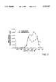
- FIG. 3Comparison of solid phase versus homogenous kinase assays.
- FIG. 4ATP dependence of the radioactive and chemiluminescent PKA assays.
- FIGS. 5A and 5BEffect of known inhibitors on PKA and PKC activity using chemiluminescent detection.
- FIGS. 6A and 6BUse of MPM-2 and 4G10 antibodies to detect activities of serine/threonine and tyrosine protein kinases.
- the inventionprovides efficient and sensitive methods and compositions for detecting, identifying and/or characterizing kinase activity and specific modulators of kinase activity, preferably protein kinase activity.
- the methodsuse a bifunctional kinase reaction product to 1) specifically capture and immobilize phosphorylated product as opposed to unreacted substrate and to 2) specifically detect the immobilized product as opposed to other immobilized molecules.
- One functionality of the productis provided by the phosphorylation reaction itself: the reaction introduces a novel molecular structural feature, or epitope, within the substrate, for which feature a specifically binding receptor is available. Conveniently, this feature comprises the phosphate group itself; for example, a phosphorylated serine or tyrosine residue of a peptide substrate.
- phosphorylationor dephosphorylation, as the reaction can generally be run in reverse
- the substratemay induce a specifically-detectable conformational change which does not necessarily comprise the phosphate group.
- the assaymay use any phosphorylation-dependent feature for which a specifically binding receptor can be obtained.
- Specific immune receptorsuch as an antibody provide convenient such receptors.
- the other functionalityis effected by using a substrate comprising a phosphorylation-independent molecular tag, different from the phosphorylation-dependent feature, for which a specifically-binding receptor is available.
- substrate tagsinclude peptide epitope tags such as FLAG, myc or His, for which specific antibodies are conveniently available, carbohydrate tags for which specific lectins are conveniently available, or other convenient, high-affinity ligand/receptor pairs, such as biotin/avidin.
- the generation of the phosphorylation-dependent featuredoes not preclude the specificity of the binding between the phosphorylation-independent tag and its corresponding receptor.
- the binding of the first receptor to the productdoes not preclude the specificity of the binding of the second receptor to the product.
- the tagsare sufficiently different to avoid cross-reactivity with the receptors.
- the reaction mixturemay also comprise a candidate agent such as a preselected kinase inhibitor or, especially for high-throughput drug screening, a library-derived candidate agent.
- Library-derived candidate agentsencompass numerous chemical classes, though typically they are organic compounds; preferably small organic compounds.
- the librariesmay comprise synthetic and/or naturally derived compounds. For example, numerous means are available for random and directed synthesis of a wide variety of organic compounds and biomolecules, including expression of randomized oligonucleotides.
- libraries of natural compounds in the form of bacterial, fungal, plant and animal extractsare available or readily produced. Additionally, natural and synthetically produced libraries and compounds are readily modified through conventional chemical, physical, and biochemical means.
- known pharmacological agentsmay be subject to directed or random chemical modifications, such as acylation, alkylation, esterification, amidification, etc., to produce structural analogs.
- the agentis provided in standard serial dilutions or in an amount determined by analogy to known modulators.
- the mixtureusually includes additional reagents, such as salts, buffers, etc. to facilitate or maximize kinase activity.
- a wide variety of solid substratesmay be used. Factors to be considered in selecting an appropriate substrate include the adhesion and functional retention of the immobilizing receptor, accessible surface area for binding, wash convenience, cost, high-throughput adaptability, etc. Frequently, the solid substrate will be the wall of the reaction reservoir itself. Preferred substrates maximize signal strength and the signal-to-noise ratio. Exemplary substrates include polystyrene microtiter plates, fine fibers, polymeric or silica-based microbeads, etc., preferably pre-activated to provide maximal protein binding. When used, microbeads are selected by size, range and structure to maximize surface area, filter retention and bead suspension time during the assay incubations.
- reaction conditionscan be employed depending on the targeted kinase(s); in vitro conditions to support activity of exemplary kinases are exemplified below and/or otherwise known in the art.
- the reactiongenerally requires the presence of an effective amount of a nucleoside triphosphate, such as ATP.
- a nucleoside triphosphatesuch as ATP.
- the reactionis carried out at room or elevated temperatures, usually in the range of 20° to 40° C., conveniently at room (ca. 25° C.) temperature.
- reactions timeis minimized, and is usually from 0.1 to 4 hours, more usually about 0.5 to 1.5 hours.
- the kinase reactionoccurs in solution.
- the rate of phosphorylationpreferably exceeds the rate of immobilization sufficient to ensure a solution-phase kinase reaction.
- the wash steps of the methodsinvolve separating unbound components from the immobilized product conjugates, usually by rinsing one to five times with a buffered medium.
- the method used for separating and washingdepends on the nature of the reaction reservoir and solid substrate. For example, where the substrate is in the form of aggregated fibers, the solid phase may be physically transferred from the reaction reservoir to a series of rinse reservoirs. With beads, the separating and washing steps are conveniently performed by filtration, frequently vacuum-assist or centrifugation.
- Labelsmay be directly detected through optical or electron density, nonradiative energy transfers, etc., or indirectly through the a binding or reaction with a reagent which, in turn, provides a detectable signal.
- the second receptormay be decorated with one or more (for signal amplification) reagents such as secondary antibodies conjugated to an enzyme for an ELISA-type assay.
- the disclosed methodsare particularly suited to automated high throughput drug screening.
- the individual sample incubation volumesare less than about 500 ⁇ l, preferably less than about 250 ⁇ l, more preferably less than about 100 ⁇ l.
- Such small sample volumesminimize the use of often scarce candidate agent, expensive enzymes, and hazardous radioactive waste.
- the methodsprovide for automation, especially computerized automation.
- the method stepsare preferably performed by a computer-controlled electromechanical robot. While individual steps may be separately automated, a preferred embodiment provides a single computer-controlled multifunction robot with a single arm axially rotating to and from a plurality of work stations performing the assays' steps.
- the computeris loaded with software which provides the instructions which direct the arm and work station operations and provides input (e.g. keyboard and/or mouse) and display (e.g. monitor) means for operator interfacing.
- PKA catalytic subunitSigma
- protein kinase CPKC
- CAMKIICa 2+ /calmodulin-dependent protein kinase II
- colorimetric kit for the detection of PKA and PKC activitycontaining the YC10 monoclonal antibody anti-phospho GFAP, recognizing phosphoserine in the context of TS(P)AARR (Panvera). The same antibody was also purchased separately.
- Peptideswere synthesized on an automated synthesizer using solid-phase FMOC chemistry with HBTU/HOBT activation (19). Amino caproic acid was used as a spacer between the biotin and the N-terminus of the peptide. Peptides were cleaved from the solid support and deprotected using reagent K (20) and then were purified using reverse phase HPLC. Electrospray mass spectrometry and analytical RP-HPLC were used for QC of the peptides. The following peptides were used as protein kinase substrates:
- bio-RRRVTSAARRSSEQ ID NO:2
- PKA and PKC substrate peptide derived from glial fibrillary acidic proteinPKA and PKC substrate peptide derived from glial fibrillary acidic protein
- bio-FRRLSISTSEQ ID NO:3 (CAMK II substrate peptide derived from phosphorylase kinase)
- peptide 4bio-MDRQTKQQPRQNVAYNREEERRRRVSHDPFAQQRPYENF (SEQ ID NO:4) derived from RIP (21)!
- the peptide substrateswere added in 90 ⁇ l assay buffer containing 20 mM Tris-HCl pH 7.5, 2.5 mM MgCl 2 , and 100 ⁇ M ATP for non-radioactive detection, or 10 ⁇ M cold ATP supplemented with 0.5 ⁇ Ci/well ⁇ - 32 P!ATP for radioactive detection, unless indicated otherwise.
- Peptide 2was added in 90 ⁇ l assay buffer containing 20 mM Tris-HCl pH 7.5, 2.5 mM MgCl 2 , 50 ⁇ M CaCl 2 , 10 ⁇ g/ml phosphatidylserine, 20 ⁇ M ATP, with or without inhibitors. PKC (0.25 ng/well in 10 ⁇ l) was added last, and the kinase reaction was allowed to proceed for 30 minutes at room temperature. Then chemiluminescent detection using YC10 antibody was performed as described below.
- Peptide 3(10 -6 M) was added in 90 ⁇ l assay buffer containing 20 mM Tris-HCl pH 7.5, 10 mM MgCl 2 , 2 mM CaCl 2 , 1 mM DTT, 0.1 mM EDTA, 100 ⁇ M ATP, 100 U calmodulin.
- CAMKIIwas added last in 10 ⁇ l, and the samples were incubated for 30 minutes at room temperature. Chemiluminescent detection using MPM-2 antibody was performed as described below.
- Peptide 4was added in 90 ⁇ l assay buffer containing 100 mM Tris-HCl pH 7.5, 25 mM MnCl 2 , 2 mM EGTA, 0.25 mM Na 3 VO 4 , 125 mM Mg acetate, and 100 ⁇ M ATP for non-radioactive detection, or 10 ⁇ M cold ATP supplemented with 0.5 ⁇ Ci/well ⁇ - 32 P!ATP for radioactive detection.
- srcwas added last (3 unit/well in 10 ⁇ l), kinase reaction was performed at room temperature for 30 minutes, and depending on the detection method, the samples were further processed as described below.
- the sampleswere divided into two. 50 ⁇ l were transferred into wells of streptavidin coated microtiter plates, to allow the capture of the biotinylated peptides for additional 50 minutes. The wells were washed five times with water, dried, broken into scintillation vials and counted. The other half of the reaction mixture was further incubated in the original plate for 50 minutes, then TCA was added to the reaction mixture (10% final concentration) and 30 ⁇ l was spotted onto P81 paper (1 ⁇ 1 inch squares). After 20 minutes the squares were washed three times with water, dried, placed into scintillation vials and counted. A microtiter plate version of the phosphocellulose capture method was also used with similar results. For src only the phosphocellulose capture method was used.
- Example 8Colorimetric and chemiluminescent detection
- the substrate peptideswere initially captured onto streptavidin coated plates prior to the kinase reaction. In later experiments the two reactions were performed simultaneously. Upon completion of the kinase reaction the wells were washed five times with PBS containing 0.2% Tween-20 (T-PBS). Primary antibody (YC10 at 1:10,000 dilution for peptide 2, MPM-2 at 1:2,000 dilution for peptide 3, and 4G10 at 1:2,000 dilution for peptide 4) was added in T-PBS for 30 minutes, then the plate was washed 5 times with T-PBS.
- Primary antibodyYC10 at 1:10,000 dilution for peptide 2, MPM-2 at 1:2,000 dilution for peptide 3, and 4G10 at 1:2,000 dilution for peptide 4
- Secondary anti-mouse antibody(horseradish peroxidase conjugated for calorimetric, or alkaline phosphatase conjugated for chemiluminescent detection) was added at 5000-fold dilution in T-PBS supplemented with 0.2% BSA for 30 minutes, after which the plate was washed five times in T-PBS. In later experiments the primary and secondary antibodies were premixed and added together for 40 minutes. Colorimetric reaction was performed for 5 minutes with o-phenylenediamine substrate, stopped, and the plate was read in a Bio Kinetics Reader EL 340 microplate reader (Bio Tek Instruments) at 492 nm.
- CSPD+Sapphire IIwas added at a threefold dilution in diethanolamine buffer (pH: 10), and after 10 minutes the plate was read with a Luminoskan EL microtiter plate luminometer (Labsystems) or Packard scintillation counter.
- ELISA-type assayscan be performed in a single plate, and thus are easier to automate. For this reason we wanted to establish an ELISA-type protein kinase assay, where the capture of the phosphorylated product is based on the strong binding and high selectivity of streptavidin to biotin.
- streptavidin capturingresulted in a decline in scintillation counts above 10 -6 M peptide concentration. This is attributable to competition of phosphorylated and nonphosphorylated biotinylated peptides to a limited number of streptavidin binding sites, and was found to occur when the number of biotin molecules exceeds the number of streptavidin binding sites (the latter information provided by the manufacturer of the streptavidin coated plate). Overall, use of peptide 1 combined with streptavidin capture resulted in good sensitivity, while the results generated with peptide 2 were much weaker.
- a monoclonal antibody (YC10) recognizing phosphoserine in the context of TS(P)AARRhas recently become commercially available. Since peptide 2 contains this sequence motif, we attempted to detect PKA activity by using this antibody. The kinase reaction was performed in microtiter plates containing the preimmobilized peptide. Then the plate was incubated with the YC10 antibody, peroxidase or alkaline phosphatase conjugated secondary antibody was added, and either a colorimetric or a chemiluminescent detection reaction was performed. To compare the sensitivity of radiolabel, colorimetry and chemiluminescence as detection systems, a dose response of PKA was performed with peptide 2 (FIG. 2) or peptide 1. The chemiluminescent assay was more sensitive than colorimetry, and more sensitive, or at least as sensitive as the radioactive assay using peptide 2, or peptide 1, respectively.
- Radioactive assayOne of the major drawbacks of the radioactive assay is its inefficiency at close to physiological ATP concentrations, due to the diluting effect of high concentration of cold ATP on the radiolabelled one.
- the ATP dependence of the chemilurninescent assaywas also tested. It was found to be sensitive at low micromolar ATP concentrations, but in contrast to the radioactive assay, the activity increased in an ATP-dependent manner (FIG. 4).
- the chemiluminescent assaywas validated using a panel of known kinase inhibitors. Since PKC activity was also found to be easily detectable by this assay (data not shown), it was also included in this experiment. As shown in FIG. 5, when the kinase reactions were performed in the presence of 20 ⁇ M ATP, IC 50 values similar to published data were obtained. At 100 ⁇ M ATP, while the assay itself was more sensitive, substantial loss of potency was observed for most of the tested inhibitors (data not shown). This result is expected, since most of these inhibitors are known to compete with ATP by binding to the ATP binding site of the kinase. As intracellular ATP concentrations are much higher (mM range) these inhibitors show much weaker potency in vivo.
- MPM-2a monoclonal antibody generated against mitotic proteins, and later shown to recognize a phosphorylated epitope has been used in numerous, mostly cell cycle related studies (22-24). Although it's exact epitope is not known, we tested whether this antibody could also function in the detection of substrate peptide phosphorylation. As FIG. 6A demonstrates, MPM-2 can detect activity of CAMK II (and presumably other kinases) when used with peptide 4 as kinase substrate.
- chemiluminescent detectioncan provide a sensitive alternative method also for tyrosine kinases.
- Sensitive detection of tyrosine kinase activity by using peptide substratesis especially problematic because the K m of these peptides is frequently in the high micromolar range (2).
- Activity of these kinaseshas already been detected by colorimetric methods using anti-phosphotyrosine antibodies (14-18).
- the disclosed assayhas many obvious advantages over the traditional radioactive protein kinase assay. All components of this assay can be stored for long periods of time with no loss in activity, and no radioactive waste is generated. The assay is also very fast and simple, the whole experiment can be easily accomplished in 2-3 hours. Since all steps are performed in one microtiter plate, automation is easier than with the radioactive assay requiring filtration and the use of corrosive materials.
- the chemiluminescent methodalso provided higher sensitivity than what was achievable by colorimetric detection, in accordance with results obtained by others comparing chemiluminescence and colorimetry (25-27). The robustness of the signal and the high sensitivity makes chemiluminescent detection a very good option for drug discovery efforts, such as high throughput screening.
- radioactive kinase assayThe limitations of the radioactive kinase assay such as requirement for non-physiologically low ATP and high substrate concentrations, resulted in the discovery of many kinase inhibitors competing with ATP, but very few inhibitors with other characteristics. Most of these inhibitors appear very potent under such artificial assay conditions, but prove to be rather weak and quite toxic in a cellular environment. Furthermore, since they all target the well conserved ATP binding site of protein kinases, their selectivity is also generally poor. In contrast to the radioactive method, the chemiluminescent assay is much more robust at close to physiological ATP concentrations, providing much more realistic testing conditions.
- Another drawback of the radioactive methodis that its sensitivity strongly depends on a peptide substrate with low K m . Efforts to identify such a peptide substrate for a novel kinase are time and energy consuming, and do not always result in success. It is especially true for tyrosine kinases, where the K m of peptide substrates is generally high (2). In contrast, the chemiluminescent assay provided a robust response even with substrates having high K m values, indicating that in case of a novel kinase efforts to identify a substrate with kinetically favorable properties may not be immediately required. Rather, the effort should be spent on identifying in vivo substrates. At present chemiluminescent assay conditions have been established for 8 kinases in our laboratory, and a highly sensitive high throughput drug screening operation is in progress for some of them.
- Chemiluminescent detectionresulted in a strong signal at 10 -7 M peptide concentrations, far below the K m of even the best peptide substrates. This, together with a requirement for low enzyme concentration, and also the robust response observed at high ATP concentration, indicate that among all presently available protein kinase assay methods chemiluminescent detection coupled with ELISA provides the highest sensitivity and best conditions to mimic the intracellular environment. Because of these favorable properties, application of this approach to drug discovery may result in the identification of novel classes of inhibitors not just for otherwise well characterized protein kinases, but also for recently discovered ones.
- the described methodhas two limitations, but none of them are inherent. 1/ An antibody is required, distinguishing between the phosphorylated and non-phosphorylated peptide substrate. This is not a problem for tyrosine kinases, and recently more and more phosphoserine and phosphothreonine specific monoclonal and polyclonal antibodies have become commercially available. We also found that the YC10 and MPM-2 antibodies, used in this study, can detect the activity of kinases other than PKA, PKC, and CAMK II, using substrates with little similarity to peptides 2 and 4, respectively. Detailed characterization of the epitopes of these antibodies is in progress.
- the streptavidin coated microtiter platehas limited peptide binding capacity. While a higher concentration of the substrate peptide is not required for the purpose of sensitivity, it would be required for determining the kinetic properties of the peptide, and also during the characterization of a test compound. This limitation can be overcome by using streptavidin coated beads with much higher biotin binding capacity.
Landscapes
- Health & Medical Sciences (AREA)
- Chemical & Material Sciences (AREA)
- Life Sciences & Earth Sciences (AREA)
- Immunology (AREA)
- Molecular Biology (AREA)
- Engineering & Computer Science (AREA)
- Organic Chemistry (AREA)
- General Health & Medical Sciences (AREA)
- Hematology (AREA)
- Biochemistry (AREA)
- Medicinal Chemistry (AREA)
- Urology & Nephrology (AREA)
- Biomedical Technology (AREA)
- Food Science & Technology (AREA)
- Physics & Mathematics (AREA)
- Analytical Chemistry (AREA)
- Microbiology (AREA)
- Cell Biology (AREA)
- General Physics & Mathematics (AREA)
- Pathology (AREA)
- Biophysics (AREA)
- Genetics & Genomics (AREA)
- Proteomics, Peptides & Aminoacids (AREA)
- Biotechnology (AREA)
- Measuring Or Testing Involving Enzymes Or Micro-Organisms (AREA)
Abstract
Description
1. Field of the Invention
The invention relates to methods for detecting kinase (an enzyme) activity.
2. Background
Protein kinases represent one of the largest group of enzymes, with critical role in many cellular signal transduction processes. Due to genome projects and other recent developments in molecular biology techniques, new kinases with uncharacterized biochemical properties and substrate specificity are discovered more and more frequently. Thus, a sensitive assay measuring the activity of these enzymes would be of great value. Traditional protein kinase assays include the use of labeled ATP as phosphate donor, and a substrate peptide as phosphoacceptor containing the respective kinase recognition motif. Following the kinase reaction the substrate peptide is captured on an appropriate filter paper. Unreacted labeled ATP and metabolites are resolved from the radioactive peptide substrate by various techniques, involving trichloroacetic acid precipitation and extensive washing. Addition of several positively charged residues allows capture on phosphocellulose paper followed by washing. Radioactivity incorporated into the substrate peptide is detected by scintillation counting (1). This assay is relatively simple, reasonably sensitive, and the peptide substrate can be adjusted both in terms of sequence and concentration to meet the assay requirements. But it also has several drawbacks: it generates radioactive waste; radioactive ATP has a half-life of only 14 days; the assay is sensitive only at low micromolar concentrations of cold ATP, while the intracellular ATP concentration is in the millimolar range; and, to achieve an appropriate signal the peptide substrate has to be used at around its Km value, which is about 5-20 μM even for the best substrates of well characterized kinases (2), while the intracellular concentration of the endogenous polypeptide substrates is presumably much lower. The subphysiological ATP and supraphysiological substrate concentrations of this assay may have adverse effect on attempts to develop inhibitors efficient under the much different in vivo conditions.
Several approaches have been tried to overcome these limitations. Some of them used synthetic peptide substrates and non-radioactive detection of their phosphorylation by fluorescence, spectrophotometry, or other methods (3-8). However, these methods are either insensitive, or quite laborious and not amenable to automation. Alternative phosphopeptide capture methods have also been tried, but these assays are still based on radioactive detection (9, 10). Cell based assays have also been developed as a read-out for the activities of various protein kinases. These approaches work under physiological conditions, but are generally much more complicated, prone to various sources of error, and the real target of a drug lead compound may not even be related to the kinase of interest (11-13). The availability of phosphotyrosine antibodies allowed the development of colorimetric ELISA assays for detection of protein tyrosine kinases using polypeptides as substrates (14-18). In addition to being non-radioactive, this approach generally proved to be sensitive and easy to automate, providing a good alternative for tyrosine kinases.
The invention provides novel methods for detecting kinase activity in solution, without the use of radioactivity. The subject methods marry the kinetic advantages and sensitivity of solution-based reaction with the efficiency, cost-effectiveness and high-throughput adaptability of solid-phase wash and detection steps, yet is conveniently practiced in a single tube. The methods may be used to assay for kinase activity per se or, by controlling for the kinase activity, for modulators of kinase activity. In addition, the invention provides kits for kinase modulator screening which include premeasured amounts of the compositions used in the disclosed methods. The general methods involve steps:
a) incubating a solution comprising a kinase, a substrate of the kinase wherein the substrate comprises a phosphorylation-independent first tag, a first receptor, and a nucleoside triphosphate, under conditions whereunder the kinase phosphorylates the substrate in solution to form a product comprising the first tag, (and usually most of the substrate), and a phosphorylation-dependent second tag;
b) further incubating the solution under conditions whereunder the first receptor immobilizes the product on a solid substrate by specifically binding one of the two tags to form a first immobilized conjugate comprising the first receptor and the product;
c) washing the solid substrate;
d) contacting the first immobilized conjugate with a second receptor under conditions whereunder the second receptor specifically binds the other one of the two tags to form a second immobilized conjugate comprising the first receptor, the product and the second receptor;
e) washing the solid substrate;
f) detecting the second receptor;
wherein the presence of the second receptor indicates the presence of the product and the presence of the product indicates the presence of the kinase activity.
For modulator screens, the solution further comprises a candidate agent and the initial incubation is under conditions whereby, but for the presence of the candidate agent, the kinase (or kinases) phosphorylates the substrate at a first, control kinase activity and so converts at least a detectable portion, and preferably, substantially all of the initial amount of substrate into product, whereby a final amount of the substrate remains. As such, depending on the stop point of the reaction, the measured activity may reflect a catalytic rate or an equilibrium constant. For these assays, a difference between the kinase activity in the presence and absence of the agent indicates that said candidate agent modulates the activity of the targeted kinase.
A wide variety of kinases, substrates, tags, receptors, labels may be used. Preferred kinases for use in the methods are protein kinases and preferred substrates are peptide or protein substrates. According to one preferred embodiment, the first tag is biotin, one of the receptors is avidin or an avidin-like protein such as streptavidin, the second tag is a phosphorylated serine or tyrosine and the other receptor is an antibody which specifically binds the product at the phosphorylated amino acid. A wide variety of means may be used for detecting the second receptor. Preferred means are cost-effective and readily automated for high-throughput analysis, such as optical detection. For example, the second receptor may comprise a label, such as an enzyme which enzyme catalyzes a chromogenic or chemiluminogenic reaction. Alternatively, the second receptor can be detected by the use of a third labeled receptor which specifically binds the second receptor.
The method is exemplified with a preferred chemiluminescent protein kinase assay using biotinylated substrate peptides captured on a streptavidin coated microtiter plate and monoclonal antibodies to detect their phosphorylation. Assay conditions were optimized and validated for sensitive measurement of protein kinase A (PKA), protein kinase C (PKC), Ca2+ /calmodulin-dependent protein kinase II (CAMKII), and src activities. The chemiluminescent detection has several advantages over currently used radioactive or colorimetric methods. The assay is fast, very simple to perform, and easily adaptable to automation and high-throughput drug screening. It provides high sensitivity and robust signal using low concentrations of enzyme and substrate. Signal amplitude shows positive correlation with ATP concentration, thus allowing the assay to function at high, close to physiological ATP levels, in contrast to the radioactive method. Overall, among the presently available methods for the detection of protein kinase activity, chemiluminescence was found to provide the highest sensitivity under conditions most closely mimicking the intracellular environment.
FIGS. 1A and 1B. Comparison of capturing substrate peptides by p81 phosphocellulose paper (FIG. 1A) and streptavidin coated microtiter plate (FIG. 1B).
FIG. 2. Comparison of three methods for the detection of PKA enzymatic activity.
FIG. 3. Comparison of solid phase versus homogenous kinase assays.
FIG. 4. ATP dependence of the radioactive and chemiluminescent PKA assays.
FIGS. 5A and 5B. Effect of known inhibitors on PKA and PKC activity using chemiluminescent detection.
FIGS. 6A and 6B. Use of MPM-2 and 4G10 antibodies to detect activities of serine/threonine and tyrosine protein kinases.
The invention provides efficient and sensitive methods and compositions for detecting, identifying and/or characterizing kinase activity and specific modulators of kinase activity, preferably protein kinase activity. The methods use a bifunctional kinase reaction product to 1) specifically capture and immobilize phosphorylated product as opposed to unreacted substrate and to 2) specifically detect the immobilized product as opposed to other immobilized molecules. One functionality of the product is provided by the phosphorylation reaction itself: the reaction introduces a novel molecular structural feature, or epitope, within the substrate, for which feature a specifically binding receptor is available. Conveniently, this feature comprises the phosphate group itself; for example, a phosphorylated serine or tyrosine residue of a peptide substrate. Alternatively, phosphorylation (or dephosphorylation, as the reaction can generally be run in reverse) of the substrate may induce a specifically-detectable conformational change which does not necessarily comprise the phosphate group. The assay may use any phosphorylation-dependent feature for which a specifically binding receptor can be obtained. Specific immune receptor, such as an antibody provide convenient such receptors.
The other functionality is effected by using a substrate comprising a phosphorylation-independent molecular tag, different from the phosphorylation-dependent feature, for which a specifically-binding receptor is available. Exemplary substrate tags include peptide epitope tags such as FLAG, myc or His, for which specific antibodies are conveniently available, carbohydrate tags for which specific lectins are conveniently available, or other convenient, high-affinity ligand/receptor pairs, such as biotin/avidin. In any event, the generation of the phosphorylation-dependent feature does not preclude the specificity of the binding between the phosphorylation-independent tag and its corresponding receptor. Similarly, the binding of the first receptor to the product does not preclude the specificity of the binding of the second receptor to the product. In addition, the tags are sufficiently different to avoid cross-reactivity with the receptors.
In addition to the kinase, substrate, NTP, and first receptor, the reaction mixture may also comprise a candidate agent such as a preselected kinase inhibitor or, especially for high-throughput drug screening, a library-derived candidate agent. Library-derived candidate agents encompass numerous chemical classes, though typically they are organic compounds; preferably small organic compounds. The libraries may comprise synthetic and/or naturally derived compounds. For example, numerous means are available for random and directed synthesis of a wide variety of organic compounds and biomolecules, including expression of randomized oligonucleotides. Alternatively, libraries of natural compounds in the form of bacterial, fungal, plant and animal extracts are available or readily produced. Additionally, natural and synthetically produced libraries and compounds are readily modified through conventional chemical, physical, and biochemical means. In addition, known pharmacological agents may be subject to directed or random chemical modifications, such as acylation, alkylation, esterification, amidification, etc., to produce structural analogs. The agent is provided in standard serial dilutions or in an amount determined by analogy to known modulators. In addition, the mixture usually includes additional reagents, such as salts, buffers, etc. to facilitate or maximize kinase activity.
A wide variety of solid substrates may be used. Factors to be considered in selecting an appropriate substrate include the adhesion and functional retention of the immobilizing receptor, accessible surface area for binding, wash convenience, cost, high-throughput adaptability, etc. Frequently, the solid substrate will be the wall of the reaction reservoir itself. Preferred substrates maximize signal strength and the signal-to-noise ratio. Exemplary substrates include polystyrene microtiter plates, fine fibers, polymeric or silica-based microbeads, etc., preferably pre-activated to provide maximal protein binding. When used, microbeads are selected by size, range and structure to maximize surface area, filter retention and bead suspension time during the assay incubations.
A wide variety of reaction conditions can be employed depending on the targeted kinase(s); in vitro conditions to support activity of exemplary kinases are exemplified below and/or otherwise known in the art. For example, the reaction generally requires the presence of an effective amount of a nucleoside triphosphate, such as ATP. For many mammalian kinases, the reaction is carried out at room or elevated temperatures, usually in the range of 20° to 40° C., conveniently at room (ca. 25° C.) temperature. For high-throughput applications, reactions time is minimized, and is usually from 0.1 to 4 hours, more usually about 0.5 to 1.5 hours. Importantly, the kinase reaction occurs in solution. Hence, for single-tub assays where the conditions to effect phosphorylation are the same as those which effect immobilization, the rate of phosphorylation preferably exceeds the rate of immobilization sufficient to ensure a solution-phase kinase reaction.
The wash steps of the methods involve separating unbound components from the immobilized product conjugates, usually by rinsing one to five times with a buffered medium. The method used for separating and washing depends on the nature of the reaction reservoir and solid substrate. For example, where the substrate is in the form of aggregated fibers, the solid phase may be physically transferred from the reaction reservoir to a series of rinse reservoirs. With beads, the separating and washing steps are conveniently performed by filtration, frequently vacuum-assist or centrifugation.
A variety of methods may be used to detect the label depending on the nature of the label and other assay components. Labels may be directly detected through optical or electron density, nonradiative energy transfers, etc., or indirectly through the a binding or reaction with a reagent which, in turn, provides a detectable signal. For example, the second receptor may be decorated with one or more (for signal amplification) reagents such as secondary antibodies conjugated to an enzyme for an ELISA-type assay.
The disclosed methods are particularly suited to automated high throughput drug screening. In a preferred embodiment, the individual sample incubation volumes are less than about 500 μl, preferably less than about 250 μl, more preferably less than about 100 μl. Such small sample volumes minimize the use of often scarce candidate agent, expensive enzymes, and hazardous radioactive waste. Furthermore, the methods provide for automation, especially computerized automation. Accordingly, the method steps are preferably performed by a computer-controlled electromechanical robot. While individual steps may be separately automated, a preferred embodiment provides a single computer-controlled multifunction robot with a single arm axially rotating to and from a plurality of work stations performing the assays' steps. The computer is loaded with software which provides the instructions which direct the arm and work station operations and provides input (e.g. keyboard and/or mouse) and display (e.g. monitor) means for operator interfacing.
The following experimental section is offered by way of illustration and not by way of limitation.
Example 1: Materials
The following reagents were used: PKA catalytic subunit (Sigma); protein kinase C (PKC, a mixture of α, β, and γ isoforms), src, and the monoclonal antibodies 4G10 and MPM-2 (Upstate Biotechnology); Ca2+ /calmodulin-dependent protein kinase II (CAMKII, New England Biolabs); colorimetric kit for the detection of PKA and PKC activity, containing the YC10 monoclonal antibody anti-phospho GFAP, recognizing phosphoserine in the context of TS(P)AARR (Panvera). The same antibody was also purchased separately. Alkaline phosphatase conjugated goat anti-mouse antibody (Tropix or Pierce); chemiluminescent substrate CSPD+Sapphire II (Tropix); γ-32 P!ATP (Du Pont NEN); streptavidin coated black microtiter plates (Labsystems or Xenopore); P81 phosphocellulose paper (Whatman). All kinase inhibitors were from LC Laboratories.
Example 2: Peptide synthesis
Peptides were synthesized on an automated synthesizer using solid-phase FMOC chemistry with HBTU/HOBT activation (19). Amino caproic acid was used as a spacer between the biotin and the N-terminus of the peptide. Peptides were cleaved from the solid support and deprotected using reagent K (20) and then were purified using reverse phase HPLC. Electrospray mass spectrometry and analytical RP-HPLC were used for QC of the peptides. The following peptides were used as protein kinase substrates:
peptide 1: bio-LRRASLG (SEQ ID NO:1) (Kemptide)
peptide 2: bio-RRRVTSAARRS (SEQ ID NO:2) (PKA and PKC substrate peptide derived from glial fibrillary acidic protein)
peptide 3: bio-FRRLSIST (SEQ ID NO:3) (CAMK II substrate peptide derived from phosphorylase kinase)
peptide 4: bio-MDRQTKQQPRQNVAYNREEERRRRVSHDPFAQQRPYENF (SEQ ID NO:4) derived from RIP (21)!
Example 3: PKA kinase reaction
The peptide substrates were added in 90 μl assay buffer containing 20 mM Tris-HCl pH 7.5, 2.5 mM MgCl2, and 100 μM ATP for non-radioactive detection, or 10 μM cold ATP supplemented with 0.5 μCi/well γ-32 P!ATP for radioactive detection, unless indicated otherwise. Freshly reconstituted PKA (1.25 ng/well in 10 μl, unless indicated otherwise) was added last and the samples were incubated for 30 minutes at room temperature. Depending on the detection method the samples were further processed as described below.
Example 4: PKC kinase reaction
Example 5: CAMKII kinase reaction
Peptide 3 (10-6 M) was added in 90 μl assay buffer containing 20 mM Tris-HCl pH 7.5, 10 mM MgCl2, 2 mM CaCl2, 1 mM DTT, 0.1 mM EDTA, 100 μM ATP, 100 U calmodulin. CAMKII was added last in 10 μl, and the samples were incubated for 30 minutes at room temperature. Chemiluminescent detection using MPM-2 antibody was performed as described below.
Example 6: SRC kinase reaction
Example 7: Radioactive detection
After performing a PKA kinase reaction for 30 minutes, the samples were divided into two. 50 μl were transferred into wells of streptavidin coated microtiter plates, to allow the capture of the biotinylated peptides for additional 50 minutes. The wells were washed five times with water, dried, broken into scintillation vials and counted. The other half of the reaction mixture was further incubated in the original plate for 50 minutes, then TCA was added to the reaction mixture (10% final concentration) and 30 μl was spotted onto P81 paper (1×1 inch squares). After 20 minutes the squares were washed three times with water, dried, placed into scintillation vials and counted. A microtiter plate version of the phosphocellulose capture method was also used with similar results. For src only the phosphocellulose capture method was used.
Example 8: Colorimetric and chemiluminescent detection
The substrate peptides were initially captured onto streptavidin coated plates prior to the kinase reaction. In later experiments the two reactions were performed simultaneously. Upon completion of the kinase reaction the wells were washed five times with PBS containing 0.2% Tween-20 (T-PBS). Primary antibody (YC10 at 1:10,000 dilution forpeptide 2, MPM-2 at 1:2,000 dilution forpeptide 3, and 4G10 at 1:2,000 dilution for peptide 4) was added in T-PBS for 30 minutes, then the plate was washed 5 times with T-PBS. Secondary anti-mouse antibody (horseradish peroxidase conjugated for calorimetric, or alkaline phosphatase conjugated for chemiluminescent detection) was added at 5000-fold dilution in T-PBS supplemented with 0.2% BSA for 30 minutes, after which the plate was washed five times in T-PBS. In later experiments the primary and secondary antibodies were premixed and added together for 40 minutes. Colorimetric reaction was performed for 5 minutes with o-phenylenediamine substrate, stopped, and the plate was read in a Bio Kinetics Reader EL 340 microplate reader (Bio Tek Instruments) at 492 nm. For chemiluminescent detection CSPD+Sapphire II was added at a threefold dilution in diethanolamine buffer (pH: 10), and after 10 minutes the plate was read with a Luminoskan EL microtiter plate luminometer (Labsystems) or Packard scintillation counter.
Example 9: Results
While filtration assays generally require one plate for reaction, and another for filtration, ELISA-type assays can be performed in a single plate, and thus are easier to automate. For this reason we wanted to establish an ELISA-type protein kinase assay, where the capture of the phosphorylated product is based on the strong binding and high selectivity of streptavidin to biotin. The binding and phosphorylation characteristics of two biotinylated PKA substrate peptides, kemptide (peptide 1) and a peptide derived from glial fibrillary acidic protein (peptide 2), were compared (FIG. 1). Following the radioactive PKA reaction the two peptides were captured either by the traditional phosphocellulose paper method or by streptavidin immobilized on a microtiter plate. Much higher PKA activity was detected usingpeptide 1 as substrate with both capture methods, in agreement with published data describing kemptide as the best substrate for this kinase (2). Because of its unfavorable kinetic properties the Km ofpeptide 2 can not be determined by this experiment, but it is definitely above 20 μM. At 10-6 M peptide concentration the binding efficiency ofpeptide 1 to streptavidin was much higher than to phosphocellulose, which require large number of positive charges. Hence, the more positively chargedpeptide 2 showed better binding to phosphocellulose. With both peptides, streptavidin capturing resulted in a decline in scintillation counts above 10-6 M peptide concentration. This is attributable to competition of phosphorylated and nonphosphorylated biotinylated peptides to a limited number of streptavidin binding sites, and was found to occur when the number of biotin molecules exceeds the number of streptavidin binding sites (the latter information provided by the manufacturer of the streptavidin coated plate). Overall, use ofpeptide 1 combined with streptavidin capture resulted in good sensitivity, while the results generated withpeptide 2 were much weaker.
A monoclonal antibody (YC10) recognizing phosphoserine in the context of TS(P)AARR has recently become commercially available. Sincepeptide 2 contains this sequence motif, we attempted to detect PKA activity by using this antibody. The kinase reaction was performed in microtiter plates containing the preimmobilized peptide. Then the plate was incubated with the YC10 antibody, peroxidase or alkaline phosphatase conjugated secondary antibody was added, and either a colorimetric or a chemiluminescent detection reaction was performed. To compare the sensitivity of radiolabel, colorimetry and chemiluminescence as detection systems, a dose response of PKA was performed with peptide 2 (FIG. 2) orpeptide 1. The chemiluminescent assay was more sensitive than colorimetry, and more sensitive, or at least as sensitive as the radioactiveassay using peptide 2, orpeptide 1, respectively.
Next we attempted to reduce the number of steps in the more sensitive chemiluminescent assay. We found that preincubation of the streptavidin plate with the biotinylated peptide substrate was not necessary: even higher sensitivity was achieved when the peptide binding and phosphorylation reactions were performed simultaneously, while the peptide and kinase were in solution (FIG. 3). With both approaches strong signal was observed at peptide concentration as low as 10-7 M. Again, above 10-6 M peptide concentration a decline in assay sensitivity was detected, presumably due to limiting biotin binding sites. Furthermore, consecutive incubations with the two antibodies can also be replaced by adding the two antibodies together, without any loss in sensitivity.
One of the major drawbacks of the radioactive assay is its inefficiency at close to physiological ATP concentrations, due to the diluting effect of high concentration of cold ATP on the radiolabelled one. The ATP dependence of the chemilurninescent assay was also tested. It was found to be sensitive at low micromolar ATP concentrations, but in contrast to the radioactive assay, the activity increased in an ATP-dependent manner (FIG. 4).
The chemiluminescent assay was validated using a panel of known kinase inhibitors. Since PKC activity was also found to be easily detectable by this assay (data not shown), it was also included in this experiment. As shown in FIG. 5, when the kinase reactions were performed in the presence of 20 μM ATP, IC50 values similar to published data were obtained. At 100 μM ATP, while the assay itself was more sensitive, substantial loss of potency was observed for most of the tested inhibitors (data not shown). This result is expected, since most of these inhibitors are known to compete with ATP by binding to the ATP binding site of the kinase. As intracellular ATP concentrations are much higher (mM range) these inhibitors show much weaker potency in vivo.
Next we assessed the generality of the chemiluminescent detection method in the context of kinases with very different substrate specificity. MPM-2, a monoclonal antibody generated against mitotic proteins, and later shown to recognize a phosphorylated epitope has been used in numerous, mostly cell cycle related studies (22-24). Although it's exact epitope is not known, we tested whether this antibody could also function in the detection of substrate peptide phosphorylation. As FIG. 6A demonstrates, MPM-2 can detect activity of CAMK II (and presumably other kinases) when used withpeptide 4 as kinase substrate.
We tested whether chemiluminescent detection can provide a sensitive alternative method also for tyrosine kinases. Sensitive detection of tyrosine kinase activity by using peptide substrates is especially problematic because the Km of these peptides is frequently in the high micromolar range (2). Activity of these kinases has already been detected by colorimetric methods using anti-phosphotyrosine antibodies (14-18). To assess the usefulness of chemiluminescence for the detection of tyrosine kinase activity we used src and abiotinylated peptide peptide 3, derived from the protein kinase RIP (21)! which we found to be the best substrate for this enzyme after screening several tyrosine containing peptides. This peptide still proved to have kinetically unfavorable properties when tested in a radioactive assay (FIG. 6B), similar to published data on other tyrosine kinase substrate peptides (2). In contrast, using the anti-phosphotyrosine antibody 4G10 and chemiluminescence for detection provided much higher assay sensitivity at a much lower peptide concentration (FIG. 6B).
Example 10: Discussion
The disclosed assay has many obvious advantages over the traditional radioactive protein kinase assay. All components of this assay can be stored for long periods of time with no loss in activity, and no radioactive waste is generated. The assay is also very fast and simple, the whole experiment can be easily accomplished in 2-3 hours. Since all steps are performed in one microtiter plate, automation is easier than with the radioactive assay requiring filtration and the use of corrosive materials. The chemiluminescent method also provided higher sensitivity than what was achievable by colorimetric detection, in accordance with results obtained by others comparing chemiluminescence and colorimetry (25-27). The robustness of the signal and the high sensitivity makes chemiluminescent detection a very good option for drug discovery efforts, such as high throughput screening.
The limitations of the radioactive kinase assay such as requirement for non-physiologically low ATP and high substrate concentrations, resulted in the discovery of many kinase inhibitors competing with ATP, but very few inhibitors with other characteristics. Most of these inhibitors appear very potent under such artificial assay conditions, but prove to be rather weak and quite toxic in a cellular environment. Furthermore, since they all target the well conserved ATP binding site of protein kinases, their selectivity is also generally poor. In contrast to the radioactive method, the chemiluminescent assay is much more robust at close to physiological ATP concentrations, providing much more realistic testing conditions.
Another drawback of the radioactive method is that its sensitivity strongly depends on a peptide substrate with low Km. Efforts to identify such a peptide substrate for a novel kinase are time and energy consuming, and do not always result in success. It is especially true for tyrosine kinases, where the Km of peptide substrates is generally high (2). In contrast, the chemiluminescent assay provided a robust response even with substrates having high Km values, indicating that in case of a novel kinase efforts to identify a substrate with kinetically favorable properties may not be immediately required. Rather, the effort should be spent on identifying in vivo substrates. At present chemiluminescent assay conditions have been established for 8 kinases in our laboratory, and a highly sensitive high throughput drug screening operation is in progress for some of them.
Chemiluminescent detection resulted in a strong signal at 10-7 M peptide concentrations, far below the Km of even the best peptide substrates. This, together with a requirement for low enzyme concentration, and also the robust response observed at high ATP concentration, indicate that among all presently available protein kinase assay methods chemiluminescent detection coupled with ELISA provides the highest sensitivity and best conditions to mimic the intracellular environment. Because of these favorable properties, application of this approach to drug discovery may result in the identification of novel classes of inhibitors not just for otherwise well characterized protein kinases, but also for recently discovered ones.
The described method has two limitations, but none of them are inherent. 1/ An antibody is required, distinguishing between the phosphorylated and non-phosphorylated peptide substrate. This is not a problem for tyrosine kinases, and recently more and more phosphoserine and phosphothreonine specific monoclonal and polyclonal antibodies have become commercially available. We also found that the YC10 and MPM-2 antibodies, used in this study, can detect the activity of kinases other than PKA, PKC, and CAMK II, using substrates with little similarity topeptides
Example 11: Parenthetical Numerically Cited References
1/ Casnellie, J. E. (1991) in Protein Phosphorylation, Part A. Methods in Enzymology vol. 200 (Hunter, T., Sefton, B. M., eds) pp. 115-120, Academic Press, San Diego
2/ Kemp, B. E., and Pearson, R. B. (1991) in Protein Phosphorylation, Part A. Methods in Enzymology vol. 200 (Hunter, T., Sefton, B. M., eds) pp. 121-134, Academic Press, San Diego
3/ Isbell, J. C., Christian, S. T., Mashburn, N. A., and Bell, P. D. (1995) Life Sci 57, 1701-1707
4/ Erdbrugger, W., Strohm, P., and Michel, M. C. (1995)Cell Signal 7, 635-642
5/ Lutz, M. P., Pinon, D. I., and Miller, L. J. (1994) Anal. Biochem. 220, 268-274
6/ McIlroy, B. K., Walters, J. D., and Johnson, J. D. (1991) Anal. Biochem. 195, 148-152
7/ Wang, Z. X., Cheng, Q., and Kililea, S. D. (1995) Anal. Biochem. 230, 55-61
8/ Zhao, Z. H., et al. (1991) Biochem. Biophys. Res. Comm. 176, 1454-1461
9/ Toomik, R., et al. (1993) Anal. Biochem. 209, 348-353
10/ Goueli, B. S., Hsiao, K., Tereba, A., Goueli, S. A. (1995) Anal. Biochem. 225, 10-17
11/ Boge, A., and Roth, R. A. (1995) Anal. Biochem. 231, 323-332
12/ King, I. C., Feng, M., and Catino, J. J. (1993) Life Sci. 53, 1465-1472
13/ Sista, P., et al. (1994) Mol. Cell. Biochem. 141, 129-134
14/ Schraag, B., et al. (1993) Anal. Biochem. 211, 233-239
15/ Farley, K., et al. (1992) Anal. Biochem. 203, 151-157
16/ Babcook, J., Watts, J., Aebersold, R., and Ziltener, H. J. (1991) Anal. Biochem. 196, 245-251
17/ Lazaro, I., et al. (1991) Anal. Biochem. 192, 257-261
18/ Angeles, T. S., et al. (1996) Anal. Biochem. 236, 49-55
19/ Geiser, T. H., Bergot, J. B., and Otteson, K. M. (1988) in Macromolecular Sequencing and Synthesis, Selected Methods and Applications (ed. Liss, A. R.) p. 199
20/ Fields, G. B. and Noble, R. L. (1990) Int. J. Peptide Protein Research 35, 161-214
21/ Hsu, H., et al. (1996)Immunity 4, 387-396
22/ Davis, F. M., et al. (1983) Proc. Natl. Acad. Sci. U.S.A. 80, 2926-2930
23/ Kuang, J., et al. (1994)Mol. Biol. Cell 5, 135-145
24/ Taagepera, S., et al. (1994)Mol. Biol. Cell 5, 1243-1251
25/ Van Poucke, S. O., and Nelis, H. J. (1995) Appl. Environ. Microbiol. 61, 4505-4509
26/ Gravel, P., et al. (1994) Anal. Biochem. 221, 66-71
27/ During, K. (1993) J. Chromatogr. 618, 105-131
All publications and patent applications cited in this specification are herein incorporated by reference as if each individual publication or patent application were specifically and individually indicated to be incorporated by reference. Although the foregoing invention has been described in some detail by way of illustration and example for purposes of clarity of understanding, it will be readily apparent to those of ordinary skill in the art in light of the teachings of this invention that certain changes and modifications may be made thereto without departing from the spirit or scope of the appended claims.
__________________________________________________________________________SEQUENCE LISTING (1) GENERAL INFORMATION: (iii) NUMBER OF SEQUENCES: 4 (2) INFORMATION FOR SEQ ID NO:1: (i) SEQUENCE CHARACTERISTICS: (A) LENGTH: 7 amino acids (B) TYPE: amino acid (C) STRANDEDNESS: Not Relevant (D) TOPOLOGY: Not Relevant (ii) MOLECULE TYPE: peptide (xi) SEQUENCE DESCRIPTION: SEQ ID NO:1: LeuArgArgAlaSerLeuGly 15 (2) INFORMATION FOR SEQ ID NO:2: (i) SEQUENCE CHARACTERISTICS: (A) LENGTH: 11 amino acids (B) TYPE: amino acid (C) STRANDEDNESS: Not Relevant (D) TOPOLOGY: Not Relevant (ii) MOLECULE TYPE: peptide (xi) SEQUENCE DESCRIPTION: SEQ ID NO:2: ArgArgArgValThrSerAlaAlaArgArgSer 1510 (2) INFORMATION FOR SEQ ID NO:3: (i) SEQUENCE CHARACTERISTICS: (A) LENGTH: 8 amino acids (B) TYPE: amino acid (C) STRANDEDNESS: Not Relevant (D) TOPOLOGY: Not Relevant (ii) MOLECULE TYPE: peptide (xi) SEQUENCE DESCRIPTION: SEQ ID NO:3: PheArgArgLeuSerIleSerThr 15 (2) INFORMATION FOR SEQ ID NO:4: (i) SEQUENCE CHARACTERISTICS: (A) LENGTH: 39 amino acids (B) TYPE: amino acid (C) STRANDEDNESS: Not Relevant (D) TOPOLOGY: Not Relevant (ii) MOLECULE TYPE: peptide (xi) SEQUENCE DESCRIPTION: SEQ ID NO:4: MetAspArgGlnThrLysGlnGlnProArgGlnAsnValAlaTyrAsn 151015 ArgGluGluGluArgArgArgArgValSerHisAspProPheAlaGln 202530 GlnArgProTyrGluAsnPhe 35 __________________________________________________________________________
Claims (11)
1. A method for detecting kinase activity in solution and without the use of radioactivity, said method comprising steps:
a) incubating a solution comprising a kinase, a first receptor, a nucleoside triphosphate and a substrate of said kinase wherein said substrate comprises a phosphorylation-independent first tag, under conditions whereby said kinase transfers a phosphate group from said nucleoside triphosphate to said substrate in solution to form a product comprising said first tag and a phosphorylation-dependent second tag;
b) further incubating said solution under conditions whereby said first receptor immobilizes said product on a solid substrate by specifically binding one of said first and said second tag to form a first immobilized conjugate comprising said first receptor and said product;
c) washing said solid substrate;
d) contacting said first immobilized conjugate with a second receptor under conditions whereby said second receptor specifically binds the other one of said first and said second tag to form a second immobilized conjugate comprising said first receptor, said product and said second receptor;
e) washing said solid substrate;
f) detecting said second receptor;
wherein the presence of said second receptor indicates the presence of said product and the presence of said product indicates the presence of said kinase activity.
2. A method according to claim 1, wherein all said steps are performed in a single tube.
3. A method according to claim 1, wherein said kinase is a protein kinase and said substrate comprises a peptide or polypeptide.
4. A method according to claim 1, wherein one of said first and second receptors is an antibody which specifically binds said product at said second tag.
5. A method according to claim 1, wherein said first tag is biotin and one of said first and second receptors is streptavidin.
6. A method according to claim 1, wherein said first tag is biotin, said first receptor is streptavidin and second receptor is an antibody which specifically binds said product at said second tag.
7. A method according to claim 1, wherein said second receptor comprises a label.
8. A method according to claim 1, wherein said second receptor comprises a label, said label comprises an enzyme, said enzyme catalyzes a chromogenic or chemiluminogenic reaction and said detecting step comprises detecting a color or luminescent change resulting from said reaction.
9. A method according to claim 1, wherein said detecting step comprises:
contacting said second immobilized conjugate with a third receptor under conditions whereby said third receptor specifically binds said second receptor to form a third immobilized conjugate comprising said first receptor, said product, said second receptor and said third receptor, and detecting said third receptor, wherein the presence of said third receptor indicates the presence of said second receptor.
10. A method according to claim 9, wherein said third receptor comprises a label.
11. A method according to claim 9, wherein said third receptor comprises a label, said label comprises an enzyme, said enzyme catalyzes a chromogenic or chemiluminogenic reaction and said detecting step comprises detecting a color or luminescent change resulting from said reaction.
Priority Applications (3)
| Application Number | Priority Date | Filing Date | Title |
|---|---|---|---|
| US08/702,970US5759787A (en) | 1996-08-26 | 1996-08-26 | Kinase assay |
| AU40914/97AAU4091497A (en) | 1996-08-26 | 1997-08-26 | Method for detecting kinase activity |
| PCT/US1997/015023WO1998009169A1 (en) | 1996-08-26 | 1997-08-26 | Method for detecting kinase activity |
Applications Claiming Priority (1)
| Application Number | Priority Date | Filing Date | Title |
|---|---|---|---|
| US08/702,970US5759787A (en) | 1996-08-26 | 1996-08-26 | Kinase assay |
Publications (1)
| Publication Number | Publication Date |
|---|---|
| US5759787Atrue US5759787A (en) | 1998-06-02 |
Family
ID=24823393
Family Applications (1)
| Application Number | Title | Priority Date | Filing Date |
|---|---|---|---|
| US08/702,970Expired - Fee RelatedUS5759787A (en) | 1996-08-26 | 1996-08-26 | Kinase assay |
Country Status (3)
| Country | Link |
|---|---|
| US (1) | US5759787A (en) |
| AU (1) | AU4091497A (en) |
| WO (1) | WO1998009169A1 (en) |
Cited By (41)
| Publication number | Priority date | Publication date | Assignee | Title |
|---|---|---|---|---|
| WO2000075667A1 (en)* | 1999-06-04 | 2000-12-14 | Kinetek Pharmaceuticals, Inc. | Assay for detecting the enzymatic activity of a phosphorylation enzyme using enhanced signal generation |
| WO2001031338A1 (en)* | 1999-10-22 | 2001-05-03 | Pharmacopeia, Inc. | Fluorescent capture assay for kinase activity |
| US6280965B1 (en) | 1999-05-26 | 2001-08-28 | Nova Southeastern University | Simple non-radioactive assay for estimating protein kinase C and protein phosphatase-1 |
| US20020072082A1 (en)* | 2000-06-22 | 2002-06-13 | Karsten Thomas P. | Ion-exchange resin / enzyme activity assay |
| US6410255B1 (en) | 1999-05-05 | 2002-06-25 | Aurora Biosciences Corporation | Optical probes and assays |
| US6468755B1 (en) | 1999-08-10 | 2002-10-22 | Joslin Diabetes Center, Inc. | Method for identifying compounds for treatment of insulin resistance |
| US20020187485A1 (en)* | 2000-10-25 | 2002-12-12 | Jakobsen Mogens Hausteen | Open substrate platforms suitable for analysis of biomolecules |
| US20020192719A1 (en)* | 1998-09-30 | 2002-12-19 | Caliper Technologies Corp. | Homogeneous assay methods |
| US20030044852A1 (en)* | 2000-08-10 | 2003-03-06 | Joslin Diabetes Center, Inc., A Massachusetts Corporation | Methods for treatment of insulin resistance |
| US6630312B2 (en) | 1999-08-10 | 2003-10-07 | Joslin Diabetes Center, Inc. | Method for identifying compounds for treatment of insulin resistance |
| US20030235865A1 (en)* | 1997-12-05 | 2003-12-25 | Epps Dennis E. | Fluorescence-based high throughput screening assays for protein kinases and phosphatases |
| US20040014159A1 (en)* | 2002-07-19 | 2004-01-22 | Goueli Said A. | Methods and kits for transferases |
| US20040033531A1 (en)* | 1999-05-21 | 2004-02-19 | Caliper Technologies Corp. | Assay methods and systems |
| US20050137146A1 (en)* | 2003-12-22 | 2005-06-23 | Alcon, Inc. | Agents for treatment of glaucomatous retinopathy and optic neuropathy |
| US20050137147A1 (en)* | 2003-12-22 | 2005-06-23 | Alcon, Inc. | Agents for treatment of diabetic retinopathy and drusen formation in macular degeneration |
| US20050159432A1 (en)* | 2003-12-22 | 2005-07-21 | Alcon, Inc. | Short form c-Maf transcription factor antagonists for treatment of glaucoma |
| US20050222127A1 (en)* | 2004-03-30 | 2005-10-06 | Alcon, Inc. | Use of Rho kinase inhibitors in the treatment of hearing loss, tinnitus and improving body balance |
| US6982318B1 (en)* | 1998-09-04 | 2006-01-03 | Cell Signaling Technology, Inc. | Production of motif-specific and context-independent antibodies using peptide libraries as antigens |
| US20060115870A1 (en)* | 2004-03-30 | 2006-06-01 | Alcon, Inc. | High throughput assay for human Rho kinase activity |
| US20060216776A1 (en)* | 2003-03-26 | 2006-09-28 | Abraham Robert T | Screening assay for agents that alter target of rapamycin activity |
| US20070026461A1 (en)* | 1998-09-04 | 2007-02-01 | Cell Signaling Technology, Inc. | Production of motif-specific and context-independent antibodies using peptide libraries as antigens |
| US20070031907A1 (en)* | 2003-03-07 | 2007-02-08 | Istituto Nazionale Per Lo Studio E La Cura Dei Tumori | Anaplastic lymphoma kinase assay, reagents and compositions thereof |
| US20070166772A1 (en)* | 2003-10-10 | 2007-07-19 | Jean-Christophe Roegel | Methods for selecting compounds using antibodies with activity as antagonist, antagonist or allosteric modulators |
| US20070172896A1 (en)* | 2005-12-06 | 2007-07-26 | Promega Corporation | Methods for cyclic nucleotide determination |
| WO2007084968A1 (en)* | 2006-01-18 | 2007-07-26 | Invitrogen Corporation | Methods for measuring kinase activity |
| US20070231838A1 (en)* | 2006-04-03 | 2007-10-04 | Garton Andrew J | Method for the assay of rock kinase activity in cells |
| US20080009026A1 (en)* | 2006-07-07 | 2008-01-10 | Invitrogen Corporation | Fluorogenic protein kinase substrates |
| US20080096238A1 (en)* | 2004-03-30 | 2008-04-24 | Alcon, Inc. | High throughput assay for human rho kinase activity with enhanced signal-to-noise ratio |
| GB2452076A (en)* | 2007-08-23 | 2009-02-25 | Mologic Ltd | Detection of enzymes by detecting binding of substrate recognition molecules to modified substrates |
| US7582461B2 (en) | 2003-07-29 | 2009-09-01 | Life Technologies Corporation | Kinase and phosphatase assays |
| US7619059B2 (en) | 2003-07-29 | 2009-11-17 | Life Technologies Corporation | Bimolecular optical probes |
| US20100092458A1 (en)* | 2008-07-18 | 2010-04-15 | Chun Wu | Regulation of apg8 phosphorylation and uses thereof |
| US7727752B2 (en) | 2003-07-29 | 2010-06-01 | Life Technologies Corporation | Kinase and phosphatase assays |
| US20110053242A1 (en)* | 1998-09-04 | 2011-03-03 | Cell Signaling Technology, Inc. | Immunoaffinity isolation of modified peptides from complex mixtures |
| US20110111424A1 (en)* | 2001-06-21 | 2011-05-12 | Cell Signaling Technology, Inc. | Analysis of ubiquitinated polypeptides |
| US20130231305A1 (en)* | 2010-03-29 | 2013-09-05 | Medvet Science Pty. Ltd. | Method of Modulating Protein 14-3-3 Functionality By Facilitating or Inhibiting Phosphorylation |
| US20140030745A1 (en)* | 2012-07-24 | 2014-01-30 | National Central University | Assay kit and analysis method |
| WO2014151147A1 (en) | 2013-03-15 | 2014-09-25 | Intellikine, Llc | Combination of kinase inhibitors and uses thereof |
| US9174994B2 (en) | 2011-11-23 | 2015-11-03 | Intellikine, Llc | Enhanced treatment regimens using mTor inhibitors |
| US9629843B2 (en) | 2008-07-08 | 2017-04-25 | The Regents Of The University Of California | MTOR modulators and uses thereof |
| US11352418B2 (en) | 2006-07-11 | 2022-06-07 | Cell Signaling Technology, Inc. | Analysis of ubiquitinated polypeptides |
Families Citing this family (6)
| Publication number | Priority date | Publication date | Assignee | Title |
|---|---|---|---|---|
| FR2768817B1 (en)* | 1997-09-19 | 1999-12-10 | Cis Bio Int | HOMOGENEOUS METHOD FOR THE DETECTION AND/OR DETERMINATION OF THE PHOSPHORYLATING ACTIVITY OF A BIOLOGICAL MATERIAL |
| US6972198B2 (en) | 1999-02-26 | 2005-12-06 | Cyclacel, Ltd. | Methods and compositions using protein binding partners |
| US6656696B2 (en) | 1999-02-26 | 2003-12-02 | Cyclacel | Compositions and methods for monitoring the phosphorylation of natural binding partners |
| GB0030727D0 (en) | 2000-12-15 | 2001-01-31 | Lumitech Uk Ltd | Methods and kits for detecting kinase activity |
| DE10323081A1 (en)* | 2003-05-22 | 2004-12-16 | Aventis Pharma Deutschland Gmbh | Use of a polypeptide |
| AU2004305039A1 (en)* | 2003-12-12 | 2005-07-07 | Qtl Biosystems Llc | Metal ion mediated fluorescence superquenching assays, kits and reagents |
Citations (5)
| Publication number | Priority date | Publication date | Assignee | Title |
|---|---|---|---|---|
| US5223402A (en)* | 1990-08-30 | 1993-06-29 | Difco Laboratories | Method of detecting microbes utilizing chemiluminescent compound |
| US5441894A (en)* | 1993-04-30 | 1995-08-15 | Abbott Laboratories | Device containing a light absorbing element for automated chemiluminescent immunoassays |
| US5610016A (en)* | 1994-03-30 | 1997-03-11 | Mochida Pharmaceutical Co., Ltd. | Method for measuring adenyl group-containing substances using heteropoly-acid |
| US5641635A (en)* | 1993-11-12 | 1997-06-24 | Eastman Kodak Company | Dry elements, test devices, test kits and methods for chemiluminescent detection of analytes using peroxidase-labeled reagents |
| US5657118A (en)* | 1996-01-23 | 1997-08-12 | Lee; John T. S. | Device and method for detection/measurement of light |
- 1996
- 1996-08-26USUS08/702,970patent/US5759787A/ennot_activeExpired - Fee Related
- 1997
- 1997-08-26AUAU40914/97Apatent/AU4091497A/ennot_activeAbandoned
- 1997-08-26WOPCT/US1997/015023patent/WO1998009169A1/enactiveApplication Filing
Patent Citations (5)
| Publication number | Priority date | Publication date | Assignee | Title |
|---|---|---|---|---|
| US5223402A (en)* | 1990-08-30 | 1993-06-29 | Difco Laboratories | Method of detecting microbes utilizing chemiluminescent compound |
| US5441894A (en)* | 1993-04-30 | 1995-08-15 | Abbott Laboratories | Device containing a light absorbing element for automated chemiluminescent immunoassays |
| US5641635A (en)* | 1993-11-12 | 1997-06-24 | Eastman Kodak Company | Dry elements, test devices, test kits and methods for chemiluminescent detection of analytes using peroxidase-labeled reagents |
| US5610016A (en)* | 1994-03-30 | 1997-03-11 | Mochida Pharmaceutical Co., Ltd. | Method for measuring adenyl group-containing substances using heteropoly-acid |
| US5657118A (en)* | 1996-01-23 | 1997-08-12 | Lee; John T. S. | Device and method for detection/measurement of light |
Non-Patent Citations (2)
| Title |
|---|
| Yano, T. et al. A Monoclonal Antibody to the Phosphorylated Form of Glial Fibrillary Acidic Protein: Application to a Non Radioactive Method for Measuring Protein Kinase Activities. Biochem. Res. Com. 29 Mar. 1991, vol. 175, No. 3, p. 1144 1151.* |
| Yano, T. et al. A Monoclonal Antibody to the Phosphorylated Form of Glial Fibrillary Acidic Protein: Application to a Non-Radioactive Method for Measuring Protein Kinase Activities. Biochem. Res. Com. 29 Mar. 1991, vol. 175, No. 3, p. 1144-1151. |
Cited By (74)
| Publication number | Priority date | Publication date | Assignee | Title |
|---|---|---|---|---|
| US20030235865A1 (en)* | 1997-12-05 | 2003-12-25 | Epps Dennis E. | Fluorescence-based high throughput screening assays for protein kinases and phosphatases |
| US6818413B2 (en)* | 1997-12-05 | 2004-11-16 | Pharmacia And Upjohn Company | Fluorescence-based high throughput screening assays for protein kinases and phosphatases |
| US20070026461A1 (en)* | 1998-09-04 | 2007-02-01 | Cell Signaling Technology, Inc. | Production of motif-specific and context-independent antibodies using peptide libraries as antigens |
| US6982318B1 (en)* | 1998-09-04 | 2006-01-03 | Cell Signaling Technology, Inc. | Production of motif-specific and context-independent antibodies using peptide libraries as antigens |
| US9085609B2 (en) | 1998-09-04 | 2015-07-21 | Cell Signaling Technology, Inc. | Production of motif-specific and context-independent antibodies using peptide libraries as antigens |
| US20110053242A1 (en)* | 1998-09-04 | 2011-03-03 | Cell Signaling Technology, Inc. | Immunoaffinity isolation of modified peptides from complex mixtures |
| US9738711B2 (en) | 1998-09-04 | 2017-08-22 | Cell Signaling Technology, Inc. | Production of motif-specific and context-independent antibodies using peptide libraries as antigens |
| US20050176071A1 (en)* | 1998-09-30 | 2005-08-11 | Caliper Life Sciences, Inc. | Homogeneous assay methods |
| US20020192719A1 (en)* | 1998-09-30 | 2002-12-19 | Caliper Technologies Corp. | Homogeneous assay methods |
| US20030087328A1 (en)* | 1999-05-05 | 2003-05-08 | Pollok Brian A. | Optical probes and assays |
| US6410255B1 (en) | 1999-05-05 | 2002-06-25 | Aurora Biosciences Corporation | Optical probes and assays |
| US20080146460A1 (en)* | 1999-05-05 | 2008-06-19 | Invitrogen Corporation | Optical probes and assays |
| US7122659B2 (en)* | 1999-05-21 | 2006-10-17 | Caliper Life Sciences, Inc. | Assay methods and systems |
| US20040033531A1 (en)* | 1999-05-21 | 2004-02-19 | Caliper Technologies Corp. | Assay methods and systems |
| US6280965B1 (en) | 1999-05-26 | 2001-08-28 | Nova Southeastern University | Simple non-radioactive assay for estimating protein kinase C and protein phosphatase-1 |
| WO2000075667A1 (en)* | 1999-06-04 | 2000-12-14 | Kinetek Pharmaceuticals, Inc. | Assay for detecting the enzymatic activity of a phosphorylation enzyme using enhanced signal generation |
| US6548266B1 (en) | 1999-06-04 | 2003-04-15 | Kinetek Pharmaceuticals, Inc. | Assay for detecting the enzymatic activity of a phosphorylation enzyme using enhanced signal generation |
| US6468755B1 (en) | 1999-08-10 | 2002-10-22 | Joslin Diabetes Center, Inc. | Method for identifying compounds for treatment of insulin resistance |
| US6630312B2 (en) | 1999-08-10 | 2003-10-07 | Joslin Diabetes Center, Inc. | Method for identifying compounds for treatment of insulin resistance |
| US6806056B2 (en) | 1999-10-22 | 2004-10-19 | Pharmacopeia Drug Discovery, Inc. | Fluorescent capture assay for kinase activity employing an anti-phosphotyrosine antibody as both the capture agent and detecting agent |
| US6406869B1 (en) | 1999-10-22 | 2002-06-18 | Pharmacopeia, Inc. | Fluorescent capture assay for kinase activity employing anti-phosphotyrosine antibodies as capture and detection agents |
| WO2001031338A1 (en)* | 1999-10-22 | 2001-05-03 | Pharmacopeia, Inc. | Fluorescent capture assay for kinase activity |
| US6927037B2 (en)* | 2000-06-22 | 2005-08-09 | Pharmacia Corporation | Ion-exchange resin/enzyme activity assay |
| US20020072082A1 (en)* | 2000-06-22 | 2002-06-13 | Karsten Thomas P. | Ion-exchange resin / enzyme activity assay |
| US20030044852A1 (en)* | 2000-08-10 | 2003-03-06 | Joslin Diabetes Center, Inc., A Massachusetts Corporation | Methods for treatment of insulin resistance |
| WO2002061387A3 (en)* | 2000-10-25 | 2003-05-30 | Exiqon As | Open substrate platforms suitable for analysis of biomolecules |
| US20020187485A1 (en)* | 2000-10-25 | 2002-12-12 | Jakobsen Mogens Hausteen | Open substrate platforms suitable for analysis of biomolecules |
| US9181326B2 (en) | 2001-06-21 | 2015-11-10 | Cell Signaling Technology, Inc. | Analysis of ubiquitinated polypeptides |
| US9587013B2 (en) | 2001-06-21 | 2017-03-07 | Cell Signaling Technology, Inc. | Immunoaffinity isolation of modified peptides from complex mixtures |
| US20110111424A1 (en)* | 2001-06-21 | 2011-05-12 | Cell Signaling Technology, Inc. | Analysis of ubiquitinated polypeptides |
| US7314729B2 (en) | 2002-07-19 | 2008-01-01 | Promega Corp. | Methods and kits for transferases |
| US20080057527A1 (en)* | 2002-07-19 | 2008-03-06 | Promega Corporation | Methods for carboxypeptidases |
| US7195884B2 (en) | 2002-07-19 | 2007-03-27 | Promega Corp. | Methods and kits for transferases |
| US20040014159A1 (en)* | 2002-07-19 | 2004-01-22 | Goueli Said A. | Methods and kits for transferases |
| US20080113396A1 (en)* | 2002-07-19 | 2008-05-15 | Goueli Said A | Kits and peptides for transferases |
| US20070184509A1 (en)* | 2002-07-19 | 2007-08-09 | Goueli Said A | Methods for transferases |
| US20040248229A1 (en)* | 2002-07-19 | 2004-12-09 | Goueli Said A. | Methods and kits for transferases |
| US20070031907A1 (en)* | 2003-03-07 | 2007-02-08 | Istituto Nazionale Per Lo Studio E La Cura Dei Tumori | Anaplastic lymphoma kinase assay, reagents and compositions thereof |
| US7135298B2 (en)* | 2003-03-26 | 2006-11-14 | The Burnham Institute For Medical Research | Screening assay for agents that alter target of Rapamycin activity |
| US8642283B2 (en)* | 2003-03-26 | 2014-02-04 | Sanford-Burnham Medical Research Institute | Screening assay for agents that alter target of rapamycin activity |
| US20060216776A1 (en)* | 2003-03-26 | 2006-09-28 | Abraham Robert T | Screening assay for agents that alter target of rapamycin activity |
| US8067536B2 (en) | 2003-07-29 | 2011-11-29 | Life Technologies Corporation | Kinase and phosphatase assays |
| US20100240080A1 (en)* | 2003-07-29 | 2010-09-23 | Life Technologies Corporation | Kinase and phosphatase assays |
| US7727752B2 (en) | 2003-07-29 | 2010-06-01 | Life Technologies Corporation | Kinase and phosphatase assays |
| US7582461B2 (en) | 2003-07-29 | 2009-09-01 | Life Technologies Corporation | Kinase and phosphatase assays |
| US7619059B2 (en) | 2003-07-29 | 2009-11-17 | Life Technologies Corporation | Bimolecular optical probes |
| US20070166772A1 (en)* | 2003-10-10 | 2007-07-19 | Jean-Christophe Roegel | Methods for selecting compounds using antibodies with activity as antagonist, antagonist or allosteric modulators |
| US20110144127A1 (en)* | 2003-12-22 | 2011-06-16 | Alcon Inc. | Agents for treatment of glaucomatous retinopathy and optic neuropathy |
| US20050137146A1 (en)* | 2003-12-22 | 2005-06-23 | Alcon, Inc. | Agents for treatment of glaucomatous retinopathy and optic neuropathy |
| US20050137147A1 (en)* | 2003-12-22 | 2005-06-23 | Alcon, Inc. | Agents for treatment of diabetic retinopathy and drusen formation in macular degeneration |
| US20050159432A1 (en)* | 2003-12-22 | 2005-07-21 | Alcon, Inc. | Short form c-Maf transcription factor antagonists for treatment of glaucoma |
| US20060115870A1 (en)* | 2004-03-30 | 2006-06-01 | Alcon, Inc. | High throughput assay for human Rho kinase activity |
| US20080096238A1 (en)* | 2004-03-30 | 2008-04-24 | Alcon, Inc. | High throughput assay for human rho kinase activity with enhanced signal-to-noise ratio |
| US20050222127A1 (en)* | 2004-03-30 | 2005-10-06 | Alcon, Inc. | Use of Rho kinase inhibitors in the treatment of hearing loss, tinnitus and improving body balance |
| US20110129821A1 (en)* | 2005-12-06 | 2011-06-02 | Promega Corporation | Methods for cyclic nucleotide determination |
| WO2007067557A3 (en)* | 2005-12-06 | 2007-12-13 | Promega Corp | Methods for cyclic nucleotide determination |
| US20070172896A1 (en)* | 2005-12-06 | 2007-07-26 | Promega Corporation | Methods for cyclic nucleotide determination |
| US10781472B2 (en) | 2005-12-06 | 2020-09-22 | Promega Corporation | Systems and methods for determining cyclic nucleotide concentration and related pathway activation |
| US10000792B2 (en) | 2005-12-06 | 2018-06-19 | Promega Corporation | Methods for cyclic nucleotide determination |
| WO2007084968A1 (en)* | 2006-01-18 | 2007-07-26 | Invitrogen Corporation | Methods for measuring kinase activity |
| US20070231838A1 (en)* | 2006-04-03 | 2007-10-04 | Garton Andrew J | Method for the assay of rock kinase activity in cells |
| US8124368B2 (en) | 2006-07-07 | 2012-02-28 | Life Technologies Corporation | Fluorogenic protein kinase substrates |
| US20080009026A1 (en)* | 2006-07-07 | 2008-01-10 | Invitrogen Corporation | Fluorogenic protein kinase substrates |
| US11352418B2 (en) | 2006-07-11 | 2022-06-07 | Cell Signaling Technology, Inc. | Analysis of ubiquitinated polypeptides |
| US8592167B2 (en) | 2007-08-23 | 2013-11-26 | Mologic Ltd | Enzyme detection device |
| GB2452076A (en)* | 2007-08-23 | 2009-02-25 | Mologic Ltd | Detection of enzymes by detecting binding of substrate recognition molecules to modified substrates |
| US10502740B2 (en) | 2007-08-23 | 2019-12-10 | Mologic Limited | Enzyme detection device |
| US9629843B2 (en) | 2008-07-08 | 2017-04-25 | The Regents Of The University Of California | MTOR modulators and uses thereof |
| US20100092458A1 (en)* | 2008-07-18 | 2010-04-15 | Chun Wu | Regulation of apg8 phosphorylation and uses thereof |
| US20130231305A1 (en)* | 2010-03-29 | 2013-09-05 | Medvet Science Pty. Ltd. | Method of Modulating Protein 14-3-3 Functionality By Facilitating or Inhibiting Phosphorylation |
| US9174994B2 (en) | 2011-11-23 | 2015-11-03 | Intellikine, Llc | Enhanced treatment regimens using mTor inhibitors |
| US9669032B2 (en) | 2011-11-23 | 2017-06-06 | Intellikine Llc | Enhanced treatment regimens using mTOR inhibitors |
| US20140030745A1 (en)* | 2012-07-24 | 2014-01-30 | National Central University | Assay kit and analysis method |
| WO2014151147A1 (en) | 2013-03-15 | 2014-09-25 | Intellikine, Llc | Combination of kinase inhibitors and uses thereof |
Also Published As
| Publication number | Publication date |
|---|---|
| WO1998009169A1 (en) | 1998-03-05 |
| AU4091497A (en) | 1998-03-19 |
Similar Documents
| Publication | Publication Date | Title |
|---|---|---|
| US5759787A (en) | Kinase assay | |
| EP1451348B1 (en) | Methods, reagents, kits and apparatus for protein function analysis | |
| Min et al. | Peptide arrays: towards routine implementation | |
| Lehel et al. | A chemiluminescent microtiter plate assay for sensitive detection of protein kinase activity | |
| US20040248323A1 (en) | Methods for conducting assays for enzyme activity on protein microarrays | |
| US8790886B2 (en) | Methods to assay kinase activity | |
| CN100357738C (en) | Method of detecting small molecule compound and its special biochip | |
| US20030186454A1 (en) | Apparatus and method for lead profiling assay | |
| JPH10511260A (en) | Assay method for individual protein kinase activities | |
| EP2786150B1 (en) | Detection of multiple analytes | |
| Jeong et al. | Kinase assay based on thiophosphorylation and biotinylation | |
| EP1182263A1 (en) | Process for detecting threonine/serine kinase activity | |
| WO1993003377A1 (en) | Method for the detection of phosphotyrosine residues | |
| US20020192719A1 (en) | Homogeneous assay methods | |
| Takeda et al. | High-throughput kinase assay based on surface plasmon resonance suitable for native protein substrates | |
| KR20080006496A (en) | Biochip for measuring phosphorylation reaction and method for measuring phosphorylation reaction using same | |
| US20070238143A1 (en) | Metal ion mediated fluorescence superquenching assays, kits and reagents | |
| US7901901B2 (en) | Assays for measuring phosphate modification enzyme activity | |
| US20020068367A1 (en) | Linker and method for solid phase combinatorial synthesis | |
| Chattopadhaya et al. | Enzyme assays on chips | |
| EP1071954A1 (en) | Comb-like solid phase for measuring analytes | |
| US20060188952A1 (en) | Homogeneous fluorescence assay for kinases, phosphatases, and phosphodiesterases | |
| CN107109463A (en) | The determination of activity of key formation enzyme | |
| CN117388499A (en) | Chemiluminescence protein chip method and kit for antigen proteins in serum | |
| Haab | Practical approaches to protein microarrays |
Legal Events
| Date | Code | Title | Description |
|---|---|---|---|
| AS | Assignment | Owner name:TULARIK, INC., CALIFORNIA Free format text:ASSIGNMENT OF ASSIGNORS INTEREST;ASSIGNOR:STRULOVICI, BERTA;REEL/FRAME:008201/0799 Effective date:19960822 | |
| FPAY | Fee payment | Year of fee payment:4 | |
| AS | Assignment | Owner name:ARROW ACQUISITION, LLC, CALIFORNIA Free format text:MERGER;ASSIGNOR:TULARIK INC.;REEL/FRAME:016309/0003 Effective date:20040813 Owner name:AMGEN SF, LLC, CALIFORNIA Free format text:CHANGE OF NAME;ASSIGNOR:ARROW ACQUISITION, LLC;REEL/FRAME:016309/0812 Effective date:20040813 | |
| AS | Assignment | Owner name:AMGEN INC., CALIFORNIA Free format text:ASSIGNMENT OF ASSIGNORS INTEREST;ASSIGNOR:AMGEN SF, LLC;REEL/FRAME:016871/0736 Effective date:20050617 | |
| FPAY | Fee payment | Year of fee payment:8 | |
| REMI | Maintenance fee reminder mailed | ||
| LAPS | Lapse for failure to pay maintenance fees | ||
| STCH | Information on status: patent discontinuation | Free format text:PATENT EXPIRED DUE TO NONPAYMENT OF MAINTENANCE FEES UNDER 37 CFR 1.362 | |
| FP | Lapsed due to failure to pay maintenance fee | Effective date:20100602 |