US5746775A - Method of making calcification-resistant bioprosthetic tissue - Google Patents
Method of making calcification-resistant bioprosthetic tissueDownload PDFInfo
- Publication number
- US5746775A US5746775AUS08/140,722US14072293AUS5746775AUS 5746775 AUS5746775 AUS 5746775AUS 14072293 AUS14072293 AUS 14072293AUS 5746775 AUS5746775 AUS 5746775A
- Authority
- US
- United States
- Prior art keywords
- biomaterial
- ethanol
- glutaraldehyde
- alcohol
- calcification
- Prior art date
- Legal status (The legal status is an assumption and is not a legal conclusion. Google has not performed a legal analysis and makes no representation as to the accuracy of the status listed.)
- Expired - Lifetime
Links
Images


Classifications
- A—HUMAN NECESSITIES
- A61—MEDICAL OR VETERINARY SCIENCE; HYGIENE
- A61L—METHODS OR APPARATUS FOR STERILISING MATERIALS OR OBJECTS IN GENERAL; DISINFECTION, STERILISATION OR DEODORISATION OF AIR; CHEMICAL ASPECTS OF BANDAGES, DRESSINGS, ABSORBENT PADS OR SURGICAL ARTICLES; MATERIALS FOR BANDAGES, DRESSINGS, ABSORBENT PADS OR SURGICAL ARTICLES
- A61L27/00—Materials for grafts or prostheses or for coating grafts or prostheses
- A61L27/36—Materials for grafts or prostheses or for coating grafts or prostheses containing ingredients of undetermined constitution or reaction products thereof, e.g. transplant tissue, natural bone, extracellular matrix
- A61L27/3683—Materials for grafts or prostheses or for coating grafts or prostheses containing ingredients of undetermined constitution or reaction products thereof, e.g. transplant tissue, natural bone, extracellular matrix subjected to a specific treatment prior to implantation, e.g. decellularising, demineralising, grinding, cellular disruption/non-collagenous protein removal, anti-calcification, crosslinking, supercritical fluid extraction, enzyme treatment
- A61L27/3687—Materials for grafts or prostheses or for coating grafts or prostheses containing ingredients of undetermined constitution or reaction products thereof, e.g. transplant tissue, natural bone, extracellular matrix subjected to a specific treatment prior to implantation, e.g. decellularising, demineralising, grinding, cellular disruption/non-collagenous protein removal, anti-calcification, crosslinking, supercritical fluid extraction, enzyme treatment characterised by the use of chemical agents in the treatment, e.g. specific enzymes, detergents, capping agents, crosslinkers, anticalcification agents
- A—HUMAN NECESSITIES
- A61—MEDICAL OR VETERINARY SCIENCE; HYGIENE
- A61L—METHODS OR APPARATUS FOR STERILISING MATERIALS OR OBJECTS IN GENERAL; DISINFECTION, STERILISATION OR DEODORISATION OF AIR; CHEMICAL ASPECTS OF BANDAGES, DRESSINGS, ABSORBENT PADS OR SURGICAL ARTICLES; MATERIALS FOR BANDAGES, DRESSINGS, ABSORBENT PADS OR SURGICAL ARTICLES
- A61L27/00—Materials for grafts or prostheses or for coating grafts or prostheses
- A61L27/36—Materials for grafts or prostheses or for coating grafts or prostheses containing ingredients of undetermined constitution or reaction products thereof, e.g. transplant tissue, natural bone, extracellular matrix
- A61L27/3604—Materials for grafts or prostheses or for coating grafts or prostheses containing ingredients of undetermined constitution or reaction products thereof, e.g. transplant tissue, natural bone, extracellular matrix characterised by the human or animal origin of the biological material, e.g. hair, fascia, fish scales, silk, shellac, pericardium, pleura, renal tissue, amniotic membrane, parenchymal tissue, fetal tissue, muscle tissue, fat tissue, enamel
- A61L27/3625—Vascular tissue, e.g. heart valves
- A—HUMAN NECESSITIES
- A61—MEDICAL OR VETERINARY SCIENCE; HYGIENE
- A61L—METHODS OR APPARATUS FOR STERILISING MATERIALS OR OBJECTS IN GENERAL; DISINFECTION, STERILISATION OR DEODORISATION OF AIR; CHEMICAL ASPECTS OF BANDAGES, DRESSINGS, ABSORBENT PADS OR SURGICAL ARTICLES; MATERIALS FOR BANDAGES, DRESSINGS, ABSORBENT PADS OR SURGICAL ARTICLES
- A61L27/00—Materials for grafts or prostheses or for coating grafts or prostheses
- A61L27/50—Materials characterised by their function or physical properties, e.g. injectable or lubricating compositions, shape-memory materials, surface modified materials
- A61L27/54—Biologically active materials, e.g. therapeutic substances
- A—HUMAN NECESSITIES
- A61—MEDICAL OR VETERINARY SCIENCE; HYGIENE
- A61L—METHODS OR APPARATUS FOR STERILISING MATERIALS OR OBJECTS IN GENERAL; DISINFECTION, STERILISATION OR DEODORISATION OF AIR; CHEMICAL ASPECTS OF BANDAGES, DRESSINGS, ABSORBENT PADS OR SURGICAL ARTICLES; MATERIALS FOR BANDAGES, DRESSINGS, ABSORBENT PADS OR SURGICAL ARTICLES
- A61L2300/00—Biologically active materials used in bandages, wound dressings, absorbent pads or medical devices
- A61L2300/10—Biologically active materials used in bandages, wound dressings, absorbent pads or medical devices containing or releasing inorganic materials
- A61L2300/102—Metals or metal compounds, e.g. salts such as bicarbonates, carbonates, oxides, zeolites, silicates
- A—HUMAN NECESSITIES
- A61—MEDICAL OR VETERINARY SCIENCE; HYGIENE
- A61L—METHODS OR APPARATUS FOR STERILISING MATERIALS OR OBJECTS IN GENERAL; DISINFECTION, STERILISATION OR DEODORISATION OF AIR; CHEMICAL ASPECTS OF BANDAGES, DRESSINGS, ABSORBENT PADS OR SURGICAL ARTICLES; MATERIALS FOR BANDAGES, DRESSINGS, ABSORBENT PADS OR SURGICAL ARTICLES
- A61L2300/00—Biologically active materials used in bandages, wound dressings, absorbent pads or medical devices
- A61L2300/20—Biologically active materials used in bandages, wound dressings, absorbent pads or medical devices containing or releasing organic materials
- A61L2300/21—Acids
- A—HUMAN NECESSITIES
- A61—MEDICAL OR VETERINARY SCIENCE; HYGIENE
- A61L—METHODS OR APPARATUS FOR STERILISING MATERIALS OR OBJECTS IN GENERAL; DISINFECTION, STERILISATION OR DEODORISATION OF AIR; CHEMICAL ASPECTS OF BANDAGES, DRESSINGS, ABSORBENT PADS OR SURGICAL ARTICLES; MATERIALS FOR BANDAGES, DRESSINGS, ABSORBENT PADS OR SURGICAL ARTICLES
- A61L2300/00—Biologically active materials used in bandages, wound dressings, absorbent pads or medical devices
- A61L2300/20—Biologically active materials used in bandages, wound dressings, absorbent pads or medical devices containing or releasing organic materials
- A61L2300/216—Biologically active materials used in bandages, wound dressings, absorbent pads or medical devices containing or releasing organic materials with other specific functional groups, e.g. aldehydes, ketones, phenols, quaternary phosphonium groups
- A—HUMAN NECESSITIES
- A61—MEDICAL OR VETERINARY SCIENCE; HYGIENE
- A61L—METHODS OR APPARATUS FOR STERILISING MATERIALS OR OBJECTS IN GENERAL; DISINFECTION, STERILISATION OR DEODORISATION OF AIR; CHEMICAL ASPECTS OF BANDAGES, DRESSINGS, ABSORBENT PADS OR SURGICAL ARTICLES; MATERIALS FOR BANDAGES, DRESSINGS, ABSORBENT PADS OR SURGICAL ARTICLES
- A61L2300/00—Biologically active materials used in bandages, wound dressings, absorbent pads or medical devices
- A61L2300/40—Biologically active materials used in bandages, wound dressings, absorbent pads or medical devices characterised by a specific therapeutic activity or mode of action
- A61L2300/45—Mixtures of two or more drugs, e.g. synergistic mixtures
- A—HUMAN NECESSITIES
- A61—MEDICAL OR VETERINARY SCIENCE; HYGIENE
- A61L—METHODS OR APPARATUS FOR STERILISING MATERIALS OR OBJECTS IN GENERAL; DISINFECTION, STERILISATION OR DEODORISATION OF AIR; CHEMICAL ASPECTS OF BANDAGES, DRESSINGS, ABSORBENT PADS OR SURGICAL ARTICLES; MATERIALS FOR BANDAGES, DRESSINGS, ABSORBENT PADS OR SURGICAL ARTICLES
- A61L2400/00—Materials characterised by their function or physical properties
- A61L2400/02—Treatment of implants to prevent calcification or mineralisation in vivo
- A—HUMAN NECESSITIES
- A61—MEDICAL OR VETERINARY SCIENCE; HYGIENE
- A61L—METHODS OR APPARATUS FOR STERILISING MATERIALS OR OBJECTS IN GENERAL; DISINFECTION, STERILISATION OR DEODORISATION OF AIR; CHEMICAL ASPECTS OF BANDAGES, DRESSINGS, ABSORBENT PADS OR SURGICAL ARTICLES; MATERIALS FOR BANDAGES, DRESSINGS, ABSORBENT PADS OR SURGICAL ARTICLES
- A61L2430/00—Materials or treatment for tissue regeneration
- A61L2430/40—Preparation and treatment of biological tissue for implantation, e.g. decellularisation, cross-linking
- Y—GENERAL TAGGING OF NEW TECHNOLOGICAL DEVELOPMENTS; GENERAL TAGGING OF CROSS-SECTIONAL TECHNOLOGIES SPANNING OVER SEVERAL SECTIONS OF THE IPC; TECHNICAL SUBJECTS COVERED BY FORMER USPC CROSS-REFERENCE ART COLLECTIONS [XRACs] AND DIGESTS
- Y10—TECHNICAL SUBJECTS COVERED BY FORMER USPC
- Y10S—TECHNICAL SUBJECTS COVERED BY FORMER USPC CROSS-REFERENCE ART COLLECTIONS [XRACs] AND DIGESTS
- Y10S623/00—Prosthesis, i.e. artificial body members, parts thereof, or aids and accessories therefor
- Y10S623/92—Method or apparatus for preparing or treating prosthetic
- Y10S623/922—Heart
Definitions
- This inventionrelates generally to materials which are resistant to in vivo calcification, and more particularly, to a method of preparing calcification-resistant biomaterials, such as bioprosthetic tissue, suitable for implantation in a living being.
- valve replacement surgeryis the only means of treating cardiac valve disease.
- Currently used replacement valvesinclude mechanical valves which may be composed entirely of a synthetic polymeric material such as polyurethane; bioprosthetic valves derived from bovine pericardium or porcine aortic valves; and aortic homografts.
- Bioprosthetic heart valveshave improved thrombogenicity and hemodynamic properties as compared to mechanical valve prostheses.
- calcificationis the most frequent cause of the clinical failure of bioprosthetic heart valves fabricated from porcine aortic valves or bovine pericardium.
- Human aortic homograft implantshave also been observed to undergo pathologic calcification involving both the valvular tissue as well as the adjacent aortic wall albeit at a slower rate than the bioprosthetic heart valves.
- Pathologic calcification leading to valvular failurein such forms as stenosis and/or regurgitation, necessitates re-implantation.
- bioprosthetic heart valves and homograftshave been limited because such tissue is subject to calcification.
- pediatric patientshave been found to have an accelerated rate of calcification so that the use of bioprosthetic heart valves is contraindicated for this group.
- pathological calcificationrefers to the undesirable deposition of calcium phosphate mineral salts. Calcification may be due to host factors, implant factors, and extraneous factors, such as mechanical stress. There is some evidence to suggest that deposits of calcium are related to devitalized cells, and in particular, cell membranes, where the calcium pump (Ca +2 --Mg +2 --ATPase) responsible for maintaining low intracellular calcium levels is no longer functioning or is malfunctioning. Calcification has been observed to begin with an accumulation of calcium and phosphorous, present as hydroxyapatite, which develops into nodules which can eventually lead to valvular failure.
- the preparation of bioprosthetic tissue prior to implantationtypically includes treatment to stabilize it against subsequent in vivo enzymatic degradation, typically by crosslinking molecules, particularly collagen, on and in the tissue.
- Various aldehydeshave been used for this purpose, including glyoxal, formaldehyde, and glutaraldehyde.
- Glutaraldehydeis the agent of choice.
- glutaraldehydeis a good sterilizing agent and it reduces the antigenicity of the tissue.
- glutaraldehydeis the only effective crosslinking agent for preparing tissues for implantation that can be used at physiologic pH under aqueous conditions.
- glutaraldehydeis now known to promote calcification.
- Non-aldehyde crosslinking agentshave been investigated, such as polyepoxides (e.g., polyglycerol polyglycidyl ethers sold under the trademark Denacol by Nagasi Chemicals, Osaka, Japan), but there have been no conclusive studies demonstrating efficacy of polyepoxide cross-linked tissues in vivo.
- polyepoxidese.g., polyglycerol polyglycidyl ethers sold under the trademark Denacol by Nagasi Chemicals, Osaka, Japan
- alcohols in biomaterial treatment protocolsare well-known, but is typically limited to its use as a solvent and/or sterilizing agent.
- alcoholhas been used in sterilizing rinses and for storage solutions.
- ethanolhas any effect on prevention of pathologic calcification. It would be advantageous to use this well-known compound in existing protocols for rendering bioprosthetic tissue calcification-resistant.
- an object of this inventionto provide a method of treating biomaterials, particularly glutaraldehyde-pretreated bioprosthetic tissue, to render the biomaterials resistant to in vivo pathologic calcification.
- this inventionprovides a method of treating a biomaterial, preferably glutaraldehyde-pretreated bioprosthetic tissue, such as porcine aortic valve components or bovine pericardium, with an alcohol to render the biomaterial resistant to calcification.
- a biomaterialpreferably glutaraldehyde-pretreated bioprosthetic tissue, such as porcine aortic valve components or bovine pericardium
- the alcoholis preferably a lower aliphatic alcohol (C1 to C3), such as methanol, ethanol, propanol or isopropanol.
- the alcoholis ethanol.
- biomaterialrefers to collagenous material which may be derived from different animal, typically mammalian, species.
- the biomaterialis typically suitable for implantation, such as bioprosthetic tissue or the like, but the invention should not be limited thereby.
- Specific examplesinclude, but are not limited to, heart valves, particularly porcine heart valves; aortic roots, walls, and/or leaflets; bovine pericardium; connective tissue derived materials such as dura mater; homograft tissues, such as aortic homografts and saphenous bypass grafts; tendons, ligaments, skin patches, arteries, veins; and the like.
- any other biologically-derived materialswhich are known, or become known, as being suitable for in-dwelling uses in the body of a living being are within the contemplation of the invention.
- the biomaterialis pretreated with glutaraldehyde. Therefore, the alcohol treatment of the present invention can be incorporated into existing protocols and standard known methodologies for preparing bioprosthetic tissue for implantation.
- pretreatment of the biomaterial with other crosslinking agentsis within the contemplation of the invention.
- any of the variety of techniques for glutaraldehyde pretreatmentmay be used.
- the biomaterialis exposed and/or stored in a solution of buffered glutaraldehyde under conditions suitable for crosslinking molecules on and in the biomaterial.
- the biomaterialmay be exposed to glutaraldehyde at appropriate temperatures (from about 4° C. to about 25° C.) and pH (from about 6 to about 8, preferably 7.1 to 7.4).
- Typical glutaraldehyde concentrations in the pretreatment solutionrange from about 0.2% to about 0.8% w/v or higher, and preferably 0.6%.
- the amount of alcohol in the treatment solutionis greater than about 50% by volume, and preferably in the range of 60% to 80%.
- the biomaterialis contacted with, or exposed to, the alcohol for a period of time sufficient to render the bioprosthetic tissue resistant to in vivo pathologic calcification, illustratively, from about 20 minutes (i.e., the period of time required for diffusion of ethanol, for example, into bioprosthetic tissue) to in excess of 96 hours.
- excessive exposure to the alcoholmay result in a decrease in the anticalcification effects of the alcohol, or may necessitate rehydration of the tissue.
- the exposure timeis preferably between about 24 to 96 hours.
- longer exposureis within the contemplation of the invention provided appropriate storage conditions are maintained as will be described below. It should be noted, that no deleterious effects on the bioprosthetic tissue have been observed during the suggested period.
- the manner in which the biomaterial is exposed to the alcoholincludes, but is not limited to vapor, plasma, liquid, and/or cryogenic application of the alcohol. Irrespective of the method of exposure, the time period should be sufficient to promote alcoholic-collagen interactions which inhibit calcification, but not so long as to cause irreparable dehydration of the tissue by the alcohol.
- the alcohol treatment solutionis preferably liquid, and is water-based, i.e., is an aqueous solution of greater than about 50% alcohol, and preferably between 60% to 80% alcohol by volume, buffered to a pH between 6.0 and 8.0, and preferably between 7.0 and 7.6, and more preferably 7.4.
- a mixture of two or more organic solventsmay be utilized in the practice of the invention provided that the combined volume of the organic solvents is greater than about 40%, preferably greater than about 50%.
- a mixture of about 40% ethanol and about 40% acetonehas proven effective (see, Example 7).
- Suitable buffers for use in the practice of the inventionare those buffers which have a buffering capacity sufficient to maintain a physiologically acceptable pH and do not cause any deleterious effects to the biomaterial or interfere with the treatment process.
- Exemplary buffersinclude, but are not limited to phosphate-buffered saline (PBS), and organic buffers, such as N-N-2-hydroxyethylpiperzine-N'-2-ethanesulfonic acid (HEPES) or morpholine propanesulphonic acid (MOPS); and buffers which include borate, bicarbonate, carbonate, cacodylate.
- the biomaterialis shaken, or agitated, during exposure to the alcohol treatment solution. Shaking can be accomplished in any manner, such as through use of an orbital shaker, or shaker stand.
- the alcohol treatment procedureis typically carried out at room temperature (25° C.). However, any temperature which is not deleterious to the tissue, for example 4° C. to about 37° C., is suitable for the practice of the invention.
- the biomaterial, treated with alcohol as noted above to reduce calcificationshould be rinsed prior to implantation or storage to remove excess alcohol and other deleterious components produced or used in the biomaterial treatment protocol, such as aldehyde fragments from the glutaraldehyde pretreatment.
- the term "rinse”includes subjecting the biomaterial to a rinsing solution, including continuously or by batch processing, wherein the biomaterial is placed in a rinsing solution which may be periodically removed and replaced with fresh solution at predetermined intervals. During rinsing, the tissue is preferably shaken, or intermittently stirred, to ensure even distribution of the rinse solution.
- Rinsingmay be accomplished by subjecting the biomaterial to a rinsing solution, such as fresh HEPES buffer at pH 7.4.
- a rinsemay comprise soaking the biomaterial in fresh rinsing solution which is replaced three times over a period of about 5 to 15 minutes.
- the rinsing solutionmay be replaced at intervals of 6 to 8 hours, or less, over a rinse period of 24 hours.
- the HEPES bufferis replaced each hour over a rinse period of 24 hours.
- the longer rinse periodsare referred to as "washes.”
- Exemplary rinsing solutionsinclude physiologically suitable solutions, such as water, saline, PBS, HEPES buffered saline, ringers lactate (pH 7.4), sodium bicarbonate (pH 7.4), tris (pH 7.4), and imidazole (pH 7.4).
- physiologically suitable solutionssuch as water, saline, PBS, HEPES buffered saline, ringers lactate (pH 7.4), sodium bicarbonate (pH 7.4), tris (pH 7.4), and imidazole (pH 7.4).
- the treated bioprosthetic tissueis ready for implantation or may be sterilized and stored until use.
- Storage in standard glutaraldehyde solutions of the type typically used for long-term storage of clinical-grade bioprosthesesmay partially reverse the beneficial effects achieved by the alcohol treatment of the present invention (see, FIG. 2).
- the treated biomaterialmay be stored in an ethanolic-glutaraldehyde solution, preferably in an amount sufficient to maintain calcification inhibition and/or sterility.
- the treated biomaterialis stored in a buffered alcohol solution containing glutaraldehyde, typically greater than about 60%, and preferably between about 60% and about 80%, alcohol and less than about 0.5%, preferably between about 0.2% to 0.5%, glutaraldehyde.
- the storage solutionis 60% ethanol and 0.2% glutaraldehyde (see Table 6 below).
- biomaterials which have been treated in accordance with the method of the inventionare stored in an aldehyde-free environment.
- treated bioprosthesesare placed in sterile bags and subjected to sterilizing radiation, such as gamma-radiation.
- sterilizing radiationsuch as gamma-radiation.
- the ethanol treatment of the present inventionis compatible with many other known sterilizing preservatives and/or techniques which are known, or can be developed, by those of skill in the art.
- the alcohol treatment solutionmay also contains one or more additional anticalcification agents, including but not limited to, a soluble salt of a metallic cation, such as Al +3 or Fe +3 , preferably in a concentration range of 0.1M to 0.001M.
- additional anticalcification agentsincluding but not limited to, a soluble salt of a metallic cation, such as Al +3 or Fe +3 , preferably in a concentration range of 0.1M to 0.001M.
- Water soluble aluminum saltsfor example, which are suitable additional anticalcification agents for use in the practice of the present invention, include without limitation, aluminum chlorate, aluminum lactate, aluminum potassium sulfate, aluminum sodium sulfate, aluminum sulfate, aluminum nitrate, and aluminum chloride.
- the soluble saltis AlCl 3 at 0.1M concentration.
- ferric saltssuch as ferric chloride, ferric nitrate, ferric bromide, ferric sodium edentate, ferric sulfate, and ferric formate
- ferric chlorideferric chloride
- ferric nitrateferric nitrate
- ferric bromideferric sodium edentate
- ferric sulfateferric formate
- ferric formateany salt of aluminum, or iron, which is soluble in the solvent system of the treatment solution, may be used in the practice of the invention.
- biomaterialswhich have been produced by a method according to the invention.
- these biomaterialsexhibit improved anti-calcification properties, and/or long-term resistance to in vivo pathologic calcification.
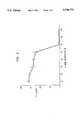
- FIG. 1is a graphical representation of the inhibition of porcine aortic valve calcification in a rat subdermal model for porcine aortic valve specimens (cusps) treated in accordance with a method of the invention
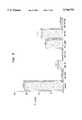
- FIG. 2is a graphical representation of the calcium content ( ⁇ g/mg) of porcine aortic valve specimens, treated in accordance with a method of the invention, following 21 day subdermal implantation in rats;
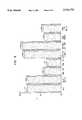
- FIG. 3is a graphical representation of the calcium content ( ⁇ g/mg) of various porcine aortic valve specimens implanted in sheep for 150 days;
- FIG. 4is a graphical representation of the 14 C cholesterol content, in ⁇ g/mg, of glutaraldehyde-pretreated porcine aortic valves as compared to glutaraldehyde-pretreated porcine aortic valves which have been treated with an aqueous solution of ethanol (40% and 80%) in accordance with a method of the invention, or with detergent (1% sodium dodecyl sulfate, SDS);
- FIG. 5is a graphical representation of the calcification content of glutaraldehyde-pretreated porcine aortic valve specimens which have been subjected to a variety of solvents known to remove lipids from tissues;
- FIG. 6is a graphical representation of T s in °C. for porcine aortic valve specimens subjected to various ethanol treatment and storage regimens.
- Glutaraldehyde-pretreated porcine aortic heart valvesboth in stent and freestyle (stentless) form, were obtained from St. Jude Medical, Inc., St. Paul, Minn. and from Medtronic, Inc., Irvine, Calif. and used in the examples set forth below.
- the biomaterialsare stabilized and preserved in glutaraldehyde following harvesting, illustratively in a 0.5% solution of glutaraldehyde in a buffer.
- Glutaraldehyde-pretreated porcine aortic valve specimenswere immersed for 24 hours in aqueous solutions of ethanol ranging in concentration from 0% (control) to 80% ethanol.
- the ethanol solutionswere buffered at pH 7.4 with HEPES (0.05M).
- the treated porcine aortic valve specimenswere implanted in two subcutaneous pouches dissected in the ventral abdominal wall of weanling rats (male, CD, Sprague-Dawley, weighing 50-60 gm). After a period of 21 days, the specimens were removed and examined for calcification by measuring the level of Ca +2 ions in the specimen. Concentrations of 50% or greater of ethanol virtually eliminated calcium accumulation in the porcine aortic valve specimens as compared to glutaraldehyde-pretreated controls.
- FIG. 2is a graphical representation of the calcium content ( ⁇ g/mg) of glutaraldehyde-pretreated porcine aortic valve cusp specimens, following 21 day implantation in rat subdermal pouches, which have been exposed to 80% ethanol for periods of 24 hours and 72 hours. Typically, 72 hours of exposure to ethanol results in more calcium accumulation than 24 hours of exposure. However, calcification levels following 72 hours exposure to ethanol were nevertheless consistently below the level of controls (glutaraldehyde-pretreated porcine aortic valve cusps).
- the calcium content of the control specimenswas 178.2 ⁇ 6.166 ⁇ g/mg dry tissue whereas the calcium content of the specimens which were subjected to 24 hours exposure to 80% ethanol, followed by a rinse with three 100 ml portions of HEPES buffered saline (pH 7.4) over about a 10 to 15 minute period, was 2.248 ⁇ 0.186 ⁇ g/mg. This represents 99% inhibition, i.e., substantial inhibition.
- the calcium content of ethanol treated porcine aortic valve specimens, subsequently rinsed or stored in a glutaraldehyde-containing solutionis shown.
- Glut. Rinsethe ethanol treated specimens were rinsed in three 100 ml portions of 0.2% glutaraldehyde buffered to a pH of 7.4 (HEPES) over about a 15 minute rinse period.
- Glut. Storagethe ethanol treated specimens were stored in 0.2% glutaraldehyde buffered to a pH of 7.4 (HEPES) for 30 days, and then rinsed with HEPES buffered saline prior to implant.
- HEPESHEPES buffered saline prior to implant.
- Table 1presents the calcium content of a set of porcine aortic heart valve specimens following implantation in a rat subdermal pouch. The specimens were untreated glutaraldehyde-pretreated porcine aortic heart valves obtained from St. Jude Medical, Inc. (control) and treated glutaraldehyde-pretreated porcine aortic heart valves which had been subjected to 80% ethanol for 24 hours.
- Specimens of glutaraldehyde-pretreated bovine pericardiumwere treated in 80% ethanol followed by a 24 hour wash.
- the calcium content of rat subdermal implants following 21 dayswas 2.95 ⁇ 0.78 ⁇ g/mg.
- the calcium content of untreated control specimenswas 121.16 ⁇ 7.49 ⁇ g/mg.
- Glutaraldehyde-pretreated porcine heart valve specimenswere obtained from St. Jude Medical, Inc. (St. Jude) and from Medtronic, Inc., (Hancock I). Control specimens were not exposed to alcohol treatment. Experimental specimens were subjected to 80% ethanol for 72 to 96 hours. Control and experimental specimens were implanted in juvenile sheep as mitral valve replacements. Five months after implant, the valves were explanted and analyzed for calcium content. The results are shown in FIG.
- FIG. 4is a graphical representation of the cholesterol content, in ⁇ g/mg, of the treated specimens and the control.
- Table 3presents the total cholesterol (CS) and phospholipid (PL) content of glutaraldehyde-pretreated porcine aortic valve specimens treated for 24 hours in either buffered aqueous solutions of alcohol or chloroform-methanol as identified therein.
- FIG. 6is a graphical representation of the collagen denaturation temperature (°C.) for specimens of glutaraldehyde-pretreated porcine aortic valves (cusps) subjected to various treatment schemes, specifically 24 hours of exposure to ethanol (80% or 100%) and detergent (SDS).
- the schemesinclude: 80% ethanol without rinsing; 100% ethanol without rinsing; 100% ethanol followed by washing with HEPES buffered saline for 1 hour; 80% ethanol followed by rinsing with HEPES buffered saline and storage in 0.2% glutaraldehyde for 24 hours; 1% SDS followed by a HEPES buffered saline rinse; and 1% SDS followed by washing with HEPES buffered saline for 1 hour.
- the controlswere glutaraldehyde-pretreated porcine aortic valve specimens obtained from St.
- rehydraterefers to restoring T s to the value of control (glutaraldehyde-pretreated porcine aortic valve specimens which were rinsed in pH 7.4 HEPES buffered saline for 24 hours).
- the ethanol treated specimens(cusps) were subjected to HEPES buffered saline (pH 7.4) for varying time periods, ranging from a rinse (i.e., pouring rinse solution over the specimen) to one hour.
- a rinsei.e., pouring rinse solution over the specimen
- the overall protein composition and valvular morphology of porcine aortic valvesare unaffected by alcoholic treatment as demonstrated by complete amino acid analysis and electron spectroscopy for chemical analyses (ESCA).
- alcohol treatmentenhances surface smoothing and anisotrophy of porcine aortic valve leaflets resulting in a surface chemistry which is comparable to fresh leaflets.
- glutaraldehyde-pretreated (control) or detergent (SDS) treated tissueshow significant differences.
- Table 5 hereinbelowpresents ESCA data of the surface carbon (C1s), nitrogen (N1s), and oxygen (O1s) concentrations (%) in porcine aortic valve specimens immersed for 24 hours in the indicated solution.
- porcine aortic valvesIn a series of experiments to exemplify additional embodiments of the invention, specimens of glutaraldehyde-pretreated porcine aortic valves were treated with 60% ethanol in a variety of protocols.
- porcine aortic valvesgenerally includes both the valve cusps, or leaflets, and an aortic wall portion
- prior experiments reported hereinabovewere conducted primarily on valve cusp tissue. In the present experiments, the two types of tissue have been separated and the data reported separately on Table 6.
- Glutaraldehyde-pretreated bioprosthetic heart valve specimensobtained from St. Jude Medical, Inc., were used as controls. Specimens of the glutaraldehyde-pretreated tissue were then subjected to treating solutions of 60% ethanol, or 60% ethanol and O.1M AlCl 3 , for 24 hours. Following ethanol treatment, the tissue was rinsed for 24 hours in neutral buffer, specifically HEPES at pH 7.4. Subsequent to rinsing, the tissue samples were sterilized and stored for 14 days. In some storage protocols, the tissue was packaged in neutral buffer and subjected to sterilizing radiation. In other storage protocols, the tissue was stored in solutions of 60% ethanol and glutaraldehyde (0.2% or 0.5%). In yet further storage protocols, the storage solution additionally contained 0.1M AlCl 3 .
- tissue samples prepared as described abovewere implanted in rat subdermal pouches and analyzed for calcium content after 21 days. The results are reported below in Table 6.
- Specimens of the glutaraldehyde-pretreated porcine aortic wall tissuewere subjected, for 24 hours, to aqueous (pH 7.4 buffered HEPES) treating solutions of 0.1M FeCl 3 ; 0.01M FeCl 3 ; 80% ethanol; 80% ethanol and 0.1M FeCl 3 ; and 80% ethanol and 0.01M FeCl 3 .
- the tissuewas rinsed in three 100 ml portions of neutral buffer, specifically HEPES at pH 7.4.
- Specimens of glutaraldehyde-pretreated porcine aortic wall tissueobtained from St. Jude Medical, Inc., were used as controls.
- the tissue samples, prepared as described above,were implanted in rat subdermal pouches and analyzed for calcium content after 21 days. The results are reported below in Table 7.
- Table 7demonstrates that incorporation of Fe +3 ions in the alcohol treatment and/or storage solutions will produce improved resistance to calcification for porcine aortic wall specimens.
Landscapes
- Health & Medical Sciences (AREA)
- Life Sciences & Earth Sciences (AREA)
- Chemical & Material Sciences (AREA)
- Engineering & Computer Science (AREA)
- Biomedical Technology (AREA)
- Medicinal Chemistry (AREA)
- Animal Behavior & Ethology (AREA)
- Veterinary Medicine (AREA)
- Dermatology (AREA)
- Oral & Maxillofacial Surgery (AREA)
- Transplantation (AREA)
- Epidemiology (AREA)
- General Health & Medical Sciences (AREA)
- Public Health (AREA)
- Molecular Biology (AREA)
- Chemical Kinetics & Catalysis (AREA)
- Botany (AREA)
- Zoology (AREA)
- Urology & Nephrology (AREA)
- General Chemical & Material Sciences (AREA)
- Cardiology (AREA)
- Heart & Thoracic Surgery (AREA)
- Vascular Medicine (AREA)
- Materials For Medical Uses (AREA)
- Agricultural Chemicals And Associated Chemicals (AREA)
- Micro-Organisms Or Cultivation Processes Thereof (AREA)
- Treatment And Processing Of Natural Fur Or Leather (AREA)
- Apparatus For Disinfection Or Sterilisation (AREA)
- Acyclic And Carbocyclic Compounds In Medicinal Compositions (AREA)
- Saccharide Compounds (AREA)
- Prostheses (AREA)
Abstract
Description
TABLE 1 ______________________________________ 21day 60 day Treatment Group Ca.sup.+2 (μg/mg) Ca.sup.+2 (82 g/mg) ______________________________________ Control 183.15 ± 0.03 236.3 ± 6.14 80% ethanol/rinse 11.1 ± 6.04 14.6 ± 10.5 80% ethanol/wash 5.16 ± 1.72 1.87 ± 0.29 80% ethanol/Glut. storage/rinse 3.13 ± 1.67 22.9 ± 8.14 80% ethanol/Glut. storage/wash 4.11 ± 2.4 18.3 ± 8.31 ______________________________________
TABLE 2 ______________________________________ T1 (sec) T2 (msec) ______________________________________ Untreated 1.84 ± 0.19 0.14 ± 0.1 Glutaraldehyde 1.78 ± 0.31 0.30 ± 0.05 Ethanol 2.36 ± 0.36 0.42 ± 0.027 ______________________________________ *Porcine aortic heart valve leaflets: as retrieved with no treatment (UNTREATED); treated with 0.6% glutaraldehyde (GLUTARALDEHYDE; treated with 80% ehtanol (ETHANOL.) All treatment solutions were buffered to pH 7.4.
TABLE 3 ______________________________________ GROUP Total CS* (nmole/mg) PL* (nmole/mg) ______________________________________ Control (glu.) 13.34 ± 0.41 17.24 ± 0.85 40% Ethanol 13.96 ± 0.71 16.5 ± 1.49 60% Ethanol 0.30 ± 0.05 4.93 ± 1.91 80% Ethanol 0.14 ± 0.02 1.08 ± 0.11 1% SDS 1.40 ± 0.1 0.94 ± 0.05 2:1 CHCl.sub.3 :Methanol 0.10 ± 0.0 0.57 ± 0.07 80% Methanol 0.28 ± 0.02 2.62 ± 0.36 80% Acetone 0.12 ± 0.02 1.94 ± 0.32 80% Acetonitrile 0.16 ± 0.04 2.76 ± 0.28 ______________________________________ *Mean ± SEM (N = 5)
TABLE 4 ______________________________________ Treatment Rinse Period T.sub.s (°C.) ______________________________________Control 24 hrs 88.33 ± 0.56 80% EtOH rinse 84.06 ± 0.32 80% EtOH 1 min. 84.49 ± 0.39 80% EtOH 2 min. 87.41 ± 0.23 80% EtOH 5 min. 87.85 80% EtOH 10 min. 87.54 80% EtOH 1 hr 87.38 ± 0.26 ______________________________________
TABLE 5 ______________________________________ ATOMIC CONCENTRATION (%) GROUP O1s N1S C1s ______________________________________ Fresh Tissue 20.41 10.06 69.52 80% Ethanol 21.89 11.93 66.18 40% Ethanol 16.45 7.78 75.76 Glutaraldehyde-Fixed 14.46 7.22 78.32 1% SDS 19.03 7.37 73.6 2:1 CHCl.sub.3 /MeOH 22.71 15.85 61.44 ______________________________________
TABLE 6 __________________________________________________________________________Exp. Storage μg Ca/Mg No. Treatment Rinse (14 Days) Cusp Wall __________________________________________________________________________1 24hr 24 hr Buffer + Irrad. 13.763 ± 3.550 40.892 ± 6.057 60% EtOH 2 24hr 24 hr Buffer + Irrad. 6.836 ± 0.262 2.75 ± 0.745 60% EtOH + 0.1 M AlCl.sub.3 3 24hr 24hr 60% EtOH + 9.157 ± 3.733 50.470 ± 1.628 60% EtOH 0.2% Glut. 4 24hr 24hr 60% EtOH + 7.029 ± 0.592 7.110 ± 0.915 60% EtOH + 0.2% Glut. 0.1 M AlCl.sub.3 5 24hr 24hr 60% EtOH + 8.791 ± 2.716 49.082 ± 4.217 60% EtOH 0.5% Glut. 6 24hr 24hr 60% EtOH + 8.689 ± 0.367 8.449 ± 0.341 60% EtOH + 0.5% Glut. 0.1 M AlCl.sub.3 7none none 60% EtOH + 1.952 ± 0.446 60.690 ± 4.716 8none none 60% EtOH + 10.326 ± 0.635 12.782 ± 3.469 0.2% Glut. + 0.1 M AlCl.sub.3 9none none 60% EtOH + 7.907 ± 3.635 39.810 ± 5.026 0.5% Glut. 10none none 60% EtOH + 9.568 ± 0.240 7.763 ± 0.368 0.5% Glut. + 0.1 M AlCl.sub.3 Control -- -- 107.059 ± 3.239 49.915 ± 2.160 (No Treatment) __________________________________________________________________________
TABLE 7 ______________________________________ TISSUE PRETREATMENT WASHING Ca (μg/mg) ______________________________________ Porcine Control No 36.46 ± 4.04 Aortic 0.1 M FeCl.sub.3 Rinse 13.37 ± 1.5 Wall 0.01 M FeCl.sub.3 Rinse 13.52 ± 2.93 80% EtOH Rinse 18.55 ± 3.61 80% EtOH + Rinse 6.31 ± 0.55 0.1M Fe 80% EtOH + Rinse 7.01 ± 1.03 0.01 M Fe ______________________________________
Claims (32)
Priority Applications (15)
| Application Number | Priority Date | Filing Date | Title |
|---|---|---|---|
| US08/140,722US5746775A (en) | 1988-04-01 | 1993-10-21 | Method of making calcification-resistant bioprosthetic tissue |
| JP51219295AJP3768528B2 (en) | 1993-10-21 | 1994-10-20 | Manufacturing method of calcification resistant bioartificial tissue |
| ES94931415TES2171470T3 (en) | 1993-10-21 | 1994-10-20 | METHOD OF MANUFACTURE OF BIOPROTESIC TISSUE RESISTANT TO CALCIFICATION. |
| IL11135194AIL111351A (en) | 1993-10-21 | 1994-10-20 | Method of making calcification-resistant bioprosthetic tissue |
| BRPI9407880-7ABR9407880B1 (en) | 1993-10-21 | 1994-10-20 | Biomaterial treatment method; Method of manufacturing a calcification resistant biomaterial for use within a human or animal. |
| AU80990/94AAU701897B2 (en) | 1993-10-21 | 1994-10-20 | Method of making calcification-resistant bioprosthetic tissue |
| DE69429667TDE69429667T2 (en) | 1993-10-21 | 1994-10-20 | METHOD FOR PRODUCING CALCIFICATION-RESISTANT BIOPROSTHETIC TISSUE |
| PCT/US1994/011937WO1995011047A1 (en) | 1993-10-21 | 1994-10-20 | Method of making calcification-resistant bioprosthetic tissue |
| NZ275032ANZ275032A (en) | 1993-10-21 | 1994-10-20 | Method of preparing calcification-resistant biomaterials, materials so prepared |
| EP94931415AEP0729364B1 (en) | 1993-10-21 | 1994-10-20 | Method of making calcification-resistant bioprosthetic tissue |
| CN94194232ACN1136780A (en) | 1993-10-21 | 1994-10-20 | Method of making anti-calcification resistant bioprosthetic tissue |
| AT94931415TATE211930T1 (en) | 1993-10-21 | 1994-10-20 | METHOD FOR PRODUCING CALCIFICATION-RESISTANT BIOPROSTHETICAL TISSUE |
| CA002174665ACA2174665C (en) | 1993-10-21 | 1994-10-20 | Method of making calcification-resistant bioprosthetic tissue |
| ZA948299AZA948299B (en) | 1993-10-21 | 1994-10-21 | Method of making calcification-resistant bioprosthetic tissue |
| NO961541ANO961541L (en) | 1993-10-21 | 1996-04-18 | Process for the preparation of calcification-resistant bioprosthetic tissue |
Applications Claiming Priority (4)
| Application Number | Priority Date | Filing Date | Title |
|---|---|---|---|
| US07/176,789US5094661A (en) | 1988-04-01 | 1988-04-01 | Calcification-resistant materials and methods of making same through use of trivalent aluminum |
| US51548490A | 1990-04-30 | 1990-04-30 | |
| US07/689,652US5368608A (en) | 1988-04-01 | 1991-04-23 | Calcification-resistant materials and methods of making same through use of multivalent cations |
| US08/140,722US5746775A (en) | 1988-04-01 | 1993-10-21 | Method of making calcification-resistant bioprosthetic tissue |
Related Parent Applications (1)
| Application Number | Title | Priority Date | Filing Date |
|---|---|---|---|
| US07/689,652Continuation-In-PartUS5368608A (en) | 1988-04-01 | 1991-04-23 | Calcification-resistant materials and methods of making same through use of multivalent cations |
Publications (1)
| Publication Number | Publication Date |
|---|---|
| US5746775Atrue US5746775A (en) | 1998-05-05 |
Family
ID=22492520
Family Applications (1)
| Application Number | Title | Priority Date | Filing Date |
|---|---|---|---|
| US08/140,722Expired - LifetimeUS5746775A (en) | 1988-04-01 | 1993-10-21 | Method of making calcification-resistant bioprosthetic tissue |
Country Status (15)
| Country | Link |
|---|---|
| US (1) | US5746775A (en) |
| EP (1) | EP0729364B1 (en) |
| JP (1) | JP3768528B2 (en) |
| CN (1) | CN1136780A (en) |
| AT (1) | ATE211930T1 (en) |
| AU (1) | AU701897B2 (en) |
| BR (1) | BR9407880B1 (en) |
| CA (1) | CA2174665C (en) |
| DE (1) | DE69429667T2 (en) |
| ES (1) | ES2171470T3 (en) |
| IL (1) | IL111351A (en) |
| NO (1) | NO961541L (en) |
| NZ (1) | NZ275032A (en) |
| WO (1) | WO1995011047A1 (en) |
| ZA (1) | ZA948299B (en) |
Cited By (64)
| Publication number | Priority date | Publication date | Assignee | Title |
|---|---|---|---|---|
| WO2001041828A1 (en)* | 1999-12-13 | 2001-06-14 | Sulzer Carbomedics Inc. | Anticalcification treatments for fixed biomaterials |
| US6372228B1 (en)* | 1994-11-15 | 2002-04-16 | Kenton W. Gregory | Method of producing elastin, elastin-based biomaterials and tropoelastin materials |
| US6471723B1 (en) | 2000-01-10 | 2002-10-29 | St. Jude Medical, Inc. | Biocompatible prosthetic tissue |
| US6630001B2 (en)* | 1998-06-24 | 2003-10-07 | International Heart Institute Of Montana Foundation | Compliant dehyrated tissue for implantation and process of making the same |
| US20040030407A1 (en)* | 2000-12-20 | 2004-02-12 | Vettivetpillai Ketharanathan | Method of creating biological and biosynthetic material for implantation |
| US20050113910A1 (en)* | 2002-01-04 | 2005-05-26 | David Paniagua | Percutaneously implantable replacement heart valve device and method of making same |
| US20050148949A1 (en)* | 2003-07-31 | 2005-07-07 | Gabrielle Thumann | Novel instrument for the transplantation of delicate micro-transplants |
| US20060110370A1 (en)* | 2004-11-23 | 2006-05-25 | Pathak Chandrashenkhar P | Treatments for reduction of cytotoxicity and viral contamination of implantable medical devices |
| WO2006066327A1 (en)* | 2004-12-24 | 2006-06-29 | Celxcel Pty Ltd | An implantable biomaterial and a method of producing same |
| US20060154230A1 (en)* | 2005-01-11 | 2006-07-13 | Cunanan Crystal M | Methods for processing biological tissue |
| US20070005129A1 (en)* | 2000-02-28 | 2007-01-04 | Christoph Damm | Anchoring system for implantable heart valve prostheses |
| US20070100440A1 (en)* | 2005-10-28 | 2007-05-03 | Jen.Cardiotec Gmbh | Device for the implantation and fixation of prosthetic valves |
| US20070255423A1 (en)* | 1998-09-21 | 2007-11-01 | Carpentier Alain F | Treating biological tissues to mitigate post-implantation calcification |
| US20080102439A1 (en)* | 2006-10-27 | 2008-05-01 | Bin Tian | Biological tissue for surgical implantation |
| US20080302372A1 (en)* | 2007-06-11 | 2008-12-11 | Edwards Lifesciences Corporation | Methods for pre-stressing and capping bioprosthetic tissue |
| US20090164005A1 (en)* | 2007-12-21 | 2009-06-25 | Edwards Lifesciences Corporation | Capping Bioprosthetic Tissue to Reduce Calcification |
| US20090171447A1 (en)* | 2005-12-22 | 2009-07-02 | Von Segesser Ludwig K | Stent-valves for valve replacement and associated methods and systems for surgery |
| US20090216312A1 (en)* | 2008-02-26 | 2009-08-27 | Helmut Straubinger | Stent for the Positioning and Anchoring of a Valvular Prosthesis in an Implantation Site in the Heart of a Patient |
| US20090216313A1 (en)* | 2008-02-26 | 2009-08-27 | Helmut Straubinger | Stent for the positioning and anchoring of a valvular prosthesis |
| US7704222B2 (en) | 1998-09-10 | 2010-04-27 | Jenavalve Technology, Inc. | Methods and conduits for flowing blood from a heart chamber to a blood vessel |
| US20100261662A1 (en)* | 2009-04-09 | 2010-10-14 | Endologix, Inc. | Utilization of mural thrombus for local drug delivery into vascular tissue |
| US7896915B2 (en) | 2007-04-13 | 2011-03-01 | Jenavalve Technology, Inc. | Medical device for treating a heart valve insufficiency |
| US7955788B2 (en) | 2003-10-30 | 2011-06-07 | Medtronic, Inc. | Bioprosthetic tissue preparation with synthetic hydrogels |
| US20110208290A1 (en)* | 2008-02-26 | 2011-08-25 | Helmut Straubinger | Stent for the positioning and anchoring of a valvular prosthesis in an implantation site in the heart of a patient |
| US20110238167A1 (en)* | 2010-03-23 | 2011-09-29 | Dove Jeffrey S | Methods of conditioning sheet bioprosthetic tissue |
| US8062355B2 (en) | 2005-11-04 | 2011-11-22 | Jenavalve Technology, Inc. | Self-expandable medical instrument for treating defects in a patient's heart |
| US8206437B2 (en) | 2001-08-03 | 2012-06-26 | Philipp Bonhoeffer | Implant implantation unit and procedure for implanting the unit |
| WO2012141454A3 (en)* | 2011-04-12 | 2012-12-13 | Hans Biomed. Cor. | Graft materials derived from mammalian cartilage |
| US8361144B2 (en) | 2010-03-01 | 2013-01-29 | Colibri Heart Valve Llc | Percutaneously deliverable heart valve and methods associated therewith |
| US8398704B2 (en) | 2008-02-26 | 2013-03-19 | Jenavalve Technology, Inc. | Stent for the positioning and anchoring of a valvular prosthesis in an implantation site in the heart of a patient |
| US8468667B2 (en) | 2009-05-15 | 2013-06-25 | Jenavalve Technology, Inc. | Device for compressing a stent |
| US8632608B2 (en) | 2002-01-03 | 2014-01-21 | Edwards Lifesciences Corporation | Treatment of bioprosthetic tissues to mitigate post implantation calcification |
| US8679174B2 (en) | 2005-01-20 | 2014-03-25 | JenaValve Technology, GmbH | Catheter for the transvascular implantation of prosthetic heart valves |
| USRE45130E1 (en) | 2000-02-28 | 2014-09-09 | Jenavalve Technology Gmbh | Device for fastening and anchoring cardiac valve prostheses |
| US20140341871A1 (en)* | 2011-12-30 | 2014-11-20 | Paul R. Morris | Regenerative tissue matrix |
| US8906601B2 (en) | 2010-06-17 | 2014-12-09 | Edwardss Lifesciences Corporation | Methods for stabilizing a bioprosthetic tissue by chemical modification of antigenic carbohydrates |
| US9119738B2 (en) | 2010-06-28 | 2015-09-01 | Colibri Heart Valve Llc | Method and apparatus for the endoluminal delivery of intravascular devices |
| US9138315B2 (en) | 2007-04-13 | 2015-09-22 | Jenavalve Technology Gmbh | Medical device for treating a heart valve insufficiency or stenosis |
| US9168130B2 (en) | 2008-02-26 | 2015-10-27 | Jenavalve Technology Gmbh | Stent for the positioning and anchoring of a valvular prosthesis in an implantation site in the heart of a patient |
| US9295551B2 (en) | 2007-04-13 | 2016-03-29 | Jenavalve Technology Gmbh | Methods of implanting an endoprosthesis |
| US9351829B2 (en) | 2010-11-17 | 2016-05-31 | Edwards Lifesciences Corporation | Double cross-linkage process to enhance post-implantation bioprosthetic tissue durability |
| US9510947B2 (en) | 2011-10-21 | 2016-12-06 | Jenavalve Technology, Inc. | Catheter system for introducing an expandable heart valve stent into the body of a patient |
| US9555162B2 (en)* | 2014-02-24 | 2017-01-31 | Medtronic, Inc. | Phospholipid reduction in biological tissue |
| US9597182B2 (en) | 2010-05-20 | 2017-03-21 | Jenavalve Technology Inc. | Catheter system for introducing an expandable stent into the body of a patient |
| US9615922B2 (en) | 2013-09-30 | 2017-04-11 | Edwards Lifesciences Corporation | Method and apparatus for preparing a contoured biological tissue |
| US9737400B2 (en) | 2010-12-14 | 2017-08-22 | Colibri Heart Valve Llc | Percutaneously deliverable heart valve including folded membrane cusps with integral leaflets |
| US9744031B2 (en) | 2010-05-25 | 2017-08-29 | Jenavalve Technology, Inc. | Prosthetic heart valve and endoprosthesis comprising a prosthetic heart valve and a stent |
| US9867699B2 (en) | 2008-02-26 | 2018-01-16 | Jenavalve Technology, Inc. | Endoprosthesis for implantation in the heart of a patient |
| US9867694B2 (en) | 2013-08-30 | 2018-01-16 | Jenavalve Technology Inc. | Radially collapsible frame for a prosthetic valve and method for manufacturing such a frame |
| US9878127B2 (en) | 2012-05-16 | 2018-01-30 | Jenavalve Technology, Inc. | Catheter delivery system for heart valve prosthesis |
| WO2019006256A1 (en) | 2017-06-29 | 2019-01-03 | St. Jude Medical, Cardiology Division, Inc. | Method of preparing calcification-resistant bioprosthetic tissue |
| US10238771B2 (en) | 2012-11-08 | 2019-03-26 | Edwards Lifesciences Corporation | Methods for treating bioprosthetic tissue using a nucleophile/electrophile in a catalytic system |
| US10709555B2 (en) | 2015-05-01 | 2020-07-14 | Jenavalve Technology, Inc. | Device and method with reduced pacemaker rate in heart valve replacement |
| US10959839B2 (en) | 2013-10-08 | 2021-03-30 | Edwards Lifesciences Corporation | Method for directing cellular migration patterns on a biological tissue |
| US11065138B2 (en) | 2016-05-13 | 2021-07-20 | Jenavalve Technology, Inc. | Heart valve prosthesis delivery system and method for delivery of heart valve prosthesis with introducer sheath and loading system |
| US11197754B2 (en) | 2017-01-27 | 2021-12-14 | Jenavalve Technology, Inc. | Heart valve mimicry |
| US11278406B2 (en) | 2010-05-20 | 2022-03-22 | Jenavalve Technology, Inc. | Catheter system for introducing an expandable heart valve stent into the body of a patient, insertion system with a catheter system and medical device for treatment of a heart valve defect |
| US11395726B2 (en) | 2017-09-11 | 2022-07-26 | Incubar Llc | Conduit vascular implant sealing device for reducing endoleaks |
| US11517428B2 (en) | 2018-11-01 | 2022-12-06 | Edwards Lifesciences Corporation | Transcatheter pulmonic regenerative valve |
| US12023416B2 (en) | 2017-05-31 | 2024-07-02 | Edwards Lifesciences Corporation | Collagen fibers and articles formed therefrom |
| US12023417B2 (en) | 2018-01-23 | 2024-07-02 | Edwards Lifesciences Corporation | Method for pre-stretching implantable biocompatible materials, and materials, and devices produced thereby |
| US12115280B2 (en) | 2010-06-17 | 2024-10-15 | Edwards Lifesciences Corporation | Methods for stabilizing a bioprosthetic tissue by chemical modification of antigenic carbohydrates |
| US12121461B2 (en) | 2015-03-20 | 2024-10-22 | Jenavalve Technology, Inc. | Heart valve prosthesis delivery system and method for delivery of heart valve prosthesis with introducer sheath |
| US12171658B2 (en) | 2022-11-09 | 2024-12-24 | Jenavalve Technology, Inc. | Catheter system for sequential deployment of an expandable implant |
Families Citing this family (22)
| Publication number | Priority date | Publication date | Assignee | Title |
|---|---|---|---|---|
| DE69523074T2 (en) | 1994-07-29 | 2002-06-06 | Edwards Lifesciences Corp., Irvine | METHOD FOR TREATING IMPLANTABLE BIOLOGICAL TISSUES TO REDUCE CALCIFICATION |
| USRE40570E1 (en) | 1994-07-29 | 2008-11-11 | Edwards Lifesciences Corporation | Apparatuses and methods for treating biological tissue to mitigate calcification |
| US5931969A (en) | 1994-07-29 | 1999-08-03 | Baxter International Inc. | Methods and apparatuses for treating biological tissue to mitigate calcification |
| US6302909B1 (en) | 1996-07-31 | 2001-10-16 | St. Jude Medical, Inc. | Calcification-resistant biomaterials |
| US6193749B1 (en) | 1996-02-05 | 2001-02-27 | St. Jude Medical, Inc. | Calcification-resistant biomaterials |
| BR9707269A (en)* | 1996-02-05 | 1999-04-13 | St Jude Medical | Bioprotective article and processes for preparing biocompatible material and for producing a bioprosthetic article |
| US5862806A (en)* | 1997-10-30 | 1999-01-26 | Mitroflow International, Inc. | Borohydride reduction of biological tissues |
| US6254635B1 (en) | 1998-02-02 | 2001-07-03 | St. Jude Medical, Inc. | Calcification-resistant medical articles |
| US6660265B1 (en) | 1999-10-15 | 2003-12-09 | The Brigham & Women's Hospital, Inc. | Fresh, cryopreserved, or minimally cardiac valvular xenografts |
| US7579381B2 (en) | 2005-03-25 | 2009-08-25 | Edwards Lifesciences Corporation | Treatment of bioprosthetic tissues to mitigate post implantation calcification |
| CN101721745B (en)* | 2009-11-09 | 2015-04-01 | 山东省千佛山医院 | Method for processing donkey pericardium for artificial heart valve and other biological repairing material |
| US9358107B2 (en) | 2011-06-30 | 2016-06-07 | Edwards Lifesciences Corporation | Systems, dies, and methods for processing pericardial tissue |
| CN108535205B (en)* | 2015-07-22 | 2021-06-22 | 杭州启明医疗器械股份有限公司 | In-vitro biological valve anti-calcification treatment method and calcification evaluation method |
| US10195024B2 (en)* | 2015-10-07 | 2019-02-05 | Boston Scientific Scimed, Inc. | Porcine small intestine submucosa leaflet material |
| EP3693030A1 (en)* | 2019-02-07 | 2020-08-12 | P+F Products + Features GmbH | Method for preparing biological tissue for surgical implantation |
| CN111588909A (en)* | 2020-02-24 | 2020-08-28 | 科凯(南通)生命科学有限公司 | Anti-calcification method for biomedical material |
| CN111494716B (en)* | 2020-04-15 | 2021-12-21 | 青岛市妇女儿童医院(青岛市妇幼保健院、青岛市残疾儿童医疗康复中心、青岛市新生儿疾病筛查中心) | Cardiac surgery autologous pericardial patch processor and rapid anti-calcification treatment method |
| CN113080187B (en)* | 2021-03-31 | 2022-06-21 | 上海纽脉医疗科技有限公司 | Composition for removing phospholipid and cell debris and method for removing phospholipid and cell debris from biological tissue |
| CN113499478B (en)* | 2021-07-07 | 2022-07-12 | 成都纽脉生物科技有限公司 | Method for processing biological tissue material |
| CN113499479B (en)* | 2021-07-19 | 2023-03-17 | 科凯(南通)生命科学有限公司 | Preparation method of modified biological material and obtained modified biological material |
| BR102021017465A2 (en)* | 2021-09-02 | 2023-03-14 | Labcor Laboratórios Ltda. | METHOD FOR THE PRODUCTION OF PRESERVED COLLAGEN CONNECTIVE TISSUE, COLLAGEN CONNECTIVE TISSUE, ITS USES AND TISSUE IMPLANT KIT |
| CN114504682A (en)* | 2021-11-16 | 2022-05-17 | 李平 | Application of umbilical artery in preparation of coronary bypass graft blood vessel material and method for preparing coronary bypass graft blood vessel material |
Citations (31)
| Publication number | Priority date | Publication date | Assignee | Title |
|---|---|---|---|---|
| US713046A (en)* | 1902-04-18 | 1902-11-11 | Otto P Amend | Process of tanning hides or other animal tissues. |
| US725648A (en)* | 1901-03-27 | 1903-04-21 | Otto P Amend | Process of tanning hides, skins, or other animal tissues. |
| US2750251A (en)* | 1954-07-09 | 1956-06-12 | Ethicon Inc | Process for preparing sutures and ligatures |
| US2970031A (en)* | 1955-08-31 | 1961-01-31 | American Cyanamid Co | Process for tanning leather |
| US3560141A (en)* | 1963-05-29 | 1971-02-02 | Ethicon Inc | Aldehyde,alum and dihydroxybenzoic acid tanned collagen articles production |
| US3922356A (en)* | 1974-04-18 | 1975-11-25 | Tee Pak Inc | Sulfite catalyzed aluminum tannage of collagen casing |
| US3974526A (en)* | 1973-07-06 | 1976-08-17 | Dardik Irving I | Vascular prostheses and process for producing the same |
| US4097234A (en)* | 1976-12-10 | 1978-06-27 | Nippi, Incorporated | Method for preparing dispersion of collagen fiber |
| US4323358A (en)* | 1981-04-30 | 1982-04-06 | Vascor, Inc. | Method for inhibiting mineralization of natural tissue during implantation |
| US4378224A (en)* | 1980-09-19 | 1983-03-29 | Nimni Marcel E | Coating for bioprosthetic device and method of making same |
| US4402697A (en)* | 1982-08-25 | 1983-09-06 | Extracorporeal Medical Specialties, Inc. | Method for inhibiting mineralization of natural tissue during implantation |
| US4405327A (en)* | 1982-08-25 | 1983-09-20 | Extracorporeal Medical Specialties, Inc. | Method for inhibiting mineralization of natural tissue during implantation |
| WO1983003335A1 (en)* | 1982-03-23 | 1983-10-13 | Carpentier, Alain, F. | Graftable biological tissue and preparation method thereof |
| WO1984001894A1 (en)* | 1982-11-12 | 1984-05-24 | American Hospital Supply Corp | Chemical sterilization of implantable biological tissue |
| WO1984001879A1 (en)* | 1982-11-12 | 1984-05-24 | American Hospital Supply Corp | Surfactant treatment of implantable biological tissue to inhibit calcification |
| US4481009A (en)* | 1982-05-13 | 1984-11-06 | American Hospital Supply Corporation | Polymer incorporation into implantable biological tissue to inhibit calcification |
| US4553974A (en)* | 1984-08-14 | 1985-11-19 | Mayo Foundation | Treatment of collagenous tissue with glutaraldehyde and aminodiphosphonate calcification inhibitor |
| US4597960A (en)* | 1983-04-19 | 1986-07-01 | Cohen Edgar C | Microencapsulated astringent hemostatic agents and methods of use |
| US4729139A (en)* | 1985-11-05 | 1988-03-08 | Baxter Travenol | Selective incorporation of a polymer into implantable biological tissue to inhibit calcification |
| EP0267434A2 (en)* | 1986-10-10 | 1988-05-18 | BAXTER INTERNATIONAL INC. (a Delaware corporation) | Process for decreasing residual aldehyde levels in implantable bioprosthetic tissue |
| US4753652A (en)* | 1984-05-04 | 1988-06-28 | Children's Medical Center Corporation | Biomaterial implants which resist calcification |
| US4770665A (en)* | 1985-11-05 | 1988-09-13 | American Hospital Supply Corporation | Elastomeric polymer incorporation into implantable biological tissue to inhibit calcification |
| US4798611A (en)* | 1986-10-14 | 1989-01-17 | Hancock Jaffe Laboratories | Enhancement of xenogeneic tissue |
| US4838888A (en)* | 1987-04-17 | 1989-06-13 | Baxter Travenol Laboratories, Inc. | Calcification mitigation of implantable bioprostheses |
| US4885005A (en)* | 1982-11-12 | 1989-12-05 | Baxter International Inc. | Surfactant treatment of implantable biological tissue to inhibit calcification |
| US4976733A (en)* | 1988-02-03 | 1990-12-11 | Biomedical Design, Inc. | Prevention of prosthesis calcification |
| US5002566A (en)* | 1989-02-17 | 1991-03-26 | Baxter International Inc. | Calcification mitigation of bioprosthetic implants |
| US5080670A (en)* | 1987-08-31 | 1992-01-14 | Koken Co., Ltd. | Bioprosthetic valve |
| US5215541A (en)* | 1982-11-12 | 1993-06-01 | Baxter International Inc. | Surfactant treatment of implantable biological tissue to inhibit calcification |
| US5447536A (en)* | 1994-02-17 | 1995-09-05 | Biomedical Design, Inc. | Method for fixation of biological tissue |
| US5476516A (en)* | 1992-03-13 | 1995-12-19 | Albert Einstein College Of Medicine Of Yeshiva University | Anticalcification treatment for aldehyde-tanned biological tissue |
Family Cites Families (3)
| Publication number | Priority date | Publication date | Assignee | Title |
|---|---|---|---|---|
| IN157379B (en)* | 1981-04-30 | 1986-03-15 | Extracorporeal Med Spec | |
| US5094661A (en)* | 1988-04-01 | 1992-03-10 | The University Of Michigan | Calcification-resistant materials and methods of making same through use of trivalent aluminum |
| WO1990009102A1 (en)* | 1989-02-17 | 1990-08-23 | Baxter International Inc. | Calcification mitigation of bioprosthetic implants |
- 1993
- 1993-10-21USUS08/140,722patent/US5746775A/ennot_activeExpired - Lifetime
- 1994
- 1994-10-20ILIL11135194Apatent/IL111351A/ennot_activeIP Right Cessation
- 1994-10-20WOPCT/US1994/011937patent/WO1995011047A1/enactiveIP Right Grant
- 1994-10-20BRBRPI9407880-7Apatent/BR9407880B1/ennot_activeIP Right Cessation
- 1994-10-20ATAT94931415Tpatent/ATE211930T1/ennot_activeIP Right Cessation
- 1994-10-20CNCN94194232Apatent/CN1136780A/enactivePending
- 1994-10-20NZNZ275032Apatent/NZ275032A/enunknown
- 1994-10-20ESES94931415Tpatent/ES2171470T3/ennot_activeExpired - Lifetime
- 1994-10-20EPEP94931415Apatent/EP0729364B1/ennot_activeExpired - Lifetime
- 1994-10-20CACA002174665Apatent/CA2174665C/ennot_activeExpired - Lifetime
- 1994-10-20AUAU80990/94Apatent/AU701897B2/ennot_activeCeased
- 1994-10-20JPJP51219295Apatent/JP3768528B2/ennot_activeExpired - Fee Related
- 1994-10-20DEDE69429667Tpatent/DE69429667T2/ennot_activeExpired - Lifetime
- 1994-10-21ZAZA948299Apatent/ZA948299B/enunknown
- 1996
- 1996-04-18NONO961541Apatent/NO961541L/enunknown
Patent Citations (34)
| Publication number | Priority date | Publication date | Assignee | Title |
|---|---|---|---|---|
| US725648A (en)* | 1901-03-27 | 1903-04-21 | Otto P Amend | Process of tanning hides, skins, or other animal tissues. |
| US713046A (en)* | 1902-04-18 | 1902-11-11 | Otto P Amend | Process of tanning hides or other animal tissues. |
| US2750251A (en)* | 1954-07-09 | 1956-06-12 | Ethicon Inc | Process for preparing sutures and ligatures |
| US2970031A (en)* | 1955-08-31 | 1961-01-31 | American Cyanamid Co | Process for tanning leather |
| US3560141A (en)* | 1963-05-29 | 1971-02-02 | Ethicon Inc | Aldehyde,alum and dihydroxybenzoic acid tanned collagen articles production |
| US3974526A (en)* | 1973-07-06 | 1976-08-17 | Dardik Irving I | Vascular prostheses and process for producing the same |
| US3922356A (en)* | 1974-04-18 | 1975-11-25 | Tee Pak Inc | Sulfite catalyzed aluminum tannage of collagen casing |
| US4097234A (en)* | 1976-12-10 | 1978-06-27 | Nippi, Incorporated | Method for preparing dispersion of collagen fiber |
| US4378224A (en)* | 1980-09-19 | 1983-03-29 | Nimni Marcel E | Coating for bioprosthetic device and method of making same |
| US4323358A (en)* | 1981-04-30 | 1982-04-06 | Vascor, Inc. | Method for inhibiting mineralization of natural tissue during implantation |
| US4648881A (en)* | 1982-03-23 | 1987-03-10 | American Hospital Supply Corporation | Implantable biological tissue and process for preparation thereof |
| US4647283A (en)* | 1982-03-23 | 1987-03-03 | American Hospital Supply Corporation | Implantable biological tissue and process for preparation thereof |
| WO1983003335A1 (en)* | 1982-03-23 | 1983-10-13 | Carpentier, Alain, F. | Graftable biological tissue and preparation method thereof |
| US4481009A (en)* | 1982-05-13 | 1984-11-06 | American Hospital Supply Corporation | Polymer incorporation into implantable biological tissue to inhibit calcification |
| US4405327A (en)* | 1982-08-25 | 1983-09-20 | Extracorporeal Medical Specialties, Inc. | Method for inhibiting mineralization of natural tissue during implantation |
| US4402697A (en)* | 1982-08-25 | 1983-09-06 | Extracorporeal Medical Specialties, Inc. | Method for inhibiting mineralization of natural tissue during implantation |
| WO1984001879A1 (en)* | 1982-11-12 | 1984-05-24 | American Hospital Supply Corp | Surfactant treatment of implantable biological tissue to inhibit calcification |
| WO1984001894A1 (en)* | 1982-11-12 | 1984-05-24 | American Hospital Supply Corp | Chemical sterilization of implantable biological tissue |
| US5215541A (en)* | 1982-11-12 | 1993-06-01 | Baxter International Inc. | Surfactant treatment of implantable biological tissue to inhibit calcification |
| US4885005A (en)* | 1982-11-12 | 1989-12-05 | Baxter International Inc. | Surfactant treatment of implantable biological tissue to inhibit calcification |
| US4597960A (en)* | 1983-04-19 | 1986-07-01 | Cohen Edgar C | Microencapsulated astringent hemostatic agents and methods of use |
| US4753652A (en)* | 1984-05-04 | 1988-06-28 | Children's Medical Center Corporation | Biomaterial implants which resist calcification |
| US4553974A (en)* | 1984-08-14 | 1985-11-19 | Mayo Foundation | Treatment of collagenous tissue with glutaraldehyde and aminodiphosphonate calcification inhibitor |
| US4770665A (en)* | 1985-11-05 | 1988-09-13 | American Hospital Supply Corporation | Elastomeric polymer incorporation into implantable biological tissue to inhibit calcification |
| US4729139A (en)* | 1985-11-05 | 1988-03-08 | Baxter Travenol | Selective incorporation of a polymer into implantable biological tissue to inhibit calcification |
| US4786287A (en)* | 1986-10-10 | 1988-11-22 | Baxter Travenol Laboratories | Process for decreasing residual aldehyde levels in implantable bioprosthetic tissue |
| EP0267434A2 (en)* | 1986-10-10 | 1988-05-18 | BAXTER INTERNATIONAL INC. (a Delaware corporation) | Process for decreasing residual aldehyde levels in implantable bioprosthetic tissue |
| US4798611A (en)* | 1986-10-14 | 1989-01-17 | Hancock Jaffe Laboratories | Enhancement of xenogeneic tissue |
| US4838888A (en)* | 1987-04-17 | 1989-06-13 | Baxter Travenol Laboratories, Inc. | Calcification mitigation of implantable bioprostheses |
| US5080670A (en)* | 1987-08-31 | 1992-01-14 | Koken Co., Ltd. | Bioprosthetic valve |
| US4976733A (en)* | 1988-02-03 | 1990-12-11 | Biomedical Design, Inc. | Prevention of prosthesis calcification |
| US5002566A (en)* | 1989-02-17 | 1991-03-26 | Baxter International Inc. | Calcification mitigation of bioprosthetic implants |
| US5476516A (en)* | 1992-03-13 | 1995-12-19 | Albert Einstein College Of Medicine Of Yeshiva University | Anticalcification treatment for aldehyde-tanned biological tissue |
| US5447536A (en)* | 1994-02-17 | 1995-09-05 | Biomedical Design, Inc. | Method for fixation of biological tissue |
Non-Patent Citations (4)
| Title |
|---|
| Okoshi, et al., A New Bioprosthetic Cardiac Valve With Reduced Calcification, ASAIO Trans. 1990, 36:M411 M414 (no month).* |
| Okoshi, et al., A New Bioprosthetic Cardiac Valve With Reduced Calcification, ASAIO Trans. 1990, 36:M411-M414 (no month). |
| Tomizawa, et al., Development of a Polyepoxy Compound Cross linked Heterologous Connective Tissue Tube, ASAIO Journal 1992, 38: M357 M361.* |
| Tomizawa, et al., Development of a Polyepoxy Compound Cross-linked Heterologous Connective Tissue Tube, ASAIO Journal 1992, 38: M357-M361. |
Cited By (174)
| Publication number | Priority date | Publication date | Assignee | Title |
|---|---|---|---|---|
| US6372228B1 (en)* | 1994-11-15 | 2002-04-16 | Kenton W. Gregory | Method of producing elastin, elastin-based biomaterials and tropoelastin materials |
| US6630001B2 (en)* | 1998-06-24 | 2003-10-07 | International Heart Institute Of Montana Foundation | Compliant dehyrated tissue for implantation and process of making the same |
| US7704222B2 (en) | 1998-09-10 | 2010-04-27 | Jenavalve Technology, Inc. | Methods and conduits for flowing blood from a heart chamber to a blood vessel |
| US7736327B2 (en) | 1998-09-10 | 2010-06-15 | Jenavalve Technology, Inc. | Methods and conduits for flowing blood from a heart chamber to a blood vessel |
| US8597226B2 (en) | 1998-09-10 | 2013-12-03 | Jenavalve Technology, Inc. | Methods and conduits for flowing blood from a heart chamber to a blood vessel |
| US8216174B2 (en) | 1998-09-10 | 2012-07-10 | Jenavalve Technology, Inc. | Methods and conduits for flowing blood from a heart chamber to a blood vessel |
| US8236241B2 (en) | 1998-09-21 | 2012-08-07 | Edwards Lifesciences Corporation | Treating biological tissues to mitigate post-implantation calcification |
| US20070255423A1 (en)* | 1998-09-21 | 2007-11-01 | Carpentier Alain F | Treating biological tissues to mitigate post-implantation calcification |
| WO2001041828A1 (en)* | 1999-12-13 | 2001-06-14 | Sulzer Carbomedics Inc. | Anticalcification treatments for fixed biomaterials |
| US6479079B1 (en) | 1999-12-13 | 2002-11-12 | Sulzer Carbomedics Inc. | Anticalcification treatments for fixed biomaterials |
| US20030130746A1 (en)* | 2000-01-10 | 2003-07-10 | St. Jude Medical, Inc. | Biocompatible prosthetic tissue |
| US6471723B1 (en) | 2000-01-10 | 2002-10-29 | St. Jude Medical, Inc. | Biocompatible prosthetic tissue |
| US7896913B2 (en) | 2000-02-28 | 2011-03-01 | Jenavalve Technology, Inc. | Anchoring system for implantable heart valve prostheses |
| USRE45130E1 (en) | 2000-02-28 | 2014-09-09 | Jenavalve Technology Gmbh | Device for fastening and anchoring cardiac valve prostheses |
| US20070005129A1 (en)* | 2000-02-28 | 2007-01-04 | Christoph Damm | Anchoring system for implantable heart valve prostheses |
| US7022348B2 (en) | 2000-12-20 | 2006-04-04 | Vettivetpillai Ketharanathan | Method of creating biological and biosynthetic material for implantation |
| US20040030407A1 (en)* | 2000-12-20 | 2004-02-12 | Vettivetpillai Ketharanathan | Method of creating biological and biosynthetic material for implantation |
| US8303653B2 (en) | 2001-08-03 | 2012-11-06 | Philipp Bonhoeffer | Implant implantation unit and procedure for implanting the unit |
| US9949824B2 (en) | 2001-08-03 | 2018-04-24 | Jenavalve Technology, Inc. | Devices useful for implantation at a heart valve |
| US8585756B2 (en) | 2001-08-03 | 2013-11-19 | Jenavalve Technology, Inc. | Methods of treating valves |
| US8206437B2 (en) | 2001-08-03 | 2012-06-26 | Philipp Bonhoeffer | Implant implantation unit and procedure for implanting the unit |
| US11007052B2 (en) | 2001-08-03 | 2021-05-18 | Jenavalve Technology, Inc. | Devices useful for implantation at a heart valve |
| US8216301B2 (en) | 2001-08-03 | 2012-07-10 | Philipp Bonhoeffer | Implant implantation unit |
| US8579965B2 (en) | 2001-08-03 | 2013-11-12 | Jenavalve Technology, Inc. | Methods of implanting an implantation device |
| US9889002B2 (en) | 2001-08-03 | 2018-02-13 | Jenavalve Technology, Inc. | Devices useful for implantation at a heart valve |
| US8632608B2 (en) | 2002-01-03 | 2014-01-21 | Edwards Lifesciences Corporation | Treatment of bioprosthetic tissues to mitigate post implantation calcification |
| US20120310041A1 (en)* | 2002-01-04 | 2012-12-06 | Colibri Heart Valve Llc | Percutaneously Implantable Replacement Heart Valve Device and Method of Making Same |
| US8109995B2 (en) | 2002-01-04 | 2012-02-07 | Colibri Heart Valve Llc | Percutaneously implantable replacement heart valve device and method of making same |
| US8308797B2 (en) | 2002-01-04 | 2012-11-13 | Colibri Heart Valve, LLC | Percutaneously implantable replacement heart valve device and method of making same |
| US8900294B2 (en) | 2002-01-04 | 2014-12-02 | Colibri Heart Valve Llc | Method of controlled release of a percutaneous replacement heart valve |
| US8790398B2 (en) | 2002-01-04 | 2014-07-29 | Colibri Heart Valve Llc | Percutaneously implantable replacement heart valve device and method of making same |
| US20050113910A1 (en)* | 2002-01-04 | 2005-05-26 | David Paniagua | Percutaneously implantable replacement heart valve device and method of making same |
| US9554898B2 (en) | 2002-01-04 | 2017-01-31 | Colibri Heart Valve Llc | Percutaneous prosthetic heart valve |
| US9610158B2 (en) | 2002-01-04 | 2017-04-04 | Colibri Heart Valve Llc | Percutaneously implantable replacement heart valve device and method of making same |
| US9186248B2 (en)* | 2002-01-04 | 2015-11-17 | Colibri Heart Valve Llc | Percutaneously implantable replacement heart valve device and method of making same |
| US20090030511A1 (en)* | 2002-01-04 | 2009-01-29 | David Paniagua | Percutaneously implantable replacement heart valve device and method of making same |
| US9125739B2 (en) | 2002-01-04 | 2015-09-08 | Colibri Heart Valve Llc | Percutaneous replacement heart valve and a delivery and implantation system |
| US20050148949A1 (en)* | 2003-07-31 | 2005-07-07 | Gabrielle Thumann | Novel instrument for the transplantation of delicate micro-transplants |
| US7955788B2 (en) | 2003-10-30 | 2011-06-07 | Medtronic, Inc. | Bioprosthetic tissue preparation with synthetic hydrogels |
| US20060110370A1 (en)* | 2004-11-23 | 2006-05-25 | Pathak Chandrashenkhar P | Treatments for reduction of cytotoxicity and viral contamination of implantable medical devices |
| US20060193885A1 (en)* | 2004-12-24 | 2006-08-31 | Celxcel Pty Ltd | Implantable biomaterial and a method of producing same |
| WO2006066327A1 (en)* | 2004-12-24 | 2006-06-29 | Celxcel Pty Ltd | An implantable biomaterial and a method of producing same |
| US9205172B2 (en)* | 2004-12-24 | 2015-12-08 | Admedus Regen Pty Ltd | Implantable biomaterial and a method of producing same |
| AU2005318938B2 (en)* | 2004-12-24 | 2011-04-21 | Anteris Aus Operations Pty Ltd | An implantable biomaterial and a method of producing same |
| EP1835948A4 (en)* | 2004-12-24 | 2011-11-30 | Celxcel Pty Ltd | IMPLANTABLE BIOMATERIAL AND METHOD FOR PRODUCING THE SAME |
| US7989157B2 (en) | 2005-01-11 | 2011-08-02 | Medtronic, Inc. | Solution for storing bioprosthetic tissue used in a biological prosthesis |
| US20060154230A1 (en)* | 2005-01-11 | 2006-07-13 | Cunanan Crystal M | Methods for processing biological tissue |
| US20060207031A1 (en)* | 2005-01-11 | 2006-09-21 | Cunanan Crystal M | Methods for processing biological tissue |
| US7622276B2 (en) | 2005-01-11 | 2009-11-24 | Arbor Surgical Technologies, Inc. | Methods for processing biological tissue |
| US9788945B2 (en) | 2005-01-20 | 2017-10-17 | Jenavalve Technology, Inc. | Systems for implanting an endoprosthesis |
| US9775705B2 (en) | 2005-01-20 | 2017-10-03 | Jenavalve Technology, Inc. | Methods of implanting an endoprosthesis |
| US8679174B2 (en) | 2005-01-20 | 2014-03-25 | JenaValve Technology, GmbH | Catheter for the transvascular implantation of prosthetic heart valves |
| US11517431B2 (en) | 2005-01-20 | 2022-12-06 | Jenavalve Technology, Inc. | Catheter system for implantation of prosthetic heart valves |
| US10492906B2 (en) | 2005-01-20 | 2019-12-03 | Jenavalve Technology, Inc. | Catheter system for implantation of prosthetic heart valves |
| US8092521B2 (en) | 2005-10-28 | 2012-01-10 | Jenavalve Technology, Inc. | Device for the implantation and fixation of prosthetic valves |
| USRE45790E1 (en) | 2005-10-28 | 2015-11-03 | Jenavalve Technology Gmbh | Device for the implantation and fixation of prosthetic valves |
| US9044320B2 (en) | 2005-10-28 | 2015-06-02 | Jenavalve Technology Gmbh | Device for the implantation and fixation of prosthetic valves |
| USRE45962E1 (en) | 2005-10-28 | 2016-04-05 | Jenavalve Technology Gmbh | Device for the implantation and fixation of prosthetic valves |
| US8551160B2 (en) | 2005-10-28 | 2013-10-08 | Jenavalve Technology, Inc. | Device for the implantation and fixation of prosthetic valves |
| US9402717B2 (en) | 2005-10-28 | 2016-08-02 | Jenavalve Technology, Inc. | Device for the implantation and fixation of prosthetic valves |
| US9855142B2 (en) | 2005-10-28 | 2018-01-02 | JenaValve Technologies, Inc. | Device for the implantation and fixation of prosthetic valves |
| US20070100440A1 (en)* | 2005-10-28 | 2007-05-03 | Jen.Cardiotec Gmbh | Device for the implantation and fixation of prosthetic valves |
| US10363134B2 (en) | 2005-10-28 | 2019-07-30 | Jenavalve Technology, Inc. | Device for the implantation and fixation of prosthetic valves |
| US8834561B2 (en) | 2005-10-28 | 2014-09-16 | Jenavalve Technology Gmbh | Device for the implantation and fixation of prosthetic valves |
| US11116628B2 (en) | 2005-10-28 | 2021-09-14 | Jenavalve Technology, Inc. | Device for the implantation and fixation of prosthetic valves |
| US8062355B2 (en) | 2005-11-04 | 2011-11-22 | Jenavalve Technology, Inc. | Self-expandable medical instrument for treating defects in a patient's heart |
| US10314701B2 (en) | 2005-12-22 | 2019-06-11 | Symetis Sa | Stent-valves for valve replacement and associated methods and systems for surgery |
| US20090171447A1 (en)* | 2005-12-22 | 2009-07-02 | Von Segesser Ludwig K | Stent-valves for valve replacement and associated methods and systems for surgery |
| US9839515B2 (en) | 2005-12-22 | 2017-12-12 | Symetis, SA | Stent-valves for valve replacement and associated methods and systems for surgery |
| US10265167B2 (en) | 2005-12-22 | 2019-04-23 | Symetis Sa | Stent-valves for valve replacement and associated methods and systems for surgery |
| US10299922B2 (en) | 2005-12-22 | 2019-05-28 | Symetis Sa | Stent-valves for valve replacement and associated methods and systems for surgery |
| US8007992B2 (en) | 2006-10-27 | 2011-08-30 | Edwards Lifesciences Corporation | Method of treating glutaraldehyde-fixed pericardial tissue with a non-aqueous mixture of glycerol and a C1-C3 alcohol |
| US20080102439A1 (en)* | 2006-10-27 | 2008-05-01 | Bin Tian | Biological tissue for surgical implantation |
| US9918832B2 (en) | 2006-10-27 | 2018-03-20 | Edwards Lifesciences Corporation | Biological tissue for surgical implantation |
| US9339386B2 (en) | 2007-04-13 | 2016-05-17 | Jenavalve Technology, Inc. | Medical device for treating a heart valve insufficency |
| US11357624B2 (en) | 2007-04-13 | 2022-06-14 | Jenavalve Technology, Inc. | Medical device for treating a heart valve insufficiency |
| US10543084B2 (en) | 2007-04-13 | 2020-01-28 | Jenavalve Technology, Inc. | Medical device for treating a heart valve insufficiency |
| US9445896B2 (en) | 2007-04-13 | 2016-09-20 | Jenavalve Technology, Inc. | Methods for treating a heart valve insufficiency or stenosis |
| US7914575B2 (en) | 2007-04-13 | 2011-03-29 | Jenavalve Technology, Inc. | Medical device for treating a heart valve insufficiency |
| US20110238159A1 (en)* | 2007-04-13 | 2011-09-29 | Volker Guyenot | Medical device for treating a heart valve insufficiency |
| US9138315B2 (en) | 2007-04-13 | 2015-09-22 | Jenavalve Technology Gmbh | Medical device for treating a heart valve insufficiency or stenosis |
| US7896915B2 (en) | 2007-04-13 | 2011-03-01 | Jenavalve Technology, Inc. | Medical device for treating a heart valve insufficiency |
| US8685085B2 (en) | 2007-04-13 | 2014-04-01 | JenaValve Technologies GmbH | Medical device for treating a heart valve insufficiency |
| US9918835B2 (en) | 2007-04-13 | 2018-03-20 | Jenavalve Technology, Inc. | Medical device for treating a heart valve insufficency |
| US9295551B2 (en) | 2007-04-13 | 2016-03-29 | Jenavalve Technology Gmbh | Methods of implanting an endoprosthesis |
| US20080302372A1 (en)* | 2007-06-11 | 2008-12-11 | Edwards Lifesciences Corporation | Methods for pre-stressing and capping bioprosthetic tissue |
| US9101691B2 (en) | 2007-06-11 | 2015-08-11 | Edwards Lifesciences Corporation | Methods for pre-stressing and capping bioprosthetic tissue |
| US10966822B2 (en) | 2007-12-21 | 2021-04-06 | Edwards Lifesciences Corporation | Heart valve with reduced calcification |
| US10188511B2 (en) | 2007-12-21 | 2019-01-29 | Edwards Lifesciences Corporation | Bioprosthetic tissue with reduced calcification |
| US8357387B2 (en) | 2007-12-21 | 2013-01-22 | Edwards Lifesciences Corporation | Capping bioprosthetic tissue to reduce calcification |
| EP3453409A1 (en) | 2007-12-21 | 2019-03-13 | Edwards Lifesciences Corporation | Capping bioprosthetic tissue to reduce calcification |
| US20090164005A1 (en)* | 2007-12-21 | 2009-06-25 | Edwards Lifesciences Corporation | Capping Bioprosthetic Tissue to Reduce Calcification |
| US9029418B2 (en) | 2007-12-21 | 2015-05-12 | Edwards Lifesciences Corporation | Capping bioprosthetic tissue to reduce calcification |
| US8748490B2 (en) | 2007-12-21 | 2014-06-10 | Edwards Lifesciences Corporation | Capping bioprosthetic tissue to reduce calcification |
| US7972376B1 (en) | 2007-12-21 | 2011-07-05 | Edwards Lifesciences Corporation | Capping bioprosthetic tissue to reduce calcification |
| US10154901B2 (en) | 2008-02-26 | 2018-12-18 | Jenavalve Technology, Inc. | Stent for the positioning and anchoring of a valvular prosthesis in an implantation site in the heart of a patient |
| US10575947B2 (en) | 2008-02-26 | 2020-03-03 | Jenavalve Technology, Inc. | Stent for the positioning and anchoring of a valvular prosthesis in an implantation site in the heart of a patient |
| US12232957B2 (en) | 2008-02-26 | 2025-02-25 | Jenavalve Technology, Inc. | Stent for the positioning and anchoring of a valvular prosthesis in an implantation site in the heart of a patient |
| US11564794B2 (en) | 2008-02-26 | 2023-01-31 | Jenavalve Technology, Inc. | Stent for the positioning and anchoring of a valvular prosthesis in an implantation site in the heart of a patient |
| US11154398B2 (en) | 2008-02-26 | 2021-10-26 | JenaValve Technology. Inc. | Stent for the positioning and anchoring of a valvular prosthesis in an implantation site in the heart of a patient |
| US10993805B2 (en) | 2008-02-26 | 2021-05-04 | Jenavalve Technology, Inc. | Stent for the positioning and anchoring of a valvular prosthesis in an implantation site in the heart of a patient |
| US8465540B2 (en) | 2008-02-26 | 2013-06-18 | Jenavalve Technology, Inc. | Stent for the positioning and anchoring of a valvular prosthesis |
| US10702382B2 (en) | 2008-02-26 | 2020-07-07 | Jenavalve Technology, Inc. | Stent for the positioning and anchoring of a valvular prosthesis in an implantation site in the heart of a patient |
| US9707075B2 (en) | 2008-02-26 | 2017-07-18 | Jenavalve Technology, Inc. | Endoprosthesis for implantation in the heart of a patient |
| US9044318B2 (en) | 2008-02-26 | 2015-06-02 | Jenavalve Technology Gmbh | Stent for the positioning and anchoring of a valvular prosthesis |
| US20090216312A1 (en)* | 2008-02-26 | 2009-08-27 | Helmut Straubinger | Stent for the Positioning and Anchoring of a Valvular Prosthesis in an Implantation Site in the Heart of a Patient |
| US20110208290A1 (en)* | 2008-02-26 | 2011-08-25 | Helmut Straubinger | Stent for the positioning and anchoring of a valvular prosthesis in an implantation site in the heart of a patient |
| US20090216313A1 (en)* | 2008-02-26 | 2009-08-27 | Helmut Straubinger | Stent for the positioning and anchoring of a valvular prosthesis |
| US8398704B2 (en) | 2008-02-26 | 2013-03-19 | Jenavalve Technology, Inc. | Stent for the positioning and anchoring of a valvular prosthesis in an implantation site in the heart of a patient |
| US9439759B2 (en) | 2008-02-26 | 2016-09-13 | Jenavalve Technology, Inc. | Endoprosthesis for implantation in the heart of a patient |
| US9867699B2 (en) | 2008-02-26 | 2018-01-16 | Jenavalve Technology, Inc. | Endoprosthesis for implantation in the heart of a patient |
| US9168130B2 (en) | 2008-02-26 | 2015-10-27 | Jenavalve Technology Gmbh | Stent for the positioning and anchoring of a valvular prosthesis in an implantation site in the heart of a patient |
| US9877828B2 (en) | 2008-02-26 | 2018-01-30 | Jenavalve Technology, Inc. | Stent for the positioning and anchoring of a valvular prosthesis in an implantation site in the heart of a patient |
| US8790395B2 (en) | 2008-02-26 | 2014-07-29 | Jenavalve Technology Gmbh | Stent for the positioning and anchoring of a valvular prosthesis in an implantation site in the heart of a patient |
| US9987133B2 (en) | 2008-02-26 | 2018-06-05 | Jenavalve Technology, Inc. | Stent for the positioning and anchoring of a valvular prosthesis in an implantation site in the heart of a patient |
| US8317858B2 (en) | 2008-02-26 | 2012-11-27 | Jenavalve Technology, Inc. | Stent for the positioning and anchoring of a valvular prosthesis in an implantation site in the heart of a patient |
| US9265631B2 (en) | 2008-02-26 | 2016-02-23 | Jenavalve Technology, Inc. | Stent for the positioning and anchoring of a valvular prosthesis in an implantation site in the heart of a patient |
| US20100261662A1 (en)* | 2009-04-09 | 2010-10-14 | Endologix, Inc. | Utilization of mural thrombus for local drug delivery into vascular tissue |
| US8468667B2 (en) | 2009-05-15 | 2013-06-25 | Jenavalve Technology, Inc. | Device for compressing a stent |
| US8361144B2 (en) | 2010-03-01 | 2013-01-29 | Colibri Heart Valve Llc | Percutaneously deliverable heart valve and methods associated therewith |
| US9498287B2 (en) | 2010-03-23 | 2016-11-22 | Edwards Lifesciences Corporation | Methods of conditioning sheet bioprosthetic tissue |
| US9498288B2 (en) | 2010-03-23 | 2016-11-22 | Edwards Lifesciences Corporation | Methods of conditioning sheet bioprosthetic tissue |
| US10092399B2 (en) | 2010-03-23 | 2018-10-09 | Edwards Lifesciences Corporation | Methods of conditioning sheet bioprosthetic tissue |
| US20110238167A1 (en)* | 2010-03-23 | 2011-09-29 | Dove Jeffrey S | Methods of conditioning sheet bioprosthetic tissue |
| US8846390B2 (en) | 2010-03-23 | 2014-09-30 | Edwards Lifesciences Corporation | Methods of conditioning sheet bioprosthetic tissue |
| US11213385B2 (en) | 2010-03-23 | 2022-01-04 | Edwards Lifesciences Corporation | Methods of conditioning sheet bioprosthetic tissue |
| US9492230B2 (en) | 2010-03-23 | 2016-11-15 | Edwards Lifesciences Corporation | Methods of conditioning sheet bioprosthetic tissue |
| US10307251B2 (en) | 2010-05-20 | 2019-06-04 | Jenavalve Technology, Inc. | Catheter system for introducing an expandable stent into the body of a patient |
| US10856978B2 (en) | 2010-05-20 | 2020-12-08 | Jenavalve Technology, Inc. | Catheter system |
| US11278406B2 (en) | 2010-05-20 | 2022-03-22 | Jenavalve Technology, Inc. | Catheter system for introducing an expandable heart valve stent into the body of a patient, insertion system with a catheter system and medical device for treatment of a heart valve defect |
| US9597182B2 (en) | 2010-05-20 | 2017-03-21 | Jenavalve Technology Inc. | Catheter system for introducing an expandable stent into the body of a patient |
| US11147669B2 (en) | 2010-05-20 | 2021-10-19 | Jenavalve Technology, Inc. | Catheter system for introducing an expandable stent into the body of a patient |
| US12414854B2 (en) | 2010-05-20 | 2025-09-16 | Jenavalve Technology, Inc. | Catheter system for introducing an expandable stent into the body of a patient |
| US9744031B2 (en) | 2010-05-25 | 2017-08-29 | Jenavalve Technology, Inc. | Prosthetic heart valve and endoprosthesis comprising a prosthetic heart valve and a stent |
| US11589981B2 (en) | 2010-05-25 | 2023-02-28 | Jenavalve Technology, Inc. | Prosthetic heart valve and transcatheter delivered endoprosthesis comprising a prosthetic heart valve and a stent |
| US10603164B2 (en) | 2010-05-25 | 2020-03-31 | Jenavalve Technology, Inc. | Prosthetic heart valve and endoprosthesis comprising a prosthetic heart valve and a stent |
| US12115280B2 (en) | 2010-06-17 | 2024-10-15 | Edwards Lifesciences Corporation | Methods for stabilizing a bioprosthetic tissue by chemical modification of antigenic carbohydrates |
| US8906601B2 (en) | 2010-06-17 | 2014-12-09 | Edwardss Lifesciences Corporation | Methods for stabilizing a bioprosthetic tissue by chemical modification of antigenic carbohydrates |
| US9119738B2 (en) | 2010-06-28 | 2015-09-01 | Colibri Heart Valve Llc | Method and apparatus for the endoluminal delivery of intravascular devices |
| US9351829B2 (en) | 2010-11-17 | 2016-05-31 | Edwards Lifesciences Corporation | Double cross-linkage process to enhance post-implantation bioprosthetic tissue durability |
| US10973632B2 (en) | 2010-12-14 | 2021-04-13 | Colibri Heart Valve Llc | Percutaneously deliverable heart valve including folded membrane cusps with integral leaflets |
| US9737400B2 (en) | 2010-12-14 | 2017-08-22 | Colibri Heart Valve Llc | Percutaneously deliverable heart valve including folded membrane cusps with integral leaflets |
| WO2012141454A3 (en)* | 2011-04-12 | 2012-12-13 | Hans Biomed. Cor. | Graft materials derived from mammalian cartilage |
| US9510947B2 (en) | 2011-10-21 | 2016-12-06 | Jenavalve Technology, Inc. | Catheter system for introducing an expandable heart valve stent into the body of a patient |
| US20140341871A1 (en)* | 2011-12-30 | 2014-11-20 | Paul R. Morris | Regenerative tissue matrix |
| US9878127B2 (en) | 2012-05-16 | 2018-01-30 | Jenavalve Technology, Inc. | Catheter delivery system for heart valve prosthesis |
| US11590260B2 (en) | 2012-11-08 | 2023-02-28 | Edwards Lifesciences Corporation | Methods for treating bioprosthetic tissue using a nucleophile/electrophile in a catalytic system |
| US10238771B2 (en) | 2012-11-08 | 2019-03-26 | Edwards Lifesciences Corporation | Methods for treating bioprosthetic tissue using a nucleophile/electrophile in a catalytic system |
| US12318281B2 (en) | 2013-08-30 | 2025-06-03 | Jenavalve Technology, Inc. | Radially collapsible frame for a prosthetic valve and method for manufacturing such a frame |
| US9867694B2 (en) | 2013-08-30 | 2018-01-16 | Jenavalve Technology Inc. | Radially collapsible frame for a prosthetic valve and method for manufacturing such a frame |
| US10433954B2 (en) | 2013-08-30 | 2019-10-08 | Jenavalve Technology, Inc. | Radially collapsible frame for a prosthetic valve and method for manufacturing such a frame |
| US11185405B2 (en) | 2013-08-30 | 2021-11-30 | Jenavalve Technology, Inc. | Radially collapsible frame for a prosthetic valve and method for manufacturing such a frame |
| US10350064B2 (en) | 2013-09-30 | 2019-07-16 | Edwards Lifesciences Corporation | Method and apparatus for preparing a contoured biological tissue |
| US9615922B2 (en) | 2013-09-30 | 2017-04-11 | Edwards Lifesciences Corporation | Method and apparatus for preparing a contoured biological tissue |
| US10959839B2 (en) | 2013-10-08 | 2021-03-30 | Edwards Lifesciences Corporation | Method for directing cellular migration patterns on a biological tissue |
| US12178700B2 (en) | 2013-10-08 | 2024-12-31 | Edwards Lifesciences Corporation | Method for directing cellular migration patterns on a biological tissue |
| US9555162B2 (en)* | 2014-02-24 | 2017-01-31 | Medtronic, Inc. | Phospholipid reduction in biological tissue |
| US20170095594A1 (en)* | 2014-02-24 | 2017-04-06 | Medtronic, Inc. | Phospholipid Reduction in Biological Tissue |
| US12121461B2 (en) | 2015-03-20 | 2024-10-22 | Jenavalve Technology, Inc. | Heart valve prosthesis delivery system and method for delivery of heart valve prosthesis with introducer sheath |
| US11337800B2 (en) | 2015-05-01 | 2022-05-24 | Jenavalve Technology, Inc. | Device and method with reduced pacemaker rate in heart valve replacement |
| US12343255B2 (en) | 2015-05-01 | 2025-07-01 | Jenavalve Technology, Inc. | Device and method with reduced pacemaker rate in heart valve replacement |
| US10709555B2 (en) | 2015-05-01 | 2020-07-14 | Jenavalve Technology, Inc. | Device and method with reduced pacemaker rate in heart valve replacement |
| US11065138B2 (en) | 2016-05-13 | 2021-07-20 | Jenavalve Technology, Inc. | Heart valve prosthesis delivery system and method for delivery of heart valve prosthesis with introducer sheath and loading system |
| US12433745B2 (en) | 2017-01-27 | 2025-10-07 | Jenavalve Technology, Inc. | Heart valve mimicry |
| US11197754B2 (en) | 2017-01-27 | 2021-12-14 | Jenavalve Technology, Inc. | Heart valve mimicry |
| US12023416B2 (en) | 2017-05-31 | 2024-07-02 | Edwards Lifesciences Corporation | Collagen fibers and articles formed therefrom |
| WO2019006256A1 (en) | 2017-06-29 | 2019-01-03 | St. Jude Medical, Cardiology Division, Inc. | Method of preparing calcification-resistant bioprosthetic tissue |
| US10722613B2 (en) | 2017-06-29 | 2020-07-28 | St. Jude Medical, Cardiology Division, Inc. | Method of preparing calcification-resistant bioprosthetic tissue |
| EP4344679A2 (en) | 2017-06-29 | 2024-04-03 | St. Jude Medical, Cardiology Division, Inc. | Method of preparing calcification-resistant bioprosthetic tissue |
| EP4122507A1 (en) | 2017-06-29 | 2023-01-25 | St. Jude Medical, Cardiology Division, Inc. | Method of preparing calcification-resistant bioprosthetic tissue |
| US11395726B2 (en) | 2017-09-11 | 2022-07-26 | Incubar Llc | Conduit vascular implant sealing device for reducing endoleaks |
| US12023417B2 (en) | 2018-01-23 | 2024-07-02 | Edwards Lifesciences Corporation | Method for pre-stretching implantable biocompatible materials, and materials, and devices produced thereby |
| US11517428B2 (en) | 2018-11-01 | 2022-12-06 | Edwards Lifesciences Corporation | Transcatheter pulmonic regenerative valve |
| US12171658B2 (en) | 2022-11-09 | 2024-12-24 | Jenavalve Technology, Inc. | Catheter system for sequential deployment of an expandable implant |
Also Published As
| Publication number | Publication date |
|---|---|
| EP0729364A1 (en) | 1996-09-04 |
| JPH09505216A (en) | 1997-05-27 |
| AU701897B2 (en) | 1999-02-11 |
| ES2171470T3 (en) | 2002-09-16 |
| CA2174665A1 (en) | 1995-04-27 |
| JP3768528B2 (en) | 2006-04-19 |
| BR9407880A (en) | 1996-10-29 |
| AU8099094A (en) | 1995-05-08 |
| CA2174665C (en) | 2007-06-05 |
| DE69429667T2 (en) | 2002-08-29 |
| NZ275032A (en) | 1998-10-28 |
| EP0729364A4 (en) | 1996-09-25 |
| EP0729364B1 (en) | 2002-01-16 |
| NO961541L (en) | 1996-06-05 |
| DE69429667D1 (en) | 2002-02-21 |
| NO961541D0 (en) | 1996-04-18 |
| IL111351A (en) | 1999-04-11 |
| ATE211930T1 (en) | 2002-02-15 |
| IL111351A0 (en) | 1994-12-29 |
| CN1136780A (en) | 1996-11-27 |
| BR9407880B1 (en) | 2009-01-13 |
| WO1995011047A1 (en) | 1995-04-27 |
| ZA948299B (en) | 1996-07-22 |
Similar Documents
| Publication | Publication Date | Title |
|---|---|---|
| US5746775A (en) | Method of making calcification-resistant bioprosthetic tissue | |
| US5674298A (en) | Calcification-resistant bioprosthetic tissue and methods of making same | |
| US6093530A (en) | Non-calcific biomaterial by glutaraldehyde followed by oxidative fixation | |
| Chen et al. | Mechanism of efficacy of 2-amino oleic acid for inhibition of calcification of glutaraldehyde-pretreated porcine bioprosthetic heart valves. | |
| CA2344061C (en) | Crosslinking of bioprosthetic material to mitigate post-implantation calcification | |
| JP4555570B2 (en) | Treatment of bioprosthetic tissue to reduce post-transplant calcification | |
| US5558875A (en) | Method of preparing collagenous tissue | |
| CA2393438C (en) | Anticalcification treatments for fixed biomaterials | |
| CA2312376C (en) | Method for inhibiting calcification of aldehyde-fixed bioprosthetic materials | |
| Tingfei et al. | Prevention of tissue calcification on bioprosthetic heart valve by using epoxy compounds: a study of calcification tests in vitro and in vivo | |
| Golomb et al. | Development of a new in vitro model for studying implantable polyurethane calcification | |
| US5891196A (en) | Method for actively binding heparin to crosslinked biological tissues | |
| JPH0321521B2 (en) | ||
| US5882850A (en) | Method for reducing calcification of biological tissue used implantable bioprostheses | |
| Nimni et al. | Factors which affect the calcification of tissue‐derived bioprostheses | |
| AU773150B2 (en) | A method using potassium dihydrogen phosphate to reduce calcification of tissue | |
| WO2000074692A1 (en) | A method using potassium dihydrogen phosphate to reduce calcification of tissue | |
| Chen et al. | Glutaraldehyde-Pretreated Porcine Bioprosthetic Heart Valves |
Legal Events
| Date | Code | Title | Description |
|---|---|---|---|
| AS | Assignment | Owner name:UNIVERSITY OF MICHIGAN, THE BOARD OF REGENTS ACTIN Free format text:ASSIGNMENT OF ASSIGNORS INTEREST;ASSIGNORS:LEVY, ROBERT J.;HIRSCH, DANIELLE;REEL/FRAME:006836/0779;SIGNING DATES FROM 19931207 TO 19931223 | |
| STCF | Information on status: patent grant | Free format text:PATENTED CASE | |
| FEPP | Fee payment procedure | Free format text:PAYOR NUMBER ASSIGNED (ORIGINAL EVENT CODE: ASPN); ENTITY STATUS OF PATENT OWNER: LARGE ENTITY Free format text:PAYER NUMBER DE-ASSIGNED (ORIGINAL EVENT CODE: RMPN); ENTITY STATUS OF PATENT OWNER: LARGE ENTITY | |
| FPAY | Fee payment | Year of fee payment:4 | |
| REMI | Maintenance fee reminder mailed | ||
| FEPP | Fee payment procedure | Free format text:PAT HOLDER NO LONGER CLAIMS SMALL ENTITY STATUS, ENTITY STATUS SET TO UNDISCOUNTED (ORIGINAL EVENT CODE: STOL); ENTITY STATUS OF PATENT OWNER: LARGE ENTITY | |
| REFU | Refund | Free format text:REFUND - PAYMENT OF MAINTENANCE FEE, 4TH YR, SMALL ENTITY (ORIGINAL EVENT CODE: R283); ENTITY STATUS OF PATENT OWNER: LARGE ENTITY | |
| FPAY | Fee payment | Year of fee payment:8 | |
| AS | Assignment | Owner name:NATIONAL INSTITUTES OF HEALTH (NIH), U.S. DEPT. OF Free format text:CONFIRMATORY LICENSE;ASSIGNOR:UNIVERSITY OF MICHIGAN;REEL/FRAME:021568/0288 Effective date:19970602 | |
| FPAY | Fee payment | Year of fee payment:12 | |
| IPR | Aia trial proceeding filed before the patent and appeal board: inter partes review | Free format text:TRIAL NO: IPR2013-00041 Opponent name:ST. JUDE MEDICAL, INC. Effective date:20121109 | |
| IPRC | Trial and appeal board: inter partes review certificate | Kind code of ref document:K1 Owner name:THE BOARD OF REGENTS OF THE UNIVERSITY OF MICHIGAN Free format text:INTER PARTES REVIEW CERTIFICATE; TRIAL NO. IPR2013-00041, NOV. 9, 2012 INTER PARTES REVIEW CERTIFICATE; TRIAL NO. IPR2013-00041, NOV. 9, 2012 Opponent name:ST. JUDE MEDICAL, CARDIOLOGY DIVISION, INC. Effective date:20160803 |